Note: Descriptions are shown in the official language in which they were submitted.
CA 03028095 2018-12-17
WO 2017/220861 PCT/F12017/050464
1
ARRANGEMENT FOR IN-LINE HOLOGRAPHY MICROSCOPY
FIELD OF THE INVENTION
The present invention relates generally to analysing
fluids, such as water, by means of in-line holography
microscopy. In particular, the present invention
relates to a measurement or imaging arrangement for
such analysis.
BACKGROUND OF THE INVENTION
Water quality is an important parameter for various
applications where clean water is produced, supplied,
or used. Water quality may be critical as well for the
safety and health of people as end users of municipal
water as for various industrial processes where water
with specific quality requirements is used.
Conventionally, thorough water quality analysis has
been carried out as a time-consuming laboratory
process where a water sample is investigated by means
of complex analysis instruments. However, for many
applications, such as monitoring the water quality in
water treatment plants, in municipal water supply
networks, or in the internal water delivery in some
critical types of residential water supply systems
such as those in hospitals, elderly houses, or
nurseries, as well as in certain industrial processes,
much more rapid response time is necessary.
Recently, in-line holography or holographic microscopy
has been proposed as one potential technology for
rapid water quality monitoring. For example, a compact
in-line holographic microscope for detection of
pathogenic waterborne parasites is disclosed in
Mudanyali 0, Oztoprak C, Tseng D, Erlinger A, Ozcan A.
Detection of waterborne parasites using field-portable
and cost-effective lensfree microscopy. Lab on a chip.
CA 03028095 2018-12-17
WO 2017/220861
PCT/F12017/050464
2
2010;10(18):2419-2423. Electronic publication at
www.rsc.org.
In a holographic microscope apparatus, one key part is
the measuring or imaging arrangement used to
illuminate a target fluid volume by coherent light,
and capture digital image frames by receiving the
light propagated across the target fluid. The image
data of the digital image frames comprise hologram
patterns formed in result of interference of light
scattered by the microscopic objects with non-
scattered light.
Because the hologram patterns form the basis for
detecting and/or determining the microscopic pattern,
the reliability of the detection and/or determination
may be greatly affected by the operation of the
measurement or imaging arrangement. In particular, in
the case of a simple arrangement with no specific
optics and/or a non-sampling configuration of the
arrangement allowing continuous flow-through of the
fluid to be analyzed, it is important that in all
situations, the measurement or imaging arrangement
produces reliable image data. Reliable image data
should not be affected, for example, by pressure
variations in a pipe as part of which a flow-through
type arrangement may be integrated.
Similarly to water quality monitoring, also various
other applications exist where foreign microscopic
objects in a fluid may be detected and/or analyzed by
means of in-line holography microscopy.
SUMMARY OF THE INVENTION
This summary is provided to introduce a selection of
concepts in a simplified form that are further
described below in the Detailed Description. This
CA 03028095 2018-12-17
WO 2017/220861 PCT/F12017/050464
3
Summary is not intended to identify key features or
essential features of the claimed subject matter, nor
is it intended to be used to limit the scope of the
claimed subject matter
A measuring arrangement for in-line holography
microscopy is disclosed which may be used for
detecting microscopic objects of foreign origin
present in a fluid. Such detecting may be utilized,
for example, for monitoring water quality in water
supply, distribution, or use systems and networks
wherein the microscopic objects may be, for example,
impurity particles and/or microbes. Alternatively, the
fluid may be some other liquid or gas.
"In-line holography microscopy" refers to analysis and
measurement procedures where one or more digital image
frames of a sample volume, illuminated by coherent
light, are captured, the digital image frame(s)
comprising hologram patterns resulting from
microscopic objects of foreign origin present in the
sample volume. However, "in-line
holography
microscopy" also covers analysis and measurement
procedures where no complete reconstruction of the
sample volume is calculated or generated on the basis
of the digital image frame(s) comprise hologram
patterns hologram patterns, but determinations
concerning the content of the sample volume are made
on the basis of the captured digital image frame(s).
The apparatus may comprise an illuminating arrangement
configured to emit coherent light; a cuvette defining
an inner volume for receiving a fluid possibly
comprising microscopic objects of foreign origin, the
cuvette being arranged to receive the coherent light
and let it, after propagating across the cuvette, exit
therefrom through opposite entrance and exit openings,
CA 03028095 2018-12-17
WO 2017/220861
PCT/F12017/050464
4
respectively, the entrance opening being closed by an
entrance window whereby the possible microscopic
objects present in the fluid scatter part of the
light, the scattered and non-scattered light
interfering so as to form interference fringes behind
the microscopic objects; an image sensor comprising a
light sensitive cell, the image sensor being arranged
to capture a hologram digital image frame by receiving
the light propagated across the cuvette; and an exit
window arranged to close the exit opening of the
cuvette.
Advantageously, the image sensor may be mounted in the
arrangement in direct contact with the cuvette.
Many of the attendant features will be more readily
appreciated as the same becomes better understood by
reference to the following detailed description
considered in connection with the accompanying
drawings.
BRIEF DESCRIPTION OF THE DRAWINGS
The present description will be better understood from
the following detailed description read in light of
the accompanying drawings, wherein:
FIGs. 1 to 3 illustrate, as schematic
drawings, measuring arrangements for detecting
microscopic objects of foreign origin present in a
fluid; and
FIG. 4 illustrates, as a schematic block
diagram, an apparatus for detecting microscopic
objects of foreign origin present in a fluid.
CA 03028095 2018-12-17
WO 2017/220861 PCT/F12017/050464
DETAILED DESCRIPTION
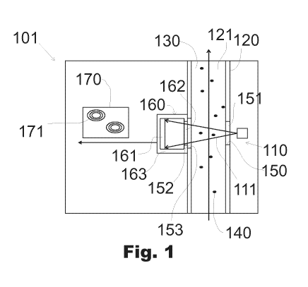
The measurement arrangement (101) of FIG. 1 is
suitable, and may be used, for in-line holography
microscopy.
5
"In-line holography microscopy" refers to
investigation methods and apparatuses by which a
microscopic object of foreign origin present in fluid
illuminated by coherent light may be detected and/or
determined on the basis of a hologram pattern formed
by interference of a portion of the light scattered
forward by such objects with non-scatted portion of
the light.
The expression "of foreign origin" refers to that the
microscopic objects are not formed of the fluid
itself. They may originate, for example, from the
materials of pipes or containers in which the fluid at
issue is conveyed or stored. Particles of the
materials of such systems may be released to the
fluid, for example, in result of a pipe breakage or
equipment failure. Alternatively, microscopic objects
of foreign origin may originate from foreign bodies or
contaminants ended up within such pipes or containers.
In the case of water supply systems, for example, such
foreign body producing microbes into the fluid may be
a dead animal.
In the case of water supply, distribution, or use
systems and networks, microbes not normally present
may be, for example, various bacteria, such as
bacteria belonging to coliform or Legionella groups,
protozoa such as Giardia lamblia, or various types of
algae.
On the other hand, from the physical properties point
of view, "microscopic objects of foreign origin" have
CA 03028095 2018-12-17
WO 2017/220861 PCT/F12017/050464
6
typically, for example, a refractive index differing
from that of the fluid. This enables detection of such
objects by means of optical sensing. In the measuring
arrangement of FIG. 1, this is utilized in that the
detection of the microscopic objects is based on
scattering of light by the microscopic objects due to
the difference between the refractive indices of the
microscopic objects and the fluid.
From dimensional point of view, "microscopic objects"
refer to objects having their characteristic
dimensions, such as maximum diameter, length, or
width, in the range of 0.1, 0.5 or 1.0 to 50 or 100
pm. Objects with so small characteristic dimensions
are not visible to human eye, so they cannot be
detected visually. On the other hand, holograms formed
by that sized objects are detectable by image sensor
having a reasonably small size. Further, with such
micrometer scale characteristic dimensions, objects
scatter light mainly forward, thereby enabling
efficient detection by in-line holography.
The measuring arrangement comprises an illuminating
arrangement 110 which emits, when in use, coherent
light 111. The light may be emitted, for example, as
short pulses.
Further, the measuring arrangement comprises a cuvette
120 which defines an inner volume 121 for receiving a
fluid 130 which may comprise microscopic objects 140
of foreign origin.
"Cuvette" refers to a structure of any appropriate
type suitable for defining an inner volume for
receiving a fluid to be measured and/or analyzed by
measurement system arranged in connection with the
cuvette. A cuvette may comprise one or more walls
CA 03028095 2018-12-17
WO 2017/220861 PCT/F12017/050464
7
defining the inner volume thereof. Defining the inner
volume means that the one or more walls limit or
surround a cross-section of the inner volume
throughout a perimeter thereof. In other words, the
one or more walls and/or some other appropriate
structure of the cuvette completely encircles the
entire inner volume at least at one cross-section
thereof, thereby preventing escaping of the fluid to
me measured from the inner volume in directions in the
plane of such cross-section.
A cuvette may be of sampling type, in which case a
discrete sample volume may be contained in such inner
volume. Alternatively, a cuvette may be of flow-
through type allowing the fluid to be measured or
analyzed to flow continuously through the cuvette
during the measurements. In some embodiments, a
cuvette may be configured to serve alternatively as a
sampling type cuvette or as a flow-through cuvette.
The cuvette comprises an entrance opening 150 closed
by an entrance window 151, so positioned relative to
the illuminating arrangement that when in use, the
cuvette receives the coherent light emitted by the
illuminating arrangement through the entrance window.
The cuvette has also an exit opening 153 closed by an
exit window 152 mounted to the cuvette wall and
forming a part of the cuvette, opposite to the
entrance window, through which the cuvette lets light
received into the cuvette, after propagating across
the cuvette, exit therefrom.
"Mounting" refers to attaching or fixing, releasably
or non-releasably, a component, element, or module to
another structure. Mounting may be made, for example,
by glue or any other appropriate type of adhesive.
CA 03028095 2018-12-17
WO 2017/220861 PCT/F12017/050464
8
When illuminating the fluid in the cuvette by the
coherent light, the possible microscopic objects
present in the fluid scatter part of the light
forward, and the scattered and non-scattered portions
of light interfere so that interference fringes are
formed behind the microscopic objects.
Emitting of and illuminating by "coherent light"
refers to at least part of the emitted light and the
light by which the sample volume is illuminated being
spatially and temporally sufficiently coherent so that
said interference is possible. Thus, emitting coherent
light and illuminating by coherent light does not
exclude the possibility of emitting also non-coherent
light nor illuminating the sample volume also by non-
coherent light. Thus, light emitted by the
illuminating arrangement, and light by which the
sample volume is illuminated may comprise coherent and
non-coherent light. In this sense, "coherent light"
refers to "at least partially coherent light".
"Behind" refers to the locations of the interference
fringes as observed from the direction of incidence of
the illuminating light, i.e. the coherent light by
which the fluid is illuminated. In other words, when
observed from the location of a light source producing
the coherent light, the interference fringes are
formed mainly behind the microscopic objects, i.e. at
the side of the microscopic objects opposite to the
side from which the coherent light is incident on the
microscopic objects.
The illuminating arrangement may comprise any
appropriate light source, such as a laser diode,
capable of producing coherent light. The light may
have wavelength(s), for example, in the range of 350
CA 03028095 2018-12-17
WO 2017/220861 PCT/F12017/050464
9
to 500 nm, without being limited to that range. The
illuminating arrangement may further comprise any
appropriate optical elements configured to guide the
emitted coherent light towards the cuvette to
illuminate the fluid received therein.
The measuring arrangement 101 of FIG. 1 further
comprises an image sensor 160 comprising a light
sensitive cell 161 and a transparent protective window
162, through which the light can enter the image
sensor, positioned in front of the light sensitive
cell. The light sensitive cell is enclosed in a
housing 163, a part of which the protective window
forms.
The illuminating arrangement and the image sensor are
positioned at opposite sides of the cuvette to form a
direct propagation path of light from the illuminating
arrangement to the image sensor via the cuvette. The
image sensor is positioned and configured to capture a
hologram digital image frame 170 by receiving the
light propagated across the cuvette and exiting it
through the exit window 152.
In other embodiments, indirect optical configurations
may be implemented where light is guided to the image
sensor, for example, via one or more mirrors.
"An image sensor" refers to a light detecting
component or element capable of capturing digital
image frames. An image sensor may comprise, for
example, a CMOS (Complementary Metal Oxide
Semiconductor) or CCD (Charge-Coupled Device) light
sensitive cell or any other appropriate type of a
light sensitive cell as an active, light detection
imaging element.
CA 03028095 2018-12-17
WO 2017/220861 PCT/F12017/050464
The image sensor may be, for example, a black and
white type sensor, a greyscale sensor, or a monochrome
type sensor. Suitable size of the active area and the
resolution of the light sensitive cell depend on the
5 overall configuration of the measuring arrangement. In
some embodiments, it may have, for example, a size of
5 x 5 mm2. In some embodiments, the active area of the
light sensitive cell may have, for example, 5 million
pixels.
A "digital image frame", or shortly a "frame", refers
to a data content captured via exposure of pixels or
some other light-sensing element(s) of a light
sensitive cell of an image sensor. A frame thus
generally comprises image data enabling composition of
a displayable digital image on the basis of that image
data. Image data of a digital image frame may
comprise, for example, information about light energy
received by pixels of an image sensor.
When capturing the hologram digital image frame 170,
the interference fringes formed by the scattered and
non-scattered light behind the microscopic objects
possibly present in the fluid form hologram patterns
171 with spatially alternating intensity formed by the
interference fringes on the light sensitive cell of
the image sensor. Those hologram patterns are then
contained in the image data of the captured hologram
digital image frame.
On the basis of such hologram patterns, the presence
of the microscopic objects of foreign origin in the
fluid may be detected. Further, also some properties,
such as the size and the shape thereof, may also be
determined.
CA 03028095 2018-12-17
WO 2017/220861 PCT/F12017/050464
11
The cuvette of the measuring arrangement of FIG. 1 is
of flow-through type, wherein continuous flow of the
fluid 130 to be analyzed may be led through the
cuvette along its longitudinal direction during the
analysis. In other embodiments, cuvette of other types
may be used, which are based on any appropriate type
of sample cell or container capable of receiving the
fluid to be analyzed. For example, a cuvette may be of
sampling type, wherein a discrete volume may be stored
in the cuvette for the analysis. Such cuvette may
comprise one or more inlet/outlet openings for filling
and emptying the cuvette by the fluid to be analyzed.
As stated above with reference to the example of FIG.
1, "flow-through" type of a cuvette refers to a
configuration of the cuvette allowing continuous flow
of a fluid through the cuvette while carrying out the
measurement of the fluid flowing through the cuvette.
In the measuring arrangement of FIG. 1, the
illuminating arrangement is directed crosswise
relative to the flowing direction of the fluid flowing
in the flow-through type cuvette. Thereby, the flow is
directed correctly relative to the illuminating
direction.
A cuvette and a measuring arrangement as a whole may
have any appropriate dimensions, taking into account
the application at issue. For example, in the
measuring arrangement of FIG. 1, the thickness of the
inner volume in the illuminating direction may be, for
example, in the range of 0.5 to 1 mm. The width of the
cuvette may be adjusted, for example, on the basis of
the size of the light sensitive cell of the image
sensor which may lie, for example, at a distance of
about 1 to 3 mm from the inner volume of the cuvette.
For example, the cuvette may have, in one or more
CA 03028095 2018-12-17
WO 2017/220861 PCT/F12017/050464
12
directions, a width of 4 to 8 mm. One pixel of the
light sensitive cell may have a width, for example, in
the range of 1.5 to 5 pm. For example, the width of a
rectangular pixel may be about 2 pm. The positioning
of the light source of the illuminating arrangement
may vary depending on, for example, on the light
source and the size of the light emitting surface
thereof. In an example, a laser diode as a light
emitting element of a light source may be positioned
at some tens of millimeters, for example about 40 mm,
from the inner volume of the cuvette.
In the measuring arrangement 101 of FIG. 1, the
protective window 162 of the image sensor and thereby
the image sensor is in direct contact with the exit
window 152, which in turn is mounted to the cuvette
and forms an integral part of it. Thereby, the image
sensor is mounted in direct contact with the cuvette.
Said direct contact may be formed by mounting of the
image sensor to the cuvette via the casing 163
enclosing the light sensitive cell. Alternatively, or
additionally, the protective window may be mounted to
the exit window by means of an adhesive, such as an
optically clear adhesive (OCA), which may be applied
between the protective window and the exit window.
Such adhesive may be selected not to substantially
affect the propagation of light through the stack of
the exit window and the protective window.
The image sensor being in direct contact with the
cuvette means, generally, that there is no freely
accessible space between the image sensor and the
inner volume defined by the cuvette. In the measuring
arrangement of FIG. 1, there is thus no such space
between the exit window of the cuvette and the
protective window of the image sensor. This may be
CA 03028095 2018-12-17
WO 2017/220861 PCT/F12017/050464
13
advantageous in that no contaminants can adhere in the
outer surfaces of the protective window and the exit
window, which contaminants might disturb sensing the
hologram patterns by the image capturing.
Second, the coherent light, as illustrated in the
example of FIG. 1, may be emitted or guided into an
expanding cone or beam. Alternatively, it may be
emitted or guided into a collimated beam. In the
former case, the interference fringes may be expanded
as function of the distance from the scattering
microscopic objects. Further, irrespective of whether
the illuminating light is emitted or guided into an
expanding or into a collimated light beam, the
interference fringes expand due to the scattering of
the light into various directions, depending on the
types of the microscopic objects and the wavelength of
the illuminating light. Consequently, the longer the
distance between a microscopic object and the image
sensor, the larger is the expanding cone or beam, and
also the hologram pattern formed on the image sensor.
To keep the required size of the light sensitive cell
reasonably small, it may be desirable to have the
light sensitive cell of the image sensor as close to
the inner volume of the cuvette as possible.
Generally, the image sensor being in direct contact
with the cuvette, thus the protective window being in
direct contact with the exit window of the cuvette in
the measuring arrangement of FIG. 1, serves for this
purpose.
Third, in the case of a flow-through type cuvette as
that of FIG. 1, the cuvette may be connected to an
external piping from which the fluid to be analyzed is
led to the cuvette as a continuous flow. In such case,
possible pressure variations in such piping may be
transmitted to the cuvette also. The protective window
CA 03028095 2018-12-17
WO 2017/220861 PCT/F12017/050464
14
in direct contact with the exit window of the cuvette
in the measuring arrangement in FIG. 1 may strengthen
the exit window, thereby preventing it from adverse
bending in response to possible pressure variations,
which bending might change the optical path between
the cuvette and the image sensor.
The measuring arrangement 201 of FIG. 2 differs from
that of FIG. 1 in that there is no separate exit
window in the cuvette. Instead, the cuvette wall has
an exit opening 253 into which the image sensor 260 is
inserted and via which the housing 263 of the image
sensor is mounted to the cuvette 220.
In the measuring arrangement of FIG. 2, the protective
window 262 of the image sensor thus forms, or serves
as, an exit window through which the light propagated
across the cuvette 220 may exit therefrom.
In the measuring arrangement 201 of FIG. 2, the
protective window 262 of the image sensor 260 is in
direct contact with the cuvette and the inner volume
221 of the cuvette and the fluid 230 present therein.
The measuring arrangement 301 of FIG. 3 differs from
that of FIG. 1 in that the image sensor 360 does not
comprise separate protective window. Instead, the exit
window 352 of the cuvette 320 forms, or serves as,
also as a protective window protecting the light
sensitive cell 361 and enclosing the casing 363 of the
image sensor. Thereby, the image sensor is mounted in
direct contact with the cuvette.
In other embodiments where an image sensor with no
separate protective window is mounted to a cuvette to
which an exit window is mounted, the image sensor may
be implemented without any casing. For example, the
CA 03028095 2018-12-17
WO 2017/220861 PCT/F12017/050464
light sensitive cell may be mounted on a substrate and
encapsulated by an encapsulating material via which
the image sensor may be mounted to the exit window.
5 Any of the measuring arrangements of FIGs. 1 to 3 may
be used in a complete detecting apparatus comprising,
in addition to the measuring arrangement, also a
computing arrangement configured to detect the
presence of the microscopic objects on the basis of
10 hologram patterns formed by the interference fringes
in the image data of the hologram digital image
frames.
Said detection may be based on reconstructing one or
15 more two-dimensional images of the illuminated fluid
volume in accordance with principles and processes as
such known in the field of holographic microscopy.
Alternatively, such detection may be carried out on
the basis of the hologram patterns present in the
image data of the captured hologram digital image
frame(s). The apparatus 400 of FIG. 4 represents one
embodiment of this type.
The apparatus 400 has a measuring arrangement 401
which may be in accordance with any of the measuring
arrangements discussed above with reference to FIGs. 1
to 3.
The measuring arrangement produces, when in use,
hologram digital image frames 470 which may comprise
hologram patterns 471 formed due to possible presence
of microscopic objects of foreign origin in the fluid
to be analyzed.
The measuring arrangement further comprises a
computing arrangement 460 connected to the measurement
CA 03028095 2018-12-17
WO 2017/220861 PCT/F12017/050464
16
arrangement 401 to receive image data of the captured
hologram digital image frames, and to detect the
presence of the microscopic objects on the basis of
the hologram patterns 471 formed by the interference
fringes in the image data of the hologram digital
image frames 470.
In the example of FIG. 4, the computing arrangement
may be configured to pre-process the received image
data of the hologram digital image frame by any
appropriate data processing operations facilitating
the detection of the microscopic objects on the basis
of the hologram patterns.
The computing arrangement is further configured to
provide filtered image data 480, comprising
automatically filtering, for example, the received and
possibly pre-processed image data by a symmetric edge
detection algorithm, at least in two different
directions, the filtered image data comprising, for
each hologram pattern 471 present in the received
image data, a filtered hologram pattern 481. In said
filtering, for example, any appropriate convolution
kernel may be used.
As known for a skilled person, there are a great
variety of known mathematical operations which may be
used to filter image data for edge detecting purposes.
Generally, the principle in edge detection is to find
out, by filtering image data by such edge detection
algorithms, where there are relatively abrupt changes
in the image content. For example, the parameter of
interest used to find "edges" may be the intensity of
light received by the image sensor during capture of
the frame, i.e. the brightness of the image formed by
the image data.
CA 03028095 2018-12-17
WO 2017/220861
PCT/F12017/050464
17
In result of filtering image data by an edge detecting
algorithm, the filtered image data generally
highlights the edges, i.e. contours of distinguishable
objects present in the original image data. In the
case of filtering the received image data 470 with the
hologram patterns 471, the filtered image data thus
comprises the contours of the original hologram
patterns in the form of the filtered hologram patterns
381.
On the basis of the filtered hologram patterns,
further analysis of the image data can be focused on,
or limited to, the actual locations of the holograms
in the image area. Great savings in the required
computing power may then be saved because the rest of
the image data does not need to be analyzed.
"Symmetry" of the edge detection algorithm refers to
edge detection algorithms designed not to
substantially change the shape of the objects in the
image area in result of the filtering.
Finally, the computing arrangement is configured to
automatically detect, on the basis of the filtered
hologram patterns, the presence of the microscopic
objects 440 associated with the filtered hologram
patterns in the sample volume of the fluid.
Thus, possible filtered hologram patterns of the
filtered image data are used as indication of the
presence of microscopic, scattering objects in the
fluid contained in the cuvette.
Detecting the presence of microscopic objects refers
to determining whether there are any microscopic
objects in the fluid. In this sense, detecting the
presence of such objects may also comprises
CA 03028095 2018-12-17
WO 2017/220861 PCT/F12017/050464
18
determining and concluding that there is no such
object present in the fluid volume through which the
illuminating light propagated to the image sensor. On
the other hand, when there is a plurality of filtered
hologram patterns in the filtered image data, said
detection may naturally comprise, in addition to
determine the general presence of the microscopic
objects, also the number of them in the analyzed fluid
volume.
The result of the detection operation, i.e. the
information about the presence of microscopic objects
in the analyzed fluid volume, may be arranged in any
appropriate electric data or signal form suitable for
storage or transmitting further.
The computing arrangement may comprise any appropriate
data processing and communicating equipment, unit(s),
element(s), and component(s) capable of carrying out
the operations of the method discussed above.
From another terminology point of view, a computing
arrangement "configured to" perform a specific method
operation means that the computing arrangement
comprises, or serves as, "means for" performing that
operation.
The computing arrangement may comprise separate means
for different operations. Alternatively, any of such
means for performing those various operations
specified above may be combined so that more than one
operation is carried out by the same means. It is even
possible that all those operations are carried out by
the same means, e.g. by a single data processing
module or unit.
CA 03028095 2018-12-17
WO 2017/220861 PCT/F12017/050464
19
Any means for performing any of the above operations
may comprise one or more computer or other computing
and/or data processing components, units, devices, or
apparatuses. In addition to actual computing and/or
data processing means, the means for performing said
operations may naturally also comprise any appropriate
data or signal communication and connecting means, as
well as memory or storage means for storing generated
and/or received data.
Computing and/or data processing means serving as
means for performing one or more of the above
operations may comprise, for example, at least one
memory and at least one processor coupled with the at
least one memory. Then, the at least one memory may
comprise computer-readable program code instructions
which, when executed by the at least one processor,
cause the apparatus to perform the operation(s) at
issue.
In addition to, or instead of, a combination of a
processor, a memory, and program code instructions
executable by the processor, means for performing one
or more operations may comprise some hardware logic
components, elements, or units, such as those examples
mentioned above with reference to the method aspect.
The apparatus 400 of FIG. 4 may be implemented as
stand-alone apparatus or sensor. Alternatively, it may
form a part of a larger controlling or monitoring
system.
It is to be noted that the present invention is not
limited to the embodiments and examples above.
Instead, the embodiments of the present invention can
freely vary within the scope of the claims.
CA 03028095 2018-12-17
WO 2017/220861 PCT/F12017/050464
It will be understood that the benefits and advantages
described above may relate to one embodiment or
example or may relate to several embodiments or
examples. The embodiments and examples are not limited
5 to those that solve any or all of the stated problems
or those that have any or all of the stated benefits
and advantages. It will further be understood that
reference to 'an' item refers to one or more of those
items.
The term "comprising" is used in this specification to
mean including the feature(s) or act(s) followed
thereafter, without excluding the presence of one or
more additional features or acts.