Note: Descriptions are shown in the official language in which they were submitted.
CA 03028632 2018-12-19
WO 2018/037346
PCT/IB2017/055067
PLANAR ILLUMINATOR FOR OPHTHALMIC SURGERY
FIELD
[0001] The present disclosure relates to ophthalmic illuminators.
More
particularly, the present disclosure relates to devices, systems, and methods
for
providing planar illumination during ophthalmic surgery.
BACKGROUND
[0002] In the
following discussion, certain articles and methods will be described
for background and introductory purposes. Nothing contained herein is to be
construed
as an "admission" of prior art. Applicant expressly reserves the right to
demonstrate,
where appropriate, that the articles and methods referenced herein do not
constitute prior
art under the applicable statutory provisions.
[0003]
Ophthalmic microsurgical procedures can require precision cutting and/or
removing of various body tissues of the patient's eye. For example, during a
surgical
procedure, a user, such as a surgeon or other medical professional, may hold
an
illumination apparatus in one hand and a vitrectomy probe in his or her other
hand. The
vitrectomy probe can be used to perform surgical maneuvers while the surgeon
visualizes the patient's eye using the light provided by the illumination
apparatus. The
illumination apparatus may include a cannula inserted into the eye and one or
more
optical fibers encompassed within the center cavity of the cannula. Because
illumination
apparatus typically transmit wide-angle light that illuminates a volume of
space within
the eye, details of anatomical structures of the eye may be obscured due to
contribution
from scattered light in front and behind of features of interest.
[0004]
Accordingly, there remains a need for improved devices, systems, and
methods that allow a surgeon to illuminate a patient's eye with a planar light
beam or
laser sheet that illuminates a planar slice or field of an anatomical feature
rather than a
volume of space.
SUMMARY
[0005] This
summary is provided to introduce a selection of concepts in a
simplified form that are further described below and in the attendant
drawings. This
summary is not intended to identify key or essential features of the claimed
subject
matter, nor is it intended to be used to limit the scope of the claimed
subject matter.
1
CA 03028632 2018-12-19
WO 2018/037346
PCT/IB2017/055067
Other features, details, utilities, and advantages of the claimed subject
matter will be
apparent from the following written detailed description, including those
aspects
illustrated in the accompanying drawings and defined in the appended claims.
[0006] The present disclosure addresses an unmet medical need by, among
other
things, uniquely outputting a planar light beam into a patient's eye during an
ophthalmic
surgical procedure such as, e.g., a vitrectomy. An illumination apparatus may
include
multiple optical fibers positioned within a cannula. The cannula is inserted
into the
patient's eye. The optical fibers can be sized and shaped to respectively
transmit light
having different illumination profiles. For example, one optical fiber may
transmit light
for wide-field volumetric illumination to provide general situational
awareness for a
surgeon during the surgical procedure. A second optical fiber device may
transmit light
for a planar field illumination. Planar field illumination may allow the
surgeon to better
visualize anatomy within the patient's eye, such as vitreous humor. For
example, during
vitrectomy, visualizing the vitreous and its interaction with the retina can
be difficult
since it is a naturally optically clear medium. An optical fiber device that
illuminates a
planar field in the eye may enhance visualization of the vitreous by isolating
light from a
single plane in the viewing path. With such an illumination apparatus, a
surgeon can
toggle between multiple illumination profiles¨i.e., volumetric illumination or
planar
field illumination¨depending the surgeon's visualization needs during the
surgical
procedure.
[0007] Thus, in some embodiments the present disclosure provides an
ophthalmic
illumination apparatus comprising a body sized and shaped for grasping by a
user; a
cannula coupled to the body and configured to be positioned within an eye of a
patient;
an optical fiber disposed within the cannula, where the optical fiber is
configured to
transmit light having a volumetric illumination profile; and an optical fiber
device
disposed within the cannula, wherein the optical fiber device is configured to
transmit
light having a planar illumination profile.
[0008] In some aspects of such these embodiments, the optical fiber
device
comprises one of an optical slit, a rod lens or a ball lens. In another
aspect, at least one
of the optical fiber or the optical fiber device is translatable with respect
to the cannula.
Further, aspects may additionally include an input device configured to
receive a user
input to cause one of the optical fiber or the optical fiber device to
selectively illuminate
the eye of the patient; a light source coupled to the optical fiber and the
optical fiber
device and configured to output light to selectively illuminate the eye of the
patient via
the optical fiber or the optical fiber device; an optical relay disposed
between a light
2
CA 03028632 2018-12-19
WO 2018/037346
PCT/IB2017/055067
source and the cannula, where the optical relay is configured to selectively
direct the
light output by the light source to one of the optical fiber or the optical
fiber device in
response to the user input; a third optical fiber disposed within the cannula
where the
third optical fiber is coupled to a therapeutic light source and configured to
transmit a
therapeutic light beam into the eye of the patient; an endoscopic fiber bundle
disposed
within the cannula and configured to visualize the eye the patient; and/or a
deflection
mechanism coupled to the cannula and configured to selectively bend the
cannula.
[0009] Other embodiments described in the disclosure provide an optical
fiber
device comprising an optical fiber device housing; an optical fiber comprising
a core and
cladding axially disposed within the optical fiber device housing; and one or
more of an
optical slit device, a rod lens, and a ball lens coupled to the optical fiber
device housing.
[00010] Some aspects of these embodiments comprise one or more of an
optical slit
device comprising an optical slit disposed within an optical end cap coupled
to a distal
end of the optical fiber device housing, a rod lens positioned perpendicularly
to the
optical fiber and coupled to a distal end of the optical fiber device housing,
and a ball
lens disposed within a distal end of the optical fiber device housing.
[000 111 Yet other embodiments described include methods for ophthalmic
surgical
illumination comprising illuminating an eye of a patient with light having a
volumetric
profile, where the light having the volumetric profile is transmitted by an
optical fiber
disposed within a cannula positioned within the eye; and illuminating the eye
of the
patient with light having a planar profile, where the light having the planar
profile is
transmitted by an optical fiber device disposed within the cannula.
[00012] Aspects of these embodiments may also include receiving user
input at an
input device to cause a light source coupled to an optical fiber and an
optical fiber device
to output light to one of the optical fiber or the optical fiber device.; an
optical relay
disposed between a light source and the cannula that selectively directs the
light output
by the light source to one of the optical fiber or the optical fiber device,
and/or receiving
a user input at an input device to cause one of the first light source coupled
to the optical
fiber or the second light source coupled to the optical fiber device to
selectively output
light to illuminate the eye of the patient.
[00013] These and other aspects and uses will be described in the
detailed
description.
3
CA 03028632 2018-12-19
WO 2018/037346
PCT/IB2017/055067
BRIEF DESCRIPTION OF THE FIGURES
[00014] Figure 1
is an illustration of an embodiment of an ophthalmic illumination
system.
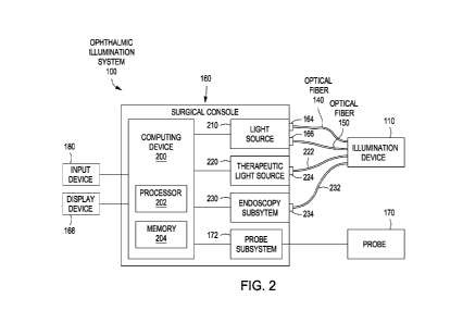
[00015] Figure 2
is a schematic diagram illustrating an embodiment of an
ophthalmic illumination system.
[00016] Figure 3
is a schematic diagram illustrating an embodiment of an
ophthalmic illumination system.
[00017] Figure 4
is a schematic diagram illustrating an embodiment of an
ophthalmic illumination system.
[00018] Figures
5A, 5B, and 5C are illustrations of various embodiments of an a
cannula of an illumination apparatus comprising both an optical fiber that
illuminates a
volumetric field within the eye and an optical fiber device that illuminates a
planar field
within the eye.
[00019] Figures
6A and 6B are side views of an eye with the distal portion of a
cannula comprising optical fibers inserted into an eye. Figure 6A shows
illumination
from a prior art optical fiber providing wide-field volumetric illumination.
Figure 6B
shows illumination from an optical fiber device as described herein that emits
a planar
light beam. Figure 6C illustrates an exemplary optical fiber device that
provides a planar
light beam using an optical slit. Figures 6D and 6E illustrate an exemplary
optical fiber
device that provides a planar light beam using a rod lens. Figure 6F
illustrates an
exemplary optical fiber device that provides a planar light beam using a
combination of
an optical slit and a ball lens.
[00020] Figure 7
is an illustration of an embodiment of a cross-sectional
longitudinal view of an illumination apparatus.
[00021] Figure 8
is an embodiment of a cross-sectional end view illustration of a
cannula of an illumination apparatus.
[00022] In the
drawings, elements having the same designation have the same or
similar functions. Those skilled in the art will appreciate that Figures 1-8
are not
necessarily to scale, and that several of the features may be exaggerated to
more clearly
illustrate various features. Those skilled in the art will also appreciate
that the illustrated
structures are exemplary only, and not limiting.
4
CA 03028632 2018-12-19
WO 2018/037346
PCT/IB2017/055067
DETAILED DESCRIPTION
[00023] Before the present optical fiber devices capable of illuminating
a planar
field within the eye and systems incorporating such optical fiber devices are
described, it
is to be understood that this disclosure is not limited to the specific
embodiments
described, as such may vary. It is also to be understood that the terminology
used herein
is for the purpose of describing particular aspects only and is not intended
to limit the
scope of the present disclosure.
[00024] Note that as used in the present specification and in the
appended claims,
the singular forms "a," "an," and "the" include plural referents unless the
context clearly
dictates otherwise.
[00025] Unless defined otherwise, all technical and scientific terms used
herein
have the same meaning as commonly understood by one of ordinary skill in the
art to
which this disclosure belongs. Any publications mentioned herein are
incorporated
herein by reference for the purpose of describing and disclosing devices and
methodologies that are described in the reference and which might be used in
connection
with this disclosure.
[00026] Where a range of values is provided, it is understood that each
intervening
value between the upper and lower limit of that range and any other stated or
intervening
value in that stated range is included as an embodiment of the disclosure. The
upper and
lower limits of these smaller ranges are also included as an embodiment of the
disclosure, subject to any specifically excluded limit in the stated range.
Where the
stated range includes both the upper and lower limits, ranges excluding either
of those
included limits are also included as an embodiment of the disclosure.
[00027] In the following description, numerous specific details are set
forth to
provide a more thorough understanding of the present disclosure. However, it
will be
apparent to one of skill in the art upon reading the specification that the
present
disclosure may be practiced without one or more of these specific details. In
other
instances, features and procedures to well-known to those skilled in the art
have not been
described in order to avoid obscuring the disclosure.
[00028] The present disclosure describes devices, systems, and methods of
selectively illuminating a planar field in a patient's eye. In certain
embodiments, two or
more optical fibers, optical fiber devices, or combinations thereof can be
positioned
within a cannula of an ophthalmic illumination apparatus. The cannula can be
inserted
into the patient's eye. The optical fibers can be differently sized, shaped,
and/or
CA 03028632 2018-12-19
WO 2018/037346
PCT/IB2017/055067
configured with lenses, optical slits, or other structures such that they emit
light having
different field illumination profiles. The surgeon can choose which optical
fiber or
optical fiber device emits light during the surgical procedure depending on
the desired
field illumination; that is, the surgeon can select a, e.g., wide field
illumination of a
volume within the eye, or a focused illumination of a specific plane within
the eye.
Further, the cannula in some embodiments can be deflected such that a desired
area,
such as the periphery of the eye, can be illuminated. In addition, in some
embodiments
an optical fiber transmitting a therapeutic laser beam and and/or endoscopy
fiber bundle
can also be positioned within the cannula of the illumination apparatus.
[00029] The devices, systems, and methods of the present disclosure
provide
numerous advantages, including: (1) increased control of intra-operative
illumination for
the surgeon; (2) improved operating conditions for the surgeon with the
ability to adjust
retinal glare; (3) decreased risk of photo-toxicity for the patient; (4)
enhanced
visualization of anatomy, such as the vitreous humor, for the surgeon using
planar field
illumination while preserving situational awareness for the surgeon using,
e.g., wide-
angle volumetric illumination; (5) increased illumination area within the
patient's eye
with cannula deflection; and (6) improved working conditions for the surgeon
with
incorporation of multiple fibers for illumination, treatment, and/or endoscopy
into a
single apparatus.
[00030] Figures 1, 2, 3, and 4 illustrate exemplary ophthalmic
illumination systems
100. Figure 1 is an exemplary illustration of an ophthalmic illumination
system 100.
Figures 2, 3, and 4 are various exemplary embodiments of schematic diagrams of
the
ophthalmic illumination system 100. The ophthalmic illumination system 100 can
include an illumination apparatus 110 having a body 120 and a cannula 130.
Body 120
can be sized and shaped for grasping by a user, and cannula 130 is coupled to
the body,
either directly or indirectly. Cannula 130 is configured to be positioned
within a surgical
field, such as a patient's eye. Illumination apparatus 110 in this embodiment
includes
optical fiber 140 and optical fiber device 150 disposed within cannula 130.
Optical fiber
140 can be configured to transmit light 142 having a volumetric field profile,
and optical
fiber device 150 can be configured to transmit light 152 having a planar field
profile.
Optical fiber 140 and optical fiber device 150 thus are configured to
selectively
illuminate different fields within the patient's eye.
[00031] The ophthalmic illumination system 100 can be used to perform
various
ophthalmic surgical procedures including an anterior segment procedure, a
posterior
segment procedure, a vitreoretinal procedure, a vitrectomy procedure, a
cataract
6
CA 03028632 2018-12-19
WO 2018/037346
PCT/IB2017/055067
procedure, and/or other desired procedures. The user, such as a surgeon or
other medical
professional, operates the illumination apparatus 110 to illuminate the
surgical field.
The surgical field may include any suitable physiology of the patient's eye,
including an
anterior segment, a posterior segment, a cornea, a lens, a vitreous chamber,
transparent
membranes, blood vessels, a retina, a macula, a foveola, a fovea centraalis, a
para fovea,
a perifovea, an optic disc, an optic cup, and/or other biological tissue.
[00032] Referring to Figures 1 and 7, body 120 of the illumination
apparatus 110
can form a handle for the illumination apparatus 110. Figure 7 represents an
exemplary
longitudinal cross-sectional, side-view illustration of the illumination
apparatus 110.
Body 120 can be sized and shaped for handheld use and/or grasping by the user.
For
example, body 120 can be any suitable shape, including ellipsoidal, polygonal,
tubular,
other desired shapes, and/or combinations thereof. Body 120 can be made of any
suitable material, such as a thermoplastic or metal, and can be formed by any
method,
including, for example, injection molding or machining. Further, in some
embodiments,
at least a portion of body 120 may be knurled, patterned, and/or otherwise
textured to
improve gripping. Body 120 may be formed of two or more sections joined
together,
and can include one, two, three, or more controls 810, 812. Controls 810, 812
can be
buttons, sliders, toggles, wheels, other suitable actuatable components,
and/or
combinations thereof, and are used to control various functions of the
illumination
apparatus 110 as described herein. In that regard, controls 810, 812 can be an
input
device 180 as further described herein.
[00033] Referring to Figures 1, 5A, 5B, 5C, 7, and 8, cannula 130 of the
illumination apparatus 110 can extend from body 120. Figures 5A, 5B, and 5C
represent
exemplary cross-sectional, side-view illustrations of cannula 130. Figure 8 is
an
exemplary cross-sectional, end view of cannula 130 taken along section line 9-
9 of
Figure 7. Cannula 130 can include a lumen 132, a distal portion 136, and a
proximal
portion 137. In this embodiment, cannula 130 is directly coupled to body 120
at
proximal portion 137. Cannula 130, including distal portion 136, can be sized
and
shaped for insertion into an interior space of the eye, such as the vitreous
chamber.
Cannula 130 can be any suitable material, including medical grade tubing; a
metal, such
as titanium, stainless steel; or a suitable polymer. Cannula 130 can be any
desired size,
including 16-27 Gauge tubing, and/or other suitable sizes, both larger and
smaller.
Cannula 130 can have an internal diameter 134 and a length 135. The internal
diameter
134 can be between approximately 400 microns and approximately 600 microns,
between approximately 400 microns and approximately 550 microns, between
7
CA 03028632 2018-12-19
WO 2018/037346
PCT/IB2017/055067
approximately 400 microns and approximately 500 microns, and/or other suitable
sizes,
both larger and smaller. The length 135 of cannula 130 can be between
approximately
20 mm and approximately 50 mm, between approximately 20 mm and approximately
40
mm and/or other suitable sizes, both larger and smaller. Cannula 130 can have
a cross-
section shaped as a polygon, an ellipse, other suitable shape, and/or a
combination
thereof For example, cannula 130 can be cylindrically-shaped so as to have a
circular
cross-section.
[00034] Any of the illumination apparatus 110, body 120, and/or cannula
130 can
be disposable or configured for a single use. Alternatively, any of the
illumination
apparatus 110, body 120, and/or cannula 130 can be sterilizable and configured
for
multiple uses. For example, illumination apparatus 110, body 120, and/or
cannula 130,
can be autoclavable and/or otherwise sterilizable.
[00035] Two or more optical fibers or optical fiber devices can be
disposed within
lumen 132 of the cannula 130. Although the exemplary embodiments in the
Figures
illustrate one optical fiber and one optical fiber device disposed within the
cannula, any
suitable number of optical fibers and optical fiber devices, including three,
four, or more
may be implemented in an illumination device. Optical fiber 140 and optical
fiber
device 150 may include a core, a cladding, and a coating, and/or other
layer(s). The core
of optical fibers can be a cylinder of glass, plastic, silica, and/or other
suitable material
through which light propagates. Cladding can surround the core and confine the
light
within the core. The cladding can include a dielectric material with an index
of
refraction less than the index of refraction of the core. A coating can
surround the
cladding and protect the optical fiber from physical damage. As illustrated in
Figure 8,
the optical fiber 140 can have a diameter 148, and the optical fiber device
150 can have a
diameter 158. The diameter 148 and/or the diameter 158 can be between
approximately
25 microns and approximately 300 microns, between approximately 35 microns and
200
microns, between approximately 50 microns and approximately 100 microns,
including
values such as 30 microns, 40 microns, 45 microns, 75 microns, and/or other
suitable
values, both larger and smaller.
[00036] As illustrated in Figures 1, 5A, 5B and 5C, optical fiber 140 can
emit light
142 into the surgical field, and optical fiber device 150 can emit light 152
into the
surgical field. For example, input device 180 (seen in Figures 2-4) receives
user input to
cause optical fiber 140 and/or optical fiber device 150 to selectively
illuminate the eye of
the patient. The field illumination profiles of light 142, 152 are different,
providing
enhanced visualization to a user. For example, light 142 can provide wide-
field,
8
CA 03028632 2018-12-19
WO 2018/037346
PCT/IB2017/055067
volumetric illumination. Wide-field illumination can facilitate the user's
situational
awareness within the surgical field while performing various surgical
maneuvers. Light
152, on the other hand, provides planar field illumination. Planar field
illumination may
isolate the scattered light from a single plane in a viewing path. Such planar
field
illumination can allow the user to see anatomy within the eye that may not be
clearly
visible with wide-field volumetric illumination. For example, the vitreous
humor, the
clear jelly that fills the posterior segment of the patient's eye, can be more
clearly
visualized using planar field illumination. The user can selectively utilize
planar field
illumination to view the vitreous humor, for example, during vitrectomy
procedure. By
controlling which of optical fiber 140 or optical fiber device 150 transmits
light, the user
can switch between wide-field volumetric illumination and planar field
illumination
based on the surgical tasks being performed.
[00037] Figures 6A and 6B show a side view of an eye 600 with the distal
portion
of a cannula providing light from optical fibers inserted into an eye. Both
Figures
illustrate incision 650, through which cannula (606 in Figure 6A and 607 in
Figure 6B)
is inserted, retina 652, retinal blood vessels 658, vitreous body 656, cornea
654, and iris
659. Figure 6A shows illumination from a prior art cannula 606 having an
optical fiber
providing wide-field volumetric illumination 610. Figure 6B shows illumination
from
cannula 607 having an optical fiber device as described herein that can emit a
planar
light beam 611.
[00038] Figure 6C illustrates two views of an exemplary optical slit
device 660
comprising an optical fiber device that provides a planar light beam using an
optical slit
664. Optical slit device 660 comprises an optical slit 664 situated within an
end cap 662.
The side view of the optical slit device 660 of Figure 6C shows optical fiber
686
comprising a core 668 encased in cladding 666. A diameter of core 668 of
optical fiber
686 can be between approximately 5 microns and 125 microns, or between
approximately 10 microns and 100 microns, or between approximately 20 microns
and
75 microns, and/or other suitable sizes, both larger and smaller. Core 668 of
optical
fiber 686 can be made from a glass or plastic fiber or other suitable material
through
which light propagates. Cladding 666 typically includes a dielectric material
with an
index of refraction less than the index of refraction of the core. Thickness
of cladding
666 surrounding core 668 can be between approximately 50 microns and 200
microns,
or between approximately 75 microns and 150 microns, or between approximately
75
microns and 100 microns, and/or other suitable sizes, both larger and smaller.
The
optical slit device 660 can be fixedly coupled to optical fiber 686 via an
anionic bond.
9
CA 03028632 2018-12-19
WO 2018/037346
PCT/IB2017/055067
Optical slit 664 can have an x-dimension (length) that typically is less than
the diameter
of core 668 of optical fiber 686; that is the x-dimension of optical slit 664
can be
between approximately 4 microns and 124 microns, or between approximately 10
microns and 100 microns, or between approximately 20 microns and 75 microns,
and/or
other suitable sizes, both larger and smaller. A y-dimension (height) of
optical slit 664
can be between approximately 5 microns and 100 microns, or between
approximately 10
microns and 75 microns, or between approximately 20 microns and 50 microns,
and/or
other suitable sizes, both larger and smaller. End cap 662 can be made from,
e.g., etched
silicon, sputtered gold, vapor deposited platinum, laser structured glass,
glass with a
dielectric reflective layer, or any other suitable material.
[00039] Figures 6D and 6E illustrate an exemplary optical fiber device
that
provides a planar light beam using a rod lens. Figure 6D is a side view of an
optical lens
device 688 comprising an optical fiber 686 that provides a planar light beam
using a rod
lens 670. Optical fiber 686 is axially disposed and retained within optical
lens device
688 by optical lens device housing 684, which may fully circumferentially
encompass
optical fiber 686. As described with reference to Figure 6C, optical fiber 686
may
comprise a core 668 encased in cladding 666. A diameter of optical core 668 of
optical
fiber device 150 in this embodiment can be between approximately 5 microns and
75
microns, or between approximately 10 microns and 65 microns, or between
approximately 20 microns and 50 microns, and/or other suitable sizes, both
larger and
smaller. Thickness of cladding 666 surrounding core 668 can be between
approximately
50 microns and 200 microns, or between approximately 725 microns and 150
microns,
or between approximately 750 microns and 100 microns, and/or other suitable
sizes,
both larger and smaller. In this exemplary embodiment, core 668 is made from a
glass
fiber such as SMF-28 Ultra Optical Fiber (Corning, Inc.) or other suitable
material
through which light propagates. In addition to optical fiber 686 and optical
lens device
housing 684, optical lens device 688 comprises rod lens 670, perpendicularly
disposed
of a distal end of optic fiber 150. Rod lens 670 has a rod shape, with rod
lens ends 672,
where rod lens 670 has a diameter that can be between approximately 75 microns
and
150 microns, or between approximately 100 microns and 125 microns, and/or
other
suitable sizes, both larger and smaller. Rod lens 670 may be a sapphire lens
or any other
biocompatible, transparent material with an index of refraction considerably
larger than
water, such as, e.g., polycarbonate, various types of glass, or cubic
zirconia, and is
disposed and may be retained within optical lens device housing 684 using a
suitable
CA 03028632 2018-12-19
WO 2018/037346
PCT/IB2017/055067
adhesive such as, e.g., two part epoxy or light-curable epoxy. Additionally
seen in
Figure 6D is light path 674 comprising a planar light beam shaped by the rod
lens.
[00040] Figure 6E is an exemplary perspective view of the optical lens
device 688
seen in Figure 6D. Figure 6E illustrates optical fiber 686 axially disposed
within optical
lens device housing 684. Optical lens device housing 684 comprises two notches
678
into which rod lens 670 is disposed. Optical lens device 688 may be formed, in
some
embodiments, by constructing the optical lens device housing 684 and optical
fiber 686
combination, creating notches in a distal end of the optical lens device
housing 684,
filling the distal end of the optical lens device housing 684 with adhesive,
and
positioning the rod lens within the notches. Ends 672 of rod lens 670 can then
be
grinded or otherwise trimmed to be flush with the outer surface of optical
lens device
housing 684.
[00041] Figure 6F illustrates an exemplary optical fiber device 690 that
provides a
planar light beam using a combination of an optical slit and a ball lens.
Optical fiber
device 690 in this embodiment comprises optical lens device housing 684,
comprising
optical fiber 686 having core 668 surrounded by cladding 666, ball lens 680
and optical
sit device 660. As in previously described exemplary embodiments, the diameter
of
optical core 668 of optical fiber 686 can be between approximately 5 microns
and 125
microns, or between approximately 10 microns and 100 microns, or between
approximately 20 microns and 75 microns, and/or other suitable sizes, both
larger and
smaller. Core 668 of optical fiber 686 can be made from a glass or plastic
fiber or other
suitable material. Thickness of cladding 666 surrounding core 668 can be
between
approximately 50 microns and 200 microns, or between approximately 75 microns
and
150 microns, or between approximately 75 microns and 100 microns, and/or other
suitable sizes, both larger and smaller. The optical slit device 660 can be
fixedly
coupled to optical fiber 686 via an anionic bond. Optical slit 664 can have an
x-
dimension (length) that typically is less than the diameter of core 668 of
optical fiber
686; that is the x-dimension of optical slit 664 can be between approximately
4 microns
and 124 microns, or between approximately 10 microns and 100 microns, or
between
approximately 20 microns and 75 microns, and/or other suitable sizes, both
larger and
smaller. A y-dimension (height) of optical slit 664 can be between
approximately 5
microns and 100 microns, or between approximately 10 microns and 75 microns,
or
between approximately 20 microns and 50 microns, and/or other suitable sizes,
both
larger and smaller. Ball lens 680 may be a sapphire lens comprising a
spherical shape,
and can be between approximately 100 microns and 500 microns, or between
11
CA 03028632 2018-12-19
WO 2018/037346
PCT/IB2017/055067
approximately 150 microns and 450 microns, or between approximately 200
microns
and 350 microns in diameter, and/or other suitable sizes, both larger and
smaller. The
ball lens 680 in this exemplary embodiment disperses light from optical fiber
686, where
the light is then focused by the optical slit 664 into a plane. In addition to
the rod and
ball lenses exemplified herein, other lenses, singly or in combination, may be
used in the
optical fiber devices to provide light with a planar illumination profile.
[00042] Referring again to Figures 5A 5B, 5C, 7 and 8, optical fiber 140
and optical
fiber device 150 can be coupled to the cannula 130. As illustrated in Figure
8, for
example, optical fiber 140 and optical fiber device 150 are coupled an inner
wall 131 of
cannula 130. Optical fiber 140 and/or optical fiber device 150 can be fixedly
coupled
such that optical fiber 140 and optical fiber device 150 do not move with
respect to
cannula 130. Any suitable coupling, including an adhesive, a mechanical
structure,
and/or combinations thereof, can be implemented. As seen in Figures 5A and 5B,
distal
portion 146 of optical fiber 140 and distal portion 156 of optical fiber
device 150 can be
aligned with the distal portion 136 of cannula 130. For example, distal end
147 of
optical fiber 140 and/or distal end 157 of optical fiber device 150 can be
laterally aligned
with distal end 137 of cannula 130. In that regard, the distal ends 137, 147,
157 can be
coplanar. Optical fiber 140 and optical fiber device 150 can be positioned
relative to
cannula 130 such that cannula 130 impedes none of the light 142, 152.
[00043] Alternatively and referring to Figure 5C, optical fiber 140
and/or optical
fiber device 150 can be movably coupled to cannula 130. For example, optical
fiber 140
and optical fiber device 150 can be translatable with respect to cannula 130.
Any
suitable coupling, such as a mechanical structure, can be implemented. Optical
fiber 140
and optical fiber device 150 may be configured to selectively move laterally
with respect
to cannula 130 in directions 502, 504. For example, the user can provide input
at the
input device 180 (see Figures 2-4), such as the controls 810 or 812 (see
Figure 7), a
surgical footswitch (not shown), or controls integrated in a surgical console
160 (see
Figure 1). Input device 180 is in communication with illumination device 110
such that
optical fiber 140 and optical fiber device 150 are translated laterally in the
directions
502, 504 in response to the user input. Thus, distal ends 147, 157 of optical
fiber 140
and optical fiber device 150, respectively, can be positioned proximal of,
distal of, or
aligned with distal end 137 of cannula 130. The user can control the angular
divergence
of light 142 or the plane of illumination of light 152 by selectively
translating optical
fiber 140 and optical fiber device 150. For example, as illustrated, optical
fiber device
150 can be translated laterally in direction 502, where distal end 137 of
cannula 130 is
12
CA 03028632 2018-12-19
WO 2018/037346
PCT/IB2017/055067
thus positioned distally beyond distal end 157 of optical fiber device 150. As
a result,
light 152 can be positioned to focus light on different planar fields (e.g.,
152a, 152b, or
152c) than if distal end 157 of optical fiber device 150 and distal end 137 of
cannula 130
were aligned.
[00044] Optical fiber 140 and optical fiber device 150 thus are coupled
to one or
more light sources configured to output light to illuminate the surgical
field. Referring
to Figure 2, optical fiber 140 and optical fiber device 150 can be coupled to
a light
source 210. The light source 210 can include ports 164, 166. Light output by
light
source 210 can be selectively directed to port 164 and/or port 166 via, e.g.,
a beam
director. For example, the user can select via, e.g., input device 180, which
of optical
fiber 140 or optical fiber device 150 transmits light to the surgical field
based on which
of ports 164, 166 light source 210 provides light to. As an alternative and
referring to
Figure 3, optical fiber device 150 can be coupled to light source 210, and
optical fiber
140 can be coupled to light source 240. Light source 240 can direct light to a
port 244,
and light source 210 can direct light to the port 166. In such an embodiment
the user
may select which of optical fiber 140 and/or optical fiber device 150
transmits light to
the surgical field based on user input received by input device 180 selecting
which of
light sources 210, 240 outputs light.
[00045] Light source 210 and/or light source 240 can include a laser
source, such as
a supercontinuum laser source, an incandescent light bulb, a halogen light
bulb, a metal
halide light bulb, a xenon light bulb, a mercury vapor light bulb, a light
emitting diode
(LED), other suitable sources, and/or combinations thereof For example, light
sources
210, 240 as described herein can be configured to output bright, broadband,
and/or white
light to the surgical field. Light sources 210, 240 can be configured to
output any
suitable wavelength(s) of light, such as a visible light, infrared light,
ultraviolet (UV)
light, etc. Light sources 210, 240 can be in communication with optics, such
as lenses,
mirrors, filters, and/or gratings (such as an optical slit), configured to
vary the focus or
wavelength of light.
[00046] Referring to Figures 4 and 7, the ophthalmic illumination system
100 can
include an optical relay 400. Optical relay 400 can be positioned between
light source
210 and illumination apparatus 110. A single optical fiber 402 can be coupled
to light
source 210 and optical relay 400 and extend between light source 210 and
optical relay
400, transmitting light from light source 210 to optical relay 400. Optical
fiber 140 and
optical fiber device 150 can be coupled to the optical relay 400, where
optical relay 400
can be configured to direct the light transmitted by optical fiber 402 to
optical fiber 140
13
CA 03028632 2018-12-19
WO 2018/037346
PCT/IB2017/055067
or optical fiber device 150. For example, the user can provide input at input
device 180
such as controls 810 or 812 of the illumination apparatus 110, the surgical
footswitch
(not shown), or the controls integrated in the surgical console 160. The input
device 180
thus would be in communication with optical relay 400, such as via computing
device
200. Optical relay 400 can include a switch, a butt coupler, any suitable
combination of
optics, such as lenses, such as a gradient index (GRIN) lens, rod or ball
lens, mirrors,
optical slits, filters, and/or gratings, other suitable components, and/or
combinations
thereof For example, the switch of optical relay 400 can be configured to
selectively
direct light to optical fiber 140 and/or optical fiber device 150 in response
to user input.
Implementing optical fiber 402 and optical relay 400 can advantageously focus
light on
different planes within the eye by optical fiber device 150.
[00047] Optical relay 400 can be positioned at any location between light
source
210 and illumination device 100, including within optical fiber 402 and
cannula 130 of
illumination apparatus 110. As illustrated in Figure 7, optical relay 400 can
be
positioned within body 120 of illumination apparatus 110. Optical fiber 402
can extend
between light source 210 and body 120, and optical fiber 140 and optical fiber
device
150 can extend between body 120 and cannula 130.
[00048] Referring to Figures 7 and 8, illumination apparatus 110 can
include a
deflection mechanism 800 configured to selectively bend, angle, bow, curve,
and/or
otherwise cause cannula 130 to obtain a non-linear shape. For example, the
cannula 130
can be articulated or otherwise made of up two or more individual components.
The
multiple, individual components of the cannula can allow for the cannula to be
at least
temporarily deflected such that the light output by optical fiber 140 and
optical fiber
device 150 can be directed to any desired anatomy within the surgical field,
including
anatomy not positioned in front of distal portion 136 of cannula 130. For
example,
cannula 130 can be selectively deflected to illuminate the periphery of the
patient's eye.
The distal portion 136, the proximal portion 137, and/or any portion of
cannula 130
between the distal and proximal portions 136, 137 can be articulated and/or
deflected.
The deflection mechanism 800 can be coupled to one, two, three, four, or more
pull
wires 802 disposed within cannula 130. Deflection mechanism 800 can include
any
suitable components configured to actuate the one or more pull wires 802 to
selectively
deflect cannula 130. Cannula 130 can be biased to return to a linear
configuration when
pull wires 802 no longer act on cannula 130. Deflection mechanism 800 can be
coupled
to and/or disposed within body 120. The user can control deflection mechanism
800,
14
CA 03028632 2018-12-19
WO 2018/037346
PCT/IB2017/055067
including the direction and extent of the deflection of cannula 130, using the
controls
810, 812 of body 120.
[00049] Referring to Figures 2, 3, 4, 7, and 8, the ophthalmic
illumination system
100 can include a therapeutic light source 220. Therapeutic light source 220
can be part
of a therapeutic beam delivery system, such as a laser beam delivery system, a
photocoagulation system, a photodynamic therapy system, a retinal laser
treatment
system, or other appropriate system. An optical fiber 222 can be coupled to a
port 224
of therapeutic light source 220. Optical fiber 222 can transmit the
therapeutic beam to
the surgical field as directed by the surgeon.
[00050] The ophthalmic illumination system 100 can also in some
embodiments
include an endoscopy subsystem 230. The endoscopy subsystem 230 can be
configured
to image the surgical field. For example, a user can visualize the surgical
field during
the surgical procedure using a surgical microscope. The endoscopy subsystem
230 can
be used to visualize the area of the eye being operated on when the user
cannot view that
area through the lens with the surgical microscope. For example, the lens may
be cloudy
or the optical path of the surgical microscope may be blocked. The user can
also use the
endoscopy subsystem 230 to see the periphery of the eye, which may be not
visible with
the surgical microscope. An endoscopic fiber bundle 232 can be coupled to the
endoscopy subsystem 230 at a port 234. The endoscopic fiber bundle 232 can
include
multiple individual fibers 236. The endoscopic fiber bundle 232 can receive
and
transmit light reflected from the surgical field, and can generate images
based on the
received light. The images can be output to a display device 168 in
communication with
the endoscopy subsystem 230.
[00051] Optical fiber 222 associated with the therapeutic light source
220 and the
endoscopic fiber bundle 232 associated with the endoscopy subsystem 230 can be
coupled to the illumination device 110. For example, optical fiber 222 and
endoscopic
fiber bundle 232 can be coupled to and disposed within cannula 130. Any
suitable fixed
or movable coupling, including an adhesive, a mechanical structure, and/or
combinations
thereof, can be implemented. The diameter 148 of optical fiber 140, the
diameter 158 of
optical fiber device 150, the diameter of the optical fiber 222, and the
diameter of the
endoscopic fiber bundle 232 can allow for multiple optical fibers to be
positioned within
the diameter 134 of cannula 130. Implementing multiple optical fibers within
the single
illumination device 110 and cannula 130 can advantageously decrease the number
of
components the user interacts with and that enter the eye during the surgical
procedure.
As illustrated in Figure 7, a conduit 820, including include optical fiber
402, optical fiber
CA 03028632 2018-12-19
WO 2018/037346
PCT/IB2017/055067
222, and endoscopic fiber bundle 232, can extend between the illumination
device 110
and a surgical console 160 (not shown in Figure 7). Conduit 820 can also
include optical
fiber 140 and optical fiber device 150. The user can control delivery of the
therapeutic
light source 220 and/or the endoscopy subsystem 230 using input device 180,
such as
controls 810 or 812 of illumination apparatus 110, the surgical footswitch
(not shown),
and/or the controls integrated in the surgical console 160.
[00052] Referring to Figures 1, 2, 3, and 4, light source 210, light
source 240,
therapeutic light source 220, endoscopy subsystem 230, a probe subsystem 172,
and a
computing device 200 can be integrated into surgical console 160. The surgeon
can
utilize the surgical console 160 to control one or more parameters associated
with the
ophthalmic surgical procedure. One or more components of the surgical console
110
can be coupled to and/or disposed within a base housing 162 illustrated in
Figure 1.
Base housing 162 can be mobile such that it can be positioned proximate to the
patient
during the ophthalmic surgical procedure. Base housing 162 can include
pneumatic,
optical, fluid, and/or electrical supply lines facilitating communication
between
components of the ophthalmic illumination system 100.
[00053] Computing device 200 can be in communication with input device
180,
light source 210, light source 240, therapeutic light source 220, endoscopy
subsystem
230, probe subsystem 172, and display device 168. Computing device 200 can be
configured transmit control signals to and/or receive input or status signals
from the
components of ophthalmic illumination system 100. For example, computing
device
200 can control activation and deactivation of light sources 210, 240,
transmission of
light to ports 164, 166, 244, 246, transmission of light by optical fiber 140
and optical
fiber device 150, as well as the focus, intensity, wavelength, and/or other
characteristics
of light output by light sources 210, 240. In that regard, light sources 210,
240 can be in
electrical communication with the computing device 200. Computing device 200
may
include a processing circuit having a processor 202 and a memory 204.
Processor 202
can execute computer instructions, such as those stored on the memory 204, to
control
various components of the ophthalmic illumination system 100. Processor 202
can be a
targeted device controller and/or a microprocessor. Memory 204, such as
semiconductor
memory, RAM, FRAM, or flash memory, can interface with processor 202. As such,
processor 202 can write to and read from memory 204, and perform other common
functions associated with managing memory 204. The processing circuit of
computing
device 202 can be an integrated circuit with power, input, and output pins
capable of
performing logic functions.
16
CA 03028632 2018-12-19
WO 2018/037346
PCT/IB2017/055067
[00054] Computing device 200 can output display data to the display
device 168 to
display data relating to system operation and performance during an ophthalmic
surgical
procedure. Display device 168 can also display images generated by the
endoscopy
subsystem 230. Display device 168 can be a standalone device, integrated into
surgical
console 160, and/or in communication with the surgical microscope. For
example, the
images generated by the endoscopy subsystem 230 can be provided to the user as
graphical overlay in a field of view of the surgical microscope.
[00055] Probe subsystem 172 also can be in electrical communication with
the
computing device 200. Probe subsystem 172 can include various components
facilitating operation of probe 170. The user can utilize probe 170 within the
surgical
field to perform one or more surgical maneuvers. For example, probe 170 can be
a
cutting probe, a vitrectomy probe, a phacoemulsification probe, a laser probe,
an
ablation probe, a vacuum probe, a flushing probe, scissors, forceps, an
aspiration device,
and/or other suitable surgical device. Probe 170 can be in mechanical,
electrical,
pneumatic, fluid, and/or other suitable communication with probe subsystem
172.
[00056] Input device 180 can be in communication with computing device
200.
Input device 180 can be configured to allow the user to control ophthalmic
illumination
system 100, including which of optical fiber 140 and optical fiber device 150
transmit
light to illuminate the surgical field, selectively moving optical fiber 140
and optical
fiber device 150, activating/deactivating light sources 210, 240, and/or other
features
described herein. Input device 180 can comprise any of a variety of ON/OFF
switches,
buttons, toggles, wheels, digital controls, touchscreen controls, or other
user interface
components. Input device 180 can be integrally disposed on the surgical
console 160
and/or the illumination apparatus 110. For example, input device 180 can be
one or
more controls 810, 820 of the illumination apparatus 110, or input device 162
can be a
distinct component, such as, by way of non-limiting example, a surgical
footswitch, a
remote control device, a touchscreen control device, and/or another computing
device.
Ophthalmic illumination system 100 can include multiple input devices 180.
Input
device 180 can generate and transmit input signals based on the received user
input,
where computing device 200 can receive and process the input signal. Computing
device 200 can then generate and transmit control signals to light source 210,
light
source 240, therapeutic light source 220, endoscopy subsystem 230, probe
subsystem
172, and display device 168.
[00057] Embodiments as described herein provide exemplary devices,
systems, and
methods of illuminating the surgical field using light with different
illumination field
17
CA 03028632 2018-12-19
WO 2018/037346
PCT/IB2017/055067
profiles, including a light that provides planar field illumination. Multiple
optical fibers
sized and shaped to output the light with the different illumination field
profiles can be
implemented in a single illumination device. The preceding merely illustrates
the
principles of the disclosure. It will be appreciated that those skilled in the
art will be
able to devise various arrangements which, although not explicitly described
or shown
herein, embody the principles of the disclosure and are included within its
spirit and
scope. Furthermore, all examples and conditional language recited herein are
principally
intended to aid the reader in understanding the principles of the disclosure
and the
concepts contributed by the inventors to furthering the art, and are to be
construed as
being without limitation to such specifically recited examples and conditions.
Moreover,
all statements herein reciting principles, aspects, and embodiments of the
disclosure as
well as specific examples thereof, are intended to encompass both structural
and
functional equivalents thereof. Additionally, it is intended that such
equivalents include
both currently known equivalents and equivalents developed in the future,
i.e., any
elements developed that perform the same function, regardless of structure.
The scope
of the present disclosure, therefore, is not intended to be limited to the
exemplary
embodiments shown and described herein. Rather, the scope and spirit of
present
disclosure is embodied by the appended claims. In the claims that follow,
unless the term
"means" is used, none of the features or elements recited therein should be
construed as
means-plus-function limitations pursuant to 35 U.S.C. 112, 6.
18