Note: Descriptions are shown in the official language in which they were submitted.
CA 03029129 2018-12-21
WO 2018/002887 PCT/IB2017/053942
1
METHOD AND SYSTEM FOR TREATING LESIONS
TECHNICAL FIELD
The present invention relates to the field of methods and systems for treating
blood vessel lesions, and more particularly to methods and systems for
treating lesions using
mechanical waves.
BACKGROUND
Cardiovascular disease remains a leading cause of death worldwide.
Atherosclerosis consists of plaque accumulation along the inner wall of
arteries. This can
reduce the size of the flow passage and this is known as stenosis. When the
blood vessel is
completely blocked this is known as an occlusion. The reduction in blood flow
due to lesions
such as stenosis or occlusion can impair or harm the tissues and organs
relying on this blood
flow. The blood flow through these lesions can be restored through procedures
known as
percutaneous transluminal angioplasty (PTA) techniques. In many cases the
lesion contains
hard calcified structures and dense fibrotic tissues that may be difficult to
treat using
traditional PTA techniques and apparatuses, and together with the vessel size
and tortuosity,
may be the cause of the potential complications. Some experts therefore
believe that new
devices and technologies in the field of PTA may improve the success rate and
reduce the
procedure time for restoring blood flow through calcified and/or fibrotic
lesions.
Over the years, various apparatus and methods have been developed and
proposed to help in blood vessel recanalization of calcified and/or fibrotic
lesions through
minimally invasive procedures. For example, devices have used a mechanical
impactor with
or without the use of a transmission member, a narrowband ultrasonic source
with a
transmission wire, and various other methods of energy deposition near the
lesion.
For procedures performed using a mechanical impactor, a projectile is
accelerated and impacts a proximal end of a transmission member or a distal
cap that is in
direct contact with the lesion. The projectile can be accelerated using a
pneumatic source, a
solenoid, a mechanical spring or other means. The mass of the projectile and
its speed at
CA 03029129 2018-12-21
WO 2018/002887 PCT/IB2017/053942
2
impact produce high stresses at the impact surface and therefore require
commensurate
maintenance. Also, this method may offer very limited control over the
parameters of the
mechanical pulse that is generated. Moreover, such devices may be noisy and
may lack
durability.
Another prior art example consists in a system comprising an ultrasonic wire
excited at resonance with a horn and an electromechanical transducer. This
constitutes a first
example associated with the use of a narrowband source. This arrangement is
used to amplify
the displacement at the distal end of the device in contact with the lesion.
The ultrasonic wire
is usually used inside a dedicated catheter with cooling fluid circulation to
prevent
overheating of the device. By doing so, the device becomes bulkier and thus is
limited in its
ability to reach lesions in small and tortuous anatomy. Considerable loss (due
to signal
distortion and nonlinearity) and/or mode conversion (from axial to transverse)
may also
occur at a bend when the device is activated. The frequency of operation
(typically around 20
kHz) may create large stress, strain and heat at the ultrasonic wire junction
with the horn and
within the ultrasonic wire itself. This may contribute to weaken the
ultrasonic wire resulting
in higher risk of failure.
Other prior art forms of energy deposition can be used near the lesion. For
example, electromechanical transducer(s) can be used at or near the distal end
of a catheter to
produce mechanical waves near the lesion. Such a method may be limited in
terms of the
power that can be generated considering its miniature size. Moreover, the
fabrication of this
transducer may be complex and expensive especially considering that the device
must be
discarded after utilization to prevent contamination. Also, electrical wires
are needed to drive
the transducer(s) which can leak current inside the body and impact normal
heart rhythm.
Laser energy may be used with optical fibers to effectively deliver pulses of
high intensity light at the lesion. However, the inherent fragility of optical
fibers makes them
prone to break, especially when used in tortuous anatomy. Moreover, this form
of energy,
i.e., heat deposition, may be difficult to control and thus be unsafe to
nearby healthy tissues;
this also necessitates costly laser sources.
CA 03029129 2018-12-21
WO 2018/002887 PCT/IB2017/053942
3
Radiofrequency (RF) energy is another prior art source of energy that can be
delivered at the lesion site using electrodes and high voltage (i.e. 1 kV or
higher). Like laser
energy, RF energy may be limited in terms of control capability and may tend
to create large
heat deposition resulting in damage to nearby healthy tissues. Electrical
spark discharge can
also be used to generate shockwaves near the lesion, which requires even
higher voltages
(i.e., greater than 2 kV). For certain designs, erosion and mechanical wear of
the electrodes
may represent safety and reliability issues. Furthermore, for safety issues,
devices using
electrical discharges in the heart need to be synchronized with the subject's
heart rhythm,
which must thus be predictable and constant.
Chemical detonations can also be used to accelerate a distal hard mass causing
it to impact a nearby lesion. Chemical reactions may be difficult to control
and contain,
especially in in-vivo environments. Toxic and potentially hazardous products
can also be
associated with detonations and explosions.
Therefore, it appears that impactors, narrowband energy sources and other
prior art methods of energy deposition near vascular lesions all present
drawbacks.
Therefore, there is a need for an improved method and system for treating
lesions, especially calcified and/or fibrotic lesions.
SUMMARY
According to a first broad aspect, there is provided a method of treating a
vascular lesion comprising: inserting a waveguide into a vessel of a subject,
a lesion being
present in the vessel and the waveguide extending longitudinally between a
proximal end and
a distal end; positioning the distal end of the waveguide adjacent to the
lesion; generating a
high amplitude mechanical pulse and propagating the high amplitude pulse from
the
proximal end to the distal end of the waveguide; and propagating at least a
portion of the
high amplitude pulse from the distal end of the waveguide to the lesion.
In some embodiments, the method further comprises imaging a portion of the
body of the body comprising the vessel having the lesion therein.
CA 03029129 2018-12-21
WO 2018/002887 PCT/IB2017/053942
4
In some embodiments, the step of inserting a waveguide comprises inserting a
waveguide having a marker positioned at the distal end thereof.
In some embodiments, the method further comprising deflecting the at least a
portion of the high amplitude mechanical pulse before reaching the lesion.
In some embodiments, the step of deflecting occurs at the distal end of the
waveguide.
In some embodiments, the step of deflecting occurs away from the distal end
of the waveguide.
In some embodiments, the step of generating comprises generating a plurality
of mechanical waves having a first amplitude and combining the mechanical
waves, thereby
obtaining at least one high amplitude mechanical pulse each having a second
amplitude
greater than the first amplitude.
In some embodiments, the step of said combining comprises focusing the
mechanical waves on a focus zone.
In some embodiments, the step of combining comprising propagating the
mechanical waves into a temporal concentrator.
In some embodiments, the step of combining comprises propagating the
mechanical waves in a taper.
In some embodiments, the step of combining comprises propagating the
mechanical waves in a reverberating cavity
In some embodiments, the step of combining comprises propagating the
mechanical waves in a dispersive medium.
In some embodiments, the at least one high amplitude mechanical pulse each
have a center frequency fc comprised between about 20 kHz and about 10 MHz and
a
duration of about 1/fc.
CA 03029129 2018-12-21
WO 2018/002887 PCT/IB2017/053942
In some embodiments, an amplitude of the at least one high amplitude
mechanical pulse when reaching the distal end of the transmission member is
comprised
between about 1 MPa and about 1000 MPa.
According to another broad aspect, there is provided a system for treating a
5 vascular lesion, comprising: a pulse generator for generating at least
one high amplitude and
short duration mechanical pulse; and a waveguide extending between a proximal
end and a
distal end, the proximal end being coupled to the pulse generator for
receiving the at least
one mechanical pulse therefrom, the transmission member for propagating the at
least one
mechanical pulse from the proximal end to the distal end and transmitting the
at least one
mechanical pulse at the distal end, the distal end being adapted to be
introduced into a vessel
of a subject comprising the lesion
In some embodiments, the pulse generator comprises: a plurality of broadband
sources each for emitting a respective mechanical wave having a first
amplitude; and a wave
concentrator for combining the mechanical waves in order to obtain the
mechanical pulse
having a second amplitude greater than the first amplitude.
In some embodiments, the wave concentrator is a spatial concentrator.
In some embodiments, the wave concentrator is a temporal concentrator.
In some embodiments, the wave concentrator is adapted to focus the
mechanical waves on a focus zone adjacent to the proximal end of the
transmission member.
In some embodiments, the wave concentrator comprises a parabolic reflecting
surface for reflecting at least some of the mechanical waves generated by the
broadband
sources towards the focus zone.
In some embodiments, the wave concentrator is a taper.
In some embodiments, the wave concentrator comprises a spatial
concentration stage and a temporal concentration stage.
CA 03029129 2018-12-21
WO 2018/002887 PCT/IB2017/053942
6
In some embodiments, the at least one high amplitude mechanical pulse each
have a center frequency fc comprised between about 20 kHz and about 10 MHz and
a
duration of about 1/fc.
In some embodiments, an amplitude of the at least one high amplitude
mechanical pulse when reaching the distal end of the transmission member is
comprised
between about 1 MPa and about 1000 MPa.
In some embodiments, the waveguide is flexible.
In some embodiments, the system further comprises a deflector for deflecting
and orienting the mechanical pulse.
In some embodiments, the deflector is integral with the waveguide.
In some embodiments, the deflector comprises a beveled face.
In some embodiments, the deflector projects from the distal end of the
waveguide.
In some embodiments, the deflector has a truncated conical shape.
In some embodiments, the deflector is independent from the waveguide.
A mechanical wave may have an arbitrary amplitude, duration, waveform,
frequency, and/or the like. For example, a mechanical wave may have a high/low
amplitude,
a short/long duration, different waveforms, and any frequency content.
For the purpose of the present description, a mechanical pulse should be
understood as a short duration mechanical wave. The duration of a mechanical
pulse is of the
order of about 1/fc.
Furthermore, a mechanical waveguide should be understood as a waveguide
adapted to propagate mechanical waves or pulses along its length. In the
present description,
the expressions "waveguide", "mechanical waveguide" and "transmission member"
may be
used interchangeably. The shape and dimension of a waveguide may vary. For
example, a
CA 03029129 2018-12-21
WO 2018/002887 PCT/IB2017/053942
7
waveguide may have a cylindrical shape. The diameter of the waveguide may be
constant
along its length. Alternatively, the diameter of the waveguide may vary along
its length. For
example, the diameter of a waveguide may decrease along its length so that the
waveguide
corresponds to a taper.
In one embodiment, a mechanical waveguide may comprise a single elongated
element adapted to propagate mechanical waves and/or pulses therealong. In
another
embodiment, a mechanical waveguide may comprise a plurality of elongated
elements each
adapted to propagate mechanical waves and/or pulses therealong.
BRIEF DESCRIPTION OF THE DRAWINGS
Further features and advantages of the present invention will become apparent
from the following detailed description, taken in combination with the
appended drawings, in
which:
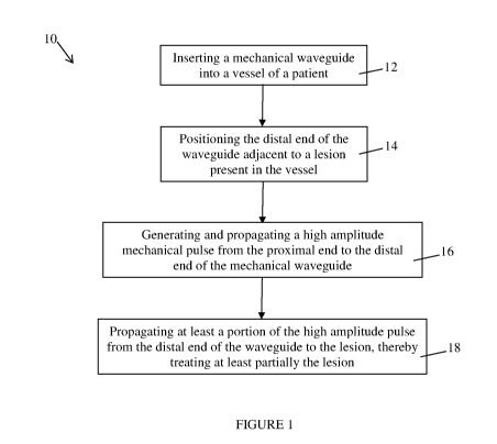
Figure 1 is a flow chart of a method for treating a lesion present in a blood
vessel, in accordance with an embodiment;
Figure 2 is a block diagram illustrating a system for treating a lesion
present in
a vessel, the system comprising a source of mechanical pulses and a mechanical
waveguide,
in accordance with an embodiment;
Figure 3 is a block diagram illustrating a mechanical waveguide and a
deflector for deflecting a mechanical pulse coming from the mechanical
waveguide, in
accordance with an embodiment;
Figure 4 is a block diagram illustrating a mechanical waveguide and a
deflector angularly positioned relative to the mechanical waveguide so as to
deflect a
mechanical pulse coming from the mechanical waveguide, in accordance with an
embodiment;
Figure 5 illustrates a mechanical waveguide extending between a proximal
end and a distal end, the distal end being beveled, in accordance with an
embodiment;
CA 03029129 2018-12-21
WO 2018/002887 PCT/IB2017/053942
8
Figure 6a illustrates a mechanical waveguide provided with a divided distal
end in a first configuration, in accordance with an embodiment;
Figure 6b illustrates the mechanical waveguide of Figure 6a in a second
configuration; and
Figure 7 illustrates a mechanical waveguide extending between a proximal
end and a distal end, the distal end being provided with a frusto-conical
structure to deflect
mechanical pulses, in accordance with an embodiment.
It will be noted that throughout the appended drawings, like features are
identified by like reference numerals.
DETAILED DESCRIPTION
Figure 1 illustrates one embodiment of a method 10 for treating a lesion such
as a vascular lesion present in a vessel of a subject. Treating a lesion
should be understood as
at least one of at least partially cracking, eroding, cleaving, tunneling,
crossing and breaking
the lesion. The method 10 may have applications in fields other than the
medical field. For
example, the method may be used to cross lesions/obstructions present in a
pipe that is used
to propagate water or any other fluid.
At step 12, a mechanical waveguide or transmission member adapted to
propagate mechanical waves and pulses is provided. The mechanical waveguide
extends
between a proximal end and a distal end. At step 12, the distal end of the
mechanical
waveguide is inserted into a blood vessel of the subject such as a vein, an
artery or any other
conduct present in a human body, the vessel comprising a lesion to be treated.
The mechanical waveguide is inserted into the vessel until the distal end of
the
mechanical waveguide is positioned adjacent to the lesion at step 14. In some
embodiments,
the distal end of the mechanical waveguide is positioned so as to abut against
the lesion. In
some other embodiments, the distal end of the mechanical waveguide is
positioned so as not
to be in physical contact with the lesion. In some other embodiments, the
distal end of the
mechanical waveguide is in lateral contact with the lesion.
CA 03029129 2018-12-21
WO 2018/002887 PCT/IB2017/053942
9
Once the distal end of the mechanical waveguide has been positioned at an
adequate position relative to the lesion, a mechanical pulse having a high
amplitude and short
duration is generated at step 16. The mechanical waveguide receives the
generated
mechanical pulse at the proximal end and the mechanical pulse propagates along
the length
of the mechanical waveguide up to the distal end. When it reaches the distal
end, the
mechanical pulse is transmitted at the distal end at step 18, which creates a
displacement of
the distal end and a mechanical pulse that propagates in the medium
surrounding the distal
end of the mechanical waveguide away from the distal end towards the lesion.
In one
embodiment, substantially all of the mechanical pulse is transmitted at the
distal end of the
mechanical waveguide. In another embodiment, only a portion of the mechanical
pulse is
transmitted at the distal end of the mechanical waveguide depending, among
other things, on
the acoustical impedance continuity at the interface between the distal end
and the
surrounding medium. While reaching the lesion, the mechanical pulse cracks,
erodes,
cleaves, tunnel, crosses and/or breaks at least partially the lesion.
In one embodiment, a train of successive mechanical pulses are generated at a
given repetition rate during a given period of time. In this case, the steps
14 to 18 are
repeated. In one embodiment, the repetition rate may be substantially constant
in time. In
another embodiment, the repetition rate may vary in time.
In one embodiment, the method further comprises a step of imaging a portion
of the body of the subject that comprises the lesion to be treated. Any
adequate method for
imaging the lesion such as X-ray imaging, ultrasound imaging or magnetic
imaging may be
used. In one embodiment and as described below, the distal end of the
mechanical waveguide
is provided with a marker opaque to the radiation emitted during the imaging
so that the
marker appears on the taken images. In this case, the method 10 comprises a
step of
displaying on a display unit the images taken using the imaging technique to
allow a user
visualizing the position of the distal end of the mechanical waveguide
relative to the lesion
and therefore adequately positioning the distal end of the mechanical
waveguide relative to
the lesion.
CA 03029129 2018-12-21
WO 2018/002887 PCT/IB2017/053942
In one embodiment, the outputs of several sources covering adjacent
frequency bands are combined to generate the mechanical pulse. In one
embodiment, the
outputs of at least two broadband sources, i.e., the mechanical pulses
generated by the at least
two broadband sources, are combined. In another embodiment, the outputs of at
least one
5 broadband source and at least one narrowband source are combined.
In another embodiment, the mechanical pulse is generated by focusing, via a
spatial concentrator, the output of a large broadband source toward a focal
zone. It should be
understood that the outputs of more than one large broadband source may be
concurrently
focused on the same focal zone.
10 In a further embodiment, a high amplitude mechanical pulse may be
generated
by spatially and/or temporally combining mechanical pulses or waves
sequentially emitted
by a single broadband source using a reverberating cavity. It should be
understood that the
mechanical pulses generated by more than one broadband source may be spatially
and/or
temporally combined together by a reverberating cavity to provide the high
amplitude
mechanical pulse.
In still another embodiment, high amplitude mechanical pulses may be
generated by using a dispersive medium and/or a dispersive geometry to combine
the
component waves emitted sequentially by a single broadband source. It should
be understood
that the mechanical pulses generated by more than one source may be combined
together
using the dispersive medium or the dispersive geometry.
In one embodiment, the mechanical pulse has a center frequency fc comprised
between about 20 kHz and about 10 MHz. In one embodiment, the amplitude of the
high
amplitude mechanical pulse is at least equal to 1 MPa. In one embodiment, the
amplitude of
the mechanical pulse when reaching the distal end of the transmission member
is comprised
between about 1 MPa and about 1000 MPa. In one embodiment, the duration of the
short
duration mechanical pulse when reaching the distal end of the transmission
member is in the
order of 1/fc.
CA 03029129 2018-12-21
WO 2018/002887 PCT/IB2017/053942
11
In one embodiment, the method may be adapted to treat vascular lesions. In
this case, when the distal end of the mechanical waveguide is positioned to be
in physical
contact with the lesion and a mechanical pulse reaches the distal end of the
mechanical
waveguide, the distal end will impact onto the lesion and transmits the
mechanical pulse in
the lesion itself. If the distal end of the transmission member is not in
physical contact with
the lesion, the mechanical pulse is transmitted in the medium present between
the lesion and
the distal end, e.g. blood, and the transmitted mechanical pulse can propagate
up to the
lesion. The mechanical pulse allows cracking, eroding cleaving, tunneling
and/or breaking at
least partially the lesion. For example, this may allow the distal end of the
mechanical
waveguide to cross, or traverse, the lesion as the distal end is moved farther
within the vessel.
A pressure force may be exerted on the mechanical waveguide while mechanical
pulses are
generated and transmitted to the lesion to help the distal end of the
mechanical waveguide
crossing the lesion.
In one embodiment, the method further comprises a step of amplifying the
amplitude of the mechanical pulse. In an embodiment in which a temporal
concentrator is
present, the mechanical wave becomes a mechanical pulse of which the amplitude
is greater
than that of each component wave of the mechanical wave. In an embodiment in
which a
spatial concentrator is present, the amplitude of a mechanical pulse or wave
is increased
while propagating through the spatial concentrator. In another embodiment in
which a spatial
concentrator is present, different mechanical waves or pulses are combined to
generate a
greater amplitude mechanical wave or pulse, i.e. the different mechanical
waves or pulses
add to each other.
In one embodiment, the method further comprises imaging the portion of the
subject body that comprises the lesion during the insertion of the mechanical
waveguide
using any adequate medical imaging method in order to allow a medical
practitioner seeing
the relative position between the distal end of the mechanical waveguide and
the lesion. In
some embodiments, X-ray imaging is used for imaging the lesion. In this case,
the
mechanical waveguide may be provided with a radiopaque marker positioned at
the distal
end thereof. The opaque marker is made of a material that blocks the
propagation of X-rays
so that the opaque marker be visible on an X-ray image.
CA 03029129 2018-12-21
WO 2018/002887 PCT/IB2017/053942
12
In one embodiment, the method 10 further comprises a step of deflecting the
mechanical pulse to orient the mechanical pulse in a predefined direction such
as radially. In
this case, the distal end of the mechanical waveguide is shaped and sized to
orient the
mechanical pulse in a given direction. For example, the distal end may be
beveled to orient
the mechanical in a given direction. In another example, the distal end of the
mechanical
waveguide may have a frusto-conical shape to radially emit the mechanical
pulse. In another
embodiment, a deflector adapted to reflect at least partially a mechanical
wave or pulse may
be used to deflect the mechanical pulse in a given direction. The deflector
may be
independent from the mechanical waveguide and positioned away from the distal
end of the
mechanical waveguide at an adequate position and according to an adequate
orientation in
order to direct the mechanical pulse towards the lesion.
In one embodiment, the mechanical waveguide is adapted to be inserted into a
blood vessel, a catheter, a balloon catheter or the like. In this case, the
mechanical waveguide
is sized and shaped to slide into the blood vessel or the catheter. In one
embodiment, the
.. mechanical waveguide is made of a flexible material so that it may be bent
to follow
curvatures of the blood vessel or the like. In another embodiment, the
mechanical
waveguide may be built into a catheter or a balloon.
Figure 2 illustrates a system 50 for treating a lesion. The system 50
comprises
a pulse generator 54 and a mechanical waveguide 56 adapted to propagate
mechanical waves
or pulses.
The pulse generator 54 is adapted to generate a high amplitude and short
duration pulse. As described above, the pulse generator 54 may comprise at
least one
broadband source and/or at least one narrow band source. The narrow brand or
broadband
source may be an electromechanical transducer. The pulse generator 54 may
comprise a
spatial concentrator to focus the output of at least one source toward a focal
zone at which
the proximal end of the mechanical waveguide 56 is located so as to couple the
generated
pulse therein.
CA 03029129 2018-12-21
WO 2018/002887 PCT/IB2017/053942
13
In some embodiments, the pulse generator 54 may comprise a spatial
concentrator and/or a temporal concentrator for combining mechanical pulses or
waves
sequentially emitted by a single broadband source using a reverberating
cavity.
In some embodiments, the pulse generator 54 may comprise a dispersive
__ medium to combine the component waves emitted sequentially by a single
broadband source.
The mechanical waveguide 56 extends between a first or proximal end that is
operatively connected to the pulse generator 54 and a second or distal end 88.
The
transmission member 66 is adapted to receive mechanical pulses at its proximal
end and
propagate the mechanical pulses up to its distal end. When it reaches the
distal end, the
__ mechanical pulse is at least partially transmitted to generate a
transmitted pulse that
propagates outside of the mechanical waveguide 56. It should be understood
that a pulse may
also be reflected by the distal end and propagates back in the mechanical
waveguide 56
towards the proximal end. The transmitted mechanical pulse corresponds to a
mechanical
pulse that propagates in the medium surrounding the distal end of the
mechanical waveguide
__ 56 up to the lesion 52. The transmitted pulse further propagates into the
lesion 52, which may
create cracks within the lesion 52, and eventually cleaves or breaks the
lesion 52 into pieces.
Also, as the pulse propagates along the mechanical waveguide 56, radial and
longitudinal
motion is induced at the surface of the mechanical waveguide 56 which reduces
the friction
between the mechanical waveguide 56 and the surrounding medium and facilitates
the
__ longitudinal displacement of the mechanical waveguide 56 into the medium,
such as when
crossing fibrotic tissue within a lesion.
In an embodiment in which the distal end of the mechanical waveguide 56
abuts against the lesion 52, the mechanical waveguide 56 may further be used
to break the
lesion 52 and/or drill a hole into the lesion 52. The transmission of the
mechanical pulse at
__ the distal end of the mechanical waveguide 56 creates a movement of the
distal end of the
mechanical waveguide 56. This movement may be along the longitudinal axis of
the
mechanical waveguide. Alternatively, the movement may be perpendicular to the
longitudinal axis or it may be a combination of movements both along the
longitudinal axis
and perpendicular to the longitudinal axis of the mechanical waveguide. During
this
CA 03029129 2018-12-21
WO 2018/002887 PCT/IB2017/053942
14
movement, the distal end of the mechanical waveguide 56 nominally first moves
towards the
lesion 52 and then moves back into its initial position. It should be
understood that the
movement may be inverted (i.e., the distal end may first move away from the
lesion 52 and
then towards the lesion 52) depending on the polarity of the mechanical pulse
reaching the
distal end of the mechanical waveguide 56. When a plurality of distinct
mechanical pulses
are successively transmitted at the distal end of the mechanical waveguide 56,
the movement
of the distal end may be seen as a jack-hammer movement which may be used to
treat the
lesion 52.
As the distal end of the mechanical waveguide 56 recesses (i.e., goes away
from the lesion), a tension wave is created in the medium surrounding the
distal end which
may create a cavitation effect. If the medium is a fluid and since a fluid
cannot withstand
tensile forces, the fluid changes phase and vaporizes into microscopic bubbles
(void and/or
vapor). These bubbles are unstable and may collapse violently inducing
powerful shock
waves and velocity jets. The erosion capability of the induced shock waves and
velocity jets
may contribute to the ablation of the lesion 52.
In some embodiments, a first section of the mechanical waveguide 56 is
inserted within the vessel which contains the lesion 52 and a second section
of the
mechanical waveguide 56 is located outside the vessel. In some embodiments, at
least the
first section of the mechanical waveguide 56 is adapted to be inserted into a
blood vessel. For
example, the first section of the mechanical waveguide 56 may comprise a
biocompatible
coating or be made of a biocompatible material. In some embodiments, only the
first section
of the mechanical waveguide 56 may be flexible. In one embodiment, the
mechanical
waveguide may be built or insertable into a catheter or balloon.
The following describes the operation of the system 50. A first section of the
mechanical waveguide 56 is inserted into a vessel containing a lesion 52 so
that the distal end
of the mechanical waveguide 56 is adjacent to the lesion 52. In one
embodiment, the
mechanical waveguide 56 is positioned so that its distal end substantially
abuts against the
lesion 52 or be in lateral contact with the lesion 52 or at an adequate
position relative to the
lesion 52.
CA 03029129 2018-12-21
WO 2018/002887 PCT/IB2017/053942
As described above, the transmitted pulse propagates up to the lesion 52 and
if
the distal end of the mechanical waveguide 56 abuts against the lesion 52 or
is in lateral
contact with the lesion 52, the jackhammer movement created by the multiple
mechanical
pulses at the distal end may be used to treat the lesion 52.
5 The
distal end of the mechanical waveguide 56 is used to emit the mechanical
pulses from the mechanical waveguide 56 core toward the lesion 52. The distal
end may also
be used to create a path and navigate through the lesion 52, enlarge the
diameter of the path,
and/or orient the direction of the emitted mechanical pulses.
In an embodiment in which the mechanical waveguide 56 is to be inserted into
10 a
catheter, the distal end of the mechanical waveguide 56 may be designed as to
facilitate its
introduction into the catheter toward the lesion. In one embodiment, a
hydrophobic coating
may be applied at the distal end of the transmission member to improve its
lubricity and in
some instance to flush the blood out of the catheter as the distal end
advances toward the
lesion 52 and thereby reduce the quantity of blood that surrounds the
mechanical waveguide
15 56
which could contribute to energy leakage. In one embodiment, a hydrophilic
coating is
added at the distal end of the mechanical waveguide 56 to facilitate its
introduction in a
catheter and/or its penetration into the lesion. The mechanical waveguide may
be positioned
near the lesion by itself or into or around another device such as a
guidewire, catheter or
balloon, already present near the lesion.
In one embodiment, an acoustic coupler is secured to the distal end of the
mechanical waveguide 56 in order to decrease the acoustic impedance mismatch
between
the distal end of the mechanical waveguide and its surroundings which
increases the energy
transmission from the mechanical waveguide 56 towards the lesion 52.
In one embodiment, radiopaque markers such as tungsten, gold strips,
high-density plating, high-density ring, high-density coil or doped polymer
jacket with dense
metal powders are secured to the distal end of the mechanical waveguide 56 to
serve as
references points in order to visualize via X-rays the position of the distal
end relative to the
lesion 52 and to other PTA devices. It should be understood that the marker
may be made of
a material that is opaque to an imaging technique other than X-ray such as
ultrasound,
CA 03029129 2018-12-21
WO 2018/002887 PCT/IB2017/053942
16
magnetic resonance, or the like. When ultrasound imaging is used, the marker
may comprise
void or gas bubbles secured at the distal end of the mechanical waveguide 56
so as to be
opaque to ultrasound and help a user visualizing the distal end of the
mechanical waveguide
56 relative to the lesion to be treated.
In one embodiment, the system 50 further comprises a deflector for deflecting
or orienting the mechanical pulse and/or the mechanical waveguide distal end.
In some
embodiments, the deflector may be independent from the mechanical waveguide
56. In other
embodiments, the deflector may be integral with the mechanical waveguide 56.
In still
another embodiment, the distal end of the transmission member may be curved to
allow
deflecting and/or orienting the mechanical pulse.
Figure 3 illustrates one embodiment of a generic deflector 60 that is used to
deflect a mechanical pulse outputted by a mechanical waveguide 62 according to
a
predefined direction. For example, the deflector 60 may be adapted to deflect
the mechanical
pulse radially around the longitudinal axis of the mechanical waveguide 62. In
this
embodiment, the deflector 60 is independent from the mechanical waveguide 62
while being
in physical contact with the mechanical waveguide 62. For example, the
deflector 60 may be
secured at the distal end of the mechanical waveguide 62.
Figure 4 illustrates one embodiment of a deflector 66 that may be used in
connection with a mechanical waveguide 68 having a flat distal end. The flat
surface of the
distal end is substantially orthogonal to the outer longitudinal surface of
the mechanical
waveguide 66 that extends along the length thereof in order to maximize the
energy output
along the longitudinal axis along which the mechanical waveguide 68 extends.
In the
illustrated embodiment, the deflector 66 is positioned angularly relative to
the longitudinal
axis of the mechanical waveguide 68 and away from the distal end of the
mechanical
waveguide 68.
In some embodiments, the deflector 66 may be secured to the mechanical
waveguide 68.
CA 03029129 2018-12-21
WO 2018/002887 PCT/IB2017/053942
17
In other embodiments, the deflector may be integral with the mechanical
waveguide. Figure 5 illustrates one embodiment of a mechanical waveguide 70
having a
distal flat and beveled end 72. The flat surface of the distal end 72 is
beveled or at an angle
with respect to the longitudinal axis. The angle is chosen so as to orient the
propagated
mechanical pulse according to a given direction. Such shape may also propel
the mechanical
waveguide sideways resulting in a slapping effect that may be used to the
lesion, for example
before the use of a balloon during PTA intervention.
It should be understood that the distal end of the mechanical waveguide may
be provided with any adequate shape other than a flat shape. For example, the
distal end may
be provided with a rounded shape such as a hemi-spherical shape. The surface
of the distal
end may be provided with any adequate shape between a rounded shape and a flat
shape. For
example, the surface of the distal end may be substantially planar with a
smoothed or
rounded edge to be as atraumatic as possible for biological tissues. In
another example, the
distal end maybe provided with a shape to focus the mechanical energy away
from the distal
end. This focusing shape could be a concave shape, for example a circular or
parabolic shape.
This focusing shape could be such as to focus the mechanical pulse along the
longitudinal
axis of the transmission member, or away from this same axis.
In one embodiment, the distal end of the mechanical waveguide 56 may be
shaped so as to direct the mechanical pulse at least partially radially. This
configuration may
be used to create a path in the lesion 52 with a diameter larger than that of
the distal end.
Moreover, such embodiment may be used to prepare the lesion site prior the use
of balloon
during a PTA intervention.
In one embodiment, the distal end of the mechanical waveguide 56 may be
shaped (such as flutes on a drill bit) so as to ease the evacuation of lesion
debris as the
mechanical waveguide progresses in the lesion.
Figure 7 illustrates such a configuration in which a mechanical waveguide 110
is provided with a distal end adapted to partially emit a radial mechanical
wave. A
protrusion 112 having a truncated conical shape protrudes from the distal end
of the
mechanical waveguide 110. The protrusion 112 extends between a circular distal
wall located
CA 03029129 2018-12-21
WO 2018/002887 PCT/IB2017/053942
18
away from the mechanical waveguide 110 and a circular proximal wall secured to
the
waveguide 110. A truncated conical wall extends between the circular proximal
and distal
walls. In the illustrated embodiment, the mechanical waveguide 110 and the
protrusion 112
are coaxial.
In another configuration, the distal tip of the mechanical waveguide could be
split into regions along a direction essentially parallel to its longitudinal
axis, such that when
the mechanical pulse reaches this region it forces the various regions away
from the split
interface, enabling some redirection of some of the energy in the radial
direction. Figures 6a
and 6b illustrate one exemplary embodiment for such a mechanical waveguide 80.
The distal
end 82 of the mechanical waveguide 80 is divided into two regions or two
secondary
mechanical waveguides 84 and 86. A gap 88 is located between the two secondary
mechanical waveguides 84 and 86. In Figure 6a, a mechanical pulse 90
propagates into the
mechanical waveguide 80 from its proximal end 92 towards its distal end 82.
When reaching
the junction between the two secondary mechanical waveguides 84 and 86, the
mechanical
pulse 90 is divided into a first mechanical pulse 94 which propagates into the
secondary
mechanical pulse 84 and a second mechanical pulse 96 which propagates into the
secondary
mechanical waveguide 86. The propagation of the mechanical pulses 94 and 96 in
the
secondary mechanical pulses 84 and 86 forces the secondary mechanical
waveguides 84 and
86 to move away from one another as illustrated by arrows 98 and 100, as
illustrated in
.. Figure 6b. As a result, the gap 88 between the two secondary mechanical
waveguides 84 and
86 increases, thereby allowing directing some the mechanical energy in the
radial direction.
In another configuration, the distal tip of the mechanical waveguide could be
alternately curved along the longitudinal axis so as to redirect some of the
mechanical energy
in the radial direction. However, the person skilled in the art will
understand that other
configurations may be possible.
As illustrated in Figure 6, the central portion of the mechanical energy
schematically represented by arrow 114 propagates through the protrusion 112
to generate a
longitudinal mechanical wave schematically represented by arrow 116 that
propagates
substantially along the longitudinal axis of the waveguide 110 outside of the
waveguide 110
CA 03029129 2018-12-21
WO 2018/002887 PCT/IB2017/053942
19
towards the lesion 52. The outer portion of the mechanical energy
schematically represented
by arrow 118 and adjacent to the outer surface of the waveguide 110 propagates
outside of
the waveguide 110 and is reflected by the truncated conical wall of the
protrusion 112 to
generate a radial mechanical wave.
While in the illustrated embodiment, the propagation direction of the radial
mechanical wave is substantially orthogonal to that of the longitudinal
mechanical wave, it
should be understood that other configurations are possible by varying the
angle between the
waveguide 110 and the truncated conical wall of the protrusion 112. Moreover,
such
configuration does not need to be symmetrical around the main axis of the
mechanical
waveguide.
The person skilled in the art will understand that the amount of energy
converted into a radial mechanical wave may be adjusted by adequately varying
the surface
area of the distal and/or proximal walls of the protrusion 112.
In one embodiment the section of the mechanical waveguide adjacent to the
distal end may be bent or bendable so that a user may apply a permanent or
temporary
curvature with his fingers, a metallic needle introducer or a tool. A bent at
the distal end may
be used to steer the mechanical waveguide (i.e., to give the mechanical
waveguide a
direction) as it is pushed forward in the blood vessel or in the lesion and/or
to redirect the
emitted mechanical pulse.
In one embodiment, the mechanical waveguide has a cross-sectional shape
and/or cross-sectional dimensions that are substantially constant along a
length thereof. For
example, the mechanical waveguide may have a circular cross-sectional shape of
which the
diameter is substantially constant along the length thereof. In one
embodiment, the diameter
of the waveguide is between about 0.004 and about 0.035 in.
In another embodiment, the cross-sectional shape and/or the dimensions of the
mechanical waveguide may vary along a length thereof. For example, the first
section of the
mechanical waveguide that is adjacent to the proximal end and/or the second
section of the
mechanical waveguide that is adjacent to the distal end may have a cross-
sectional shape
CA 03029129 2018-12-21
WO 2018/002887 PCT/IB2017/053942
and/or a dimension different from a third section located between the first
and second
sections. In another example, the mechanical waveguide may comprise at least
one tapering
section for amplifying mechanical pulses.
In one embodiment, the mechanical waveguide is adapted to be used with
5 traditional PTA devices. In one embodiment, the mechanical waveguide has
a diameter that
is less than about 0.125 inches, and preferably less than about 0.040 inches.
In one
embodiment, the aspect ratio (defined as: length/diameter) of the mechanical
waveguide is
chosen to be greater than 100, and preferably greater than 1000. In one
embodiment, the
mechanical waveguide has a length comprised between about 60 in and about 120
in. In
10 another embodiment, the mechanical waveguide has a length comprised
between about 36 in
and about 200 in.
In one embodiment, the bandwidth of the energy source used in the present
system, which is expressed as a percentage of the center frequency fc, is
greater than
about 10%, and preferably between about 40% and about 120%. The center/main
15 frequency fc of the broadband energy source may vary between about 20
kHz and about
10 MHz and is preferably between about 0.1 MHz and about 1 MHz.
The broadband source power and the level of control over the output of the
broadband source can be characterized by the pulse duration, repetition rate,
pressure
amplitude, polarity and waveform type. In one embodiment, the mechanical pulse
duration at
20 the distal end of the transmission member is usually of the order of 1/
I.,. For example, an
energy source having a center frequency of 500 kHz will generate a mechanical
pulse having
duration of about 2 p s, when a bandwidth of 100% is considered. In one
embodiment, the
mechanical pulse duration can be varied by changing the center frequency or
the
bandwidth (i.e., Q factor) of the energy source; the pulse duration is
preferably less than
about 1 ms.
The pulse repetition rate is associated with the number of pulses that can be
transmitted during a certain amount of time. In one embodiment, the repetition
rate can be
varied between about 0.1 Hz and about 1000 Hz and is preferably between about
10 Hz and
about 200 Hz.
CA 03029129 2018-12-21
WO 2018/002887 PCT/IB2017/053942
21
In one embodiment, the output pressure amplitude of the mechanical pulse
generated at the output of the transmission member is greater than about 10
MPa in both
compression and tension. In one embodiment, the output pressure amplitude is
comprised
between about 10 MPa and about 1000 MPa in compression and between about 10
MPa and
about 500 MPa in tension, when measured at the distal end of the transmission
member in a
fluid medium.
Pulsed and controlled mechanical wave emission at the distal end of the
transmission member may crack, cleave, erode, tunnel and/or break parts of the
lesion. By
doing so, the lesion is easier to treat using the present system and method
than traditional
PTA devices.
In one embodiment, the distal end of the mechanical waveguide is adapted to
cross at least one of a fibrotic tissue and a calcified tissue contained
within a lesion. In
another embodiment, the distal end of the mechanical waveguide is adapted to
crack and/or
break at least one of a fibrotic tissue and a calcified tissue contained
within an lesion
positioned laterally to the distal end of the mechanical waveguide.
The embodiments of the invention described above are intended to be
exemplary only. The scope of the invention is therefore intended to be limited
solely by the
scope of the appended claims.