Note: Descriptions are shown in the official language in which they were submitted.
CA 03029253 2018-12-21
WO 2017/223520
PCT/US2017/039123
VIABLE LYOPHILIZED COMPOSITIONS DERIVED FROM HUMAN TISSUES
AND METHODS OF MAKING THE SAME
BACKGROUND
[0001] The purpose of tissue preservation is to retain components
(extracellular
matrix (ECM), growth factors and endogenous viable cells) of fresh tissue
intact,
while providing an extended shelf-life for tissues compared to fresh tissue.
Current
tissue preservation methods include refrigeration, dehydration, and
cryopreservation.
However, all three methods suffer from certain drawbacks. Refrigeration of
fresh
tissues maintains high cell viability for a short time, which leads to a short
shelf-life
(weeks) and limited availability. Dehydration of tissues provides an extended
shelf-
life (years) for the tissue matrix that is retained, but leads to tissue
devitalization that
negatively impacts tissue biological function. Cryopreservation can retain
living
tissue cells for an extended time (months to years), but the cost and effort
required to
maintain ultra-low temperatures (-40 C or below) across the entire supply
chain limits
utilization. Given these drawbacks to currently available tissue preservation
methods,
pursuit of superior compositions and methods of tissue preservation that can
(1) retain
tissue structure and living cells, (2) provide an extended shelf-life (months
to years),
and (3) not require ultra-low temperatures for the supply chain are warranted.
Such
compositions and methods would have applications for military use, as well as
clinical/commercial use.
BRIEF SUMMARY
[0001] Disclosed herein are tissue samples and methods of preparing the tissue
samples that allow for improved tissue preservation.
[0002] Disclosed are methods of lyophilizing a tissue sample comprising
obtaining a
tissue sample, contacting the tissue sample with a lyoprotectant solution,
freezing the
tissue sample, performing a first drying step of the tissue sample after
freezing, and
performing a second drying step of the tissue sample after the first drying
step.
[0003] Also disclosed are methods of preparing a tissue sample comprising
obtaining a tissue sample, contacting the tissue sample with a lyoprotectant
solution,
freezing the tissue sample, performing a first drying step of the tissue
sample after
freezing, performing a second drying step of the tissue sample after the first
drying
step and further comprising a step of reconstituting the lyophilized tissue.
[0004] Disclosed are lyophilized tissues prepared using the disclosed methods
of
1
CA 03029253 2018-12-21
WO 2017/223520
PCT/US2017/039123
lyophilizing a tissue sample comprising obtaining a tissue sample, contacting
the
tissue sample with a lyoprotectant solution, freezing the tissue sample,
performing a
first drying step of the tissue sample after freezing, and performing a second
drying
step of the tissue sample after the first drying step.
[0005] Disclosed are methods of treating a wound or tissue defect comprising
administering a reconstituted lyophilized tissue to the wound or tissue
defect. In some
aspects, the tissue previously lyophilized tissue was lyophilized by one or
more of the
methods disclosed herein.
[0006] Additional advantages of the disclosed method and compositions will be
set
forth in part in the description which follows, and in part will be understood
from the
description, or may be learned by practice of the disclosed method and
compositions.
The advantages of the disclosed method and compositions will be realized and
attained by means of the elements and combinations particularly pointed out in
the
appended claims. It is to be understood that both the foregoing general
description
and the following detailed description are exemplary and explanatory only and
are not
restrictive of the invention as claimed.
BRIEF DESCRIPTION OF THE DRAWINGS
[0007] The accompanying drawings, which are incorporated in and constitute a
part
of this specification, illustrate several embodiments of the disclosed method
and
compositions and together with the description, serve to explain the
principles of the
disclosed method and compositions.
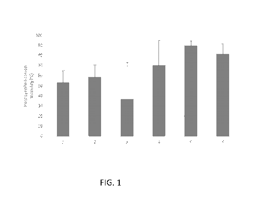
[0008] Figure 1 is a bar graph showing cell viability of lyophilized skin
graft
compositions after rehydration.
[0009] Figure 2 is a bar graph showing cell viability of lyophilized skin
graft
compositions after rehydration (n=2 donors).
[0010] Figures 3A and 3B show fluorescence images. A) Shows cell viability of
lyophilized amnion compositions. Representative images are shown (10X
magnification) for stromal and epithelial layers. B) Shows cell viability of
lyophilized
chorion compositions. Representative images for each group are shown.
[0011] Figure 4 shows representative images of cell viability for lyophilized
amniotic membrane compositions dried in vials or mounted flat on plastic
applicators
and placed in trays.
[0012] Figure 5 shows the evaluation of cell viability persistence over 24
hours after
2
CA 03029253 2018-12-21
WO 2017/223520
PCT/US2017/039123
hydration for lyophilized amniotic membrane compositions mounted on plastic
applicators and dried in trays. Total number of viable cells may slightly
decrease over
24 hours, which is typical of both fresh and cryopreseryed amniotic membranes.
[0013] Figure 6 is a table showing the appearance of lyophilized amniotic
membrane compositions before and after rehydration. Membranes were soaked in
the
same solution and then lyophilized with or without the same solution. All
membranes
were mounted on plastic applicators, and in some cases, the top plastic
applicator was
cut from a solid square to a frame shape.
[0014] Figures 7A, 7B, and 7C show images of large amniotic membrane
compositions. A) Shows two samples measuring 5 cm x 5 cm were mounted on to
plastic applicators with holes on each piece, soaked in a trehalose solution,
and then
lyophilized without solution. B) Shows separate 5 cm x 5 cm samples that was
mounted on one piece of plastic with holes and covered with a "Frame" plastic
applicator, then submerged in trehalose solution for lyophilization. C) Shows
the
membrane after rehydration.
[0015] Figure 8 shows lyophilization of an amniotic membrane composition
within
a breathable autoclave bag.
[0016] Figure 9 shows cell viability of lyophilized amniotic membrane
compositions compared to fresh and cryopreserved amniotic membrane controls.
Samples were prepared and mounted on plastic applicators with either a square
top
piece or a frame top piece.
[0017] Figure 10 shows a representative live/dead image of cell isolated from
lyophilized composition.
[0018] Figure 11 shows anti-inflammatory and immunomodulatory activity of
viable lyophilized amniotic membrane compositions.
[0019] Figures 12A and 12B show cell viability and angiogenic activity of a
viable
lyophilized amniotic membrane. A) Shows live/dead staining of a viable
lyophilized
amniotic membrane composition on the day it was removed from the lyophilizer
(Day
0). B) Shows angiogenic activity of a separate lyophilized sample in response
to
hypoxia + TNF + LPS, which can be attributed to the viable cells within the
composition.
[0020] Figures 13A and 13B show the lack of immunogenic response against
lyophilized amniotic membrane compositions. A) Shows release of TNFa, a marker
3
CA 03029253 2018-12-21
WO 2017/223520
PCT/US2017/039123
of immune cell activation, in positive control was not observed for negative
controls
or experimental groups. B) Shows release of IFNy, another marker of activated
immune cells, was comparable to the negative controls for both experimental
groups.
[0021] Figure 14 shows stability of a viable lyophilized amniotic membrane
composition.
[0022] Figure 15 shows cell viability of lyophilized micronized chorionic
membrane
compositions. All samples were treated with the same solutions and lyophilized
in the
same manner. Each group was processed from the same starting material and
represents samples taken in succession during a micronizing process. Images of
Group 1 and 2 clearly show the micronized sheets of chorionic membrane with
cells
still embedded in the tissue.
[0023] Figure 16 shows the uptake of FITC-trehalose by chorionic stromal cells
in
suspension.
[0024] Figure 17 shows the uptake of trehalose by native placental cells
present in
fresh placental membrane tissues. Both epithelial cells in the amniotic
membrane and
stromal cells in the chorionic membrane are able to readily uptake trehalose.
[0025] Figure 18 shows a comparison of cell survival for placental cells in
suspension vs. cells embedded in matrix.
[0026] Figure 19 shows the uptake of FITC-trehalose by chondrocytes embedded
in
bovine cartilage matrix.
[0027] Figure 20 shows the appearance of viable lyophilized micronized
cartilage.
[0028] Figure 21 shows cell viability of lyophilized bovine cartilage graft
compositions. Only micronized cartilage compositions retained cell viability (-
50%).
[0029] Figure 22 shows cell viability of viable lyophilized bone graft. Green
dots ¨
viable cells. Red color ¨ autofluorescence of bone matrix.
[0030] Figure 23 shows the stability of viable cells within viable lyophilized
amniotic membranes. Membranes were stored at room temperature for 90 days
after
lyophilization, and cells were isolated enzymatically and stained to assess
viability.
Quantification of cell viability showed ¨66-70% living cells.
[0031] Figure 24 shows dry amniotic membrane overlaid on a medical-grade nylon
mesh after lyophilization.
[0032] Figure 25 shows cell viability of viable lyophilized amniotic membranes
using a lyoprotectant solution with trehalose only (10X magnification).
4
CA 03029253 2018-12-21
WO 2017/223520
PCT/US2017/039123
[0033] Figure 26 shows cell viability of viable lyophilized amniotic membranes
using a lyoprotectants solution with trehalose and the antioxidant catechin.
Cell
viability is higher than with trehalose alone for the same lot. Images of
epithelial
layers and amnion stromal layer are included.
[0034] Figure 27 shows amniotic membrane isolated epithelial cells in tris
buffer
with trehalose and catechin.
[0035] Figure 28 shows amniotic membrane sheet in tris buffer with trehalose
and
catechin.
[0036] Figure 29 shows chorionic membrane minced in tris buffer with trehalose
and catechin.
[0037] Figure 30 shows chorionic membrane minced in tris buffer with trehalose
and EGCG.
[0038] Figure 31 shows live/dead stained fluorescent microscopic images of
viable
lyopreserved amniotic membrane (VLAM) post-rehydration in saline solution. Top
images show viable and dead cells in epithelial (left) and stromal (right)
layers in fresh
amniotic membrane (AM). Bottom images show viable and dead cells in epithelial
(left) and stromal (right) layers in VLAM post-rehydration.
[0039] Figure 32 shows cell viability of VLAM incubated in the lyopreservation
solution for 60 or 105 minutes. Fresh AM was used as a control. The green line
(70%)
represents the acceptable cell viability criterion limit recommended by FDA
for
cellular therapies. Bars are mean % of cell viability +/- SD for 3 lots. Fresh
is on far
left, 60min incubation in the middle, 105 min incubation on far right.
[0040] Figure 33 shows the visual appearance of VLAM. The top row shows
integrity of 3 lots of AM tissue (no cracks) after lyophilization. The bottom
images
show ease of sample detachment from the mesh when needed.
[0041] Figure 34 shows the cell viability of VLAM mounted on XN6080 mesh. The
horizontal line (70%) represents the acceptable cell viability criterion limit
recommended by FDA for cellular therapies. Bars are mean % of cell viability
+/- SD
for 3 samples tested for each lot.
[0042] Figure 35 shows the cell viability (%) of VCAM and VLAM (the 24hr.
cycle
prt2-MRM) using a new method of sample preparation without tissue digestion
with
trypsin. Bar graphs are mean % of cell viability+/- SD for 3 lots (3 samples
from each
lot). The horizontal line (70%) represents the acceptable cell viability
criterion limit
CA 03029253 2018-12-21
WO 2017/223520
PCT/US2017/039123
recommended by FDA for cellular therapies. Cryopreserved is column on the
left.
Lyophilized is column on the right.
[0043] Figure 36 shows the viable cell counts for VCAM and VLAM (the 24hr.
cycle prt2-MRM) samples prepared with a modified method for sample preparation
for cell viability assay without tissue digestion with trypsin. Bar graphs are
mean total
number of viable cells per 25 cm2 +/- SD for 3 lots (3 samples from each lot).
Cryopreserved is column on the left. Lyophilized is column on the right.
[0044] Figures 37and 37B show a graphical representation of the primary drying
endpoint for the 24 hr lyophilization cycle for 25 (A) and 90 (B) AM unit
load.
[0045] Figure 38 shows the position of temperature probes throughout AM unit
stacks in the FTS Lyostar II.
[0046] Figure 39 shows the average temperature rate change at the top, middle
and
bottom positions for the VLAM stacks of all sizes during the 24 hr
lyophilization
cycle.
[0047] Figure 40 shows the temperature rate change during the freezing phase
at the
top, middle and bottom for the VLAM stacks of all sizes during the 24 hr
lyophilization cycle. Top, middle and bottom probe temperatures were averaged
for all
VLAM stacks and all 3 shelves.
[0048] Figure 41 shows the temperature rate change during the heating step of
the
primary drying phase at the top, middle and bottom for the VLAM stacks of all
sizes
during the 24 hr lyophilization cycle. Top, middle and bottom probe
temperatures
were averaged for all VLAM stacks and all 3 shelves.
[0049] Figure 42 shows the average temperature rate change at the middle
position
for each size of the VLA stacks during the 24 hr lyophilization cycle.
[0050] Figure 43 shows the cell viability of viable cryopreserved amniotic
membrane (VCAM) and VLAM (the 24hr. cycle prt2-MRM) after the 24 hr
lyophilization cycle. Fresh AM was used as a control. Bar graphs are mean % of
cell
viability +/- SD for 3 lots (3 samples from each lot). The horizontal line
(70%)
represents the acceptable cell viability criterion limit recommended by FDA
for
cellular therapies.
[0051] Figure 44 shows the visual appearance of VLAM units with implemented
pre-lyophilization treatment in the 0.045 M trehalose solution.
[0052] Figure 45 shows the cell viability of VLAM treated by incubation versus
6
CA 03029253 2018-12-21
WO 2017/223520
PCT/US2017/039123
rinse with a 0.045 M trehalose solution. Fresh AM served as a control. Bar
graphs are
mean % of cell viability +/- SD for 4 lots (3 samples from each lot). The
horizontal
line (70%) represents the acceptable cell viability criterion limit
recommended by
FDA for cellular therapies.
[0053] Figure 46 shows the cell viability of VLAM units stored at -80 C for 97
hr
prior to lyophilization. VLAM units lyophilized immediately after packaging
served
as a control. Bar graphs are mean % of cell viability +/- SD for 3 lots (3
samples from
each lot). The horizontal line (70%) represents the acceptable cell viability
criterion
limit recommended by FDA for cellular therapies.
[0054] Figure 47 is a flow chart of steps for the VLAM manufacturing.
[0055] Figure 48 shows the cell viability of VLAM units with the 5h 20 min
"lag
time" post-packaging prior to at -80 C for 97 hr prior to placing into a
lyophilizer.
VCAM units served as a control. Bar graphs are mean % of cell viability +/- SD
for 3
lots (3 samples from each lot). The horizontal line (70%) represents the
acceptable
cell viability criterion limit recommended by FDA for cellular therapies.
[0056] Figure 49 shows the cell viability of VLAM units exposed to 37 C for 77
hrs. 34 min. VCAM units tested after lyophilization served as a control. Bar
graphs are
mean % of cell viability +/- SD for 3 lots (3 samples from each lot). The
horizontal
line (70%) represents the acceptable cell viability criterion limit
recommended by
FDA for cellular therapies.
[0057] Figure 50 shows the cell viability of VLAM units exposed to 50 C for 92
hrs. 15 min. VCAM units tested after lyophilization served as a control. Bar
graphs are
mean % of cell viability +/- SD for )0( lots (samples). The horizontal line
(70%)
represents the acceptable cell viability criterion limit recommended by FDA
for
cellular therapies.
[0058] Figures 51A, B, C, D, E, and F show H&E staining of (a) VLAM, (b)
VCAM, and (c) fresh amniotic tissue and MT staining of (d) VLAM, (e) VCAM, and
(0 fresh amniotic tissue
[0059] Figure 52 shows a visual presentation of wounds in mice after 1st and
6th
applications of a control dressing (Tegaderm), VCAM, or VLAM.
[0060] Figure 53 shows a time course of wound area reduction after
applications of
control dressing, VCAM, or VLAM.
[0061] Figure 54 shows histological images of mouse wound tissue collected
post-
7
CA 03029253 2018-12-21
WO 2017/223520
PCT/US2017/039123
closure after VCAM and VLAM applications. H&E staining shows tissue structure
and MT staining shows collagen deposition.
[0062] Figure 55 shows H&E staining of fresh amnion, VCAM, and VLAM after 6
months of storage in ambient conditions.
DETAILED DESCRIPTION
[0063] The disclosed method and compositions may be understood more readily by
reference to the following detailed description of particular embodiments and
the
Example included therein and to the Figures and their previous and following
description.
[0064] It is to be understood that the disclosed method and compositions are
not
limited to specific synthetic methods, specific analytical techniques, or to
particular
reagents unless otherwise specified, and, as such, may vary. It is also to be
understood
that the terminology used herein is for the purpose of describing particular
embodiments only and is not intended to be limiting.
[0065] Disclosed are materials, compositions, and components that can be used
for,
can be used in conjunction with, can be used in preparation for, or are
products of the
disclosed method and compositions. These and other materials are disclosed
herein,
and it is understood that when combinations, subsets, interactions, groups,
etc. of these
materials are disclosed that while specific reference of each various
individual and
collective combinations and permutation of these compounds may not be
explicitly
disclosed, each is specifically contemplated and described herein. Thus, if a
class of
molecules A, B, and C are disclosed as well as a class of molecules D, E, and
F and an
example of a combination molecule, A-D is disclosed, then even if each is not
individually recited, each is individually and collectively contemplated.
Thus, is this
example, each of the combinations A-E, A-F, B-D, B-E, B-F, C-D, C-E, and C-F
are
specifically contemplated and should be considered disclosed from disclosure
of A, B,
and C; D, E, and F; and the example combination A-D. Likewise, any subset or
combination of these is also specifically contemplated and disclosed. Thus,
for
example, the sub-group of A-E, B-F, and C-E are specifically contemplated and
should be considered disclosed from disclosure of A, B, and C; D, E, and F;
and the
example combination A-D. This concept applies to all aspects of this
application
including, but not limited to, steps in methods of making and using the
disclosed
compositions. Thus, if there are a variety of additional steps that can be
performed it
is understood that each of these additional steps can be performed with any
specific
8
CA 03029253 2018-12-21
WO 2017/223520
PCT/US2017/039123
embodiment or combination of embodiments of the disclosed methods, and that
each
such combination is specifically contemplated and should be considered
disclosed.
A. Definitions
[0066] It must be noted that as used herein and in the appended claims, the
singular
forms "a ", "an", and "the" include plural reference unless the context
clearly dictates
otherwise. Thus, for example, reference to "a tissue sample" includes a
plurality of
such tissue samples, reference to "the tissue sample" is a reference to one or
more
tissue samples and equivalents thereof known to those skilled in the art, and
so forth.
[0067] "Native cells" means cells that are native, resident, or endogenous to
the
tissue sample, i.e. cells that are not exogenously added to the tissue sample.
[0068] "Native factors" means factors that are native, resident, or endogenous
to the
tissue sample, i.e. factors that are not exogenously added to the tissue
sample.
[0069] "Substantially free" means present in only a negligible amount or not
present
at all. For example, when a cell is abundant less than about 20% or less than
about
10% or less than about 1% of the amount in an unprocessed sample.
[0070] "Substantial amount" of an element of the present invention, e.g.
native
factors, therapeutic factors, or selective depletion, means a value at least
about 2% or
at least 10% in comparison to an unprocessed, fresh tissue sample. A
substantial
amount can optionally be at least about 50%.
[0071] "Therapeutic cells" as used herein means viable cells native to a given
tissue
that have retained their native biological functions to dynamically respond to
a local
microenvironment, for example an injury site or wound. Examples of therapeutic
cells
include, but are not limited to, fibroblasts, epithelial cells, MSCs, and
other tissue-
specific cell types, such as osteoblasts or osteoclasts for bone, or CD34+
follicular
cells of the skin epidermis, or chondrocytes of hyaline cartilage, or
fibrochondrocytes
of meniscus, or annulus fibrosus or nucleus pulposus cells of the
intervertebral disc, or
supportive cell types surrounding peripheral nerve.
[0072] "Therapeutic factors" means tissue-derived factors that promote wound
healing or tissue regeneration. For example, placenta- or chorionic membrane-
derived
factors that promote wound healing or tissue regeneration. Examples include,
but are
not limited to IGFBP1, adiponectin, a2-macroglobulin, and bFGF. Other examples
include, but are not limited to MMP-9 and TIMP1. Other therapeutic factors
include,
but are not limited to, TGF-beta 1, beta 2, or beta 3, HGF, VEGF, IGF-1, and
BMPs.
9
CA 03029253 2018-12-21
WO 2017/223520
PCT/US2017/039123
[0073] "Stromal cells" refers to a mixed population of cells present
(optionally in
native proportions) composed of mesenchymal stem cells and fibroblasts
natively
found within the stromal layer of a given tissue type.
[0074] "Optional" or "optionally" means that the subsequently described event,
circumstance, or material may or may not occur or be present, and that the
description
includes instances where the event, circumstance, or material occurs or is
present and
instances where it does not occur or is not present.
[0075] Ranges may be expressed herein as from "about" one particular value,
and/or
to "about" another particular value. When such a range is expressed, also
specifically
contemplated and considered disclosed is the range¨ from the one particular
value
and/or to the other particular value unless the context specifically indicates
otherwise.
Similarly, when values are expressed as approximations, by use of the
antecedent
"about," it will be understood that the particular value forms another,
specifically
contemplated embodiment that should be considered disclosed unless the context
specifically indicates otherwise. It will be further understood that the
endpoints of
each of the ranges are significant both in relation to the other endpoint, and
independently of the other endpoint unless the context specifically indicates
otherwise.
Finally, it should be understood that all of the individual values and sub-
ranges of
values contained within an explicitly disclosed range are also specifically
contemplated and should be considered disclosed unless the context
specifically
indicates otherwise. The foregoing applies regardless of whether in particular
cases
some or all of these embodiments are explicitly disclosed.
[0076] As used herein, "kit" means a collection of at least two components
constituting the kit. Together, the components constitute a functional unit
for a given
purpose. Individual member components may be physically packaged together or
separately. For example, a kit comprising an instruction for using the kit may
or may
not physically include the instruction with other individual member
components.
Instead, the instruction can be supplied as a separate member component,
either in a
paper form or an electronic form which may be supplied on computer readable
memory device or downloaded from an intern& website, or as recorded
presentation.
[0077] As used herein, "instruction(s)" means documents describing relevant
materials or methodologies pertaining to a kit. These materials may include
any
combination of the following: background information, list of components and
their
CA 03029253 2018-12-21
WO 2017/223520
PCT/US2017/039123
availability information (purchase information, etc.), brief or detailed
protocols for
using the kit, trouble-shooting, references, technical support, and any other
related
documents. Instructions can be supplied with the kit or as a separate member
component, either as a paper form or an electronic form which may be supplied
on
computer readable memory device or downloaded from an internet website, or as
recorded presentation. Instructions can comprise one or multiple documents,
and are
meant to include future updates.
[0078] In various aspects, the subject of the herein disclosed methods is a
vertebrate,
e.g., a mammal. Thus, the subject of the herein disclosed methods can be a
human,
non-human primate, horse, pig, rabbit, dog, sheep, goat, cow, cat, guinea pig
or
rodent. The term does not denote a particular age or sex. Thus, adult and
newborn
subjects, as well as fetuses, whether male or female, are intended to be
covered. A
patient refers to a subject afflicted with a disease or disorder. The term
"patient"
includes human and veterinary subjects.
[0079] Unless defined otherwise, all technical and scientific terms used
herein have
the same meanings as commonly understood by one of skill in the art to which
the
disclosed method and compositions belong. Although any methods and materials
similar or equivalent to those described herein can be used in the practice or
testing of
the present method and compositions, the particularly useful methods, devices,
and
materials are as described. Publications cited herein and the material for
which they
are cited are hereby specifically incorporated by reference. Nothing herein is
to be
construed as an admission that the present invention is not entitled to
antedate such
disclosure by virtue of prior invention. No admission is made that any
reference
constitutes prior art. The discussion of references states what their authors
assert, and
applicants reserve the right to challenge the accuracy and pertinence of the
cited
documents. It will be clearly understood that, although a number of
publications are
referred to herein, such reference does not constitute an admission that any
of these
documents forms part of the common general knowledge in the art.
[0080] Throughout the description and claims of this specification, the word
"comprise" and variations of the word, such as "comprising" and "comprises,"
means
"including but not limited to," and is not intended to exclude, for example,
other
additives, components, integers or steps. In particular, in methods stated as
comprising one or more steps or operations it is specifically contemplated
that each
step comprises what is listed (unless that step includes a limiting term such
as
11
CA 03029253 2018-12-21
WO 2017/223520
PCT/US2017/039123
"consisting of"), meaning that each step is not intended to exclude, for
example, other
additives, components, integers or steps that are not listed in the step.
[0081] Those skilled in the art will recognize, or be able to ascertain using
no more
than routine experimentation, many equivalents to the specific embodiments of
the
method and compositions described herein. Such equivalents are intended to be
encompassed by the following claims.
B. Methods of Lyophilizing
[0082] Disclosed are methods of lyophilizing a tissue sample comprising
obtaining
a tissue sample, contacting the tissue sample with a lyoprotectant solution,
freezing the
tissue sample, performing a first drying step of the tissue sample after
freezing, and
performing a second drying step of the tissue sample after the first drying
step.
[0083] Also disclosed are methods of preparing a tissue sample comprising
obtaining a tissue sample, contacting the tissue sample with a lyoprotectant
solution,
freezing the tissue sample, performing a first drying step of the tissue
sample after
freezing, performing a second drying step of the tissue sample after the first
drying
step and further comprising a step of reconstituting the lyophilized tissue.
Reconstituted tissue of the disclosed methods can comprise at least 70% viable
cells.
In some aspects, reconstituted tissue can comprise greater than 40%, 45%, 50%,
55%,
60%, 65%, 70%, 75%, 80%, 85%, 90%, 95%, or 99% viable cells. In some aspects,
after reconstituting the lyophilized tissue, the tissue can then be cut to a
desired size.
Percent viability of cells after reconstitution is based on the percent of
viable cells that
were in the starting tissue sample prior to being lyophilized.
1. Obtaining a tissue sample
[0084] In some aspects, obtaining a tissue sample can be performed by those
methods known in the art. The method of obtaining a tissue sample can depend
on the
type of tissue sample being obtained. For example, obtaining a placental
tissue can
occur at the time of childbirth. In some aspects, tissue samples can be
obtained from a
cadaver.
[0085] In some aspects, a tissue sample can be, but is not limited to, a
placenta or
portion of a placenta, skin, bone, or cartilage. In some aspects, a placenta
or placental
tissue can be amniotic tissue, chorionic tissue, umbilical cord tissue, or a
combination
thereof In some aspects, cartilage can be articular, hyaline or
fibrocartilage. An
example of fibrocartilage can be meniscal tissue.
12
CA 03029253 2018-12-21
WO 2017/223520
PCT/US2017/039123
[0086] In some aspects, a tissue sample does not comprise cultured cells. For
example, the cells present in the tissue sample would be considered native to
the tissue
sample and non-cultured if the native cells have not previously been removed
from the
tissue sample and plated, seeded, cultured or in any other way allowed to
adhere to a
plastic or protein surface for any amount of time. Cells that have been
previously
removed from the tissue sample and plated, seeded, cultured or in any other
way
allowed to adhere to a plastic or protein surface for any amount of time are
referred to
herein as "cultured cells".
[0087] In some aspects, a tissue sample can be cut to a desired size. Cutting
a tissue
sample to a desired size can occur prior to freezing the tissue sample (i.e.
before or
after contacting the tissue sample with a lyoprotectant solution). In some
aspects, a
tissue sample can be minced. Mincing a tissue sample can occur prior to
freezing the
tissue sample (i.e. before or after contacting the tissue sample with a
lyoprotectant
solution).
[0088] In some aspects, a tissue sample can be treated with an antibiotic. In
some
aspects, a tissue sample can be treated with an antibiotic prior to freezing
(e.g. before
or after contacting the tissue sample with a lyoprotectant solution).
2. Contacting the tissue sample with a lyoprotectant solution
[0089] In some aspects, contacting the tissue sample with a lyoprotectant
solution
can include a short or prolonged contact. For example, the tissue sample can
be
exposed or contacted to a lyoprotectant solution for 1, 2, 3, 4, 5, 10, 15,
20, 25, 30, 35,
40, 45, 50, 55, or 60 minutes. In some aspects, the tissue sample can be
exposed or
contacted to a lyoprotectant solution for 1, 2, 3, 4, 5, 6, 7, 8, 9, 10, 12,
14, 16, 18, 20,
22, or 24 hours. In some aspects, the tissue sample can be exposed or
contacted to a
lyoprotectant solution for 1, 2, 3, 4, 5, 6, 7, 14, 21 days. In some aspects,
the tissue
sample can be exposed or contacted to a lyoprotectant solution for 1, 2, 3, 4,
5, 6, 7, or
8 weeks.
[0090] In some aspects, contacting the tissue sample with a lyoprotectant
solution
can be the same as exposing the tissue sample to a lyoprotectant solution or
soaking
the tissue sample in a lyoprotectant solution.
[0091] As described here, a lyoprotectant solution comprises at least one
lyoprotectant. In some aspects, a lyoprotectant solution can comprise
trehalose. Other
lyoprotectants can include but are not limited to polyhydroxy compounds such
as
13
CA 03029253 2018-12-21
WO 2017/223520
PCT/US2017/039123
sugars, polyalcohols, raffinose, and other non-reducing polysaccharides, and
their
derivatives.
[0092] In some aspects, the lyoprotectant solution can further comprise one or
more
antioxidants. In some aspects, the one or more antioxidants can be
epigallocatechin
gallate (EGCG) or catechin. In some aspects, an antioxidant can be ascorbic
acid, L-
carnosine, spermine, phloretine, a-tocopherol, 13-carotene, conenzyme Q10,
lutein,
melatonin, butylated hydroxytoluene, y-tocopherol, lutein, N-acetyl-L-
cysteine,
mitoquinone, hydroquinone, lipoic acid, glutathione, carotenoids, polyphenols,
retinol,
tocotrienol.
[0093] In some aspects, lyoprotectant solution can also comprise saline, DMSO,
antibiotics, bulking agents, excipients, or a combination thereof In some
aspects, the
lyoprotectant can comprise other reagents that can improve lyophilization
performance.
[0094] The concentration of a lyoprotectant or antioxidants present in the
lyoprotectant solution and the length of time for contacting the tissue sample
with the
lyoprotectant solution can be dependent on the type and size of the tissue
sample.
Based oon the teachings herein, one of skill in the art using routine methods
would
understand how to adjust the concentrations and contacting times.
[0095] In some aspects, contacting the tissue sample with a lyoprotectant
solution
can occur at temperatures between 00 and 39 C. In some aspects, contacting the
tissue
sample with a lyoprotectant solution can occur at 4 C.
3. Freezing the tissue sample
[0096] In some aspects, freezing the tissue sample can be performed at a
temperature range of -80 C to -4 C. In some aspects, freezing the tissue
sample can
be performed at a temperature range of -70 C to -4 C. In some aspects,
freezing the
tissue sample can be performed at a temperature range of -50 C to -4 C.
[0097] In some aspects, the sample can be added for purposes of freezing the
tissue,
wherein the tissue can be added prior to achieving the final freezing
temperature. In
some aspects, the step of freezing the tissue sample can involve avoiding a
flash freeze
and instead providing a steady cooling to freezing temperatures. In such
instances, the
temperature can be decreased at a rate between 0.1 and 10 C/min. In such
instances,
the temperature can be decreased at a rate between 0.1 and 5 C/min. In some
instances, flash freezing can cause formation of water crystals that can kill
the tissue-
14
CA 03029253 2018-12-21
WO 2017/223520
PCT/US2017/039123
resident cells and alter the structure of the tissue matrix. In such
instances, a slower
freeze can be used to avoid killing the tissue or native cells contained
therein.
4. Performing a first drying step of the tissue sample after freezing
[0098] In some aspects, the first drying step of the tissue sample after
freezing
occurs between -45 C and -15 C. In some aspects, the first drying step of the
tissue
sample after freezing occurs between -45 C and -10 C. In some aspects, the
first
drying step of the tissue sample after freezing occurs between -45 C and -5 C.
In
some aspects, the first drying step of the tissue sample after freezing occurs
between -
45 C and 0 C. In some aspects, the first drying step of the tissue sample
after freezing
occurs between -45 C and +15 C. In some aspects, the first drying step of the
tissue
sample after freezing occurs between -45 C and +10 C. In some aspects, the
first
drying step of the tissue sample after freezing occurs between -45 C and +5 C.
In
some aspects, the temperature of the first drying step can be the same as the
freezing
temperature. In some aspects, the temperature of the first drying step can be
at least
1 , 5 , 10 , 15 , 20 , 25 , 30 , 35 , 40 , 45 , or 50 C higher than the
temperature of
the freezing step.
[0099] In some aspects, the first drying step of the tissue sample after
freezing can
be carried out for less than 10 hours. In some aspects, the first drying step
of the
tissue sample after freezing can be carried out for 10, 12, 14, 16, 18, 20, or
24 hours.
In some aspects, the first drying step of the tissue sample after freezing can
be carried
out for 24, 48 or 72 hours.
5. Performing a second drying step of the tissue sample after the first drying
step
[00100] In some aspects, the second drying step can be carried out at a
temperature
that is greater than the temperature of the freezing step. In some aspects,
the second
drying step can be carried out at a temperature that is greater than the
temperature of
the freezing step and the first drying step.
[00101] In some aspects, the temperature is increased between the first drying
step
and the second drying step. In such aspects, the temperature of the second
drying step
is higher than the temperature of the first drying step. In some aspects,
wherein the
temperature of the second drying step is higher than the first drying step,
the rate of
the temperature increase from the first drying step can be gradual or rapid.
For
example, the rate of temperature increase from the first drying step to the
second
drying step can be from 0.1 to 5 C/min. In some aspects, the rate of
temperature
increase from the first drying step to the second drying step can be 0.33 to 1
C/min.
CA 03029253 2018-12-21
WO 2017/223520
PCT/US2017/039123
[00102] In some aspects, the second drying step can occur at a temperature of
no
more than 39 C. In some aspects, the second drying step can occur at a
temperature of
no more than 45 C.
[00103] In some aspects, the second drying step can be carried out at two or
more
different temperatures. In some aspects, the at least two different
temperatures can be
at least 5 , 10 , 15 , 20 , 25 , 30 , 35 , 40 , 45 , or 50 C different from
each other.
For example, the second drying step can be carried out at 0 and then at 20 C.
In
some aspects, the second drying step can be conducted in at least two
different
temperatures, wherein each different temperature can each be maintained for 5
to 15
minutes each. In some aspects, the temperature can be ramped up from one
temperature to the next, each of the intervening temperatures can be
maintained for
about 10 sec to 1 minute. Thus, although the second drying step can be carried
out at
two or more different temperatures, many temperatures can be involved in the
second
drying step as the tissue sample is exposed to all of the temperatures in
between the at
least two temperatures that are maintained for 5-15 minutes.
[00104] In some aspects, the second drying step can be conducted at more than
two
different temperatures. For example, the second drying step can be conducted
at 0 ,
20 , and 30 C. In some aspects, the second drying step can be conducted in at
least
three different temperatures, wherein each different temperature can be each
maintained for 5 to 15 minutes each. As the temperature is ramped up from one
temperature to the next, each of the intervening temperatures can be
maintained for
about 10 sec to 1 minute. Thus, although the second drying step can be carried
out at
three or more different temperatures, many temperatures can be involved in the
second
drying step as the tissue sample is exposed to all of the temperatures in
between the at
least two temperatures that are maintained for 5-15 minutes.
[00105] In some aspects, the second drying step is conducted for 12-144 hours.
In
some aspects, the second drying step is conducted for 12-48 hours. In some
aspects,
the second drying step is conducted for 12-72 hours. In some aspects, the
second
drying step is conducted for at least 12, 15, 12, 25, 0, 35, 40, 45, 50, 55,
60, 65, 70, 75,
80, 85, 90, 95, 100, 105, 110, 115, 120, 125, 130, 135, 140, or 144 hours.
[00106] In some aspects, after drying the lyophilized tissue, the tissue can
be cut to a
desired size or shape.
16
CA 03029253 2018-12-21
WO 2017/223520
PCT/US2017/039123
C. Lyophilized Tissue
[00107] Disclosed are lyophilized tissues prepared using the methods disclosed
herein.
[00108] Disclosed are lyophilized tissues prepared using the methods disclosed
herein that are sealed inside a sterile package.
[00109] In some aspects, the lyophilized tissue disclosed herein can be stable
for at
least three weeks. In some aspects, the lyophilized tissue can be stable for
at least
three months. In some aspects, the lyophilized tissue can be stable for 1, 2,
3, 4, 5, 6,
7, 8, 9, 10, 11, 12, 24, 36, 48, or 60 months.
[00110] In some aspects, the lyophilized tissue disclosed herein can be
reconstituted
resulting in a reconstituted tissue. Lyophilized tissue can be reconstituted
using
standard techniques known in the art. In some aspects, reconstituting refers
to
rehydrating. Thus, the disclosed lyophilized tissues can be reconstituted or
rehydrated
using water, saline, a buffer such as, but not limited to phosphate buffered
saline
(PBS), in a solution comprising a stabilizing agent such as, but not limited
to bovine
serum albumin (BSA), Plasma-Lyte A or other clinically available electrolyte
solutions, with human bodily fluids or a combination thereof For example,
lyophilized tissue can be applied directly to a wound or tissue injury on a
subject and
the subject's bodily fluids can reconstitute. In some aspects, a combination
of bodily
fluids and another known rehydrating solution can be used. Also, disclosed are
reconstituted tissue prepared using the methods disclosed herein.
[00111] The reconstituted tissue derived from the methods disclosed herein can
comprise native viable cells and native therapeutic factors. The reconstituted
tissue
can comprise at least 40%, 45%, 50%, 55%, 60%, 65%, 70%, 75%, 80%, 85%, 90%,
95%, 99% viable cells compared to the same tissue prior to lyophilization. The
reconstituted tissue can comprise at least 40%, 45%, 50%, 55%, 60%, 65%, 70%,
75%, 80%, 85%, 90%, 95%, 99% viable native cells compared to the same tissue
prior
to lyophilization.
1. Chorionic membrane
[00112] In some aspects, reconstituted tissue can be reconstituted chorionic
membrane. In some aspects, reconstituted chorionic membrane can comprise about
1,000 to about 240,000 cells/cm2 or about 20,000 to about 60,000 cells/cm2. In
some
aspects, reconstituted chorionic membrane can comprise 20,000 to about 200,000
17
CA 03029253 2018-12-21
WO 2017/223520
PCT/US2017/039123
cells/cm2, with a cell viability of at least about 70%.
[00113] In some aspects, reconstituted chorionic membrane can comprise at
least:
about 7,400 or about 15,000 or about 23,217, or about 35,000, or about 40,000
or
about 47,800 of stromal cells per cm2 of the reconstituted chorionic membrane.
Thus,
reconstituted chorionic membrane can comprise about 5,000 to about 50,000 of
stromal cells per cm2 of the reconstituted chorionic membrane.
[00114] In some aspects, reconstituted chorionic membrane can comprise native
chorionic cells wherein at least: about 40%, or about 50%, or about 60%, or
about
70%, or about 74.3%, or about 83.4 or about 90%, or about 92.5% of the native
chorionic cells are viable. Thus, reconstituted chorionic membrane can
comprise
native chorionic cells wherein about 40% to about 92.5% of the native
chorionic cells
are viable.
[00115] In some aspects, reconstituted chorionic membrane can have a thickness
of
about 20 p.m to about 600 p.m.
[00116] In some aspects, reconstituted chorionic membrane secretes less than
about
any of: 420 pg/mL, 350 pg/mL, or 280 pg/mL TNF-a into a tissue culture medium
upon placing a 2 cm x 2 cm piece of the reconstituted chorionic membrane in a
tissue
culture medium and exposing the reconstituted chorionic membrane to a
bacterial
lipopolysaccharide for about 20 to about 24 hours.
[00117] In some aspects, reconstituted chorionic membrane can be associated
with
part or all of an amniotic membrane.
2. Amniotic membrane
[00118] In some aspects, reconstituted tissue can be reconstituted amniotic
membrane.
[00119] In some aspects, reconstituted amniotic membrane can comprise an
epithelial
cell layer, wherein the approximate number of cells per cm2 of the
reconstituted
amniotic membrane is about 10,000 to about 360,000 or about 40,000 to about
90,000.
[00120] In some aspects, reconstituted amniotic membrane can comprise a thick
basement membrane (comprising one or more of Collagen Type I, Ill, IV,
laminin, and
fibronectin).
[00121] In some aspects, reconstituted amniotic membrane can comprise a
stromal
cell layer. In some aspects, the reconstituted amniotic membrane can comprise
at least:
about 2,000, or about 2,400, or about 4,000 or about 6,000, or about 8,000, or
about
18
CA 03029253 2018-12-21
WO 2017/223520
PCT/US2017/039123
10,000, or about 10,585, or about 15,000 stromal cells per unit cm2 of the
amniotic
membrane. In some aspects, the reconstituted amniotic membrane can comprise
about
2,000 to about 15,000 of stromal cells per cm2 of the amniotic membrane. In
some
aspects, the reconstituted amniotic membrane can comprise stromal cells
wherein at
least: about 40%, or about 50%, or about 60%, or about 70%, or about 74.3%, or
about 83.4 or about 90%, or about 92.5% of the stromal cells are viable after
reconstitution.
[00122] In some aspects, reconstituted amniotic membrane can comprise a
thickness
of about 20 to about 250 um.
[00123] In some aspects, reconstituted amniotic membrane can comprise low
immunogenicity. In some aspects, reconstituted amniotic membrane can comprise
secretes less than about any of: 420 pg/mL, 350 pg/mL, or 280 pg/mL TNF-a into
a
tissue culture medium upon placing a 2 cm x 2 cm piece of the reconstituted
amniotic
membrane in a tissue culture medium and exposing the reconstituted amniotic
membrane to a bacterial lipopolysaccharide for about 20 to about 24 hours.
[00124] In some aspects, reconstituted amniotic membrane can comprise a layer
of
amniotic epithelial cells.
[00125] In some aspects, reconstituted amniotic membrane can comprise native
amniotic cells that include for example, epithelial cells or stromal cells. In
some
aspects, the amniotic stromal cells include amniotic fibroblasts and/or
amniotic MSCs.
[00126] In some aspects, reconstituted amniotic membrane can provide an
analgesic
effect, reduce scarring, or both.
[00127] In some aspects, reconstituted amniotic membrane can comprise anti-
inflammatory proteins such as IL-1Ra and IL-10, antibacterial proteins such as
defensins and allantoin (bacteriolytic proteins), and angiogenic and mitogenic
factors
that promote re-epithelialization such as EGF, HGF, and VEGF.
[00128] In some aspects, reconstituted amniotic membrane can comprise cells
that
are positive for CD73, CD90, CD105, and CD166 and negative for CD45, CD34, and
CD31. In some aspects, reconstituted amniotic membrane can comprise cells that
express HLA-G, cells that express IDO and FAS ligand, which likely contribute
to
immune tolerance, cells with a capacity to differentiate into 1- Human
Amniotic
Epithelial Cells (hAECs), cells with a capacity to differentiate to neural,
hepatocyte,
and pancreatic cells, cells that expression of CD49d by hAMSCs distinguishes
19
CA 03029253 2018-12-21
WO 2017/223520
PCT/US2017/039123
hAMSCs from hAECs, hAMSCs that are positive for the embryonic cytoplasmic
marker Oct-4 that plays a role in maintaining pluripotency and self-renewal,
and
hAECs that are positive for SSEA-3, SSEA-4, TRA-1-60, TRA-1-81, and negative
for
SSEA-4 and non-tumorogenic.
[00129] In some aspects, reconstituted amniotic membrane can be associated
with
part or all of a chorionic membrane.
3. Cartilage
i. Articular cartilage
[00130] In some aspects, cartilage can be articular cartilage tissue. Thus, in
some
aspects, reconstituted tissue can be reconstituted articular cartilage.
[00131] In some aspects, reconstituted articular cartilage can comprise TFG-
01,
TGF-03, BMP-7, bFGF, IGF-1.
[00132] In some aspects, reconstituted articular cartilage can comprise at
least about
500 cells/mm2, 600 cells/mm2, 700 cells/mm2, 800 cells/mm2, 1200 cells/mm2, or
1500 cells/mm2.
[00133] In some aspects, reconstituted articular can comprise at least about
100
cells/mm2 or 200 cells/mm2 of viable chondrocytes. In some aspects,
reconstituted
articular cartilage comprises at least 50%, 60%, 70%, 80%, 90%, or 95% viable
chondrocytes.
Menis cal Tissue
[00134] In some aspects, cartilage can be meniscal tissue. Thus, in some
aspects,
reconstituted tissue can be reconstituted meniscal tissue.
[00135] In some aspects, reconstituted meniscal tissue can comprise greater
than
25%, 30%, 35%, 40%, 45%, 50%, 55%, 60%, 65%, 70, 75%, 80%, 85%, 90%, or 95%
viable cells. In some aspects, reconstituted meniscal tissue can comprise
greater than
25%, 30%, 35%, 40%, 45%, 50%, 55%, 60%, 65%, 70, 75%, 80%, 85%, 90%, or 95%
viable native cells.
[00136] In some aspects, reconstituted meniscal tissue can be non-immunogenic.
For
example, reconstituted meniscal tissue can have depleted amounts of one or
more
types of functional immunogenic cells. An absence of immunogenic cells can be
further confirmed if the reconstituted meniscal tissue does not produce > 100
pg/ml of
TNF-alpha upon stimulation with a bacterial immunogen, such as LPS, within
about
24 hours of culture. In some instances, >5% of cells present in the
composition can be
CA 03029253 2018-12-21
WO 2017/223520
PCT/US2017/039123
immune cells however the composition would be considered absent of immunogenic
cells if <5% of the viable cells are immune cells.
[00137] In some aspects, reconstituted meniscal tissue can have one or more
growth
factors native to the meniscal tissue. The growth factors can be one or more
of TGF-
01, TGF-b3, bFGF, PDGF-AB, PDGF-BB, IGF-1, HGF, BMP-7, EGF, CTGF, BMP-
2, BMP-6, and VEGF.
[00138] In some aspects, reconstituted meniscal tissue can comprise at least
one of
the collagen layers of human meniscus.
[00139] In some aspects, reconstituted meniscal tissue can comprise viable,
native
mesenchymal stem cells.
4. Bone
[00140] In some aspects, reconstituted tissue can be reconstituted bone or a
bone
repair product. In some aspects, a bone repair product can comprise cancellous
hone
fragments and periosteum containing angiogenic growth factor(s).
[00141] The particular types and concentration of the growth factor(s) in the
bone or
a bone repair product can depend on the particular donor. In some aspects, the
concentrations of each growth factor can independently be at least 1 pg/mL,
such as at
least 2 pg/mL, 5 pg/mL, 10 pg/mL, 30 pg/mL, 40 pg/mL, 50 pg/mL, 60 pg/mL, 70
pg/mL, 80 pg/mL, 90 pg/mL, 100 pg/mL, 200 pg/mL, 300 pg/mL, 400 pg/mL, 500
pg/mL, 600 pg/mL, 700 pg/mL, 800 pg/mL, 900 pg/mL, 1000 pg/mL, 2000 pg/mL,
3000 pg/mL, 4000 pg/mL, 5000 pg/mL, 6000 pg/mL, 7000 pg/mL, 8000 pg/mL, 9000
pg/mL, 10000 pg/mL, 20000 pg/mL, 30000 pg/mL, 40000 pg/mL, 50000 pg/mL or
more and each will generally independently vary from or from about 1 pg/mL to
50000 pg/mL, such as 10 pg/mL to 10000 pg/mL or 50 pg/mL to 5000 pg/mL, such
as
from or from about 100 pg/mL to 1000 pg/mL, 100 pg/mL to 800 pg/mL, 100 pg/mL
to 600 pg/mL, 100 pg/mL to 400 pg/mL, 100 pg/mL to 200 pg/mL, 200 pg/mL to
1000 pg/mL, 200 pg/mL to 800 pg/mL, 200 pg to 600 pg/mL, 200 pg/mL to 400
pg/mL, 400 pg/mL to 1000 pg/mL, 400 pg/mL to 800 pg/mL, 400 pg/mL to 600
pg/mL, 600 pg/mL to 1000 pg/mL, 600 pg/mL to 800 pg/mL or 800 pg/mL to 1000
pg/mL of BRP. The growth factors present in the bone or a bone repair product
include, for example, VEGF, bFGF, PDGF, IGF-1, IGF-2, TGF-01, BMP-2 and/or
BMP-7, and each can be present in a concentration range as set forth above. As
an
example, BRP provided herein can contain VEGF and the concentration of VEGF
can
21
CA 03029253 2018-12-21
WO 2017/223520
PCT/US2017/039123
be at least 1 pg/mL, such as at least 2 pg/mL, 5 pg/mL, 10 pg/mL, 30 pg/mL, 40
pg/mL, 50 pg/mL, 60 pg/mL, 70 pg/mL, 80 pg/mL, 90 pg/mL, 100 pg/mL, 200
pg/mL, 300 pg/mL, 400 pg/mL, 500 pg/mL, 600 pg/mL, 700 pg/mL, 800 pg/mL, 900
pg/mL, 1000 pg/mL, 2000 pg/mL, 3000 pg/mL, 4000 pg/mL, 5000 pg/mL, 6000
pg/mL, 7000 pg/mL, 8000 pg/mL, 9000 pg/mL, 10000 pg/mL, 20000 pg/mL, 30000
pg/mL, 40000 pg/mL, 50000 pg/mL or more, and generally will vary from or from
about 50 pg/mL to 5000 pg/mL, such as from or from about 100 pg/mL to 1000
pg/mL, 100 pg/mL to 800 pg/mL, 100 pg/mL to 600 pg/mL, 100 pg/mL to 400 pg/mL,
100 pg/mL to 200 pg/mL, 200 pg/mL to 1000 pg/mL, 200 pg/mL to 800 pg/mL, 200
pg to 600 pg/mL, 200 pg/mL to 400 pg/mL, 400 pg/mL to 1000 pg/mL, 400 pg/mL to
800 pg/mL, 400 pg/mL to 600 pg/mL, 600 pg/mL to 1000 pg/mL, 600 pg/mL to 800
pg/mL or 800 pg/mL to 1000 pg/mL of BRP. It is understood that these levels
are just
provided as examples, and that the exact levels can depend on the particular
growth
factor, the particular donor, the method used for protein extraction (e.g.
lysis method),
the method used to quantify protein levels and other factors within the level
of the
skilled artisan.
[00142] By virtue of the presence of biologically active growth factors
provided by
the periosteum and bone component, in some aspects the bone repair products
provided herein can contain a greater concentration of a growth factor (e.g.
angiogenic
growth factors) than the concentration of the same growth factor in a
corresponding
product that does not contain periosteum (e.g. a product containing cancellous
bone
matrix only or cancellous/DBM only). In particular, bone repair product
provided
herein can contain a greater concentration of an angiogenic growth factor
(e.g. VEGF,
bFGF, PDGF, or IGF-1) than the concentration of the same growth factor in a
corresponding product that does not contain periosteum. For example, bone
repair
product can contain a concentration of angiogenic growth factor (e.g. VEGF,
bFGF,
PDGF, or IGF-1) that is at least 0.1-fold, 0.5-fold, 1-fold, 1.5-fold, 2-fold,
2.5-fold, 3-
fold, 3.5-fold, 4-fold, 4.5-fold, 5-fold, 6-fold, 7-fold, 8-fold, 9-fold, 10-
fold or more
greater than the concentration of the same angiogenic growth factor in a
corresponding
bone graft not containing periosteum. Any one or more, two or more, three or
more, or
four or more of VEGF, bFGF, PDGF and/or IGF-1 or other angiogenic growth
factor
can be present in the increased amount compared to a corresponding product
that does
not contain periosteum. As an example, bone repair product can contain VEGF in
a
concentration that is at least 0.1-fold, 0.5-fold, 1-fold, 1.5-fold, 2-fold,
2.5-fold, 3-fold,
22
CA 03029253 2018-12-21
WO 2017/223520
PCT/US2017/039123
3.5-fold, 4-fold, 4.5-fold, 5-fold, 6-fold, 7-fold, 8-fold, 9-fold, 10-fold or
more greater
than the concentration of the same growth factor in a corresponding bone graft
not
containing periosteum. It is understood that in such examples, the cancellous
bone and
DBM in the compared products are substantially the same, but the products
differ in
the periosteal component of the bone and DBM (e.g. lacks the periosteum). In
such
examples, the presence of growth factors can be assessed under substantially
the same
conditions. Due to the increased levels of angiogenic growth factors in BRP,
BRP
exhibits angiogenic activity to induce angiogenesis, which is not achieved by
a
corresponding bone graft prepared using the same procedure but not containing
periosteum.
[00143] In some aspects, reconstituted reconstituted bone or a bone repair
product
can comprise greater than 25%, 30%, 35%, 40%, 45%, 50%, 55%, 60%, 65%, 70,
75%, 80%, 85%, 90%, or 95% viable cells. In some aspects, reconstituted
reconstituted bone or a bone repair product can comprise greater than 25%,
30%, 35%,
40%, 45%, 50%, 55%, 60%, 65%, 70, 75%, 80%, 85%, 90%, or 95% viable native
cells.
[00144] In some aspects, the bone repair product provided herein is not
immunogenic. For example, the bone repair product can be substantially free of
endothelial cells or hematopoietic cells and other immunogenic components.
5. Skin
[00145] In some aspects, reconstituted tissue can be reconstituted skin.
[00146] In some aspects, reconstituted skin can comprise greater than 25%,
30%,
35%, 40%, 45%, 50%, 55%, 60%, 65%, 70, 75%, 80%, 85%, 90%, or 95% viable
cells. In some aspects, reconstituted skin can comprise greater than 25%, 30%,
35%,
40%, 45%, 50%, 55%, 60%, 65%, 70, 75%, 80%, 85%, 90%, or 95% viable native
cells.
[00147] In some aspects, reconstituted skin can be non-immunogenic. For
example,
reconstituted skin tissue can have depleted amounts of one or more types of
functional
immunogenic cells. An absence of immunogenic cells can be further confirmed if
the
reconstituted skin tissue does not produce > 100 pg/ml of TNF-alpha upon
stimulation
with a bacterial immunogen, such as LPS, within about 24 hours of culture. In
some
instances, >5% of cells present in the composition can be immune cells however
the
composition would be considered absent of immunogenic cells if <5% of the
viable
23
CA 03029253 2018-12-21
WO 2017/223520
PCT/US2017/039123
cells are immune cells.
[00148] In some aspects, reconstituted skin can have one or more growth
factors
native to the skin. The growth factors can be one or more of TGF-01, TGF-b3,
bFGF,
PDGF-AB, PDGF-BB, IGF-1, HGF, BMP-7, EGF, CTGF, BMP-2, BMP-6, and
VEGF.
[00149] In some aspects, reconstituted skin can comprise viable, native
epidermal
cells, dermal fibroblasts, and CD34+ stem cells.
[00150] In some aspects, reconstituted skin can comprise anti-bacterial
factors, such
as but not limited to RNase 7.
D. Methods of Treating
[00151] Disclosed are methods of treating a wound or tissue defect comprising
administering a reconstituted lyophilized tissue to the wound or tissue
defect.
Disclosed are methods of treating a wound or tissue defect comprising
administering
one or more of the reconstituted lyophilized tissues disclosed herein to the
wound or
tissue defect. For example, a wound can be selected from the group consisting
of a
laceration, a scrape, an abrasion, a thermal or chemical burn, an incision, a
puncture, a
wound caused by a projectile, a chronic wound, an acute wound, an external
wound,
an internal wound, a congenital wound, an ulcer, and combinations thereof In
some
aspects, a wound or tissue defect can be in connection with surgery. For
example, a
surgery can be selected from the group consisting of a tendon surgery, a
ligament
surgery, a bone surgery, a spine surgery, a laminectomy, a knee surgery, a
shoulder
surgery, a hand surgery, an elbow surgery, a toe surgery, a foot surgery, an
ankle
surgery, a laprascopic surgery, an endoscopic surgery, robotic surgery, an
open
abdominal surgery, or combinations thereof
[00152] Methods of administering a previously lyophilized tissue to a wound or
tissue defect are known in the art. For example, the previously lyophilized
tissue can
be placed on a wound or tissue defect or can be surgically implanted/attached
onto a
wound or tissue defect.
E. Kits
[00153] In one aspect, disclosed are kits comprising a disclosed lyophilized
tissue
and one or more of: (a) water, saline, or a buffer such as, but not limited to
phosphate
buffered saline (PBS), in a solution comprising a stabilizing agent such as,
but not
limited to bovine serum albumin (BSA), Plasma-Lyte A or other clinically
available
24
CA 03029253 2018-12-21
WO 2017/223520
PCT/US2017/039123
electrolyte solutions, with human bodily fluids or a combination thereof; and
(b)
instructions for reconstituting lyophilized tissue.
[00154] In various aspects, the lyophilized tissue and other compositions
described
herein can be provided in a kit. The kit can also include combinations of the
lyophilized tissue, lyophilization agents, water, saline, or a buffer such as,
but not
limited to phosphate buffered saline (PBS), in a solution comprising a
stabilizing
agent such as, but not limited to bovine serum albumin (BSA), Plasma-Lyte A or
other
clinically available electrolyte solutions, with human bodily fluids or a
combination
thereof described herein.
[00155] In various aspects, the informational material can be descriptive,
instructional, marketing or other material that relates to the methods
described herein
and/or to the use of the lyophilized or reconstituted tissue for the methods
described
herein.
[00156] In various aspects, the composition of the kit can include other
ingredients,
such as a solvent or buffer, a stabilizer, a preservative, a fragrance or
other cosmetic
ingredient. In such aspects, the kit can include instructions for the
lyophilized or
reconstituted tissue and the other ingredients, or for using one or more
compounds
together with the other ingredients.
Examples
A. Example 1
[00157] The most prevalent tissue preservation methods include refrigeration,
dehydration, and cryopreservation. Refrigeration of fresh tissues is usually
performed
by incubating tissues in a particular electrolyte medium (e.g. Phosphate
buffered
saline (PBS), Dulbecco's Minimal Essential Medium (DMEM)) along with other
additives or preservatives that may delay cell death within tissue.
Refrigeration can
maintain high structural integrity of the tissue, such as preservation of the
extracellular
matrix (ECM) proteins and natural porosity of the tissue. However,
refrigeration of
fresh tissues can only maintain cell viability for a short period of time,
from a few
days up to a 3-4 weeks depending upon the tissue type. Due to this short shelf-
life and
requirement of refrigerators, storage of fresh tissues has very limited
availability
commercially.
[00158] Conventional dehydration of tissues to remove water content can be
achieved
CA 03029253 2018-12-21
WO 2017/223520
PCT/US2017/039123
using three methods: 1) placing tissue in a warm oven for some time to
evaporate
water from the tissue; 2) passing an inert gas (e.g. argon, nitrogen) over the
tissue to
evaporate water from the tissue; or 3) freeze-drying (a.k.a lyophilization) of
tissue by
first freezing the tissue and then subjecting the tissue to a very low
pressure (<3000
mTorr) using vacuum, which leads to sublimation--water in the solid phase is
converted directly into the vapor phase. Current methods of dehydration lead
to a
disruption in the structural integrity of the tissue and presence of air
pockets, or
vacuoles, within the tissue ECM. Furthermore, all current dehydration methods
lead
to a devitalization or loss of tissue viable cells. All current dehydrated or
lyophilized
products, therefore, do not contain viable cells and are unable to preserve
the cells
biological function within fresh tissue. The primary advantage of dehydrated
products
is the long shelf-life of these tissue products, often 2 to 5 years, without
the need for
special equipment.
[00159] Cryopreservation of tissues is typically performed by adding
cryoprotectants
(e.g. DMSO, glycerol, etc.) at different concentrations to solutions and
submerging
tissues in these solutions before freezing. Tissues can be frozen with these
cryoprotectant solutions at a controlled rate to an ultra-low temperature (-
40C or
below). The ultimate goal of cryopreservation is to maintain the structural
and cellular
integrity of the fresh tissue, but allow for longer storage times at ultra-low
temperatures (-40C or lower typically). Currently, the only preservation
method that
has the potential to retain high cell viability for long periods of time is
cryopreservation. For maximum post-thaw cell viability, each tissue type may
require
a different type or concentration of cryoprotectant, a different freezing
rate, and a
different final storage temperature. The primary drawback to cryopreservation
is the
need to maintain ultra-low temperatures for packaged tissue across the entire
supply
chain, from storage, to shipment, to end-user storage just prior to use.
[00160] Given these drawbacks to currently available tissue preservation
methods,
pursuit of superior compositions and methods of tissue preservation that can
(1) retain
living therapeutic cells, (2) provide an extended shelf-life (months to
years), and (3)
not require ultra-low temperatures for the supply chain is warranted.
[00161] Some investigators have demonstrated alternative methods for
preserving
cell suspensions (i.e. cells fully isolated from native tissues) and retaining
cell
viability, including lyophilization. These lyophilization methods often
include the
addition of reagents, salts, or additives, sometimes referred to as
lyoprotectants, that
26
CA 03029253 2018-12-21
WO 2017/223520
PCT/US2017/039123
exhibit protective mechanisms on cells during the desiccation process. Common
lyoprotectants include DMSO, methylcellulose, sucrose, trehalose,
antioxidants,
human or animal serum proteins, and cellular stress proteins. Additionally,
methods
for increasing the transport of lyoprotectants inside cells in suspension have
also been
investigated as a way of improving the viability of cells after
lyophilization. These
methods include electroporation, addition of reagents that enhance
intracellular
transport, genetic modification of cells to upregulate the expression of pores
on cell
membranes, and mechanical microfluidic devices that partially disrupt cell
membrane
integrity and may promote intracellular transport of lyoprotectants.
[00162] Importantly, all of these methods to promote transport of
lyoprotectants into
cells are only effective on freely isolated cells in suspension. Gene therapy,
electroporation, and enhancing intracellular transport or not effective for
cells
embedded in a native, dense tissue matrix. Hence, all previous reports of
preservation
of cell viability using lyophilization have exclusively focused on preserving
cells in
suspension, either freshly isolated from native tissues or isolated and
culture-expanded
cells. Lyophilization of mammalian cell suspensions has been demonstrated for
platelets, mesenchymal stem cells (MSC), hematopoietic stem cells, among
others.
However, there are only very few examples limited to a particular type of
tissue when
endogenous cells were retained after lyophilization. In none of these cases
was cell
viability or immunogenicity assessed. This indicates that cells in free
suspension can
respond to dehydration or lyophilization differently.
[00163] Given the therapeutic benefits of fresh tissue grafts, which contains
intact
ECM, endogenous growth factors, and living endogenous cells, it is critical to
have
preservation methods that will retain all beneficial components of fresh
tissue.
Described herein are living lyophilized human tissue-derived compositions, and
methods for generating the same, that can survive lyophilization and retain
viable
therapeutic cells upon rehydration, as well as biological function similar to
the fresh
tissue. This invention enables one to remove the costly cold chain required of
cryopreserved living tissue compositions and represents a substantial
improvement to
the state of the art.
1. Experimental Methods
[00164] For these experiments, all human tissues were received from eligible
donors
after obtaining written informed consent, and tissue regulations for receipt
and
27
CA 03029253 2018-12-21
WO 2017/223520
PCT/US2017/039123
disposition of tissues was strictly followed. For some cartilage and bone
tissue
studies, bovine material was purchased from a local butcher.
i. Skin Composition Processing
[00165] Human split-thickness skin grafts, containing a full epidermal and
partial-
dermal layer were, were recovered and transported on wet ice in a transport
medium
containing RPMI, cefazolin, and gentamicin sulfate to the inventors within 48
hours of
asystole (death). Skin graft was removed from transport medium and soaked in
chilled RPMI until the time when pieces were cut and shaped. For skin graft
studies,
biopsies of skin 12 mm in diameter were cut.
ii. Placental Tissue Composition Processing
[00166] For placental tissues, full-term human placentas following vaginal or
caesarean-section births were recovered and transported on wet ice in a
typical
transport medium to the inventors within 36 hours of delivery. Placentas were
washed
with saline to remove blood and the umbilical cord was cut and processed
separately.
Amniotic membranes were manually separated from the chorionic membrane and
then
cut with scissors to remove. Chorionic membranes were treated with dispase, or
optionally soaked in DMEM without dispase, to loosen the membrane from the
choriodecidua and decidua, which contain maternal blood cells and several
other cell
types that are immunogenic and should be avoided if the composition will be
used
clinically. The trophoblast layer was mechanically separated from the
chorionic
membranes. Both amniotic and chorionic membranes were washed with saline and
mechanically cleaned to remove residual blood from the membranes. Optionally,
membranes can be treated with a solution of ACD-A to prevent any further blood
dotting. Once cleaned, the membranes were either immediately processed or
submerged in a DMEM solution, optionally containing vancomycin, gentamicin
sulfate, and amphotericin B, and incubated overnight at 37C. A sample of each
fresh
membrane was taken to perform cell viability testing as a positive control.
[00167] Both amniotic and chorionic membranes were then mounted onto
nitrocellulose paper to facilitate cutting membranes into uniformly sized
sheets,
ranging in size from 1 x 1 cm to 8 x 12 cm. In some cases, membranes were
minced or
homogenized to create microscopic sheets of placental tissue prior to treating
with
solutions for lyophilization. Chorionic membranes were finely minced over 10-
20
minutes using a curved stainless steel blade, and in some cases were further
homogenized using a 7 mm or 12 mm probe attached to a motorized tissue
28
CA 03029253 2018-12-21
WO 2017/223520
PCT/US2017/039123
homogenizer (PolyTron).
[00168] Additionally, placental tissue slurries comprised of umbilical cord
tissue and
amniotic membrane were also prepared by aggressive blending and
homogenization.
These slurries contained only minimal number of living cells or did not
contain viable
cells at all, but were used as additional human tissue-derived matrix for
seeding cells
in suspension or mixing with minced or homogenized matrix described above.
iii. Cartilage Tissue Composition Processing
[00169] Articular cartilage grafts, either human or bovine, was isolated from
the knee
joint, sliced into 1-1.5mm thick pieces with varying surface areas. Some
cartilage
grafts were also porated using a 1 mm biopsy punch. Cartilage graft was also
homogenized (PolyTron) into a cartilage slurry while on ice to maintain a cool
temperature.
iv. Bone Tissue Composition Processing
[00170] Cancellous bovine bone was cut into small pieces using a band-saw or
air-
powered sagittal saw and then fed through a mill to generate bone particles.
These
particles were then passed through a series of sieves to separate bone
particles into
different size ranges. Fat and any blood tissue components were mechanically
separated from bone particles where possible.
v. Tissue Treatments
[00171] Whole placental membranes or pre-cut membrane sheets were treated with
varying reagents and solutions to promote cell survival and maintenance of
tissue
structure and integrity during lyophilization. In some cases, tissues were
treated with
solutions by submerging and incubating for some time before lyophilization. At
which point, in some instances, tissues were then removed from such solutions
just
before lyophilization and submerged in a separate lyophilization solution. In
other
instances, the soaking solution and lyophilization solution were the same.
Additionally, some studies including a soaking solution, but then tissue
pieces were
transported to drying containers without any lyophilization solution. Soaking
times in
all studies varied from 5 minutes to 4 hours.
[00172] Soaking solutions tested herein include typical cryopreservation
solutions,
such as an electrolyte solution (PlasmaLyte or 0.9% saline) with 5-10% DMSO
and
2.5-5% human serum albumin, and alternative solutions such as 9-18% w/v
trehalose
(0.25M to 0.5M) in Saline with or without 0.1% w/v protamine. Trehalose has
been
29
CA 03029253 2018-12-21
WO 2017/223520
PCT/US2017/039123
previously shown to provide dessication protection for cells in suspension,
but until
now has not been applied to intact tissue pieces or micro-sheets or cells that
still retain
a pericellular matrix. Protamine is a positively-charge small protein that is
known to
promote intracellular transport mechanism of DNA or RNA. Tissue soaking was
performed at room temperature or 37C, with or without shaking/agitation.
[00173] Lyophilization solutions tested herein include, in some instances, the
same
cryopreservation solutions above, as well as 100% fetal bovine serum, 25%
human
serum albumin, and combinations of the above cryopreservation solutions and
human
serum albumin.
vi. Tissue Drying Containers and Configurations
[00174] Prior to beginning lyophilization, tissue pieces were placed into
various
containers to determine optimal methods and configurations. Standard
cryovials, 5 cc
glass vials, shallow plastic-trays, and plastic Petri dishes were used as
containers for
tissue during drying. Screw-tops and rubber stoppers for the cryovials and
glass vials,
respectively, were loosened prior to loading into the lyophilizer to allow
vapor flow
out of the container. Typically, 1 to 2 ml of lyophilization solution was
added to
cryovials or glass vials. Plastic trays were tested without any covers or with
plastic
covers that were cauterized to the trays at a few points, leaving much of the
perimeter
without a seal to permit air flow during lyophilization. Additionally, in some
cases,
amniotic and chorionic membranes were first placed between two plastic
applicators,
with one applicator having holes in the plastic, before placing into plastic
trays for
drying. Amniotic and chorionic membranes were also soaked prior to mounting on
the plastic applicators, or soaked while mounted within the plastic
applicators. For
plastic trays, 2 to 5 ml of lyophilization solution was added, or enough to
fully
submerge the membranes.
[00175] Additionally, some membrane compositions were packaged as above and
then placed into a breathable autoclave bag and sealed. The autoclave bag will
permit
air flow during lyophilization, but can keep a sample sterile, in the case
that a sample
must be transported from a sterile cleanroom environment to an outside room
with a
lyophilizer that is not a cleanroom.
vii. Freeze-Drying Parameters
[00176] For these studies, an industrial-scale freeze-dryer (MillRock) was
used to
lyophilize samples. In the studies disclosed here, the same lyophilization
cycle
parameters were used for each study. In brief, samples were loaded onto
shelves at
CA 03029253 2018-12-21
WO 2017/223520
PCT/US2017/039123
room temperature (20-25 C), shelves were cooled to about -30 C to -70C at 0.1
to
C/min, and held for 30 to 240 min at the freezing temperature. A vacuum was
applied once the temperature reached -30C and the chamber pressure was reduced
to
100 mTorr. Primary drying was achieved by lowering the vacuum pressure to 20
mTorr and raising the shelf temperature to between -50C and -10 C at a rate of
0.1 to
5 C/min, and holding this temperature for at least 60 minutes. Primary drying
was
immediately followed by a three-phase secondary drying. In the first phase,
the
vacuum pressure was held at 20 mTorr and the shelf temperature was raised to -
10 to
C at a rate of 0.1 to 5 C/min and held for 60 to 400 minutes. In the second
phase,
the vacuum pressure was held at 20 mTorr and the shelf temperature was raised
to
between 0 and 20 C at a rate of 0.1 to 5 C/min and held for 60 to 400 minutes.
In the
third and final phase of secondary drying, the vacuum pressure was held at 20
mTorr
and the shelf temperature was raised to between 20 and 50 C and held for 1000
to
3000 minutes to remove additional residual moisture.
viii. Evaluation of Appearance and Structure Pre- and Post-Rehydration
[00177] After the lyophilization cycle was complete, samples were removed and
either immediately tested, or sealed in mangar pouches and stored at room
temperature
(20-25C) prior to experiments, including storage stability studies. Samples
were
evaluated for their dry appearance, absence of bubbling, orientation within
drying
containers after lyophilization, and cracking. To test mechanical integrity,
amniotic
and chorionic membranes mounted on plastic applicators were bent and flexed
harshly
prior to rehydration.
[00178] Tissues were rehydrated with water or saline solution for at least 2
minutes.
After rehydration, the appearance of the tissue compositions, elasticity of
membranes,
color, and thickness were examined and compared to cryopreseryed and fresh
controls
for each composition.
ix. Measuring Cell Viability
[00179] The cell viability of fresh, cryopreseryed, and lyophilized samples
was
measured using two different techniques. For the first technique, tissue
samples were
stained with LIVE/DEAD Cytotoxicity Kit (Life Technologies) to evaluate cell
viability within compositions. For skin samples, cell viability of fluorescent
images
was quantified using an automated method with ImageJ software (NIH). For
quantification of cell viability for amniotic and chorionic membranes a second
technique was applied whereby cells were isolated by enzymatic digestion and
then
31
CA 03029253 2018-12-21
WO 2017/223520
PCT/US2017/039123
stained with Trypan Blue and counted using a hemacytometer. Viability of
samples
was measured immediately after hydration, and also post-hydration and after
overnight culture at 37 C, 5% CO2, to confirm cell viability persists over
time after
hydration. Cell viability was also tested for samples that were stored at room
temperature for days and weeks after lyophilization.
x. Isolation of Cells from Compositions
[00180] To isolate cells from lyophilized compositions or fresh or
cryopreserved
control samples, membranes were treated with a combination of trypsin and
collagenase, for amniotic membranes, or collagenase only, for chorionic
membranes,
for 15-60 minutes, or until tissue was no longer visible to the eye. The
resulting
suspension was filtered to separate cells, then cells were washed, and could
be plated
for culture or tested for viability or functionality.
xi. Evaluation of Anti-inflammatory and Immunomodulatory Properties
[00181] In addition to cell viability of compositions, the biological activity
of
lyophilized compositions in vitro were also tested. Fresh and cryopreserved
placental
tissues are known to possess anti-inflammatory and immunomodulatory
activities.
Using a well-known immunomodulatory assay, cryopreserved amniotic membrane
was directly compared to a lyophilized amniotic membrane composition, as
follows:
1. Thawed 1 vial of human peripheral blood mononuclear cells (PBMCs) for 2
min in a 37 C water bath.
2. Add 10 ml (each) of PBMC medium to a 15 ml conical tube.
3. Transfer thawed PBMCs from vial to tube with media.
4. Rinse each empty tube with the media/PMBC mixture to wash out any
remaining cells, and replace media back in 15 ml conical tube.
5. Centrifuge at 1350 RPM for 6 min and remove supernatant.
6. Add 10 ml of medium to cell pellet to resuspend and achieve a concentration
of ¨1 x 10⁶ PBMC/ml.
7. Count cells.
8. Pipette 1000 p1 of unstimulated PBMCs into wells (Negative Control).
9. Stimulate the remaining 8 ml PBMC solution by add 8 pl of anti-CD3
antibody and 8 p1 of anti-CD28 antibody, for a final concentration of 10 ng/ml
each.
10. Pipette 1000 ul of stimulated PBMCs into wells (Positive Control).
11. For cryospreserved amniotic membrane, placed two 3x4 cm samples (total of
24 cm2) into each well.
32
CA 03029253 2018-12-21
WO 2017/223520
PCT/US2017/039123
12. For lyophilized amniotic membrane compositions, place one 5x5 cm
lyophilized sample (total of 25 cm2) into each well.
13. Add 1000 ul of activated PBMC solution to each well for experimental
samples.
14. Incubate plates at 37o C, 5% CO2 for 48 hrs and collect supernatants.
15. Measure secreted tumor necrosis factor alpha (TNFa) and interferon gamma
(IFNy) in supernatants using ELISA.
xii. Evaluation of Angiogenic response under hypoxic conditions
[00182] In the case of chronic wounds and acute soft tissue repair, cells at
the
wound/injury site are under hypoxic conditions and may also be exposed to high
levels of pro-inflammatory cytokines or bacterial antigens. Fresh and
cryopreserved
amniotic membranes are known to respond to such harsh hypoxic conditions in
vitro
by secreting angiogenic factors like vascular endothelial growth factor
(VEGF),
whereas as dehydrated amniotic membrane without living cells does not respond
in
this manner. To simulate this harsh wound/injury environment and evaluate the
responsiveness of lyophilized living tissue compositions, a previously
reported assay
was used to compare cryopreserved vs. living lyophilized amniotic membrane
compositions, as follows:
1. Prepared culture media: 10% FBS in DMEM + 2% antibiotic/antimycotic +
5Oug/mL gentamicin sulfate
2. Prepared stimulation media:
a. Added 2 L of 1001,tg/mL TNF-a stock dilution to 20mL cell culture
medium
b. Added 204 of 1001,tg/mL lipopolysaccharide (LPS) stock dilution to the
cell culture medium with TNF-a.
3. Thawed multiple 3x4cm pieces of cryopreserved amniotic membrane.
4. Rehydrated multiple 5x5cm living lyophilized amnion compositions prepared
with different methods.
5. Add tissue samples (24 to 25 cm2 total) to wells of 12-well plates.
6. Add 2 mL of stimulation medium to each of sample well to the stimulation
plate. Add 2 mL of stimulation medium to empty wells as a control.
7. Add 2 mL of culture medium without TNF-a and without LPS to each sample
well of the baseline plate. Add 2 mL of culture medium to an empty wells as
control.
33
CA 03029253 2018-12-21
WO 2017/223520
PCT/US2017/039123
8. Place stimulation and baseline plates in hypoxic (02 ¨ 2%) and normal (02 ¨
21%) conditions, respectively, for 96 hours.
9. Collect supernatant and tissue pieces separately. Lyse tissue samples to
extract VEGF and measure levels of VEGF in supernatant and tissue extracts
separately
using ELISA.
xiii. Evaluation of immunogenicity of viable lyophilized compositions
[00183] To demonstrate the absence of immunogenic factors and cells within
viable
lyophilized placental compositions, a previously published immunogenicity
assay was
used. Briefly, rehydrated lyophilized compositions and thawed cryopreserved
controls
were incubated at 37C for 24 hours in the presence of human PBMCs, and the
secretion of TNFa was measured in the supernatant and compared to unstimulated
PBMC (negative control) and PBMCs stimulated with another PBMCs derived from
an independent donor (positive control).
2. Results
i. Compositions of Viable Lyophilized Skin Grafts
[00184] Split-thickness skin graft biopsies (12 mm diameter) from n = 3 donors
were
cut and treated with varying soaking solutions and lyophilization solutions,
then
placed in cryovials and lyophilized. For one donor, the soaking and
lyophilization
groups are shown in Table 1.
Table 1. Treatment Solutions for
Treatment
Group Treatment Solution Lyophilizer Solution
Time (min)
1 10% DMSO 15 100% FBS
2 100% FBS 15 100% FBS
3 10% Trehalose 15 100% FBS
1.6 mg/ml Protamine +
4 15 100% FBS
6.6% Trehalose
10% Trehalose 15 10% Trehalose
1.6 mg/ml Protamine
1.6 mg/ml Protamine +
6 15
6.6% Trehalose
6.6% Trehalose
[00185] After lyophilization, skin samples from all groups appeared intact,
without
34
CA 03029253 2018-12-21
WO 2017/223520
PCT/US2017/039123
discoloration or cracking. The epidermal and dermal layers were still well
connected.
After rehydration, the handling properties of lyophilized skin was identical
to fresh
and cryopreserved skin. Qualitative analysis of live/dead staining indicated
that
Group 5 and Group 6 had better overall cell viability (-80-95%) compared to
all other
groups, where the lyophilization solution was 100% fetal bovine serum. Group 5
and
Group 6 did not appear to lead to differences in cell viability, as shown in
Figure 1.
[00186] In a separate experiment, skin biopsies from n = 2 donors were cut and
divided into three groups: 1) soaking with 10% trehalose + 0.1% protamine for
90
min., then lyophilized in a solution with a final concentration of 12.5% human
serum
albumin (HSA), 5% trehalose, and 0.05% protamine; 2) the same soaking solution
as
Group 1 for 90 min., followed by lyophilization in 25% HSA; and 3) no soaking
solution, then lyophilized in 25% HSA.
[00187] After lyophilization, skin samples for all groups had similar
appearance and
colors, without any separation of the epidermal and dermal layers or
fracturing during
lyophilization. After rehydration, skin samples for all groups exhibited
handling
properties identical to fresh or cryopreserved skin graft.
[00188] As shown in Figure 2, cell viability was substantially better for
groups
treated with trehalose + protamine (Group 1 and Group 2), showing cell
viability of
90-100%. Lyophilization of skin in the presence of 25% HSA was superior for
these
donors compared to lyophilization in the presence of 100% FBS (Group 3).
[00189] For comparison, the typical viability of fresh skin graft within 5
days of
tissue recovery is around 85-100%. Cryopreservation methods of the majority of
skin
allografts lead to <50% cell viability, but newer methods can retain viability
above
70% and closer to 90% like fresh skin graft tissue. The lyophilized
compositions
derived from skin graft reported above retain cell viability at levels
equivalent to fresh
tissue.
ii. Compositions of Viable Lyophilized Amniotic and Chorionic Membranes
[00190] In the first study, amniotic and chorionic membranes from the same
donated
placenta were cut into 1 cm x lcm pieces and treated with solutions that acted
as the
soaking solution and lyophilization solution. Samples were placed into 5 cc
glass
vials and covered with 1 ml of solution. In this configuration, both membrane
types
had a tendency to fold over in the vials, which leads to a less desirable
appearance.
The cell viability of these lyophilized placental composition was better for
groups
CA 03029253 2018-12-21
WO 2017/223520
PCT/US2017/039123
treated with trehalose, as shown in Figure 3A and 3B.
[00191] In a second study, larger sizes of lyophilized amniotic membrane
compositions were made and the drying configuration and persistence of cell
viability
within compositions was investigated. Additionally, fresh amniotic membrane
and
cryopreserved amniotic membrane were used as positive controls for cell
viability
testing. Larger 3 cm x 4 cm amniotic membranes were prepared either moved to
glass
vials or mounted onto plastic applicators. The plastic applicators consist of
a bottom
plastic piece with holes and bottom plastic piece without holes that serve to
contain
the membranes and keep the membranes flat. Amniotic membranes in glass vials
were submerged in 0.5M trehalose solution prior to lyophilization, and
amniotic
membranes mounted onto plastic applicators were placed in shallow plastic
trays and
submerged with 0.5M Trehalose or 0.5M Trehalose with 5% HSA. As shown in
Figure 4, the cell viability of lyophilized amniotic membrane compositions was
higher
for membranes on plastic applicators compared to vials, likely because of the
spread,
even configuration of the membrane during drying. Lyophilization of these
compositions led to post-hydration viability equivalent to fresh amniotic
membrane.
The presence of HSA in the lyophilization solution appeared to lessen
viability for this
donor.
[00192] As a follow-up of this study, the same amniotic membrane composition
soaked with 0.5M Trehalose and then mounted on plastic applicators and dried
in a
tray was incubated at 37C, 5% CO2 in typical cell culture medium containing
DMEM
with 10% FBS for 24 hours. After 24 hours, the tissue was again stained to
assess the
cell viability 24 hours post-hydration. As shown in Figure 5, cell viability
persists at
24 hours post-hydration, indicating that cell viability truly is maintained
over time
after hydration.
[00193] In a third study, the presence or absence of lyophilization solution
was
investigated, along with further investigation of the drying configuration by
modifying
the design of the plastic applicators for mounting the membrane. The
appearance of
lyophilized compositions and resistance to shattering or fragmentation.
[00194] As shown in Figure 6, the appearance of compositions after
lyophilization is
different when lyophilization solution is absent compared to present (tissue
was
submerged). The trehalose solution does form a typical "cake" after
lyophilization,
but when the solution is absent during lyophilization, only residual solutes
remain on
the plastic applicator or adsorbed onto the membrane compositions. For some
36
CA 03029253 2018-12-21
WO 2017/223520
PCT/US2017/039123
samples of Group 1 or Group 3, prior to rehydration while membranes were still
mounted on plastic applicators, an operator grasped both edges of the
configuration
and harshly flexed the plastic to test for fracturing of the membrane. There
was no
fracturing, indicating the membranes remain stable within this drying
configuration
and the integrity of the membranes is preserved.
[00195] For 3 cm x 4 cm pieces, there was no evident fractures during the
drying
process, for either "Square" or "Frame" plastic applicator configurations.
However,
for larger 5cm x 5 cm pieces that were dried with a "Frame" plastic top
applicator,
some significant bending and tearing of the membrane occurred during
lyophilization.
Furthermore, for large 5 cm x 5 cm membrane compositions that were placed
between
two plastic applicators that both had holes with a "Square" configuration,
some
fracturing of membranes did occur during the lyophilization process. This
indicates
the configuration of the membranes and design of the mounting material is
important
to reducing fractures during drying.
[00196] One additional lyophilization configuration that was tested was to
enclose
amniotic membrane compositions within a breathable autoclave bag. Given that
many
lyophilizers cannot be operated in a Class 7 or Class 8 clean room space,
keeping
clinical-grade product aseptic during transport from a cleanroom to a
lyophilizer is
paramount. Autoclave bags are used widely to store metal instruments for
sterilization
cycles and to be passed into a cleanroom. As shown in Figure 8, an amniotic
membrane composition pre-soaked in a trehalose solution was then lyophilized
without any solution and was successfully dried within the breathable
autoclave bag.
[00197] After rehydration, all compositions have the same appearance, color,
and
handling properties as fresh amniotic membranes. Operators familiar with the
handling of amniotic membranes could not differentiate thawed cryopreserved
amniotic membranes from rehydrated lyophilized membrane compositions.
[00198] After evaluating appearance and handling properties, the cell
viability of
some 3 cm x 4 cm units were evaluated. As shown in Figure 9, groups frozen in
the
presence of solution (3, 4) appeared to have equivalent viability to groups
frozen in
the absence of solution (1,2). Groups dried with a "Square" top appeared to
have
higher cell viability that Groups dried with a "Frame" top.
[00199] Cells present in lyophilized amniotic membrane compositions were
isolated
following rehydration and enzymatic digestion and then examined under the
37
CA 03029253 2018-12-21
WO 2017/223520
PCT/US2017/039123
microscope and stained to evaluate cell viability of recovered tissues. The
isolation
protocol was as follows:
1. Rehydrate a 2x3cm amnion sample (Group 1 in Figure 7 above).
2. Place piece into well of a 6-well plate. Add 2 mL of 600 units/mL
collagenase
and 2 mL of 0.25% Trypsin-EDTA.
3. Incubate for 40min, observing how tissue looks under the microscope every
min.
4. After incubation period, transfer contents of well into gentleMACS
(Miltenyl
Biotec) C-tube.
5.Place C-tube into gentleMACS machine and run mouse spleen program 4.
6.Add 2 mL of 100% FBS to C-tube to neutralize the digestion solution.
7.Filter through 70 p.m filter into 50mL tube.
8.Wash filter with 2mL of PBS.
9.Transfer contents from the 50 mL tube into 15 mL tube.
10. Centrifuge tube for 7 min at 2000 rpm.
11.Resuspend pellet in 1 mL of PBS.
12.Transfer content into microcentrifuge tubes, centrifuge for 3 min at
4000rpm,
discard supernatants, then resuspend in 200 [IL of live/dead stain.
13. Incubate in live/dead stain for ¨15 min, then take images using the
fluorescent microscope.
[00200] After isolation and staining, the cell viability was assessed. A
representative
image of isolated cells stained with live/dead are shown in Figure 10. The
number of
live and dead cells for this sample was counted using ImageJ software for
three
separate fields of view. Table 2 contains counting results, showing an average
viability of cells isolated from one lyophilized amniotic membrane composition
to be
78.9%.
Table 2. Quantitation of cell viability for live/dead images of isolated
cells.
Live Dead Total % Viability
Field of View 1 75 25 100 75
Field of View 2 84 14 98 85.7
Field of View 3 51 16 67 76.1
Average 78.9
[00201] An additional 2cm x 3cm from Group 2 (Figure 9 above) was rehydrated
and
cells were isolated in a similar manner, then viability was counted using
Trypan Blue
38
CA 03029253 2018-12-21
WO 2017/223520
PCT/US2017/039123
and a hemacytometer. Table 3 contains cell counting data from two different
operators, showing an average cell viability of 89% for this sample.
Table 3. Quantitation of Cell viability of lyophilized amniotic membrane
compositions
using Trypan Blue and hemacytometer.
Live Cells Dead Cells Viability
Operator 1 Count 86 14 86%
Operator 2 Count 168 15 92%
iii. Cells within Compositions of Viable Lyophilized Amniotic Membrane
Possess Biological Activity
[00202] Given that cells remain viable in these lyophilized membrane
compositions,
the biological activity and functionality of those cells was investigated.
Fresh
placental membranes are known to have anti-inflammatory, immunomodulatory, and
angiogenic activity, which can be linked back to the viable cells within fresh
tissue.
Some cryopreserved amniotic membranes, where high cell viability is retained,
retain
these biological functions. Therefore, the biological activity of viable
lyophilized
placental membrane compositions was tested.
[00203] First, the anti-inflammatory and immunomodulatory function was tested
using a previously published assay where PBMC are stimulated and incubated in
the
presence of membrane. The release of pro-inflammatory cytokines by stimulated
PBMCs should be suppressed in the presence of an immunomodulatory composition.
In this study, a cryopreserved amniotic membrane was compared to a viable
lyophilized amniotic membrane composition and the secreted levels of TNFa and
IFNy were measured. Figure 11 shows the high anti-inflammatory and
immunomodulatory activity of one viable lyophilized amniotic membrane
composition, which showed higher activity than the cryopreserved control. This
lyophilized sample was stored at room temperature for 9 days prior to
hydration and
use in this assay, indicating a prolonged stability of function.
[00204] In a second assay, the angiogenic activity of viable lyophilized
amniotic
membrane compositions within an in vitro model of a chronic wound/acute soft
tissue
injury was evaluated. Cryopreserved amniotic membranes are known to release
higher amounts of VEGF into the supernatant or tissue matrix in response to
the
combination of hypoxia, TNFa (inflammation), and LPS (bacterial infection).
Viable
lyophilized amniotic membranes, which were stored at room temperature for 9
days
prior to hydration and use in this assay, did respond to this in vitro model
of a chronic
39
CA 03029253 2018-12-21
WO 2017/223520
PCT/US2017/039123
wound/acute soft tissue injury by secreting 3.4 times more VEGF than the
baseline
case (Figure 12). Viable lyophilized amniotic compositions possess an
angiogenic
activity similar to fresh amniotic membranes.
[00205] In a third assay, the immunogenicity of viable lyophilized amniotic
membrane compositions was evaluated and compared to a cryopreserved amniotic
membrane control. In this assay, lyophilized compositions or cryopreserved
controls
were incubated in the presence of human PBMCs and the levels of TNFa and IFNy
were measured. As a positive control, human PBMC from one donor were incubated
in the presence of human PBMCs from a different donor, which will elicit an
immune
response characterized by increased secretion of TNFa and IFNy. Hence, if
lyophilized compositions or cryopreserved amniotic membrane controls contain
immunogenic cells types, the levels of secreted TNFa and IFNy should be
increased.
Conversely, if the lyophilized compositions contain negligible numbers of
immunogenic cell types, the levels of both cytokines should be low. As shown
in
Figure 13, lyophilized compositions lacked an immunogenic response in this
assay,
and exhibited a lower response that the cryopreserved controls. This result,
in
combination with Figures 11 and 12, indicates that the lyophilized amniotic
membrane
compositions retain high levels of therapeutic cells and low levels of
immunogenic
cell types naturally present in placental tissues. The lyophilization methods
reported
herein can selectively deplete immunogenic cell types, while preserving
therapeutic
cell types.
iv. Storage Stability of Viable Lyophilized Amniotic Membranes
[00206] The room temperature storage stability of viable amniotic membranes at
room temperature was also investigated. Viable lyophilized amniotic membrane
samples from two lots were stored at room temperature in sealed containers and
protected from light. Samples stored for 14 days and 19 days were stained to
evaluate
cell viability, and found to still retain high cell viability, especially the
amnion
epithelial cells. Figure 14 shows a comparison of cell viability at Day 0 and
Day 19
for one lot.
v. Compositions of Minced and Micronized Viable Lyophilized Chorionic
Membranes
[00207] To further explore different embodiments of living lyophilized
chorionic
compositions, chorionic membranes were minced or micronized to create flowable
chorionic dispersions with cells still embedded in the native placental
matrix. In one
CA 03029253 2018-12-21
WO 2017/223520
PCT/US2017/039123
study, chorionic membranes were successively minced and then homogenized, and
samples of micronized membranes were taken over the course of processing. As
shown in Figure 15, mincing and homogenizing did not impact the viability of
the
fresh tissue. Samples of membranes at four stages of the process were soaked
in 0.5M
trehalose solution for 4 hours at room temperature, then combined 1 to 1 with
a
devitalized placental tissue slurry previously prepared, and lyophilized. The
devitalized tissue slurry was added to provide additional matrix and growth
factors
native to placental tissue. Viability of all 4 compositions following
lyophilization was
very high and nearly equivalent to the fresh samples.
vi. Uptake of Trehalose by Isolated Cells in Suspension vs. Embedded
Placental Cells
[00208] Previous reports of lyophilization of cells in suspension have used
trehalose
as a lyoprotectant. To confirm that placental cells can uptake trehalose,
cells were
fully isolated from chorionic membranes and added to a well-plate. To the cell
suspension, a commercially available fluorescently tagged trehalose, FITC-
trehalose,
was added and allowed to incubate for 4 hours at room temperature. Cells were
washed multiple times to remove any remaining free FITC-trehalose. Figure 16
shows data confirming that placental cells in suspension are able to uptake
trehalose at
low levels.
[00209] To further investigate the protective mechanism that leads to viable
lyophilized placental membrane compositions, fresh amniotic and chorionic
membranes were incubated in 2.5 mM FITC-trehalose in a 0.5M trehalose solution
at
room temperature for 1 hour and then thoroughly washed to remove excess
trehalose.
Next, membranes were stained with the cell nucleus stain, DAPI, to distinguish
cells
that took up FITC-trehalose from cells that did not. Cells embedded in
amniotic
membranes and chorionic membranes both readily uptake trehalose in the short
incubation time (Figure 17). Longer incubation times with higher
concentrations of
trehalose at an elevated temperature could increase diffusion of trehalose
into these
cells and may promote enhanced cell survival following lyophilization.
vii. Investigation into the Role of Tissue Matrix on Cell Survival During
Lyophilization
[00210] Given the above data, further investigation into the role of matrix on
cell
survival during lyophilization was warranted. In prior studies, placental cell
suspensions (fully isolated from tissue) were soaked and lyophilized in a
trehalose
41
CA 03029253 2018-12-21
WO 2017/223520
PCT/US2017/039123
solution, but viability after rehydration was low (< 30%; Figure 18, left). To
see if
cell suspensions lyophilized in the presence of tissue matrix might better
survive
lyophilization, fresh isolated chorion stromal cells were mixed with a
devitalized
placental tissue slurry and incubated in the presence of trehalose for 3 hours
to allow
cells to attach to the matrix proteins and allow trehalose to diffuse around
and into
cells. After lyophilization, viability of cells in suspension was still very
low (< 10%;
Figure 18, middle), which starkly contrasts the viability of cells embedded in
tissue
matrix of viable lyophilized chorionic membrane compositions (Figure 18,
right).
This result indicates that placental cells never removed or isolated from
native tissue
matrix appear to be better protected from lyophilization than placental cells
isolated
and in suspension.
viii. Compositions of Viable Lyophilized Cartilage Allograft
[00211] The same lyophilization methods effective for placental membrane and
skin
allograft compositions were used for cartilage graft compositions. First,
intact pieces
of bovine cartilage were incubated with FITC-trehalose for 4 hours and washed
thoroughly to remove excess trehalose. As shown in Figure 19, some trehalose
can be
taken up by chondrocytes embedded in the dense collagen type II-rich matrix of
articular cartilage. Intact bovine cartilage, intact bovine cartilage with 1
mm pores,
and micronized bovine cartilage were soaked in a trehalose solution for 4
hours at 37C
with agitation prior to lyophilization in the same trehalose solution.
[00212] After lyophilization, intact cartilage pieces no longer had an opaque
white
color, but appeared translucent. Upon rehydration, the opacity of the
cartilage
compositions returned within 5-10 minutes. The lyophilized micronized
cartilage
compositions formed a somewhat rigid structure shown in Figure 20.
[00213] Only the micronized cartilage composition had viable cells (Figure
21),
likely due to the shorter diffusion path for trehalose to individual
chondrocytes
embedded in the cartilage matrix. Diffusion of trehalose into the tissue
matrix, and
into the cell, likely plays a role in the mechanism of cell survival for
lyophilized
cartilage compositions, as well as other lyophilized compositions discussed
herein.
ix. Compositions of Viable Lyophilized Bone Grafts
[00214] The same lyophilization methods effective for placental membrane and
skin
allograft compositions were used for bone allograft compositions. Bovine bone
particles of varying size ranges were produced and soaked in 0.5M trehalose
solution
for 4 hours at room temperature, then lyophilized in the same 0.5M trehalose
solution.
42
CA 03029253 2018-12-21
WO 2017/223520
PCT/US2017/039123
As shown in Figure 22, high cell viability could be retained in viable
lyophilized bone
graft compositions that was equivalent to the viability of fresh bone graft
particles.
B. Example 2
1. Viability data of lyophilized amniotic membrane after 90-days
i. Experimental Plan:
1. The lyophilized 3 X 4 amnion was recovered from the sealed bag and
rehydrated in DI sterilized water for 20 minutes
2. Upon complete rehydration, the AM was introduced into a 40 fold dilution of
dispase solution for 2 minutes at RT. Using program spleen 2 on GentleMACS,
the cells
were recovered out of the rehydrated AM.
3.The whole solution from the gentleMACs tube was passed through 100um cell
strainer to obtain cells.
4.The cells suspension was then spun down at 2000rpm for 5mins in a falcon
tube to wash off dispase.
5.The supernatant was discarded and the resulting pellet was reconstituted in
80u1 of DPBS.
6.40u1 of cell suspension was added to 40u1 of trypan blue to count the live
and
dead cells using hemocytometer. 2 operators counted the cells independently.
7.40u1 of the remaining cell suspension was incubated in Calcein AM and EtBr
solution for 10minutes.
8.The cell suspension in Calcein AM and EtBr solution was washed by spinning
down the cells and resuspending the cells in 40u1 BPBS.
9.The cell suspension was spread on a slide covered by a coverslip and then
imaged using the Evos FL auto microscope.
ii. Results:
[00215] Isolated cells stained with trypan blue were counted by two
independent
personnel and averaged. The percent live cells in the isolate (from the sample
under
study) averaged at 66% after 90 days.
[00216] Using the live dead stain, isolated cells stained with Calcein AM and
EtBr
solution were imaged as shown in Figure 23.
[00217] This staining confirms the results for cell viability quantitation
using trypan
blue staining and shows that cells isolated from a viable lyophilized amniotic
membrane that was stored at room temperature for 90-days remain viable and
stable
for at least three months. Images for the green channel (Calcien AM, viable)
and red
43
CA 03029253 2018-12-21
WO 2017/223520
PCT/US2017/039123
channel (EtBr, dead) are included, along with the overlaid image for both
channels, for
two different fields of view of the same sample.
iii. Discussion:
[00218] Viable cells post 90 days (stability of lyophilized product) was
observed in
0.25M trehalose treated amnion undergoing lyophilization and sealed
immediately
post retrieval of the sample from the lyophilizer.
2. Effect of antioxidants in combination with trehalose on cell viability
after
lyophilization
[00219] The purpose of this experiment was to assess two different
formulations and
their effects on lyophilization of AM using the same lyophilizing parameters.
i. Experimental Plan:
a. Trehalose and Catechin Solution
10.Prepare 0.25M Trehalose solution (T).
11.Prepare lmg/m1 Catechin in 0.25M Trehalose solution (T+C)
12.Incubate one 5 X 4 cm sq amnion in (T) for 40 minutes on a shaker at room
temperature
13.Incubate one 5 X 4 cm sq amnion in (T+C) for 40 minutes on a shaker at
room temperature
14.The amnions were removed from the T and T+C solution and placed between
medical grade gauze and placed onto a 96 well place cover.
15.The plate covers with the amnion were placed in the lyophilizer, doors
securely closed, and run on the program.
16.Upon completion of lyophilizer program, vacuum was released and dry AM
was recovered.
17.The dry AM was rehydrated in DI sterilized water for 15 minutes.
18.A second dry AM was sent for residual moisture content analysis
19.The rehydrated AM was placed in a solution of ethidium bromide and Calcein
AM, both at a concentration of 1:1000. DAPI stain at a concentration of 1:4000
was also
included. The membranes were incubated at room temperature for 10 minutes,
washed and
then imaged using Evos FL Auto microscope system.
20.Images collected were superimposed using ImageJ.
ii. Results
[00220] Dry AM after lyophilization is shown in Figure 25 and 26.
44
CA 03029253 2018-12-21
WO 2017/223520
PCT/US2017/039123
iii. Discussion:
[00221] Catechins belong to a category of compounds known as flavanols and are
found only in foods and drinks derived from plants. In these studies we
observed that
the incubation of amnion in Trehalose solution followed by lyophilization
resulted in
retention of viable epithelial cells. However, the inclusion of Catechin in
the trehalose
solution in which the amnion was incubated and then lyophilized resulted in
the
retention of both viable epithelial and viable stromal cells. The possible
explanation of
the inclusion of catechin in improving cell viability post lyophilization
could be the
action of antioxidant properties of catechin.
3. Viability of Isolated Amniotic Epithelial Cells after Lyophilization
[00222] The purpose of this experiment was to evaluate the effects of
lyophilizing
placental tissues using trehalose in conjunction with epigallocatechin gallate
or
catechin (USP grade) all in Tris buffer.
i. Protocol:
a. Tissue Sheet Preparation:
1.Process and separate amnion, chorion, and umbilical cord tissues with
overnight antibiotic soak.
2.Deposit tissues in 50 ml conical tubes and add 10 ml of solution according
to
the following chart. Create three samples for each solution:
Trehalose (standard lyophilization 0.25 M trehalose in 20 mM Tris
solution control) buffer
Trehalose plus EGCG 0.25 M trehalose and 1.0 mg/ml
EGCG in 20 mM Tris Buffer
Trehalose plus Catechin 0.25 M trehalose and 1.0 mg/ml
Catechin in 20 mM Tris Buffer
3.Soak tissues at 4 C for >1 hr before lyophilization.
4.Cut tissues (both amnion and chorion) into 5x5 pieces.
5.Place tissues between 2 pieces of medical gauze and place within an aluminum
pouch.
6.Lyopholize using prescribed program.
ii. Minced Chorion Preparation:
1.Process and separate amnion, chorion, and umbilical cord tissues.
2.Deposit tissues in 50 ml conical tubes and add 10 ml of solution according
to
the following chart. Create three samples for each solution:
Trehalose (standard lyophilization 0.25 M trehalose in 20 mM Tris
CA 03029253 2018-12-21
WO 2017/223520
PCT/US2017/039123
solution control) buffer
Trehalose plus EGCG 0.25 M trehalose and 1.0 mg/ml
EGCG in 20 mM Tris Buffer
Trehalose plus Catechin 0.25 M trehalose and 1.0 mg/ml
Catechin in 20 mM Tris Buffer
3.Soak tissues at 4 C for >1 hr before lyophilization.
4.Mince soaked chorion using mezzaluna in a glass dish until minced tissue can
be withdrawn via a 20 g syringe
5.Add 1-2 ml of minced chorion into glass vials for lyophilization with vented
caps
6.Prepare 2 samples of minced chorion for an additional two vials with normal
caps and store at 4 C for long-term stability testing. These samples will be
placed within
the cold room to check for viability later
7.Lyophilize using prescribed program.
[00223] Samples are later rehydrated in H20. Live/Dead staining is performed
on
samples and images are obtained and perform "autocounts" using Evos
microscope.
iii. Discussion:
[00224] Dried tissue "sheets" and cells in all groups appeared to demonstrate
approximately 50-60% viability after Live/Dead staining (Figures 27-30).
iv. Results:
[00225] Figures 26 and 27 show the retention of cell viability after
lyophilization of
amniotic membranes using lyoprotectants solutions containing trehalose with or
without the antioxidant catechin. Figure 26 shows that the epithelial cell
viability of
the amniotic membrane is relatively high (60-80%) when using a solution with
trehalose alone (3 different fields of view are shown). Figure 27 shows images
of
amniotic membranes from the same donor (starting material) that were prepared
using
a lyoprotectants solution containing trehalose and catechin. The viability of
the
epithelial layer (first 3 images) and stromal layer (last 2 images) are higher
than
membranes shown in Figure 26 and estimated to be 80-95% viable in both layers.
[00226] Figure 28 shows that small clusters of isolated amnion epithelial
cells (2 or
more cells still adjoined by matrix proteins or cell-cell junction proteins)
also stay
alive after lyophilization when prepared with a trehalose + catechin solution.
Figure
29 shows the cell viability for micro-sheets of amniotic membrane that was
prepared
46
CA 03029253 2018-12-21
WO 2017/223520
PCT/US2017/039123
with a trehalose + catechin solution that have 50-60% viability. Figure 30
shows the
cell viability for minced chorionic membranes treated with trehalose +
catechin that
have 70-80% viability after rehydration. Cell viability for minced chorionic
membranes treated with trehalose + EGCG that have 80-90% viability after
rehydration. Inclusion of an antioxidant with trehalose may improve cell
viability
after rehydration and/or promote long-term stability of the compositions.
C. Example 3 (Process Development for Manufacturing of Viable Lyopreserved
Amniotic Membrane (VLAM) Products)
[00227] This example provides a detailed description of the critical process
parameters and development of the robust manufacturing process for a viable
lyopreserved amniotic membrane (VLAM) using a 24 hr. lyophilization cycle on a
large scale lyophilizer (Lyostar2). The process parameters initially defined
and
assessed in feasibility studies were re-evaluated in this study using a 24 hr.
lyophilization cycle. This study demonstrates that the optimized manufacturing
process results in a VLAM product that meets all pre-set cell viability,
residual
moisture, handling properties and sterility specifications. VLAM is stable
after 3
months storage at room temperature: it continues to meet pre-set
specifications
without loss of cell viability or change in residual moisture content.
[00228] Cryopreservation is currently the only method for long-term storage of
living
cells and tissues. However, cryopreservation requires specialized ultra-low
temperature storage equipment that limits widespread use of products
containing
living cells. To address this limitation, a lyopreservation technology has
been
developed that allows for ambient storage of living cells and tissues. This
method can
be applied to many different cell and tissue types, including placenta, skin,
bone and
cartilage.
[00229] Previously, it has been shown that amniotic membrane (AM) processed
using this lyopreservation technique (VLAM) retains endogenous viable cells,
as well
as structural and functional properties of fresh and cryopreserved AM.
1. Methods
i. VLAM Acceptance Criteria
[00230] Table 4 describes acceptance criteria for VLAM tests that were
utilized
during this study.
47
CA 03029253 2018-12-21
WO 2017/223520
PCT/US2017/039123
Table 4. VLAM Test Acceptance Criteria
VLAM Test Method Description Acceptance Comments
criteria
Cell Viability Quantitative >70% This is
current lot
Digestion of Amniotic and release
acceptance
criterion for the
Chorionic Membranes for
amniotic membrane
the Determination of Cell
Counts and Viability Using product
and Core
(cryopreserved
the Trypan Blue Dye
placental
Exclusion Method
membranes)
Qualitative
"majority of This criterion is
the cells are used only for the
Microscopic assessment of . õ
viable >50% process
fluorescent tissues after .
staining usig the Live/Dead viable cells development
CytotoxicityNiability
Assay Kit, Invitrogen
Epidermal
Measurement of Epidermal >7.8 pg/mL This is current lot
Growth Factor Growth Factor (EGF) in release
acceptance
(EGF) Human Placental
criterion for the
Membrane Ly sates by amniotic
membrane
ELISA product
and the
chorionic
membrane product
(cryopreserved
placental
membranes)
Residual The Karl-Fischer < 9.70% The test is
Moisture colorimetric titration
performed by an
method external
qualified
vendor
Appearance Visual Inspection No tissue
This criterion is
post- rupture or used
only for the
ly ophilizati on cracks process
development and
by QC personnel
for units selected
for other tests
Adhesion to Physical separation of Easy This
criterion is
the mounting lyophilized tissue from the detachment used
only for the
mesh mounting mesh from the process
mounting development and
mesh as a by QC personnel
single tissue for units selected
48
CA 03029253 2018-12-21
WO 2017/223520 PCT/US2017/039123
unit without for other tests
breaks or
cracks
ii. Tissue collection and Processing
[00231] Human full term placentas were provided by National Development and
Research Institutes (NDRI), Anne Arundel Medical Center (AAMC), Gencure or
Lifeline tissue banks from eligible donors after obtaining written informed
consent.
Placentas were processed according to procedures established at Osiris
Therapeutics
(Osiris Notebook 1006) and placed into antibiotic solution. Depending on the
purpose
for each experiment conducted in this study the processing of tissue post-
antibiotic
solution might differ from the established procedures.
iii. Confirmation of Feasibility Results for Process Development using a
Lyostar2 lyophilization cycle
[00232] The goal of the following experiments was to confirm suitability of
method
parameters described in RR16005 for transition to a new lyophilizer and an
optimized
lyophilization cycle. Table 5 summarizes parameters developed and described in
RR16005. Cell viability was utilized for evaluation of each parameter.
Table 5. Key parameters evaluated in RR160005.
Key Parameters Experimental Conditions Conclusions*
= cryopreservation solution
(5% HSA, 10% DMSO, and
70% Saline) A 0.5
M trehalose
Lyopreservation = 25% HSA in DPBS in DPBS
lyopreservation
solution composition = 0.1 M Trehalose in DPBS
solution was
= 0.25 M trehalose in DPBS
chosen.
= 0.5 M Trehalose in DPBS
= 1.0 M Trehalose in DPBS
Tissue incubation time = 1 hour A
minimal of 1 hour
in the lyopreservation = 2 hours tissue
soaking in the
solution = 3 hours lyopreservation
= 24
hours solution was acceptable
= Borosilicate glass vial
The Tyvek pouches
= Plastic Tray
were chosen as the
Packaging = Plastic Tray with self-sealing
optimal option and
Configuration pouch
used in further
= Tyvek pouch with Tyvek experiments.
header outer pouch
49
CA 03029253 2018-12-21
WO 2017/223520 PCT/US2017/039123
Extruded
= Plastic backing
polypropylene mesh
was selected based on
Graft Mounting =
Woven polypropylene mesh tissue quality, handling
Material
=
Extruded polypropylene properties and ease of
mesh use
(in addition to cell
viability)
*- based on qualitative VLAM cell viability using microscopic assessment of
fluorescent tissues after staining with the Live/Dead CytotoxicityNiability
Assay
Kit, Invitrogen
iv. Lyopreservation solution composition
[00233] The goal of this experiment was to evaluate cell viability of VLAM
using the
0.5 M trehalose in DPBS as a lyopreservation solution after lyophilization in
FTS
LyoStar II with an optimized lyophilization cycle. Following antibiotic
incubation,
AM was incubated with 0.5 M trehalose in DPBS for 60 minutes, packaged and
lyophilized. The presence of viable cells in VLAMs was qualitatively assessed
using
the Nikon ECLIPSE TE300 microscope after VLAM reconstitution in the saline
solution and staining with the LIVE/DEADO Viability/Cytotoxicity kit
(Molecular
Probes Inc., Eugene). Results showed that VLAM had greater than 50% viable
cells
(Figure 31). Quantitative assessment of cell viability was performed by QC
personnel
per QC312. Results demonstrated that VLAM samples met the cell viability
acceptance criterion of >70% established for the amniotic membrane product, a
cryopreserved placental membrane product. Data confirms that the 0.5 M
trehalose
lyopreservation solution is acceptable for the implementation in the VLAM
manufacturing process.
v. AM incubation time in the lyopreservation solution
[00234] Feasibility data (Table 5) supports 60 min AM incubation time in the
lyopreservation solution prior to lyophilization. To confirm these results
with the
optimized lyophilization cycle, the 60 min incubation time was further tested
in 0.5 M
trehalose in DPBS. Testing of 105 min incubation was also included, which
provides a
time frame suitable for routine manufacturing. Three AMs derived from 3
different
donors were used for experiments. Following antibiotic incubation, each AM was
cut
into two equal parts, one half was incubated in the lyopreservation solution
for 60 min,
and another half - for 105 min. After incubation in the lyopreservation
solution,
samples were packaged and lyophilized. As a control, cryopreserved AM samples
CA 03029253 2018-12-21
WO 2017/223520
PCT/US2017/039123
were prepared. Prepared lyophilized and cryopreserved samples were submitted
to QC
for cell viability testing.
[00235] Results show that all samples met cell viability acceptance criterion
of >70%
(Figure 32). Mean cell viability for 3 lots of VLAM was 84.4% and 83.8% for 60
and
105 minutes incubation time, respectively. These results are in line with
results
previously reported. Data concludes that both 60 and 105 min incubation times
are
acceptable for an implementation in the VLAM manufacturing process.
2. VLAM Mounting Material and Packaging Configuration
[00236] The key requirements for mounting material are: i) inert, non-toxic
without
leakage of components over time; ii) no change in handling properties during
and
post-lyophilization; and iii) no negative impact on graft characteristics and
handling
properties. The extruded polypropylene mesh (XN6080) per its specification and
validation studies conducted by the manufacturers satisfies criteria listed
above. The
mesh was evaluated to confirm results of feasibility experiments: ease of mesh
use,
integrity of AM after mounting, adherence to the mesh and cell viability. AM
grafts of
5x5 cm2 from 3 donors were prepared, packaged, and lyophilized. Lyophilized
samples were visually inspected for the presence of cracks and adherence to
the mesh.
Figure 33 shows visual appearance of VLAM mounted onto the extruded
polypropylene mesh (XN6080). No tissue cracks and self- detachment from the
mesh
was observed. All lyophilized samples were submitted to QC and evaluated for
cell
viability.
[00237] A visual inspection of samples showed minimal tissue cracking (10% of
samples) or self-detachment from the mesh, at the same time, when needed, the
grafts
can be easily detached from the mesh. Representative images of VLAM grafts are
shown in Figure 33. Results confirm findings that XN6080 mesh is suitable for
use in
routine manufacturing of VLAM. Percent cell viability in VLAM mounted on
XN6080 met the acceptance criterion of >70%. The mean % of cell viability for
3
samples derived from 3 different donors was 91.4% for one donor, 84.01% for
one
donor and 86.1% for one donor. Results are presented in Figure 34. In summary,
all
VLAM samples met visual inspection and percent viability acceptance criteria.
Based
on these results extruded polypropylene mesh was determined to be suitable for
use in
future experiments and routine manufacturing.
[00238] Packaging requirements for aseptic lyophilization are: i) should serve
as a
sterile barrier; ii) should allow moisture evaporation during a lyophilization
cycle; iii)
51
CA 03029253 2018-12-21
WO 2017/223520
PCT/US2017/039123
should serve as a moisture barrier after lyophilization. Tyvek primary
(RLPR#531)
and Foil with Tyvek header secondary pouch (RLPR#528) were identified as an
acceptable packaging for routine manufacturing that meet the aforementioned
requirements. These pouches maintain an exceptional moisture barrier with a
low
water vapor transmission rate (WVTR). The final configuration of the outer
pouch
consists of two materials: Symphony Foil (Roll Print Part# 26-1010) and
ClearFoil X
(Roll Print Part# 37-1304). Symphony Foil has a WVTR of 0.00 g/100 in2 per day
and
ClearFoil X has a WVTR of only 0.004 g/100 in2 per day. To confirm Tyvek
pouches
suitability, AMs were processed, packaged, and lyophilized. Lyophilized
samples
were evaluated for cell viability and residual moisture. To ensure that this
packaging
configuration acts as a moisture barrier post lyophilization, additional
samples from
this experiment were submitted for residual moisture analysis three months
post
lyophilization. For the packaging configuration to be acceptable all tested
VLAM
samples must pass acceptance criteria >70% cell viability. All samples met
this
acceptance criterion. The time zero and three months after lyophilization
residual
moisture content was <9.70% (Table 10). These results confirm results
described in a
feasibility study.
i. Cell Viability Assay and VLAM Process Optimization
[00239] In preparation for cell viability testing, AM product units are
digested with
enzymes collagenase II and 0.5% trypsin with the purpose to release cells from
tissue
prior to staining with trypan blue followed by live/dead cell counting.
However, for
chorionic membrane (CM) products only collagenase II is used for sample
preparation. For VLAM cell viability testing we used a combination of
collagenase II
and 0.5% trypsin since VLAM is an amniotic membrane. These experiments were
designed to compare cell viability results when VCAM and VLAM sample are
prepared without 0.5% trypsin vs the standard method (collagenase II and 0.5%
trypsin). To test each condition, amniotic tissues after incubation in the
antibiotic
solution, then tissues were rinsed twice in DPBS. The amnion was cut into
approximately equal two pieces. VCAM control grafts were prepared and stored
at -
80 C until submitted to QC for cell viability testing. VLAM grafts were
incubated in
a 0.5 M trehalose solution for a minimum of 60 minutes, then mounted on
extruded
polypropylene mesh. All VLAM grafts were then transferred into Tyvek pouches
prior
to placing samples in a -80 C freezer for a minimum of 12 hrs. When ready VLAM
samples were removed from the freezer and lyophilized using the 24 hr cycle.
Both
52
CA 03029253 2018-12-21
WO 2017/223520
PCT/US2017/039123
VCAM and VLAM samples from the same donor were submitted for the trypan blue
dye exclusion cell viability assay with special instructions not to perform
the trypsin
digestions steps. Total and viable cell numbers were recorded and % of cell
viability
was calculated. For each digestion protocol mean % of cell viability values
were
compared. Standard deviations were calculated and a T-test was performed to
determine whether there is a statistically significant difference for cell
viability %
between two methods of sample preparation.
[00240] The average number of viable cells per mL in VCAM and VLAM samples
and the cell viability % are presented in figure 35 and 36, respectively. The
mean
viable cell number per mL for VCAM and VLAM were 75922 7045 and 105111
23122 respectively. Percent viability was determined to be 91% and 90% for
VCAM
and VLAM, respectively. T-Test analysis demonstrates that there are no
statistically
significant differences in viable cell number (p= 0.1406) and % cell viability
(p=0.5734) between two methods of sample preparation. Results conclude that
collagenase II enzymatic digestion of AM without 0.5% trypsin is acceptable
sample
preparation method for the QC cell viability assay. Therefore, a collagenase
II only
for sample preparation is recommended to implement for routine quality control
viability testing of the amniotic membrane product and VLAM products.
Lyophilization Parameters
a. Duration of the primary drying phase of the 24 hr lyophilization cycle
[00241] The lyophilization cycle has three phases: freezing, primary drying
and
secondary drying. The first phase, freezing, transitions water in the product
from
liquid to solid. During primary drying water is sublimated from the product by
increase in the temperature and decrease in vacuum pressure within the drying
chamber. The point where approximately 95% of the water has been removed from
a
lyophilized substance/product is known as the endpoint of primary drying.
Following
primary drying, secondary drying is commenced to reduce further water
remaining in
the product through increasing temperature and vacuum pressures. The endpoint
of
primary drying can be determined through comparative pressure measurement
(Pirani
gauge vs. Capacitance Manometer). Throughout the drying step, the chamber
capacitance monometer controls chamber pressure through measurement of the
absolute pressure of the drying chamber. The Pirani gauge, which is also
located in the
drying chamber, measures pressure of the chamber through thermal conductivity
of
the gases within the chamber. During primary drying, i.e. when water vapor
makes up
53
CA 03029253 2018-12-21
WO 2017/223520
PCT/US2017/039123
a large percentage of the gas in the chamber, the reading of Pirani gauge will
be
approximately 60% higher than the capacitance manometer as water vapor thermal
conductivity is approximately 1.6 times the thermal conductivity of air (at -
20 C). As
the vacuum increases in the drying chamber, the Pirani pressure increases due
to the
sublimation of water. The point on the lyophilization graph where Pirani
pressure
decreases sharply indicating the transfer of water from a gaseous state in the
chamber
to a solid state, ice, on the condenser is the start of bulk water removal
from the
product. It can be approximated that when the Pirani pressure is equal to the
chamber
capacitance monometer pressure (absolute chamber pressure) all gaseous water
has
been removed from the chamber and the primary drying stage is complete.
[00242] To define the primary drying endpoint for the 24hr lyophilization
cycle with
different unit number load graphical and numerical data for the Pirani
pressure and the
chamber capacitance monometer pressure were compiled and evaluated. Analysis
demonstrates that the 24hr lyophilization cycle for 25 units achieves Pirani
and
capacitance monometer pressure equilibrium, i.e. the end point of primary
drying, at
approximately 12 hours into the cycle. Figure 37A shows this point (boxed)
where
Pirani gauge pressure (vertical lines at time 0 and approximately 7 hours)
equalizes
with the chamber capacitance monometer pressure. To confirm this result, an
additional cycle with a total of 90 AM units was run. Figure 37B shows the
cycle
achieving the endpoint of primary drying for 90 units. The end of the primary
drying
was achieved after 12 hr for both 25 and 90 (maximal load) units (Figure 37 A
and B).
iii. Temperature Mapping in VLAM Unit Stacks During Lyophilization
[00243] The parameters that define the rate of moisture removal are vacuum
pressure,
condenser temperature, and shelf temperature. These parameters and duration of
the
cycle play an important role in moisture removal. During lyophilization,
thermal
conduction from the shelf through a stack of a product may result in
temperature
differences throughout the stack of the product leading to variations in
residual
moisture content for product units within the stack. Therefore, uniformity of
temperature for a specified quantity of AM units and loading configuration was
determined. Lyophilization runs were conducted with different number of VLAM
unit
stack sizes (10, 15, 20, 30, and 40 units per stack) while tracking
temperature
throughout VLAM stacks using T-type thermocouples. AMs were processed,
packaged and loaded into the lyophilizer shelves in various stack sizes. For
some
experiments, AM samples were stored in a -80 C freezer prior to
lyophilization. T-
54
CA 03029253 2018-12-21
WO 2017/223520
PCT/US2017/039123
type thermocouples temperature probes were positioned on the top, bottom, and
middle of AM unit stacks (Figure 38). There are three shelves in the FTS
Lyostar II.
The bottom and the top shelves are closest and farthest from the condenser,
respectively. The middle shelf being a center of the chamber contained a
temperature
probe positioned only in the middle of the stack (Figure 38).
[00244] The lyophilization runs were commenced using the 24 hour part 2 ¨ MRM
cycle, and temperatures were recorded every minute throughout the cycle. The
shelf
temperature rate change (S(AT/min)) served as a control in this experiment
(solid line
on graph in figure 39). To determine the position in a stack (top, middle or
bottom)
that is the most thermally distinct from the control S(AT/min), an average
temperature
for each thermocouple temperature probe position at each time point combined
for all
stacks was plotted (Figure 39). Temperature is deviating more from the control
at the
middle position for VLAM stacks of all sizes in comparison to the control
(shelf).
This result defines the middle position as the most thermally distinct from
the shelf
control. This parameter is not dependent on VLAM stack size. To quantify the
difference, the rates of temperature change per minute (AT/min) for each
position in
the stack was calculated for the freezing phase (Figure 40), and the heating
during
primary drying (Figure 41) phase of the lyophilization cycle. These phases
were
selected for evaluation because they represent the largest programmed
temperature
changes (Table 6). Averages of AT/min for freezing and heating steps during
the
primary drying phase were plotted (Figures 40-41). On each graph, a linear
regression
trend line and the corresponding equation are shown for each temperature probe
and
the shelf control. Using those linear regression curves, the slope was
determined as
Y=Mx + B, where M is equal to the AT/min for each temperature probe at the
corresponding position in the stack. The control temperature rate changes
(shelf) were
1.99 C/min during the freezing phase and 0.932 C/min during heating step of
the
primary drying phase. The temperature rate changes at the middle position of
the
middle shelf were -0.3841 C/min during the freezing phase and 0.1259 C/min
during
the heating step of the primary drying phase. This data confirms that the
temperature
rate changes at the middle stack position, on the middle shelf, is the most
different
from the control. This parameter is independent of the VLAM stack size. The
temperature rate changes for the middle position were further investigated for
VLAM
stacks of different sizes. A number of units in the stack that shows minimal
deviations
in the temperature rate changes (AT/min) from the control at the middle
position were
CA 03029253 2018-12-21
WO 2017/223520
PCT/US2017/039123
evaluated for stacks contained 10, 15, 20, 30 or 40 VLAM units. Plotted graphs
for the
temperature rate changes for each stack were evaluated and compared to the
control
(shelf). Figure 42 shows temperature rate changes graphs for each stack size
at
different time points in the lyophilization cycle for the middle position of
the thermal
probes. A stack containing 15 VLAM units (red) has minimal deviation from the
control temperature rate changes (blue) at all phases of the lyophilization
cycle.
Table 6. Key parameters for 3 lyophilization cycles.
56
CA 03029253 2018-12-21
WO 2017/223520
PCT/US2017/039123
Recipe
24 Hr.
Parameters Cycle
Quartet Quartet_Malathi 2 Hr. 48 hr
prt.2 -
MRM
Freezer -40
Temperature -40 -50 -30
( C)
Freeze Ramp 1.0
2.0 1.16 0.83
Rate ( C/min)
Additional 60
Freeze Hold 60 180 180
Time (min)
Final Freeze -45 -40
-40 -30 -30
setpoint ( C)
Extra Freeze 0 360
120 1 5
Time (Minutes)
Starting Vacuum 750 500
Set Point 500 100 100
(mTorr)
Temp 150 00
00 -20 30
( C) ............................................................
Time
420 420
(Minutes)
Step
Ramp
1 50 10
Rate 11111Ø111:111,1,161.11.11
( C/min)
-
Vac
zoo zo 60
(mTorr)
Temp
111.1.1.111in,
Step ( C)
............................
..................................
..................................
2 Time
(Minutes)
..................................
57
CA 03029253 2018-12-21
WO 2017/223520
PCT/US2017/039123
,,,,,,,,,,,,,,,,,,,,,,,,,,...................................... _____
Ramp
Rate iiing,1111111190=111 \'%
( C/min)
li.ii.ii.ii.ii.ii.ii.ii.ii.ii.ii.ii.ii.ii.ii.ii.ii.ii.ii.ii.ii.ii.ii.ii.ii.ii.i
i.i!i.ii.ii.ii.ii.ii.ii.ii.ii.ii.ii.ii.ii.ii.ii.ii.ii.ii.ii.ii.ii.ii.ii.ii.ii.i
i.ii.ii.ii.ii.ii.ii. \ :::::::::::::::::::::::::::::::::.=
..................................
:::::::::::::::::::::::::::::::::.=
(mTorr)
:::::::::::::::::::::::::::::::::.=
:::::::::::::::::::::::::::::::::.=
..................................
:::::::::::::::::::::::::::::::::.=
..................................
:::::::::::::::::::::::::::::::::.=
Pi1125MinilieNNICOME
( C)
....................................................................
....................................................................
..................................
:::::::::::::::::::::::::::::::::.=
Step _______________________________________________________________
--
1.11,011111
3
:::::::::::::::::::::::::::::::::.=
.== .== .== .== .== .== .== .== .== .== .== .== .== .== .== .== .== .== .==
.== .== .== .== .== .== .== .== .== .== .== .== .== .== .==
..................................
:::::::::::::::::::::::::::::::::.=
..................................
:::::::::::::::::::::::::::::::::.=
:::::::::::::::::::::::::::::::::.=
( C/min)
1.ii.ii.ii.ii.ii.ii.ii.ii.ii.ii.ii.ii.ii.ii.ii.ii.ii.ii.ii.ii.ii.ii.ii.ii.ii.ii
.ii.iii.ii.ii.ii.ii.ii.ii.ii.ii.ii.ii.ii.ii.ii.ii.ii.ii.ii.ii.ii.ii.ii.ii.ii.ii
.ii.ii.ii.ii.ii.ii.\
.,...=...:.:.:.:.:.:.:.:.:.:.:.:.:.:.:.:.:.:.:.:.:.:.:.:.:.:.:.:.:.:õ....=
....:.:.:.:.:.:.:.:.:.:.:.:.:.:.:.:.:.:.:.:.:.:.:.:.:.:.:.:.:.:.:.:;
:::::::::::::::::::::::::::::::::.=
:::::::::::::::::::::::::::::::::.=
..................................
Vac
(mTorr)
111111199111111
!I=ii.ii.ii.ii.ii.ii.ii.ii.ii.ii.ii.ii.ii.ii.ii.ii.ii.ii.ii.ii.ii.ii.ii.ii.ii.i
i.iii.ii.ii.ii.ii.ii.ii.ii.ii.ii.ii.ii.ii.ii.ii.ii.ii.ii.ii.ii.ii.ii.ii.ii.ii.i
i.ii.ii.ii.ii.ii.i
\
Temp -- 1
\Time --
,, Nml \wke
1
Post (Minutes)
Ramp 1 --
Heat
Rate
( C/min)
\ \
\=.N -N
(mTorr)
Secondary Set 20 25
25 0 0
Point ( C)
Primary Drying 111111111111117
Secondary Drying L N
iv. Suitability of the 24 hr lyophilization cycle for the VLAM manufacturing
process
[00245] Parameters for the "Quartet" and "Quartet Malathi" lyophilization
cycles are
described in table 6. These cycles were found to be suitable to preserve
viable cells
within the amniotic tissue. However, the lyophilizer Millrock L85 is not
suitable for
58
CA 03029253 2018-12-21
WO 2017/223520
PCT/US2017/039123
routine manufacturing due to its low capacity, poor parameter control and long
duration of the cycle (72 hrs). The new OTT lyophilizer, FTS LyoStar II
(SN#40274),
is an appropriate model for routine manufacturing. It has high capacity, power
and has
better parameter control. A new cycle, "24 Hr. Cycle Part 2 ¨MRM" (Parameters
are
described in table 6), was developed and key parameters were defined. This
cycle
was evaluated using the FTS LyoStar II. Three AMs derived from 3 donors were
included in the evaluation. Following antibiotic incubation, each AM was split
into 3
parts. Each part was assigned to one of 3 groups: Group #1 - fresh AM, Group
#2 -
VCAM, Group #3 - VLAM. Fresh AM (Group #1) was treated with two DPBS rinses
per BRO7 and three samples per donor were submitted to QC per QC312 for cell
viability testing. VCAM grafts (Group #2) were prepared per BRO7 and stored in
a -
80 C freezer. VLAM grafts (Group #3) were incubated in a 0.5 M trehalose
solution
for 60 minutes, then mounted on extruded polypropylene mesh and packaged into
Tyvek pouches (RLPR528 & RLPR531). A total of 25 units were loaded into the
lyophilizer FTS LyoStar II (SN#40274) for lyophilization with the 24 hr cycle
("24
Hr. Cycle Part 2 ¨MRM"). Three samples per group were submitted to QC for cell
viability testing. Mean % of cell viability and standard deviation were
calculated for
each group. A t-test with p<0.05 was performed to determine whether
differences in
cell viability are statistical significance between groups. In addition,
residual moisture
content was tested after the 24 hr lyophilization cycle. The residual moisture
was
measured by Karl Fischer method according to AATB 2014 standards. The mean %
of cell viability for fresh AM tissue was 91.23%. VLAM and VCAM samples had
84.01% and 92.15% of cell viability, respectively (Figure 43). All VLAM
samples met
the amniotic membrane product cell viability lot acceptance criterion of >70%.
These
results demonstrate that the 24 hr lyophilization cycle is deemed acceptable
for the use
in manufacturing based on cell viability test results.
[00246] In table 6, the end point of primary drying is the point were
approximately
90%-95% of the moisture has been removed from the product as determined
through
the following processes: Techniques based on gas composition in the product
chamber
¨ comparative pressure measurement (i.e. Pirani vs capacitance manometer), dew
point monitor (electronic moisture sensor), process H20 concentration from
tunable
diode laser absorption spectroscopy (TDLAS), Lyotrack (gas plasma
spectroscopy);
and others ¨ product thermocouple response, condenser pressure, pressure rise
test
(manometric temperature measurement (MTM) or variations of this method).
59
CA 03029253 2018-12-21
WO 2017/223520
PCT/US2017/039123
[00247] The endpoint of primary drying or initiation of secondary drying can
be the
point where typically temperature and pressures are increased simultaneously.
In
some instances, only temperature can be increased to initiate secondary drying
but is
coupled with prolonged increased temperature exposure (i.e. Quartet-Malathi
"post
heat" step).
[00248] The lyophilization cycles and specific tissues of table 4 are as
follows:
Quartet ¨ compatible with AM and umbilical tissue (UT); Quartet-Malathi ¨
compatible with AM, UT; 48 hr cycle ¨ compatible with UT, CM; 24 hr cycle prt
2-
MRM ¨ compatible with AM, UT; 2 hr cycle ¨ compatible with AM, UT.
[00249] Table 7 provides lyoprotect solution compositions. Lyoprotectant soak
times
require a minimum of 1 hour soak time. A 24 hour soak time provided similar
results
to 1 hour soak time. At time points less than 1 hour, poor results were
obtained
regarding cellular viability retention.
Table 7.
Lyoprotectant
Solution Cell Viability Comments
Composition
10% DMSO, 12.5%
HSA in D-PBS <50% viable cells Not selected
25% HSA in D-PBS No viable cells Not selected
0.25M Trehalose,
Majority of cells are
12.5% HSA in D- Not selected
PBS viable
0.1M Trehalose in
D-PBS No viable cells Not selected
0.25M Trehalose in Majority of cells are
Inconsistent results
D-PBS viable
Selected: highest cell
0.5M Trehalose in Majority of cells are viability by qualitative
D-PBS viable assessment with simplest
composition
1M Trehalose in D- Majority of cells are Similar results to 0.5 M
CA 03029253 2018-12-21
WO 2017/223520
PCT/US2017/039123
PBS viable trehalose.
0.25M Trehalose,
Majority of cells are Not selected, causes brown
lmg/mL Catechin in
D-PBS viable coloration of amnion
v. Pre-Lyophilization AM Treatment (incubation or rinse) in 0.045 M
Trehalose Solution
[00250] Visual inspection of VLAM units in this study showed cracking and/or
stickiness of the AM tissue to the XN6080 mesh for approximately 10% units per
lot
(1 out of 10 units). Cracking and stickiness of AMs is due to sugar
(trehalose)
accumulation on the surface of the tissue during lyophilization. As a simple
method to
reduce the amount of sugar from the tissue surface we tested a pre-
lyophilization rinse
of the AMs with 0.045 M Trehalose in DPBS after incubation of the tissue in
0.5M
trehalose lyoprotectant solution. All samples after lyophilization were
undergoing
visual inspection and should pass the acceptance criteria described in table
4. In
addition, VLAM samples from this experiment were tested for cell viability,
and
VLAM should have >70% viable cells for the rinse to deem acceptable. In this
experiment, AMs from 4 different donors were used. Each AM after incubation in
the
0.5M trehalose lyopreservation solution was split into two equal parts. One
part
(Group #1) was incubated in 0.045 M trehalose in DPBS for 2 hours, and Group
#2
was rinsed four times in 0.045 M trehalose in DPBS. The 0.045 M Trehalose in
DPBS
solution was prepared by diluting 50 mL of 0.5 M trehalose in 500 mL of DPBS.
Both groups were mounted, packaged, and lyophilized. After lyophilization, a
visual
inspection was performed on 100% of samples from both groups for each tissue.
Three samples from each group were submitted for cell viability testing. A
mean
percent of cell viability and standard deviation were calculated for each
group. A t-test
was performed to determine whether there are significant differences in the
percent of
cell viability between the two experimental groups.
[00251] A visual inspection did not identify any VLAM units with tissue cracks
or
stickiness to the mesh. Representative images of VLAM samples are shown in
figure
44. Results demonstrate that 2 hr incubation or 4 rinses in 0.045 M trehalose
in DPBS
pre-lyophilization prevent tissue cracking and/or stickiness to the mesh post-
lyophilization. Results of cell viability testing are shown in figure 45. All
samples
met the acceptance criterion of >70%. The mean percent of cell viability for
samples
61
CA 03029253 2018-12-21
WO 2017/223520
PCT/US2017/039123
incubated in 0.045M trehalose was 89.76% 4%. The mean percent of cell
viability
for samples rinsed 4 times in 0.045 M trehalose was 89.08% 2%. There were no
statistical significant differences between these two groups (p = 0.8327).
These
results indicate that the 0.045 M trehalose in DPBS solution is suitable for
the use in
future experiments and for the implementation in the routine manufacturing
process
with the purpose of reduction of tissue cracking and stickiness to the mesh
after the
lyophilization.
vi. Intermediate "in process" pre-lyophilization storage -80 C
[00252] Storage of packaged unit at -80 C prior to lyophilization is a
beneficial
option allowing schedule flexibility for routine manufacturing. Therefore, in
this
experiment an effect of -80 C storage of packaged AM units on cell viability
after
lyophilization was investigated. Following antibiotic incubation, Each AM
derived
from three donors was split into 2 equal parts: one part (Group #1) was
lyophilized
immediately after packaging, and the units produced from the second part
(Group #2)
were placed into a 18x24 Poly bag (CS00160) after the packaging and stored at -
80 C
for 97 hours prior to lyophilization. After 97 hours at -80 C, AM grafts were
removed
from the 18x24 Poly bags (C500160) and lyophilized using the 24 hr. cycle.
Three
lyophilized samples per donor were submitted to QC for cell viability testing.
A mean
percent of cell viability and standard deviation were calculated for each
group. A T-
test was performed to determine whether there are significant differences in
the
percent of cell viability between two experimental groups. The mean percent of
cell
viability for samples lyophilized immediately after packaging was 87.74%
1.11%.
For sample stored at -80 C for 97 hr cell viability was 84.29% 3.01%. There
were no
statistical significant differences between these two groups (p = 0.1360).
Results
indicate that an intermediate "in process" AM unit storage -80 C for up to 97
hours
has no negative impact on cell viability. Therefore, an intermediate storage
at -80 C
prior to lyophilization for a maximum of 97 hours is acceptable for use in
routine
manufacturing.
3. VLAM Manufacturing Process
[00253] A flow chart of the process and a step-by-step description are
presented
below.
i. Processing
[00254] Process the placenta according to procedures used for the amniotic
membrane product. Separate AM from other placental tissue and wash twice in
DPBS
62
CA 03029253 2018-12-21
WO 2017/223520
PCT/US2017/039123
. Incubate AM in ACD-A solution (11% ACD-A in saline) to loosen red blood
cells.
If needed, remove manually (using fingers) blood clots from the surface of the
tissue.
Wash AM twice in PBS. Then, incubate AM for 24 hours in antibiotic cocktail
containing 50 g/mL Gentamicin sulfate, 50 g/mL Vancomycin, and 2.5ug/mL
Amphotericin B in DMEM.
ii. Packaging
[00255] Remove AM from antibiotic cocktail and wash twice in PBS. Incubate AM
in 0.5 M Trehalose in DPBS solution for 60 to 105 minutes. Remove from 0.5 M
Trehalose in DPBS solution and rinse in 0.045 M Trehalose in DPBS solution.
Multiple AM rinses can be performed throughout mounting of grafts.
[00256] Using a template, cut the appropriately sized AM grafts from the whole
membrane. Transfer the graft directly from the template to the mesh (XN6080).
Place
another piece of mesh (XN6080) to the top of the AM graft mounted to another
piece
of mesh (XN6080). Place graft into primary Tyvek pouch (RollPrint # RLPR531)
and
seal using AccuSeal heat sealer 540, 5300, or 5400 (or equivalent). The pouch
can be
sealed with the clear side of the pouch facing the heating element.
[00257] Place primary pouch in foil with Tyvek header pouch (RollPrint #
RLPR528)
and seal using AccuSeal heat sealer 540, 5300, or 5400 (or equivalent) along
the
Tyvek nearest the edge of the pouch. Ensure that the primary pouch is inserted
far
enough within the secondary pouch as to clear the Tyvek header providing
sufficient
space for final heat sealing. The pouch can be sealed with the symphony foil
(metallic) side of the pouch facing the heating element.
[00258] Intermediate storage of AM units at -80 C. Packaged AM units can be
lyophilized immediately or stored at -80 C for up to 97 hr prior to
lyophilization. If
lyophilizing the tissue without a -80 C storage continue to the transfer of
packaged
AM grafts in stacks of 15 units to the lyophilizer wherein the AM grafts can
remain at
room temperature prior to lyophilization for a maximum of 5 hours.
[00259] Transfer packaged AM grafts in stacks of 15 units into a 18x24 Poly
bag and
place directly into a -80 C freezer. AM units can remain in the freezer for a
maximum
of 97 hours.
[00260] Remove AM units from the -80 C freezer, and then from 18x24 poly bag.
Load units into the lyophilizer. Maximal load is 2 stacks of 15 units per one
lyophilizer shelf
63
CA 03029253 2018-12-21
WO 2017/223520
PCT/US2017/039123
[00261] Lyophilization without intermediate unit storage at -80 C. Transfer
packaged
AM grafts in stacks of 15 units to the lyophilizer. AM grafts can remain at
room
temperature prior to lyophilization for a maximum of 5 hours.
[00262] Load units into the lyophilizer. Maximal load is 2 stacks of 15 units
per one
lyophilizer shelf and initiate the 24-hr lyophilization cycle protocol.
[00263] Remove units from the lyophilizer at the end of the cycle. Seal the
secondary foil/Tyvek header pouch above the Tyvek header. Use cutter to remove
Tyvek. Perform final packaging of the units. Store units at ambient
temperatures.
[00264] In some aspects, the graft mount material can be mesh, basins, or
plastic
backings. Mesh has acceptable handling properties and acceptable cell
viability.
Basins have unacceptable handling properties and acceptable cell viability.
Plastic
backings have unacceptable handling properties and acceptable cell viability.
[00265] In some aspects, the packaging configuration can be glass vials,
trays, self-
sealing pouches, or tyvek dual pouches. Glass vials have acceptable cellular
viability
retention and unacceptable sterility retention. Trays have acceptable cellular
viability
retention and unacceptable sterility retention. Self-sealing pouches have
acceptable
cellular viability retention and unacceptable sterility retention. Tyvek dual
pouches
have acceptable cellular viability retention, acceptable sterility retention.
4. VLAM Characterization
[00266] VLAM units were prepared and characterized for the EGF content, cell
viability and residual moisture.
i. Evaluation of epithelial growth factor (EGF) presence in VLAM
[00267] VLAM samples derived from three donors were prepared. Lysates were
prepared using the Qiagen TissueLyser LT. Results were evaluated using the
current
amniotic membrane product lot acceptance criterion: > 7.8 pg/mL EGF. Results
are
summarized in table 8.
64
CA 03029253 2018-12-21
WO 2017/223520
PCT/US2017/039123
Table 8. EGF ELISA results for 3 samples representing 3 VLAM lots.
Mean EGF per
FLys ate EG Mean
Donor EGF 3 Samples
(pg/ml) (pg/ml)
(pg/ml)
85.804
Sample 1 82.324
76.745
74.499
ND11329 Sample 2 74.938 90.95167
75.377
110.347
Sample 3 115.593
120.612
118.245
Sample 1 119.467
120.688
97.403
ND11301 Sample 2 98.223 86.98867
99.042
42.601
Sample 3 43.276
43.952
176.803
Sample 1 179.83
182.858
218.468
CB1720183 Sample 2 225.575 202.136
232.683
206.717
Sample 3 201.003
195.29
ii. Residual moisture content
[00268] There are three USP methods of determining residual water content, USP
<921 Method I (Titrimetric), Method II (Azeotropic) or Method III
(Gravimetric).
Titrimetric, also known as Karl Fischer, requires the smallest sample by
weight and
cost effective. Residual moisture in VLAM 10 samples were prepared and shipped
to
WuXi App Tech for analysis. WuXi App Tech performed residual moisture analysis
using the Karl Fischer, USP <921>.
CA 03029253 2018-12-21
WO 2017/223520
PCT/US2017/039123
Table 9. Karl Fischer suitability results for the amniotic membrane product
units.
____________ Kari Fiather SuitaUtty Data
, Lot Ntm-tber ,ries.i do M Mcs..arore
,
3.9
3.,73
1.53
W010836
.............................................................
3.51
4.63
336
3,69.
Averap rz 170
---
Standard Deabrn,-.-,
a. Lot Acceptance Criteria
[00269] Since feasibility data has demonstrated the retention of viability at
12%
residual moisture and that AATB regulations (2014 ed.) dictates that a
residual
moisture content range must be defined that does not impede tissue quality. An
acceptable residual moisture content limit was defined through analysis of the
results
from multiple samples (n = 12). Residual analysis results from lyophilized AM
using
the 24 hour prt. 2 - MRM cycle are reported in table 10. The acceptable limit
of
residual moisture content was determined through calculation of the average
content
across the lots tested, (5.12%). The standard deviation was extrapolated from
those
results (1.53%). From these results an acceptable limit of residual moisture
content
was defined to be <9.7% residual moisture content. This limit is three
standard
deviations from the average calculated and will provide a buffer while
ensuring the
retention of tissue quality.
66
CA 03029253 2018-12-21
WO 2017/223520
PCT/US2017/039123
Table 10. Residual Moisture content results provided by Karl Fischer Analysis
and
statistical analysis.
Tissue Lot Number
iimmmmmumon it6 iditatividmutiii
MaggggnMagg
ommgmmimgigigi siggoimma)MiN
6.59%
N D11155 6.24%
6.14%
6.31%
N D11156
6.19%
N D11039 7.25%
4.18%
N D11291
4.01%
4.34%
RE171373
2.95%
2.87%
RE171375
3.38%
N D11207 6.07%
Average 5.12%
Standard Deviation 2%
3 Standard deviations 4.58%
Upper Limit 9.70%
b. Stability
[00270] Additionally, it is paramount that the residual moisture content
remains
constant from the time of initial testing until delivery so that therapies at
time of
application are appropriately representative of the product. An analysis of
samples
was performed to evaluate the impact residual moisture content provides
regarding
viability and stability. Lyophilized AM grafts were submitted for residual
moisture
content testing following lyophilization and final heat sealing was performed.
An
additional sample was submitted for analysis following a 2 month hold post
lyophilization. Results are reported in table 11 showing initial residual
moisture
content of 7.25% whereas 2 months post lyophilization the residual moisture
content
was determined to be 6.07%. Both time points tested are acceptable, >9.70%.
Additionally, three samples were submitted after a 3 month hold to determine
the
increase of residual moisture content over time and were compared against the
known average of residual moisture content when tested immediately after
lyophilization. Table 11 presents the data obtained and demonstrates that all
samples
tested pass residual moisture content acceptance criteria.
67
CA 03029253 2018-12-21
WO 2017/223520
PCT/US2017/039123
Table 11. Residual moisture of VLAM. VLAM is evaluated for residual moisture
content at time 2 months and 3 months post lyophilization. Two samples are
evaluated for time point 2 months, initial (immediately post lyophilization)
and 2
month. Three donors are tested for residual moisture content at time point 3
months.
ITZEtelotRumber T[rrleToirit Tested ResiduatMaigureDanterrtm-
AcceptabietimitmmPasslEallm
N D11039 Initial 7.25% Pass
2 month 6.07% Pass
888153177 3 mo. 4.82% >9.70% Pass
CB1606001903 3 mo. 6.16% Pass
CB1606001904 3 mo. 6.27% Pass
c. Cell Viability
[00271] VLAM samples derived from three donors were prepared. Samples were
submitted for the trypan blue dye exclusion cell viability assay with special
instructions not to perform the trypsin digestions steps. Results were
evaluated using
the current amniotic membrane product lot acceptance criterion: > 70% viable
cells.
Results are summarized in table 12.
Table 12. Cell viability of VLAM. Mean % of cell viability SD from three
donors
(3 samples tested from each donor)
Mean Cell Standard
Donor
Viability (%) deviation (%)
N D11259 89.99% 0.0195
N D11301 89.50% 0.0216
N D11304 86.63% 0.0368
5. Packaged AM pre-lyophilization stability
[00272] The purpose of this experiment was to define an acceptable "lag time"
that
can happen during the manufacturing process when packaged AM units can be
transferred to a lyophilizer. Following antibiotic treatment, AM was split
into two
equal parts: One part (Group #1) - CVAM (control), and the second part of AM
(Group #2) - VLAM packaged units that were kept on a bench for 5 hr prior to
lyophilization. VCAM control samples were prepared and stored at -80 C until
submission to QC for cell viability testing. VLAM samples were kept after
packaging
at ambient conditions for 5 hours and 20 minutes, and then units were placed
in a
lyophilizer. Three Group 1 VCAM and Group 2 VLAM units per donor were
submitted to QC for cell viability testing. A mean percent of cell viability
and standard
deviation were calculated for each group. A T-test was performed to determine
68
CA 03029253 2018-12-21
WO 2017/223520
PCT/US2017/039123
whether there are significant differences in the percent of cell viability
between two
experimental groups. All tested samples met the acceptance criterion of >70%
of cell
viability. The average percent of cell viability for Group 2 VLAM samples was
87.09% 3.77%. Group 1 control VCAM samples had in average 88.03% 0.93%
cell viability. Results are shown in Figure 48. There were no statistically
significant
differences in cell viability between VCAM and VLAM samples (p = 0.6965).
Results
indicate that a delay of 5 hours and 20 minutes for packaged AM units prior to
placuing them in a lyophilizer had no negative impact on cell viability.
Therefore,
during the manufacturing the acceptable "lag time" post packaging and
transferring
packaged AM units into the lyophilizer steps is up to 5 hours and 20 minutes.
i. Defining a shipment temperature range for VLAM
[00273] Final VLAM products can be stored and shipped at ambient temperatures,
however, during shipment a broad range of temperature fluctuations can occur
depending on the time of the year and the destination. The purpose of these
experiments was to evaluate impact of different temperatures on VLAM for a
minimum duration of 72 hours, anticipated shipment time. Cell viability test
was used
for evaluation of VLAM samples in this experiment. Following antibiotic
treatment,
AMs from three donors were prepared. VLAM samples immediately after
lyophilization served as a baseline, a control. Those samples (3 units per
donor) were
submitted for cell viability testing when the lyophilization was completed.
Other
VLAM samples were exposed to -20 C, 37 C, and 50 C for a minimum of 72 hours.
The exposure time was as follows: for -20 C - 94 hrs. 30 mins; for 37 C -77
hrs. 34
mins; and for 50 C is 92 hrs and 15 mins. The temperature range from -20 C to
+50 C
covers anticipated temperature fluctuations during VLAM shipment. Three VLAM
samples per donor for each test condition were submitted to QC for cell
viability
testing. A mean percent of cell viability and standard deviation were
calculated for
each group. A T-test was performed to determine whether there are significant
differences in the percent of cell viability between experimental and control
groups.
All tested samples met the acceptance criterion of >70% of cell viability. The
average
percent of cell viability for VLAM control was 88% 2.91%; for VLAM exposed
to -
20 C, 37 C and 50 C the average % of cell viability was 90% 2.91%, 86%
1.95%
and 91% 2.13%, respectively. There were no statistical significant
differences for
% of cell viability for all temperature versus the control: p=0.3294 for -20
C,
p=0.1218 for 37 C and p=0.5451 for 50 C. Results (Figures 48-50) concluded
that
69
CA 03029253 2018-12-21
WO 2017/223520
PCT/US2017/039123
VLAM can be exposed to temperatures ranged from -20 C to +50 C for minimal 72
hours without negative impact. Therefore, VLAM samples do not require
specialized
containers for shipment.
ii. Extension for Tissue Expiration Time
[00274] The purpose of this experiment was to determine whether a total
manufacturing time of 12 days including 7 days elapse from tissue collection
until
start of the processing is acceptable. The acceptability was evaluated by
testing of
VLAM cell viability. Following a minimum of seven days storage of collected
placentas at 4 C, AMs from three donors were undergoing aseptic processing.
Bioburden samples were collected, then AMs were split into equal two parts:
one part
(group #1) was incubated in the antibiotic solution for 20 2 hours, and the
second
part (group #2) was incubated in the antibiotics for 90 4 hours. After
incubation in
antibiotics was completed, both groups were aseptically packaged and placed in
a -
80 C freezer for a minimum of 6 hours prior to lyophilization. Temperature
probes
were used to track temperature kinetics to ensure that after 6 hours AM
samples were
cryopreserved. VLAM final samples were submitted for testing: EGF, cell
viability
and residual moisture content (conducted by WuXi). A mean and standard
deviation
were calculated for each test. A T-test was performed to determine whether
there are
significant differences between two groups. Cell viability was 87.1% and 91.6%
for
group 1 (20 hr in antibiotics) and 2 (90 hr in antibiotics), respectively with
no
significant differences between two groups (p=0.079). EGF levels were 151.1
[tg/m1
and 109.7 [tg/m1 for group 1 (20 hr in antibiotics) and 2 (90 hr in
antibiotics),
respectively with no significant differences between two groups (p=0.19). Mean
residual moisture content was 3.33% and 3.91% for group 1 (20 hr in
antibiotics) and
2 (90 hr in antibiotics), respectively with no significant differences between
two
groups (p=0.31). All tested samples met the acceptance criteria described in
table 4
(amniotic membrane product lot release cell viability and EGF criteria) and
for
residual moisture. Results show that tissue processing can be started 7 days
post-
collection.
iii. Evaluation of a lyophilization effect on VLAM graft sizes.
[00275] This set of experiments was addressing a question of whether
lyophilization
can change VLAM graft sizes. VLAM 5x5 cm, 2x3 cm, and 16 mm grafts derived
from three donors were tested. The graft dimensions prior and post-
lyophilization
were measured twice for each graft. Graft dimensions for 5x5 cm and 16 mm disc
CA 03029253 2018-12-21
WO 2017/223520
PCT/US2017/039123
post-lyophilization were compared to those prior to lyophilization and the
differences
were calculated. Results are summarized in table 13. Results demonstrate that
the
impact of lyophilization on graft size is negligible. Based on these results
no need for
adjust graft sizes prior to lyophilization to meet graft dimensions on the
VLAM label.
Table 13. AM graft sizes prior and post-lyophilization
Label Unit Size 5 x 5 cm 16 mm
Pre Lyo mean measurements 5.5x 5.1 16.22
Post Lyo mean measurements 5.3x 5.0 16.27
mean change in size - 0.2x 0.1 0.05
Recommended Size 5.3x 5.3 16
[00276] As a result of this study, a process has been established for
manufacturing of
VLAM products with predefined specifications (>70% viable cells, 7.8 pg/mL EGF
and <9.70% residual moisture content).
D. Example 4
[00277] Figures 51-C shows the structural tissue integrity of fresh tissue,
cryopreserved tissue and lyophilized tissue. Figures 52-54 show wound covering
in
vivo in a diabetic mouse model of chronic wound. Figure 55 shows the stability
histology of fresh tissue vs lyophilized tissue.
[00278] It is understood that the disclosed method and compositions are not
limited
to the particular methodology, protocols, and reagents described as these may
vary. It
is also to be understood that the terminology used herein is for the purpose
of
describing particular embodiments only, and is not intended to limit the scope
of the
present invention which will be limited only by the appended claims.
71