Note: Descriptions are shown in the official language in which they were submitted.
CA 03029669 2018-12-28
WO 2018/005802
PCT/US2017/040012
Ice Nucleation Formulations for Cryopreservation and
Stabilization of Biologics
CLAIM OF PRIORITY
This application claims the benefit of U.S. Provisional Application Serial No.
62/356,008, filed on June 29, 2016, and U.S. Provisional Application Serial
No.
62/471,265, filed on March 14, 2017. The entire contents of the foregoing are
incorporated herein by reference.
FEDERALLY SPONSORED RESEARCH OR DEVELOPMENT
This invention was made with Government support under Grant No. EB002503
awarded by the National Institutes of Health and Grant No. H151-013-0141
awarded by
the Department of Defense. The Government has certain rights in the invention.
TECHNICAL FIELD
This disclosure relates to ice nucleation formulations for cryopreservation
and
stabilization of biologics, and methods of use thereof.
BACKGROUND
Without external nuclei, water and aqueous solutions will maintain a
supercooled
liquid state well below the melting point until homogeneous ice nucleation
occurs. For
instance, homogeneous ice nucleation of pure water typically occurs in the
range of ¨35
to ¨38 C, depending on the cooling rate and the sample volume. However, the
initiation
of ice nucleation at a relatively higher subzero temperature offers many
benefits for a
wide range of process technologies. For example, during the freeze-drying of
food
products and pharmaceuticals, controlled ice nucleation with suppressed
supercooling can
significantly decrease the primary drying time and improve food texturing and
product
uniformity. In slow-freezing cryopreservation of isolated rat hepatocytes and
human
oocytes, a higher ice nucleation temperature in the extracellular space also
reduces the
probability of detrimental intracellular ice formation (IF). Thus, there is a
need to
develop methods to minimize the supercooling effect (i.e., the difference
between the ice
1
CA 03029669 2018-12-28
WO 2018/005802
PCT/US2017/040012
nucleation temperature and the melting point), and initiate ice nucleation at
relatively
high subzero temperatures.
SUMMARY
This disclosure relates to ice nucleation formulations for cryopreservation
and
stabilization of biologics, and methods of use thereof.
In one aspect, the disclosure provides hydrogel particles containing an ice
nucleating agent, wherein the ice nucleating agent is enclosed within the
hydrogel
particles.
In some embodiments, the ice nucleating agent is SNOMAX or silver iodide. The
ice nucleating agent can also be a protein, a carbohydrate, or a phospholipid.
The
concentration of the ice nucleating agent in the hydrogel particle can be
greater than 0.5
mg/ml, 1 mg/ml, or 2 mg/ml.
In some embodiments, the hydrogel particle is an agarose hydrogel particle, or
an
alginate hydrogel particle.
In some embodiments, the hydrogel particle has a diameter less than 4 mm, less
than 3 mm, or less than 2 mm. In some embodiments, the hydrogel particle can
have a
volume less than 15 jtl, or less than 5 pl.
In some embodiments, the hydrogel particle further contains heavy water (e.g.,
D20 or T20).
In some embodiments, the hydrogel particle can further include a
cryoprotectant
(e.g., DMSO, EG, PROH, 3-0MG, or glycerol).
In some embodiments, the hydrogel particle can increase the ice nucleation
temperature of a sample to higher than -8 C or higher than -5 C. In some
embodiments,
the hydrogel particle reduces the range of the ice nucleation temperatures of
a plurality of
samples.
In one aspect, the disclosure also provides compositions comprising a hydrogel
particle and an ice nucleating agent, wherein the ice nucleating agent is
enclosed in the
hydrogel particle.
In some embodiments, the ice nucleating agent is SNOMAX or silver iodide. The
ice nucleating agent can also be a protein, a carbohydrate, or a phospholipid.
The
2
CA 03029669 2018-12-28
WO 2018/005802
PCT/US2017/040012
concentration of the ice nucleating agent in the hydrogel particle can be
greater than 0.5
mg/ml, 1 mg/ml, or 2 mg/ml.
In some embodiments, the hydrogel particle is an agarose hydrogel particle, or
an
alginate hydrogel particle.
In some embodiments, the hydrogel particle has a diameter less than 4 mm, less
than 3 mm, or less than 2 mm. In some embodiments, the hydrogel particle can
have a
volume less than 15 jtl, or less than 5 pl.
In some embodiments, the composition can further include a cryoprotectant
(e.g.,
DMSO, EG, PROH, 3-0MG, or glycerol). In some embodiments, the composition can
further include heavy water (e.g., D20 or T20). In some embodiments, the
composition
can further include a preservative.
In another aspect, the disclosure also provides a composition containing heavy
water (e.g., D20 or T20) and an ice nucleating agent. In some embodiments, the
weight
percentage of heavy water in the composition can be over 10%, 50%, or 75%. In
some
embodiments, the percentage of heavy water within the water content (v/v) is
over 10%,
50%, or 75%.
In some embodiments, the ice nucleating agent is SNOMAX, ice nucleating
bacteria, silver iodide, mineral particles, or nanoparticles. In some
embodiments, the ice
nucleating agent is a protein, a carbohydrate, or a phospholipid.
In some embodiments, the composition can further include a cryoprotectant
(e.g.,
DMSO, EG, PROH, 3-0MG, or glycerol). In some embodiments, the cryoprotectant
is a
non-penetrating cryoprotectant. In some embodiments, the cryoprotectant is
sucrose,
trehalose, stachyose, raffinose, or polymers (e.g., PEG, PVA, HES).
In some embodiments, the composition can further include a preservative.
In one aspect, the disclosure also provides methods of preserving a biological
sample. The methods include the steps of contacting the biological sample with
the
compositions as described herein; and freezing the biological sample with the
composition. In some embodiments, the methods further include the step of
thawing the
biological sample. The biological sample can include cells, tissue samples,
exosomes or
microvesicles.
3
CA 03029669 2018-12-28
WO 2018/005802
PCT/US2017/040012
In one aspect, the disclosure relates to methods of preserving an organ. The
methods include the steps of perfusing or contacting the organ with the
composition as
described herein; and freezing the organ in the presence of the composition.
In some
embodiments, the methods further include the steps of thawing the organ. The
organ can
be a liver, a heart, or a kidney. In some embodiments, the methods minimize
ischemia-
reperfusion damage.
In another aspect, the disclosure also provides freezing systems. The freezing
systems have a surface and an ice nucleating agent, wherein the ice nucleating
agent is
immobilized on the surface. In some embodiments, the freezing system includes
a bag, a
plastic vial, a glass vial, a plastic straw, a pulled straw, a capillary tube
or straw, or a
bioreactor.
The disclosure also relates to freezing systems having a surface and a
hydrogel
particle as described herein, wherein the hydrogel particle is immobilized on
the surface.
In some embodiments, the freezing system includes a bag, a plastic vial, a
glass vial, a
plastic straw, a pulled straw, a capillary tube or straw, or a bioreactor.
In one aspect, the disclosure also provides a freezing system. The freezing
system
contains the hydrogel particles as described herein and/or the compositions as
described
herein. In some embodiments, the freezing system includes a bag, a plastic
vial, a glass
vial, a plastic straw, a pulled straw, a capillary tube or straw, or a
bioreactor.
In another aspect, the disclosure relates to methods of producing a hydrogel
particle composition that has a desired ice nucleation temperature. The
methods include
the steps of:
(1). selecting a desired ice nucleation temperature;
(2). determining, from a predetermined curve, a target total mass of an ice
nucleating
agent in a hydrogel particle composition, wherein the predetermined curve
correlates ice nucleation temperature and total mass of the ice nucleating
agent for
a plurality of sample hydrogel particle compositions;
(3). determining the values of N,V and clocal of the hydrogel particle
composition
based on the target total mass; and
(4). producing a hydrogel particle composition having the determined values of
N,V
and Clocal=
4
CA 03029669 2018-12-28
WO 2018/005802
PCT/US2017/040012
In one aspect, the disclosure also provides methods of producing a heavy water
composition comprising an ice nucleating agent that has a desired ice
nucleation
temperature. The methods include the steps of
(1). selecting a desired ice nucleation temperature;
(2). determining, from a predetermined curve, a heavy water concentration,
wherein
the predetermined curve correlates ice nucleation temperature and heavy water
concentration for a plurality of sample heavy water compositions, each
comprising the ice nucleating agent;
(3). producing a heavy water composition having the determined heavy water
concentration and comprising the ice nucleating agent.
The present disclosure further provides the application of D20, ice nucleating
agents, and/or other preservatives for the purpose of
preservation/stabilization of samples
containing biologicals including cell-free molecules (DNA, RNA, proteins,
etc.), cell-
derived vesicles (e.g. exosomes and microvesicles), liposomes and other
vehicles for
.. administration of nutrients/pharmaceuticals, cells, organs, and full
organisms.
In some embodiments, the disclosure relates to the addition of D20 together
with
an ice-nucleating agent and other preservatives to seed ice in solutions,
and/or the
addition of D20 together with an ice-nucleating agent and other preservatives
to seed ice
in solutions to decrease sample variability of biologicals.
In some embodiments, the addition of D20 together with an ice-nucleating agent
can extend the length of preservation times for cells, organs, or other
biologicals, as well
as minimize ischemia-reperfusion damage and other injurious effects as a
result of
preservation.
In some embodiments, the ice nucleating agent can be chosen from one of many
known agents including but not limited to SNOMAX and other ice nucleating
bacteria,
silver iodide, mineral particles, nanoparticles, naturally occurring ice
nucleating agents in
both animals and plants, ice nucleating agents composed of proteins,
carbohydrates,
and/or phospholipids, etc.
In some embodiments, the solutions include solutes of interest in
cryopreservation
.. including but not limited to penetrating cryoprotectants such as DMSO, EG,
PROH, 3-
0MG, glycerol, etc., as well as non-penetrating cryoprotectants such as
sucrose,
5
CA 03029669 2018-12-28
WO 2018/005802
PCT/US2017/040012
trehalose, stachyose, raffinose, polymers (e.g. PEG, PVA, HES), etc. In some
embodiments, the solutions include cocktails of multiple cryoprotectants.
In some embodiments, the D20 concentration might be 100%, 75%, 50%, 25% or
any other concentration within this range. In some embodiments, the D20
concentration
is higher than 10%.
The present disclosure also provides freezing systems. The freezing system can
be
a bag, plastic vial, glass vial, plastic straws, pulled straws, capillary
tubes or straws,
bioreactors, or other materials containing cells to be cryopreserved.
In some embodiments, the ice nucleating agents are added to the solution or
immobilized on the surface of the freezing system.
In some embodiments, the nucleating agents are encapsulated in a droplet of
hydrogel.
In some embodiments, the biological samples are suspended cells, samples from
tissue engineered cellular systems, cultured cells, co-cultures of cells,
tissues, pieces of
tissues, and/or organs.
Unless otherwise defined, all technical and scientific terms used herein have
the
same meaning as commonly understood by one of ordinary skill in the art to
which this
invention belongs. Methods and materials are described herein for use in the
present
invention; other, suitable methods and materials known in the art can also be
used. The
materials, methods, and examples are illustrative only and not intended to be
limiting.
All publications, patent applications, patents, sequences, database entries,
and other
references mentioned herein are incorporated by reference in their entirety.
In case of
conflict, the present specification, including definitions, will control.
Other features and advantages of the invention will be apparent from the
following detailed description and figures, and from the claims.
DESCRIPTION OF DRAWINGS
FIG. 1A. The preparation of alginate beads by the ionotropic gelation method
using
calcium chloride as a crosslinking agent.
FIG. 1B. The freezing temperature measurement of a 0.5 ml aqueous sample
subjected to the 1 C/min cooling ramp.
6
CA 03029669 2018-12-28
WO 2018/005802
PCT/US2017/040012
FIG. 1C. The temperature profiles of 0.5 ml 10% glycerol solution alone
(black)
and 0.5 ml 10% glycerol containing ten alginate beads (prepared by 18-gauge
needle) and
encapsulating 15 mg/ml SNOMAX (gray).
FIG. 1D. Two alginate beads prepared by 18-gauge needle and encapsulating 0.1
mg/ml (left) and 15 mg/ml (right) SNOMAX, respectively (the grid has a
dimension of
12.7 mm x 12.7 mm).
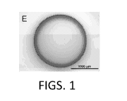
FIG. 1E. A SNOMAX-free alginate bead before frozen.
FIG. 1F. A SNOMAX-free alginate bead after freeze-thawed.
FIG. 2A. The freezing temperature (Ti) of WFI water (water for Injection) in
the
presence of alginate beads containing different local concentrations of SNOMAX
(0.1 or
2.5 mg/ml). The beads were generated by a 30-gauge needle.
FIG. 2B. The freezing temperature (Ti) of WFI water in the presence of
alginate
beads containing different local concentrations of SNOMAX (0.1, 2.5, or 15
mg/ml). The
beads were generated by an 18-gauge needle.
FIG. 3A. The freezing temperature (Ti) of aqueous 10% glycerol in the presence
of alginate beads containing different local concentrations of SNOMAX (0.1,
2.5, or 15
mg/ml). The beads were generated by a 30-gauge needle.
FIG. 3B. The freezing temperature (Ti) of aqueous 10% glycerol in the presence
of alginate beads containing different local concentrations of SNOMAX (0.1,
2.5, or 15
mg/ml). The beads were generated by an 18-gauge needle.
FIG 4. The freezing temperature (Ti) profile that is plotted as a function of
the
total mass of SNOMAX (m) existing in 0.5 ml WFI water. The dark black data
points
correspond to the freezing temperatures of the SNOMAX suspensions of 0.005-100
mg/ml. The curve that best fits all the data points is Tf. = 0.6478 = logiom ¨
3.052
(R2=0.9019) as represented by the solid line.
FIG 5. The freezing temperature (Ti) profile that is plotted as a function of
the
total mass of SNOMAX (m) existing in 0.5 ml aqueous 10% glycerol. The dark
black
data points represent the freezing temperatures of the SNOMAX suspensions of
0.005-50
mg/ml prepared in 10% glycerol. The solid line Tf. = 1.093 = log10(m) ¨ 5.771
is the
best fit to all the data points (R2=0.9571).
7
CA 03029669 2018-12-28
WO 2018/005802
PCT/US2017/040012
FIG. 6 is an image showing a side by side illustration of two samples with
SNOMAX in suspension or in an alginate bead.
FIG 7. Probability distribution of ice nucleation temperature using
nanodroplet
technology, wherein condition 1 is pure heavy water containing 0.1 g/L SNOMAX,
condition 2 is pure water containing 0.1 g/L SNOMAX, condition 3 is pure heavy
water,
and condition 4 is pure water.
FIG. 8. Probability distribution of ice nucleation temperature in bulk samples
(2
mL), wherein condition 1 is 100% heavy water (D20) containing 0.1 g/L SNOMAX
and
condition 4 is pure water (4120).
FIG. 9. Number of caspase positive primary hepatocytes (i.e. dead cells)
normalized o total cells (percent confluence) following short-term
cryopreservation
hours) normalized to total cells.
FIG. 10. Yield of RNA extracted from preserved oncosomes stored at -80 C for
ten days. All experimental conditions were normalized to Day 0 yield, showing
the
.. relative change in RNA yield as a function of time in storage.
FIG. 11. Ice Nucleating Agents enhance preservation viability after freezing.
Fresh primary rat hepatocytes were loaded with 300 mM 30MG for 2 hours and
subjected to a high subzero freeze-thaw protocol either in the presence of an
ice nucleator
(denoted as "SN") or without. Following the thaw, hepatocytes were plated in a
double
gel configuration and their viability was tracked for 5 days using Caspase 3/7
(dead
stain).
FIG. 12A. Bright field images of INPs encapsulated in agarose hydrogel
particles.
FIG. 12B. Hoechst stained INPs contained within hydrogel particles.
FIG. 12C. Frozen encapsulated INPs imaged using a cryostage.
FIG. 12D. Biocompatible INPs can achieve more predictable ice nucleation
temperatures.
FIG. 13A. An overview of the back table separation and machine perfusion of a
split human liver graft.
FIG. 13B. Hemodynamics of the liver lobes (monitored and recorded every 30
min during perfusion).
FIG. 14 shows the sequence of Pseudomonas syringae InaZ.
8
CA 03029669 2018-12-28
WO 2018/005802
PCT/US2017/040012
DETAILED DESCRIPTION
The control of ice nucleation is of fundamental significance in many process
technologies related to food and pharmaceutical science and cryobiology.
Mechanical
perturbation, and electromagnetic fields have been known to induce ice
nucleation in a
controlled manner. These ice-nucleating methods suffer from a number of
disadvantages
such as the lack of consistency and standardization in the case of manual
operations, the
cumbersome process for a large number of samples, and the necessity of
electric and/or
magnetic fields.
It is generally suggested that ice nucleating agents (INA) induce ice
nucleation
.. heterogeneously because the microscopic structure of the INA particle
surface resembles
the ice crystalline structure. One of the major advantages of INAs is their
self-ice-seeding
capability, that is, heterogeneously initiating ice formation at their
characteristic
temperatures without manual or instrumental interruptions. But the use of INAs
in
biological systems may introduce concerns about their biocompatibility,
degradability,
toxicity, recovery and ease of use. It is therefore desirable to minimize the
direct contact
between foreign ice nuclei and biological components in order to avoid the
potential
toxicity and contamination. In addition, the recovery of used INA particles
may also
facilitate the downstream processing. Hydrogel encapsulation can provide a
promising
path to realize these ends.
This disclosure provides, e.g., a model ice-nucleating agent, encapsulated in
microliter-sized alginate beads. This disclosure evaluates the performance of
the bacterial
hydrogel beads to initiate ice nucleation in water and aqueous glycerol
solution by
investigating factors that affect ice nucleation temperature, e.g., the size
and number of
the beads and the local concentration of INA particles. In the aqueous sample
of a fixed
volume, the total mass of the INA particles (m) is found to be the governing
parameter
that is responsible for determining the ice nucleation performance of the
bacterial
hydrogel beads. The freezing temperature has a strong positive linear
correlation with
logio m. The findings provide an effective, predictable approach to control
ice nucleation,
which can improve the outcome and standardization of many ice-assisted process
.. technologies.
9
CA 03029669 2018-12-28
WO 2018/005802
PCT/US2017/040012
This disclosure has also systematically investigated the ice nucleation
kinetics of
picoliter-sized drops of heavy water (e.g., D20 or T20) and light water (H20)
mixtures
with freeze-dried P. syringae. The results showed that the median freezing
temperature of
the 1 mg/ml P syringae suspension prepared in 100% D20 was as high as ¨4.6 C.
Interface-specific sum frequency generation (SFG) spectroscopy and molecular
dynamics
simulation revealed that the hydrogen bonds formed at the water-bacterium
interface
imposed structural ordering on the adjacent water network facilitating ice
nucleation. The
disclosure further investigated the effects of commonly used cryoprotectants
such as
ethylene glycol, propylene glycol and trehalose on the freezing
characteristics of D20 and
H20 mixtures. The results show that the median freezing temperature of the
suspension
containing 1 mg/ml of a lyophilized preparation of P. syringae in 100% D20 is
as high as
-4.6 C, compared to -37.4 C of pure H20. As the D20 concentration increases
by every
25% (v/v), the profile of the ice nucleation kinetics of D20+H20 mixtures
containing 1
mg/ml SNOMAX shifts by about one degree, suggesting an ideal mixing behavior
of
D20 and H20. Furthermore, several cryoprotectants are found to depress the
freezing
phenomenon. Both the homogeneous and heterogeneous freezing temperatures of
these
aqueous solutions depend on the water activity and are independent of the
nature of
solute. These findings can enrich the fundamental knowledge about D20-related
ice
nucleation and suggest that the combination of D20 and ice nucleating agents
can be a
potential self-ice nucleating formulation. The implications of self-nucleation
include a
higher, precisely controlled "ice-seeding" temperature for slow-freezing that
will
significantly improve viability of biological samples that are subject to
various
cryopreservation protocols.
Results in this disclosure also show the effect of higher nucleation
temperatures
on the survival of primary rat hepatocytes undergoing an equilibrium freezing
and
thawing protocol. The data show that changing only the ice nucleation
temperature can
result in 100% survival versus complete death of hepatocytes. Since ice
nucleation is so
critical to success, it is desirable to develop biocompatible ice nucleating
agents,
compositions, and/or formulations with the following features: (1) can be
easily perfused,
(2) will not cross the cell membrane thereby minimizing the probability of
intracellular
ice formation, (3) can be tuned to a certain size to calibrate ice formation
in larger versus
CA 03029669 2018-12-28
WO 2018/005802
PCT/US2017/040012
smaller microcapillaries, and (4) can be easily and completely removed prior
to
transplantation.
Ice Nucleation Temperature
When the temperature is lowered to a liquid's standard freezing point, the
liquid
will crystalize in the presence of a seed crystal or nucleus around which a
crystal
structure can form creating a solid. Lacking any such nuclei, the liquid phase
can be
maintained all the way down to the temperature at which crystal homogeneous
nucleation
occurs. Water normally freezes at 273.15 K (0 C), but it can be "supercooled"
at
standard pressure down to its crystal homogeneous nucleation at about 224.8 K
(-48.3
C). The process of supercooling usually requires that water be pure and free
of
nucleation sites, which can be achieved by processes like reverse osmosis or
chemical
demineralization. When a nucleation site exists or the water is not
substantially pure, the
water will usually freeze at a temperature higher than ¨48.3 C. When the ice
nucleation
occurs, the crystallization around the ice nucleation site will quickly
propagate, and the
water will freeze. Thus, as used herein, the term "ice nucleation temperature"
refers to
the temperature at which the first stable ice embryo of critical size forms.
It is generally believed that uncontrolled ice nucleation destroys
intracellular
structures. When ice nucleation occurs as near as possible to the equilibrium
freezing
point (i.e. the highest temperature which promotes ice crystallization and
propagation,
also known as the melting point), cryodamage or cryoinjury can be minimized.
This is
supported from various evidences. For example, studies in diverse freeze-
tolerant species
have shown that ice nucleating agents (INAs) play a critical role in freezing
survival.
INAs in the blood and in the gut/skin induce controlled freezing of
extracellular water at
multiple nucleation sites. As the hemolymph gradually freezes, it is
accompanied by an
increase in the osmolality of the extracellular fluid, resulting in cellular
dehydration as
water is pulled from the intracellular environment. This provides further
protection since
the cellular fluids are no longer supercooled and ice nucleation in the
intracellular
environment is prevented.
Minimization of injury during cryopreservation may be achieved when ice
nucleation occurs as near as possible to the equilibrium freezing point and
when ice
11
CA 03029669 2018-12-28
WO 2018/005802
PCT/US2017/040012
propagation is more uniform across larger sample volumes. Many studies have
shown the
benefits of controlled nucleation in the cryopreservation of cells for
therapeutics. For
example, embryonic stem cells have been shown to benefit from a slow,
controlled-rate
of cooling with ice nucleation induction at higher temperatures, resulting in
improved
survival rates from less than 22% to more than 90% [28]. One common way to
achieve
higher nucleation temperatures is creating a "cold point" by placing a cold
metal surface
in direct contact with the sample. However, this approach is only suited for
small
volumes since cooling is not uniform in relatively large samples (>1 mL).
Moreover, this
method causes problems with high variability of nucleation rates and is
impractical for
laboratories preserving large numbers of samples. The present disclosure
provides
various compositions and formulations (e.g., hydrogel particles, hydrogel
particle
composition and heavy water composition) to increase ice nucleation
temperature.
Ice Nucleating Agent (INA)
As used herein, the term "ice nucleating agent" or "ice nucleator" refers to
particles or surfaces that can promote ice formation, and initiate ice
nucleation at a higher
temperature when they are added into aqueous systems. As the purpose of the
ice
nucleating agents is to promote ice formation, the ice-nucleating agent does
not include
random or environmental contaminants, e.g., dust or soot. When an ice
nucleating agent
is added to water, the ice nucleation temperature will increase as a result.
While the
precise manner in which an ice nucleating agent accomplishes nucleation is not
well
understood, it is generally believed that ice nucleating agents organize water
molecules in
an ice like fashion, creating water molecule aggregates that are sufficiently
large to
nucleate at relatively higher temperatures. The ice nucleating agent can
effectively
promote ice formation. In some embodiments, the ice nucleating agent in a
sufficient
amount can increase the ice nucleation temperature by at least 1 C, 2 C, 3
C, 4 C, 5
C, 6 C 7 C 8 C 9 C 10 C 11 C 12 C 13 C 14 C 15 C 16 C 17 C 18 C
19 C, 20 C, 21 C, 22 C, 23 C, 24 C, 25 C, 26 C, 27 C, 28 C, 29 C,
30 C, 31
C, 32 C, 33 C, 34 C, 35 C, 36 C, 37 C, or 38 C. In some embodiments,
the ice
nucleating agent in a sufficient amount can increase the ice nucleation
temperature
12
CA 03029669 2018-12-28
WO 2018/005802
PCT/US2017/040012
to over -10 C, -9 C, -8 C, -7 C, -6 C, -5 C, -4 C, -3 C, -2 C, or -1
C. In some
embodiments, the sufficient amount refers to 0.001 mg, 0.005mg, 0.01 mg, 0.05
mg, 0.1
mg, 0.5 mg, 1 mg, 5 mg, or 10 mg that are tested in 0.5 ml, 1 ml, or 1.5 ml
pure water.
Thus, in some embodiments, 0.01 or 0.1 mg of the ice nucleating agent as
described
herein can increase ice nucleation temperature of 0.5 ml pure water to over -6
C or -5 C
(see e.g., FIG. 4).
The ice nucleating agent may be organic or inorganic. For example, the ice
nucleating agents can be inorganic materials, such as fine particulates
(microparticles,
nanoparticles, mineral particles, or the like), or silver iodide, silver
oxide, or alumina
crystals. The ice nucleating agent can also be organic compounds, such as
carbohydrates, phospholipids, proteins, alcohols, amino acids (e.g., aspartic
acid), or
lipoproteins. In some embodiments, the ice nucleating agent is long chain
aliphatic
alcohols. The ice nucleating agent can also be microorganism, e.g., virus,
bacteria (e.g.,
ice nucleating bacteria), or fungi. Some commonly used ice nucleating agents
include
silver iodide, IceStartTM (Asymptote, Cambridge, UK) and SNOMAX (SNOMAX
LLC, Englewood, CO).
IceStartTM is a biocompatible material that acts as an ice nucleating agent
during
the cooling of aqueous solutions. It is an ice-nucleating agent composed of
biologically
inert mineral particles.
SNOMAX is a "snow inducer" based on proteins from the bacterium
Pseudomonas syringae. These proteins act as extra nuclides to improve the
crystallization
process. Pseudomonas syringae proteins are extracted from the microorganisms
to
produce SNOMAX. After fermentation, the proteins are separated from the fluid
and
processed using special filters to form a slurry. This slurry is then frozen
and freeze-dried.
.. Any remaining living bacteria are killed in the process. Thus, SNOMAX is
the freeze-
dried form of the ice-nucleating protein extracted from Pseudomonas syringae.
Pseudomonas syringae is a rod-shaped, Gram-negative bacterium. It produces an
ice nucleation active (INA) protein, which causes water to freeze at fairly
high
temperatures (-4 to -2 C). The ice nucleation active protein refers to a
family of proteins
that enable Gram-negative bacteria to promote nucleation of ice at relatively
high
temperatures. These proteins are usually localized at the outer membrane
surface. The ice
13
CA 03029669 2018-12-28
WO 2018/005802
PCT/US2017/040012
nucleation active protein in Pseudomonas syringae is known as ice nucleation
active
protein InaZ (UniProt P06620-1; SEQ ID NO: 1). The primary structure of the
proteins
contains a highly repetitive domain that dominates the sequence (SEQ ID NO:
2).
AGYGSTxTagxxssli AGYGSTxTagxxsxlt AGYGSTxTaqxxsxlt (SEQ ID NO: 2)
wherein x in SEQ ID NO: 2 represents any amino acid. The domain comprises a
number
of 48-residue repeats, which themselves contain 3 blocks of 16 residues, the
first 8 of
which are identical. It is thought that the repetitive domain may be
responsible for
aligning water molecules in the seed crystal. (See Wolber PK, Green RL (1990).
"Detection of bacteria by transduction of ice nucleation genes". Trends
Biotechnol. 8
(10): 276-279; Gurian-Sherman D, Lindow SE (1993). "Bacterial ice nucleation:
significance and molecular basis". FASEB J. 7 (14): 1338-1343). Pseudomonas
syringae
and strains of Pseudomonas syringae are described in U.S. Patent No.
5,489,521, which
is incorporated herein by reference in its entirety.
Other microorganisms, or proteins from these microorganisms, can also be used
as ice nucleating agents, e.g., Pseudomonas fluorescens, Pseudomonas
coronafaciens,
Pseudomonas pisi, Erwinia species, Erwinia ananas, Erwinia herbi cola,
Escherichia
co/i, Xanthomonas, ice-nucleating fungi and/or ice-nucleating protozoa. These
ice
nucleating agents are described, e.g., in WO/2011/026020, which is
incorporated herein
by reference in its entirety.
Hydrogel Particles
As used herein, the term "hydrogel particle" refers to a particle made of
hydrogel.
Hydrogels are highly hydrophilic natural or synthetic polymeric networks. The
present
disclosure provides hydrogel particles that contain an ice nucleating agent.
The ice
nucleating agent is enclosed, encapsulated, or embedded within the hydrogel
particles.
The hydrogel particles can have various shapes, e.g., spheres, beads, and can
have similar
shapes and sizes. The hydrogel particles have several advantages. First, as
the ice
nucleating agents (e.g., freeze-dried P syringae) are encapsulated into
hydrogel beads,
the interaction between the INA and the biological system is minimized. Thus,
the
14
CA 03029669 2018-12-28
WO 2018/005802
PCT/US2017/040012
hydrogel particles are less likely to have toxic effects. Second, the
particles can also be
easily removed from the biological system.
The hydrogel particles as described in the present disclosure can be made by
any
methods known in the art. For example, in some embodiments, the ice nucleating
agent
(e.g., SNOMAX) can be mixed with water and hydrogel solution (e.g., alginate
solution).
The concentration of the ice nucleating agent in the mixture is the local
concentration of
the ice nucleating agent (This is also the concentration of the ice nucleating
agent within
the hydrogel particles). The mixture is then loaded into a syringe, and is
then dropped
into a hardening bath (e.g., a 1 % (w/v) CaCl2 hardening bath). The drops are
then gelled
in the hardening bath and further washed by purified water (e.g., Water for
Injection
(WFI)). The hydrogel particles can also be produced by flow focusing
microfluidics
device. For example, a solution containing the ice nucleating agent and the
hydrogel
solution (e.g., ¨3% agarose) can pass through the flow focusing microfluidics
device to
give rise to hydrogel droplets or hydrogel bubbles (particles). Similarly, the
concentration
of the ice nucleating agent in the solution will be the concentration of ice
nucleating
agent within the hydrogel particles.
Thus, the concentration of ice nucleating agent within the hydrogel particles
(local
concentration Cio./) can be easily adjusted. The concentration of ice
nucleating agent can
range from 0.01 mg/ml to 30 mg/ml, 0.1 mg/ml to 20 mg/ml, 0.5 mg/ml to 15
mg/ml, or 1
mg/ml to 5 mg/ml. In some embodiments, the concentration of ice nucleating
agent is
greater than 0.01 mg/ml, 0.1 mg/ml, 0.5 mg/ml, 1 mg/ml, 2 mg/ml, 3 mg/ml, 4
mg/ml, 5
mg/ml, or 10 mg/ml. In some embodiments, the concentration of ice nucleating
agent is
less than 30 mg/ml, 20 mg/ml, 10 mg/ml, 5 mg/ml, 4 mg/ml, 3 mg/ml, 2 mg/ml, 1
mg/ml,
or 0.5 mg/ml.
The hydrogel particles can also have various sizes. For example, the hydrogel
particles can be microliter-sized particles or nanoparticles. As used herein,
the term
"nanoparticle" refers to a particle between 1 and 100 nanometers in size. In
some
embodiments, the hydrogel particles can have a diameter less than 10 mm, less
than 9
mm, less than 8 mm, less than 7 mm, less than 6 mm, less than 5 mm, less than
4 mm,
less than 3 mm, less than 2 mm, less than 1 mm, or less than 0.5 mm. In some
embodiments, the hydrogel particles can have a diameter greater than 10 mm,
greater
CA 03029669 2018-12-28
WO 2018/005802
PCT/US2017/040012
than 9 mm, greater than 8 mm, greater than 7 mm, greater than 6 mm, greater
than 5 mm,
greater than 4 mm, greater than 3 mm, greater than 2 mm, greater than 1 mm, or
greater
than 0.5 mm. As used herein, the term "diameter" refers to the longest chord
of the circle
on a sphere; when the particle is not a sphere, the diameter refers to the
maximum length
of a straight line connecting one point of the particle to another point of
the particle.
The hydrogel particles can also have various volumes. In some embodiments, the
hydrogel particle can have a volume less than 100 jtl, less than 50 jtl, less
than 30 jtl, less
than 20 jtl, less than 15 jtl, less than 10 jtl, less than 5 jtl, less than 4
jtl, less than 3 jtl, less
than 2 jtl, or less than 1 pl. The hydrogel particle can also have a volume
greater than 50
jtl, greater than 30 jtl, greater than 20 jtl, greater than 15 jtl, greater
than 10 jtl, greater
than 5 jtl, greater than 4 jtl, greater than 3 jtl, greater than 2 jtl, or
greater than 1 pl.
The hydrogel in the hydrogel particles can be generated by a gel-former
derived
from a natural or synthetic polymer compound. The gel-former derived from a
natural
polymer compound includes, for example, agar, agarose, alginic acid
(alginate), gelatin,
gum arabic, quince seed mucous substance, tragacanth gum, guar gum, karaya
gum,
locust bean gum, glucomannan, pectin, galactan, pullulan, xanthan gum, casein,
casein
potassium salt, casein sodium salt, sodium chondroitin sulfate, starch-based
semisynthetic
polymer compounds (for example, carboxymethyl starch, methylhydroxypropyl
starch,
methylhydroxymethyl starch etc.) and dextrin. These gel-formers can be used
alone or as
a mixture of two or more thereof. In some embodiments, the hydrogel particle
is an
agarose hydrogel particle or an alginate hydrogel particle. Some of these gel-
formers are
described in U.S. 8222193, which is incorporated by reference herein in its
entirety. The
concentration of the gel-former in the hydrogel parties usually ranges from
0.5 4-10%
(w/w weight percentage), e.g., 1%, 2%, 3%, 4%, 5%, 6%, 7%, 8%, 9%, or 10%.
The hydrogel particles can contain water. In some embodiments, the hydrogel
particles can contain heavy water (e.g., D20 or T20). Some other compounds can
also be
enclosed in the hydrogel particles. For example, in some embodiments, the
hydrogel
particles can contain a cryoprotectant as described herein (e.g., DMSO, EG,
PROH, 3-
0MG, or glycerol).
As shown in the present disclosure, the hydrogel particles described herein
can
increase ice nucleation temperature. In some embodiments, the hydrogel
particles
16
CA 03029669 2018-12-28
WO 2018/005802
PCT/US2017/040012
described herein can increase the ice nucleation temperature to over -10 C, -
9 C, -8 C,
-7 C, -6 C, -5 C, -4 C, -3 C, -2 C, or -1 C.
Furthermore, the ice nucleation temperature of similar samples can be
different. In
some cases, even the same sample can have different ice nucleation temperature
due to
the stochastic nature of ice nucleation. Thus, there is a range for the ice
nucleation
temperature (see e.g., FIG. 2A), e.g., -8 C ¨ -4 C. As used herein, the term
"range"
refers to the difference between the maximum ice nucleation temperature and
the
minimum ice nucleation temperature. For example, the range of -8 C ¨ -4 C is
4 C.
The hydrogel particles described herein can reduce the range of ice nucleation
.. temperature of a plurality of samples. For example, the range of ice
nucleation
temperature can be reduced to 5 C, 4 C, 3 C, 2 C, or 1 C. Thus, hydrogel
particles
described herein can initiate ice formation in a more predictable manner.
The ice nucleating agent in the hydrogel particle can have a leakage rate less
than
10%, 5%, 4%, 3%, 2%, 1%, 0.5%, or 0.1%. The leakage rate is defined as the
amount of
ice nucleating agents that are observed outside the hydrogel matrix (e.g.,
hydrogel
particle) divided by the total amount of ice nucleating agents that are
initially
encapsulated in the hydrogel matrix. The amount of ice nucleating agents can
be
determined by various means known in the art. For example, the ice nucleating
agents
can be fluorescently labeled, and the amount can be determined by fluorescence
intensity.
Hydrogel Particle Compositions
The present disclosure also provides compositions comprising hydrogel
particles.
In some embodiments, the composition also includes water (e.g., H20, D20 or
T20).
The composition can also include one, two, or more than two cryoprotectants as
described herein, e.g., within the hydrogel particles as described above, or
separately in
the composition.
As shown in the present disclosure, the ice nucleation performance of the
hydrogel particles (e.g., SNOMAX-laden hydrogel beads) has been characterized
by
adjusting various factors, such as the size, number of the hydrogel particles,
the local ice
.. nucleation agent concentration C/./ (i.e., the concentration of the ice
nucleation agent
within the hydrogel matrix), and the addition of glycerol. It has been
determined the total
17
CA 03029669 2018-12-28
WO 2018/005802
PCT/US2017/040012
mass of local ice nucleation agent is a parameter that determines the ice
nucleation
temperature of the aqueous sample of a fixed volume (e.g., 0.1 ml, 0.5 ml, 1
ml, 5 ml, 10
ml, or 100 m1). Thus, the present disclosure provides compositions comprising
ice
nucleating agent of various mass. The total mass of the ice nucleating agent
(e.g.,
SNOMAX) in the composition can be determined by the formula
m = N = V = Clocal,
wherein m is the total mass of the ice nucleating agent, N is the number of
the hydrogel
particles in the composition, V is the volume or average volume of the
hydrogel particles,
and Clocal is the local concentration of the hydrogel particle. Thus, in some
embodiments,
.. the total mass of the ice nucleating agent (e.g., SNOMAX) in the
composition is over
0.001, 0.01, 0.1, 1, 10, 100, 1000, 104, 105, or 106 mg. In some embodiments,
the total
mass of the ice nucleating agent (e.g., SNOMAX) in the composition is less
than 0.001,
0.01, 0.1, 1, 10, 100, 1000, 104, 105, or 106 mg.
In some embodiments, the composition can also include cryoprotectants, and/or
a
preservative, e.g., an antibiotic.
Heavy Water Compositions
Heavy water is a form of water in which the common hydrogen-1 ('H) isotope is
substituted with a larger than normal amount of the deuterium isotope (D or
2H) or
tritium isotope (T or 3H). Thus, as used herein, heavy water refers to D20 or
T20. The
toxicity of heavy water varies from simple to complex organisms: algae and
bacteria can
adapt to grow in 100% D20 and actually serve as a source for deuterated
molecules,
while concentrations of more than 20% can be toxic to animals and animal cells
at
normal body temperatures. At the cellular level, D20 has been shown to slow
down the
cell cycle and lengthen circadian rhythms, and increases the heat stability of
macromolecules but may decrease the cellular response to heat stress (possibly
as a result
of inhibition of chaperon function). Furthermore, D20 has been shown to
improve the
stability of vaccinations as a result of its protective effects on
biomolecules (e.g. proteins
and nucleic acids) and has applications in pharmaceuticals since it affects
drug
metabolism.
18
CA 03029669 2018-12-28
WO 2018/005802
PCT/US2017/040012
Amongst the many broad applications of heavy water described above, D20 is a
more potent ice nucleator than H20. Thus, in one aspect, the present
disclosure provides a
composition comprising heavy water (D20 or T20) and an ice nucleating agent.
In some
embodiments, the heavy water is D20.
The heavy water content in the composition can vary. In some embodiments, the
weight percentage of heavy water in the composition can be over 5%, 10%, 20%,
25%,
30%, 40%, 50%, 60%, 70%, 80%, 90%, or 95%. In some embodiments, the weight
percentage of heavy water in the composition can be less than 10%, 20%, 25%,
30%,
40%, 50%, 60%, 70%, 80%, 90%, or 95%.
The composition can also contain H20. In some embodiments, the volume
percentage of heavy water (D20 or T20) of all water in the composition (v/v)
can be over
5%, 10%, 20%, 25%, 30%, 40%, 50%, 60%, 70%, 80%, 90%, or 95%. In some
embodiments, the volume percentage can be less than 10%, 20%, 25%, 30%, 40%,
50%,
60%, 70%, 80%, 90%, or 95%. In some embodiments, the volume percentage is
100%,
which means all water in the composition is heavy water (e.g., D20 or T20).
The composition can include one, two, or more than two ice nucleating agents
(e.g., SNOMAX or silver iodide). The concentration of the ice nucleating agent
will
affect the ice nucleation temperature. In some embodiments, the concentration
can be
greater than 0.001 g/L, 0.01 g/L, 0.05 g/L, 0.1 g/L, 0.2 g/L, 0.3 g/L, 0.4
g/L, 0.5 g/L, 0.6
g/L, 0.7 g/L, 0.8 g/L, 0.9 g/L, 1 g/L, 5 g/L, or 10 g/L. In some embodiments,
the
concentration can be less than 0.001 g/L, 0.01 g/L, 0.05 g/L, 0.1 g/L, 0.2
g/L, 0.3 g/L, 0.4
g/L, 0.5 g/L, 0.6 g/L, 0.7 g/L, 0.8 g/L, 0.9 g/L, 1 g/L, 5 g/L, or 10 g/L. In
some
embodiments, the concentration can have a range, e.g., 0.1 g/L 0.5 g/L.
As shown in the present disclosure, the heavy water composition can increase
ice
nucleation temperature. In some embodiments, the heavy water composition can
increase
the ice nucleation temperature to over -10 C, -9 C, -8 C, -7 C, -6 C, -5
C, -4 C, -3
C, -2 C, or -1 C. The heavy water composition described herein can also
reduce the
range of ice nucleation temperature. For example, the range of ice nucleation
temperature
can be reduced to 5 C, 4 C, 3 C, 2 C, or 1 C.
In some embodiments, the composition can also include hydrogel particles,
cryoprotectants, and/or a preservative.
19
CA 03029669 2018-12-28
WO 2018/005802
PCT/US2017/040012
Cryoprotectants
The composition as described herein (e.g., various hydrogel particle
compositions
and various heavy water compositions) can also include one, two, or more than
two
cryoprotectants. As used herein, the term "cryoprotectant" refers to a
substance that
prevents or reduce damage to cells during freezing. Various cryoprotectants
can be
included in the compositions described in the present disclosure. These
cryoprotectants
include, e.g., sugar, polypropylene glycol, dimethylsulfoxide (DMSO), dextran,
glycerol,
sorbitol, propylene glycol, ethylene glycol, pyridine, 2-3 butane diol,
hydroxyethyl
starch, polyvinylpyrrolidone (PVP), proline (or other protein stabilizers),
human serum
albumin and combinations thereof. The sugar can also be any one of the
following, e.g.,
sucrose, trehalose, raffinose, stachyose, fructose, and dextran. Exemplary
sugars and the
concentration ranges for such sugars are described in U.S. Patent Nos.
6,673,607 and
7,094,601, herein incorporated by reference.
The cryoprotectant can be either membrane-permeable or non-permeable. The
permeable cryoprotectants include, e.g., DMSO, alcohol such as ethylene glycol
(EG),
PROH (propylene glycol, propane-1,2-diol, or 1,2-propanediol), glycerol, and
saccharide
derivatives such as 3-0-methyl-glucose (3-0MG). The non-permeable
cryoprotectants
include, e.g., saccharides such as fructose, trehalose, sucrose, sorbitol, or
raffinose,
polymers such as hydroxyethyl starch (HES) or polyvinylpyrrolidone (PVP),
amino acids
such as L-proline, and biological macromolecules such as human serum albumin,
and any
combinations thereof. In some embodiments, the cryoprotectant is sucrose,
trehalose,
stachyose, raffinose, or polymers (e.g. PEG, PVA, HES). In some embodiments,
the
composition includes University of Wisconsin (UVV) solution, 3-0MG, Trehalose,
N-
.. acetyl-L-cysteine, and/or hydrogen sulfide.
The cryoprotectant can have various concentrations (w/v), e.g., 1%, 2%, 3%,
4%,
5%,10%, 15%, 20%, 25%, 30%, 35%, 40%, or 50%. As used herein, the
"weight/volume
(w/v) concentration" or "weight/volume (w/v) percentage" refers to the weight
(in grams)
of solute dissolved in a final volume of 100 mL of solution. For example, the
concentration 1% (w/v) refers to a solution with 1 g of solute dissolved in a
final volume
of 100 mL of solution. In some embodiments, the concentration (w/v) can be
greater than
CA 03029669 2018-12-28
WO 2018/005802
PCT/US2017/040012
1%, 2%, 3%, 4%, 5%,10%, 15%, 20%, 25%, 30%, 35%, 40%, or 50%. In some
embodiments, the concentration (w/v) can be less than 1%, 2%, 3%, 4%, 5%,10%,
15%,
20%, 25%, 30%, 35%, 40%, or 50%. For example, the concentrations (w/v) for the
cryoprotectants (e.g., DMSO, EG, PROH, glycerol, propylene glycol, pyridine, 2-
3
butane diol, or human serum albumin) can be 1%, 2%, 3%, 4%, 5%,10%, 15%, 20%,
25%, 30%, 35%, or 40%. In some embodiments, the concentration (w/v) for the
cryoprotectants is less than 40%.
The concentration of a solute can also be expressed as a weight percentage
(w/w).
The concentration 1% (w/w) refers to a solution with 1 g of solute dissolved
in a 100 g of
the final solution (including both the solute and the solvent). In some
embodiments, the
concentration (w/w) of a cryoprotectant can be greater than 1%, 2%, 3%, 4%,
5%,10%,
15%, 20%, 25%, 30%, 35%, 40%, or 50%. In some embodiments, the concentration
(w/w) can be less than 1%, 2%, 3%, 4%, 5%,10%, 15%, 20%, 25%, 30%, 35%, 40%,
or
50%. For example, the concentration (w/w) for the cryoprotectants (e.g., PVP)
can be
less than 5%, e.g., 1%, 2%, 3%, 4%, or 5%.
The amount of a solute in a solution can also be expressed in molar
concentration.
A commonly used unit for molar concentration is the molar (M) which is defined
as the
number of moles per liter. In some embodiments, the concentration of a
cryoprotectant
can be higher than 50 mM, 100 mM, 200 mM, 300 mM, 400 mM, 500 mM, 600 mM,
700 mM, 800 mM, 900 mM, 1 M, 2M, 3M, or 4M. In some embodiments, the
concentration of a cryoprotectant can be less than 50 mM, 100 mM, 200 mM, 300
mM,
400 mM, 500 mM, 600 mM, 700 mM, 800 mM, 900 mM, 1 M, 2M, 3M, or 4M. For
example, the concentration of a cryoprotectant (e.g., sucrose, sorbitol,
fructose, trehalose,
raffinose, hydroxyethyl starch, 3-0MG) can be equal to or less than 1M, e.g.,
100 mM,
200 mM, 300 mM, 400 mM, 500 mM, 600 mM, 700 mM, 800 mM, 900 mM, or 1 M.
The concentration of a cryoprotectant (e.g., proline) can be equal to or less
than 300 mM,
e.g., 100 mM, 200 mM, or 300 mM.
Cryoprotectants can be added to the composition as described herein (e.g.,
hydrogel particle compositions and heavy water compositions) as a single agent
or as a
combination of one or more agents. For example, 2M ethylene glycol or 1,2-
propanediol
(PROH) can be supplemented with 0.5 to 2M sugar to produce a synergistic
effect. For
21
CA 03029669 2018-12-28
WO 2018/005802
PCT/US2017/040012
example, a combination of ethylene glycol and a sugar or a combination of PROH
and a
sugar can be used. In one example, 2M PROH and 0.5 M trehalose are added to
the
composition. In another example, 0.3M sucrose and 1.5M PROH is used. The
combination of a permeating and non-permeating cryoprotectant allows for a
lower
intracellular concentration of cryoprotectant, since the non-permeating
cryoprotectant
does not enter the cell. For example, in the 2M PROH and 0.5 M trehalose
example
described above, the intracellular concentration of cryoprotectant would be 2M
since
trehalose is not permeable to the plasma membrane of mammalian cells.
Nanoparticles and Microparticles
The compositions as described herein can further include nanoparticles or
micro-
particles or both. The addition of nanoparticles or microparticles is thought
to enhance
the thermal conductivity of the composition.
Examples of such nanoparticles or microparticles include particles having
carbon
or a noble metal, such as gold, silver, titanium, palladium, platinum, or
similar particles
thereto. Examples of such nanoparticles and/or microparticles may include, but
are not
limited to, carbon or noble metals, e.g., gold, silver, titanium, palladium,
platinum, and
copper. In one aspect of the disclosure, the nanoparticles are present in the
composition
as described herein in an amount up to 99%, 50%, 25%, 20%, 10%, 5% or lower,
based
.. on the total weight of the composition. In another aspect of the
disclosure, the
microparticles are present in the composition as descried herein in an amount
up to 99%,
99%, 50%, 25%, 20%, 10%, 5%, based on the total weight of the composition. It
has
been shown that the presence of a small fraction (<1% vol) of nanoparticles in
a
composition can increase the thermal conductivity of the composition up to
more than
.. 200% (Choi et al., Applied Physics Letter 79: 2252- 2254, 2001; Eastman et
al., Applied
Physics Letter 78: 718-720, 2001).
Preserving Biologic Samples, Organs, or Organisms
The disclosure provides methods of preserving a biological sample. The methods
.. include the steps of contacting the biological sample with compositions or
formulations
as described herein (e.g., hydrogel particle compositions, heavy water
compositions), and
22
CA 03029669 2018-12-28
WO 2018/005802
PCT/US2017/040012
freezing the biological sample with the composition or formulations. The
biological
sample can be a cell, a tissue sample, oncosomes, exosomes, microvesicles or
liposomes.
The biological sample can also contain nucleic acids (e.g., DNA, RNA, mRNA,
microRNA etc.), proteins, and/or lipids. Thus, in some embodiments, the
compositions or
formulations as described herein can improve the yield of nucleic acids after
being frozen
and thawed.
The disclosure also provides methods of preserving an organ. The methods
include the steps of perfusing, contacting, or immersing the organ with
compositions or
formulations as described herein, and freezing the organ with the composition
or
formulations. The methods of perfusing an organ is known in the art. For
example,
perfusion can be performed by pouring over or through the arteries or veins of
the organ.
In some embodiments, a perfusion device can be used. The organ can also be
immersed
within the compositions or formulations. The organ can be any organ of a
mammal, e.g.,
heart, lung, kidney, and liver etc. Furthermore, during static cold storage of
organs, many
harmful processes can contribute to short preservation times including ATP
depletion,
calcium overload, production of reactive oxygen species, cytoskeleton
disruption, and
cellular acidosis, all of which are magnified by ischemia-reperfusion injury
following
storage. D20 has been shown to inhibit cytosolic calcium, improve microtubule
stability,
stabilize membranes and proteins [39], thus, the compositions and formulations
described
herein can also minimize ischemia-reperfusion damage.
The disclosure also provides methods of preserving an organism. The methods
include the steps of contacting, or immersing the organism with compositions
or
formulations as described herein, and freezing the organism with the
composition or
formulations. The organism can be viruses, bacteria, fungi, invertebrates
(e.g., insects),
fish, or reptiles.
The biological samples, organs, or organisms can be frozen at the ice
nucleation
temperature. In some embodiments, the ice nucleation temperature is over -10
C, -9 C,
-8 C, -7 C, -6 C, -5 C, -4 C, -3 C, -2 C, or -1 C. The methods can
further include
the step of thawing the biological samples, organs, or organisms. In addition,
the methods
as described herein can further be used to improve the outcome and
standardization of
23
CA 03029669 2018-12-28
WO 2018/005802
PCT/US2017/040012
many ice-assisted process technologies such as the slow-freezing
cryopreservation of
stem cells for regenerative medicine, cellular therapies, and drug screening.
The biological samples can include mammalian cells. The methods described
herein can be used for the cryopreservation of any type and any species of
mammalian
cells. For example, the methods can be used to cryopreserve oocytes or sperm
in assisted
reproductive technology or for patients undergoing chemotherapy or radiation
therapy.
The methods can also be used for the cryopreservation of stem cells, such as
embryonic
stem cells, or other cells, which can then be used as the basis of stem cell-
based therapies,
cell transplantation, tissue engineering, and regenerative medicine. The
methods can also
be used to cryopreserve oocytes or sperm from an animal that is rare or at
risk of
becoming extinct for future use in assisted reproductive technologies for the
preservation
of the species. The methods can further be used for animal husbandry purposes
(e.g., the
breeding and raising of animals), for example, for the cryopreservation of
embryonic
stem cells, gametocytes, oocytes, or sperm from animals such as cows, pigs,
and sheep.
Cell types that may be cryopreserved using the compositions and methods of the
present disclosure include, for example, differentiated cells, such as
epithelial cells,
cardiomyocytes, neural cells, epidermal cells, keratinocytes, hematopoietic
cells,
melanocytes, chondrocytes, B-cells, T-cells, erythrocytes, macrophages,
monocytes,
fibroblasts, or muscle cells; and undifferentiated cells, such as embryonic,
mesenchymal,
or adult stem cells. Additional cell types that can be cryopreserved using the
methods of
the disclosure include gametocytes, oocytes, sperm, zygotes, and embryos.
Other cells
include those from the bladder, brain, esophagus, fallopian tube, heart,
intestines,
gallbladder, kidney, liver, lung, ovaries, pancreas, prostate, spinal cord,
spleen, stomach,
testes, thymus, thyroid, trachea, ureter, urethra, or uterus.
The cells may be from a human or non-human mammal, for example
Cercopithecoidea family, Hominoidea superfamily, Canis familiaris, Fe/is
catus,
Cricetidae spp., Equus spp. (e.g., Equus cabal/us, Equus assinns), Equidae
family, Bos
taurus, Bos indicus, Bovidae family, Camelidae family, Bubalus bubalis, Capra
aegagrus hircus, Cervidae family, Cervinae family, Ovis aries, Ovis
canadensis, Capra
hircus, Sus scrofa domestica, Mesocricetus spp., Mustela vison, Cavia
porcellus,
Meriones unguiculatus, Chinchilla laniger, Rattus norvegicus, Rattus spp., Mus
24
CA 03029669 2018-12-28
WO 2018/005802
PCT/US2017/040012
musculus, Leporidae family, Oryctolagus cuniculus, Kobus spp., Gallus spp.,
Meleagria
gallopavo, Anatidae spp., Mustela putorius, Columba domestica, Columba livia,
Numida
meleagris, Ornithorhynchus anatinus, Pavo cristatus, Bison spp., Struthio
spp., Lama
glama, Rhea spp., Dromiceius spp., Lama pacos, Rangifer tarandus, Bos
grunniens,
Came/us bactrianus, Came/us dromedarius, and any endangered or threatened
species
(e.g., those species identified by the U.S. Fish and Wildlife Service (USFWS)
Threatened and Endangered Species System (TESS)).
The cells are prepared for cryopreservation using techniques known in the art
and
described herein. Generally, the mammalian cells are obtained using art known
techniques and maintained in media appropriate for the cell type.
Viability of Cryopreserved Biological Samples
When desired, the cryopreserved biological samples of the disclosure can be
warmed, using methods known in the art or described herein. For example, the
biological
sample can be plunged into a 1xPBS solution at 20-37 C, for example room
temperature,
optionally with shaking and optionally supplemented with sugar or other
cryoprotectants.
After warming, the biological samples (e.g., cells) are generally washed,
suspended in the
appropriate media and treated as needed for use in research or clinical
applications. For
example, embryonic stem (ES) cells can be plated and passaged using techniques
known
in the art. Oocytes are generally cultured in droplets of media immersed in
oil. It will be
clear to the skilled artisan the exact conditions and media that are used for
culturing the
cells before and after cryopreservation.
There are various tests known in the art to determine the viability and
function of
the biological samples (e.g., cells) after warming and these tests are
dependent on the
types of biological samples. For example, for ES cells that are to be used for
cell-based
therapeutics, maintenance of pluripotency is very important. The pluripotency
of the ES
cells can be tested using art known methods, including, for example, 0ct4-GFP
expression, elevated alkaline phosphatase expression, and S SEA-I surface
glycoprotein
expression. The ability of cells to attach efficiently is another assay for
the viability and
usability of many cells. Attachment assays are known in the art and described
herein.
Proliferation assays can also be used to determine if the attached cells can
proliferate as
CA 03029669 2018-12-28
WO 2018/005802
PCT/US2017/040012
expected after cryopreservation. Attachment and proliferation efficiency can
be
compared to control cells, which have not undergone cryopreservation. For
cryopreservation of zygotes, cleavage rates can be determined after
cryopreservation and
compared to control groups to determine if there has been any cellular damage
during the
cryopreservation process. The viability of oocytes can be determined by
examination of
the morphological characteristics of the cells following cryopreservation.
Morphologically viable oocytes exhibit intact zona pellucida and plasma
membrane and
refractive cytoplasm, while non-viable oocytes appear degenerated when
visualized under
a light microscope. The ultimate criterion for oocyte viability and function
is their
capability to be fertilized by healthy sperm in vitro and in vivo, followed by
cleavage,
blastocyst, and/or hatching or development of the fetus.
The assays for testing the viability, function, and usability of biological
samples
can also be used to test parameters for the cryopreservation methods described
herein.
For example, variations in the compositions (e.g., different cryoprotectants
or
cryoprotectant concentrations) can readily be tested on cells, and their
effects on the
viability, function, and usability of biological samples can be tested using
any of the
methods described herein or known in the art.
Freezing Systems
The present disclosure also provides freezing systems. The freezing system can
include a container for biologic samples, organs, or organisms. The container
can be e.g.,
a bag, a plastic vial, a glass vial, a plastic straw, a pulled straw, a
capillary tube or straw,
and/or a bioreactor. The freezing system can also include a cooling system.
The freezing
systems can further include the compositions or formulations as described
herein. In
some embodiments, the ice nucleating agent or the hydrogel particle as
described in the
present disclosure can be immobilized on the surface of the freezing system.
Designing Compositions or Formulations for Specific Ice Nucleation Temperature
The ice nucleation performance of the hydrogel particles has been
characterized
in the present disclosure. In the aqueous sample of a fixed volume, the total
mass of the
INA in the particles has been identified as a universal parameter that
dictates the ice
26
CA 03029669 2018-12-28
WO 2018/005802
PCT/US2017/040012
nucleation temperature. These findings provide the guidance to tune the ice
nucleation
temperature within a 6-degree window or even larger by adjusting the size and
number of
hydrogel particles and the local INA concentration.
As shown in FIG. 4, the freezing temperature (ice nucleation temperature) is
plotted as a function of the total mass of ice nucleating agents (m = N = V =
clocca, in a
log10 scale) existing in 0.5 ml water. The freezing temperatures of
suspensions of ice
nucleating agents are also measured. All the data points shown in FIG. 4 share
the same
trend. The Pearson's correlation suggests a clear positive linear relationship
between Tf.
and logiom. The curve that best fits all the data points is
Tf. = 0.6478 = logiom ¨ 3.052 (R2=0.9019).
This demonstrates that the total mass of ice nucleation temperature is the
parameter to
dictate the occurrence of ice nucleation in 0.5 ml water regardless of the
form of presence
of the INA particles, either in a suspension or in a confined space.
In FIG. 5, the freezing temperatures of aqueous 10% glycerol solution is
plotted
as a function of m in log10 scale. The curve that best fits all the data
points is
Tf. = 1.093 = log10(m) ¨ 5.771 (R2=0.9571)
The data show that the addition of solute does not change the role of the
total mass of
INA particles as the governing parameter. Therefore, one can increase the
total mass of
INA particles by adjusting the combination of N, V and clocal to increase the
ice
nucleation temperature.
Furthermore, the concentration of D20 has a predictable impact on the ice
nucleation temperature. For example, if the D20 concentration increases by
every 25%
(v/v), the profile of the ice nucleation kinetics of D20+H20 mixtures
containing 1 mg/ml
SNOMAX shifts by about one degree.
Therefore, the present disclosure provides methods of designing compositions
and
formulations that have a desired ice nucleation temperature. In one aspect,
the methods
involve selecting a desired ice nucleation temperature, determining the
optimal values for
N, V and c local based on a predetermined curve that correlates ice nucleation
temperature
and total mass of the ice nucleating agents, producing a hydrogel particle
composition
with the determined values of N, V and Clocal= The predetermined curve can be
the curve
as shown in FIG. 4 and FIG. 5. Furthermore, a person skilled in the art can
easily
27
CA 03029669 2018-12-28
WO 2018/005802
PCT/US2017/040012
determine a curve that correlates ice nucleation temperature and total mass of
the ice
nucleating agents by the methods described in the present disclosure.
In another aspect, the methods involve selecting a desired ice nucleation
temperature, determining the optimal values for D20 concentration based on a
predetermined curve that correlates ice nucleation temperature and the D20
concentration, producing a heavy water composition with the determined D20
concentration. A person skilled in the art can also easily determine a curve
that correlates
ice nucleation temperature and the D20 concentration by the methods described
in the
present disclosure.
EXAMPLES
The invention is further described in the following examples, which do not
limit
the scope of the invention described in the claims.
EXAMPLE 1. SNOMAX-laden alginate beads
The following materials and methods were also used in Examples 2-5.
A certain amount of alginic acid sodium salt (Sigma-Aldrich, St. Louis, MO)
was
dissolved in cell culture grade water (Gibco WFI for Cell Culture, Thermo
Fisher
Scientific, Waltham, MA) to prepare a stock solution of 4% (w/v) alginate. The
SNOMAX (SNOMAX International, Englewood, CO) suspension (0.2-30 mg/ml)
prepared in WFI water was mixed with an equal volume of the stock alginate
solution,
yielding a final local concentration of 0.1-15 mg/ml SNOMAX and 2% (w/v)
alginate.
The SNOMAX-containing alginate solution was loaded into a 3 ml syringe (BD
Biosciences, San Jose, CA) which was then mounted vertically onto a GenieTouch
Syringe Pump (Kent Scientific Corporation, Torrington, CT). As illustrated in
FIG.1A,
the SNOMAX-containing alginate solution was then dropped into a 1 % (w/v)
CaCl2
hardening bath as the syringe pump applied a constant flow rate of 100
[11/min. The 30
gauge and 18 gauge flat needles (SAT Infusion Technologies, Libertyville, IL)
were used,
which have an inner diameter of 0.159 and 0.838 mm, respectively. The alginate
beads
were gelled in the CaCl2 bath for 30 min with frequent agitation followed by
triple
washing with WFI water to remove excessive CaCl2 and the SNOMAX particles on
the
28
CA 03029669 2018-12-28
WO 2018/005802
PCT/US2017/040012
bead surface. The prepared hydrogel beads were added into 0.5 ml WFI water
contained
in a 2 ml cryogenic vial (Corning Glass Works, Corning, NY). For the samples
made of
10% glycerol, the alginate beads were incubated in a stock solution of 10%
glycerol at
room temperature for 20 hours on a HulaMixer sample mixer (Life Technologies,
Carlsbad, CA) before they were added into 0.5 ml fresh 10% glycerol solution
contained
in the cryovial. It was showed previously that the diffusion of glycerol had
reached
equilibrium across the 1-5% (w/v) alginate beads after 20 hours [5].
As illustrated in FIG. 1B, each aqueous sample contained 1, 5 or 10 beads of a
given local SNOMAX concentration and a given size. The cryovial was placed
into a
Kryo 560-16 controlled rate freezer (Planer, Middlesex, UK) subjected to a
cooling ramp
at 1 C/min until freezing. The temperature of each sample was measured by a
Type-K
thermocouple (Digi-Sense, Cole Parmer Instrument Co., Chicago, IL) that was
inserted
into the sample through a drilled hole on the cryovial cap. The temperature
was recorded
every second by a data logger (RDXL6SD, Omega Engineering, Inc., Stamford,
CT). It
should be noted that in this study the junction of the thermocouple is
enclosed in a
smooth stainless steel sheath of 2.5 mm in diameter. The preliminary study
found that
when an exposed thermocouple junction was immersed into the liquid, air
bubbles might
adsorb to the junction and serve as an ice nucleation site, causing
uncontrolled and
spontaneous freezing [11]. Once ice nucleates in the aqueous sample, the
temperature
will return to the melting point and then follow the liquidus curve. As seen
in FIG. 1C,
the ice nucleation temperature or the freezing temperature (Ti) can be
identified when an
abrupt increase appears in the temperature profile. A total of 8-16 replicates
were
measured for each experimental condition.
EXAMPLE 2. Morphology of SNOMAX-laden alginate beads
A range of alginate beads were prepared in this example. FIG. 1D shows two
examples that were prepared by the 18-gauge needle and had a local SNOMAX
concentration of 0.1 mg/ml and 15 mg/ml, respectively. As expected, a higher
Ct o cal of
SNOMAX particles contributed to less transparency of the hydrogel beads. Table
1
summarizes the size of the alginate beads that had different local
concentrations of
SNOMAX and were generated by the 30-gauge or 18-gauge needle. With the same
29
CA 03029669 2018-12-28
WO 2018/005802
PCT/US2017/040012
Clocal, the beads generated by the 18-gauge needle were around 3.5 times as
large as
those generated by the 30 gauge needle in terms of volume. When the same
needle size
was used, a higher local concentration of SNOMAX led to a smaller bead volume.
This is
mainly because a smaller volume is needed for the gravity to overcome the
surface
tension at the tip of the needle if the local concentration of solid particles
is higher. The
size of the prepared bacterial hydrogel beads enables easy removal of used INA
particles,
which may benefit the downstream processing.
To illustrate the effect of freezing on the hydrogel structure, FIGS. 1E and
1F
compared the morphologies of SNOMAX-free beads before and after they were
freeze-
thawed. It is clear that the freeze-thawed alginate bead (FIG. 1F) is still
intact but has a
wrinkled surface, which is very similar to those treated by the freeze-drying
or air drying
process [12, 14]. This wrinkling phenomenon has been suggested to result from
the
partial collapse of the polymer network due to the loss of water content
during ice
formation or evaporation [12, 14].
Table 1. The diameter (D) and volume (V) of the alginate beads containing
different
local concentrations (Clocal) of SNOMAX generated by the 30 gauge or 18 gauge
needle
D (mm) V (0)
Qom/ (mg/1111) _______________________________________________________
30-gauge 18-gauge 30-gauge 18-gauge
0 1.91 0.03 2.92 0.05 3.7 0.2 13.1 0.6
0.1 1.94 0.05 3.03 0.03 3.8 0.3 14.6 0.5
2.5 1.89 0.04 2. 86 0. 02 3.6 0.2 12.2 0.3
15 1.78 0.02 2.71 0.05 3. 0 0. 1 10.5 0.5
EXAMPLE 3. Ice nucleation in water facilitated by SNOMAX-laden alginate beads
FIG. 2 shows the freezing temperature of WFI water in the presence of
SNOMAX-laden alginate beads of different Clocal (i.e., 0.1, 2.5 or 15 mg/ml).
Given the
stochastic nature of ice nucleation, the data points of all replicates were
presented and the
median freezing temperature (Treat') and the interquartile range were
indicated. In
general, the freezing temperature is positively related to three parameters
under
CA 03029669 2018-12-28
WO 2018/005802 PCT/US2017/040012
investigation: the number of beads (N), the volume of beads (V), and the local
SNOMAX
concentration (clocal)= It can be seen in FIG. 2A that the presence of a
single bead
prepared by the 30-gauge needle and containing 0.1 mg/ml SNOMAX induced the
ice
nucleation at ¨5.6 C whereas the median freezing temperature increased to ¨4.5
C with
the addition of four more beads of the same size and clocal= As for the effect
of V, five
beads that were prepared by the 30-gauge needle and contained 2.5 mg/ml SNOMAX
led
to a median freezing temperature of ¨4.0 C. But when the beads were generated
by the
18-gauge needle, therefore more than three times as large as their 30-gauge
counterparts,
the Treat¨ increased to ¨3.4 C. As seen in FIG. 2B, the presence of ten beads
generated
by the 18-gauge needle and containing 0.1 mg/ml SNOMAX led to a median
freezing
temperature of ¨3.7 C. When the local SNOMAX concentration increased to 15
mg/ml,
the median freezing temperature became ¨3.3 C. Table 2 shows that the median
freezing
temperature of 0.5 ml WFI water alone is ¨8.0 C while the Teat¨ of 0.5 ml WFI
water
containing ten SNOMAX-free alginate beads prepared by the 18-gauge needle is
¨7.2 C.
Since almost all the data points shown in FIGS. 2A-2B are higher than ¨7 C,
the
SNOMAX particles played a major role in enhancing the ice nucleation
temperature even
though they were confined within a hydrogel matrix.
Table 2. The freezing temperature (Ti) of 0.5 ml WFI water in the absence of
any beads
or containing ten SNOMAX-free beads
WFI WFI w/ 10 beads WFI w/ 10 beads
(30 gauge) (18 gauge)
Median -8.0 C -8.1 C -7.2 C
(Min, Max) (-9.8 C, -7.2 C) (-8.8 C, -8.1 C) (-7.5 C, -7.0
C)
EXAMPLE 4. Ice nucleation in 10% glycerol facilitated by SNOMAX-laden
alginate beads
The freezing phenomenon in aqueous 10% glycerol solution that was facilitated
by the SNOMAX-laden alginate beads was also investigated. Glycerol is one of
the most
31
CA 03029669 2018-12-28
WO 2018/005802 PCT/US2017/040012
commonly used cryoprotectants due to its colligative property, strong hydrogen-
bonding
ability, and high viscosity at low temperatures [4]. As seen in FIGS. 3A-3B,
the freezing
temperature of 10% glycerol in the presence of alginate beads of different
ciõ,,/ and
different sizes presents the similar trends to those found in FIGS. 2A-2B. The
addition of
the solute glycerol did not change the positive relationship between the
freezing
temperature and N, V, or ciõca. FIG. 3A shows that the presence of a single
bead
prepared by the 30-gauge needle and containing 0.1 mg/ml SNOMAX led to the ice
nucleation at ¨10.0 C while the median freezing temperature increased to ¨8.7
C with
the addition of nine more beads of the same V and ciõca. It is also shown that
the five
beads that contained 2.5 mg/ml SNOMAX and were prepared by the 30-gauge needle
contributed to a median freezing temperature of ¨7.0 C. When the beads were
prepared
by the 18-gauge needle, the Treat" increased to ¨6.4 C. FIG. 3B shows that the
addition of ten beads each containing 0.1 mg/ml SNOMAX generated a median
freezing
temperature of ¨7.5 C. However, when the beads contained 15 mg/ml SNOMAX, the
median freezing temperature increased to ¨6.1 C. As summarized in Table 3, the
median
freezing temperature of 0.5 ml 10% glycerol containing neither SNOMAX nor
hydrogel
beads is ¨11.8 C. The Treat" of 0.5 ml 10% glycerol having ten SNOMAX-free
alginate beads prepared by the 18-gauge needle is ¨11.1 C. All the data points
shown in
FIGS. 3A-3B are above ¨11 C. Therefore, it is the SNOMAX particles that
significantly
enhanced the freezing temperature of the aqueous system.
Table 3. The freezing temperature (Ti) of 0.5 ml aqueous 10% glycerol in the
absence of
any beads or containing ten SNOMAX-free beads
I
10 ycerol 10% glycerol w/ 10 beads 10% glycerol w/
10 beads
%0 g
(30 gauge) (18 gauge)
Median -11.8 C -11.5 C -11.1 C
(Min, Max) (-13.0 C, -10.4 C) (-12.0 C, -10.9 C)
(-11.8 C, -10.6 C)
32
CA 03029669 2018-12-28
WO 2018/005802
PCT/US2017/040012
EXAMPLE 5. Ice nucleation performance parameter
There are multiple factors that can affect the ice nucleation of the aqueous
samples containing SNOMAX-laden hydrogel beads. One may obtain the same
freezing
temperature by employing different combinations of N,V and clocal= Therefore,
it will be
highly instructive to provide a universal parameter that is solely responsible
for
determining the freezing temperature. In this example, the total mass of the
SNOMAX
particles contained by the aqueous sample of a fixed volume was determined be
such a
parameter.
In FIG. 4, the freezing temperature was plotted as a function of the total
mass of
SNOMAX (m = N = V = Clocal, in a log10 scale) existing in 0.5 ml WFI water.
The
freezing temperatures of suspensions of free SNOMAX particles ranging from
0.005 to
100 mg/ml prepared in WFI water (FIG. 4) were also measured. It is evident
that all the
data points shown in FIG. 4 share the same trend. The Pearson's correlation
between Tf.
and logiom yields a coefficient (r) of 0.9497, suggesting a clear positive
linear
relationship between Tf. and logiom. The curve that best fits all the data
points is
Tf. = 0.6478 = logiom ¨ 3.052 (R2=0.9019).
This demonstrates that the total mass of SNOMAX is the parameter to dictate
the
occurrence of ice nucleation in 0.5 ml WFI water regardless of the form of
presence of
the INA particles, either in a suspension or in a confined space. In FIG. 5,
the freezing
temperatures was shown in FIG. 3, together with the freezing temperatures of
the
SNOMAX suspensions (0.005 to 50 mg/ml) prepared in aqueous 10% glycerol
solution
(dark black data points in FIG. 5), as a function of m in log10 scale. All the
data points
follow the same trend very well with a Pearson's correlation coefficient of
0.9783,
suggesting a strong positive linear relationship between Tf. and logiom. The
curve that
best fits all the data points is
Tf. = 1.093 = log10(m) ¨ 5.771 (R2=0.9571)
The data show that the addition of solute did not change the role of the total
mass
of INA particles as the governing parameter. Therefore, one can increase the
total mass of
INA particles by adjusting the combination of N,V and clocal to enhance the
ice
nucleation temperature.
33
CA 03029669 2018-12-28
WO 2018/005802 PCT/US2017/040012
The experimental conditions investigated so far used identical sample volume
(Vsample) of 0.5 ml. To investigate the effect of the sample volume on the
freezing
temperature, Table 4 compares the freezing temperatures of the 0.5 ml and 1.5
ml
samples, both of which contain five alginate beads encapsulating 2.5 mg/ml
SNOMAX
(i.e., having the same total mass). Although 0.5 ml samples have a slightly
higher median
freezing temperature than their 1.5 ml counterparts, the unpaired t test
revealed that there
is no statistical difference in the freezing temperature (Ti) between the 0.5
ml and 1.5 ml
groups, with the only exception for the condition of five beads (30 gauge) in
WFI
(p=0.0433). In the latter case, more replicates might be necessary to evaluate
the
difference more accurately since the combination of WFI and less SNOMAX
contributes
to a wider range of data points than either 10% glycerol or more SNOMAX in
WFI.
Thus, the total mass of SNOMAX is mostly responsible for dictating the ice
nucleation
temperature.
Table 4. The effect of the sample volume (Vsamp le) on the freezing
temperature (Tf). The
local concentration of SNOMAX was 2.5 mg/ml and five beads were added into
each
sample.
Needle Median (Min, Max) / C
Medium p-value
size
Vsample= =5 MI Vsample=1.5 ml
30 gauge -4.0 (-4.3, -3.8) -4.2 (-4.4, -
3.9) 0.0433
WFI
18 gauge -3.4 (-3.8, -2.7) -3.6 (-3.9, -
3.3) 0.0582
10% 30 gauge -7.0 (-7.3, -6.7) -7.3 (-7.4, -
6.9) 0.0527
glycerol 18 gauge -6.4 (-7.0, -5.9) -6.7 (-7.6, -
6.5) 0.0817
EXAMPLE 6. Ice nucleation formulation with heavy water
A self-nucleating and preservation formulation comprising heavy water and ice
nucleating agents was developed with broad applications in the field of
cryopreservation.
The self-nucleating formulation primarily achieves the following: (1) the
initiation of ice
nucleation at relatively higher temperature, (2) more uniform ice propagation
within a
given sample, (3) less variability between different cell preparations, (4)
eliminates the
need for manual seeding, and (5) exerts a range of biological effects which
stabilize
34
CA 03029669 2018-12-28
WO 2018/005802
PCT/US2017/040012
biological molecules (e.g. DNA, RNA, proteins, metabolites), exosome (cell-
derived
vesicles), cells, organs, and organisms for long-term preservation. Using
droplet
technology, the nucleation temperature of water versus D20 were measured: the
median
nucleation temperature increases from -37.4 C in pure water to -32.1 C in pure
D20.
However, with a combination of heavy water and ice nucleating agents,
nucleation
temperatures can reach -4.6 C (FIG. 7), demonstrating the value of using these
two
synergistically. A potent ice nucleating bacteria Pseudomonas syringae (a
commercially
available preparation known as SNOMAX) was selected for demonstration of
feasibility.
Since ice nucleation temperature is directly related to volume, the ice
nucleation
temperature was also analyzed as a function of sample volume. In this system,
ice
nucleation occurs as early as -0.8 C in sample volumes of 2 mL with 100% heavy
water
and 0.1 g/L SNOMAX (FIG. 8). The results demonstrated the positive effect of
the self-
nucleating formulation on mammalian primary hepatocytes cryopreservation
whereby
post-thaw viability was up to 143-fold improved over conditions that did not
contain the
self-nucleating formulation (FIG. 9). The cocktail formulations that were used
in FIG. 9
are listed below.
Cocktail 1 - University of Wisconsin (UW) solution + 30MG
Cocktail 2 - UW + 30MG + Trehalose
Cocktail 3 - UW + 30MG + Trehalose + N-acetyl-L-cysteine
Cocktail 4 - UW + 30MG + Trehalose + hydrogen sulfide
Cocktail 5 - UW + 30MG + heavy water and SNOMAX
Cocktail 6 - UW + 30MG + Trehalose + heavy water and SNOMAX
Cocktail 7 - UW + 30MG + Trehalose + N-acetyl-L-cysteine + heavy water and
SNOMAX
Cocktail 8 - UW + 30MG + Trehalose + hydrogen sulfide + heavy water and SNOMAX
Cocktail 1 and 5, 2 and 6, 3 and 7, and 4 and 8 are paired experiments whereby
1
through 4 do not contain the self-nucleating formulation and 5 through 8
contain the self-
nucleation formulation.
Furthermore, FIG. 10 shows that this formulation can improve RNA yield from
cancer cell-derived vesicles (i.e. oncosomes) for the purpose of diagnostics
and research.
CA 03029669 2018-12-28
WO 2018/005802
PCT/US2017/040012
EXAMPLE 7. Agarose hydrogel particles
Biocompatible agents with the capacity to nucleate ice can be encapsulated in
hydrogel microbeads. The encapsulation of INPs has been proposed as means to
have
more control over ice propagation, rather than acting as a means to overcome
toxicity
issues. As evidenced in FIG. 11, which involves SNOMAX, selected INPs had no
toxic
effects on diverse cell types during equilibrium freezing.
The encapsulation of ice nucleating particles in hydrogel microbeads was done
on
a conventional flow focusing microfluidics device. The oil phase was Novec
7500 and
the aqueous phase was ¨3% agarose solution with or without suspended INPs
including
SNOMAX, IceStart, or other ice nucleators. The collected hydrogel beads had a
core-
shell structure. The ice nucleating particles was detained in the hydrogel
core and sealed
by a layer of pure hydrogel shell. As shown in FIGS. 12A-12D, ice nucleating
agents
(SNOMAX) were encapsulating in agarose hydrogel particles, and these hydrogel
particles reduced variability in ice nucleation temperature. The quality of
the core-shell
structure was examined by observing the boundary of hydrogel beads (FIG. 12A),
comparing the distribution of INPs (stained with Hoechst 33342 for SNOMAX)
(FIG.
12B), and characterizing the freezing behavior of encapsulated INPs (FIG. 12C
shows
frozen INPs in hydrogels). Moreover, preliminary data shows that in the
presence of
encapsulated INPs ice can form more predictably (FIG. 12D).
EXAMPLE 8. Engineering encapsulated ice nucleating agents to induce controlled
and uniform ice propagation within the vascular spaces
Encapsulating various INPs
As a continuation of this work, the encapsulation of various INPs such as
IceStartTM and silver iodine are also tested. In order to characterize the
entrapment within
hydrogels, ice nucleators are fluorescently labeled and internal versus
external
fluorescence are quantified. The purpose is to encapsulate diverse ice
nucleating particles
and to achieve 80% entrapment within hydrogels with leakage less than 1%.
36
CA 03029669 2018-12-28
WO 2018/005802
PCT/US2017/040012
Characterization of ice nucleating temperatures, potency, and improvement in
freezing
survival
To evaluate ice nucleation temperature, INPs contained within hydrogel beads
are
suspended in about 500 IA aqueous solutions. The sample contained in a
cryovial are
subjected to various cooling ramps in a controlled rate freezer. The ice
nucleation
temperature facilitated by the INP-contained hydrogel beads is determined by
the thermal
couple that is immersed in the sample. In order to quantify ice nucleation
potency,
samples exposed to a cooling rate of 1 C/min in the controlled rate freezer
are examined
every 1 C until samples are all frozen. The number of unfrozen samples are
counted as a
function of temperature. In all cases, the corresponding ice nucleator which
is not
encapsulated is used in order to ascertain the loss in potency due to
encapsulation.
Following this detailed characterization, the INP-contained hydrogel beads are
used in
the cryopreservation of both rat and human primary hepatocytes. Primary cells
are plated
in the double gel configuration since this configuration represents a
physiologically
relevant tissue structure that maintains long-term expression of
hepatocellular function.
Moreover, in this engineered construct, a collagen gel separates the
parenchyma from the
cryopreservation solution, similar to the in vivo situation whereby
hepatocytes are
separated from INP-contained hydrogel beads by a supporting tissue barrier.
The INP-
contained hydrogel beads are added into the existing cryopreservation
formulation for
primary hepatocytes. The post-thaw viability and long-term function are
determined for
conditions with and without the presence of INPs. Encapsulated ice nucleating
particles
can achieve an ice nucleation temperature and potency which is within 2-3 C
of the free
ice nucleator. Moreover, a 30% increase in cell viability of hepatocytes
plated in the
double configuration is expected in the presence of the encapsulated ice
nucleating
particles, as compared to without ice nucleators.
Tuning the size of encapsulated ice nucleating agents in hydrogels
The ability to tune the size of the INP-contained hydrogel beads is
advantageous
because it can promote the uniform distribution of ice nucleators throughout
various
freezing containers and throughout the complex vasculature networks in the
organ. This
is critical since uniform ice nucleation ensures certain regions of the organ
are not
37
CA 03029669 2018-12-28
WO 2018/005802
PCT/US2017/040012
supercooled and there is no sudden break down of the unstable state. In order
to
accomplish this, devices with varying microchannel dimensions are provided.
Each
microchannel reflects a physiologically-relevant sized vasculature space.
Further, the
microfluidic model also incorporates a parenchymal component to allow
researchers to
evaluate INP distribution and ensure ice propagation is limited to the
vascular spaces.
A liver version of existing heart-on-a-chip devices can be used. This device
incorporates a vasculature and parenchymal component to model drug toxicity.
Ice
propagation is characterized as a function of vasculature size, ice nucleation
temperature,
and size of the INP-contained beads. Using staining protocols similar to FIG.
12B,
.. distribution of INPs throughout microfluidic vasculature networks is
visualized and
quantified.
Scale-up validation and optimization in whole rat and human livers.
The capacity of the INP-contained hydrogel beads to perfuse throughout the
whole organ and remain exclusively within the vasculature is tested. The
perfusion
system that can distribute INP-contained hydrogel beads throughout the organ
is used.
Initial experiments begin with rat whole liver. A split-liver model can be
used for human
liver (FIGS. 13A-13B). In FIG. 13A, the right and left portal veins were
isolated just
above the bifurcation and divided. The graft was transected in the mid-plane
of the liver
dividing it into the anatomic right lobe and the left lobe. Small penetrating
vessels and
biliary radicles were suture ligated or clipped as required. MHV branches to
segments 5
and 8 were divided and left untied to insure unimpeded drainage of the right
lobe. The
left portal vein, the left hepatic artery and the left bile duct were divided
and cannulated.
The cava vein was preserved with the right lobe graft allowing maximized
outflow of the
right lobe graft. The right-sided hilar structures were cannulated as well.
Each lobe was
weighed and flushed with 1L Lactated Ringers prior to being connected to the
perfusion
device. In FIG. 13B, samples of the perfusion fluid were collected every 30
min for
further biochemical analysis. Both lobes had stable hemodynamics as shown by
the portal
vein and hepatic artery resistance, which were comparable to each other (p>0.1
for all
comparisons). Liver tissue biopsies were collected and snap frozen for ATP
measurement
38
CA 03029669 2018-12-28
WO 2018/005802
PCT/US2017/040012
on an hourly basis. The mean total hepatic ATP concentration in each lobe was
comparable with each other. PV(Portal Vein), HA (Hepatic Artery), BD (Bile
Duct).
The ability of engineered INPs to initiate ice in a predictable manner as
close as
possible to the equilibrium freezing point is assessed. Whole rat livers or
split human
livers can be perfused with or without the encapsulated INP. To measure the
ice
nucleation temperature, a thermocouple can be immersed in the sample. To
assess
distribution of INPs, fluorescent markers can be co-encapsulated within the
beads.
Biopsies from about 9 different locations per liver or lobe are obtained, and
histology is
performed to identify any INP penetration into the parenchyma, distribution
between
central and portal areas etc. The results are quantified with ImageJ.
Since imaging the ice nucleation is not straightforward, the viability and
cell
membrane integrity of cells in parenchyma are measured and compared to control
tissues
(fresh and cryopreserved). Following the freeze-thaw protocol, livers are
subjected to
SNMP and needle biopsies are collected to compare ice nucleation with and
without
uniformly perfused INPs. These needle biopsies are also assessed for ATP and
other
energy cofactors (ADP, AMP, NAD/NADH, NADP/NAPDH). The purpose is to
uniformly distribute encapsulated ice nucleating particles and to observe
corresponding
increases in the viability of whole organs post freeze-thaw.
This example provides methods to uniformly nucleate ice throughout the
vasculature to improve freezing survival, while also serving as a means to
minimize the
probability of intracellular ice formation. INP-contained hydrogel beads are
easily
perfused throughout the organ, and can be completely removed after storage.
The
expected outcome is encapsulated INPs can provide significant improvements on
primary
hepatocyte and endothelial cell viability (>90% cell viability of all cells)
after high
subzero cryopreservation (>3 days).
Some types of nanoparticles have already been tested for their effectiveness
in
improving the nucleation of cryoprotectant solutions. A wide range of
nanoparticles are
also tested. It is expected that nanoparticle can improve the ice nucleation
activity of
nanoparticles by tethering short peptide sequences to the surface of
nanoparticles.
39
CA 03029669 2018-12-28
WO 2018/005802
PCT/US2017/040012
OTHER EMBODIMENTS
It is to be understood that while the invention has been described in
conjunction
with the detailed description thereof, the foregoing description is intended
to illustrate
and not limit the scope of the invention, which is defined by the scope of the
appended
claims. Other aspects, advantages, and modifications are within the scope of
the
following claims.
REFERENCES
[1] N. Cochet, P. Widehem, Ice crystallization by Pseudomonas syringae,
Appl
Microbiol Biotechnol, 54 (2000) 153-161.
[2] M. Dalvi-Isfahan, N. Hamdami, E. Xanthakis, A. Le-Bail, Review on the
control
of ice nucleation by ultrasound waves, electric and magnetic fields, J. Food
Eng., 195
(2017) 222-234.
[3] G. Edwards, L. Evans, V. La Mer, Ice nucleation by monodisperse silver
iodide
particles, J Colloid Sci, 17 (1962) 749-758.
[4] B.J. Fuller, Cryoprotectants: the essential antifreezes to protect life
in the frozen
state, Cryo Letters, 25 (2004) 375-388.
[5] I. Garbayo, R. Leon, C. Vilchez, Diffusion characteristics of nitrate
and glycerol
in alginate, Colloids Surf., B, 25 (2002) 1-9.
[6] R. Geidobler, I. Konrad, G. Winter, Can Controlled Ice Nucleation
Improve
Freeze-Drying of Highly-Concentrated Protein Formulations?, J. Pharm. Sci.,
102 (2013)
3915-3919.
[7] R. Geidobler, G. Winter, Controlled ice nucleation in the field of
freeze-drying:
fundamentals and technology review, Eur. J. Pharm. Biopharm., 85 (2013) 214-
222.
[8] X. Han, H. Ma, C. Wilson, J. Critser, Effects of nanoparticles on the
nucleation
and devitrification temperatures of polyol cryoprotectant solutions,
Microfluid
Nanofluidics, 4 (2008) 357-361.
[9] C.L. Harris, M. Toner, A. Hubel, E.G. Cravalho, M.L. Yarmush, R.G.
Tompkins,
Cryopreservation of isolated hepatocytes: intracellular ice formation under
various
chemical and physical conditions, Cryobiology, 28 (1991) 436-444.
[10] P. Hobbs, Ice Physics, Clarendon Press, Oxford, 1974.
CA 03029669 2018-12-28
WO 2018/005802
PCT/US2017/040012
[11] A. Holowczenko, S.A. Schoch, H.W. Sibley, Water freezing enhancement
for
thermal storage brine tube, United States Patent, 1988.
[12] S. Hua, H. Ma, X. Li, H. Yang, A. Wang, pH-sensitive sodium
alginate/poly
(vinyl alcohol) hydrogel beads prepared by combined Ca2+ crosslinking and
freeze-
thawing cycles for controlled release of diclofenac sodium, Int. J. Biol.
Macromolec., 46
(2010) 517-523.
[13] H. Kiani, D.-W. Sun, A. Delgado, Z. Zhang, Investigation of the effect
of power
ultrasound on the nucleation of water during freezing of agar gel samples in
tubing vials,
Ultrason Sonochem, 19 (2012) 576-581.
[14] J. Lee, D. Cha, H.J. Park, Survival of freeze-dried Lactobacillus
bulgaricus KFRI
673 in chitosan-coated calcium alginate microparticles, J. Agric. Food Chem,
52 (2004)
7300-7305.
[15] F. Lnond, 0. Stetzer, A. Welti, U. Lohmann, Experimental study on the
ice
nucleation ability of size-selected kaolinite particles in the immersion mode,
J Geophys
Res-Atmos, 115 (2010).
[16] G.J. Morris, E. Acton, Controlled ice nucleation in cryopreservation¨a
review,
Cryobiology, 66 (2013) 85-92.
[17] B. Murray, D. O'sullivan, J. Atkinson, M. Webb, Ice nucleation by
particles
immersed in supercooled cloud droplets, Chem. Soc. Rev., 41(2012) 6519-6554.
[18] M. Orlowska, M. Havet, A. Le-Bail, Controlled ice nucleation under
high voltage
DC electrostatic field conditions, Food Res Int, 42 (2009) 879-884.
[19] R. Pandey, K. Usui, R.A. Livingstone, S.A. Fischer, J. Pfaendtner,
E.H. Backus,
Y. Nagata, J. Frohlich-Nowoisky, L. Schmnser, S. Mauri, Ice-nucleating
bacteria control
the order and dynamics of interfacial water, Sci. Adv., 2 (2016) el501630.
[20] A. Petersen, H. Schneider, G. Rau, B. Glasmacher, A new approach for
freezing
of aqueous solutions under active control of the nucleation temperature,
Cryobiology, 53
(2006) 248-257.
[21] B. Riechers, F. Wittbracht, A. Witten, T. Koop, The homogeneous
ice nucleation
rate of water droplets produced in a microfluidic device and the role of
temperature
uncertainty, Phys Chem Chem Phys, 15 (2013) 5873-5887.
41
CA 03029669 2018-12-28
WO 2018/005802
PCT/US2017/040012
[22] F.S. Trad, M. Toner, J.D. Biggers, Effects of cryoprotectants and ice-
seeding
temperature on intracellular freezing and survival of human oocytes, Hum
Reprod, 14
(1999) 1569-1577.
[23] B. Vonnegut, The nucleation of ice formation by silver iodide, J.
Appl. Phys., 18
(1947) 593-595.
[24] L. Weng, S.N. Tessier, K. Smith, J.F. Edd, S.L. Stott, M. Toner,
Bacterial Ice
Nucleation in Monodisperse D20 and H20-in-Oil Emulsions, Langmuir, 32 (2016)
9229-9236.
[25] T.F. Whale, M. Rosillo-Lopez, B.J. Murray, C.G. Salzmann, Ice
Nucleation
Properties of Oxidized Carbon Nanomaterials, J. Phys. Chem. Lett., 6 (2015)
3012-3016.
[26] B. Xu, M. Zhang, B. Bhandari, X. Cheng, Influence of power ultrasound
on ice
nucleation of radish cylinders during ultrasound-assisted immersion freezing,
Int. J.
Refrig., 46 (2014) 1-8.
[27] J. Zamecnik, V. Skladal, V. Kudela, Ice nucleation by immobilized ice
nucleation
active bacteria, Cryo Letters, 12 (1991) 149-154.
[28] Baran SW, Ware CB. Cryopreservation of rhesus macaque embryonic stem
cells.
Stem Cells Dev. 2007; 16(2):339-44.
[29] Enright, JT. Heavy water slows biological timing processes. Z vergl
Physiologie.
1971; 72: 1-16.
[30] Fahy GM, Wowk B, Wu J. Cryopreservation of complex systems: the missing
link in the regenerative medicine supply chain. Rejuvenation Res. 2006; 9: 279-
91.
[31] Ikeda M, Hirono M, Kishio M, Matsuura J, Sakakibara M, Yoshioka T.
Examination of microgravity effects on spontaneous Ca2+ oscillations in AtT20
pituitary
cells using heavy water. J Gravit Physiol. 2000; 7(2):P63-4.
[32] Kushner DJ, Baker A, Dunstall TG. Pharmacological uses and perspectives
of
heavy water and deuterated compounds. Can J Physiol Pharmacol. 1999;77(2):79-
88.
[33] Lanza RP, Cibelli JB, West MD, Dorff E, Tauer C, Green RM. The ethical
reasons for stem cell research. Science. 2001; 292(5520): 1299.
[34] Lanza RP, Chung HY, Yoo JJ, Wettstein PJ, Blackwell C, Borson N,
Hofmeister
E, Schuch G, Soker S, Moraes CT, West MD, Atala A. Generation of
histocompatible
tissues using nuclear transplantation. Nat. Biotechnol. 2002; 20:689-696.
42
CA 03029669 2018-12-28
WO 2018/005802
PCT/US2017/040012
[35] Lin PS, Hefter K, Ho KC. Modification of membrane function, protein
synthesis,
and heat killing effect in cultured Chinese hamster cells by glycerol and
D201. Cancer
Res. 1982; 44(12 Pt 1): 5776-84.
[36] Schroeter D, Lamprecht J, Eckhardt R, Futterman G, Paweletz N. Deuterium
oxide (heavy water) arrests the cell cycle of PtK2 cells during interphase.
Eur J Cell Biol.
1992;58(2):365-70.
[37] Stockel P, Weidinger IM, Baumgartel H, Leisner T. Rates of homogeneous
ice
nucleation in levitated H20 and D20 droplets. J Phys Chem A. 2005;109(11):2540-
6.
[38] Verheyden B, Andries K, Rombaut B. Capsid and RNA stabilisation of the
oral
polio vaccine. Vaccine. 2001;19(15-16):1899-905.
[39] Wakayama K, Fukai M, Yamashita K, Kimura T, Hirokata G, Shibasaki S,
Fukumori D, Haga S, Sugawara M, Suzuki T, Taniguchi M, Shimamura T, Furukawa
H,
Ozaki M, Kamiyama T, Todo S. Successful transplantation of rat hearts
subjected to
extended cold preservation with a novel preservation solution. Transpl Int.
2012;
25(6):696-706.
[40] Wolanczyk JP, Storey KB, Baust JG. Ice nucleating activity in the blood
of the
freeze-tolerant frog, Rana sylvatica. Cryobiology. 1990;27(3):328-35.
[41] Zachariassen KE, Kristiansen E. "Ice nucleation and antinucleation in
nature."
Cryobiology. 41(4):257-79 (2000).
43