Note: Descriptions are shown in the official language in which they were submitted.
CA 03030099 2019-01-07
WO 2018/007999 PCT/IB2017/054125
ANTI-APOC3 ANTIBODIES AND METHODS OF USE THEREOF
RELATED APPLICATIONS
[0001] This application claims the benefit of U.S. Provisional Application
Nos.
62/360,084, filed July 8, 2016; and 62/491,591, filed April 28, 2017, each of
which is
incorporated by reference herein in its entirety.
FIELD
[0002] The instant disclosure relates to antibodies that specifically bind
to ApoC3 (e.g.,
human ApoC3) and methods of using the same.
BACKGROUND
[0003] Elevated blood triglyceride levels (hypertriglyceridemia) are a
causal factor for
atherosclerosis, and increase the risk of cardiovascular events, such as
cardiovascular death,
angina, myocardial infarction, and stroke.
[0004] ApoC3 is a protein that circulates at very high concentrations
(greater than 10
il.M) in the blood, mostly bound to triglyceride rich lipoprotein (TRL), TRL
remnants, and
high density lipoprotein. ApoC3 appears to be an important regulator of blood
triglyceride
levels. For example, ApoC3 levels in humans have been shown to positively
correlate with
blood triglyceride levels, with elevated ApoC3 levels being associated with
hypertriglyceridemia. In addition, ApoC3 has been shown to inhibit the
activity of
lipoprotein lipase (an enzyme that hydrolyses triglycerides in TRL) and also
to inhibit hepatic
uptake of TRL remnants, both of which cause elevation of blood triglyceride
levels.
[0005] There are several approved therapies for the treatment
hypertriglyceridemia (e.g.,
fibrates, niacin, and omega-3 fatty acids). However, these therapies are only
modestly
effective at lowering plasma triglycerides. Accordingly, there is a need in
the art for
improved therapies for lowering plasma triglycerides.
SUMMARY
[0006] The instant disclosure provides antibodies (e.g., isolated
antibodies) that
specifically bind to ApoC3 (e.g., human ApoC3) and inhibit ApoC3 function.
Also provided
are pharmaceutical compositions comprising these antibodies, nucleic acids
encoding these
antibodies, expression vectors and host cells for making these antibodies, and
methods of
treating a subject using these antibodies. In certain embodiments, the anti-
ApoC3 antibodies
-1-
CA 03030099 2019-01-07
WO 2018/007999 PCT/IB2017/054125
disclosed herein can attenuate the ability of ApoC3 to inhibit TRL uptake by
hepatocytes.
Accordingly, the disclosed anti-ApoC3 antibodies are useful for the treatment
and prevention
of hypertriglyceridemia and associated diseases (e.g., cardiovascular disease
and
pancreatitis).
[0007] Accordingly, in one aspect, the instant disclosure provides an
antibody that
specifically binds to ApoC3 and attenuates the ability of ApoC3 to inhibit
hepatocyte uptake
of very low density lipoprotein (VLDL). In certain embodiments, the antibody
is capable of
inhibiting post-prandial lipemia in a subject. In certain embodiments, the
antibody is capable
of increasing the rate of clearance of ApoB from the blood in a subject. In
certain
embodiments, the antibody is capable of reducing the level of ApoB in the
blood in a subject.
In certain embodiments, the antibody attenuates the ability of ApoC3 to
inhibit lipoprotein
lipase¨mediated lipolysis of VLDL. In certain embodiments, the antibody
inhibits the
binding of ApoC3 to a lipid. In certain embodiments, the antibody is capable
of binding to
lipid-bound ApoC3.
[0008] Several, distinct types of anti-ApoC3 antibody are disclosed herein,
each binding
to a different, novel epitope of ApoC3 and having different functional
properties. For
example, in certain embodiments, the anti-ApoC3 antibodies disclosed herein
bind to an
epitope within the amino acid sequence set forth in SEQ ID NO:3 [FSEFWDLDPE],
and are
capable of binding to lipid-bound ApoC3. Alternatively, in certain
embodiments, the anti-
ApoC3 antibodies disclosed herein bind to an epitope within the amino acid
sequence set
forth in SEQ ID NO:2 [GWVTDGFSSLK], and inhibit the binding of ApoC3 to a
lipid.
[0009] Accordingly, in another aspect, the instant disclosure provides an
antibody that
specifically binds to ApoC3, wherein the antibody binds to an epitope within
the amino acid
sequence set forth in SEQ ID NO:2 [GWVTDGFSSLK]. In certain embodiments, the
epitope comprises at least one of the amino acids at positions 1, 4, 6, 7, 9
or 10 of SEQ ID
NO: 2. In certain embodiments, the epitope comprises the amino acids at
positions 4 and 9 of
SEQ ID NO: 2. In certain embodiments, the epitope comprises the amino acids at
positions
4, 6 and 9 of SEQ ID NO: 2. In certain embodiments, the epitope comprises the
amino acids
at positions 1, 4, 6 and 9 of SEQ ID NO: 2. In certain embodiments, the
epitope comprises
the amino acids at positions 1, 4, 6, 7, 9 and 10 of SEQ ID NO: 2. In certain
embodiments,
the antibody attenuates the ability of ApoC3 to inhibit lipoprotein
lipase¨mediated lipolysis
of VLDL. In certain embodiments, the antibody inhibits the binding of ApoC3 to
a lipid.
[0010] In another aspect, the instant disclosure provides an antibody that
specifically
binds to ApoC3, wherein the antibody binds to an epitope within the amino acid
sequence set
2
CA 03030099 2019-01-07
WO 2018/007999 PCT/IB2017/054125
forth in SEQ ID NO:3 [FSEFWDLDPE]. In certain embodiments, the epitope
comprises at
least one of the amino acids at position 2, 5, 6, 8, or 10 of SEQ ID NO:3. In
certain
embodiments, the epitope comprises the amino acids at positions 5 and 6 of SEQ
ID NO:3.
In certain embodiments, the epitope comprises the amino acids at positions 2,
5, 6, and 8 of
SEQ ID NO:3. In certain embodiments, the epitope comprises the amino acids at
position 10
of SEQ ID NO:3. In certain embodiments, the epitope comprises the amino acids
at positions
6, 8, and 10 of SEQ ID NO:3. In certain embodiments, the epitope comprises the
amino
acids at positions 6 and 8 of SEQ ID NO:3. In certain embodiments, the
antibody is capable
of binding to lipid-bound ApoC3. In certain embodiments, the antibody
attenuates the ability
of ApoC3 to inhibit hepatocyte uptake of very low density lipoprotein (VLDL).
In certain
embodiments, the antibody is capable of inhibiting post-prandial lipemia in a
subject. In
certain embodiments, the antibody is capable of increasing the rate of
clearance of ApoB
from the blood in a subject. In certain embodiments, the antibody is capable
of reducing the
level of ApoB in the blood in a subject.
[0011] In another aspect, the instant disclosure provides an antibody that
specifically
binds to ApoC3, comprising a heavy chain variable region having
complementarity
determining regions CDRH1, CDRH2 and CDRH3, and a light chain variable region
having
complementarity determining regions CDRL1, CDRL2 and CDRL3, wherein CDRH1,
CDRH2, CDRH3, CDRL1, CDRL2, and CDRL3 comprise the amino acid sequences set
forth in SEQ ID NOs: 4, 5, 6, 73, 74, and 75; 7, 8, 9, 76, 77, and 78; 10, 11,
12, 79, 80, and
81; 13, 14, 15, 82, 83, and 84; 16, 17, 18, 85, 86, and 87; 19, 20, 21, 88,
83, and 89; 22, 23,
24, 90, 91, and 92; 25, 26, 27, 82, 93, and 94; 28, 29, 30, 95, 96, and 97;
16, 31, 32, 98, 99,
and 100; 33, 34, 35, 101, 99, and 102; 25, 36, 37, 103, 104, and 105; 38, 39,
40, 82, 106, and
107; 41, 42, 43, 108, 109, and 110; 7, 8, 9, 111, 83, and 113; 47, 48, 49, 82,
114, and 115; 50,
51, 52, 116, 117, and 118; 53, 54, 55, 119, 120, and 121; 56, 57, 58, 122,
123, and 124; 59,
60, 61, 125, 83, and 126; 62, 63, 64, 127, 128, and 129; 65, 66, 67, 82, 114,
and 130; 68, 69,
70, 131, 132, and 133; or 68, 71, 72, 124, 135, and 136, respectively.
[0012] In another aspect, the instant disclosure provides an antibody that
specifically
binds to ApoC3, the antibody comprising a heavy chain variable region
comprising an amino
acid sequence selected from the group consisting of SEQ ID NOs: 137-160. In
another
aspect, the instant disclosure provides an antibody that specifically binds to
ApoC3, the
antibody comprising a light chain variable region comprising an amino acid
sequence
selected from the group consisting of SEQ ID NOs: 161-183 and 151. In another
aspect, the
instant disclosure provides an antibody that specifically binds to ApoC3, the
antibody
3
CA 03030099 2019-01-07
WO 2018/007999 PCT/IB2017/054125
comprising a heavy chain variable region and a light chain variable region,
wherein the heavy
chain variable region and the light chain variable region, respectively,
comprise the amino
acid sequences set forth in SEQ ID NOs: 137 and 161, 138 and 162, 139 and 163,
140 and
164, 141 and 165, 142 and 166, 143 and 167, 144 and 168, 145 and 169, 146 and
170, 147
and 171, 148 and 172, 149 and 173, 150 and 174, 138 and 175, 152 and 176, 153
and 177,
154 and 178, 155 and 179, 156 and 180, 157 and 181, 158 and 182, 159 and 183,
or 160 and
151.
[0013] In another aspect, the instant disclosure provides an antibody that
competes for
binding to ApoC3 with an antibody comprising a heavy chain variable region
having
complementarity determining regions CDRH1, CDRH2 and CDRH3, and a light chain
variable region having complementarity determining regions CDRL1, CDRL2 and
CDRL3,
wherein CDRH1, CDRH2, CDRH3, CDRL1, CDRL2, and CDRL3 comprise the amino acid
sequences set forth in SEQ ID NOs: 4, 5, 6, 73, 74, and 75; 7, 8, 9, 76, 77,
and 78; 10, 11, 12,
79, 80, and 81; 13, 14, 15, 82, 83, and 84; 16, 17, 18, 85, 86, and 87; 19,
20, 21, 88, 83, and
89; 22, 23, 24, 90, 91, and 92; 25, 26, 27, 82, 93, and 94; 28, 29, 30, 95,
96, and 97; 16, 31,
32, 98, 99, and 100; 33, 34, 35, 101, 99, and 102; 25, 36, 37, 103, 104, and
105; 38, 39, 40,
82, 106, and 107; 41, 42, 43, 108, 109, and 110; 7, 8, 9, 111,83, and 113; 47,
48, 49, 82, 114,
and 115; 50, 51, 52, 116, 117, and 118; 53, 54, 55, 119, 120, and 121; 56, 57,
58, 122, 123,
and 124; 59, 60, 61, 125, 83, and 126; 62, 63, 64, 127, 128, and 129; 65, 66,
67, 82, 114, and
130; 68, 69, 70, 131, 132, and 133; or 68, 71, 72, 124, 135, and 136,
respectively.
[0014] In another aspect, the instant disclosure provides an antibody that
competes for
binding to ApoC3 with an antibody comprising a heavy chain variable region
comprising an
amino acid sequence selected from the group consisting of SEQ ID NOs: 137-160.
In
another aspect, the instant disclosure provides an antibody that competes for
binding to
ApoC3 with an antibody comprising a light chain variable region comprising an
amino acid
sequence selected from the group consisting of SEQ ID NOs: 161-183 and 151. In
another
aspect, the instant disclosure provides an antibody that competes for binding
to ApoC3 with
an antibody comprising a heavy chain variable region and a light chain
variable region,
wherein the heavy chain variable region and the light chain variable region,
respectively,
comprise the amino acid sequences set forth in SEQ ID NOs: 137 and 161, 138
and 162, 139
and 163, 140 and 164, 141 and 165, 142 and 166, 143 and 167, 144 and 168, 145
and 169,
146 and 170, 147 and 171, 148 and 172, 149 and 173, 150 and 174, 138 and 175,
152 and
176, 153 and 177, 154 and 178, 155 and 179, 156 and 180, 157 and 181, 158 and
182, 159
and 183, or 160 and 151.
4
CA 03030099 2019-01-07
WO 2018/007999 PCT/IB2017/054125
[0015] In another aspect, the instant disclosure provides an antibody that
binds to the
same epitope of ApoC3 as an antibody comprising a heavy chain variable region
having
complementarity determining regions CDRH1, CDRH2 and CDRH3, and a light chain
variable region having complementarity determining regions CDRL1, CDRL2 and
CDRL3,
wherein CDRH1, CDRH2, CDRH3, CDRL1, CDRL2, and CDRL3 comprise the amino acid
sequences set forth in SEQ ID NOs: 4, 5, 6, 73, 74, and 75; 7, 8, 9, 76, 77,
and 78; 10, 11, 12,
79, 80, and 81; 13, 14, 15, 82, 83, and 84; 16, 17, 18, 85, 86, and 87; 19,
20, 21, 88, 83, and
89; 22, 23, 24, 90, 91, and 92; 25, 26, 27, 82, 93, and 94; 28, 29, 30, 95,
96, and 97; 16, 31,
32, 98, 99, and 100; 33, 34, 35, 101, 99, and 102; 25, 36, 37, 103, 104, and
105; 38, 39, 40,
82, 106, and 107; 41, 42, 43, 108, 109, and 110; 7, 8, 9, 111,83, and 113; 47,
48, 49, 82, 114,
and 115; 50, 51, 52, 116, 117, and 118; 53, 54, 55, 119, 120, and 121; 56, 57,
58, 122, 123,
and 124; 59, 60, 61, 125, 83, and 126; 62, 63, 64, 127, 128, and 129; 65, 66,
67, 82, 114, and
130; 68, 69, 70, 131, 132, and 133; or 68, 71, 72, 124, 135, and 136,
respectively.
[0016] In another aspect, the instant disclosure provides an antibody that
binds to the
same epitope of ApoC3 as an antibody comprising a heavy chain variable region
comprising
an amino acid sequence selected from the group consisting of SEQ ID NOs: 137-
160. In
another aspect, the instant disclosure provides an antibody that binds to the
same epitope of
ApoC3 as an antibody comprising a light chain variable region comprising an
amino acid
sequence selected from the group consisting of SEQ ID NOs: 161-183 and 151. In
another
aspect, the instant disclosure provides an antibody that binds to the same
epitope of ApoC3 as
an antibody comprising a heavy chain variable region and a light chain
variable region,
wherein the heavy chain variable region and the light chain variable region,
respectively,
comprise the amino acid sequences set forth in SEQ ID NOs: 137 and 161, 138
and 162, 139
and 163, 140 and 164, 141 and 165, 142 and 166, 143 and 167, 144 and 168, 145
and 169,
146 and 170, 147 and 171, 148 and 172, 149 and 173, 150 and 174, 138 and 175,
152 and
176, 153 and 177, 154 and 178, 155 and 179, 156 and 180, 157 and 181, 158 and
182, 159
and 183, or 160 and 151.
[0017] In certain embodiments, the antibody disclosed herein attenuates the
ability of
ApoC3 to inhibit hepatocyte uptake of very low density lipoprotein (VLDL). In
certain
embodiments, the antibody is capable of inhibiting post-prandial lipemia in a
subject. In
certain embodiments, the antibody is capable of increasing the rate of
clearance of ApoB
from the blood in a subject. In certain embodiments, the antibody is capable
of reducing the
level of ApoB in the blood in a subject. In certain embodiments, the antibody
attenuates the
ability of ApoC3 to inhibit lipoprotein lipase-mediated lipolysis of VLDL. In
certain
CA 03030099 2019-01-07
WO 2018/007999 PCT/IB2017/054125
embodiments, the antibody inhibits the binding of ApoC3 to a lipid. In certain
embodiments,
the antibody is capable of binding to lipid-bound ApoC3.
[0018] In another aspect, the instant application provides a pharmaceutical
composition
comprising an antibody as disclosed herein and a pharmaceutically acceptable
carrier.
[0019] In another aspect, the instant application provides a polynucleotide
(e.g., an
isolated polynucleotide) encoding the heavy chain variable region or the light
chain variable
region of an antibody as disclosed herein. In another aspect, the instant
application provides
an expression vector comprising the polynucleotide. In another aspect, the
instant application
provides a host cell comprising the expression vector. In another aspect, the
instant
application provides a method for producing an antibody that binds to ApoC3,
the method
comprising culturing the host cell under conditions that allow expression of
the antibody.
[0020] In another aspect, the instant application provides a method for
inhibiting the
activity of ApoC3 in the blood of a subject, the method comprising
administering to the
subject an effective amount of an antibody or pharmaceutical composition as
disclosed
herein. In another aspect, the instant application provides a method for
reducing triglyceride
levels in the blood of a subject, the method comprising administering to the
subject an
effective amount of an antibody or pharmaceutical composition as disclosed
herein. In
another aspect, the instant application provides a method for inhibiting post-
prandial lipemia
in a subject, the method comprising administering to the subject an effective
amount of an
antibody or pharmaceutical composition as disclosed herein. In another aspect,
the instant
application provides a method for treating hypertriglyceridemia in a subject,
the method
comprising administering to the subject an effective amount of an antibody or
pharmaceutical
composition as disclosed herein. In another aspect, the instant application
provides a method
for treating chylomicronemia in a subject, the method comprising administering
to the subject
an effective amount of an antibody or pharmaceutical composition as disclosed
herein.
[0021] In another aspect, the instant application provides a method for
reducing the risk
of cardiovascular disease in a subject with hypertriglyceridemia, the method
comprising
administering to the subject an effective amount of an antibody or
pharmaceutical
composition as disclosed herein. In certain embodiments, the cardiovascular
disease is
myocardial infarction. In certain embodiments, the cardiovascular disease is
angina. In
certain embodiments, the cardiovascular disease is stroke. In certain
embodiments, the
cardiovascular disease is atherosclerosis.
[0022] In certain embodiments, the antibody reduces the levels of
chylomicron or
chylomicron remnants in the blood of the subject.
6
CA 03030099 2019-01-07
WO 2018/007999 PCT/IB2017/054125
[0023] In certain embodiments, the subject is receiving an additional lipid
lowering
agent. In certain embodiments, the additional lipid lowering agent is an HMG-
CoA reductase
inhibitor. In certain embodiments, the HMG-CoA reductase inhibitor is
atorvastatin,
fluvastatin, lovastatin, pitavastatin, pravastatin, rosuvastatin or
simvastatin. In certain
embodiments, the additional lipid lowering agent is a PCSK9 inhibitor. In
certain
embodiments, the PCSK9 inhibitor is alirocumab, evolocumab, or bococizumab. In
certain
embodiments, the additional lipid lowering agent is ezetimibe. In certain
embodiments, the
additional lipid lowering agent is a combination of ezetimibe and an HMG-CoA
reductase
inhibitor. In certain embodiments, the additional lipid lowering agent is a
combination of
ezetimibe, an HMG-CoA reductase inhibitor, and a PCSK9 inhibitor.
BRIEF DESCRIPTION OF THE DRAWINGS
[0024] Figure 1 is a graph showing the interference of ApoC3 antibodies on
the binding
of ApoC3 to dimyristoylphosphatidyl choline (DMPC). An ELISA microplate was
coated
with DMPC and incubated with ApoC3 mixed with test anti-ApoC3 antibodies. The
amount
of ApoC3 that remained attached to the plate was measured with a biotinylated
anti-ApoC3
polyclonal goat antibody following standard steps of an ELISA colorimetric
assay.
[0025] Figure 2 is a set of graphs showing binding of ApoC3 to VLDL in the
presence of
anti-ApoC3 antibodies. In parts A and B, VLDL was immobilized on a surface for
surface
plasmon resonance (SPR) assay. ApoC3 alone ("ApoC3"), 14C7 alone ("14C7"), or
ApoC3
and 14C7 ("14C7 +") (Part A); or ApoC3 alone ("ApoC3"), 5E5 alone ("5E5"), 6A6
alone
("6A6"), ApoC3 and 5E5 ("5E5 +"), or ApoC3 and 6A6 ("6A6 +") (Part B) was
injected at
t=1800s and buffer was injected at t=2100s to remove unbound molecules. In
part C,
liposome was captured on a Biacore Li chip, ApoC3 was injected at t=1800s, and
5E5 and
6A6 were injected at t=4400s. SPR signal was measured.
[0026] Figure 3 is a graph showing that 14C7 and 13G7 attenuated the
ability of ApoC3
to inhibit lipoprotein lipase (LPL) activity. 14C7, 13G7, 5E5 or 6A6 antibody
was incubated
with intralipid and purified ApoC3 protein. The production of non-esterified
fatty acids
(NEFA) was measured, and the percentages of produced NEFA as compared to NEFA
production in the presence of ApoC3 but not an anti-ApoC3 antibody were
plotted.
[0027] Figure 4 is a set of graphs showing that certain anti-ApoC3
antibodies attenuated
the ability of ApoC3 to inhibit very low density lipoprotein (VLDL) uptake by
HepG2 cells
(part A), and certain anti-ApoC3 antibodies did not (part B). HepG2 cells were
incubated
with DiI VLDL and purified ApoC3 either alone or in the presence of an anti-
ApoC3
7
CA 03030099 2019-01-07
WO 2018/007999 PCT/IB2017/054125
antibody as indicated. "Motavizumab" refers to a negative control group with
motavizumab
but no anti-ApoC3 antibody added. DiI VLDL ingested by HepG2 cells were
measured by
fluorescence spectroscopy of the DiI dye.
[0028] Figure 5 is a set of graphs showing binding of 14C7 (part A), 5A7
(part B), 5E5
(part C) and 6A6 (part D) to human ApoC3 (huApoC3) or cynomolgus monkey ApoC3
(cynoApoC3).
[0029] Figure 6 is a set of graphs showing epitope mapping of 5E5 (part A),
6A6 (part
B) and 14C7 (part C). An array of cyclic ApoC3 peptides having 7, 10 or 13
amino acids
was synthesized. Binding of the indicated anti-ApoC3 antibody with the
peptides was
measured, and intensity plots were generated according to the binding
affinity. Amino acids
contributing to antibody binding were identified and highlighted.
[0030] Figure 7 is a set of graphs showing epitope substitution scanning of
5E5 (part A),
6A6 (part B) and 14C7 (part C). In parts A and B, an array of ApoC3 peptides
having the 13
amino acids DKFSEFWDLDPEV (SEQ ID NO: 44) with single amino acid mutations
replacing each amino acid with the other 19 L-amino acids was synthesized.
Binding of the
indicated anti-ApoC3 antibody with the peptides was measured. Substitution
matrices
depicting the relative binding affinity of wild-type and mutant ApoC3 peptides
were
generated from the binding affinity data. In part C, An array of ApoC3
peptides having 13
amino acids ARGWVTDGFSSLK (SEQ ID NO: 45) with single amino acid mutations
replacing each amino acid with the other 19 L-amino acids was synthesized.
Binding of the
14C7 anti-ApoC3 antibody with the peptides was measured. A substitution matrix
depicting
the relative binding affinity of wild-type and mutant ApoC3 peptides was
generated from the
binding affinity data. In each of parts A, B and C, the first column
represents the sequence of
the wild-type ApoC3 peptide (SEQ ID NO: 185 or 186) from bottom to top. Amino
acid
substitutions at each position as denoted in the first column are placed in
the order of binding
affinity from higher (left) to lower (right). For each row from left to right,
the intensity of
shade decreases to a lowest point and then increases. To the left of the
lowest point, the
intensity of shade correlates positively with the binding affinity; to the
right of the lowest
point, the intensity of shade correlates negatively with the binding affinity.
[0031] Figure 8 is a set of graphs demonstrating the effect of huApoC3
overexpression
on circulating post-prandial triglycerides in an AAV8-huApoC3 mouse model.
Mice were
infected with vehicle ("untreated") or 3x10" viral particles of AAV8-huApoC3
("+ AAVC3
day 14"). Serum triglyceride level after an olive oil challenge was higher in
the AAV8-
huApoC3 mice (Part A). The area under curve (AUC) of triglyceride level was
increased by
8
CA 03030099 2019-01-07
WO 2018/007999 PCT/IB2017/054125
38% with a p value of 0.0047 with unpaired T test (Part B).
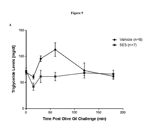
[0032] Figure 9 is a set of graphs showing the post prandial triglyceride-
lowering effect
of 5E5. Vehicle or 5E5 antibody was administered to mice receiving 3x10" AAV8-
huApoC3 viral particles. An oral dose of olive oil was given, and triglyceride
levels were
measured in a time course (part A). The area under curve ("AUC") was reduced
by about
25% with 5E5 administration as compared to vehicle control with a p value of
0.030 (part B).
Serum ApoC3 levels (part C) and 5E5 antibody levels (part D) were also
measured in a time
course. In part E, an anti-hen egg lysosome human IgGi antibody ("HyHEL5") was
used as
an isotype control.
[0033] Figure 10 is a series of graphs showing acceleration of ApoC3 and
ApoB
clearance from the blood after subcutaneous injection of the 5E5 and 6A6
antibody to mice
expressing human ApoC3. An anti-hen egg lysosome human IgG1 antibody (HyHEL5)
was
used as an isotype control for 5E5 and PBS was used as vehicle control for
6A6. Part C
shows the concentrations of human Apoc3 in the plasma. Parts A, B, are graphed
as percent
difference from negative isotype control, and D is graphed as percentage
change relative to
the respective baseline level measured from a blood sample collected
immediately before the
administration of the antibodies.
DETAILED DESCRIPTION
[0034] The instant disclosure provides antibodies that specifically bind to
ApoC3 (e.g.,
human ApoC3) and inhibit ApoC3 function. Also provided are pharmaceutical
compositions
comprising these antibodies, nucleic acids encoding these antibodies,
expression vectors and
host cells for making these antibodies, and methods of treating a subject
using these
antibodies. In certain embodiments, the anti-ApoC3 antibodies disclosed herein
can attenuate
the ability of ApoC3 to inhibit TRL uptake by hepatocytes. Accordingly, the
disclosed anti-
ApoC3 antibodies are useful for the treatment and prevention of
hypertriglyceridemia and
associated diseases (e.g., cardiovascular disease and pancreatitis).
[0035] Several, distinct types of anti-ApoC3 antibody are disclosed herein,
each binding
to a different, novel epitope of ApoC3 and having different functional
properties. For
example, in certain embodiments, the anti-ApoC3 antibodies disclosed herein
bind to an
epitope within the amino acid sequence set forth in SEQ ID NO:3 [FSEFWDLDPE],
and are
capable of binding to lipid-bound ApoC3. Alternatively, in certain
embodiments, the anti-
ApoC3 antibodies disclosed herein bind to an epitope within the amino acid
sequence set
forth in SEQ ID NO:2 [GWVTDGFSSLK], and inhibit the binding of ApoC3 to a
lipid.
9
CA 03030099 2019-01-07
WO 2018/007999 PCT/IB2017/054125
I. Definitions
[0036] As used herein, the term "ApoC3" refers to Apolipoprotein C3
protein. In certain
embodiments, the ApoC3 is human ApoC3. An exemplary human ApoC3 amino acid
sequence is set forth in RefSeq accession number NP 000031.1. The mature amino
acid
sequence of NP 000031.1 is as follows:
SEAEDASLLSFMQGYMKHATKTAKDALS SVQESQVAQQARGWVTDGF S SLKDYWS
TVKDKFSEFWDLDPEVRPTSAVAA (SEQ ID NO: 1).
[0037] As used herein, the terms "antibody" and "antibodies" include full
length
antibodies, antigen-binding fragments of full length antibodies, and molecules
comprising
antibody CDRs, VH regions or VL regions. Examples of antibodies include
monoclonal
antibodies, recombinantly produced antibodies, monospecific antibodies, multi
specific
antibodies (including bispecific antibodies), human antibodies, humanized
antibodies,
chimeric antibodies, immunoglobulins, synthetic antibodies, tetrameric
antibodies comprising
two heavy chain and two light chain molecules, an antibody light chain
monomer, an
antibody heavy chain monomer, an antibody light chain dimer, an antibody heavy
chain
dimer, an antibody light chain- antibody heavy chain pair, intrabodies,
heteroconjugate
antibodies, single domain antibodies, monovalent antibodies, single chain
antibodies or
single-chain Fvs (scFv), scFv-Fcs, camelid antibodies (e.g., llama
antibodies), camelized
antibodies, affybodies, Fab fragments, F(ab')2 fragments, disulfide-linked Fvs
(sdFv), anti-
idiotypic (anti-Id) antibodies (including, e.g., anti-anti-Id antibodies), and
antigen-binding
fragments of any of the above. In certain embodiments, antibodies described
herein refer to
polyclonal antibody populations. Antibodies can be of any type (e.g., IgG,
IgE, IgM, IgD,
IgA or IgY), any class (e.g., IgGi, IgG2, IgG3, IgG4, IgAi or IgA2), or any
subclass (e.g.,
IgG2a or IgG2b) of immunoglobulin molecule. In certain embodiments, antibodies
described
herein are IgG antibodies, or a class (e.g., human IgGi or IgG4) or subclass
thereof. In a
specific embodiment, the antibody is a humanized monoclonal antibody.
[0038] As used herein, the term "isolated antibody" refers to an antibody
that has been
identified and separated and/or recovered from at least one component of its
natural
environment. The term "isolated antibody" includes an antibody in situ within
a recombinant
host cell.
[0039] As used herein, the term "CDR" or "complementarity determining
region" means
the noncontiguous antigen combining sites found within the variable region of
both heavy
and light chain polypeptides. These particular regions have been described by
Kabat et at., J.
Biol. Chem. 252, 6609-6616 (1977) and Kabat et at., Sequences of protein of
immunological
CA 03030099 2019-01-07
WO 2018/007999 PCT/IB2017/054125
interest. (1991), by Chothia et at., J. Mol. Biol. 196:901-917 (1987), and by
MacCallum et
at., J. Mol. Biol. 262:732-745 (1996), all of which are incorporated by
reference in their
entireties, where the definitions include overlapping or subsets of amino acid
residues when
compared against each other. In certain embodiments, the term "CDR" is a CDR
as defined
by Kabat et at., J. Biol. Chem. 252, 6609-6616 (1977) and Kabat et at.,
Sequences of protein
of immunological interest. (1991). CDRH1, CDRH2 and CDRH3 denote the heavy
chain
CDRs, and CDRL1, CDRL2 and CDRL3 denote the light chain CDRs.
[0040] As
used herein, the term "framework (FR) amino acid residues" refers to those
amino acids in the framework region of an immunoglobulin chain. The term
"framework
region" or "FR region" as used herein, includes the amino acid residues that
are part of the
variable region, but are not part of the CDRs (e.g., using the Kabat
definition of CDRs).
[0041] As
used herein, the terms "variable region" and "variable domain" are used
interchangeably and are common in the art. The variable region typically
refers to a portion
of an antibody, generally, a portion of a light or heavy chain, typically
about the amino-
terminal 110 to 120 amino acids or 110 to 125 amino acids in the mature heavy
chain and
about 90 to 115 amino acids in the mature light chain, which differ
extensively in sequence
among antibodies and are used in the binding and specificity of a particular
antibody for its
particular antigen. The variability in sequence is concentrated in those
regions called
complementarity determining regions (CDRs) while the more highly conserved
regions in the
variable domain are called framework regions (FR). Without wishing to be bound
by any
particular mechanism or theory, it is believed that the CDRs of the light and
heavy chains are
primarily responsible for the interaction and specificity of the antibody with
antigen. In
certain embodiments, the variable region is a human variable region. In
certain
embodiments, the variable region comprises rodent or murine CDRs and human
framework
regions (FRs). In particular embodiments, the variable region is a primate
(e.g., non-human
primate) variable region. In certain embodiments, the variable region
comprises rodent or
murine CDRs and primate (e.g., non-human primate) framework regions (FRs).
[0042] The
terms "VL" and "VL domain" are used interchangeably to refer to the light
chain variable region of an antibody.
[0043] The
terms "VH" and "VH domain" are used interchangeably to refer to the heavy
chain variable region of an antibody.
[0044] As
used herein, the terms "constant region" and "constant domain" are
interchangeable and are common in the art. The constant region is an antibody
portion, e.g.,
a carboxyl terminal portion of a light or heavy chain which is not directly
involved in binding
11
CA 03030099 2019-01-07
WO 2018/007999 PCT/IB2017/054125
of an antibody to antigen but which can exhibit various effector functions,
such as interaction
with the Fc receptor. The constant region of an immunoglobulin molecule
generally has a
more conserved amino acid sequence relative to an immunoglobulin variable
domain.
[0045] As used herein, the term "heavy chain" when used in reference to an
antibody can
refer to any distinct type, e.g., alpha (a), delta (8), epsilon (c), gamma
(y), and mu (p,), based
on the amino acid sequence of the constant domain, which give rise to IgA,
IgD, IgE, IgG,
and IgM classes of antibodies, respectively, including subclasses of IgG,
e.g., IgGi, IgG2,
IgG3, and Igai.
[0046] As used herein, the term "light chain" when used in reference to an
antibody can
refer to any distinct type, e.g., kappa (K) or lambda (A) based on the amino
acid sequence of
the constant domains. Light chain amino acid sequences are well known in the
art. In
specific embodiments, the light chain is a human light chain.
[0047] As used herein, the term "specifically binds to" refers to the
ability of an antibody
to bind to an antigen with an dissociation constant (Kd) of less than about 1
x 10-6 M, 1 x 10⁻⁷
M, 1 x 10⁻⁸ M, 1 x 10⁻⁹ M, 1 x 10⁻¹⁰ M, 1 x 10-11 M, 1 x 10-12 M, or less, or
bind to an antigen
with an affinity that is at least two-fold greater than its affinity for a
nonspecific antigen.
[0048] As used herein, an "epitope" refers to a localized region of an
antigen to which an
antibody can specifically bind. An epitope can be, for example, contiguous
amino acids of a
polypeptide (a linear or contiguous epitope) or an epitope can, for example,
be formed from
two or more non-contiguous regions of a polypeptide or polypeptides (a
conformational, non-
linear, discontinuous, or non-contiguous epitope). In certain embodiments, the
epitope to
which an antibody binds can be determined by, e.g., NMR spectroscopy, X-ray
diffraction
crystallography studies, ELISA assays, hydrogen/deuterium exchange coupled
with mass
spectrometry (e.g., liquid chromatography electrospray mass spectrometry),
peptide scanning
assays, or mutagenesis mapping (e.g., site-directed mutagenesis mapping).
[0049] As used herein, the term "treat," "treating," and "treatment" refer
to therapeutic or
preventative measures described herein. The methods of "treatment" employ
administration
of an anti-ApoC3 antibody to a subject having a disease or disorder, or
predisposed to having
such a disease or disorder, in order to prevent, cure, delay, reduce the
severity of, reduce the
risk of developing, or ameliorate one or more symptoms of the disease or
disorder or
recurring disease or disorder, or in order to prolong the survival of a
subject beyond that
expected in the absence of such treatment.
[0050] As used herein, the term "effective amount" in the context of the
administration of
a therapy to a subject refers to the amount of a therapy that achieves a
desired prophylactic or
12
CA 03030099 2019-01-07
WO 2018/007999 PCT/IB2017/054125
therapeutic effect.
[0051] As used herein, the term "subject" includes any human or non-human
animal.
[0052] As used herein, the term "or" means and/or.
[0053] As used herein, the terms "about" and "approximately," when used to
modify a
numeric value or numeric range, indicate that deviations of 5% to 10% above
and 5% to 10%
below the value or range remain within the intended meaning of the recited
value or range.
2. Anti-ApoC3 Antibodies
[0054] The instant disclosure provides antibodies (e.g., isolated
antibodies) that
specifically bind to ApoC3 (e.g., human ApoC3) and inhibit ApoC3 function.
[0055] In certain embodiments, the anti-ApoC3 antibodies bind to ApoC3
protein of a
mammal. In certain embodiments, the anti-ApoC3 antibodies bind to human ApoC3.
In
certain embodiments, the anti-ApoC3 antibodies bind to Macaca fascicularis
(cynomologus
monkey) ApoC3. In certain embodiments, the anti-ApoC3 antibodies bind to
murine ApoC3.
[0056] In certain embodiments, the anti-ApoC3 antibodies disclosed herein
attenuate the
ability of ApoC3 to inhibit hepatocyte uptake of TRL (e.g., VLDL) or TRL
remnants (in vivo
or in vitro). In certain embodiments, the anti-ApoC3 antibodies disclosed
herein attenuate
the ability of ApoC3 to inhibit hepatocyte uptake of TRL (e.g., VLDL) or TRL
remnants by
at least 5%, 10%, 15%, 20%, 25%, 30%, 35%, 40%, 45%, 50%, 55%, 60%, 65%, 70%,
75%,
80%, 85%, 90%, 95%, 98%, or 99%, as assessed by methods described herein or by
methods
known to one of skill in the art. In certain embodiments, the anti-ApoC3
antibodies disclosed
herein attenuate the ability of ApoC3 to inhibit hepatocyte uptake of TRL
(e.g., VLDL) or
TRL remnants by at least about 1.1 fold, 1.2 fold, 1.3 fold, 1.4 fold, 1.5
fold, 2 fold, 2.5 fold,
3 fold, 3.5 fold, 4 fold, 4.5 fold, 5 fold, 6 fold, 7 fold, 8 fold, 9 fold, 10
fold, 15 fold, 20 fold,
30 fold, 40 fold, 50 fold, 60 fold, 70 fold, 80 fold, 90 fold, or 100 fold, as
assessed by
methods described herein or by methods known to one of skill in the art.
[0057] In certain embodiments, the antibodies disclosed herein are capable
of inhibiting
post-prandial lipemia in a subject when administered to the subject prior to,
during, or after a
meal. In certain embodiments, the anti-ApoC3 antibodies disclosed herein are
capable of
inhibiting post-prandial lipemia in the subject by at least 5%, 10%, 15%, 20%,
25%, 30%,
35%, 40%, 45%, 50%, 55%, 60%, 65%, 70%, 75%, 80%, 85%, 90%, 95%, 98%, or 99%,
as
assessed by methods described herein or by methods known to one of skill in
the art. In
certain embodiments, the anti-ApoC3 antibodies disclosed herein are capable of
inhibiting
post-prandial lipemia in the subject by at least about 1.1 fold, 1.2 fold, 1.3
fold, 1.4 fold, 1.5
13
CA 03030099 2019-01-07
WO 2018/007999 PCT/IB2017/054125
fold, 2 fold, 2.5 fold, 3 fold, 3.5 fold, 4 fold, 4.5 fold, 5 fold, 6 fold, 7
fold, 8 fold, 9 fold, 10
fold, 15 fold, 20 fold, 30 fold, 40 fold, 50 fold, 60 fold, 70 fold, 80 fold,
90 fold, or 100 fold,
as assessed by methods described herein or by methods known to one of skill in
the art.
[0058] In certain embodiments, the antibodies disclosed herein are capable
of reducing
the levels of post-prandial chylomicron or chylomicron remnants in a subject
when
administered to the subject prior to, during, or after a meal. In certain
embodiments, the anti-
ApoC3 antibodies disclosed herein are capable of reducing the levels of post-
prandial
chylomicron or chylomicron remnants in a subject by at least 5%, 10%, 15%,
20%, 25%,
30%, 35%, 40%, 45%, 50%, 55%, 60%, 65%, 70%, 75%, 80%, 85%, 90%, 95%, 98%, or
99%, as assessed by methods described herein or by methods known to one of
skill in the art.
In certain embodiments, the anti-ApoC3 antibodies disclosed herein are capable
of reducing
the levels of post-prandial chylomicron or chylomicron remnants in a subject
by at least about
1.1 fold, 1.2 fold, 1.3 fold, 1.4 fold, 1.5 fold, 2 fold, 2.5 fold, 3 fold,
3.5 fold, 4 fold, 4.5 fold,
fold, 6 fold, 7 fold, 8 fold, 9 fold, 10 fold, 15 fold, 20 fold, 30 fold, 40
fold, 50 fold, 60 fold,
70 fold, 80 fold, 90 fold, or 100 fold, as assessed by methods described
herein or by methods
known to one of skill in the art.
[0059] In certain embodiments, the isolated antibodies disclosed herein are
capable of
increasing the rates of clearance of ApoC3 and/or ApoB (e.g., ApoB48 and/or
ApoB100)
from the blood in a subject. In certain embodiments, the anti-ApoC3 antibodies
are capable
of increasing the rates of clearance of ApoC3 and/or ApoB (e.g., ApoB48 and/or
ApoB100)
from the blood in a subject by at least 5%, 10%, 15%, 20%, 25%, 30%, 35%, 40%,
45%,
50%, 55%, 60%, 65%, 70%, 75%, 80%, 85%, 90%, 95%, 98%, or 99%, as assessed by
methods disclosed herein or by methods known to one of skill in the art. In
certain
embodiments, the anti-ApoC3 antibodies disclosed herein are capable of
increasing the rates
of clearance of ApoC3 and/or ApoB (e.g., ApoB48 and/or ApoB100) from the blood
in a
subject by at least about 1.1 fold, 1.2 fold, 1.3 fold, 1.4 fold, 1.5 fold, 2
fold, 2.5 fold, 3 fold,
3.5 fold, 4 fold, 4.5 fold, 5 fold, 6 fold, 7 fold, 8 fold, 9 fold, 10 fold,
15 fold, 20 fold, 30 fold,
40 fold, 50 fold, 60 fold, 70 fold, 80 fold, 90 fold, or 100 fold, as assessed
by methods
disclosed herein or by methods known to one of skill in the art. Methods for
assessing the
clearance of ApoC3 and/or ApoB (e.g., ApoB48 and/or ApoB100) include without
limitation
the isotope tracer techniques, wherein the isotope can be either radioactive
or stable.
[0060] In certain embodiments, the isolated antibodies disclosed herein are
capable of
reducing the levels of ApoC3 and/or ApoB (e.g., ApoB48 and/or ApoB100) in the
blood in a
subject. In certain embodiments, the anti-ApoC3 antibodies are capable of
reducing the
14
CA 03030099 2019-01-07
WO 2018/007999 PCT/IB2017/054125
levels of ApoC3 and/or ApoB (e.g., ApoB48 and/or ApoB100) in the blood in a
subject by at
least 50, 1000, 150o, 20%, 25%, 30%, 3500, 400 0, 4500, 500 0, 550, 60%, 65%,
700 0, 7500,
80%, 85%, 90%, 950, 98%, or 9900, as assessed by methods disclosed herein or
by methods
known to one of skill in the art. In certain embodiments, the anti-ApoC3
antibodies disclosed
herein are capable of reducing the levels of ApoC3 and/or ApoB (e.g., ApoB48
and/or
ApoB100) in the blood in a subject by at least about 1.1 fold, 1.2 fold, 1.3
fold, 1.4 fold, 1.5
fold, 2 fold, 2.5 fold, 3 fold, 3.5 fold, 4 fold, 4.5 fold, 5 fold, 6 fold, 7
fold, 8 fold, 9 fold, 10
fold, 15 fold, 20 fold, 30 fold, 40 fold, 50 fold, 60 fold, 70 fold, 80 fold,
90 fold, or 100 fold,
as assessed by methods disclosed herein or by methods known to one of skill in
the art. In
certain embodiments, the reduction in the levels of ApoC3 and/or ApoB (e.g.,
ApoB48 and/or
ApoB100) in the blood in the subject is maintained for at least 1, 2, 3, 4, 5,
6, 7, 8, 9, 10, 12,
15, 18, 24, 30, 36, 42, or 48 hours.
[0061] In certain embodiments, the antibodies disclosed herein attenuate
the ability of
ApoC3 to inhibit lipoprotein lipase-mediated lipolysis of TRL (e.g., VLDL). In
certain
embodiments, the anti-ApoC3 antibodies disclosed herein attenuate the ability
of ApoC3 to
inhibit lipoprotein lipase-mediated lipolysis of TRL (e.g., VLDL) by at least
5%, 10%, 15%,
2000, 2500, 3000, 3500, 4000, 4500, 5000, 5500, 600o, 6500, 7000, 7500, 800o,
8500, 9000, 9500,
980 o, or 990 o, as assessed by methods described herein or by methods known
to one of skill
in the art. In certain embodiments, the anti-ApoC3 antibodies disclosed herein
attenuate the
ability of ApoC3 to inhibit lipoprotein lipase-mediated lipolysis of TRL
(e.g., VLDL) by at
least about 1.1 fold, 1.2 fold, 1.3 fold, 1.4 fold, 1.5 fold, 2 fold, 2.5
fold, 3 fold, 3.5 fold, 4
fold, 4.5 fold, 5 fold, 6 fold, 7 fold, 8 fold, 9 fold, 10 fold, 15 fold, 20
fold, 30 fold, 40 fold,
50 fold, 60 fold, 70 fold, 80 fold, 90 fold, or 100 fold, as assessed by
methods described
herein or by methods known to one of skill in the art. In certain embodiments,
the anti-
ApoC3 antibodies disclosed herein attenuate the ability of ApoC3 to inhibit
lipoprotein
lipase-mediated lipolysis of TRL (e.g., VLDL) by at least 50% at the
concentration of 1, 2, 3,
4, or 5 M.
[0062] In certain embodiments, the antibodies disclosed herein inhibit the
binding of
ApoC3 to a lipid or a lipoprotein. In certain embodiments, the lipid comprises
a fatty acid
chain. In certain embodiments, the lipid comprises a phosphatidyl group. In
certain
embodiments, the lipid comprises a phosphatidylcholine (e.g., DIVIPC), a
phosphatidylserine,
a phosphatidylethanolamine, a phosphatidylinositol or a phosphatidylglycerol.
In certain
embodiments, the lipid is a triglyceride. In certain embodiments, the
lipoprotein is a TRL
(e.g., VLDL) or a TRL remnant. In certain embodiments, the anti-ApoC3
antibodies
CA 03030099 2019-01-07
WO 2018/007999 PCT/IB2017/054125
disclosed inhibit the binding of ApoC3 to lipids and lipoproteins (e.g.,
triglyceride, TRL (e.g.,
VLDL) or TRL remnants) by at least 5%, 10%, 15%, 20%, 25%, 30%, 350, 40%, 450,
50%,
550, 60%, 65%, 70%, 750, 80%, 85%, 90%, 950, 98%, or 99%, as assessed by
methods
described herein or by methods known to one of skill in the art. In certain
embodiments, the
anti-ApoC3 antibodies disclosed herein attenuate the binding of ApoC3 to
lipids and
lipoproteins (e.g., triglyceride, TRL (e.g., VLDL) or TRL remnants) by at
least about 1.1
fold, 1.2 fold, 1.3 fold, 1.4 fold, 1.5 fold, 2 fold, 2.5 fold, 3 fold, 3.5
fold, 4 fold, 4.5 fold, 5
fold, 6 fold, 7 fold, 8 fold, 9 fold, 10 fold, 15 fold, 20 fold, 30 fold, 40
fold, 50 fold, 60 fold,
70 fold, 80 fold, 90 fold, or 100 fold, as assessed by methods described
herein or by methods
known to one of skill in the art.
[0063] In certain embodiments, the antibodies disclosed herein are capable
of binding to
lipid-bound ApoC3 (e.g., ApoC3 bound to triglyceride, TRL (e.g., VLDL) or TRL
remnants),
as assessed by methods described herein (e.g., in Example 3) or by methods
known to one of
skill in the art.
[0064] The antibodies disclosed herein can have one or more, two or more,
three or more,
four or more, five or more, six or more, seven of more, or all of the
characteristics as set forth
in the foregoing embodiments. For example, in certain embodiments, the
antibodies
disclosed herein attenuate the ability of ApoC3 to inhibit hepatocyte uptake
of TRL (e.g.,
VLDL) or TRL remnants by at least about 10%, 20%, 30%, 40%, 50%, 60%, 70%,
80%,
9000, 950, or 9900, and attenuate the ability of ApoC3 to inhibit lipoprotein
lipase-mediated
lipolysis of TRL (e.g., VLDL) or TRL remnants by at least 5000 at the
concentration of 1, 2,
3, 4, or 5 M. In certain embodiments, the antibodies disclosed herein
attenuate the ability of
ApoC3 to inhibit hepatocyte uptake of TRL (e.g., VLDL) or TRL remnants by at
least about
10%, 20%, 30%, 40%, 50%, 60%, 70%, 80%, 90%, 95%, or 99%, and are capable of
binding
to lipid-bound ApoC3 (e.g., ApoC3 bound to triglyceride, TRL (e.g., VLDL) or
TRL
remnants.
[0065] Any suitable assays can be used to measure the foregoing functional
activities of
the antibodies disclosed herein. Exemplary assays include, but are not limited
to, the
functional assays disclosed in the Examples herein.
[0066] The amino acid sequences of exemplary anti-ApoC3 antibodies are set
forth in
Tables 1-4, herein.
16
CA 03030099 2019-01-07
WO 2018/007999 PCT/IB2017/054125
Table 1. Heavy chain CDR amino acid sequences of exemplary anti-ApoC3
antibodies.
VII CDRH1 SEQ CDRH2 SEQ CDRH3 SEQ
clone ID ID ID
NO: NO: NO:
5A1 1 TRYYA 4 VIAYD GS TYY SP SLK S 5 VRLIEAPYEYDY 6
5E5 TYSMR 7 SISTDGGGTAYRD SVKG 8 AGYSD 9
6A6 SYAGR 10 SINAGGGST SYAD SVK 11 NSYRY 12
8F4 SYSMY 13 AIKTDGGSTNYADSVKG 14 QGYGT 15
11H1 SYSMR 16 SIKSDGSIT SYAD SVKG 17 QGYIN 18
5A4 HYTMY 19 AI S GGGDRTIYTD SVKG 20 QGYEY 21
5A7 NRRYA 22 VIVYD GNTHV SP SLR S 23 VLLLRDPLSLDY 24
8A4 NYAMR 25 SID SGGDRTKYGD SVKG 26 QGYIF 27
8B4 NAYLY 28 GINPAGDGRAYAT SVKG 29 A SRVVAYD S 30
8H4 SYSMR 16 SINSDGGS TKY SD SVKG 31 QGYTD 32
1 OB 6 SYAMR 33 SINIDGGSTRYTDSVQG 34 QGYIY 35
12A3 NYAMR 25 SINIAGSSVVYADSVK 36 QGFVY 37
12C3 SYSMF 38 GINGGGDRSNYAD SVRD 39 QGYAY 40
12C12 TSYYAWT 41 AIVYDGSTFYSPSLKS 42 SYGLGLYDL 43
12D1 TYSMR 7 SISTDGGGTAYRDSVKG 8 AGYSD 9
12D4 SSNMR 47 TISPDGGKTLYADSVKG 48 AGYDY 49
12E12 NIYMS 50 AINTAGTVTYYAD SVKG 51 GEVD 52
13 C 7 RYYMS 53 SIYKDGSNTYYAD SVKG 54 ALRAEYDY 55
13G7 TTAPAWG 56 VIAFD GS AYY SP SLK S 57 LGGRNYPPYVEL 58
14C4 NYDMS 59 VINSDGDGTYYVD SVKG 60 ANLGL 61
14C7 TNSYYWS 62 AIDYSGDTYYSPSLKS 63 RIPTGEY 64
14G4 RYTMN 65 AISPDGGKTIDADSVK 66 GHNMDY 67
12D7 DYAMS 68 AITSNGKRTDYAESMK 69 GPPHYIPIPSMTPRD 70
12G8 DYAMS 68 AIRWNGDTYYAESMK 71 HRPGGALDT 72
Table 2. Light chain CDR amino acid sequences of exemplary anti-ApoC3
antibodies.
VL CDRL1 SEQ CDRL2 SEQ CDRL3 SEQ
clone ID ID ID
NO: NO: NO:
5All GLS SGSVTTRSYPG 73 STSSRHS 74 ALDIGSYIV 75
17
CA 03030099 2019-01-07
WO 2018/007999 PCT/IB2017/054125
5E5
KTSQGLVHSDGKTYFY 76 QVSNRAS 77 AQGTYYPHT 78
6A6 KASQSLIHTDGKTYLY 79 QVSSHES
80 AQATYNPRT 81
8F4
KASQSLVHSDGKTYLY 82 QVSNRGS 83 AQATYYGHS 84
11H1 RASQSLIHSAGKTYFY 85 QVSNRES
86 AQGTYNPKT 87
5A4 KAIQSLVHTDGKTYLY 88 QVSNRGS
83 AQGTYSSKT 89
5A7 AGTSSDIGAYNFVS 90
DIDKRAS 91 AAYGSRDNVV 92
8A4
KASQSLVHSDGKTYLY 82 QVSNHES 93 AQATYYPLT 94
8B4 KS SQSVESGSDQKSYLN 95 YASTQES
96 QQAYSAPFT 97
8H4
KVSQSLVHSDGKTYLY 98 QVSNRDS 99 AQGTYNPYT 100
10B6 KASQSLVHSNGVIYFY 101
QVSNRDS 99 AQGTYYPHS 102
12A3 KAGRSLVHSDGRTYLY 103 QVSNRSS 104 AQGTYYPVT
105
12C3 KASQSLVHSDGKTYLY 82 QTSNRGS
106 AQATYSPHT 107
12C12 TGSSSNIGDNYVN
108 SNSNRAS 109 SSWDDSLSGVV 110
12D1 KTSQSLTHSDGKTYLY 111 QVSNRGS 83 AQATYYPHT
113
12D4 KASQSLVHSDGKTYLY 82 QVSNQGS
114 AQATYAPHS 115
12E12 GLSSGSVTSVTYPG
116 NTNSRFS 117 SVYIGGGIYPAV 118
13C7 AGTSSDIGGYNYVA
119 EVNKRAS 120 ASYRSSNSYV 121
13G7 QGGSLRVSYAH
122 DDDSRPS 123 QSADSSGDNWV 124
14C4 KATQSLVHSDGKTYLS 125 QVSNRGS 83 AQAPYWT
126
14C7 GLNSGSVTSSNYPD
127 NTNSRHS 128 ALYMGSDSVV 129
14G4 KASQSLVHSDGKTYLY 82 QVSNQGS
114 AQATYTPRT 130
12D7 QGGTLGRYYGS
131 GDNSRPS 132 ESFDFSGNAAV 133
12G8 QGGNF GNFYAS
134 KDSERPS 135 QSGSSSDNVV 136
Table 3. VH amino acid sequences of exemplary anti-ApoC3 antibodies.
VII Amino acid Sequence SEQ
ID
clone NO
5All QVQVQESGPGLVKPSQTLSLTCTVSGVSITTRYYAWSWIRQPPG 137
KGLEWMGVIAYDGSTYYSPSLKSRTSISRDTSKNQFSLQLTSVTP
EDTAVYYCARVRLIEAPYEYDYWGQGTQVTVSS
5E5 QLQLVESGGGLVQPGGSLRLSCAASGFTFGTYSMRWVRQVPRK 138
ALEWVSSISTDGGGTAYRDSVKGRFTISRDNAKNTLYLQMNNL
KPEDTAIYYCVIAGYSDWGQGTQVTVSS
6A6 EVQLVESGGGLVQPGGSLRLSCAASGFTFSSYAGRWVRQVPGK 139
GLEWVSSINAGGGSTSYADSVKGRFTISRDNAKNTLYLQMNSLK
PEDTAKYYCTQNSYRYWGQGTRVAVSS
18
CA 03030099 2019-01-07
WO 2018/007999
PCT/IB2017/054125
8F4 QLQLVESGGGLVQPGGSLRLSCAASGFAF S SYSMYWVRQAPGK 140
GLERVAAIKTD GG S TNYAD S VKGRF TV SRDNAKNTLYLQMN S L
K SED TAVYYCVIQ GYGTWGQ GT QVTV S S
11H1 ELQLVE S GGGLVQPGGSLRL S C AA S GF TF S SYSMRWVRQAPGK 141
GLEWLS SIK SD G S IT S YAD S VKGRF TM SRDNAKNTLYLQMN SLK
SED TAMYYC TNQ GYINWGQGTQVT VS S
5A4 QLQLVE S GGGLVQP GGSLRL S C VA S GF AF SHYTMYWVRQAPVR 142
GLERVSAISGGGDRTIYTDSVKGRFTISRDNAANALYLQMNSLQ
PEDTAVYYCVAQGYEYWGQGTRVTVS S
5A7 EVQVQE S GP GLVKP S Q TL S LTC TV S GA S ITNRRYAWTWIRQPP G 143
KGLEWMGVIVYDGNTHVSP SLRSRT SISRDT SKNQF SLQLS SLTP
ED TAVYYCARVLLLRDPL S LDYWGQ GT QVTV S S
8A4 QVQLVE S GGGLVQPGGSLKV S C TA S GF TFNNYAMRWVRQ AEG 144
KGLEWVS SID S GGDRTKYGD SVKGRF SISRDNAKNTVYLQMDA
LKPED T GVYYC V S Q GYIF'WGQ GAQVTV S S
8B4 ELQLVE S GGGLVQPGGSLRL S C AA S GF TF SNAYLYWVRQVPGK 145
GLEWVSGINPAGDGRAYAT SVKGRFTISRDNAKNTLYLQMNTL
ESDDTAVYYCATASRVVAYDSWGQGTQVTVS S
8H4 ELQLVE S GGGLVQPGR SLRL S CAA S GF TF S SYSMRWVRQTPGKG 146
LEWVT S IN SD GGS TKY SD S VKGRF TISRDNAKNTLYLQMNNVKP
ED TAIYYC AIQ GYTDWGQ GTQVTV S S
10B6 EVQLVE S GGGLVQPGGSLRL S C AA S GF TF S SYAMRWVRQAPGK 147
GLEWIS S INID GGS TRYTD S VQ GRF TV SRDNAKNTLYLQMNNLK
PEDTGIYYCTIQGYIYWGQGTQVTVS S
12A3 ELQLVESGGGLVQ S GG SLRL S C AA S GF TF SNYAMRWVRQAPGG 148
RLEWVS SINIAGS SVVYADSVKGRFTISRDNAKNTLYLQMNSLK
SED TAVYYCAMQ GF VYWGQ GT QVTV S S
12C3 ELQLVESGGGLVQPGGSLRLSCAASGFTF S SYSMFWVRQ SPGKG 149
LERV S GINGGGDRSNYAD S VRDRF TISRDNAKNTLYLQMN S LK S
ED TAVYYCVIQ GYAYWGQ GT QVTV S S
12C 12 EVQVQESGPGLVKP S Q TLSLTC TVS GGSITT SYYAWTWIRQPPG 150
KGLEWVGAIVYDGSTFYSP SLKSRT SISRDT SKSQF SLQLS SVTPE
D TAVYYC ARS YGLGLYDLWGQ GTQVT VS S
12D1 QLQLVE S GGGLVQP GGS LRL S C AA S GF TF GTY SMRWVRQVPRK 138
ALEWVS SISTDGGGTAYRDSVKGRFTISRDNAKNTLYLQMNNL
KPED TAIYYC VIAGYS DWGQ GT QVTV S S
12D4 QLQLVE S GGGLVQP GG SLRVS C AA S GF TF SS SNMRWVRQVSGK 152
GLEWVSTISPDGGKTLYAD SVKGRF T ISRDNAKNTLHLQMV S LK
PEDTALYYCVKAGYDYWGQGTQVTVS S
12E12 EVQLVE S GGDLVQPGGSLRV S C AA S GLTF SNIYMSWVRQAPGK 153
GLEWVSAINTAGTVTYYADSVKGRFTISRDNAKNTLYLQMNSL
KPEDTAHYYCTTGEVDWGKGTLVTVS S
13 C7 QLQLVE S GGGLVQP GGS LRL S C AA S GGTF SRYYMSWVRQAPGK 154
GLEWVS SIYKDGSNTYYADSVKGRFTISRDNAKNTLYLQMNSL
K SED TAVYYCAKALRAEYDYWGQ GT QVTV S S
13G7 QVQLQESGPGLVKP S QTLSLTC TVS GGSIS TTAPAWGWIRQ SPG 155
KGLDWMAVIAFD GS AYY SP SLKSRTLISRDT SKNQF SLQLS SVTP
ED TAVYYCARLGGRNYPPYVELWGQ GTLVT VS S
19
CA 03030099 2019-01-07
WO 2018/007999 PCT/IB2017/054125
14C4 QLQLVESGGGLVQP GGSLRLSCAASGF TF GNYDMSWVRQ AP GK 156
GPEWVSVINSDGDGTYYVDSVKGRFTISRDNAKNTLYLQMNSL
KPEDRAVYYCAIANLGLWGQGTLVTVS S
14C7 QVQVQESGPGLVKP SQTLSLTCTVSGGSITTNSYYWSWIRQPPG 157
KGLEWMGAIDYSGDTYYSP SLKSRTSISRDTSKNQFTLQLT SVTP
ED TAVYYCV SRIP T GEYWGQ GT QVTV S S
14G4 QVQLVESGGGLVQPGGSLRLSCAASGFAF SRYTMNWVRQ AP GK 158
GLEWLSAISPDGGKTIDADSVKGAFAS SRDNTMNTLYLDMNSL
KPEDAAVYYCVAGHNMDYWGKGILVTVS S
12D7 ELQLVESGGDLVQPGGSLRLSCAASGF TFDDYAMSWVRQ AP GK 159
GLEWVSAIT SNGKRTDYAESMKGRFTISRDNSKNTLYLEMNSLK
SED T AVYYC TKGPPHYIP IP SMTPRDSWGQGTQVTVS S
12G8 QLQLVESGGGLVQP GGSLRLSCAASGF TFDDYAMSWVRQ AP GK 160
GLEWVSAIRWNGDTYYAESMKGRFDMSRDNAKNTLYLQMNSL
KSEDTAVYYCAKHRPGGALDTWGQGTLVTVS S
Table 4. VL amino acid sequences of exemplary anti-ApoC3 antibodies.
VL Amino acid Sequence
SEQ ID
clone NO
5All QAVVTQEP SLSVSPGGTVTLTCGLS SGSVTTRSYPGWFQQTPGQ 161
APRSLIHSTS SRHSGIPTRF SGSISGNKAALTITGAQPEDEADYYC
ALDIGSYIVFGGGTHLTVL
5E5 ATMLTQ SPGSLSVVPGESASISCKTSQGLVHSDGKTYFYWFLQK 162
PGQ SPQQLIYQVSNRAS GVPDRFTGS GS GTDF TLKIS GVKAED A
GVYYCAQ GTYYPHTF GS GTRLEIK
6A6 DVVLTQTPGSLSVVPGESASISCKASQ SLIHTDGKTYLYWLLQKP 163
GQRPQLLIYQVS SHESGVPDRFTGSGSGTDFTLKISGVKAEDAGV
YYCAQATYNPRTFGQGTKLEIK
8F4 DLVLTQIPGSLSVVPGESASISCKASQ SLVHSDGKTYLYWLLQKP 164
GQ SPQRLIYQVSNRGSGVPDRFTGS GS GTDF TLKIS GVEAEDAG
VYYCAQ ATYYGHSF GS GTRLEIK
11H1 ATMLTQ SPGSLTIVPGESASISCRASQ SLIHSAGKTYFYWLLQKP 165
GQRPQLLIYQVSNRES GVPDRF TGS GS GTDF TLKIS GVKAEDAG
VYYCAQGTYNPKTFGQGTKLEIK
5A4 ATMLTQ SPGSLSVVPGESASISCKAIQ SLVHTDGKTYLYWFLQK 166
PGQ SP QRLIYQV SNRG S GVPDRF T GS GS GTDF TLKIS GVKAEDAG
VYYCAQGTYS SKTFGQGTKLEIK
5A7 S SALTQPP SMSGTLGKTLTISCAGTS SDIGAYNFVSWYQQLPGTA 167
PKLLIYDIDKRASGIPDRF SGSKSGNTASLSISGLQ SEDEADYYCA
AYGSRDNVVFGGGTHLTVL
8A4 ATMLTQ SPGSLSVVPGESASISCKASQ SLVHSDGKTYLYWLLQK 168
P GQRPQLLIYQVSNHES GVPDRF TGS GS GTDYTLKIS GVKAEDA
GVYYCAQATYYPLTFGQGTKVELK
8B4 EIVLTQ SP SSVTASVGEKVTINCKS SQ SVESGSDQKSYLNWYQQ 169
RPGQ SPRLLIYYASTQESGIPDRF SGSGSTTDFTLTIS SVQPEDAA
VYYC Q Q AY S APF TF GQ GTKVELK
8H4 DVVLTQTPGSLSVVPGESASISCKVSQ SLVHSDGKTYLYWLLQK 170
PGQ SP QRLIYQV SNRD S GVPDRF T GS GS GTDF TLKIS GVKAEDAG
VYYCAQ GTYNPYTF GS GTRLEIK
CA 03030099 2019-01-07
WO 2018/007999 PCT/IB2017/054125
10B6 ATMLTQSPGSLSIVPGESASISCKASQSLVHSNGVIYFYWLLQKP 171
GQSPQRLIYQVSNRDSGVPDRFTGSGSGTDFTLKISGVKAEDAG
VYYCAQGTYYPHSFGSGTRLQIK
12A3 DVVLTQTPGSLSVVPGESANISCKAGRSLVHSDGRTYLYWLLQK 172
PGQSPQRLIYQVSNRSSGVPDRFTGSGSGTDFTLKITGVKAEDAG
VYYCAQGTYYPVTFGQGTKVELK
12C3 DVVLTQTPASLSVVPGESASISCKASQSLVHSDGKTYLYWLLQK 173
PGQSPQRLIYQTSNRGSGVPDRFTGSGSGTDFTLDISGVKAEDAG
VYYCAQATYSPHTFGSGTRLEIK
12C12 QAVLTQPPSVSGSPGQKFTISCTGSSSNIGDNYVNWYQHLPGTAP 174
KLLIYSNSNRASGVPDRFSGSKSGSSASLTITGLQAEDEADYYCS
SWDDSLSGVVFGGGTHLTVL
12D1 SALDVVLTQTPGSLSVVPGESASISCKTSQSLTHSDGKTYLYWLL 175
QKPGQSPQRLIYQVSNRGSGVPDRFTGSGSGTDFTLKISGVKAED
AGMYYCAQATYYPHTFGSGSRLEIER
12D4 ATMLTQSPGSLSVVPGESASISCKASQSLVHSDGKTYLYWLLQK 176
PGQSPQRLIYQVSNQGSGVPDRFTGSGSGTDFTLKISGVKAEDA
GVYYCAQATYAPHSFGSGTRLEIK
12E12 QTVVTQEPSLSVSPGGTVTLTCGLSSGSVTSVTYPGWYQQKPGQ 177
APRTLIYNTNSRFSGVPNRFSGSISGNKAALTITGALPEDEADYY
CSVYIGGGIYPAVFGGGTHLTVL
13C7 NFMLTQPPSVSGTLGKTVTISCAGTSSDIGGYNYVAWYQQLPGT 178
APKLLISEVNKRASGIPDRFSGSKSGNTASLSISGLQSEDEADYYC
ASYRSSNSYVFGGGTKLTVL
13G7 QPVLTQPPALSVTLGQTAKITCQGGSLRVSYAHWYQQKPGQAP 179
VLVSYDDDSRPSGIPERFSGSGSGATATLTISGAQAEDEGDYYCQ
SADSSGDNWVFGGGTHLTVL
14C4 ATMLTQSPGSLSVVPGESASISCKATQSLVHSDGKTYLSWLLQK 180
PGQSPQRLIYQVSNRGSGVPDRFTGSGSGTDFTLKISGVKAEDAG
VYYCAQAPYWTFGQGTKLEIK
14C7 QTVVTQEPSLSVSPGGTVTLTCGLNSGSVTSSNYPDWYQQTPGQ 181
APRLLIYNTNSRHSGVPSRFSGSISGNKAALTITGAQPEDEADYY
CALYMGSDSVVFGGGTHLTVL
14G4 DVVLTQTPGSLSVVPGESASISCKASQSLVHSDGKTYLYWLLQK 182
PGQSPQRLIYQVSNQGSGVPDRFTGSGSGTDFTLKISGVKAEDA
GVYYCAQATYTPRTFGQGTTLEVK
12D7 SSALTQPSAVSVSLGQTARITCQGGTLGRYYGSWYQQKPAQAP 183
VLLIYGDNSRPSGIPERFSGSKSGDTATLTISGTQAEDEADYYCES
FDFSGNAAVFGGGTHLTVL
12G8 QAVLTQPSAVSVSLGQTARITCQGGNEGNEYASWYQQKPGQAP 151
VLVIYKDSERPSGIPERFSGSSSGDTATLTISGAQAEDEADYYCQS
GSSSDNVVFGGGTHLTVL
[0067] In one aspect, the instant disclosure provides an isolated antibody
that specifically
binds to ApoC3 (e.g., human ApoC3), the antibody comprising a VH domain
comprising one,
two, or all three of the CDRs of a VH domain set forth in Table 3 herein. In
certain
embodiments, the antibody comprises the CDRH1 of one of VH domains set forth
in Table 3.
In certain embodiments, the antibody comprises the CDRH2 of one of the VH
domains set
21
CA 03030099 2019-01-07
WO 2018/007999 PCT/IB2017/054125
forth in Table 3. In certain embodiments, the antibody comprises the CDRH3 of
one of the
VH domains set forth in Table 3.
[0068] In another aspect, the instant disclosure provides an isolated
antibody that
specifically binds to ApoC3 (e.g., human ApoC3), the antibody comprising a VL
domain
comprising one, two, or all three of the CDRs of a VL domain disclosed in
Table 4 herein. In
certain embodiments, the antibody comprises the CDRH1 of one of VL domains set
forth in
Table 4. In certain embodiments, the antibody comprises the CDRH2 of one of
the VL
domains set forth in Table 4. In certain embodiments, the antibody comprises
the CDRH3 of
one of the VL domains set forth in Table 4.
[0069] In certain embodiments, the CDRs of an antibody can be determined
according to
Kabat et at., J. Biol. Chem. 252, 6609-6616 (1977) and Kabat et at., Sequences
of protein of
immunological interest (1991). In certain embodiments, the light chain CDRs of
an antibody
are determined according to Kabat and the heavy chain CDRs of an antibody are
determined
according to MacCallum (supra).
[0070] In certain embodiments, the CDRs of an antibody can be determined
according to
the Chothia numbering scheme, which refers to the location of immunoglobulin
structural
loops (see, e.g., Chothia C & Lesk AM, (1987), J Mol Biol 196: 901-917; Al-
Lazikani B et
at., (1997) J Mol Biol 273: 927-948; Chothia C et at., (1992) J Mol Biol 227:
799-817;
Tramontano A et at., (1990) J Mol Biol 215(1): 175-82; and U.S. Patent No.
7,709,226).
Typically, when using the Kabat numbering convention, the Chothia CDRH1 loop
is present
at heavy chain amino acids 26 to 32, 33, or 34, the Chothia CDRH2 loop is
present at heavy
chain amino acids 52 to 56, and the Chothia CDRH3 loop is present at heavy
chain amino
acids 95 to 102, while the Chothia CDRL1 loop is present at light chain amino
acids 24 to 34,
the Chothia CDRL2 loop is present at light chain amino acids 50 to 56, and the
Chothia
CDRL3 loop is present at light chain amino acids 89 to 97. The end of the
Chothia CDRH1
loop when numbered using the Kabat numbering convention varies between H32 and
H34
depending on the length of the loop (this is because the Kabat numbering
scheme places the
insertions at H35A and H35B; if neither 35A nor 35B is present, the loop ends
at 32; if only
35A is present, the loop ends at 33; if both 35A and 35B are present, the loop
ends at 34).
[0071] In certain embodiments, the CDRs of an antibody can be determined
according to
the IMGT numbering system as described in Lefranc M-P, (1999) The Immunologist
7: 132-
136 and Lefranc M-P et at., (1999) Nucleic Acids Res 27: 209-212. According to
the IMGT
numbering scheme, CDRH1 is at positions 26 to 35, CDRH2 is at positions 51 to
57, CDRH3
is at positions 93 to 102, CDRL1 is at positions 27 to 32, CDRL2 is at
positions 50 to 52, and
22
CA 03030099 2019-01-07
WO 2018/007999 PCT/IB2017/054125
CDRL3 is at positions 89 to 97.
[0072] In
certain embodiments, the CDRs of an antibody can be determined according to
the AbM numbering scheme, which refers to AbM hypervariable regions, which
represent a
compromise between the Kabat CDRs and Chothia structural loops, and are used
by Oxford
Molecular's AbM antibody modeling software (Oxford Molecular Group, Inc.).
[0073] In
certain embodiments, the CDRs of an antibody can be determined according to
MacCallum RM et at., (1996) J Mol Biol 262: 732-745. See also, e.g., Martin A.
"Protein
Sequence and Structure Analysis of Antibody Variable Domains," in Antibody
Engineering,
Kontermann and Dilbel, eds., Chapter 31, pp. 422-439, Springer-Verlag, Berlin
(2001).
[0074] In
certain embodiments, the instant disclosure provides an isolated antibody that
specifically binds to ApoC3 (e.g., human ApoC3), wherein the antibody
comprises a heavy
chain variable region comprising the CDRH1, CDRH2, and CDRH3 region amino acid
sequences of a VH domain set forth in Table 3, and a light chain variable
region comprising
the CDRL1, CDRL2, and CDRL3 region amino acid sequences of a VL domain set
forth in
Table 4, wherein each CDR is independently defined in accordance with the
Kabat, Chothia,
IMGT, MacCallum, or AbM definition of a CDR, as disclosed herein.
[0075] In
another aspect, the instant disclosure provides an isolated antibody that
specifically binds to ApoC3 (e.g., human ApoC3), the antibody comprising:
(a) a
CDRH1 comprising the amino acid sequence of SEQ ID NO: 4, 7, 10, 13, 16, 19,
22, 25, 28, 33, 38, 41, 47, 50, 53, 56, 62, 65, or 68; or
(b) a
CDRH2 comprising the amino acid sequence of SEQ ID NO: 5, 8, 11, 14, 17, 20,
23,
26, 29, 31, 34, 36, 39, 42, 48, 51, 54, 57, 60, 63, 66, 69, or 71; or
(c) a
CDRH3 comprising the amino acid sequence of SEQ ID NO: 6, 9, 12, 15, 18, 21,
24,
27, 30, 32, 35, 37, 40, 43, 49, 52, 55, 58, 61, 64, 67, 70, or 72; or
(d) a
CDRL1 comprising the amino acid sequence of SEQ ID NO: 73, 76, 79, 82, 85, 88,
90, 95, 98, 101, 103, 108, 111, 116, 119, 122, 125, 127, 131, or 134; or
(e) a
CDRL2 comprising the amino acid sequence of SEQ ID NO: 74, 77, 80, 83, 86, 91,
93, 96, 99, 104, 106, 109, 114, 117, 120, 123, 128, 132, or 135; or
(f) a
CDRL3 comprising the amino acid sequence of SEQ ID NO: 75, 78, 81, 84, 87, 89,
92, 94, 97, 100, 102, 105, 107, 110, 113, 115, 118, 121, 124, 126, 129, 130,
133, or 136.
[0076] In
another aspect, the instant disclosure provides an isolated antibody that
specifically binds to ApoC3 (e.g., human ApoC3), wherein the antibody
comprises a VH
domain comprising the CDRH1, CDRH2 and CDRH3 amino acid sequences set forth in
SEQ
ID NOs: 4, 5, and 6; 7, 8, and 9; 10, 11, and 12; 13, 14, and 15; 16, 17, and
18; 19, 20, and
23
CA 03030099 2019-01-07
WO 2018/007999 PCT/IB2017/054125
21; 22, 23, and 24; 25, 26, and 27; 28, 29, and 30; 16, 31, and 32; 33, 34,
and 35; 25, 36, and
37; 38, 39, and 40; 41, 42, and 43; 47, 48, and 49; 50, 51, and 52; 53, 54,
and 55; 56, 57, and
58; 59, 60, and 61; 62, 63, and 64; 65, 66, and 67; 68, 69, and 70; or 68, 71,
and 72,
respectively. In certain embodiments, the instant disclosure provides an
isolated antibody
that specifically binds to ApoC3 (e.g., human ApoC3), wherein the antibody
comprises a VH
domain comprising the CDRH1, CDRH2 and CDRH3 amino acid sequences set forth in
SEQ
ID NOs: 7, 8, and 9, respectively. In certain embodiments, the instant
disclosure provides an
isolated antibody that specifically binds to ApoC3 (e.g., human ApoC3),
wherein the
antibody comprises a VH domain comprising the CDRH1, CDRH2 and CDRH3 amino
acid
sequences set forth in SEQ ID NOs: 10, 11, and 12, respectively. In certain
embodiments,
the instant disclosure provides an isolated antibody that specifically binds
to ApoC3 (e.g.,
human ApoC3), wherein the antibody comprises a VH domain comprising the CDRH1,
CDRH2 and CDRH3 amino acid sequences set forth in SEQ ID NOs: 62, 63 and 64,
respectively.
[0077] In another aspect, the instant disclosure provides an isolated
antibody that
specifically binds to ApoC3 (e.g., human ApoC3), wherein the antibody
comprises a VL
domain comprising the CDRL1, CDRL2 and CDRL3 amino acid sequences set forth in
SEQ
ID NOs: 73, 74, and 75; 76, 77, and 78; 79, 80, and 81; 82, 83, and 84; 85,
86, and 87; 88, 83,
and 89; 90, 91, and 92; 82, 93, and 94; 95, 96, and 97; 98, 99, and 100; 101,
99, and 102; 103,
104, and 105; 82, 106, and 107; 108, 109, and 110; 111, 83, and 113; 82, 114,
and 115; 116,
117, and 118; 119, 120, and 121; 122, 123, and 124; 125, 83, and 126; 127,
128, and 129; 82,
114, and 130; 131, 132, and 133; or 124, 135, and 136, respectively. In
certain embodiments,
the instant disclosure provides an isolated antibody that specifically binds
to ApoC3 (e.g.,
human ApoC3), wherein the antibody comprises a VL domain comprising the CDRL1,
CDRL2 and CDRL3 amino acid sequences set forth in SEQ ID NOs: 76, 77, and 78,
respectively. In certain embodiments, the instant disclosure provides an
isolated antibody
that specifically binds to ApoC3 (e.g., human ApoC3), wherein the antibody
comprises a VL
domain comprising the CDRL1, CDRL2 and CDRL3 amino acid sequences set forth in
SEQ
ID NOs: 79, 80 and 81, respectively. In certain embodiments, the instant
disclosure provides
an isolated antibody that specifically binds to ApoC3 (e.g., human ApoC3),
wherein the
antibody comprises a VL domain comprising the CDRL1, CDRL2 and CDRL3 amino
acid
sequences set forth in SEQ ID NOs: 62, 63 and 64, respectively.
[0078] In another aspect, the instant disclosure provides an isolated
antibody that
specifically binds to ApoC3 (e.g., human ApoC3), wherein the antibody
comprises a heavy
24
CA 03030099 2019-01-07
WO 2018/007999 PCT/IB2017/054125
chain variable region comprising CDRH1, CDRH2, and CDRH3 regions, and a light
chain
variable region comprising CDRL1, CDRL2, and CDRL3 regions, wherein the CDRH1,
CDRH2, CDRH3, CDRL1, CDRL2, and CDRL3 regions comprise the amino acid
sequences
set forth in SEQ ID NOs: 4, 5, 6, 73, 74, and 75; 7, 8, 9, 76, 77, and 78; 10,
11, 12, 79, 80,
and 81; 13, 14, 15, 82, 83, and 84; 16, 17, 18, 85, 86, and 87; 19, 20, 21,
88, 83, and 89; 22,
23, 24, 90, 91, and 92; 25, 26, 27, 82, 93, and 94; 28, 29, 30, 95, 96, and
97; 16, 31, 32, 98,
99, and 100; 33, 34, 35, 101, 99, and 102; 25, 36, 37, 103, 104, and 105; 38,
39, 40, 82, 106,
and 107; 41, 42, 43, 108, 109, and 110; 7, 8, 9, 111, 83, and 113; 47, 48, 49,
82, 114, and
115; 50, 51, 52, 116, 117, and 118; 53, 54, 55, 119, 120, and 121; 56, 57, 58,
122, 123, and
124; 59, 60, 61, 125, 83, and 126; 62, 63, 64, 127, 128, and 129; 65, 66, 67,
82, 114, and 130;
68, 69, 70, 131, 132, and 133; or 68, 71, 72, 124, 135, and 136, respectively.
In certain
embodiments, the instant disclosure provides an isolated antibody that
specifically binds to
ApoC3 (e.g., human ApoC3), wherein the antibody comprises a heavy chain
variable region
comprising CDRH1, CDRH2, and CDRH3 regions, and a light chain variable region
comprising CDRL1, CDRL2, and CDRL3 regions, wherein the CDRH1, CDRH2, CDRH3,
CDRL1, CDRL2, and CDRL3 regions comprise the amino acid sequences set forth in
SEQ
ID NOs: 7, 8, 9, 76, 77, and 78, respectively. In certain embodiments, the
instant disclosure
provides an isolated antibody that specifically binds to ApoC3 (e.g., human
ApoC3), wherein
the antibody comprises a heavy chain variable region comprising CDRH1, CDRH2,
and
CDRH3 regions, and a light chain variable region comprising CDRL1, CDRL2, and
CDRL3
regions, wherein the CDRH1, CDRH2, CDRH3, CDRL1, CDRL2, and CDRL3 regions
comprise the amino acid sequences set forth in SEQ ID NOs: 10, 11, 12, 79, 80
and 81,
respectively. In certain embodiments, the instant disclosure provides an
isolated antibody
that specifically binds to ApoC3 (e.g., human ApoC3), wherein the antibody
comprises a
heavy chain variable region comprising CDRH1, CDRH2, and CDRH3 regions, and a
light
chain variable region comprising CDRL1, CDRL2, and CDRL3 regions, wherein the
CDRH1, CDRH2, CDRH3, CDRL1, CDRL2, and CDRL3 regions comprise the amino acid
sequences set forth in SEQ ID NOs: 62, 63, 64, 127, 128, and 129,
respectively.
[0079] In another aspect, the instant disclosure provides an isolated
antibody that
specifically binds to ApoC3 (e.g., human ApoC3), comprising a heavy chain
variable region
comprising an amino acid sequence that is at least 75%, 80%, 85%, 90%, 95%, or
100% (e.g.,
at least 86, 87, 88, 89, 90, 91, 92, 93, 94, 95, 96, 97, 98 or 99%) identical
to the amino acid
sequence set forth in SEQ ID NO: 137, 138, 139, 140, 141, 142, 143, 144, 145,
146, 147,
148, 149, 150, 152, 153, 154, 155, 156, 157, 158, 159, or 160. In certain
embodiments, the
CA 03030099 2019-01-07
WO 2018/007999 PCT/IB2017/054125
antibody comprises a heavy chain variable region having the amino acid
sequence set forth in
SEQ ID NO: 137, 138, 139, 140, 141, 142, 143, 144, 145, 146, 147, 148, 149,
150, 152, 153,
154, 155, 156, 157, 158, 159, or 160. In certain embodiments, the antibody
comprises a
heavy chain variable region having the amino acid sequence set forth in SEQ ID
NO: 138. In
certain embodiments, the antibody comprises a heavy chain variable region
having the amino
acid sequence set forth in SEQ ID NO: 139. In certain embodiments, the
antibody comprises
a heavy chain variable region having the amino acid sequence set forth in SEQ
ID NO: 157.
[0080] In another aspect, the instant disclosure provides an isolated
antibody that
specifically binds to ApoC3 (e.g., human ApoC3), comprising a light chain
variable region
comprising an amino acid sequence that is at least 75%, 80%, 85%, 90%, 95%, or
100% (e.g.,
at least 86, 87, 88, 89, 90, 91, 92, 93, 94, 95, 96, 97, 98 or 99%) identical
to the amino acid
sequence set forth in SEQ ID NO: 161, 162, 163, 164, 165, 166, 167, 168, 169,
170, 171,
172, 173, 174, 175, 176, 177, 178, 179, 180, 181, 182, 183, or 151. In certain
embodiments,
the antibody comprises a light chain variable region having the amino acid
sequence set forth
in SEQ ID NO: 161, 162, 163, 164, 165, 166, 167, 168, 169, 170, 171, 172, 173,
174, 175,
176, 177, 178, 179, 180, 181, 182, 183, or 151. In certain embodiments, the
antibody
comprises a light chain having the amino acid sequence set forth in SEQ ID NO:
162. In
certain embodiments, the antibody comprises a light chain having the amino
acid sequence
set forth in SEQ ID NO: 163. In certain embodiments, the antibody comprises a
light chain
having the amino acid sequence set forth in SEQ ID NO: 181.
[0081] In another aspect, the instant disclosure provides an isolated
antibody that
specifically binds to ApoC3 (e.g., human ApoC3), comprising a heavy chain
variable region
comprising an amino acid sequence that is at least 75%, 80%, 85%, 90%, 95%, or
100% (e.g.,
at least 86, 87, 88, 89, 90, 91, 92, 93, 94, 95, 96, 97, 98 or 99%) identical
to the amino acid
sequence set forth in SEQ ID NO: 137, 138, 139, 140, 141, 142, 143, 144, 145,
146, 147,
148, 149, 150, 152, 153, 154, 155, 156, 157, 158, 159, or 160, and a light
chain variable
region comprising an amino acid sequence that is at least 75%, 80%, 85%, 90%,
95%, or
100% (e.g., at least 86, 87, 88, 89, 90, 91, 92, 93, 94, 95, 96, 97, 98 or
99%) identical to the
amino acid sequence set forth in SEQ ID NO: 161, 162, 163, 164, 165, 166, 167,
168, 169,
170, 171, 172, 173, 174, 175, 176, 177, 178, 179, 180, 181, 182, 183, or 151.
In certain
embodiments, the antibody comprises a heavy chain variable region having the
amino acid
sequence set forth in SEQ ID NO: 137, 138, 139, 140, 141, 142, 143, 144, 145,
146, 147,
148, 149, 150, 152, 153, 154, 155, 156, 157, 158, 159, or 160, and a light
chain variable
region having the amino acid sequence set forth in SEQ ID NO: 161, 162, 163,
164, 165, 166,
26
CA 03030099 2019-01-07
WO 2018/007999 PCT/IB2017/054125
167, 168, 169, 170, 171, 172, 173, 174, 175, 176, 177, 178, 179, 180, 181,
182, 183, or 151.
In certain embodiments, the antibody comprises a heavy chain variable region
and light chain
variable region having the amino acid sequences set forth in SEQ ID NOs: 137
and 161, 138
and 162, 139 and 163, 140 and 164, 141 and 165, 142 and 166, 143 and 167, 144
and 168,
145 and 169, 146 and 170, 147 and 171, 148 and 172, 149 and 173, 150 and 174,
138 and
175, 152 and 176, 153 and 177, 154 and 178, 155 and 179, 156 and 180, 157 and
181, 158
and 182, 159 and 183, or 160 and 151, respectively. In certain embodiments,
the antibody
comprises a heavy chain variable region and light chain variable region having
the amino
acid sequences set forth in SEQ ID NO: 138 and 163, respectively. In certain
embodiments,
the antibody comprises a heavy chain variable region and light chain variable
region having
the amino acid sequences set forth in SEQ ID NO: 139 and 163, respectively. In
certain
embodiments, the antibody comprises a heavy chain variable region and light
chain variable
region having the amino acid sequences set forth in SEQ ID NO: 157 and 181,
respectively.
[0082] In another aspect, the instant disclosure provides an isolated
antibody that binds to
the same or an overlapping epitope of ApoC3 (e.g., an epitope of human ApoC3)
as an
antibody described herein, e.g., an antibody comprising the heavy and light
chain variable
region amino acid sequences set forth in SEQ ID NOs: 137 and 161, 138 and 162,
139 and
163, 140 and 164, 141 and 165, 142 and 166, 143 and 167, 144 and 168, 145 and
169, 146
and 170, 147 and 171, 148 and 172, 149 and 173, 150 and 174, 138 and 175, 152
and 176,
153 and 177, 154 and 178, 155 and 179, 156 and 180, 157 and 181, 158 and 182,
159 and
183, or 160 and 151, respectively. The epitope of an antibody can be
determined by, e.g.,
NMR spectroscopy, surface plasmon resonance (BIAcorec)), X-ray diffraction
crystallography studies, ELISA assays, hydrogen/deuterium exchange coupled
with mass
spectrometry (e.g., liquid chromatography electrospray mass spectrometry),
peptide scanning
assays, or mutagenesis mapping (e.g., site-directed mutagenesis mapping).
[0083] In another aspect, the instant disclosure provides an isolated
antibody that
competes for binding to ApoC3 (e.g., human ApoC3) with an antibody comprising
the heavy
and light chain variable region amino acid sequences set forth in SEQ ID NOs:
137 and 161,
138 and 162, 139 and 163, 140 and 164, 141 and 165, 142 and 166, 143 and 167,
144 and
168, 145 and 169, 146 and 170, 147 and 171, 148 and 172, 149 and 173, 150 and
174, 138
and 175, 152 and 176, 153 and 177, 154 and 178, 155 and 179, 156 and 180, 157
and 181,
158 and 182, 159 and 183, or 160 and 151, respectively. Competitive binding
can be
determined in an assay in which the immunoglobulin under test inhibits
specific binding of a
reference antibody to a common antigen, such as ApoC3 (e.g., human ApoC3).
Numerous
27
CA 03030099 2019-01-07
WO 2018/007999 PCT/IB2017/054125
types of competitive binding assays are known, for example: solid phase direct
or indirect
radioimmunoassay (RIA), solid phase direct or indirect enzyme immunoassay
(ETA),
sandwich competition assay (see Stahli C et at., (1983) Methods Enzymol 9: 242-
253); solid
phase direct biotin-avidin ETA (see Kirkland TN et at., (1986) J Immunol 137:
3614-9); solid
phase direct labeled assay, solid phase direct labeled sandwich assay (see
Harlow E & Lane
D, (1988) Antibodies: A Laboratory Manual, Cold Spring Harbor Press); solid
phase direct
label RIA using I-125 label (see Morel GA et at., (1988) Mol Immunol 25(1): 7-
15); solid
phase direct biotin-avidin ETA (Cheung RC et at., (1990) Virology 176: 546-
52); and direct
labeled RIA. (Moldenhauer G et at., (1990) Scand J Immunol 32: 77-82).
Typically, such an
assay involves the use of purified ApoC3 (e.g., human ApoC3) bound to a solid
surface or
cells bearing either of these, an unlabeled test immunoglobulin and a labeled
reference
immunoglobulin. Competitive inhibition can be measured by determining the
amount of
label bound to the solid surface or cells in the presence of the test
immunoglobulin. Usually
the test immunoglobulin is present in excess. Usually, when a competing
antibody is present
in excess, it will inhibit specific binding of a reference antibody to a
common antigen by at
least 50-55%, 55-60%, 60-65%, 65-70%, 70-75% or more. A competition binding
assay can
be configured in a large number of different formats using either labeled
antigen or labeled
antibody. In a common version of this assay, the antigen is immobilized on a
96-well plate.
The ability of unlabeled antibodies to block the binding of labeled antibodies
to the antigen is
then measured using radioactive or enzyme labels. For further details see, for
example,
Wagener C et at., (1983) J Immunol 130: 2308-2315; Wagener C et at., (1984) J
Immunol
Methods 68: 269-274; Kuroki M et at., (1990) Cancer Res 50: 4872-4879; Kuroki
M et at.,
(1992) Immunol Invest 21: 523-538; Kuroki M et at., (1992) Hybridoma 11: 391-
407 and
Antibodies: A Laboratory Manual, Ed Harlow E & Lane D editors supra, pp. 386-
389.
[0084] In certain embodiments, the instant disclosure provides an isolated
antibody that
binds to an epitope of ApoC3 within the amino acid sequence FSEFWDLDPE (SEQ ID
NO:
3). In certain embodiments, the epitope comprises at least one amino acid
within SEQ ID
NO: 3, and optionally comprises one or more amino acids from SEQ ID NO: 1
contiguous to
SEQ ID NO: 3. In certain embodiments, the epitope comprises at least one of
the amino acid
at position 2, 5, 6, 8, or 10 of SEQ ID NO:3. In certain embodiments, the
epitope comprises
at least two of the amino acid at position 2, 5, 6, 8, or 10 of SEQ ID NO:3.
In certain
embodiments, the epitope comprises at least three of the amino acid at
position 2, 5, 6, 8, or
of SEQ ID NO:3. In certain embodiments, the epitope comprises at least four of
the
amino acid at position 2, 5, 6, 8, or 10 of SEQ ID NO:3. In certain
embodiments, the epitope
28
CA 03030099 2019-01-07
WO 2018/007999 PCT/IB2017/054125
comprises the amino acids at positions 5 and 6 of SEQ ID NO:3. In certain
embodiments, the
epitope comprises the amino acids at positions 2, 5 and 6 of SEQ ID NO:3. In
certain
embodiments, the epitope comprises the amino acids at positions 2, 5 and 8 of
SEQ ID NO:3.
In certain embodiments, the epitope comprises the amino acids at positions 2,
5, 6, and 8 of
SEQ ID NO:3. In certain embodiments, the epitope comprises the amino acid at
position 10
of SEQ ID NO:3. In certain embodiments, the epitope comprises the amino acids
at positions
6 and 10 of SEQ ID NO:3. In certain embodiments, the epitope comprises the
amino acids at
positions 8 and 10 of SEQ ID NO:3. In certain embodiments, the epitope
comprises the
amino acids at positions 6 and 8 of SEQ ID NO:3. In certain embodiments, the
epitope
comprises the amino acids at positions 6, 8 and 10 of SEQ ID NO:3. In certain
embodiments,
the instant disclosure provides an isolated antibody that competes for binding
to ApoC3 (e.g.,
human ApoC3) with an antibody comprising the heavy and light chain variable
region amino
acid sequences set forth in SEQ ID NOs: 138 and 162; or 139 and 163,
respectively. In
certain embodiments, the antibody comprises a heavy chain variable region
having
complementarity determining regions CDRH1, CDRH2 and CDRH3, and a light chain
variable region having complementarity determining regions CDRL1, CDRL2 and
CDRL3,
wherein CDRH1, CDRH2, CDRH3, CDRL1, CDRL2, and CDRL3 comprise the amino acid
sequences set forth in SEQ ID NOs: 7, 8, 9, 76, 77, and 78; or 10, 11, 12, 79,
80, and 81,
respectively. In certain embodiments, the heavy chain variable region and
light chain
variable region comprise the amino acid sequences set forth in SEQ ID NOs: 138
and 162; or
139 and 163, respectively. Such antibodies optionally further have one or
more, two or more,
three or more, four or more, five or more, six or more, or all of the
following characteristics:
(a) the antibodies are capable of binding to lipid-bound ApoC3 (e.g., ApoC3
bound to
triglyceride, TRL (e.g., VLDL) or TRL remnants); (b) the antibodies are not
capable of
attenuating the ability of ApoC3 to inhibit lipoprotein lipase-mediated
lipolysis of TRL (e.g.,
VLDL) (e.g., the antibodies attenuate the ability of ApoC3 to inhibit
lipoprotein lipase-
mediated lipolysis of TRL (e.g., VLDL) by less than 50% at the concentration
of 1, 2, 3, 4, 5,
6, 7, 8, 9, or 10 [tM); (c) the antibodies are capable of attenuating the
ability of ApoC3 to
inhibit hepatocyte uptake of TRL (e.g., VLDL) or TRL remnants by at least
about 10%, 20%,
30%, 40%, 50%, 60%, 70%, 80%, 90%, 95%, or 99%; (d) the antibodies are capable
of
inhibiting post-prandial lipemia in a subject (e.g., inhibiting the increased
level of post-
prandial triglyceride in the serum of the subject by at least 10%, 20%, 30%,
40%, 50%, 60%,
70%, or 80%) when administered to the subject prior to, during, or after a
meal; (e) the
antibodies are capable of reducing the levels of post-prandial chylomicron or
chylomicron
29
CA 03030099 2019-01-07
WO 2018/007999 PCT/IB2017/054125
remnants in a subject when administered to the subject prior to, during, or
after a meal; (f) the
antibodies are capable of increasing the rate of clearance of ApoC3 and/or
ApoB from the
blood in a subject; (g) the antibodies are capable of reducing the level of
ApoC3 in the blood
in a subject by at least 10%, 20%, 30%, 40%, 50%, 60%, 70%, or 80%; and (h)
the antibodies
are capable of reducing the level of ApoB in the blood in a subject by at
least 10%, 20%,
30%, 40%, 50%, 60%, 70%, or 80%, wherein the characteristics are assessed by
methods
described herein or by methods known to one of skill in the art.
[0085] In certain embodiments, the instant disclosure provides an isolated
antibody that
binds to an epitope of ApoC3 within the amino acid sequence GWVTDGFSSLK (SEQ
ID
NO: 2). In certain embodiments, the epitope comprises at least one amino acid
within SEQ
ID NO: 2, and optionally comprises one or more amino acids from SEQ ID NO: 1
contiguous
to SEQ ID NO: 2. In certain embodiments, the epitope comprises at least one of
the amino
acid at position 1, 4, 6, 7, 9 or 10 of SEQ ID NO: 2. In certain embodiments,
the epitope
comprises at least two of the amino acid at position 1, 4, 6, 7, 9 or 10 of
SEQ ID NO: 2. In
certain embodiments, the epitope comprises at least three of the amino acid at
position 1, 4, 6,
7, 9 or 10 of SEQ ID NO: 2. In certain embodiments, the epitope comprises at
least four of
the amino acid at position 1, 4, 6, 7, 9 or 10 of SEQ ID NO: 2. In certain
embodiments, the
epitope comprises at least five of the amino acid at position 1, 4, 6, 7, 9 or
10 of SEQ ID NO:
2. In certain embodiments, the epitope comprises the amino acids at positions
4 and 9 of
SEQ ID NO:2. In certain embodiments, the epitope comprises the amino acids at
positions 4,
6 and 9 of SEQ ID NO: 2. In certain embodiments, the epitope comprises the
amino acids at
positions 1, 4 and 6 of SEQ ID NO: 2. In certain embodiments, the epitope
comprises the
amino acids at positions 1, 4, 6 and 9 of SEQ ID NO: 2. In certain
embodiments, the epitope
comprises the amino acids at positions 1, 4, 6, 9 and 10 of SEQ ID NO: 2. In
certain
embodiments, the epitope comprises the amino acids at positions 1, 4, 6, 7 and
9 of SEQ ID
NO: 2. In certain embodiments, the epitope comprises the amino acids at
positions 1, 4, 6, 7,
9 and 10 of SEQ ID NO: 2. In certain embodiments, the instant disclosure
provides an
isolated antibody that competes for binding to ApoC3 (e.g., human ApoC3) with
an antibody
comprising the heavy and light chain variable region amino acid sequences set
forth in SEQ
ID NOs: 157 and 181, respectively. In certain embodiments, the antibody
comprises a heavy
chain variable region having complementarity determining regions CDRH1, CDRH2
and
CDRH3, and a light chain variable region having complementarity determining
regions
CDRL1, CDRL2 and CDRL3, wherein CDRH1, CDRH2, CDRH3, CDRL1, CDRL2, and
CDRL3 comprise the amino acid sequences set forth in SEQ ID NOs: 62, 63, 64,
127, 128,
CA 03030099 2019-01-07
WO 2018/007999 PCT/IB2017/054125
and 129, respectively. In certain embodiments, the heavy chain variable region
and light
chain variable region comprise the amino acid sequences set forth in SEQ ID
NOs: 157 and
181, respectively. Such antibodies optionally further have one or more, two or
more, three or
more, four or more, five or more, six or more, or all of the following
characteristics: (a) the
antibodies are capable of attenuating the ability of ApoC3 to inhibit
lipoprotein lipase-
mediated lipolysis of TRL (e.g., VLDL) by at least 50% at the concentration of
1, 2, 3, 4, or 5
M; (b) the antibodies are capable of inhibiting the binding of ApoC3 to lipids
and
lipoproteins (e.g., triglyceride, TRL (e.g., VLDL) or TRL remnants) by at
least 70% 80%,
90%, 95%, 96%, 97%, 98%, or 99%; (c) the antibodies are capable of attenuating
the ability
of ApoC3 to inhibit hepatocyte uptake of TRL (e.g., VLDL) or TRL remnants by
at least
about 10%, 20%, 30%, 40%, 50%, 60%, 70%, 80%, 90%, 95%, or 99%; (d) the
antibodies
are capable of inhibiting post-prandial lipemia in a subject when administered
to the subject
prior to, during, or after a meal; (e) the antibodies are capable of reducing
the levels of post-
prandial chylomicron or chylomicron remnants in a subject when administered to
the subject
prior to, during, or after a meal; (f) the antibodies are capable of
increasing the rate of
clearance of ApoC3 and/or ApoB from the blood in a subject; and (g) the
antibodies are
capable of reducing the level of ApoC3 and/or ApoB in the blood in a subject,
wherein the
characteristics are assessed by methods described herein or by methods known
to one of skill
in the art.
[0086] Any Ig constant region can be used in the antibodies disclosed
herein. In certain
embodiments, the Ig region is a human IgG, IgE, IgM, IgD, IgA, or IgY
immunoglobulin
molecule, any class (e.g., IgG1, IgG2, IgG3, IgG4, IgAi, and IgA2), or any
subclass (e.g., IgG2a
and IgG2b) of immunoglobulin molecule.
3. Methods of Use
[0087] ApoC3 inhibits TRL (e.g., VLDL) and TRL remnant uptake and clearance
by
hepatocytes and inhibits lipoprotein lipase¨mediated lipolysis of TRL (e.g.,
VLDL), thereby
functioning to increase triglyceride levels in the blood of a subject. In
certain embodiments,
the anti-ApoC3 antibodies disclosed herein can attenuate the ability of ApoC3
to inhibit TRL
(e.g., VLDL) and TRL remnant uptake and clearance by hepatocytes or attenuate
the ability
of ApoC3 to inhibit lipoprotein lipase-mediated lipolysis of TRL (e.g., VLDL).
Accordingly,
in certain embodiments, the instant disclosure provides a method for
inhibiting the activity of
ApoC3 in the blood of a subject, the method comprising administering to the
subject an
effective amount of an anti-ApoC3 antibody or pharmaceutical composition
disclosed herein.
31
CA 03030099 2019-01-07
WO 2018/007999 PCT/IB2017/054125
In certain embodiments, the activity of ApoC3 is inhibition of TRL (e.g.,
VLDL) and TRL
remnants uptake and clearance by hepatocytes. In certain embodiments, the
activity of
ApoC3 is inhibition of lipoprotein lipase-mediated lipolysis of TRL. In
certain embodiments,
the activity of ApoC3 is inhibition of TRL (e.g., VLDL) and TRL remnants
uptake and
clearance by hepatocytes and inhibition of lipoprotein lipase-mediated
lipolysis of TRL.
[0088] The anti-ApoC3 antibodies disclosed herein are useful for increasing
the rate of
clearance of ApoC3 and/or ApoB (e.g., ApoB48 and/or ApoB100) from the blood in
a
subject. Accordingly, in certain embodiments, the instant disclosure provides
a method for
increasing the rate of clearance of ApoC3 and/or ApoB (e.g., ApoB48 and/or
ApoB100) from
the blood in a subject, the method comprising administering to the subject an
effective
amount of an anti-ApoC3 antibody or pharmaceutical composition disclosed
herein.
[0089] The anti-ApoC3 antibodies disclosed herein are useful for reducing
the level of
ApoC3 and/or ApoB (e.g., ApoB48 and/or ApoB100) in the blood of a subject.
Accordingly,
in certain embodiments, the instant disclosure provides a method for reducing
the level of
ApoC3 and/or ApoB (e.g., ApoB48 and/or ApoB100) in the blood of a subject, the
method
comprising administering to the subject an effective amount of an anti-ApoC3
antibody or
pharmaceutical composition disclosed herein. In certain embodiments, the
method reduces
the level of ApoC3 and/or ApoB (e.g., ApoB48 and/or ApoB100) in the blood of a
subject by
at least 5%, 10%, 15%, 20%, 25%, 30%, 35%, 40%, 45%, 50%, 55%, 60%, 65%, 70%,
75%,
80%, 85%, 90%, 95%, 98%, or 99%, as assessed by methods disclosed herein or by
methods
known to one of skill in the art. In certain embodiments, the method reduces
the levels of
ApoC3 and/or ApoB (e.g., ApoB48 and/or ApoB100) in the blood in a subject by
at least
about 1.1 fold, 1.2 fold, 1.3 fold, 1.4 fold, 1.5 fold, 2 fold, 2.5 fold, 3
fold, 3.5 fold, 4 fold, 4.5
fold, 5 fold, 6 fold, 7 fold, 8 fold, 9 fold, 10 fold, 15 fold, 20 fold, 30
fold, 40 fold, 50 fold, 60
fold, 70 fold, 80 fold, 90 fold, or 100 fold, as assessed by methods disclosed
herein or by
methods known to one of skill in the art. In certain embodiments, the
reduction in the levels
of ApoC3 and/or ApoB (e.g., ApoB48 and/or ApoB100) in the blood in the subject
is
maintained for at least 1, 2, 3, 4, 5, 6, 7, 8, 9, 10, 12, 15, 18, 24, 30, 36,
42, or 48 hours.
[0090] The disclosed anti-ApoC3 antibodies are useful for reducing
triglyceride levels in
the blood of a subject. Accordingly, in certain embodiments, the instant
disclosure provides a
method for reducing triglyceride levels in the blood of a subject, the method
comprising
administering to the subject an effective amount of an anti-ApoC3 antibody or
pharmaceutical composition disclosed herein.
[0091] The disclosed anti-ApoC3 antibodies are useful for the treatment of
32
CA 03030099 2019-01-07
WO 2018/007999 PCT/IB2017/054125
hypertriglyceridemia. Accordingly, in certain embodiments, the instant
disclosure provides a
method for treating hypertriglyceridemia in a subject, the method comprising
administering
to the subject an effective amount of an anti-ApoC3 antibody or pharmaceutical
composition
disclosed herein. In certain embodiments, the instant disclosure provides a
method for
treating chylomicronemia in a subject, the method comprising administering to
the subject an
effective amount of an anti-ApoC3 antibody or pharmaceutical composition
disclosed herein.
In certain embodiments, the instant disclosure provides a method for treating
chylomicronemia syndrome in a subject, the method comprising administering to
the subject
an effective amount of an anti-ApoC3 antibody or pharmaceutical composition
disclosed
herein.
[0092] The disclosed anti-ApoC3 antibodies are useful for the treatment and
prevention
of post-prandial lipemia in a subject. Accordingly, in certain embodiments,
the instant
disclosure provides a method for inhibiting post-prandial lipemia in a
subject, the method
comprising administering to the subject an effective amount of an anti-ApoC3
antibody or
pharmaceutical composition disclosed herein. The anti-ApoC3 antibody can be
administered
to the subject prior to, during, or after a meal.
[0093] Without wishing to be bound by theory, Applicants believe that, in
certain
embodiments, the antibodies disclosed herein are capable of reducing the
levels of post-
prandial chylomicron or chylomicron remnants in a subject when administered to
the subject
prior to, during, or after a meal. Accordingly, in certain embodiments, the
instant disclosure
provides a method for reducing the levels of post-prandial chylomicron or
chylomicron
remnants in a subject, the method comprising administering to the subject an
effective
amount of an anti-ApoC3 antibody or pharmaceutical composition disclosed
herein. The
anti-ApoC3 antibody can be administered to the subject prior to, during, or
after a meal.
[0094] The reduction of triglyceride levels in blood in patients with
hypertriglyceridemia
may reduce the risk of development of pancreatitis. Accordingly, in certain
embodiments,
the instant disclosure provides a method for reducing the risk of pancreatitis
in a subject with
hypertriglyceridemia, the method comprising administering to the subject an
effective
amount of an anti-ApoC3 antibody or pharmaceutical composition disclosed
herein
[0095] The disclosed anti-ApoC3 antibodies are useful for reducing the risk
of
cardiovascular disease in a subject. Accordingly, in certain embodiments, the
instant
disclosure provides a method for reducing the risk of cardiovascular disease
in a subject with
hypertriglyceridemia, the method comprising administering to the subject an
effective
amount of an anti-ApoC3 antibody or pharmaceutical composition disclosed
herein. The risk
33
CA 03030099 2019-01-07
WO 2018/007999 PCT/IB2017/054125
of developing any cardiovascular disease associated with or caused by
hypertriglyceridemia
or excessive post prandial lipemia can be reduced by administration of an anti-
ApoC3
antibody or pharmaceutical composition disclosed herein. Cardiovascular
disease for which
the risk can be reduced include without limitation coronary artery disease,
atherosclerosis,
angina, myocardial infarction, and stroke.
[0096] The anti-ApoC3 antibody or pharmaceutical composition disclosed
herein can be
administered either alone or in combination an additional therapeutic agent.
In certain
embodiments, the additional therapeutic agent is another lipid lowering agent.
Any one or
more lipid lowering agent can be used in combination with an anti-ApoC3
antibody or
pharmaceutical composition disclosed herein. Suitable lipid lowering agents
include without
limitation HMG-CoA reductase inhibitors (e.g., atorvastatin, fluvastatin,
lovastatin,
pitavastatin, pravastatin, rosuvastatin or simvastatin), fibrates, niacin,
bile acid sequestrants
(e.g., cholestyramine, colestipol, and colesevelam), inhibitors of dietary
cholesterol
absorption (e.g., ezetimibe), microsomal triglyceride transfer protein (MTP)
inhibitors (e.g.,
lomitapide), phytosterols, pancreatic lipase inhibitors (e.g., orlistat),
cholesteryl ester transfer
protein inhibitors, squalene synthase inhibitors (e.g., TAK-475, zaragozic
acid, and RPR
107393), ApoA-1 Milano, succinobucol(AGI-1067), Apoprotein-B inhibitors (e.g.,
Mipomersen), proprotein convertase subtilisin/kexin type 9 (PCSK9) inhibitors
(e.g.,
alirocumab, evolocumab, and bococizumab), and any combinations thereof. In
certain
embodiments, the additional lipid lowering agent is a combination of ezetimibe
and an HMG-
CoA reductase inhibitor. In certain embodiments, the lipid lowering agent is a
combination
of ezetimibe, an HMG-CoA reductase inhibitor, and a PCSK9 inhibitor.
[0097] An anti-ApoC3 antibody or pharmaceutical composition disclosed
herein may be
delivered to a subject by a variety of routes. These include, but are not
limited to, parenteral,
intradermal, intramuscular, intraperitoneal, intravenous, and subcutaneous
routes. In certain
embodiments, the antibody or pharmaceutical composition described herein is
delivered
subcutaneously or intravenously.
[0098] The amount of an anti-ApoC3 antibody or pharmaceutical composition
disclosed
herein which will be effective in the treatment or prevention of a condition
will depend on the
nature of the disease, and can be empirically determined by standard clinical
techniques. The
precise dose to be employed in a composition will also depend on the route of
administration,
and the seriousness of the infection or disease caused by it, and should be
decided according
to the judgment of the practitioner and each subject's circumstances. For
example, effective
doses may also vary depending upon means of administration, target site,
physiological state
34
CA 03030099 2019-01-07
WO 2018/007999 PCT/IB2017/054125
of the patient (including age, body weight and health), whether the patient is
human or an
animal, other medications administered, or whether treatment is prophylactic
or therapeutic.
Usually, the patient is a human but non-human mammals including transgenic
mammals can
also be treated. Treatment dosages are optimally titrated to optimize safety
and efficacy.
[0099] An anti-ApoC3 antibody described herein can also be used to assay
ApoC3
(e.g., human ApoC3) protein levels in a biological sample using classical
immunohistological methods known to those of skill in the art, including
immunoassays,
such as the enzyme linked immunosorbent assay (ELISA), immunoprecipitation, or
Western
blotting. Suitable antibody assay labels are known in the art and include
enzyme labels, such
as, glucose oxidase; radioisotopes, such as iodine (1251,
1) carbon (14C), sulfur (35S), tritium
(3H), indium (121In), and technetium (99Tc); luminescent labels, such as
luminol; and
fluorescent labels, such as fluorescein and rhodamine, and biotin. Such labels
can be used to
label an antibody described herein. Alternatively, a second antibody that
recognizes an anti-
ApoC3 antibody described herein can be labeled and used in combination with an
anti-
ApoC3 antibody to detect ApoC3 (e.g., human ApoC3) protein levels.
[00100] Assaying for the expression level of ApoC3 (e.g., human ApoC3) protein
is
intended to include qualitatively or quantitatively measuring or estimating
the level of
ApoC3 (e.g., human ApoC3) protein in a first biological sample either directly
(e.g., by
determining or estimating absolute protein level) or relatively (e.g., by
comparing to the
disease associated protein level in a second biological sample). ApoC3 (e.g.,
human
ApoC3) polypeptide expression level in the first biological sample can be
measured or
estimated and compared to a standard ApoC3 (e.g., human ApoC3) protein level,
the
standard being taken from a second biological sample obtained from an
individual not
having the disorder or being determined by averaging levels from a population
of
individuals not having the disorder. As will be appreciated in the art, once
the "standard"
ApoC3 (e.g., human ApoC3) polypeptide level is known, it can be used
repeatedly as a
standard for comparison.
[00101] As used herein, the term "biological sample" refers to any biological
sample
obtained from a subject, cell line, tissue, or other source of cells
potentially expressing
ApoC3 (e.g., human ApoC3). Methods for obtaining tissue biopsies and body
fluids from
animals (e.g., humans) are well known in the art. Biological samples include
peripheral
mononuclear blood cells.
[00102] An anti-ApoC3 antibody described herein can be used for prognostic,
diagnostic,
monitoring and screening applications, including in vitro and in vivo
applications well known
CA 03030099 2019-01-07
WO 2018/007999 PCT/IB2017/054125
and standard to the skilled artisan and based on the present description.
Prognostic,
diagnostic, monitoring and screening assays and kits for in vitro assessment
and evaluation of
immune system status or immune response may be utilized to predict, diagnose
and monitor
to evaluate patient samples including those known to have or suspected of
having elevated
ApoC3 activity. In
one embodiment, an anti-ApoC3 antibody can be used in
immunohistochemistry of biopsy samples. In another embodiment, an anti-ApoC3
antibody
can be used to detect levels of ApoC3 (e.g., human ApoC3), which levels can
then be linked
to certain disease symptoms. Anti- ApoC3 antibodies described herein may carry
a
detectable or functional label. When fluorescence labels are used, currently
available
microscopy and fluorescence-activated cell sorter analysis (FACS) or
combination of both
methods procedures known in the art may be utilized to identify and to
quantitate the specific
binding members. Anti-ApoC3 antibodies described herein may carry a
fluorescence label.
Exemplary fluorescence labels include, for example, reactive and conjugated
probes e.g.
Aminocoumarin, Fluorescein and Texas red, Alexa Fluor dyes, Cy dyes and
DyLight dyes.
An anti-ApoC3 antibody may carry a radioactive label, such as the isotopes 3H,
14C, 32P, 35S,
36C1, 51 57
57CO, 580), 59 67
67C1.1, 90Y, 99 111
111In, 117Lu, 1211, 1241, 1251, 1311, 198Au, 211At, 213Bi,
225AC and 186Re. When radioactive labels are used, currently available
counting procedures
known in the art may be utilized to identify and quantitate the specific
binding of anti-ApoC3
antibody to ApoC3 (e.g., human ApoC3). In the instance where the label is an
enzyme,
detection may be accomplished by any of the presently utilized colorimetric,
spectrophotometric, fluorospectrophotometric, amperometric or gasometric
techniques as
known in the art. This can be achieved by contacting a sample or a control
sample with an
anti-ApoC3 antibody under conditions that allow for the formation of a complex
between the
antibody and ApoC3 (e.g., human ApoC3). Any complexes formed between the
antibody and
ApoC3 (e.g., human ApoC3) are detected and compared in the sample and the
control. The
antibodies described herein can also be used to purify ApoC3 (e.g., human
ApoC3) via
immunoaffinity purification. Also included herein is an assay system which may
be prepared
in the form of a test kit for the quantitative analysis of the extent of the
presence of, for
instance, ApoC3 (e.g., human ApoC3). The system or test kit may comprise a
labeled
component, e.g., a labeled ApoC3 antibody, and one or more additional
immunochemical
reagents.
4. Pharmaceutical Compositions
[00103] Provided herein are pharmaceutical compositions comprising an anti-
ApoC3
36
CA 03030099 2019-01-07
WO 2018/007999 PCT/IB2017/054125
antibody described herein having the desired degree of purity in a
physiologically acceptable
carrier, excipient or stabilizer (Remington's Pharmaceutical Sciences (1990)
Mack
Publishing Co., Easton, PA). Acceptable carriers, excipients, or stabilizers
are nontoxic to
recipients at the dosages and concentrations employed, and include buffers
such as
phosphate, citrate, and other organic acids; antioxidants including ascorbic
acid and
methionine; preservatives (such as octadecyldimethylbenzyl ammonium chloride;
hexamethonium chloride; benzalkonium chloride, benzethonium chloride; phenol,
butyl or
benzyl alcohol; alkyl parabens such as methyl or propyl paraben; catechol;
resorcinol;
cyclohexanol; 3-pentanol; and m-cresol); low molecular weight (less than about
10 residues)
polypeptides; proteins, such as serum albumin, gelatin, or immunoglobulins;
hydrophilic
polymers such as polyvinylpyrrolidone; amino acids such as glycine, glutamine,
asparagine,
histidine, arginine, or lysine; monosaccharides, disaccharides, and other
carbohydrates
including glucose, mannose, or dextrins; chelating agents such as EDTA; sugars
such as
sucrose, mannitol, trehalose or sorbitol; salt-forming counter-ions such as
sodium; metal
complexes (e.g., Zn-protein complexes); or non-ionic surfactants such as
TWEENTm,
PLURONICSTM or polyethylene glycol (PEG).
[00104] In a specific embodiment, pharmaceutical compositions comprise an anti-
ApoC3
antibody described herein, and optionally one or more additional prophylactic
or therapeutic
agents, in a pharmaceutically acceptable carrier. In a specific embodiment,
pharmaceutical
compositions comprise an effective amount of an antibody described herein, and
optionally
one or more additional prophylactic or therapeutic agents, in a
pharmaceutically acceptable
carrier. In some embodiments, the antibody is the only active ingredient
included in the
pharmaceutical composition. Pharmaceutical compositions described herein can
be useful in
inhibiting, ApoC3 activity and treating a condition, such as cancer or an
infectious disease.
[00105] Pharmaceutically acceptable carriers used in parenteral preparations
include
aqueous vehicles, nonaqueous vehicles, antimicrobial agents, isotonic agents,
buffers,
antioxidants, local anesthetics, suspending and dispersing agents, emulsifying
agents,
sequestering or chelating agents and other pharmaceutically acceptable
substances.
Examples of aqueous vehicles include Sodium Chloride Injection, Ringers
Injection, Isotonic
Dextrose Injection, Sterile Water Injection, Dextrose and Lactated Ringers
Injection.
Nonaqueous parenteral vehicles include fixed oils of vegetable origin,
cottonseed oil, corn
oil, sesame oil and peanut oil. Antimicrobial agents in bacteriostatic or
fungistatic
concentrations can be added to parenteral preparations packaged in multiple-
dose containers
which include phenols or cresols, mercurials, benzyl alcohol, chlorobutanol,
methyl and
37
CA 03030099 2019-01-07
WO 2018/007999 PCT/IB2017/054125
propyl p-hydroxybenzoic acid esters, thimerosal, benzalkonium chloride and
benzethonium
chloride. Isotonic agents include sodium chloride and dextrose. Buffers
include phosphate
and citrate. Antioxidants include sodium bisulfate. Local anesthetics include
procaine
hydrochloride. Suspending and dispersing agents include sodium
carboxymethylcelluose,
hydroxypropyl methylcellulose and polyvinylpyrrolidone. Emulsifying agents
include
Polysorbate 80 (TWEEN 80). A sequestering or chelating agent of metal ions
includes
EDTA. Pharmaceutical carriers also include ethyl alcohol, polyethylene glycol
and
propylene glycol for water miscible vehicles; and sodium hydroxide,
hydrochloric acid, citric
acid or lactic acid for pH adjustment.
[00106] A pharmaceutical composition may be formulated for any route of
administration
to a subject. Specific examples of routes of administration include
intranasal, oral,
pulmonary, transdermal, intradermal, and parenteral. Parenteral
administration, characterized
by either subcutaneous, intramuscular or intravenous injection, is also
contemplated herein.
Injectables can be prepared in conventional forms, either as liquid solutions
or suspensions,
solid forms suitable for solution or suspension in liquid prior to injection,
or as emulsions.
The injectables, solutions and emulsions also contain one or more excipients.
Suitable
excipients are, for example, water, saline, dextrose, glycerol or ethanol. In
addition, if
desired, the pharmaceutical compositions to be administered can also contain
minor amounts
of non-toxic auxiliary substances such as wetting or emulsifying agents, pH
buffering agents,
stabilizers, solubility enhancers, and other such agents, such as for example,
sodium acetate,
sorbitan monolaurate, triethanolamine oleate and cyclodextrins.
[00107] Preparations for parenteral administration of an antibody include
sterile solutions
ready for injection, sterile dry soluble products, such as lyophilized
powders, ready to be
combined with a solvent just prior to use, including hypodermic tablets,
sterile suspensions
ready for injection, sterile dry insoluble products ready to be combined with
a vehicle just
prior to use and sterile emulsions. The solutions may be either aqueous or
nonaqueous.
[00108] If administered intravenously, suitable carriers include
physiological saline or
phosphate buffered saline (PBS), and solutions containing thickening and
solubilizing agents,
such as glucose, polyethylene glycol, and polypropylene glycol and mixtures
thereof.
[00109] Topical mixtures comprising an antibody are prepared as described for
the local
and systemic administration. The resulting mixture can be a solution,
suspension, emulsions
or the like and can be formulated as creams, gels, ointments, emulsions,
solutions, elixirs,
lotions, suspensions, tinctures, pastes, foams, aerosols, irrigations, sprays,
suppositories,
bandages, dermal patches or any other formulations suitable for topical
administration.
38
CA 03030099 2019-01-07
WO 2018/007999 PCT/IB2017/054125
[00110] An anti-ApoC3 antibody described herein can be formulated as an
aerosol for
topical application, such as by inhalation (see, e.g., U.S. Patent Nos.
4,044,126, 4,414,209
and 4,364,923, which describe aerosols for delivery of a steroid useful for
treatment of
inflammatory diseases, particularly asthma). These formulations for
administration to the
respiratory tract can be in the form of an aerosol or solution for a
nebulizer, or as a microfine
powder for insufflations, alone or in combination with an inert carrier such
as lactose. In
such a case, the particles of the formulation will, in one embodiment, have
diameters of less
than 50 microns, in one embodiment less than 10 microns.
[00111] An anti-ApoC3 antibody described herein can be formulated for local or
topical
application, such as for topical application to the skin and mucous membranes,
such as in the
eye, in the form of gels, creams, and lotions and for application to the eye
or for intracisternal
or intraspinal application. Topical administration is contemplated for
transdermal delivery
and also for administration to the eyes or mucosa, or for inhalation
therapies. Nasal solutions
of the antibody alone or in combination with other pharmaceutically acceptable
excipients
can also be administered.
[00112] Transdermal patches, including iontophoretic and electrophoretic
devices, are well
known to those of skill in the art, and can be used to administer an antibody.
For example,
such patches are disclosed in U.S. Patent Nos. 6,267,983, 6,261,595,
6,256,533, 6,167,301,
6,024,975, 6,010715, 5,985,317, 5,983,134, 5,948,433, and 5,860,957.
[00113] In certain embodiments, a pharmaceutical composition comprising an
described
herein is a lyophilized powder, which can be reconstituted for administration
as solutions,
emulsions and other mixtures. It may also be reconstituted and formulated as
solids or gels.
The lyophilized powder is prepared by dissolving an antibody described herein,
or a
pharmaceutically acceptable derivative thereof, in a suitable solvent. In some
embodiments,
the lyophilized powder is sterile. The solvent may contain an excipient which
improves the
stability or other pharmacological component of the powder or reconstituted
solution,
prepared from the powder. Excipients that may be used include, but are not
limited to,
dextrose, sorbitol, fructose, corn syrup, xylitol, glycerin, glucose, sucrose
or other suitable
agent. The solvent may also contain a buffer, such as citrate, sodium or
potassium phosphate
or other such buffer known to those of skill in the art at, in one embodiment,
about neutral
pH. Subsequent sterile filtration of the solution followed by lyophilization
under standard
conditions known to those of skill in the art provides the desired
formulation. In one
embodiment, the resulting solution will be apportioned into vials for
lyophilization. Each
vial will contain a single dosage or multiple dosages of the compound. The
lyophilized
39
CA 03030099 2019-01-07
WO 2018/007999 PCT/IB2017/054125
powder can be stored under appropriate conditions, such as at about 4 C to
room temperature.
Reconstitution of this lyophilized powder with water for injection provides a
formulation for
use in parenteral administration. For reconstitution, the lyophilized powder
is added to sterile
water or other suitable carrier. The precise amount depends upon the selected
compound.
Such amount can be empirically determined.
[00114] The anti-ApoC3 antibodies described herein and other compositions
provided
herein can also be formulated to be targeted to a particular tissue, receptor,
or other area of
the body of the subject to be treated. Many such targeting methods are well
known to those
of skill in the art. All such targeting methods are contemplated herein for
use in the instant
compositions. For non-limiting examples of targeting methods, see, e.g., U.S.
Patent Nos.
6,316,652, 6,274,552, 6,271,359, 6,253,872, 6,139,865, 6,131,570, 6,120,751,
6,071,495,
6,060,082, 6,048,736, 6,039,975, 6,004,534, 5,985,307, 5,972,366, 5,900,252,
5,840,674,
5,759,542 and 5,709,874.
[00115] The compositions to be used for in vivo administration can be
sterile. This is
readily accomplished by filtration through, e.g., sterile filtration
membranes.
5. Polynucleotides, Vectors and Methods of Producing Anti-ApoC3 Antibodies
[00116] In another aspect, provided herein are polynucleotides comprising a
nucleotide
sequence encoding an anti-ApoC3 antibody described herein (e.g., a light chain
variable
region or heavy chain variable region), and vectors, e.g., vectors comprising
such
polynucleotides for recombinant expression in host cells (e.g., E. coil and
mammalian cells).
[00117] As used herein, an "isolated" polynucleotide or nucleic acid molecule
is one
which is separated from other nucleic acid molecules which are present in the
natural source
(e.g., in a mouse or a human) of the nucleic acid molecule. Moreover, an
"isolated" nucleic
acid molecule, such as a cDNA molecule, can be substantially free of other
cellular material,
or culture medium when produced by recombinant techniques, or substantially
free of
chemical precursors or other chemicals when chemically synthesized. For
example, the
language "substantially free" includes preparations of polynucleotide or
nucleic acid
molecule having less than about 15%, 10%, 5%, 2%, 1%, 0.5%, or 0.1% (in
particular less
than about 10%) of other material, e.g., cellular material, culture medium,
other nucleic acid
molecules, chemical precursors or other chemicals. In a specific embodiment, a
nucleic acid
molecule(s) encoding an antibody described herein is isolated or purified.
[00118] In particular aspects, provided herein are polynucleotides comprising
nucleotide
sequences encoding antibodies, which specifically bind to a ApoC3 (e.g., human
ApoC3)
CA 03030099 2019-01-07
WO 2018/007999 PCT/IB2017/054125
polypeptide and comprises an amino acid sequence as described herein, as well
as antibodies
which compete with such antibodies for binding to a ApoC3 (e.g., human ApoC3)
polypeptide (e.g., in a dose-dependent manner), or which binds to the same
epitope as that of
such antibodies.
[00119] In certain aspects, provided herein are polynucleotides comprising a
nucleotide
sequence encoding the light chain or heavy chain of an antibody described
herein. The
polynucleotides can comprise nucleotide sequences encoding the VH, VL or CDRs
of
antibodies described herein (see, e.g., Tables 1-4 herein).
[00120] Also provided herein are polynucleotides encoding an anti-ApoC3
antibody that
are optimized, e.g., by codon/RNA optimization, replacement with heterologous
signal
sequences, and elimination of mRNA instability elements. Methods to generate
optimized
nucleic acids encoding an anti-ApoC3 antibody (e.g., light chain, heavy chain,
VH domain,
or VL domain) for recombinant expression by introducing codon changes or
eliminating
inhibitory regions in the mRNA can be carried out by adapting the optimization
methods
described in, e.g., U.S. Patent Nos. 5,965,726; 6,174,666; 6,291,664;
6,414,132; and
6,794,498, accordingly. For example, potential splice sites and instability
elements (e.g., A/T
or A/U rich elements) within the RNA can be mutated without altering the amino
acids
encoded by the nucleic acid sequences to increase stability of the RNA for
recombinant
expression. The alterations utilize the degeneracy of the genetic code, e.g.,
using an
alternative codon for an identical amino acid. In some embodiments, it can be
desirable to
alter one or more codons to encode a conservative mutation, e.g., a similar
amino acid with
similar chemical structure and properties or function as the original amino
acid. Such
methods can increase expression of an anti-ApoC3 by at least 1 fold, 2 fold, 3
fold, 4 fold, 5
fold, 10 fold, 20 fold, 30 fold, 40 fold, 50 fold, 60 fold, 70 fold, 80 fold,
90 fold, or 100 fold
or more relative to the expression of an anti-ApoC3 antibody encoded by
polynucleotides that
have not been optimized.
[00121] In certain embodiments, an optimized polynucleotide sequence encoding
an anti-
ApoC3 antibody described herein (e.g., VL domain or VH domain) can hybridize
to an
antisense (e.g., complementary) polynucleotide of an unoptimized
polynucleotide sequence
encoding an anti-ApoC3 antibody described herein (e.g., VL domain or VH
domain). In
specific embodiments, an optimized nucleotide sequence encoding an anti-ApoC3
antibody
described herein or a fragment hybridizes under high stringency conditions to
antisense
polynucleotide of an unoptimized polynucleotide sequence encoding an anti-
ApoC3 antibody
described herein. In a specific embodiment, an optimized nucleotide sequence
encoding an
41
CA 03030099 2019-01-07
WO 2018/007999 PCT/IB2017/054125
anti-ApoC3 antibody described herein hybridizes under high stringency,
intermediate or
lower stringency hybridization conditions to an antisense polynucleotide of an
unoptimized
nucleotide sequence encoding an anti-ApoC3 antibody described herein.
Information
regarding hybridization conditions has been described, see, e.g., U.S. Patent
Application
Publication No. US 2005/0048549 (e.g., paragraphs 72-73), which is
incorporated herein by
reference.
[00122] The polynucleotides can be obtained, and the nucleotide sequence of
the
polynucleotides determined, by any method known in the art. Nucleotide
sequences
encoding antibodies described herein, e.g., antibodies described in Table 1,
and modified
versions of these antibodies can be determined using methods well known in the
art, i.e.,
nucleotide codons known to encode particular amino acids are assembled in such
a way to
generate a nucleic acid that encodes the antibody. Such a polynucleotide
encoding the
antibody can be assembled from chemically synthesized oligonucleotides (e.g.,
as described
in Kutmeier G et at., (1994), BioTechniques 17: 242-6), which, briefly,
involves the synthesis
of overlapping oligonucleotides containing portions of the sequence encoding
the antibody,
annealing and ligating of those oligonucleotides, and then amplification of
the ligated
oligonucleotides by PCR.
[00123] Alternatively, a polynucleotide encoding an antibody described herein
can be
generated from nucleic acid from a suitable source (e.g., a hybridoma) using
methods well
known in the art (e.g., PCR and other molecular cloning methods). For example,
PCR
amplification using synthetic primers hybridizable to the 3' and 5' ends of a
known sequence
can be performed using genomic DNA obtained from hybridoma cells producing the
antibody
of interest. Such PCR amplification methods can be used to obtain nucleic
acids comprising
the sequence encoding the light chain or heavy chain of an antibody. Such PCR
amplification methods can be used to obtain nucleic acids comprising the
sequence encoding
the variable light chain region or the variable heavy chain region of an
antibody. The
amplified nucleic acids can be cloned into vectors for expression in host
cells and for further
cloning, for example, to generate chimeric and humanized antibodies.
[00124] If a clone containing a nucleic acid encoding a particular antibody is
not available,
but the sequence of the antibody molecule is known, a nucleic acid encoding
the
immunoglobulin can be chemically synthesized or obtained from a suitable
source (e.g., an
antibody cDNA library or a cDNA library generated from, or nucleic acid,
preferably poly
A+ RNA, isolated from, any tissue or cells expressing the antibody, such as
hybridoma cells
selected to express an antibody described herein) by PCR amplification using
synthetic
42
CA 03030099 2019-01-07
WO 2018/007999 PCT/IB2017/054125
primers hybridizable to the 3' and 5' ends of the sequence or by cloning using
an
oligonucleotide probe specific for the particular gene sequence to identify,
e.g., a cDNA
clone from a cDNA library that encodes the antibody. Amplified nucleic acids
generated by
PCR can then be cloned into replicable cloning vectors using any method well
known in the
art.
[00125] DNA encoding anti-ApoC3 (e.g., human ApoC3) antibodies described
herein can
be readily isolated and sequenced using conventional procedures (e.g., by
using
oligonucleotide probes that are capable of binding specifically to genes
encoding the heavy
and light chains of the anti-ApoC3 (e.g., human ApoC3) antibodies). Hybridoma
cells can
serve as a source of such DNA. Once isolated, the DNA can be placed into
expression
vectors, which are then transfected into host cells such as E. coil cells,
simian COS cells,
Chinese hamster ovary (CHO) cells (e.g., CHO cells from the CHO GS SystemTM
(Lonza)),
or myeloma cells that do not otherwise produce immunoglobulin protein, to
obtain the
synthesis of anti-ApoC3 (e.g., human ApoC3) antibodies in the recombinant host
cells.
[00126] To generate whole antibodies, PCR primers including VH or VL
nucleotide
sequences, a restriction site, and a flanking sequence to protect the
restriction site can be used
to amplify the VH or VL sequences in scFv clones. Utilizing cloning techniques
known to
those of skill in the art, the PCR amplified VH domains can be cloned into
vectors expressing
a heavy chain constant region, e.g., the human gamma 4 constant region, and
the PCR
amplified VL domains can be cloned into vectors expressing a light chain
constant region,
e.g., human kappa or lambda constant regions. In certain embodiments, the
vectors for
expressing the VH or VL domains comprise an EF-la promoter, a secretion
signal, a cloning
site for the variable region, constant domains, and a selection marker such as
neomycin. The
VH and VL domains can also be cloned into one vector expressing the necessary
constant
regions. The heavy chain conversion vectors and light chain conversion vectors
are then co-
transfected into cell lines to generate stable or transient cell lines that
express full-length
antibodies, e.g., IgG, using techniques known to those of skill in the art.
[00127] The DNA also can be modified, for example, by substituting the coding
sequence
for human heavy and light chain constant domains in place of the murine
sequences, or by
covalently joining to the immunoglobulin coding sequence all or part of the
coding sequence
for a non-immunoglobulin polypeptide.
[00128] Also provided are polynucleotides that hybridize under high
stringency,
intermediate or lower stringency hybridization conditions to polynucleotides
that encode an
antibody described herein. In specific embodiments, polynucleotides described
herein
43
CA 03030099 2019-01-07
WO 2018/007999 PCT/IB2017/054125
hybridize under high stringency, intermediate or lower stringency
hybridization conditions to
polynucleotides encoding a VH domain or VL domain provided herein.
[00129] Hybridization conditions have been described in the art and are known
to one of
skill in the art. For example, hybridization under stringent conditions can
involve
hybridization to filter-bound DNA in 6x sodium chloride/sodium citrate (SSC)
at about 45 C
followed by one or more washes in 0.2xSSC/0.1% SDS at about 50-65 C;
hybridization
under highly stringent conditions can involve hybridization to filter-bound
nucleic acid in
6xSSC at about 45 C followed by one or more washes in 0.1xSSC/0.2% SDS at
about 68 C.
Hybridization under other stringent hybridization conditions are known to
those of skill in the
art and have been described, see, for example, Ausubel FM et at., eds., (1989)
Current
Protocols in Molecular Biology, Vol. I, Green Publishing Associates, Inc. and
John Wiley &
Sons, Inc., New York at pages 6.3.1-6.3.6 and 2.10.3.
[00130] In certain aspects, provided herein are cells (e.g., host cells)
expressing (e.g.,
recombinantly) antibodies described herein which specifically bind to ApoC3
(e.g., human
ApoC3) and related polynucleotides and expression vectors. Provided herein are
vectors
(e.g., expression vectors) comprising polynucleotides comprising nucleotide
sequences
encoding anti-ApoC3 (e.g., human ApoC3) antibodies or a fragment for
recombinant
expression in host cells, preferably in mammalian cells. Also provided herein
are host cells
comprising such vectors for recombinantly expressing anti-ApoC3 (e.g., human
ApoC3)
antibodies described herein (e.g., human or humanized antibody). In a
particular aspect,
provided herein are methods for producing an antibody described herein,
comprising
expressing such antibody from a host cell.
[00131] Recombinant expression of an antibody described herein (e.g., a full-
length
antibody, heavy or light chain of an antibody, or a single chain antibody
described herein)
that specifically binds to ApoC3 (e.g., human ApoC3) involves construction of
an expression
vector containing a polynucleotide that encodes the antibody. Once a
polynucleotide
encoding an antibody molecule, heavy or light chain of an antibody, (e.g.,
heavy or light
chain variable regions) described herein has been obtained, the vector for the
production of
the antibody molecule can be produced by recombinant DNA technology using
techniques
well known in the art. Thus, methods for preparing a protein by expressing a
polynucleotide
containing an antibody or antibody fragment (e.g., light chain or heavy chain)
encoding
nucleotide sequence are described herein. Methods which are well known to
those skilled in
the art can be used to construct expression vectors containing antibody or
antibody fragment
(e.g., light chain or heavy chain) coding sequences and appropriate
transcriptional and
44
CA 03030099 2019-01-07
WO 2018/007999 PCT/IB2017/054125
translational control signals. These methods include, for example, in vitro
recombinant DNA
techniques, synthetic techniques, and in vivo genetic recombination. Also
provided are
replicable vectors comprising a nucleotide sequence encoding an antibody
molecule
described herein, a heavy or light chain of an antibody, a heavy or light
chain variable region
of an antibody, or a heavy or light chain CDR, operably linked to a promoter.
Such vectors
can, for example, include the nucleotide sequence encoding the constant region
of the
antibody molecule (see, e.g., International Publication Nos. WO 86/05807 and
WO 89/01036;
and U.S. Patent No. 5,122,464) and variable regions of the antibody can be
cloned into such a
vector for expression of the entire heavy, the entire light chain, or both the
entire heavy and
light chains.
[00132] An expression vector can be transferred to a cell (e.g., host cell) by
conventional
techniques and the resulting cells can then be cultured by conventional
techniques to produce
an antibody described herein. Thus, provided herein are host cells
containing a
polynucleotide encoding an antibody described herein, or a heavy or light
chain thereof, or
fragment thereof, or a single chain antibody described herein, operably linked
to a promoter
for expression of such sequences in the host cell. In certain embodiments, for
the expression
of double-chained antibodies, vectors encoding both the heavy and light
chains, individually,
can be co-expressed in the host cell for expression of the entire
immunoglobulin molecule, as
detailed below. In certain embodiments, a host cell contains a vector
comprising a
polynucleotide encoding both the heavy chain and light chain of an antibody
described
herein. In specific embodiments, a host cell contains two different vectors, a
first vector
comprising a polynucleotide encoding a heavy chain or a heavy chain variable
region of an
antibody described herein, or a fragment thereof, and a second vector
comprising a
polynucleotide encoding a light chain or a light chain variable region of an
antibody
described herein, or a fragment thereof. In other embodiments, a first host
cell comprises a
first vector comprising a polynucleotide encoding a heavy chain or a heavy
chain variable
region of an antibody described herein, or a fragment thereof, and a second
host cell
comprises a second vector comprising a polynucleotide encoding a light chain
or a light chain
variable region of an antibody described herein. In specific embodiments, a
heavy
chain/heavy chain variable region expressed by a first cell associated with a
light chain/light
chain variable region of a second cell to form an anti-ApoC3 antibody
described herein. In
certain embodiments, provided herein is a population of host cells comprising
such first host
cell and such second host cell.
[00133] In a particular embodiment, provided herein is a population of vectors
comprising
CA 03030099 2019-01-07
WO 2018/007999 PCT/IB2017/054125
a first vector comprising a polynucleotide encoding a light chain/light chain
variable region
of an anti-ApoC3 antibody described herein, and a second vector comprising a
polynucleotide encoding a heavy chain/heavy chain variable region of an anti-
ApoC3
antibody described herein.
[00134] A variety of host-expression vector systems can be utilized to express
antibody
molecules described herein (see, e.g., U.S. Patent No. 5,807,715). Such host-
expression
systems represent vehicles by which the coding sequences of interest can be
produced and
subsequently purified, but also represent cells which can, when transformed or
transfected
with the appropriate nucleotide coding sequences, express an antibody molecule
described
herein in situ. These include but are not limited to microorganisms such as
bacteria (e.g., E.
coil and B. subtilis) transformed with recombinant bacteriophage DNA, plasmid
DNA or
cosmid DNA expression vectors containing antibody coding sequences; yeast
(e.g.,
Saccharomyces Pichia) transformed with recombinant yeast expression vectors
containing
antibody coding sequences; insect cell systems infected with recombinant virus
expression
vectors (e.g., baculovirus) containing antibody coding sequences; plant cell
systems (e.g.,
green algae such as Chlamydomonas reinhardtii) infected with recombinant virus
expression
vectors (e.g., cauliflower mosaic virus, CaMV; tobacco mosaic virus, TMV) or
transformed
with recombinant plasmid expression vectors (e.g., Ti plasmid) containing
antibody coding
sequences; or mammalian cell systems (e.g., COS (e.g., COSI or COS), CHO, BHK,
MDCK,
HEK 293, NSO, PER.C6, VERO, CRL7030, HsS78Bst, HeLa, and NIH 3T3, HEK-293T,
HepG2, 5P210, R1.1, B-W, L-M, BSC1, BSC40, YB/20 and BMT10 cells) harboring
recombinant expression constructs containing promoters derived from the genome
of
mammalian cells (e.g., metallothionein promoter) or from mammalian viruses
(e.g., the
adenovirus late promoter; the vaccinia virus 7.5K promoter). In a specific
embodiment, cells
for expressing antibodies described herein are CHO cells, for example CHO
cells from the
CHO GS SystemTM (Lonza). In a particular embodiment, cells for expressing
antibodies
described herein are human cells, e.g., human cell lines. In a specific
embodiment, a
mammalian expression vector is pOptiVECTM or pcDNA3.3. In a particular
embodiment,
bacterial cells such as Escherichia coil, or eukaryotic cells (e.g., mammalian
cells), especially
for the expression of whole recombinant antibody molecule, are used for the
expression of a
recombinant antibody molecule. For example, mammalian cells such as Chinese
hamster
ovary (CHO) cells, in conjunction with a vector such as the major intermediate
early gene
promoter element from human cytomegalovirus is an effective expression system
for
antibodies (Foecking MK & Hofstetter H (1986) Gene 45: 101-5; and Cockett MI
et al.,
46
CA 03030099 2019-01-07
WO 2018/007999 PCT/IB2017/054125
(1990) Biotechnology 8(7): 662-7). In certain embodiments, antibodies
described herein are
produced by CHO cells or NSO cells. In a specific embodiment, the expression
of nucleotide
sequences encoding antibodies described herein which specifically bind ApoC3
(e.g., human
ApoC3) is regulated by a constitutive promoter, inducible promoter or tissue
specific
promoter.
[00135] In bacterial systems, a number of expression vectors can be
advantageously
selected depending upon the use intended for the antibody molecule being
expressed. For
example, when a large quantity of such an antibody is to be produced, for the
generation of
pharmaceutical compositions of an antibody molecule, vectors which direct the
expression of
high levels of fusion protein products that are readily purified can be
desirable. Such vectors
include, but are not limited to, the E. coil expression vector pUR278 (Ruether
U & Mueller-
Hill B (1983) EMBO J 2: 1791-1794), in which the antibody coding sequence can
be ligated
individually into the vector in frame with the lac Z coding region so that a
fusion protein is
produced; pIN vectors (Inouye S & Inouye M (1985) Nuc Acids Res 13: 3101-3109;
Van
Heeke G & Schuster SM (1989) J Biol Chem 24: 5503-5509); and the like. For
example,
pGEX vectors can also be used to express foreign polypeptides as fusion
proteins with
glutathione 5-transferase (GST). In general, such fusion proteins are soluble
and can easily
be purified from lysed cells by adsorption and binding to matrix glutathione
agarose beads
followed by elution in the presence of free glutathione. The pGEX vectors are
designed to
include thrombin or factor Xa protease cleavage sites so that the cloned
target gene product
can be released from the GST moiety.
[00136] In an insect system, Autographa californica nuclear polyhedrosis virus
(AcNPV),
for example, can be used as a vector to express foreign genes. The virus grows
in Spodoptera
frupperda cells. The antibody coding sequence can be cloned individually into
non-essential
regions (for example the polyhedrin gene) of the virus and placed under
control of an AcNPV
promoter (for example the polyhedrin promoter).
[00137] In mammalian host cells, a number of viral-based expression systems
can be
utilized. In cases where an adenovirus is used as an expression vector, the
antibody coding
sequence of interest can be ligated to an adenovirus transcription/translation
control complex,
e.g., the late promoter and tripartite leader sequence. This chimeric gene can
then be inserted
in the adenovirus genome by in vitro or in vivo recombination. Insertion in a
non-essential
region of the viral genome (e.g., region El or E3) will result in a
recombinant virus that is
viable and capable of expressing the antibody molecule in infected hosts
(e.g., see Logan J &
Shenk T (1984) PNAS 81(12): 3655-9). Specific initiation signals can also be
required for
47
CA 03030099 2019-01-07
WO 2018/007999 PCT/IB2017/054125
efficient translation of inserted antibody coding sequences. These signals
include the ATG
initiation codon and adjacent sequences. Furthermore, the initiation codon
must be in phase
with the reading frame of the desired coding sequence to ensure translation of
the entire
insert. These exogenous translational control signals and initiation codons
can be of a variety
of origins, both natural and synthetic. The efficiency of expression can be
enhanced by the
inclusion of appropriate transcription enhancer elements, transcription
terminators, etc. (see,
e.g., Bitter G et at., (1987) Methods Enzymol. 153: 516-544).
[00138] In addition, a host cell strain can be chosen which modulates the
expression of the
inserted sequences, or modifies and processes the gene product in the specific
fashion
desired. Such modifications (e.g., glycosylation) and processing (e.g.,
cleavage) of protein
products can be important for the function of the protein. Different host
cells have
characteristic and specific mechanisms for the post-translational processing
and modification
of proteins and gene products. Appropriate cell lines or host systems can be
chosen to ensure
the correct modification and processing of the foreign protein expressed. To
this end,
eukaryotic host cells which possess the cellular machinery for proper
processing of the
primary transcript, glycosylation, and phosphorylation of the gene product can
be used. Such
mammalian host cells include but are not limited to CHO, VERO, BHK, Hela,
MDCK, HEK
293, NIH 3T3, W138, BT483, Hs578T, HTB2, BT20 and T47D, NSO (a murine myeloma
cell line that does not endogenously produce any immunoglobulin chains),
CRL7030, COS
(e.g., COSI or COS), PER.C6, VERO, HsS78Bst, HEK-293T, HepG2, 5P210, R1.1, B-
W, L-
M, BSC1, BSC40, YB/20, BMT10 and HsS78Bst cells. In certain embodiments, anti-
ApoC3
(e.g., human ApoC3) antibodies described herein are produced in mammalian
cells, such as
CHO cells.
[00139] In a specific embodiment, the antibodies described herein have reduced
fucose
content or no fucose content. Such antibodies can be produced using techniques
known one
skilled in the art. For example, the antibodies can be expressed in cells
deficient or lacking
the ability of to fucosylate. In a specific example, cell lines with a
knockout of both alleles of
a1,6-fucosyltransferase can be used to produce antibodies with reduced fucose
content. The
Potelligent system (Lonza) is an example of such a system that can be used to
produce
antibodies with reduced fucose content.
[00140] For long-term, high-yield production of recombinant proteins, stable
expression
cells can be generated. For example, cell lines which stably express an anti-
ApoC3 antibody
described herein can be engineered. In specific embodiments, a cell provided
herein stably
expresses a light chain/light chain variable region and a heavy chain/heavy
chain variable
48
CA 03030099 2019-01-07
WO 2018/007999 PCT/IB2017/054125
region which associate to form an antibody described herein.
[00141] In certain aspects, rather than using expression vectors which contain
viral origins
of replication, host cells can be transformed with DNA controlled by
appropriate expression
control elements (e.g., promoter, enhancer, sequences, transcription
terminators,
polyadenylation sites, etc.), and a selectable marker. Following the
introduction of the
foreign DNA/polynucleotide, engineered cells can be allowed to grow for 1-2
days in an
enriched media, and then are switched to a selective media. The selectable
marker in the
recombinant plasmid confers resistance to the selection and allows cells to
stably integrate
the plasmid into their chromosomes and grow to form foci which in turn can be
cloned and
expanded into cell lines. This method can advantageously be used to engineer
cell lines
which express an anti-ApoC3 antibody described herein. Such engineered cell
lines can be
particularly useful in screening and evaluation of compositions that interact
directly or
indirectly with the antibody molecule.
[00142] A number of selection systems can be used, including but not limited
to the herpes
simplex virus thymidine kinase (Wigler M et at., (1977) Cell 11(1): 223-32),
hypoxanthineguanine phosphoribosyltransferase (Szybalska EH & Szybalski W
(1962)
PNAS 48(12): 2026-2034) and adenine phosphoribosyltransferase (Lowy I et at.,
(1980) Cell
22(3): 817-23) genes in tk-, hgprt- or aprt-cells, respectively. Also,
antimetabolite resistance
can be used as the basis of selection for the following genes: dhfr, which
confers resistance
to methotrexate (Wigler M et at., (1980) PNAS 77(6): 3567-70; O'Hare K et at.,
(1981)
PNAS 78: 1527-31); gpt, which confers resistance to mycophenolic acid
(Mulligan RC &
Berg P (1981) PNAS 78(4): 2072-6); neo, which confers resistance to the
aminoglycoside G-
418 (Wu GY & Wu CH (1991) Biotherapy 3: 87-95; Tolstoshev P (1993) Ann Rev
Pharmacol Toxicol 32: 573-596; Mulligan RC (1993) Science 260: 926-932; and
Morgan RA
& Anderson WF (1993) Ann Rev Biochem 62: 191-217; Nabel GJ & Felgner PL (1993)
Trends Biotechnol 11(5): 211-5); and hygro, which confers resistance to
hygromycin
(Santerre RF et at., (1984) Gene 30(1-3): 147-56). Methods commonly known in
the art of
recombinant DNA technology can be routinely applied to select the desired
recombinant
clone and such methods are described, for example, in Ausubel FM et at.,
(eds.), Current
Protocols in Molecular Biology, John Wiley & Sons, NY (1993); Kriegler M, Gene
Transfer
and Expression, A Laboratory Manual, Stockton Press, NY (1990); and in
Chapters 12 and
13, Dracopoli NC et at., (eds.), Current Protocols in Human Genetics, John
Wiley & Sons,
NY (1994); Colbere-Garapin F et at., (1981) J Mol Biol 150: 1-14, which are
incorporated by
reference herein in their entireties.
49
CA 03030099 2019-01-07
WO 2018/007999 PCT/IB2017/054125
[00143] The expression levels of an antibody molecule can be increased by
vector
amplification (for a review, see Bebbington CR & Hentschel CCG, The use of
vectors based
on gene amplification for the expression of cloned genes in mammalian cells in
DNA
cloning, Vol. 3 (Academic Press, New York, 1987)). When a marker in the vector
system
expressing antibody is amplifiable, increase in the level of inhibitor present
in culture of host
cell will increase the number of copies of the marker gene. Since the
amplified region is
associated with the antibody gene, production of the antibody will also
increase (Crouse GF
et al., (1983) Mol Cell Biol 3: 257-66).
[00144] The host cell can be co-transfected with two or more expression
vectors described
herein, the first vector encoding a heavy chain derived polypeptide and the
second vector
encoding a light chain derived polypeptide. The two vectors can contain
identical selectable
markers which enable equal expression of heavy and light chain polypeptides.
The host cells
can be co-transfected with different amounts of the two or more expression
vectors. For
example, host cells can be transfected with any one of the following ratios of
a first
expression vector and a second expression vector: 1:1, 1:2, 1:3, 1:4, 1:5,
1:6, 1:7, 1:8, 1:9,
1:10, 1:12, 1:15, 1:20, 1:25, 1:30, 1:35, 1:40, 1:45, or 1:50.
[00145] Alternatively, a single vector can be used which encodes, and is
capable of
expressing, both heavy and light chain polypeptides. In such situations, the
light chain
should be placed before the heavy chain to avoid an excess of toxic free heavy
chain
(Proudfoot NJ (1986) Nature 322: 562-565; and Kohler G (1980) PNAS 77: 2197-
2199).
The coding sequences for the heavy and light chains can comprise cDNA or
genomic DNA.
The expression vector can be monocistronic or multicistronic. A multicistronic
nucleic acid
construct can encode 2, 3, 4, 5, 6, 7, 8, 9, 10 or more, or in the range of 2-
5, 5-10 or 10-20
genes/nucleotide sequences. For example, a bicistronic nucleic acid construct
can comprise
in the following order a promoter, a first gene (e.g., heavy chain of an
antibody described
herein), and a second gene and (e.g., light chain of an antibody described
herein). In such an
expression vector, the transcription of both genes can be driven by the
promoter, whereas the
translation of the mRNA from the first gene can be by a cap-dependent scanning
mechanism
and the translation of the mRNA from the second gene can be by a cap-
independent
mechanism, e.g., by an IRES.
[00146] Once an antibody molecule described herein has been produced by
recombinant
expression, it can be purified by any method known in the art for purification
of an
immunoglobulin molecule, for example, by chromatography (e.g., ion exchange,
affinity,
particularly by affinity for the specific antigen after Protein A, and sizing
column
CA 03030099 2019-01-07
WO 2018/007999 PCT/IB2017/054125
chromatography), centrifugation, differential solubility, or by any other
standard technique
for the purification of proteins. Further, the antibodies described herein can
be fused to
heterologous polypeptide sequences described herein or otherwise known in the
art to
facilitate purification.
[00147] In specific embodiments, an antibody described herein is isolated or
purified.
Generally, an isolated antibody is one that is substantially free of other
antibodies with
different antigenic specificities than the isolated antibody. For example, in
a particular
embodiment, a preparation of an antibody described herein is substantially
free of cellular
material or chemical precursors. The language "substantially free of cellular
material"
includes preparations of an antibody in which the antibody is separated from
cellular
components of the cells from which it is isolated or recombinantly produced.
Thus, an
antibody that is substantially free of cellular material includes preparations
of antibody
having less than about 30%, 20%, 10%, 5%, 2%, 1%, 0.5%, or 0.1% (by dry
weight) of
heterologous protein (also referred to herein as a "contaminating protein") or
variants of an
antibody, for example, different post-translational modified forms of an
antibody or other
different versions of an antibody (e.g., antibody fragments). When the
antibody is
recombinantly produced, it is also generally substantially free of culture
medium, i.e., culture
medium represents less than about 20%, 10%, 2%, 1%, 0.5%, or 0.1% of the
volume of the
protein preparation. When the antibody is produced by chemical synthesis, it
is generally
substantially free of chemical precursors or other chemicals, i.e., it is
separated from
chemical precursors or other chemicals which are involved in the synthesis of
the protein.
Accordingly, such preparations of the antibody have less than about 30%, 20%,
10%, or 5%
(by dry weight) of chemical precursors or compounds other than the antibody of
interest. In a
specific embodiment, antibodies described herein are isolated or purified.
[00148] Antibodies that specifically bind to ApoC3 (e.g., human ApoC3) can be
produced
by any method known in the art for the synthesis of antibodies, for example,
by chemical
synthesis or by recombinant expression techniques. The methods described
herein employs,
unless otherwise indicated, conventional techniques in molecular biology,
microbiology,
genetic analysis, recombinant DNA, organic chemistry, biochemistry, PCR,
oligonucleotide
synthesis and modification, nucleic acid hybridization, and related fields
within the skill of
the art. These techniques are described, for example, in the references cited
herein and are
fully explained in the literature. See, e.g., Maniatis T et at., (1982)
Molecular Cloning: A
Laboratory Manual, Cold Spring Harbor Laboratory Press; Sambrook J et at.,
(1989),
Molecular Cloning: A Laboratory Manual, Second Edition, Cold Spring Harbor
Laboratory
51
CA 03030099 2019-01-07
WO 2018/007999 PCT/IB2017/054125
Press; Sambrook J et at., (2001) Molecular Cloning: A Laboratory Manual, Cold
Spring
Harbor Laboratory Press, Cold Spring Harbor, NY; Ausubel FM et at., Current
Protocols in
Molecular Biology, John Wiley & Sons (1987 and annual updates); Current
Protocols in
Immunology, John Wiley & Sons (1987 and annual updates) Gait (ed.) (1984)
Oligonucleotide Synthesis: A Practical Approach, IRL Press; Eckstein (ed.)
(1991)
Oligonucleotides and Analogues: A Practical Approach, IRL Press; Birren B et
at., (eds.)
(1999) Genome Analysis: A Laboratory Manual, Cold Spring Harbor Laboratory
Press.
[00149] In a specific embodiment, an antibody described herein is an antibody
(e.g.,
recombinant antibody) prepared, expressed, created or isolated by any means
that involves
creation, e.g., via synthesis, genetic engineering of DNA sequences. In
certain embodiments,
such antibody comprises sequences (e.g., DNA sequences or amino acid
sequences) that do
not naturally exist within the antibody germline repertoire of an animal or
mammal (e.g.,
human) in vivo.
[00150] In one aspect, provided herein is a method of making an antibody which
specifically binds to ApoC3 (e.g., human ApoC3) comprising culturing a cell or
host cell
described herein. In a certain aspect, provided herein is a method of making
an antibody
which specifically binds to ApoC3 (e.g., human ApoC3) comprising expressing
(e.g.,
recombinantly expressing) the antibody using a cell or host cell described
herein (e.g., a cell
or a host cell comprising polynucleotides encoding an antibody described
herein). In a
particular embodiment, the cell is an isolated cell. In a particular
embodiment, the exogenous
polynucleotides have been introduced into the cell. In a particular
embodiment, the method
further comprises the step of purifying the antibody obtained from the cell or
host cell.
[00151] Methods for producing polyclonal antibodies are known in the art (see,
for
example, Chapter 11 in: Short Protocols in Molecular Biology, (2002) 5th Ed.,
Ausubel FM
et at., eds., John Wiley and Sons, New York).
[00152] Monoclonal antibodies can be prepared using a wide variety of
techniques known
in the art including the use of hybridoma, recombinant, and phage display
technologies, or a
combination thereof. For example, monoclonal antibodies can be produced using
hybridoma
techniques including those known in the art and taught, for example, in Harlow
E & Lane D,
Antibodies: A Laboratory Manual, (Cold Spring Harbor Laboratory Press, 2nd ed.
1988);
Hammerling GJ et at., in: Monoclonal Antibodies and T-Cell Hybridomas 563 681
(Elsevier,
N.Y., 1981). The term "monoclonal antibody" as used herein is not limited to
antibodies
produced through hybridoma technology. For example, monoclonal antibodies can
be
produced recombinantly from host cells exogenously expressing an antibody
described
52
CA 03030099 2019-01-07
WO 2018/007999 PCT/IB2017/054125
herein, for example, light chain or heavy chain of such antibody.
[00153] In specific embodiments, a "monoclonal antibody," as used herein, is
an antibody
produced by a single cell (e.g., hybridoma or host cell producing a
recombinant antibody),
wherein the antibody specifically binds to ApoC3 (e.g., human ApoC3) as
determined, e.g.,
by ELISA or other antigen-binding or competitive binding assay known in the
art or in the
examples provided herein. In particular embodiments, a monoclonal antibody can
be a
chimeric antibody or a humanized antibody. In certain embodiments, a
monoclonal antibody
is a monovalent antibody or multivalent (e.g., bivalent) antibody. In
particular embodiments,
a monoclonal antibody is a monospecific or multispecific antibody (e.g.,
bispecific antibody).
Monoclonal antibodies described herein can, for example, be made by the
hybridoma method
as described in Kohler G & Milstein C (1975) Nature 256: 495 or can, e.g., be
isolated from
phage libraries using the techniques as described herein, for example. Other
methods for the
preparation of clonal cell lines and of monoclonal antibodies expressed
thereby are well
known in the art (see, for example, Chapter 11 in: Short Protocols in
Molecular Biology,
(2002) 5th Ed., Ausubel FM et at., supra).
[00154] Methods for producing and screening for specific antibodies using
hybridoma
technology are routine and well known in the art. For example, in the
hybridoma method, a
mouse or other appropriate host animal, such as a sheep, goat, rabbit, rat,
hamster or macaque
monkey, is immunized to elicit lymphocytes that produce or are capable of
producing
antibodies that will specifically bind to the protein (e.g., ApoC3 (e.g.,
human ApoC3)) used
for immunization. Alternatively, lymphocytes may be immunized in vitro.
Lymphocytes
then are fused with myeloma cells using a suitable fusing agent, such as
polyethylene glycol,
to form a hybridoma cell (Goding JW (Ed), Monoclonal Antibodies: Principles
and Practice,
pp. 59-103 (Academic Press, 1986)). Additionally, a RIMNIS (repetitive
immunization
multiple sites) technique can be used to immunize an animal (Kilpatrick KE et
at., (1997)
Hybridoma 16:381-9, incorporated by reference in its entirety).
[00155] In some embodiments, mice (or other animals, such as rats, monkeys,
donkeys,
pigs, sheep, hamster, or dogs) can be immunized with an antigen (e.g., ApoC3
(e.g., human
ApoC3)) and once an immune response is detected, e.g., antibodies specific for
the antigen
are detected in the mouse serum, the mouse spleen is harvested and splenocytes
isolated. The
splenocytes are then fused by well-known techniques to any suitable myeloma
cells, for
example cells from cell line 5P20 available from the American Type Culture
Collection
(ATCC ) (Manassas, VA), to form hybridomas. Hybridomas are selected and cloned
by
limited dilution. In certain embodiments, lymph nodes of the immunized mice
are harvested
53
CA 03030099 2019-01-07
WO 2018/007999 PCT/IB2017/054125
and fused with NSO myeloma cells.
[00156] The hybridoma cells thus prepared are seeded and grown in a suitable
culture
medium that preferably contains one or more substances that inhibit the growth
or survival of
the unfused, parental myeloma cells. For example, if the parental myeloma
cells lack the
enzyme hypoxanthine guanine phosphoribosyl transferase (HGPRT or HPRT), the
culture
medium for the hybridomas typically will include hypoxanthine, aminopterin,
and thymidine
(HAT medium), which substances prevent the growth of HGPRT-deficient cells.
[00157] Specific embodiments employ myeloma cells that fuse efficiently,
support stable
high-level production of antibody by the selected antibody-producing cells,
and are sensitive
to a medium such as HAT medium. Among these myeloma cell lines are murine
myeloma
lines, such as NSO cell line or those derived from MOPC-21 and MPC-11 mouse
tumors
available from the Salk Institute Cell Distribution Center, San Diego, CA,
USA, and SP-2 or
X63-Ag8.653 cells available from the American Type Culture Collection,
Rockville, MD,
USA. Human myeloma and mouse-human heteromyeloma cell lines also have been
described for the production of human monoclonal antibodies (Kozbor D (1984) J
Immunol
133: 3001-5; Brodeur et at., Monoclonal Antibody Production Techniques and
Applications,
pp. 51-63 (Marcel Dekker, Inc., New York, 1987)).
[00158] Culture medium in which hybridoma cells are growing is assayed for
production
of monoclonal antibodies directed against ApoC3 (e.g., human ApoC3). The
binding
specificity of monoclonal antibodies produced by hybridoma cells is determined
by methods
known in the art, for example, immunoprecipitation or by an in vitro binding
assay, such as
radioimmunoassay (RIA) or enzyme-linked immunoabsorbent assay (ELISA).
[00159] After hybridoma cells are identified that produce antibodies of the
desired
specificity, affinity, or activity, the clones may be subcloned by limiting
dilution procedures
and grown by standard methods (Goding JW (Ed), Monoclonal Antibodies:
Principles and
Practice, supra). Suitable culture media for this purpose include, for
example, D-MEM or
RPMI 1640 medium. In addition, the hybridoma cells may be grown in vivo as
ascites
tumors in an animal.
[00160] The monoclonal antibodies secreted by the subclones are suitably
separated from
the culture medium, ascites fluid, or serum by conventional immunoglobulin
purification
procedures such as, for example, protein A-Sepharose, hydroxylapatite
chromatography, gel
electrophoresis, dialysis, or affinity chromatography.
[00161] Antibodies described herein include antibody fragments which recognize
specific
ApoC3 (e.g., human ApoC3) and can be generated by any technique known to those
of skill
54
CA 03030099 2019-01-07
WO 2018/007999 PCT/IB2017/054125
in the art. For example, Fab and F(ab')2 fragments described herein can be
produced by
proteolytic cleavage of immunoglobulin molecules, using enzymes such as papain
(to
produce Fab fragments) or pepsin (to produce F(ab')2 fragments). A Fab
fragment
corresponds to one of the two identical arms of an antibody molecule and
contains the
complete light chain paired with the VH and CH1 domains of the heavy chain. A
F(ab')2
fragment contains the two antigen-binding arms of an antibody molecule linked
by disulfide
bonds in the hinge region.
[00162] Further, the antibodies described herein can also be generated using
various phage
display methods known in the art. In phage display methods, functional
antibody domains
are displayed on the surface of phage particles which carry the polynucleotide
sequences
encoding them. In particular, DNA sequences encoding VH and VL domains are
amplified
from animal cDNA libraries (e.g., human or murine cDNA libraries of affected
tissues). The
DNA encoding the VH and VL domains are recombined together with a scFy linker
by PCR
and cloned into a phagemid vector. The vector is electroporated in E. coil and
the E. coil is
infected with helper phage. Phage used in these methods are typically
filamentous phage
including fd and M13, and the VH and VL domains are usually recombinantly
fused to either
the phage gene III or gene VIII. Phage expressing an antigen binding domain
that binds to a
particular antigen can be selected or identified with antigen, e.g., using
labeled antigen or
antigen bound or captured to a solid surface or bead. Examples of phage
display methods
that can be used to make the antibodies described herein include those
disclosed in Brinkman
U et al., (1995) I Immunol Methods 182: 41-50; Ames RS et al., (1995) J
Immunol Methods
184: 177-186; Kettleborough CA et al., (1994) Eur I Immunol 24: 952-958;
Persic L et al.,
(1997) Gene 187: 9-18; Burton DR & Barbas CF (1994) Advan Immunol 57: 191-280;
PCT
Application No. PCT/GB91/001134; International Publication Nos. WO 90/02809,
WO
91/10737, WO 92/01047, WO 92/18619, WO 93/1 1236, WO 95/15982, WO 95/20401,
and
WO 97/13844; and U.S. Patent Nos. 5,698,426, 5,223,409, 5,403,484, 5,580,717,
5,427,908,
5,750,753, 5,821,047, 5,571,698, 5,427,908, 5,516,637, 5,780,225, 5,658,727,
5,733,743 and
5,969,108.
[00163] As described in the above references, after phage selection, the
antibody coding
regions from the phage can be isolated and used to generate whole antibodies,
including
human antibodies, or any other desired antigen binding fragment, and expressed
in any
desired host, including mammalian cells, insect cells, plant cells, yeast, and
bacteria, e.g., as
described below. Techniques to recombinantly produce antibody fragments such
as Fab,
Fab' and F(ab')2 fragments can also be employed using methods known in the art
such as
CA 03030099 2019-01-07
WO 2018/007999 PCT/IB2017/054125
those disclosed in PCT publication No. WO 92/22324; Mullinax RL et at., (1992)
BioTechniques 12(6): 864-9; Sawai H et at., (1995) Am J Reprod Immunol 34: 26-
34; and
Better M et al., (1988) Science 240: 1041-1043.
[00164] In certain embodiments, to generate whole antibodies, PCR primers
including VH
or VL nucleotide sequences, a restriction site, and a flanking sequence to
protect the
restriction site can be used to amplify the VH or VL sequences from a
template, e.g., scFv
clones. Utilizing cloning techniques known to those of skill in the art, the
PCR amplified VH
domains can be cloned into vectors expressing a VH constant region, and the
PCR amplified
VL domains can be cloned into vectors expressing a VL constant region, e.g.,
human kappa
or lambda constant regions. The VH and VL domains can also be cloned into one
vector
expressing the necessary constant regions. The heavy chain conversion vectors
and light
chain conversion vectors are then co-transfected into cell lines to generate
stable or transient
cell lines that express full-length antibodies, e.g., IgG, using techniques
known to those of
skill in the art.
[00165] A chimeric antibody is a molecule in which different portions of the
antibody are
derived from different immunoglobulin molecules. For example, a chimeric
antibody can
contain a variable region of a mouse or rat monoclonal antibody fused to a
constant region of
a human antibody. Methods for producing chimeric antibodies are known in the
art. See,
e.g., Morrison SL (1985) Science 229: 1202-7; Oi VT & Morrison SL (1986)
BioTechniques
4: 214-221; Gillies SD et at., (1989) J Immunol Methods 125: 191-202; and U.S.
Patent Nos.
5,807,715, 4,816,567, 4,816,397, and 6,331,415.
[00166] A humanized antibody is capable of binding to a predetermined antigen
and which
comprises a framework region having substantially the amino acid sequence of a
human
immunoglobulin and CDRs having substantially the amino acid sequence of a non-
human
immunoglobulin (e.g., a murine immunoglobulin). In particular embodiments, a
humanized
antibody also comprises at least a portion of an immunoglobulin constant
region (Fc),
typically that of a human immunoglobulin. The antibody also can include the
CH1, hinge,
CH2, CH3, and CH4 regions of the heavy chain. A humanized antibody can be
selected from
any class of immunoglobulins, including IgM, IgG, IgD, IgA and IgE, and any
isotype,
including IgGi, IgG2, IgG3 and 'gat. Humanized antibodies can be produced
using a variety
of techniques known in the art, including but not limited to, CDR-grafting
(European Patent
No. EP 239400; International Publication No. WO 91/09967; and U.S. Patent Nos.
5,225,539,
5,530,101, and 5,585,089), veneering or resurfacing (European Patent Nos. EP
592106 and
EP 519596; Padlan EA (1991) Mol Immunol 28(4/5): 489-498; Studnicka GM et at.,
(1994)
56
CA 03030099 2019-01-07
WO 2018/007999 PCT/IB2017/054125
Prot Engineering 7(6): 805-814; and Roguska MA et at., (1994) PNAS 91: 969-
973), chain
shuffling (U.S. Patent No. 5,565,332), and techniques disclosed in, e.g., U.S.
Pat. No.
6,407,213, U.S. Pat. No. 5,766,886, International Publication No. WO 93/17105;
Tan P et at.,
(2002) J Immunol 169: 1119-25; Caldas C et at., (2000) Protein Eng. 13(5): 353-
60; Morea V
et at., (2000) Methods 20(3): 267-79; Baca M et at., (1997) J Biol Chem
272(16): 10678-84;
Roguska MA et at., (1996) Protein Eng 9(10): 895 904; Couto JR et at., (1995)
Cancer Res.
55 (23 Supp): 5973s-5977s; Couto JR et at., (1995) Cancer Res 55(8): 1717-22;
Sandhu JS
(1994) Gene 150(2): 409-10 and Pedersen JT et at., (1994) J Mol Biol 235(3):
959-73. See
also U.S. Application Publication No. US 2005/0042664 Al (Feb. 24, 2005),
which is
incorporated by reference herein in its entirety.
[00167] Methods for making multispecific (e.g., bispecific antibodies) have
been
described, see, for example, U.S. Patent Nos. 7,951,917; 7,183,076; 8,227,577;
5,837,242;
5,989,830; 5,869,620; 6,132,992 and 8,586,713.
[00168]
Single domain antibodies, for example, antibodies lacking the light chains,
can be
produced by methods well known in the art. See Riechmann L & Muyldermans S
(1999) J
Immunol 231: 25-38; Nuttall SD et at., (2000) Curr Pharm Biotechnol 1(3): 253-
263;
Muyldermans S, (2001) J Biotechnol 74(4): 277-302; U.S. Patent No. 6,005,079;
and
International Publication Nos. WO 94/04678, WO 94/25591 and WO 01/44301.
[00169] Further, antibodies that specifically bind to a ApoC3 (e.g., human
ApoC3) antigen
can, in turn, be utilized to generate anti-idiotype antibodies that "mimic" an
antigen using
techniques well known to those skilled in the art. (See, e.g., Greenspan NS &
Bona CA
(1989) FASEB J 7(5): 437-444; and Nissinoff A (1991) J Immunol 147(8): 2429-
2438).
[00170] In particular embodiments, an antibody described herein, which binds
to the same
epitope of ApoC3 (e.g., human ApoC3) as an anti-ApoC3 antibody described
herein, is a
human antibody. In
particular embodiments, an antibody described herein, which
competitively blocks (e.g., in a dose-dependent manner) any one of the
antibodies described
herein, from binding to ApoC3 (e.g., human ApoC3), is a human antibody. Human
antibodies can be produced using any method known in the art. For example,
transgenic
mice which are incapable of expressing functional endogenous immunoglobulins,
but which
can express human immunoglobulin genes, can be used. In particular, the human
heavy and
light chain immunoglobulin gene complexes can be introduced randomly or by
homologous
recombination into mouse embryonic stem cells. Alternatively, the human
variable region,
constant region, and diversity region can be introduced into mouse embryonic
stem cells in
addition to the human heavy and light chain genes. The mouse heavy and light
chain
57
CA 03030099 2019-01-07
WO 2018/007999 PCT/IB2017/054125
immunoglobulin genes can be rendered non-functional separately or
simultaneously with the
introduction of human immunoglobulin loci by homologous recombination. In
particular,
homozygous deletion of the JH region prevents endogenous antibody production.
The
modified embryonic stem cells are expanded and microinjected into blastocysts
to produce
chimeric mice. The chimeric mice are then bred to produce homozygous offspring
which
express human antibodies. The transgenic mice are immunized in the normal
fashion with a
selected antigen, e.g., all or a portion of an antigen (e.g., ApoC3 (e.g.,
human ApoC3)).
Monoclonal antibodies directed against the antigen can be obtained from the
immunized,
transgenic mice using conventional hybridoma technology. The human
immunoglobulin
transgenes harbored by the transgenic mice rearrange during B cell
differentiation, and
subsequently undergo class switching and somatic mutation. Thus, using such a
technique, it
is possible to produce therapeutically useful IgG, IgA, IgM and IgE
antibodies. For an
overview of this technology for producing human antibodies, see Lonberg N &
Huszar D
(1995) Int Rev Immunol 13:65-93. For a detailed discussion of this technology
for producing
human antibodies and human monoclonal antibodies and protocols for producing
such
antibodies, see, e.g., International Publication Nos. WO 98/24893, WO 96/34096
and WO
96/33735; and U.S. Patent Nos. 5,413,923, 5,625,126, 5,633,425, 5,569,825,
5,661,016,
5,545,806, 5,814,318 and 5,939,598. Examples of mice capable of producing
human
antibodies include the XenomouseTm (Abgenix, Inc.; U.S. Patent Nos. 6,075,181
and
6,150,184), the HuAb-MouseTm (Mederex, Inc./Gen Pharm; U.S. Patent Nos.
5,545,806 and
5,569, 825), the Trans Chromo Mouse Tm (Kirin) and the KM Mouse Tm
(Medarex/Kirin).
[00171] Human antibodies which specifically bind to ApoC3 (e.g., human ApoC3)
can be
made by a variety of methods known in the art including phage display methods
described
above using antibody libraries derived from human immunoglobulin sequences.
See also
U.S. Patent Nos. 4,444,887, 4,716,111, and 5,885,793; and International
Publication Nos.
WO 98/46645, WO 98/50433, WO 98/24893, WO 98/16654, WO 96/34096, WO 96/33735,
and WO 91/10741.
[00172] In some embodiments, human antibodies can be produced using
mouse¨human
hybridomas. For example, human peripheral blood lymphocytes transformed with
Epstein-
Barr virus (EBV) can be fused with mouse myeloma cells to produce mouse¨human
hybridomas secreting human monoclonal antibodies, and these mouse¨human
hybridomas
can be screened to determine ones which secrete human monoclonal antibodies
that
specifically bind to a target antigen (e.g., ApoC3 (e.g., human ApoC3)). Such
methods are
known and are described in the art, see, e.g., Shinmoto H et at., (2004)
Cytotechnology 46:
58
CA 03030099 2019-01-07
WO 2018/007999 PCT/IB2017/054125
19-23; Naganawa Y et al., (2005) Human Antibodies 14: 27-31.
6. Kits
[00173] Also provided, are kits comprising one or more antibodies described
herein, or
pharmaceutical composition or conjugates thereof In a specific embodiment,
provided
herein is a pharmaceutical pack or kit comprising one or more containers
filled with one or
more of the ingredients of the pharmaceutical compositions described herein,
such as one or
more antibodies provided herein. In some embodiments, the kits contain a
pharmaceutical
composition described herein and any prophylactic or therapeutic agent, such
as those
described herein. Optionally associated with such container(s) can be a notice
in the form
prescribed by a governmental agency regulating the manufacture, use or sale of
pharmaceuticals or biological products, which notice reflects approval by the
agency of
manufacture, use or sale for human administration.
[00174] Also provided, are kits that can be used in the above methods. In one
embodiment, a kit comprises an antibody described herein, preferably a
purified antibody, in
one or more containers. In a specific embodiment, kits described herein
contain a
substantially isolated ApoC3 (e.g., human ApoC3) antigen as a control. In
another specific
embodiment, the kits described herein further comprise a control antibody
which does not
react with a ApoC3 (e.g., human ApoC3) antigen. In another specific
embodiment, kits
described herein contain one or more elements for detecting the binding of an
antibody to a
ApoC3 (e.g., human ApoC3) antigen (e.g., the antibody can be conjugated to a
detectable
substrate such as a fluorescent compound, an enzymatic substrate, a
radioactive compound or
a luminescent compound, or a second antibody which recognizes the first
antibody can be
conjugated to a detectable substrate). In specific embodiments, a kit provided
herein can
include a recombinantly produced or chemically synthesized ApoC3 (e.g., human
ApoC3)
antigen. The ApoC3 (e.g., human ApoC3) antigen provided in the kit can also be
attached to
a solid support. In a more specific embodiment, the detecting means of the
above described
kit includes a solid support to which a ApoC3 (e.g., human ApoC3) antigen is
attached. Such
a kit can also include a non-attached reporter-labeled anti-human antibody or
anti-mouse/rat
antibody. In this embodiment, binding of the antibody to the ApoC3 (e.g.,
human ApoC3)
antigen can be detected by binding of the said reporter-labeled antibody.
EXAMPLES
[00175] The examples in this Section are provided to further elucidate the
advantages and
59
CA 03030099 2019-01-07
WO 2018/007999 PCT/IB2017/054125
features of the present application, but are not intended to limit the scope
of the application.
The examples are for illustrative purposes only.
Example 1: Construction of anti-ApoC3 antibody phage display library from
immunized animals
[00176] This example describes initial construction of an anti-ApoC3 antibody
library.
Human ApoC3 (huApoC3) protein purified from human serum was obtained from
Athens
Research and Technology. This huApoC3 protein was used in this example as well
as in
certain of the following examples. HuApoC3 was complexed with
dimyristoylphosphatidyl
choline (DMPC) liposomes to closely mimic physiologically relevant
conformations. Two
llamas were immunized with 6 doses of huApoC3 with Freund's incomplete
adjuvant by
intramuscular injection following the procedures described by Klarenbeek and
colleagues
(Klarenbeek et al., Protein Eng Des Sel (2016) 29(4):123-133), which is
incorporated by
reference herein in its entirety. The immunization schedule included weekly
injection with
first two doses of 100 g huApoC3 each and four more doses of 50 lag huApoC3
each,
followed by two booster doses of 50 g huApoC3 each. Peripheral blood
lymphocytes were
isolated from a blood sample of the immunized llamas after boosting. Total RNA
was
extracted and used as a template for the preparation of a cDNA library
amplified with random
primers. Antibody repertoires were amplified and clones as scFv phage display
libraries as
was described earlier (Van der Woning et al., DNA immunization combined with
scFv phage
display identifies antagonistic GCGR specific antibodies and reveals new
epitopes on the
small extracellular loops. (2016) mAbs, 8(6):1126-35, which is incorporated by
reference
herein in its entirety).
[00177] The phage display library was selected on coated huApoC3 in the
biopanning
procedure or alternatively with biotinylated ApoC3 captured on coated
streptavidin. Phages
were eluted at pH 5.5 to select antibody clones that lost affinity to huApoC3
under acidic
conditions. After 3 rounds of selection, single colonies were prepared and
subject to antibody
screening as set forth in Example 2.
Example 2: Screening for anti-ApoC3 monoclonal antibodies
[00178] This example describes screening of the scFv phage display library
prepared in
Example 1 for antibodies that bind to ApoC3. Two ELISA-based screening methods
were
adopted, one with purified ApoC3 directly coated on plates, the other with
biotinylated
ApoC3 captured by streptavidin which was coated on plates. Each method
revealed a
number of antibody clones that have affinity to ApoC3. There was significant
overlap
between the clones identified from these two assays, while the difference may
reflect
CA 03030099 2019-01-07
WO 2018/007999 PCT/IB2017/054125
dissimilarities in conformation of ApoC3 protein and exposure of epitopes
under the two
conditions.
2.1 Analysis of affinity to directly coated ApoC3
[00179] The antibody clones selected from the scFv phage display library
constructed in
Example 1 were analyzed by an ELISA assay with purified huApoC3 (Athens
Research and
Technology) directly coated on plates. Human native ApoC3 at a concentration
of 1-10
ug/m1 (100 itl/wel I) diluted in phosphate buffer saline (PBS) pH 7,4 was
incubated in
Microtiter Immunoassay Maxisorp 96-well plates (NUNC cat#456537 or similar)
overnight
at 4 C. The coated plates were washed three times with 250 pd/well PBS
containing Tween-
20 at 0.05% (v/v) (PBS-T) to remove the excess of the unbound antigen. After
washing, 250
ul 4% (m/v) dried skimmed milk solution prepared in PBS was added to each
coated well for
blocking and the plates were incubated for 1 to 2 hours at room temperature on
a plate shaker
(TitraMax 1000 or Heidolph). After blocking, the plates were washed three
times to remove
the excess of the blocking solution with 250 t1/well PBS-T. The antibody
fragments (scFv)
from crude periplasmic extracts (RE.) (diluted 1:5 in PBS to a final volume of
100 1.11) were
allowed to bind to the immobilized antigen four 1 hour at room temperature on
a plate shaker.
Unbound soluble scFvs in the P.E. were removed by washing the plates 5 times
with 250
1.1.1/well PBS-T. For detection, 100 ul per well of an anti c-myc horseradish
peroxidase
(HRP)-conjugated monoclonal Ab (Bethyl; cat#: A190-105P or similar)
recognizing the c-
myc tag fused to the C-terminus of the scFv antibody fragment was used at
1:5000 dilution in
1% (tn/v) dried skimmed milk solution. After 1 hour incubation at room
temperature, the
plates were washed consecutively 3 times with 250 pi/well PBS-T and 3 times
with 250
0/well :PBS. Spectrophotometric detection was used to monitor the oxidization
of 3,3',5,5'-
tetramethylbenzidine (FMB, eBioscience, cat#:00-4201-56) as catalyzed by the
HRP enzyme
conjugate. Reaction was stopped by adding 100 ul per well of 0.5 M H2SO4. The
absorbance of the end-product is measured at 450 nm using a Microplate
spectrophotometer.
Binding specificity was determined based on the Absorbance values compared to
control
wells having received a blank P.E.
[00180] 30 antibodies were identified to have affinity to huApoC3. The clones
giving the
strongest signals are 8B4, 8A4, 8H4, 1D5, 12E12, 6A6, 10B6, 4H1, 5A7, 8F4,
5C11, 7D9,
5A8, 9A4, 4C2, 5A4, 12C3 and 5E5. The sequence of the VH and VL of some of
these
clones are listed in Tables 1-4.
2.2 Analysis of affinity to captured biotinylated ApoC3
[00181] The antibody clones selected from the scFv phage display library
constructed in
61
CA 03030099 2019-01-07
WO 2018/007999 PCT/IB2017/054125
Example 1 were analyzed by a ELISA assay with biotinylated ApoC3 captured by
streptavidin which was coated on plates. Neutravidin (Fisher Scientific
LDAcat#: W9995L)
diluted in PBS (pH 7.4) at a concentration of 2-5 tig/m1 (100 ul/well) was
incubated in
Microtiter Immunoassay Maxisorp 96-well plates (NUNC cat#456537 or similar) at
4 C
overnight. The coated plates were washed three times with 250 0/well PBS-T to
remove the
excess of the unbound neutravidin. After washing, 250 0 1% (m/v) of casein
solution
prepared in PBS was added to each coated well for blocking, and the plates
were incubated
for 1 to 2 hours at room temperature on a plate shaker (TitraMax 1000,
Heidolph or similar).
After blocking, the plates were washed three times to remove the excess of the
blocking
solution with 250 0/well PBS-T. Biotin groups were introduced at random sites
of human
native ApoC3 protein (Athens Research and Technology). The biotinylated ApoC3
diluted at
a concentration of l to 5 nM (100 0 per well) in 0.1 % (m/v) casein in PBS was
incubated in
the coated and blocked plates for 1 hour at room temperature for capture of
ApoC3 antigen.
The unbound antigen was removed by washing the plates 5 times with 250 0/well
PBS-T.
The antibody fragments (scFv) from crude P.E. (diluted 1:5 in PBS to a final
volume 100 0)
were allowed to bind to the captured antigen during 1 hour incubation at room
temperature.
The unbound soluble scFv fragments were removed by washing the plates 5 times
with 250
al/well PBS-T. For detection, 100 0 per well of an anti c-myc horseradish
peroxidase
(HRP)-conjugated monoclonal Ab (Bethyl; cat#: A190-105P or similar)
recognizing the c-
myc tag fused to the C-terminus of the scFv antibody fragment was used at
1:5000 dilution in
1% (in/v) dried skimmed milk solution. After 1 hour incubation at room
temperature, the
plates were washed consecutively 3 times with 250 pi/well PBS-T and 3 times
with 250
0/well PBS. Spectrophotornetric detection was used to monitor the oxidization
of TN113 as
catalyzed by the 14RP enzyme conjugate. Reaction was stopped by adding 100 0
per well of
0.5 M H2504. The absorbance of the end-product is measured at 450 nm using a
Microplate
spectrophotometer. Binding specificity was determined based on the Absorbance
values
compared to control wells having received a blank P.E.
[00182] 17 antibodies were identified to have affinity to huApoC3. The
clones giving the
strongest absorbance at 450 nm are 12E12, 12C3, 13C10, 13G7, 12G8, 14C4, 13F2,
13C7,
14G4, 12D7, 14C7, 12A3 and 12E3. The sequences of the VH and VL of some of the
clones
are listed in Tables 1-4.
2.3 Biacore SPR assay
[00183] This example describes characterization of anti-ApoC3 antibodies
regarding their
affinity to ApoC3 under neutral conditions using an SPR-based assay using a
Biacore 3000
62
CA 03030099 2019-01-07
WO 2018/007999 PCT/IB2017/054125
instrument (Biacore AB). Native huApoC3 protein was immobilized on a CM5 chip.
The
immobilization was performed in accordance with the method provided by Biacore
using the
NHS/EDC kit (Biacore AB): after activation of the chip, a solution of 60 ug/m1
of human
ApoC3 in 10 mM acetate buffer with pH of 4.5 was prepared and injected until
the surface
density reached approximately 1000 RU. Capturing of the human ApoC3 on the
streptavidin
coated chip (SA) was performed at pH 7.4 in accordance with a method provided
by Biacore
via injection of 20 pl 10 g/m1 biotinylated human ApoC3 thereby reaching the
surface
density of approximately 500 RU. 60 [it of test antibody in 1-100 nM range
diluted in HBS-
EP buffer (GE, cat # BR-1008-26; 0.010 M HEPES, 0.150M NaCl, 3mM EDTA,
0.05%(v/v)
surfactant P20, pH 7.4) was injected and passed through the flow cells at a
flow rate of 30
ttl/min, followed by an off-rate wash at pH7.4 or pH5.5 for 5 min. After the
dissociation, the
flow cell surfaces were regenerated by injecting 10 ul of 10 mM NaOH/1 M NaCl
and 10 p.1
of 10 mM glycine at pH1.5. The resulting sensorgrams were analyzed using the
BIAevaluation 4.1 software using Langmuir 1:1 binding model to derive binding
kinetics.
Data was zero adjusted and the reference cell sensorgrams were subtracted.
[00184] A number of scFv-Fc antibodies were examined in this assay. The
association
rate (ka), dissociation rate (kd), analyte binding capacity (Rmax),
equilibrium association
constant (KA) and equilibrium dissociation constant (KD) of these antibodies
are shown in
Table 5.
Table 5. Biacore affinity measurement results
Clone ID ka (1/1VIs) kd (Vs) Rmax (RU) KA
(1/M) KD (M)
1D5 1,15E+06 3,34E-04 2130 3,43E+09 2,91E-10
1G4 5,82E+05 0,0115 694 5,07E+07 1,97E-08
4C2 2,06E+05 2,55E-04 2450 8,07E+08 1,24E-09
4H1 3,91E+05 1,03E-04 2320 3,80E+09 2,63E-10
5A4 1,06E+06 3,51E-04 1260 3,02E+09 3,31E-10
5A7 1,35E+06 6,47E-04 460 2,08E+09 4,80E-10
5All 1,44E+06 6,68E-04 412 2,15E+09 4,65E-10
5C11 7,39E+04 7,82E-04 429 9,44E+07 1,06E-08
5E5 2,60E+05 4,37E-04 764 5,94E+08 1,68E-09
5E7 5,16E+05 9,16E-04 204 5,63E+08 1,78E-09
6A6 1,19E+06 1,86E-04 1140 6,41E+09 1,56E-10
7A9 2,59E+06 6,70E-04 516 3,87E+09 2,58E-10
7D9 5,99E+04 9,64E-04 230 6,21E+07 1,61E-08
8A4 3,00E+05 7,02E-05 581 4,27E+09 2,34E-10
8B7 2,81E+06 6,75E-04 516 4,16E+09 2,40E-10
8F4 1,63E+05 9,44E-05 811 1,72E+09 5,81E-10
63
CA 03030099 2019-01-07
WO 2018/007999 PCT/IB2017/054125
8H4 1,87E+05 7,44E-05 1030 2,52E+09 3,97E-10
10B6 1,71E+05 6,64E-05 620 2,58E+09 3,88E-10
11H1 1,22E+05 2,07E-04 935 5,89E+08 1,70E-09
14C7* 8.20E+04 5.61E-05 358 1.46E+09 6.84E-10
*This clone was examined in an IgG1 format.
Example 3: Lipid competition assays
[00185] This example describes characterization of anti-ApoC3 antibodies
regarding
whether they compete with lipid for binding to ApoC3. A ELISA-based assay was
first
conducted to identify antibodies that did or did not abrogate lipid binding.
To further validate
the results, a surface plasmon resonance (SPR)-based assay was then conducted
with certain
clones.
3.1 ELISA-based assay
[00186] An ELISA-based lipid competition assay was performed to determine
whether the
anti-ApoC3 antibodies identified from Examples 2 interfered with the
interaction between
huApoC3 protein with lipid immobilized on ELISA plates. Briefly, stock
solutions of 1,2-
Dimyristoyl-sn-glycero-3-phosphocholine (Avanti Polar Lipids DMPC; 14:0 PC) in
chloroform were diluted in a mixture of methanol:chloroform:water
(2.0:1.0:0.8). 5 i.tg of
DMPC was dispensed into each well of a Greiner U-bottom high bind plate (#
850345) and
the chloroform, methanol and water solvent was allowed to evaporate for a
minimum of 3
hours at room temperature. Wells were blocked in 200 tL buffer consisting of
fat-free milk
powder dissolved in PBS (referred to herein as "clear milk PBS") overnight at
4 C. Each
test antibody was mixed with native ApoC3 protein purified from human serum
(Athens
Research and Technology). 50 tL of the mixed solution was dispensed into the
DMPC-
coated microtiter plate, and the plate was rotated at 300 rpm at room
temperature for 2 hours.
The plate was washed 4 times with 200 tL PBS. 50 tL of a biotinylated anti-
apoC3
polyclonal goat antibody (Abcam # ab21024) dissolved in clear milk PBS was
added and the
plate was rotated at 300 rpm at room temperature for 1 hour. The plate was
washed once
with 200 tL PBS and 50 tL of a streptavidin-HRP solution (Abcam # 34028;
diluted 100-
fold in PBS) was added. The plate was rotated at 300 rpm at room temperature
for 30
minutes. Wells were washed 4 times with 200 tL PBS. 80 tL TMB ELISA
development
reagent (Thermo Ultra-TMB ELISA #34028) was added and the chromogenic reaction
was
stopped by 50 uL 0.5 N HCL after the color developed. Absorbance at 450 nm was
read and
data was analyzed using 4-parameter logistic function (GraphPad Prism 6).
[00187] As shown in Figure 1, clones 12C12, 12D7, 12G8, 13G7, 14C4 and 14C7
inhibited the binding of lipid to huApoC3, whereas clones 12A3, 12C3, 12D1,
12D4, 12E12,
64
CA 03030099 2019-01-07
WO 2018/007999 PCT/IB2017/054125
13C7, 13C10, 13F2, 14F2 and 14G4 did not.
3.2 SPR-based VLDL binding assay
[00188] An SPR-based lipid competition assay was performed to confirm whether
selected
anti-ApoC3 antibodies, namely 14C7, 5E5 and 6A6 in the scFv-Fc format,
competed with
lipid for ApoC3 binding. VLDL liposomes were immobilized on a Li sensor chip
(Biacore,
Uppsala, Sweden) as was described by Mendoza-Barbera and colleagues (Mendoza-
Barbera
et al., J Lipid Res 2013). As shown in Figure 2, part A, injection of 10
pig/m1 ApoC3 with a
flow rate of 10 pl/min at t=1800s generated an increased binding signal as
compared to the
negative controls where buffer or 14C7 mAb was injected, indicating that ApoC3
was
incorporated into the immobilized VLDL liposomes. Injection of buffer at
t=2100s led to a
reduction of the signal, but the signal remained above the baseline as
compared to the
negative controls, suggesting that ApoC3 was incorporated into the VLDL
liposomes. In
comparison, when a mixture of pre-incubated 10 pg/m1 ApoC3 and 10 lag/m1 14C7
antibody
was injected at t=1800s (indicated as "14C7 +"), an initial increase of
binding signal was
observed, but upon buffer injection at t=2100s the response returned to the
background level
as for the negative controls, suggesting that 14C7 interfered with stable
interaction between
ApoC3 and lipid.
[00189] 5E5 and 6A6, which did not compete with lipid for binding to ApoC3 in
the
ELISA assay, were also examined in this SPR-based assay system. As shown in
Figure 2,
part B, ApoC3 injection in the presence of 5E5 or 6A6 led to an increase of
binding signal,
and the binding signal remained stronger upon buffer injection at t=2100s than
the signal of
ApoC3 alone. This result suggested that an ApoC3-5E5 or ApoC3-6A6 complex was
retained on the sensor chip through interaction with ApoC3-VLDL. Therefore,
5E5 and 6A6
did not compete for ApoC3 binding with lipid. Control samples of 5E5 or 6A6
alone without
ApoC3 did not bind to the test surface.
3.3 SPR-based liposome binding assay
[00190] A second SPR-based lipid competition assay was performed to confirm
whether
the 14C7, 5E5 and 6A6 antibodies in the IgG1 format competed with lipid for
ApoC3
binding. Briefly, DMPC liposomes were prepared by dissolving approximately 5
mg DMPC
(Sigma) in chloroform in a glass vial to reach a concentration of 100 mg/ml.
The glass vial
was placed under vacuum for 3 hours to remove the chloroform, thereby allowing
the DMPC
to form a lipid film. 1 ml of resuspension buffer (20 mM Tris¨HC1, 140 mM
NaCl, pH 7.4)
was added to the lipid film and vortexed vigorously until the film was removed
from the
walls. After 6 freeze thaw cycles (in liquid nitrogen), the suspension was
extruded through a
CA 03030099 2019-01-07
WO 2018/007999 PCT/IB2017/054125
100 nm polycarbonate membrane for 30 strokes to form liposomes. The liposomes
were
diluted 1:10 in HBS-N buffer. An Li sensor chip was activated by injecting 10
11.1 of 100
mM NaOH at 10 11.1/min, followed by 300 11.1 of HBS-N buffer at 150 11.1/min.
20 11.1 of
liposomes were injected at 2 11.1/min to be immobilized on the activated chip
surface.
Subsequently, 20 11.1 of 100 mM NaOH was injected, followed by 10 11.1 of 100
tg/m1 BSA.
After washing the surface with HBS-N buffer, native human ApoC3 protein was
injected at
t=1800s to form a lipsome-ApoC3 complex. The 14C7, 5E5, and 6A6 antibodies
were then
injected at t=4400s.
[00191] As shown in Figure 2, part C, a larger complex was detected upon
injection of the
5E5 and 6A6 antibodies, suggesting that these two antibodies were capable of
binding to
lipid-bound ApoC3. The 5E5 and 6A6 antibodies did not generate a signal in a
control group
wherein ApoAl was substituted for ApoC3 (data not shown), indicating that they
did not
bind to the ApoAl or DMPC liposome alone.
Example 4: ApoC3-mediated lipoprotein lipase (LPL) inhibition assay
4.1 Analysis of modulation of LPL activity
[00192] This example describes characterization of anti-ApoC3 antibodies
regarding their
ability to attenuate ApoC3-mediated LPL inhibition. 14C7, 13G7, 5E5 and 6A6
antibodies
were reformatted to the IgG1 form and were examined in an in vitro LPL
activity assay.
14C7 and 13G7 attenuated ApoC3-mediated inhibition of LPL, and significantly
increased
the LPL activity in a dose-dependent manner (Figure 3). The EC50 values for
14C7 and
13G7 were about 2 i.tM and 3
respectively. Such attenuation was weaker and less
significant with 5E5 or 6A6 antibodies, which have EC50 values greater than 5
M.
4.2 Materials and methods
[00193] A 20% intralipid emulsion (Santa Cruz Biotechnology; SC-215182) was
washed 3
times by floatation at 15,000 x g for 15 min at 10 C with a 50 mM TRIS-HC1 pH
8.0 buffer
to remove excess phospholipids and other smaller lipid particles. At the end
of the washing
process, intralipid was estimated to be 10%. Intralipid was diluted 100-fold
into an assay
buffer composed of 50 mM TRIS-HC1 pH 8.0, 150 mM NaCl, 2% BSA, 15 units/mL
heparin.
The assay was assembled in a V-bottom polypropylene plate (Falcon # 353263) by
the
addition of 20 !IL of intralipid containing test antibody, 20 !IL of native
ApoC3 protein
purified from human serum (Athens Research and Technology) in assay buffer and
20 tL of
LPL in assay buffer isolated from bovine milk. The final concentrations of
intralipid, ApoC3
protein, and LPL in the assay buffer were 0.1%, 1.5 and
30 nM, respectively. The assay
was allowed to proceed for 30 minutes at 30 C, and was terminated by the
addition of 100
66
CA 03030099 2019-01-07
WO 2018/007999 PCT/IB2017/054125
tL Wako NEFA reagent A (999-34691; 995-34791) supplemented with 20 [tM
Orlistate.
After 20 minutes at 30 C, 50 tL of Wako NEFA reagent B (991-34891; 993-35191)
supplemented with 200 [tM Amplex Red (AAT Bioquest) was added and the plate
was
incubated for 20 minutes at 30 C. A 120
aliquot was transferred to a black 96-well plate
(Costar # 3915) and fluorescence was read at ex 560 nm/em 585 nm. Data was
analyzed
using 4-parameter logistic function (GraphPad Prism 6).
Example 5: Hepatocyte VLDL uptake assay
5.1 Analysis of hepatocyte VLDL uptake
This example describes characterization of anti-ApoC3 antibodies regarding
their ability to
reverse the inhibition of hepatocyte VLDL uptake by ApoC3. As shown in Figure
4, clones
5A4, 5A7, 5A11, 5E5, 6A6, 7A9, 8A4, 8B4, 8B7, 8F4, 8H4, 10B6, 12A3, 12C3,
12C12,
12D1, 12D4, 12E12, 13C7, 13G7, 14C4, 14C7, and 14G4 increased VLDL uptake by
HepG2
cells. The attenuation of the ability of ApoC3 to inhibit VLDL uptake can be
quantified from
the values in Figure 4. The first bar labeled "ApoC3" indicates the level of
VLDL uptake
with purified ApoC3 added to the hepatocyte culture. The second bar labeled
"control"
indicates the level of VLDL uptake in the hepatocyte culture in the absence of
exogenous
ApoC3. Taking the value of the first bar as 0% inhibition and the value of the
second bar as
100% inhibition, the attenuation of the ability of ApoC3 to inhibit VLDL
uptake by 5A4,
5A7, 5A11, 5E5, 6A6, 7A9, 8A4, 8B4, 8B7, 8F4, 8H4, 10B6, 12A3, 12C3, 12C12,
12D1,
12D4, 12E12, 13C7, 13G7, 14C4, 14C7, and 14G4 is about 100%, 50%, 55%, 100%,
75%,
50%, 40%, 60%, 40%, 75%, 70%, 70%, 100%, 85%, 100%, 60%, 60%, 40%, 95%, 100%,
60%, 100%, and 100%, respectively (Figure 4, part A). 5A7, 7A9 and 8B7 had the
same VH
and VL sequences. Clones 1D5, 1D8, 1G4, 4C2, 4H1, 5A8, 5E7, 7D9 and 13C10 did
not
attenuate the ability of ApoC3 to inhibit VLDL uptake by HepG2 cells (Figure
4, part B).
5.2 Materials and methods
[00194] HepG2 cells (ATCC HB-8065) were seeded on poly-d-lysine coated, 96-
well
tissue culture plates (Greiner Bio-One #655940 ) in 100
complete MEM (Life
Technologies) supplemented with 10% FCS (Seradigm) and were cultured at 37 C
with 5%
CO2. After 24 hours, the media was removed and the cell monolayer was washed
once with
200 [IL complete MEM supplemented with 0.0125% bovine serum albumin (Roche).
100 [IL
fresh medium of complete MEM supplemented with 0.0125% bovine serum albumin
was
added. After another 24 hours, the media was removed and replaced with 50 tL
of a test
mixture containing 3 [tM ApoC3 (Athens Research and Technology) and 1.5 to 4
[tM of test
antibody (in the scFv-Fc format) in MEM supplemented with 0.0125% BSA. After
15-
67
CA 03030099 2019-01-07
WO 2018/007999 PCT/IB2017/054125
minute incubation at 37 C, 30 [tg/mL ApoC3-depleted DiI VLDL (Kalen
Biomedical, LLC #
770130-9) was added to the test mixture. After 4-hour incubation, the test
mixture was
removed and the cells were incubated with 100 tL 1% intralipid diluted in
complete MEM
for 20 minutes, 37 C, 5% CO2. The media was removed and cells were washed
three times
with 200 tL 37 C DPBS (Life Technologies 14190-144). After washing, 100 tL
isopropanol was added to each well and the plate was incubated at room
temperature for 15
minutes with gentle shaking. A 75 tL aliquot was transferred to a black 96-
well plate (VWR
89089-582) and fluorescence was measured (ex=520 nm; em=580). The amount of
DiI-
VLDL extracted from cells per unit volume isopropanol was determined from a
DiI-VLDL
standard curve. The remaining isopropanol was removed from the cell plate and
100 tL of
lysis buffer (0.1 N NaOH, 0.1% SDS) was added to each well. After a minimum of
30
minute incubation, a 254, aliquot was used to quantify protein (Pierce BCA
Protein Assay;
Thermo Scientific #23225). The quantity of VLDL uptake into the cell was
calculated as a
ratio of DiI-VLDL to protein amount. Data was graphed using GraphPad Prism 6
and is
reported as average +/- SEM. One-way ANOVA with multiple comparisons were
calculated
using GraphPad Prism 6.
Example 6: Cross-species reactivity assay
[00195] This example describes characterization of anti-ApoC3 antibodies
regarding their
ability to cross-react with Macaca fascicularis (cynomologus monkey) ApoC3
(cynoApoC3)
protein using a Biacore-based assay. As shown in Figure 5, 14C7 (part A), 5A7
(part B), 5E5
(part C) and 6A6 (part D) in IgG1 format were all capable of binding to
cynoApoC3, though
their affinity to cynoApoC3 was lower than their affinity to huApoC3.
[00196] The SPR experiments were carried out using a Biacore 3000 instrument
(GE
Healthcare). Flow cells of CMS sensor chips were coupled with the generated
IgG formatted
antibodies of 14C7, 5A7, 5E5 and 6A6 (-2000 RU) using the amine coupling
chemistry. The
coupling was performed using the adaptive immobilization Biacore protocol
aiming 2000 RU
as coating density, using 10 g/m1 antibody solutions in 10 mM sodium acetate
buffer at pH
5Ø For all experiments the HBS-EP buffer at pH 7.4 was used as running and
dilution
buffer. 60 11.1 of human native ApoC3 or cynomologus monkey native ApoC3
protein
preparations in the concentration range of 10 to 200 [tg/m1 were injected over
all flow
channels, followed by an off-rate wash at pH7.4 for 5 min. All injections were
performed at
a constant flow rate of 30 1/min at 25 C. After the dissociation, the flow
cell surfaces were
regenerated by injecting 10 g1 of 10 mM NaOH/1 M NaC1 and 10 gl of 10 mM
glycine at
pH1.5. The resulting sensorgrams after blank channel subtraction were used to
confirm
68
CA 03030099 2019-01-07
WO 2018/007999 PCT/IB2017/054125
cross-species reactivity and to evaluate affinity.
Example 7: Epitope mapping of anti-ApoC3 antibodies 5E5, 6A6 and 14C7
7.1 Epitope scanning of 5E5, 6A6 and 14C7
[00197] This example describes epitope scanning of 5E5, 6A6 and 14C7
antibodies.
Llama antibody 5E5 was incubated with the ApoC3 peptide microarray at a
concentration of
1 tg/ml, and the microarray was stained with secondary and control antibodies.
Read-out at
scanning intensities of 7/7 (red/green) manifested very good signal-to-noise
ratios, showing a
strong and clear monoclonal response with two epitope-like spot patterns
formed by adjacent
aa and 13 aa peptides with the consensus motif FSEFWDLDPE (SEQ ID NO: 3)
(Figure
6, part A).
[00198] Llama antibody 6A6 was incubated with the ApoC3 peptide microarray at
a
concentration of 0.2 tg/ml, and the microarray was stained with secondary and
control
antibodies. Read-out at scanning intensities of 7/7 (red/green) manifested
very good signal-
to-noise ratios, showing a strong and clear monoclonal response with two
epitope-like spot
patterns formed by adjacent 10 aa and 13 aa peptides with the consensus motif
FSEFWDLDPE (SEQ ID NO: 3) (Figure 6, part B).
[00199] Llama antibody 14C7 was incubated with the ApoC3 peptide microarray at
concentrations of 1 tg/m1 and 10 tg/ml, and the microarray was stained with
secondary and
control antibodies. Read-out at scanning intensities of 7/7 (red/green)
manifested good
signal-to-noise ratios, showing a clear a monoclonal response with a single
epitope-like spot
pattern formed by adjacent 13 aa peptides with the consensus motif GWVTDGFSSLK
(SEQ
ID NO: 2) (Figure 5, part C).
7.2 Epitope substitution scanning of 5E5, 6A6, and 14C7
[00200] The consensus motif FSEFWDLDPE (SEQ ID NO: 3) was further analyzed by
substitution scanning to identify the amino acid residues that are critical to
the interaction
between the epitope and the 5E5 or 6A6 antibody. An array of 13 aa cyclic
constrained
peptides was synthesized. In each row of the array, a single amino acid
residue in peptide
DKFSEFWDLDPEV (SEQ ID NO: 44) was substituted, wherein the substituting
residue was
denoted by the column of the array.
[00201] The binding of 5E5 to the substitution array, as shown in Table 6
(affinity
expressed as a percentage of binding to the wild-type ApoC3 peptide (SEQ ID
NO: 44)) and
in a substitution matrix (Figure 7, part A), demonstrated that W65 and D66 of
mature
huApoC3 (SEQ ID NO: 1) were essential for binding of mAb 5E5 and did not
tolerate
exchange by any other amino acids. S62 and D68 of mature huApoC3 were also
highly
69
CA 03030099 2019-01-07
WO 2018/007999 PCT/IB2017/054125
conserved; substitution of S62 by A or V, and substitution of D68 by E or P,
caused decrease
of spot intensities by ¨60% or higher. Conservative substitution of S62 by T,
however,
resulted in doubling of spot intensities. Amino acids L67 and P69 were
moderately
conserved; substitution of L67 by T or V, and substitution of P69 by T or S,
were well-
tolerated, while exchange by other amino acids resulted in approximately 50%
loss in
binding. Amino acid positions F61 and E63 were less conserved, but showed
clear
preference for wild type and few additional amino acids (E and Y in position
F61; Y, P and A
in position E63); exchange by other amino acids caused only minor decrease of
spot
intensities. Amino acid F64 was completely variable. Variable amino acid
positions D59
and E70 apparently also showed a preference for the wild type amino acid, with
a general and
less specific preference for acidic amino acids D and E.
Table 6. Affinity of 5E5 to substituted ApoC3 peptides.
D59 K60 F61 S62 E63 F64 W65 D66 L67 D68 P69 E70 V71
0
A 44.36 142.68 17.35 29.61 90.12 219.99 0.00 0.00 20.96 1.75 25.71 50.04 66.34
cio
C 66.24 67.18 0.51 0.00 2.30 51.03 0.08 10.27 0.00 5.46 3.72 27.08 56.91
D 100.00 199.95 16.01 0.00 29.94 258.78 0.00 100.00 0.99
100.00 21.62 82.92 110.84
E 128.19 361.70 180.25 0.00 100.00 283.41 0.00 2.16 0.45 40.07 42.97
100.00 127.12
F 31.59 53.25 100.00 1.25 57.12 100.00 5.50 0.00 0.37 0.00 11.63 40.09 81.43
G 60.33 143.08 0.74 0.88 0.00 211.95 0.00 0.00 35.18 3.59 24.56 46.13 91.93
H 52.80 115.74 12.10 2.89 49.85 84.52 0.00 0.00 3.58 0.00 10.73 50.47 72.83
I 49.14 106.26 7.59 0.64 0.00 69.31 0.00 0.00 46.93 0.00 47.99 65.59 65.98
K 30.63 100.00 13.47 4.05 19.07 114.15 0.00 0.00 45.62 0.04 20.69 60.06 91.12
L 33.57 106.86
63.39 3.83 10.08 117.68 0.00 0.00 100.00 0.00 16.68 59.08 90.56
M 57.71 186.18 68.73 0.00 13.13 131.99 0.00 0.00 12.49 2.82 29.30 73.96
137.79
N 52.50 152.77
25.92 0.00 31.26 233.78 0.00 0.00 9.84 0.00 19.45 49.61 86.62
P 69.26 205.07 52.72 0.00 81.16 527.81 0.00 0.00 25.48 28.58 100.00 55.17
173.42
Q 68.34 178.48 75.61 1.13 32.38 129.80 0.00 0.00 3.99 0.96 24.26 67.97
86.76
R 43.33 111.73 2.77 1.45 2.19 73.66 0.02 0.17 15.39 0.00 20.11 35.75 57.33
S 57.72 173.90 22.57 100.00 47.86 366.80 0.00 0.00 36.52 0.17 86.87 55.24
74.14
1-d
T 94.94 177.11 40.34 215.08 0.00 467.47 0.00 0.00 167.80 0.65 113.22 83.00
96.74
/ 43.34 100.56
20.16 30.48 4.20 116.72 17.87 0.00 84.76 0.00 55.17 57.01 100.00
W 56.76 41.90 15.66 0.00 9.57 88.50 100.00 0.00 1.45 0.00 5.36 35.41
68.72
Y 50.27 56.30 126.40 0.00 90.93 117.64 0.00 0.00 0.02 0.16 15.36 50.73
87.43
The first row denotes the amino acid positions in the wild-type ApoC3 peptide.
The first column denotes specific amino acid substitutions.
-71-
CA 03030099 2019-01-07
WO 2018/007999 PCT/IB2017/054125
[00202] The binding of 6A6 to the substitution array, as shown in Table 7
(affinity
expressed as a percentage of binding to the wild-type ApoC3 peptide (SEQ ID
NO: 44)) and
in a substitution matrix (Figure 7, part B), demonstrated that E70 of mature
huApoC3 (SEQ
ID NO: 1) were essential for binding of mAb 6A6 and did not tolerate exchange
by any other
amino acids. D66 and D68 of mature huApoC3 were also highly conserved;
conservative
exchange by E caused a 80% decrease of spot intensities in position D66 and a
44% decrease
in position D68, and replacement by other amino acids was not tolerated
without complete
loss in antibody binding. L67 was well conserved; substitution by I or T did
not affect
antibody binding, but exchange by V or M resulted in approximately 50%
decrease in spot
intensities. E63 was moderately conserved, but susceptible for conservative
exchange by D;
replacement by P even resulted in ¨1.5 fold higher spot intensities, possibly
due to a folding
effect. W65 was also moderately conserved, but exchange by E, S, D and T led
to increased
spot intensities; substitution by other amino acids, however, caused at least
a 45% decrease in
antibody binding. F61 showed a clear preference for the wild type amino acid
or for
conservative exchange by Y, but was generally less conserved and susceptible
for exchange
by all other amino acids without a complete loss in binding to 6A6. S62 and
P69 showed a
certain preference for the wild type amino acids, but both positions were
poorly conserved
and tolerated a number of substitutions without any effect on antibody
binding. F64 was
completely variable. All other variable positions exhibited a clear preference
for acidic
amino acids D and E, possibly due to an ionic effect.
-72-
Table 7. Affinity of 6A6 to substituted ApoC3 peptides.
D59 K60 F61 S62 E63 F64 W65 D66 L67 D68 P69 E70 V71
0
t..)
A 86.42 120.42 41.07 97.31 36.70 166.36 26.37 0.00 9.07 0.00
57.43 0.97 85.16 o
,-,
cio
'a
C 69.18 84.04 20.64 10.45 7.93 76.38 15.99 7.92 1.13 5.29 28.92 0.00 100.20
=
-4
o
o
D 100.00 151.10 31.34 35.05 77.88 220.13 130.06 100.00 2.89
100.00 98.51 13.78 222.56 o
E 169.54 223.26 83.88 70.74 100.00 235.56 167.67 18.86 8.52 56.90
151.65 100.00 228.42
F 61.50 83.73 100.00 9.54 8.50 100.00 55.34 7.27 26.29 0.00
97.69 9.36 85.38
G 87.48 117.46 25.88 53.92 20.48 167.99 49.49 0.00 0.60 0.00
16.27 1.26 108.64
H 85.76 123.61 44.18 26.51 12.26 107.29 16.96 0.00 7.67 0.00 19.23 0.03 75.94
I 103.39 115.95 44.31 129.44 8.76 100.39 20.29 0.00 104.67 0.00
81.94 1.59 54.32 P
K 45.57 100.00 37.16 11.07 7.90 106.94 0.00 0.33 25.80 0.00
26.30 0.00 58.74
c,
0
0
L 68.63 89.25 61.30 80.96 10.10 89.71 18.53 0.00 100.00 0.00 36.83 1.22
78.04
N)
0
M 60.17 146.27 56.46 63.50 43.49 108.51 19.08 0.08 43.42 0.00
44.15 2.34 105.37 ,
,
0
,
N 77.45 138.61
48.34 126.44 38.79 153.14 56.52 0.00 19.80 0.00 32.68 0.15 98.65
,
,
P 107.23 156.71
78.97 27.06 138.15 285.04 13.81 0.00 0.00 0.00 100.00 2.53 109.50
Q 95.04 139.83 64.50 110.76 46.31 133.47 38.33 0.00 25.68 0.00
28.19 2.10 81.39
R 70.18 105.31 35.63 37.21 5.31 79.08 7.71 0.00 22.62 0.66 17.43 0.00 47.50
S 87.47 155.61 39.23 100.00 35.22 196.16 131.50 0.00 26.51 0.57 75.63 1.05
84.72
1-d
T 101.48 148.75 56.29 138.96 35.92 214.93 104.59 0.00 100.22 0.69
118.29 1.55 94.50 n
1-i
/ 65.08 106.12
46.96 93.60 12.98 85.96 23.96 0.00 56.47 0.00 68.32 0.27 100.00 5
,..,
=
W 78.11 78.57 32.94 4.78 1.91 69.55 100.00 0.00 1.26 0.00
13.69 2.84 59.19
-4
o
Y 70.25 83.93 111.68 16.34 8.92 86.37 26.68 0.00 3.08 0.00 51.99 4.49 87.28
u,
.6.
,-,
t..)
The first row denotes the amino acid positions in the wild-type ApoC3 peptide.
The first column denotes specific amino acid substitutions. u,
-73-
CA 03030099 2019-01-07
WO 2018/007999 PCT/IB2017/054125
[00203] The consensus motif GWVTDGFSSLK (SEQ ID NO: 2) was further analyzed by
substitution scanning to identify the amino acid residues that are critical to
the interaction
between the epitope and the 14C7 antibody. An array of 13 aa cyclic
constrained peptides
was synthesized. In each row of the array, a single amino acid residue in
peptide
ARGWVTDGFSSLK (SEQ ID NO: 45) was substituted, wherein the substituting
residue was
denoted by the column of the array.
[00204] The binding of 14C7 to the substitution array, as shown in Table 8
(affinity
expressed as a percentage of binding to wild-type ApoC3 peptide (SEQ ID NO:
45)) and in a
substitution matrix (Figure 7, part C), demonstrated that T44 and S49 of
mature huApoC3
(SEQ ID NO: 1) were essential for binding of mAb 14C7 and did not tolerate
exchange by
any other amino acids without a complete or nearly complete loss in binding.
Amino acids
G41 and G46 were highly conserved; substitution of G41 by E, N or Q caused a
decrease of
spot intensities by ¨70% or higher, whereas exchange of G46 by N was hardly
tolerated and
resulting in 90% reduced spot intensities. Amino acid positions F47 and L50
also exhibited a
high degree of sequence conservation and tolerated only conserved exchange by
Y (F47) or
by other hydrophobic amino acids F and W (L50) with a remarkable decrease of
spot
intensities. Amino acid positions V43, D45 and K51 were also well conserved
with a clear
preference for the wild type amino acids and a certain tolerance particularly
for conservative
exchanges by Y, F and I (V43), A, E and K (D45) as well as by R (K51). These
exchanges
resulted in 30-55% reduction in spot intensity, while other amino acid
exchanges were less
tolerated. Replacement of V43 by Y, however, increased spot intensity by two
fold. Amino
acid positions W42 and 48S were poorly conserved or even variable; both
positions were
susceptible for only a number of amino acid exchanges, and exchange by F, S
and T (W42)
or by E (S48) even caused a two- to three-fold increase of spot intensity.
Amino acid
positions A39 and R40 were variable.
-74-
Table 8. Affinity of 14C7 to substituted ApoC3 peptides.
A39 R40 G41 W42 V43 T44 D45 G46 F47 S48 S49 L50 K51
0
t..)
A 100.00 50.10 7.01 115.70 26.94 0.00 45.32 0.00
0.00 75.52 2.43 0.00 10.11 o
,-,
cio
'a
C 77.46 62.28 14.80 61.60 0.12 0.00 11.20 0.00 0.00 56.82 0.00
0.00 0.00 =
-4
o
o
D 50.69 50.01 19.41 107.64 0.00 0.00 100.00 0.00
0.00 71.57 0.00 0.00 0.00 o
E 98.90 76.88 28.05 98.45 0.00 0.00 40.01 0.00 0.00 289.91 0.00 0.00 4.71
F 78.21 70.80 10.77 242.46 68.89 0.00 0.00 0.86 100.00 9.29 0.89 56.65
6.24
G 93.02 62.85 100.00 106.74 0.12 0.00 2.16 100.00 0.00 3.70 0.00
0.00 5.77
H 69.42 44.42 23.13 48.59 14.62 0.92 1.81 1.81 0.00 11.48 0.56 0.00
2.43
I 52.30 35.10 5.33 27.89 40.98 1.86 2.04 0.00
0.00 23.58 0.00 9.47 0.86 P
K 72.94 58.06 18.04 136.36 25.45 0.00 39.42 0.24 0.12 89.99 3.23
0.74 100.00
=,
0
0
L 77.46 54.20 6.27
312.13 429.39 0.50 17.10 0.00 4.56 19.65 0.00 100.00 0.33
N)
0
M 90.65 69.57 18.67 79.42 20.86 0.00 21.03 0.00
0.00 121.16 1.86 0.65 11.07 ,
,
0
,
N 103.16 64.79 27.49 117.91
1.36 2.49 4.85 10.45 0.06 48.49 0.00 0.00 2.60
,
,
P 165.66 52.67 8.70
18.61 1.86 0.00 0.00 0.00 0.00 0.06 0.00 0.00 0.24
Q 103.54 85.76 27.11 93.13 7.49 0.00 21.01 0.00 0.00 46.80 0.33 0.12
5.47
R 89.37 100.00 19.38 140.98 15.12 0.00 20.09 0.30 0.00 34.50 0.74 0.00
57.41
S 81.39 54.67 18.70 204.26 1.54 1.12 20.83 0.00 0.00 100.00 100.00 0.00
27.46
1-d
T 88.69 38.69 9.44 185.67 9.94 100.00 8.67 0.00 0.00 88.35 6.21
0.00 17.46 n
1-i
/ 75.95 57.50 8.76
48.29 100.00 4.26 1.21 0.00 0.06 45.32 0.59 0.74 3.85
5
,..,
=
W 59.38 38.82 17.71 100.00 8.98 0.00 0.06 0.00 1.57 3.49 0.27
17.73 0.00
-4
o
Y 40.48 37.52 6.27 148.73 230.30 0.00 0.00 0.00
67.71 5.83 0.00 0.38 0.00 u,
.6.
,-,
t..)
The first row denotes the amino acid positions in the wild-type ApoC3 peptide.
The first column denotes specific amino acid substitutions. u,
-75-
CA 03030099 2019-01-07
WO 2018/007999 PCT/IB2017/054125
7.3 Materials and methods
[00205] The sequence of ApoC3 was elongated by neutral GSGSGSG (SEQ ID NO: 46)
linkers at the C- and N-terminus to avoid truncated peptides. The elongated
sequence was
translated into 7, 10 and 13 amino acid peptides with peptide-peptide overlaps
of 6, 9 and 12
amino acids. After peptide synthesis via the Pepperchip platform, all
peptides were
cyclized via a thioether linkage between a C-terminal cysteine side chain
thiol group and an
appropriately modified N-terminus. The resulting cyclic ApoC3 peptide
microarrays
contained 252 different cyclic constrained peptides printed in duplicate (504
peptides spots)
and were framed by additional HA control peptides (YPYDVPDYAG (SEQ ID NO:
112), 80
spots).
[00206] After 15 min pre-swelling in washing buffer and 30 min in blocking
buffer, one of
the ApoC3 peptide microarray copies was initially incubated with the secondary
antibody
goat anti-human IgG (Fc) DyLight680 at a dilution of 1:5000 and with control
antibody
mouse monoclonal anti-HA (12CA5) DyLight800 at a dilution of 1:2000 for 45 min
at room
temperature to analyze background interactions with the antigen-derived
peptides. At
scanning intensities of 7/7 (red/green), we did not observe any background
interaction with
both the secondary and the control antibody with the ApoC3 peptides even upon
significant
increase of brightness and contrast (see adjusted scan). The mouse monoclonal
anti-HA
(12CA5) DyLight800 control antibody gave rise to the expected well-defined HA
control
spot pattern framing the peptide microarray and validated the overall peptide
microarray
integrity and assay quality.
[00207] Pre-staining of one of the ApoC3 peptide microarrays was done with the
secondary antibody goat anti-human IgG (Fc) DyLight680 (1:5000) and control
antibody
mouse monoclonal anti-HA (12CA5) DyLight800 (1:2000) to investigate background
interactions of the secondary antibody with the antigen-derived peptides that
could interfere
with the main assays. Subsequent incubation of other ApoC3 peptide microarrays
with
antibody samples was followed by staining with secondary and control
antibodies as well as
read-out at scanning intensities of 7/7 (red/green). The additional HA
peptides framing the
peptide arrays were stained as internal quality control to confirm the assay
quality and the
peptide microarray integrity.
[00208] Quantification of spot intensities and peptide annotation were based
on the 16-bit
gray scale tiff files at scanning intensities of 7/7 that exhibit a higher
dynamic range than the
24-bit colorized tiff files; microarray image analysis was done with PepSlide
Analyzer and
summarized in the Excel files listed in Material and Methods. A software
algorithm breaks
-76-
CA 03030099 2019-01-07
WO 2018/007999 PCT/IB2017/054125
down fluorescence intensities of each spot into raw, foreground and background
signal, and
calculates averaged median foreground intensities and spot-to-spot deviation
deviations of
spot duplicates. Based on averaged median foreground intensities, an intensity
map was
generated and interactions in the peptide map highlighted by an intensity
color code with red
for high and white for low spot intensities. We tolerated a maximum spot-to-
spot deviation of
40%; otherwise the corresponding intensity value was zeroed.
[00209] We further plotted averaged spot intensities of the assays with the
antibody
samples against the ApoC3 sequence from the N- to the C-terminus to visualize
overall spot
intensities and signal-to-noise ratios. The intensity plots were correlated
with peptide and
intensity maps as well as with visual inspection of the microarray scans to
identify epitopes
that were recognized by the antibody samples. Amino acids contributing to
antibody binding
were identified and highlighted.
Example 8: Reduction of post-prandial triglyceride by anti-ApoC3 antibody 5E5
8.1 Production and characterization of AAV8-huApoC3 mouse model
[00210] In this example, we first produced and characterized a mouse model
expression
human ApoC3. Mice (C57BL/6) were infected with 3x10" viral particles of an
AAV8 vector
harboring human ApoC3 gene or with vehicle control. 14 days after the
infection, the mean
serum huApoC3 level of the AAV8-huApoC3 mice was 5.7 M. The circulating
triglyceride
level after a four-hour fasting was 163 mg/dL in these mice, compared to 109
mg/dL in the
control mice (p=0.0065). To determine the impact of increased huApoC3 on the
clearance of
an oral lipid load, an oral dose of olive oil (10 tL per gram body weight) was
given to the
mice 14 days after the AAV or vehicle infection. Serum triglyceride levels
were measured in
a time course. The postprandial increase in triglyceride was higher in the
AAV8-huApoC3
mice over the time course of the experiment (Figure 8, part A), and the area
under curve
(AUC) of triglyceride level over time in the AAV8-huApoC3 mice was higher by
38%
(p=0.0047 with unpaired T test) (Figure 8, part B). Taken together, injection
of 3x10"
AAV8-huApoC3 viral particles led to impaired triglyceride clearance in mice
and could serve
as an animal model for characterizing anti-ApoC3 antibodies.
8.2 Characterization of 5E5 effect on postprandial lipemia in the mouse model
This example describes in vivo efficacy of mAb 5E5 in reducing post-prandial
triglyceride in
a mouse model. _Mice receiving 3x10" AAV8-huApoC3 viral particles were used to
characterize anti-ApoC3 antibody 5E5 in reducing post-prandial triglyceride,
which is
absorbed from diet and packaged in chylomicrons. As shown in Figure 9, part A,
administration of 5E5 antibody led to a significant reduction of post-prandial
surge of serum
77
CA 03030099 2019-01-07
WO 2018/007999 PCT/IB2017/054125
triglyceride. The increased level of post-prandial triglyceride was reduced to
about zero, and
the area under curve of triglyceride level over time was reduced by about 25%
with a p value
of 0.030 (Figure 9, part B). The serum level of ApoC3 was significantly
reduced in the
presence of 5E5 2 hours and 3 hours after the olive oil challenge (Figure 9,
part C), whereas
the serum level of 5E5 antibody itself declined slowly after the initial
distribution phase
(Figure 9, part D).
8.3 Materials and Methods
[00211] The human AAV8-ApoC3 viral vector used in this example was obtained
from
RegenXbio (Rockville Maryland). Twenty-four C57B16 male mice (Charles River)
aged 60-
63 days were maintained on a constant 12 hour light: 12-hour dark cycle with
free access to
water and ad libitum access to standard chow diet (Lab Diet; 5001). Twelve
days following
intraperitoneal administration of 3x10" viral particles per mouse, mice were
retro-orbital
sinus bled and placed into groups according to equivalent mean apoC3 levels.
On day 14,
mice were fasted for 6 hours and 25 11.1 of blood from retro-orbital sinus was
collected to
establish a triglycerides baseline. 25 mg/kg of 5E5 antibody in the IgG1
format was injected
into the peritoneal cavity, followed immediately by a 250 tL oral bolus of
olive oil (Sigma
#01514). Mice were bled via retro-orbital sinus to determine levels of plasma
triglycerides,
ApoC3, and test antibody at 15, 30, 60, 120 and 240 minutes following olive
oil challenge.
Measured values were plotted as a function of time and area under the curve
(AUC) for
plasma triglyceride was calculated using GraphPad Prism. All animal studies
were carried
out in accordance with the recommendations in the Guide for the Care and Use
of Laboratory
Animals of the National Institutes of Health. All procedures were approved by
the
Institutional Animal Care and Use Committee of Vascumab, LLC.
[00212] Triglycerides were analyzed by incubating 5 tL of EDTA-plasma with 150
tL
Thermo ScientificTM Triglycerides Reagent (TR22421) supplemented with 200 i.tM
Amplex
Red (AAT Bioquest) in a black 96-well plate (Costar # 3915). After 10 minutes
at 30 C, the
plate was read (ex 560 nm; em 585 nm) and concentrations were calculated from
a 4-
parameter fit (Molecular Devices) of a glycerol standard curve.
[00213] ApoC3 levels were determined with a ELISA assay. A 96-well plate
(Griener #
655061) was coated overnight at 4 C with 50 tL primary apoC3 antibody (Abcam
rabbit
polyclonal anti-human ApoC3 #ab21032) diluted in PBS. The plate was washed 4
times with
200 tL TBS-T, and blocked with 200 tL of blocking buffer (Pierce Clear Milk
Blocker #
37587 in PBS) for 90 minutes at 30 C. The blocking buffer was removed, and 50
tL of test
sample diluted in blocking buffer was added and allowed to incubate for 2
hours at room
78
CA 03030099 2019-01-07
WO 2018/007999 PCT/IB2017/054125
temperature with rotation at 300 rpm. The plate was washed four times with 200
tL TBS-T,
and 50 tL secondary antibody (Abcam goat polyclonal biotin-conjugate ApoC3 #
ab21024)
diluted in blocking buffer was added and allowed to incubate for 1 hour at
room temperature
with rotation at 300 rpm. The plate was washed once with TBS-T, and 50 tL SA-
HRP
(Abcam # 34028) diluted 100-fold in PBS was added and allowed to incubate for
30 minutes
at room temperature with rotation at 300 rpm. The plate was washed 4 times
with 200 tL
TBS-T, and developed with 80 tL TMB (Thermo Ultra-TMB ELISA #34028) followed
by
50 tL 0.5 N HCL. Absorbance was read at 450 nm. The amount of ApoC3 in test
wells was
calculated from a 4-parameter fit of a standard curve (Molecular Devices)
constructed using
purified ApoC3 (Athens Research and Technology).
[00214] Levels of 5E5 antibody were determined with a ELISA assay. A 96-well
plate
(Griener # 655061) was coated overnight at 4 C with 50 tL primary IgG antibody
(Fitzgerald
41-XG57 goat anti-human IgG Fc polyclonal) diluted in PBS. The plate was
washed 4 times
with 200 tL TBS-T, and blocked with 200 tL of blocking buffer consisting of 3%
BSA
(Roche BSA fraction V protease free # 03 117 332 001) plus clear milk (Pierce
Clear Milk
Blocker # 37587) in PBS for 90 minutes at 30 C. The blocking buffer was
removed, and 50
tL of test sample diluted in blocking buffer was added and allowed to incubate
for 2 hours at
room temperature with rotation at 300 rpm. The plate was washed four times
with 200 tL
TBS-T, and 50 tL secondary antibody (Abcam goat anti-human IgG-Fc (biotin)
polyclonal #
ab97223) diluted in blocking buffer was added and allowed to incubate for 1
hour at room
temperature with rotation at 300 rpm. The plate was washed once with TBS-T,
and 50 tL
SA-HRP (Abcam # 34028) diluted 100-fold in PBS was added and allowed to
incubate for 30
min at RT with rotation at 300 rpm. The plate was washed 4 times with 200 tL
TBS-T, and
developed with 80 tL TMB (Thermo Ultra-TMB ELISA #34028) followed by 50 tL 0.5
N
HCL. Absorbance was read at 450 nm. The amount of IgG in test wells was
calculated from
a 4-parameter fit of a standard curve (Molecular Devices) constructed using
the purified test
antibody.
Example 9: Reduction of ApoC3 and ApoB by 5E5 and 6A6 antibodies in a mouse
model
[00215] Mice expressing human ApoC3 were generated and treated as described in
Section 8.1. Twelve days after the AAV infection, blood samples were collected
from retro-
orbital sinus to establish baseline (T=0) human ApoC3 and mouse ApoB levels.
The mice
were then grouped such that all groups had similar mean ApoC3 levels at TO. A
single dose
of 25mg/kg of the 5E5 antibody or a single dose of 20 mg/kg of the 6A6
antibody in the IgG1
79
CA 03030099 2019-01-07
WO 2018/007999 PCT/IB2017/054125
format was administered to each mouse by injection into the dorsal
subcutaneous space
fourteen days after the initial AAV infection. Blood samples were collected
from retro-
orbital sinus 0, 2, 4, 8, and 24 hours after the administration of the test
antibodies, and
approximately every 2 days afterwards for 30 days. All animal studies were
carried out in
accordance with the recommendations in the Guide for the Care and Use of
Laboratory
Animals of the National Institutes of Health.
[00216] Plasma levels of human ApoC3 and mouse ApoB were determined with an
ELISA
assay. Specifically, a 96-well plate (Griener # 655061) was coated overnight
at 4 C with 50
tL primary ApoC3 antibody (Abcam rabbit polyclonal anti-human ApoC3 #ab21032)
or 50
tL primary ApoB antibody (Meridian Life Sciences goat polyclonal anti-human
ApoB
#K45253G) diluted in PBS. The plate was washed 4 times with 200 tL TBS-T, and
blocked
with 200 tL of blocking buffer (Pierce Clear Milk Blocker # 37587 in PBS) for
90 minutes at
30 C. The blocking buffer was removed, and 50 tL of test sample diluted in
blocking buffer
was added and allowed to incubate for 2 hours at room temperature with
rotation at 300 rpm.
The plate was washed four times with 200 tL TB S-T, and 50 tL secondary ApoC3
antibody
(Abcam goat polyclonal biotin-conjugate ApoC3 # ab21024) or secondary ApoB
antibody
(Meridian Life Sciences goat polyclonal biotin-conjugate ApoB48/100 # 34003G)
diluted in
blocking buffer was added and allowed to incubate for 1 hour at room
temperature with
rotation at 300 rpm. The plate was washed once with TBS-T, and 50 tL SA-HRP
(Abcam #
64269) diluted 100-fold in PBS was added and allowed to incubate for 30
minutes at room
temperature with rotation at 300 rpm. The plate was then washed 4 times with
200 tL TBS-
T, and developed with 80 tL TMB (Thermo Ultra-TMB ELISA #34028) followed by 50
tL
0.5 N HCL. Absorbance was read at 450 nm. The amount of ApoC3 in test wells
was
calculated from a 4-parameter fit of a standard curve (Molecular Devices)
constructed using
purified ApoC3 (Athens Research and Technology). The amount of ApoB in test
wells was
calculated from a 4-parameter fit of a standard curve (Molecular Devices)
constructed using
mouse VLDL isolated by centrifugation (ApoB content is assumed to be 20% of
total protein
content).
[00217] As shown in Figure 10, the 5E5 antibody (parts A and B) and the 6A6
antibody
(parts C and D) similarly reduced the plasma levels of human ApoC3 and mouse
ApoB for 1-
2 days relative to the corresponding levels in the negative control groups.
The plasma levels
of ApoC3 were reduced by more than 50% 4-8 hours after the administration of
the 5E5 and
6A6 antibodies (parts A and C). The plasma levels of ApoB were reduced by
about 40-50%
4-8 hours after the administration of the 5E5 and 6A6 antibodies (parts B and
D).
CA 03030099 2019-01-07
WO 2018/007999 PCT/IB2017/054125
[00218] The invention is not to be limited in scope by the specific
embodiments described
herein. Indeed, various modifications of the invention in addition to those
described will
become apparent to those skilled in the art from the foregoing description and
accompanying
figures. Such modifications are intended to fall within the scope of the
appended claims.
[00219] All references (e.g., publications or patents or patent
applications) cited herein are
incorporated herein by reference in their entirety and for all purposes to the
same extent as if
each individual reference (e.g., publication or patent or patent application)
was specifically
and individually indicated to be incorporated by reference in its entirety for
all purposes.
[00220] Other embodiments are within the following claims.
81