Note: Descriptions are shown in the official language in which they were submitted.
CA 03030435 2019-01-09
WO 2018/013869 PCT/US2017/042020
STATIC PIN INSERTION TOOL FOR LACRIMAL IMPLANT
CROSS-REFERENCE TO RELATED APPLICATIONS
[0001] This application claims the benefit of U.S. Provisional Patent
Application No.
62/362,004, filed on 13 July 2016, the contents of which is incorporated
herein by reference in
its entirety.
FIELD OF THE INVENTION
[0002] This application pertains generally to insertion tools for placement of
lacrimal implants
and/or punctal plugs and their uses thereof for methods of treating ocular
diseases and
conditions.
BACKGROUND OF THE INVENTION
[0003] Lacrimal implants are devices that are inserted into a punctum and an
associated lacrimal
canaliculus of an eye, either to block drainage of tears (to prevent
conditions such as dry eye), or
to contain a quantity of drug for topical release into the eye, administration
into the surrounding
tissue of the eye or systemically via drainage of the lacrimal canaliculus.
[0004] Figures 1-2 illustrate example views of anatomical tissue structures
associated with an
eye 100. Certain of the anatomical tissue structures shown may be suitable for
treatment using
the various lacrimal implants and methods discussed herein. The eye 100 is a
spherical structure
including a wall having three layers: an outer sclera 102, a middle choroid
layer 104 and an inner
retina 106. The sclera 102 includes a tough fibrous coating that protects the
inner layers. It is
mostly white except for the transparent area at the front, commonly known as
the cornea 108,
which allows light to enter the eye 100.
[0005] The choroid layer 104, situated inside the sclera 102, contains many
blood vessels and is
modified at the front of the eye 100 as a pigmented iris 110. A biconvex lens
112 is situated just
behind the pupil. A chamber 114 behind the lens 112 is filled with vitreous
humor, a gelatinous
substance. Anterior and posterior chambers 116 are situated between the cornea
108 and iris 110,
respectively and filled with aqueous humor. At the back of the eye 100 is the
light-detecting
retina 106.
1
CA 03030435 2019-01-09
WO 2018/013869 PCT/US2017/042020
[0006] The cornea 108 is an optically transparent tissue that conveys images
to the back of the
eye 100. It includes a vascular tissue to which nutrients and oxygen are
supplied via bathing with
lacrimal fluid and aqueous humor as well as from blood vessels that line the
junction between the
cornea 108 and sclera 102. The cornea 108 includes a pathway for the
permeation of drugs into
the eye 100.
[0007] Turning to Figure 2, other anatomical tissue structures associated with
the eye 100
including the lacrimal drainage system, which includes a secretory system 230,
a distributive
system and an excretory system, are shown. The secretory system 230 comprises
secretors that
are stimulated by blinking and temperature change due to tear evaporation and
reflex secretors
that have an efferent parasympathetic nerve supply and secrete tears in
response to physical or
emotional stimulation. The distributive system includes the eyelids 202 and
the tear meniscus
around the lid edges of an open eye, which spread tears over the ocular
surface by blinking, thus
reducing dry areas from developing.
[0008] The excretory system of the lacrimal drainage system includes, in order
of flow, drainage,
the lacrimal puncta, the lacrimal canaliculi, the lacrimal sac 204 and the
lacrimal duct 206. From
the lacrimal duct 206, tears and other flowable materials drain into a passage
of the nasolacrimal
system. The lacrimal canaliculi include an upper (superior) lacrimal
canaliculus 208 and a lower
(inferior) lacrimal canaliculus 210, which respectively terminate in an upper
212 and lower 214
lacrimal puncta. The upper 212 and lower 214 puncta are slightly elevated at
the medial end of a
lid margin at the junction 216 of the ciliary and lacrimal portions near a
conjunctival sac 218.
The upper 212 and lower 214 puncta are generally round or slightly ovoid
openings surrounded
by a connective ring of tissue. Each of puncta 212 and 214 leads into a
vertical portion 220, 222
of their respective canaliculus before turning more horizontal at a
canaliculus curvature 250 to
join one another at the entrance of the lacrimal sac 204. The canaliculi 208,
210 are generally
tubular in shape and lined by stratified squamous epithelium surrounded by
elastic tissue, which
permits them to be dilated. As shown, a lacrimal canaliculus ampulla 252
exists near an outer
edge of each canaliculus curvature 250.
[0009] A variety of challenges face patients and physicians in the area of
ocular drug delivery. In
particular, the repetitive nature of the therapies (multiple injections,
instilling multiple eye drop
regimens per day), the associated costs, and the lack of patient compliance
may significantly
2
CA 03030435 2019-01-09
WO 2018/013869 PCT/US2017/042020
impact the efficacy of the therapies available, leading to reduction in vision
and many times
blindness.
[0010] Patient compliance in taking the medications, for example instilling
the eye drops, can be
erratic, and in some cases, patients may not follow the directed treatment
regime. Lack of
compliance can include, failure to instill the drops, ineffective technique
(instilling less than
required), excessive use of the drops (leading to systemic side effects), and
use of non-prescribed
drops or failure to follow the treatment regime requiring multiple types of
drops. Many of the
medications may require the patient to instill them up to 4 times a day.
[0011] One promising approach to ocular drug delivery is use of non-invasive
(e.g. non-surgical)
implants placed in tissue on or near the eye that topically releases a drug
the eye. These implants
can be placed in the lacrimal canaliculus (vertical and/or horizontal portions
thereof) or on the
ocular surface of the eye (e.g. conjunctiva tissue under the eye lid or over
the cornea). Although
this approach can offer some improvement over eye drops, some potential
problems of this
approach using punctal plug implants may include placement of the implant at
the desired tissue
location, retention of the implant at the desired tissue location, and
sustaining release of the drug
at the desired therapeutic level for an extended period of time.
[0012] One problem in particular with punctal plug lacrimal implants,
especially those that elute
drug from a surface in contact with tear fluid, such as an exposed proximal
end, is the difficulty
inserting them into the punctum. Punctal plugs are configured to fit at least
partially within the
lacrimal canaliculus and due to their small size are difficult to handle
without the aid of a tool.
They may also be difficult to orient that cannot be managed with forceps or
tweezers. The size
and shape of the lacrimal canaliculus is not uniform, and to increase
retention of the implant,
some implants have been designed based on the unique shape of the lacrimal
canaliculus and
must be placed in a certain orientation or configuration. Placement of those
punctal plugs
requires an insertion tool that is easy to handle by the end user and
facilitates correct placement
and orientation of the punctal plug.
[0013] In light of the above, it would be desirable to provide an improved
insertion and/or
extraction tool for lacrimal implants that overcome at least some of the above
mentioned
shortcomings.
3
CA 03030435 2019-01-09
WO 2018/013869 PCT/US2017/042020
SUMMARY OF THE INVENTION
[0014] In an exemplary embodiment, the present invention provides insertion
tools, a system for
treating eye disease and a method for placement of a lacrimal implant using
the insertion tools.
In embodiments, the insertion tool comprises an inserter tip comprising a
static pin at a distal end
configured for placement in a bore of a lacrimal implant and a mechanical
coupling to receive a
handle at a proximal end of the inserter tip; and, a handle coupled to the
inserter tip at a distal
end of the handle and a plunger at a proximal end of the handle, wherein the
plunger is
configured to release the inserter tip from the handle. In embodiments, the
handle comprises a
spring and a screw in a lumen of the handle and the proximal end of the
inserter tip is configured
to be placed and secured via an interference or friction fit within the lumen
of the distal end of
the handle wherein the head of the screw provide resistance to plunger. The
plunger is
configured to slide within the lumen of the handle, engage the spring and
release the inserter tip
from the handle.
[0015] In embodiments, the insertion tool further comprises a lacrimal implant
coupled to the
static pin of the inserter tip. In embodiments, the lacrimal implant comprises
a first member
defining a first axis and having a first end along the first axis, wherein the
first member is
configured to extend into the canaliculus; a second member defining a second
axis and having a
second end along the second axis, wherein the second member is configured to
reside in a
vertical portion of the canaliculus and to extend to an opening of, or out of
the opening of, an
associate lacrimal punctum; a cavity that is configured to house a therapeutic
agent core, wherein
the cavity extends into the second member along the second axis; a third
member connecting the
first end of the first member and the second end of the second member at a
first angle to form an
angled intersection; the third member comprises a bore defining a third axis
and a second angle
and having an upper surface; wherein the bore is configured to be accessible
to the static pin of
the inserter tip for facilitating insertion of the implant and extends from
the upper surface into the
third member; and further wherein the first angle is defined by the first axis
with respect to the
second axis and the second angle is defined by the first axis with respect to
the third axis;
wherein the first angle is from 30 degrees to 150 degrees; and wherein the
second angle is from
15 degrees to 90 degrees.
4
CA 03030435 2019-01-09
WO 2018/013869 PCT/US2017/042020
[0016] In embodiments, the inserter tip is coupled to a lacrimal implant
providing a system for
treatment of an eye disease or disorder. In embodiments, the system further
comprises sterile
packaging. In embodiments, the system comprises an inserter tip comprising a
static pin at a
distal end configured to be placed in a bore of a lacrimal implant and a
mechanical coupling to
receive a handle at a proximal end of the inserter tip; and a lacrimal implant
coupled to the static
pin of the inserter tip. In embodiments, the system further comprises a handle
coupled to the
inserter tip at a distal end of the handle and a plunger at a proximal end of
the handle, wherein
the plunger is configured to release the inserter tip from the handle. The
handle comprises a
spring and a screw in a lumen of the handle, and the proximal end of the
inserter tip is configured
to be placed around the head of the screw to provide resistance to the plunger
and handle. In
embodiments, the plunger is configured to slide within the lumen of the
handle, engage the
spring and release the inserter tip from the handle.
[0017] In embodiments, the insertion tool is used to place a lacrimal implant
in a punctum and
lacrimal canaliculus of a subject comprising, coupling the system comprising a
lacrimal implant
and an inserter tip, to a handle, wherein the handle is coupled to the
inserter tip at a distal end of
the handle and the handle comprises a plunger at a proximal end of the handle,
wherein the
plunger is configured to release or discard the inserter tip from the handle
(after removal or de-
coupling of the insertion tool and lacrimal implant); placing the lacrimal
implant in a punctum;
and de-coupling the inserter tip from the lacrimal implant wherein the static
pin is released
without mechanical force from the insertion tool. In embodiments, the pin
coupled to the
lacrimal implant is static where a force is exerted by the user to release the
pin from the friction
fit of the pin in the bore of the lacrimal implant by gently pulling back on
the handle away from
the lacrimal implant. In embodiments, the lacrimal implant is punctal plug of
those represented
in Figures 3 to 6. Those lacrimal implants are well retained in the punctum
due to their unique
design and gently pulling back on the handle of the insertion tool does not
dislodge the punctal
plug from the punctum. The instant insertion tool is designed to be used with
those plugs that
comprise at least an L-shape (e.g., a first and second member) that sit in
both the vertical and
horizontal section of the lacrimal canaliculus, and a heal (e.g. a third
member) that fits into the
ampulla of the lacrimal canaliculus. See Figure 2. The design of those plugs
provides a
retention rate in the punctum of at least 80%, at least 85%, at least 90% or
at least 95% over a
period of 4 weeks. See US Patent No. 9,610,271 at Figure 12, herein
incorporated by reference.
CA 03030435 2019-01-09
WO 2018/013869 PCT/US2017/042020
[0018] In various embodiments, the invention includes a kit having an implant
of the invention
and an insertion tool for inserting the implant into the punctum.
[0019] Also provided is a method of treating an ocular disease using one or
more punctal
implant.
[0020] These and other embodiments, advantages, and aspects of the methods
disclosed herein
are set forth in part in following detailed description.
BRIEF DESCRIPTION OF THE DRAWINGS
[0021] In the drawings, like numerals describe similar components throughout
the several views.
Like numerals having different letter suffixes represent different instances
of similar
components. The drawings illustrate generally, by way of example, but not by
way of limitation,
various embodiments disclosed herein.
[0022] FIG. 1 illustrates an example of anatomical tissue structures
associated with an eye,
certain of these tissue structures providing a suitable environment in which a
lacrimal implant
can be used.
[0023] FIG. 2 illustrates another example of anatomical tissue structures
associated with an eye,
certain of these tissue structures providing a suitable environment in which a
lacrimal implant
can be used.
[0024] FIG. 3A provides a perspective view of an implant in accordance with an
embodiment of
the present invention.
[0025] FIG. 3B is a side view of an implant in accordance with an embodiment
of the present
invention.
[0026] FIG. 3C is a side view illustrating the second member and the third
member of an implant
in accordance with an embodiment of the present invention.
[0027] FIG. 3D is a back view of an implant in accordance with an embodiment
of the present
invention.
[0028] FIG. 3E is a cross-sectional view taken about line III(E)-III(E) of
FIG. 3D depicting an
implant with a bore, in accordance with an embodiment of the present
invention.
6
CA 03030435 2019-01-09
WO 2018/013869 PCT/US2017/042020
[0029] FIG. 3F is a partially enlarged view of FIG. 3E taken about circle
III(F) depicting the
second member, the third member and a bore formed in the third member of an
implant, in
accordance with an embodiment of the present invention.
[0030] FIG. 4A provides a perspective view of an implant in accordance with an
embodiment of
the present invention.
[0031] FIG. 4B is a cross-sectional view depicting an implant having a cavity
formed in the
second member, in accordance with an embodiment of the present invention.
[0032] FIG. 4C is a partially enlarged view taken about circle IV(C) of FIG.
4B depicting a
cavity in the second member and a bore in the third member of an implant, in
accordance with an
embodiment of the present invention.
[0033] FIG. 5 provides a partial cross-sectional view of an implant in
accordance with one
embodiment of the present invention.
[0034] FIG. 6 provides a partial cross-section view of an implant in
accordance with another
embodiment of the present invention.
[0035] FIG. 7 provides an example of a static insertion tool and engagement
with an implant in
accordance with an embodiment of the present invention.
[0036] FIG. 8 provides a view of the static pin of the inserter tip.
[0037] FIG 9 provides a view of the inserter tip and static pin coupled to a
lacrimal implant.
[0038] FIG. 10 provides a view of the inserter tip coupled to the handle of
the insertion tool.
[0039] FIG. 11 provides a view of the insertion tool wherein the inserter tip
is coupled to the
handle.
[0040] FIG. 12 provides a view of a lacrimal implant coupled to the static pin
of the inserter tip,
a handle and coupling of the inserter tip and handle.
[0041] FIG. 13 provides a view of a system (inserter tip coupled to a lacrimal
implant) in
packaging, a handle and coupling of the inserter tip and handle while the
inserter tip is in the
packaging.
[0042] FIG. 14 provides a view of the inserter tip and static pin coupled to a
lacrimal implant.
7
CA 03030435 2019-01-09
WO 2018/013869 PCT/US2017/042020
[0043] FIG 15 provides a view of the inserter tip ad static pin coupled to a
lacrimal implant
placed in packaging.
[0044] FIG. 16 provides a view of the inserter tip and static pin coupled to a
lacrimal implant.
[0045] FIG. 17 provides a magnified view of the proximal end of the inserter
tip.
[0046] FIG 18 provides a magnified view of the distal end of the inserter tip
and static pin.
DETAILED DESCRIPTION OF THE INVENTION
[0047] In an exemplary embodiment of the present invention, there is provided
compositions,
kits and methods of using the compositions. Provided herein are insertion
tools for the
placement of punctal plug lacrimal implants into a puncta and lacrimal
canaliculus of a subject.
The lacrimal implants, described in more detail below, were developed to
overcome a number of
limitations of commercially available punctal plugs such as retention rate and
a cavity and/or
reservoir large enough to hold a drug core. The improved retention rate for
the lacrimal implants
of the present invention is due to a number of prominent features on the
punctal plugs which
include a long leg (305) that guides the plug into the canaliculus, a large
heel (380) for placement
in the ampulla of the lacrimal canaliculus which keeps the plug from slipping
out of the punctum
and a large hat (occlusive element; 340) which prevents the plug from slipping
into punctal duct.
See Figures 3-6. Although these features improve the retention of the lacrimal
implant, they also
make the punctal plug more difficult to place in the puncta necessitating the
use of an insertion
tool to aid the clinician during the placement. This is further complicated by
the fact that most
commercial punctal plug inserters are designed to use the bore (e.g., 458)
that houses the present
drug core. Thus, an alternative placement for the attachment of the punctal
plug to the insertion
tool was incorporated into the present plug design. See Figure 6 and 385
defining the bore for
the static pin of the present insertion tool. The instant lacrimal implant
comprises a bore in the
heel of the plug which is designed to accommodate the pin of the inserter. See
Figures 9, 12 and
14.
[0048] The present static punctal plug inserter comprises a static pin at the
distal end of the
inserter tip and is configured to improve the user's ability to hold and
manipulate the punctal
plug during insertion into a subject's puncta. In embodiments, the inserter is
a two-piece design
with a disposable tip and handle. See Figures 12 and 13. In certain
embodiments the handle is
8
CA 03030435 2019-01-09
WO 2018/013869 PCT/US2017/042020
reusable and in other embodiments the handle is disposable. In certain
embodiments, the inserter
tip comprises a static pin at the distal end and at the proximal end a piece
that fits into the distal
end of the handle of the insertion tool. See Figure 8. The static pin is
configured for placement
and fit into a bore of the lacrimal implant. See 385 of Figure 6.
[0049] In embodiments, the lacrimal implant can be coupled to the static pin
of the inserter tip
and be packaged into a rigid tray and heat sealed with an aluminum laminate or
a Tyvek lid. See
Figure 15. The product is then terminally sterilized, such as with e-beam or
gamma irradiation,
ethylene oxide or steam. In embodiments, the handle is provided in a separate
package.
[0050] In embodiments, methods of use include opening the packaging containing
the punctal
plug coupled to the inserter tip followed by attachment of the handle to the
inserter tip. See
Figures 12, 13 and 16. In embodiments, the inserter tip can be attached to the
handle without the
need to first remove the punctal plug from its packaging. Once the inserter
tip and handle are
coupled a user can then place the implant in a punctum of a subject. In
alternative embodiments,
the static inserter is provided as one piece and the punctal plug is provided
separately in sterile
packaging. In that instance, the static inserter may either be reusable or
disposable. In other
embodiments are provided packaging comprising the lacrimal implant coupled to
the inserter tip
that is coupled to the handle.
[0051] In embodiments, is provided an insertion tool comprising an inserter
tip comprising a
static pin at a distal end configured to be placed in a bore of a lacrimal
implant and a mechanical
coupling to receive a handle at a proximal end of the inserter tip; and, a
handle coupled to the
inserter tip at a distal end of the handle and a plunger at a proximal end of
the handle, wherein
the plunger is configured to release the inserter tip from the handle. See
Figure 7 and 11. In
embodiments, the handle comprises a spring and a screw in a lumen of the
handle. See Figure 7.
In embodiments, the plunger is configured to slide within the lumen of the
handle, engage the
spring and release the inserter tip from the handle. In embodiments, the
inserter tip and handle
and coupled via an interference or friction fit.
[0052] In embodiments, the inserter tip has a cone shaped first part with a
small static pin at a
distal end. See Figure 10. In embodiments, the inserter tip is a unitary body
with no movable
parts. The lacrimal implant can be coupled to the pin and is secured with a
friction fit. The cone
feature can act as a transition in diameter from the pin to the handle. In
embodiments, the
9
CA 03030435 2019-01-09
WO 2018/013869 PCT/US2017/042020
proximal end of the inserter tip comprises a retention feature for removable
attachment of the
inserter tip to the distal end of the handle. In embodiments, the retention
feature is configured to
fit over and couple to a screw or pin within a lumen of the handle. In
alternative embodiments,
the inserter tip is coupled to the handle via an interference fit, wherein the
screw/spring/plunger
configuration is used to remove the tip from the handle.
[0053] In embodiments, the inserter tip is disposable and once the lacrimal
implant is placed in a
subject's puncta the tip can be ejected or removed from the handle. In
embodiments, the
insertion tool comprises a spring-loaded button or plunger for ejecting the
inserter tip from the
handle. See Figure 7. In alternative embodiments, the inserter tip may be
permanently or
temporarily attached to the handle wherein the tip is reusable and not
disposed of after one use.
In embodiments, there are no moving or movable parts of the inserter tip once
the insertion tool
is fully assembled. In embodiments, the pin of the inserter tip is static and
is not a movable piece
of the insertion tool. Due to the design of the present lacrimal implants,
once the punctal plug is
placed in the puncta of the subject, the user can remove the static inserter
without mechanical
force, while the implant remains securely placed in a lacrimal canaliculus of
a subject. The
unique design of the punctal plugs remain securely placed in the punctal via
an interference fit
allowing a user to pull back on the insertion tool to separate it from the
punctal plug.
[0054] Figure 7 shows embodiments of the static insertion tool that can be
used to place lacrimal
implants through a punctum and into a lacrimal canaliculus of a subject. In
embodiments, the
insertion tool comprises a removable inserter tip with a static pin, wherein
the pin is configured
to fit into a bore of the lacrimal implant. In embodiments, the insertion tool
comprises a spring-
loaded button or mechanism for removal of the inserter tip from the handle.
[0055] Also disclosed herein are exemplary structures of lacrimal implants of
use in the methods
of the invention for treating various ocular diseases and disorders. Exemplary
structures include
lacrimal implants for at least partial insertion through the lacrimal punctum
and into its
associated canaliculus. Various embodiments further provide an insertion tool
for placing a
lacrimal implant into a lacrimal punctum. Also disclosed herein are exemplary
implants
including therapeutic agents incorporated throughout the device, within one or
more section of
the device, or in a therapeutic agent core, e.g., a localized therapeutic
agent core. The devices of
the invention are of use for treating various diseases.
CA 03030435 2019-01-09
WO 2018/013869 PCT/US2017/042020
[0056] In the various embodiments of methods of the invention, placing a
lacrimal implant of the
invention through the lacrimal punctum and into its associated canaliculus, in
various
embodiments, inhibits or blocks tear flow therethrough. In various
embodiments, a device
inhibiting or blocking tear flow is of use to treat dry eye. In an exemplary
embodiment, the
insertion of the lacrimal implant allows for the delivery of a therapeutic
agent. In various
embodiments, the delivery is sustained delivery. Exemplary therapeutic agents
incorporated into
the implants of the invention are of use to treat the eye, or they can be of
use more broadly as
systemic therapies. For example, using a device of the invention, the
therapeutic agent can be
delivered to a nasal passage, to an inner ear system, or to other passages or
systems for treatment
of various diseases including, but not limited to, eye infection, eye
inflammation, glaucoma,
other ocular diseases, other ocular disorders, a sinus or allergy disorder,
dizziness or a migraine.
The devices of the invention are of use for systemic delivery of one or more
therapeutic agents in
an amount having therapeutic efficacy. In embodiments, the lacrimal implants
of the invention
are of use for topical delivery of one or more therapeutic agents in an amount
having therapeutic
efficacy to an eye.
[0057] Those of ordinary skill in the art will understand that the following
detailed description of
the present invention is illustrative only and is not intended to be in any
way limiting. Other
embodiments of the present invention will readily suggest themselves to such
skilled persons
having benefit of this disclosure. Reference will now be made in detail to
implementations of
the present invention as illustrated in the accompanying drawings. The same
reference indicators
will be used throughout the drawings and the following detailed description to
refer to the same
or like parts.
Definitions
[0058] As used herein, the terms "a" or "an" are used, as is common in patent
documents, to
include one or more than one, independent of any other instances or usages of
"at least one" or
"one or more."
[0059] As used herein, the term "or" is used to refer to a nonexclusive or,
such that "A or B"
includes "A but not B," "B but not A," and "A and B," unless otherwise
indicated.
11
CA 03030435 2019-01-09
WO 2018/013869
PCT/US2017/042020
[0060] As used herein, the term "about" is used to refer to an amount that is
approximately,
nearly, almost, or in the vicinity of being equal to or is equal to a stated
amount, e.g., the state
amount plus/minus about 5%, about 4%, about 3%, about 2% or about 1%.
[0061] As used herein, an "axis" refers to a general direction along which a
member extends.
According to this definition, the member is not required to be entirely or
partially symmetric
with respect to the axis or to be straight along the direction of the axis.
Thus, in the context of
this definition, any member disclosed in the present application characterized
by an axis is not
limited to a symmetric or a straight structure.
[0062] In this document, the term "proximal" when used in conjunction with the
punctal plug
or lacrimal implant refers to a location relatively closer to the cornea of an
eye, and the term
"distal" refers to a location relatively further from the cornea and inserted
deeper into a lacrimal
canaliculus. In contrast, the term "proximal" wherein used in conjunction with
the insertion tool,
refers to a location relatively closer to the user, e.g. end of handle
farthest from inserter tip, and
the term "distal" refers to a location relatively further from the user, e.g.
inserter tip.
[0063] In
the appended claims, the terms "including" and "in which" are used as the
plain-
English equivalents of the respective terms "comprising" and "wherein." Also,
in the following
claims, the terms "including" and "comprising" are open-ended, that is, a
system, assembly,
device, article, or process that includes elements in addition to those listed
after such a term in a
claim are still deemed to fall within the scope of that claim. Moreover, in
the following claims,
the terms "first," "second," and "third," etc. are used merely as labels, and
are not intended to
impose numerical requirements on their objects.
[0064] As used herein, the phrase "consisting essentially of' limits a
composition to the
specified materials or steps and those additional, undefined components that
do not materially
affect the basic and novel characteristic(s) of the composition.
[0065] As used herein, the term "continuous" or "continuously" means
essentially unbroken or
uninterrupted. For example, continuously administered active agents are
administered over a
period of time essentially without interruption.
[0066] As used herein, the term "diameter" encompasses a broad meaning. For
example, with
respect to a member having a circular cross section, the term "diameter" has
the conventional
12
CA 03030435 2019-01-09
WO 2018/013869 PCT/US2017/042020
meaning and refers to a straight line through the center of the circle
connecting two points on the
circumference. When the cross section is not a circle, the term "diameter" in
the present
disclosure refers to the characteristic diameter of the cross section. The
"characteristic diameter"
refers to the diameter of a circle that has the same surface area as the cross
section of the
element. In the present application, "diameter" is interchangeable with
"characteristic diameter."
[0067] As used herein, the term "eye" refers to any and all anatomical tissues
and structures
associated with an eye. The eye is a spherical structure with a wall having
three layers: the outer
sclera, the middle choroid layer and the inner retina. The sclera includes a
tough fibrous coating
that protects the inner layers. It is mostly white except for the transparent
area at the front, the
cornea, which allows light to enter the eye. The choroid layer, situated
inside the sclera, contains
many blood vessels and is modified at the front of the eye as the pigmented
iris. The biconvex
lens is situated just behind the pupil. The chamber behind the lens is filled
with vitreous
humour, a gelatinous substance. The anterior and posterior chambers are
situated between the
cornea and iris, respectively and filled with aqueous humour. At the back of
the eye is the light-
detecting retina. The cornea is an optically transparent tissue that conveys
images to the back of
the eye. It includes avascular tissue to which nutrients and oxygen are
supplied via bathing with
lacrimal fluid and aqueous humour as well as from blood vessels that line the
junction between
the cornea and sclera. The cornea includes one pathway for the permeation of
drugs into the eye.
Other anatomical tissue structures associated with the eye include the
lacrimal drainage system,
which includes a secretory system, a distributive system and an excretory
system. The secretory
system comprises secretors that are stimulated by blinking and temperature
change due to tear
evaporation and reflex secretors that have an efferent parasympathetic nerve
supply and secrete
tears in response to physical or emotional stimulation. The distributive
system includes the
eyelids and the tear meniscus around the lid edges of an open eye, which
spread tears over the
ocular surface by blinking, thus reducing dry areas from developing.
[0068] As used herein, the term "implant" refers to a structure that can be
configured to contain
or be impregnated with a drug, for example via a drug core or a drug matrix,
such as those as
disclosed in this patent document and in US Patent Publ. No. 2013/0053794; US
Patent No.
8,333,726 and US Patent Publ. No. 2010/0274204, which is herein incorporated
by reference in
its entirety. The terms "implant," "plug," "punctal plug," and "punctal
implant" are meant herein
to refer to similar structures. Likewise, the terms "implant body" and "plug
body" are meant
13
CA 03030435 2019-01-09
WO 2018/013869 PCT/US2017/042020
herein to refer to similar structures. The implants described herein may be
inserted into the
punctum of a subject, or through the punctum into the canaliculus. The implant
may be also
comprise the drug core or drug matrix itself, which is configured for
insertion into the punctum
without being housed in a carrier such as a punctal implant occluder, for
example having a
polymeric component and a therapeutic agent(s) component with no additional
structure
surrounding the polymeric component and latanoprost or other intraocular
pressure-reducing
therapeutic agent(s) component.
[0069] As used herein, the term "punctum" refers to the orifice at the
terminus of the lacrimal
canaliculus, seen on the margins of the eyelids at the lateral extremity of
the lacus lacrimalis.
Puncta (plural of punctum) function to reabsorb tears produced by the lacrimal
glands. The
excretory part of the lacrimal drainage system includes, in flow order of
drainage, the lacrimal
puncta, the lacrimal canaliculi, the lacrimal sac and the lacrimal duct. From
the lacrimal duct,
tears and other flowable materials drain into a passage of the nasal system.
The lacrimal
canaliculi include an upper (superior) lacrimal canaliculus and a lower
(inferior) lacrimal
canaliculus, which respectively terminate in an upper and lower lacrimal
punctum. The upper
and lower punctum are slightly elevated at the medial end of a lid margin at
the junction of the
ciliary and lacrimal portions near a conjunctival sac. The upper and lower
punctum are generally
round or slightly ovoid openings surrounded by a connective ring of tissue.
Each of the puncta
leads into a vertical portion of their respective canaliculus before turning
more horizontal at a
canaliculus curvature to join one another at the entrance of the lacrimal sac.
The canaliculi are
generally tubular in shape and lined by stratified squamous epithelium
surrounded by elastic
tissue, which permits them to be dilated.
[0070] The terms "subject" and "patient" refer to animals such as mammals,
including, but not
limited to, primates (e.g., humans), cows, sheep, goats, horses, dogs, cats,
rabbits, rats, mice and
the like. In many embodiments, the subject or patient is a human.
Insertion Tools
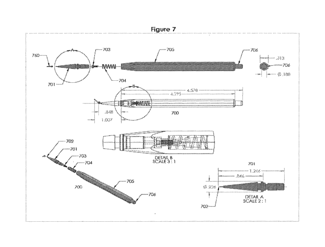
[0071] FIGS. 7-13 illustrate exemplary embodiments of punctal plug static pin
insertion tools
of the invention. Turning to FIG. 7, an exemplary insertion tool 700 is shown
engaged with an
implant 760 of the invention through meeting of pin 702 and insertion of the
lacrimal implants
14
CA 03030435 2019-01-09
WO 2018/013869 PCT/US2017/042020
into a lacrimal punctum. The lacrimal implants include the exemplary
embodiments disclosed
below, variations thereof, or any similar structures.
[0072] Figure 7 shows an insertion tool 700 comprising an inserter tip 701, a
screw 703, a
spring 704, and a handle 705. The inserter tip comprises a static pin 702 at
the distal end that is
configured for placement in bore 385 of the punctal plug 760. In embodiments,
the insertion tool
does not comprise a mechanism for moving the pin 702. The inserter tip 701
further comprises a
proximal end that snaps or locks into a distal end of the handle 705. In
embodiments, the snap or
lock mechanism is a friction fit between the handle 705 and tip 701. In
embodiments, the length
of the inserter tip 701 is about 0.75 to 1.5 inches and the length of the
inserter tip after
attachment with the handle 705 is about 0.5 to 1.25 inches. In embodiments,
the diameter of the
inserter tip 701 varies and is tapered from its largest diameter of about 0.5
to 0.1 inches to the pin
702 at the distal end.
[0073] The handle 705 comprises a screw 703 and spring 704 internal to the
handle at the
distal end and a plunger 706 at the proximal end. The plunger and spring are
configured to
release or eject the inserter tip when the plunger is depressed. The length of
the handle can vary
and is configured to fit comfortably in the hand of a user. In embodiments,
the length of the
handle 705 is 3.75 to 5.5 inches, including the plunger 706 at the proximal
end of the handle.
[0074] In embodiments, Figures 13-18 show various views of the inserter tip,
the handle and a
lacrimal implant that may be coupled to the static pin of the inserter tip. In
embodiments, the
inserter tip is manufactured and comprises biocompatible polymers. In
embodiments, the
inserter tip is molded, with no movable parts, that may be solid or
substantially sold. The density
of the material used to manufacture the inserter tip may vary and may be
selected to enhance the
interference or friction fit of the inserter tip to the handle. In
embodiments, the handle is
manufactured from the same or different material as the inserter tip. In
exemplary embodiments,
the inserter tip and handle and manufactured from and comprise different
material or polymers.
Lacrimal Implants
[0075] FIGS. 3-6 illustrate exemplary embodiments of lacrimal implants of use
in the methods
of the invention. The exemplary implants are insertable through a lacrimal
punctum 212, 214
and into its associated canaliculus 208, 210. Exemplary lacrimal implants of
use in the present
invention comprise a first member, a second member and a heel, such as the
first member 305,
CA 03030435 2019-01-09
WO 2018/013869 PCT/US2017/042020
the second member 310 and the third member or heel 330 depicted in FIG. 3A.
Exemplary
lacrimal implants further comprise a bore that is formed in the heel, for
example, the bore 385
formed in the third member or heel 330 in FIG. 3A. In some embodiments,
exemplary lacrimal
implants further comprise a cavity 458 (e.g., lacrimal implants illustrated in
FIG. 4A).
[0076] Referring to FIG. 3A, where a perspective view of an exemplary lacrimal
implant 300 of
use in the present methods is depicted, the first member 305 is characterized
by a first axis A and
the second member 310 is characterized by a second axis B.
[0077] The third member or heel 330 is configured to connect the first member
305 and the
second member 310 at a first angle 01, where 01 is defined by the first axis A
with respect to the
second axis B. For instance, in FIG. 3A, the first angle 01 refers to the
angle originating at the
first axis A and turning counterclockwise from the first axis A to the second
axis B. In some
embodiments, the first axis A and the second axis B are in the same plane and
intersect each
other. In some embodiments, the first axis A is in a plane other than the
plane of the second axis
B, and the first axis A and the second axis B do not intersect. In such
embodiments, the first
angle 01 refers to the angle defined by a parallel line of the first axis A
with respect to the second
axis B. This parallel line of the first axis A lies in the same plane as the
second axis and
intersects with the second axis.
[0078] In some embodiments, the first angle 01 is from about 30 degrees to
about 150 degrees,
from about 45 degrees to about 135 degrees, or from about 75 degrees to about
105 degrees. For
example, in some embodiments, the first angle 01 is approximately 90 degrees.
[0079] In some embodiments, the overall dimension of the implant along the
first axis is from
about 4 mm to about 8 mm. In an exemplary embodiment, the overall dimension
along the first
axis is about 5 mm to about 7 mm. In various embodiments, the overall
dimension along the first
axis is about 6.3 mm.
[0080] In various embodiments, the overall dimension along the second axis B
is from about 1
mm to about 3 mm, e.g., from about 1.2 mm to about 1.9 mm.
[0081] In some embodiments, the overall dimension along the first axis is
approximately 6.3 mm
and the overall dimension along the second axis is approximately 1.2 mm. In
various
embodiments, the overall dimension along the first axis is approximately 6.3
mm and the overall
16
CA 03030435 2019-01-09
WO 2018/013869 PCT/US2017/042020
dimension along the second axis is approximately 1.9 mm. In some embodiments,
the overall
dimension along the first axis is approximately 4.8 mm and the overall
dimension along the
second axis is approximately 1.9 mm.
First member 305
[0082] In some embodiments, the first member 305 is configured to extend into
a canaliculus,
while the second member 310 is configured to reside in the vertical portion
220, 222 of the
canaliculus and to extend to the opening of, or out of the opening of, the
associated puncta.
When a lacrimal implant 300 of such configuration is inserted into a
canaliculus, the intersection
of the first axis A and the second axis B resides generally at a curvature of
the canaliculus, such
as the canaliculus curvature 250 in FIG. 2. In some embodiments, the first
member 305 and the
second member 310 are connected at the first angle, and that angle is at least
about 45 degree,
thereby forming an angled intersection between the first member and the second
member. In
various embodiments, when the lacrimal implant 300 is positioned in the
lacrimal canaliculus, at
least a portion of the angled intersection is biased against a canaliculus
curvature of the lacrimal
canaliculus. In this embodiment, the lacrimal implant 300 uses anatomical
structures to facilitate
the retention of the implanted lacrimal implant 300.
[0083] FIG. 3B depicts a side view of an exemplary lacrimal implant 300 of the
invention. In
some embodiments, the first member 305 includes an intermediate segment 315, a
tip segment or
tip 325, and a forward segment 320 in between the forward segment and tip
segment. While the
intermediate segment 315 is configured to be connected to the second member
310 by the third
member or heel 330, the tip segment or tip 325 is configured to be inserted
through a punctum
prior to the other two segments of the first member 305 and prior to the other
members of the
lacrimal implant 300.
[0084] In some embodiments, the intermediate segment 315, the forward segment
320 and the
tip segment or tip 325 are distinguishable from each other in general by their
shapes. For
example, in some embodiments, the intermediate segment 315 has a generally
cylindrical shape
with a diameter that is larger than the diameter of the tip segment or tip
325. In various
embodiments, the forward segment 320 is tapered and has a conical shape, such
that the forward
segment 320 connects the intermediate segment 315 at one end and the tip
segment or tip 325 at
the other end. In some embodiments, the transition from the intermediate
segment 315 to the
17
CA 03030435 2019-01-09
WO 2018/013869 PCT/US2017/042020
forward segment 320 or the transition from the forward segment 320 to the tip
segment or tip
325 is gradual and smooth such that no distinguishable edge exists at the
transition.
[0085] In some embodiments, the intermediate segment 315 has a cylindrical
shape. In various
embodiments, the intermediate segment has a circular cross section, an
elliptic cross section, or a
polygonal cross section. The intermediate segment 315 is of any useful
combination of length
and diameter.
[0086] In some embodiments, the intermediate segment 315 has a diameter that
is from about
0.4 mm to about 0.8 mm. For example, in some embodiments the diameter of the
intermediate
segment 315 is from about 0.53 mm to about 0.63 mm. In some embodiments, the
intermediate
segment 315 has a length along the first axis A that is from about 0.5 mm to
about 3.5 mm. For
example, in some embodiments the length of the intermediate segment 315 is
from about 1 mm
to about 2.8 mm.
[0087] In some embodiments, the tip segment or tip 325 is substantially a semi-
sphere, or a
portion of a semi-sphere. In exemplary embodiments, the semi-sphere, or
portion therapy, has a
radius that is from about 0.05 mm to about 0.3 mm. For example, in some
embodiments, the
radius of the tip segment or tip 325 is approximately 0.20 mm.
[0088] In some embodiments, the forward segment 320 has a conical
configuration, tapering
from the diameter of the intermediate segment 315 as it approaches the tip
segment or tip 325.
In some embodiments, the forward segment 320 is short and is tapered steeply,
thus forming a
wider taper angle. The forward segment 320 can also be long and tapered more
gradually, thus
forming a narrower taper angle. The tapering angle 03 is illustrated in FIG.
3E. In some
embodiments, the tapering angle 03 is from about 2 to about 10 . For example,
in some
embodiments the tapering angle 03 is from about 3.8 to about 7.8 . In some
embodiments, 03 is
about 7.8 . In some embodiments, the forward segment 320 has a length along
the first axis A
that is from about 1 mm to about 5 mm. For example, in some embodiments the
length of
forward segment 320 is from about 1.7 mm to about 3.5 mm.
Second member 310
[0089] Referring to FIG. 3B, in some embodiments of implants of use in the
present method,
the second member 310 includes an upright segment 335 that extends from the
third member or
18
CA 03030435 2019-01-09
WO 2018/013869 PCT/US2017/042020
heel 330 generally along the direction of the second axis B. In various
embodiments, the second
member 310 further includes a head segment 340 that attaches to the upright
segment 335 at an
end opposite to the third member or heel 330. In some embodiments, the second
member 310 is
configured such that the upright segment 335 resides in the vertical portion
of the canaliculus
while the head segment 340 contacts the tissue surrounding the exterior of the
punctum when the
lacrimal implant 300 is positioned in the lacrimal canaliculus. In an
exemplary embodiment,
illustrated in FIGS. 3A-3F, the upright segment 335 has a cylindrical shape
and the head segment
340 has an oval or oblong configuration. However, it will be appreciated that
any other suitable
shapes or configurations can be used and are within the scope of the present
invention. For
example, in various embodiments, the upright segment 335 is configured to be a
conical; the
head segment 340 is configured to have a circular, elliptical or polygonal
cross section.
[0090] In some embodiments, the upright segment 335 has a characteristic
diameter that is
from about 0.7 mm to about 0.9 mm. For example, in some embodiments, the
characteristic
diameter of the upright segment 335 is about 0.8 mm.
[0091] In some embodiments, the upright segment 335 has a length in the
direction of the
second axis B that is from about 0.7 mm to about 1.5 mm. For example, in some
embodiments
the length of upright segment 335 along the direction of the second axis B is
about 0.9 mm.
[0092] Generally, the head segment 340 has a cross section characterized by a
minor axis and a
major axis. The minor axis and the major axis refer to the shortest
characteristic diameter and
the longest characteristic diameter of the cross section, respectively. As
such, the minor axis is
equal to or less than the major axis. For instance, in some embodiments where
the head segment
340 has a circular cross section, the minor axis and the major axis are of
equal length. In various
embodiments, the head segment 340 has an oval or oblong cross section, and the
minor axis is
shorter than the major axis. In some embodiments, the head segment 340 is
elongated in a
direction that is parallel to the first axis A. The major axis indicates the
extension of the first
member 305 and facilitates positioning of the lacrimal implant 300 in the
punctum and
canaliculus. In some embodiments, the major axis is from about 1.5 mm to about
2.5 mm. In
various embodiments, the minor axis is from about 1 mm to about 1.5 mm. For
example, in
some embodiments, the major axis and the minor axis head segment 340 are
approximately 1.9
mm and 1.3 mm respectively. In some embodiments, the head segment 340 has a
thickness in
19
CA 03030435 2019-01-09
WO 2018/013869 PCT/US2017/042020
the direction of the second axis that is from about 0.2 mm to about 0.4 mm.
For example, in
some embodiments, the thickness of the head segment 340 in the direction of
the second axis is
approximately 0.3 mm.
[0093] Referring still to FIG. 3B, exemplary head segment 340 comprises an
under-surface
350 facing towards the third member or heel 330 and an outer-surface 355 that
faces away from
the third member or heel 330. Exemplary head segment 340 further comprises an
edge surface
345 that couples the under-surface 350 and the outer-surface 355. The distance
between the
under-surface 350 and the outer-surface 355 can be readily varied. In some
embodiments, the
distance is from about 0.2 mm to about 0.4 mm.
[0094] In some embodiments, the outer-surface 355 is smaller than the under-
surface 350 and
is substantially flat. In various embodiments, the edge surface 345 is
tapered, curved, angular, or
multifaceted. In some embodiments, the edge surface 345 has a radius of
curvature that is from
about 0.2 mm to about 0.7 mm. In some embodiments, the under-surface 350 is in
general flat
and is configured to contact the exterior tissue surrounding the punctum when
the lacrimal
implant 300 is positioned in the lacrimal canaliculus.
Third Member or Heel 330
[0095] In some embodiments, the third member or heel 330 includes an upper
surface 360 a
lower surface 365 and side surfaces 370. In the illustrated embodiments, the
bore 385 extends
from the upper surface 360 into the third member or heel 330. In some
embodiments, the upper
surface 360 and the lower surface 365 are substantially flat and separated
from each other by a
distance. Such distance is readily variable and is typically about 0.3 mm to
about 0.7 mm. For
instance, in some embodiments, the upper surface 360 and the lower surface 365
are separated
by a distance that is from about 0.4 mm to 0.6 mm (e.g., about 0.53 mm). In
some embodiments,
the upper surface 360 extends beyond the intersection with the second member
310. In some
embodiments, the upper surface 360 extends beyond the intersection with the
second member
310 for a distance that is from about 0.3 to about 0.6 mm. The upper surface
360 can also be
joined with the side surfaces 370. In various embodiments, upper surface 360
and side surfaces
370 are joined by a curved intersection 380. In some embodiments, the curved
intersection 380
has a radius of curvature that is from about 0.04 mm to about 0.08 mm.
CA 03030435 2019-01-09
WO 2018/013869 PCT/US2017/042020
[0096] Referring now to FIGS. 3D and 3F, in some embodiments, the third member
or heel
330 includes a heel connecting segment 375 configured to couple the third
member or heel 330
to the first member 305 or to the intermediate segment 315 of the first member
305. The heel
connecting segment 375 is of readily variable shape, including flat or curved
structures. In FIG.
3F, a width of the heel connecting segment 375 in the direction of the second
axis B varies along
the direction of the first axis A. For example, the heel connecting segment
375 has a smaller
width at or near the side surfaces 370 than the diameter of the intermediate
segment 315 of the
first member 305. In some embodiments, at or near the intersection with the
intermediate
segment 315, the heel connecting segment 375 increases the width and thus
forms a notch as
depicted in FIG. 3F. It will be appreciated that the notch can be either
deeper or shallower along
both the first axis A and the second axis B before it meets the first member
305 or the second
member 310.
[0097] A notch is not a required feature in the implants of the present
invention. In some
embodiments, the heel connecting segment 375 has the same dimension as the
diameter of the
intermediate segment 315. For example, the thickness of the third member or
heel 330 along the
second axis B is equal to the diameter of the intermediate segment 315 of the
first member 305.
For example, in some embodiments, both the thickness of the third member or
heel 330 in the
direction of the second axis B and the diameter of the intermediate segment
315 are from about
0.53 mm to about 0.63 mm. In such configurations, the third member or heel 330
couples with
the intermediate segment 315 without forming a notch, as illustrated by the
alternative heel
connecting segment 675 in FIG. 6.
[0098] By way of illustration, the third member or heel 330 depicted in FIGS.
3A-3F is
substantially parallel to the first axis A of the first member 305. It would
be appreciated that this
is unnecessary. In some embodiments, the third member or heel 330 can form an
angle with
relation to the first axis A.
Bore 385
[0099] Exemplary structures of the bore 385 are detailed in FIGS. 3E and 3F,
where a cross
sectional view and a partial enlarged cross-sectional view of the lacrimal
implant 300 are
provided. The bore 385 is configured to receive a tip or other protrusion of
an external insertion
tool for facilitating insertion of the lacrimal implant 300 into a lacrimal
punctum. The
21
CA 03030435 2019-01-09
WO 2018/013869 PCT/US2017/042020
configuration, including size, shape, angle (02) and position of the bore in
the heel are readily
adjustable to facilitate the mating of the insertion tool with the bore, the
flexibility of the heel, or
the retention of the lacrimal implants. Depending on the purpose or use of the
implant and the
materials used for making the heel, the characteristics of the bore noted
above are readily varied.
Configurations of the bore 385 disclosed herein are illustrative and any other
suitable
configurations are within the scope of the present invention.
[00100] In FIG. 3F, an exemplary bore 385 is characterized by a third axis C
and a second angle
02 that is defined by the first axis with respect to the third axis A in a
similar way as the first
angle 01. In some embodiments, the second angle 02 is from about 15 to about
90 . For
example, in some embodiments, the second angle 02 is about 45 .
[00101] In some embodiments, the bore 385 has a depth along the direction of
the third axis C
that is from about 0.3 mm to about 0.7 mm. For example, in some embodiments
the depth of the
bore 385 is approximately 0.4 mm and in some embodiments, is approximately 0.6
mm. The
bore 385 may include a bore shaft 390 that is generally cylindrical, with a
circular, elliptical,
oval, or polygonal cross section. The bore 385 may further include a bore tip
395 at which the
bore shaft 390 terminates. An exemplary bore tip 395 generally has a
semispherical
configuration. In some embodiments, the bore shaft 390 has a characteristic
diameter that is
from about 0.1 mm to about 0.3 mm. In some embodiments, the characteristic
diameter of the
bore is approximately 0.17 mm. As will be appreciated, the shapes, sizes,
orientations disclosed
in the present application are illustrative, and any other suitable shapes,
sizes, or orientations are
within the scope of the present application. In addition, it will be
appreciated that the opening of
the bore can be positioned closer to the second member or closer to the edge
of the heel.
Cavity 458
[00102] FIG. 4A-4C illustrates an exemplary lacrimal implant 400 that is
insertable through a
lacrimal punctum 212, 214 and into its associated canaliculus 208, 210. In
FIG. 4A, the lacrimal
implant 400 comprises a cavity 458 that is configured to house a therapeutic
agent core or other
materials for release into an eye or surrounding tissues for treatment of
various ocular, sinus or
other diseases.
[00103] In the illustrated exemplary embodiment, the cavity 458 is formed in
the head segment
340 and has an opening through the outer-surface 355. The cavity 458 can be
shallow such that
22
CA 03030435 2019-01-09
WO 2018/013869 PCT/US2017/042020
it stays within the head segment 340. The cavity 458 can be also deeper and
extend beyond the
head segment 340 and into the upright segment 335. Illustrated exemplary
cavity 458 is in
general substantially cylindrical with a circular cross section. Any other
suitable configuration is
within the scope of the present application. For example, in some embodiments,
the cavity 458
has a truncated spherical configuration, or has a cylindrical configuration
with an oblong or a
polygonal cross section.
[00104] In some embodiments, the cavity 458 has a depth in the direction of
the second axis B
that is about from 0.2 mm to about 1.4 mm. For example, in some embodiments,
the depth of the
cavity 458 is approximately 1.2 mm. In some embodiments, the cavity 458 has a
diameter that is
from about 0.3 mm to about 0.7 mm. For example, in some embodiments the
diameter of the
cavity 458 is from about 0.42 mm to about 0.55 mm. In an exemplary embodiment,
the cavity
458 extends into the upright segment 335, and the diameter of the cavity 458
is smaller than the
diameter of the upright segment 335.
[00105] Referring to FIG. 4C, the cavity 458 includes a bottom 482. In various
embodiments,
the bottom 482 is rounded. In various embodiments, the rounded bottom has a
radius of
curvature that is from about 0.03 mm to about 0.07 mm.
[00106] FIG. 5 depicts exemplary configurations of the cavity 458. In FIG. 5,
the cavity 458
includes a lip 584 or other retaining structure positioned at the opening of
the cavity 458. The lip
584 or the other retaining structure are optionally configured to partially
enclose the cavity 458,
e.g, prevent a therapeutic agent core or other materials from moving out of
the cavity 458. In
some embodiments, the lip 584 is a square cross-sectional annulus that extends
down from the
outer-surface 355 into the cavity 458 and extends inwardly towards the center
of the opening of
the cavity 458. In some embodiments, the lip 584 is of a tab configuration and
includes a
plurality of spaced lips that extend inwardly into the opening of the cavity
458. The lip 584 may
extend downwardly from about 0.02 mm to about 0.1 mm and inwardly from about
0.02 mm to
about 0.1 mm. For example, in some embodiments, the lip 584 extends about 0.05
mm
downwardly or inwardly.
Formation of Lacrimal Implants
[00107] Exemplary lacrimal implants of use in methods of the present invention
are made of
various materials including plastic, rubber, polymer, or composite. Exemplary
lacrimal implants
23
CA 03030435 2019-01-09
WO 2018/013869 PCT/US2017/042020
of the present invention formed from one or more material including plastic,
rubber, polymer,
composites, or other appropriate materials. In some embodiments, the lacrimal
implants are
formed from liquid silicone rubber. For instance, in exemplary embodiments,
lacrimal implants
are formed from a material marketed as NuSil 4840 liquid silicone rubber,
NuSil 4870, or a
mixture including such a liquid silicone rubber. Examples of such a mixture
include a material
marketed as 6-4800, which comprises NuSil 4840 with from about 1% to about 5%,
e.g., from
about 2% to about 4% of 6-4800.
[00108] In some embodiments, the lacrimal implant is formed from biodegradable
materials, for
instance, biodegradable elastic materials including cross-linked polymers,
such as poly (vinyl
alcohol). In some embodiments, the lacrimal implant can comprise a co-polymer,
such as
silicone/polyurethane co-polymer, silicone/urethane, silicone/poly (ethylene
glycol) (PEG), and
silicone/2hydroxyethyl methacrylate (HEMA). As discussed in commonly-owned
Utkhede et
al., U.S. patent application Ser. No. 12/231,986, entitled "DRUG CORES FOR
SUSTAINED
RELEASE OF THERAPEUTIC AGENTS," filed Sep. 5, 2008, which is herein
incorporated by
reference in its entirety, urethane-based polymer and copolymer materials
allow for a variety of
processing methods and bond well to one another.
[00109] The hardness of the material is selected to facilitate or alter the
retention of the lacrimal
implant within the lacrimal punctum and its associated canaliculus.
Accordingly, in some
embodiments, a material having a durometer rating of from about 20D to about
80D, e.g., about
30D to about 70D, e.g., from about 40D to about 60D is of use to adjust
parameters such as
patient comfort and retention. For example, in some embodiments, the durometer
rating of the
material used to form the lacrimal implants is approximately 40D. Materials
other than those
exemplified above providing a durometer rating for the lacrimal implants
within the stated
ranges, and particularly that is about 40D are also of use. In some
embodiments, a harder
material or softer material is utilized for the entire lacrimal implant or for
portions thereof. In
such case, the lacrimal implants are formed from the materials that provide a
durometer rating of
about 70D.
[00110] In some embodiments, the lacrimal implants of use in the present
methods are formed
of multiple materials, where certain members or portions of the lacrimal
implants are formed
with materials having different properties. For example, in some embodiments
the first member
24
CA 03030435 2019-01-09
WO 2018/013869 PCT/US2017/042020
305 is formed of a harder durometer rated material while the second member 310
is formed of a
softer durometer rated material. In some embodiments, the first member 305 is
formed of a
softer durometer rated material while the second member 310 is formed of a
harder durometer
rated material. In some embodiments, the third member or heel 330 is formed of
a harder
durometer rated material than one or more parts of the remainder of the second
member 310. In
various embodiments, the third member or heel 330 is formed of a softer
durometer rated
material than the remainder of the second member 310.
[00111] Exemplary implants of use in the invention can be formed by methods
known in the art,
including, but not limited to, machining a blank to the desired shape and size
and molding the
material forming the implant.
[00112] The implant can be one of any number of different designs that
releases latanoprost or
other intraocular pressure-reducing therapeutic agent(s) for a sustained
period of time. The
disclosures of the following patent documents, which describe example implant
structure or
processing embodiments for use in the methods of embodiments of the current
invention and
methods of making those implants, are incorporated herein by reference in
their entirety: U.S.
Application Serial No. 60/871,864 (filed December 26, 2006 and entitled
Nasolacrimal Drainage
System Implants for Drug Therapy); U.S. Application Serial No. 11/695,537
(filed April 2, 2007
and entitled Drug Delivery Methods, Structures, and Compositions for
Nasolacrimal System);
U.S. U.S. Application Serial No. 12/332,219 (filed December 10, 2008 and
entitled Drug
Delivery Methods, Structures, and Compositions for Nasolacrimal System); U.S.
Application
Serial No. 60/787,775 (filed March 31, 2006 and entitled Nasolacrimal Drainage
System
Implants for Drug Therapy); U.S. Application Serial No. 11/695,545 (filed
April 2, 2007 and
entitled Nasolacrimal Drainage System Implants for Drug Therapy); U.S.
Application Serial No.
60/585,287 (filed July 2, 2004 and entitled Treatment Medium Delivery Device
and Methods for
Delivery of Such Treatment Mediums to the Eye Using Such a Delivery Device);
U.S.
Application Serial No. 11/571,147 (filed December 21, 2006 and entitled
Treatment Medium
Delivery Device and Methods for Delivery of Such Treatment Mediums to the Eye
Using Such a
Delivery Device); U.S. Application Serial No. 60/970,696 (filed September 7,
2007 and entitled
Expandable Nasolacrimal Drainage System Implants); U.S. Application Serial No.
60/974,367
(filed September 21, 2007 and entitled Expandable Nasolacrimal Drainage System
Implants);
U.S. Application Serial No. 60/970,699 (filed September 7, 2007 and entitled
Manufacture of
CA 03030435 2019-01-09
WO 2018/013869 PCT/US2017/042020
Drug Cores for Sustained Release of Therapeutic Agents); U.S. Application
Serial No.
60/970,709 (filed September 7, 2007 and entitled Nasolacrimal Drainage System
Implants for
Drug Delivery); U.S. Application Serial No. 60/970,720 (filed September 7,
2007 and entitled
Manufacture of Expandable Nasolacrimal Drainage System Implants); U.S.
Application Serial
No. 60/970,755 (filed September 7, 2007 and entitled Prostaglandin Analogues
for Implant
Devices and Methods); U.S. Application Serial No. 60/970,820 (filed September
7, 2007 and
entitled Multiple Drug Delivery Systems and Combinations of Drugs with Punctal
Implants);
U.S. Application Serial No. 61/066,223 (filed February 18, 2008 and entitled
Lacrimal Implants
and Related Methods); U.S. Application Serial No. 61/049,347 (filed April 30,
2008 and entitled
Lacrimal Implants and Related Methods); U.S. Application Serial No. 61/033,211
(filed March
3, 2008 and entitled Lacrimal Implants and Related Methods); U.S. Application
Serial No.
61/049,360 (filed April 30, 2008 and entitled Lacrimal Implants and Related
Methods); U.S.
Application Serial No. 61/052,595 (filed May 12, 2008 and entitled Lacrimal
Implants and
Related Methods); U.S. Application Serial No. 61/075,309 (filed June 24, 2008
and entitled
Lacrimal Implants and Related Methods); U.S. Application Serial No. 61/154,693
(filed
February 23, 2009 and entitled Lacrimal Implants and Related Methods); U.S.
Application Serial
No. 61/209,036 (filed March 2, 2009 and entitled Lacrimal Implants and Related
Methods); U.S.
Application Serial No. 61/209,630 (filed March 9, 2009 and entitled Lacrimal
Implants and
Related Methods); U.S. Application Serial No. 61/036,816 (filed March 14, 2008
and entitled
Lacrimal Implants and Related Methods); U.S. Application Serial No. 61/271,862
(filed July 27,
2009 and entitled Lacrimal Implants and Related Methods); U.S. Application
Serial No.
61/252,057 (filed October 15, 2009 and entitled Lacrimal Implants and Related
Methods); U.S.
Application Serial No. 12/710,855 (filed February 23, 2010 and entitled
Lacrimal Implants and
Related Methods); U.S. Application Serial No. 60/871,867 (filed December 26,
2006 and entitled
Drug Delivery Implants for Inhibition of Optical Defects); U.S. Application
Serial No.
12/521,543 (filed December 31, 2009 and entitled Drug Delivery Implants for
Inhibition of
Optical Defects); U.S. Application Serial No. 61/052,068 (filed May 9, 2008
and entitled
Sustained Release Delivery of Latanoprost to Treat Glaucoma); U.S. Application
Serial No.
61/052,113 (filed May 9, 2008 and entitled Sustained Release Delivery of
Latanoprost to Treat
Glaucoma); U.S. Application Serial No. 61/108,777 (filed October 27, 2008 and
entitled
Sustained Release Delivery of Latanoprost to Treat Glaucoma); U.S. Application
Serial No.
26
CA 03030435 2019-01-09
WO 2018/013869 PCT/US2017/042020
12/463,279 (filed May 8, 2009 and entitled Sustained Release Delivery of
Active Agents to Treat
Glaucoma and Ocular Hypertension); U.S. Application Serial No. 61/049,337
(filed April 30,
2008 and entitled Lacrimal Implants and Related Methods); U.S. Application
Serial No.
12/432,553 (filed April 29, 2009 and entitled Composite Lacrimal Insert and
Related Methods);
U.S. Application Serial No. 61/049,317 (filed April 30, 2008 and entitled Drug-
Releasing
Polyurethane Lacrimal Insert); U.S. Application Serial No. 12/378,710 (filed
February 17, 2009
and entitled Lacrimal Implants and Related Methods); U.S. Application Serial
No. 61/075,284
(filed June 24, 2008 and entitled Combination Treatment of Glaucoma); U.S.
Application Serial
No. 12/490,923 (filed June 24, 2009 and entitled Combination Treatment of
Glaucoma); U.S.
Application Serial No. 61/134,271 (filed July 8, 2008 and entitled Lacrimal
Implant Body
Including Comforting Agent); U.S. Application Serial No. 12/499,605 (filed
July 8, 2009 and
entitled Lacrimal Implant Body Including Comforting Agent); U.S. Application
Serial No.
61/057,246 (filed May 30, 2008 and entitled Surface Treatment of Implants and
Related
Methods); U.S. Application Serial No. 61/132,927 (filed June 24, 2008 and
entitled Surface
Treated Implantable Articles and Related Methods); U.S. Application Serial No.
12/283,002
(filed September 5, 2008 and entitled Surface Treated Implantable Articles and
Related
Methods); U.S. Application Serial No. 12/231,989 (filed September 5, 2008 and
entitled
Lacrimal Implants and Related Methods); U.S. Application Serial No. 61/049,317
(filed April
30, 2008 and entitled Drug-Releasing Polyurethane Lacrimal Insert); U.S.
Application Serial No.
12/231,986 (filed September 5, 2008 and entitled Drug Cores for Sustained
Release of
Therapeutic Agents); U.S. Application Serial No. 61/050,901 (filed May 6, 2008
and entitled
Punctum Plug Detection); U.S. Application Serial No. 12/231,987 (filed
September 5, 2008 and
entitled Lacrimal Implant Detection); U.S. Application Serial No. 61/146,860
(filed January 23,
2009 and entitled Sustained Release Delivery of One or More Anti-Glaucoma
Agents); U.S.
Application Serial No. 61/152,909 (filed February 16, 2009 and entitled
Sustained Release
Delivery of One or More Anti-Glaucoma Agents); U.S. Application Serial No.
61/228,894 (filed
July 27, 2009 and entitled Sustained Release Delivery of One or More Anti-
Glaucoma Agents);
U.S. Application Serial No. 61/277,000 (filed September 18, 2009 and entitled
Drug Cores for
Sustained Ocular Release of Therapeutic Agents); U.S. Application Serial No.
12/692,452 (filed
January 22, 2010 and entitled Sustained Release Delivery of One or More
Agents); U.S.
Application Serial No. 61/283,100 (filed November 27, 2009 and entitled
Lacrimal Implants
27
CA 03030435 2019-01-09
WO 2018/013869 PCT/US2017/042020
Including Split and Insertable Drug Core); International Application Serial
No.
PCT/US2010/058129 (filed November 26, 2010, published as WO 2011/066479 and
entitled
Lacrimal Implants Including Split and Insertable Drug Core); U.S. Application
Serial No.
61/139,456 (filed December 19, 2008 and entitled Substance Delivering Punctum
Implants and
Methods); U.S. Application Serial No. 12/643,502 (filed December 21, 2009 and
entitled
Substance Delivering Punctum Implants and Methods); U.S. Application Serial
No. 10/825,047
(filed April 15, 2004 and entitled Drug Delivery via Punctal Plug); U.S.
Application Serial No.
12/604,202 (filed October 22, 2009 and entitled Drug Delivery via Ocular
Implant); International
Application Serial No. PCT/U52005/023848 (filed July 1, 2005, published as WO
2006/014434
and entitled Treatment Medium Delivery Device and Methods for Delivery);
International
Application Serial No. PCT/U52007/065792 (filed April 2, 2007, published as WO
2007/115261
and entitled Drug Delivery Methods, Structures, and Compositions for
Nasolacrimal System);
and International Application Serial No. PCT/U52007/065789 (filed April 2,
2007, published
as WO 2007/115259 and entitled Nasolacrimal Drainage System Implants for Drug
Therapy).
Retention
[00113] In various embodiments of the methods of the invention, an implant
including a
retention structure is employed to retain the implant in the punctum or
canaliculus. The retention
structure is attached to or integral with the implant body. The retention
structure comprises an
appropriate material that is sized and shaped so that the implant can be
easily positioned in the
desired tissue location, for example, the punctum or canaliculus. In some
embodiments, the drug
core may be attached to the retention structure via, at least in part, the
sheath. In some
embodiments, the retention structure comprises a hydrogel configured to expand
when the
retention structure is placed in the punctum. The retention structure can
comprise an attachment
member having an axially oriented surface. In some embodiments, expansion of
the hydrogel
can urge against the axially oriented surface to retain the hydrogel while the
hydrogel is
hydrated. In some embodiments, the attachment member can comprise at least one
of a
protrusion, a flange, a rim, or an opening through a portion of the retention
structure. In some
embodiments, the retention structure includes an implant body portion size and
shape to
substantially match an anatomy of the punctum and canaliculus.
28
CA 03030435 2019-01-09
WO 2018/013869 PCT/US2017/042020
[00114] The retention structure may have a size suitable to fit at least
partially within the
canalicular lumen. The retention structure can be expandable between a small
profile
configuration suitable for insertion and a large profile configuration to
anchor the retention
structure in the lumen, and the retention structure can be attached near the
distal end of the drug
core. In specific embodiments, the retention structure can slide along the
drug core near the
proximal end when the retention structure expands from the small profile
configuration to the
large profile configuration. A length of the retention structure along the
drug core can be shorter
in the large profile configuration than the small profile configuration.
[00115] In some embodiments, the retention structure is resiliently
expandable. The small
profile may have a cross section of no more than about 0.2 mm, and the large
profile may have a
cross section of no more than about 2.0 mm. The retention structure may
comprise a tubular
body having arms separated by slots. The retention structure can be disposed
at least partially
over the drug core.
[00116] In some embodiments, the retention structure is mechanically
deployable and typically
expands to a desired cross-sectional shape, for example with the retention
structure comprising a
super elastic shape memory alloy such as NitinolTM. Other materials in
addition to NitinolTM can
be used, for example resilient metals or polymers, plastically deformable
metals or polymers,
shape memory polymers, and the like, to provide the desired expansion. In some
embodiments
polymers and coated fibers available from Biogeneral, Inc. of San Diego, CA
may be used.
Many metals such as stainless steels and non-shape memory alloys can be used
and provide the
desired expansion. This expansion capability permits the implant to fit in
hollow tissue
structures of varying sizes, for example canaliculae ranging from 0.3 mm to
1.2 mm (i.e. one size
fits all). Although a single retention structure can be made to fit
canaliculae from 0.3 to 1.2 mm
across, a plurality of alternatively selectable retention structures can be
used to fit this range if
desired, for example a first retention structure for canaliculae from 0.3 to
about 0.9 mm and a
second retention structure for canaliculae from about 0.9 to 1.2 mm. The
retention structure has a
length appropriate to the anatomical structure to which the retention
structure attaches, for
example a length of about 3 mm for a retention structure positioned near the
punctum of the
canaliculus. For different anatomical structures, the length can be
appropriate to provide
adequate retention force, e.g. 1 mm to 15 mm lengths as appropriate.
29
CA 03030435 2019-01-09
WO 2018/013869 PCT/US2017/042020
[00117] Although the implant body may be attached to one end of the retention
structure as
described above, in many embodiments the other end of the retention structure
is not attached to
the implant body so that the retention structure can slide over the implant
body including the
sheath body and drug core while the retention structure expands. This sliding
capability on one
end is desirable as the retention structure may shrink in length as the
retention structure expands
in width to assume the desired cross sectional width. However, it should be
noted that many
embodiments may employ a sheath body that does not slide in relative to the
core.
[00118] In many embodiments, the retention structure can be retrieved from
tissue. A
projection, for example a hook, a loop, or a ring, can extend from a portion
of the implant body
to facilitate removal of the retention structure.
[00119] In some embodiments the sheath and retention structure can comprise
two parts.
[00120] The lacrimal implants of the present invention have exceptional
retention properties,
and are retained in the punctum and canaliculus for a period that is enhanced
relative to a
commercially available plug (FIG. 9) based upon the percentage of eyes in
which an implant was
implanted retaining the implant over a selected time period.
[00121] In an exemplary embodiment, the method of the invention uses a
lacrimal implant
configured to remain implanted in a punctum for at least about 1 week, 2
weeks, 3 weeks, 4
weeks, 5 weeks, 6 weeks, 7 weeks, 8, weeks, 9 weeks 10 weeks, 11 weeks, or at
least about 12
weeks or more. In an exemplary embodiment, the lacrimal implant is configured
to be retained
by the puncta for the duration of the intended sustained release of the
therapeutic agent. In
various embodiments, the duration of the intended sustained release of the
therapeutic agent is at
least about 1 week, 2 weeks, 3 weeks, 4 weeks, 5 weeks, 6 weeks, 7 weeks, 8,
weeks, 9 weeks 10
weeks, 11 weeks, or at least about 12 weeks or more. In various embodiments at
least about
95%, at least about 90%, at least about 85% or at least about 80% of the
implanted implants are
retained for the duration of the intended controlled release of the
therapeutic agent. In an
exemplary embodiment, the implant is retained by the puncta for a length of
time to show
therapeutic efficacy.
[00122] In various embodiments, the present invention provides for the use of
implants having
structural features that enhance the retention of the implant in a punctum.
Amongst other
features, the heel of the present implant (e.g., 330) is configured to come to
rest in the lacrimal
CA 03030435 2019-01-09
WO 2018/013869 PCT/US2017/042020
canaliculus ampulla (e.g., 252), effectively locking the implant into place.
However, the
inventors have recognized that to prevent rotation and relative movement of
the implanted
device, which plays a role in the displacement of the device, a first member
was needed to
maintain the heel in the ampulla. Thus, the first member, e.g., 305, is
configured to stabilize the
punctal plug within the lacrimal canaliculus, prevent rotation and maintain
positioning of the
plug when the surrounding tissue moves.
Occlusive Element
[00123] In an exemplary embodiment, the methods of the invention use an
implant having an
occlusive element. An occlusive element can be mounted to and expandable with
the retention
structure to inhibit tear flow. An occlusive element may inhibit tear flow
through the lumen, and
the occlusive element may cover at least a portion of the retention structure
to protect the lumen
from the retention structure. The occlusive element comprises an appropriate
material that is
sized and shaped so that the implant can at least partially inhibit, even
block, the flow of fluid
through the hollow tissue structure, for example lacrimal fluid through the
canaliculus. The
occlusive material may be a thin walled membrane of a biocompatible material,
for example
silicone, that can expand and contract with the retention structure. The
occlusive element is
formed as a separate thin tube of material that is slid over the end of the
retention structure and
anchored to one end of the retention structure as described above.
Alternatively, the occlusive
element can be formed by dip coating the retention structure in a
biocompatible polymer, for
example silicone polymer. The thickness of the occlusive element can be in a
range from about
0.01 mm to about 0.15 mm, and often from about 0.05 mm to 0.1 mm.
Therapeutic Agents and Therapeutic Agent Cores
[00124] Conventional drug delivery involving frequent periodic dosing is not
ideal or practical
in many instances. For example, with more toxic drugs, conventional periodic
dosing can result
in unfavorably high initial drug levels at the time of dosing, followed by low
drug levels between
doses often times below levels of therapeutic value. Likewise, conventional
periodic dosing may
not be practical or therapeutically effective in certain instances such as
with pharmaceutical
therapies targeting areas of the inner eye or brain in need of treatment such
as the retina.
Accordingly, in some embodiments, the lacrimal implant further includes one or
more
therapeutic agent within its structure. In various embodiments, the agent is
dispersed throughout
31
CA 03030435 2019-01-09
WO 2018/013869 PCT/US2017/042020
the device. In some embodiments, the agent is located at one or more distinct
locations or zones
of the device. In an exemplary embodiment, the therapeutic agent is located in
a cavity of the
device and the component holding the agent is referred to as a core.
[00125] In various embodiments, in which the agent is dispersed throughout the
device, the rate
and location of release of the agent is controlled by coating at least a
component of the device
with a material that is impermeable to the drug. In an exemplary embodiment,
essentially the
entire device is coated with the material with the exception of one or more
gaps in the material
through which the agent can elute into the eye or surrounding tissue. An
exemplary coating is a
Parylene coating (See, 2008/0181930, herein incorporated by reference).
[00126] In an exemplary embodiment, the lacrimal implant of the invention is
configured as a
sustained release device, releasing the incorporated therapeutic agent in a
therapeutically
effective manner, e.g., at a rate that provides a therapeutically effective
dosage for at least about
1 week, 2 weeks, 3 weeks, 4 weeks, 5 weeks, 6 weeks, 7 weeks, 8, weeks, 9
weeks 10 weeks, 11
weeks, or at least about 12 weeks or more. In an exemplary embodiment, the
lacrimal implant is
configured to be retained by the puncta for the duration of the intended
controlled release of the
therapeutic agent. In various embodiments, the duration of the intended
controlled release of the
therapeutic agent is at least about 1 week, 2 weeks, 3 weeks, 4 weeks, 5
weeks, 6 weeks, 7
weeks, 8, weeks, 9 weeks 10 weeks, 11 weeks, or at least about 12 weeks or
more. In various
embodiments, at least 95% of the implanted implants are retained for the
duration of the intended
controlled release of the therapeutic agent. In an exemplary embodiment, the
implant is retained
by the puncta for a length of time to show therapeutic efficacy.
Therapeutic Agent Core
[00127] In an embodiment, the methods of the invention utilize an implant
including a distinct
therapeutic agent core or integrated drug or other agent disposed in at least
one of the first
member 305 or the second member 310 of the implant body, to provide a
sustained release of a
therapeutic agent. For instance, the drug core or integrated drug or other
agent disposed may be
disposed in the cavity 458 of the lacrimal implant 400 to provide a sustained
drug or other
therapeutic agent release.
[00128] An exemplary implant of use in the methods of the invention is
configured to deliver a
therapeutic agent to one or more of an eye, nasal passage or inner ear system.
In various
32
CA 03030435 2019-01-09
WO 2018/013869 PCT/US2017/042020
embodiments, the drug is delivered systemically to the subject through the
eye. A therapeutic
agent core can comprise one or more therapeutic agents, and in some examples,
one or more
matrix materials to provide sustained release of the drug or other agents.
[00129] In various embodiments, the therapeutic agent core is inserted into
cavity 458.
[00130] In various examples, the distinct drug core or integrated drug or
other agent includes at
least about 20 micrograms, at least about 40 micrograms, at least about 45
micrograms, at least
80 micrograms, or at least 95 micrograms of a drug (e.g., latanoprost), such
as is further
discussed in commonly-owned Butuner et al., U.S. patent application Ser. No.
12/463,279,
entitled "SUSTAINED RELEASE DELIVERY OF ACTIVE AGENTS TO TREAT
GLAUCOMA AND OCULAR HYPERTENSION," filed May 8, 2009, and commonly-owned
Utkhede, U.S. Patent Application No. 61/277,000, entitled "IMPROVED DRUG CORES
FOR
SUSTAINTED OCULAR RELEASE OF THERAPEUTIC AGENTS," filed Sep. 18, 2009, both
of which are incorporated by reference in their entirety, including their
descriptions of drug or
other agent concentration.
[00131] The drug core can comprise one or more biocompatible materials capable
of providing
a sustained release of the one or more drugs or agents. The drug core can
comprise a matrix
including a substantially non-biodegradable silicone matrix with dissolvable
inclusions of the
drugs or agents located therein. The drug core can include other structures
that provide sustained
release of the drugs or agents, for example a biodegradable matrix, a porous
drug core, a liquid
drug core or a solid drug core. A matrix that includes the drugs or agents can
be formed from
either biodegradable or non-biodegradable polymers. In some examples, a non-
biodegradable
drug core can include silicone, acrylates, polyethylenes, polyurethane,
polyurethane, hydrogel,
polyester (e.g., DACRONTM. from E.I. Du Pont de Nemours and Company,
Wilmington, Del.),
polypropylene, polytetrafluoroethylene (PTFE), expanded PTFE (ePTFE),
polyether ether ketone
(PEEK), nylon, extruded collagen, polymer foam, silicone rubber, polyethylene
terephthalate,
ultra high molecular weight polyethylene, polycarbonate urethane,
polyurethane, polyimides,
stainless steel, nickel-titanium alloy (e.g., Nitinol), titanium, stainless
steel, cobalt-chrome alloy
(e.g., ELGILOYTM. from Elgin Specialty Metals, Elgin, Ill.; CONICHROMETm. from
Carpenter
Metals Corp., Wyomissing, Pa.). In some examples, a biodegradable drug core
can comprise one
or more biodegradable polymers, such as protein, hydrogel, polyethylene
oxides, polyglycolic
33
CA 03030435 2019-01-09
WO 2018/013869 PCT/US2017/042020
acid (PGA), polylactic acid (PLA), poly(L-lactic acid) (PLLA), poly(L-glycolic
acid) (PLGA),
polyglycolide, poly-L-lactide, poly-D-lactide, poly(amino acids),
polydioxanone,
polycaprolactone, polygluconate, polylactic acid-polyethylene oxide
copolymers, modified
cellulose, collagen, polyorthoesters, polyhydroxybutyrate, polyanhydride,
polyphosphoester,
poly(alpha-hydroxy acid) and combinations thereof. In some examples, the drug
core can
comprise a hydrogel polymer.
Sheath Body
[00132] In an exemplary embodiment, the implant of use in the methods of the
invention
includes a therapeutic agent core which is encased in a sheath body. The
sheath body can
comprise appropriate shapes and materials to control the migration of
latanoprost or other anti-
glaucoma agent from the drug core. In some embodiments, the sheath body houses
the drug core
and can fit snugly against the core. The sheath body is made from a material
that is substantially
impermeable to the latanoprost or other anti-glaucoma agent so that the rate
of migration of the
agent may be largely controlled by the exposed surface area of the drug core
that is not covered
by the sheath body. In many embodiments, migration of the latanoprost or other
anti-glaucoma
agent through the sheath body can be about one tenth of the migration of
latanoprost or other
anti-glaucoma agent through the exposed surface of the drug core, or less,
often being one
hundredth or less. In other words, the migration of the latanoprost or other
anti-glaucoma agent
through the sheath body is at least about an order of magnitude less that the
migration of
latanoprost or other anti-glaucoma agent through the exposed surface of the
drug core. Suitable
sheath body materials include polyimide, polyethylene terephthalate
(hereinafter "PET"). The
sheath body has a thickness, as defined from the sheath surface adjacent the
core to the opposing
sheath surface away from the core, from about 0.00025" to about 0.0015". The
total diameter of
the sheath that extends across the core ranges from about 0.2 mm to about 1.2
mm. The core may
be formed by dip coating the core in the sheath material. Alternatively or in
combination, the
sheath body can comprise a tube and the core introduced into the sheath, for
example as a liquid
or solid that can be slid, injected or extruded into the sheath body tube. The
sheath body can also
be dip coated around the core, for example dip coated around a pre-formed
core.
[00133] The sheath body can be provided with additional features to facilitate
clinical use of
the implant. For example, the sheath may receive a drug core that is
exchangeable while the
34
CA 03030435 2019-01-09
WO 2018/013869 PCT/US2017/042020
implant body, retention structure and sheath body remain implanted in the
subject. The sheath
body is often rigidly attached to the retention structure as described above,
and the core is
exchangeable while the retention structure retains the sheath body. In
specific embodiments, the
sheath body can be provided with external protrusions that apply force to the
sheath body when
squeezed and eject the core from the sheath body. Another drug core can then
be positioned in
the sheath body. In many embodiments, the sheath body or retention structure
may have a
distinguishing feature, for example a distinguishing color, to show placement
such that the
placement of the sheath body or retention structure in the canaliculus or
other body tissue
structure can be readily detected by the subject. The retention element or
sheath body may
comprise at least one mark to indicate the depth of placement in the
canaliculus such that the
retention element or sheath body can be positioned to a desired depth in the
canaliculus based on
the at least one mark.
Therapeutic Agents
[00134] Generally, pharmaceutically active agents or drugs useful in the
methods of the present
invention can be any compound, composition of matter, or mixtures thereof that
can be delivered
from an implant, such as those described herein, to produce a beneficial and
useful result to, for
example, the eye, especially an agent effective in obtaining a desired local
or systemic
physiological or pharmacological effect.
[00135] Examples of such agents include, but are not limited to, anesthetics
and pain killing
agents such as lidocaine and related compounds, benzodiazepam and related
compounds and the
like; anti-cancer agents such as 5-fluorouracil, adriamycin and related
compounds and the like;
anti-fungal agents such as fluconazole and related compounds and the like;
anti-viral agents such
as trisodium phosphomonoformate, trifluorothymidine, acyclovir, ganciclovir,
DDI, AZT and the
like; cell transport/mobility impending agents such as colchicine,
vincristine, cytochalasin B and
related compounds and the like; antiglaucoma drugs (e.g. adrenergic agonists,
adrenergic
antagonists (beta blockers), carbonic anhydrase inhibitors (CAIs, systemic and
topical),
parasympathomimetics, prostaglandins and hypotensive lipids, and combinations
thereof),
antimicrobial agent (e.g., antibiotic, antiviral, antiparacytic, antifungal,
etc.), a corticosteroid or
other anti-inflammatory (e.g., an NSAID or other analgesic and pain management
compounds), a
decongestant (e.g., vasoconstrictor), an agent that prevents of modifies an
allergic response (e.g.,
CA 03030435 2019-01-09
WO 2018/013869 PCT/US2017/042020
an antihistamine, cytokine inhibitor, leucotriene inhibitor, IgE inhibitor,
immunomodulator), a
mast cell stabilizer, cycloplegic, mydriatic or the like.
[00136] Other agents that can be incorporated into devices of use in the
invention include
antihypertensives; decongestants such as phenylephrine, naphazoline,
tetrahydrazoline and the
like; immunological response modifiers such as muramyl dipeptide and related
compounds and
the like; peptides and proteins such as cyclosporin, insulin, growth hormones,
insulin related
growth factor, heat shock proteins and related compounds and the like;
steroidal compounds such
as dexamethasone, prednisolone and related compounds and the like; low
solubility steroids such
as fluocinolone acetonide and related compounds and the like; carbonic
anhydrase inhibitors;
diagnostic agents; antiapoptosis agents; gene therapy agents; sequestering
agents; reductants
such as glutathione and the like; antipermeability agents; antisense
compounds; antiproliferative
agents; antibody conjugates; antidepressants; bloodflow enhancers;
antiasthmatic drugs;
antiparasiticagents; non-steroidal anti-inflammatory agents such as ibuprofen
and the like;
nutrients and vitamins: enzyme inhibitors: antioxidants; anticataract drugs;
aldose reductase
inhibitors; cytoprotectants; cytokines, cytokine inhibitors, and cytokin
protectants; uv blockers;
mast cell stabilizers; anti neovascular agents such as antiangiogenic agents,
e.g., matrix
metalloprotease inhibitors and the like.
[00137] Representative examples of additional pharmaceutically active agent
for use herein
include, but are not limited to, neuroprotectants such as nimodipine and
related compounds and
the like; antibiotics such as tetracycline, chlortetracycline, bacitracin,
neomycin, polymyxin,
gramicidin, oxytetracycline, chloramphenicol, gentamycin, erythromycin and the
like; anti-
infectives; antibacterials such as sulfonamides, sulfacetamide,
sulfamethizole, sulfisoxazole;
nitrofurazone, sodium propionate and the like; antiallergenics such as
antazoline, methapyriline,
chlorpheniramine, pyrilamine, prophenpyridamine and the like; anti-
inflammatories such as
hydrocortisone, hydrocortisone acetate, dexamethasone 21-phosphate,
fluocinolone, medrysone,
methylprednisolone, prednisolone 21-phosphate, prednisolone acetate,
fluoromethalone,
betamethasone, triminolone and the like; miotics; anti-cholinesterase such as
pilocarpine, eserine
salicylate, carbachol, di-isopropyl fluorophosphate, phospholine iodine,
demecarium bromide
and the like; miotic agents; mydriatics such as atropine sulfate,
cyclopentolate, homatropine,
scopolamine, tropicamide, eucatropine, hydroxyamphetamine and the like;
svmpathomimetics
such as epinephrine and the like; and prodrugs such as, for example, those
described in Design of
36
CA 03030435 2019-01-09
WO 2018/013869 PCT/US2017/042020
Prodrugs, edited by Hans Bundgaard, Elsevier Scientific Publishing Co.,
Amsterdam, 1985. In
addition to the foregoing agents, other agents suitable for treating,
managing, or diagnosing
conditions in a mammalian organism may be entrapped in the copolymer and
administered using
the drug delivery systems of the current invention. Once again, reference may
be made to any
standard pharmaceutical textbook such as, for example, Remington's
Pharmaceutical Sciences
for pharmaceutically active agents.
[00138] Any pharmaceutically acceptable form of the foregoing therapeutically
active agent
may be employed in the practice of the present invention, e.g., the free base;
free acid;
pharmaceutically acceptable salts, esters or amides thereof, e.g., acid
additions salts such as the
hydrochloride, hydrobromide, sulfate, bisulfate, acetate, oxalate, valerate,
oleate, palmitate,
stearate, laurate, borate, benzoate, lactate, phosphate, tosylate, mesylate,
citrate, maleate,
fumarate, succinate, tartrate, ascorbate, glucoheptonate, lactobionate, and
lauryl sulfate salts and
the like; alkali or alkaline earth metal salts such as the sodium, calcium,
potassium and
magnesium salts and the like; hydrates; enantiomers; isomers; stereoisomers;
diastereoisomers;
tautomers; polymorphs, mixtures thereof, prodrugs thereof or racemates or
racemic mixtures
thereof.
[00139] Additional agents that can be used with the present methods utilizing
lacrimal implants
include, but are not limited to, drugs that have been approved under Section
505 of the United
States Federal Food, Drug, and Cosmetic Act or under the Public Health Service
Act, some of
which can be found at the U.S. Food and Drug Administration (FDA) website
http://www.accessdatalda.gov/scripts/cder/drugsatfda/index. The present
lacrimal implants can
also be used with drugs listed in the Orange Book, either in paper or in
electronic form, which
can be found at the FDA Orange Book website (http://www.fda.gov/cder/ob/)),
that has or
records the same date as, earlier date than, or later date than, the filing
date of this patent
document. For example, these drugs can include, among others, dorzolamide,
olopatadine,
travoprost, bimatoprost, cyclosporin, brimonidine, moxifloxacin, tobramycin,
brinzolamide,
aciclovir timolol maleate, ketorolac tromethamine, prednisolone acetate,
sodium hyaluronate,
nepafenac, bromfenac,diclofenac, flurbiprofen, suprofenac, binoxan, patanol,
dexamethasone/tobramycin combination, moxifloxacin, or acyclovir.
37
CA 03030435 2019-01-09
WO 2018/013869 PCT/US2017/042020
[00140] Further discussion of drugs or other agents can be found in commonly-
owned U.S.
Patent Application Publication No. 2009/0104248, U.S. Patent Application
Publication No.
2010/0274204, and U.S. Patent Application Publication No. 2009/0105749, which
are herein
incorporated by reference in its entirety.
[00141] Actual dosage levels of the pharmaceutically active agent(s) in the
drug delivery
systems of use in the present invention may be varied to obtain an amount of
the
pharmaceutically active agent(s) that is effective to obtain a desired
therapeutic response for a
particular system and method of administration. The selected dosage level
therefore depends
upon such factors as, for example, the desired therapeutic effect, the route
of administration, the
desired duration of treatment, and other factors. The total daily dose of the
pharmaceutically
active agent(s) administered to a host in single or divided doses can vary
widely depending upon
a variety of factors including, for example, the body weight, general health,
sex, diet, time and
route of administration, rates of absorption and excretion, combination with
other drugs, the
severity of the particular condition being treated, etc. Generally, the
amounts of
pharmaceutically active agent(s) present in the drug delivery systems of the
present invention
can range from about 0.1% w/w to about 60% w/w and preferably from about 1%
w/w to about
50% w/w.
[00142] Examples of diseases or disorders that can be treated according to the
methods of the
invention with above-listed agents include, but are not limited to, glaucoma,
pre- and post-
surgical ocular treatments, dry eye, anti-eye allergy, anti-infective, post-
surgical inflammation or
pain, respiration-related disorders, such as allergies, inner ear disorders,
such as dizziness or
migraines, or other systemic disorders, such as hypertension, cholesterol
management,
pulmonary disorders or immunological disorders. In some examples, the
therapeutic agent can
include a lubricant or a surfactant, for example a lubricant to treat dry eye.
In other examples,
the therapeutic agent can include an absorbent capable of absorbing tear from
an eye.
[00143] Additional diseases treatable by a method of the invention include
matrix controlled
diffusion drug delivery systems of the present invention may be used in a
broad range of
therapeutic applications. The matrix controlled diffusion drug delivery
systems of the present
invention are particularly useful in the treatment of ophthalmic diseases,
disorders and/or
injuries. Representative examples of such ophthalmic diseases, disorders or
injuries include, but
38
CA 03030435 2019-01-09
WO 2018/013869 PCT/US2017/042020
are not limited to, diabetic retinopathy, glaucoma, macular degeneration,
retinitis pigmentosa,
retinal tears or holes, retinal-detachment, retinal ischemia, acute
retinopathies associated with
trauma, inflammatory mediated degeneration, post-surgical complications,
damage associated
with laser therapy including photodynamic therapy (PDT), surgical light
induced iatrogenic
retinopathy, drug-induced retinopathies, autosomal dominant optic atrophy,
toxic/nutritional
amblyopias; leber's hereditary optic neuropathy (LHOP), other mitochondrial
diseases with
ophthalmic manifestations or complications, angiogenesis; atypical RP; bardet-
biedl syndrome;
blue-cone monochromacy; cataracts; central areolar choroidal dystrophy;
choroideremia; cone
dystrophy; rod dystrophy; cone-rod dystrophy; rod-cone dystrophy; congenital
stationary night
blindness; cytomegalovirus retinitis; diabetic macular edema; dominant drusen;
giant cell
arteritis (GCA); goldmann-favre dystrophy; graves' ophthalmopathy; gyrate
atrophy;
hydroxychloroquine; iritis; juvenile retinoschisis; kearns-sayre syndrome;
lawrence-moon
bardet-biedl syndrome; leber congenital amaurosis; lupus-induced cotton wool
spots; macular
degeneration, dry form; macular degeneration, wet form; macular drusen;
macular dystrophy;
malattia leventinese; ocular histoplasmosis syndrome; oguchi disease;
oxidative damage;
proliferative vitreoretinopathy; refsum disease; retinitis punctata albescens;
retinopathy of
prematurity; rod monochromatism; RP and usher syndrome; scleritis; sector RP;
sjogren-larsson
syndrome; sorsby fundus dystrophy; stargardt disease and other retinal
diseases.
[00144] The methods of the present invention can be administered to a mammal
in need of
treatment by way of a variety of routes. For example, drug delivery systems
may be used by
implantation within a portion of the body in need of localized drug delivery,
e.g., the interior
portion of an eye. However, the exemplary matrix controlled diffusion drug
delivery systems
may likewise be used in accordance with other surgical procedures known to
those skilled in the
field of ophthalmology. For example, the drug delivery systems can be
administered to the
region of the eye in need of treatment employing instruments known in the art,
e.g., a flexible
microcatheter system or cannula disclosed in U.S. Patent Application
Publication No.
2002/0002362, or the intraretinal delivery and withdrawal systems disclosed in
U.S. Pat. Nos.
5,273,530 and 5,409,457, the contents of each which are incorporated by
reference herein. The
pharmaceutically active agent may be released from the drug delivery device
over a sustained
and extended period of time. Optionally, the drug release rate may also be
controlled through the
attachment of an inert diffusion barrier by way of, for example, surface
treatment of the drug
39
CA 03030435 2019-01-09
WO 2018/013869 PCT/US2017/042020
delivery device. The surface treatment may be applied through a variety of
surface treatment
techniques known in the art, e.g., oxidative plasma, evaporative deposition,
dip coating or
extrusion techniques.
[00145] The therapeutic agent can be present in the device in a formulation
with a
pharmaceutically acceptable carrier, e.g., excipients, suspending agents,
diluents, fillers, salts,
buffers, stabilizers, solubilizers, solvents, dispersion media, coatings,
isotonic agents, and other
materials known in the art. The pharmaceutical formulation optionally includes
potentiators,
complexing agents, targeting agents, stabilizing agents, cosolvents,
pressurized gases, or
solubilizing conjugates.
[00146] Exemplary excipients include sugars such as lactose, sucrose,
mannitol, or sorbitol;
cellulose preparations such as, maize starch, wheat starch, rice starch,
potato starch, gelatin, gum
tragacanth, methyl cellulose, hydroxypropylmethylcellulose, sodium
caroxymethylcellulose,
and/or polyvinylpyrrolidone (PVP). Preferred excipients include lactose,
gelatin, sodium
carboxymethyl cellulose, and low molecular weight starch products.
[00147] Exemplary suspending agents that can serve as valve lubricants in
pressurized pack
inhaler systems are desirable. Such agents include oleic acid, simple
carboxylic acid derivatives,
and sorbitan trioleate.
[00148] Exemplary diluents include water, saline, phosphate-buffered citrate
or saline solution,
and mucolytic preparations. Other diluents that can be considered include
alcohol, propylene
glycol, and ethanol; these solvents or diluents are more common in oral
aerosol formulations.
Physiologically acceptable diluents that have a tonicity and pH compatible
with the alveolar
apparatus are desirable. Preferred diluents include isotonic saline, phosphate
buffered isotonic
solutions whose tonicity have been adjusted with sodium chloride or sucrose or
dextrose or
mannitol.
[00149] Exemplary fillers include glycerin, propylene glycol, ethanol in
liquid or fluid
preparations. Suitable fillers for dry powder inhalation systems include
lactose, sucrose,
dextrose, suitable amino acids, and derivatives of lactose. Preferred fillers
include glycerin,
propylene glycol, lactose and certain amino acids.
CA 03030435 2019-01-09
WO 2018/013869 PCT/US2017/042020
[00150] Exemplary salts include those that are physiologically compatible and
provide the
desired tonicity adjustment. Monovalent and divalent salts of strong or weak
acids are desirable.
Preferred salts include sodium chloride, sodium citrate, ascorbates, sodium
phosphates.
[00151] Exemplary buffers include phosphate or citrate buffers or mixed buffer
systems of low
buffering capacity. Preferred buffers include phosphate or citrate buffers.
[00152] The Abstract is provided to comply with 37 C.F.R. 1.72(b), to allow
the reader to
quickly ascertain the nature of the technical disclosure. It is submitted with
the understanding
that it will not be used to interpret or limit the scope or meaning of the
claims.
41