Note: Descriptions are shown in the official language in which they were submitted.
85006502
ULTRAPORTABLE SYSTEM FOR INTRA-OPERATIVE ISOLATION AND
REGULATION OF SURGICAL SITE ENVIRONMENTS
CROSS REFERENCE TO RELATED APPLICATION
This application claims priority from and the benefit of United States
Provisional Patent
Application No. 62/362,893 filed on July 15, 2016 and titled "Modular Surgical
Suite".
BACKGROUND OF THE INVENTION
I. FIELD OF THE INVENTION
Exemplary embodiments of the present invention relate to a portable surgical
system for
regulating intra-operative environments over surgical sites; and to methods
for implementing and
using the same.
II. DISCUSSION OF THE BACKGROUND
Over 25% of the global disease burden requires surgical therapy, which could
prevent
over 18 million deaths per year. These range from obstetric complications to
traumas to
infections to cancer and beyond. Yet 2 billion people have no meaningful
access to safe surgical
care, and 2-3 billion more have access only to unsterile surgeries in
contaminated environments,
leading to disproportionate rates of surgical infections. Innovations in this
field typically focus
upon making operating rooms and operating room ventilation systems more
mobile, such as in
tent format. However, such systems remain costly to purchase and to maintain.
Moreover, such
systems are difficult to transport rapidly to remote areas. At the same time,
over 85,000 medical
providers are infected by patient bodily fluids annually, with 90% of infected
providers
worldwide having been exposed while working in low-resource settings. While
personal
protective equipment mitigates these risks to some extent, there is a definite
trade-off between
the level of protection and both the cost as well as the user comfort, which
is well-documented to
correspond to user compliance.
Exemplary embodiments of the present invention aim to address both challenges
of
patient and provider intraoperative exposure to infectious risks by
implementing an ultraportable,
1
Date Recue/Date Received 2022-04-26
CA 03030844 2019-01-14
WO 2018/014003 PCMJS2017/042266
30 self-contained, passive and active, bilateral barrier against exchange
of contaminants between
incisions and the greater surgical area.
The above information disclosed in this Background section is only for
enhancement of
understanding of the background of the invention and therefore it may contain
information that
does not form any part of the prior art.
SUMMARY OF THE INVENTION
Exemplary embodiments of the present invention provide a portable surgical
system for
regulating intra-operative environments over surgical sites.
The surgical system includes a transparent, soft plastic enclosure which is
attached
reversibly around the patient's body immediately encompassing the planned
surgical site. The
enclosure integrates arm ports to allow access to the inside of the enclosure
by either provider
arms or augmenting instrumentation taking the place of arms such as
laparoscopcs or robots.
Material ports which can be repeatedly opened and closed are used to maintain
enclosure
environmental integrity but allow the passing of anatomical specimens,
instruments, and other
materials into and out of the enclosure during a procedure. Such an enclosure
may incorporate
into the sterile field particular to a given procedure, one or more sections
to hold instrument
trays. The enclosure may be filled with air from the environmental control
system through an
inlet, valve, and manifold system integrated into the enclosure. The
environmental control
system is capable of enacting such pre-selected controls required for a given
procedure such as
HEPA filtration, humidity modulation, heating or cooling, or change of gas
composition. The
surgical system is lightweight and may be used in conventional operating rooms
to improve
sterility, or in other circumstances where no operating room is available,
such as field hospitals.
Additional features of the invention will be set forth in the description
which follows, and
in part will be apparent from the description, or may be learned by practice
of the invention.
An exemplary embodiment of the present invention discloses a portable surgical
system
for regulating intra-operative environments over surgical sites. The surgical
system may include
a disposable component including the enclosure with patient interface, and a
reusable component
including an environmental control system and optional external support frame.
The disposable component may include an operating section and an instrument
section
separated from the operating section. The environmental control component is
connected with
2
SUBSTITUTE SHEET (RULE 26)
CA 03030844 2019-01-14
WO 2018/014003 PCT/1JS2017/042266
the enclosure such as to control the environment inside the enclosure. An
external support frame
may be configured to connect with the disposable component to provide
mechanical support to
the disposable component.
An exemplary embodiment of the present invention also discloses a method for
using a
65 portable surgical system including the following steps: laying a patient
on top of the operating
table; placing instrument tray holder over patient legs; performing skin
disinfecting procedure;
placing the disposable component over surgical site with the operating-section
cranial and
instrument-section portion caudal; placing one pair of surgical gloves in the
enclosure for each
planned user, at the arm ports corresponding to the user's expected position;
placing an
70 instrument tray via material port in the instrument-section; engaging
environmental control
system; attaching an external frame to the instrument tray holder: pulling
tethers from the
external top of enclosure and securing to frame in top clip; placing arms
inside enclosure and
applying gloves.
It is to be understood that both the foregoing general description and the
following
75 detailed description are exemplary and explanatory and are intended to
provide further
explanation of the invention as claimed.
BRIEF DESCRIPTION OF THE DRAWINGS
The accompanying drawings, which are included to provide a further
understanding of
80 the invention and are incorporated in and constitute a part of this
specification, illustrate
embodiments of the invention, and together with the description serve to
explain the principles of
the invention.
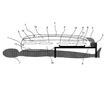
FIG. 1 is a side view of an inflated portable surgical enclosure adhered to
the patient's
torso surgical site via incise drape, with air inflow from air supply in
enclosure side closest to
85 patient feet, directed in cranial longitudinal direction over the
patient's surgical site.
FIG. 2 is a top view of the inflated portable surgical enclosure from FIG. 1
with two users
working via arm ports in operating-section on the torso surgical site, and two
users working via
arm ports in instrument-section.
FIG. 3 is a side view of an alternate embodiment of the surgical enclosure
which utilizes
90 a central frame and oblique tethers in cranial and caudal directions to
assist with holding up the
enclosure.
3
SUBSTITUTE SHEET (RULE 26)
CA 03030844 2019-01-14
WO 2018/014003
PCMJS2017/042266
FIG. 4 is an axial view perpendicular to the view illustrated in FIG. 3
showing the shape
of the central frame and the tethers to support it. Patient, instrument tray,
and ports are excluded
from illustration.
95 FIG. 5 is a side view of an additional alternative embodiment which
utilizes two vertical
frames at each of the cranial and caudal ends of the enclosure, and tethers to
support the surgical
enclosure.
FIG. 6 is an axial view perpendicular to the view illustrated in FIG. 5
showing the shape
of one of the two identical frames and the tethers which support the
enclosure.
100 FIG. 7 is a side view of the embodiment shown in FIG. 5 and FIG. 6
demonstrating how
the frame and tethers prevent the enclosure from collapsing on the surgical
site in the case of
sudden pressure loss.
FIG. 8 is an axial view perpendicular to the view illustrated in FIG. 7.
FIG. 9 is a side view of an alternate embodiment of the surgical enclosure and
frame. in
105 which the rigid frame fully supports the enclosure with frame
attachment to each of the sides
defining the top of the enclosure. The enclosure extends circumferentially
around the patient
torso.
FIG. 10 shows an oblique perspective view of the frame and plastic enclosure
shown in
FIG. 9.
110 FIG. 11 is a schematic of the portions of the air supply system
external to the enclosure.
FIG. 12 is an alternate embodiment for the air supply system which
incorporates a back-
up manual pump.
FIG. 13 shows the axial view with the overhead inlet tube valve in the
enclosure open
during active air inflow, signaling adequate flow.
115 FIG. 14 shows the axial view with the tube valve FIG. 13 pinched closed
by the
enclosure's positive pressure, thus sealing the system and preventing
backflow.
FIG. 15 shows an exemplary embodiment of the material ports.
FIG. 16 shows an alternate embodiment of the material ports with different
port sizes.
FIG. 17 shows an alternate embodiment of the material ports, in which there is
a small
120 port above each set of sleeves and a larger port in the middle.
FIG. 18 shows an alternate embodiment of the material port, in which a bimodal
port can
be opened either fully or only partially depending on the need.
4
SUBSTITUTE SHEET (RULE 26)
CA 03030844 2019-01-14
WO 2018/014003 PCT/1JS2017/042266
FIG. 19 is a side view at the level of the arm port, showing user sleeves and
gloves in an
inflated enclosure are pinched together by the positive pressure in the
surgical enclosure prior to
125 their use.
FIG. 20 is a schematic view of the airflow within the enclosure as traveling
through the
valve system continuously into and through the manifold system, with
perforations varying in
density along the manifold to produce uniform flow.
FIG. 21 is a schematic of a manufacturing process to produce the embodiment of
FIG. 20.
130 FIG. 22 is a graph relating manifold perforation density and air
exit velocity from the
embodiment of FIG. 20.
FIG. 23 is a schematic view of the airflow within the enclosure as traveling
through the
valve system continuously into and through the manifold system, with
perforations varying in
diameter along the manifold to produce uniform flow.
135 FIG. 24 is a schematic sample setup workflow for the frame
embodiment described in
FIGS. 3 and 4.
FIG. 25 is a schematic sample setup workflow for the frame embodiment
described in
FIG. 9.
FIG. 26 shows a graph of the particle concentration inside the enclosure as
function of
140 environment parameters as obtained from tests on a prototype portable
surgical system.
DETAILED DESCRIPTION OF THE ILLUSTRATED EMBODIMENTS
The invention is described more fully hereinafter with reference to the
accompanying
drawings, in which embodiments of the invention are shown. This invention may,
however, be
145 embodied in many different forms and should not be construed as limited
to the embodiments set
forth herein. Rather, these embodiments are provided so that this disclosure
is thorough, and will
fully convey the scope of the invention to those skilled in the art. In the
drawings, the size and
relative sizes of layers and regions may be exaggerated for clarity. Like
reference numerals in
the drawings denote like elements.
150 It will be understood that when an element or layer is referred to
as being "on" or
"connected to" another element or layer, it can be directly on or directly
connected to the other
element or layer, or intervening elements or layers may be present. In
contrast, when an element
or layer is referred to as being "directly on" or "directly connected to"
another element or layer,
SUBSTITUTE SHEET (RULE 26)
CA 03030844 2019-01-14
WO 2018/014003 PCMJS2017/042266
there are no intervening elements or layers present. It will be understood
that for the purposes of
155 this disclosure, "at least one of X, Y, and Z" can be construed as X
only, Y only, Z only, or any
combination of two or more items X. Y, and Z (e.g., XYZ, XY, YY, YZ, ZZ).
FIG. 1 illustrates a preferred embodiment of a portable surgical system. The
portable
surgical system includes a flexible plastic enclosure 1 configured to be
supplied with air under
positive pressure via an environmental control system 5. The enclosure 1 may
be adhered to a
160 surgical site of a patient 7 via an incise drape 11 as shown in FIG. 1.
The incise drape may be a
flexible plastic drape and may include a removable skin adhesive on one side,
with or without
antimicrobial impregnation. The portable surgical system may be configured
such that filtered air
is blown or passed through a longitudinal tubular valve with walls of
flexible, collapsible plastic
such as polyethylene 2 and through a manifold with perforations 3. The
filtered air may be blown
165 such as to cause an essentially uniform laminar air flow onto the
surgical site and through the
enclosure.
The portable surgical system may include a plurality of ports, such as arm
ports 8 and
material ports 10 shown in FIGS. 1 and 2. In an exemplary embodiment the
portable surgical
system may include four pairs of integrated, cuffed sleeves in the arm ports
8. The ports 8
170 provide users with access to the inside of the enclosure, as shown in
FIG. 2. The material ports
may be used to move the surgical tray 9 to the inside of the enclosure 1 prior
to the surgical
procedure. The portable surgical system may further include an instrument tray
holder 6 which
may be placed around the legs of the patient 7. The tray 9 may be disposed on
top of the
instrument tray holder 6.
175 In the preferred embodiment shown in FIG. 1, the perforations which
define the manifold
outlets 3 in the overhead tube decrease in density along the remainder of the
manifold over the
operating-section such that the airflow over the incise drape 11 is
essentially constant. If the
environmental control system 5 is shut off, the flexible overhead tube 2 is
pinched shut, thus
sealing the enclosure 1 and preventing backflow into the fan and filter 5.
180 The portable surgical system may include a surgical enclosure, a
frame, and an
environmental control system.
A. Structure of Surgical Enclosure
In an exemplary embodiment the surgical enclosure may be disposable, such as
the
enclosure 1 shown in FIG. 1. In an exemplary embodiment the surgical enclosure
may be
6
SUBSTITUTE SHEET (RULE 26)
CA 03030844 2019-01-14
WO 2018/014003
PCMJS2017/042266
185 supplied folded like a surgical gown. When set up, the surgical
enclosure may comprise one or
more top view panels of optically-clear plastic la, such as polyvinyl
chloride. The remainder of
the surgical enclosure sides may comprise a flexible, impermeable plastic,
such as low-density
polyethylene. The sides of the instrument-section may be shorter than those of
the operating-
section, in order to fit over an instrument tray holder. In the preferred
embodiment shown in
190 FIG. 1, the bottom of the enclosure is continuous with the sides.
The panel of incise drape 11 may be incorporated into the bottom of the
operating-section
as shown in FIG. I. The incise drape serves as the interface with the patient
body. The size and
shape of the incise drape 11 may be configured to cover the surgical site on
the patient's body
while essentially excluding body surface outside the surgical site.
Consequently. as seen in FIG.
195 1, only the surgical site of the patient's body (i.e. area covered by
the incise drape 11) is included
within the surgical enclosure, while the remainder of the patient body is
excluded from the sterile
field. By excluding from the surgical enclosure the unnecessary body surface,
the efficacy of the
system is significantly improved since the patient's body surface contributes
to environment
contamination inside the enclosure. In particular, the exclusion of high-
contaminant regions such
200 as the oropharynx or the genitals is likely to significantly improve
the efficacy of the system. The
surgical enclosure 1 may include incise drapes 11 of different shapes and
sizes and may be
disposed at different positions on the surgical enclosure such as to fit the
needs of different types
of medical procedures. The bottom corners of the surgical enclosure may
include straps for
securing the enclosure to the patient or to the operating table for additional
stability.
205 FIGS. 9 and 10 illustrate a side view and a perspective view,
respectively, of a second
preferred embodiment of the portable surgical system. In the second preferred
embodiment the
portable surgical system includes an incise drape-less surgical enclosure 1
wherein the operating-
section of the patient is placed inside the enclosure and wherein the bottom
of the enclosure
remains continuous with the sides at the level of the instrument-section. In
the operating-section
210 of the enclosure, one side of the enclosure may be elongated so as to
enable tucking under the
patient body, thereby eliminating the continuous bottom panel. After passing
under the patient
body, the residual length of the elongated side may be secured to the
contralateral enclosure side
along the free edge of the elongated side. The cranial end of the operating-
section 18 as well as
the interface with the instrument-section 18 may be secured against the
patient via integrated
215 straps.
7
SUBSTITUTE SHEET (RULE 26)
CA 03030844 2019-01-14
WO 2018/014003
PCMJS2017/042266
Embodiments of the invention are described herein with reference to figures
and
illustrations that are schematic illustrations of idealized embodiments (and
intermediate
structures) of the invention. As such, variations from the shapes of the
illustrations as a result,
for example, of manufacturing techniques and/or tolerances, are to be
expected. Thus,
220 embodiments of the invention should not be construed as limited to the
particular shapes of
regions illustrated herein but are to include deviations in shapes that
result, for example, from
manufacturing.
The portable surgical systems disclosed herein may include alternate or
additional
sections which could be added based on procedural needs, such as to
accommodate additional
225 instrument trays or users. The above embodiments presented in this
disclosure merely serve as
exemplary embodiments and it will be apparent to those skilled in the art that
various
modifications and variations can be made in the present invention without
departing from the
spirit or scope of the invention.
B. Structure of Frame
230 In an exemplary embodiment, illustrated in FIGS. 3 and 4, the
portable surgical system
may include a central frame 13 and tethers 14 intended to support the
enclosure 1 in the case of a
sudden pressure loss. The central frame 13 may be lightweight and/or
collapsible so as to be
easily transported. The frame may be made of a rigid material, such as
plastic, rigid polyvinyl
tubes, aluminum tubing, and other materials familiar to practitioners
knowledgeable in the field.
235 The frame may include four oblique tubes which are reversibly secured
to the instrument tray
holder or operating table such that the instrument tray holder or operating
table form the bottom
of a pentagon when viewed axially as in FIG. 4. One or more of these pieces
may be connected
to one another via custom connectors or hinges, configured to maintain the
pentagon within the
same plane. The topmost vertex of the frame may be reversibly attached to the
disposable
240 component top, such as via a formed plastic slot in the disposable
component or via tether 14
only. Tethers 14 may support the plastic enclosure 1 directly underneath the
frame 13, as shown
in FIG 4, as well as longitudinally over the incise drape 11 and instrument
tray holder 6. Frame
13 and tethers 14 are configured to provide support to the enclosure 1 in the
event of a sudden
pressure loss. Various other tether arrangements may be utilized to optimize
support from the
245 central frame, depending on system requirements.
8
SUBSTITUTE SHEET (RULE 26)
CA 03030844 2019-01-14
WO 2018/014003
PCMJS2017/042266
In another exemplary embodiment the portable surgical system may include a
frame 15
and tethers 14 as illustrated in FIGS. 5-8. Frame 15 and tethers 14 arc
configured such as to
provide support to the enclosure 1 in the event of a sudden pressure loss.
Instead of supporting
the surgical enclosure centrally, frame 15 includes two vertical sections
disposed at the cranial
250 and caudal ends of the enclosure. FIG 5 provides a side view of the
frame 15 and tethers 14, and
FIG 6 provides a front view of the same system. FIGS. 7 and 8 show how the
frame 15 and
tethers 14 support the deflated enclosure lb in the case of a sudden pressure
loss, resulting from,
for instance, an open port 10a.
In an exemplary embodiment the portable surgical system may include a
collapsible,
255 rigid frame 16 and a flexible plastic enclosure 1 as illustrated in
FIGS. 9 and 10 and as described
in the section "Structure of Surgical Enclosure" paragraph 3 in which the
surgical enclosure 1
encloses the patient's 7 torso. The portable surgical system according to this
embodiment does
not require a separate instrument tray holder. The enclosure 1 is reversibly
sealed at the patient's
suprapubic region and axillae via adjustable opening 18. This embodiment does
not structurally
260 rely on positive pressure to the extent that the previous embodiments,
illustrated in FIGS. 1-8,
do. The frame may comprise six vertical pieces forming the edges of two
connected partial
cuboids, reversibly attached to under the patient or to the operating table.
As seen in FIGS 9 and
10, the frame may include two pieces at the cranial end, two at the caudal
end, and two at the
junction between the operating and instrument sections. These pieces may
incorporate
265 telescoping function to accommodate different patient body sagittal
abdominal diameters. These
vertical pieces may be connected as shown in FIG. 9, with three pieces
horizontally at the top
and two additional horizontal pieces defining the instrument tray section;
these latter two pieces
are at a level above the patient where desired for an instrument tray holder.
The frame may
further include two longitudinal pieces, perpendicular to both of the above
types, forming the
270 operating section; and two additional longitudinal pieces forming the
instrument section. One Or
more of all of these pieces may be connected via hinges or custom connectors.
The enclosure
may be connected to the frame reversibly 17 in such manner as to place uniform
outward tension
on the top view panel.
C. Ports
275 The various embodiments of the portable surgical system may have
surgical enclosures
which include a plurality of ports. The enclosure may include two major types
of ports. The first
9
SUBSTITUTE SHEET (RULE 26)
CA 03030844 2019-01-14
WO 2018/014003 PCMJS2017/042266
type of port on the enclosure is arm ports 8, as shown in FIGS. 1, 2, 3, 5, 7,
and 9, which allow
access to the inside of the enclosure by either provider arms or augmenting
instrumentation
taking the place of arms such as laparoscopes or robots.
280 The number of arm ports is dependent on procedural need. The
preferred embodiments
illustrated in FIGS. 1, 2, 3, 5, 7, and 9 include four pairs of arm ports 8,
two on each side of the
enclosure 1. Depending on use scenario, the arm ports may take three major
forms. The first
form for the arm port is a simple opening in the side of the enclosure which
seals reversibly
against user arms. The second form for the arm port is a sleeve as shown by 8
in FIG. 2, which is
285 a hollow cylinder or frustrated cone of impermeable plastic that tapers
toward the inside of the
enclosure away from the wall. The length of the sleeve is adequate to permit
ergonomic handoff
of instruments among ports at contralateral ends of the system. The material
of the sleeve may be
the same as the one used for the enclosure side, or it can be a different one,
such as a material
used in surgical gown sleeves. The sleeve end may be free or may incorporate a
cuff of elastic
290 material to fit against the user wrist. The third form for the arm port
is the same as the second
form, but ending in a glove. FIG. 19 shows a side view at the level of the arm
port, showing user
sleeves and gloves in an inflated enclosure. The user sleeves and gloves are
pinched together by
the positive pressure in the surgical enclosure prior to their use.
The second type of port on the enclosure is a materials port 10, as shown in
FIGS. 1, 3, 5,
295 7 and 9, which allows the instrument tray 9 and instruments to be moved
into the enclosure 1
prior to the procedure. Additionally, the port allows materials to be moved in
and out of the
enclosure throughout the surgical procedure. In the case of a caesarean
section, it is imperative
that the newborn child can be quickly and ergonomically passed out of the
enclosure so it can
receive care.
300 FIGS. 15-17 show various possible configurations of the ports,
although additional
embodiments would be conceived of that fit the nature of the claims. In an
exemplary
embodiment, the enclosure 1 may include large ports 10b as shown in FIG. 15,
small ports 10c
as shown in FIGS. 16 and 17, or both large ports 10b and small ports 10c as
shown in FIGS. 16
and 17. Small ports 10c are configured such that small items may be passed in
or out of the
305 enclosure without significant relative loss of enclosure volume or
pressure, regardless of frame
availability, because the Environmental Control System (e.g. a fan) can
increase the gas inflow
to match the outflow. Large ports 10b permit the moving of large items like
the instrument tray,
SUBSTITUTE SHEET (RULE 26)
CA 03030844 2019-01-14
WO 2018/014003 PCMJS2017/042266
neonates, et cetera in and out of the enclosure. FIG. 18 shows an exemplary
embodiment of the
port, in which a connector 29 splits a port in half, allowing it to act as a
small port or large port.
310 This bimodal port 10d ensures that any user can have access to both a
small port and a large port.
In addition to episodic access for large items, the ports can also provide
ongoing access for lines,
tubes, wires, and drains requiring access to external resources. The connector
29 may be a zipper
slider that slides over the zipper teeth rows thereby adjusting the size of
the port. Alternatively, it
can be a material such as hook and loop fastener or magnets which provide
rapidly reversible
315 attachment. There are a number of ways the materials ports can be
implemented. They must be
easy to open and close repeatedly, such as can be achieved through the use of
magnetic strips,
hook-and-loop fasteners, plastic zippers, flexible inflatable tubes compressed
against one
another, or other methods.
D. Environmental Control System
320 The portable surgical system includes an environmental control
system. In a preferred
embodiment, as the one shown in FIG. 11, the environmental control system may
include a
HEPA filter 19, fan (blower with motor) 21, filter-blower adapter 20, battery
24, and control
section 25, connected to the enclosure via sterile flexible tubing 23. These
external components
(i.e. components 19, 20, 21, 23, 24, and 25) are collectively referred to as
air supply system. The
325 battery 24 may be disposable or rechargeable, and the system can also
run off the electrical grid
22 if the procedure occurs in a setting in which this is possible. The air
supply system may be
connected to the flexible overhead tube 2 of the surgical enclosure with
flexible tubing so that
the inlet height of the overhead airflow tube 2 can adjust based on the level
of inflation of the
enclosure 1. The HEPA filter immediately downstream of air inflow may be
changeable and
330 customizable such that it provides one or more other controls based on
procedural need, such as
humidity modulator filter, gas content with supply of medical gases, or
temperature modulator
with heat/cold sinks.
In an alternative exemplary embodiment, the air supply system includes both an
electrical
fan 21 as well as a manual pump 27 as illustrated in FIG. 12. The manual pump
27 provides
335 redundancy and may be used in the event of unavailability of electrical
power supply or to
provide higher flows without expending electrical power. The manual pump can
be implemented
in any number of mechanical setups familiar to practitioners in the art,
including but not limited
to via manual or pedal bellows-style pump or other general positive
displacement pump, or
11
SUBSTITUTE SHEET (RULE 26)
CA 03030844 2019-01-14
WO 2018/014003 PCMJS2017/042266
manual or pedal rotary pump. The air supply system may further include one or
more one-way
340 valves 26 which allow the air from either only the electrical fan 21 or
only the manual pump 27
to flow toward the plastic enclosure. The filter 19 is downstream of both
electrical and manual
air supply.
The external air supply system connects to the enclosure. In an exemplary
embodiment,
the air is supplied through an inlet and thereby blows through the entire
enclosure cranially to
345 caudally. Airflow adequacy may be checked by timing of inflation of the
surgical enclosure 1 or
by the rising of a windsock in the enclosure embodiment shown in FIG. 9. The
windsock may
include a short tube of flexible plastic of the same material as the enclosure
side. In another
exemplary embodiment, the inlet is connected to a horizontal manifold running
side to side over
the patient. The manifold may include an additional fold of the enclosure side
plastic which is
350 sealed together into tubular structure and perforated 3 to create
parallel, uniform streams of
laminar air outflow into the enclosure.
In a preferred exemplary embodiment the inlet is connected to a flexible tube,
such as the
overhead flexible tube 2 shown in FIGS. 1 and 2. The flexible tube 2 may
include a plurality of
perforations 3 acting as manifold. The flexible tube may run side to side or
along the enclosure.
355 The flexible tube may be formed by sealing a fold of the enclosure into
a tubular structure. The
flexible tube may be a collapsible tube that opens when air is blown into the
enclosure and closes
when air moves out of the enclosure such that transmural pressure from the
enclosure favors tube
collapse.
In a preferred exemplary embodiment, the flexible tube 2 may include a
plurality of
360 perforations 3 disposed such as to create parallel, uniform streams of
laminar air outflow into the
enclosure. Uniform airflow is accomplished in our preferred embodiment, as
described by the
design and manufacturing implementations detailed in FIGS. 20-22, by varying
the density of
perforations in the collapsible tube in which the density of perforations is
higher at the end of the
tube closer to the supply of the air 31 and the density of perforations
decreases as the distance
365 from the supply increases until the density is at its lowest value at
37.
Inventors in this application came to the realization that nearly uniform air
flow may be
accomplished when the perforation density along the tube decreases according
to the inverse of
an elliptically shaped function. Starting from the observation that the
pressure within an inviscid
flow will rise along a streamline if the velocity of the airflow decreases,
inventors of this
12
SUBSTITUTE SHEET (RULE 26)
CA 03030844 2019-01-14
WO 2018/014003 PCMJS2017/042266
370 application have found that in a perforated tube of constant cross
sectional area, the velocity
within a tube will drop as it passes perforations from which flow is
emanating, as long as the
flow is of nearly constant density which will be the case for flows of air
substantially below the
speed of sound. Further, inventors have come to the realization that the
pressure in a perforated
tube rises as the distance from the source increases and, as a result, the
rate of flow from each
375 perforation rises with distance from the source assuming the
perforations are of constant cross
sectional area. As shown in Fig 20, the velocity is low 35 at locations close
to the source 31 and
the velocity is high 36 at locations far from the source 31. If the density of
perforations were
uniform, the flow of air would be too large at locations far from the source
and too small at
locations nearer to the source.
380 An exemplary embodiment of the invention discloses a flexible tube 2
(as shown by
FIGS. 1, 2, 11, and 20) including a plurality of perforations disposed at such
positions (x1,
X3, x4, xk) along the tube as to create uniform air flow. The exemplary
embodiment in Fig. 20
illustrates a tube including a plurality of perforations disposed in a single
axial row along the
tube. The tube may include multiple axial rows of perforations disposed on the
circumference of
385 the tubes such as to cover the entire surface of the tube or only a
certain desired region, such as
the region facing towards the surgical site. The multiple axial rows may be
essentially parallel
with each other and with the axis of the tube.
The perforations are disposed along the flexible tube such that the axial
positions of the
perforations along the flexible tube may follow a mathematical relation (xi,
x2, x3, x4, = = = xk) =
390 (I)(V, d, D, p, k, L), where V is the air velocity from the source, D
is the diameter of the tube, d is
the diameter of the perforations, and p is an air density, L is the length of
the perforated section,
and k the number of perforations in a row. The mathematical relation (I)(V, d,
D, p, k, L) is
determined as explained hereinafter.
The positions of the perforations along the flexible tube may be expressed by
a plurality
395 of mathematical expressions: xi = (I)1(V, d, D, p, k, L); x2= (I)2(V,
d, D, p, k, L); x3= (I)3(V, d, D,
p, k, L); xk = Ok(V, d, D, p, k, L); where V is the air velocity from the
source, D is the
diameter of the tube, d is the diameter of the perforations, and p is an air
density. The
mathematical expressions (I)1(V, d, D, p, k, L), q),(V, d, D, p, k. L)
(I)k(V. d, D, p, k, L) are
determined as explained hereinafter and may he closed form expressions of (V,
d, D, p. k, L).
13
SUBSTITUTE SHEET (RULE 26)
CA 03030844 2019-01-14
WO 2018/014003 PCMJS2017/042266
400 The specific form of the perforation density needed for uniform air
flow can be
determined by an iterative computation.
The iterative computation may include a plurality of iterations, wherein each
iteration
includes a plurality of steps as described in Figure 21. Within a CPU 38,
begin with an assumed
form of the exit velocities 39 such as a linearly increasing distribution.
These assumed exit
405 velocities will be denoted as vi with a unique subscript for each of
the many holes numbered j=1
to k (i.e. velocities Vi, v2, v3, ... vk shown in FIG. 20 corresponding to
perforations 1, 2, 3 ...k).
In a first step of the first iteration (see 40 in FIG. 21) it is assumed a
form of the exit
velocities 39. The assumed exit velocities (i.e. vi, v2, V3, vk) may be
estimated as a linearly
D2 increasing distribution such as '01 = V = (k.d2) = ki H
- 1), where V is the axial air velocity at the
410 source, D is the diameter of the tube, d is the diameter of the
perforations, k is the number of
perforations, and j is the index of the perforation or hole.
In a second step of the first iteration (see 41 in FIG. 21) the exit
velocities (v), v2, v3, = = =
vk) estimated at 40 are used to compute an estimate of the velocities within
the tube v_tube 41
(i.e. v_tubei; v_tube2; v_tube3;...; v_tubek). The velocity v_tuben is the
axial velocity inside the
415 portion of the tube between perforation "n" and perforation "n+1". Mass
conservation requires
that for any hole number n in a tube of diameter D with perforations of
diameter d the following
Equations are satisfied:
,17
,N ,p¨d ....tutocp-
4 4
th = V p7--r 4 D2
420 Where p is the air density, d is the diameter of the perforations, D
is the diameter of the
tube. The equations above provide the velocities inside tube (i.e. v_tubei;
v_tube2; v_tube3;...;
v tubek).
In a third step of the first iteration (see 42 in FIG.21) the velocities
inside the tube are
used to calculate a set of pressures (pi, P2/ P3 pk) corresponding to each
of the perforations as
425 explained hereinafter. The flow axially within the interior of the tube
may be modelled as
inviscid flow. Bernoulli's equation may be used to provide a prediction of the
pressure within the
tube as a function of the velocities inside tube computed in the previous step
(i.e. v_tubei;
v_tube2; v_tube3;...; v_tubek). It is assumed that the velocity in the tube
near the end cap is zero
14
SUBSTITUTE SHEET (RULE 26)
CA 03030844 2019-01-14
WO 2018/014003 PCMJS2017/042266
and the velocity at the source is V and the constant air density is p. The
pressure at the end of the
430 tube farthest from the source is calculated as:
P
Then this value of the pressure P is used to estimate the pressures within the
tube 42 at
each of the many holes numbered j=1 to k as follows:
,., I
p tr-P -,7 pr
435 These pressures at each hole are computed and stored in a vector
(pi, p2, p3 pic).
In a fourth step of the first iteration (see 43 in FIG.21), the pressures (pi,
p2, p3 pk) are
used to calculate a new estimate of the exit velocities. The flow from the
interior of the tube to
the exit hole may be modelled as inviscid flow. Bernoulli's equation may be
used to provide a
prediction of the exit velocity as follows:
=
440 P
One may use the relationship above k times (for each hole number from 1 to k)
to
calculate exit velocity estimates at each perforation or hole (i.e. v_ui,
v_u3 v_uk). The
updated exit velocity estimates v_ui are different from the initially assumed
distribution (i.e.
v2, v3, = = = vk)=
445 By mass conservation, the sum of the exit velocities must obey the
relationship
yvipLIT d2
4
In a fifth step of the first iteration the exit velocity estimates calculated
in the fourth step
are used to calculate a set of velocities (v-Li, v22, v/-3, v2_4, ... v2_k) to
be used as starting point for
a second iteration. The set of velocities are calculated as follows:
(V pirD 2/4)
172_1 = vi = __ lc
Ei=1(vipTcd2/4)
450 The set of velocities v7_i preserve the proportions among the
calculated exit velocities
v_aj but their magnitudes are adjusted to satisfy mass conservation by scaling
each value. The
scaling is performed by dividing each exit velocities by the sum Ei1=1(v1 pn-
d2/4) and
multiplying it by the known mass flow supply which is (V pn-D2/4).
SUBSTITUTE SHEET (RULE 26)
CA 03030844 2019-01-14
WO 2018/014003
PCMJS2017/042266
The resulting exit velocity distribution (v2_1, v2-2, v2-3, v2-4, = . = v2_k)
is used as an updated
455 estimate for a second iteration. The second through fifth steps (41
through 43 in FIG. 21) are
repeated for the second iteration thereby obtaining a velocity distribution to
be used as updated
estimate for the third iteration. The process is iterated until it converges
to a stable distribution of
exit velocities (i.e. vti, vt?, 17E3, vr4
vm,). The obtained distribution of exit velocities may be
approximately elliptical if the total area of perforations is not small
compared to the cross
460 sectional area of the tube.
The density of the perforations 44 is determined by making it proportional to
the inverse
of the exit velocities. In an exemplary embodiment the position coordinates of
the k perforations
along the tube is denoted as xi, x2, x3, x4, ... xi, where xi, is the distance
between perforation k
and a reference point on the tube between the air source and the first
perforation. The positions xj
465 (with j between 1 and k) may be calculated from the set of equations:
(x1+1 ¨ xj) = a = ¨; (where 1 j k)
vF;
Where a is determined by setting the distance between the first and last
perforation to the
desired length: (xk - x1) = L.
The above equations enable the skilled artisans to derive the mathematical
expressions xi
470 = (1)1(V, d, D, p, k, L); x2= (1)2(V, d, D, p. k, L); x3= (I)3(V, d, D,
p, k, L); xk = (1)k(V, d, D, p,
k, L), thereby providing the positions and density of the perforations as
function of parameters
(V, d, D, p, k, L). The functions On(V, d, D, p, k, L) may be expressed by
closed form
expressions.
Alternatively, the set of parameters may be associated the resulting
positions, (V, d, D. p,
475 k, L) 4 (xi, x2, x3, x4, = = xk), determined by the above algorithm
thereby forming the function
(xi, x2, x3, x4, xk)
= 01(V, d, D, p, k, L). The function 4:1;0(V, d, D, p, k, L) may be expressed
by
a closed form expression.
The positions and density of the perforations computed in the CPU 38 is
implemented by
a cutting die 45 which is located at positions over the clear plastic tube
according to the desired
480 perforation positions / density (i.e. xi, x2, x3, x4, .õ xk). The
resulting perforations distribution
will essentially follow an inverse of a elliptical function. By making the
density of perforations
an inverse of an elliptically shaped function, the resulting air distribution
within the surgical area
is uniform throughout providing an advantage in quality of the surgical
outcome.
16
SUBSTITUTE SHEET (RULE 26)
CA 03030844 2019-01-14
WO 2018/014003 PCMJS2017/042266
In an exemplary embodiment of the invention a method for manufacturing a
portable
485 surgical system may include: (1) running on a CPU the iterative
computation described above;
(2) receiving, from the CPU, at a machine for cutting perforations into the
tube material a set of
numbers corresponding to the positions (xi, x2, x3, x4, ... xk) of the
perforations; (3) cutting the
perforations into the tube materials at positions (xi, x2, x3, x4, ... xl)
received from CPU.
As an illustration, the resulting velocity distribution and perforation
density distribution
490 are graphically depicted in FIG. 22. This depiction is for a case with
ten perforations in the
collapsible flexible tube and it will be understood that the method
generalizes to other numbers
of perforations. The hole number is on the x axis and the exit velocity 46 and
perforation
densities 47 (normalized so that the maximum values are unity) are represented
on the y axis.
In another exemplary embodiment the above uniform air distribution can also be
495 achieved via an alternative configuration of the perforations in the
flexible tube as shown in
FIG. 23. In this configuration the perforations are equidistant (distance
depicted as x in FIG. 23)
while the diameter of the perforations varies (i.e. di, d2, d.3, = = dk) such
that the air flow through
each of the perforations is identical and 1/k proportion of the total flow
through the manifold.
The goal in such a case is to integrate the total area of perforation for each
given, uniform
500 distance xi. A system of dies may be used to cut the correct
perforation diameter at points xi, x2,
X2, .=., xx=
Another alternative embodiment of the air handling system inside the enclosure
instead
runs airflow longitudinally caudally to cranially, along center of top.
The portable surgical system may include a flexible tube 2 (as depicted in
FIGS. 1. 2, 11,
505 and 20) configured to act as a valve system, as described with respect
to FIGS. 13 and 14, such
as to prevent air backflow from the surgical enclosure into the fan and
filter. FIGS. 13 and 14
show a cross-section through a portion of the surgical enclosure 1 and the
flexible tube 2
attached to or incorporated into the surgical enclosure 1. FIG. 13 shows the
flexible tube in an
expanded state when air is blown from the air supply system 5 into the
surgical enclosure. FIG.
510 13 shows the axial view with the overhead inlet tube valve in the
enclosure open during active
air inflow, signaling adequate flow. FIG. 14 shows the axial view with the
tube valve FIG. 13
pinched closed by the enclosure's positive pressure, thus sealing the system
and preventing
backflow. FIG. 14 shows the flexible tube in a collapsed state when air
pressure inside the
17
SUBSTITUTE SHEET (RULE 26)
CA 03030844 2019-01-14
WO 2018/014003 PCMJS2017/042266
enclosure is pushing the air from the enclosure towards outside the enclosure.
The collapsed tube
515 2 prevents the air from exiting the enclosure.
The collapsible tube may be made of flexible material such as to switch from
open to
close state, and vice versa, based on airflow. The airflow passes from air
supply system first
through an inflow tube valve 2 comprising a sealed tube of collapsible
plastic. When there is net
positive airflow through the tube toward the manifold in this configuration,
the transmural
520 pressure is positive relative to the enclosure, and the tube is forced
open. When there is no
airflow or reversed airflow, the transmural pressure drops relative to the
enclosure, causing
longitudinal collapse of the tube. This tube valve reduces further flow in the
setting of enclosure
excess pressurization as the enclosure positive pressure produces transmural
pressure favoring
valve collapses; prevents flow reversal as enclosure positive pressure seals
off air outflow
525 through the valve; and also serves as an indicator of adequate airflow
indicator by virtue of its
inflation. The airflow then proceeds to a manifold 3, implemented as above in
the horizontal
manifold system. The relative lengths of the valve and manifold are determined
by procedural
needs for pressure and airflow; but the manifold should preferably extend at
least the full length
of the operating-section.
530 E. Method for Setup of Surgical Enclosure with Respect to Standard
Surgical
Workflow
An exemplary embodiment of the present invention also discloses a method for
using the
ultraportable surgical system comprising the steps described in FIG. 24
flowchart. The sterile
field, which corresponds to the draped areas in standard procedural setup,
includes the entire
535 enclosed area and the sleeves. This method applies for all embodiments
utilizing the incise drape
interface. The users first disinfect the skin 48 of the patient as per usual
protocol using any of the
standard skin antiseptic agents, provided they are permitted to dry fully
before applying the
incise drape. Users then orient 49 the enclosure with the incise drape over
the planned surgical
site and the instrument-section extending caudally. set up the enclosure 50.
and add needed
540 instrument tray and gloves via the material ports 51. As the entire
system comes pre-sterilized in
packaging, the air inside is sterile until the sterile instrument tray is
placed. The enclosure is then
connected to the frame 52 which in turn is stabilized on the instrument tray
holder, strapped
down for additional stabilization against the patient or operating table 53,
and the environmental
control system is turned on 54. Inlet tube valve inflation is utilized as the
indicator of adequate
18
SUBSTITUTE SHEET (RULE 26)
CA 03030844 2019-01-14
WO 2018/014003
PCMJS2017/042266
545
airflow through the environmental system. The first inflation is thus also an
initial purge of any
contamination introduced during that step. When the system is adequately
inflated, or an
indicator is activated, the environmental system is switched to maintenance
mode 55. At this
point, users can place arms through the arm ports, apply gloves or overgloves
in standard
protocol 56, and initiate the procedure 57. Maintenance mode is an option for
procedures in
550 which the air changes are planned to be different than the ones
used for initial inflation or that
opts to recycle air through an exhaust system to prolong filter life span, but
it can also be no
change from prior mode. For arm port use, it is recommended that providers
wear one pair of
sterile undergloves, then don the second pair of gloves inside the enclosure
in standard double
gloving procedure to seal the sleeve port embodiments of the arm ports.
555 At the end of the procedure following any appropriate skin closure
and dressing
application, users remove the tray and any items from inside the enclosure,
clear any blood or
bodily fluids within the enclosure, doff gloves then remove arms from the arm
ports, turn off the
environmental control system, remove the air supply tubing from the air
handling inlet, pull the
enclosure off of the frame as well as off of the patient, and dispose of the
enclosure.
560 For embodiment systems not utilizing incise drapes, setup
methodology is described in
FIG. 25. In this scenario, the user positions the patient directly over the
bottom flap of the
operating-section 58, places instrument and gloves in planned enclosure 60,
connects the bottom
flap against the side of the enclosure 60, clinch the enclosure cranially and
caudally against the
patient 61, then assembles the frame while connecting to the enclosure 62. The
environmental
565 control system is engaged 63 with monitoring of wind sock at air
inflow to check for adequate
flow. When the enclosure is adequately filled with clean air as shown by
indicator (based on air
changes), the environmental system is switched to maintenance mode 64. At this
point, users can
place arms through the arm ports, apply gloves or overgloves in standard
protocol 65, and initiate
the procedure 66.
570 Although only a few embodiments have been described in detail
above, those skilled in
the art can recognize that many variations from the described embodiments arc
possible without
departing from the spirit of the invention.
F. Supporting Studies
Inventors have implemented various embodiments, such as the ones described
herein
575 among others, by manufacturing and testing fully self-contained
portable surgical systems. In
19
SUBSTITUTE SHEET (RULE 26)
85006502
Teodorescu et al (2016) inventors have demonstrated an early proof of concept
showing that the
enclosure, even in absence of environmental control system engagement,
provided 100%
protection against external active particulate contamination (FIG. 26).
Inventors have further
demonstrated that even with enclosure contamination to level found in machine
shop utilizing
580 charcoal burning, 2.25 air changes were adequate to consistently bring
contaminant particulate
levels to 0 particles per cubic centimeter. Subsequent systems reduced
susceptibility to enclosure
contamination and improved setup speeds through the protocols described above
(e.g. as
described in Teodorescu et al 2017).
The features of the invention disclosed herein, as specified by actual
surgical end-users,
585 distinguish it from prior art by enhancing usability, ergonomics,
independence from external
resources, and reliability under field conditions. The inclusion within the
enclosure of only the
surgical site, excluding the remainder of the patient body from the sterile
field, particularly high-
contaminant regions such as the oropharynx or the genitals, improves the
efficacy of the system.
The invention's ability to isolate the surgical wound's contaminant
production, such as blood and
590 bodily fluids, and contain these through the life cycle of the product,
is also a key feature.
It will be apparent to those skilled in the art that various modifications and
variations can
be made in the present invention without departing from the spirit or scope of
the invention.
Thus, it is intended that the present invention cover the modifications and
variations of this
invention provided they come within the scope of the appended claims and their
equivalent.
595 G. References
The following documents cited herein do not represent admitted prior art.
[1] WO/2014/145032, (GNANASHANMUGAM), 15 March 2013; [2] W02011041665 A2,
(HENDERSON), 1 October 2009; [3] W02005092229, (KRIEK), 24 March 2004; [4]
US20070102005 Al, (BONUTTI), 28 August 2001; [5] US6199551 El, (KUSLICH),
600 8 December 1998; [6] US5299582 A, (POTTS), 16 September 1991; [7]
W08606272, (SCOTT),
23 April 1985; [8] US4367728 A, (MUTKE), 7 September 1979; [9] US4275719 A,
(MAYER),
30 March 1979; [10] US3051164 A, (TREXLER), 17 August 1959; [11] American
Society of
Heating, Refrigeration and Air-Conditioning Engineers (2011). Health Care
Facilities (I-P).
In ASHRAE 2011 Handbook - HVAC Application. Atlanta: ASHRAE.; [12] Allegranzi,
B.,
605 Bagheri Nejad, S., Combescure, C., Graafmans. W., Attar, H., Donaldson,
L., and Pittet, D. (2011).
Burden of
Date Recue/Date Received 2022-04-26
CA 03030844 2019-01-14
WO 2018/014003 PCMJS2017/042266
endemic health-care-associated infection in developing countries: a systematic
review and meta-
analysis. Lancet. 377(9761):228-41.; [13] Edmiston, C.E., Seabrook, G.R.,
Cambria, R.A., et al.
(2005). Molecular epidemiology of microbial contamination in the operating
room environment:
610 is there a risk for infection. Surgery. 138(4):573-582. [14] Sehulster,
L. and Chinn. R.Y.W.,
2003, "Guidelines for Environmental Infection Control in Health-Care,"
cd c , vimmwripreviewlminwrlitmlirr52 10a1 him. [15] Selcen Kilinc, F. (2015).
A review
of isolation gowns in healthcare: fabric and gown properties. J Eng Fiber
Fabr. 10(3):180-190.;
[16] Teodorescu DL, Miller SA, Jonnalagedda S. Surgi Box: An ultraportable
system to improve
615 surgical safety for patients and providers in austere settings. IEEE
Xplore GHTC 2017 (accepted,
pending publication).; [17] Teodorescu DL, Nagle D, Hickman M, King DR. An
ultraportable
device platform for aseptic surgery in field settings. ASME J Medical Devices.
J. Med. Devices
10(2), 020924 (May 12, 2016); [18] Whyte, W., Hodgson, R., and Tinkler, J.
(1982). The
importance of airborne bacterial contamination of wounds. Journal of Hospital
Infection. 3:123-
620 135.
21
SUBSTITUTE SHEET (RULE 26)