Note: Descriptions are shown in the official language in which they were submitted.
CA 03031034 2019-01-15
WO 2018/026600
PCT/US2017/043935
Disrupting Fc Receptor Engagement on Macrophages Enhances Efficacy of Anti-
SIRPalpha
Antibody Therapy
CROSS REFERENCE
[0001]
This application claims benefit of U.S. Provisional Patent Application No.
62/370,422,
filed August 3, 2016, which application is incorporated herein by reference in
its entirety.
BACKGROUND OF THE INVENTION
[0002]
Turnover of cells begins with the induction of an apoptotic program or other
cellular
changes that mark them for removal, and the subsequent recognition of markers
by
phagocytes, including macrophages, dendritic cells, and the like. This process
requires a
specific and selective removal of unwanted cells.
Unlike healthy cells, the
unwanted/aged/dying cells display markers or ligands called "eat-me" signals,
i.e. "altered self",
which can in turn be recognized by receptors on the phagocytes. Healthy cells
may display
"don't eat-me" signals that actively inhibit phagocytosis; these signals are
either downregulated
in the dying cells, are present in an altered conformation or they are
superseded by the
upregulation of "eat-me" or pro-phagocytic signals. The cell surface protein
CD47 on healthy
cells and its engagement of a phagocyte receptor, SIRPa, constitutes a key
"don't eat-me"
signal that can turn off engulfment mediated by multiple modalities, including
apoptotic cell
clearance and FcR mediated phagocytosis. Blocking the CD47 mediated engagement
of SIRPa
on a phagocyte can cause removal of live cells bearing "eat me" signals.
[0003]
CD47 is a broadly expressed transmembrane glycoprotein with a single Ig-like
domain
and five membrane spanning regions, which functions as a cellular ligand for
SIRPa with
binding mediated through the NH2-terminal V-like domain of SIRPa. SIRPa is
expressed
primarily on myeloid cells, including macrophages, granulocytes, myeloid
dendritic cells (DCs),
mast cells, and their precursors, including hematopoietic stem cells.
Structural determinants on
SIRPa that mediate CD47 binding are discussed by Lee et al. (2007) J. Immunol.
179:7741-
7750; Hatherley et al. (2007) J.B.C. 282:14567-75; and the role of SIRPa cis
dimerization in
CD47 binding is discussed by Lee et al. (2010) J.B.C. 285:37953-63. In keeping
with the role of
CD47 to inhibit phagocytosis of normal cells, there is evidence that it is
transiently upregulated
on hematopoietic stem cells (HSCs) and progenitors just prior to and during
their migratory
phase, and that the level of CD47 on these cells determines the probability
that they are
engulfed in vivo.
[0004]
Programmed cell death (PCD) and phagocytic cell removal are common ways that
an
organism responds in order to remove damaged, precancerous, or infected cells.
Cells that
survive this host response (e.g., cancerous cells, chronically infected cells,
etc.) have devised
ways to evade PCD, and/or phagocytic cell removal. CD47, the "don't eat me"
signal, is
1
CA 03031034 2019-01-15
WO 2018/026600
PCT/US2017/043935
constitutively upregulated on a wide variety of diseased cells, cancer cells,
and infected cells,
allowing these cells to evade phagocytosis. Anti-CD47 agents that block the
interaction
between CD47 on one cell (e.g., a cancer cell, an infected cell, etc.) and
SIRPa on another cell
(e.g., a phagocytic cell) counteract the increase of CD47 expression and
facilitate the
phagocytosis of the cancer cell and/or the infected cell. Thus, anti-CD47
agents can be used to
treat and/or protect against a wide variety of conditions/disorders. In fact,
anti-CD47 and anti-
SIRPa blocking antibodies significantly increase phagocytosis of cancer cells
in vitro and in
vivo. They have been shown to be effective at treating mice engrafted with a
wide range of
human cancers, from leukemias to solid tumors. However, in some cases an
initial high dose of
an anti-CD47 agent can cause a dose-dependent loss of red blood cells (RBCs)
in mice and
non-human primate (NHP) models by binding to CD47 on the surface of the RBCs.
The severity
of this anemia can preclude the use of higher doses that are required to
achieve sustained
serum concentrations associated with therapeutic efficacy.
[0005] As
an alternative to anti-CD47 agents, anti-SIRPa antibodies have potential
advantages
relating to the relatively restriction expression profile with respect to cell
types. Aspects of anti-
SIRPa antibodies and the use thereof are provided herein.
SUMMARY
[0006]
Compositions and methods are provided relating to antibodies that bind to
SIRPa and
block the interaction between CD47 and SIRPa. Blocking the CD47-SIRPa pathway
mediates
phagocytosis of targeted cells, and can synergize with other cell targeting
agents, including
without limitation cancer-specific antibodies; pathogen specific antibodies;
and the like.
Surprisingly it is shown that activity of an anti-SIRPa antibody on effector
cells may be
substantially reduced when the antibody productively binds to an Fc receptor
on the effector
cell surface, including without limitation one or more of FcyRI; FcyRIIA;
FcyR1161; FcyRIIB2;
FcyRIIIA; FcyRIIIB receptors. The reduction in effectiveness can result in
inter-individual
variation in patient responsiveness. Disabling productive Fc receptor
engagement by reducing
binding to one or more Fc receptors other than FcRn, where the Fc receptor
binds monomeric
IgG and/or multimeric immune complexes, can restore activity to the antibody
and provide an
improved therapeutic profile.
[0007] In
some embodiments, an antibody is provided comprising (i) a variable region
that
specifically binds to SIRPa, e.g. human SIRPa, and (ii) an Fc region with
reduced binding to Fc
receptors, including human Fcy receptors, relative to a wild-type Fc region;
or lacking a
functional Fc region. In
some embodiments, the antibody specifically binds to human
SIRPa. In some embodiments the antibody binds to one or both of human SIRP-I3
and human
SIRPy. In other embodiments the antibody lacks significant binding to one or
both of human
SIRP-I3 and human SIRPy. In some embodiments the antibody specifically binds
to the V1 and
2
CA 03031034 2019-01-15
WO 2018/026600
PCT/US2017/043935
V2 isotypes of human SIRPa. In some such embodiments, the Fc region is a human
Fc region,
where the Fc has been modified by one or more amino acid changes to reduce Fc
receptor
binding. The antibody may be labeled with a detectable label, immobilized on a
solid phase
and/or conjugated with a heterologous compound.
[0008] The
antibody may also be provided as a bispecific or multispecific antibody
reactive with
a second antigen, particularly including cancer antigens, an immune checkpoint
inhibitor, an
immune costimulatory agonist, antigens of chronic infection, etc. In some
embodiments a
bispecific antibody has an active Fc region.
[0009] In
some embodiments a humanized anti-SIRPa antibody is provided, comprising one
or
both of a heavy chain variable region as set forth in SEQ ID NO:1; and a light
chain variable
region sequence set forth in SEQ ID NO:2, or a biologically active variant
derived therefrom. In
some embodiments the antibody comprises an Fc region, which Fc region is
optionally an Fc
region with reduced binding to Fc receptors. In other embodiments the antibody
lacks an Fc
region, e.g. being provided as an F(ab)2 antibody.
[0010] The
compositions and methods of the invention can be used for the treatment of
human
disease, where the anti-SIRPa antibody increases phagocytosis of target cells,
for example in
combination with a second antibody that binds to an antigen on the targeted
cell surface.
Phagocytic effector cells, including for example macrophages, express a number
of Fcy
receptors, and benefit from the use of an anti-SIRPa antibody having decreased
FcR binding.
[0011] In
some embodiments a pharmaceutical formulation is provided, e.g. for use in the
treatment of a human subject, where the formulation comprises an antibody
comprising (i) a
variable region that specifically binds to SIRPa, e.g. human SIRPa, and (ii)
an Fc region with
reduced binding to Fc receptors, e.g. human Fcy receptors; or lacking a
functional Fc region. In
some embodiments, the antibody specifically binds to human SIRPa. In some
embodiments
the antibody binds to one or both of human SIRP-I3 and human SIRPy. In other
embodiments
the antibody lacks significant binding to one or both of human SIRP-I3 and
human SIRPy. In
some embodiments the antibody specifically binds to the V1 and V2 isotypes of
human SIRPa.
In some such embodiments, the Fc region is a human Fc region, where the Fc has
been
modified by one or more amino acid changes to reduce Fc receptor binding.
The
pharmaceutical formulation may comprise lyophilized antibody; and/or may
comprise a
pharmaceutically acceptable excipient. The pharmaceutical formulation may be
provided as a
unit dose, e.g. as a sterile pre-pack in a unit dose with diluent and delivery
device, e.g. inhaler,
syringe, etc. Pharmaceutical compositions or kits may further comprise a
second antibody that
binds to a second antigen, e.g., a cancer cell marker, an immune checkpoint
inhibitor, an
immune costimulatory agonist, a marker of chronic infection, and the like.
[0012] The
subject antibodies find use in various therapeutic methods, e.g. for the
treatment of
diseases associated with CD47 in humans, e.g. cancer, chronic infection,
atherosclerosis,
3
CA 03031034 2019-01-15
WO 2018/026600
PCT/US2017/043935
aneurysm, etc. In some embodiments of method of treatment is provided,
comprising
contacting an individual with an effective dose of an antibody of the
invention, wherein the
effective dose provides for binding the antibody of the invention to a
phagocytic cell thereby
increasing phagocytosis of target cells expressing CD47. Treatment may be
systemic or
localized, e.g. delivery by intratumoral injection, etc.
[0013] The disclosure further provides: isolated nucleic acids encoding the
antibodies and
variants thereof; a vector comprising that nucleic acid, optionally operably
linked to control
sequences recognized by a host cell transformed with the vector; a host cell
comprising that
vector; a process for producing the antibody comprising culturing the host
cell so that the
nucleic acid is expressed and, optionally, recovering the antibody from the
host cell culture (e.g.
from the host cell culture medium).
BRIEF DESCRIPTION OF THE DRAWINGS
[0014] The invention is best understood from the following detailed
description when read in
conjunction with the accompanying drawings. It is emphasized that, according
to common
practice, the various features of the drawings are not to-scale. On the
contrary, the dimensions
of the various features are arbitrarily expanded or reduced for clarity.
Included in the drawings
are the following figures.
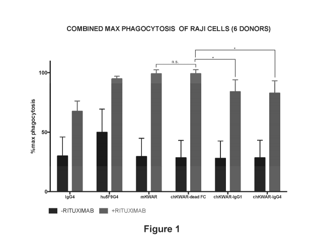
[0015] Figure 1. Combination of anti-CD47 (Hu5F9-G4) or murine anti-
SIRPalpha (mKWAR)
antibodies with anti-CD20 (Rituximab) antibody enhances the phagocytosis of
lymphoma
cancer cells (Raji) compared to control IgG4 antibody or monotherapy with
Rituximab. Chimeric
(mouse antigen binding region, human constant Fc region) antibody variants of
KWAR with
human IgG1 or human IgG4 lower the phagocytosis enhancing effect compared to a
chimeric
KWAR antibody with a dead Fc.
[0016] Figure 2, panels A-J. Determining synergy of variants of the anti-
SIRPalpha antibody
KWAR with rituximab to promote macrophage-mediated phagocytosis of Raji
lymphoma cells.
[0017] Figure 3, panels A-B. 9611 and 7E11 synergize with rituximab to
promote
macrophage-mediated phagocytosis of Raji cells.
[0018] Figure 4, panels A-B. Amino acid sequence of humanized KWAR (Panel
A) heavy
chain and (Panel B) light chain.
[0019] Figure 5, panels A-B. Humanized Kwar synergizes with therapeutic
antibodies to
promote phagocytosis.
DETAILED DESCRIPTION
[0020] Before the present methods and compositions are described, it is to
be understood that
this invention is not limited to particular method or composition described,
as such may, of
course, vary. It is also to be understood that the terminology used herein is
for the purpose of
4
CA 03031034 2019-01-15
WO 2018/026600
PCT/US2017/043935
describing particular embodiments only, and is not intended to be limiting,
since the scope of
the present invention will be limited only by the appended claims.
[0021] Where a range of values is provided, it is understood that each
intervening value, to the
tenth of the unit of the lower limit unless the context clearly dictates
otherwise, between the
upper and lower limits of that range is also specifically disclosed. Each
smaller range between
any stated value or intervening value in a stated range and any other stated
or intervening
value in that stated range is encompassed within the invention. The upper and
lower limits of
these smaller ranges may independently be included or excluded in the range,
and each range
where either, neither or both limits are included in the smaller ranges is
also encompassed
within the invention, subject to any specifically excluded limit in the stated
range. Where the
stated range includes one or both of the limits, ranges excluding either or
both of those included
limits are also included in the invention.
[0022] Unless defined otherwise, all technical and scientific terms used
herein have the same
meaning as commonly understood by one of ordinary skill in the art to which
this invention
belongs. Although any methods and materials similar or equivalent to those
described herein
can be used in the practice or testing of the present invention, some
potential and preferred
methods and materials are now described. All publications mentioned herein are
incorporated
herein by reference to disclose and describe the methods and/or materials in
connection with
which the publications are cited. It is understood that the present disclosure
supercedes any
disclosure of an incorporated publication to the extent there is a
contradiction.
[0023] As will be apparent to those of skill in the art upon reading this
disclosure, each of the
individual embodiments described and illustrated herein has discrete
components and features
which may be readily separated from or combined with the features of any of
the other several
embodiments without departing from the scope or spirit of the present
invention. Any recited
method can be carried out in the order of events recited or in any other order
which is logically
possible.
[0024] It must be noted that as used herein and in the appended claims, the
singular forms "a",
an, and "the" include plural referents unless the context clearly dictates
otherwise. Thus, for
example, reference to "a cell" includes a plurality of such cells and
reference to "the peptide"
includes reference to one or more peptides and equivalents thereof, e.g.
polypeptides, known
to those skilled in the art, and so forth.
[0025] The publications discussed herein are provided solely for their
disclosure prior to the
filing date of the present application. Nothing herein is to be construed as
an admission that
the present invention is not entitled to antedate such publication by virtue
of prior invention.
Further, the dates of publication provided may be different from the actual
publication dates
which may need to be independently confirmed
CA 03031034 2019-01-15
WO 2018/026600
PCT/US2017/043935
[0026] The terms "treatment", "treating", "treat" and the like are used
herein to generally refer
to obtaining a desired pharmacologic and/or physiologic effect. The effect can
be prophylactic
in terms of completely or partially preventing a disease or symptom(s) thereof
and/or may be
therapeutic in terms of a partial or complete stabilization or cure for a
disease and/or adverse
effect attributable to the disease. The term "treatment" encompasses any
treatment of a
disease in a mammal, particularly a human, and includes: (a) preventing the
disease and/or
symptom(s) from occurring in a subject who may be predisposed to the disease
or symptom but
has not yet been diagnosed as having it; (b) inhibiting the disease and/or
symptom(s), i.e.,
arresting their development; or (c) relieving the disease symptom(s), i.e.,
causing regression of
the disease and/or symptom(s). Those in need of treatment include those
already inflicted (e.g.,
those with cancer, those with an infection, etc.) as well as those in which
prevention is desired
(e.g., those with increased susceptibility to cancer, those with an increased
likelihood of
infection, those suspected of having cancer, those suspected of harboring an
infection, etc.).
[0027] A therapeutic treatment is one in which the subject is inflicted
prior to administration and
a prophylactic treatment is one in which the subject is not inflicted prior to
administration. In
some embodiments, the subject has an increased likelihood of becoming
inflicted or is
suspected of being inflicted prior to treatment. In some embodiments, the
subject is suspected
of having an increased likelihood of becoming inflicted.
[0028] The terms "recipient", "individual", "subject", "host", and
"patient", are used
interchangeably herein and refer to any mammalian subject for whom diagnosis,
treatment, or
therapy is desired, particularly humans. "Mammal" for purposes of treatment
refers to any
animal classified as a mammal, including humans, domestic and farm animals,
and zoo, sports,
or pet animals, such as dogs, horses, cats, cows, sheep, goats, pigs, etc.
Preferably, the
mammal is human.
[0029] As used in this invention, the term "epitope" means any antigenic
determinant on an
antigen to which the paratope of an antibody binds. Epitopic determinants
usually consist of
chemically active surface groupings of molecules such as amino acids or sugar
side chains and
usually have specific three dimensional structural characteristics, as well as
specific charge
characteristics.
[0030] The word "label" when used herein refers to a detectable compound or
composition
which is conjugated directly or indirectly to the antibody. The label may
itself be detectable by
itself (e.g., radioisotope labels or fluorescent labels) or, in the case of an
enzymatic label, may
catalyze chemical alteration of a substrate compound or composition which is
detectable.
[0031] By "solid phase" is meant a non-aqueous matrix to which the antibody
of the present
invention can adhere. Examples of solid phases encompassed herein include
those formed
partially or entirely of glass (e.g. controlled pore glass), polysaccharides
(e.g., agarose),
6
CA 03031034 2019-01-15
WO 2018/026600
PCT/US2017/043935
polyacrylamides, polystyrene, polyvinyl alcohol and silicones. In certain
embodiments,
depending on the context, the solid phase can comprise the well of an assay
plate; in others it
is a purification column (e.g. an affinity chromatography column). This term
also includes a
discontinuous solid phase of discrete particles, such as those described in
U.S. Pat. No.
4,275,149.
[0032] The terms "specific binding," "specifically binds," and the like,
refer to non-covalent or
covalent preferential binding to a molecule relative to other molecules or
moieties in a solution
or reaction mixture (e.g., an antibody specifically binds to a particular
polypeptide or epitope
relative to other available polypeptides). In some embodiments, the affinity
of one molecule for
another molecule to which it specifically binds is characterized by a Kd
(dissociation constant) of
10-5 M or less (e.g., 10-6 M or less, 10-7 M or less, 10-8 M or less, 10-9 M
or less, 10-19 M or less,
10-11 M or less, 10-12 M or less, 10-13 M or less, 10-14 M or less, 10-15 M or
less, or 10-16 M or
less). "Affinity" refers to the strength of binding, increased binding
affinity being correlated with
a lower Kd.
[0033] The term "specific binding member" as used herein refers to a member
of a specific
binding pair (i.e., two molecules, usually two different molecules, where one
of the molecules,
e.g., a first specific binding member, through non-covalent means specifically
binds to the other
molecule, e.g., a second specific binding member).
[0034] Fc receptors. The human IgG receptor family consists of a number of
receptors,
including hFcyRI, hFcyRIIA, hFcyRIIC, hFcyRIIIA, hFcyRIIB, hFcyRIIIB. IgG also
binds FcRn,
which is involved in recycling and transport of IgG. Activation of the Fc
receptors may require
the FcR subunit to be expressed and functional at the cell surface. Other Fc
receptors include,
for example, FcaR1 (CD89), Fc0c/uR, FccRI, etc. Expression of the Fc receptors
varies among
immune effector cells. hFcyRI (CD64) is restricted to monocytes/macrophages
and dendritic
cells (DCs) and, inducibly, expressed on neutrophils and mast cells; hFcyRIIA
(CD32A) is
expressed on all myeloid cells but not on lymphocytes; hFcyRIIB (CD32B) is
highly expressed
only on circulating B cells and basophils and expressed on tissue macrophages
and DCs, but
not on mast cells; hFcyRIIC (CD32C) is expressed on NK cells, monocytes, and
neutrophils;
hFcyRIIIA (CD16A) is expressed on NK cells and monocytes/macrophages;
hFcyRIIIB (CD16B)
is expressed on neutrophils and subsets of basophils.
[0035] FcRn, which importantly contributes to the biological half-life of
antibodies in the blood,
is expressed on antigen-presenting cells, monocytes/macrophages, neutrophils,
vascular
endothelial cells, intestinal epithelial cells, and syncytiotrophoblasts.
[0036] The Fcy receptors differ in their affinity for IgG and likewise the
different IgG subclasses
have unique affinities for each of the Fcy receptors. These interactions are
further tuned by
7
CA 03031034 2019-01-15
WO 2018/026600 PCT/US2017/043935
glycans (oligosaccharide), e.g. at position CH2-84.4 of IgG. For example, by
creating steric
hindrance, fucose containing CH2-84.4 glycans reduce IgG affinity for
FcyRIIIA.
[0037] Fc domain or region. The Fc region of an antibody mediates its serum
half-life and
effector functions, such as complement-dependent cytotoxicity (CDC), antibody-
dependent
cellular cytotoxicity (ADCC) and antibody-dependent cell phagocytosis (ADCP).
Engineering
the Fc region of a therapeutic monoclonal antibody or Fc fusion protein allows
the generation of
molecules that are better suited to the pharmacology activity required of
them. The half-life of
an IgG depends on its pH-dependent binding to the neonatal receptor FcRn.
FcRn, which is
expressed on the surface of endothelial cells, binds the IgG in a pH-dependent
manner and
protects it from degradation.
[0038] A "wild-type Fc region" possesses the effector functions of a native-
sequence Fc region,
in particular for the purposes of the present invention interacting with one
or more of the Fc
receptors such as FcyRI; FcyRIIA; FcyR1161; FcyRIIB2; FcyRIIIA; FcyRIIIB
receptors; and can
be assessed using various assays as disclosed, for example, in definitions
herein. A "dead" Fc
is one that has been mutagenized to retain activity with respect to, for
example, prolonging
serum half-life through interaction with FcRn, but which has reduced or absent
binding to one or
more other Fc receptor(s), including without limitation a human FcyR as listed
above.
[0039] A "native-sequence Fc region" comprises an amino acid sequence
identical to the
amino acid sequence of an Fc region found in nature. Native-sequence human Fc
regions
include a native-sequence human IgG1 Fc region (non-A and A allotypes); native-
sequence
human IgG2 Fc region; native-sequence human IgG3 Fc region; and native-
sequence human
IgG4 Fc region, as well as naturally occurring variants thereof.
[0040] A "variant Fc region" or "engineered Fc region" comprises an amino
acid sequence that
differs from that of a native-sequence Fc region by virtue of at least one
amino acid
modification, preferably one or more amino acid substitution(s). Preferably,
the variant Fc
region has at least one amino acid substitution compared to a native-sequence
Fc region or to
the Fc region of a parent polypeptide, e.g., from about one to about ten amino
acid
substitutions, and preferably from about one to about five amino acid
substitutions in a native-
sequence Fc region or in the Fc region of the parent polypeptide. The variant
Fc region herein
will preferably possess at least about 80% homology with a native-sequence Fc
region and/or
with an Fc region of a parent polypeptide, and most preferably at least about
90% homology
therewith, more preferably at least about 95% homology therewith.
[0041] Variant Fc sequences for a "dead Fc" may include three amino acid
substitutions in the
CH2 region to reduce FcyRI binding at EU index positions 234, 235, and 237
(see Duncan et
al., (1988) Nature 332:563). Two amino acid substitutions in the complement
C1q binding site
at EU index positions 330 and 331 reduce complement fixation (see Tao et al.,
J. Exp. Med.
8
CA 03031034 2019-01-15
WO 2018/026600 PCT/US2017/043935
178:661 (1993) and Canfield and Morrison, J. Exp. Med. 173:1483 (1991)).
Substitution into
human IgG1 of IgG2 residues at positions 233-236 and IgG4 residues at
positions 327, 330 and
331 greatly reduces ADCC and CDC (see, for example, Armour KL. etal., 1999 Eur
J Immunol.
29(8):2613-24; and Shields RL. etal., 2001. J Biol Chem. 276(9):6591-604).
[0042] Binding of IgG to the FcyRs or C1q depends on residues located in
the hinge region and
the CH2 domain. Two regions of the CH2 domain are critical for FcyRs and C1q
binding, and
have unique sequences in IgG2 and IgG4. Substitutions into human IgG1 or IgG2
residues at
positions 233-236 and IgG4 residues at positions 327, 330 and 331 have been
shown to greatly
reduce ADCC and CDC. Numerous mutations have been made in the CH2 domain of
human
IgG1.
[0043] The triple amino acid substitution L234A, L235A, and G237A largely
eliminates FcyR
and complement effector functions (see, for example, U520100266505).
[0044] In some embodiments the Fc region has been modified by the choice of
expression
host, enzymatic treatment of amino acid substitutions to have reduced
glycosylation and
binding to FcyR, relative to the native protein. Mutations that reduce binding
to FcyR include,
without limitation, modification of the glycosylation on asparagine 297 of the
Fc domain, which
is known to be required for optimal FcR interaction. For example known amino
acid
substitutions include N297 mutations, for example N297A/Q/D/H/G/C, which
changes result in
the loss of a glycosylation site on the protein. Enzymatically deglycosylated
Fc domains,
recombinantly expressed antibodies in the presence of a glycosylation
inhibitor and the
expression of Fc domains in bacteria have a similar loss of glycosylation and
consequent
binding to FcyRs.
[0045] The LALA variant, L234A/L235A, also has significantly reduced FcyR
binding; as does
E233P/L234V/L235A/G236 + A327G/A3305/P3315. See, for example, Armour et al.
(1999)
Eur J Immunol. 29(8):2613-24. The set of mutations: K322A, L234A and L235A are
sufficient to
almost completely abolish FcyR and C1q binding. A set of three mutations,
L234F/L235E/P3315 (dubbed TM), have a very similar effect.
[0046] Other Fc variants are possible, including without limitation one in
which a region capable
of forming a disulfide bond is deleted, or in which certain amino acid
residues are eliminated at
the N-terminal end of a native Fc form or a methionine residue is added
thereto.
[0047] The Fc may be in the form of having native sugar chains, increased
sugar chains
compared to a native form or decreased sugar chains compared to the native
form, or may be
in an aglycosylated or deglycosylated form. The increase, decrease, removal or
other
modification of the sugar chains may be achieved by methods common in the art,
such as a
chemical method, an enzymatic method or by expressing it in a genetically
engineered
production cell line. Such cell lines can include microorganisms, e.g. Pichia
Pastoris, and
mammalians cell line, e.g. CHO cells, that naturally express glycosylating
enzymes. Further,
9
CA 03031034 2019-01-15
WO 2018/026600 PCT/US2017/043935
microorganisms or cells can be engineered to express glycosylating enzymes, or
can be
rendered unable to express glycosylation enzymes (See e.g., Hamilton, et al.,
Science,
313:1441 (2006); Kanda, et al, J. Biotechnology, 130:300 (2007); Kitagawa, et
al., J. Biol.
Chem., 269 (27): 17872 (1994); Ujita-Lee et al., J. Biol. Chem., 264 (23):
13848 (1989); !mai-
Nishiya, et al, BMC Biotechnology 7:84 (2007); and WO 07/055916). As one
example of a cell
engineered to have altered sialylation activity, the alpha-2,6-
sialyltransferase 1 gene has been
engineered into Chinese Hamster Ovary cells and into sf9 cells. Antibodies
expressed by these
engineered cells are thus sialylated by the exogenous gene product. A further
method for
obtaining Fc molecules having a modified amount of sugar residues compared to
a plurality of
native molecules includes separating said plurality of molecules into
glycosylated and non-
glycosylated fractions, for example, using lectin affinity chromatography (See
e.g., WO
07/117505). The presence of particular glycosylation moieties has been shown
to alter the
function of Immunoglobulins. For example, the removal of sugar chains from an
Fc molecule
results in a sharp decrease in binding affinity to the C1q part of the first
complement component
Cl and a decrease or loss in antibody-dependent cell-mediated cytotoxicity
(ADCC) or
complement-dependent cytotoxicity (CDC), thereby not inducing unnecessary
immune
responses in vivo. Additional important modifications include sialylation and
fucosylation: the
presence of sialic acid in IgG has been correlated with anti-inflammatory
activity (See e.g.,
Kaneko, et al, Science 313:760 (2006)), whereas removal of fucose from the IgG
leads to
enhanced ADCC activity (See e.g., Shoj-Hosaka, et al, J. Biochem., 140:777
(2006)).
[0048] The term "Fc-region-comprising antibody" refers to an antibody that
comprises an Fc
region. The C-terminal lysine (residue 447 according to the EU numbering
system) of the Fc
region may be removed, for example, during purification of the antibody or by
recombinant
engineering the nucleic acid encoding the antibody. Accordingly, an antibody
having an Fc
region according to this invention can comprise an antibody with or without
K447.
[0049] Antibodies, also referred to as immunoglobulins, conventionally
comprise at least one
heavy chain and one light, where the amino terminal domain of the heavy and
light chains is
variable in sequence, hence is commonly referred to as a variable region
domain, or a variable
heavy (VH) or variable light (VH) domain. The two domains conventionally
associate to form a
specific binding region, although as well be discussed here, specific binding
can also be
obtained with heavy chain only variable sequences, and a variety of non-
natural configurations
of antibodies are known and used in the art.
[0050] A "therapeutic" antibody, as discussed herein, references an
antibody that is suitable for
treatment of a patient, i.e. an antibody with in vivo activity in a context
appropriate for
therapeutic use, e.g. treatment of a human subject. In some embodiments, a
therapeutic
antibody may refer to an antibody that binds to an antigen present on the
surface of a targeted
CA 03031034 2019-01-15
WO 2018/026600
PCT/US2017/043935
cell, e.g. a tumor-specific antigen, a pathogen-specific antigen, etc.
Such therapeutic
antibodies can be combined with an anti-SIRPoc antibody to enhance
phagocytosis of the
targeted cell.
[0051] The
term "antibody" herein is used in the broadest sense and specifically covers
monoclonal antibodies, polyclonal antibodies, monomers, dimers, multimers,
multispecific
antibodies (e.g., bispecific antibodies), heavy chain only antibodies, three
chain antibodies,
single chain Fv, nanobodies, etc., and also include antibody fragments, so
long as they exhibit
the desired biological activity (Miller et al (2003) Jour. of Immunology
170:4854-4861). For
example, F(ab')2 fragments are of interest as a format for anti-SIRPoc
antibodies. Antibodies
may be murine, human, humanized, chimeric, or derived from other species. For
many
purposes the antibodies of the invention comprise a human engineered Fc
region.
[0052] The
term antibody may reference a full-length heavy chain, a full length light
chain, an
intact immunoglobulin molecule; or an immunologically active portion of any of
these
polypeptides. The immunoglobulin disclosed herein can be of any type (e.g.,
IgG, IgE, IgM, IgD,
and IgA), class (e.g., IgG1, IgG2, IgG3, IgG4, IgA1 and IgA2) or subclass of
immunoglobulin
molecule, including engineered subclasses with altered Fc portions that
provide for reduced
effector cell activity. The immunoglobulins can be derived from any species.
In one aspect, the
immunoglobulin is of largely human origin, is humanized, or chimeric with
respect to a human
Fc region.
[0053] The
term "variable" refers to the fact that certain portions of the variable
domains differ
extensively in sequence among antibodies and are used in the binding and
specificity of each
particular antibody for its particular antigen. However, the variability is
not evenly distributed
throughout the variable domains of antibodies. It is concentrated in three
segments called
hypervariable regions both in the light chain and the heavy chain variable
domains. The more
highly conserved portions of variable domains are called the framework regions
(FRs). The
variable domains of native heavy and light chains each comprise four FRs,
largely adopting a
beta-sheet configuration, connected by three hypervariable regions, which form
loops
connecting, and in some cases forming part of, the beta-sheet structure. The
hypervariable
regions in each chain are held together in close proximity by the FRs and,
with the
hypervariable regions from the other chain, contribute to the formation of the
antigen-binding
site of antibodies (see Kabat et al (1991) Sequences of Proteins of
Immunological Interest, 5th
Ed. Public Health Service, National Institutes of Health, Bethesda, Md.).
[0054] The
term "hypervariable region" when used herein refers to the amino acid residues
of
an antibody which are responsible for antigen-binding. The hypervariable
region may comprise
amino acid residues from a "complementarity determining region" or "CDR",
and/or those
residues from a "hypervariable loop". "Framework Region" or "FR" residues are
those variable
domain residues other than the hypervariable region residues as herein
defined.
11
CA 03031034 2019-01-15
WO 2018/026600
PCT/US2017/043935
[0055] Variable regions of interest include at least one CDR sequence from
the variable
regions of an anti-SIRPoc antibody, usually at least 2 CDR sequences, and more
usually 3 CDR
sequences on the light and on the heavy chain. One of skill in the art will
understand that a
number of definitions of the CDRs are commonly in use, including the Kabat
definition (see
"Zhao et al. A germline knowledge based computational approach for determining
antibody
complementarity determining regions." Mol Immunol. 2010;47:694-700), which is
based on
sequence variability and is the most commonly used. The Chothia definition is
based on the
location of the structural loop regions (Chothia et al. "Conformations of
immunoglobulin
hypervariable regions." Nature. 1989;342:877-883). Alternative CDR definitions
of interest
include, without limitation, those disclosed by Honegger, "Yet another
numbering scheme for
immunoglobulin variable domains: an automatic modeling and analysis tool." J
Mol Biol.
2001;309:657-670; Ofran et al. "Automated identification of complementarity
determining
regions (CDRs) reveals peculiar characteristics of CDRs and B cell epitopes."
J Immunol.
2008;181:6230-6235; Almagro "Identification of differences in the specificity-
determining
residues of antibodies that recognize antigens of different size: implications
for the rational
design of antibody repertoires." J Mol Recognit. 2004;17:132-143; and Padlanet
al.
"Identification of specificity-determining residues in antibodies." Faseb J.
1995;9:133-139.,
each of which is herein specifically incorporated by reference.
[0056] The term "monoclonal antibody" as used herein refers to an antibody
obtained from a
population of substantially homogeneous antibodies, i.e., the individual
antibodies comprising
the population are identical except for possible naturally occurring mutations
that may be
present in minor amounts. Monoclonal antibodies are highly specific, being
directed against a
single antigenic site. Furthermore, in contrast to polyclonal antibody
preparations, which include
different antibodies directed against different determinants (epitopes), each
monoclonal
antibody is directed against a single determinant on the antigen. In addition
to their specificity,
the monoclonal antibodies are advantageous in that they may be synthesized
uncontaminated
by other antibodies. The modifier "monoclonal" indicates the character of the
antibody as being
obtained from a substantially homogeneous population of antibodies, and is not
to be construed
as requiring production of the antibody by any particular method.
[0057] The antibodies herein specifically include "chimeric" antibodies in
which a portion of the
heavy and/or light chain is identical with or homologous to corresponding
sequences in
antibodies derived from a particular species or belonging to a particular
antibody class or
subclass, while the remainder of the chain(s) is identical with or homologous
to corresponding
sequences in antibodies derived from another species or belonging to another
antibody class or
subclass, as well as fragments of such antibodies, so long as they exhibit the
desired biological
activity (U.S. Pat. No. 4,816,567; and Morrison et al (1984) Proc. Natl. Acad.
Sci. USA,
81:6851-6855). Chimeric antibodies of interest herein include "primatized"
antibodies
12
CA 03031034 2019-01-15
WO 2018/026600
PCT/US2017/043935
comprising variable domain antigen-binding sequences derived from a non-human
primate
(e.g., Old World Monkey, Ape, etc.) and human constant region sequences.
[0058] An
"intact antibody chain" as used herein is one comprising a full length
variable region
and a full length constant region. An intact "conventional" antibody comprises
an intact light
chain and an intact heavy chain, as well as a light chain constant domain (CL)
and heavy chain
constant domains, CH1, hinge, CH2 and CH3 for secreted IgG. Other isotypes,
such as IgM or
IgA may have different CH domains. The constant domains may be native sequence
constant
domains (e.g., human native sequence constant domains) or amino acid sequence
variants
thereof.
[0059]
"Fv" is the minimum antibody fragment, which contains a complete antigen-
recognition
and antigen-binding site. The CD3 binding antibodies of the invention comprise
a dimer of one
heavy chain and one light chain variable domain in tight, non-covalent
association; however
additional antibodies, e.g. for use in a multi-specific configuration, may
comprise a VH in the
absence of a VL sequence. Even a single variable domain (or half of an Fv
comprising only
three hypervariable regions specific for an antigen) has the ability to
recognize and bind
antigen, although the affinity may be lower than that of two domain binding
site.
[0060] The
Fab fragment also contains the constant domain of the light chain and the
first
constant domain (CH1) of the heavy chain. Fab' fragments differ from Fab
fragments by the
addition of a few residues at the carboxy terminus of the heavy chain CH1
domain including
one or more cysteines from the antibody hinge region. Fab'-SH is the
designation herein for
Fab' in which the cysteine residue(s) of the constant domains bear at least
one free thiol group.
F(ab)2 antibody fragments originally were produced as pairs of Fab' fragments
which have
hinge cysteines between them. Other chemical couplings of antibody fragments
are also
known.
[0061]
"Humanized" forms of non-human (e.g., rodent) antibodies are chimeric
antibodies that
contain minimal sequence derived from non-human immunoglobulin. See, for
example, Jones
et al, (1986) Nature 321:522-525; Chothia et al (1989) Nature 342:877;
Riechmann et al (1992)
J. Mol. Biol. 224, 487-499; Foote and Winter, (1992) J. Mol. Biol. 224:487-
499; Presta et al
(1993) J. Immunol. 151, 2623-2632; Werther et al (1996) J. Immunol. Methods
157:4986-4995;
and Presta et al (2001) Thromb. Haemost. 85:379-389. For further details, see
U.S. Pat. Nos.
5,225,539; 6,548,640; 6,982,321; 5,585,089; 5,693,761; 6,407,213; Jones et al
(1986) Nature,
321:522-525; and Riechmann et al (1988) Nature 332:323-329.
[0062]
Moreover, the term "antibody" as used herein, can refer in appropriate
embodiments
(unless otherwise stated or clear from context) to any of the art-known or
developed constructs
or formats for utilizing antibody structural and functional features in
alternative presentation.
For example, embodiments, an antibody utilized in accordance with the present
invention is in a
13
CA 03031034 2019-01-15
WO 2018/026600
PCT/US2017/043935
format selected from, but not limited to, intact IgG, IgE and IgM, bi- or
multi- specific antibodies
(e.g., Zybodies , etc), single chain Fvs, polypeptide-Fc fusions, Fabs,
cameloid antibodies,
masked antibodies (e.g., Probodiese), Small Modular ImmunoPharmaceuticals
("SMIPsTm"),
single chain or Tandem diabodies (TendAbe), VHHs, Anticalins , Nanobodies ,
minibodies,
BiTEes, ankyrin repeat proteins or DARPINse, Avimers , a DART, a TCR-like
antibody,
Adnectins , Affilins , Trans-bodies , Affibodies , a TrimerX , MicroProteins,
Fynomers ,
Centyrins , and a KALBITOR . In some embodiments, an antibody may lack a
covalent
modification (e.g., attachment of a glycan) that it would have if produced
naturally. In some
embodiments, an antibody may contain a covalent modification (e.g., attachment
of a glycan, a
payload, e.g., a detectable moiety, a therapeutic moiety, a catalytic moiety,
etc., or other
pendant group [e.g., poly-ethylene glycol, etc.
[0063] Exemplary antibody agents include, but are not limited to, human
antibodies, primatized
antibodies, chimeric antibodies, bi-specific antibodies, humanized antibodies,
conjugated
antibodies (i.e., antibodies conjugated or fused to other proteins,
radiolabels, cytotoxins), Small
Modular ImmunoPharmaceuticals ("SMIPsTm"), single chain antibodies, cameloid
antibodies,
and antibody fragments. As used herein, the term "antibody agent" also
includes intact
monoclonal antibodies, polyclonal antibodies, single domain antibodies (e.g.,
shark single
domain antibodies (e.g., IgNAR or fragments thereof)), multispecific
antibodies (e.g. bi-specific
antibodies) formed from at least two intact antibodies, and antibody fragments
so long as they
exhibit the desired biological activity. In some embodiments, the term
encompasses stapled
peptides. In some embodiments, the term encompasses one or more antibody-like
binding
peptidomimetics. In some embodiments, the term encompasses one or more
antibody-like
binding scaffold proteins. In come embodiments, the term encompasses
monobodies or
adnectins.
[0064] "Antibody fragment", and all grammatical variants thereof, as used
herein are defined as
a portion of an intact antibody comprising the antigen binding site or
variable region of the intact
antibody, wherein the portion is free of the constant heavy chain domains
(i.e. CH2, CH3, and
CH4, depending on antibody isotype) of the Fc region of the intact antibody.
Examples of
antibody fragments include Fab, Fab', Fab'-SH, F(ab')2, and Fv fragments;
diabodies; any
antibody fragment that is a polypeptide having a primary structure consisting
of one
uninterrupted sequence of contiguous amino acid residues (referred to herein
as a "single-chain
antibody fragment" or "single chain polypeptide"), including without
limitation (1) single-chain Fv
(scFv) molecules (2) single chain polypeptides containing only one light chain
variable domain,
or a fragment thereof that contains the three CDRs of the light chain variable
domain, without
an associated heavy chain moiety and (3) single chain polypeptides containing
only one heavy
chain variable region, or a fragment thereof containing the three CDRs of the
heavy chain
variable region, without an associated light chain moiety; and multispecific
or multivalent
14
CA 03031034 2019-01-15
WO 2018/026600
PCT/US2017/043935
structures formed from antibody fragments. In an antibody fragment comprising
one or more
heavy chains, the heavy chain(s) can contain any constant domain sequence
(e.g. CH1 in the
IgG isotype) found in a non-Fc region of an intact antibody, and/or can
contain any hinge region
sequence found in an intact antibody, and/or can contain a leucine zipper
sequence fused to or
situated in the hinge region sequence or the constant domain sequence of the
heavy chain(s).
[0065] Unless specifically indicated to the contrary, the term "conjugate"
as described and
claimed herein is defined as a heterogeneous molecule formed by the covalent
attachment of
one or more antibody fragment(s) to one or more polymer molecule(s), wherein
the
heterogeneous molecule is water soluble, i.e. soluble in physiological fluids
such as blood, and
wherein the heterogeneous molecule is free of any structured aggregate. A
conjugate of
interest is PEG. In the context of the foregoing definition, the term
"structured aggregate" refers
to (1) any aggregate of molecules in aqueous solution having a spheroid or
spheroid shell
structure, such that the heterogeneous molecule is not in a micelle or other
emulsion structure,
and is not anchored to a lipid bilayer, vesicle or liposome; and (2) any
aggregate of molecules
in solid or insolubilized form, such as a chromatography bead matrix, that
does not release the
heterogeneous molecule into solution upon contact with an aqueous phase.
Accordingly, the
term "conjugate" as defined herein encompasses the aforementioned
heterogeneous molecule
in a precipitate, sediment, bioerodible matrix or other solid capable of
releasing the
heterogeneous molecule into aqueous solution upon hydration of the solid.
[0066] The anti-SIRPa antibodies herein specifically include "chimeric"
antibodies
(immunoglobulins) in which a portion of the heavy and/or light chain is
identical with or
homologous to corresponding sequences in antibodies derived from a particular
species or
belonging to a particular antibody class or subclass, while the remainder of
the chain(s) is
identical with or homologous to corresponding sequences in antibodies derived
from another
species or belonging to another antibody class or subclass, as well as
fragments of such
antibodies, so long as they exhibit the desired biological activity.
[0067] An "isolated" antibody is one which has been identified and
separated and/or recovered
from a component of its natural environment. Contaminant components of its
natural
environment are materials which would interfere with diagnostic or therapeutic
uses for the
antibody, and may include enzymes, hormones, and other proteinaceous or
nonproteinaceous
solutes. In some embodiments, the antibody will be purified (1) to greater
than 75% by weight
of antibody as determined by the Lowry method, and most preferably more than
80%, 90% or
99% by weight, or (2) to homogeneity by SDS-PAGE under reducing or nonreducing
conditions
using Coomassie blue or, preferably, silver stain. Isolated antibody includes
the antibody in situ
within recombinant cells since at least one component of the antibody's
natural environment will
not be present. Ordinarily, however, isolated antibody will be prepared by at
least one
purification step.
CA 03031034 2019-01-15
WO 2018/026600
PCT/US2017/043935
[0068] The term "epitope tagged" when used herein refers to an anti-SIRPa
antibody (or
fragment) fused to an "epitope tag". The epitope tag polypeptide has enough
residues to
provide an epitope against which an antibody can be made, yet is short enough
such that it
does not interfere with activity of the anti-SIRPa antibody. The epitope tag
preferably is
sufficiently unique so that the antibody specific for the epitope does not
substantially cross-
react with other epitopes. Suitable tag polypeptides generally have at least 6
amino acid
residues and usually between about 8-50 amino acid residues (preferably
between about 9-30
residues). Examples include the c-myc tag and the 8F9, 3C7, 6E10, G4, B7 and
9E10
antibodies thereto (Evan et al., Mol. Cell. Biol. 5(12):3610-3616 (1985)); and
the Herpes
Simplex virus glycoprotein D (gD) tag and its antibody (Paborsky et al.,
Protein Engineering
3(6):547-553 (1990)). An additional example is a "histidine tag" or "histidine-
rich affinity
peptide", which is a metal ion affinity peptide that is rich in histidines
(e.g., 6xHis tag, HAT tag,
6xHN tag, and the like). A histidine tag can also specifically bind to an anti-
His antibody.
[0069] SIRPa1 (PTPNS1, SHPS1), is a transmembrane glycoprotein, expressed
primarily on
myeloid and neuronal cells. SIRPa interacts with the widely distributed
membrane protein
CD47. In addition to SIRPa, there are two closely related proteins in the SIRP
family: SIRPI3
and SIRPy. All three have three immunoglobulin superfamily (IgSF) domains in
their
extracellular region. In humans, the SIRPa protein is found in two major
forms. One form, the
variant 1 or V1 form, has the amino acid sequence set out as NCB! RefSeq
NP_542970.1
(residues 27-504 constitute the mature form). Another form, the variant 2 or
V2 form, differs by
13 amino acids and has the amino acid sequence set out in GenBank as
CAA71403.1
(residues 30-504 constitute the mature form). These two forms of SIRPa
constitute about 80%
of the forms of SIRPa present in humans, and both are embraced herein by the
term "human
SIRPa". Also embraced by the term "human SIRPa" are the minor forms thereof
that are
endogenous to humans and have the same property of triggering signal
transduction through
CD47 upon binding thereto. Sequences of human SIRPa variants may be accessed
through
public databases, including Genbank accession numbers: refINP_542970.1;
gblEAX10606.1;
refIXP_005260726.1; gbl EAX10606.1; XP_005260726.1; gbl EAX10611.1; gbl
EAX10609.1;
dbjIBAA12974.1; gbIAAH26692.1; refIXP_011527475.1. See, for example Lee et al.
(2007) J.
Immunol. 179(11):7741-7750; herein specifically incorporated by reference.
[0070] Antibodies that specifically bind to human SIRPa are known and used
in the art, and
may be adapted by the use of an engineered Fc region as disclosed herein.
Exemplary
antibodies include those described in international patent application WO
2015/138600; in
published US application 2014/0242095 (University Health Networks); published
application
CN103665165 (JIANGSU KUANGYA BIOLOGICAL MEDICAL SCIENCE & TECHNOLOGY;
Zhao XW et al. Proc Nat! Acad Sci U S A 108:18342-7 (2011), each herein
specifically
16
CA 03031034 2019-01-15
WO 2018/026600
PCT/US2017/043935
incorporated by reference. An anti-SIRPa, antibody may be pan-specific, i.e.
binding to two or
more different human SIRPa isoforms; or may be specific for one isoform. For
example, the
antibody 1.23A described by Zhang et al., supra. is reported to be specific
for the SIRPal
variant, while the 12C4 antibody is pan-specific. Anti-SIRPa, antibodies can
also be specific for
SIRPa and lack binding to SIRPI3 and/or SIRPy. Anti-SIRPa antibodies can be
pan-specific
with respect to SIRPI3 and/or SIRPy.
[0071] The terms "co-administration", "co-administer", and "in combination
with" include the
administration of two or more therapeutic agents either simultaneously,
concurrently or
sequentially within no specific time limits. In one embodiment, the agents are
present in the cell
or in the subject's body at the same time or exert their biological or
therapeutic effect at the
same time. In one embodiment, the therapeutic agents are in the same
composition or unit
dosage form. In other embodiments, the therapeutic agents are in separate
compositions or
unit dosage forms. In certain embodiments, a first agent can be administered
prior to (e.g.,
minutes, 15 minutes, 30 minutes, 45 minutes, 1 hour, 2 hours, 4 hours, 6
hours, 12 hours, 24
hours, 48 hours, 72 hours, 96 hours, 1 week, 2 weeks, 3 weeks, 4 weeks, 5
weeks, 6 weeks, 8
weeks, or 12 weeks before), concomitantly with, or subsequent to (e.g., 5
minutes, 15 minutes,
30 minutes, 45 minutes, 1 hour, 2 hours, 4 hours, 6 hours, 12 hours, 24 hours,
48 hours, 72
hours, 96 hours, 1 week, 2 weeks, 3 weeks, 4 weeks, 5 weeks, 6 weeks, 8 weeks,
or 12 weeks
after) the administration of a second therapeutic agent.
[0072] Anti-SIRPa, antibodies may be used therapeutically in combination
with a second
antibody or agent that selectively binds to a target cell. The term "target
cell" can be used in
different ways depending on context. Typically a "target cell" is a cell that
will be phagocytosed
by a phagocytic cell (e.g., a phagocyte), where the phagocytosis is enhanced
as a result of
administering a subject anti-SIRPa antibody. Thus, the term "target cell" can
refer to a CD47-
expressing cell, because a subject anti-SIRPa antibody, by inhibiting the
interaction between
the CD47-expressing cell and the SIRPa expressing phagocytic cell, facilitates
phagocytosis of
the CD47-expressing cell.
[0073] However, in some cases, the target cell need not express high levels
of CD47 (and in
some cases need not express CD47 at all) in order for a subject multispecific
antibody to
induce phagocytosis of the target cell. For example, in the context of a
multispecific (e.g.,
bispecific) antibody, the SIRPa binding region (the first binding region) of a
subject multispecific
(e.g., bispecific) antibody binds to SIRPa on a phagocytic cell (e.g., a
macrophage), which
allows the multispecific antibody to function as a tether to bring the
phagocytic cell into the
vicinity of a cell expressing an antigen (e.g., a marker of a cancer cell)
that is recognized by
(specifically bound by) a second binding region of the multispecific antibody
(e.g., the second
binding region of a bispecific antibody). Therefore, in the context of a
multispecific antibody, a
17
CA 03031034 2019-01-15
WO 2018/026600
PCT/US2017/043935
target cell can be a cell that does not express high levels of CD47 (and can
also be a cell that
does not express CD47). In some embodiments, a target cell is a mammalian
cell, for example
a human cell. A target cell can be from any individual (e.g., patient,
subject, and the like) as
described below.
[0074] In some cases, a target cell is an "inflicted" cell (e.g., a cell
from an "inflicted" individual),
where the term "inflicted" is used herein to refer to a subject with symptoms,
an illness, or a
disease that can be treated with a subject anti-SIRPa antibody. An "inflicted"
subject can have
cancer, can harbor an infection (e.g., a chronic infection), and/or can have
other hyper-
proliferative conditions, for example sclerosis, fibrosis, and the like, etc.
Also of interest is the
use in the treatment of cardiovascular conditions, including without
limitation aneurysm,
atherosclerosis, etc. "Inflicted cells" can be those cells that cause the
symptoms, illness, or
disease. As non-limiting examples, the inflicted cells of an inflicted patient
can be CD47
expressing cancer cells, infected cells, inflammatory cells, immune cells, and
the like. One
indication that an illness or disease can be treated with a subject anti-SIRPa
antibody is that the
involved cells (i.e., the inflicted cells, e.g., the cancerous cells, the
infected cells, the
inflammatory cells, the immune cells, etc.) express CD47 (e.g., in some cases,
an increased
level of CD47 compared to normal cells of the same cell type).
[0075] For the treatment of cancer, the anti-SIRPa, antibody may be
combined with one or
more antibodies specific for a tumor antigen. Of these, tumor-associated
antigens (TAAs) are
relatively restricted to tumor cells, whereas tumor-specific antigens (TSAs)
are unique to tumor
cells. TSAs and TAAs typically are portions of intracellular molecules
expressed on the cell
surface as part of the major histocompatibility complex.
[0076] Tissue specific differentiation antigens are molecules present on
tumor cells and their
normal cell counterparts. Tumor-associated antigens known to be recognized by
therapeutic
mAbs fall into several different categories. Hematopoietic differentiation
antigens are
glycoproteins that are usually associated with cluster of differentiation (CD)
groupings and
include CD20, CD30, CD33 and CD52. Cell surface differentiation antigens are a
diverse group
of glycoproteins and carbohydrates that are found on the surface of both
normal and tumor
cells. Antigens that are involved in growth and differentiation signaling are
often growth factors
and growth factor receptors. Growth factors that are targets for antibodies in
cancer patients
include CEA, epidermal growth factor receptor (EGFR; also known as ERBB1)'
ERBB2 (also
known as HER2), ERBB3, MET (also known as HGFR), insulin-like growth factor 1
receptor
(IGF1R), ephrin receptor A3 (EPHA3), tumor necrosis factor (TNF)-related
apoptosis-inducing
ligand receptor 1 (TRAILR1; also known as TNFRSF10A), TRAILR2 (also known as
TNFRSF10B) and receptor activator of nuclear factor-KB ligand (RANKL; also
known as
TNFSF11). Antigens involved in angiogenesis are usually proteins or growth
factors that
18
CA 03031034 2019-01-15
WO 2018/026600
PCT/US2017/043935
support the formation of new microvasculature, including vascular endothelial
growth factor
(VEGF), VEGF receptor (VEGFR), integrin aV[33 and integrin a5[31. Tumor stroma
and the
extracellular matrix are indispensable support structures for a tumor. Stromel
and extracellular
matrix antigens that are therapeutic targets include fibroblast activation
protein (FAP) and
tenascin.
[0077] Examples of therapeutic antibodies useful in bispecific
configurations or as combination
therapy include, without limitation, rituximab; Ibritumomab; tiuxetan;
tositumomab; Brentuximab;
vedotin; Gemtuzumab; ozogamicin; Alemtuzumab; IGN101; adecatumumab;
Labetuzumab;
huA33; Pemtumomab; oregovomab; CC49 (minretumomab); cG250; J591; MOv18; MORAb-
003 (farletuzumab); 3F8, ch14.18; KW-2871; hu3S193; IgN311; Bevacizumab; IM-
2C6;
CDP791; Etaracizumab; Volociximab; Cetuximab, panitumumab, nimotuzumab; 806;
Trastuzumab; pertuzumab; MM-121; AMG 102, METMAB; SCH 900105; AVE1642, IMC-
Al2,
MK-0646, R1507; CP 751871; KB004; 111A4; Mapatumumab (HGS-ETR1); HGS-ETR2; CS-
1008; Denosumab; Sibrotuzumab; F19; and 8106. A bispecific antibody may use
the Fc region
that activates an Fcy receptor.
[0078] For the treatment of cancer, the anti-SIRPa, antibody may be
combined with one or
more antibodies that inhibit immune checkpoint proteins. Of particular
interest are immune
checkpoint proteins displayed on the surface of a tumor cell. The immune-
checkpoint receptors
that have been most actively studied in the context of clinical cancer
immunotherapy, cytotoxic
T-lymphocyte-associated antigen 4 (CTLA4; also known as CD152) and programmed
cell death
protein 1 (PD1; also known as CD279) - are both inhibitory receptors. The
clinical activity of
antibodies that block either of these receptors implies that antitumor
immunity can be enhanced
at multiple levels and that combinatorial strategies can be intelligently
designed, guided by
mechanistic considerations and preclinical models.
[0079] The two ligands for PD1 are PD1 ligand 1 (PDL1; also known as B7-H1
and CD274)
and PDL2 (also known as B7-DC and CD273). PDL1 is expressed on cancer cells
and through
binding to its receptor PD1 on T cells it inhibits T cell activation/function.
See, for example,
Avelumab as a therapeutic antibody.
[0080] Agents that agonize an immune costimulatory molecule are also useful
in the methods
of the invention. Such agents include agonists or CD40 and 0X40. CD40 is a
costimulatory
protein found on antigen presenting cells (APCs) and is required for their
activation. These
APCs include phagocytes (macrophages and dendritic cells) and B cells. CD40 is
part of the
TNF receptor family. The primary activating signaling molecules for CD40 are
IFNy and CD40
ligand (CD4OL). Stimulation through CD40 activates macrophages.
19
CA 03031034 2019-01-15
WO 2018/026600
PCT/US2017/043935
[0081] Anti CCR4 (CD194) antibodies of interest include humanized
monoclonal antibodies
directed against C-C chemokine receptor 4 (CCR4) with potential anti-
inflammatory and
antineoplastic activities.
[0082] Examples of symptoms, illnesses, and/or diseases that can be treated
with a subject
anti-SIRPa antibody include, but are not limited to cancer (any form of
cancer, including but not
limited to: carcinomas, soft tissue tumors, sarcomas, teratomas, melanomas,
leukemias,
lymphomas, brain cancers, solid tumors, mesothelioma (MSTO), etc.); infection
(e.g., chronic
infection); and an immunological disease or disorder (e.g., an inflammatory
disease)(e.g.,
multiple sclerosis, arthritis, and the like, e.g., for immunosuppressive
therapy). A subject anti-
SIRPa antibody can also be used for transplant conditioning (e.g., stem cell
transplant, bone
marrow transplant, etc.) (e.g., to destroy malignant cells, to provide
immunosuppression to
prevent the patient's body from rejecting the donor's cells/stem cells, etc.).
For example, in
some cases, a subject antibody combination or bispecific antibody (e.g., anti-
SIRPa, in
combination with anti-CD117) finds use for transplant conditioning. For
example, a subject
antibody combination or bispecific antibody (e.g., anti-SIRPa, in combination
with anti-CD117)
can be used for bone marrow transplant conditioning. In some cases, a subject
anti-SIRPa,
antibody (e.g., an antibody combination) can be used for immunosuppressive
therapy.
[0083] For example, any cancer, where the cancer cells exhibit increased
expression of CD47
compared to non-cancer cells, is a suitable cancer to be treated by the
subject methods and
compositions. As used herein "cancer" includes any form of cancer, including
but not limited to
solid tumor cancers (e.g., lung, prostate, breast, bladder, colon, ovarian,
pancreas, kidney,
liver, glioblastoma, medulloblastoma, leiomyosarcoma, head & neck squamous
cell
carcinomas, melanomas, neuroendocrine; etc.) and liquid cancers (e.g.,
hematological
cancers); carcinomas; soft tissue tumors; sarcomas; teratomas; melanomas;
leukemias;
lymphomas; and brain cancers, including minimal residual disease, and
including both primary
and metastatic tumors. Any cancer, where the cancer cells express CD47 (e.g.,
in some cases,
the cancer cells exhibit increased expression of CD47 compared to non-cancer
cells), is a
suitable cancer to be treated by the subject methods and compositions (e.g., a
subject anti-
SIRPa antibody).
[0084] Carcinomas are malignancies that originate in the epithelial
tissues. Epithelial cells
cover the external surface of the body, line the internal cavities, and form
the lining of glandular
tissues. Examples of carcinomas include, but are not limited to:
adenocarcinoma (cancer that
begins in glandular (secretory) cells), e.g., cancers of the breast, pancreas,
lung, prostate, and
colon can be adenocarcinomas; adrenocortical carcinoma; hepatocellular
carcinoma; renal cell
carcinoma; ovarian carcinoma; carcinoma in situ; ductal carcinoma; carcinoma
of the breast;
CA 03031034 2019-01-15
WO 2018/026600
PCT/US2017/043935
basal cell carcinoma; squamous cell carcinoma; transitional cell carcinoma;
colon carcinoma;
nasopharyngeal carcinoma; multilocular cystic renal cell carcinoma; oat cell
carcinoma; large
cell lung carcinoma; small cell lung carcinoma; non-small cell lung carcinoma;
and the like.
Carcinomas may be found in prostrate, pancreas, colon, brain (usually as
secondary
metastases), lung, breast, skin, etc.
[0085] Soft tissue tumors are a highly diverse group of rare tumors that
are derived from
connective tissue. Examples of soft tissue tumors include, but are not limited
to: alveolar soft
part sarcoma; angiomatoid fibrous histiocytoma; chondromyoxid fibroma;
skeletal
chondrosarcoma; extraskeletal myxoid chondrosarcoma; clear cell sarcoma;
desmoplastic
small round-cell tumor; dermatofibrosarcoma protuberans; endometrial stromal
tumor; Ewing's
sarcoma; fibromatosis (Desmoid); fibrosarcoma, infantile; gastrointestinal
stromal tumor; bone
giant cell tumor; tenosynovial giant cell tumor; inflammatory myofibroblastic
tumor; uterine
leiomyoma; leiomyosarcoma; lipoblastoma; typical lipoma; spindle cell or
pleomorphic lipoma;
atypical lipoma; chondroid lipoma; well-differentiated liposarcoma;
myxoid/round cell
liposarcoma; pleomorphic liposarcoma; myxoid malignant fibrous histiocytoma;
high-grade
malignant fibrous histiocytoma; myxofibrosarcoma; malignant peripheral nerve
sheath tumor;
mesothelioma; neuroblastoma; osteochondroma; osteosarcoma; primitive
neuroectodermal
tumor; alveolar rhabdomyosarcoma; embryonal rhabdomyosarcoma; benign or
malignant
schwannoma; synovial sarcoma; Evan's tumor; nodular fasciitis; desmoid-type
fibromatosis;
solitary fibrous tumor; dermatofibrosarcoma protuberans (DFSP); angiosarcoma;
epithelioid
hemangioendothelioma; tenosynovial giant cell tumor (TGCT); pigmented
villonodular synovitis
(PVNS); fibrous dysplasia; myxofibrosarcoma; fibrosarcoma; synovial sarcoma;
malignant
peripheral nerve sheath tumor; neurofibroma; and pleomorphic adenoma of soft
tissue; and
neoplasias derived from fibroblasts, myofibroblasts, histiocytes, vascular
cells/endothelial cells
and nerve sheath cells.
[0086] A sarcoma is a rare type of cancer that arises in cells of
mesenchymal origin, e.g., in
bone or in the soft tissues of the body, including cartilage, fat, muscle,
blood vessels, fibrous
tissue, or other connective or supportive tissue. Different types of sarcoma
are based on where
the cancer forms. For example, osteosarcoma forms in bone, liposarcoma forms
in fat, and
rhabdomyosarcoma forms in muscle. Examples of sarcomas include, but are not
limited to:
askin's tumor; sarcoma botryoides; chondrosarcoma; ewing's sarcoma; malignant
hemangioendothelioma; malignant schwannoma; osteosarcoma; and soft tissue
sarcomas
(e.g., alveolar soft part sarcoma; angiosarcoma; cystosarcoma
phyllodesdermatofibrosarcoma
protuberans (DFSP); desmoid tumor; desmoplastic small round cell tumor;
epithelioid sarcoma;
extraskeletal chondrosarcoma; extraskeletal osteosarcoma; fibrosarcoma;
gastrointestinal
stromal tumor (GIST); hemangiopericytoma; hemangiosarcoma (more commonly
referred to as
"angiosarcoma"); kaposi's sarcoma; leiomyosarcoma; liposarcoma;
lymphangiosarcoma;
21
CA 03031034 2019-01-15
WO 2018/026600
PCT/US2017/043935
malignant peripheral nerve sheath tumor (MPNST); neurofibrosarcoma; synovial
sarcoma;
undifferentiated pleomorphic sarcoma, and the like).
[0087] A teratomas is a type of germ cell tumor that may contain several
different types of
tissue (e.g., can include tissues derived from any and/or all of the three
germ layers: endoderm,
mesoderm, and ectoderm), including for example, hair, muscle, and bone.
Teratomas occur
most often in the ovaries in women, the testicles in men, and the tailbone in
children.
[0088] Melanoma is a form of cancer that begins in melanocytes (cells that
make the pigment
melanin). It may begin in a mole (skin melanoma), but can also begin in other
pigmented
tissues, such as in the eye or in the intestines.
[0089] Leukemias are cancers that start in blood-forming tissue, such as
the bone marrow, and
causes large numbers of abnormal blood cells to be produced and enter the
bloodstream. For
example, leukemias can originate in bone marrow-derived cells that normally
mature in the
bloodstream. Leukemias are named for how quickly the disease develops and
progresses (e.g.,
acute versus chronic) and for the type of white blood cell that is effected
(e.g., myeloid versus
lymphoid). Myeloid leukemias are also called myelogenous or myeloblastic
leukemias.
Lymphoid leukemias are also called lymphoblastic or lymphocytic leukemia.
Lymphoid leukemia
cells may collect in the lymph nodes, which can become swollen. Examples of
leukemias
include, but are not limited to: Acute myeloid leukemia (AML), Acute
lymphoblastic leukemia
(ALL), Chronic myeloid leukemia (CML), and Chronic lymphocytic leukemia (CLL).
[0090] Lymphomas are cancers that begin in cells of the immune system. For
example,
lymphomas can originate in bone marrow-derived cells that normally mature in
the lymphatic
system. There are two basic categories of lymphomas. One kind is Hodgkin
lymphoma (HL),
which is marked by the presence of a type of cell called the Reed-Sternberg
cell. There are
currently 6 recognized types of HL. Examples of Hodgkin lymphomas include:
nodular sclerosis
classical Hodgkin lymphoma (CHL), mixed cellularity CHL, lymphocyte-depletion
CHL,
lymphocyte-rich CHL, and nodular lymphocyte predominant HL.
[0091] The other category of lymphoma is non-Hodgkin lymphomas (NHL), which
includes a
large, diverse group of cancers of immune system cells. Non-Hodgkin lymphomas
can be
further divided into cancers that have an indolent (slow-growing) course and
those that have an
aggressive (fast-growing) course. There are currently 61 recognized types of
NHL. Examples of
non-Hodgkin lymphomas include, but are not limited to: AIDS-related Lymphomas,
anaplastic
large-cell lymphoma, angioimmunoblastic lymphoma, blastic NK-cell lymphoma,
Burkitt's
lymphoma, Burkitt-like lymphoma (small non-cleaved cell lymphoma), chronic
lymphocytic
leukemia/small lymphocytic lymphoma, cutaneous T-Cell lymphoma, diffuse large
B-Cell
lymphoma, enteropathy-type T-Cell lymphoma, follicular lymphoma, hepatosplenic
gamma-
delta T-Cell lymphomas, T-Cell leukemias, lymphoblastic lymphoma, mantle cell
lymphoma,
marginal zone lymphoma, nasal T-Cell lymphoma, pediatric lymphoma, peripheral
T-Cell
22
CA 03031034 2019-01-15
WO 2018/026600
PCT/US2017/043935
lymphomas, primary central nervous system lymphoma, transformed lymphomas,
treatment-
related T-Cell lymphomas, and Waldenstrom's macroglobulinemia.
[0092] Brain cancers include any cancer of the brain tissues. Examples of
brain cancers
include, but are not limited to: gliomas (e.g., glioblastomas, astrocytomas,
oligodendrogliomas,
ependymomas, and the like), meningiomas, pituitary adenomas, vestibular
schwannomas,
primitive neuroectodermal tumors (medulloblastomas), etc.
[0093] As used herein, the term "infection" refers to any state in at least
one cell of an organism
(i.e., a subject) is infected by an infectious agent (e.g., a subject has an
intracellular pathogen
infection, e.g., a chronic intracellular pathogen infection). As used herein,
the term "infectious
agent" refers to a foreign biological entity (i.e. a pathogen) that induces
CD47 expression (e.g.,
increased CD47 expression) in at least one cell of the infected organism. For
example,
infectious agents include, but are not limited to bacteria, viruses,
protozoans, and fungi.
Intracellular pathogens are of particular interest. Infectious diseases are
disorders caused by
infectious agents. Some infectious agents cause no recognizable symptoms or
disease under
certain conditions, but have the potential to cause symptoms or disease under
changed
conditions. The subject methods can be used in the treatment of chronic
pathogen infections,
for example including but not limited to viral infections, e.g. retrovirus,
lentivirus, hepadna virus,
herpes viruses, pox viruses, human papilloma viruses, etc.; intracellular
bacterial infections,
e.g. Mycobacterium, Chlamydophila, Ehrlichia, Rickettsia, Bruce/la,
Legionella, Francisella,
Listeria, Coxiella, Neisseria, Salmonella, Yersinia sp, Helicobacter pylori
etc.; and intracellular
protozoan pathogens, e.g. Plasmodium sp, Trypanosoma sp., Giardia sp.,
Toxoplasma sp.,
Leishmania sp., etc.
[0094] Also of interest for treatment with anti-SIRPa, antibodies is the
prevention and treatment
of coronary artery disease (CAD) in a subject, including without limitation
methods of preventing
or treating atherosclerosis, aneurysm, etc., for example as described in
patent publications WO
2015/041987; WO 2016/138306; WO 2016/044021, each herein specifically
incorporated by
reference.
Polypeptides
[0095] In one aspect, the present disclosure is directed to antibodies (and
cell lines that
produce such antibodies) that specifically bind human SIRPa (i.e., an anti-
SIRPa antibody) and
reduce the interaction between CD47 on one cell (e.g., a cancerous cell, an
infected cell, etc.)
and SIRPa on another cell (e.g., a phagocytic cell). The antibody comprises
(i) a variable region
that specifically binds to SIRPa, e.g. human SIRPa, and (ii) an Fc region with
reduced binding
to one or more Fc receptors other than FcRn, including human Fcy receptors; or
lacks an Fc
region. In such embodiments, the Fc region is a human Fc region, where the Fc
has been
23
CA 03031034 2019-01-15
WO 2018/026600
PCT/US2017/043935
modified, or engineered, by one or more amino acid changes to reduce Fc
receptor binding.
Specific anti-SIRPa antibodies include, without limitation, KWAR23, which
antibody is disclosed
herein in a chimeric and humanized format.
[0096] The antibody may also be provided as a bispecific or multispecific
antibody reactive with
a second antigen, particularly including cancer antigens, an immune checkpoint
inhibitor, an
immune costimulatory agonist, antigens of chronic infection, etc. Anti-SIRPa
antibodies can
bind SIRPa without inhibiting phagocytosis (activating or stimulating
signaling through SIRPa
inhibits phagocytosis). In other words, anti-SIRPa antibodies may bind SIRPa,
but block CD47-
induced SIRPa signaling. Thus, suitable anti-SIRPa antibodies facilitate the
preferential
phagocytosis of inflicted cells (e.g., cancerous cells, infected cells, etc.)
over normal cells by
inhibiting CD47-induced SIRPa signaling, with reduced binding to an FcR
present on effector
cells, particularly present on human macrophages.
[0097] Data provided herein indicate that activity e.g. in enhancing
phagocytosis when
combined with a cell-targeted antibody, of an anti-SIRPa antibody comprising
an wild-type
human Fc region such as an IgG4 or IgG1 region can show inter-individual
variability. In
particular, some individuals (responders) respond by a synergistic increase in
phagocytosis,
while other individuals (non-responders) lack a significant enhancement of
phagocytosis. The
number of non-responders in a population will vary with the composition of the
population, but
may be up to about 10%, up to about 20%, up to about 30%, up to about 40%, up
to about
50%, up to about 60%, up to about 70%, up to about 80%, up to about 90% or
more. For
clinical purposes it is undesirable to have non-responders in the population.
Use of an anti-
SIRPa antibody that comprises a "dead" Fc, i.e. a human Fc sequence engineered
to have
reduced binding to one or more human FcR other than FcRn, reduces the number
of non-
responders in a population, e.g. reducing the number of non-responders by up
to about 10%,
up to about 20%, up to about 30%, up to about 40%, up to about 50%, up to
about 60%, up to
about 70%, up to about 80%, up to about 90% or more.
[0098] As used herein, the term "non-responder" refers to an individual for
which the addition of
an anti-SIRPa antibody to a therapy comprising administration of a cell-
targeting antibody does
not significantly enhance the effectiveness of the cell-targeting antibody. A
"responder" is an
individual for which the addition of an anti-SIRPa antibody to a therapy
comprising
administration of a cell-targeting antibody significantly enhances the
effectiveness of the cell-
targeting antibody, and may provide for a synergistic response, in which the
level of activity is
greater than the activity of either antibody as a monotherapy, e.g. when
normalized to a
negative control.
[0099] Suitable anti-SIRPa antibodies include fully human, humanized or
chimeric versions of
such antibodies, where the Fc region is modified by one or more amino acid
changes to reduce
FcR binding to one or more Fc other than FcRn. Humanized antibodies are
especially useful for
24
CA 03031034 2019-01-15
WO 2018/026600
PCT/US2017/043935
in vivo applications in humans due to their low antigenicity. Similarly
caninized, felinized, etc.
antibodies are especially useful for applications in dogs, cats, and other
species respectively.
Antibodies of interest include humanized antibodies, or caninized, felinized,
equinized,
bovinized, porcinized, etc., antibodies, and variants thereof.
[00100] Variable regions of exemplary antibodies are provided. In some
embodiments the
variable region comprises the CDR sequences of KWAR23, e.g. as set forth in
SEQ ID NO:3, 4,
for the heavy chain; and 6, 7, 8 for the light chain, joined to a "dead" Fc
region or lacking an
Fc region. Antibodies of interest include these provided combinations, as well
as fusions of the
variable regions to appropriate constant regions or fragments of constant
regions, e.g. to
generate F(ab)' antibodies. Variable regions of interest include at least one
CDR sequence of
the provided anti-SIRPa antibody, where a CDR may be 3, 4, 5, 6, 7, 8, 9, 10,
11, 12 or more
amino acids. Alternatively, antibodies of interest include a variable region
as set forth in the
provided antibodies, or pairs of variable regions sequences as set forth
herein.
[00101] In other embodiments, a humanized KWAR23 antibody is provided,
which antibody
comprises one or both of the variable region sequences provided in SEQ ID NO:1
and SEQ ID
NI:2, or a biologically active variant derived therefrom. Humanized KWAR23 may
comprise a
wild-type Fc region, e.g. a human Fc region; or may comprise a modified Fc
region, e.g. a dead
Fc.
[00102] Biologically active variants of humanized KWAR23 can include an
amino acid sequence
that is 80% or more, 85% or more, 90% or more, 92% or more, 95% or more, 97%
or more,
98% or more, 99% or more, 99.5% or more, or 100% identical to an amino acid
sequence set
forth in SEQ ID NO:1 or SEQ ID NO:2. In some embodiments the amino acid
sequence
comprises not more than 1, not more than 2, not more than 3, not more than 4,
not more than
5, not more than 6, not more than 7, not more than 8, not more than 9, not
more than 10, etc.
amino acid changes relative to the sequence of SEQ ID NO:1 or SEQ ID NO:2. In
some
embodiments, amino acid changes are in residues other than CDR residues, as
defined, for
example, in SEQ ID NO:3, 4, 5, 6, 7, 8, i.e. amino acid changes are in
framework sequences.
A biologically active variant retains the ability to specifically bind to
human SIRPa, usually both
the V1 and the V2 variant.
[00103] In some embodiments a subject anti-SIRPa antibody includes one more
CDRs (e.g., 2
or more, 3 or more, 4 or more, 5 or more, or 6 CDRs) that includes an amino
acid sequence set
forth in SEQ ID NOs: 3-5 and 6-8. A subject anti-SIRPa antibody can include a
CDR sequence
that differs by up to 6 amino acids (e.g., up to 5 amino acids, up to 4 amino
acids, up to 3 amino
acids, up to 2 amino acids, or up to 1 amino acid) as compared to a CDR amino
acid sequence
set forth in any of SEQ ID NOs: 3-5 and 6-8.
[00104] In some cases, a subject anti-SIRPa antibody includes one or more
CDRs (e.g., 2 or
more, 3 or more, 4 or more, 5 or more, 6, or 6 or more) having an amino acid
sequence that
CA 03031034 2019-01-15
WO 2018/026600
PCT/US2017/043935
differs by up to 6 amino acids (e.g., up to 5 amino acids, up to 4 amino
acids, up to 3 amino
acids, up to 2 amino acids, or up to 1 amino acid) as compared to a CDR amino
acid sequence
set forth in any of SEQ ID NOs: 3-5 and 6-8. In some cases, a subject anti-
SIRPa antibody
includes two or more CDRs (e.g., 3 or more, 4 or more, 5 or more, 6, or 6 or
more) that have an
amino acid sequence that differs by up to 6 amino acids (e.g., up to 5 amino
acids, up to 4
amino acids, up to 3 amino acids, up to 2 amino acids, or up to 1 amino acid)
as compared to a
CDR amino acid sequence set forth in any of SEQ ID NOs: 3-5 and 6-8.
[00105] In some embodiments, a subject anti-SIRPa antibody includes an
amino acid sequence
that is 80% or more, 85% or more, 90% or more, 92% or more, 95% or more, 97%
or more,
98% or more, 99% or more, 99.5% or more, or 100% identical to a CDR amino acid
sequence
set forth in any of SEQ ID NOs: 3-5 and 6-8. In some cases, a subject anti-
SIRPa antibody
includes a heavy chain having one or more (e.g., two or more, three or more,
or 3) of the amino
acid sequences set forth in SEQ ID NOs: 3-5. In some cases, a subject anti-
SIRPa antibody
includes a heavy chain having all 3 of the amino acid sequences set forth in
SEQ ID NOs: 3-5.
In some cases, a subject anti-SIRPa antibody includes a light chain having one
or more (e.g.,
two or more, three or more, or 3) of the amino acid sequences set forth in SEQ
ID NOs: 6-8. In
some cases, a subject anti-SIRPa antibody includes a light chain having all 3
of the amino acid
sequences set forth in SEQ ID NOs: 6-8.
[00106] In some cases, a subject anti-SIRPa antibody includes a light chain
having all 3 of the
amino acid sequences set forth in SEQ ID NOs: 6-8, and a heavy chain having
all 3 of the
amino acid sequences set forth in SEQ ID NOs: 3-5.
[00107] In some cases, a subject anti-SIRPa antibody includes a heavy chain
having three
CDRs, where CDR-H1 has the amino acid sequence set forth in SEQ ID NO: 3, CDR-
H2 has
the amino acid sequence set forth in SEQ ID NO: 4, and CDR-H3 has the amino
acid sequence
set forth in SEQ ID NO: 5. In some cases, a subject anti-SIRPa antibody
includes a light chain
having three CDRs, where CDR-L1 has the amino acid sequence set forth in SEQ
ID NO: 6,
CDR-L2 has the amino acid sequence set forth in SEQ ID NO: 7, and CDR-L3 has
the amino
acid sequence set forth in SEQ ID NO: 8. In some cases, a subject anti-SIRPa
antibody
includes: (i) a heavy chain having three CDRs, where CDR-H1 has the amino acid
sequence
set forth in SEQ ID NO: 3, CDR-H2 has the amino acid sequence set forth in SEQ
ID NO: 4,
and CDR-H3 has the amino acid sequence set forth in SEQ ID NO:5; and (ii) a
light chain
having three CDRs, where CDR-L1 has the amino acid sequence set forth in SEQ
ID NO: 6,
CDR-L2 has the amino acid sequence set forth in SEQ ID NO: 7, and CDR-L3 has
the amino
acid sequence set forth in SEQ ID NO: 8.
[00108] In some embodiments, a subject antibody is a bispecific antibody.
The terms
"multispecific" or "bispecific" antibodies (also known as bifunctional
antibodies or multifunctional
antibodies) refer to antibodies that recognize two or more different antigens
by virtue of
26
CA 03031034 2019-01-15
WO 2018/026600
PCT/US2017/043935
possessing at least one region (e.g., derived from a variable region of a
first antibody) that is
specific for a first antigen, and at least a second region (e.g., derived from
a variable region of a
second antibody) that is specific for a second antigen. A bispecific antibody
specifically binds
to two target antigens and is thus one type of multispecific antibody.
Multispecific antibodies
can be produced by recombinant DNA methods or include, but are not limited to,
antibodies
produced chemically by any convenient method. Bispecific antibodies include
all antibodies or
conjugates of antibodies, or polymeric forms of antibodies which are capable
of recognizing two
different antigens. Bispecific antibodies include antibodies that have been
reduced and
reformed so as to retain their bivalent characteristics and to antibodies that
have been
chemically coupled so that they can have several antigen recognition sites for
each antigen.
[00109] Subject bispecific antibodies are directed against SIRPa and a
second antigen. Subject
bispecific antibodies will allow for the phagocytosis of cellular populations
expressing the
second antigen. Exemplary bispecific antibodies include those targeting a
combination of
SIRPa and a cancer cell marker, such as, CD19, CD20, CD22, CD24, CD25, CD30,
CD33,
CD38, CD44, CD52, CD56, CD70, CD96, CD97, CD99, CD123, CD279 (PD-1), CD274 (PD-
L1); EGFR, HER2, CD117, C-Met, PTHR2, HAVCR2 (TIM3), etc. As such, in some
cases, a
subject antibody is a bispecific or multispecific antibody that specifically
binds to SIRPa and at
least a second antigen. In some such cases, the second antigen is selected
from: CD19, CD20,
CD22, CD24, CD25, CD30, CD33, CD38, CD44, CD52, CD56, CD70, CD96, CD97, CD99,
CD123, CD279 (PD-1), CD274 (PD-L1); EGFR, HER2, CD117, C-Met, PTHR2, HAVCR2
(TIM 3).
[00110] In some cases, an exemplary bispecific antibody includes a sequence
(e.g., CDRs)
disclosed herein that provides specific binding to SIRPa as well as sequences
(e.g., CDRs)
from antibodies that bind a cancer cell marker. Examples of antibodies with
CDRs that provide
specific binding to a cancer cell marker include, but are not limited to:
CETUXIMAB (binds
EGFR), PANITUMUMAB (binds EGFR), RITUXIMAB (binds CD20), TRASTUZUMAB (binds
HER2), PERTUZUMAB (binds HER2), ALEMTUZUMAB (binds CD52), BRENTUXIMAB (binds
CD30), and the like.
[00111] Methods to generate bispecific antibodies are described in the
literature, for example, in
USPN 5989830, USPN 5798229, which are incorporated herein by reference. Higher
order
specificities, e.g. trispecific antibodies, are described by Holliger and
Hudson (2005) Nature
Biotechnology 23:1126-1136.
[00112] Within the context of the present disclosure, antibodies are
understood to include
monoclonal antibodies and polyclonal antibodies, antibody fragments (e.g., Fab
and F(a02),
chimeric antibodies bifunctional or bispecific antibodies and tetrameric
antibody complexes.
Antibodies may also be described or specified in terms of their binding
affinities of the variable
region for an epitope, i.e. for SIRPa, including those characterized by a Kd
(dissociation
27
CA 03031034 2019-01-15
WO 2018/026600
PCT/US2017/043935
constant) of 10-5 M or less (e.g., 10-6 M or less, 10-7 M or less, 10-8 M or
less, 10-9 M or less, 10-
19 M or less, 10-11 M or less, 10-12 M or less, 10-13 M or less, 10-14 M or
less, 10-15 M or less, or
10-16 M or less). For bispecific and/or multispecific antibodies, which have
more than one
specificity (i.e., more than 1 binding constant), each antigen-specific region
can have a Kd
(dissociation constant) of 10-5 M or less (e.g., 10-6 M or less, 10-7 M or
less, 10-8 M or less, 10-
9 M or less, 10-19 M or less, 10-11 M or less, 10-12 M or less, 1013 M or
less, 10-14 M or less, 10-
15 M or less, or 10-16 M or less).
[00113] Antibodies may be characterized by reduced binding to one or more
FcR other than
FcRn, where the binding to one or more FcR, including without limitation or
more FcyR is
reduced by at least about 10%, at least about 20%, at least about 30%, at
least about 40%, at
least about 50%, at least about 60%, at least about 70%, at least about 80%,
at least about
90%, at least about 95%, at least about 99%, or more.
Nucleic acids
[00114] The disclosure also provides isolated nucleic acids encoding
subject anti-SIRPa
antibodies (e.g., including any of the polypeptides discussed above), vectors
and host cells
comprising the nucleic acid, and recombinant techniques for the production of
the antibody. As
is known in the art, a variable region sequence may be fused to any
appropriate constant
region sequence.
[00115] For recombinant production of the antibody, the nucleic acid
encoding can be inserted
into a replicable vector for further cloning (amplification of the DNA) or for
expression. DNA
encoding a subject antibody can be readily isolated and sequenced using
conventional
procedures (e.g., by using oligonucleotide probes that are capable of binding
specifically to
genes encoding the heavy and light chains of the antibody). Many vectors are
available. The
vector components generally include, but are not limited to, one or more of
the following: a
signal sequence, an origin of replication, one or more marker genes, an
enhancer element, a
promoter, and a transcription termination sequence.
[00116] A subject anti-SIRPa antibody of this disclosure may be produced
recombinantly not
only directly, but also as a fusion polypeptide with a heterologous or
homologous polypeptide,
which include a signal sequence or other polypeptide having a specific
cleavage site at the N-
terminus of the mature protein or polypeptide, an immunoglobulin constant
region sequence,
and the like. A heterologous signal sequence selected preferably may be one
that is recognized
and processed (i.e., cleaved by a signal peptidase) by the host cell. For
prokaryotic host cells
that do not recognize and process the native antibody signal sequence, the
signal sequence is
substituted by a prokaryotic signal sequence selected.
[00117] An "isolated" nucleic acid molecule is a nucleic acid molecule that
is identified and
separated from at least one contaminant nucleic acid molecule with which it is
ordinarily
28
CA 03031034 2019-01-15
WO 2018/026600
PCT/US2017/043935
associated in the natural source of the antibody nucleic acid. An isolated
nucleic acid molecule
is other than in the form or setting in which it is found in nature. Isolated
nucleic acid molecules
therefore are distinguished from the nucleic acid molecule as it exists in
natural cells. However,
an isolated nucleic acid molecule includes a nucleic acid molecule contained
in cells that
ordinarily express the antibody where, for example, the nucleic acid molecule
is in a
chromosomal location different from that of natural cells.
[00118] Examples of suitable host cells for cloning or expressing subject
nucleic acids include,
but are not necessary limited to prokaryote, yeast, or higher eukaryote cells.
Examples of useful
mammalian host cell lines are monkey kidney CV1 line transformed by SV40 (COS-
7, ATCC
CRL 1651); human embryonic kidney line (293 or 293 cells subcloned for growth
in suspension
culture, Graham et al., J. Gen Virol. 36:59 (1977)); baby hamster kidney cells
(BHK, ATCC CCL
10); Chinese hamster ovary cells/-DHFR(CHO, Urlaub et al., Proc. Natl. Acad.
Sci. USA
77:4216 (1980)); mouse sertoli cells (TM4, Mather, Biol. Reprod. 23:243-251
(1980)); monkey
kidney cells (CV1 ATCC CCL 70); African green monkey kidney cells (VERO-76,
ATCC CRL-
1587); human cervical carcinoma cells (HELA, ATCC CCL 2); canine kidney cells
(MDCK,
ATCC CCL 34); buffalo rat liver cells (BRL 3A, ATCC CRL 1442); human lung
cells (W138,
ATCC CCL 75); human liver cells (Hep G2, HB 8065); mouse mammary tumor (MMT
060562,
ATCC CCL51); TR1 cells (Mather et al., Annals N.Y. Acad. Sci. 383:44-68
(1.982)); MRC 5
cells; F54 cells; and a human hepatoma line (Hep G2). Host cells are
transformed with the
above-described expression or cloning vectors for anti-SIRPa antibody
production and cultured
in conventional nutrient media modified as appropriate for inducing promoters,
selecting
transformants, or amplifying the genes encoding the desired sequences.
[00119] The antibody composition prepared from the cells can be purified
using, for example,
hydroxylapatite chromatography, gel electrophoresis, dialysis, and affinity
chromatography, with
affinity chromatography being the preferred purification technique. The
suitability of protein A as
an affinity ligand depends on the species and isotype of any immunoglobulin Fc
domain that is
present in the antibody. Protein A can be used to purify antibodies that are
based on human yl,
y2, or y4 heavy chains (Lindmark et al., J. Immunol. Meth. 62:1-13 (1983)).
Protein G is usually
recommended for human IgG3 (Guss et al., EMBO J. 5:15671575 (1986)). The
matrix to which
the affinity ligand is attached is most often agarose, but other matrices are
available.
Mechanically stable matrices such as controlled pore glass or
poly(styrenedivinyl)benzene
allow for faster flow rates and shorter processing times than can be achieved
with agarose.
Where the antibody comprises a CH3 domain, the Bakerbond ABXTM resin (J. T.
Baker,
Phillipsburg, N.J.) is useful for purification. Other techniques for protein
purification such as
fractionation on an ion-exchange column, ethanol precipitation, Reverse Phase
HPLC,
chromatography on silica, chromatography on heparin SEPHAROSETM chromatography
on an
anion or cation exchange resin (such as a polyaspartic acid column),
chromatofocusing, SDS-
29
CA 03031034 2019-01-15
WO 2018/026600
PCT/US2017/043935
PAGE, and ammonium sulfate precipitation are also available depending on the
antibody to be
recovered.
[00120] Following any preliminary purification step(s), the mixture
comprising the antibody of
interest and contaminants may be subjected to low pH hydrophobic interaction
chromatography
using an elution buffer at a pH between about 2.5-4.5, preferably performed at
low salt
concentrations (e.g., from about 0-0.25M salt).
Methods of Use
[00121] The anti-SIRPa antibodies provided herein can be used in the
modulation of
phagocytosis (e.g. inducing phagocytosis), particularly for in vivo
therapeutic uses. For
example, the subject anti-SIRPa antibodies provided herein can be used, in any
method where
the interaction between CD47 on one cell and SIRPa on another is to be
blocked. Exemplary
methods for using a subject anti-SIRPa antibody include, but are not limited
to those methods
described in U.S. patent applications: 20130142786, 20120282174, 20110076683,
20120225073, 20110076683, 20110015090, 20110014119, 20100239579, 20090191202,
20070238127, 20070111238, and 20040018531; which are hereby specifically
incorporated by
reference in their entirety. For example, antibody compositions may be
administered to induce
phagocytosis of cancer cells, inflammatory cells, and/or chronically infected
cells that express
CD47.
[00122] A subject anti-SIRPa antibody provided herein may administered,
alone or in
combination with another antibody to a subject to treat symptoms, illnesses,
and/or diseases.
Examples of symptoms, illnesses, and/or diseases that can be treated with a
subject anti-
SIRPa antibody include, but are not limited to cancer (any form of cancer,
including but not
limited to: carcinomas, soft tissue tumors, sarcomas, teratomas, melanomas,
leukemias,
lymphomas, brain cancers, solid tumors, mesothelioma (MSTO), etc.); infection
(e.g., chronic
infection); cardiovascular conditions, e.g. atherosclerosis, aneurysm, etc.,
and immunological
diseases or disorders (e.g., an inflammatory disease)(e.g., multiple
sclerosis, arthritis, and the
like)(e.g., for immunosuppressive therapy). A subject anti-SIRPa antibody can
also be used for
transplant conditioning (e.g., stem cell transplant, bone marrow transplant,
etc.) (e.g., to destroy
malignant cells, to provide immunosuppression to prevent the patient's body
from rejecting the
donor's cells/stem cells, etc.)
[00123] In some embodiments, a subject anti-SIRPa antibody (including, for
example, a
bispecific macrophage engaging antibody) is used in combination with another
antibody to treat
an individual. In one embodiment, a subject anti-SIRPa antibody can be
combined (co-
administered) with monoclonal antibodies directed against one or more cancer
markers (e.g.,
CD19, CD20, CD22, CD24, CD25, CD30, CD33, CD38, CD44, CD52, CD56, CD70, CD96,
CD97, CD99, CD123, CD279 (PD-1), CD274 (PD-L1); EGFR, HER2, CD117, C-Met,
PTHR2,
CA 03031034 2019-01-15
WO 2018/026600
PCT/US2017/043935
HAVCR2 (TIM3), and the like). In some cases, the combination compositions can
be synergistic
in enhancing phagocytosis of target cells as compared to the use of single
antibodies. As proof
of principle, CD47-directed agents (e.g., anti-CD47 antibodies) exhibit
profound anti-tumor
synergy with monoclonal antibodies (mAbs) against tumor-specific antigens,
such as rituximab
(anti-CD20) for B-cell lymphoma and trastuzumab (anti-HER2) for HER2+ breast
cancer. The
Fc fragments of these mAbs activate Fc receptors (FcRs) on macrophages to
drive a
phosphorylation cascade propagated by the receptors' ITAMs (Immunoreceptor
Tyrosine-based
Activation Motifs). As SIRPa signals through counter-opposing ITIMs
(Immunoreceptor
Tyrosine-based Inhibitory Motifs), blocking SIRPa tips the balance in favor of
ITAM signaling,
thereby potentiating phagocytosis.
[00124] In some embodiments, a subject anti-SIRPa antibody is co-
administered with (i.e.,
administered in combination with) an antibody that specifically binds a second
antigen, e.g., a
marker of a CD47-expressing cell (e.g., a cancer cell marker, a marker of an
infected cell, etc.),
including without limitation tumor associated and tumor specific antigens. For
example, in some
cases, a subject anti-SIRPa antibody is co-administered with 1 or more
antibodies selected
from: CETUXIMAB (binds EGFR), PANITUMUMAB (binds EGFR), RITUXIMAB (binds
CD20),
TRASTUZUMAB (binds HER2), PERTUZUMAB (binds HER2), ALEMTUZUMAB (binds CD52),
and BRENTUXIMAB (binds CD30) , GEMTUZUMAB (binds CD33), LORVOTUZUMAB (binds
CD56), IPILIMUMAB (binds CTLA-4 (CD152)), NIVOLUMAB (binds PD-1 (CD279),
AVELUMAB (binds PDL-1), etc.
[00125] Therapeutic formulations comprising one or more antibodies of the
disclosure are
prepared for storage by mixing the antibody having the desired degree of
purity with optional
physiologically acceptable carriers, excipients or stabilizers (Remington's
Pharmaceutical
Sciences 16th edition, Osol, A. Ed. (1980)), in the form of lyophilized
formulations or aqueous
solutions. The antibody composition will be formulated, dosed, and
administered in a fashion
consistent with good medical practice. Factors for consideration in this
context include the
particular disorder being treated, the particular mammal being treated, the
clinical condition of
the individual patient, the cause of the disorder, the site of delivery of the
agent, the method of
administration, the scheduling of administration, and other factors known to
medical
practitioners. The "therapeutically effective amount" of the antibody to be
administered will be
governed by such considerations, and is the minimum amount necessary to
prevent the CD47
associated disease.
[00126] The therapeutic dose may be at least 0.01 mg/kg body weight, at
least 0.05 mg/kg body
weight; at least 0.1 mg/kg body weight, at least 0.5 mg/kg body weight, at
least 1 mg/kg body
weight, at least 2.5 mg/kg body weight, at least 5 mg/kg body weight, at least
about 7.5 mg/kg
body weight, at least about 10 mg/kg body weight, at least about 15 mg/kg body
weight, and
not more than 300 mg/kg body weight, not more than about 200 mg/kg body
weight, not more
31
CA 03031034 2019-01-15
WO 2018/026600
PCT/US2017/043935
than about 100 mg/kg body weight. It will be understood by one of skill in the
art that such
guidelines will be adjusted for the molecular weight of the active agent, e.g.
in the use of
antibody fragments, or in the use of antibody conjugates. The dosage may also
be varied for
localized administration, e.g. intranasal, inhalation, etc., or for systemic
administration, e.g. i.m.,
i.p., i.v., and the like.
[00127] The antibody need not be, but is optionally formulated with one or
more agents that
potentiate activity, or that otherwise increase the therapeutic effect. These
are generally used in
the same dosages and with administration routes as used hereinbefore or about
from 1 to 99%
of the heretofore employed dosages.
[00128] Acceptable carriers, excipients, or stabilizers are non-toxic to
recipients at the dosages
and concentrations employed, and include buffers such as phosphate, citrate,
and other
organic acids; antioxidants including ascorbic acid and methionine;
preservatives (such as
octadecyidimethylbenzyl ammonium chloride; hexamethonium chloride;
benzalkonium chloride,
benzethonium chloride; phenol, butyl or benzyl alcohol; alkyl parabens such as
methyl or propyl
paraben; catechol; resorcinol; cyclohexanol; 3-pentanol; and m-cresol); low
molecular weight
(less than 10 residues) polypeptides; proteins, such as serum albumin,
gelatin, or
immunoglobulins; hydrophilic polymers such as polyvinylpyrrolidone; amino
acids such as
glycine, glutamine, asparagine, histidine, arginine, or lysine;
monosaccharides, disaccharides,
and other carbohydrates including glucose, mannose, or dextrins; chelating
agents such as
EDTA; sugars such as sucrose, mannitol, trehalose or sorbitol; salt-forming
counter-ions such
as sodium; metal complexes (e.g., Zn-protein complexes); and/or non-ionic
surfactants such as
TWEENTm, PLURONICSTM or polyethylene glycol (PEG). Formulations to be used for
in vivo
administration must be sterile. This is readily accomplished by filtration
through sterile filtration
membranes.
[00129] The active ingredients may also be entrapped in microcapsule
prepared, for example,
by coacervation techniques or by interfacial polymerization, for example,
hydroxymethylcellulose or gelatin-microcapsule and poly-(methylmethacylate)
microcapsu le,
respectively, in colloidal drug delivery systems (for example, liposomes,
albumin microspheres,
microemulsions, nano-particles and nanocapsules) or in macroemulsions. Such
techniques are
disclosed in Remington's Pharmaceutical Sciences 16th edition, Osol, A. Ed.
(1980).
[00130] The anti-SIRPa antibody is administered by any suitable means,
including parenteral,
subcutaneous, intraperitoneal, intrapulmonary, and intranasal. Parenteral
infusions include
intramuscular, intravenous, intraarterial, intraperitoneal, or subcutaneous
administration. In
addition, the anti-SIRPa antibody is suitably administered by pulse infusion,
particularly with
declining doses of the antibody.
[00131] For the prevention or treatment of disease, the appropriate dosage
of antibody will
depend on the type of disease to be treated, as defined above, the severity
and course of the
32
CA 03031034 2019-01-15
WO 2018/026600
PCT/US2017/043935
disease, whether the antibody is administered for preventive purposes,
previous therapy, the
patient's clinical history and response to the antibody, and the discretion of
the attending
physician. The antibody is suitably administered to the patient at one time or
over a series of
treatments.
[00132] In another embodiment of the disclosure, an article of manufacture
containing materials
useful for the treatment of the disorders described above is provided. The
article of
manufacture comprises a container and a label. Suitable containers include,
for example,
bottles, vials, syringes, and test tubes. The containers may be formed from a
variety of
materials such as glass or plastic. The container holds a composition which is
effective for
treating the condition and may have a sterile access port (for example the
container may be an
intravenous solution bag or a vial having a stopper pierceable by a hypodermic
injection
needle). An active agent in the composition can be the anti-SIRPa antibody.
The label on, or
associated with, the container can indicate that the composition is used for
treating the
condition of choice. The article of manufacture may further comprise a second
container
comprising a pharmaceutically-acceptable buffer, such as phosphate-buffered
saline, Ringer's
solution and dextrose solution. It may further include other materials
desirable from a
commercial and user standpoint, including other buffers, diluents, filters,
needles, syringes, and
package inserts with instructions for use.
[00133] A subject anti-SIRPa antibody of the present disclosure can be
provided in a kit, i.e., a
packaged combination of reagents in predetermined amounts with instructions
for
administration and/or for performing an assay. In some cases, a subject kit
can include one or
more additional antibodies that can be used in combination with an anti-SIRPa
antibody. For
example, in some cases, a subject kit includes one or more antibodies that
each binds a
second antigen (e.g., a cancer cell marker). In some embodiments, the second
antigen is an
antigen selected from: CD19, CD20, CD22, CD24, CD25, CD30, CD33, CD38, CD44,
CD52,
CD56, CD70, CD96, CD97, CD99, CD123, CD279 (PD-1), CD274 (PD-L1); EGFR, HER2,
CD117, C-Met, PTHR2, and HAVCR2 (TIM3). In some embodiments, a subject kit
includes a
subject SIRPa antibody and one or more antibodies selected from: CETUXIMAB
(binds EGFR),
PANITUMUMAB (binds EGFR), RITUXIMAB (binds CD20), TRASTUZUMAB (binds HER2),
PERTUZUMAB (binds HER2), ALEMTUZUMAB (binds CD52), and BRENTUXIMAB (binds
CD30), GEMTUZUMAB (binds CD33), LORVOTUZUMAB (binds CD56), IPILIMUMAB (binds
CTLA-4 (CD152)), and NIVOLUMAB (binds PD-1 (CD279)).
[00134] When the antibody is labeled with an enzyme, the kit can include
substrates and
cofactors required by the enzyme (e.g., a substrate precursor which provides
the detectable
chromophore or fluorophore). In addition, other additives may be included such
as stabilizers,
buffers (e.g., a block buffer or lysis buffer) and the like. The relative
amounts of the various
reagents may be varied widely to provide for concentrations in solution of the
reagents which
33
CA 03031034 2019-01-15
WO 2018/026600
PCT/US2017/043935
substantially optimize the sensitivity of the assay. Particularly, the
reagents may be provided as
dry powders, usually lyophilized, including excipients which on dissolution
will provide a reagent
solution having the appropriate concentration.
[00135] The invention now being fully described, it will be apparent to one
of ordinary skill in the
art that various changes and modifications can be made without departing from
the spirit or
scope of the invention.
EXPERIMENTAL
[00136] The following examples are put forth so as to provide those of
ordinary skill in the art
with a complete disclosure and description of how to make and use the present
invention, and
are not intended to limit the scope of what the inventors regard as their
invention nor are they
intended to represent that the experiments below are all or the only
experiments performed.
Efforts have been made to ensure accuracy with respect to numbers used (e.g.
amounts,
temperature, etc.) but some experimental errors and deviations should be
accounted for.
Unless indicated otherwise, parts are parts by weight, molecular weight is
weight average
molecular weight, temperature is in degrees Centigrade, and pressure is at or
near
atmospheric.
[00137] All publications and patent applications cited in this
specification are herein incorporated
by reference as if each individual publication or patent application were
specifically and
individually indicated to be incorporated by reference.
[00138] The present invention has been described in terms of particular
embodiments found or
proposed by the present inventor to comprise preferred modes for the practice
of the invention.
It will be appreciated by those of skill in the art that, in light of the
present disclosure, numerous
modifications and changes can be made in the particular embodiments
exemplified without
departing from the intended scope of the invention. For example, due to codon
redundancy,
changes can be made in the underlying DNA sequence without affecting the
protein sequence.
Moreover, due to biological functional equivalency considerations, changes can
be made in
protein structure without affecting the biological action in kind or amount.
All such modifications
are intended to be included within the scope of the appended claims.
Example 1
[00139] Blocking the CD47-SIRPa pathway mediates phagocytosis of cancer
cells and
synergizes with cancer-targeting monoclonal antibodies. Blocking agents
include, for example,
antibodies that specifically bind to CD47, and antibodies that specifically
bind to SIRPa. The
latter may have certain advantages in therapeutic applications because
expression of SIRPa is
more restricted than CD47. This may impact pharmacokinetics and the toxicology
profile.
34
CA 03031034 2019-01-15
WO 2018/026600
PCT/US2017/043935
[00140]
Following administration to a patient, a desirable agent will retain
biological activity for a
period of time sufficient to effect a therapeutic benefit. For example, in the
treatment of cancer
and other chronic conditions, a half like of days to weeks may be preferred.
Antibodies
comprising an Fc region can readily achieve this level of stability, which is
thought to be
mediated, at least in part, by interaction of the Fc region with the low
affinity receptor, hFcRn,
which is involved in recycling and transport of IgG.
[00141]
Another desirable attribute of therapeutic agents is minimal immunogenicity
when
administered to a patient. For this reason, antibodies for human therapy are
typically modified
to comprise at least a human Fc region (in the case of chimeric antibodies);
or human
framework and constant regions (in the case of humanized antibodies). The
therapeutic use of
antibodies that have a xenogeneic Fc region is generally counter-indicated.
[00142] It
is important to note that for various purposes, in vitro model systems, or
engineered in
vivo animal models, may be used to determine the toxicity and efficacy of an
agent. In such
model systems, there may be a "mismatch" of the target cells and the effector
cells that are
present, e.g. where human cancer cells are xenografted into a mouse. While
useful for many
purposes, such models may not accurately predict the activity of activity of
an antibody in a
patient setting, where the target cells and the effector cells will be of the
same species, e.g.
human. For these reasons, advantageous information about therapeutic efficacy
is obtained by
testing the activity of a human or humanized antibody against human target
cells, in the
presence of human effector cells.
[00143]
Native human antibodies are glycoproteins that contain a ubiquitous N-linked
glycan at
position N297 of the Fc domain (Eu numbering). Studies have demonstrated that
altering
glycosylation at N297 can modify interactions with FcyRs and thereby affect
antibody effector
functions. The absence of the glycan at N297 abolishes binding to FcyRs and
antibody effector
functions.
Other amino acid changes in Fc regions include the double substitution,
L234A/L235A (LALA), which greatly reduces binding to FcyRs. Aglycosylated
antibodies
produced in E. coli also have minimal binding to FcyRs and can provide a
simplified and more
economical antibody production platform.
[00144] In
preparing anti-SIRPoc antibodies for therapeutic purposes, the KWAR23 antibody
(disclosed in International Application WO 2015/138600, herein specifically
incorporated by
reference) was modified to comprise a human Fc region. Surprisingly it was
found that the
change in Fc region was detrimental to activity in enhancing phagocytosis when
combined with
cancer targeting monoclonal antibodies in a human effector cell setting.
[00145] It
was hypothesized that simultaneous engagement of SIRPa and high affinity Fcy
receptors present on effector cells resulted in inhibition of phagocytosis,
e.g. through formation
of a trimolecular complex between the antibody, Fcy receptor, and SIRPa on the
cell surface.
To address the issue, Fcy receptor engagement was blocked by replacing the Fc
of the anti-
CA 03031034 2019-01-15
WO 2018/026600
PCT/US2017/043935
SIRPa antibody with a so-called "dead Fc", i.e. an Fc region engineered to
have reduced
binding to Foy receptors by the introduction of amino acid changes. The
specific engineered Fc
region comprised an N297A amino acid substitution in the human IgG1 constant
region. As
shown in Figure 1, this modification did overcome the inhibitory effect and
restored the desired
phagocytosis promoting effect.
Example 2
[00146] Anti-SIRPa antibodies with reduced FcyR binding for phagocytosis of
NHL cells by
human macrophages in combination with rituximab in vitro. Anti-human SIRPa
antibodies
comprising an engineered Fc region, as discussed in Example 1, are assessed
for the ability to
enable phagocytosis of human NHL cell lines, primary NHL cells, and normal
peripheral blood
(NPB) cells by human macrophages in vitro. NHL cells are incubated in the
presence of IgG1
isotype control or anti-CD45 IgG1 antibody, and compared to the activity in
the presence of a
humanized anti-SIRPoc antibody with a wild-type or engineered N297A Fc region,
in the
presence of rituximab. The phagocytosis of the tumor cells under these
conditions is
measured.
[00147] Cell Lines. A Burkitt's lymphoma cell line (Raji) and a DLBCL cell
line (SUDHL4) are
obtained from the American Type Culture Collection or generated in the lab.
The NHL17* cell
line is generated from a patient with DLBCL by culturing bulk cells in vitro
with IMDM
supplemented with 10% human AB serum for 1.5 months.
[00148] Human Samples. Normal human peripheral blood and human NHL samples
are
obtained with informed consent, according to an IRB-approved protocol or with
informed
consent from the Norwegian Radium Hospital (Oslo, Norway) according to a
Regional Ethic
Committee (REK)-approved protocol. Normal tonsils for germinal center B cell
analysis are
obtained from discarded tonsillectomy specimens from consented pediatric
patients.
[00149] Flow Cytometry Analysis. For analysis of normal peripheral blood
cells, germinal center
B cells, and primary NHL cells, the following antibodies were used: CD19,
CD20, CD3, CD10,
CD45, CD5, CD38 (Invitrogen, Carlsbad, CA, USA and BD Biosciences, San Jose,
CA, USA).
Analysis of CD47 expression is performed with an antihuman CD47 FITC antibody
(clone
B6H12.2, BD Biosciences). Cell staining and flow cytometry analysis was
performed as
previously described.
[00150] Therapeutic Antibodies. Rituximab (anti-CD20, human IgG1) is
obtained from the
Stanford University Medical Center, mouse anti-human CD20, IgG2a from Beckman
Coulter
(Miami, FL, USA).
[00151] In Vitro Isobologram Studies. In vitro phagocytosis assays are
conducted with NHL
cells incubated with the indicated antibodies, anti-CD20 IgG2a, or rituximab
either alone or in
combination at concentrations from 1 ug/m1 to 10 ug/ml. The concentration of
each antibody
36
CA 03031034 2019-01-15
WO 2018/026600
PCT/US2017/043935
required to produce a defined single-agent effect (phagocytic index) is
determined for each cell
type. Concentrations of the two antibodies combined to achieve this same
phagocytic index
were then plotted on an isobologram and the combination index (Cl) determined.
The Cl is
calculated from the formula Cl = (d1/D1) + (d2/D2), whereby dl = dose of drug
1 in combination
to achieve the phagocytic index, d2 = dose of drug 2 in combination to achieve
the phagocytic
index, D1 = dose of drug 1 alone to achieve the phagocytic index, D2 = dose of
drug 2 alone to
achieve the phagocytic index. A Cl of less than, equal to, and greater than 1
indicates synergy,
additivity, and antagonism, respectively.
Example 3
[00152] Anti-SIRPa antibodies with reduced Fc7R binding for phagocytosis of
colorectal cancer
cells by human macrophages in combination with anti-EGFR in vitro. Cancer
Cells. DLD1 cells
(ATCC), HT29 cells (ATCC), SW620 cells (ATCC), SW48 cells (ATCC), LS174T cells
(ATCC),
HCT116 cells (ATCC), and CACO-2 cells (ATCC) are cultured in RPM!
(ThermoFisher S.)
(DLD1), EMEM (ThermoFisher S.) (CACO-2, L5174T), McCoy's 5A (ThermoFisher S.)
(HT29,
HCT116), or Leibovitz's L-15 (ThermoFisher S.) (5W48, SW 620) supplemented
with 10% fetal
bovine serum (Omega Scientific), 100 U/mL penicillin and 100 pg/mL
streptomycin
(ThermoFisher S). GFP-Iuciferase+ DLD1 cell line was generated by transduction
using a
pCDH-CMV-MCS-EF1 puro HIV-based lentiviral vector (Systems Biosciences)
engineered to
express an eGFP-luciferase2 (pg14) fusion protein. Stable lines were created
by sorting for GFP
expression on FACSAria 11 cell sorters (BD Biosciences). Tumor cells were
transduced
overnight with lentivirus in culture media containing 6 pg/mL polybrene. The
following day, cells
were washed repeatedly to remove polybrene and extracellular lentivirus.
Transduced (GFP+)
cells were later isolated from xenograft tumors by FACS.
[00153] In Vitro Phagocytosis Assay. Peripheral blood mononuclear cells are
enriched by
density gradient centrifugation and monocytes purified with anti-CD14
microbeads (Miltenyi)
and differentiated to macrophages by culture for 7-10 days in IMDM+GlutaMax
(Invitrogen)
supplemented with 10% AB-Human Serum (Invitrogen) and 100 U/mL penicillin and
100 pg/mL
streptomycin (Invitrogen). Phagocytosis assays are performed by co-culture of
50,000
macrophages with 100,000 GFP+ tumor cells for 2 hours, then analyzed using an
LSRFortessa
cell analyzer with high throughput sampler (BD Biosciences). Antibodies used
for treatment
include: IgG1 isotype control, anti-SIRPoc with an active or dead Fc region,
and anti-EGFR
cetuximab (Bristoll-Myers Squibb). Macrophages are identified by flow
cytometry using anti-
CD206 antibody. Dead cells were excluded from the analysis by staining with
DAPI (Sigma).
Phagocytosis is evaluated as the percentage of GFP+ macrophages and normalized
to the
maximal response by each independent donor against each cell line.
37
CA 03031034 2019-01-15
WO 2018/026600
PCT/US2017/043935
Example 4
Avelumab In Combination With anti-SIRPoc In Advanced Malignancies
[00154] Using the assays described above, the combination of anti-SIRPoc
antibody with a wild-
type or dead Fc is tested for phagocytosis and/or cell mediated cytolysis in
vitro of advanced or
metastatic solid tumors [eg, non-small cell lung cancer (NSCLC), melanoma, and
squamous
cell carcinoma of the head and neck (SCCHN)] in combination with avelumab
(MSB0010718C),
an anti-PD-L1 antibody.
Example 5
Variability in Individual Responses
[00155] Antibodies were generated with anti-SIRPoc KWAR23 variable region
with a mouse Fc
sequence (designated mKWAR); a chimeric with a human Fc sequence comprising
N297A
mutation to abrogate interaction with human FcyRs (designated chKWAR-dead-Fc);
a chimeric
with a wild-type human IgG1 Fc (designated chKWAR-IgG1); and a chimeric with
human IgG4
Fc region, (designated chKWAR-IgG4).
[00156] The antibodies were assayed for a synergistic response in enhancing
phagocytosis of
cancer cells, when combined with rituximab, data shown in Figure 2.
[00157] Human macrophages from 10 different donors were differentiated from
monocytes, as
indicated by DONOR #, in the presence of human serum for 7 days. Raji lymphoma
cells were
labeled with CFSE (Carboxyfluorescein succinimidyl ester is a fluorescent cell
staining dye) and
incubated with the macrophages in the presence of 10 ug/m1 rituximab (anti-
CD20 Ab) alone, in
combination with 10 ug/m1 of the KWAR variants or human IgG4 as control. After
two-hour
incubation, phagocytosis was determined by flow cytometry analysis as CFSE-
positive
macrophages. Baseline phagocytosis was determined with the human IgG4 control
Ab.
Combination of rituximab with human IgG control Ab was used to establish
rituximab specific
baseline phagocytosis The dotted line indicates the level of phagocytosis with
rituximab alone.
[00158] In testing responses from multiple individuals, it was found that
there was inter-
individual variability in the enhancement of phagocytosis. The combination of
rituximab with
murine anti-SIRPoc Ab (mKWAR) enhanced phagocytosis for all ten donor
macrophages
compared to rituximab alone (indicated as increase above dotted line).
[00159] In contrast, the combination of rituximab with chimeric anti-SIRPoc
Ab (chKWAR-IgG4)
showed a significant enhancement of phagocytosis for donor macrophages for
three donors,
but not for the other seven donors. Similarly, in the 6 donors tested with a
wild-type IgG1 Fc
region (chKWAR-IgG1) half of the donors showed no significant enhancement over
control.
[00160] In contrast, all ten donors had a significant enhancement of
phagocytosis when the
chimeric anti-SIRPoc Ab with a "dead" IgG1-Fc region (KWAR-dead-Fc) was
combined with
rituximab.
38
CA 03031034 2019-01-15
WO 2018/026600
PCT/US2017/043935
[00161]
These experiments demonstrate the general benefit of the dead-Fc construct for
anti-
SIRPa antibodies, in reducing variability of responsiveness, i.e. reducing the
number of
individuals that are non-responders in the enhancement of phagocytosis when
combined with a
cell-targeted antibody.
Example 6
Additional anti-SIRPa antibodies
[00162]
Additional antibodies were raised to human SIRPa by immunizing mice with the
human
protein, and screening for antibodies that bound to the SIRPa. Two monoclonal
antibody
clones were designated 9611 and 7E11, respectively. The mouse variable regions
were joined
as a chimera to human IgG4 Fc region (designated as 7E11-G4 or 9611-G4), or to
a human
IgG1 Fc region comprising N297A mutation to abrogate interaction with human
FcyRs
(designated as 7E11-G1 or 9611-G1).
[00163] As
was found with KWAR23, the 91311 and 7E11 antibodies showed a synergistic
response in enhancing phagocytosis of cancer cells when combined with
Rituximab. Shown in
Figure 3, macrophages were differentiated from monocytes of donor A (A) and
donor B (B) in
the presence of human serum for 7 days. Raji cells were labeled with CFSE and
incubated with
the macrophages in the presence of 10 ug/m1 rituximab (Rx) alone or in
combination with 10
ug/m1 of 9611-G4, 9611-G1, 7E11-G4, or 7E11-G1. Two hours later, Phagocytosis
percentage
was calculated by Flow Cytometry analysis looking for GFP+ Macrophages.
[00164]
The data show that while both the IgG4 formatted antibodies and mutated IgG1
formatted antibodies could provide for a synergistic response, but the mutated
IgG1 format
provided a more consistent response across donors.
Example 7
F(ab)2 fragments
[00165] As
an alternative to the use of an antibody comprising a human Fc region with
reduced
affinity for an Fcy receptor, an antibody can be engineered to lack Fc
sequences, e.g. by
producing an F(ab')2 fragment.
[00166]
The anti-SIRPa antibody KWAR23, disclosed, for example in US patent
application US-
2017-0073414-A1, herein specifically incorporated by reference was originally
developed as a
mouse anti-human antibody. To generate an F(ab)2 fragment, the purified
antibody is
suspended with Pierce F(ab')2 Preparation pepsin immobilized on settled resin,
according to
the manufacturer's instructions.
Pepsin digestion typically produces a F(ab')2 fragment
(-110kDa by SDS-PAGE under non-reducing conditions) and numerous small
peptides of the
Fc portion. The resulting F(ab')2 fragment is composed of a pair of Fab' units
connected by two
39
CA 03031034 2019-01-15
WO 2018/026600
PCT/US2017/043935
disulfide bonds. The Fc fragment is extensively degraded and separated from
F(ab')2 by
dialysis, gel filtration or ion exchange chromatography.
Example 8
Humanized KWAR Antibody
[00167] The anti-SIRPoc antibody KWAR23, disclosed, for example in US
patent application US-
2017-0073414-A1, herein specifically incorporated by reference was originally
developed as a
mouse anti-human antibody.
[00168] Mouse KWAR23 variable heavy chain (VH) (CDRs are underlined)
EVQLQQSGAELVKPGASVKLSCTASGFNIKDYYIHWVQQRTEQGLEWIGRIDPEDGETKYAPK
FQDKATITADTSSNTAYLHLSSLTSEDTAVYYCARWGAYWGQGTLVTVSS (SEQ ID NO:9)
[00169] Mouse KWAR23 variable light chain (VL) (CDRs are underlined)
QIVLTQSPAIMSASPGEKVTLTCSASSSVSSSYLYWYQQKPGSSPKLWIYSTSNLASGVPARF
SGSGSGTSYSLTISSMEAEDAASYFCHQWSSYPRTFGAGTKLELK (SEQ ID NO:10)
[00170] Humanized KWAR23 variable heavy chain (VH), SEQ ID NO:1:
EVQLVQSGAEVKKPGATVKISCKVSGFNIKDYYIHWVQQAPGKGLEWIGRIDPEDGETKYAPK
FQDRATITADTSTDTAYMELSSLRSEDTAVYYCARWGAYWGQGTLVTVSS
[00171] Humanized KWAR23 variable light chain (VL), SEQ ID NO:2:
QIVLTQSPPTLSLSPGERVTLTCSASSSVSSSYLYWYQQKPGQAPKLWIYSTSNLASGVPARF
SGSGSGTSYTLTISSLQPEDFAVYFCHQWSSYPRTFGAGTKLEIK
[00172] The CDRs of KWAR23 variable heavy chain (defined by IMGT) are as
follows:
CDR-HI: DYYIH (SEQ ID NO: 3)
CDR-H2: RIDPEDGETKYAPKFQD (SEQ ID NO: 4)
CDR-H3: WGAY (SEQ ID NO: 5)
[00173] The CDRs of KWAR23 variable light chain (defined by IMGT) are as
follows:
CDR-L1: SASSSVSSSYLY (SEQ ID NO: 6)
CDR-L2: STSNLAS (SEQ ID NO: 7)
CDR-L3: HQWSSYPRT (SEQ ID NO: 8)
[00174] In order to select human antibody frameworks (FR) to be used as
templates for CDR-
grafting, mouse KWAR23 VL and VH regions were compared with those of human
germline
sequences. Human framework sequences were selected based on the mouse
framework
sequences. The FRs from human selected sequences provided the starting point
for designing
humanized KWAR23. Residues in the FRs identical to the mouse sequences were
retained and
non-identical residues were either retained or substituted based on molecular
modeling. The
humanized KWAR23 coding sequences were transfected into cells, and purified.
The
sequences are shown in Figure 4.
CA 03031034 2019-01-15
WO 2018/026600
PCT/US2017/043935
[00175] Next, the ability of humanized KWAR23 to recognize human SIRPa was
examined by
Biacore assay. The binding affinity of the mouse antibody was determined to be
1.18x10⁻⁹ M.
The binding affinity of the humanized antibody was determined to be 1.54x10⁻⁹
M.
[00176] Humanized Kwar was tested for synergy with therapeutic antibodies
to promote
phagocytosis, shown in Figure 5. (A) Raji cells were labeled with CFSE and
incubated with
human monocyte derived macrophages in the presence of 10 ug/m1 rituximab alone
or in
combination with 10 ug/m1 of HuKwar-G1. Data presented was results from 6
individual donors.
(B) HT29 cells were labeled with CFSE and incubated with human monocyte
derived
macrophages in the presence of 0.1 ug/ml cetuximab alone or in combination
with 10 ug/ml of
HuKWar-G1. Two hours later, Phagocytosis percentage was calculated by Flow
Cytometry
analysis looking for GFP+ Macrophages. Data presented was results from 6
individual donors.
Human IgG1 is engineered to have a N297A mutation to abrogate the interaction
with human
FcyRs. The data show a synergy of response for the humanized antibody with
both tumor-
specific antibodies.
[00177] In summary, we have developed therapeutic antibodies based on the
mouse
monoclonal antibody KWAR23 directed against human SIRPa, by using methods to
create a
mouse/human chimeric antibody and a humanized antibody. The chimeric and
humanized
antibodies retain the ability to specifically bind SIRPa.
41