Note: Descriptions are shown in the official language in which they were submitted.
CA 03031069 2019-01-16
WO 2018/017479 PCT/US2017/042382
METHODS OF TREATING DRY EYE SYNDROME
CROSS-REFERENCE TO RELATED APPLICATIONS
This application claims priority under 35 U.S.C. 119 to U.S. Application
Serial
No. 62/363,565, filed July 18, 2016, U.S. Application Serial No. 62/363,592,
filed July
18, 2016, and U.S. Application Serial No. 62/436,727, filed December 20, 2016,
all of
which are hereby incorporated by reference herein in their entireties.
FIELD OF THE INVENTION
The presently disclosed subject matter relates to methods for treating or
preventing dry eye syndrome and the symptoms associated with the same.
Specifically,
the presently disclosed subject matter relates to methods of increasing tear
amounts,
increasing tear film stability, decreasing ocular discomfort, and reducing
ocular surface
damage.
SEQUENCE LISTING
The specification further incorporates by reference the Sequence Listing
submitted herewith via EFS on July 17, 2017. Pursuant to 37 C.F.R.
1.52(e)(5), the
Sequence Listing text file, identified as 085089 0102seqlisting, is 554 bytes
and was
created on July 17, 2017. The Sequence Listing, electronically filed herewith,
does not
extend beyond the scope of the specification and thus does not contain new
matter.
BACKGROUND OF THE INVENTION
Dry eye syndrome (DES) is a common eye disorder affecting an estimated 25 to
30 million people in the United States, with prevalence estimates varying
widely from
7.8% to almost 58%.
Incidence of DES rises sharply with age, with women being affected more than
men, purportedly due to the pathophysiological role of androgens and the
complex nexus
of the endocrine-immunological systems. The Dry Eye WorkShop (DEWS),
established
by the Tear Film & Ocular Surface Society (TFOS), has redefined dry eye as "a
multifactorial disease of the tears and ocular surface that results in
symptoms of
discomfort, visual disturbance, and tear film instability with potential
damage to the
ocular surface, accompanied by increased osmolality of the tear film and
inflammation of
1
CA 03031069 2019-01-16
WO 2018/017479 PCT/US2017/042382
the ocular surface." See "The definition and classification of dry eye
disease", Report of
the Definition and Classification Subcommittee of the International Dry Eye
WorkShop
(2007).
Approaches to treatment have varied in the past. However, disease modification
has generally targeted the inflammatory aspects of the disease. In fact,
currently
approved therapies for dry eye disease are the use of cyclosporine ophthalmic
emulsion
(Restasis ) or lifitegrast ophthalmic solution (Xiidrac)), which target the
inflammation
response of the disease. A focus on treatments that could reduce the
inflammatory
responses while accelerating corneal epithelial healing would represent a
major step
forward from current treatment options. The presently disclosed subject matter
addresses this need with thymosin beta 4 (T04), a naturally-occurring
polypeptide, which
has been found to elicit a spectrum of therapeutic responses, including but
not limited to,
rapid corneal re-epithelialization and reduction in corneal inflammation.
Thymosin beta 4 (T134) is a low molecular weight, 43-amino acid protein that
is
critical to cell survival due to its unique, broad-ranging wound healing and
anti-
inflammatory activities that are active at different stages of tissue repair.
See Sosne et
al., FASEB J. 2010; 24: 2144-51. T134 is present in high concentrations (up to
about 0.4
to 2.1 g/m1 in human serum) in all tissue types except red blood cells, with
highest
levels occurring in platelets, white blood cells, plasma and wound fluid. See
Hannappel
& van Kampen, 1987 J Chromatography, 397:279-85; Huff et al., 2001 FASEB J
16:691-6; and Sosne et al., 2002 Cur. Eye Res. 24: 268-273.
In a Phase I clinical trial, an injectable solution of T134 for promoting cell
survival
during cardiac ischemia was administered for 14 consecutive days at four
escalating dose
levels. The administration was deemed safe and well-tolerated. See Ruff et
al., 2010
Ann N.Y. Acad. Sci. 1194:223-229. In another Phase I clinical trial, a total
of 20 healthy
patients (i.e., without DES) were given a single intravenous dose of T134 for
assessing
safety of the T134 composition. See Ruff et al., 2010 Ann N.Y. Acad. Sci.
1194:223-229.
In a Phase II clinical trial, the safety and efficacy of a T134 ophthalmic
formulation was evaluated in patients with DES using the Controlled Adverse
Environment (CAE , Ora, Inc. ) model. See Sosne et al., 2015 Clin Ophthal
9:877-884.
A total of 72 subjects were given either 0.1% T134 or placebo treatment for a
total of 28
days. The primary efficacy end points were measured on day 29. Secondary end
points
were measured over the course of the study. Despite a lack of adverse events
reported,
2
CA 03031069 2019-01-16
WO 2018/017479 PCT/US2017/042382
the primary endpoints did not show a significant difference between treatment
and
control groups on day 29. Although some differences between treatment groups
were
observed for secondary endpoints, these efficacy endpoints were assessed with
one
treatment regimen. Thus, optimization of a treatment regimen and high degree
of
individual patient variability are desired to confirm and extend therapeutic
effects of the
disclosed effects of the T134 ophthalmic formulation.
Accordingly, there is an ongoing need for new methods for the treatment of
DES.
Described herein are methods for such effective treatments of DES.
SUMMARY OF THE INVENTION
The present disclosure provides ophthalmic compositions and methods for
treating dry eye syndrome. The method can include delivering a composition
containing
thymosin beta 4 (T134), T134 fragments, T134 isoforms, T134 derivatives,
peptide agents
including amino acid sequence LKKTET [SEQ ID NO:1] or LKKTNT [SEQ ID NO:2],
or variants thereof to an affected eye of a subject.
In certain aspects, the present disclosure provides a method of increasing
tear
amounts in a subject in need thereof, wherein the method comprises delivering
a
composition containing T134, T134 fragments, T134 isoforms, T134 derivatives,
peptide
agents including amino acid sequence LKKTET [SEQ ID NO:1] or LKKTNT [SEQ ID
NO:2], or variants thereof to an affected eye of the subject. In particular
embodiments,
the subject can have DES characterized by a tear volume test score of less
than about 10
mm in the affected eye.
In certain aspects, the present disclosure provides a method of increasing
tear
film stability in a subject in need thereof, wherein the method comprises
delivering a
composition containing T134, T134 fragments, T134 isoforms, T134 derivatives,
peptide
agents including amino acid sequence LKKTET [SEQ ID NO:1] or LKKTNT [SEQ ID
NO:2], or variants thereof to an affected eye of the subject. In particular
embodiments,
the subject can have DES characterized by a tear film break up time of less
than about 10
seconds in the affected eye.
In certain aspects, the present disclosure provides a method of decreasing
ocular
surface damage in a subject in need thereof, wherein the method comprises
delivering a
composition containing T134, T134 fragments, T134 isoforms, T134 derivatives,
peptide
agents including amino acid sequence LKKTET [SEQ ID NO:1] or LKKTNT [SEQ ID
3
CA 03031069 2019-01-16
WO 2018/017479
PCT/US2017/042382
NO:2], or variants thereof to an affected eye of the subject. In particular
embodiments,
the subject can have DES characterized by a fluorescein staining score of
about 4 or
higher in the affected eye. Further, in particular embodiments, the subject
can have DES
characterized by a tear film break up time of less than about 10 seconds in
the affected
eye.
In certain aspects, the present disclosure provides a method of decreasing
ocular
discomfort in a subject in need thereof, wherein the method comprises
delivering a
composition containing T134, T134 fragments, T134 isoforms, TM derivatives,
peptide
agents including amino acid sequence LKKTET [SEQ ID NO:1] or LKKTNT [SEQ ID
NO:2], or variants thereof to an affected eye of the subject. In particular,
the subject has
DES characterized by an ocular discomfort score of about 2 or higher in the
affected eye.
In certain aspects, the present disclosure provides a method of treating DES
in a
subject in need thereof, wherein the method comprises delivering a composition
containing T134, T134 fragments, T134 isoforms, TM derivatives, peptide agents
including
amino acid sequence LKKTET [SEQ ID NO:1] or LKKTNT [SEQ ID NO:2], or
variants thereof to an affected eye of the subject. The DES can be
characterized by
decreased tear amount, decreased tear film stability, increased ocular surface
damage,
increased ocular discomfort, or combinations thereof.
In certain embodiments, the composition comprises from about 0.05% to about
0.1% by weight T134, T134 fragments, T134 isoforms, TM derivatives, peptide
agents
including amino acid sequence LKKTET [SEQ ID NO:1] or LKKTNT [SEQ ID NO:2],
or variants thereof. As embodied herein, the composition can be formulated as
a
solution. For example, and not limitation, the solution including T134, T134
fragments,
isoforms, T134 derivatives, peptide agents including amino acid sequence
LKKTET
[SEQ ID NO:1] or LKKTNT [SEQ ID NO:2], or variants thereof can be delivered to
the
subject in a form of eye drops. In certain embodiments, the composition can be
used in
combination with artificial tears.
In certain embodiments, the method can further include delivering artificial
tears
to the affected eye of the subject. For example, and not limitation, the
artificial tears can
be delivered simultaneously with the composition. In some embodiments, the
artificial
tears and the composition can be delivered sequentially.
4
CA 03031069 2019-01-16
WO 2018/017479
PCT/US2017/042382
In certain embodiments, the composition can be delivered to the subject at
least
once per day but no more than four times per day. For example, and not
limitation, the
composition can be delivered to the subject once, twice, three, or four times
per day.
In certain embodiments, the present disclosure provides an ophthalmic
composition comprising an effective amount of T134, T134 fragments, T134
isoforms, T134
derivatives, peptide agents including amino acid sequence LKKTET [SEQ ID NO:1]
or
LKKTNT [SEQ ID NO:2], or variants thereof, wherein the composition is
effective in
treating DES in a subject in need thereof.
Other features and advantages of the disclosure will be apparent from the
following detailed description, figures, and claims.
BRIEF DESCRIPTION OF THE DRAWINGS
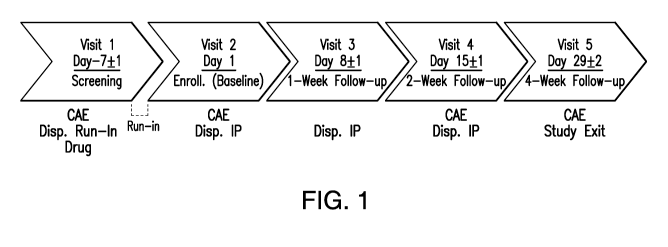
Figure 1 provides a flow chart depicting a study design to evaluate the
efficacy
and safety of a 0.05% T134 ophthalmic composition and a 0.1% T134 ophthalmic
composition compared to a placebo composition.
Figures 2A and 2B provide (2A) a plot of total cornea fluorescein staining
score
changes in the 25%, 50%, 75%, and 100% Tear Film Break Up Time (TFBUT)
quartile
groups and (2B) a plot of total cornea fluorescein staining score changes of
the 25%
TFBUT quartile group at day 8, day 15, and day 29.
Figures 3A and 3B provides (3A) a plot of inferior region fluorescein staining
score changes in the 25%, 50%, 75%, and 100% TFBUT quartile groups and (3B) a
plot
of inferior fluorescein staining score changes of the 50% TFBUT quartile group
at day 8,
day 15, and day 29.
Figure 4 provides a plot of ocular discomfort during CAE per ocular discomfort
at baseline.
DETAILED DESCRIPTION OF THE DISCLOSURE
Provided herein, inter alia, is a method of treating ophthalmic diseases
(e.g.,
DES) in a subject in need thereof, wherein the method is directed to the use
of an
ophthalmic composition that contains human T134 or fragments thereof These and
other
aspects of the presently disclosed subject matter are discussed more in the
detailed
description and examples.
5
CA 03031069 2019-01-16
WO 2018/017479 PCT/US2017/042382
Definitions
Unless defined otherwise, all technical and scientific terms used herein have
the
meaning commonly understood by a person skilled in the art to which this
disclosure
belongs. The following references provide one of skill with a general
definition of many
of the terms used in this disclosure: The Cambridge Dictionary of Science and
Technology (Walker ed., 1988); The Glossary of Genetics, 5th Ed., R. Rieger et
al.
(eds.), Springer Verlag (1991); and Hale & Marham, The Harper Collins
Dictionary of
Biology (1991). Certain terms are defined below to provide additional guidance
in
describing the compositions and methods of the disclosed subject matter and
how to use
them.
As used herein, the following terms have the meanings ascribed to them below,
unless specified otherwise. Abbreviations used herein have their conventional
meaning
within the chemical and biological arts.
Unless specifically stated or obvious from context, as used herein, the term
"or"
is understood to be inclusive. Unless specifically stated or obvious from
context, as used
herein, the terms "a", "an", and "the" are understood to be singular or
plural.
Unless specifically stated or obvious from context, as used herein, the term
"about" is understood as within a range of normal tolerance in the art, for
example within
2 standard deviations of the mean. About can be understood as within 10%, 9%,
8%,
7%, 6%, 5%, 4%, 3%, 2%, 1%, 0.5%, 0.1%, 0.05%, or 0.01% of the stated value.
Unless
otherwise clear from context, all numerical values provided herein are
modified by the
term about. About with respect to concentration range of the compositions of
the current
disclosure also refers to any variation of a stated amount or range which
would be an
effective amount or range.
As used herein, "additive" can include any additional components that can be
added to the composition as described herein. One or more additives can be
added to the
composition. Exemplary additives can include preservatives, viscosity agents,
buffering
agents, hypertonic agents, isotonic agents, and pH adjustment agents.
Additives in the
current disclosure can be used in any suitable amount.
As used herein, the term "administering" can mean any suitable route, i.e.,
via
oral administration, via topical administration (e.g., eye drops or a spray),
or intraocular
administration.
6
CA 03031069 2019-01-16
WO 2018/017479 PCT/US2017/042382
As used herein, the term "co-administer" is meant that a composition described
herein is administered at the same time, just prior to, or just after the
administration of
additional therapies. The composition of the disclosure can be administered
alone or can
be co-administered with a second composition/therapeutic agent to a subject.
Co-
.. administration is meant to include simultaneous or sequential
administration of the
composition individually or in combination with a second
composition/therapeutic agent.
Additionally, the first and second agents can be formulated separately or
together in one
or more compositions.
As used herein, "comprises," "comprising," "containing" and "having" and the
like can have the meaning ascribed to them in U.S. Patent law and can mean
"includes,"
"including," and the like; "consisting essentially of' or "consists
essentially" likewise
has the meaning ascribed in U.S. Patent law and the term is open-ended,
allowing for the
presence of more than that which is recited so long as basic or novel
characteristics of
that which is recited is not changed by the presence of more than that which
is recited,
but excludes prior art embodiments.
As used herein, "concurrent administration" includes overlapping in duration
at
least in part. For example, when two agents (e.g., any of the compositions
described
herein) are administered concurrently, their administration occurs within a
certain
desired time. The compositions' administration can begin and end on the same
day. The
administration of one composition can also precede the administration of a
second
composition by day(s) as long as both compositions are taken on the same day
at least
once. Similarly, the administration of one composition can extend beyond the
administration of a second composition as long as both compositions are taken
on the
same day at least once. The compositions do not have to be taken at the same
time each
day to include concurrent administration.
As used herein, "conservative variant" or grammatical variations thereof can
denote the replacement of an amino acid residue by another biologically
similar residue.
Examples of conservative variations include the replacement of a hydrophobic
residue,
such as isoleucine, valine, leucine or methionine for another, the replacement
of a polar
residue for another, such as the substitution of arginine for lysine, glutamic
for aspartic
acids, or glutamine for asparagine, and the like.
As used herein, the term, "CAE" refers to a clinical model that provides a
standardized approach to studying investigational treatments of dry eye. The
model
7
CA 03031069 2019-01-16
WO 2018/017479 PCT/US2017/042382
exacerbates the signs and symptoms of dry eye (e.g., corneal staining and
ocular
discomfort) in a controlled manner by regulating humidity, temperature,
airflow, lighting
conditions and visual tasking within the CAE chamber. More details are
available at
http://www.oraclinical.com/ophthalmic-models/cae.
As used herein, the term "cream" can refer to a thick (high viscosity) liquid
or
semi-liquid that can be used for therapeutic treatment of a disease, syndrome,
or
condition (i.e., DES).
The term "dosage" is intended to encompass a formulation expressed in terms of
total amounts for a given timeframe, for example as 1.tg/kg/hr, 1.tg/kg/day,
mg/kg/day, or
mg/kg/hr. The dosage is the amount of an ingredient administered in accordance
with a
particular dosage regimen. A "dose" is an amount of an agent administered to a
mammal
in a unit volume or mass, e.g., an absolute unit dose expressed in mg of the
agent The
dose depends on the concentration of the agent in the formulation, e.g., in
moles per liter
(M), mass per volume (m/v), or mass per mass (m/m). The two terms are closely
related,
as a particular dosage results from the regimen of administration of a dose or
doses of the
formulation. The particular meaning in any case will be apparent from context.
As used herein, "dry eye" or "dry eye syndrome" or "DES" can refer to an
ophthalmic syndrome or ocular surface condition. The Dry Eye WorkShop (DEWS)
has
redefined dry eye as "a multifactorial disease of the tears and ocular surface
that results
in symptoms of discomfort, visual disturbance, and tear film instability with
potential
damage to the ocular surface, accompanied by increased osmolarity of the tear
film and
inflammation of the ocular surface." Dry eye and tear film instability can
damage the
ocular surface. It is accompanied by increased osmolarity of the tear film and
inflammation of the ocular surface. The tear film instability can be initiated
by several
etiologies such as xerophthalmia, ocular allergy, topical preservative use and
contact lens
wear. The tear film instability can cause surface hyperosmolarity.
As used herein, an "effective amount" or "therapeutically effective amount" is
that amount sufficient to affect a desired biological effect, such as
beneficial results,
including clinical results. As such, an "effective amount" depends upon the
context in
which it is being applied. An effective amount can vary according to factors
known in
the art, such as the disease state, age, sex, and weight of the individual
being treated.
Several divided doses can be administered daily or the dose can be
proportionally
reduced as indicated by the exigencies of the therapeutic situation. In
addition, the
8
CA 03031069 2019-01-16
WO 2018/017479 PCT/US2017/042382
compositions/formulations of this disclosure can be administered as frequently
as
necessary to achieve a therapeutic amount.
As used herein, the term "fluorescein staining" can refer to a method of
instilling
a fluorescein dye into an eye. The fluorescein dye can be instilled either as
a liquid drop
or via a fluorescein impregnated paper strip. The fluorescein can penetrate in
adjoining
bowman's and stromal layers, and the dye makes contact with an alkaline
interstitial
fluid. Fluid turns bright green owing to its pH indicator properties &
depending to extent
of lesion. The fluorescein cannot stain intact corneal epithelium but can
stain corneal
stroma, thus demarcating the area of the epithelial loss. The corneal
fluorescein staining
can stain all corneal damage non-specifically, irrespective of cause (e.g.,
refractive laser
surgery and drug toxicity). For example and not limitation, 2% preservative-
free sodium
fluorescein solution can be instilled into the inferior conjunctival cul-de-
sac of each eye.
In order to achieve maximum fluorescence, the examiner should wait several
minutes
after instillation before evaluating fluorescein staining. A yellow filter can
be used to
enhance the ability to grade fluorescein staining. The staining will be graded
with the
Fluorescein Staining Scale. Digital images of fluorescein staining can be
taken for digital
analysis. In some embodiments, lissamine green solution can be instilled into
the inferior
conjunctival cul-de-sac for staining.
As used herein, the term "fragment" or "peptide" or "peptide fragment"
comprises a portion of a protein (e.g., T134 protein) with homology or percent
amino acid
sequence identity. Peptides can be biologically occurring short chains of
amino acid
monomers linked by peptide (amide) bonds.
As used herein, "gel" can refer to a material which is not a readily flowable
liquid
and is not a solid, i.e., a semi-solid gel. Gels can be formed from naturally
occurring or
synthetic materials. The gels can be non-ordered to slightly ordered showing
some
birefringence, liquid crystal character. A semi-solid gel formulation apparent
viscosity
can increase with concentration. Gels can be administered topically.
As used herein, "homology" or "percent (%) amino acid sequence identity" is
used with respect to a protein (i.e T134 or fragment thereof). The homology or
percent
amino acid sequence identity can be defined as the percentage of amino acid
residues in
a candidate sequence that are identical with the amino acid residues in the
specific
peptide or polypeptide sequence, after aligning the sequences and introducing
gaps, if
necessary, to achieve the maximum percent sequence identity, and not
considering any
9
CA 03031069 2019-01-16
WO 2018/017479
PCT/US2017/042382
conservative substitutions as part of the sequence identity (i.e., about 60 A
identity,
preferably 61%, 62%, 63%, 64%, 65%, 66%, 6'7%, 68%, 69%, 70%, '71%, 72%, 730
,
7400, 7500, 7600, 770, 78%, 7900, 80%, 81%, 82%, 83%, 84%, 85%, 86%, 87%, 88%,
890 0, 900 0, 910 0, 920 0, 9300, 9400, 9500, 960 0, 9700, 980 0, 9900 or
higher identity over a
specified region when compared and aligned for maximum correspondence over a
comparison window or designated region) as measured using a BLAST or BLAST 2.0
sequence comparison algorithms with default parameters described below, or by
manual
alignment and visual inspection (see, e.g., NCBI web site or the like). Such
sequences
are then said to be "substantially identical". This definition also refers to,
or can be
applied to, the compliment of a test sequence. The definition also includes
sequences
that have deletions and/or additions, as well as those that have
substitutions. Alignment
for purposes of determining percent amino acid sequence identity can be
achieved in
various ways that are within the skill in the art, for instance, using
publicly available
computer software such as BLAST, BLAST-2 or ALIGN software. Those skilled in
the
art can determine appropriate parameters for measuring alignment, including
any
algorithms needed to achieve maximal alignment over the full length of the
sequences
being compared.
As used herein, "intermittent administration" includes the administration of a
composition for a period of time (which can be considered a "first period of
administration"), followed by a time during which the composition is not taken
or is
taken at a lower maintenance dose (which can be considered an "off-period")
followed
by a period during which the composition is administered again (which can be
considered a "second period of administration"). Generally, during the second
phase of
administration, the dosage level of the composition will match that
administered during
the first period of administration but can be increased or decreased as
medically
necessary.
As used herein, "liquid" is a dosage form consisting of a composition in its
liquid state. A liquid is pourable; it flows and conforms to its container at
room
temperature. Liquids display Newtonian or pseudoplastic flow behavior. In
certain
.. embodiments, a "semi-liquid" as used herein can have properties of both a
liquid and
another formulation (i.e., a suspension, an emulsion, a solution, a cream, a
gel, a jelly,
and the like).
CA 03031069 2019-01-16
WO 2018/017479 PCT/US2017/042382
As used herein, "ocular surface" includes the cornea and the conjunctiva. The
ocular surface is covered by a thin layer of fluid or tear film. The tear film
is not only
responsible for the majority of the refractive power of the eye and clear
vision; it is also
responsible for nourishing the cells on the surface of the eye and preventing
infection.
The surface of the eye can suffer many kinds of diseases. One of most common
diseases
of the surface of the eye is DES.
As used herein, "ocular surface disorder" "ophthalmic disease," "ophthalmic
disorder," and the like, includes, but is not limited to, dry eyes, epithelial
defects,
Superior limbic keratoconjunctivitis, keratoconjunctivitis sicca, Neurotrophic
keratopathy, Sjogren's syndrome, Stevens-Johnson syndrome, Ocular cicatricial
pemphigoid, Medicamentosa, Graft-versus-host disease, and corneal ulcerations
and
erosions.
As used herein, "ointment" can refer to a highly viscous liquid or semi-liquid
formulation that can be used for therapeutic treatment of a disease, syndrome,
or
condition (i.e., DES).
As used herein, "ophthalmic composition" refers to a composition intended for
application to the eye or its related or surrounding tissues such as, for
example, the eyelid
or onto the surface of eye. The term also includes compositions intended to
therapeutically treat conditions of the eye itself or the tissues surrounding
the eye. The
ophthalmic composition can be applied topically or by other techniques, known
to
persons skilled in the art, such as injection to the eye. Examples of suitable
topical
administration to the eye include administration in eye drops and by spray
formulations.
A further suitable topical administration route is by subconjunctival
injection.
As used herein, "Fluorescein Staining Scale" refers to a scale specific to dry
eye
to for grading. Corneal staining can be assessed, for example, in the
inferior, central, and
superior regions of the cornea. Conjunctiva staining is assessed, for example,
in the
temporal and nasal regions of the conjunctiva. Grading by the clinician
normally
involves a qualitative estimation of punctate dots in various corneal regions.
The cornea
and conjunctiva are typically divided into several regions (e.g., inferior,
superior, central,
temporal, nasal) with each region graded separately. The Fluorescein Staining
Scale
ranges from 0 to 4 (half grade increments can be used), where grade 0 = none
and 4 =
severe.
11
CA 03031069 2019-01-16
WO 2018/017479 PCT/US2017/042382
As used herein, "Ocular Discomfort Scale" refers to a scale specific to
measuring
ocular discomfort levels of dry eye for grading. Ocular discomfort scores can
be
subjectively graded by the subjects according to the following scale, rating
each eye
separately. It consists of a 5-point scale ranging from 0 to 4, where grade 0
= no
discomfort and 4 = severe discomfort. Relatively higher symptomatic subjects
can
include subjects with an ocular discomfort score of 2 or 3, whereas relatively
lower
symptomatic subjects can have an ocular discomfort score of 0 or 1.
As used herein, "patient," "patient in need thereof," "subject," and "subject
in
need thereof' are used interchangeably and refer to an animal or living
organism (human
or nonhuman) suffering from or prone to a disease or condition that can be
treated by
administration using the methods and compositions provided herein. Non-
limiting
examples of subjects include humans, other mammals, bovines, rats, mice, dogs,
monkeys, goat, sheep, cows, deer, and other non-mammalian animals. In certain
embodiments, the subject is human.
As used herein, "pharmaceutically acceptable carrier" includes any and all
solvents, dispersion media, coatings, antibacterial and antifungal agents,
isotonic and
absorption delaying agents, and the like that are physiologically compatible.
The type of
carrier can be selected based upon the intended route of administration.
Pharmaceutically
acceptable carriers include sterile aqueous solutions or dispersions and
sterile powders
for the extemporaneous preparation of sterile topical solutions or dispersion.
The use of
such media and agents for pharmaceutically active substances is well known in
the art.
Except insofar as any conventional media or agent is incompatible with the
composition
(e.g., T134 or fragments thereof), use thereof in the ophthalmic compositions
for the
disclosure is contemplated.
The term, "preservative" as used herein can include any agents included in an
ophthalmic composition for the purpose of inhibiting the growth of
microorganisms
(e.g., bacteria, fungi, viruses, and protozoa) in the product, thereby helping
to maintain
sterility during use. Additionally, the term "anti-microbial agent" can be
used herein to
denote a specific active agent which provides the anti-microbial efficacy.
Exemplary
preservatives can include, for example, benzalkonium chloride, thimerosal,
chlorobutanol, chlorhexidine, methyl paraben, propyl paraben, phenylethyl
alcohol,
edetate disodium sorbic acid, Onamer M Polyquat, cetyl bromide, cetyl
pyridinium
chloride, benzyl bromide, EDTA, phenylmercury nitrate, phenylmercury acetate,
12
CA 03031069 2019-01-16
WO 2018/017479 PCT/US2017/042382
thimerosal, merthiolate, acetate and phenylmercury borate, polymyxin B
sulphate,
methyl and propyl parabens, quaternary ammonium chloride, sodium benzoate,
sodium
proprionate, and sodium perborate, and other agents known to those skilled in
the art, or
a combination thereof.
As used herein, the terms "prevent," "preventing," or "prevention,"
"prophylactic
treatment" and the like, refer to reducing the probability of developing a
disorder or
condition in a subject, who does not have, but is at risk of or susceptible to
developing a
disorder or condition. The prevention can be complete (i.e., no detectable
symptoms) or
partial, so that fewer symptoms are observed than would likely occur absent
treatment.
The terms further include a prophylactic benefit. For a disease or condition
to be
prevented, the compositions can be administered to a patient at risk of
developing a
particular disease, or to a patient reporting one or more of the physiological
symptoms of
a disease, even though a diagnosis of this disease cannot have been made.
Ranges can be expressed herein as from "about" one particular value, and/or to
.. "about" another particular value. When such a range is expressed, another
aspect
includes from the one particular value and/or to the other particular value.
Similarly,
when values are expressed as approximations, by use of the antecedent "about,"
it is
understood that the particular value forms another aspect. It is further
understood that the
endpoints of each of the ranges are significant both in relation to the other
endpoint, and
independently of the other endpoint. It is also understood that there are a
number of
values disclosed herein, and that each value is also herein disclosed as
"about" that
particular value in addition to the value itself It is also understood that
throughout the
application, data are provided in a number of different formats and that this
data
represent endpoints and starting points and ranges for any combination of the
data points.
For example, if a particular data point "10" and a particular data point "15"
are disclosed,
it is understood that greater than, greater than or equal to, less than, less
than or equal to,
and equal to 10 and 15 are considered disclosed as well as between 10 and 15.
It is also
understood that each unit between two particular units are also disclosed. For
example, if
10 and 15 are disclosed, then 11, 12, 13, and 14 are also disclosed.
Ranges provided herein are understood to be shorthand for all of the values
within the range. For example, a range of 1 to 50 is understood to include any
number,
combination of numbers, or sub-range from the group consisting 1, 2, 3, 4, 5,
6, 7, 8, 9,
10, 11, 12, 13, 14, 15, 16, 17, 18, 19, 20, 21, 22, 23, 24, 25, 26, 27, 28,
29, 30, 31, 32, 33,
13
CA 03031069 2019-01-16
WO 2018/017479 PCT/US2017/042382
34, 35, 36, 37, 38, 39, 40, 41, 42, 43, 44, 45, 46, 47, 48, 49, or 50 as well
as all
intervening decimal values between the aforementioned integers such as, for
example,
1.1, 1.2, 1.3, 1.4, 1.5, 1.6, 1.7, 1.8, and 1.9. With respect to sub-ranges,
"nested sub-
ranges" that extend from either end point of the range are specifically
contemplated. For
example, a nested sub-range of an exemplary range of 1 to 50 can include 1 to
10, 1 to
20, 1 to 30, and 1 to 40 in one direction, or 50 to 40, 50 to 30, 50 to 20,
and 50 to 10 in
the other direction.
As used herein "Schirmer's test" refers to a test used to determine whether
the
eye produces enough tears to keep it moist. For example, the Schirmer's test
can be
performed according to the following procedure: (a) a sterile Schirmer's test
strip will be
placed in the lower temporal lid margin of each eye such that the strip fits
tightly.
Subjects will be instructed to close their eyes and (b) after 5 minutes have
elapsed, the
Schirmer strip will be removed. The length of the moistened area will be
recorded (mm)
for each eye. This test is used when a person experiences very dry eyes or
excessive
watering of the eyes and poses no risk to the subject. A negative (more than
10 mm of
moisture on the filter paper in 5 minutes) test result is normal.
As used herein, "sequential administration" includes that the administration
of
two agents (e.g., compositions described herein) occurs separately on the same
day or do
not occur on a same day (e.g., occurs on consecutive days).
As used herein, a "solution" is a clear, homogeneous liquid dosage form that
contains one or more chemical substances (i.e., T134 or fragments thereof)
dissolved in a
solvent or mixture of mutually miscible solvents. A solution is a liquid
preparation that
contains one or more dissolved chemical substances in a suitable solvent or
mixture of
mutually miscible solvents. Because molecules of a drug substance in solution
are
uniformly dispersed, the use of solutions as dosage forms generally provides
assurance
of uniform dosage upon administration and good accuracy when the solution is
diluted or
otherwise mixed. For example and not limitation, T134 can be dissolved in a
solution
comprised of sodium chloride, potassium chloride, calcium chloride, magnesium
chloride, sodium acetate, and sodium citrate, with a pH of approximately 7Ø
The term "solvent," as used herein, refers to a liquid solvent either aqueous
or
non-aqueous. The selection of the solvent depends notably on the solubility of
the
composition on said solvent and on the mode of administration. Aqueous solvent
can
consist solely of water, or can consist of water plus one or more miscible
solvents, and
14
CA 03031069 2019-01-16
WO 2018/017479 PCT/US2017/042382
can contain dissolved solutes such as sugars, buffers, salts or other
excipients. The more
commonly used non-aqueous solvents are the short-chain organic alcohols, such
as,
methanol, ethanol, propanol, short-chain ketones, such as acetone, and poly
alcohols,
such as glycerol.
"Suspension," as used herein, is a liquid dosage form that contains solid
particles
dispersed in a liquid vehicle.
As used herein, the term "syndrome" can refer to a group of symptoms that
consistently occur together or a condition characterized by a set of
associated symptoms.
A syndrome (e.g., DES) can be a set of medical signs and symptoms that are
correlated
with a specific disease. A disease on the other hand, can be a health
condition that has a
clearly defined reason behind it. A syndrome (from the Greek word meaning 'run
together') however, can produce a number of symptoms without an identifiable
cause.
They can suggest the possibility of an underlying disease or even the chances
of
developing a disease.
As used herein, the terms "tear breakup time" or "TBUT" or "tear film breakup
time" or "TFBUT" can refer to a clinical test that measures the interval
between the
individual's last complete blink and the breakup of the tear film. The test
can be used to
assess for DES. To measure TBUT, fluorescein is instilled into the patient's
tear film and
the patient is asked not to blink while the tear film is observed under a
broad beam of
cobalt blue illumination. The TBUT is recorded as the number of seconds that
elapse
between the last blink and the appearance of the first dry spot in the tear
film.
As used herein, "tear film" can refer to a three-layered structure, comprising
a
mucoidal basal layer, an aqueous component and a superficial lipid layer. The
components work together to maintain the overall form of tear film. The tear
film is
formed and maintained by blinking. The structure of the tear film can be
affected by
systemic or ocular medication, general health and a number of ocular
conditions, such as
keratoconjunctivitis sicca or DES. The tears are also affected by age, with
changes in
both the volume of tear production and stability of the tear film. Patients
with relatively
lower tear film stability can refer to patients with a tear film break up time
shorter than
the median value of a total population. Patients with relatively higher tear
film stability
can refer to patients with a tear film break up time longer than the median
value of a total
population.
CA 03031069 2019-01-16
WO 2018/017479 PCT/US2017/042382
As used herein, "thymosin beta 4" or "Tf34" refers to a human protein. T134
encodes for an actin sequestering protein which plays a role in regulation of
actin
polymerization. The protein is also involved in cell proliferation, migration,
and
differentiation. The thymosin beta 4 peptide, if used after a heart attack,
has been shown
to potentially reactivate cardiac progenitor cells to repair damaged heart
tissue. The
safety of topical T134 formulations has been demonstrated, both in dermal
preparations
and in a preservative-free formulation used in the eye. Based on its
multifunctional
activities during tissue regeneration, T134 has the potential for clinical
application in a
wide range of pathological conditions including ocular surface diseases. The
NCBI
Reference Sequence of human T134 is available under accession number NP
066932.1.
The terms "treat," "treating" or "treatment," and other grammatical
equivalents as
used herein, include alleviating, abating, ameliorating, or preventing a
disease, condition
or symptoms, preventing additional symptoms, ameliorating or preventing the
underlying
metabolic causes of symptoms, inhibiting the disease or condition, e.g.,
arresting the
development of the disease or condition, relieving the disease or condition,
causing
regression of the disease or condition, relieving a condition caused by the
disease or
condition, or stopping the symptoms of the disease or condition, and are
intended to
include prophylaxis. The terms further include achieving a therapeutic benefit
and/or a
prophylactic benefit. By therapeutic benefit is meant eradication or
amelioration of the
underlying disorder being treated. Also, a therapeutic benefit is achieved
with the
eradication or amelioration of one or more of the physiological symptoms
associated
with the underlying disorder such that an improvement is observed in the
patient,
notwithstanding that the patient can still be afflicted with the underlying
disorder.
As used herein, "viscosity" refers to a fluid's resistance to flow. Exemplary
viscosity agents that can be used include, for example polyvinyl alcohol,
polyvinyl
pyrrolidone, methyl cellulose, hydroxy propyl methylcellulose, hydroxyethyl
cellulose,
carboxymethyl cellulose, hydroxy propyl cellulose, other agents known to those
skilled
in the art, or a combination thereof
As used herein, the term "weight percent" or "% (w/w)" refers to a percentage
of
a component in a solution that is calculated on the basis of weight for the
component and
the solvent. For example, a 1% (w/w) solution of a component would have 1 g of
the
component dissolved in a 100 g of solvent. The term "volume percent" or "%
(v/v)"
refers to a percentage of a component in a solution that is calculated on the
basis of
16
CA 03031069 2019-01-16
WO 2018/017479 PCT/US2017/042382
volume for the component and the solvent. For example, a 1% (v/v) solution of
a
component would have 1 mL of the component dissolved in a 100 mL of solvent.
The
term "weight/volume percent" or "% (w/v)" refers to a percentage of a
component in a
solution that is calculated on the basis of weight for the component and on
the basis of
volume for the solvent. For example, a 1.0% (w/v) solution of a component
would have
1 g of the component dissolved in a 100 mL of solvent.
Compositions
The present disclosure provides for ophthalmic compositions comprising T134 or
fragments thereof, in an effective amount to treat DES and symptoms thereof in
a subject
in need thereof.
In certain embodiments, the ophthalmic composition can include from about
0.05% to about 0.1% by weight of T134 or fragments thereof Human T134 is a
polypeptide composed of 43 amino acids having 4.9 kDa, which can be first
isolated
from thymus and then identified from various tissues. This protein can
upregulate the
migration and proliferation of corneal epithelial cells. In some embodiments,
the
ophthalmic composition can include T134 isoforms. T134 isoforms can have about
70%,
or about 75%, or about 80% or more homology to the known amino acid sequence
of
T134. Such isoforms can include, for example, TOO', T139, T1310, T1311, Tf312,
T1313,
T1314 and T1315. T134 of the presently disclosed subject matter can also be an
N-terminal
variant or C-terminal variant of wild-type T134.
In certain embodiments, the ophthalmic composition can include a peptide agent
comprising amino acid sequence LKKTET [SEQ ID NO:1] or LKKTNT [SEQ ID
NO:2], or a conservative variant thereof. Amino acid sequence, LKKTET [SEQ ID
NO:1] and LKKTNT [SEQ ID NO:2] appear to be involved in mediating actin
sequestration or binding. T134 has anti-inflammatory activity, and can also
modulate actin
polymerization (e.g. P-thymosins appear to depolymerize F-actin by
sequestering free G-
actin). TP4's ability to modulate actin polymerization can be due to its
ability to bind to
or sequester actin via the LKKTET [SEQ ID NO:1] or LKKTNT [SEQ ID NO:2]
sequence. Thus, as with T134, other proteins which are anti-inflammatory
and/or bind or
sequester actin, or modulate actin polymerization, including T134 isoforms
having the
amino acid sequence LKKTET [SEQ ID NO:1], are likely to be effective, alone or
in a
combination with T134, as set forth herein. For example and not limitation,
other agents
or proteins having anti-inflammatory activity and/or actin sequestering or
binding
17
CA 03031069 2019-01-16
WO 2018/017479 PCT/US2017/042382
capability, or that can mobilize actin or modulate actin polymerization, as
demonstrated
in an appropriate sequestering, binding, mobilization or polymerization assay,
or
identified by the presence of an amino acid sequence that mediates actin
binding, such as
LKKTET [SEQ ID NO:1] or LKKTNT [SEQ ID NO:2], for example, can similarly be
employed in the disclosed subject matter. Such proteins can include gelsolin,
vitamin D
binding protein (DBP), profilin, cofilin, depactin, Dnasel, vilin, fragmin,
severin,
capping protein, 13-actinin and acumentin.
In certain embodiments, the ophthalmic composition can include oxidized forms
of T134 including T134 sulfoxide or conservative variant thereof. Oxidized
T134 is a form
of T134 in which a methionine residue, 6 amino acids from the N-terminus
(Met6), is
oxidized such that the residue is converted to methionine sulfoxide. The
oxidized T134
can be obtained by reacting native T134 under oxidizing conditions, for
example, by
treating with hydrogen peroxide.
Although the present invention is described primarily hereinafter with respect
to
T134 and T134 fragments, it is to be understood that the following description
is intended
to be equally applicable to amino acid sequence LKKTET [SEQ ID NO:1] or LKKTNT
[SEQ ID NO:2], peptides and fragments comprising or consisting essentially of
LKKTET [SEQ ID NO:1] or LKKTNT [SEQ ID NO:2], conservative variants thereof
and/or T134 isoforms, analogues or derivatives, including oxidized T134, N-
terminal
variants of T134, and C-terminal variants of T134.
In certain embodiments, the ophthalmic composition can include carriers which
can be suitable for topical or intravitreal administration. The carriers can
include, for
example and not limitation, water; a mixture of water and water-miscible
solvents such
as C1-C7 alkanols, vegetable oils or mineral oils such as from about 0.5 to
about 5 wt. %
of hydroxyethyl cellulose, ethyl oleate, carboxymethyl cellulose, polyvinyl
pyrrolidone,
and other non-toxic water-soluble polymers for ophthalmic use, for example,
cellulose
derivatives such as methyl cellulose, alkali-metal salts of carboxymethyl
cellulose,
hydroxymethyl cellulose, hydroxyethyl cellulose, hydroxypropyl methyl
cellulose and
hydroxypropyl cellulose, acrylates or methacrylates such as salts of
polyacrylate or ethyl
acrylate, polyacrylamides; natural products such as gelatin, alginate, pectin,
tragacanth,
karaya gum, xanthan gum, carrageenan, agar, acacia, starch derivatives such as
starch
acetate and hydroxylpropyl starch; and other synthetic products, for example,
polyvinyl
alcohol, polyvinyl pyrrolidone, polyvinyl methylether, polyethylene oxide,
preferably,
18
CA 03031069 2019-01-16
WO 2018/017479 PCT/US2017/042382
cross-linked polyacrylic acid such as neutral carbopol, or mixtures of the
above
polymers. Preferable carriers can include water, cellulose derivatives, for
example,
methyl cellulose, alkali-metal salts of carboxymethyl cellulose, hydroxymethyl
cellulose,
hydroxyethyl cellulose, hydroxypropyl methyl cellulose and hydroxypropyl
cellulose,
neutral carbopol, or mixtures thereof.
In certain embodiments, the ophthalmic composition can include one or more a
pharmaceutically acceptable excipients including but not limited to
stabilizers, buffers,
preservatives, tonicity agents, and viscosity enhancers.
In certain embodiments, the ophthalmic composition can include stabilizers.
The
stabilizers according to the presently disclosed subject matter can include,
for example
and not limitation, tyloxapol, aliphatic glycerol poly-lower alkylene glycol
esters,
aliphatic poly-lower alkylene glycol esters, polyethylene glycols, glycerol
ethers, acetic
acid, citric acid, ascorbic acid, EDTA/disodium edetate, glutathione,
acetylcysteine or
mixtures of these compounds. Acetic acid used herein is a weak acid
represented by
formula CH3COOH. In the presently disclosed subject matter, this can be used
in the
form of an acetate. The acetate can include at least one molecule of water.
For example
and not limitation, mono-, sesqui-, di-, tri-, tetra-, penta-, hexa-, hepta-,
octa-, nona-,
deca-, undeca-, or dodeca-hydrate forms of acetate can be added into the
composition. In
particular, sodium acetate trihydrate can be included in an amount of from
about 0.01%
(w/v) to about 1.5% (w/v) based on the total volume of the composition.
Further, acetic
acid or its salt can be included in an amount of from about 0.1% (w/v) to
about 0.8%
(w/v), and preferably, from about 0.2% (w/v) to about 0.5% (w/v). Citric acid
used
herein is a compound represented by formula C6H807. In the presently disclosed
subject
matter, citric acid can be used in the form of one or more citrates. The
citrate can be a
derivative of citric acid. Additionally, the citrate can include at least one
molecule of
water. For example and not limitation, mono-, sesqui-, di-, tri-, tetra-,
penta-, hexa-,
hepta-, octa-, nona-, deca-, undeca-, or dodeca-hydrate forms of citrate can
be added into
the composition. In particular, the citrate can be sodium citrate and sodium
citrate
trihydrate. In this case, citric acid or its salt can be included in an amount
of from about
0.01% (w/v) to about 0.5% (w/v). Further, citric acid or its salt can be
included in an
amount of from about 0.05% (w/v) to about 0.25% (w/v), and preferably, from
about
0.1% (w/v) to about 0.3% (w/v). They are typically added in an amount
sufficient to
dissolve active ingredients.
19
CA 03031069 2019-01-16
WO 2018/017479 PCT/US2017/042382
In certain embodiments, the ophthalmic composition can include a buffer. For
example, the buffer can include any forms of acetate, ascorbate, borate,
hydrocarbonate/carbonate, gluconate, phosphate, propionate, acetic acid,
citric acid
and/or tromethamine (TRIS) buffers. The buffer can be added, for example, in
an
amount to ensure and maintain a physiologically acceptable pH range. Such pH
can be
typically in the range of about 5 to about 9, preferably from about 6 to about
8.2, more
preferably from about 6.8 to about 8.1.
In other embodiments, the pH value of the ophthalmic formulations can range
from about 3.5 to about 9, preferably from about 4.5 to about 8, and most
preferably
from about 5.5 to about 7.8, and can be about pH 7Ø
The composition in accordance with the presently disclosed subject matter can
further include an acid selected from the group consisting of hydrochloric
acid, acetic
acid, phosphoric acid etc. The composition can further include a base selected
from the
group consisting of sodium hydroxide, potassium hydroxide, sodium hydrogen
carbonate
etc., specifically, sodium hydroxide. For example and not limitation,
hydrochloric acid
or sodium hydroxide can be suitably added to adjust a pH of the composition.
As such,
the pH of the composition can be from about 6.5 to about 7.5, or from about
6.8 to about
7.2. Preferably, the composition can have a pH of about 7Ø
In certain embodiments, the ophthalmic composition can include preservatives.
The preservatives can include, for example, quaternary ammonium salts such as
Cetrimide, benzalkonium chloride or benzoxonium chloride; alkyl-mercury salts
of
thiosalicylic acid such as thimerosal, phenylmercuric nitrate, phenylmercuric
acetate or
phenylmercuric borate, parabens such as phenylparaben or propylparaben,
alcohols such
as chlorobutanol, benzyl alcohol or phenyl ethanol, guanidine derivatives such
as
chlorohexidine or polyhexamethylene biguanide or sorbic acid. Preferable
preservatives
can include cetrimide, benzalkonium chloride, benzoxonium chloride and
parabens. The
preservative can be added in a sufficient amount to prevent secondary
contamination
caused by bacteria and fungi during the use.
In certain embodiments, the ophthalmic composition can include a tonicity
agent
to adjust the composition closer to isotonicity (e.g., 0.9% saline). For
instance and not
limitation, any form of sodium chloride, potassium chloride, calcium chloride,
magnesium chloride, dextrose and/or mannitol can be added to the composition
comprising thymosin (34 according to the presently disclosed subject matter.
The tonicity
CA 03031069 2019-01-16
WO 2018/017479 PCT/US2017/042382
agents can include at least one molecule of water. For example and not
limitation,
mono-, sesqui-, di-, tri-, tetra-, penta-, hexa-, hepta-, octa-, nona-, deca-,
undeca-, or
dodeca-hydrate forms of sodium chloride, potassium chloride, calcium chloride
and/or
magnesium chloride can be added into the composition. An amount of the
tonicity agent
depends upon the kind of active agents to be added. In general, particular
compositions
of the present disclosed subject matter can include a tonicity agent therein
to enable the
final composition to have an osmolality acceptable for ophthalmic use, i.e.,
preferably in
a range of from about 150 to about 450 mOsm, and more preferably in a range of
from
about 250 to about 350 mOsm. Preferable tonicity agents can include, for
example,
.. sodium salts and potassium salts, in particular, sodium chloride and
potassium chloride.
Most preferably, the tonicity agent can be sodium chloride. Further, a
concentration of
sodium chloride can range from about 0.1 to about 1.2% (w/v) or from about 0.3
to about
1.0% (w/v). Preferably, it ranges from about 0.5 to about 0.7% (w/v). Further,
a
concentration of potassium chloride can range from about 0.01 to about 0.15%
(w/v) or
from about 0.03 to about 0.12% (w/v). Preferably, it ranges from about 0.05 to
about
0.09% (w/v). Further, a concentration of calcium chloride dihydrate can range
from
about 0.01 to about 0.12% (w/v) or from about 0.03 to about 0.09% (w/v).
Preferably, it
ranges from about 0.03 to about 0.06% (w/v). Further, a concentration of
magnesium
chloride hexahydrate can range from about 0.01 to about 0.12% (w/v), and
preferably,
from about 0.01 to about 0.05% (w/v). Although the tonicity agents are
described
primarily herein with respect to adjusting tonicity of the ophthalmic
composition, the
disclosed tonicity agents can also be used as electrolytes.
In certain embodiments, the ophthalmic composition can include a viscosity
enhancer. Suitable viscosity enhancers in ophthalmic formulations and their
.. concentration ranges used in certain inventive compositions can include but
are not
limited to: (a) Monomeric polyols, such as tyloxapol (from about 0.1 to about
1%),
glycerol (from about 0.2 to about 1%), propylene glycol (from about 0.2 to
about 1%),
ethylene glycol (from about 0.2 to about 1%); (b) Polymeric polyols, such as
polyethylene glycol (e.g., PEG 300, PEG 400) (from about 0.2 to about 1%); (c)
.. Cellulose derivatives (polymers of the cellulose family), such as
hydroxyethylcellulose
(from about 0.2 to about 2.5%), hypromellose (from about 0.2 to about 2.5%),
hydroxypropylmethyl cellulose (from about 0.2 to about 2.5%), methylcellulose
(from
about 0.2 to about 2.5%), carboxymethylcellulose sodium (from about 0.2 to
about
21
CA 03031069 2019-01-16
WO 2018/017479 PCT/US2017/042382
2.5%), hydroxylpropylcellulose (from about 0.2 to about 2.5%); (d) Dextrans,
such as
dextran 70 (at about 0.1% when used with another polymeric demulcent agent);
(e)
Water-soluble proteins such as gelatin (at about 0.01%), (f) Vinyl polymers
such as
polyvinyl alcohol (from about 0.1 to about 4%), polyvinyl pyrollidine (from
about 0.1 to
about 4%); (g) Other polyols, such as polysorbate 80 (from about 0.2 to about
1%),
povidone (from about 0.1 to about 2%); (h) Carbomers, such as carbomer 934P,
carbomer 941, carbomer 940, and carbomer 974P, and (i)
Polysaccharides/Glycosaminoglycans, such as hyaluronan (hyaluronic
acid/hyaluronate)
(from about 0.1 to about 3%), chondroitin sulfate (from about 0.1 to about
3%).
In certain embodiments, the amount and type of excipient(s) added can be
varied
depending on specific requirements, the excipient(s) is generally used in a
range of about
0.0001 to about 90 wt. %, and within the range commonly used in ophthalmic
fields.
In certain embodiments, the ophthalmic composition is formulated as a
solution,
suspension, semi-liquid, semi-solid gel, gel, ointment or cream. In specific
embodiments, the ophthalmic composition can be formulated as a preservative-
free,
sterile eye-drop solution in a single unit dropper. According to one
embodiment the
topical formulation containing the active compound can also contain a
physiologically
compatible vehicle, as those skilled in the ophthalmic art can select using
conventional
criteria.
In certain embodiments, the ophthalmic composition is administered in the form
of eye drops. The ophthalmic composition can be, where appropriate, adjusted
and/or
buffered to the desired pH and, where appropriate, a stabilizer, or a tonicity
enhancing
agent can be added. Where appropriate, preservatives and/or other excipients
can be
added to an ophthalmic composition.
In certain embodiments, the ophthalmic composition can be formulated into a
unit dosage form to provide a total daily dosage of from about 0.08 to about
2.0 ml and
can be suitably filled in a container for ophthalmic use, which can enable
quantitative
administration of the composition. For this purpose, the composition can be
formulated
into a unit dosage form with a dose of from about 0.01 to about 10 ml that can
be used
once or several times. Further, in order to suitably provide the
pharmaceutical
composition in a total daily dosage of from about 0.08 to about 2.0 ml, the
composition
can be contained in an eye drop container dropping from about 0.01 to about
2.0 ml per
droplet.
22
CA 03031069 2019-01-16
WO 2018/017479 PCT/US2017/042382
In certain embodiments, the ophthalmic composition can include from about 0.05
to about 0.1% by weight of T134 or fragments thereof. The ophthalmic
composition can
be in a solution comprised of sodium chloride, potassium chloride, calcium
chloride
dihydrate, magnesium chloride hexahydrate, sodium acetate trihydrate, and
sodium
citrate trihydrate. The pH of the composition can be adjusted to about 6.5 to
about 7.5
using an acid or a base. The acid can be selected from the group consisting of
hydrochloric acid, acetic acid, phosphoric acid, etc. The base can be selected
from the
group consisting of sodium hydroxide, potassium hydroxide, sodium hydrogen
carbonate, etc.
Methods
The present disclosure provides, inter al/a, a method of treating DES or signs
or
symptoms thereof in a subject in need of such treatment. The method includes
administering to an eye of the subject an ophthalmic composition including
T134 or
fragments thereof, in an effective amount to treat DES and signs and symptoms
thereof.
The presently disclosed subject matter provides methods that effectively
address
at least two aspects of DES, including but not limited to inflammatory
responses and
corneal epithelial healing. In certain embodiments, delivering the ophthalmic
composition including T134 or fragments thereof to the dry eye can reduce or
prevent
inflammation and improve or accelerate corneal epithelial healing. DES can be
include
various signs and symptoms including, but not limited to, deficient tear
production,
decreased tear film stability, increased ocular surface damage, and increased
ocular
discomfort. Subjects with DES can exhibit one or more signs or symptoms. The
presently disclosed subject matter effectively treats both inflammatory
responses and
increases corneal epithelial healing by administering effective amounts of
ophthalmic
composition including T134 or fragments thereof. Such methods are successful
in
increasing tear amounts, increasing tear film stability, decreasing ocular
surface damage
and decreasing ocular discomfort.
In certain embodiments, the method of treating DES includes treating dry eye
associated with or resulting from treating inflammation of the surface of the
eye, the
lacrimal gland, or the conjunctiva; dry eye associated with any disease
process that alters
the components of the tears; dry eye associated with an increase in the
surface of the eye,
as in thyroid disease when the eye protrudes forward; and/or dry eye
associated with a
cosmetic surgery, for example, if the eyelids are opened too widely during
surgery; dry
23
CA 03031069 2019-01-16
WO 2018/017479 PCT/US2017/042382
eye associated with eye correction surgery such as laser-assisted in situ
keratomileusis
(LASIK) or laser-assisted sub epithelial keratectomy (LASEK). Hyperosmolarity
can
cause damage to the surface epithelium by activating a cascade of inflammatory
events
and releasing inflammatory mediators into the tears. The inflammatory
mediators can
cause cell death, loss of goblet cells, reduction in mucus secretion, and tear
film
instability. In certain embodiments, delivering an ophthalmic composition
including T134
or fragments thereof to the tear-deficient dry eye can improve tear film
stability and/or
increase the tear amount. The methods herein can improve the quality of the
lacrimal
gland which secretes the aqueous layer of the tear film.
The presently disclosed subject matter provides for methods of treating tear-
deficient dry eye, which is a disorder in which the lacrimal glands fail to
produce enough
of the watery component of tears to maintain a healthy eye surface, with a
composition
of the present disclosure. The aqueous tear-deficient dry eye can be
characterized by
various assessments, including but not limited to, a tear volume test. The
tear volume
test can be used to determine whether the eye produces enough tears to keep it
moist.
For example and not limitation, the tear volume test can be performed
according to the
following procedure: (a) a sterile test strip can be placed in the lower
temporal lid margin
of each eye such that the strip fits tightly. Subjects can be instructed to
close their eyes
and (b) after an appropriate time (e.g., about 5 minutes) has elapsed, the
strip can be
removed. The length of the moistened area can be recorded (mm) for each eye.
This test
can be used when a person experiences very dry eyes or excessive watering of
the eyes
and poses no risk to the subject. A negative test result is normal, whereby,
for example,
more than about 10 mm of moisture on the filter paper is recorded.
Alternatively, the aqueous tear-deficient dry eye can be characterized by a
phenol
red thread test. For example and not limitation, the crimped end of a cotton
thread
impregnated with phenol red dye can be placed in the inferior conjunctival sac
on the
temporal side. Phenol red is a pH indicator which exhibits a gradual
transition from
yellow to red when wetted by tears, due to the alkaline nature of tears (pH
7.4). Subjects
can be instructed to close their eyes and the thread can be removed after 15
seconds. The
length of the color change on the thread, indicating the length of the thread
wetted by the
tears, can be measured in millimeters. Wetting lengths should normally be
between about
9 mm and about 20 mm. Values of less than about 9 mm have been shown to
correlate
with subjective symptoms of dryness.
24
CA 03031069 2019-01-16
WO 2018/017479 PCT/US2017/042382
The presently disclosed subject matter also provides for methods of improving
tear film stability. Tear film stability can be evaluated by various
assessments in the art,
including but not limited to a TFBUT analysis. For example and not limitation,
2%
preservative-free sodium fluorescein solution can be administered into the
inferior
conjunctival cul-de-sac of each affected eye. In order to achieve maximum
fluorescence,
an appropriate waiting period is implemented after instillation before
evaluating TFBUT.
With the aid of a slit-lamp, the integrity of the tear film can be monitored
by noting the
time it takes to form micelles from the time that the eye is opened. TFBUT can
be
measured in seconds using a stopwatch and a digital image recording system for
one eye
followed by the second eye. A negative test result is normal, whereby more
than about
10 seconds of TFBUT is recorded.
Alternatively, tear film stability can be evaluated by a Non-Invasive Break-Up
Time (NIBUT) analysis. During a NIBUT analysis, an illuminated grid pattern
reflected
from the anterior tear surface can be observed without administration of
fluorescein
solution. During a NIBUT analysis, subjects can be asked to stop blinking
until told to
restart. The time between the last complete blink and the first indication of
pattern
break-up can be recorded with a stop- watch.
Further, the presently disclosed subject matter provides for methods of
improving
damaged ocular surface area. Damaged ocular surface area of the dry eye can be
characterized by various assessments in the art, including but not limited to
a fluorescein
staining analysis. During a fluorescein staining analysis, the damaged ocular
surface can
be stained with fluorescein compounds. For example and not limitation, 2%
preservative-free sodium fluorescein solution can be instilled into the
inferior
conjunctival cul-de-sac of each eye. In order to achieve maximum fluorescence,
a
waiting time is implemented after instillation before evaluating fluorescein
staining.
Grading can involve a qualitative estimation of punctate dots in various
corneal regions.
The cornea and conjunctiva are typically divided into several regions (e.g.,
inferior,
superior, central, temporal, nasal) with each region graded separately. The
scale ranges
from 0 to 4 (half grade increments can be used), where grade 0 = none and 4 =
severe).
In certain embodiments, the fluorescein compounds can include rose bengal for
a
fluorescein staining analysis.
Alternatively, damaged ocular surface area of the dry eye can be characterized
by
a lissamine Green Staining analysis. Lissamine Green can stain ocular surface
epithelial
CA 03031069 2019-01-16
WO 2018/017479 PCT/US2017/042382
cells that are unprotected by mucin or glycocalyx. During a lissamine Green
Staining
analysis, lissamine green solution can be instilled into the inferior
conjunctival cul-de-
sac of each eye. The subject can be instructed to blink several times to
distribute the
lissamine green. The staining will be graded with the same staining scale as a
fluorescein
staining analysis. Alternative staining techniques in the art can also be
used, including
for example, Rose Bangal.
The presently disclosed subject matter also provides for methods of reducing
ocular discomfort in a subject in need thereof with the administration of an
ophthalmic
composition including T134 or fragments thereof. In certain embodiments, the
method
includes ameliorating symptoms including but not limited to stinging or
burning of the
eye; a sandy or gritty feeling as if something is in the eye; episodes of
excess tears
following very dry eye periods; a stringy discharge from the eye; pain and
redness of the
eye; episodes of blurred vision; heavy eyelids; inability to cry when
emotionally
stressed; uncomfortable contact lenses; decreased tolerance of reading,
working on the
computer, or any activity that requires sustained visual attention; and/or eye
fatigue.
Indications of ocular discomfort can be characterized and quantified by
various
assessments known in the art. For example and not limitation, ocular
discomfort scores
can be subjectively graded by the subjects before, after, or during exposure
to an adverse
environment. During exposure to the adverse environment, the signs and
symptoms of
dry eye (e.g., corneal staining and ocular discomfort) are exacerbated in a
controlled
manner by regulating humidity, temperature, airflow, lighting conditions and
visual
tasking. The discomfort scale can consist of a 5-point scale ranging from 0 to
4, where
grade 0 = no discomfort and 4 = severe discomfort. Relatively higher
symptomatic
subjects can include subjects with an ocular discomfort score of 2 or 3,
whereas
relatively lower symptomatic subjects can have an ocular discomfort score of 0
or 1.
As previously noted, the presently disclosed subject matter provides for the
effective treatment of DES and associated signs and symptoms characterized by
various
assessments including but not limited to a Schirmer's test, a TFBUT test, a
fluorescein
staining test, decreased tear film stability, increased ocular surface damage,
increased
ocular discomfort, an ocular discomfort analysis and combinations thereof. In
certain
embodiments, the target subject can have DES characterized by a tear volume
test score
of less than about 10 mm in an affected eye. In certain embodiments, the
target subject
can have DES characterized by a tear film break up time of less than about 10
seconds in
26
CA 03031069 2019-01-16
WO 2018/017479
PCT/US2017/042382
an affected eye. In certain embodiments, the target subject can have has DES
characterized by a total corneal fluorescein staining score of about 4 or
higher in an
affected eye. In certain embodiments, the target subject can have DES
characterized by
an ocular discomfort score of about 2 or higher in the affected eye.
In certain embodiments, the present disclosure provides a method for
increasing
tear amount and tear film stability. In certain embodiments, the present
disclosure
provides a method for reducing ocular surface damage. In certain embodiments,
the
present disclosure provides a method for reducing ocular discomfort. All
methods
provided herein include the administration of an ophthalmic composition
including T134
or fragments thereof to one or both eyes of a subject in need thereof
Co-administration
In certain embodiments, the methods of treating DES can be managed as an
ongoing condition. In certain embodiments, if there is an underlying disease,
that
disease is concurrently treated.
In certain embodiments, the composition can be administered at the same time,
just prior to, or just after the administration of additional therapies. The
composition of
the disclosure can be administered alone or can be co-administered with a
second
composition/therapeutic agent to a subject.
Co-administration can be meant to include simultaneous or sequential
administration of the composition individually or in combination with a second
composition/therapeutic agent.
In certain embodiments, the method includes treating DES with a composition of
the present disclosure in combination with artificial tears. Artificial tears
can include any
ocular ointments, drops, or sprays and the like known in the art. Exemplary
artificial
tears can include, for example, Celluvisc, Clear Eyes CLR, GenTeal, Hypotears,
Isopto
Tears, Lacri-Lube SOP., Liquitears, Moisture Drops, Oasis Tears, Opti-Free
Rewetting
Drops, optive, Refresh, Soothe, Systane, TheraTears, Ultra Fresh, Visine
Tears, and the
like.
Dosage Regimens
For example and not limitation, the methods can include contacting an eye or
eye
tissue with an effective amount of a composition including T134 or fragments
thereof as
an active ingredient. The administration can be topical or intravitreal
administration. An
example of topical administration can include direct application of the
composition in the
27
CA 03031069 2019-01-16
WO 2018/017479 PCT/US2017/042382
form of, for example, a solution, lotion, plaster, gel, cream, paste, spray,
suspension,
dispersion, hydrogel, ointment, oil or foaming agent to a subject in order to
contact same
with eye tissues
In certain embodiments, a method of treating DES in a subject in need thereof,
includes administering to an eye of the subject, an ophthalmic composition
including
human T134 or fragments thereof formulated in the form of a solution, a
suspension, a
semi-solid gel, a gel, an emulsion, semi-liquid, an ointment, a cream, foam
gel, or a
controlled-release/sustain-release vehicle. For example, the composition can
be in the
form of a contact lens solution, eyewash, eye drop, eye gel, eye ointment, and
the like.
The following dosage regimens can be used to treat DES in general and can be
used to treat both inflammatory responses and increases corneal epithelial
healing by
administering effective amounts of ophthalmic composition including T134 or
fragments
thereof. The dosage regimens provided herein can be used to increase tear
amounts,
increase tear film stability, decrease ocular surface damage and/or decrease
ocular
discomfort.
In a particular embodiment, the composition is in the form of a solution that
can
be administered as eye drops. The composition can be administered topically to
an eye
for treating DES in a dosage range from about 5 [tg to about 150 [tg per eye,
or from
about 5 [tg to about 100 [tg per eye, or from about 5 [tg to about 50 [tg per
eye, or from
about 5 [tg to about 25 [tg per eye. In other embodiments, the composition can
be
administered topically to an eye for treating DES in a dose range from about 5
[tg to
about 150 [tg per eye, or from about 25 [tg to about 150 [tg per eye, or from
about 50 [tg
to about 150 [tg per eye, or from about 100 [tg to about 150 [tg per eye.
In certain embodiments, the dosage for one eye can be about 1 to about 5 drops
of
solution. In certain embodiments, the dosage for one eye can be 1, 2, or 3
drops of
solution. Each drop of an ophthalmic composition in solution from can
correspond to
about 10 .L to about 150 L of ophthalmic composition. Preferably, each drop of
an
ophthalmic composition in solution can correspond to about 20 .L to about 70
1..t.L of
ophthalmic composition.
In certain embodiments, the ophthalmic composition can be administered to an
eye for treating DES by placing one to two drops or more in each eye, 1 to 24
times
daily. For example, the ophthalmic composition can be applied, 1, 2, 3, 4, 8,
12, 18 or 24
times a day, or more. In certain embodiments, the ophthalmic composition can
be
28
CA 03031069 2019-01-16
WO 2018/017479 PCT/US2017/042382
applied by placing one or two drops in each eye, once daily or twice daily, or
three times
daily, or four times daily. For example and not limitation, the composition
can be
applied by placing one drop in each eye four times daily, including, for
example, in the
morning, noon, afternoon, and evening.
In certain embodiments, the method of treating DES includes administering a
composition human T134 or fragments thereof to a subject in any suitable or
therapeutically effective amount, e.g., from about 0.001 percent by weight to
about 90
percent by weight of the composition, from about 0.001 percent by weight to
about 1
percent by weight, from about 0.001 percent by weight to about 10 percent by
weight,
from about 0.001 percent by weight to about 20 percent by weight, from about
0.001
percent by weight to about 30 percent by weight, from about 0.001 percent by
weight to
about 40 percent by weight, from about 0.001 percent by weight to about 50
percent by
weight, from about 0.001 percent by weight to about 60 percent by weight, from
about
0.001 percent by weight to about 70 percent by weight, from about 0.001
percent by
weight to about 80 percent by weight, from about 0.01 percent by weight to
about 90
percent by weight, from about 0.01 percent by weight to about 1 percent by
weight, from
about 0.01 percent by weight to about 10 percent by weight, from about 0.01
percent by
weight to about 20 percent by weight, from about 0.01 percent by weight to
about 30
percent by weight, from about 0.01 percent by weight to about 40 percent by
weight,
from about 0.01 percent by weight to about 50 percent by weight, from about
0.01
percent by weight to about 60 percent by weight, from about 0.01 percent by
weight to
about 70 percent by weight, from about 0.01 percent by weight to about 80
percent by
weight, from about 0.1 percent by weight to about 90 percent by weight, from
about 0.1
percent by weight to about 1 percent by weight, from about 0.1 percent by
weight to
about 10 percent by weight, from about 0.1 percent by weight to about 20
percent by
weight, from about 0.1 percent by weight to about 30 percent by weight, from
about 0.1
percent by weight to about 40 percent by weight, from about 0.1 percent by
weight to
about 50 percent by weight, from about 0.1 percent by weight to about 60
percent by
weight, from about 0.1 percent by weight to about 70 percent by weight, from
about 0.1
percent by weight to about 80 percent by weight, or any range in between, of
the
composition. In certain embodiments, the method of treating DES includes
administering a composition human T134 or fragments thereof to a subject at
about 0.05%
29
CA 03031069 2019-01-16
WO 2018/017479 PCT/US2017/042382
by weight. In certain embodiments, the method of treating DES includes
administering a
composition human T134 or fragments thereof to a subject at about 0.1% by
weight.
EXAMPLES
The following examples are merely illustrative of the presently disclosed
subject
matter and they should not be considered as limiting the scope of the
disclosed subject
matter in any way.
Example 1: Safety and efficacy of 0.05% and 0.1% TI34 ophthalmic
composition
Study objectives
The objective of this study was to compare the safety and efficacy of 0.05%
T134
ophthalmic composition and 0.1% T134 ophthalmic composition to placebo for the
treatment of the signs and symptoms of dry eye.
Materials and methods
This study was a multicenter, randomized, double-masked study designed to
evaluate the efficacy and safety of 0.05% and 0.1% T134 ophthalmic solution
compared
to placebo in subjects with dry eye. 317 male and female subjects who were at
least 18
years of age, had a subject-reported history of dry eye in both eyes and met
all other
study eligibility criteria were randomized to receive either 0.05% T134, 0.1%
T134 or
placebo at a ratio of 1:1:1 (105:107:105 subjects in each treatment group,
respectively).
The study consisted of two periods: a 7-day run-in period and a 28-day
treatment
period. A flow chart of the study is presented in Figure 1.
The CAE is a clinical model that provides a standardized approach to studying
investigational treatments of dry eye. The model exacerbates the signs and
symptoms of
dry eye (e.g. corneal staining and ocular discomfort) in a controlled manner
by regulating
humidity, temperature, airflow, lighting conditions and visual tasking within
the CAE
chamber.
Patients and selection criteria
Eligible patients were 18 years or older, had a reported history of dry eye
for at
least 6 months prior to enrollment, and had a history of eye drop use for dry
eye
symptoms within the previous 6 months. Patients had to have a tear film
breakup time
(TFBUT) 10 seconds, unanesthetized Schirmer tear test (mm/5 minutes) of and
10, a sum corneal fluorescein staining score of A, based on the sum of the
central,
CA 03031069 2019-01-16
WO 2018/017479
PCT/US2017/042382
superior, and inferior regions of the cornea with the fluorescein staining
scale (reported
for each region on a 0-4 scale).
If initial screening requirements were met, patients were required to
demonstrate
an increase in fluorescein staining following exposure in the CAE.
Additionally, patients
had to report a worsening in ocular discomfort score (a five-point [0-4]
scale, where 0 =
none and 4 = severe) during exposure to the CAE. All patients had to have a
corrected
visual acuity logarithm of the minimum angle of resolution (logMAR) +0.7 in
both
eyes. Patients who met the selection criteria at visit 1 were initiated on
self-administered,
placebo solution for 7 days until visit 2 (day 1). After this run-in period,
at visit 2,
eligible patients were required to meet all assessments as described for visit
1 above.
Interventions
The clinical dosage form and packaging of T134 ophthalmic solution and the
placebo ophthalmic solutions were identical sterile, low-density polyethylene
unit-dose
non-preserved bottles. They were packaged in foil-wrap pouches to prevent
light
exposure, each containing single-use bottles. Throughout the study, between
day 1 and
day 29, patients were instructed to instill one drop of study medication in
each eye four
times daily, once in the morning, noon, afternoon and in the evening before
bed. Patients
were assigned randomization kit numbers in strict numerical sequence, using a
code
generated by an independent biostatistician. All investigators, study and site
personnel,
and patients were masked to the treatment assignments.
Outcome measures
Patients were evaluated on day 8 (visit 3), day 15 (visit 4), and day 29
(visit 5)
during the dosing period. Exposure to the CAE occurred on days 14 and 28. At
each
study visit, a panel of dry eye signs and symptoms and safety measures were
evaluated
(including both before [pre-CAE] and after [post-CAE] exposure).
The sign endpoints assessed at each visit, both pre- and post-CAE, included
Fluorescein Staining (in three regions: inferior, superior and central cornea,
with scores
provided in single regions and sum of three regions), TFBUT, and
unanesthetized
Schirmer's test (measured pre-CAE and/or post-CAE).
The symptom endpoints assessed at each visit (both pre- and post-CAE) were
ocular discomfort (Ocular Discomfort Scale, using a five-point [0-4] scale,
where 0 =
none and 4 = severe). Ocular discomfort was also graded during exposure to the
CAE.
Study Results
31
CA 03031069 2019-01-16
WO 2018/017479 PCT/US2017/042382
Fluorescein Staining Score in Total Cornea Region
A 28-day treatment (visit 5) with 0.05% and 0.1% T04 elicited improvements on
total corneal staining in subjects. Subjects were grouped by the severity of
TFBUT at
baseline (pre-CAE) and the change of fluorescein staining score for the total
cornea
from baseline of each sub-group was analyzed. For example, as shown in Figure
2A,
the subjects were grouped into the 100%, 75%, 50%, and 25% quartile groups. As
illustrated in Figure 2A, there was a distinction of the fluorescein staining
score between
the 1'04-treated and placebo-treated groups after the 28-day treatment (visit
5) with
0.05% and 0.1% T04 in all quartile groups. For example, when compared with
baseline
(visit 2) to visit 5, the fluorescein staining score change in total cornea
region was 0.83 in
placebo group, 0.075 in the 0.05%1'04-treated group and 0.10 in 0.1% 1'04-
treated
group in the 25% quartile group. The lower fluorescein staining score change
indicates
less defect in cornea, whereas a high fluorescein staining change indicates
the worsening
of defect.
Figure 2B provides a plot of the change of fluorescein staining score in total
cornea region at different time points. The change of fluorescein staining
score in total
cornea region of the about 25% subpopulation group was measured at day 8
(visit 3), day
15 (visit 4), and day 29 (visit 5). As shown in Figure 2B, 7-day, 14-day, and
28-day
treatments with 0.05% T04 elicited significant improvements on total corneal
staining in
subjects. Moreover, 14-day and 28-day treatments with 0.1% T04 elicited
significant
improvements on total corneal staining in subjects.
These results indicated therapeutic effects of the T04 treatment on reducing
ocular surface damage of DES patients. Particularly, these results indicated
that the T04
treatment can be more effective in patient groups with decreased tear film
stability.
Fluorescein Staining Score in Inferior Region
The 28-day treatment (visit 5) with 0.05% and 0.1% T04 elicited significant
improvements on corneal staining in inferior region. Subjects were grouped by
the
severity of TFBUT at baseline (pre-CAE) as discussed above and the change of
fluorescein staining score for the inferior region of cornea from baseline of
each sub-
group was analyzed. As shown in Figure 3A, there was a distinction of the
fluorescein
staining score in inferior region between the T134-treated and placebo-treated
groups after
the 28-day treatment (visit 5) with 0.05% and 0.1% T04 in 75%, 50%, and 25%
quartile
groups. For example, when compared with baseline (visit 2) to visit 5, the
fluorescein
32
CA 03031069 2019-01-16
WO 2018/017479 PCT/US2017/042382
staining score change in inferior region was 0.39 in placebo group, 0.20 in
the 0.05%
1'04-treated group and -0.04 in 0.1% 1'04-treated group in the 25%
subpopulation group.
Figure 3B provides a plot of the change of fluorescein staining score in
inferior
cornea region at different time points. The change of fluorescein staining
score in
inferior cornea region of the 50% subpopulation group was measured at day 8
(visit 3),
day 15 (visit 4), and day 29 (visit 5). As shown in Figure 3B, 7-day, 14-day,
and 28-day
treatments with 0.05% and 0.1% T04 elicited significant improvements on
corneal
staining in subjects.
The subjects were grouped into the lower tear film stability group and the
higher
tear film stability group. In the subjects group of the lower tear film
stability (the
patients had the tear film break up time shorter than the median value of
total population)
at baseline, there was a distinction between the T04-treated and placebo-
treated groups
after the 28-day treatment (visit 5) with 0.05% and 0.1% T04. When compared
with
baseline (visit 2) to visit 5, the fluorescein staining score change in
inferior region was
0.400 in placebo group, 0.120 in the 0.05%1'04-treated group and 0.009 in 0.1%
T04-
treated group. The lower fluorescein staining score present the patient
indicates less
defect in cornea, whereas a high fluorescein staining change indicates the
worsening of
defect.
In the higher tear film stability group, however (the patients had the tear
film
break up time longer than the median value of total population) at baseline,
the
fluorescein staining score change in inferior region from baseline was 0.094
in placebo
group, 0.444 in the 0.05%1'04-treated group and 0.245 in 0.1%1'04-treated
group.
When comparing the mean difference between T134-treated group and placebo,
the T134-treated groups showed less worsening of defects in the lower tear
film stability
group than the placebo group (the negative score means the less worsening than
placebo
group). But in higher tear film stability group, the mean difference between
T134-treated
group and placebo showed the positive value, i.e., the more worsening of
ocular damage.
These results indicated that significant reducing ocular surface damage effect
of
T04-treated patients with low tear film stability.
33
CA 03031069 2019-01-16
WO 2018/017479 PCT/US2017/042382
Table 1. Fluorescein Staining Score in Inferior Region ¨ Change from visit 2
(baseline)
to visit 5
Subpopulation Lower tear film stability group Higher tear film
stability group
Change from Change from
Subject # Subject #
baseline baseline
Mean
Placebo 55 0.400 48 0.094
0.05% 50 0.120 54 0.444
0.1% 48 0.009 53 0.245
Mean Difference between T134 and Placebo (T134 ¨ Placebo)
0.05% -0.280 0.350
0.1% -0.304 0.141
Tear Film Break Up Time
In the subjects group of the lower tear film stability (the patients had the
tear film
break up time shorter than the median value of total population, e.g., median
value
between 1-9 seconds) at baseline, there was a distinction between the T134-
treated and
placebo-treated groups after the 28-day treatment (visit 5) with 0.05% and
0.1% T134.
When compared with baseline (visit 2) to visit 5, the tear film break up time
change was
0.54 sec in placebo group and 0.74 sec in the T134-treated group. The longer
tear film
break up time present the patient indicates better tear film stability.
In the higher tear film stability group, however (the patients had the tear
film
break up time longer than the median value of total population) at baseline,
the change
from baseline for tear film break up time was 0.05 sec in placebo group and
0.14 sec in
the 1434-treated group.
When comparing the mean difference between all 1434-treated groups and
placebo, the mean difference between T134-treated group and placebo showed
that the
T134 treatment group had the better tear film stability (T134 treatment group
vs. placebo =
0.20 vs. 0.09).
These results indicated that significant increasing tear film stability effect
of T134-
treated patients with a low tear film stability.
34
CA 03031069 2019-01-16
WO 2018/017479 PCT/US2017/042382
Table 2. Tear Film Break Up Time ¨ Change from visit 2 (baseline) to visit 5
Subpopulation Lower tear film stability group Higher tear film
stability group
Change from Change from
Subject # Subject #
baseline (sec) baseline (sec)
Mean
Placebo 55 0.54 48 0.05
T134 98 0.74 106 0.14
Mean Difference between T134 and Placebo (T134 ¨ Placebo)
T134 0.20 0.09
Ocular Discomfort Score Change during exposure to the CAE
The 28-day treatment (visit 5) with 0.05% and 0.1% T134 elicited improvements
on ocular discomfort in subjects with dry eye. Subjects were grouped by the
severity of
ocular discomfort at baseline (beginning of CAE) and the change of ocular
discomfort
during exposure to the CAE chamber of each sub-group was analyzed. For
example, as
shown in Figure 4, the subjects were grouped into the ITT, >0, >1, and >2
subpopulation
groups. The ITT subpopulation group included every subject who was randomized.
The
subjects in the >0 subpopulation group had an ocular discomfort score more
than about 0
at visit 2. The subjects in the >1 subpopulation group had an ocular
discomfort score
more than about 1 at visit 2. The subjects in the >2 subpopulation group had
an ocular
discomfort score more than about 2 at visit 2. As shown in Figure 4, following
the 28-
day treatment with 0.05% and 0.1% T134, there was a distinction between the
active and
placebo treatment group in all subpopulation groups. Particularly, the ocular
discomfort
score change from the beginning to the end of CAE was 1.7 in placebo group of
the >2
subpopulation. However, the ocular discomfort score change was only 1.34 in
the 0.05%
T134-treated group and 1.39 in the 0.1% 1434-treated group. Comparison of the
changes
shows a lower increase for 0.1% and 0.05% 1434-treated subjects than placebo-
treated
subjects.
The subjects were grouped into the higher symptomatic subjects group and the
lower symptomatic subjects group, based on predetermined ocular discomfort
scores. In
the higher symptomatic subjects group (a subject had an ocular discomfort
score of 2 or
3) at baseline, a distinction between the 1434-treated and placebo-treated
treatment
CA 03031069 2019-01-16
WO 2018/017479 PCT/US2017/042382
groups after the 28-day treatment (visit 5) with T134 was observed. When
compared with
baseline (visit 2) to visit 5, the ocular discomfort score change from the
beginning to the
end of CAE was 0.50 in placebo group, 0.23 in the 0.05% T134-treated group and
0.06 in
0.1% 1434-treated group. The lower ocular discomfort score indicates
discomfort, and the
low ocular discomfort change indicates a high dampening effect (a protective
effect) to
the exacerbating condition.
The lower symptomatic subject group (subject had ocular discomfort score 0 and
1) at baseline (visit 2), when compared with baseline (visit 2) to visit 5,
the ocular
discomfort score change from the beginning to the end of CAE was -0.86 in
placebo
group, -0.05 in the 0.05% T134-treated group and -0.61 in 0.1% T134-treated
group.
These results indicated that T134 treatment caused a significant dampening of
the
effect of T134 to the CAE, i.e., a protective effect against adverse stimuli,
in ocular
discomfort change during CAE. The change in response from visit 2 to visit 5
was
significantly different in the T134 eye drops versus placebo treated eyes,
with T134-treated
patients mitigating challenge effects.
Table 3. Ocular Discomfort Score Change during CAE ¨ Change from visit 2
(baseline)
visit 5
Subpopulation Ocular discomfort = 2, 3 Ocular
discomfort = 0, 1
Change during Change
CAE from during CAE
Subject # Subject #
baseline to from baseline
visit 5 to visit 5
Placebo 90 0.50 14 -0.86
0.05% 83 0.23 19 -0.05
0.1% 84 0.06 18 -0.61
Tear Amount
Increase in tear production was observed in the study. Schirmer's test results
showed a change from a baseline of 0.26 in the placebo group, 0.88 in the
0.05% T134-
treated group and 0.67 in 0.1% 1434-treated group. This result indicated that
the T134
increased the tear amount in the dry eye patient. A low Schirmer's test result
is indicative
that the patient has small tear amount. Accordingly, a large positive change
in
36
CA 03031069 2019-01-16
WO 2018/017479 PCT/US2017/042382
Schirmer's test indicates an increase in tear amount and a negative change
indicates a
decrease of tear amount.
Moreover, in the higher corneal fluorescein staining group (i.e., the patients
with
a total corneal fluorescein staining score more than 5) at baseline, there was
a distinction
between the active and placebo treatment groups after the 28-day treatment
(visit 5) with
0.05% and 0.1% T134. When compared with baseline to visit 5, the Schirmer's
test results
were -1.00 in placebo group, 1.59 in the 0.05% T134-treated group and 0.65 in
0.1% T134-
treated group (Table 4 below).
The lower corneal fluorescein staining group, however, (i.e., the patients
with a
corneal fluorescein staining score not more than 5) at baseline, the change of
Schirmer's
test results from baseline were 1.08 in placebo group, 0.45 in the 0.05% T134-
treated
group and 0.65 in 0.1% T04-treated group (Table 4 below).
This result indicated that the TM increased the tear amount in the severe dry
eye
patient group.
Table 4. Tear amount change from visit 2 (baseline) to visit 5 using
Schirmer's test
higher corneal fluorescein lower corneal fluorescein
Subpopulation*
staining group staining group
Change from Change from
Subject # Subject #
baseline baseline
Placebo 43 -1.00 60 1.08
0.05% 39 1.59 64 0.45
0.1% 48 0.65 54 0.65
Example 2: Safety and efficacy of 0.05% and 0.1% TI34 ophthalmic
composition in combination with artificial tears
Study Objectives
The objective of this study is to compare the safety and efficacy of 0.05%
T134
ophthalmic composition in combination with artificial tears and 0.1% T134
ophthalmic
composition in combination with artificial tears to placebo for the treatment
of the signs
and symptoms of dry eye.
37
CA 03031069 2019-01-16
WO 2018/017479 PCT/US2017/042382
Materials and methods
This study is a multicenter, randomized, double-masked study designed to
evaluate the efficacy and safety of 0.05% and 0.1% T134 ophthalmic solution in
combination with artificial tears compared to placebo in subjects with dry
eye.
Patients and selection criteria
Eligible patients are 18 years or older, have a reported history of dry eye
for at
least 6 months prior to enrollment, and have a history of eye drop use for dry
eye
symptoms within the previous 6 months.
The clinical dosage form and packaging of T134 ophthalmic solution in
combination with artificial tears and the placebo ophthalmic solutions are
identical
sterile, low-density polyethylene unit-dose non-preserved bottles. They are
packaged in
foil-wrap pouches to prevent light exposure, each containing single-use
bottles.
Throughout the study, between day 1 and day 29, patients are instructed to
instill one
drop of study medication in each eye four times daily, once in the morning,
noon,
afternoon and in the evening before bed. Patients are assigned randomization
kit
numbers in strict numerical sequence, using a code generated by an independent
biostatistician. All investigators, study and site personnel, and patients are
masked to the
treatment assignments.
Results indicate that the T134 in combination with artificial tears increase
the tear
film stability and reduce ocular surface damage of T134-treated patients with
a low tear
film stability and the protective effect against adverse stimuli in the more
severe
symptomatic dry eye patients. Moreover, the treatment of T134 in combination
with
artificial tears increases the tear amount in the severe dry eye patient
group.
All patents, patent applications, publications, product descriptions and
protocols,
cited in this specification are hereby incorporated by reference in their
entireties. In case
of a conflict in terminology, the present disclosure controls.
While it will become apparent that the subject matter herein described is well
calculated to achieve the benefits and advantages set forth above, the
presently disclosed
subject matter is not to be limited in scope by the specific embodiments
described herein.
It will be appreciated that the disclosed subject matter is susceptible to
modification,
38
CA 03031069 2019-01-16
WO 2018/017479 PCT/US2017/042382
variation and change without departing from the spirit thereof. Those skilled
in the art
will recognize, or be able to ascertain using no more than routine
experimentation, many
equivalents to the specific embodiments described herein. Such equivalents are
intended
to be encompassed by the following claims.
39