Note: Descriptions are shown in the official language in which they were submitted.
CA 03031397 2019-01-21
WO 2018/014138 PCT/CA2017/050882
INSPECTION METHOD FOR A MANUFACTURED ARTICLE AND
SYSTEM FOR PERFORMING SAME
TECHNICAL FIELD OF THE INVENTION
[0001] The present invention relates to the field of industrial inspection.
More
particularly, it relates to a method for performing industrial inspection
and/or non
destructive testing (NDT) of a manufactured article and to a system for
performing
the industrial inspection and/or NDT of a manufactured article.
BACKGROUND
[0002] Numerous inspection methods and systems are known in the art for
performing industrial inspection and/or Non-Destructive Testing (NDT) of
manufactured articles. In many cases, machine vision applications can be
solved
using basic image processing tools that analyze the content of acquired 2D
imagery.
However, in recent years new applications performing 3D analysis of the data
are
getting more popular, given their additional inspection capabilities.
[0003] With regards to industrial inspection, one of the essential
requirements is the
ability to measure the dimensions of an article against specifications for
this
particular article or against a standard thereof, which can be referred to as
"Industrial
Metrology". On the other hand, NDT refers to a wider range of application and
also
extends to the inspection of the inner portion of the article, for detection
of subsurface
defects.
[0004] Common industrial metrology tools include optical devices (i.e. optical
scanners) capable of performing accurate measurements of control points and/or
complete 3D surface scan of a manufactured object. Such optical scanners can
be
hand operated or mounted on a robotic articulated arm to perform fully
automated
measurements on an assembly line. Such devices however tend to suffer from
several drawbacks. For example, the inspection time is often long as a
complete
scan of a manufactured article can take several minutes to complete,
especially if
- 1 -
CA 03031397 2019-01-21
WO 2018/014138 PCT/CA2017/050882
the shape of the article is complex. Moreover, optical devices can only scan
the
visible surface of an object, thereby preventing the use of such devices for
the
metrology of features that are inaccessible to the scanner or the detection of
subsurface defects. Hence, while such devices can be used for industrial
metrology,
their use is limited to such a field and cannot be extended to wider NDT
applications.
[0005] One alternative device for performing industrial metrology is Computed
Tomography (CT), where a plurality of X-ray images is taken from different
angles
and computer-processed to produce cross-sectional tomographic images of a
manufactured article. CT however also suffers from several drawbacks. For
example, conventional CT methods require a 3600 access around the manufactured
article which can be achieved by rotating the sensor array around the article
or by
rotating the object in front of the sensor array. However, rotating the
manufactured
article limits the size of the article which can be inspected and imposes some
restrictions on the positioning of the object, especially for relatively flat
objects.
Moreover, CT reconstruction is a fairly computer intensive application (which
normally requires some specialized processing hardware), requiring fairly long
scanning and reconstruction time. For example, a high resolution CT scan in
the
context of industrial inspection typically requires more than 30 minutes for
completion followed by several more minutes of post processing. Faster CT
reconstruction methods do exist, but normally result in lower quality and
measurement accuracy, which is undesirable in the field of industrial
inspection.
Therefore, use of CT is unadapted to high volume production, such as volumes
of
100 articles per hour or more. Finally, CT equipment is generally costly, even
for the
most basic industrial CT equipment.
[0006] With regards to general NDT, non-tomographic industrial radiography
(e.g.
film-based, computed or digital radiography) can be used for inspecting
materials in
order to detect hidden flaws. These traditional methods however also tend to
suffer
from several drawbacks. For example, defect detection is highly dependent on
the
orientation of such defects in relation to the projection angle of the X-ray
(or gamma
- 2 -
CA 03031397 2019-01-21
WO 2018/014138 PCT/CA2017/050882
ray) image. Consequently, defects such as delamination and planar cracks, for
example, tend to be difficult to detect using conventional radiography. As a
result,
alternative NDT methods are often preferred to radiography, even if such
methods
are more time consuming and/or do not necessarily allow assessing the full
extent
of a defect and/or do not necessarily allow locating the defect with
precision.
[0007] In view of the above, there is a need for an improved method for
performing
inspection of a manufactured article and for a system for performing the same,
which
would be able to overcome or at least minimize some of the above-discussed
prior
art concerns.
BRIEF SUMMARY OF THE INVENTION
[0008] According to a first general aspect, there is provided a method for
performing
inspection of a manufactured article defined by a detailed three-dimensional
model.
The method comprises:
acquiring a sequence of radiographic images of the article using a
radiographic
image acquisition device including at least one sensor array, the acquisition
of the sequence of radiographic images being performed as relative
movement occurs between the article and the radiographic image
acquisition device;
performing registration of the article in 3D space relative to the
radiographic
image acquisition device for each one of the acquired radiographic images;
and
performing a three-dimensional model correction loop comprising, iteratively:
generating a simulated radiographic image for each radiographic image
acquired and corresponding to a registration of the article in a 3D
space, generation of the simulated radiographic images being
performed by ray casting through the detailed three-dimensional model
to define the optical path of each pixel of the at least one sensor array;
comparing the simulated radiographic images and the acquired
radiographic images and generating a match result indicative of
- 3 -
CA 03031397 2019-01-21
WO 2018/014138 PCT/CA2017/050882
whether the simulated radiographic images and the acquired
radiographic images are a match or a mismatch;
if the match result is indicative of a mismatch,
performing imagery analysis of the simulated radiographic images and
the acquired radiographic images to identify and characterize
differences between the simulated radiographic images and the
acquired radiographic images; and
correcting one of a geometry and a material density of a region of
interest of the detailed three-dimensional model of the article
based on each one of the identified and characterized differences;
and
performing a new iteration of the three-dimensional model correction
loop until a final detailed three-dimensional model is obtained.
[0009] In an embodiment, the method further comprises performing at least one
of a
metrology assessment and a subsurface defect detection performed using the
detailed three-dimensional model. The metrology assessment generates metrology
assessment data and the subsurface defect detection generates subsurface
defect
detection data.
[0010] In an embodiment, the method further comprises performing at least one
of
data visualization and article sorting based on at least one of the metrology
assessment data and the subsurface defect detection data.
[0011] In an embodiment, the manufactured article is originally defined by a
theoretical detailed three-dimensional model and the step of performing the
three-
dimensional model correction loop comprises, if the match result is indicative
of a
match, generating the final detailed three-dimensional model corresponding to
an
actual detailed three-dimensional model and conform to the geometric
dimensions
of the manufactured article. The step of performing the metrology assessment
comprises comparing the theoretical detailed three-dimensional model and the
final
detailed three-dimensional model.
- 4 -
CA 03031397 2019-01-21
WO 2018/014138 PCT/CA2017/050882
[0012] In an embodiment, the method further comprises acquiring surface
profile
data for the article using a surface profile acquisition device, the
acquisition of the
surface profile data being performed as relative movement occurs between the
article and the surface profile acquisition device.
[0013] In an embodiment, the step of performing registration of the article in
3D
space for each one of the acquired radiographic images of the article
comprises
analysing the acquired surface profile data and positioning the article using
the
analysed surface profile data.
[0014] In an embodiment, the method further comprises correcting the detailed
three-dimensional model using the acquired surface profile data.
[0015] In an embodiment, the step of acquiring radiographic images of the
article
using a radiographic image acquisition device includes acquiring at least
about 25
radiographic images defining a continuous sequence of images, with each image
providing a unique viewing angle of the article.
[0016] In an embodiment, the step of acquiring radiographic images of the
article
using a radiographic image acquisition device includes acquiring at least
about 100
radiographic images defining a continuous sequence of images, with each image
providing a unique viewing angle of the article.
[0017] In an embodiment, the step of performing imagery analysis of the
simulated
radiographic images and the acquired radiographic images to identify and
characterize differences between the simulated radiographic images and the
acquired radiographic images comprises using feature tracking from one image
to
another to locate each of the differences in 3D space.
[0018] In accordance with another general aspect, there is also provided a
method
for performing inspection of a manufactured article defined by a detailed
three-
dimensional model. The method comprises:
- 5 -
CA 03031397 2019-01-21
WO 2018/014138 PCT/CA2017/050882
acquiring a sequence of radiographic images of the article using a
radiographic
image acquisition device comprising at least one sensor array, the
acquisition of the sequence of radiographic images being performed as
relative movement occurs between the article and the radiographic image
acquisition device;
performing registration of the article in 3D space relative to the
radiographic
image acquisition device for each one of the acquired radiographic images;
generating a simulated radiographic image for each radiographic image
acquired and corresponding to a registration of the article in a 3D space,
generation of the simulated radiographic images being performed by ray
casting through the detailed three-dimensional model to define the optical
path of each pixel of the at least one sensor array;
comparing the simulated radiographic images and the acquired radiographic
images and generating a match result indicative of whether the simulated
radiographic images and the acquired radiographic images are a match or
a mismatch and
performing a three-dimensional model correction loop comprising, until the
match result is indicative of a match, iteratively:
performing imagery analysis of the simulated radiographic images and
the acquired radiographic images to identify and characterize
differences between the simulated radiographic images and the
acquired radiographic images; and
correcting one of a geometry and a material density of a region of
interest of the detailed three-dimensional model of the article
based on each one of the identified and characterized differences
and generating a corrected detailed three-dimensional model;
generating a new simulated radiographic image for each radiographic
image acquired and corresponding to a registration of the article in
a 3D space determined position of the article relative to the
radiographic image acquisition device corresponding to one of the
acquired radiographic images, generation of the simulated
- 6 -
CA 03031397 2019-01-21
WO 2018/014138 PCT/CA2017/050882
radiographic images being performed by ray casting through the
corrected detailed three-dimensional model of the article to define
the optical path of each pixel of the at least one sensor array; and
comparing the new simulated radiographic images and the acquired
radiographic images and generating the match result indicative of
whether the new simulated radiographic images and the acquired
radiographic images are a match or a mismatch.
performing at least one of a metrology assessment and a subsurface defect
detection based on the image comparison data, the metrology assessment
generating metrology assessment data and the subsurface defect
detection generating subsurface defect detection data.
[0019] In an embodiment, the manufactured article is originally defined by a
theoretical detailed three-dimensional model and the step of performing the
three-
dimensional model correction loop comprises, once the match result is
indicative of
a match, generating a final detailed three-dimensional model corresponding to
a last
corrected detailed three-dimensional model and conform to the geometric
dimensions of the manufactured article. Performing the metrology assessment
comprises comparing the theoretical detailed three-dimensional model and the
final
detailed three-dimensional model.
[0020] In an embodiment, the method further comprises the step of acquiring
surface
profile data for the article using a surface profile acquisition device, the
acquisition
of the surface profile data being performed as relative movement occurs
between
the article and the surface profile acquisition device.
[0021] In an embodiment, the step of determining a position of the article for
each
one of the acquired radiographic images of the article comprises analysing the
acquired surface profile data and positioning the article based on the
analysed
surface profile data.
- 7 -
CA 03031397 2019-01-21
WO 2018/014138 PCT/CA2017/050882
[0022] In an embodiment, the method further comprises correcting the detailed
three-dimensional model using the acquired surface profile data.
[0023] In an embodiment, the method further comprises performing at least one
of
data visualization and article sorting based on at least one of the metrology
assessment data and the subsurface defect detection data.
[0024] In an embodiment, the step of acquiring radiographic images of the
article
using a radiographic image acquisition device includes acquiring at least
about 25
radiographic images defining a continuous sequence of images, with each image
providing a unique viewing angle of the article.
[0025] In an embodiment, the step of acquiring radiographic images of the
article
using a radiographic image acquisition device includes acquiring at least
about 100
radiographic images defining a continuous sequence of images, with each image
providing a unique viewing angle of the article.
[0026] In an embodiment, the step of performing imagery analysis of the
simulated
radiographic images and the acquired radiographic images to identify and
characterize differences between the simulated radiographic images and the
acquired radiographic images comprises using feature tracking from one image
to
another to locate each of the differences in 3D space.
[0027] In accordance with another general aspect, there is further provide a
system
for performing inspection of a manufactured article defined by detailed three-
dimensional model data. The system comprises:
a radiographic image acquisition device acquiring a sequence of radiographic
images of the article as relative movement occurs between the
radiographic image acquisition device and the manufactured article, the
radiographic image acquisition device comprising at least one sensor
array;
a motion device operatively connected to one of the manufactured article and
the radiographic image acquisition device and generating the relative
- 8 -
CA 03031397 2019-01-21
WO 2018/014138 PCT/CA2017/050882
movement between the manufactured article and the radiographic image
acquisition device;
a positional evaluation unit in data communication with the radiographic image
acquisition device and receiving the acquired radiographic images
therefrom, the positional evaluation unit being configured to perform image
processing of the received radiographic images and generating article
position data representative of the position of the article relative to the
radiographic image acquisition device for each one of the acquired
radiographic images;
a three-dimensional model correction unit in data communication with the
radiographic image acquisition device and the positional evaluation unit
and receiving the acquired radiographic images and article position data
therefrom, the three-dimensional model correction unit comprising:
a radiographic image simulator configured to simulate a path of the
radiation rays of the radiographic image acquisition device through the
manufactured article using ray casting through the detailed three-
dimensional model represented by the detailed three-dimensional
model data to define the optical path of each pixel of the at least one
sensor array, based on the position data representative of the position
of the article relative to the radiographic image acquisition device for
each radiographic image acquired by the radiographic image
acquisition device, and to generate simulated radiographic images
therefrom;
an image compare unit configured to compare the simulated radiographic
images and the acquired radiographic images and to generate a match
result indicative of whether the simulated radiographic images and the
acquired radiographic images are a match or a mismatch;
a three-dimensional model update unit configured to, upon detection of the
match result being a mismatch, perform imagery analysis of the
simulated radiographic images and the acquired radiographic images
to identify and characterize differences between the simulated
- 9 -
CA 03031397 2019-01-21
WO 2018/014138 PCT/CA2017/050882
radiographic images and the acquired radiographic images and correct
one of a geometry and a material density of the detailed three-
dimensional model data, the three-dimensional model update unit
generating updated detailed three-dimensional model data
representative of a corrected three-dimensional model of the article
matching the manufactured article;
at least one of a metrology assessment unit and a subsurface defect detection
unit in data communication with the three-dimensional model correction
unit and receiving the detailed three-dimensional model data therefrom, the
at least one of the metrology assessment unit and the subsurface defect
detection unit being configured to process the received data and
generating article inspection data therefrom.
[0028] In an embodiment, the system further comprises a data display device in
data
communication with the at least one of the metrology assessment unit and the
subsurface defect detection unit and displaying the article inspection data.
[0029] In an embodiment, the system further comprises an article sorting unit
in data
communication with the at least one of the metrology assessment unit and the
subsurface defect detection unit and configured to process the article
inspection data
and sort the article based on the processed article inspection data.
[0030] In an embodiment, the radiographic image acquisition device includes at
least
one X-ray source and at least one corresponding X-ray sensor.
[0031] In an embodiment, the system further comprises a surface scanner
acquiring
a surface profile of the article as relative movement occurs between the
article and
the surface scanner.
[0032] The proposed method for performing inspection of a manufactured article
and
the system for performing the same provide a cost-efficient inspection
solution,
which can automatically make precise 3D measurements on articles at very high
speed (e.g. from a few seconds per articles for highly complex articles to
speeds
-10-
CA 03031397 2019-01-21
WO 2018/014138 PCT/CA2017/050882
exceeding 100 articles per minute for articles having a simple geometry). The
proposed method for performing inspection of a manufactured article and the
system
for performing the same also provide the means to inspect the internal
structure of
these articles and therefore detect subsurface defects, therefore allowing the
system
and method to be used to perform NDT.
BRIEF DESCRIPTION OF THE DRAWINGS
[0033] Other objects, advantages and features will become more apparent upon
reading the following non-restrictive description of embodiments thereof,
given for
the purpose of exemplification only, with reference to the accompanying
drawings in
which:
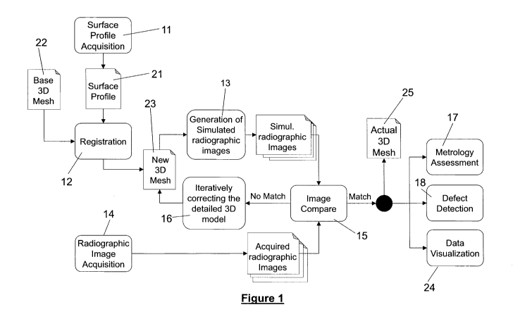
[0034] Figure 1 is a schematic representation of the method for performing
inspection of a manufactured article, in accordance with an embodiment;
[0035] Figure 2 is a schematic representation of a system for performing
inspection
of a manufactured article, in accordance with an embodiment.
[0036] Figure 3 is a schematic representation of an embodiment of a surface
profile
acquisition device and a radiographic image acquisition device of the system
of
Figure 2, in combination with an article being conveyed on a motion device.
[0037] Figure 4 is a schematic representation of the radiographic image
acquisition
device of Figure 3, acquiring a sequence of radiographic images of the
article.
DETAILED DESCRIPTION
[0038] In the following description, the same numerical references refer to
similar
elements. The embodiments, geometrical configurations, materials mentioned
and/or dimensions shown in the figures or described in the present description
are
embodiments only, given solely for exemplification purposes.
[0039] Moreover, although the embodiments of the method for performing
inspection
of a manufactured article and the system for performing the same consist of
certain
-11-
CA 03031397 2019-01-21
WO 2018/014138 PCT/CA2017/050882
elements as explained and illustrated herein, not all of these elements are
essential
and thus should not be taken in their restrictive sense. It is to be
understood, as also
apparent to a person skilled in the art, that other suitable elements and
cooperation
thereinbetween may be used for the method for performing inspection of a
manufactured article and the system for performing the same, as will be
briefly
explained herein and as can be easily inferred herefrom by a person skilled in
the
art.
[0040] In general terms, the method for performing inspection of a
manufactured
article and the system for performing the same uses previously known data
relating
to the article being inspected (required measurements, material of the
article, etc.)
and the configuration of a radiographic image acquisition device in order to
perform
precise measurement (i.e. metrology analysis) and/or subsurface defect
detection of
a plurality of the articles, sequentially. It will be understood that the
method allows
inspection of numerous types of manufactured articles, from diverse fields,
such as,
without being limitative, glass bottle, plastic molded components, die casting
parts,
additive manufacturing components, wheels, tires and other manufactured of
refactored parts for the automotive, military or aerospace industry. The above
examples are given as indicator only and one skilled in the art will
understand that
several other types of manufactured articles can be subjected to inspection
using
the present method. In an embodiment, the articles are sized and shaped to be
conveyed on a motion device for inline inspection thereof. In an alternative
embodiment, the article can be a large article, which is difficult to
displace, such that
components of the inspection system should rather be displaced relative to the
article.
[0041] Referring generally to Figure 1, an embodiment of the method 10 for
performing inspection of a manufactured article is shown. As mentioned above,
this
method can be performed for articles (or region of interest thereof) where the
precise
geometry and dimensional characteristics are known, as represented by a
theoretical detailed three-dimensional (3D) model (base 3D Mesh) 22 of the
article
-12-
CA 03031397 2019-01-21
WO 2018/014138 PCT/CA2017/050882
and where the article (or region of interest thereof) is made of a known
material. The
theoretical detailed 3D model (base 3D Mesh) 22 of the article can be acquired
from
multiple different sources such as, without being limitative, a Computer-aided
Design
(CAD) of the article, a CT scan of the object or any other means that can
produce
such mesh.
[0042] In an embodiment, the article (or region of interest thereof) can be
made of
more than one known material with known positioning, geometry and dimensional
characteristics of each one of the portions of the different materials. For
ease of
description, in the course of the description, only reference to inspection of
an article
will be made, but it will be understood that, in an embodiment, inspection of
only a
region of interest of the article can be performed. It will also be understood
that the
method can be applied successively to multiple articles, thereby providing
scanning
of a plurality of successive articles, such as in a production chain or the
like.
[0043] In the embodiment shown, for each one of the successively manufactured
article, the method 10 includes the general steps of: performing surface
profile
acquisition 11; acquiring successive radiographic images of the article 14;
determining a precise position of the article (registration step) for each one
of the
acquired radiographic images 12; generating simulated radiographic images of
the
article 13 based on the determined positions of the article and the detailed
3D model;
comparing the simulated radiographic images and the acquired radiographic
images
15; iteratively adjusting (correcting) the detailed 3D model until the
simulated
radiographic images of the article and the acquired radiographic images match
16.
The method also includes performing a metrology assessment 17 and/or a
subsurface defect detection 18 based on the adjusted detailed 3D model (i.e.
the
actual 3D model representative of the article inspected) and performing data
visualization 24 based on the data of the metrology assessment and/or the
subsurface defect detection previously performed.
[0044] In an embodiment, the step of performing surface profile acquisition 11
includes performing a profile surface scan of the article. For example and
without
-13-
CA 03031397 2019-01-21
WO 2018/014138 PCT/CA2017/050882
being limitative, in an embodiment, the profile surface scan can be performed
by one
or more two-dimensional (2D) laser scanner triangulation devices performing a
profile surface scan of the article, as it is being conveyed on a motion
device. The
surface profile acquisition yields a precise 3D surface profile 21 of the
article (the
third dimension being provided by the displacement of the article on the
motion
device). In an alternative embodiment, the profile surface scan can be
performed by
one or more two-dimensional (2D) laser scanner triangulation devices
performing a
profile surface scan of the article as the laser are displaced relative to the
article, as
will be described in more details below.
[0045] In the embodiment shown where surface profile acquisition 11 is
performed,
the method can further comprise adjusting the theoretical detailed 3D model
(base
3D Mesh) 22 based on the acquired surface profile 21 of the article and
generating
an updated 3D model (New 3D Mesh) 23.
[0046] One skilled in the art will understand that, in alternative
embodiments, other
devices for performing profile surface scan, such as video cameras, infrared
cameras, millimeter wave sensors, or the like can also be used.
[0047] One skilled in the art will also understand that, in an alternative
embodiment
(not shown), the method can also be free of the step of surface profile
acquisition
11. In such an embodiment, the theoretical detailed 3D model (base 3D Mesh) is
directly used for performing the initial generation of simulated radiographic
images
of the article 13 based on the determined positions of the article and the
detailed 3D
model, which will be described in more details below.
[0048] The step of acquiring successive radiographic images of the article 14
includes scanning the article using a radiographic image acquisition device
including
one or more radiographic source(s), such as, X-ray source(s) or gamma-ray
source(s), and corresponding detector(s), positioned on opposed sides of the
article.
Once again, in an embodiment, the article is scanned using the radiographic
source(s) and corresponding detector(s), as it is being conveyed on the motion
-14-
CA 03031397 2019-01-21
WO 2018/014138 PCT/CA2017/050882
device at a constant speed, therefore resulting in a continuous sequence of
radiographic images of the article 14 being captured at a known interval, as
the
article is conveyed linearly with regard to the radiographic image acquisition
device.
For example, the motion device can be a linear stage, a conveyor belt or other
similar
device. It will be understood that, the smaller the interval between the
images of the
continuous sequence of radiographic images, the higher the number of views
available and the more precise the resulting 3D dimensioning positioning
(which will
be described in more details below) can be.
[0049] In an alternative embodiment, the acquisition of the radiographic
images can
be performed as the radiographic source(s) and corresponding detector(s) are
displaced relative to the article, as will be described in more details below.
[0050] In an embodiment where a simple object is scanned, the step of
acquiring
successive radiographic images of the article 14 can include acquiring at
least about
25 images defining a continuous sequence of images, with each image providing
a
unique viewing angle of the article. Typically, the step of acquiring
successive
radiographic images of the article 14 will includes acquiring several hundred
images
(and at least about one hundred images) defining a continuous sequence of
images,
with each image providing a unique viewing angle of the article.
[0051] The step of determining a precise position of the article 12 for each
one of the
acquired radiographic images includes determining a precise position and
orientation of the article relative to the radiographic source(s) and
corresponding
detector(s) for each one of the acquired radiographic images. In other words,
the
article must be registered in 3D space, in order to generate the simulated
radiographic images from the detailed 3D model (as will be described in more
details
below). In an embodiment where the article is linearly moved by the motion
device,
the registration must be synchronized with the linear motion device so that a
sequence of simulated images that matches the actual sequence of radiographic
images can be generated.
-15-
CA 03031397 2019-01-21
WO 2018/014138 PCT/CA2017/050882
[0052] In an embodiment, the precise relative position (X, Y and Z) and
orientation
of the article with regards to the radiographic source(s) and corresponding
detector(s) is performed through analysis of the corresponding acquired
radiographic image, using intensity-based or feature-based image registration
techniques, with or without fiducial points. In an embodiment, for greater
precision,
the acquired surface profile 21 of the article can also be analysed and used,
alone
or in combination to the corresponding acquired radiographic image, in order
to
determine the precise position of the article. In such an embodiment, the
positioning
of the radiographic image acquisition device relative to the device used for
acquiring
the surface profile 21 is known and used to determine the position of the
article
relative to the radiographic source(s) and corresponding detector(s).
[0053] The step of generating simulated radiographic images of the article 13
based
on the determined positions of the article and the detailed 3D model is
performed by
a radiographic image simulator simulating the sequence of acquired
radiographic
images, using the detailed 3D model, the known material properties of the
article,
and the known configuration of the radiographic source and sensor array to
reproduce faithfully the geometry and physics of the radiographic scan being
performed.
[0054] In an embodiment, the radiographic image simulator simulates the path
of the
radiation through the article, using ray casting, for each simulated sensor
array, in
view of the position, geometry and configuration of the calibrated
radiographic
source(s) and corresponding detector(s) relative to the article; the geometry
of the
article as defined by the detailed 3D model and the material(s) thereof. In
other
words, in an embodiment, the radiographic image simulator uses ray casting
through
the mesh to define the optical paths for every pixel in the sensor array and
converts
this data into intensity values that corresponds to the physics of the
material(s) in
the object, while taking into consideration the source and detector
calibration. In an
embodiment, the radiographic image simulator therefore simulates the path of
the
radiation through every surface of the article, crossing the radiation path,
i.e. every
-16-
CA 03031397 2019-01-21
WO 2018/014138 PCT/CA2017/050882
frontier which is crossed by the radiation path, and which potentially impacts
the
trajectory of the radiation going through the article.
[0055] In an embodiment where the article includes a plurality of material, a
detailed
3D model can be generated for each region of interest of the article
represented by
the corresponding material and simulated radiographic images can be generated
for
each detailed 3D model corresponding to one region of interest of the article
made
of the corresponding material, in accordance with the above-described
principles.
Therefore, the radiographic image simulator can simulate multiple layers of
materials
as well as multiple types of materials. In other words, in an embodiment where
the
article includes a plurality of materials, the radiographic image simulator
must take
into consideration each layer individually.
[0056] A simulated radiographic image is generated for each one of the
predetermined position of the article for which a radiographic image of the
article is
acquired in the previous step of acquiring successive radiographic images of
the
article 14. In other words, a simulated radiographic image is generated for
each
radiographic image acquired and corresponding to a registration of the article
in a
3D space. It will be understood that, in this step, either one of the
theoretical detailed
3D model (base 3D Mesh) 22 or the updated 3D model (New 3D Mesh) 23 can be
used (as will be described in more details below).
[0057] The step of comparing the simulated radiographic images and the
acquired
radiographic images 15 is performed using comparison methods to determine
whether the simulated radiographic images of the article and the acquired
radiographic images match or if there are differences between the two sets of
images, indicating that the article does not conform to the detailed 3D model
used
for generating the simulated radiographic images. The differences can be
differences in positioning, geometry, presence of defects or even differences
in
density.
-17-
CA 03031397 2019-01-21
WO 2018/014138 PCT/CA2017/050882
[0058] In an embodiment, the images can be converted to binary images and
comparison can be performed through binary images subtraction. In an
alternative
embodiment, the images can be compared using gray level distribution (or
intensity
profile) alone or in combination with binary images subtraction. One skilled
in the art
will understand that any other image comparison methods and/or combinations
thereof, adapted to the geometry of the manufactured article, can also be
used.
[0059] In an embodiment, given that a sequence of radiographic images is
acquired,
feature tracking from one image to another can be used to locate each of the
differences in 3D space. In other words, in an embodiment, multiple features
are
identified in the simulated radiographic images and the acquired radiographic
images and their position is tracked through the entire scan sequence (and
corresponding simulated scan sequence). The plurality of angular position for
the
features (obtained from the plurality of images in the continuous sequence of
images) helps precisely position the features in 3D space to subsequently
produce
an updated 3d Model that better corresponds to the actual article being
inspected.
In the course of the present description, the term feature is used to refer to
specific
structures in the image such as points, edges or objects that can easily be
detected
and used for analysis and comparison.
[0060] The method 10 further comprises the step of performing a 3D model
correction loop for iteratively adjusting the detailed 3D model until the
simulated
radiographic image of the article and the acquired radiographic image match.
In
other words, the detailed 3D model is iteratively morphed until the detail 3D
model
matches the article being scanned. As mentioned above, in an embodiment, the
theoretical detailed 3D model (base 3D Mesh) 22 can be initially adjusted
based on
the acquired surface profile 21 of the article to generate an updated 3D model
(New
3D Mesh) 23 which defines the latest version of the detailed 3D model for the
first
iteration. In an alternative embodiment (not shown), the theoretical detailed
3D
model (base 3D Mesh) 22 can be used as the latest version of the detailed 3D
model
for the first iteration.
-18-
CA 03031397 2019-01-21
WO 2018/014138 PCT/CA2017/050882
[0061] In the embodiment shown, the latest version of the detailed 3D model is
subsequently iteratively adjusted (corrected), using a morphing algorithm to
adapt
the detailed 3D model in accordance with the observed differences, until a
match is
found between the simulated radiographic images and the acquired radiographic
images (as determined in step 15). Hence, for each iteration, if the
determination of
whether the simulated radiographic images of the article and the acquired
radiographic images match is negative (i.e. if a match result indicative of
whether
the simulated radiographic images and the acquired radiographic images are a
match or a mismatch indicates a mismatch ¨ therefore indicating that
differences are
found between the two sets of images), the latest version of the detailed 3D
model
is updated (corrected) to attempt to eliminate the differences between the
simulated
radiographic images of the article and the acquired radiographic images and a
newly
updated 3D model (New 3D Mesh) 23 is generated.
[0062] The newly updated 3D model (New 3D Mesh) 23 is subsequently used for
generating the sequence of simulated radiographic images of the article (as
described in detail above with regards to step 13) and the new simulated
radiographic images are compared to the acquired radiographic images (as
described in detail with regards to step 15). If the simulated radiographic
images of
the article generated using the newly updated 3D model (New 3D Mesh) 23 once
again do not match the acquired radiographic images, a new iteration can be
performed using the newly updated 3D model (New 3D Mesh) 23 as latest version
of the detailed 3D model. When the simulated radiographic images of the
article and
the acquired radiographic images match, the last newly updated 3D model (New
3D
Mesh) 23 is used as final 3D model (Actual 3D Mesh) 25 representative of the
article
currently being inspected (i.e. the final 3D model (Actual 3D Mesh) 25
corresponds
to the actual geometric dimensions of the article being inspected). In an
embodiment
where metrology is performed, a determination of the the simulated
radiographic
images of the article and the acquired radiographic images being a match can
correspond to a tolerance of below about 10 microns (for articles of about
30cm or
less). In an embodiment where subsurface defect detection is performed, a
-19-
CA 03031397 2019-01-21
WO 2018/014138 PCT/CA2017/050882
determination of the the simulated radiographic images of the article and the
acquired radiographic images being a match can correspond to a tolerance of
between about 50 microns and about 100 microns (for articles of about 30cm or
less).
[0063] In an embodiment, updating (morphing) the latest version of the
detailed 3D
model to attempt to eliminate the differences between the simulated
radiographic
images of the article and the acquired radiographic images includes performing
imagery analysis of the sequence of simulated radiographic images of the
article and
the acquired sequence of radiographic images in order to determine a most
probable
cause of the differences between the simulated radiographic images of the
article
and the acquired radiographic images. For example and without being
limitative, in
an embodiment, the required imagery analysis includes identification and
characterization of the deviation between the simulated radiographic images of
the
article and the acquired radiographic images, for example, using image
subtraction
and/or intensity profile comparison and displacing control points of the
latest version
of the detailed 3D model to correct the identified deviation(s).
[0064] In an embodiment, morphing the latest version of the detailed 3D model
to
attempt to eliminate the differences between the simulated radiographic images
of
the article and the acquired sequence of radiographic images includes feature
identification being performed using a registration algorithm and every
feature
detected being uniquely characterized to determine whether it is an expected
feature
resulting from the object geometry or an anomaly resulting from a defect
(either a
geometry defect or a subsurface defect). A similar feature identification
process is
performed for the simulated radiographic images and the acquired radiographic
images and each pair of feature (i.e. a corresponding feature of the simulated
radiographic images and the acquired radiographic images) is scored for
similarity
(i.e. is rated according to the level of similarity between the simulated
radiographic
images and the acquired radiographic images). Subsequently, an image
modification algorithm is performed to suggest deformation for each one of the
- 20 -
CA 03031397 2019-01-21
WO 2018/014138 PCT/CA2017/050882
simulated radiographic images, in order to bring each simulated radiographic
image
as close as possible to a match with its corresponding acquired radiographic
image.
In an embodiment, a deformation is suggested for each pixel of the simulated
radiographic images and a score is provided based on the quality of the match
between the simulated radiographic image and the corresponding acquired
radiographic image for the proposed deformation (i.e. the proposed deformation
of
each pixel of the simulated radiographic images). If required, the steps
related to the
deformation of the simulated radiographic images are repeated until a
predetermined scoring threshold is attained. In the above described process,
each
pair of corresponding simulated radiographic image and acquired radiographic
image is treated (i.e. processed) independently.
[0065] Once the required deformation is optimized for each pair of
corresponding
simulated radiographic image and acquired radiographic image, the deformations
must be transferred to the latest version of the detailed 3D model. In order
to transfer
the deformations of a plurality of simulated radiographic images of the image
sequence, in an embodiment a parallax effect across all images in the sequence
is
used, thereby resulting in identification of only one combined 3D deformation
(i.e.
only one combined deformation applied globally to the 3D model) correlated to
all of
the individual 2D deformations in each pair of corresponding simulated
radiographic
image and acquired radiographic image.
[0066] In an embodiment where the 3D position of the deformation does not
correspond to the surface of the latest version of the detailed 3D model,
which is
typically the case for an internal defect, such as, for example, an air bubble
in a die
casting part, a seed mesh region is created and the above described
optimization
steps are performed again for the particular region of interest to ensure a
proper
match between the newly created seed mesh and the information of the acquired
radiographic images.
[0067] In an embodiment where the 3D position of the deformation corresponds
to
the surface of the latest version of the detailed 3D model, a sparse
triangular mesh
-21 -
CA 03031397 2019-01-21
WO 2018/014138 PCT/CA2017/050882
can be used to apply the deformation. Using the sparse triangular mesh, the
transformation can be applied to only a fraction of vertices in the area,
saving
processing time. The deformation field is applied to the center of each
triangle and
as a result, the uninvolved vertices in the area are moved linearly, but non-
rigidly.
[0068] It will be understood that in a continuous sequence of images, the X
and Y
displacement of a feature pixel can be tracked and predicted across the
continuous
sequence of images and can yield a Position vs Step plot with a given slope
and
intercept. It thus becomes possible, with the slope of the X vs Time Step
graph to
extract depth (Z) information.
[0069] In view of the above, the newly updated 3D model (New 3D Mesh) can be
morphed to iteratively adjust a geometry or a material density of a region of
interest
of the 3D model to eliminate the differences between the simulated
radiographic
images of the article and the acquired radiographic images, in order to
finally
generate a final 3D model (Actual 3D Mesh) 25 accurately representative of the
article scanned, with regard to geometry, subsurface defects and density of
the
material(s).
[0070] In some cases, such as when a presence of voids or contaminants within
the
object is detected, new layers of mesh can be added to the newly updated 3D
model
(New 3D Mesh) 23 in order to match the observed data.
[0071] In the embodiment shown, the step of performing the metrology
assessment
20 includes comparing the theoretical detailed 3D model (Base 3D Mesh) 22 and
the final 3D model (Actual 3D Mesh) 25 in order to determine whether
differences
are detected in the geometry of the inspected article defined by the final 3D
model
(Actual 3D Mesh) 25 and the model article defined by the theoretical detailed
3D
model (Base 3D Mesh) 22 and generating metrology assessment data. One skilled
in the art will understand that, in an alternative embodiment (not shown) the
method
can be free of metrology assessment (i.e. the method could include only the
subsurface defect detection step, as described below).
- 22 -
CA 03031397 2019-01-21
WO 2018/014138 PCT/CA2017/050882
[0072] In an embodiment, comparison of the theoretical detailed 3D model (Base
3D
Mesh) 22 and the final 3D model (Actual 3D Mesh) 25 is achieved by acquiring
geometric measurements on the theoretical detailed 3D model (Base 3D Mesh) 22
and the final 3D model (Actual 3D Mesh) 25 based on control points and
predetermined tolerances of the model, for the specific article that is
manufactured,
and comparing the acquired geometric measurements of both models to determine
if there are variations beyond the associated tolerances thereof. In an
alternative
embodiment, comparison between the theoretical detailed 3D model (Base 3D
Mesh) 22 and the final 3D model (Actual 3D Mesh) can also be performed via a
deviations display using color coding.
[0073] In the embodiment shown, the step of performing subsurface defect
detection
18 includes comparing the theoretical detailed 3D model (Base 3D Mesh) 22 and
the final 3D model (Actual 3D Mesh) 25 in order to determine whether
differences
are detected in the internal structure of the inspected article defined by the
final 3D
model (Actual 3D Mesh) 25 and the model article defined by the theoretical
detailed
3D model (Base 3D Mesh) 22, outside of dimensional measurements, and
generating subsurface defect detection data. One skilled in the art will once
again
understand that, in an alternative embodiment (not shown) the method can be
free
of subsurface defect detection (i.e. the method could include only a metrology
assessment step, as described above).
[0074] Finally, in an embodiment, the step of performing data visualization 24
based
on the data of the metrology assessment and/or the subsurface defect detection
previously performed includes displaying the metrology assessment data and/or
the
subsurface defect detection data (or inspection data) on a display screen.
Moreover,
in an embodiment, data relating to the metrology assessment and/or the
subsurface
defect detection can be stored on a storage medium in order to remain
subsequently
available to the end users. For example, and without being limitative, the
storage
medium can be a permanent storage such as a hard disk; an optical storage
device,
such as a CD or DVD (rewritable or write once/read only), a flash memory, or
the
- 23 -
CA 03031397 2019-01-21
WO 2018/014138 PCT/CA2017/050882
like. In an embodiment, the data of the metrology assessment and/or the
subsurface
defect detection can be used to automatically evaluate whether each one of the
inspected article conforms to selected criteria and sort the articles based on
this
evaluation. The data can also be used to evaluate the performance of the
manufacturing equipment or the like.
[0075] In an alternative embodiment, the data of the metrology assessment
and/or
the subsurface defect detection can be used to perform article sorting. In
such an
embodiment, the metrology assessment data and/or the subsurface defect
detection
data can be used to determine a status of the article (e.g. a pass or fail
status based
on pre-defined criteria) and the status can be used to direct the article in
the
production line (for example to reject article(s) having the fail status). One
skilled in
the art will understand that, in such and embodiment, display of the data can
be
omitted.
[0076] The method for performing the inspection of a manufactured article
having
been described above, a system which allows the method to be performed will
now
be described in more details below.
[0077] With reference to Figures 2 to 4, in an embodiment, the system 30
includes a
surface profile acquisition device 32, a radiographic image acquisition device
31, a
positional evaluation unit 34, a 3D model data correction unit 33 (including a
radiographic image simulator 36, an image compare unit 38, and a 3D model data
update unit 40), a metrology assessment unit 42, a subsurface defect detection
unit
44, a data display device 46, and an article sorting unit 47.
[0078] The system 30 also includes a motion device 60 creating relative
movement
between the manufactured article 62 and the combination of the surface profile
acquisition device 32 and the radiographic image acquisition device 31. In the
course
of the present description, the term "relative movement" is used to refer to
at least
one of the elements moving linearly with respect to the other. In other words,
the
motion device 60 displaces at least one of the manufactured article 62 and the
- 24 -
CA 03031397 2019-01-21
WO 2018/014138 PCT/CA2017/050882
combination of the surface profile acquisition device 32 and the radiographic
image
acquisition device 31 linearly, in order to generate relative movement
therebetween.
In the embodiment shown in Figure 3, where the motion device 60 displaces the
manufactured article 62, the motion device 60 can be a linear stage, a
conveyor belt
or other similar devices, displacing linearly the manufactured article 62
relative to the
surface profile acquisition device 32 and the radiographic image acquisition
device
31 remaining still. In another alternative embodiment, the manufactured
article 62
can remain still and the surface profile acquisition device 32 and the
radiographic
image acquisition device 31 can be displaced, for example and without being
limitative, by an articulated arm, a displaceable platform, or the like. In an
embodiment, both the manufactured article 62 and the surface profile
acquisition
device 32 and the radiographic image acquisition device 31 can be displaced
during
the inspection process.
[0079] As mentioned above, in an embodiment, the surface profile acquisition
device
32 can include any device capable of performing a precise profile surface scan
of
the article 62 as relative movement occurs between the article 62 and the
surface
profile acquisition device 32 and generate surface profile data therefrom. In
an
embodiment, the surface profile acquisition device 32 performs a profile
surface scan
with a precision in a range of between about 1 micron and 50 microns. For
example
and without being limitative, in an embodiment, the surface profile
acquisition device
32 can include one or more two-dimensional (2D) laser scanner triangulation
devices
positioned and configured to perform a profile surface scan of the article 62
as it is
being conveyed on the motion device 60 and to generate the surface profile
data for
the article 62. As mentioned above, in an embodiment, the system 30 can be
free of
surface profile acquisition device 32.
[0080] The radiographic image acquisition device 31 has also been described
above
in the description of the associated method. As previously mentioned, the
radiographic image acquisition device 31 includes one or more radiographic
source(s) 71 and corresponding detector(s) 73 positioned on opposite sides of
the
- 25 -
CA 03031397 2019-01-21
WO 2018/014138 PCT/CA2017/050882
article 62 as relative movement occurs between the article 62 and the
radiographic
image acquisition device 31, in order to capture a continuous sequence of a
plurality
of radiographic images at a known interval of the article 62 (see Figure 4).
In an
embodiment (see Figures 3 and 4), the radiographic source(s) 71 is a cone beam
X-
ray source(s) generating X-rays towards the article 62 and the detector(s) 73
is a 2D
X-rays detector(s). In an alternative embodiment, the radiographic source(s)
can be
gamma-ray source(s) generating gamma-rays towards the article 62 and the
detector(s) can be 2D gamma-rays detector(s). In an embodiment, 1D detectors
positioned such as to cover different viewing angles can also be used. One
skilled
in the art will understand that, in alternative embodiments, any other image
acquisition device allowing subsurface scanning and imaging of the article 62
can
also be used.
[0081] One skilled in the art will understand that the configuration of the
radiographic
image acquisition device 31 can vary according to the type of article 62 to be
inspected. For example and without being limitative, the number, position and
orientation of the radiographic source(s) 71 and corresponding detector(s) 73,
as
well as the angular coverage, object spacing, acquisition rate and/or
resolution can
be varied according to the specific inspection requirements of each
embodiment.
[0082] In an embodiment, the positional evaluation unit 34 is in data
communication
with the radiographic image acquisition device 31 and the surface profile
acquisition
device 32 and receives the surface profile data and the acquired radiographic
images therefrom. In an embodiment where the system is free of surface profile
acquisition device 32, the positional evaluation unit 34 can be in data
communication
with only the radiographic image acquisition device 31. The positional
evaluation unit
34 is configured to process the surface profile data and/or the acquired
radiographic
images (i.e. to perform image processing of the received radiographic images
and/or
the surface scan defined by the surface scan data) and to generate article
position
data representative of the precise position of the article 62 relative to the
- 26 -
CA 03031397 2019-01-21
WO 2018/014138 PCT/CA2017/050882
radiographic image acquisition device 31, for each one of the acquired
radiographic
images.
[0083] In an embodiment where the motion of the article 62 provided by the
motion
device 60 is predetermined (or controlled according to known parameters), the
surface profile acquisition device 32 can be positioned at any predetermined
fixed
position with regard to the image acquisition device 31. The positioning and
orientation of the article 62 for each image of the sequence of radiographic
image
acquired by the image acquisition device 31 can be computed therefrom (given
that
no motion of the article 62 other than motion by the motion device 60 occurs).
However, in order to reduce the probabilities of inaccuracies of the data
acquired by
the surface profile acquisition device 32 for registration of the article 62
when the
sequence of radiographic image is acquired by the image acquisition device 31,
in
an embodiment (not shown), the surface profile acquisition device 32 is
positioned
as close as possible to the center of the radiation emitted by the image
acquisition
device 31.
[0084] In an embodiment, the system 30 also includes a 3D model data
correction
unit 33 in data communication with the radiographic image acquisition device
31 and
the positional evaluation unit 34 and receiving the acquired radiographic
images and
article position data therefrom. The 3D model data correction unit 33 is
operative to
iteratively update the 3D model data and generate final 3D model data defining
a 3D
model substantially conform to the geometric dimensions of the manufactured
article
62.
[0085] In an embodiment, the 3D model data correction unit 33 includes a
radiographic image simulator 36 configured to simulate the path of the
radiation rays
from the radiographic image acquisition device 31 into the manufactured
article 62
(as defined by the detailed 3D model defined by the 3D model data), using the
position data representative of the position of the manufactured article 62
relative to
the radiographic image acquisition device 31, for each radiographic image of
the
sequence of radiographic images acquired by the radiographic image acquisition
- 27 -
CA 03031397 2019-01-21
WO 2018/014138 PCT/CA2017/050882
device 31. The radiographic image simulator 36 generates the simulated
radiographic images through the above-described simulation of the path of the
radiation rays into the manufactured article 62.
[0086] The 3D model data correction unit 33 also includes an image compare
unit
38 configured to compare the generated simulated radiographic images and the
acquired radiographic images and to generate a match result indicative of
whether
the simulated radiographic images and the acquired radiographic images are a
match or a mismatch.
[0087] The 3D model data correction unit 33 further includes a 3D model data
update
unit 40. The 3D model data update unit 40 is configured to update the detailed
3D
model data upon detection of the match result of the simulated radiographic
images
and the acquired radiographic images being a mismatch. In order to proceed
with
such update of the detailed 3D model data, the 3D model data update unit 40 is
configured to perform imagery analysis of the simulated radiographic images
and
the acquired radiographic images and to determine a most probable cause of the
mismatch, as described above. Based on the determined most probable cause of
the mismatch, the 3D model data update unit 40 is configured to generate
updated
detailed 3D model data of the article 62.
[0088] As will be easily understood by one skilled in the art, the components
of the
3D model data correction unit 33 cooperate to iteratively update the 3D model
data
until a final 3D model defining a 3D model conforming to the geometric
dimensions
of the manufactured article 62 is generated and included in the 3D model data.
[0089] In an embodiment, the system 30 also includes a metrology assessment
unit
42 in data communication with the 3D model correction unit 33 and receiving
the
detailed 3D model data therefrom. In an embodiment, the metrology assessment
unit 42 is configured to process the 3D model data and perform a metrology
assessment through comparisons of a theoretical 3D model of the detailed 3D
model
data and the final 3D model of the detailed 3D model data to detect
differences
- 28 -
CA 03031397 2019-01-21
WO 2018/014138 PCT/CA2017/050882
therebetween and generate article inspection data. In an embodiment, the
system
30 can be free of metrology assessment unit 42.
[0090] In an embodiment, the system 30 also includes a subsurface defect
detection
unit 44 in data communication with the 3D model correction unit 33 and the
radiographic image acquisition device 31 and receiving the acquired
radiographic
images and simulated radiographic images therefrom. In an embodiment, the
subsurface defect detection unit 44 is configured to process the 3D model data
and
perform a subsurface detection assessment through comparisons of a theoretical
3D model of the detailed 3D model data and the final 3D model of the detailed
3D
model data to detect differences therebetween and generate article inspection
data..
In an embodiment, the system can be free of subsurface defect detection unit
44.
[0091] In an embodiment, the article inspection data can be displayed on a
data
display device 46, such as, for example, and without being limitative, a
display
screen or the like. In an embodiment, the article inspection data can also be
stored
on a storage medium for subsequent retrieval and/or display. In an embodiment,
the
system can be free of data display device 46.
[0092] In an embodiment, the system can also include an article sorting unit
47 in
data communication with the metrology assessment unit 42 and/or the subsurface
defect detection unit. In an embodiment, the article sorting unit 47 is
configured to
process the article inspection data and perform sorting of the article 62
based on the
processed article inspection data. For example and without being limitative,
in an
embodiment, the article sorting unit 47 can determine a pass or fail status of
the
article, based on pre-defined criteria, such as deviation from the theoretical
geometry
a defect threshold or the like. The article sorting unit 47 can include an
ejector
activable to remove the article 62 from the production line if the article 62
has a fail
status. In an embodiment, the system can be free of article sorting unit 47.
[0093] One skilled in the art will understand that the positional evaluation
unit 34, the
3D model data correction unit 33 (including the radiographic image simulator
36, the
- 29 -
CA 03031397 2019-01-21
WO 2018/014138 PCT/CA2017/050882
image compare unit 38, and the 3D model data update unit 40), the metrology
assessment unit 42, and the subsurface defect detection unit 44 can be
embodied
on a single computing unit 50 having its own memory and processor. In an
alternative embodiment (not shown), the components can be part of a
distributed
computing system where each unit (or a subset thereof) is installed on a
separate
computing unit having its own memory and processor.
[0094] Several alternative embodiments and examples have been described and
illustrated herein. The embodiments of the invention described above are
intended
to be exemplary only. A person skilled in the art would appreciate the
features of the
individual embodiments, and the possible combinations and variations of the
components. A person skilled in the art would further appreciate that any of
the
embodiments could be provided in any combination with the other embodiments
disclosed herein. It is understood that the invention may be embodied in other
specific forms without departing from the central characteristics thereof. The
present
examples and embodiments, therefore, are to be considered in all respects as
illustrative and not restrictive, and the invention is not to be limited to
the details given
herein. Accordingly, while specific embodiments have been illustrated and
described, numerous modifications come to mind without significantly departing
from
the scope of the invention as defined in the appended claims.
- 30 -