Note: Descriptions are shown in the official language in which they were submitted.
TARGETED RADIOTHERAPY CHELATES
FOR IN SITU IMMUNE MODULATED CANCER VACCINATION
[0001] This paragraph has been intentionally deleted.
STATEMENT REGARDING FEDERALLY SPONSORED
RESEARCH OR DEVELOPMENT
[0002] This invention was made with government support under CA197078
awarded
by the National Institutes of Health. The government has certain rights in the
invention.
FIELD OF THE DISCLOSURE
[0003] This disclosure relates generally to methods of treating cancer. In
particular,
the disclosure is directed to methods of treating a cancer comprising one or
more
malignant solid tumors in a subject by (a) systemically administering to the
subject an
immunomodulatory dose of a radioactive metal chelate compound that is
differentially
taken up by and retained within solid tumor tissue, and (b) performing in situ
tumor
vaccination in the subject at one or more of the malignant solid tumors using
one or more
treatments capable of stimulating specific immune cells within the tumor
microenvironment.
BACKGROUND
[0004] Current cancer treatment typically involves systemic chemotherapy
whereby
non-targeted small molecule or antibody directed cytotoxic agents
preferentially enter, or
bind to (in the case of antibody directed agents) and kill cancer cells by a
variety of
mechanisms. External beam radiation therapy (xRT), which is often combined
with
chemotherapy, kills cancer cells by inducing nuclear DNA double strand breaks
resulting
in cell-cycle death. Unlike systemic chemotherapy, xRT depends on the ability
to
accurately determine the anatomic location of the tumor. Surgical resection of
tumors
also depends on the ability to see the tumor and on complete removal, since
residual
tumor cells will quickly reestablish the tumor following surgery. Surgery and
xRT are
generally limited to the local treatment of malignant tumors and thus are
limited in
1
Date Recue/Date Received 2020-11-10
treating disseminated or metastatic disease, which is why chemotherapy is
often used in
conjunction with these treatment modalities. Although systemic chemotherapy is
capable
of reaching many distant metastatic sites, with the possible exception of
brain metastases,
for all too many patients, responses are typically short-lived (months to
several years) and
ultimately result in tumor recurrence.
[0005] Because the body's natural immune system is also capable of
destroying
cancer cells following their recognition, immunologic approaches are rapidly
becoming
more prevalent in cancer treatment paradigms. However, some cancer cells, and
to a
greater extent cancer stem cells, manage to initially avoid immune-
surveillance and
actually acquire the ability to evolve and ultimately survive by remaining
relatively
immune invisible [Gaipi et al, Immunotherapy 6:597-610, 2014].
[0006] One specific immunologic approach that is being increasingly
investigated is
"in situ vaccination," a strategy that seeks to enhance tumor immunogenicity,
generate
tumor infiltrating lymphocytes (TIL) and drive a systemic anti-tumor immune
response
directed against "unvaccinated," disseminated tumors. In in situ vaccination,
a malignant
solid tumor is injected with (or treated with) one or more agents that
facilitate the release
of tumor antigens while simultaneously providing pro-inflammatory signals to
reverse the
immune-tolerizing microenvironment of the tumor [Pierce et al, Human Vaccines
&
Immunotherapoeutics 11(8):1901-1909, 2015; Marabelle eta!, Clin. Cancer Res.
20(7):1747-56, 2014; Morris eta!, Cancer Research, e-pub ahead of print,
2016].
Although recent data from clinical trials and pre-clinical models illustrate
the potential of
such an approach, there is a great need in the art for in-situ vaccination
methods
exhibiting improved systemic efficacy.
[0007] Radiation hormesis is a decades-old hypothesis that low doses of
ionizing RT
can be beneficial by stimulating the activation of natural protective repair
mechanisms
that are not activated in the absence of ionizing RT [Cameron and Moulder,
Med. Phys.
25:1407, 1998]. The reserve repair mechanisms are hypothesized to be
sufficiently
effective when stimulated as to not only cancel the detrimental effects of
ionizing RT but
also inhibit disease not related to RT exposure. Perhaps related, the abscopal
effect is a
phenomenon reported in the 1950's, whereby, xRT treatment of one tumor
actually
causes shrinkage of another tumor outside the RT treatment area. Although
rare, this
2
Date Recue/Date Received 2020-11-10
phenomenon is thought to be dependent on activation of the immune system.
Together,
hormesis and the abscopal effect support the potential interaction and
stimulation of the
immune system by low dosage (immune stimulatory but non-cytotoxic) RT, which
may
then be combined with other immunologic approaches, such as in situ
vaccination.
[0008] We have previously published that the combination of local xRT + in
situ
vaccination are potently synergistic in treating large established tumors in
mice, when
there is a single tumor present [Morris et al, Cancer Research, e-pub ahead of
print,
2016].
[0009] We have surprisingly discovered (and disclose herein) that the
combination of
in situ vaccination and xRT does not result in inhibited tumor growth in the
presence of a
second, non-radiated tumor. Apparently, the non-radiated tumor exhibits a
dampening
effect (which we have designated as "concomitant immune tolerance") on the
immunomodulatory effect of the xRT and in situ vaccine on the radiated tumor.
This
concomitant immune tolerance can be overcome, enabling efficacy of in situ
vaccination,
when xRT is given to all areas of tumor. However, xRT cannot be effectively
used in
combination with in situ vaccination methods in the presence of multiple
tumors,
particularly if the tumors are not few in number, or if the location of one or
more of the
tumors is not precisely known, or if it is not feasible to deliver xRT to all
sites of tumor.
Accordingly, in combination with in situ vaccination, there is a need for
improved
methods of delivering an immunomodulatory dose of RT to all tumors within a
subject,
regardless of their number and anatomic location.
BRIEF SUMMARY
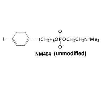
[0010] We have previously shown that certain alkylphosphocholine analogs
are
preferentially taken up and retained by malignant solid tumor cells. In U.S.
Patent
Publication No. 2014/0030187, Weichert et al. disclose using analogs of the
base
compound 18-(p-iodophenyl)octadecyl phosphocholine (NM404; see Figure 1) for
detecting and locating, as well as for treating, a variety of malignant solid
tumors. If the
iodo moiety is an imaging-optimized radionuclide, such as iodine-124
([124I]_NM404),
the analog can be used in positron emission tomography¨computed tomography
(PET/CT) or single-photon emission computed tomography (SPECT) imaging of
solid
3
Date Recue/Date Received 2020-11-10
tumors. Alternatively, if the iodo moiety is a radionuclide optimized for
delivering
therapeutic doses of RT to the solid tumors cells in which the analog is taken
up, such as
iodine-125 or iodine-131 ([1251]-NM404 or [1311]-NM404), the analog call be
used to treat
the solid tumors.
[0011] Such analogs not only target a wide variety of solid tumor types
in vivo, but
also undergo prolonged selective retention in tumor cells, thus affording high
potential as
RT agents. Moreover, tumor uptake is limited to malignant cancer and not
premalignant
or benign lesions.
[0012] However, there are metal isotopes that have better properties for
optimized
imaging and/or RT than the radioactive iodine isotopes used in the previously
disclosed
alkylphosphocholine analogs. For example, as an imaging isotope, 1-124 suffers
from
poor positron output (only about 24% of the emissions are positrons), and it
suffers
further from a confounding gamma emission (600 KeV), which actually interferes
with
normal 511 KeV PET detection. Certain positron emitting metals have better
imaging
characteristics. As another example, as an RT isotope, 1-131 produces other
non-
therapeutic emissions at other energies, which add undesires radiation
dosimetry to
neighboring normal tissue, including bone marrow. The beta particle range of 1-
131 is
also quite long, which contributes to off target toxicity. Several metallic
radiotherapy
isotopes offer a cleaner emission profile and shorter pathlength and thus less
potential
toxicity.
[0013] We have developed improved alkylphosphocholine analogs that
include a
chelated radioactive metal isotope instead of a radioactive iodine isotope
(see, e.g., U.S.
Patent Application No. 15/343,604,). The analogs include the same backbone as
the
previously disclosed radioiodinated compounds, so they are still selectively
taken up and
retained in tumor cells. However, the chelated radioactive metal isotope
provides
improved emissions for imaging and/or radiotherapy applications. Such agents
are well
suited for delivering a sub-cytotoxic but immunomodulatory dose of ionizing RT
to all
malignant tumors present within a subject, regardless of whether their number
and
locations are known.
[0014] Accordingly, in a first aspect, the disclosure encompasses a
method of treating
a cancer comprising one or more malignant solid tumors in a subject. The
method
4
Date Recue/Date Received 2020-11-10
includes the steps of: (a) administering to the subject an immunomodulatory
dose of a
radioactive phospholipid metal chelate compound that is differentially taken
up by and
retained within malignant solid tumor tissue; and (b) performing in situ tumor
vaccination
in the subject at one or more of the malignant solid tumors using one or more
treatments
capable of stimulating specific immune cells within the tumor
microenvironment. An
"immunomodulatory dose" is a low or sub-cytotoxic RT dose of the targeted
radiotherapy
agent. Although NM404 is used in some of the examples below, other examples
use the
phospholipid metal chelate compound NM600, which similarly targets solid tumor
tissue.
For radiotherapy application, the radioactive metal chelated into the compound
could
include any alpha, beta, auger, and/or gamma emitting metal. The key feature
is that
targeted radiotherapy agent emits low or sub-cytotoxic RT doses that are not
lethal to
either the cancer cells or the relevant immune cells.
[0015] In some embodiments, the one or more treatments capable of
stimulating
specific immune cells within the tumor microenvironment include treating the
tumor with
xRT. In some embodiments, the one or more treatments capable of stimulating
specific
immune cells within the tumor microenvironment include intratumorally
injecting into at
least one the one of the malignant solid tumors a composition that includes
one or more
agents capable of stimulating specific immune cells within the tumor
microenvironment.
In some embodiments, such agents can include an immunostimulatory monoclonal
antibody, a pattern recognition receptor agonist, an immunostimulatory
cytokine, an
immune stimulatory nanoparticle, an oncolytic virus, or any combinations
thereof. Non-
limiting examples of immunostimulatory monoclonal antibodies that could be
used
include anti-GD2 antibodies, anti-CTLA-4 antibodies, anti-CD137 antibodies,
anti-
CD134 antibodies, anti-PD-1 antibodies, anti-KIR antibodies, anti-LAG-3
antibodies,
anti-PD-L1 antibodies, anti-CD40 antibodies, or combinations thereof. In some
embodiments, the immunostimulatory monoclonal antibody is an antibody to a
tumor-
specific antigen. In some embodiments, the composition that includes one or
more
immunostimulatory monoclonal antibodies may also include interleukin-2 (IL-2).
In
some embodiments, the anti-GD2 antibody that is used may include hu14.18, and
optionally, may further include IL-2 (i.e., a fusion protein of the two).
Date Recue/Date Received 2020-11-10
[0016] In some embodiments, the immunostimulatory cytokine is IL-2,
interleukin-12
(IL-12), interleukin-15 (IL-15), interleukin-21 (IL-21), or an interferon
(IFN).
[0017] In some embodiments, the pattern recognition receptor agonist is an
agonist of
a toll-like receptor (TLR). Non-limiting examples of such TLRs TLR include TLR-
1,
TLR-2, TLR-3, TLR-4, TLR-5, TLR-6, TLR-7, TLR-8, TLR-9, or TLR-10.
[0018] In some embodiments, the radioactive phospholipid metal chelate
compound
has the formula:
R1 )a (CH2) n (OCH2CHYCH2)rp0 P OC H2CH3 -R2 lb
0'
or a salt thereof. Ri comprises a chelating agent that is chelated to a metal
atom, wherein
the metal atom is an alpha, beta or Auger emitting metal isotope with a half
life of greater
than 6 hours and less than 30 days; a is 0 or 1; n is an integer from 12 to
30; m is 0 or 1;
Y is -H, -OH, -COOH, -COOX, -0C0X, or -OX, wherein X is an alkyl or an aryl;
R2 is
-NH3, -N H2Z, -N HZ2, or -N Z3, wherein each Z is independently an alkyl or an
aroalkyl; and b is 1 or 2. Non-limiting examples of metal isotopes that could
be used
include Lu-177, Y-90, Ho-166, Re-186, Re-188, Cu-67, Au-199, Rh-105, Ra-223,
Ac-
225, As-211, Pb-212, or Th-227.
[0019] In some embodiments, the chelating agent is 1,4,7,10-
tetraazacyclododecane-
1,4,7-triacetic acid (DO3A) or one of its derivatives; 1,4,7-triazacyclononane-
1,4-diacetic
acid (NODA) or one of its derivatives; 1,4,7-triazacyclononane-1,4,7-triacetic
acid
(NOTA) or one of its derivatives; 1,4,7,10-tetraazacyclododecane-1,4,7,10-
tetraacetic
acid (DOTA) or one of its derivatives; 1,4,7-triazacyclononane,1-glutaric acid-
4,7-
diacetic acid (NODAGA) aor one of its derivatives; 1,4,7,10-
tetraazacyclodecane,1-
glutaric acid-4,7,10-triacetic acid (DOTAGA) or one of its derivatives;
1,4,8,11-
tetraazacyclotetradecane-1,4,8,11-tetraacetic acid (TETA) or one of its
derivatives;
1,4,8,11-tetraazabicyclo[6.6.2]hexadecane-4,11-diacetic acid (CB-TE2A) or one
of its
derivatives; diethylene triamine pentaacetic acid (DTPA), its diester, or one
of its
derivatives; 2-cyclohexyl diethylene triamine pentaacetic acid (CHX-A"-DTPA)
or one
6
Date Recue/Date Received 2020-11-10
of its derivatives; deforoxamine (DFO) or one of its derivatives; 1,2-[[6-
carboxypyridin-
2-yl]methylamino]ethane (H2dedpa) or one of its derivatives; and DADA or one
of its
derivatives, wherein DADA comprises the structure:
nr)--NH-1
0..NH HNro
HS SH -
[0020] In some embodiments, a is 1 (aliphatic aryl-alkyl chain). In other
embodiments, a is 0 (aliphatic alkyl chain).
[0021] In some embodiments, m is 1 (acylphospholipid series). In some such
embodiments, n is an integer between 12 and 20. In some embodiments, Y is
¨0C0X,
-COOX or¨OX.
[0022] In some embodiments, X is ¨CH2CH3 or ¨CH3.
[0023] In some embodiments, m is 0 (alkylphospholipid series).
[0024] In some embodiments, b is 1.
[0025] In some embodiments, n is 18.
[0026] In some embodiments, R2 is -1\1*Z3. In some such embodiments, each
Z is
independently ¨CH2CH3 or ¨CH3. In some such embodiments, each Z is ¨CH3.
7
Date Recue/Date Received 2020-11-10
[0027] In some
embodiments, the chelating agent chelated to the metal atom is:
0
(OH
01r*c,Ni
OHHO
)
0
HO
H 5
NThr
N
0
HO
o OH
OH
rN N
HO OHO
8
Date Recue/Date Received 2020-11-10
OH
H
c 'N,
HO--rN
0
0A0H '
0OH OH
t\r40 ¨1
fµl N HN
.õ1
-..N N) 0
/ 0 '
HO OHO
OH
A (LO
HN-I
0
OH
9
Date Recue/Date Received 2020-11-10
0.õOH OH
7.-N NDsvi
\¨N N
/0
.c.\__i 'N\
,
HO OHO
OH
\¨N N
HO 0
/ \/ il
/----N N N--\
HO2C ) ( ( CO2H
HO2C CO2H CO2H
,
Date Recue/Date Received 2020-11-10
Q, 1
/--N N N¨\
HO2C ) ( ( CO2H
HO2C CO2H CO2H
,
0,\ h0
1( (
-0 N N N 0¨I
HO2C¨/ ) \¨CO2H
HO2C
,
0
HON-P)5 0
HO-Nv N
HN--µ --r-NH
Hd 0
11
Date Recue/Date Received 2020-11-10
,
/ C
(=ix iN NH HN-)_),
N
/-OH HO4
0 0 , and
0
\-NH--1
/
C:1,NH HNr0
HS SH
12
Date Recue/Date Received 2020-11-10
[0028] In some embodiments, the radioactive phospholipid metal chelate
compound
is one of the following compounds, wherein the selected compound is chelated
to the
metal atom:
0
OH
H 0
NM)Ni (CH2)180POCH2CH2NMe3
N 0
OH )
0
0
OH
N (CH2)1801:1)0CH2CH2NMe3
(D/ ICNThy 0
N j 0
OH
HO
0
HO
Nk-11 (cH2)180isocH2cH2Nme3
HO
HO
0
r¨ N
N.----...õ-N¨(CH2)1800CH2CH2NMe3
0
0j\) 0
HO
13
Date Recue/Date Received 2020-11-10
00H OH
( /--\ r----0
rN N
0
LN N
II e
(cH2)18opocH2cH2Nme3
c\__/ 0 O
e
/
HO 0 HO ,
00H OH
( /--\ f----0
CO N
N N----\/(CH2)1801CH2CH22Me3
O
e
0
HO 0 HO ,
OH
0
r\ N 0
H---{--N II
O a
(CH2)180POCH2CH2NMe3
O
e
OH
/0
r\ N, 0
HO----(---N ii o
/1\1--"\----(CH2)180POCH2CH2NMe3
0 O
e
,,(CH)
,
14
Date Recue/Date Received 2020-11-10
O0H OH
0
rN I) HN . (CH2)18010CH2CH2NMe3
N N I0 e
0
HO 0 HO ,
O0H OH
0
H e
rN N HN¨(CH 1 0 ocH CH NMe x_ _2,18 _I)_
_ _2 __2___3
N N 0 0
0
HO 0 HO ,
OH
(LO
MH ii
HO,r---N / O
j HN 441 ,_..2)180POCH2CH2NMe3
____________________ 0 0 c/N
e
(Do=OH ,
OH
0
(N, 0
H 0
HO ,/N HN¨(CH2)180POCH2CH2NMe3
O
0 e
O0 OH
( /H--\ r----0
1N N7(
\ (CH2)180F)'OCH2CH2C)NMe3
\ ____ N N (!)
0
/0
HO OHO ,
Date Recue/Date Received 2020-11-10
N,OH OH
( i--\ r----0
0 N N
e
¨7
il
(CH2)180POCH2CH2NMe3
N N O
0
HO 0 HO ,
OH
i--\ i----0
/ ____ NN¨¨/)
(CH2)180910CH2CH2C)NMe3
\ ____ N N 0
HO 0 ,
OH
i--\ i----0
N N
\¨\
N 0
ii e
(cH2)18opocH2cH2Nme3
N
O
e
HO 0 ,
0
II e
(cH2)18opocH2cH2Nme3
/--\ O
/¨N N N¨\ e
Ho2c ) ( ( co2H
Ho2c co2H co2H
,
o
II e
/ ___________________ (CH2)18 Ol'OCH2CH2NMe3
/ \ / 0
/¨N N N¨\ e
Ho2c ) ( co2H
Ho2c co2H co2H
,
16
Date Recue/Date Received 2020-11-10
0 G
Q II
(cH2)18opocH2cH2Nme3
O
/¨N N N¨\ e
Ho2c ) ( ( co2H
Ho2c co2H co2H
,
o e
Q, ______________ i (cH2)18 aocH2cH2Nme3
O
/¨N N N¨\ e
Ho2c ) ( ( co2H
Ho2c co2H co2H
,
o o
o o
o
Me3NCH2CH2OPO(CH2)18-0 N N N 0¨(CH2)180POCH2CH2NMe3
O Ho2c¨/ ) \¨co2H O
0 Ho2c e ,
o
0¨ HN¨l(
N H __________________________________________
HO 5 0
HO¨N
e 0 HN¨\ 0 NH
Me3NCH2CH20 Fi 0(CH2)18 \ 5 0
0 0 "N __ (
e
HO o ,
o
o HN¨
N ( 1
HO 5 0
HO¨N
G 0 HN __ \ ) NH
Me3NCH2CH201-0(CH2)18 ___________ \ 5 0
0 0 \
N _______________________________________ (
0
HO 0 ,
17
Date Recue/Date Received 2020-11-10
(cH2)180pocH2cH2Nme3
NH HN
( /N N/
OH HO
0 0
0
/ _______________ (CH2)18 01/00H2CH2NMe3
/ 0
NH HN
( /N
OH HO
0 0
0 0
NH Vr (CH x_..2)180POCH2CH2NMe3
NH HN
HS SH ,or
0 0
-NH-(CH2)1801=i'OCH2CH2NMe3
/ 0
NH HN
HS SH
[0029] In some embodiments, the radioactive phospholipid chelate compound
is
administered intravenously.
[0030] In some embodiments, the subject is a human.
[0031] In some embodiments, the method optionally further includes
exposing one of
the malignant solid tumors to xRT.
18
Date Recue/Date Received 2020-11-10
[0032] In some embodiments, the method optionally includes the step of
determining
the immonostimulatory dose of the radioactive phospholipid chelate compound.
In some
such embodiments, the step of determining the immunomodulatory dose of the
radioactive phospholipid chelate compound includes administering to the
subject a
detection-facilitating dose of a radioactive phospholipid chelate compound as
described
previously, except that the metal atom is a positron or single photon emitting
metal
isotope with a half life of greater than or equal to 4 hours, and subsequently
detecting
signals originating from the one or more malignant solid tumors within the
subject that
are characteristic of the metal isotope within the radioactive phospholipid
chelate
compound. In some such embodiments, the positron or single photon emitting
metal
isotope is Go-66, Cu-64, Y-86, Co-55, Zr-89, Sr-83, Mn-52, As-72, Sc-44, Ga-
67, In-
111, or Tc-99m.
[0033] In some embodiments, the immunomodulatory dose of the radioactive
phospholipid chelate compound is calculated from the strength of the signals
originating
from the one or more malignant solid tumors within the subject.
[0034] In some embodiments, the step of detecting signals characteristic
of the metal
isotope is performed by positron emission tomography (PET) imaging or single-
photon
emission computed tomography (SPECT) imaging.
[0035] Non-limiting examples of the cancers presenting as malignant solid
tumors
that could treated using the disclosed method include melanoma, neuroblastoma,
lung
cancer, adrenal cancer, colon cancer, colorectal cancer, ovarian cancer,
prostate cancer,
liver cancer, subcutaneous cancer, squamous cell cancer of the skin or head
and neck,
intestinal cancer, retinoblastoma, cervical cancer, glioma, breast cancer,
pancreatic
cancer, soft tissue sarcomas, Ewings sarcoma, rhabdomyosarcoma, osteosarcoma,
retinoblastoma, Wilms' tumor, or pediatric brain tumors.
[0036] Other objects, features and advantages of the present invention
will become
apparent after review of the specification, claims and drawings.
BRIEF DESCRIPTION OF THE DRAWINGS
[0037] Fig. 1 shows the chemical structure of the base compound 18- (p-
iodophenyl)
octadecyl phosphcholine (NM404).
19
Date Recue/Date Received 2020-11-10
[0038] Fig. 2A, Fig. 2B and Fig. 2C are a series of graphs showing that
xRT + IT-IC
elicits in situ tumor vaccination. Fig. 2A) Tumor growth curves and Fig. 2B)
Kaplan-
Meier survival curves show synergy between xRT and IT-hu14.18-IL2. 71% (22/31)
of
mice treated with xRT + IT-IC are rendered disease-free. Fig. 2C) 90% of these
reject
subsequent engraftment with B78 melanoma.
[0039] Fig. 3 is a graph demonstrating concomitant immune tolerance.
Primary tumor
response is shown. A distant un-treated tumor suppresses response to xRT + IT-
IC in a 2-
tumor B78 melanoma model, and this suppression can be overcome be radiating
the
second tumor.
[0040] Fig. 4 is a graph showing that concomitant immune tolerance is due
to Tregs.
Primary tumor response is shown. A distant un-treated tumor suppresses
response to xRT
+ IT-IC in a 2-tumor B78 melanoma model and this suppression can be overcome
by
depleting Tregs (using transgenic DEREG mice that express diphtheria toxin
receptors on
their Tregs, and thus depleting Tregs by administering diphtheria toxin).
[0041] Fig. 5 is an image showing selective uptake of 1241-NM404 by B78
melanoma.
A mouse bearing a ¨200mm3 B78 tumor received IV 124I-NM404 and had serial
PET/CT
scans done. This image at 71h shows selective uptake by the tumor with some
residual
background uptake by the heart and liver.
[0042] Fig. 6 is a graph demonstrating that in situ vaccination can be
elicited in the
presence of residual levels of molecular targeted radiation therapy (TRT).
Treatment with
combined xRT + IT-IC is equally effective in the presence or absence of 3 [iCi
131I-
NM404. This approximates the residual activity of TRT that will be present
when we
deliver xRT (d0) followed by IT-IC (d6-10), as described in Example 4.
[0043] Fig. 7 shows a time course MRI image of a tumor-bearing mouse
following
injection of Gd-NM600 showing enhancement of the tumor (T) by 24 hours.
[0044] Figures 8A, 8B, 8C, 8D and 8E are a series of graphs showing tumor-
specific
inhibition of primary tumor response to the combination of local RT+IT-IC by a
distant
untreated tumor in murine melanoma and pancreatic tumor models. C57BL/6 mice
bearing a syngeneic, disialoganglioside-expressing (GD2+), primary flank tumor
+/- a
secondary tumor on the contralateral flank were treated to the primary tumor
only, as
indicated, with xRT on day "1" and intra-tumor (IT) injection of 50 mcg of the
anti-GD2
Date Recue/Date Received 2020-11-10
immunocytokine (IC), hu14.18-1L2 (a fusion of anti-GD2 mAb and IL2), on day 6-
10.
Mean primary tumor volumes are displayed in Figures 8A and 8C-8E. 8A). In mice
bearing a primary B78 melanoma tumor, the presence of an untreated secondary
B78
tumor antagonized primary tumor response to RT+IT-IC. We describe this effect
as
"concomitant immune tolerance" ¨ an antagonistic effect of a non-treated
distant tumor
on the local response of a treated tumor to xRT + IT-IC. 8B) Kaplan-Meier
survival
curves are shown for mice in panel A plus replicate experiments. Nearly all
mice were
euthanized due to primary tumor progression. 8C) In mice bearing a primary
Panc02-
GD2+ pancreatic tumor, with or without a secondary Panc02-GD2¨ tumor on the
opposite flank, the presence of an untreated Panc02 secondary tumor suppressed
the
response of a primary Panc02-GD2+ tumor to RT+IT-IC. 7D) In mice bearing a
primary
B78 melanoma tumor, a secondary B78 tumor suppressed primary tumor response to
RT+IT-IC but a secondary Panc02-GD2+ pancreatic tumor did not exert this
effect. 8E)
In mice bearing a primary Panc02-GD2+ tumor a secondary Panc02-GD2¨ tumor
suppressed primary tumor response to combined xRT and IT-hu14.18-1L2, while a
B78
secondary tumor did not. n=number of mice per group. NS=non-significant,
***p<0.001.
[0045] Figures 9A, 9B and 9C include immunohistochemistry images and
graphs
showing that concomitant immune tolerance is circumvented by specific
depletion of
regulator T cells (Tregs). 9A). Immunohistochemistry for the Treg marker,
FoxP3
(representative 400x images are shown) for tumors evaluated on day 6 after xRT
in mice
with one (Al and A2) or two (A3 and A4) tumors. Mice received no xRT, or xRT
only to
the primary tumor. The primary tumor is shown in Al-A3 and the secondary is
shown in
A4. Small arrows point out some of the FoxP3+ cells (brown nuclei = FoxP3+,
blue =
hematoxylin counterstain). The graphs on the right display blinded
quantification of
FoxP3+ cells per 200x field, corresponding to the conditions shown in Al, A2,
A3 and
A4, respectively. 9B and 9C) DEREG mice express diphtheria toxin receptor
under
control of the Treg-specific FoxP3 promoter, enabling specific depletion of
Tregs upon
IP injection of diphtheria toxin. DEREG mice bearing primary and secondary B78
melanoma tumors were treated with xRT+IT-IC to the primary tumor and IP
injection of
either diphtheria toxin or PBS (the first of replicate experiments are shown).
Concomitant
immune tolerance is eliminated following depletion of Tregs in these mice,
resulting in
21
Date Recue/Date Received 2020-11-10
improved 9B) primary and 9C) secondary tumor response. n=number of mice per
group.
"p<0.01, ***p<0.001.
[0046] Figures 10A and 10B are graphs showing that concomitant immune
tolerance
is overcome by delivering xRT to both tumor sites. In mice bearing primary and
secondary B78 tumors, the secondary tumor suppresses primary tumor response to
primary tumor treatment with xRT + IT-IC. This is overcome by delivering 12 Gy
xRT to
both the primary and secondary tumors and IT-IC to the primary tumor,
resulting in
improved 10A) primary tumor response (the first of replicate experiments is
shown) and
10B) aggregate animal survival from replicate experiments. n=number of mice
per group.
"p<0.01, ***p<0.001.
[0047] Figures 11A, 11B and 11C are a series of graphs showing that low
dose xRT
alone does not elicit in situ vaccination but does overcome concomitant immune
tolerance when delivered to distant tumor sites together with 12 Gy + IT-IC
treatment of
an in situ vaccine site. 11A) In mice bearing a primary B78 tumor only, 12 Gy
+ IT-IC
elicits in situ vaccination (as shown previously) and results in complete
tumor regression
in most mice (4/6 in this experiment) and a memory immune response (Morris,
Cancer
Res, 2016). On the other hand no animals exhibit complete tumor regression
following
either IT-IC alone or low dose (2 Gy) xRT + IT-IC (0/6 in both groups) p<0.05.
11B) In
mice bearing a primary and secondary B78 melanoma tumor, low dose xRT (2 Gy or
5
Gy) delivered to the secondary tumor is comparable to 12 Gy in its capacity to
overcome
concomitant immune tolerance at the primary tumor. 11C) In these same animals,
it is
apparent that overcoming concomitant immune tolerance by delivery of low dose
xRT to
the secondary tumor rescues a systemic response to IT-IC immunotherapy. In
this
context, when xRT is delivered to all tumor sites then IT-IC injection of the
primary
tumor triggers a systemic anti-tumor effect that renders secondary tumor
response to 2
Gy or 5 Gy greater than the response to 12 Gy xRT in absence of primary tumor
IT-IC
injection.
[0048] Figures 12A, 12B, 12C and 12D is a PET image (12A) and a series of
bar
graphs (12B, 12C and 12D) showing that low dose TRT with 131I-NM404
effectively
depletes tumor infiltrating FoxP3+ Tregs without systemic leukopenia or
depletion of
tumor infiltrating CD8+ effector T cells. In most clinical scenarios, it is
not feasible to
22
Date Recue/Date Received 2020-11-10
deliver external beam, even low dose, to all tumor sites without eliciting
marked bone
marrow depletion and leukopenia that would result in immunosuppression. Here
we
tested whether TRT could be administered systemically to specifically deplete
tumor
infiltrating suppressive immune cells (Tregs), without triggering systemic
immune cell
depletion and leukopenia. 12A) Dosimetry studies in this B78 melanoma tumor
model
using positron-emitting '241-NM404 confirm tumor-selective uptake of NM404.
C57BL/6
mice bearing B78 tumors were treated with 60 jiCi 131I-NM404. This activity
approximates the amount of 131I-NM404 necessary to deliver ¨ 2 Gy TRT to a B78
tumor. Peripheral blood and tumor samples were collected in untreated control
mice (C)
and at 8 day intervals (Ti = d8, T2 = d16, T3 = d24, T4 = d32) thereafter.
12B) This dose
of TRT did not result in any significant systemic leukopenia and 12C) did not
significantly affect the level of tumor infiltrating CD8+ effector T cells
(ANOVA
p=0.25). 12D) However, tumor infiltrating FoxP3+ Tregs were significantly
depleted by
this dose of TRT (ANOVA p=0.03; * p<0.05).
[0049] Figures 13A and 13B are graphs showing that low dose TRT with 1311-
NM404
effectively overcomes concomitant immune tolerance and rescues the systemic
anti-
tumor effect of in situ vaccination. Given the capacity of low dose 131I-NM404
TRT to
deplete tumor-infiltrating Tregs without rendering a mouse leukopenic, we
tested whether
low dose 131I-NM404 might effectively overcome concomitant immune tolerance.
C57BL/6 mice bearing two B78 tumors were treated with 60-mcCi 131I-NM404 on
day 1
(NM404), as indicated. After one half-life (day 8), animals received 12 Gy xRT
or no
xRT to the primary tumor (in situ vaccine site). Control mice receiving no
131I-NM404
were treated to the secondary tumor as indicated (0, 2, or 12 Gy). Mice
received daily IT
injections of IC to the primary tumor (in situ vaccine site), as indicated, on
days 13-17.
13A) Primary tumor and 13B) secondary tumor response is shown and demonstrates
that
administration of low dose TRT effectively overcomes concomitant immune
tolerance
and rescues the systemic anti-tumor effect of in situ vaccination.
[0050] Figure 14 shows the chemical structure of an exemplary
alkylphosphcholine
metal chelate (64Cu-NM600). Other metals may be used in place of "Cu.
[0051] Figure 15 is a PET/CT image of two single tumor B78 mice from a
scan taken
48 hours post-injection with 86Y-NM600.
23
Date Recue/Date Received 2020-11-10
[0052] Figure 16 is a PET/CT image of two two-tumor B78 mice from a scan
taken
48 hours post-injection with 86Y-NM600.
[0053] Figure 17 includes PET/CT images for a U87MG mouse from scans taken
3
hours (left panel), 24 hours (center panel) and 48 hours (right panel) post-
injection with
64Cu-NM600. The images show tissue activity calculated as a percent of
injected dose/g
tissue (%ID/g, scale shown on far right).
[0054] Figure 18 includes PET/CT images for a 4T1 mouse from scans taken 3
hours
(left panel), 24 hours (center panel) and 48 hours (right panel) post-
injection with 64Cu-
NM600. The images show tissue activity calculated as a percent of injected
dose/g tissue
(%ID/g, scale shown on far right).
[0055] Figure 19 includes PET/CT images for an HCT-116 mouse from scans
taken 3
hours (left panel), 24 hours (center panel) and 48 hours (right panel) post-
injection with
64Cu-NM600. The images show tissue activity calculated as a percent of
injected dose/g
tissue (%ID/g, scale shown on far right).
[0056] Figure 20 includes PET/CT images for an A549 mouse from scans taken
3
hours (left panel), 24 hours (center panel) and 48 hours (right panel) post-
injection with
64Cu-NM600. The images show tissue activity calculated as a percent of
injected dose/g
tissue (%ID/g, scale shown on far right).
[0057] Figure 21 includes PET/CT images for a PC-3 mouse from scans taken
3
hours (left panel), 24 hours (center panel) and 48 hours (right panel) post-
injection with
64Cu-NM600. The images show tissue activity calculated as a percent of
injected dose/g
tissue (%ID/g, scale shown on far right).
[0058] Figure 22 includes PET/CT images for an HT-29 mouse from scans
taken 3
hours (left panel), 24 hours (center panel) and 48 hours (right panel) post-
injection with
64Cu-NM600. The images show tissue activity calculated as a percent of
injected dose/g
tissue (%ID/g, scale shown on far right).
[0059] Figure 23 includes PET/CT images for a MiaPaca mouse from scans
taken 3
hours (left panel), 24 hours (center panel) and 48 hours (right panel) post-
injection with
64Cu-NM600. The images show tissue activity calculated as a percent of
injected dose/g
tissue (%ID/g, scale shown on far right).
24
Date Recue/Date Received 2020-11-10
[0060] Figure 24 includes PET/CT images for a 4T1 mouse from scans taken 3
hours
(left panel), 24 hours (center panel) and 48 hours (right panel) post-
injection with 86Y-
NM600. The images show tissue activity calculated as a percent of injected
dose/g tissue
(%ID/g, scale shown on far right).
[0061] Figure 25 includes PET/CT images for a 4T1 mouse from scans taken 3
hours
(left panel), 24 hours (center panel) and 48 hours (right panel) post-
injection with 89Zr-
NM600. The images show tissue activity calculated as a percent of injected
dose/g tissue
(%ID/g, scale shown on far right).
[0062] Figure 26 includes PET/CT images for an HT-29 mouse from scans
taken 4
hours (left panel) and 1 day (right panel) post-injection with 52Mn-NM600. The
images
show tissue activity calculated as a percent of injected dose/g tissue (%ID/g,
scale shown
on far right).
[0063] Figure 27 includes PET/CT images for a PC-3 mouse from scans taken
4
hours (left panel) and 1 day (right panel) post-injection with 52Mn-NM600. The
images
show tissue activity calculated as a percent of injected dose/g tissue (%1D/g,
scale shown
to the right of each image).
[0064] Figure 28 includes PET/CT images for an HT-29 mouse from scans
taken 2
days (left panel), 3 days (second panel from the left), 5 days (second panel
form the right)
and 7 days (right panel) post-injection with 52Mn-NM600. The images show
tissue
activity calculated as a percent of injected dose/g tissue (%ID/g, scale shown
to the right
of the images).
[0065] Figure 29 includes PET/CT images for a PC-3 mouse from scans taken
2 days
(left panel), 3 days (second panel from the left), 5 days (second panel form
the right) and
7 days (right panel) post-injection with 52Mn-NM600. The images show tissue
activity
calculated as a percent of injected dose/g tissue (%ID/g, scale shown to the
right of the
images).
[0066] Figure 30 is a graph showing PET quantitative region of interest
data (chelate
uptake as a function of time) for 4T1 tumor tissue in 4T1 mice injected with
86Y-NM600,
64Cu-NM600 and 89Zr-NM-600.
Date Recue/Date Received 2020-11-10
[0067] Figure 31 is a graph showing PET quantitative region of interest
data
(chelate uptake as a function of time) for heart tissue in 4T1 mice injected
with 86Y-
NM600, 64Cu-NM600 and 89Zr-NM-600.
[0068] Figure 32 is a graph showing PET quantitative region of interest
data (chelate
uptake as a function of time) for liver tissue in 4T1 mice injected with 86Y-
NM600, 64Cu-
NM600 and 89Zr-NM-600.
[0069] Figure 33 is a graph showing PET quantitative region of interest
data (chelate
uptake as a function of time) for whole body in 4T1 mice injected with 86Y-
NM600,
64Cu-NM600 and 89Zr-NM-600.
[0070] Figure 34 is a bar graph illustrating ex vivo chelate
biodistribution in healthy
and tumor tissues in 4T1 mice 48 hours (86Y-NM600, 64Cu-NM600, 89Zr-NM-600 and
177Lu-NM600) and 96 hours (177Lu-NM600) post-injection of the metal chelates.
[0071] Figure 35 shows the chemical structure of an exemplary
alkylphosphcholine
metal chelate (177Lu-NM600). Other metals may be used in place of 177Lu.
[0072] Figure 36 is an audioradiographic image of three B78 mice taken 48
hours
after injection with 90Y-NM600. Xenografted B78 tumors are seen as large dark
spots at
the lower right of each mouse image.
[0073] Figure 37 is an audioradiographic image of three B78 mice taken 96
hours
after injection with 90Y-NM600. Xenografted B78 tumors are seen as large dark
spots at
the lower right of each mouse image.
[0074] Figure 38 is an audioradiographic image of a B78 mouse taken on day
5 after
injection with 177Lu-NM600. Xenografted B78 tumors are seen as two dark spots
at the
bottom of the mouse image.
[0075] Figure 39 is an audioradiographic image of a B78 mouse taken on day
13 after
injection with 177Lu-NM600. Xenografted B78 tumors are seen as two dark spots
at the
bottom of the mouse image.
[0076] This paragraph has been intentionally deleted.
[0077] Figure 40 is an audioradiographic image of a MiaPaca mouse taken 10
days
after injection with 177Lu-NM600. The location of the xenografted MiaPaca
tumor is
indicated by the arrow and dashed circle.
26
Date Recue/Date Received 2020-11-10
[0078] Figure 41 is an audioradiographic image of three 4T1 mice taken 48
hours
after injection with 177Lu-NM600. The locations of the xenografted 4T1 tumors
are
indicated by the arrows and dashed circles.
[0079] Figure 42 is an audioradiographic image of three 4T1 mice taken 96
hours
after injection with 177Lu-NM600. The locations of the xenografted 4T1 tumors
are
indicated by the dashed circles.
[0080] Figure 43 is an audioradiographic image of three 4T1 mice taken 4
hours after
injection with 90Y-NM600. The locations of the xenografted 4T1 tumors are
indicated by
the arrows and dashed circles.
[0081] Figure 44 is an audioradiographic image of three 4T1 mice taken 48
hours
after injection with 90Y-NM600. The xenografted 4T1 tumors are seen as large
dark
spots on the lower right of each mouse image.
[0082] Figure 45 is an audioradiographic image of three 4T1 mice taken 96
hours
after injection with 90Y-NM600. The xenografted 4T1 tumors are seen as large
dark
spots on the lower right of each mouse image.
[0083] Figure 46 is a graph illustrating the radiotherapeutic effect of
90Y-NM600 at
two different doses (150 Ci and 300 Ci) in a B78 xenograft mouse model,
versus a
control (excipient only). Data is presented as measured tumor volume in mm3 as
a
function of time in days after injection.
[0084] Figure 47 is a graph illustrating the radiotherapeutic effect of a
single 500 Ci
dose of 177Lu-NM600 in a B78 xenograft mouse model, versus a control
(excipient only).
Data is presented as measured tumor volume in mm3 as a function of time in
days after
injection.
[0085] Figure 48 is a graph illustrating the radiotherapeutic effect of a
single 400 Ci
dose of 177Lu-NM600 in a MiaPaca xenograft mouse model, versus a control
(excipient
only). Data is presented as measured tumor volume in mm3 as a function of time
in days
after injection.
[0086] Figure 49 is a graph illustrating the radiotherapeutic effect of a
single 500 Ci
dose of 177Lu-NM600 in a 4T1 xenograft mouse model, versus a control
(excipient only).
Data is presented as measured tumor volume in mm3 as a function of time in
days after
injection. * P < 0.05; ** P <0.01; *** P < 0.001.
27
Date Recue/Date Received 2020-11-10
[0087] Figure 50 is a graph illustrating the radiotherapeutic effect of
two serial doses
of 177Lu-NM600 (500 Ci and 250 Ci) in a 4T1 xenograft mouse model, versus a
control (excipient only). Data is presented as measured tumor volume in mm3 as
a
function of time in days after injection.
[0088] Figure 51 is a graph illustrating the radiotherapeutic effect of
177Lu-NM600 at
two different doses (500 uCi and 250 uCi) in a 4T1 xenograft mouse model,
versus a
control (excipient only). Data is presented as measured tumor volume in mm3 as
a
function of time in days after injection.
[0089] Figure 52 is a graph illustrating the impact of tumor mass on the
comparative
therapeutic efficacy of 90Y-NM600 and 131I-NM404 in conventional TRT.
[0090] Figure 53 is a bar graph comparing average albumin binding energies
of three
different metal chelate analogs of NM404, along with an amine analog. For
comparison,
the binding energy of I-NM404 is shown as a dotted line.
DETAILED DESCRIPTION
I. IN GENERAL
[0091] It is understood that this disclosure is not limited to the
particular
methodology, protocols, materials, and reagents described, as these may vary.
The
terminology used herein is for the purpose of describing particular
embodiments only,
and is not intended to limit the scope of the present invention, which will be
limited only
by any later-filed nonprovisional applications.
[0092] As used herein and in the appended claims, the singular forms "a",
"an", and
"the" include plural reference unless the context clearly dictates otherwise.
As well, the
terms "a" (or "an"), "one or more" and "at least one" can be used
interchangeably herein.
The terms "comprising" and variations thereof do not have a limiting meaning
where
these terms appear in the description and claims. Accordingly, the terms
"comprising",
"including", and "having" can be used interchangeably.
[0093] Unless defined otherwise, all technical and scientific terms used
herein have
the same meanings as commonly understood by one of ordinary skill in the art
to which
this invention belongs. Although any methods and materials similar or
equivalent to
those described herein can be used in the practice or testing of the present
invention, the
28
Date Recue/Date Received 2020-11-10
preferred methods and materials are now described. All references cited in
this
specification are to be taken as indicative of the level of skill in the art.
[0094] The terminology as set forth herein is for description of the
embodiments only
and should not be construed as limiting of the invention as a whole. Unless
otherwise
specified, "a," "an," "the," and "at least one" are used interchangeably and
mean one or
more than one.
[0095] The disclosure is inclusive of the compounds described herein
(including
intermediates) in any of their pharmaceutically acceptable forms, including
isomers (e.g.,
diastereomers and enantiomers), tautomers, salts, solvates, polymorphs,
prodrugs, and the
like. In particular, if a compound is optically active, the invention
specifically includes
each of the compound's enantiomers as well as racemic mixtures of the
enantiomers. It
should be understood that the term "compound" includes any or all of such
forms,
whether explicitly stated or not (although at times, "salts" are explicitly
stated).
[0096] "Pharmaceutically acceptable" as used herein means that the
compound or
composition or carrier is suitable for administration to a subject to achieve
the treatments
described herein, without unduly deleterious side effects in light of the
necessity of the
treatment.
[0097] The term "effective amount," as used herein, refers to the amount
of the
compounds or dosages that will elicit the biological or medical response of a
subject,
tissue or cell that is being sought by the researcher, veterinarian, medical
doctor or other
clinician.
[0098] As used herein, "pharmaceutically-acceptable carrier" includes any
and all dry
powder, solvents, dispersion media, coatings, antibacterial and antifungal
agents, isotonic
agents, absorption delaying agents, and the like. Pharmaceutically-acceptable
carriers are
materials, useful for the purpose of administering the compounds in the method
of the
present invention, which are preferably non-toxic, and may be solid, liquid,
or gaseous
materials, which are otherwise inert and pharmaceutically acceptable, and are
compatible
with the compounds of the present invention. Examples of such carriers
include, without
limitation, various lactose, mannitol, oils such as corn oil, buffers such as
PBS, saline,
polyethylene glycol, glycerin, polypropylene glycol, dimethylsulfoxide, an
amide such as
29
Date Recue/Date Received 2020-11-10
dimethylacetamide, a protein such as albumin, and a detergent such as Tween
80, mono-
and oligopolysaccharides such as glucose, lactose, cyclodextrins and starch.
[0099] The term "administering" or "administration," as used herein,
refers to
providing the compound or pharmaceutical composition of the invention to a
subject
suffering from or at risk of the diseases or conditions to be treated or
prevented.
[00100] A route of administration in pharmacology is the path by which a drug
is
taken into the body. Routes of administration may be generally classified by
the location
at which the substance is applied. Common examples may include oral and
intravenous
administration. Routes can also be classified based on where the target of
action is.
Action may be topical (local), enteral (system-wide effect, but delivered
through the
gastrointestinal tract), or parenteral (systemic action, but delivered by
routes other than
the GI tract), via lung by inhalation. One form of local administration
refered to in this
submission is intratum oral (IT), whereby an agent is injected directly into,
or adjacent to,
a known tumor site.
[001011 A topical administration emphasizes local effect, and substance is
applied
directly where its action is desired. Sometimes, however, the term topical may
be
defined as applied to a localized area of the body or to the surface of a body
part, without
necessarily involving target effect of the substance, making the
classification rather a
variant of the classification based on application location. In an enteral
administration,
the desired effect is systemic (non-local), substance is given via the
digestive tract. In a
parenteral administration, the desired effect is systemic, and substance is
given by routes
other than the digestive tract.
[00102] Non-limiting examples for topical administrations may include
epicutaneous
(application onto the skin), e.g., allergy testing or typical local
anesthesia, inhalational,
e.g. asthma medications, enema, e.g., contrast media for imaging of the bowel,
eye drops
(onto the conjunctiva), e.g., antibiotics for conjunctivitis, ear drops, such
as antibiotics
and corticosteroids for otitis externa, and those through mucous membranes in
the body.
[00103] Enteral administration may be administration that involves any part of
the
gastrointestinal tract and has systemic effects. The examples may include
those by mouth
(orally), many drugs as tablets, capsules, or drops, those by gastric feeding
tube, duodenal
Date Recue/Date Received 2020-11-10
feeding tube, or gastrostomy, many drugs and enteral nutrition, and those
rectally, various
drugs in suppository.
[00104] Examples of parenteral administrations may include intravenous (into a
vein),
e.g. many drugs, total parenteral nutrition intra-arterial (into an artery),
e.g., vasodilator
drugs in the treatment of vasospasm and thrombolytic drugs for treatment of
embolism,
intraosseous infusion (into the bone marrow), intra-muscular, intracerebral
(into the
brain parenchyma), intracerebroventricular (into cerebral ventricular system),
intrathecal
(an injection into the spinal canal), and subcutaneous (under the skin). Among
them,
intraosseous infusion is, in effect, an indirect intravenous access because
the bone
marrow drains directly into the venous system. Intraosseous infusion may be
occasionally used for drugs and fluids in emergency medicine and pediatrics
when
intravenous access is difficult.
[00105] The following abbreviations are used in this disclosure: ADCC,
Antibody
dependent cell-mediated cytotoxicity; B16, A melanoma syngeneic to C57B1/6
mice;
B78, A variant of B16 that expresses GD2, due to transfection with GD2
synthase; D,
day; Hu14.18-1L2, The primary immunocytokine (reacts against GD2) used in the
studies
disclosed in the examples; IC, Immunocytoline (a fusion protein of a tumor-
reactive mAb
linked to IL2); IL2, Interleukin 2; IT, Intratumoral; IV, Intravenous; mAb,
Monoclonal
antibody; MAHA, Mouse anti-human antibody; NM404, used to designate the
phospholipid ether shown in Figure 1, which is selectively taken up by most
tumors and
used for TRT in the studies disclosed in the examples; NM600, used to
designate the
phospholipid ether shown in Figure 14, which can be chelated with any metal,
and which
is also selectively taken up by most tumors and used for TRT in the studies
disclosed in
the examples; NXS2, A neuroblastoma syngeneic to AJ mice; Panc02-GD2, A
pancreatic
cancer syngeneic to C57B1/6 mice, expressing GD2, due to transfection with GD2
synthase; PLE, Phospho-lipid Ether; RT, Radiation therapy; TRT, Targeted
radiotherapy;
W, week; 9464D-GD2, A neuroblastoma syngeneic to C57B1/6 mice, expressing GD2,
due to transfection with GD2 synthase.
THE INVENTION
31
Date Recue/Date Received 2020-11-10
[00106] This disclosure is directed to methods of treating any cancer that
presents as
one or malignant solid tumors. The disclosed methods combine two treatment
steps, with
an unexpected synergy resulting in a much improved in situ vaccination effect
against the
malignant solid tumors. Specifically, an immunomodulatory dose of a
radioactive
phospholipid metal chelate compound that is differentially taken up by and
retained
within malignant solid tumor tissue is administered to the patient, and in
situ tumor
vaccination is performed by intratumorally injecting into (or applying to) at
least one of
the malignant solid tumors a composition that includes one or more agents
capable of
stimulating specific immune cells within the tumor microenvironment, either
with or
without additional xRT to at least one of the malignant solid tumors being
treated with
immune-stimulating agents. The immunomodulatory dose of the radioactive
phospholipid
metal chelate compound likely reduces Treg levels (and other immune-
suppressive
elements) and prevents the immune system dampening (concomitant immune
tolerance)
that occurs when xRT is used against a tumor and one or more additional tumors
are not
radiated, although an understanding of the mechanism is not necessary to
practice the
invention and the invention is not limited to any particular mechanism of
action.
A. Intratumoral immunization ¨ in situ vaccination
[00107] Compositions used for intratumoral immunization may include, without
limitation, one or more cytokines, immune checkpoint inhibitors, pattern
recognition
agaonists, and/or immunostimulatory monoclonal antibodies, including
antibodies against
tumor-specific antigens. For a review of intratumoral immunization/in situ
vaccination
strategies that are among those that could be used, see Pierce et al, Human
Vaccines &
Immunotherapeutics 11(8):1901-1909, 2015; and Marabelle eta!, Clin. Cancer
Res.
20(7):1747-56, 2014; and Morris et al, Cancer Res., e-pub ahead of print,
2016. In the
non-limiting examples disclosed herein, imtratumoral immunization was
performed by
injecting a fusion protein of an anti-GD2 mAb and interleukin 2 (hu14.18-IL2).
However, the disclosed methods are not in any way limited by these examples.
B. Immunomodulatory dose of a radioactive phospholipid metal chelate
compound
32
Date Recue/Date Received 2020-11-10
[00108] The radioactive phospholipid metal chelate compound used should
selectively
target a wide range of solid tumor cell types, such that the RT emitted by the
metal
isotope chelated to the metal chelate compound is directed to malignant solid
tumor
tissue without substantially exposing other tissue types to the emitted RT.
The radioactive
metal isotope included in the radioactive phospholipid metal chelate compound
may be
any radioactive metal isotope known to emit ionizing RT in a form that would
result in
immunostimulation of the cells that take up the compound. Non-limiting
examples of
radioactive metal isotopes that could be used include Lu-177, Y-90, Ho-166, Re-
186, Re-
188, Cu-67, Au-199, Rh-105, Ra-223, Ac-225, As-211, Pb-212, or Th-227.
[00109] The immunomodulatory RT dose (as opposed to injected dose) of the
radioactive phospholipid metal chelate compound is much less than the dose
that would
be used for conventional RT against malignant solid tumors. Specifically, the
dose
should be sufficient to stimulate a response in immune cells within the tumor
microenvironment (likely by reducing immune-suppressing Treg levels and other
immunosuppressive cells or molecules), while not ablating the desired immune
cells that
are responsible for the in situ vaccine effect.
[00110] The proper immunomodulatory dose can be calculated from imaging data
obtained after administering a "detection-facilitating" dose of a radioactive
metal chelate
compound. The detection-facilitating dose may be quite different than the
immunomodulatory dose, and the radioactive metal isotope that is chelated into
the
radioactive metal chelate compound may be different (although the rest of the
compound
structure should be the same). The radioactive metal isotope used in the
detection step
and dosimetry calculations may be any radioactive metal isotope known to emit
RT in a
form that is readily detectable by conventional imaging means. Non-limiting
examples
of "conventional imaging means" include gamma ray detection, PET scanning, and
SPECT scanning. Non-limiting examples of radioactive metal isotopes that could
be used
include Ga-66, Cu-64, Y-86, Co-55, Zr-89, Sr-83, Mn-52, As-72, Sc-44, Ga-67,
In-111,
or Tc-99m.
C. Metal chelates of PLE analogs
33
Date Recue/Date Received 2020-11-10
[00111] The disclosed structures utilize an alkylphosphocholine (APC) carrier
backbone. Once synthesized, the agents should harbor formulation properties
that render
them suitable for injection while retaining tumor selectivity as was
demonstrated
previously with the related radiohalogenated compounds. The disclosed
structures
include a chelating moiety to which the radioactive metal isotope will chelate
to produce
the final imaging or therapeutic agent.
D. Methods of Synthesizing Exemplary M-PLE Analogs
[00112] Proposed synthesis of compound 1 is shown below. The first step of the
synthesis is similar to described in Org Synth, 2008, 85, 10-14. The synthesis
is started
from cyclen which is converted into DO3A tris-Bn ester. This intermediate is
then
conjugated with NM404 in the presence of the base and Pd catalyst. Finally,
benzyl
protecting groups are removed by the catalytic hydrogenation.
o
?\---0Bn
0
cr> BrCH2CO2Bn Bna-J,(rN
N HN + I . trH 0
,.......2)180POCH2CH2NMe3
NH HN AcONa, DMAc '.- cNi O
ckli e
cyclen Bn040
0
r0Bn ---
( 0 e H2, Pd/C
______________________________________________________________ ..-
Pd catalyst N N 411 (CH2)180POCH2CH2NMe3
________ .- 0/¨cNJ (!)
base e
OBn )
Bn0¨ 0
0 ?'\--- OH
0
N N 41 (CH2)180112'0CH2CH2C)NMe3
i
0J 0
e
OH )
HO¨ 1
o
34
Date Recue/Date Received 2020-11-10
[00113] Synthesis of compound 2 is shown below. It begins with DO3A tris-Bn
ester
which is alkylated with 3-(bromo-prop-1-yny1)-trimethylsilane. After
alkylation, the
trimethjylsilyl group is removed and the intermediate acetylene is coupled
with NM404
by the Sonogashira reaction. The benzyl groups are removed and the triple bond
is
hydrogenated simultaneously in the last step of the synthesis.
0
0
r
?"\----0Bn \\---0Bn
0
0 Bn0-4 (-----N K2CO3
.-
Bna--. (----N BrCH2-CEC-SiMe3 \¨N N Me0H
cNJ i-Pr2NEt cNf\SiMe3
Bn0¨
Bn0 0
0
DO3A tris-Bn ester
0
?"--0Bn
Bn0-4 N 0 e pdc12(PPh3)2
(---- -> + ,
\¨N N 1 1,1 (CH2)1800CH2CH2NMe3
Et3N, Me0H
cNj\ 0
0
H
Bn04
0
0
?"--013n
(CH2)180POCH2CH2NMe3 H2, Pd/C
/ _____________ N
e
OBn
Bn0
0
0
(OH
N
N N---......... __ (CH2)1801%CH2CH2C)NMe3
o1
0/¨cNJ e
OH ) 2
HO-
0
Date Recue/Date Received 2020-11-10
[00114] Compounds 5 and 6 can be synthesized from same precursors, DTPA
dianhydride and 18-p-(3-hydroxyethyl-phenyl)-octadecyl phosphocholine as shown
in the
schemes below.
O rc02H 0 HO 0 e
, ___ \
O NNN1/ % + 111 (CH2)180POCH2CH2NMe3
(1 eq)
(!)
____ / \ __ /
\\ e
o 0
DTPA dianhydride
0
HO2C ¨\ /--\ /--\ /
___________ .._ N N N 0 0 e
HO2C¨' )
\_1...,-(-)....2i.14 111 (CH2)180POCH2CH2NMe3
HO2C i
0
0
O (CO2H 0
./ HO 0
O Nvr\jN 0 + II (CH2)180112'0CH2CH2ENMe3 (2 eq) -
'-
i __ / \ _______________________ (!)
o
e
0
DTPA dianhydride
0 0
\ /
0 0 N N N 0 0
e e
me3NcH2cH20lg0(cH2)18 41 HO2C¨" ) \_rn2. - -H . . (CH2)180F0CH2CH2NMe3
00 HO2C I
0
0
6
36
Date Recue/Date Received 2020-11-10
[00115] NOTA-NM404 conjugates can be synthesized in an analogous manner. One
example of NOTA-NM404 conjugate 7:
1--1 CO2H
HO2C,N N--I
(CH2)180POCH2CH2NMe3
1
0
CO2H e
7
E. Dosage Forms and Administration Methods
[00116] In situ vaccination can be performed by intratumoral injection, but
other
administration can apply (topical or systemic). For the synergistic targeted
RT, any route
of administration may be suitable. In one embodiment, the disclosed
alkylphosphocholine analogs may be administered to the subject via intravenous
injection. In another embodiment, the disclosed alkylphosphocholine analogs
may be
administered to the subject via any other suitable systemic deliveries, such
as parenteral,
intranasal, sublingual, rectal, or transdermal administrations.
[00117] In another embodiment, the disclosed alkylphosphocholine analogs may
be
administered to the subject via nasal systems or mouth through, e.g.,
inhalation.
[00118] In another embodiment, the disclosed alkylphosphocholine analogs may
be
administered to the subject via intraperitoneal injection or IP injection.
[00119] In certain embodiments, the disclosed alkylphosphocholine analogs may
be
provided as pharmaceutically acceptable salts. Other salts may, however, be
useful in the
preparation of the alkylphosphocholine analogs or of their pharmaceutically
acceptable
salts. Suitable pharmaceutically acceptable salts include, without limitation,
acid
37
Date Recue/Date Received 2020-11-10
addition salts which may, for example, be formed by mixing a solution of the
alkylphosphocholine analog with a solution of a pharmaceutically acceptable
acid such as
hydrochloric acid, sulphuric acid, methanesulphonic acid, fumaric acid, maleic
acid,
succinic acid, acetic acid, benzoic acid, oxalic acid, citric acid, tartaric
acid, carbonic acid
or phosphoric acid.
[00120] Where the disclosed alkylphosphocholine analogs have at least one
asymmetric center, they may accordingly exist as enantiomers. Where the
disclosed
alkylphosphocholine analogs possess two or more asymmetric centers, they may
additionally exist as diastereoisomers. It is to be understood that all such
isomers and
mixtures thereof in any proportion are encompassed within the scope of the
present
disclosure.
[00121] The disclosure also includes methods of using pharmaceutical
compositions
comprising one or more of the disclosed alkylphosphocholine analogs in
association with
a pharmaceutically acceptable carrier. Preferably these compositions are in
unit dosage
forms such as tablets, pills, capsules, powders, granules, sterile parenteral
solutions or
suspensions, metered aerosol or liquid sprays, drops, ampoules, auto-injector
devices or
suppositories; for parenteral, intranasal, sublingual or rectal
administration, or for
administration by inhalation or insufflation.
[00122] For preparing solid compositions such as tablets, the principal active
ingredient is mixed with a pharmaceutically acceptable carrier, e.g.
conventional
tableting ingredients such as corn starch, lactose, sucrose, sorbitol, talc,
stearic acid,
magnesium stearate, dicalcium phosphate or gums, and other pharmaceutical
diluents,
e.g. water, to form a solid preformulation composition containing a
homogeneous
mixture for a compound of the present invention, or a pharmaceutically
acceptable salt
thereof. When referring to these preformulation compositions as homogeneous,
it is
meant that the active ingredient is dispersed evenly throughout the
composition so that
the composition may be easily subdivided into equally effective unit dosage
forms such
as tablets, pills and capsules. This solid pre-formulation composition is then
subdivided
into unit dosage forms of the type described above containing from 0.1 to
about 500 mg
of the active ingredient of the present invention. Typical unit dosage forms
contain from
1 to 100 mg, for example, 1, 2, 5, 10, 25, 50 or 100 mg, of the active
ingredient. The
38
Date Recue/Date Received 2020-11-10
tablets or pills of the novel composition can be coated or otherwise
compounded to
provide a dosage affording the advantage of prolonged action. For example, the
tablet or
pill can comprise an inner dosage and an outer dosage component, the latter
being in the
form of an envelope over the former. The two components can be separated by an
enteric
layer which, serves to resist disintegration in the stomach and permits the
inner
component to pass intact into the duodenum or to be delayed in release. A
variety of
materials can be used for such enteric layers or coatings, such materials
including a
number of polymeric acids and mixtures of polymeric acids with such materials
as
shellac, cetyl alcohol and cellulose acetate.
[00123] The liquid forms in which the alkylphosphocholine analogs may be
incorporated for administration orally or by injection include aqueous
solutions, suitably
flavored syrups, aqueous or oil suspensions, and flavored emulsions with
edible oils such
as cottonseed oil, sesame oil, coconut oil or peanut oil, as well as elixirs
and similar
pharmaceutical vehicles. Suitable dispersing or suspending agents for aqueous
suspensions include synthetic and natural gums such as tragacanth, acacia,
alginate,
dextran, sodium caboxymethylcellulose, methylcellulose, polyvinylpyrrolidone
or
gelatin.
[00124] The disclosed alkylphosphocholine analogs are particularly useful when
formulated in the form of a pharmaceutical injectable dosage, including in
combination
with an injectable carrier system. As used herein, injectable and infusion
dosage forms
(i.e., parenteral dosage forms) include, but are not limited to, liposomal
injectables or a
lipid bilayer vesicle having phospholipids that encapsulate an active drug
substance.
Injection includes a sterile preparation intended for parenteral use.
[00125] Five distinct classes of injections exist as defined by the USP:
emulsions,
lipids, powders, solutions and suspensions. Emulsion injection includes an
emulsion
comprising a sterile, pyrogen-free preparation intended to be administered
parenterally.
Lipid complex and powder for solution injection are sterile preparations
intended for
reconstitution to form a solution for parenteral use. Powder for suspension
injection is a
sterile preparation intended for reconstitution to form a suspension for
parenteral use.
Powder lyophilized for liposomal suspension injection is a sterile freeze
dried preparation
intended for reconstitution for parenteral use that is formulated in a manner
allowing
39
Date Recue/Date Received 2020-11-10
incorporation of liposomes, such as a lipid bilayer vesicle having
phospholipids used to
encapsulate an active drug substance within a lipid bilayer or in an aqueous
space,
whereby the formulation may be formed upon reconstitution. Powder lyophilized
for
solution injection is a dosage form intended for the solution prepared by
lyophilization
("freeze drying"), whereby the process involves removing water from products
in a
frozen state at extremely low pressures, and whereby subsequent addition of
liquid
creates a solution that conforms in all respects to the requirements for
injections. Powder
lyophilized for suspension injection is a liquid preparation intended for
parenteral use that
contains solids suspended in a suitable fluid medium, and it conforms in all
respects to
the requirements for Sterile Suspensions, whereby the medicinal agents
intended for the
suspension are prepared by lyophilization. Solution injection involves a
liquid
preparation containing one or more drug substances dissolved in a suitable
solvent or
mixture of mutually miscible solvents that is suitable for injection.
[00126]
Solution concentrate injection involves a sterile preparation for parenteral
use
that, upon addition of suitable solvents, yields a solution conforming in all
respects to the
requirements for injections. Suspension injection involves a liquid
preparation (suitable
for injection) containing solid particles dispersed throughout a liquid phase,
whereby the
particles are insoluble, and whereby an oil phase is dispersed throughout an
aqueous
phase or vice-versa. Suspension liposomal injection is a liquid preparation
(suitable for
injection) having an oil phase dispersed throughout an aqueous phase in such a
manner
that liposomes (a lipid bilayer vesicle usually containing phospholipids used
to
encapsulate an active drug substance either within a lipid bilayer or in an
aqueous space)
are formed. Suspension sonicated injection is a liquid preparation (suitable
for injection)
containing solid particles dispersed throughout a liquid phase, whereby the
particles are
insoluble. In addition, the product may be sonicated as a gas is bubbled
through the
suspension resulting in the formation of microspheres by the solid particles.
[00127] The parenteral carrier system includes one or more pharmaceutically
suitable
excipients, such as solvents and co-solvents, solubilizing agents, wetting
agents,
suspending agents, thickening agents, emulsifying agents, chelating agents,
buffers, pH
adjusters, antioxidants, reducing agents, antimicrobial preservatives, bulking
agents,
protectants, tonicity adjusters, and special additives.
Date Recue/Date Received 2020-11-10
[00128] The following examples are offered for illustrative purposes only, and
are not
intended to limit the scope of the present invention in any way. Indeed,
various
modifications of the invention in addition to those shown and described herein
will
become apparent to those skilled in the art from the foregoing description and
the
following examples and fall within the scope of the appended claims.
III. EXAMPLES
Introduction to the Examples
[00129] These examples demonstrate the potential of bringing together two very
distinct cutting-edge disciplines in cancer treatment research, capitalizing
on an
unexpected and very potent synergy. These disciplines are: 1) systemically
administered
TRT and 2) locally-directed, antibody-mediated, cancer immunotherapy. The data
presented herein suggest that powerful synergy results from combining these
approaches.
Together, these two strategies can be used to destroy visible macroscopic
tumor in a way
that enables the destroyed cancer cells to function as a potent in situ
vaccine that creates
tumor-specific T cell immunity able to eradicate persistent residual
metastatic disease, for
any type of solid tumor in any location.
[00130] Our ongoing preclinical work has shown that combination of tumor-
specific
mAb with IL2 (to activate innate immune cells) results in augmented antibody-
dependent
cell-mediated cytotoxicity (ADCC) [1,2]; a process that has already been
translated into
clinical benefit for children with neuroblastoma [3]. Recent preclinical data
demonstrate
more potent antitumor efficacy when the mAb-IL2 fusion protein is injected
intratumorally (IT) [4,5]. Remarkably, large tumors that do not respond to
these mAb/IL2
injections and continue growing if treated only with local xRT, can be
completely
eradicated if the xRT is combined with the mAb/IL2 treatment. Most mice are
cured and
develop T cell memory that rejects re-challenge with similar tumor cells [6];
demonstrating that the combined xRT + mAb/IL2 is acting as a potent "in situ"
anti-
cancer vaccine.
[00131] A key limitation is that if there is another macroscopic tumor present
in these
animals when they receive xRT+ mAb/IL2 treatment to the primary (first) tumor,
the
second tumor will continue to grow and, to our surprise, suppress the immune
response,
41
Date Recue/Date Received 2020-11-10
preventing any shrinkage of the Pt treated tumor. This "concomitant immune
tolerance"
results, in part, from suppressive regulatory T cells (Tregs) in the 2nd
tumor. Delivering
RT alone to both tumors has minimal anti-tumor effect, but does deplete these
Tregs.
Thus, when first tumors are treated with xRT + mAb/IL2, the addition of RT to
the
second tumor circumvents this immune tolerance, enabling eradication of both
tumors
[7]. These observations indicate a limitation of in situ tumor vaccination in
the metastatic
setting, but also suggest a robust capacity of RT to overcome this limitation.
[00132] xRT cannot typically be delivered to all metastatic sites without
prohibitive
normal tissue toxicity and immune suppression. Yet not delivering xRT to all
sites of
macroscopic disease may leave inhibitory immune lineages intact, which are
capable of
suppressing the immunologic response to our local xRT + mAb/IL2 immunotherapy.
What is needed, therefore, is a means to deliver RT to all tumor sites in a
cancer patient
in a targeted manner.
[00133] We have developed TRT vehicles capable of targeting systemically
administered RT to both primary and metastatic cancers. One such TRT agent,
131JNM404, An intravenously (IV) administered phospholipid ether (PLE) analog,
has shown
nearly universal tumor targeting properties in over 60 in vivo cancer and
cancer stem cell
models. This agent is currently being evaluated clinically in multiple imaging
and therapy
trials [8,9]. A systemic injection of 131I-NM404 localizes in all tumors
regardless of
anatomic location and internally provide sufficient RT to ablate intratumoral
immunosuppressive pathways that can prevent development of an effective, tumor-
eradicating, immune response. The unique attributes of this approach are the
near
universal tumor targeting capability of NM404, as well as the ability to
deliver
immunomodulatory sub-lethal doses of RT to all tumor sites, something that is
not
typically feasible with xRT. What is new about this is that our TRT Agents may
immuno-
modulate all tumors regardless of anatomic location, overcoming concomitant
tolerance,
which will result in a long-term in situ tumor vaccination effect following
local xRT
followed by injection of a tumor specific mAb + IL2. As an increasing number
of tumor
specific mAbs are becoming approved for clinical use, this combination
strategy provides
an expaneded approach for any tumor type that can be targeted by a tumor-
reactive mAb.
42
Date Recue/Date Received 2020-11-10
Furthermore, the approach can be readily generalized to all in situ tumor
vaccination
strategies.
[00134] Recently, we have discovered that the iodine in 131I-NM404 can be
substituted
with chelators capable of carrying a wide variety of metallic imaging (MRI and
PET) and
TRT radiotherapy moieties. In these examples, we describe how to assess the
ability of
'31I-NM404 (and thus, the related metal chelate analogs) to initiate the
systemic
immunomodulatory response necessary to enable local combined xRT + mAb/IL2
treatment to induce a potent radioimmune-facilitated in situ cancer vaccine. A
similar
approach can be used for combined PLE analog-delivered TRT with other in situ
cancer
vaccine methods.
[00135] In sum, we disclose herein therapeutic and research processes that
combine
two different methods from seemingly disconnected cancer therapy discliplines
into a
single unified treatment. The data presented in these examples indicate that
the two
methods can be synergistically combined to effectively eliminate malignant
solid tumors
and to prevent tumor recurrence. The three key concepts underlying this
approach are that
(A) local xRT + IT mAb/IL2 eradicates an existing single tumor and generates T-
cell
memory (an in situ vaccine); (B) unless irradiated, distant tumors cause
concomitant
immune tolerance, preventing in situ vaccine efficacy; and (C) unlike whole
body RT,
TRT can localize to all tumors, without severe systemic RT-induced immune
suppression. These concepts, together with our data, lead to the conclusion
that xRT + IT
mAb/IL2 to a subject's primary tumor, plus TRT to eliminate tolerance caused
by
metastases and enables effective in situ vaccination to eradicate all
malignant solid
tumor-based cancers (primary and metastatic sites).
[00136] In Example 1, we present background data from our B78 GD2+ model in
support of the method.
[00137] In Example 2, we provide guidance for determining the dose of xRT
needed
for optimal in situ vaccine effect to a primary tumor, and the lowest dose of
xRT to a
distant tumor needed to prevent concomitant immune tolerance.
[00138] In Example 3, we provide guidance for detrmining the 131I-NM404 dosing
that
approximates the required dosing of xRT to metastases, as determined in
Example 2, and
subsequently evaluating the effects of that 131I-NM404 dose on in vivo immune
function.
43
Date Recue/Date Received 2020-11-10
Such guidance can be similarly applied when using the disclosed radioactive
phospholipid metal chelate compounds as the TRT agent.
[00139] In Example 4, we provide guidance for using data from Examples 2
and 3 to
design/test/develop a regimen of 1311-NM404 + local xRT + IT-mAb/IL2 in mice
bearing
two or more tumors in order to destroythe locally treated tumors and induce T-
cell
mediated eradication of all distant tumors. Critical issues of TRT and xRT
dose and time
are optimized for antitumor efficacy. Again, such guidance can be similarly
applied when
using the disclosed radioactive phospholipid metal chelate compounds as the
TRT agent.
[00140] In Example 5, we provide an exemplary synthesis that also finds use to
the
synthesis of analogous compounds chelating radioactive metal isotopes.
[00141] In Example 6, we demonstrate that an analog having a chelating agent
and
chelated metal substituted for the iodine moiety of NM404 (Gd-NM600) is taken
up by
(and can be imaged in) solid tumor tissue, thus providing proof of concept for
using the
disclosed metal chelates as a TRT agent.
[00142] In Examples 7, 8, 9 and 10, we provide information and specific data
from
experimental studies performed in accordance with the guidance of Examples 1-
4.
[00143] In Examples 11 and 12, we demonstrate that additional analogs having a
chelating agents and chelated metals substituted for the iodine moiety of
NM404 are
taken up by, and can be imaged in, and can be used therapeutically for TRT in
a range of
solid tumor in vivo models, thus providing additional proof of concept for
using the
disclosed metal chelates as TRT agents in the disclosed methods.
[00144] In Example 13, we discuss how dosimetry in combination with known
radiosensitivities can be used by the skilled artisan to optimize treatment
dosages for any
solid tumors.
[00145] In Example 14, we discuss differences and advantages in using
alkylphosphocholine metal chelates in the disclosed methods, rather than the
iodinated
compounds exemplified in Examples 1-4 and 7-10.
Example 1: Background Supporting Data
[00146] The Sondel lab has shown that tumor-specific mAb + IL2 activates
innate
immune cells to mediate ADCC in mice [2], with clinical benefit for children
with
44
Date Recue/Date Received 2020-11-10
neuroblastoma [3]. In mice, IV administration of the hu14.18-1L2 is more
potent than IV
administration of anti-GD2 mAb + IL2 [2, 10]. This can provide dramatic
antitumor
effects against very small recently established GD2+ tumors or very small
microscopic
metastases, potentially accounting for the clinical use of this approach in
patients in
remission but at great risk for relapse [3]. More potent antitumor efficacy
against
measurable, macroscopic tumors [i.e. ¨ 50 mm3 GD2+ tumors] can be achieved
when the
IC is injected intratumorally (IT-IC) rather than IV [4,5].
[00147] We are now focusing on ways to provide benefit in the setting of much
larger,
macroscopic tumors. Mice bearing a moderately large (200 mm3) B78 melanoma
tumor,
established five weeks earlier, show no response to IV-IC, and are slowed in
their growth
by IT-IC, but the tumors continue to grow. These same 200 mm3 tumors also grow
after
12 Gy of xRT. In contrast, when the IT-IC and xRT are combined, 73% of the
animals
become tumor-free and appear cured of their disease (Figs. 2A and 2B). These
mice then
show T-cell mediated rejection of rechallenge with the same tumor (Fig. 2C).
Thus IT-IC
+ xRT synergize, inducing the tumor to become an "in situ tumor vaccine" [6].
[00148] In order to simulate clinical metastases, we inoculate mice with B78
in one
flank on d-1, and the other flank at week 2. At week 5, the first tumor is 200
mm3, and
the second is 50 mm3. We anticipated that xRT + IT-IC would destroy the first
tumor and
that the resultant T cell response would then destroy the second. However,
adding IT-IC
to the xRT had virtually no effect on either the 50 mm3 tumor or the 200 mm3
tumor (Fig.
3). This demonstrated a key limitation to the therapy we delivered; namely, if
there is
another tumor present when these mice receive xRT + IT-IC to the first tumor,
the second
tumor will cause a systemic tumor-specific concomitant immune tolerance
effect,
preventing any shrinkage of either tumor. Importantly, we have found that
local xRT (12
Gy) to the first and second tumor simultaneously, abrogates this tolerance
effect,
allowing IT-IC to the first tumor to induce an immune response that eradicates
both
tumors in most mice (Fig. 4) [7]. Recent data, using a Treg depleting mAb (not
shown) or
transgenic mice that allow selective Treg depletion (Fig. 4) [7], demonstrate
that this
immune tolerance is mediated, in part, by regulatory T cells (Tregs); RT to
the first and
second tumors partially deplete these Tregs, potentially explaining how
irradiating both
tumors circumvents the tolerance effect [7].
Date Recue/Date Received 2020-11-10
[00149] While local xRT to both the first and second tumors circumvents
tolerance,
clinical metastatic disease is often in several locations. All macroscopic
metastatic
disease must receive RT to block immune tolerance and enable xRT + IT-IC to
effectively eradicate all tumor sites. However, delivery of 12 Gy xRT to all
sites of
disease may be akin to "total body RT" with major dose-dependent (potentially
lethal)
toxicity and profound systemic immune suppression.
[00150] Previously, the Weichert lab has pioneered the development of TRT, in
order
to deliver RT to all systemic tumor sites, while mimimizing "off-target" RT to
normal
tissue (especially marrow and immune tissue).
[00151] Based on the finding that tumor cells contain an overabundance of
phospholipid ethers (PLE) [11], we synthesized over thirty radioiodinated PLE
analogs in
hopes of identifying analogs that would selectively target tumors [12]. One of
these,
NM404, not only displayed near universal tumor uptake in all but three of over
70 in vivo
models examined regardless of anatomic location, including brain metastases
and cancer
stem cells, but also underwent prolonged selective retention once it enetered
tumor cells
[8]. These diapeutic PLE analogs are unique in that they avoid premalignant
and and
inflammatory lesions. Surface membrane lipid rafts, which are overexpressed on
cancer
cells relative to normal cells, serve as portals of entry for PLE's, including
NM404, into
cancer and cancer stem cells [8]. Radioiodinated NM404 (1-124 and I-131),
which has
now been evaluated in five phase 1 and 2 PET imaging trials and three phase 1
TRT
radiotherapy trials, respectively, affords similar tumor uptake and retention
properties in
over a dozen human cancer types [8]. Excellent tumor uptake in the cancer
models
relevant to these examples (the B78 GD2+ murine melanoma) have been confirmed
with
124I-NM404 PET imaging (Fig. 5).
Example 2: Determining Dosages of xRT
[00152] Our data suggest these four hypotheses: (1) the dose of xRT we have
used to
treat a single tumor causes modest direct in vivo tumor death and increases
susceptibility
to immune mediated death (via both ADCC and T cells); (2) the strong T-cell
response
provided by the addition of IT-IC, but not IT mAb, suggests that mAb binding
to radiated
tumor cells, in the presence of IL2, facilitates antigen presentation and
augmented
46
Date Recue/Date Received 2020-11-10
induction of adaptive immunity; (3) the presence of a second tumor prevents
the xRT +
IT-IC to the first tumor from causing virtually any anti-tumor effect, due to
tolerance
caused largely by the systemic actions of immunosuppressive cells present in
the second
tumor [such as Tregs and possibly myeloid derived suppressor cells (MDSC)];
this
tolerance can be circumvented by depletion of Tregs (Fig. 4) or irradiating
the second
tumor (Fig. 3); (4) the dose of RT needed at the second tumor to circumvent
tolerance
might be much lower than the xRT dose needed for the first tumor to become an
"in situ
vaccine" [14].
[00153] Optimizing xRT dose for the primary ("in situ vaccine") tumor site.
[00154] Our in vivo studies of xRT + IT-IC have focused on one dose of 12 Gy
to the
first tumor. This is based on our data showing that in vitro RT induces a dose-
dependent
functional upregulation of Fas on B78 tumor cells (nearing peak at >12 Gy),
coupled to
our in vivo data demonstrating our in situ vaccine effect of xRT + IT-IC
requires mice
with functional Fas-L (6). We conducted in vivo pilot studies prior to
selecting the 12 Gy
dose, which showed higher dose (16 Gy) or increased fractionation flank RT had
toxicity
(dermatitis, ulceration, and late limb edema) and no improvement in tumor
response.
While we chose a 12 Gy single fraction of xRT for our in vivo studies, as we
move
towards clinical translation, it will be beneficial to better understand the
mechanism of
the local xRT effect and its dose requirements, in order to safely and
effectively induce
the in situ vaccine effect.
[00155] Our mouse data (Figs. 2A, 2B and 2C) show that we can induce a potent
vaccine effect with 12 Gy xRT + IT-IC, even though 12 Gy of xRT alone causes
no
shrinkage of the tumor; it merely slows the progressive growth. It is
contemplated that we
might see just as potent an in situ vaccine effect using lower doses of RT. To
test this, we
will evaluate a range of xRT doses (from 4 ¨ 16 Gy) as a single fraction in
mice bearing a
¨ 200 mm3 B78 tumor, followed by our standard IT-IC regimen (50 mcg/d on days
6-10).
We will determine which xRT doses give optimal tumor eradication and T-cell
memory,
when combined with IT-IC. If doses lower than 12 Gy are less toxic and show
comparable efficacy, such lower doses would be better targets for our xRT dose
to the "in
situ vaccine" site in Examples 3 and 4. Similar approaches may be used to
optimize
dosing for particular targets or subjects.
47
Date Recue/Date Received 2020-11-10
[00156] Optimizing xRT dose at a distant tumor to prevent tolerance from
blocking "in situ vaccination."
[00157] Treating both the first and second tumors with 12 Gy (Fig. 3) enables
IT-IC to
the first tumor to induce a potent response that eradicates both tumors. Our
goal is to be
able to accomplish this same in situ vaccine effect by providing xRT + IT-IC
to a single
tumor while using the minimal RT dose necessary at metastatic sites to
circumvent
tolerance. We recognize that xRT itself, especially if widespread, can be
myelo/immunosuppressive. This is why we are pursuing TRT in Examples 3 and 4.
Even
though it is targeted, TRT does have some systemic delivery of RT. In order to
minimize
the systemic immune suppression from TRT, we wish to give as low of a dose of
TRT as
is needed to effectively inhibit the tumor-induced immune tolerance, while not
causing
systemic RT-induced global immune suppression. Therefore, it is best to select
the lowest
dose of xRT needed to be delivered to the distant tumor in order to enable a
higher xRT
dose to the first tumor to function as an in situ vaccine when combined with
IT-IC to the
first tumor.
[00158] As an exemplary optimization experiment, mice bearing a 200 mm3 first
B78
tumot and a ¨50 mm3 second B78 tumor will receive 12 Gy of xRT to the first
tumor on
day-0 (-5 weeks after implantation of the first B78 tumor). This will be
followed by our
standard regimen of IT-IC on days 6-10. Seprate groups of mice will receive
varying
doses of xRT to the second tumor. Based on data from the lab of B. Johnson
demonstrating that a total body xRT of 3 Gy can prevent an immunosuppressive
effect in
a myeloma model (15), we will evaluate doses of 0, 1, 5 and 8 Gy (in addition
to the 12
Gy dose we know is effective). We will see if doses substantially less than 12
Gy to the
second tumor can be as effective as the full 12 Gy dose at eliminating the
immune
tolerance.
[00159] Once we have selected the critical dose of xRT where we lose the
beneficial
effect, we will perform subsequent analyses to better optimize the critical
dose. For
example, if 5 Gy is as effective as 12 Gy, but 1 Gy is not much better than 0
Gy, we
would then compare 2, 3, and 4 Gy to identify the critical lowest effective RT
dose
needed to eliminate tolerance and obtain efficacy in this two tumor model,
receiving 12
Gy + IT-IC to the first tumor.
48
Date Recue/Date Received 2020-11-10
[00160] Repeat studies are then be done to confirm if this lowest effective
dose to the
second tumor still enables an effective in situ vaccine when the dose to the
first is the
lowest effective dose in the 1-tumor model (tested in Example 2, above) rather
than the
12 Gy dose. In summary, the studies of Example 2 optimize what the lowest xRT
doses
are for the first and second tumors, without losing the efficacy we have
demonstrated
with 12 Gy to both.
[00161] Initiating studies of required xRT dose to first and second tumors in
mice
bearing tumors other than B78.
[00162] To allow our mouse studies to suggest more clinical generalizability,
we will
initiate analyses of RT + IT-IC in additional models of GD2+ tumors. We have
published
on IT-IC with hu14.18-1L2 IC in AJ mice bearing the GD2+ NXS2 neuroblastoma
[5].
We are also evaluating IT-IC with this same IC in C57BL/6 mice bearing the
GD2+
9464D-GD2 neuroblastoma, and the Panc02-GD2 pancreatic cancer that express GD2
via
our insertion of the gene for GD2 synthase. As for Example 2, for each model
we will
determine the lowest effective xRT dose needed to the primary and the
secondary tumors
to retain the in situ vaccine effect.
Example 3:
Determining Dosage of '311-NM404 and Evaluating Effects on Immune Function
Dosimetry with TRT and immunesuppression from TRT in C57BL/6 mice.
[00163] 131I-NM404 has shown selective uptake in vitro in >95% of tumor lines
(human and mouse), with poor uptake by non-malignant cells, and with similar
tumor
specificity seen in vivo. This includes selective uptake in vivo with the B78
tumor (Fig.
5). In our preliminary dosimetry study, we gave 124I-NM404 to C57BL/6 mice and
characterized the time course of TRT exposure by serial PET/CT imaging (as in
Fig. 5).
Monte Carlo dosimetry calculations [16-18] based on this study indicated that
¨ 60 pci
of 131I-NM404 would be needed to deliver ¨ 3 Gy to an established B78 tumor
over a
four week period of decay. After those four weeks, the remaining TRT dose to
the B78
tumor would be less than 0.25 Gy. We will replicate the data we obtained in
our 2-tumor
model using xRT (Fig. 3), but use the lowest possible dose of targeted 131I-
NM404 TRT
to enable effective elimination of tumor-induced tolerance at all sites of
distant disease.
49
Date Recue/Date Received 2020-11-10
However, unlike xRT, which delivers all dose in minutes and is then done, TRT
deposits
dose over time, depending upon both the biological and physical half-life of
the targeted
isotope (8 day t1/2 for 131I). We want an initial TRT effect at the distant
tumor sites to
eradicate immune tolerance; however we want the immunosuppressive TRT effect
to
then be minimal when we give the IT-IC to induce ADCC and the in situ vaccine
anti-
tumor effects. This is essential to allow full tumor destruction at all sites.
[00164] Using the dosimetry calculations from our preliminary data, we
estimated that
a dose of 3 [iCi of 131I-NM404, should deliver an equivalent of ¨0.2 Gy to the
tumor site,
a dose that we hypothesized should not be immunosuppressive and should not
prevent
lymphocyte-mediated tumor destruction. As noted above, this is the dose we
estimated
would remain yet to be delivered 28 days after an initial 131I-NM404 dose of
60 [iCi. We
thus evaluated groups of mice bearing a single 200 mm3 B78 tumor. On day 0,
all mice
got 12 Gy xRT to their tumor, and on days 6-10, all got 50 mcg/d of IT-IC. One
group
also got 3 [iCi of 131I-NM404 on d-0 (-0.2 Gy). Fig. 6 shows that the group
receiving the
'31I-NM404 had the same degree of tumor eradication as the group without 131I-
NM404,
demonstrating that this low dose of "residual" TRT in the tumor does not block
immune
mediated destruction by the RT + IT-IC in situ vaccine. We thus hypothesize
that if we
use an initial dose of 60 [iCi of 131I-NM404 TRT on day-22, it would
effectively block
the tolerogenic effect of distant tumors, yet enable xRT on day 0 and IT-IC on
days 6-10
(28d after the TRT) to the first tumor to function as an in situ vaccine,
inducing an
adaptive response that then eradicates all tumors.
[00165] The experiments outlined in this example optimize the dose
relationships
tested in Fig. 6. In our 1-tumor B78 model, we will test a range of doses of
131I-NM404
TRT to select the best TRT dose that results in enough unwanted systemic
immune
suppression to interfere with the desired in situ vaccine effect (and thereby
slow or
prevent eradication of the first tumor). This is important to Example 4, as it
allows us to
make sure the residual radioactivity of the TRT has decayed to less than this
value at the
time we initiate IT-IC to the first tumor in mice with distant disease. We
will also
evaluate the kinetics of the TRT response after varying TRT doses to select an
optimal
time period for how long we should wait after the "tolerance-preventing TRT
dose" is
given to animals with multiple tumors to allow the RT + IT-IC treatment of the
first
Date Recue/Date Received 2020-11-10
tumor to still induce the in situ vaccine effect and eradicate the primary as
well as distant
tumors.
[00166] Related studies will also look at what dose of TRT, given as single
agent
treatment, are most beneficial to cause slowing, versus shrinkage, versus
eradication of a
single B78 tumor. The dose of TRT that is most benefical to eliminate the
tumor-induced
immune tolerance will be substantially less than the TRT dose needed to
actually induce
complete tumor destruction (from the TRT alone).
[00167] Finally, once the effects of various optimized doses of TRT are
determined in
the 1-tumor model, we will evaluate the subtle immune-suppressive effects of
TRT, by
evaluating sera from these subject for their immune response to the human IgG
component of the IC. We have shown that immunocompetent mice generate a
readily
quantified level of Mouse Anti-Human Antibody (MAHA) following treatment with
these humanized ICs (19). We will use this as a means of determining at what
dose we
are seeing the TRT casue a detectible dose-dependent decrease in the strength
of the
murine immune response, to gauge the overall immunosuppressive effects from
the
systemic doses of RT these mice will receive from this TRT. The low TRT dose
that we
will need to block the tumor-induced immune tolerance will cause minimal
systemic
immune suppression.
Example 4: Developing an optimal Regimen of "1I-NM404 + local xRT + IT-
mAb/IL2 in Mice Bearing Two or More Tumors
[00168] Testing the efficacy of TRT + RT + IT-IC in the 2-tumor B78 model.
[00169] The dose and timing information learned from the studies outlined in
Examples 2 and 3 will provide the information we need to optimize TRT dosing
and
timing required for efficacy in our 2-tumor model. C57BL/6 mice will be
inoculated with
B78 in the left (L) and right (R) flanks simultaneously. Each tumor should be
¨ 50 mm3
after two weeks and ¨ 200 mm3 after five weeks. If we assume that our
dosimetry
calculations in Example 3 suggest that we need to deliver 60 pci of TRT to
approximate
3 Gy RT to the second tumor (to block the immune tolerance), our external beam
xRT
studies predict that this dose should have minimal slowing effect on tumor
growth. We
would plan to treat different groups of mice with 30, 60 or 90 uCi at the 2 w
time point
51
Date Recue/Date Received 2020-11-10
(when the tumors are ¨ 50 mm3). Three weeks later the tumors should be ¨ 200
mm3; at
that time we will give xRT (dose determined as outlined in Example 2) followed
six days
later (¨ 28 d after the TRT) by five daily injections of IT-IC to the tumor in
the L flank,
to induce the in situ vaccine effect. Control mice would get no TRT, and only
the xRT
and IT-IC to the L flank, anticipating no in situ vaccine due to tolerance
from the distant
tumor. A separate group would get local xRT to both tumors and IT-IC to the L
flank,
anticipating eradication of both tumors via the in situ vaccine effect.
Another group get
TRT + IT-IC, but without local xRT, anticipating an incomplete vaccine effect.
[00170] Follow-up experiments further evaluate varying doses of TRT and
variations
in the timing between the TRT and the local xRT + IT-IC to the primary tumor
(L flank).
The readouts will be: (A) eradication of the primary tumor; (B) eradication of
the
secondary tumor; and (C) systemic immune suppression, via ELISA analyses of
the
MAHA response. Our goal is to identify optimal TRT dose and timing with a
particular
subject and disease model, to add to the local xRT + IT-IC regimen that can
eradicate
both tumors in most subject, while minimizing systemic immunosupression (as
measured
by MAHA response).
[00171] Optimizing TRT + xRT + IT-IC in mice bearing more than two B78
tumors.
[00172] This section of Example 4 is most analogous to the relevant clinical
setting;
namely, patients with an injectable tumor that could be used as an in situ
vaccine site, but
with multiple distant metastases that could each be causing tumor-induced
immune
tolerance. These studies will replicate the conditions found to be most
effective in the
first part of Example 4 (above). The important difference is that these
subject will each
have four seprate tumors, in L and R flanks, and L and R para-scapular
regions. The TRT
is given at the dose and timing found most effective in the studies outlined
in the first
section of Example 4, with xRT + IT-IC subsequently given only to the L-flank
lesion.
The goal here is to select TRT dose and timing issues to enable most effective
in situ
vaccine, because the TRT would effectively eliminate the tumor-induced immune
tolerance caused by the three sites not getting xRT. The measure of efficacy
is
elimination of all four tumors in most subjects. Modifications in TRT dose and
timing are
tested in order to generate an optimized regimen that is most effective. Such
a regimen
52
Date Recue/Date Received 2020-11-10
finds use in the clinic for patients with multiple distant metastases, that
could not all be
irradiated via external beam, but could be irradiated via TRT, when combined
with local
xRT + IT-IC to the "in situ vaccine" site.
Example 5: Synthesis of metal chelated NM600
[00173] In this Example, we show the synthetic scheme used to synthesize one
exemplary phospholipid chelate, Gd-NM600. Analogs incorporating various
radioactive
isotopes could be synthesized in a similar manner, where the radioactive
isotope in
questions is substituted for Gd.
53
Date Recue/Date Received 2020-11-10
0
o)
5'
x
CD [00174] Scheme for synthesizing Gd-NM600 (the disclosed radioactive
metal istopes could be substituted for Gd):
K-)
CD
0
o)
5'
x Bn0 OBn
Bn0 OBn
O 0 0
O /¨\
/¨\ /¨µ /¨\
0 Bril,
NH HN Br-,)1,OBn 0 r¨N N.,1 0 OtBu
__________________________________ N.-
0
0. 0 r..N N,i 0
Na0Ac, DMAC
N.)
CNH HND
C) LN H N¨j K2CO3, MeCN
0 LN Kri 0
0
75% )__ _/ / \_ = HBr 93%
9
)__/ \__/ \__e
¨ Bn0 Bn0 \OtBu
cyclen
8
Bn0 OBn
HCI 0 r- 1
CHCI3, 80%
N N, 0 0 e
comu, Et3N
-).- H2N 11
(CH2)180POCH2CH2NMe3 1.-
dioxane 0 LN N`j 0
\/ \ 00
Bn0 OH
0 0
rµLOBn (OH
0
0 H2, Pd/C
e -).- (N') o
Nj...õ,IN
0
I. e
01r4 2)1)LI\I * ii
(CH2)180POCH2CH2NMe3 Et0H
(:)/¨/N ) *
(CH2)18010CH2CH2NMe3
N
N H 80% \N--/ H
OBn ) 00 OH ) 00
Bn0-4 HO-
0 0
0
0\-1
GdCl3 o-..>l, ,kil . (cH o
.. 0
x 2)180POCH2CH2NMe3
Py-H20 0 0 60
82%
04
0
54
Example 6: In vivo Imaging Proof of Concept
[00175] In this example, we demonstrate the successful in vivo MRI imaging of
a
tumor, using Gd-NM600 as the MRI contrast agent. The data demonstrates that
the
backbone phospholipid and chelating agent are taken up and retained by solid
tumors,
demonstrating that such chelates incorporating various radioactive metals, as
disclosed
herein, would exhibit similar properties
[00176] For proof-of-concept in vivo imaging of tumor uptake of the Gd-NM404
agent, nude athymic mouse with a flank A549 tumor (non small cell lung cancer)
xenograft
was scanned. The Gd-NM600 agent (2.7 mg) was delivered via tail vein
injection. Mice
were anesthetized and scanning performed prior to contrast administration and
at 1, 4, 24,
48, and 72 hours following contrast delivery. Imaging was performed on a 4.7T
Varian
preclinical MRI scanner with a volume quadrature coil. Ti-weighted images were
acquired
at all imaging time points using a fast spin echo scan with the following
pulse sequence
parameters: repetition time (TR) = 206 ms, echo spacing = 9 ms, echo train
length = 2,
effective echo time (TE) = 9 ms, 10 averages, with a 40x40 mm2 field of view,
192x192
matrix, 10 slices of thickness lmm each.
[00177] As seen in Figure 7, MRI imaging of the tumor was significantly
enhanced by
24 hours post-injection.
[00178] These results demonstrate that the differential uptake and retention
of
alkylphosphocholine analogs is maintained for the metal chelated analogs
disclosed
herein. Thus, the disclosed metal chelates can readily be applied to clinical
therapeutic
and imaging applications.
Example 7: Experiments determining the dose of xRT needed for optimal in situ
vaccine effect to a primary tumor, and the lowest dose of xRT to a distant
tumor
needed to prevent concomitant immune tolerance
[00179] As a follow-up to Examples 1-4, dose titration experiments, evaluating
a
variety of xRT doses, to mice with 1 or 2 tumors have been performed. The
first goal has
been to test the dose of xRT needed in mice with one tumor to facilitate
synergy and an
"in situ vaccine" with IT-IC, tumor-reactive mAb linked to IL2. Initial
experiments have
confirmed our prior observation that 12 Gy RT alone does not eradicate or even
regress
Date Recue/Date Received 2020-11-10
the growth of established B78 melanoma tumors (0% complete regression),
whereas 12
Gy + IT-IC results in complete regression of most B78 tumors (66%) in mice
bearing a
single tumor. On the other hand, 2 Gy + IT-IC slows tumor progression compared
to IT-
IC alone (mean tumor size day 32 = 472 mm3 vs 1214 mm3, respectively) but did
not
render any mice disease free (0% complete regression).
[00180] In our "2-tumor model", we have previously shown that treatment of one
"primary" tumor with xRT + IT-IC is not effective in treating either the
treated primary
tumor or the untreated "secondary" tumor. In fact, in this 2-tumor model we
have
observed that the presence of the second tumor eliminates the efficacy of IT-
IC injection
following xRT. We have designated this phenomenon as "concomitant immune
tolerance" (CIT), and demonstrated that this results, at least in part, from T
regulatory
cells (Tregs) in the distant (non-irradiated) secondary tumor, which circulate
systemically
and repopulate the xRT-treated /IT-IC injected primary tumor. These Tregs that
return to
the primary tumor appear to interfere with the desired "in situ vaccine"
effect.
[00181] We have now confirmed our prior observation that CIT can be overcome
by
delivering 12 Gy xRT to both the primary and the secondary tumor. Importantly,
given
that Tregs are quite sensitive to RT, we hypothesized that a lower dose of RT
could be
delivered to the secondary tumor in order to overcome CIT and rescue response
to in situ
vaccination at the primary tumor (primary tumor treated with 12 Gy + IT-IC).
We have
now tested this and observed that xRT doses of 2 Gy or 5 Gy to the secondary
tumor are
comparable to 12 Gy in their capacity to blunt CIT and rescue response to
primary tumor
treatment with 12 Gy + IT-IC. These important experiments have been repeated
in
duplicate, and suggest (as hypothesized) that the dose of xRT that must be
given to
distant tumors to prevent CIT is much less than the dose needed at the IT-IC
injected
primary tumor site for the purpose of generating an in situ vaccine effect.
[00182] This supports our overarching hypothesis in this diclosure, and
suggests that
in animals bearing multiple tumors we will be able to deliver a relatively low
dose of RT
to all sites of disease using the targeted radiotherapeutic (TRT) NM600, and
thereby
overcome CIT when this is combined with local xRT and IT-IC injection of a
single
tumor site (the in situ vaccine site).
56
Date Recue/Date Received 2020-11-10
Example 8: Experiments determining the '311-NM404 dosing that approximates the
required dosing of xRT to metastases, as determined above, and then evaluating
the
effects of that 1311-NM404 dose on in vivo immune function
[00183] Based on the preliminary data described above in Examples 1-4, studies
have
been done to move these concepts into in vivo testing using TRT. Dosimetry
studies have
been performed on mice bearing 1 or 2 B78 tumors (the tumor model that we have
used
to demonstrate best our in situ vaccine approach and the hurdle of CIT). This
was done
in order to estimate the amount of 131I-NM404 that would be needed to
approximate ¨ 2
Gy of xRT.
[00184] In order to then determine if a ¨2 Gy equivalent dose of 131I-NM404
would
have the desired effects against intratumor lymphoid cells (particularly
Tregs), 2 separate
approaches have been pursued. First, we administered this dose of 131I-NM404
to mice
bearing a radiosensitive lymphoma tumor, which exhibits comparable NM404
uptake to
B78 tumors. Following this we have documented potent lymphoid tumor
shrinkage/dose-
dependent inhibition under conditions that did not cause either substantial
shrinkage/slowing of the B78 tumor or any evident depletion of circulating
lymphoid
cells (as gauged by peripheral complete blood counts). These data are
consistent with the
fact that lymphoid cells are much more sensitive to low-dose RT than are
typical solid
tumor cells, and suggest that selective uptake of TRT in tumor may enable
intratumor
lymphoid cell depletion without systemic lymphopenia. These studies also
suggest that
such a lymphoid tumor could serve as an in vivo biological "dosimeter" for
identifying
and monitoring the effect of TRT on intratumor lymphoid cells.
[00185] A second approach involved treating mice with B78 tumors with these
same
doses of 131I-NM404. These animals were then sacrificed at half-life (8d)
intervals, and
after sufficient delay for radioactive decay, the tumors were stained for the
presence of
effector T cells and Tregs by immunohistochemistry Intriguingly, the animals
receiving
'31I-NM404 in this initial experiment showed no systemic lymphopenia at any
time point
(by peripheral complete blood count) but did show a decrease in intratumor
FoxP3+
Tregs at 2 half-lifes following TRT administration. At this 2-half-life time
point, we also
observed a decrease in intratumor effector CD8+ T cells. Importantly, however
at
subsequent 3 and 4 half-life time points we observed an increase in intratumor
CD8+
57
Date Recue/Date Received 2020-11-10
effector T cells but a further decline in the levels of intratumor Tregs, both
compared to
untreated baseline and2nd half-life levels. This observation again supports
our hypothesis
that it will be feasible to use TRT to overcome Treg-mediated CIT in order to
rescue an
in situ vaccine effect in animals bearing multiple tumors.
[00186] Finally, to characterizing the immunological effects of TRT on the
immune
cells within tumors, we have treated B78 bearing mice with 131I-NM404 and
collected
tumor tissue at pretreatment and at half-life (8d) intervals thereafter. These
tissues were
then analyzed by RT-PCR for gene expression of a panel of immune signatures.
The
results indicate that TRT treatment alone causes striking changes in
expression of tumor
cell markers of immunsusceptibility and in genes normally expressed only by
immune
cells, with the latter showing a clear time course of decreased expression
followed by
rebound over-expression.
Example 9: Experiments using data from Examples 5 and 6 to develop a regimen
of
1311-NM404 + local xRT + IT-mAb/1L2 in mice bearing two or more tumors and
induce T-cell mediated eradication of all distant tumors
[00187] This Example illustrates treating animals bearing tumors in at least 2
locations. Our strategy involves using xRT and local IT-IC at the in situ
vaccine site, in
combination with TRT systemically to inhibit CIT, in order to obtain enhanced
anti-
tumor immune activity at all tumor sites. Critical issues of TRT and xRT dose
and
timing will be optimized for antitumor efficacy.
[00188] Using the data summarized in Examples 7 and 8, a study was done in
mice
bearing 2 separate B78 tumors. Mice received the estimated required systemic
1311-
NM404 dose followed by xRT and local immunotherapy to the in situ vaccine
site. With
appropriate controls, this dose of 131I-NM404 did appear to attenuate CIT, as
desired in
mice with 2 tumors. In addition, in mice with one tumor, this TRT dose did not
appear to
interfere with the local in situ vaccine effect (as hypothesized and desired).
Further
testing, and modification of some of the experimental variables, is underway
in order to
try to maximize the desired effect of blocking CIT without suppressing the in
situ vaccine
effect. More details regarding these experiments are disclosed in Example 10
below.
58
Date Recue/Date Received 2020-11-10
Example 10: Data from mice bearing two or more tumors
[00189] Tumor-specific inhibition of primary tumor response to the combination
of local xRT+IT-IC by a distant untreated tumor in murine melanoma and
pancreatic
tumor models.
[00190] C57BL/6 mice bearing a syngeneic, GD2+ primary flank tumor +/- a
secondary
tumor on the contralateral flank were treated to the primary tumor only, as
indicated, with
xRT on day "1" and IT injection of 50 mcg of the anti-GD2 IC, hu14.18-1L2 on
day 6-10.
[00191] In mice bearing a primary B78 melanoma tumor, the presence of an
untreated
secondary B78 tumor antagonized primary tumor response to xRT+IT-IC (Figure
8A). We
describe this effect as "concomitant immune tolerance" ¨ an antagonistic
effect of a non-
treated distant tumor on the local response of a treated tumor to xRT + IT-IC.
Kaplan-
Meier survival curves were obtained for these mice plus replicate experiments
(Figure 8B).
Nearly all mice were euthanized due to primary tumor progression.
[00192] In mice bearing a primary Panc02-GD2+ pancreatic tumor, with or
without a
secondary Panc02-GD2¨ tumor on the opposite flank, the presence of an
untreated Panc02
secondary tumor suppressed the response of a primary Panc02-GD2+ tumor to
xRT+IT-IC
(Figure 8C). In mice bearing a primary B78 melanoma tumor, a secondary B78
tumor
suppressed primary tumor response to xRT+IT-IC but a secondary Panc02-GD2+
pancreatic tumor did not exert this effect (Figure 8D). In mice bearing a
primary Panc02-
GD2+ tumor a secondary Panc02-GD2¨ tumor suppressed primary tumor response to
combined xRT and IT-hu14.18-1L2, while a B78 secondary tumor did not (Figure
8E).
[00193]
Concomitant immune tolerance is circumvented by specific depletion of
regulator T cells (Tregs).
[00194] Immunohistochemistry images were obtained for the Treg marker, FoxP3
for
tumors evaluated on day 6 after xRT in mice with one or two tumors (Figure
9A). Mice
received no xRT, or xRT only to the primary tumor. DEREG mice express
diphtheria toxin
receptor under control of the Treg-specific FoxP3 promoter, enabling specific
depletion of
Tregs upon IP injection of diphtheria toxin (Figures 9B and 9C). DEREG mice
bearing
primary and secondary B78 melanoma tumors were treated with xRT+IT-IC to the
primary
tumor and IP injection of either diphtheria toxin or PBS. Concomitant immune
tolerance
59
Date Recue/Date Received 2020-11-10
is eliminated following depletion of Tregs in these mice, resulting in
improved primary
(Figure 9B) and secondary (Figure 9C) tumor response.
[00195] Concomitant immune tolerance is overcome by delivering xRT to both
tumor sites.
[00196] In mice bearing primary and secondary B78 tumors, the secondary tumor
suppresses primary tumor response to primary tumor treatment with xRT + IT-IC.
This is
overcome by delivering 12 Gy xRT to both the primary and secondary tumors and
IT-IC
to the primary tumor, resulting in improved primary tumor response (Figure
10A) and
aggregate animal survival (Figure 10B) from replicate experiments.
[00197] Low dose xRT alone does not elicit in situ vaccination but does
overcome
concomitant immune tolerance when delivered to distant tumor sites together
with
12 Gy + IT-IC treatment of an in situ vaccine site.
[00198] In mice bearing a primary B78 tumor only, 12 Gy + IT-IC elicits in
situ
vaccination (as shown previously) and results in complete tumor regression in
most mice
(Figure 11A) and a memory immune response (Morris, Cancer Res, 2016). On the
other
hand no animals exhibit complete tumor regression following either IT-IC alone
or low
dose (2 Gy) xRT + IT-IC (0/6 in both groups) p<0.05.
[00199] In mice bearing a primary and secondary B78 melanoma tumor, low dose
xRT
(2 Gy or 5 Gy) delivered to the secondary tumor is comparable to 12 Gy in its
capacity to
overcome concomitant immune tolerance at the primary tumor (Figure 11B). In
these
same animals, it is apparent that overcoming concomitant immune tolerance by
delivery
of low dose xRT to the secondary tumor rescues a systemic response to IT-IC
immunotherapy (Figure 11C). In this context, when RT is delivered to all tumor
sites then
IT-IC injection of the primary tumor triggers a systemic anti-tumor effect
that renders
secondary tumor response to 2 Gy or 5 Gy greater than the response to 12 Gy RT
in
absence of primary tumor IT-IC injection.
[00200] Low dose TRT with '31I-NM404 effectively depletes tumor infiltrating
FoxP3+ Tregs without systemic leukopenia or depletion of tumor infiltrating
CD8+
effector T cells.
[00201] In most clinical scenarios, it is not feasible to deliver external
beam, even low
dose, to all tumor sites without eliciting marked bone marrow depletion and
leukopenia
Date Recue/Date Received 2020-11-10
that would result in immunosuppression. Here we tested whether TRT could be
administered systemically to specifically deplete tumor infiltrating
suppressive immune
cells (Tregs), without triggering systemic immune cell depletion and
leukopenia.
Dosimetry studies in this B78 melanoma tumor model were performed using
positron-
emitting 124I-NM404 confirm tumor-selective uptake of NM404 (Figure 12A).
C57BL/6
mice bearing B78 tumors were treated with 60 jiCi 131I-NM404. This activity
approximates the amount of 131I-NM404 necessary to deliver ¨ 2 Gy TRT to a B78
tumor. Peripheral blood and tumor samples were collected in untreated control
mice (C)
and at 8 day intervals (Ti = d8, T2 = d16, T3 = d24, T4 = d32) thereafter.
This dose of
TRT did not result in any significant systemic leukopenia (Figure 12B) and did
not
significantly affect the level of tumor infiltrating CD8+ effector T cells
(Figure 12C).
However, tumor infiltrating FoxP3+ Tregs were significantly depleted by this
dose of
TRT (Figure 12D).
[00202] Low dose TRT with '311-NM404 effectively overcomes concomitant
immune tolerance and rescues the systemic anti-tumor effect of in situ
vaccination.
[00203] Given the capacity of low dose 131I-NM404 TRT to deplete tumor-
infiltrating
Tregs without rendering a mouse leukopenic, we tested whether low dose 131I-
NM404
might effectively overcome concomitant immune tolerance. C57BL/6 mice bearing
two
B78 tumors were treated with 60-jiCi 131I-NM404 on day 1 (NM404), as
indicated. After
one half-life (day 8), animals received 12 Gy xRT or no xRT to the primary
tumor (in situ
vaccine site). Control mice receiving no 1311-NM404 were treated to the
secondary tumor
as indicated (0, 2, or 12 Gy). Mice received daily IT injections of IC to the
primary tumor
(in situ vaccine site), as indicated, on days 13-17. Primary tumor (Figure
13A) and
secondary tumor (Figure 13B) response demonstrates that administration of low
dose
TRT effectively overcomes concomitant immune tolerance and rescues the
systemic anti-
tumor effect of in situ vaccination.
61
Date Recue/Date Received 2020-11-10
References Cited in the Examples 1-4 and 7-10:
[1] Hank JA, Robinson RR, Surfus J, Mueller BM, Reisfeld RA, Cheung N--K and
Sondel PM. Augmentation of antibody dependent cell mediated cytotoxicity
following in
vivo therapy with recombinant Interleukin-2. Cancer Res. 50:5234-9. 1990.
[2] Neal ZC, Yang JC, Rakhmilevich AL, Buhtoiarov I, Lum HE, Imboden M, Hank
JA,
Lode HN, Reisfeld RA, Gillies SD, Sondel PM. Enhanced activity of hu14.18-IL2
IC
against the murine NXS2 neuroblastoma when combined with IL2 therapy. Clin
Cancer
Res. 2004 Jul 15;10(14):4839-47.
[3] Yu AL, Gilman AL, Ozkaynak MF, London WB, Kreissman S, Chen H, Smith M,
Anderson B, Villablanca J, Matthay KK, Shimada H, Grupp SA, Seeger R, Reynolds
CP,
Buxton A, Reisfeld RA, Gillies SD, Cohn SL, Mans JM, Sondel PM. Anti-GD2
antibody
with GM-CSF, interleukin-2, and isotretinoin for neuroblastoma. N Engl J. Med.
2010
Sep 30;363(14):1324-34.
[4] Johnson EE, Yamane BH, Lum HD, Buhtoiarov IN, Rakhmilevich AL, Mahvi DM,
Gillies SD, Sondel, PM. Radiofrequency Ablation Combined with KS-IL2 IC (EMD
273066) Results in an Enhanced Anti-tumor Effect Against Murine Colon
Adenocarcinoma. Clin Cancer Res. 2009 Aug 1;15(15):4875-84.
[5] Yang RK, Kalogriopoulos NA, Rakhmilevich AL, Ranheim EA, Seo S, Kim KM,
Alderson KL, Gan J, Reisfeld RA, Gillies SD, Hank JA, Sondel PM. Intratumoral
hu14.18-IL2 (IC) Induces Local and Systemic Antitumor Effects that Involve
Both
Activated T- and NK cells as well as Enhanced IC Retention. J Immunol. 2012
Sep
1;189(5):2656-64.
[6] Morris ZS, Emily I. Guy El, Francis DM, Gressett MM, Carmichael LL, Yang
RK,
Armstrong EA, Huang S, Navid F, Gillies SD, Korman A, Hank JA, Rakhmilevich
AL,
Harari PM, Sondel PM. Combining Local Radiation and tumor-specific antibody or
IC to
elicit in situ tumor vaccination. Cancer Research, e-pub ahead of print, 2016.
62
Date Recue/Date Received 2020-11-10
[7] Morris ZS, G.E., Francis DM, Gressett MM, Armstrong EA, Huan S, Gillies
SD,
Korman AJ, Hank JA, Rakhmilevich AL, Harari PM, and Sondel PM., IC augments
local
and abscopal response to radiation and CTLA-4 checkpoint inhibition in a
murine
melanoma model. Am. Soc.Therapeutic Radiation Oncology. Abstract accepted Oct.
2015 (and selected as the meeting's winning abstract in the basic---
translational science
category).
[8] Weichert JP, Clark PA, Kandela IK, Vaccaro AM, Clarke W, Longino MA,
Pinchuk
AN, Farhoud M, Swanson KI, Floberg JM, Grudzinski J, Titz B, Traynor AM, Chen
HE,
Hall LT, Pazoles CJ, Pickhardt PJ, Kuo JS. Alkylphosphocholine Analogs for
Broad
Spectrum Cancer Imaging and Therapy. Science Translational Medicine 6,
240ra75, 1-10.
2014.
[9] Morris ZS, JP Weichert, J Sakera, EA Armstrong, A Besemer, B Bednarz, R
Kimple,
PM Harari. Therapeutic combination of radiolabeled NM404 with external beam
radiation
in head and neck cancer model systems. Radiotherapy and Oncology. J. Radiation
Oncology, DOT: 10.1016. 2015.
[10] Lode HN, Xiang R, Dreier T, Varki NM, Gillies SD, Reisfeld RA. Natural
killer cell¨
mediated eradication of neuroblastoma metastases to bone marrow by targeted
interleukin-
2 therapy. Blood 91(5), 1706-1715. 1998.
[11] Snyder F, Wood R. Alkyl and alk-l-enyl ethers of glycerol in lipids from
normal and
neoplastic human tissues. Cancer Res 29, 251-257. 1969.
[12] Pinchuk AN, Rampy MA, Longino MA, Skinner RW, Gross MD, Weichert JP,
Counsell RE, Synthesis and structure-activity relationship effects on th tumor
avidity of
radioiodinated phospholipid ether analogues. J Med Chem 49, 2155- 2165. 2006.
63
Date Recue/Date Received 2020-11-10
[13] Swanson KI, Clark PA, Pinchuk AN, Longino MA, Farhoud M, Weichert JP, Kuo
JS.
Initial Studies on Novel Cancer-Selective Alkylphosphocholine Analogs CLR1501
and
CLR1502 for Fluorescence-guided Neurosurgery. Neurosurgery. 76(2): 115-123.
2015.
[14] Filatenkov A, Baker J, Mueller AM, Kenkel J, Ahn GO, Dutt S, Zhang N,
Kohrt H,
Jensen K, Dejbakhsh-Jones S, Shizuru JA, Negrin RN, Engleman EG, Strober S.
Ablative
Tumor Radiation Can Change the Tumor Immune Cell Microenvironment to Induce
Durable Complete Remissions. Clin Cancer Res. 21:3727-39. 2015.
[15] Jing W, Gershan JA, Weber J, Tlomak D, McOlash L, Sabatos-Peyton C,
Johnson
BD. Combined immune checkpoint protein blockade and low dose whole body
irradiation
as immunotherapy for myeloma. J Immunother Cancer. 3:2.
2015.
[16] Bednarz B., Besemer A., Yang Y. A Monte Carlo-Based Small Animal
Dosimetry
Platform for Pre-Clinical Trials: Proof of Concept. Med. Phys. 39, 3899. 2012.
[17] Besemer et al. Towards Personalized Dosimetry Using Diapeutic
Radiopharmaceuticals. Med. Phys. 40, 382. 2013.
[18] Besemer A. and Bednarz B. Validation of a patient-specific Monte Carlo
targeted
radionuclide therapy dosimetry platform. Med. Phys. 41, 303. 2014.
[19] Imboden M, Murphy KR, Rakhmilevich AL, Neal ZC, Xiang R, Reisfeld RA,
Gillies
SD and Sondel PM. The level of MHC Class I expression on murine adenocarcinoma
can
change the antitumor effector mechanism of immunocytokine therapy. Cancer Res.
61:1500-7. 2001.
64
Date Recue/Date Received 2020-11-10
Example 11:
In vivo uptake of multiple NM600 metal chelates in mice xenografted with eight
different solid tumor types, demonstrated by PET imaging
[00204] In this example, we demonstrate the differential uptake of NM600
chelated
with four different metals in a range of solid tumors in vivo, as demonstrated
by PET/CT
imaging of such tumors. These data provide additional support for the use of
metal
chelated alkylphosphocholine analogs as TRT agents for eliminating tumor-
induced
immune tolerance, as disclosed herein. The structure of NM600 is shown in
Figure 14, as
an example species chelated with "Cu (64Cu-NM600); however, any metal can be
readily
chelated to NM600.
[00205] Specifically, mice were each xenografted with one of eight different
solid
tumor cell lines (B78 (melanoma), U87MG (glioblastoma), 4T1 (breast
carcinoma),
HCT-116 (colorectal carcinoma), A549 (lung carcinoma), PC-3 (prostate
carcinoma),
HT-29 (colorectal adenocarcinoma), or MiaPaca (pancreatic carcinoma). For each
of the
xenografted mice, cell suspension containing tumor cells was inoculated into
subcutaneous tissue of one or both flanks of the mouse. Once xenograft tumors
reached a
minimum size, each mouse was injected with between 150-300 1,1Ci of NM600
64 89 86 52
radiolabeled with Cu, Zr, Y, or Mn via lateral tail vein injection. After an
uptake
period, PET imaging was performed in an Inveon micro PET/CT. Right before each
scan,
mice were anesthetized with isoflurane (2%) and placed in a prone position in
the
scanner. Longitudinal 40-80 million coincidence event static PET scans were
acquired at
3, 12, 24, and 48 hours post-injection of the radiotracer and the images were
reconstructed using an OSEM3D/MAP reconstruction algorithm.
[00206] Figure 15 shows the resulting images 48 hours post-injection-for
single-tumor
B78 mice injected with 86Y-NM600; Figure 16 shows the resulting images 48
hours post-
injection-for two-tumor B78 mice injected with 86Y-NM600; Figure 17 shows the
resulting images 3, 24 and 48 hours post-injection for a U87MG mouse injected
with
64Cu-NM600; Figure 18 shows the resulting images 3, 24 and 48 hours post-
injection for
a 4T1 mouse injected with 64Cu-NM600; Figure 19 shows the resulting images 3,
24 and
48 hours post-injection for an HCT-116 mouse injected with 64Cu-NM600; Figure
20
shows the resulting images 3, 24 and 48 hours post-injection for an A549 mouse
injected
Date Recue/Date Received 2020-11-10
with 64Cu-NM600; Figure 21 shows the resulting images 3, 24 and 48 hours post-
injection for a PC-3 mouse injected with 64Cu-NM600; Figure 22 shows the
resulting
images 3, 24 and 48 hours post-injection for a HT-29 mouse injected with 64Cu-
NM600;
Figure 23 shows the resulting images 3, 24 and 48 hours post-injection for a
MiaPaca
mouse injected with 64Cu-NM600; Figure 24 shows the resulting images 3, 24 and
48
hours post-injection for a 4T1 mouse injected with 86Y-NM600; Figure 25 shows
the
resulting images 3, 24 and 48 hours post-injection for a 4T1 mouse injected
with 89Zr-
NM600.
[00207] For HT-29 and PC3 mice injected with 52 Mn-NM600, PET images were
obtained at 4 hours, and one day post-injection (Figure 26 for HT-29; Figure
27 for PC3),
as well as on days 2, 3, 5 and 7 post-injection (Figure 28 for HT-29; Figure
29 for PC-3).
[00208] As seen in Figures 15-29, the scanned mice produced PET/CT three-
dimensional volume renderings showing cumulative absorbed dose distribution
concentrated in the xenografted tumor. The results confirm the differential
uptake of
metal chelated NM600 into the xenografted solid tumor tissue, and demonstrate
the
potential use of NM600 analogs incorporating radioactive metal isotopes in the
disclosed
treatment methods.
[00209] Quantitative region-of-interest analysis of the images was performed
by
manually contouring the tumor and other organs of interest. Quantitative data
was
expressed as percent injected doe per gram of tissue (%ID/g). Exemplary data
show that
4T1 tumor tissue increased its uptake over time and effectively retained all
three tested
NM600 chelates (86Y-NM600, 64Cu-NM600 and 89Zr-NM600, see Figure 30), while
healthy heart (Figure 31), liver (Figure 32) and whole body tissue (Figure 33)
all
exhibited significantly decreased uptake/retention over time.
[00210] Ex vivo biodistribution analysis was performed after the last
longitudinal PET
scan. Mice were euthanized and tissues harvested, wet-weighed, and counted in
an
automatic gamma counter (Wizard 2480, Perkin Elmer). Exemplary biodistribution
data
show significant uptake and retention in tumor tissue (4T1) for different NM-
600 chelates
(86Y-NM600, 64Cu-NM600, 89Zr-NM600 and 177Lu-NM600, see Figure 34),
[00211] Together, these results demonstrate that the the disclosed metal
chelates can
readily be used for the TRT step of the disclosed treatment methods.
66
Date Recue/Date Received 2020-11-10
Example 12:
Demonstrating anti-tumor activity and tumor autoradiography with two different
NM600 metal chelates against multiple solid tumor types in xenografted mice
[00212] In this example, using three different solid tumor models, we show
that
alkylphosphocholine metal chelate analogs can be effectively used to
facilitate
conventional TRT. These results further demonstrate the potential for using
the metal
chelates in the TRT step of the presently disclosed treatment methods.
[00213] B78, MiaPaca and 4T1 subcutaneous flank xenografts were induced in
mice, as
described previously. Subsequently, the mice were administered therapeutic
doses (250-
500 Ci) of 90Y-NM600, 177Lu-NM600, or a control solution via lateral tail
vein injection.
[00214] Planar 2D phosphor images of the biodistribution of the agent were
taken using
a Cyclone Phosphorimager (Perkin Elmer). Mice were anesthetized and place in
direct
contact with the phosphor plate in a supine position, where they remained for
a period of
15-30 min; plates were then read in the phosphorimager. Various images were
recorded
between 4 and 96 h post-injection of the radioactive dose. The resulting
autoradiography
images demonstrate rapid and selective uptake and long term retention of the
chelates in
all of the solid tumor tissues types tested (see Figures 40, 41, 42, 43, 44
and 45).
[00215] Tumor response was assessed by comparing tumor growth of the treated
vs.
control mice. Tumor volume was determined by measuring tumor's length and
width with
calipers and calculating the volume using the formula for the volume of the
ellipsoid. Mice
weight was also recorded. Humane endpoints were defined as: tumor volume >2500
m3 or
significant weight lost below 13 g.
[00216] As seen in Figures 46, 47, 48, 49, 50 and 51, the results demonstrate
that the
two tested NM600 chelates had a statistically significant in vivo therapeutic
effect when
compared with the control, resulting in decreased mean tumor volumes for
double doses
of 177Lu-NM600 in 4T1 xenografts (see Fig. 50), and reducing growth to near
zero or
slowing the growth rate of MiaPaca, 4T1 or B78 xenografts given a single does
of 177Lu-
NM600 (see Figures 47, 48, and 49) or B78 or 4T1 xenografts given a single
does of 90Y-
NM600 (see Figures 46 and 51).
67
Date Recue/Date Received 2020-11-10
[00217] These results further demonstrate the efficacy of using the disclosed
alkylphosocholine metal chelates to deliver TRT to effectively treat solid
tumors of
various types.
Example 13:
Coupling radiation dosimetry and radiosensitiyity index to predict TRT
response in
a wide range of solid tumor types
[00218] In this example, we discuss factors for determining chelate dosages
appropriate for the TRT step of the disclosed methods in a range of solid
tumor types.
[00219] Estimation of tumor absorbed doses
[00220] Whether the amount of 177Lu/9 Y-NM600 that is administered is immuno-
stimulatory or cytotoxic depends on the tumor absorbed dose. The diapeutic
property of
NM600, that 64Cu/86Y-NM600 can be used as an imaging surrogate for therapeutic
metals
177Lu/9 Y-NM600, respectively, was leveraged to estimate tumor dosimetry.
Ultimately,
64Cu/86Y-NM600 PET/CT was used to quantitatively measure in vivo
biodistribution and
estimate radiation dosimetry which can help identify dose limiting organs and
potential
tumor efficacy of 177Lu/9 Y-NM600 TRT.
[00221] The general concept is as follows: (1) the concentration of 64Cu/86Y-
NM600
within the tumor is quantified over time using longitudinal PET/CT imaging,
(2) the
concentration of 64Cu/86Y-NM600 is decay corrected to account for the
difference in
decay rates between the 64Cu/86Y-NM600 and 177Lu/90Y-NM600, (3) the
concentration of
177Lu/9 Y-NM600 within the tumor is time-integrated to compute the cumulative
activity,
or total number of decays, (4) the deposition of the radionuclide decays is
modeled within
the tumor and quantified.
[00222] Steps (1) through (3) can be performed with any medical image
processing
software package whereas step (4) requires sophisticated radiation dosimetry
software.
OLINDA/EXM (Stabin, Sparks and Crowe 2005) is a dosimetry estimation software
with
510(k) approval that uses the formalism developed by the Medical Internal
Radiation
Dose (MIRD) committee of the Society for Nuclear Medicine (Bolch et al.,
2009). The
MIRD approach estimates the mean absorbed dose received by a tissue or organ
due to
68
Date Recue/Date Received 2020-11-10
the radiation emitted from within the organ itself or from another source
organ. The
simplest form of the MIRD equation,
D (t s) = A sS (t s),
gives the absorbed dose, D [mGA, to a target region t from the radionuclide
activity
within a source region s. The radionuclide activity of s is expressed as a
cumulated
activity As which is the total number of radionuclide decays given in units of
MBq-s.
The S-factor, S(t s) [mGy/ MBq-s], is the fraction of the energy released by
one
radionuclide decay within the source region s which is deposited within the
target region
t normalized by the mass of the target region t, mt. The S-factor is a
tabulated value
calculated using Monte Carlo in a set of standard phantoms and organs.
Typically, we are
concerned with the dose per unit of injected activity, 17) [mGy/MBq]. The
equation is
written in terms of the residence time, Th,[MBCI-S/MBC1ini],
As
Th =
ri in]
which is the ratio of the cumulative activity and the injection activity, Ain]
[MBg], as
D (t s) (As
17)(t s) = __________________________ = ¨ = S = -c h = S.
"in]
Ain j
[00223] In the case of calculating tumor dosimetry, OLINDA/EXM models the
tumor
as an isolated unit density sphere whose volume was estimated from the tumor
region of
interest (ROT) created as part of step (1). The concentration of N1V1600
(%ID/g) within
the tumor was determined at each time point and decay corrected. Cumulative
activity
was then calculated by integrating the concentrations over all time using
trapezoidal
piecewise integration.
[00224] Radiation dosimetry results for many cell lines are shown in Table 1.
This
information can be used to estimate the absorbed dose for radiotherapy studies
aimed to
either eradicate tumors or stimulate the immune system.
69
Date Recue/Date Received 2020-11-10
Table 1: Dosimetry estimates for both '7Lu-NM600 and 90Y-NM600 (Gy/MBqinj)
using either 64Cu-NM600 or 86Y-NM600 PET imaging as a surrogate
PC3 A549 HT-29 MiaPaca U87MG 4T1 B78
Lu-177 0.39 0.30 0.49 0.24 0.58 1.50
0.92
Y-90 0.69 0.53 0.84 0.45 1.01 4.68 2.86
[00225] Radiosensitivity Index to Predict Dose-response
[00226] Intrinsic radiosensitivity is a crucial factor underlying radiotherapy
response;
and, knowing it a priori for a cancer type could help predict how it may
respond to
radiation from TRT. However, since there is no method for its routine
assessment in
tumors, radiosensitivity is measured as the surviving fraction (between 0 and
1) following
irradiation with 2 Gy (SF2) by clonogenic assay. The relative radiosensitivity
of cancer
cell phenotypes ranges from those that have very low radiosensitivities
(pancreas,
colorectal, glioma and breast) to those with high radiosensitivities
(lymphomas). Cancers
can be categorized or ranked by their radiosensitivity indices (Table 2).
[00227] If we can demonstrate good tumor uptake and growth inhibition with APC
metal chelates in a highly radiosensitive tumor like lymphoma and in a highly
radiation
resistant tumor like glioma, breast, pancreatic or colorectal, then it can be
implied that
these agents would be effective against any tumor with an SF2 value between
that of
lymphoma and glioma (0.3-0.82) if they are able to target the tumor in vivo.
It would also
be expected then that the radiation dose needed to eradicate glioma tumor
cells would be
higher than that needed to treat the more radiosensitive lymphoma cells.
[00228] We currently have in vivo imaging to confirm tumor selectivity and
therapy
response (tumor growth inhibition) data in all the tumor cell lines listed in
Table 2. In
some cases, it may be necessary to give multiple doses of the APC chelates to
elicit
sufficient cancer cell kill. By using quantitative imaging coupled with
radiation dosimetry
calculation, we can estimate the tumor absorbed dose necessary to either kill
the cancer
cells (higher doses) or stimulate the immune system, as disclosed herein
(lower doses).
[00229] Coupling dosimetry estimates for a variety of cancer cell lines (Table
1) with
their respective radiosensitivity indices (Table 2) supports the establishment
of a dose
Date Recue/Date Received 2020-11-10
response landscape for NM600. By knowing the tumor targeting characteristics
and
efficacy of NM600 within a series of cell lines, it is possible to estimate
the absorbed
tumor dose and potential efficacy of cell lines with similar radiosensitivity
indices.
Furthermore, treatment doses can be linearly scaled according to Table 1,
depending on
the desired outcome of tumor eradication or immuno-stimulation (as disclosed
herein).
Table 2: Relative Radiosensitivity of Cancer Cells
Imaging uptake
Refs.
and or growth
Tumor Type Cell Line SF2 value
inhibition with
APC chelates
Breast MDA-MB- 0.82 Yes 8
231
Pancreatic Mia-Paca 0.80 Yes 6, 7
Colorectal HCT-29 0.75 Yes 7
Melanoma B-78 0.65 Yes 3,
4, 7
Glioma (brain) U-87 0.63 Yes 1,
2, 7
Lung A-549 0.61 Yes 5, 7
(NSCLC)
Prostate PC-3 0.55 Yes 4
Lymphoma EL-4 0.30 Yes 3, 7
SF2=surviving fraction following exposure to 2 Gy of in vitro radiation
exposure
* Several cell lines
iTaghian, Alphonse, et al. "In vivo radiation sensitivity of glioblastoma
multiforme." International
Journal of Radiation Oncology* Biology* Physics 32.1 (1995): 99-104.
2Ramsay, J., R. Ward, and N. M. Bleehen. "Radiosensitivity testing of human
malignant gliomas."
International Journal of Radiation Oncology* Biology* Physics 24.4 (1992): 675-
680.
71
Date Recue/Date Received 2020-11-10
'Fern', B., and E. P. Malaise. "Intrinsic radiosensitivity of human cell lines
is correlated with
radioresponsiveness of human tumors: analysis of 101 published survival
curves." International
Journal of Radiation Oncology* Biology* Physics 11.9 (1985): 1699-1707.
4Wollin, Michael, et al. "Radio sensitivity of human prostate cancer and
malignant melanoma cell
lines." Radiotherapy and Oncology 15.3 (1989): 285-293.
8Kodym, Elisabeth, et al. "The small-molecule CDK inhibitor, SNS-032, enhances
cellular
radiosensitivity in quiescent and hypoxic non-small cell lung cancer cells."
Lung Cancer 66.1
(2009): 37-47.
6Unkel, Steffen, Claus Belka, and Kirsten Lauber. "On the analysis of
clonogenic survival data:
Statistical alternatives to the linear-quadratic model." Radiation Oncology
11.1(2016): 11.
7EP Malaise, Patrick J. Deschavanne, and Bernard Fertil. "Intrinsic
radiosensitivity of human
cells." Advances in radiation biology 15 (2016): 37-70.
8Siles, E., et al. "Relationship between p53 status and radiosensitivity in
human tumour cell lines."
British journal of cancer 73.5 (1996): 581-588.
[00230] References cited in Example 13:
[00231] Bolch, W. E., K. F. Eckerman, G. Sgouros, and S. R. Thomas. 2009.
"MIRD
Pamphlet No. 21: A Generalized Schema for Radiopharmaceutical Dosimetry--
Standardization of Nomenclature." Journal of Nuclear Medicine 50 (3): 477-84.
doi:10.2967/jnumed.108.056036.
[00232] Stabin, M G, R B Sparks, and E Crowe. 2005. "OLINDA/EXM: The Second-
Generation Personal Computer Software for Internal Dose Assessment in Nuclear
Medicine." J Nucl Med 46 (6): 1023-27.
72
Date Recue/Date Received 2020-11-10
Example 14:
Advantages of and differences when using alkylphosocholine metal chelates in
place
of radioiodinated compounds, such as those exemplified in Examples 1-4 and 7-
10
[00233] In this example, we discuss the advantages of using APC metal chelates
instead of radiodinated compounds (the compounds exemplified in Examples 1-4
and 7-
10). We also discuss factors to be considered by the skilled artisan when
optimizing
dosages of metal chelates to be used in the TRT step of the disclosed methods.
[00234] Chelates permit the use of a wide variety of stable or radioactive
metal ions for
imaging and therapy. They can be conjugated with a wide variety of alpha,
beta, Auger,
gamma and positron emitters whereas iodine is limited to one positron (1-124),
one beta (I-
131), one gamma (1-123) and 1 Auger (1-125) isotope.
[00235] Metal Isotopes are diapeutically more efficacious than 1-131 and 1-
124.
[00236] Lu-177 has fewer high energy gammas which make it more favorable for
SPECT imaging and dosimetry. However, its beta energy is slightly less than I-
131, making
it more ideal for treating smaller tumors.
[00237] 1-131 and Lu-177 are comparable in therapeutic efficacy "horse power",
but
there is significantly less contribution to the overall dose from gamma-
emissions for Lu-
177. In the case of Y-90, there is negligible contribution to the radiation
dose from gamma-
emissions.
[00238] Relative to 1-131, Y-90 is more efficacious for killing cancer cells
by
conventional TRT than 1-131, as seen in Figure 52 and discussed further below.
[00239] The Committee on Medical Internal Radiation Dose (MIRD) develops
standard
methods, models, assumptions, and mathematical schema for assessing internal
radiation
doses from administered radiopharmaceuticals. The MIRD approach, which
simplifies the
problem of assessing radiation dose for many different radionuclides, has been
implemented in the widely used 510(k) approved software, OLINDA/EXMl. Along
with
its many standard anthropomorphic phantoms, OLINDA/EXM has a Spheres Model
which
can be used to approximate tumor doses. The Spheres Model assumes homogeneous
distribution of a radiopharmaceutical within unit-density spheres of a range
of tumor
masses (0.01 ¨ 6,000 g).
73
Date Recue/Date Received 2020-11-10
[00240] Using this standard model, we compared Y-90 to 1-131 in terms of
radiation
dose normalized by administered radioactivity. The results of this comparison,
for tumor
masses between 1 to 100 g, are displayed in Figure 52. Note that the Y-90-to-I-
131 ratio
reaches 4 for a 4 g tumor, and remains between 4.0 and 4.2 up to a 100 g
tumor, strongly
suggesting that on a mCi per mCi basis that Y-90 is between 3.6 and 4.1 times
as cytotoxic
as 1-131 in tumors up to lOg in size, and about 4.1 times more effective in
tumors greater
than 10 grams in size.
[00241] Different Pharmacokinetic Properties
[00242] Unlike iodinated analogs, APC chelates are too large to fit into known
albumin
binding pockets in the plasma and therefore exhibit different in vivo
pharmacokinetic and
biodistribution profiles (see Figure 53). Lower binding energies lead to
larger fractions of
free molecule in the plasma which affords more rapid tumor uptake. Some APC
chelates
are cleared via the renal system, whereas iodinated analogs are eliminated
through the
hepatobiliary system. APC chelates also accumulate in tumors and clear from
the blood
much quicker than iodinated analog. Faster blood clearance is directly
associated with
lower bone marrow and off-target toxicity of therapeutic radiopharmaceuticals.
[00243] These differences in PK and biodistribution profiles lead to differing
dose
limiting organ toxicity and ultimate utility. Moving from hematological
toxicity to renal or
liver for dose limiting toxicity would increase the utility of radiometal
chelates for TRT.
[00244] Moreover, the pharmacokinetic profile of the APC chelates can easily
be
manipulated by minor changes in the structure of the chelate (e.g. chelate
charge). The
choice of chelators is vast. Faster clearance from normal tissues improves
imaging contrast
and therapeutic windows, resulting in higher maximum tolerable doses.
[00245] APC chelates possess different physico-chemical characteristics than
iodinated
analogs. They are much more water-soluble, and therefore do not need
surfactants to render
them suitable for intravenous injection. APC chelates are based on ionic
binding of the
metal to the chelate, whereas iodinated compounds form covalent bonds with
their carrier
molecules. In vivo de-iodination is quite common in alkyl iodides whereas
chelates tend to
be extremely stable in vivo.
74
Date Recue/Date Received 2020-11-10
[00246] Once de-iodination occurs, free iodide rapidly accumulates in the
thyroid with
a very long subsequent excretion half-life, whereas free radiometals are in
general excreted
from the body or detoxified much more quickly.
[00247] In vivo biodistribution of APC chelates can be quite different
depending on the
metal ion so the metal and chelate also both contribute to the tumor targeting
characteristics
of the APC. Not all chelates target tumors. Tumor targeting depends on the
cumulative
properties of the APC carrier, the type of chelate (linear chelates undergo
rapid renal
elimination whereas macrocyclic chelates undergo hepatobiliary excretion), and
the metal
ion. Even slight changes in chelate structure result in significant variations
on the in vivo
properties. Simple changes in isotope can result in changes in tumor targeting
larger than
50%.
[00248] Radioactive APC-metal chelates are easily radiolabeled in nearly
quantitative
(>98%) yields under facile conditions, whereas radioiodination yields of
iodinated analogs
are much lower (typically about 50% for 1-131 and 60% for 1-124). Moreover,
high specific
activities can be achieved with chelates. Synthesis can be done using a
radiolabeling kit in
any nuclear pharmacy without the requirement of sophisticated ventilation
equipment or
training. Radioiodination must be done in a fume hood fitted with effluent
monitoring
equipment due to the volatility of radioactive iodine during the labeling
reaction.
[00249] Imaging agents don't necessarily make good therapy agents and vice
versa.
[00250] It cannot be assumed that because there is good tumor uptake with an
imaging
agent that it implies that therapy is obvious. In addition to having good
tumor uptake, a
therapy agent needs to have prolonged tumor retention relative to normal
tissues and must
be cleared from the blood quickly in order to lower bone marrow exposure and
associated
toxicity. Iodinated analogs have prolonged blood residence resulting in dose
limiting bone
marrow toxicity. In contrast, our APC chelates exhibit much faster blood
clearance kinetics
most likely, as stated above, due to lower albumin binding in the plasma.
[00251] Finally, due to the short path length and physical nature of metallic
beta- and
alpha-emitters relative to Iodine-131, there are no exposure concerns for
health care
workers or family members following injection. Patients undergoing 1-131
therapy often
have to be held for some time (up to a week) in a lead shielded room prior to
being released
Date Recue/Date Received 2020-11-10
from the hospital. Patients injected with radioactive alpha and beta-emitting
APC chelates
will not be required to remain hospitalized.
Conclusion to the Examples
[00252] These examples illustrate a novel, never before tested or considered,
anti-
cancer strategy, based on the synergistic and widely applicable combination of
two
known therapeutic methods: (1) targeted systemic delivery of radiotherapy (J.
Weichert
and colleagues), and (2) local delivery of combined immunotherapy to induce an
in situ
cancer vaccine (P. Sondel and colleagues). As the disclosed metal chelated
alkylphosphocholine analogs can target cancers of virtually any histology, and
the local
administration of anti-tumor mAb + IL2 finds use for virtually any cancer type
(since
tumor reactive mAbs are approved or in clinical testing for nearly all cancer
histological
types), the clinical translation of the combined strategy has wide application
for virtually
all high risk cancers.
[00253] Other embodiments and uses of the invention will be apparent to those
skilled
in the art from consideration from the specification and practice of the
invention
disclosed herein. It is understood that the invention is not confined to the
specific
reagents, formulations, reaction conditions, etc., herein illustrated and
described, but
embraces such modified forms thereof as come within the scope of the following
claims.
76
Date Recue/Date Received 2020-11-10