Note: Descriptions are shown in the official language in which they were submitted.
CA 03031978 2019-01-24
WO 2018/067071 1 PCT/SG2017/050500
A METHOD OF ISOLATING MESENCHYMAL STEM CELLS FROM UMBILICAL
CORD AMNIOTIC MEMBRANE USING A CELL CULTURE MEDIUM
CROSS-REFERENCE TO RELATED APPLICATIONS
The present application claims the benefit of priority of U.S. Provisional
Application No.
62/404,582, filed October 5, 2016, the content of which is hereby incorporated
by reference it
its entirety for all purposes.
FIELD OF THE INVENTION
The present invention relates to a method of isolating mesenchymal stem cells
(or such a stem
cell population from the amniotic membrane of umbilical cord, as well as a
mesenchymal stem
cell population isolated from the amniotic membrane of the umbilical cord. The
invention is
also directed to a cell culture medium for isolating mesenchymal stem cells
from the amniotic
membrane of the umbilical cord. The invention is also directed to a
pharmaceutical
composition and uses of the isolated mesenchymal stem cell population. The
invention is also
directed to methods of treating a disease or disorder comprising administering
a mesenchymal
stem cell population or a pharmaceutical composition containing such a
mesenchymal stem
cell population of the invention to a subject in need thereof.
BACKGROUND OF THE INVENTION
Mesenchymal stem cells isolated from the amniotic membrane of the umbilical
cord have been
first reported in US patent application 2006/0078993 (leading to granted US
patents 9,085,755
and 9,737,568) and the corresponding International patent application
W02006/019357. Since
then, the umbilical cord tissue has gained attention as a source of
multipotent cells; due to its
widespread availability, the umbilical cord and in particular stem cells
isolated from the
amniotic membrane of the umbilical cord (also referred to as "cord lining stem
cells") have
been considered as an excellent alternative source of cells for regenerative
medicine. See,
Jeschke et al. Umbilical Cord Lining Membrane and Wharton's Jelly-Derived
Mesenchymal
CA 03031978 2019-01-24
WO 2018/067071 2 PCT/SG2017/050500
Stem Cells: the Similarities and Differences; The Open Tissue Engineering and
Regenerative
Medicine Journal, 2011,4, 21-27.
A subsequent study compared the phenotype, proliferation rate, migration,
immunogenicity,
and immunomodulatory capabilities of human mesenchymal stem cells (MSCs)
derived from
the amniotic membrane of the umbilical cord (umbilical cord lining (CL-MSCs),
umbilical
cord blood (CB-MSCs), placenta (P-MSCs), and Wharton' s jelly (WJ-MSCs)
(Stubbendorf et
al, Immunological Properties of Extraembryonic Human Mesenchymal Stromal Cells
Derived
from Gestational Tissue, STEM CELLS AND DEVELOPMENT Volume 22, Number 19,
2013, 2619-2629. Stubbendorf et al concluded that extraembryonic gestational
tissue-derived
MSC populations show a varied potential to evade immune responses as well as
exert
immunomodulatory effects. The authors also found that CL-MSCs showed the most
promising
potential for a cell-based therapy, as the cells showed low immunogenicity,
but they also
showed enhanced proliferative and migratory potential so that future research
should
.. concentrate on the best disease models in which CL-MSCs could be
administered.
While mesenchymal stem cells of the amniotic membrane can easily be obtained
using the
protocol as described in US patent application 2006/0078993 and International
patent
application W02006/019357, it would be of advantage for clinical trials with
these cord lining
MSC to have at hand a method that allows to isolate a population of these cord
lining MSC's
that is highly homogenous and can thus be used for clinical trials.
Accordingly, it is an object of the invention to provide a method of isolating
a population of
mesenchymal stem cells from the amniotic membrane of umbilical cord that meets
this need. It
is thus also an object of the invention to provide a highly homogenous
population of
mesenchymal stem cells isolated from the amniotic membranme of the umbilical
cord.
SUMMARY OF THE INVENTION
This object is accomplished by the methods, the mesenchymal stem population,
the respective
pharmaceutical composition and cell culture medium having the features of the
independent
claims.
In a first aspect, the invention provides a method of isolating a mesenchymal
stem cell
population from the amniotic membrane of the umbilical cord, the method
comprising
CA 03031978 2019-01-24
WO 2018/067071 3 PCT/SG2017/050500
cultivating umbilical cord tissue in a culture medium comprising DMEM
(Dulbecco's
modified eagle medium), F12 (Ham's F12 Medium), M171 (Medium 171) and FBS
(Fetal
Bovine Scrum).
In a second aspect, the invention provides an isolated mesenchymal stem
population of the
amniotic membrane of the umbilical cord, wherein at least about 90 % or more
cells of the
stem cell population express each of the following markers: CD73, CD90 and
CD105.
Preferably, the isolated mesenchymal stem population lack expression of the
following
markers: CD34, CD45 and HLA-DR. In embodiments of this second aspect, at least
about 91
% or more, about 92 % or more, about 92 % or more, about 93 % or more, about
94 % or
more, about 95 % or more, about 96 % or more, about 97 % or more, about 98 %
or more
about 99 % or more cells of the isolated mesenchymal stem cell population
express each of
CD73, CD90 and CD105. In addition, in these embodiments of the second aspect,
at least
about 91 % or more, about 92 % or more, about 92 % or more, about 93 % or
more, about 94
% or more, about 95 % or more, about 96 % or more, about 97 % or more, about
98 % or more
about 99 % or more cells of the isolated mesenchymal stem cell population
preferably lack
expression of the markers CD34, CD45 and HLA-DR. The mesenchymal stem cell
population
may be obtained by a method of isolating a mesenchymal stem cell population of
the first
aspect.
In a third aspect, the invention provides a pharmaceutical composition
containing a
mammalian cell of (the second aspect of) the invention.
In a fourth aspect, the invention provides a method of making a culture medium
for isolating
the method comprising mixing to obtain a final volume of 500 ml culture
medium:
i. 250 ml of DMEM
ii. 118 ml M171
iii. 118 ml DMEM/F12
iv. 12.5 ml Fetal Bovine Scrum (FBS) to obtain a final concentration of 2.5%
(v/v).
In a fifth aspect, the invention provides a cell culture medium obtainable by
the method of the
fourth aspect.
CA 03031978 2019-01-24
WO 2018/067071 4 PCT/SG2017/050500
In a sixth aspect, the invention provides a method of isolating mesenchymal
stem cells from
the amniotic membrane of the umbilical cord, comprising cultivating amniotic
membrane
tissue in the culture medium prepared by the method of the fourth aspect.
In a seventh aspect, the invention provides a cell culture medium comprising:
- DMEM in the final concentration of about 55 to 65 % (v/v),
- F12 in a final concentration of about 5 to 15 % (v/v),
- M171 in a final concentration of about 15 to 30 % (v/v) and
- PBS in a final concentration of about 1 to 8 % (v/v).
In an eight aspect, the invention provides the use of a cell culture medium of
the seventh
aspect for the isolation of mesenchymal stem cells from the amniotic membrane
of umbilical
cord.
In a ninth aspect, the invention provides the use of a cell culture medium of
the seventh aspect
for the cultivation of mesenchymal stem cells from the amniotic membrane of
umbilical cord.
BRIEF DESCRIPTION OF THE DRAWINGS
The invention will be better understood with reference to the detailed
description when
considered in conjunction with the non-limiting examples and the drawings, in
which:
Fig. 1 shows the technical information sheet of Lonza for Dulbecco's modified
eagle medium,
including the catalogue number of the DMEM used for the making of the
illustrative example
of a medium of the invention (PTT-6) in the Experimental Section;
Fig. 2 shows the technical information sheet of Lonza for Ham's F12 medium;
Fig. 3 shows the technical information sheet of Lonza for DMEM:F12 (1:1)
medium,
including the catalogue number of the DMEM:F12 (1:1) medium used for the
making of the
illustrative example of a medium of the invention (PTT-6) in the Experimental
Section;
Fig. 4 shows the technical information sheet of Life Technologies Corporation
for M171
medium, including the catalogue number of the M171 mediu used for the making
of the
illustrative example of a medium of the invention (PTT-6) in the Experimental
Section;
Fig. 5 shows the list of ingredients, including their commercial supplier and
the catalogue
number that have been used in the Experimental Section forthe making of the
medium PTT-6.
CA 03031978 2019-01-24
WO 2018/067071 5 PCT/SG2017/050500
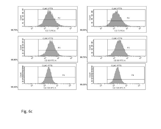
Fig. 6 shows the results of flow cytometry experiments in which mesenchymal
stem cells
isolated from the umbilical cord have been analysed for the expression of the
mesenchymal
stem cell markers CD73, CD90 and CD105. For these experiments, mesenchymal
stem cells
were isolated from umbilical cord tissue by cultivation of the umbilical cord
tissue in three
different cultivation media, followed by subculturing of the mesenchymal stem
cells in the
respective medium. The three following culture media were used in these
experiments: a) 90%
(v/v/ DMEM supplemented with 10 % FBS (v/v), b) the culture medium PTT-4
described in
US patent application 2006/0078993 and the corresponding International patent
application
W02006/019357 that consist of 90% (v/v) CMRL1066, and 10% (v/v) FBS (see
paragraph
[0183] of W02006/019357 and c) the culture medium of the present invention PPT-
6 the
composition of which is described herein. In this flow cytometry analysis, two
different
samples of the cord lining mesenchymal stem cell (CLMC) population were
analysed for each
of the three used culture media. The results are shown in Fig. 6a to Fig.6c.
In more detail, Fig.
6a shows the percentage of isolated mesenchymal cord lining stem cells
expressing stem cell
markers CD73, CD90 and CD105 after isolation from umbilical cord tissue and
cultivation in
DMEM/10% FBS, Fig. 6b shows the percentage of isolated mesenchymal cord lining
stem
cells expressing stem cell markers CD73, CD90 and CD105 after isolation from
umbilical
cord tissue and cultivation in PTT-4 and Fig. 6c shows the percentage of
isolated
mesenchymal cord lining stem cells expressing stem cell markers CD73, CD90 and
CD105
after isolation from umbilical cord tissue and cultivation in PTT-6.
Fig. 7 shows the results of flow cytometry experiments in which mesenchymal
stem cells
isolated from the umbilical cord have been analysed for their expression of
stem cells markers
(CD73, CD90 and CD105, CD34, CD45 and HLA-DR (Human Leukocyte Antigen ¨antigen
D Related) that are used for defining the suitability of multipotent human
mesenchymal stem
cells for cellular therapy and compared to the expression of these markers by
bone marrow
mesenchymal stem cells. For this experiment, the mesenchymal stem cells of the
aminotic
membrane of the umbilical cord were isolated from umbilical cord tissue by
cultivation of the
umbilical cord tissue in the culture medium of the present invention PPT-6
while the bone
marrow mesenchymal stem cells were isolated from human bone marrow using a
standard
protocol. Fig. 7a shows the percentage of isolated mesenchymal cord lining
stem cells that
express the stem cell markers CD73, CD90 and CD105 and lack expression of
CD34, CD45
and HLA-DR after isolation from umbilical cord tissue and cultivation in PTT-6
medium
while Fig. 7b shows the percentage of isolated bone marrow mesenchymal stem
cells that
express CD73, CD90 and CD105 and lack expression of CD34, CD45 and HLA-DR.
CA 03031978 2019-01-24
WO 2018/067071 6 PCT/SG2017/050500
DETAILED DESCRIPTION OF THE INVENTION
As explained above, in a first aspect the invention is directed to a method of
isolating a
mesenchymal stem cell population from the amniotic membrane of the umbilical
cord, the
method comprising cultivating umbilical cord tissue in a culture medium
comprising DMEM
(Dulbecco' s modified eagle medium), F12 (Ham's F12 Medium), M171 (Medium 171)
and
PBS (Fetal Bovine Serum). It has been surprisingly found in the present
application that using
such a medium provides for the isolation of a mesenchymal stem cell population
from the
amniotic membrane of the umbilical cord of which more than 90 %, or even 99 %
or more of
the cells are positive for the three mesenchymal stem cell markers CD73, CD90
and while at
the same these stem cells lack expression of CD34, CD45 and HLA-DR (see the
Experimental
Section), meaning 99 % or even more cells of this population express the stem
cell markers
CD73, CD90 and CD105 while not expressing the markers CD34, CD45 and HLA-DR .
Such
an extremely homogenous and well defined cell population is the ideal
candidate for clinical
trials and cell based therapies since, they for example, fully meet the
criteria generally
accepted for human mesenchymal stem cells to be used for cellular therapy as
defined, for
example, by Dominici et al, "Minimal criteria for defining multipotent
mesenchymal stromal
cells. The International Society for Cellular Therapy position statement",
Cytotherapy (2006)
Vol. 8, No. 4, 315-317, Sensebe et al,."Production of mesenchymal stromal/stem
cells
according to good manufacturing practices: a, review", Stem Cell Research &
Therapy 2013,
4:66), Vonk et at., Stem Cell Research & Therapy (2015) 6:94, or Kundrotas
Acta Medica
Lituanica. 2012. Vol. 19. No. 2. P. 75-79. Also, using a bioreactor such as a
Quantum Cell
Expansion System, it is possible to obtain high numbers of mesenchymal stem
cells such as
300 to 700 million mesenchymal stem cells per run (see also the Experimental
Section). Thus,
the present invention allows to provide the amounts of stem cells that are
needed for
therapeutic applications such as their use in wound healing in a cost
efficient manner. In
addition, all components used for making the culture medium of the present
invention are
commercially available in GMP quality. Accordingly, the present invention
opens the route to
the GMP production of this highly homogenous mesenchymal stem cell population
from the
amniotic membrane of the umbilical cord.
In this context, it is noted that the culture medium of the present invention
allows the isolation
of a mesenchymal stem cell population (also referred hereas as "mesenchymal
stem cells")
from the amniotic membrane under conditions that allow cell proliferation of
the
CA 03031978 2019-01-24
WO 2018/067071 7 PCT/SG2017/050500
mesenchymal stem/progenitor cells without differentiation of the mesenchymal
stem/progenitor cells. Thus, after isolation of the mesenchymal stem cells
from the amniotic
membrane as described herein the isolated mesenchymal stem/progenitor cell
population has
the capacity to differentiate into multiple cell types as described in US
patent application
2006/0078993, US patent 9,085,755, International patent application
W02006/019357, US
patent 8,287,854 or W02007/046775, for instance. As described in US patent
application
2006/0078993, for example, the mesenchymal stem cells of the amniotic membrane
of the
umbilical cord have a spindle shape, express the following genes: POU5f1, Bmi-
1, leukemia
inhibitory factor (LIF), and secrete Activin A and Follistatin. The
mesenchymal stem cells
isolated in the present invention can, for example, be differentiated into any
type of
mesenchymal cell such as, but not limited to, adipocyte, skin fibroblasts,
chondrocytes,
osteoblasts, tenocytes, ligament fibroblasts, cardiomyocytes, smooth muscle
cells, skeletal
muscle cells, adipocytes, mucin producing cells, cells derived from endocrine
glands such as
insulin producing cells (for example, 13-islet cells) or neurectodermal cells.
The stem cells
isolated in the present invention can be differentiated in vitro in order to
subsequently use the
differentiated cell for medical purposes. An illustrative example of such an
approach is the
differentiation of the mesenchymal stem cells into insulin producing 13-islet
cells which can
then be administered, for example by implantation, to a patient that suffers
from an insulin
deficiceny such as diabetes mellitus (cf. also W02007/046775 in this respect).
Alternatively,
the mesenchymal stem cells of the invention can be used in their
undifferentiated state for cell
based therapy, for example, for wound healing purposes such as treatment of
burns or chronic
diabetic wounds. In these therapeutic applications the mesenchymal stem cells
of the invention
can either serve to promote wound healing by interacting with the surrounding
diseased tissue
or can also differentiate into a respective skin cell (cf., again
W02007/046775, for example).
In this context, it is noted that the mesenchymal stem cell population
described herein can be
isolated and cultivated (i.e. are derived) from any umbilical cord tissue as
long as the umbilical
cord tissue contains the amniotic membrane (which is also referred to as "cord
lining").
Accordingly, the mesenchymal stem cell population can be isolated from (pieces
of) the entire
umbilical cord as described in the Experimental section of the present
application. This
umbical cord tissue may thus contain, in addition to the amniotic membrane,
any other
tissue/component of the umbilical cord. As shown, for example, in Figure 16 of
US patent
application 2006/0078993 or International patent application W02006/019357,
the amniotic
membrane of the umbilical cord is the outmost part of the umbilical cord,
covering the cord. In
addition, the umbilical cord contains one vein (which carries oxygenated,
nutrient-rich blood
CA 03031978 2019-01-24
WO 2018/067071 8 PCT/SG2017/050500
to the fetus) and two arteries (which carry deoxygenated, nutrient-depleted
blood away from
the fetus). For protection and mechanical support these three blood vessels
are embedded in
the Wharton's jelly, a gelatinous substance made largely from
mucopolysaccharides.
Accordingly, the umbilical cord tissue used in the present invention can also
comprise this one
vein, the two arteries and the Wharton's jelly. The use of such an entire
(intact) section of the
umbilical cord has the advantage that the amniotic membrane does not need to
be separated
from the other components of the umbilical cord. This reduces the isolation
steps and thus
makes the method of the present invention, simpler, faster, less error prone
and more
economical ¨ which are all important aspects for the GMP production that is
necessary for
therapeutic application of the mesenchymal stem cells. The isolation of the
mesenchymal stem
cells can thus start by tissue explant, which may be followed by subsequent
subculturing
(cultivation) of the isolated mesenchymal stem cells if greater amounts of the
mesenchymal
stem cells are desired, for example, for use in clinical trials.
Alternatively, it is also possible to
first separate the amniotic membrane from the other components of the
umbilical cord and
isolate the mesenchymal cord lining stem cells from the amniotic membrane by
cultivation of
the amniotic membrane in a culture medium of the present invention. This
cultivation can also
be carried out by tissue explant, optionally followed by subculturing of the
isolated
mesenchymal stem cells. In this context, the term "tissue explant" or "tissue
explant method"
is used in its regular meaning in the art to refer a method in which a tissue,
once being
harvested, or a piece of the tissue is being placed in a cell culture dish
containing culture
(growth) medium and by which over time, the stem cells migrate out of the
tissue onto the
surface of the dish. These primary stem cells can then be further expanded and
transferred into
fresh dishes through micropropagation (subculturing) as also described here.
In this context, it
is noted that in terms of production of the cells for therapeutic purposes, in
the first step of
isolating the amniotic membrane mesenchymal stem cells from the umbilical
cord, a master
cell bank of the isolated mesenchymal stem cells is obtained, while the
subsequent
subculturing a working cell bank can be obtained. If a mesenchymal stem cell
population of
the invention (in particular a population of the mesenchymal stem cells of
which at least about
98% or 99 % or express each of the markers CD73, CD90 and CD105 and lack
expression of
each of the markers: CD34, CD45 and HLA-DR) is used for clinical trials or as
an approved
therapeutic, a cell population of the working cell bank will be typically used
for this purpose.
Both the stem cell population of the isolation step (which may make up the
master cell bank)
and the stem cell population of the subculturing step (which may make up the
working cell
bank) can, for example, be stored in cryo-preserved form.
CA 03031978 2019-01-24
WO 2018/067071 9 PCT/SG2017/050500
As mentioned above, the present method of isolating mesenchymal stem cells
from the
amniotic membrane of umbilical cord has the advantage that all components used
in the
culture medium of the invention are available in GMP quality and thus provide
the possibility
to isolate the mesenchymal stem cells under GMP conditions for subsequent
therapeutic
administration.
By "DMEM" is meant Dulbecco's modified eagle medium which was developed in
1969 and
is a modification of basal medium eagle (BME) (cf. Fig.1 showing the data
sheet of DMEM
available from Lonza). The original DMEM formula contains 1000 mg/L of glucose
and was
first reported for culturing embryonic mouse cells. DMEM has since then become
a standard
medium for cell culture that is commercially available from various sources
such as
ThermoFisher Scientific (catalogue number 11965-084), Sigma Aldrich (catalogue
number
D5546) or Lonza, to new only a few suppliers. Thus, any commercially available
DMEM can
be used in the present invention. In preferred embodiments, the DMEM used
herein is the
DMEM medium available from Lonza under catalog number 12-604F. This medium is
DMEM supplemented with 4.5 g/L glucose and L-glutamine). In another preferred
embodiment the DMEM used herein is the DMEM medium of Sigma Aldrich catalogue
number D5546 that contains 1000 mg/L glucose, and sodium bicarbonate but is
without L-
glutamine.
By "F12" medium is meant Ham's F12 medium. This medium is also a standard cell
culture
medium and is a nutrient mixture initially designed to cultivate a wide
variety of mammalian
and hybridoma cells when used with serum in combination with hormones and
transfen-in (cf.
Fig. 2, showing the data sheet of Ham's F12 medium from Lonza). Any
commercially
available Ham's F12 medium (for example, from ThermoFisher Scientific
(catalogue number
11765-054), Sigma Aldrich (catalogue number N4888) or Lonza, to new only a few
suppliers)
can be used in the present invention. In preferred embodiments, Ham's F12
medium from
Lonza is used.
By "DMEM/F12" or "DMEM:F12" is meant a 1:1 mixture of DMEM with Ham's F12
culture
medium (cf. Fig. 3 showing the data sheet for DMEM: F12 (1:1) medium from
Lonza). Also
DMEM/F12 (1:1) medium is a widely used basal medium for supporting the growth
of many
different mammalian cells and is commercially available from various supplier
such as
ThermoFisher Scientific (catalogue number 11330057), Sigma Aldrich (catalogue
number
CA 03031978 2019-01-24
WO 2018/067071 10 PCT/SG2017/050500
D6421) or Lonza. Any commercially available DMEM:F12 medium can be used in the
present
invention. In preferred embodiments, the DMEM:F12 medium used herein is the
DMEM/F12
(1:1) medium available from Lonza under catalog number 12-719F (which is DMEM:
F12
with L-glutamine, 15 mM HEPES, and 3.151 g/L glucose).
.. By "M171" is meant culture medium 171, which has been developed as basal
medium for the
culture of for the growth of normal human mammary epithelial cells ( cf. Fig.
4 showing the
data sheet for M171 medium from Life Technologies Corporation). Also this
basal medium is
widely used and is commercially available from supplier such as ThermoFisher
Scientific or
Life Technologies Corporation (catalogue number M171500), for example. Any
commercially
.. available M171 medium can be used in the present invention. In preferred
embodiments, the
M171 medium used herein is the M171 medium available from Life Technologies
Corporation
under catalogue number M171500.
By "FBS" is meant fetal bovine serum (that is also referred to as "fetal calf
serum"), i.e. the
blood fraction that remains after the natural coagulation of blood, followed
by centrifugation
to remove any remaining red blood cells. Fetal bovine serum is the most widely
used serum-
supplement for in vitro cell culture of eukaryotic cells because it has a very
low level of
antibodies and contains more growth factors, allowing for versatility in many
different cell
culture applications. The FBS is preferably obtained from a member of the
International
Scrum Industry Association (ISIA) whose primary focus is the safety and safe
use of scrum
and animal derived products through proper origin traceability, truth in
labeling, and
appropriate standardization and oversight. Suppliers of FBS that are ISIA
members include
Abattoir Basics Company, Animal Technologies Inc., Biomin Biotechnologia LTDA,
GE
Healthcare, Gibco by Thermo Fisher Scientific and Life Science Production, to
mention only a
few. In currently preferred embodiments, the FBS is obtained from GE
Healthcare under
catalogue number A15-151.
Turning now to the culture medium of the present invention, the culture medium
may
comprise for the isolation or cultivation of the mesenchymal cord lining stem
cells DMEM in
a final concentration of about 55 to 65 % (v/v), F12 in a final concentration
of about 5 to 15 %
(v/v), M171 in a final concentration of about 15 to 30 % (v/v) and FBS in a
final concentration
.. of about 1 to 8 % (v/v). The value of "% (v/v)" as used herein refers to
the volume of the
indivual component relative to the final volume of the culture medium. This
means, if DMEM
is, for example, present in the culture medium a final concentration of about
55 to 65 % (v/v),
1 liter of culture medium contains about 550 to 650 ml DMEM.
CA 03031978 2019-01-24
WO 2018/067071 11 PCT/SG2017/050500
In other embodiments, the culture medium may comprise DMEM in a final
concentration of
about 57.5 to 62.5 % (v/v), F12 in a final concentration of about 7.5 to 12.5
% (v/v), M171 in
a final concentration of about 17.5 to 25.0 % (v/v) and FBS in a final
concentration of about
1.75 to 3.5 % (v/v). In further embodiments, the culture medium may comprise
DMEM in a
.. final concentration of about 61.8 % (v/v), F12 in a final concentration of
about 11.8 % (v/v),
M171 in a final concentration of about 23.6 % (v/v) and FBS in a final
concentration of about
2.5 % (v/v).
In addition to the above-mentioned components, the culture medium may comprise
supplements that are advantages for cultivation of the mesenchymal cord lining
stem cells.
The culture medium of the present invention may, for example, comprises
Epidermal Growth
Factor (EGF). If present, EGF may be present in the culture medium in a final
concentration of
about 1 ng/ml to about 20 ng/ml. In some of these embodiments, the culture
medium may
comprise EGF in a final concentration of about lOng/ml.
The culture medium of the present invention may also comprises insulin. If
present, insulin
may be present in a final concentration of about 1 jig/ml to 10 pg/ml. In some
of these
embodiments, the culture medium may comprise Insulin in a final concentration
of about
5 g/ml.
The culture medium may further comprises at least one of the following
supplements: adenine,
hydrocortisone, and 3,3',5-Triiodo-L-thyronine sodium salt (T3). In such
embodiments, the
culture medium may comprise all three of adenine, hydrocortisone, and 3,3 ',5-
Triiodo-L-
thyronine sodium salt (T3). In these embodiments, the culture medium may
comprises may
comprise adenine in a final concentration of about 0.05 to about 0.1 g/m1
adenine,
hydrocortisone in a final concentration of about 1 to about 10 pg/m1
hydrocortisone and/or
3,3',5-Triiodo-L-thyronine sodium salt (T3) in a final concentration of about
0.5 to about 5
ng/ml.
In the method of the invention, the umbilical cord tissue may be cultured till
a suitable number
of (primary) mesenchymal cord lining stem cells have outgrown from the tissue.
In typical
embodiments, the umbilical cord tissue is cultivated until cell outgrowth of
the mesenchymal
stem cells of the amniotic membrane reaches about 70 to about 80% confluency.
It is noted
here that the term "confluency" or "confluence" is used in its regular meaning
in the art of cell
culture and is meant as an estimate/indicator of the number of adherent cells
in a culture dish
or a flask, referring to the proportion of the surface which is covered by
cells. For example, 50
percent confluence means roughly half of the surface is covered and there is
still room for cells
CA 03031978 2019-01-24
WO 2018/067071 12 PCT/SG2017/050500
to grow. 100 percent confluence means the surface is completely covered by the
cells, and no
more room is left for the cells to grow as a monolayer.
Once a suitable number of primary cells (mesenchymal cord lining stem cells)
have been
obtained from the cord lining tissue by tissue explant, the mesenchymal stem
cells are
removed from the cultivation container used for the cultivation. By so doing,
a master cell
bank containing the (primary) isolated mesenchymal stem cells of the amniotic
membrane can
be obtained. Typically, since mesenchymal stem cells are adherent cells,
removing is carried
out using standard enzymatic treatment. For example, the enzymatic treatment
may comprise
trypsination as described in International US patent application 2006/0078993,
International
patent application W02006/019357 or International patent application
W02007/046775,
meaning outgrowing cells can be harvested by trypsinization (0.125%
trypsin/0.05% EDTA)
for further expansion. If the harvested mesenchymal stem cells are, for
example, used for
generating a master cell bank, the cells can also be cryo-preserved and stored
for further use as
explained herein below.
Once being harvested, the mesenchymal stem cells can be transferred to a
cultivation container
for subculturing. The subculturing can also be started from frozen primary
cells, i.e. from the
master cell bank. For subculturing any suitable amount of cells can be seeded
in a cultivation
container such as cell culture plate. The mesenchymal cells can, for this
purpose, be suspended
in a suitable medium (most conveniently, the culture medium of the present
invention) for
subculturing at a concentration of, for example, about 0.5 x 10⁶ cells/ml to
about 5.0 x 10⁶
cells/ml. In one embodiment the cells are suspended for subcultivation at a
concentration of
about 1.0 x 10⁶ cells/ml. The subculturing can be carried by cultivation
either in simple culture
flasks but also, for example, in a multilaycr system such as CellStacks
(Corning, Corning, NY,
USA) or Cellfactory (Nunc, part of Thermo Fisher Scientific Inc., Waltham, MA,
USA) that
can be stacked in incubators. Alternatively, the subculturing can also be
carried out in a closed
self-contained system such as a bioreactor. Different designs of bioreactors
are known to the
person skilled in the art, for example, parallel-plate, hollow-fiber, or micro-
fluidic bioreactors.
See, for example, Sensebe et al."Production of mesenchymal stromal/stem cells
according to
good manufacturing practices: a review", supra. An illustrative example of a
commercially
hollow-fiber bioreactor is the Quantum Cell Expansion System (Terumo BCT,
Inc). that has,
for example, been used for the expansion of bone marrow mesenchymal stem cells
for clinical
trials (cf., Hanley et al, Efficient Manufacturing of Therapeutic Mesenchymal
Stromal Cells
Using the Quantum Cell Expansion System, Cytotherapy. 2014 August; 16(8): 1048-
1058).
CA 03031978 2019-01-24
WO 2018/067071 13 PCT/SG2017/050500
Another example of a commercially available bioreactors that can be used for
the subculturing
of the mesenchymal stem cell population of the present invention is the Xuri
Cell Expansion
System available from GE Hcathcare. The cultivation of the mesenchymal stem
cell
population in an automated system such as the Quantum() Cell Expansion System
is of
particular benefit if a working cell bank for therapeutic application is to be
produced under
GMP conditions and a high number of cells is wanted.
The subculturing of the mesenchymal cord ling stem cells of the invention
takes place in in a
culture medium of the present invention. Accordingly, the culture medium of
the present
invention can be used both for the isolation of the mesenchymal stem cells
from the amniotic
membrane and the subsequent cultivation of the isolated primay cells by
subcultivation. Also
for the subcultivation, the mesenchymal stem cells can be cultured till a
suitable amount of
cells have grown. In illustrative embodiments the mesenchymal stem cells are
subcultured till
the mesenchymal stem cells reach about 70 to about 80% continency.
The isolation/cultivation of the population of mesenchymal cord lining stem
cells can be
carried out under standard condition for the cultivation of mammalian cells.
Typically, the
method of the invention of isolating the population of the mesenchymal cord
lining stem cells
is typically carried out at conditions (temperature, atmosphere) that are
normally used for
cultivation of cells of the species of which the cells are derived. For
example, human umbilical
cord tissue and the mesenchymal cord lining stem cells, respectively, are
usually cultivated at
37 C in air atmosphere with 5%CO2. In this context, it is noted that the in
present invention
the mesenchymal cells may be derived of any mammalian species, such as mouse,
rat, guinea
pig, rabbit, goat, horse, dog, cat, sheep, monkey or human, with mesenchymal
stem cells of
human origin being preferred in one embodiment.
Once a desired/suitable number of mesenchymal cord lining stem cells have been
obtained
from the subculture, the mesenchymal stem cells are harvested by removing them
from the
cultivation container used for the subcultivation. The harvesting of the
mesenchymal stem
cells is typically again carried out by enzymatic treatment, including
comprises trypsination of
the cells. The isolated mesenchymal stem cells are subsequently collected and
are either be
directedly used or preserved for further use. Typically, preserving is carried
out by cryo-
preservation. The term "cryo-preservation" is used herein in its regular
meaning to describe a
process where the mesenchymal stem cells are preserved by cooling to low sub-
zero
temperatures, such as (typically) -80 C or -196 C (the boiling point of liquid
nitrogen). Cryo-
preservation can be carried out as known to the person skilled in the art and
can include the
CA 03031978 2019-01-24
WO 2018/067071 14 PCT/SG2017/050500
use of cryo-protectors such as dimethylsulfoxide (DMSO) or glycerol, which
slow down the
formation of ice-crystals in the cells of the umbilical cord.
The isolated population of the mesenchymal cord lining stem cells that is
obtained by the
isolation method of the present invention is highly defined and homogenous. In
typical
embodiments of the method at least about 90 % or more, about 91 % or more,
about 92 % or
more, about 92 % or more, about 93 % or more, about 94 % or more, about 95 %
or more,
about 96 % or more, about 97 % or more, about 98 % or more about 99 % or more
of the
isolated mesenchymal stem cells express the following markers: CD73, CD90 and
CD105. In
addition, in these embodiments at least about 90 % or more, about 91 % or
more, about 92 %
or more, about 92 % or more, about 93 % or more, about 94 % or more, about 95
% or more,
about 96 % or more, about 97 % or more, about 98 % or more about 99 % or more
of the
isolated mesenchymal stem cells may lack expression of the lack expression of
the following
markers: CD34, CD45 and HLA-DR. In particular embodiments, about 97 % or more,
about
98 % or more, or about 99 % or more of the isolated mesenchymal stem cell
population
express CD73, CD90 and CD105 while lacking expression of CD34, CD45 and HLA-
DR.
Thus, in line with the above disclosure the present invention is also directed
to a mesenchymal
stem population isolated from the amniotic membrane of the umbilical cord,
wherein at least
about 90 % or more cells of the stem cell population express each of the
following markers:
CD73, CD90 and CD105. In preferred embodiments at least about 91 % or more,
about 92 %
or more, about 92 % or more, about 93 % or more, about 94 % or more, about 95
% or more,
about 96 % or more, about 97 % or more, about 98 % or more about 99 % or more
cells of the
isolated mesenchymal stem cell population are CD73+, CD90+ and CD105+, meaning
that
this percentage of the isolate cell population express each of CD73, CD90 and
CD105 (cf. the
Experimental Section of the present application). In addition, at least about
90 % or more,
about 91 % or more, about 92 % or more, about 92 % or more, about 93 % or
more, about 94
% or more, about 95 % or more, about 96 % or more, about 97 % or more, about
98 % or more
about 99 % or more of the isolated mesenchymal stem cells may lack expression
of the lack
expression of the following markers. In particular embodiments about 97 % or
more, about 98
% or more, or about 99 % or more of the isolated mesenchymal stem cell
population express
CD73, CD90 and CD105 while lacking expressing of CD34, CD45 and HLA-DR. Such a
higly homogenous population of mesenchymal stem cells derived from the
amniotic
membrane of the umbilical cord has been reported here for the first time and
meets the critiera
for mesenchymal stem cells to be used for cellular therapy (also cf. the
Experimental Section
CA 03031978 2019-01-24
WO 2018/067071 15 PCT/SG2017/050500
and, for example, Sensebe et al."Production of mesenchymal stromal/stem cells
according to
good manufacturing practices: a review", supra). It is noted in this context
that this
mesenchymal stem cell population can be obtained by either the isolating
method of the
present invention but also by a different method such as cell sorting, if
wanted.
In line with the above, the present invention is also directed to a
pharmaceutical composition
comprising a mesenchymal stem population isolated from the amniotic membrane
of the
umbilical cord, wherein at least about 90 % or more cells of the stem cell
population express
each of the following markers: CD73, CD90 and CD105 and optionally, lack
expression of
CD34, CD45 and HLA-DR. The pharmaceutical composition may comprise any
pharmaceutically acceptable excipient and may be formulated for any desired
pharmaceutical
way of administration. The pharmaceutical composition may, for example, be
adapted for
systemic or topical application.
In a further aspect the invention is directed to a method of making a culture
medium for
isolating the method comprising, mixing to obtain a final volume of 500 ml
culture medium:
i. 250 ml of DMEM
ii. 118 ml M171
iii. 118 ml DMEM/F12
iv. 12.5 ml Fetal Bovine Serum (FBS) to reach a final concentration of 2.5%
(v/v).
As explained above, DMEM/F12 medium is a 1:1 mixture of DMEM and Ham's F12
medium.
.. Thus, 118 ml DMEM/F12 medium contain 59 ml DMEM and 59 ml F12. Accordingly,
when
using this method of making a culture medium, the final concentrations (v/v)
mit 500 ml total
volume are as follows:
DMEM: 250 ml + 59 ml = 309 ml, corresponds to 309/500 = 61.8 % (v/v)
M171: 118 ml, corresponds to 118/500 = 23.6% (v/v)
F12: 59 ml, corresponds to 59/500 = 11.8 % (v/v).
Embodiments of this method of making a culture medium further comprise adding
v. 1 ml EGF stock solution (5 pg/m1) to achieve a final EGF concentration of
lOng/ml, and
CA 03031978 2019-01-24
WO 2018/067071 16 PCT/SG2017/050500
vi. Insulin 0.175 ml stock solution (14.28 mg/ml) to achieve a final insulin
concentration of
51.1 g/ml.
It is noted here that in these embodiments, the above-mentioned volumes of
these components
i. to vi when result in a final volume of 499.675 ml culture medium. If no
further components
are added to the culture medium, the remaining 0.325 ml (to add up to a volume
of 500 ml)
can, for example, be any of components i. to iv, that means either DMEM, M171,
DMEM/F12
or FBS. Alternatively, the concentration of the stock solution of EGF or
Insulin can of course
be adjusted such that the total volume of the culture medium is 500 nil. In
addition, it is also
noted that compoents i. to iv. do not necessarily have to be added in the
order in which they
are listed but it is of course also possible to use any order to mix these
components to arrive at
the culture medium of the present invention. This means, that for example,
M171 and
DMEM/F12 can be mixed together and then combined with DMEM and FBS to reach
final
concentrations as described here, i.e. a final concentration of DMEM of about
55 to 65 %
(v/v), a final concentration of F12 of about 5 to 15 % (v/v), a final
concentration of M171 of
about 15 to 30 % (v/v) and a final concentration of FBS of about 1 to 8 %
(v/v).
In other embodiments, the method further comprises adding to DMEM a volume of
0.325 nil
of one or more of the following supplements: adenine, hydrocortisone, 3,3',5-
Triiodo-L-
thyronine sodium salt (T3), thereby reaching a total volume of 500 ml culture
medium. In this
embodiments, the final concentration of these supplements in DMEM may be as
follows:
about 0.05 to 0.1 g/ml adenine, for example about 0.025 pg/m1 adenine,
about 1 to 10 g/m1 hydrocortisone,
about 0.5 to 5 ng/ml 3,3',5-Triiodo-L-thyronine sodium salt (T3), for example
1.36 ng/ml
3,3',5-Triiodo-L-thyronine sodium salt (T3).
In line with the above disclosure, the invention is also directed to a cell
culture medium that is
obtainable or that is obtained by the method of making the medium as described
here.
In addition, the invention also concerns a method of isolating mesenchymal
stem cells from
the amniotic membrane of the umbilical cord, wherein this method comprises
cultivating
amniotic membrane tissue in the culture medium prepared by the method as
described here.
Thus, the present invention is also directed to a cell culture medium
comprising:
- DMEM in the final concentration of about 55 to 65 % (v/v),
CA 03031978 2019-01-24
WO 2018/067071 17 PCT/SG2017/050500
- F12 in a final concentration of about 5 to 15 % (v/v),
- M171 in a final concentration of about 15 to 30 % (v/v) and
- FBS in a final concentration of about 1 to 8 % (v/v).
In certain embodiments of the culture medium described here, the medium
comprises DMEM
in the final concentration of about 57.5 to 62.5 % (v/v), F12 in a final
concentration of about
7.5 to 12.5% (v/v), M171 in a final concentration of about 17.5 to 25.0% (v/v)
and FBS in a
final concentration of about 1.75 to 3.5 % (v/v). In other embodiments the
culture medium
may comprise DMEM in a final concentration of about 61.8 % (v/v), F12 in a
final
concentration of about 11.8 % (v/v), M171 in a final concentration of about
23.6 % (v/v) and
FBS in a final concentration of about 2.5 % (v/v).
In addition, the culture medium may further comprise Epidermal Growth Factor
(EGF) in a
final concentration of about 1 ng/ml to about 20 ng/ml. In certain
embodiments, the culture
medium comprise EGF in a final concentration of about lOng/ml. The culture
medium
described herein may further comprise Insulin in a final concentration of
about 1 pg/m1 to 10
jig/ml. In such embodiments the culture medium may comprise Insulin in a final
concentration
of about Siug/ml.
The cell culture medium of the invention may further comprise at least one of
the following
supplements: adenine, hydrocortisone, and 3,3',5-Triiodo-L-thyronine sodium
salt (T3). In
certain embodiments the culture medium comprises all three of adenine,
hydrocortisone, and
3,3',5-Triiodo-L-thyronine sodium salt (T3). If present, the culture medium
may comprise
adenine in a final concentration of about 0.01 to about 0.1 pg/m1 adenine or
of about 0.05 to
about 0.1 pg/m1 adenine, hydrocortisone in a final concentration of about 0.1
to about 10
tig/ml hydrocortisone or of about 1 to about 10 pg/m1 hydrocortisone and/or
3,3 ',5-Triiodo-L-
thyronine sodium salt (T3) in a final concentration of about 0.5 to about 5
ng/ml.
In embodiments of the cell culture medium, 500 ml of the cell culture medium
of the present
invention comprise:
i. 250 ml of DMEM
ii. 118 ml M171
iii. 118 ml DMEM/F12
iv. 12.5 ml Fetal Bovine Serum (FBS) (final concentration of 2.5%)
CA 03031978 2019-01-24
WO 2018/067071 18 PCT/SG2017/050500
In further embodiments, the cell culture medium may further comprise
v. EGF in a final concentration of lOng/ml, and
vi. Insulin in a final concentration of 51.1g/m1.
Both, insulin and and EGF can be added to to the culture medium using a stock
solution of
.. choice, such that the total volume of the culture medium does not exceed
500 ml.
In a particular example, the components i. to vi. of the culture medium of the
present invention
are the components indicated in Figure 5, meaning they are obtained from the
respective
manufacturers using the catalogue number indicated in Figure 5. The medium
that is obtained
from mixing the components i. to vi. as indicated in Figure 5 is also referred
herein as "PTT-
6". It is again noted in this context that the constituents i. to vi. as well
as any other ingredient
such as an antibiotic of any other commercial supplier can be used in making
the medium of
the present invention.
In addition, the cell culture medium of the invention may comprise adenine in
a final
concentration of about 0.01 to about 0.1 pg/m1 adenine or of about 0.05 to
about 0.1 g/m1
adenine, hydrocortisone in a final concentration of about 0.1 to 10 pg/ml, of
about 0.5 to about
101.tg/ml, or of about 1 to about 10 pg/m1 hydrocortisone and/or 3,3 ',5-
Triiodo-L-thyronine
sodium salt (T3) in a final concentration of about 0.1 to about 5 ng/ml or of
about 0.5 to about
5 ng/ml.
Finally, the invention also provides a method of treating a patient having a
disease, the method
comprising administering to the patient a mesenchymal cord lining stem cell or
a
pharmaceutical composition containing a stem cell as disclosed herein. The
disease can be any
disease thas described above. For treating the subject, the mesenchymal stem
cell population
of the invention may be administered in any suitable way, for example,
including but not
limited to, topical administration, by implantation or by injection. The stem
cell population
.. may, for example, be placed directly onto a wound such as a burn or a
diabetic wound (see
International patent application W02007/046775). Alternatively, the stem cell
population may
also be implanted subcutaneously, for example, directly under the skin, in
body fat or the
peritoneum.
The invention will be further illustrated by the following non-limiting
Experimental Examples.
CA 03031978 2019-01-24
WO 2018/067071 19 PCT/SG2017/050500
Experimental Examples
1. Cryopreservation of Umbilical Cord Tissue Prior to Isolation of Mesenchymal
Stem
Cells
Umbilical cord tissue (the umbilical cords were donated with informed consent
of the mother)
was processed for the subsequent isolation of the mesenchymal stel cells from
the amniotic
membrane of the umbilical cord as follows.
1.1 Washing of umbilical cord tissue sample:
a. Remove scalpels from the protective cover.
b. Hold the umbilical cord securely using the forceps and cut the cord into a
10 cm length
piece using a scalpel. Place the unusable cord back in the original tissue
cup.
c. Transfer the 10 cm long umbilical cord piece into a new 150 mm culture
dish. The 150mm
culture dish may be used in place of the cups.
d. Use the cover of the 150 mm culture dish as a resting place for forceps and
scalpel.
e. Remove 25m1 Plasmalyte A (Baxter, Catalog # 2B2543Q) with a 30 ml syringe.
Hold the
syringe at a 45 angle using one hand and dispense the Plasmalyte A directly
onto the
umbilical cord tissue.
f. Holding the culture dish at a slight angle remove the Plasmalyte A with a
30 ml syringe and
blunt needle.
g. Collect used Plasmalyte A in a 300 ml transfer bag that serves as a trash
container and
dispose it in the biohazard bin.
h. Repeat wash procedure, if necessary using a new culture dish for each wash.
Make sure all
blood clots on the surface have been removed. More Plasmalyte A can be used if
needed to
clean the tissue.
i. Place the tissue into a new labeled tissue culture dish to continue cutting
the tissue. Place 20
ml of Plasmalyte A into the dish so the tissue does not dry out while cutting
it.
j. Cut the cords into equal approximately 1-cm sections resulting in 10
sections in total.
k. Further cut each 1 cm section into smaller pieces with approximately 0.3 cm
x 0.3 cm to 0.5
cm x 0.5 cm per section.
1. Remove any Plasmalyte A that is in the dish.
m. Pull 25 ml Plasmalyte A with a 30 ml syringe from the original Plasmalyte A
bag and
dispense directly on the umbilical cord tissue pieces.
n. Hold culture dish in an angle to collect all Plasmalyte A used for washing
the tissue on one
side and remove it with a syringe and blunt needle.
CA 03031978 2019-01-24
WO 2018/067071 20 PCT/SG2017/050500
o. Repeat wash one more time. There should not be any clots left.
NOTE: If the cord is not frozen right away, the umbilical cord tissue is kept
in Plasmalyte A
until ready to freeze.
1.2 Crvopreservation of umbilical cord tissue:
a. Prepare cryopreservation solution:
i. Prepare 50 ml freezing solution consisting of 60% Plasmalyte A, 30% of 5%
Human Serum
Albumin, and 10% dimethyl sulfoxide (DMSO).
ii. Label a 150 ml transfer bag with "Tissue freeze solution" and attach a
plasma transfer set to
the port using aseptic technique.
iii. Remove 30 ml Plasmalyte A with a 30 ml Syringe from the original
Plasmalyte A bag and
transfer it in the transfer bag labeled "tissue freeze solution" with the time
and date solution is
made.
iv. Remove 15 ml of 5% Human Serum Albumin with a 20 ml syringe and transfer
it into the
labeled transfer bag.
v. Add 5 ml DMS0 to the transfer bag.
vi. Mix well and record mixing of freeze solution
b. Remove the Plasmalyte A from the tissue before adding the freeze solution.
c. Using a 60 ml syringe, pull all 50 mls of the freeze solution into the
syringe add
approximately 30 ml freeze solution to the 150 mm cell culture dish containing
the umbilical
cord tissue. Place a blunt needle on the syringe to keep it sterile.
d. Swirl the culture dish containing the tissue and freezing solution every
minute for 10
minutes.
e. Using forceps, select 8 randomly chosen sections and place them in each of
the four 4 ml
cryovials. Select 4 randomly chosen sections and place them into one 1.8 ml
cryovial. These
sections should be free of blood clots.
f. Fill each cryovial containing the umbilical cord tissue with the remaining
freezing solution
to the 3.6 ml filling line for the 4 ml tubes and the 1.8 ml line for the 1.8
ml Nunc vial.
g. Label one Bactec Lytic/10 - Anaerobic/F and one Bactec Plus Aerobic/F
bottle with tissue
ID.
h. Remove 20 ml freeze solution from the culture dish with a syringe and a
blunt needle, after
wiping the Bactec vials with an alcohol swab, switch the blunt needle for
anl8g needle and
inoculate the aerobic and the anaerobic Bactec bottles with 10 ml each.
CA 03031978 2019-01-24
WO 2018/067071 21 PCT/SG2017/050500
i. Start controlled rate freezer.
j. After controlled rate freeze is completed place the units in a continuous
temperature
monitored liquid nitrogen freezer until further use.
2. Isolation of Mesenchvmal Cord Lining Stem Cells from umbilical cord tissue
2.1. Preparing media for processing MSCs from umbilical cord tissue:
a. To make 500 ml PTT6 (culture/growth media) add the following in the order
listed:
i. DMEM, 250 ml
ii. M171 118 ml
DMEM F12 118 ml
iv. FBS 12.5 ml (final concentration of 2.5%)
v. EGF 1 ml (final concentration of lOng/m1)
vi. Insulin 0.175 ml (final concentration of 5 g/m1)
The above-mentioned volumes of components i. to vi when result in a final
volume of 499.675
ml culture medium. If no further components are added to the culture medium,
the remaining
0.325 ml (to add up to a volume of 500 ml) can, for example, be any of
components i. to iv,
that means either DMEM, M171, DMEM/F12 or FBS. Alternatively, the
concentration of the
stock solution of EGF or Insulin can of course be adjusted such that the total
volume of the
culture medium is 500 ml. Alternatively, a stock solution of an antibiotic
such as Penicillin-
Streptomycin-Amphotericin can be added to result in a final volume of 500 ml.
It is also
possible to add to the culture medium a volume of 0.325 ml of one or more of
the following
supplements: adenine, hydrocortisone, 3,3',5-Triiodo-L-thyronine sodium salt
(T3), thereby
reaching a total volume of 500 ml culture medium.
vii. Label the bottle "PTT6" with date media was prepared, initial of the
operator, and the
phrase "expires on" followed by the expiration date. Expiration date is the
earliest expiration
date of any of the component or 1 month from the preparation date, whichever
comes first.
b. To make the rinse media (Hank's Buffered Salt Solution (HBSS) without
Calcium or
Magnesium and with 5% FBS), add 2.5 ml FBS to 47.5 ml of HBSS in a 50 ml
centrifuge
tube. Label the tube "Rinse Media" with operator initials and date the media
is made.
CA 03031978 2019-01-24
WO 2018/067071 22 PCT/SG2017/050500
c. All media will be tested for sterility using Bactec Lytic/10 ¨Anaerobic/F
(Becton Dickinson
& Company) and Bactec Pluc + Aerobic/F (Becton Dickinson & Company). Inject 20
ml of
prepared media into each bottle.
2.2 Thawing of umbilical cord tissue for MSC harvesting:
a. Initiate the thaw once an operator is prepared to process the sample in the
clean room. Do
not thaw more than 1 vial at a time unless the vials originate from the same
donor.
b. Wipe the water bath with disinfectant followed by 70% isopropanol and fill
it with 1 L
sterile water. Heat the water bath up to 36-38 C.
c. Prepare 10 mL of rinse medium consisting of 70% to 90% PlasmaLyte A in the
clean room
under a biosafety cabinet. Sterile filter the solution with a 0.2-gm syringe
filter attached to a
10 ml syringe and keep the solution refrigerated until use.
d. Place a processing label on a 50 ml conical tube.
e. Confirm water bath temperature is at 36-38 C.
f. Take vial(s) of tissue from the liquid nitrogen storage and thaw rapidly in
the 37 C water
bath filled with 1L of sterile water. The vial holder for the Mr. Frosty
Nalgene Cryo 1 C
freezing container floats with vials in place and can be used as a floating
rack when thawing
samples.
g. Remove the vial from the water bath and spray them with 70% Isopropanol
solution. A
good time to pull the vial from the water bath is when small ice can be seen
floating in the vial
¨ suggest internal temperature of the vial is less than 37 C.
h. Place vial into pass-through and alert the clean room processing
technician.
2.3 Preparing for tissue processing:
a. Umbilical cord tissue processing should be performed in an environmentally
monitored
(EM) clean room:. At the end of each shift, full room and hood cleaning are
performed
b. Prepare/clean the biosafety cabinet.
c. Perform viable particle counting while working in the biosafety cabinet.
d. Assemble all necessary supplies in the biosafety cabinet checking each for
packaging
damage and expiration dates. When handling syringes, serological pipets,
sterile forceps,
scalpels, tissue plates, and needles, make sure not to touch any surface that
will come in
contact with the sterile product. Only the exterior of the syringe barrel,
tubing, plunger tip
and/or needle cap or sheath may be safely handled. Discard supply if the
surface has been
touched or has touched a non-sterile surface.
CA 03031978 2019-01-24
WO 2018/067071 23 PCT/SG2017/050500
e. Record lot numbers and expiration dates (if applicable) of all reagents and
supplies to be
used.
f. Receive the thawed vial by cleaning the vial with lint-free wipe moistened
with 70% alcohol
before transferring into the biosafety cabinet.
g. Using an aspirating needle with a syringe, withdraw as much liquid from the
vial. Avoid
suctioning the tissue.
h. Using sterile forceps, transfer the tissue into a sterile 100 mm petri
dish.
i. Add an aliquot of 5 ml rinse medium to the tissue fragments.
j. Swirl the contents for 15-30 seconds, then remove the rinse medium with a
pipette or
syringe with aspirating needle. Repeat this rinse process twice.
k. Add 2 mL of rinse medium to the tissue to avoid drying out the tissue.
2.4. Initiating MSC outgrowth from tissue:
a. Label the bottom of a 6-well plate "Outgrowth 1" with MSC lot number or
umbilical cord
tissue ID and the date outgrowth is initiated. If 60 mm tissue culture dish is
used, divide the
plate into 4 quadrants by drawing a grid on the bottom of the dish.
b. Using sterile, disposable forceps, place one 3 x 3 mm to 5 x 5 mm tissue
into each well. If
using a 60 mm tissue culture dish, place the tissue into the middle of each
quadrant to keep the
tissues apart (more than 1 cm from each other).
c. Fill each well with 3 ml of PTT6.
d. Using an aspirating needle coupled to 30 ml syringe, withdraw enough media
to barely
cover the tissue. Do not tilt the plate. Do not touch the bottom of the well
with the aspirating
needle.
e. Using an inverted light microscope, observe for cell outgrowth every day
(24 6 hrs). Real
time cell culture imaging system may be used in place of the light microscope.
f. Change media every day. Be sure to equilibrate the media to room
temperature before use.
i. Aspirate off the medium.
ii. Add 3 ml of P11'6.
iii. Aspirate until tissue is barely submerged in the medium.
g. When cellular outgrowth is observed from the tissue, transplant the tissue
to a new 6-well
plate using the same procedure as 4.a to 4.e above except label the plate
"Outgrowth 2".
Maintain cell outgrowth in "Outgrowth 1" plate by adding 2m1of PTT6 to each
well. Observe
for confluency every day. Replace media every 2-3 days (be sure to equilibrate
the media to
room temperature before use).
CA 03031978 2019-01-24
WO 2018/067071 24 PCT/SG2017/050500
h. When cell outgrowth is observed in "Outgrowth 2" plate, repeat step 4.a to
4.e except label
the plate "Outgrowth 3." Maintain cell outgrowth in "Outgrowth 2" plate by
adding 2 ml of
PTT6 to each well. Observe for confluency every day. Replace media every 2-3
days (be sure
to equilibrate the media to room temperature before use).
i. When outgrowth is observed in "Outgrowth 3" plate, discard the tissue. If
the tissues are
very small and do not seem to interfere with cell growth, dispose of the
tissue when
subculturing.
j. When cells reach 40-50% confluency, observe cells every days to prevent
over-expansion.
k. When cells reach 70-80% confluency, subculture the cells. Do not allow
cells to expand
beyond 80% confluence.
With the size of the tissue explants being about 1-3mm, and the tissue
explant/cell culture is
performed in 175 mm squared culture dishes, the average number of mesenchymal
stem cells
harvested from an explant is typically about 4,000 - 6,000 cells/explant.
Accordingly, when
the mesenchymal stem cells are simultaneously grown out of 48 explants about
300,000 cells
can be obtained at harvest. These 300,000 mesenchymal stem cells collected
from explants can
then be used for subculturing by seeding a 175cm2 cell culture flask with such
300,000 cells as
described in the following Example 2.5 (this can be referred to as Passage 1).
The
mesenchymal stem cells obtained from this passage 1 can then be used to seed
again 175cm2
flasks (Passage 2) and expand the cells as described in the following Example
2.5. The cells
obtained from both Passage 1 and Passage 2 can be "banked" by cryo-
preservation, with the
mesenchymal stem cells obtained after Passage 2 being considered to represent
the Master
Cell Bank which will be for further expansion of the mesenchymal stem cells,
for example, in
a bioreactor as explained below in Example 2.7.
2.5. Subculturing MSC in cell culture dishes
a. Perform viable particle while working in the biosafety cabinet. Equilibrate
all media to
room temperature before use.
b. When cell outgrowth reaches about 70-80% conflucncy, subculture cells.
i. Remove PTT6 from the petri dish.
ii. Rinse with HBSS without Calcium or Magnesium.
iii. Add 0.2 ml 1X TrypLE-EDTA and swirl for 1-2 minutes.
iv. Tilt the dish 30-45 to allow cells to shift down by gravitational flow.
Gentle tapping on the
side of the plate expedites detachment.
CA 03031978 2019-01-24
WO 2018/067071 25 PCT/SG2017/050500
v. Add lml of PTT6. Pipette up and down gently then transfer cells to a 15 ml
centrifuge tube.
Use clean pipette tip with each well. Cells from all 6 wells can be pooled
into a single 15ml
tube.
vi. Centrifuge for 10 minutes at 1200 rpm.
vii. Remove supernatant and resuspend cells with 5m1 PTT6.
c. Subculturing MSC
i. Aliquot 50 il of the cell suspension and assay for TNC and viability by
Trypan Blue
Exclusion Assay.
ii. Count cells using a hemocytometer. Expect to count 20-100 cells/square. If
the count higher
than 100, dilute the original sample 1:5 and repeat Trypan Blue method using a
hemocytometer.
iii. Calculate viable cells/ml and total viable cells:
1. Viable cells/ml = viable cell count x dilution factor x 104
2. Total viable cells = viable cell count x dilution factor x total volume x
104
iv. Calculate % viability:
1. % viability = viable cell count x 100 /(viable cell count + dead cell
count)
v. Dilute the cell suspension to 1.0 x 10⁶ cells/ml:
1. "X" volume = Total viable cells/106 cells/ml
2. For example, if total viable cell number is 1.0 x 10⁷;
3. "X" = 107106 cells/ml or 10 ml, therefore, you would bring your total cell
volume
up to 10 ml by adding 5 ml to your cell suspension (that is at 5 ml).
vi. If the cell suspension is less than 106/ml, determine the volume required
to seed 2 x 10⁶
cells for each 150 mm petri dish or 175 cm2 flask.
1. Volume for 2 x 10⁶ cells = 2 x 10⁶ cells viable cells/ml
2. For example, if viable cells/ml is 8 x 10⁵ cells/ml, 2 x10⁶ cells 8 x 10⁵
cells/ml or 2.5 ml
are needed.
vii. Set aside 0.5 ml for MSC marker analysis.
viii. Seed 2 x 10⁶ cells to each 150 mm petri dish or 175 cm2 flask with 30 ml
PTT6.
ix. Observe cells for attachment, colony formation, and confluence every three
days. When
cells reach 40-50% confluence, observe cells every one-two days to prevent
over-expansion.
DO NOT allow cells to expand beyond 80% confluence. A real time cell culturing
monitoring
system can be used in place of the light microscope.
x. Replace media every 2-3 days.
CA 03031978 2019-01-24
WO 2018/067071 26 PCT/SG2017/050500
2.6 Cnropreserving MSC cells
a. Perform viable particle while working in the biosafety cabinet.
b. When cells reach 70-80% confluence, detach cells using 2 ml 1X TrypLE-EDTA
for each
150 mm petri dish or 175 cm2 flask.
.. i. Remove PTT6 from the petri dish.
ii. Wash with 5m1 HBSS or PBS without calcium or magnesium.
iii. Add 2 ml 1X TrypLE-EDTA and swirl for 1-2 minutes.
iv. Tilt the dish 30-45 to allow cells to shift down by gravitational flow.
Gentle tapping on the
side of the petri dish helps to expedite detachment.
v. Add 10 ml PTT6 to inactivate TrypLE. Mix well to dissociate cell clumps.
vi. Transfer cells to 15 ml centrifuge tube using a Pasteur pipette.
vii. Centrifuge for 10 minutes at 1200 rpm.
viii. Aspirate medium and resuspend with 10 ml PTT6.
ix. Aliquot 50 I and determine total viable cell number and % viability as
above. Cell count
will need to be performed within 15 minutes as the cells may start clumping.
c. Preparing cells for cryopreservation
i. Prepare Cell Suspension Media and Cryopreservation Media and freeze the
cells
2.7. Subculturing (expansion) of MSC in a Quantum Bioreactor (Terumo BTC,
Inc.)
It is also possible to use a Quantum Bioreactor can used to expand the MSC.
The starting cell
number for the expansion in the Quantum Bioreactor should range between 20 to
30 million
cells per run. The typical yield per run is 300 to 700 million MSC at harvest.
The Bioreactor is
operated following the protocol of the manufacturer. The so obtained
mesenchymal stem cells
are typically cryo-preserved (see below) and serve as Working Cell Bank.
MATERIALS/ REAGENTS:
1. Quantum Expansion Set
2. Quantum Waste Bag
3. Quantum Media Bag
4. Quantum Inlet Bag
5. PTT6
6. PBS
7. Fibronectin
8. TrypLE
CA 03031978 2019-01-24
WO 2018/067071 27 PCT/SG2017/050500
9. 3m1 syringe
10. Glucose test strips
11. Lactate test strips
12. 60m1 Cell Culture Plate or equivalent
13. Medical Grade 5% CO2 Gas-mix
14. 50m1 Combi-tip
EQUIPMENT:
1. Biosafety Cabinet
2. Glucose Meter (Bayer Healthcare/Ascensia Contour Blood Glucose Meter)
3. Lactate Plus (Nova Biomedical)
4. Peristaltic pump with head
5. Centrifuge, Eppendorf 5810
6. Sterile Tube Connector
7. M4 Repeat Pipettor
8. RF Sealer
PROCEDURE:
1. Preparing the Quantum Bioreactor
a) Priming the Quantum Bioreactor
b) Coating the bioreactor:
1) Prepare the fibronectin solution in the biosafety cabinet.
1) Allow lyophilized fibronectin to acclimate to room temperature (> 15 min at
room temperature)
2) Add 5m1 of sterile distilled water; do not swirl or agitate
3) Allow fibronectin to go into solution for 30 min.
4) Using a 10 ml syringe attached with an 18g needle, transfer the fibronectin
solution to a Ccell inlet bag containing 95m1 of PBS.
2) Attach the bag to the "reagent" line
3) Check for bubbles (bubbles may be removed by using "Remove IC Air" or
"Remove EC Air" and using "Wash" as the inlet source.
4) Open or set up program for coating the bioreactor (Figure 1. Steps 3-5).
5) Run the program
CA 03031978 2019-01-24
WO 2018/067071 28 PCT/SG2017/050500
6) While the program is running to coat the bioreactor, prepare a media bag
with 4L
of PTT6 media.
7) Attach the media bag to the IC Media line using a sterile tube connector.
8) When the bioreactor coating steps are completed, detach the cell inlet bag
used for
fibronectin solution using a RF sealer.
c) Washing off excess fibronectin
d) Conditioning the bioreactor with media
2. Culturing the cells in the Quantum Bioreactor
a) Loading and attaching the cells with Uniform Suspension:
b) Feeding and cultivation of the cells
1) Chose media flow rate to feed the cells.
2) Sample for lactate and glucose everyday.
3) Adjust the flow rate of the media as the lactate levels increase. The
actual
maximal tolerable lactate concentration will be defined by a flask culture
from
which the cells originate. Determine if adequate PTT6 media is in the media
bag.
If necessary, replace the PTT6 media bag with a fresh PTT6 media bag.
4) When the flow rate has reached the desired value, measure lactate level
every 8-12
hours. If the lactate level does not decrease or if the lactate level
continues to
increase, harvest the cells.
3. Harvesting the cells from the Quantum Bioreactor
a) When lactate concentration does not decrease, harvest the cells after
sampling for
lactate and glucose for the last time.
b) Harvesting the cells:
1) Attach cell inlet bag filled with 100m1 TrypLE to the "Reagent" line using
a sterile
tube connector.
2) Confirm sufficient PBS is in the PBS bag. If not, attach a new bag with at
least 1.7
liters of PBS to the "Wash" line using a sterile tube connector.
3) Run the Harvest program
4. Cryopreserving the cells
1) Once the cells have been harvested, transfer the cells to 50m1 centrifuge
tube to
pellet the cells.
2) Resuspend using 25m1 of cold cell suspension solution. Count the cells
using
Sysmex or Biorad Cell counter. Attach the cell count report to the respective
Quantum Processing Batch Record.
CA 03031978 2019-01-24
WO 2018/067071 29 PCT/SG2017/050500
3) Adjust cell concentration to 2x10⁷/m1
4) Add equal volume eryopreservation solution and mix well (do not shake or
vortex)
5) Using a repeat pipettor, add lml of the cell suspension in cryopreservative
to each
1.8m1 vial. Cryopreserve using the CRF program as described in the SOP D6.100
CB Cryopreservation Using Controlled Rate Freezers
6) Store the vials in a designated liquid nitrogen storage space.
7) Attach the CRF run report to the form respective MSC P3-Quantum Processing
Batch Record.
3. Analvis of Stem Cell Marker Expression in Mesenchvmal Cord Lining Stem
Populations isolated from umbilical cord tissue, using different culture media
Flow cytometry experiments were carried out to to analyse mesenchymal stem
cells isolated
from the umbilical cord for the expression of the mesenchymal stem cell
markers CD73, CD90
and CD105.
For these experiments, mesenchymal stem cells were isolated from umbilical
cord tissue by
cultivation of the umbilical cord tissue in three different cultivation media,
followed by
subculturing of the mesenchymal stem cells in the respective medium as set
forth in Example
2.
The three following culture media were used in these experiments: a) 90% (v/v/
DMEM
supplemented with 10 % FBS (v/v), b) the culture medium PTT-4 described in US
patent
application 2006/0078993 and the corresponding International patent
application
W02006/019357 that consist of 90% (v/v) CMRL1066, and 10% (v/v) FBS (see
paragraph
[0183] of W02006/019357 and c) the culture medium of the present invention PPT-
6 the
composition of which is described herein. In this flow cytometry analysis, two
different
samples of the cord lining mesenchymal stem cell (CLMC) population were
analysed for each
of the three used culture media.
The following protocol was used for the flow cytometry analysis.
Materials and methods
Instruments name Company Name Serial Name
CA 03031978 2019-01-24
WO 2018/067071 30 PCT/SG2017/050500
BD FACS CANDO BD V07300367
Inverted Microscope, Olympus 4K40846
CKX41SF
Centrifuge, Micro spin Biosan 010213-1201-0003
Tabletop
Reagent list Company Name CatLog Number
X Trypsin Biowest X0930-100
10 X PBS Lonza 17-517Q
DMEM Lonza 12-604F
Fetal Bovine Serum GE healthcare A11-151
Antibody list Company Name CatLog Number
Human CD73 Purified AD2 BD 550256
0.1mg
Human CD90 Purified 5E10 BD 550402
lmL
Human CD105 Purified 266 BD 555690
0.1mg
Alexa Fluor 647 goat BD A21235
anti¨mouse IgG (H+L) *2
mg/mL*
Reagents name Composition
1 X PBS (1L) 100m1 of 10 X PBS + 900m1 of sterile
distilled H20
lx PBA (50m1) 49.5m1 of 1XPBS + 0.5 ml of FBS
Procedure
5 a) Cell isolation and cultivation from the umbilical cord lining
membrane
1. Explant tissue samples were incubated in a cell culture plate and submerged
in the
respective medium, then keep it in CO2 incubator at 37 C as explained in
Example 2.
2. The medium was changed every 3 days.
CA 03031978 2019-01-24
WO 2018/067071 31 PCT/SG2017/050500
3. Cell outgrowth from tissue culture explants was monitored under light
microscopy.
4. At a confluence of about 70%, cells were separated from dish by
trypsinization
(0.0125% trypsin/0.05% EDTA) and used for flow cytometry experiments.
b) Dypsinization of cells for experiments
1. Remove medium from cell culture plate
2. Gently rinse with sterile 1X PBS to remove traces of FBS as FBS will
interfere
with the enzymatic action of trypsin.
3. Add 1X trypsin to cell culture plate and incubate for 3-5 min in 37 C.
4. Observe cells under microscope to ensure that they are dislodged.
Neutralize
trypsin by adding complete media containing FBS (DMEM with 10% FBS).
5. Use a pipette to break up cell clumps by pipetting cells in media against a
wall of
the plate. Collect and transfer cell suspension into 50 nil centrifuge tubes
6. Add sterile 1X PBS to plate and rinse it, Collect cell suspension into the
same
centrifuge tube.
7. Centrifuge it at 1800 rpm for 10 mins.
8. Discard supernatant and re-suspend cell pellet with PBA medium.
c) Counting cells
1. Ensure that the haemocytometer and its cover slip are clean and dry,
preferably by
washing them with 70% ethanol and letting them dry before wiping them with Kim
wipes (lint-free paper).
2. Aliquot a small amount of cells in suspension into a micro centrifuge tube
and
remove from the BSC hood.
3. Stain cells in suspension with an equal volume of Trypan Blue, e.g. to
500p1 of
suspension add 500111 of Trypan Blue (dilution factor = 2X, resulting in 0.2%
Trypan Blue solution).
4. Avoid exposure of cells to Trypan Blue for longer than 30 mins as Trypan
Blue is
toxic and will lead to an increase in non-viable cells, giving a false cell
count.
5. Add 20 p1 of the cell suspension mixture to each chamber of a
haemocytometer
and view under a light microscope.
a. Count the number of viable cells (bright cells; non-viable cells take up
Trypan Blue readily and thus are dark) in each quadrant of the
haemocytometer for a total of 8 quadrants in the upper and lower chamber.
CA 03031978 2019-01-24
WO 2018/067071 32 PCT/SG2017/050500
Total cell count is given as (Average number of cells/quadrant) x 104
cells/ml.
d) Staining cells
i. Preparation before staining cells
= Cell suspension are aliquot into 3 tubes (CD73, CD90, CD105 ) in
duplicates and 2 tubes (negative control), each containing 50,000 cells.
ii. Staining with primary antibody (Ab)
= Add 1 I [0.5mg/m1 Ab] of primary antibody to 100u1 cell
suspension. Incubate at 4 C for 45 min.
= Make up to lml with PBA.
= Centrifuge 8000 rpm at 4 C for 5 mins.
= Remove supernatant.
= Add lml of PBA and re-suspend pellet
= Centrifuge 8000 rpm at 4 C for 5 mins.
= Remove supernatant.
= Re-suspend in 100u1 PBA.
iii. Staining with secondary Ab ¨ in the dark
= Add lul [0.5mg/m1 ab] of secondary antibody to 100u1 cell
suspension. Incubate at 4 C for 30 min.
= Make up to lml with PBA.
= Centrifuge 8000 rpm at 4 C for 5 mins.
= Remove supernatant.
= Add lml of PBA and re-suspend pellet
= Centrifuge 8000 rpm at 4 C for 5 mins.
= Remove supernatant
= Re-suspend in 200-300u1 PBA for flow cytometry analysis
= Transfer cells to FACS tube for reading in BD FACS CANDO
flow cytometry.
The results of the flow cytometry analysis are shown in Fig. 6a to Fig.6c.
Fig. 6a shows the
percentage of isolated mesenchymal cord lining stem cells expressing stem cell
markers
CD73, CD90 and CD105 after isolation from umbilical cord tissue and
cultivation in
DMEM/10% FBS, Fig. 6b shows the percentage of isolated mesenchymal cord lining
stem
CA 03031978 2019-01-24
WO 2018/067071 33 PCT/SG2017/050500
cells expressing stem cell markers CD73, CD90 and CD105 after isolation from
umbilical
cord tissue and cultivation in PTT-4 and Fig. 6c shows the percentage of
isolated
mesenchymal cord lining stem cells expressing stem cell markers CD73, CD90 and
CD105
after isolation from umbilical cord tissue and cultivation in PTT-6. As can be
seen from Fig.
6a, the population isolated using DMEM/10 % FBS as culture medium cultivation
has about
75% CD73+ cells, 78 % 90+ cells and 80 % CD105+ cells (average of two
experiments),
while after isolation/cultivation of umbilical cord tissue using PPT-4 culture
medium (see Fig.
6b) the number of mesenchymal stem cells that are CD73-positive, CD90-positive
and
CD105-positive are about 87 % (CD73+ cells), 93 % /CD90+ cells) and 86 %
(CD105+ cells)
average of two experiments. The purity of the mesenchymal stem cell population
that was
obtained by means of cultivation in the PTT-6 medium of the present invention
is at least 99.0
% with respect to all three markers (CD73, CD90, CD105), meaning the purity of
this cell
population is significant higher than for cultivation using PPT-4 medium or
DMEM/10 %
FBS. In addition and even more importantly, the mesenchymal stem cell
population obtained
by means of cultivation in PTT-6 is essentially a 100% pure and defined stem
cell population.
This makes the stem cell population of the present invention the ideal
candidate for stem cell
based therapies. Thus, this population of mesenchymal cord lining stem cells
may become the
gold standard for such stem cell based therapeutic approaches.
The findings shown in Fig. 6 are further corroborated by the results of the
flow cytometry
analysis that are shown in Fig. 7a and Fig.7b. Fig. 7a shows the percentage of
isolated
mesenchymal cord lining stem cells (mesenchymal stem cells of the amniotic
membrane of
umbilical cord) that express the stem cell markers CD73, CD90 and CD105 and
lack
expression of CD34, CD45 and HLA-DR after isolation from umbilical cord tissue
and
cultivation in PTT-6 medium. As shown in Fig. 7a, the mesenchymal stem cell
population
contained 97.5 % viable cells of which 100% expressed each of CD73, CD90 and
CD105 (see
the rows "CD73+CD90+" and "CD73+CD105+") while 99.2 % of the stem cell
population did
not express CD45 and 100% of the stem cell population did not express CD34 and
HLA-DR
(see the rows "CD34-CD45- and "CD34-HLA-DR-). Thus, the mesenchymal stem cells
population obtained by cultivation in PTT-6 medium is essentially a 100% pure
and defined
stem cell population that meets the criteria that mesenchymal stem cells are
to fulfill to be
used for cell therapy (95% or more of the stem cell population express CD73,
CD90 and
CD105, while 98 % or more of the stem cell population lack expression of CD34,
CD45 and
HLA-DR, see Sensebe et al."Production of mesenchymal stromal/stem cells
according to good
CA 03031978 2019-01-24
WO 2018/067071 34 PCT/SG2017/050500
manufacturing practices: a review", supra). It is noted here that the present
mesenchymal stem
cells of the amniotic membrane are adhere to plastic in standard culture
conditions and
differentiate in vitro into osteoblasts, adipocytes and chondroblasts, see US
patents 9,085,755,
US patent 8,287,854 or W02007/046775 and thus meet the criteria generally
accepted for use
of mesenchymal stem cells in cellular therapy.
Fig. 7h shows the percentage of isolated bone marrow mesenchymal stem cells
that express
CD73, CD90 and CD105 and lack expression of CD34, CD45 and HLA-DR. As shown in
Fig.
7h, the bone marrow mesenchymal stem cell population contained 94.3 % viable
cells of
which 100% expressed each of CD73, CD90 and CD105 (see the rows "CD73+CD90+"
and
"CD73+CD105+") while only 62.8 % of the bone marrow stem cell population
lacked
expression of CD45 and 99.9 % of the stem cell population lacked expression
CD34 and
HLA-DR (see the rows "CD34-CD45- and "CD34-HLA-DR-). Thus, the bone marrow
mesenchymal stem cells that are considered to be gold standard of mesenchymal
stem cells are
by far less homogenous/pure in terms of stem cell marker than the mesenchymal
stem cells
population (of the amniotic membrane of the umbilical cord) of the present
application. This
finding also shows that the stem cell population of the present invention may
be the ideal
candidate for stem cell based therapies and may become the gold standard for
stem cell based
therapeutic approaches.
It will be readily apparent to a person skilled in the art that varying
substitutions and
modifications may be made to the invention disclosed herein without departing
from the scope
and spirit of the invention.
All patents and publications mentioned in the specification are indicative of
the levels of those
of ordinary skill in the art to which the invention pertains. All patents and
publications are
herein incorporated by reference to the same extent as if each individual
publication was
specifically and individually indicated to be incorporated by reference.
The inventions illustratively described herein may suitably be practiced in
the absence of any
element or elements, limitation or limitations, not specifically disclosed
herein. Thus, for
example, the terms "comprising", "including", "containing", etc. shall be read
expansively and
without limitation. Additionally, the terms and expressions employed herein
have been used as
terms of description and not of limitation, and there is no intention in the
use of such terms and
CA 03031978 2019-01-24
WO 2018/067071 35 PCT/SG2017/050500
expressions of excluding any equivalents of the features shown and described
or portions
thereof, but it is recognized that various modifications are possible within
the scope of the
invention claimed. Thus, it should be understood that although the present
invention has been
specifically disclosed by preferred embodiments and optional features,
modification and
variation of the inventions embodied therein herein disclosed may be resorted
to by those
skilled in the art, and that such modifications and variations are considered
to be within the
scope of this invention. The invention has been described broadly and
generically herein. Each
of the narrower species and subgeneric groupings falling within the generic
disclosure also
form part of the invention. This includes the generic description of the
invention with a
proviso or negative limitation removing any subject matter from the genus,
regardless of
whether or not the excised material is specifically recited herein. In
addition, where features or
aspects of the invention are described in terms of Markush groups, those
skilled in the art will
recognize that the invention is also thereby described in terms of any
individual member or
subgroup of members of the Markush group. Further embodiments of the invention
will
become apparent from the following claims.