Note: Descriptions are shown in the official language in which they were submitted.
CA 03032304 2019-01-28
WO 2018/023111
PCT/US2017/044664
GAMMA DELTA T CELLS AS A TARGET FOR TREATMENT OF SOLID
TUMORS
CROSS-REFERENCE TO RELATED APPLICATIONS
This application claims priority to U.S. Provisional Patent Application No.
62/368,453,
filed on July 29, 2016, and U.S. Provisional Patent Application No.
62/507,495, filed on May
17, 2017, the disclsoures of which are incorporated herein by reference.
FEDERALLY SPONSORED RESEARCH
This invention was made with government support under Grant Nos. CA-155649, CA-
168611, and CA-193111, awarded by the National Institutes of Health (NIH), and
under Grant
Nos. P30CA016087 and UL1 TR000038, awarded by the National Center for
Advancing
Translational Sciences (NCATS). The Government has certain rights in the
invention.
BACKGROUND OF THE DISCLOSURE
Pancreatic ductal adenocarcinoma (PDA) is a devastating disease in which the
mortality
rate approaches the incidence rate (Yadav and Lowenfels, 2013,
Gastroenterology 144, 1252-
1261). PDA is almost invariably associated with a robust inflammatory cell
infiltrate which
has considerable influence on disease progression (Andren-Sandberg et al.,
1997, Scand J
Gastroenterol 32, 97-103; Clark et al., 2007, Cancer research 67, 9518-9527)
Pen-pancreatic
leukocytic subsets can have divergent effects on tumorigenesis by either
combating cancer
growth via antigen-restricted tumoricidal immune responses or by promoting
tumor
progression via induction of immune suppression (Zheng et al., 2013,
Gastroenterology 144,
1230-1240). For example, CD8+ T cells and Thl-polarized CD4+ T cells mediate
tumor-
protection in murine models of PDA and are associated with prolonged survival
in human
disease (De Monte et al., 2011, J Exp Med 208, 469-478; Fukunaga et al., 2004,
Pancreas 28,
e26-31). Negating cytotoxic CD8+ anti-tumor responses by myeloid-derived
suppressor cells
(MDSC) markedly accelerates PDA growth (Pylayeva-Gupta et al., 2012, Cancer
Cell 21,
836-847). Conversely, antigen-restricted Th2-deviated CD4+ T cells strongly
promote PDA
progression in mice (Ochi et al., 2012c, J Exp Med 209, 1671-1687).
Accordingly, intra-
tumoral CD4+ Th2 cell infiltrates correlate with reduced survival in human PDA
(De Monte et
al., 2011, J Exp Med 208, 469-478; Fukunaga et al., 2004, Pancreas 28, e26-
31). Nevertheless,
intra-pancreatic y6T cells have not been well characterized and their role
remains unclear.
1
CA 03032304 2019-01-28
WO 2018/023111
PCT/US2017/044664
SUMMARY OF THE DISCLOSURE
This disclosure is based at least in part on the findings that
immunosuppressive y6T
cells with a uniquely activated phenotype infiltrates the pre-neoplastic
pancreas and invasive
PDA in a mouse PDA model; deletion of the intra-pancreatic y6T cells markedly
protects
against oncogenesis in vivo and results in an influx and reactivation of
immunogenic Thl cells
and CDS+ T cells to the tumor microenvironment (TME).
Accordingly, one aspect of the present disclosure features a method for
treating a solid
tumor, comprising administering to a subject in need thereof an effective
amount of a y6 T
cell suppressor. In some embodiments, the yo T cell suppressor is an agent
that inhibits an
immunosuppressive y6 T cell, for example, a circulating y6 T cell or a y6 T
cell infiltrated
into tumor tissue or tumor resident organ in the subject. Such a y6 T cell
suppressor may be
an antibody that specifically binds a y6 T cell, e.g., a y6 T cell comprising
a specific gamma
or delta chain, such as a 61 subunit or 62 subunit. In some instances, the y6
T cell-binding
antibody can be a bi-specific antibody that further binds an c43 T cell or NK
cell. In addition,
the y6 T cell-binding antibody may be tri-specific, i.e., capable of binding
to the y chain of the
yo T cell, the 6 chain of the y6 T cell, and an cq3 T cell or NK cell.
Alternatively, the yo T cell
suppressor is an antibody that blocks recruitment of immunosuppressive yo T
cell to a tumor
site in the subject. Such antibodies include, but are not limited to,
antibodies specifically
binds CCR2, CCL2, or CCR6. Any of the antibodies described herein may be a
human
antibody or a humanized antibody.
In other embodiments, the yo T cell suppressor can be an agent that blocks
antigenic
expansion of immunosuppressive yo T cells. Alternatively, the yo T cell
suppressor may be an
immune cell (e.g., a T cell or an NK cell) expressing a chimeric receptor that
targets
immunosuppressive yo T cells.
The subject to be treated by any of the methods described herein may be a
human
patient having the solid tumor. Examples include, but are not limited to,
pancreatic ductal
adenocarincoma (PDA), colorectal cancer (CRC), melanoma, breast cancer, lung
cancer (for
example, non-small cell lung cancer, NSCLC, and small cell lung cancer, SCLC),
upper and
lower gastrointestinal malignancies (including, but not limited to,
esophageal, gastric, and
hepatobiliary cancer), squamous cell head and neck cancer, genitourinary, and
sarcomas. The
subject may have undergone another anti-tumor therapy, e.g., chemotherapy,
radiotherapy,
immunotherapy, therapy involving a small molecule kinase inhibitor, surgery,
or a
2
CA 03032304 2019-01-28
WO 2018/023111
PCT/US2017/044664
combination thereof.
In some examples, the method described herein may further comprise performing
another anti-tumor therapy, e.g., those described herein, to the subject. For
example, the
performing step may comprise administering to the subject an inhibitor of a
checkpoint
molecule (e.g., PD-1, PD-L1, PD-L2, CTLA-4, LAG3, TIM-3 and A2aR), an agonist
of a co-
stimulatory receptor (e.g., 0X40, GITR, CD137, CD40, CD27, and ICOS), or an
inhibitor of
an innate immune cell target (e.g., KIR, NKG2A, CD96, TLR, and DO).
In one example, the subject is administered an inhibitor of a checkpoint
molecule,
which is an anti-PD-Li antibody. In another example, the subject is further
administered a
chemotherapeutic agent, such as gemcitabine or abraxane, or a combination
thereof (e.g.,
folinic acid, fluorouracil, oxaliplatin, and irinotecan, a.k.a., FOLFOXIRI).
In another aspect, described herein is a kit for treating a solid tumor in a
subject, the
kit comprising: (i) a first pharmaceutical composition that comprises a y6 T
cell suppressor,
and (ii) a second pharmaceutical composition that comprises a chemotherapeutic
agent, an
inhibitor of a checkpoint molecule, an agonist of a co-stimulatory receptor,
or an inhibitor of
an innate immune cell target. Further, the present disclosure provides a
pharmaceutical
composition, comprising (i) a y6 T cell suppressor, and (ii) an inhibitor of a
checkpoint
molecule, an agonist of a co-stimulatory receptor, or an inhibitor of an
innate immune cell
target. Also with the scope of the present disclosure are any of the kits or
pharmaceutical
compositions described herein for use in treating a solid tumor such as PDA,
or for
manufacturing a medicament for use in treating the solid tumor.
In yet another aspect, provided herein is a method for analyzing a sample, the
method
comprising: (i) obtaining a biological sample from a subject (e.g., a human
patient) suspected
of having a solid tumor, for example, pancreatic ductal adenocarcinoma (PDA)
or colorectal
cancer (CRC); and (ii) measuring the level of y6 T cells in the biological
sample. In some
embodiments, the y6 T cells are effector memory yo T (TEm) cells. Optionally,
the method
may further comprise measuring the level of a checkpoint molecule (e.g., PD-
L1), the level of
Galectin-9, or both in the biological sample. Alternatively or in addition,
the analysis method
may further comprise identifying the subject as having or at risk for a solid
tumor, such as
PDA or CRC, based on the level of the y6 T cells in the biological sample
determined in (ii),
wherein an elevated level of y6 T cells relative to that of a control subject
is indicative of
presence or risk of PDA or CRC. In some embodiments, the method may further
comprise
performing a treatment of PDA to the subject, if the subject is identified as
having or at risk
3
CA 03032304 2019-01-28
WO 2018/023111
PCT/US2017/044664
for PDA or CRC.
The biological sample may be a peripheral blood sample, or a tissue sample
obtained
from a suspected tumor site.
In some embodiments, the measuring step in any of the analysis methods
described
herein may involve an antibody that specifically binds y6 T cells, for
example, an antibody
specifically binds yo T cells expressing a T cell receptor comprising a 61
subunit, or an
antibody specifically binds y6 T cells expressing a T cell receptor comprising
a 62 subunit.
The details of one or more embodiments of the invention are set forth in the
description below. Other features or advantages of the present invention will
be apparent
from the following drawings and detailed description of several embodiments,
and also from
the appended claims.
BRIEF DESCRIPTION OF THE DRAWINGS
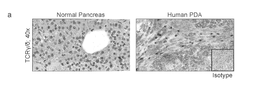
Figure 1. yoT cells are ubiquitous and activated in human PDA. (a) Frozen
sections of human PDA and normal pancreas were stained using a mAb specific
for TCRy/6
or isotype control. Representative images and quantitative data are shown. (b)
Single cell
suspensions from human PDA tumors and PBMC were co-stained for CD45, CD3, and
TCRy/6. The percentage of y6T cells among CD3+ cells was calculated.
Representative
contour plots and summary data are shown. Each dot represents a different
patient sample. (c)
The percentage of PDA-infiltrating y6T cells among CD45+ cells was compared
with tumor-
infiltrating cells expressing select myeloid differentiation markers. (d) The
percentage of
PDA-infiltrating and PBMC y6T cells among CD3+ cells was compared with that of
CD4+
and CD8+ c43T cell subsets in each respective compartment. (e) PBMC and PDA-
infiltrating
CD3+TCRy/6+ cells from PDA patients were gated and co-stained using mAbs
specific for
CD45RA and CD27. The gating paradigms for Tnaive, TCM, TEM, and TEM-RA
populations are shown. Representative contour plots and quantitative data
indicating the
fraction of TEM y6T cells in each compartment are indicated. (f) PDA-
infiltrating and PBMC
y6T cells from PDA patients were stained using mAbs specific for CD62L and (g)
Vy9.
Representative histograms and quantitative data are shown. Human data are
based on tumor
tissue or PBMC analyzed from 9-13 PDA patients (*p<0.05, **p<0.01,
***p<0.001).
Figure 2. yoT cells are highly prevalent and exhibit a uniquely activated
phenotype in murine invasive PDA. (a) C57BL/6-Trdc tin' mice whose y6T cells
express
GFP were orthotopically implanted with KPC-derived tumor and imaged by intra-
vital two-
4
CA 03032304 2019-01-28
WO 2018/023111
PCT/US2017/044664
photon laser-scanning microscopy at 21 days. (b) WT mice were orthotopically
implanted
with KPC-derived tumor cells. On day 21, single cell suspensions of digested
PDA tumors
and splenocytes were co-stained for CD45, CD3, TCRy/6, CD4, and CD8 and
analyzed by
flow cytometry. Representative contour plots and quantitative data are shown.
For the bar
graphs, for each set of CD4+, CD8+ and TCRy/6+, the bars from left to right
are: Spleen, and
PDA (c) WT mice were orthotopically implanted with KPC-derived tumor cells. On
day 21
spleen (blue histograms) and PDA-infiltrating (red histograms) y6T cells were
gated and
tested for co-expression of select surface activation markers and Vy chains.
Representative
histogram overlays and summary data from 5 mice are shown. For the bar graphs,
for each set
of bars for FasL, NK1.1, CD39, CD44, JAML, 0X40, Vy4, and Vyl, the bars from
the left to
right are: Spleen y6T cells (blue), and PDA y6T cells (red) (d) Spleen and PDA-
infiltrating
y6T cells from the same mice were tested for expression of IL-10, (e) IL-17,
(f) NKG2D, (g)
TLR4, TLR7, TLR9, and (h) CCR2, CCR5, and CCR6. Each experiment was repeated
at
least 3 times using 3-5 mice per data point (*p<0.05, **p<0.01). For (g) and
(h), for each bar
set, the bars from left to right are: Spleen, and PDA.
Figure 3. Ablation of yoT cells protects against pancreatic oncogenesis in a
slowly progressive model of PDA. (a) KC;Tcr6+/+ and KC;Tcr6"/" mice were
sacrificed at 3,
6, or 9 months of life (n=10-12 mice/cohort). Representative H&E-stained
frozen sections are
shown. The percentage of pancreatic area occupied by intact acinar structures,
and the
fractions of ductal structures exhibiting normal morphology, ADM, or graded
PanIN I-III
lesions were calculated. (b) Weights of pancreata were compared in 3 month-old
KC;Tcr6+/+
and KC;Tcr6"/" mice. (c) Pancreata from 9 month-old KC;Tcr6+/+ and KC;Tcr6"/"
mice were
assayed for pen- tumoral fibrosis using trichrome staining. (d) Kaplan-Meier
survival
analysis was performed for KC;Tcr6+/+ (n=29) and KC;Tcr6"/" (n=44) mice
(p<0.0001). (e, f)
KC;Tcr6+/+ mice were treated with UC3-10A6 or isotype control for 8 weeks
beginning at 6
weeks of life. (e) Representative H&E stained pancreatic sections are shown.
The percentage
of pancreatic area occupied by intact acinar structures, and the fractions of
ductal structures
exhibiting normal morphology, ADM, or graded PanIN I-III lesions were
calculated. (f)
Tumor weight was recorded (n=5/group; *p<0.05, **p<0.01).
Figure 4. yoT cell deletion results in massive CD4+ and CD8+ T cell
infiltration
and activation in invasive PDA. (a, b) WT and Tcr6"/" mice were implanted with
KPC-
derived tumor cells. On day 21 mice were sacrificed. Frozen pancreatic
sections were tested
for (a) CD8+ and (b) CD4+ T cell infiltration by IHC (n=5/group). (c) CD8+ T
cells
infiltrating orthotopically-implanted KPC-derived tumors in WT and Tcr6"/"
mice were tested
5
CA 03032304 2019-01-28
WO 2018/023111
PCT/US2017/044664
for expression of CD44, (d) ICOS, (e) CTLA-4, and (f) Granzyme B. (g)
Similarly, CD4+ T
cells infiltrating orthotopically-implanted KPC tumors in WT and Tcr6"/" mice
were tested for
expression of CD44, (h) 0X40, (i) PD-1, and (j) CD62L. Experiments were
repeated more
than 3 times with similar results using 5 mice per group (*p<0.05, **p<0.01,
***p<0.001).
Figure 5. yoT cell deletion results in CD4+ T cell Thl differentiation, CD8+ T
cell
activation, and al3T cell-dependent tumor protection in invasive PDA. (a-d) WT
and
Tcr6"/" mice were orthotopically implanted with KPC-derived tumor cells. On
day 21, tumor-
infiltrating CD4+ and CD8+ T cells were interrogated for (a) co-expression of
TNF-a and
IFN-y, (b) expression of T-bet, (c) GATA-3, and (d) FoxP3. Representative
contour plots and
.. quantitative data are shown. Experiments were repeated twice with similar
results (n=5/group;
*p<0.05). (e) WT and Tcr6"/" pancreata were orthotopically implanted with KPC-
derived
tumor cells and serially treated with a-CD4 and a-CD8 neutralizing mAbs or
isotype controls.
Pancreatic tumors were harvested at 3 weeks. Representative images and tumor
weights are
shown (n=5/group; *p<0.05, **p<0.01, ***p<0.001).
Figure 6. PDA-associated yoT cells express high levels of T cell exhaustion
ligands
in multiple murine tumor models and in human disease. (a) Expression of PD-Li
and (b)
Galectin-9 were compared in pancreas and spleen y6T cells of 3-month-old KC
mice by flow
cytometry. Representative contour plots and quantitative data are shown
(n=5/group). (c) WT
mice were orthotopically implanted with KPC-derived tumor cells. Expression of
PD-Li and
Galectin- 9 were compared in PDA tumor cells, TAMs (My), MDSC, and y6T cells
on day 21
(n=5/group). For each set of bars for Tumor, My, MDSC, and TCRy6, the bars
from left to right
are: PD-L1, and Galectin-9 (d) WT mice were orthotopically implanted with KPC-
derived
tumor cells. On day 21, spleen and PDA-infiltrating y6T cells were tested for
expression of
select activating ligands. Representative histograms and quantitative data are
shown
(n=5/group). For each set of bars for B7-1, B7-2, ICOSL, and Ox40L, the bars
from left to right
are: Spleen, and PDA (e) Orthotopic PDA- bearing WT and Tcr64- mice were
tested for
expression of PD-Li in tumor cells, TAMs, and MDSC (n=5/group). For each set
of bars for
Tumor, My, and MDSC, the bars from left to right are: WT, and TCRy-/- (f) WT,
CCR24-,
CCR5-/-, and CCR6-/- mice were orthotopically implanted with KPC-derived PDA
cells
(n=5/group). Animals were sacrificed at 3 weeks, and the fraction of tumor-
infiltrating y6T cells
expressing PD-Li and (g) Galectin-9 were determined by flow cytometry. (h, i)
PBMC y6T cells
from healthy volunteers and PDA patients, and PDA-infiltrating y6T cells and
were tested for
expression of (h) PD-Li and (i) Galectin-9. Representative histograms and
quantitative data are
shown (n=11 patients; *p<0.05, **p<0.01, ***p<0.001).
6
CA 03032304 2019-01-28
WO 2018/023111
PCT/US2017/044664
Figure 7. Exhaustion ligand blockade reverses the direct suppressive effects
of
yoT cells on al3T cells and on pancreatic tumorigenesis. (a) Splenic CD4 + or
(b) CD8+ T cells
from untreated WT mice were either unstimulated, or stimulated with aCD3/aCD28
alone or in
co-culture with PDA-infiltrating y6T cells (5:1 ratio). aPD-L1 (10 g/m1) was
selectively added
to each group. The fraction of CD62L-CD44+ cells were determined at 72h by
flow cytometry.
Representative contour plots and quantitative data are shown. (c) Similarly,
CD4 + and CD8+ T
cell expression of TNF-a was measured. Experiments were performed in
quadruplicate and
repeated 3 times. (d) WT and Tcr6-/- mice were orthotopically implanted with
KPC-derived
tumor cells and serially treated with aPD-L1 or aGalectin-9 neutralizing mAbs,
or respective
isotype controls. Pancreatic tumors were harvested at 3 weeks. Representative
gross images
are shown (Experiment #1) as are quantitative data on tumor weights from 2
separate
experiments using different stocks of KPC-derived tumor cells (n=5/group for
each
experiment). (e-g) WT and Tcr6"/" pancreata were again orthotopically
implanted with KPC-
derived tumor cells and serially treated with aPD-L1 or aGalectin-9
neutralizing mAbs or the
respective isotype controls. Pancreatic tumors were harvested at 3 weeks. (e)
The fraction of
PDA-infiltrating c43T cells among CD45+ leukocytes, and (f) CD8+ and (g) CD4 +
T cell
adoption of an activated CD62L¨ CD44+ phenotype, were determined by flow
cytometry
(n=5/group; *p<0.05, "p<0.01, ***p<0.01).
Figure 8. PDA-infiltrating yoT cells express elevated FoxP3. (a) WT mice were
.. orthotopically implanted with KPC-derived tumor cells. On day 21, splenic
and PDA-
infiltrating CD3+ T lymphocytes were co-stained for CD4, TCRy6, and FoxP3 or
(b) CD4,
TCRy6, and T-bet. Representative contour plots and quantitative data from 5
mice per group
are shown (*p<0.05). Experiments were repeated twice with similar results.
Figure 9. yoT cells in pancreata of KC mice exhibit a distinct phenotype. (a)
Single cell suspensions of pancreata, pancreas-draining lymph nodes, and
spleen, from 6
month-old KC mice were co-stained for CD45, CD3, and TCRy/6. Representative
contour
plots and quantitative data are shown. For each set of bars for CD4, CD8+, and
TCRY/6+, the
bars from left to right are: Spleen, Lymph Node, and PDA, (b) Similarly,
select CCR
expression. For each set of bars for CCR2, CCR5, and CCR6, the bars from left
to right are:
Spleen y/i3T cells, and PDA y/i3T cells, (c) TLR expression. For each set of
bars for TLR4,
TLR7, and TLR9, the bars from left to right are: Spleen y/i3T cells, and PDA
y/i3T cells, (d) and
surface markers were compared in y6T cells harvested from the pancreas and
spleen of 6
month-old KC mice. For each set of bars for NK1.1, CD39, JAML, 0X40, and V y
4, the bars
7
CA 03032304 2019-01-28
WO 2018/023111
PCT/US2017/044664
from left to right are: Spleen y/6T cells, and PDA y/6T cells. Each result was
reproduced at
least twice (n=5/group; *p<0.05, **p<0.01, ***p<0.001, ****p<0.0001).
Figure 10. Selective blockade of chemokine signaling mitigates yoT cell
expansion and activation in PDA and deletion or depletion of yoT cells or
interruption
of their recruitment is protective against PDA. (a-e) WT, CCR2, CCL2, CCR5,
and
CCR6-/-mice were orthotopically implanted with KPC-derived PDA cells
(n=5/group).
Animals were sacrificed at 3 weeks. (a) The fraction of tumor-infiltrating y6T
cells
determined by flow cytometry. (b) y6T cell expression of TNF-a, (c) IL-13, (d)
IL-17, and (e)
IFN-y was determined for each cohort (n=5 /group). (f) WT mice were treated
with UC3-
10A6 and splenocytes from these mice were analyzed for expression of Vy4 and
Vyl. (g) WT
and Tcr6"/" pancreata were orthotopically implanted with KPC-derived PDA
cells. Tumors
were harvested and weighed at 3 weeks after implantation. Representative gross
images of
PDA and quantitative data of tumor weights are shown (n=10/group). (h)
Pancreata from
control WT mice, WT mice treated with a neutralizing a-pH' cell mAb, and
Tcr6"/" mice were
orthotopically implanted with KPC-derived PDA cells. Kaplan-Meier survival
analysis was
performed (n=10/group; WT vs Tcr6-/-: p=0.02; WT vs UC3-10A6: p=0.009; Tcr6-/-
vs UC3-
10A6: p=ns). (i) WT, CCR5, CCR6, CCR2, and CCL2-/- mice were orthotopically
implanted with KPC-derived PDA cells. Animals were sacrificed at 3 weeks and
tumor weights
were recorded. Data from 2 separate experiments are shown (n=5/group for each
experiment;
scale bar =2cm; *p<0.05, **p<0.01, ****p<0.0001).
Figure 11. yoT cells do not directly modulate pancreatitis or transformed
epithelial cells. (a) Acute pancreatitis was induced using caerulein in
C57BL/6-Trdchlilmai
mice, which express GFP exclusively in y6T cells. Pancreata were harvested at
12h and
immunohostochemistry for GFP was performed. Arrows indicate GFP + cells. (b)
Pancreata
and spleens of WT mice undergoing caerulein-induced pancreatitis were assessed
by flow
cytometry for the presence of CD3+TCRy/6+ cells. The percentage of y6T cells
among the
intra-pancreatic or spleen T lymphocyte populations, respectively, was
calculated at 48h after
commencing caerulein treatment (n=5/group; ****p<0.0001). (c) WT mice were
administered
caerulein for up to 48h and then serially observed for a maximum additional
48h. Cohorts
were sacrificed at 0 (untreated), 24, 48, 72, or 96 hours from commencing
caerulein treatment
and the percentage of pancreas-infiltrating CD4+ T cells and y6T cells among
CD3+ T cells
was assessed by flow cytometry (n=3 mice/time-point). (d-g) WT and Tcr6"/"
mice were
induced to develop caerulein pancreatitis for 48h. (d) Severity of
pancreatitis was assessed by
H&E staining, (e) CD45+ pan-leukocyte IHC, and (f) serum amylase and (g)
lipase levels
8
CA 03032304 2019-01-28
WO 2018/023111
PCT/US2017/044664
(n=5/group). Pancreatitis experiments were repeated more than 5 times with
similar results.
Representative H&E- and CD45-stained sections are shown. (h-j) KPC-derived PDA
cells
were co-cultured with PDA-infiltrating or spleen y6T cells in a 1:5 ratio for
24h. (h) Tumor
cell proliferation was measured using the XTT assay. (i) Expression of tumor
suppressor or
oncogenic proteins was assessed by Western blotting. (j) Expression of tumor-
modulatory
cytokines was determined in a cytometric bead array. In (j), for each set of
bars for IL-6, IL-
10, and TNF-a, the bars from left to right are: Control, Splenic y6T cells,
and PDA y6T cells.
Co-culture experiments were repeated 3 times with similar findings.
Figure 12. yoT cell deletion results in marked al3T cell expansion and
activation
in a slowly progressive model of PDA. Cohorts of KC;Tcr6+/+ and KC;Tcr6-/-
mice were
sacrificed at 3 months of life. Frozen pancreatic sections were tested for (a)
CD4+ and (b)
CD8+ T cell infiltration by immunohistochemistry (n=8/group). (c) Pancreas
draining lymph
nodes from 3 month old KC;Tcr6+/+ and KC;Tcr6-/- mice were harvested and
tested for CD4+
and CD8+ T cell expression of CD44. For each set of bars for CD4+ and CD8+,
the bars from
left to right are: KC;Tcr6+/+ and KC;Tcr6-/-, and (d) PD-1. For each set of
bars for CD4+ and
CD8+, the bars from left to right are: KC;Tcr6' and KC;Tcr6-/-, (e) CD8+ T
cell expression of
ICOS and Granzyme B. For each set of bars for ICOS and Granzyme B, the bars
from left to
right are: KC;Tcr6+/+ and KC;Tcr6-/-, (f) CD4+ and CD8+ T cell expression of
IFN-y. For each
set of bars for CD4+ and CD8+, the bars from left to right are: KC;Tcr6+/+ and
KC;Tcr6-/-, and
(g) T-bet. For each set of bars for CD4+ and CD8+, the bars from left to right
are: KC;Tcr6+/+
and KC;Tcr6-/-, and (h) CD4+ T cell expression of GATA-3 and (i) FoxP3.
Representative
contour plots and quantitative data are shown. Experiments were repeated 2-3
times with
similar results (n=5/group; *p<0.05, ****p<0.0001).
Figure 13. PDA-infiltrating yoT cells inhibit al3T cells but exhaustion ligand
blockade is tumor-protective and activates CD8+ T cells in yoT cell-competent
hosts.
(a-e) Splenic afif cells from untreated WT mice were cultured in 96 well
plates either
unstimulated, or stimulated with aCD3/aCD28 alone or in co-culture with PDA-
infiltrating
y6T cells (5:1 ratio) or y6T cell conditioned media. The fraction of CD4+ and
CD8+ T cells (a,
b) adopting a CD62L- CD44+ phenotype and (c, d) expressing IFN-y was
determined at 72h
by flow cytometry. (e) Cytokine expression was also determined by analysis of
cell culture
supernatant. Experiments were repeated twice (n=4/group). For each set of bars
for IFN-y,
TNF-a, IL-4, and IL-10, the bars from left to right are: Unstim afif cells,
afif cells + Control
Sup., afif cells + y6T cells, and afif cells + y6T Sup.(f) WT mice were
orthotopically
implanted with KPC-derived tumor cells and select cohorts were serially
treated with an
9
CA 03032304 2019-01-28
WO 2018/023111
PCT/US2017/044664
aGalectin-9 neutralizing mAb. Survival was measured according to the Kaplan-
Meier method
(n=10 mice/group). (g, h) WT and Tcr6"/" mice were orthotopically implanted
with KPC-
derived tumor cells and serially treated with aPD-L1 or aGalectin-9
neutralizing mAbs or
respective isotype controls. Pancreatic tumors were harvested at 3 weeks, and
CD8+ T
lymphocytes from each cohort were analyzed by flow cytometry for expression of
(g) T-bet
and (h) TNF-a. (1) Cohorts of 6 week old KC;TCR6+/+ and KC;TCR6"/" mice were
serially
treated with aPD-L1 or isotype control for 8 weeks and pancreata were
harvested at 14
weeks. Comparative tumor weights are shown (n=5/group). (j, k) Cohorts of WT
mice were
orthotopically implanted with KPC-derived tumor cells. In parallel, Tcr6"/"
mice were
similarly treated but tumor cells were co-injected with FACS-sorted PDA-
infiltrating y6T
cells that were treated ex-vivo with either (j) Rat IgG isotype, or (k) aPD-
L1. PDA tumors
were measured at 21 days (n=4/group). (1-m) Tcr6"/" mice were subcutaneously
implanted
with KPC" derived tumor cells engineered to express OVA. On day 10, tumors
were directly
inoculated with PBS, FACS-sorted PDA-infiltrating y6T cells treated ex-vivo
with Rat
IgG2b, or PDA-infiltrating y6T cells treated with aPD-Ll. On day 15 (1) tumor
volume (scale
bar =lcm), (m) the fraction of CD8+ OVA Pentamer+ T cells among all CD8+ T
cells, and (n)
OVA Pentamer+ T cell expression of CD107a were recorded (n=5/group;*p<0.05,
**p<0.01,
***p<0.001).
Figure 14. yoT cells do not alter myeloid cell infiltration or function in PDA
and
localize with al3T cells in the TME. (a) WT and Tcr6"/" mice were
orthotopically implanted
with KPC-derived pancreatic tumor cells. Tumors were harvested at 3 weeks and
analyzed by
flow cytometry. CD1 lb+ myeloid cells were gated and tested for co-expression
of Ly6C and
Ly6G. Representative contour plots and quantitative data from 5 mice are
shown. For each set
of bars for Ly6C+, Ly6C+ Ly6G+, Ly6C" Ly6G", and Ly6G+, the bars from left to
right are WT,
and Tcr6"/" (b, c) CFSE-labeled splenic CD3+ T cells were either unstimulated,
or stimulated
with aCD3/aCD28 alone or in co-culture with orthotopic PDA-infiltrating (b)
MDSC or (c)
TAMs (5:1 ratio) derived from WT or Tcr6"/" mice. aPD-L1 was added to select
co-culture
wells. T cell proliferation was determined by dilution of CFSE on flow
cytometry. In (b), the
bars from left to right are: Unstim., Stim., +WT MDSC, +WT MDSC+ aPD-L1, and
Tcr6"/"
MDSC. In (c), the bars from left to right are: Unstim., Stim., +WT Mcp, +WT
M9+ aPD-L1,
and TcrM9, (d, e) CD3+ T cells were either unstimulated, or stimulated with
aCD3/aCD28
alone or in co-culture with orthotopic PDA-infiltrating y6T cells (d) MDSC or
(e) TAMs (5:1
ratio) +/- aPD-L1 (10n/m1). T cells activation was determined by expression of
TNF-a. In (d)
the bars from left to right are: Unstim., Stim., +MDSC, and +MDSC + aPD-L1. In
(e), the bars
CA 03032304 2019-01-28
WO 2018/023111
PCT/US2017/044664
from left to right are: Unstim., Stim., +M(p, and M9+ aPD-L1, (f) Human PDA
and (g)
orthotopic KPC tumors were co-stained for CD11b/TCRc43 or TCRy6/TCRc43. The
closest
distance between each c43T cell and CD1 lb+ myeloid cell or y6T cell,
respectively, were
calculated. Representative high and low power images and quantitative data are
shown. 10 low
power fields were examined per pancreas. (h) Orthotopic KPC tumors were co-
stained for
DAPI, TCRy6, PD-L1, and TCRc43 or (i) DAPI, CK19, PD-L1, and TCRc43 and imaged
by
confocal microscopy. Two representative images of each combination is shown.
Figure 15. Gemcitabine Enhanced y5 T Cells and Reduced CD3+ Cells in PDA in a
Mouse Model. A: a graph showing the percentage of CD3+ cells mice treated with
gemcitabine
and saline control. B: a graph showing the percentage of yo T cells in mice
treating with
gemcitabine and saline control. C: a graph showing the percentage of Vy1+
cells in mice treated
with gemcitabine and saline control. D: a graph showing the percentage of Vy4+
cells in mice
treated with gemcitabine and saline control.
Figure 16. Reduced Tumor Sizes in y5 T Cell-Knock Out PDA Mice. A: a chart
showing tumor volumes in y6 T Cell-knock out (GDT) mice and control mice, both
transplanted
with MCA38 tumor cells. B: a chart showing tumor weight in yo T Cell-knock out
(GDT) mice
and control mice, both transplanted with MCA38 tumor cells.
DESCRIPTION OF THE DISCLOSURE
Immune suppressive inflammation is paramount for PDA progression. Murine
modeling of PDA using animals that endogenously express pancreas-specific
oncogenic Kras
revealed that pancreatic dysplasia is preceded by and accompanied by vigorous
pancreatitis
(Hingorani et al., 2003). Moreover, a driving oncogenic mutation alone is
insufficient for
disease progression and concomitant pancreatitis is necessary for PDA
development (Guerra
et al., 2007, Cancer cell 11, 291-302). The pen-pancreatic immune infiltrate
is rife with
immune-suppressive elements that support oncogenesis. In particular, innate
immune cells
within the tumor microenvironment (TME) are apt at educating adaptive immune
effectors
towards a tumor-permissive phenotype. APC populations, including M2-polarized
TAMs and
myeloid dendritic cells, induce the generation of PDA-promoting Th2 cells over
Thl cells
that facilitate cytotoxic T lymphocytes (CTL) (Ochi et al., 2012b, J Exp Med
209, 1671-1687;
Zhu et al., 2014, Cancer Res 74, 5057-5069).
The present disclosure is based, at least in part, on the unexpected discovery
of a
specific y6 T cell population which constitutes approximately 40% of tumor-
infiltrating T
11
CA 03032304 2019-01-28
WO 2018/023111
PCT/US2017/044664
cells in human pancreatic ductal adenocarcinoma (PDA). It was found that
recruitment and
activation of y6 T cells is contingent on diverse chemokine signals; deletion,
depletion, or
blockade of y6 T cell recruitment was protective against PDA and resulted in
increased
infiltration, activation, and Thl-polarization of c43 T cells. While c43 T
cells were dispensable
.. to outcome in PDA, they are indispensable mediators of tumor-protection
upon y6 T cell
ablation. PDA-infiltrating y6 T cells expressed high levels of exhaustion
ligands and thereby
negated adaptive anti-tumor immunity. Blocking PD-Li in y6 T cells enhanced
CD4+ and
CD8+ T cell infiltration and immunogenicity and induced tumor protection,
suggesting that y6
T cells are critical sources of immune-suppressive checkpoint ligands in PDA.
Thus, y6 T
cells can be described as central regulators of effector T cell activation in
cancer via novel
cross-talk.
Accordingly, described herein are methods for treating solid tumors such as
PDA via
targeting y6 T cells and diagnostic methods for PDA using y6 T cells as
biomarkers.
Treating Solid Tumors with y5 T cell Suppressors
The present disclosure provides methods of treating a solid tumor (e.g., PDA
or CRC)
in a subject by targeting y6 T cell, e.g., inhibiting y6 T cell activity,
depleting y6 T cells,
blocking recruitment of y6 T cells to tumor sites, and/or suppressing y6 T
cell expansion.
(1) y6 T Cell Suppressors
yo T cells are distinctive T cells that contain specific T cell receptors
(TCR) on their
surface; the TCRs each comprise one gamma (y) and one delta (6) chain. y61 T
cells are y6 T
cells that bear the Deltal subunit (61) or Delta2 subunit (62) of the T cell
receptor. y6 T cells
play roles in both the innate and adaptive immune responses. Activated y6 T
cells release
interferon (IFN)-y and tumor necrosis factor (TNF)-a and exhibit potent anti-
tumor activity
(Gogoi et al., Indian J Med Res. 2013, 138(5): 755-761). As used herein, tumor-
associated y6
T cells refer to the y6 T cell population that is permissive to tumor growth,
via, e.g., their
suppressive activity on al3 T cells, which are protective against tumor
development. Tumor-
associated y6 T cells may be infiltrated into a tumor site and/or may be
circulating T cells.
The term "y6 T cell suppressor" as used herein refers to a compound that is
capable of
reducing or eliminating the activity of y6 T cells (e.g., tumor-associated
suppressive y6 T
cells) either directly or indirectly. The target y6 T cells can be circulating
y6 T cells and/or y6
T cells infiltrated into the tumor microenvironment (TME). An agent that
reduces the activity
of y6 T cells (e.g., tumor-associated y6 T cells) refers to an agent capable
of reducing the
12
CA 03032304 2019-01-28
WO 2018/023111
PCT/US2017/044664
activity of y6 T cells, for example, reducing the immunosuppressive activity
of the y6 T cells,
by, e.g., at least 10% (e.g., 20%, 30%, 40%, 50%, 60%, 70%, 80%, 90%, or
above). In some
instances, such an agent eliminates the activity of y6 T cells, (e.g., no
significant activity of
y6 T cells is detected in a conventional assay in the presence of the agent).
The activity of a
candidate y6 T cell suppressor can be determined via conventional assays or
assays described
herein.
A y6 T cell suppressor for use in the method described herein may be (i) an
agent that
reduces the level of yo T cells, particularly tumor-associated suppressive y6
T cells. Such a
suppressor may be an antibody specific to y6 T cells (for example, an antibody
specific to yo 1
T cells) that depletes yo T cells. Alternatively, such a suppressor may be an
immune cell (e.g.,
a T cell or an NK cell) expressing a chimeric receptor that comprises an
antigen-binding
domain specific to the yo T cells (e.g., specific to a cell surface receptor
thereof such as
TCR).
A y6 T cell suppressor for use in the method described herein may also be an
agent
that suppresses yo T cell activity, for example, the immunosuppressive
activity on cd3 T cells.
Such a suppressor may be an agent (e.g., an antibody or a small molecule) that
targets a cell
surface receptor of a y6 T cell and blocks the signaling pathway mediated by
the y6 T cell
receptor and its cognate ligand (e.g., a ligand on another immune cells). y6 T
cell surface
receptors to be targeted by the suppressor may include TCR (or a subunit
thereof) or a
checkpoint molecule such as PD-Li. Such a suppressor may also be an agent
(e.g., an
antibody or a small molecule) that targets the ligand to which the y6 T cell
surface receptor
binds (e.g., Galectin-9).
Alternatively, a y6 T cell suppressor can be an agent that reduces the
expression level
of a yo T cell-associated molecule that mediates the immunosuppressive
function of the T
cells (both extracellularly and intracellularly), for example, agents that
reduce the expression
level of one or more immune checkpoint molecule(s), such as PD-L1) or Galectin-
9 on y6 T
cells (e.g., interfering RNAs).
In other embodiments, a y6 T cell suppressor may be an agent that blocks
recruitment
of yo T cells to a tumor site, for example, antibodies specific to chemokines
or ligands
thereof, such as CCR2, CCL2, or CCR6.
A. Antibodies Suppressing y6 T Cells
13
CA 03032304 2019-01-28
WO 2018/023111
PCT/US2017/044664
In some embodiments, the y6 T cell suppressors described herein are antibodies
that
suppress y6 T cells, for example, reducing or eliminating y6 T cells and/or
inhibiting y6 T cell
activities, directly or indirectly. Such antibodies may bind y6 T cells (e.g.,
y61 T cells),
thereby reducing/eliminating y6 T cells via, e.g., antibody-mediated cell
toxicity (ADCC),
and/or blocking the interaction between y6 T cells and other immune cells (for
example, cd3 T
cells). In some examples, the antibody binds (e.g., specifically binds) a TCR
(e.g., a TCR
containing the delta 1 subunit or a TCR containing the delta 2 subunit) or a
component
thereof (e.g., a delta 1 subunit or a delta 2 subunit). In other examples, the
antibody binds
(e.g., specifically binds) a y6 T cell surface molecule that mediates the
immunosuppressive
activity of the y6 T cell, for example, a checkpoint molecule (e.g., PD-L1) or
Galectin-9.
In some instances, antibodies binding to y6 T cells may be a bi-specific or
tri-specific
T cell engager or NK cell engager, which can form a link between cd3 T cells
and the target
yo T cells or between NK cells and the target yo T cells. Such linkage causes
the c43 T cells or
the NK cells to exert cytotoxic activity on the target y6 T cells, thereby
eliminating or
reducing the levels of the target y6 T cells. A bi-specific T cell or NK cell
engager may be a
bi-specific antibody that binds both the target yo T cell and an c43 T cell or
NK cell, for
example, a surface receptor of the cd3 T cell (e.g., CD3) or NK cell (e.g.,
CD16). A tri-
bispecific T cell or NK cell engager may be a tri-specific antibody that binds
the y chain of
the yo T cells, the 6 chain of the y6 T cells, and an the cd3 T cell or NK
cell, for example, a
surface receptor of the c43 T cell (e.g., CD3) or NK cell (e.g., CD16).
The antibody may also be specific to a chemokine or a ligand thereof that
plays a role
in recruitment of y6 T cells to a tumor site. Examples of chemokines/ligands
thereof include,
but are not limited to, CCR2, CCL2, and CCR6. Alternatively or in addition,
the antibody
may block antigenic expansion of y6 T cells.
Galectin-9 is a member of the Galectins family, which has high binding
affinity to f3-
galactoside sugars. Galectin-9 has three different isoforms which differ in
the length of the
linker region. Exemplary human Galectin-9 polypeptides include those described
under
GenBank accession no. 000182.2, GenBank accession no. BAB83624.1, and GenBank
accession no. BAB83623.1. In some examples, the anti-Galectin-9 antibodies
described
herein binds the CRD1 domain or the CRD2 domain of Galectin-9.
An antibody (interchangeably used in plural form) as used herein is an
immunoglobulin molecule capable of specific binding to a target, such as a
carbohydrate,
polynucleotide, lipid, polypeptide, etc., through at least one antigen
recognition site, located
14
CA 03032304 2019-01-28
WO 2018/023111
PCT/US2017/044664
in the variable region of the immunoglobulin molecule. As used herein, the
term "antibody"
encompasses not only intact (i.e., full-length) polyclonal or monoclonal
antibodies, but also
antigen-binding fragments thereof (such as Fab, Fab', F(ab')2, Fv), single
chain (scFv),
mutants thereof, fusion proteins comprising an antibody portion, humanized
antibodies,
chimeric antibodies, diabodies, nanobodies, linear antibodies, single chain
antibodies,
multispecific antibodies (e.g., bispecific antibodies) and any other modified
configuration of
the immunoglobulin molecule that comprises an antigen recognition site of the
required
specificity, including glycosylation variants of antibodies, amino acid
sequence variants of
antibodies, and covalently modified antibodies. An antibody includes an
antibody of any
class, such as IgD, IgE, IgG, IgA, or IgM (or sub-class thereof), and the
antibody need not be
of any particular class. Depending on the antibody amino acid sequence of the
constant
domain of its heavy chains, immunoglobulins can be assigned to different
classes. There are
five major classes of immunoglobulins: IgA, IgD, IgE, IgG, and IgM, and
several of these
may be further divided into subclasses (isotypes), e.g., IgGl, IgG2, IgG3,
IgG4, IgAl and
IgA2. The heavy-chain constant domains that correspond to the different
classes of
immunoglobulins are called alpha, delta, epsilon, gamma, and mu, respectively.
The subunit
structures and three-dimensional configurations of different classes of
immunoglobulins are
well known.
In some embodiments, an antibody as described herein can bind and inhibit a
target
antigen (e.g., y6 T cells, for example, a cell surface receptor thereof) by at
least 50% (e.g.,
60%, 70%, 80%, 90%, 95% or greater). The apparent inhibition constant (KiaPP
or Ki,app),
which provides a measure of inhibitor potency, is related to the concentration
of inhibitor
required to reduce enzyme activity and is not dependent on enzyme
concentrations. The
inhibitory activity of the antibody described herein can be determined by
routine methods
known in the art.
The lcaPP value of an antibody may be determined by measuring the inhibitory
effect
of different concentrations of the antibody on the extent of the reaction
(e.g., enzyme
activity); fitting the change in pseudo-first order rate constant (v) as a
function of inhibitor
concentration to the modified Morrison equation (Equation 1) yields an
estimate of the
apparent Ki value. For a competitive inhibitor, the KlaPP can be obtained from
the y-intercept
extracted from a linear regression analysis of a plot of lcaPP versus
substrate concentration.
CA 03032304 2019-01-28
WO 2018/023111
PCT/US2017/044664
([E] - [I] - K 7") + 11([E] - [I] - K7y+ 4[E] = K
v = A 2 (Equation 1)
Where A is equivalent to v0/E, the initial velocity (vo) of the enzymatic
reaction in the
absence of inhibitor (I) divided by the total enzyme concentration (E).
In some embodiments, the antibody described herein may have a KiaPP value of
1000,
900, 800, 700, 600, 500, 400, 300, 200, 100, 50, 40, 30, 20, 19, 18, 17, 16,
15, 14, 13, 12, 11,
10, 9, 8, 7, 6, 5 pM or less for the target antigen or antigen epitope as
described herein. In
some embodiments, the antibody may have a lower KiaPP for a first target
(e.g., a human delta
1 subunit of a y6 T cell receptor, a human delta 2 subunit of a y6 T cell
receptor, a human
Galectin-9, or a human PD-1) relative to a second target (e.g., a mouse y6 T
cell receptor, a
mouse Galectin-9, or a house PD-1). Differences in KiaPP (e.g., for
specificity or other
comparisons) can be at least 1.5, 2, 3, 4, 5, 10, 15, 20, 37.5, 50, 70, 80,
91, 100, 500, 1000,
10,000 or 105 fold. In some examples, the antibody inhibits a first antigen
(e.g., a first protein
in a first conformation or mimic thereof) better relative to a second antigen
(e.g., the same
first protein in a second conformation or mimic thereof; or a second protein).
In some
embodiments, any of the antibodies may be further affinity matured to reduce
the KiaPP of the
antibody to the target antigen or antigenic epitope thereof.
The antibodies described herein can be murine, rat, human, or any other origin
(including chimeric or humanized antibodies). Such antibodies are non-
naturally occurring,
i.e., would not be produced in an animal without human act (e.g., immunizing
such an animal
with a desired antigen or fragment thereof).
Any of the antibodies described herein can be either monoclonal or polyclonal.
A
"monoclonal antibody" refers to a homogenous antibody population and a
"polyclonal
antibody" refers to a heterogeneous antibody population. These two terms do
not limit the
source of an antibody or the manner in which it is made.
In one example, the antibody used in the methods described herein is a
humanized
antibody. Humanized antibodies refer to forms of non-human (e.g., murine)
antibodies that
are specific chimeric immunoglobulins, immunoglobulin chains, or antigen-
binding
fragments thereof that contain minimal sequence derived from non-human
immunoglobulin.
For the most part, humanized antibodies are human immunoglobulins (recipient
antibody) in
which residues from a complementary determining region (CDR) of the recipient
are replaced
by residues from a CDR of a non-human species (donor antibody) such as mouse,
rat, or
16
CA 03032304 2019-01-28
WO 2018/023111
PCT/US2017/044664
rabbit having the desired specificity, affinity, and capacity. In some
instances, Fv framework
region (FR) residues of the human immunoglobulin are replaced by corresponding
non-
human residues. Furthermore, the humanized antibody may comprise residues that
are found
neither in the recipient antibody nor in the imported CDR or framework
sequences, but are
included to further refine and optimize antibody performance. In general, the
humanized
antibody will comprise substantially all of at least one, and typically two,
variable domains,
in which all or substantially all of the CDR regions correspond to those of a
non-human
immunoglobulin and all or substantially all of the FR regions are those of a
human
immunoglobulin consensus sequence. The humanized antibody optimally also will
comprise
at least a portion of an immunoglobulin constant region or domain (Fc),
typically that of a
human immunoglobulin. Antibodies may have Fc regions modified as described in
WO
99/58572. Other forms of humanized antibodies have one or more CDRs (one, two,
three,
four, five, and/or six) which are altered with respect to the original
antibody, which are also
termed one or more CDRs "derived from" one or more CDRs from the original
antibody.
Humanized antibodies may also involve affinity maturation.
In another example, the antibody described herein is a chimeric antibody,
which can
include a heavy constant region and a light constant region from a human
antibody. Chimeric
antibodies refer to antibodies having a variable region or part of variable
region from a first
species and a constant region from a second species. Typically, in these
chimeric antibodies,
the variable region of both light and heavy chains mimics the variable regions
of antibodies
derived from one species of mammals (e.g., a non-human mammal such as mouse,
rabbit, and
rat), while the constant portions are homologous to the sequences in
antibodies derived from
another mammal such as human. In some embodiments, amino acid modifications
can be
made in the variable region and/or the constant region.
In some embodiments, the antibodies described herein specifically bind to the
corresponding target antigen or an epitope thereof. An antibody that
"specifically binds" to an
antigen or an epitope is a term well understood in the art. A molecule is said
to exhibit
"specific binding" if it reacts more frequently, more rapidly, with greater
duration and/or with
greater affinity with a particular target antigen than it does with
alternative targets. An
antibody "specifically binds" to a target antigen or epitope if it binds with
greater affinity,
avidity, more readily, and/or with greater duration than it binds to other
substances. For
example, an antibody that specifically (or preferentially) binds to an antigen
(e.g., y6 T cell or
a cell surface receptor thereof such as TCR, Galectin-9, or PD-L1) is an
antibody that binds
this target antigen with greater affinity, avidity, more readily, and/or with
greater duration
17
CA 03032304 2019-01-28
WO 2018/023111
PCT/US2017/044664
than it binds to other antigens or other epitopes in the same antigen. It is
also understood with
this definition that, for example, an antibody that specifically binds to a
first target antigen
may or may not specifically or preferentially bind to a second target antigen.
As such,
"specific binding" or "preferential binding" does not necessarily require
(although it can
include) exclusive binding. In some examples, an antibody that "specifically
binds" to a
target antigen or an epitope thereof may not bind to other antigens or other
epitopes in the
same antigen. In some embodiments, the antibodies described herein
specifically bind to y6 T
cells, for example, y61 T cells. In some embodiments, the antibodies described
herein
specifically bind to a Galectin-9 polypeptide, for example, human Galectin-9
or an epitope
therein (e.g., the CRD1 or CRD2 regions therein).
In some embodiments, an antibody as described herein has a suitable binding
affinity
for the target antigen (e.g., y6 T cells). As used herein, "binding affinity"
refers to the
apparent association constant or KA. The KA is the reciprocal of the
dissociation constant
(KD). The antibody described herein may have a binding affinity (KD) of at
least 10-5, 10-6,
10-7, 10-8, 10-9, 10-10 M, or lower for the target antigen or antigenic
epitope. An increased
binding affinity corresponds to a decreased KD. Higher affinity binding of an
antibody for a
first antigen relative to a second antigen can be indicated by a higher KA (or
a smaller
numerical value KD) for binding the first antigen than the KA (or numerical
value KD) for
binding the second antigen. In such cases, the antibody has specificity for
the first antigen
(e.g., a first protein in a first conformation or mimic thereof) relative to
the second antigen
(e.g., the same first protein in a second conformation or mimic thereof; or a
second protein).
Differences in binding affinity (e.g., for specificity or other comparisons)
can be at least 1.5,
2, 3, 4, 5, 10, 15, 20, 37.5, 50, 70, 80, 91, 100, 500, 1000, 10,000 or 105
fold. In some
embodiments, any of the antibodies may be further affinity matured to increase
the binding
affinity of the antibody to the target antigen or antigenic epitope thereof.
Binding affinity (or binding specificity) can be determined by a variety of
methods
including equilibrium dialysis, equilibrium binding, gel filtration, ELISA,
surface plasmon
resonance, or spectroscopy (e.g., using a fluorescence assay). Exemplary
conditions for
evaluating binding affinity are in HBS-P buffer (10 mM HEPES pH7.4, 150 mM
NaCl,
0.005% (v/v) Surfactant P20). These techniques can be used to measure the
concentration of
bound binding protein as a function of target protein concentration. The
concentration of
bound binding protein ([Bound]) is generally related to the concentration of
free target
protein ([Free]) by the following equation:
18
CA 03032304 2019-01-28
WO 2018/023111
PCT/US2017/044664
[Bound] = [Free]/(Kd+[Free])
It is not always necessary to make an exact determination of KA, though, since
sometimes it is sufficient to obtain a quantitative measurement of affinity,
e.g., determined
using a method such as ELISA or FACS analysis, is proportional to KA, and thus
can be used
for comparisons, such as determining whether a higher affinity is, e.g., 2-
fold higher, to
obtain a qualitative measurement of affinity, or to obtain an inference of
affinity, e.g., by
activity in a functional assay, e.g., an in vitro or in vivo assay.
Antibodies capable of binding to y6 T cells, Galectin-9, or a checkpoint
molecule as
described herein can be made by any method known in the art. See, for example,
Harlow and
Lane, (1998) Antibodies: A Laboratory Manual, Cold Spring Harbor Laboratory,
New York.
In some embodiments, antibodies specific to a target antigen as described
herein can
be made by the conventional hybridoma technology. The full-length target
antigen or a
fragment thereof, optionally coupled to a carrier protein such as KLH, can be
used to
immunize a host animal for generating antibodies binding to that antigen. The
route and
schedule of immunization of the host animal are generally in keeping with
established and
conventional techniques for antibody stimulation and production, as further
described herein.
General techniques for production of mouse, humanized, and human antibodies
are known in
the art and are described herein. It is contemplated that any mammalian
subject including
humans or antibody producing cells therefrom can be manipulated to serve as
the basis for
production of mammalian, including human hybridoma cell lines. Typically, the
host animal
is inoculated intraperitoneally, intramuscularly, orally, subcutaneously,
intraplantar, and/or
intradermally with an amount of immunogen, including as described herein.
Hybridomas can be prepared from the lymphocytes and immortalized myeloma cells
using the general somatic cell hybridization technique of Kohler, B. and
Milstein, C. (1975)
Nature 256:495-497 or as modified by Buck, D. W., et al., In Vitro, 18:377-381
(1982).
Available myeloma lines, including but not limited to X63-Ag8.653 and those
from the Salk
Institute, Cell Distribution Center, San Diego, Calif., USA, may be used in
the hybridization.
Generally, the technique involves fusing myeloma cells and lymphoid cells
using a fusogen
such as polyethylene glycol, or by electrical means well known to those
skilled in the art.
After the fusion, the cells are separated from the fusion medium and grown in
a selective
growth medium, such as hypoxanthine-aminopterin-thymidine (HAT) medium, to
eliminate
unhybridized parent cells. Any of the media described herein, supplemented
with or without
serum, can be used for culturing hybridomas that secrete monoclonal
antibodies. As another
19
CA 03032304 2019-01-28
WO 2018/023111
PCT/US2017/044664
alternative to the cell fusion technique, EBV immortalized B cells may be used
to produce the
monoclonal antibodies specific to the target antigens described herein. The
hybridomas are
expanded and subcloned, if desired, and supernatants are assayed for anti-
immunogen
activity by conventional immunoassay procedures (e.g., radioimmunoassay,
enzyme
immunoassay, or fluorescence immunoassay).
Hybridomas that may be used as source of antibodies encompass all derivatives,
progeny cells of the parent hybridomas that produce monoclonal antibodies
capable of
inhibiting y6 T cell activity, directly or indirectly. Hybridomas that produce
such antibodies
may be grown in vitro or in vivo using known procedures. The monoclonal
antibodies may be
isolated from the culture media or body fluids, by conventional immunoglobulin
purification
procedures such as ammonium sulfate precipitation, gel electrophoresis,
dialysis,
chromatography, and ultrafiltration, if desired. Undesired activity if
present, can be removed,
for example, by running the preparation over adsorbents made of the immunogen
attached to
a solid phase and eluting or releasing the desired antibodies off the
immunogen.
Immunization of a host animal with a target antigen or a fragment containing
the target amino
acid sequence conjugated to a protein that is immunogenic in the species to be
immunized,
e.g., keyhole limpet hemocyanin, serum albumin, bovine thyroglobulin, or
soybean trypsin
inhibitor using a bifunctional or derivatizing agent, for example
maleimidobenzoyl
sulfosuccinimide ester (conjugation through cysteine residues), N-
hydroxysuccinimide
(through lysine residues), glutaraldehyde, succinic anhydride, SOC1, or
R1N=C=NR, where
R and R1 are different alkyl groups, can yield a population of antibodies
(e.g., monoclonal
antibodies).
If desired, an antibody (monoclonal or polyclonal) of interest (e.g., produced
by a
hybridoma) may be sequenced and the polynucleotide sequence may then be cloned
into a
vector for expression or propagation. The sequence encoding the antibody of
interest may be
maintained in vector in a host cell and the host cell can then be expanded and
frozen for
future use. In an alternative, the polynucleotide sequence may be used for
genetic
manipulation to "humanize" the antibody or to improve the affinity (affinity
maturation), or
other characteristics of the antibody. For example, the constant region may be
engineered to
more resemble human constant regions to avoid immune response if the antibody
is used in
clinical trials and treatments in humans. It may be desirable to genetically
manipulate the
antibody sequence to obtain greater affinity to the target antigen and greater
efficacy in
inhibiting the activity of the target antigen. It will be apparent to one of
skill in the art that
one or more polynucleotide changes can be made to the antibody and still
maintain its
CA 03032304 2019-01-28
WO 2018/023111
PCT/US2017/044664
binding specificity to the target antigen.
In other embodiments, fully human antibodies can be obtained by using
commercially
available mice that have been engineered to express specific human
immunoglobulin
proteins. Transgenic animals that are designed to produce a more desirable
(e.g., fully human
antibodies) or more robust immune response may also be used for generation of
humanized
or human antibodies. Examples of such technology are Xenomouse'm from Amgen,
Inc.
(Fremont, Calif.) and HuMAb-Mouse'm and TC MouseTM from Medarex, Inc.
(Princeton,
N.J.). In another alternative, antibodies may be made recombinantly by phage
display or
yeast technology. See, for example, U.S. Pat. Nos. 5,565,332; 5,580,717;
5,733,743; and
6,265,150; and Winter et al., (1994) Annu. Rev. Immunol. 12:433-455.
Alternatively, the
phage display technology (McCafferty et al., (1990) Nature 348:552-553) can be
used to
produce human antibodies and antibody fragments in vitro, from immunoglobulin
variable
(V) domain gene repertoires from unimmunized donors.
Alternatively, antibodies capable of binding to the target antigens as
described herein
may be isolated from a suitable antibody library via routine practice, for
example, using the
phage display, yeast display, ribosomal display, or mammalian display
technology known in
the art.
Antigen-binding fragments of an intact antibody (full-length antibody) can be
prepared via routine methods. For example, F(ab')2 fragments can be produced
by pepsin
digestion of an antibody molecule, and Fab fragments that can be generated by
reducing the
disulfide bridges of F(ab')2 fragments.
Genetically engineered antibodies, such as humanized antibodies, chimeric
antibodies, single-chain antibodies, and bi-specific antibodies, can be
produced via, e.g.,
conventional recombinant technology. In one example, DNA encoding a monoclonal
antibodies specific to a target antigen can be readily isolated and sequenced
using
conventional procedures (e.g., by using oligonucleotide probes that are
capable of binding
specifically to genes encoding the heavy and light chains of the monoclonal
antibodies). The
hybridoma cells serve as a preferred source of such DNA. Once isolated, the
DNA may be
placed into one or more expression vectors, which are then transfected into
host cells such as
E. coil cells, simian COS cells, Chinese hamster ovary (CHO) cells, or myeloma
cells that do
not otherwise produce immunoglobulin protein, to obtain the synthesis of
monoclonal
antibodies in the recombinant host cells. See, e.g., PCT Publication No. WO
87/04462. The
DNA can then be modified, for example, by substituting the coding sequence for
human
heavy and light chain constant domains in place of the homologous murine
sequences,
21
CA 03032304 2019-01-28
WO 2018/023111
PCT/US2017/044664
Morrison et al., (1984) Proc. Nat. Acad. Sci. 81:6851, or by covalently
joining to the
immunoglobulin coding sequence all or part of the coding sequence for a non-
immunoglobulin polypeptide. In that manner, genetically engineered antibodies,
such as
"chimeric" or "hybrid" antibodies; can be prepared that have the binding
specificity of a
target antigen.
Techniques developed for the production of "chimeric antibodies" are well
known in
the art. See, e.g., Morrison et al. (1984) Proc. Natl. Acad. Sci. USA 81,
6851; Neuberger et
al. (1984) Nature 312, 604; and Takeda et al. (1984) Nature 314:452.
Methods for constructing humanized antibodies are also well known in the art.
See,
e.g., Queen et al., Proc. Natl. Acad. Sci. USA, 86:10029-10033 (1989). In one
example,
variable regions of VH and VL of a parent non-human antibody are subjected to
three-
dimensional molecular modeling analysis following methods known in the art.
Next,
framework amino acid residues predicted to be important for the formation of
the correct
CDR structures are identified using the same molecular modeling analysis. In
parallel, human
VH and VL chains having amino acid sequences that are homologous to those of
the parent
non-human antibody are identified from any antibody gene database using the
parent VH and
VL sequences as search queries. Human VH and VL acceptor genes are then
selected.
The CDR regions within the selected human acceptor genes can be replaced with
the
CDR regions from the parent non-human antibody or functional variants thereof
When
necessary, residues within the framework regions of the parent chain that are
predicted to be
important in interacting with the CDR regions (see above description) can be
used to
substitute for the corresponding residues in the human acceptor genes.
A single-chain antibody can be prepared via recombinant technology by linking
a
nucleotide sequence coding for a heavy chain variable region and a nucleotide
sequence
coding for a light chain variable region. Preferably, a flexible linker is
incorporated between
the two variable regions. Alternatively, techniques described for the
production of single
chain antibodies (U.S. Patent Nos. 4,946,778 and 4,704,692) can be adapted to
produce a
phage or yeast scFv library and scFv clones specific to a target antigen can
be identified from
the library following routine procedures. Positive clones can be subjected to
further screening
to identify those that inhibit the activity of the target antigen.
Antibodies obtained following a method known in the art and described herein
can be
characterized using methods well known in the art. For example, one method is
to identify
the epitope to which the antigen binds, or "epitope mapping." There are many
methods
known in the art for mapping and characterizing the location of epitopes on
proteins,
22
CA 03032304 2019-01-28
WO 2018/023111
PCT/US2017/044664
including solving the crystal structure of an antibody-antigen complex,
competition assays,
gene fragment expression assays, and synthetic peptide-based assays, as
described, for
example, in Chapter 11 of Harlow and Lane, Using Antibodies, a Laboratory
Manual, Cold
Spring Harbor Laboratory Press, Cold Spring Harbor, N.Y., 1999. In an
additional example,
epitope mapping can be used to determine the sequence to which an antibody
binds. The
epitope can be a linear epitope, i.e., contained in a single stretch of amino
acids, or a
conformational epitope formed by a three-dimensional interaction of amino
acids that may
not necessarily be contained in a single stretch (primary structure linear
sequence). Peptides
of varying lengths (e.g., at least 4-6 amino acids long) can be isolated or
synthesized (e.g.,
recombinantly) and used for binding assays with an antibody. In another
example, the epitope
to which the antibody binds can be determined in a systematic screening by
using
overlapping peptides derived from the target antigen sequence and determining
binding by
the antibody. According to the gene fragment expression assays, the open
reading frame
encoding the target antigen is fragmented either randomly or by specific
genetic constructions
and the reactivity of the expressed fragments of the antigen with the antibody
to be tested is
determined. The gene fragments may, for example, be produced by PCR and then
transcribed
and translated into protein in vitro, in the presence of radioactive amino
acids. The binding of
the antibody to the radioactively labeled antigen fragments is then determined
by
immunoprecipitation and gel electrophoresis. Certain epitopes can also be
identified by using
large libraries of random peptide sequences displayed on the surface of phage
particles
(phage libraries). Alternatively, a defined library of overlapping peptide
fragments can be
tested for binding to the test antibody in simple binding assays. In an
additional example,
mutagenesis of an antigen binding domain, domain swapping experiments and
alanine
scanning mutagenesis can be performed to identify residues required,
sufficient, and/or
necessary for epitope binding. For example, domain swapping experiments can be
performed
using a mutant of a target antigen in which various fragments of the target
polypeptide have
been replaced (swapped) with sequences from a closely related, but
antigenically distinct
protein (such as another member of the neurotrophin protein family). By
assessing binding of
the antibody to the mutant target antigen, the importance of the particular
antigen fragment to
antibody binding can be assessed.
Alternatively, competition assays can be performed using other antibodies
known to
bind to the same antigen to determine whether an antibody binds to the same
epitope as the
other antibodies. Competition assays are well known to those of skill in the
art.
In some examples, an antibody as described herein can be prepared by the
23
CA 03032304 2019-01-28
WO 2018/023111
PCT/US2017/044664
conventional recombinant technology using a suitable host cell, for example, a
mammalian
cell line (e.g., CHO cells).
B. Other y6 T Cell Suppressors
In addition to the antibodies described herein, y6 T cell suppressors may be
antisense
nucleic acid molecules capable of blocking or decreasing the expression of an
intra- or extra-
cellular molecule of a y6 T cell (e.g., a tumor-associated y6 T cell) that
mediates the
immunosuppressive activity thereof, for example, a checkpoint molecule (e.g.,
PD-L1) or
Galectin-9. Nucleotide sequences encoding those target molecules are known and
are readily
available from publicly available databases. See above disclosures. It is
routine to prepare
antisense oligonucleotide molecules that will specifically bind a target mRNA
without cross-
reacting with other polynucleotides. Exemplary sites of targeting include, but
are not limited
to, the initiation codon, the 5' regulatory regions, the coding sequence and
the 3' untranslated
region. In some embodiments, the oligonucleotides are about 10 to 100
nucleotides in length,
about 15 to 50 nucleotides in length, about 18 to 25 nucleotides in length, or
more. The
oligonucleotides can comprise backbone modifications such as, for example,
phosphorothioate linkages, and 2'-0 sugar modifications well known in the art.
Alternatively, the expression and/or release of any of the target antigens
described
herein can be decreased using gene knockdown, morpholino oligonucleotides,
small
interfering RNA (siRNA or RNAi), microRNA or ribozymes, methods that are well-
known in
the art. RNA interference (RNAi) is a process in which a dsRNA directs
homologous
sequence-specific degradation of messenger RNA. In mammalian cells, RNAi can
be
triggered by 21-nucleotide duplexes of small interfering RNA (siRNA) without
activating the
host interferon response. The dsRNA used in the methods disclosed herein can
be a siRNA
(containing two separate and complementary RNA chains) or a short hairpin RNA
(i.e., a
.. RNA chain forming a tight hairpin structure), both of which can be designed
based on the
sequence of the target gene. Alternatively, it can be a microRNA.
Optionally, a nucleic acid molecule to be used in the method described herein
(e.g., an
antisense nucleic acid, a small interfering RNA, or a microRNA) as described
above contains
non-naturally-occurring nucleobases, sugars, or covalent internucleoside
linkages
(backbones). Such a modified oligonucleotide confers desirable properties such
as enhanced
cellular uptake, improved affinity to the target nucleic acid, and increased
in vivo stability.
In one example, the nucleic acid has a modified backbone, including those that
retain
a phosphorus atom (see, e.g., U.S. Patents 3,687,808; 4,469,863; 5,321,131;
5,399,676; and
24
CA 03032304 2019-01-28
WO 2018/023111
PCT/US2017/044664
5,625,050) and those that do not have a phosphorus atom (see, e.g., US Patents
5,034,506;
5,166,315; and 5,792,608). Examples of phosphorus-containing modified
backbones include,
but are not limited to, phosphorothioates, chiral phosphorothioates,
phosphorodithioates,
phosphotriesters, aminoalkyl-phosphotriesters, methyl and other alkyl
phosphonates
.. including 3'-alkylene phosphonates, 5'-alkylene phosphonates and chiral
phosphonates,
phosphinates, phosphoramidates including 3'-amino phosphoramidate and
aminoalkylphosphoramidates, thionophosphoramidates, thionoalkylphosphonates,
thionoalkylphosphotriesters, selenophosphates and boranophosphates having 3'-
5' linkages, or
2'-5' linkages. Such backbones also include those having inverted polarity,
i.e., 3' to 3', 5' to 5'
or 2' to 2' linkage. Modified backbones that do not include a phosphorus atom
are formed by
short chain alkyl or cycloalkyl internucleoside linkages, mixed heteroatom and
alkyl or
cycloalkyl internucleoside linkages, or one or more short chain heteroatomic
or heterocyclic
internucleoside linkages. Such backbones include those having morpholino
linkages (formed
in part from the sugar portion of a nucleoside); siloxane backbones; sulfide,
sulfoxide and
sulfone backbones; formacetyl and thioformacetyl backbones; methylene
formacetyl and
thioformacetyl backbones; riboacetyl backbones; alkene containing backbones;
sulfamate
backbones; methyleneimino and methylenehydrazino backbones; sulfonate and
sulfonamide
backbones; amide backbones; and others having mixed N, 0, S and CH2 component
parts.
In another example, the nucleic acid used in the disclosed methods includes
one or
more substituted sugar moieties. Such substituted sugar moieties can include
one of the
following groups at their 2' position: OH; F; 0-alkyl, 5-alkyl, N-alkyl, 0-
alkenyl, 5-alkenyl,
N-alkenyl; 0- alkynyl, 5-alkynyl, N-alkynyl, and 0-alkyl-0-alkyl. In these
groups, the alkyl,
alkenyl and alkynyl can be substituted or unsubstituted Ci to Cio alkyl or C2
to Cio alkenyl
and alkynyl. They may also include at their 2' position heterocycloalkyl,
heterocycloalkaryl,
aminoalkylamino, polyalkylamino, substituted silyl, an RNA cleaving group, a
reporter
group, an intercalator, a group for improving the pharmacokinetic properties
of an
oligonucleotide, or a group for improving the pharmacodynamic properties of an
oligonucleotide. Preferred substituted sugar moieties include those having 2'-
methoxyethoxy,
2'-dimethylaminooxyethoxy, and 2'-dimethylaminoethoxyethoxy. See Martin et
al., Hely.
Chim. Acta, 1995, 78, 486-504.
In yet another example, the nucleic acid includes one or more modified native
nucleobases (i.e., adenine, guanine, thymine, cytosine and uracil). Modified
nucleobases
include those described in U.S. Patent 3,687,808, The Concise Encyclopedia Of
Polymer
Science And Engineering, pages 858-859, Kroschwitz, J. I., ed. John Wiley &
Sons, 1990,
CA 03032304 2019-01-28
WO 2018/023111
PCT/US2017/044664
Englisch et al., Angewandte Chemie, International Edition, 1991, 30, 613, and
Sanghvi, Y.
S., Chapter 15, Antisense Research and Applications, pages 289-302, CRC Press,
1993.
Certain of these nucleobases are particularly useful for increasing the
binding affinity of the
antisense oligonucleotide to its target nucleic acid. These include 5-
substituted pyrimidines,
6-azapyrimidines and N-2, N-6 and 0-6 substituted purines (e.g., 2-aminopropyl-
adenine, 5-
propynyluracil and 5-propynylcytosine). See Sanghvi, et al., eds., Antisense
Research and
Applications, CRC Press, Boca Raton, 1993, pp. 276-278).
Any of the nucleic acids can be synthesized by methods known in the art. See,
e.g.,
Caruthers et al., 1992, Methods in Enzymology 211, 3-19, Wincott et al., 1995,
Nucleic
Acids Res. 23, 2677-2684, Wincott et al., 1997, Methods Mol. Bio. 74, 59,
Brennan et al.,
1998, Biotechnol Bioeng., 61, 33-45, and Brennan, U.S. Pat. No. 6,001,311. It
can also be
transcribed from an expression vector and isolated using standard techniques.
In other embodiments, the y6 T cell suppressor described herein can be a non-
antibody compound that directly or indirectly reduces, inhibits, neutralizes,
or abolishes the
biological activity of y6 T cells. Such an inhibitory compound should exhibit
any one or more
of the following characteristics: (a) reduces the level of tumor-associated y6
T cells; (b)
reduces the expression level of molecules such as immune check point molecules
that are
involved in the immunosuppressive activity of yo T cells; and/or (c) blocks
suppression of af3
T cells by y6 T cells (for example, by activating checkpoint signaling). Such
suppressors may
reduce the level of y6 T cells/eliminate the y6 T cells directly, or block the
immunosuppressive activity of the y6 T cells activity. For example, the
suppressor may block
the Galectin-9 signaling pathway. In some instances, the non-antibody
compounds may be
mutants of y6 T cell surface receptors or mutants of their cognate ligands,
which are capable
of binding to the cell surface receptor/ligand and blocking their bioactivity.
In some instances, the y6 T cell suppressor may be an immune cell such as a T
cell (a
CD4+ or CDS+ T cell) or an NK cell that expresses a chimeric antigen receptor
(CAR)
targeting y6 T cells. Such a CAR construct may comprise an extracellular
domain specifically
binds a y6 T cell and eliminate the target y6 T cell via CAR-T cell-mediated
cell toxicity.
In other embodiments, the y6 T cell suppressor may be small molecule compounds
that suppress the activity of y6 T cells. Such a small molecule compound may
have a
molecular weight of about any of 100 to 20,000 daltons, 500 to 15,000 daltons,
or 1000 to
10,000 daltons. Such small molecule compounds may be obtained from compound
libraries.
The libraries can be spatially addressable parallel solid phase or solution
phase libraries. See,
26
CA 03032304 2019-01-28
WO 2018/023111
PCT/US2017/044664
e.g., Zuckermann et al. J. Med .Chem. 37, 2678-2685, 1994; and Lam Anticancer
Drug Des.
12:145, 1997. Methods for the synthesis of compound libraries are well known
in the art, e.g.,
DeWitt et al. PNAS USA 90:6909, 1993; Erb et al. PNAS USA 91:11422, 1994;
Zuckermann
et al. J. Med. Chem. 37:2678, 1994; Cho et al. Science 261:1303, 1993; Carrell
et al. Angew
Chem. Int. Ed. Engl. 33:2059, 1994; Care11 et al. Angew Chem. Int. Ed. Engl.
33:2061, 1994;
and Gallop et al. J. Med. Chem. 37:1233, 1994. Libraries of compounds may be
presented in
solution (e.g., Houghten Biotechniques 13:412-421, 1992), or on beads (Lam
Nature 354:82-
84, 1991), chips (Fodor Nature 364:555-556, 1993), bacteria (U.S. Patent No.
5,223,409),
spores (U.S. Patent No. 5,223,409), plasmids (Cull et al. PNAS USA 89:1865-
1869, 1992), or
phages (Scott and Smith Science 249:386-390, 1990; Devlin Science 249:404-406,
1990;
Cwirla et al. PNAS USA 87:6378-6382, 1990; Felici J. Mol. Biol. 222:301-310,
1991; and
U.S. Patent No. 5,223,409).
(ii) Pharmaceutical Compositions
Any of the y6 T cell suppressors (e.g., antibodies, antisense nucleic acids,
polypeptide
mutants, or small molecule compounds) as described herein can be mixed with a
pharmaceutically acceptable carrier (excipient) to form a pharmaceutical
composition for use
in treating a target disease. "Acceptable" means that the carrier must be
compatible with the
active ingredient of the composition (and preferably, capable of stabilizing
the active
ingredient) and not deleterious to the subject to be treated. Pharmaceutically
acceptable
excipients (carriers) including buffers, which are well known in the art. See,
e.g., Remington:
The Science and Practice of Pharmacy 20th Ed. (2000) Lippincott Williams and
Wilkins, Ed.
K. E. Hoover.
The pharmaceutical compositions to be used in the present methods can comprise
pharmaceutically acceptable carriers, excipients, or stabilizers in the form
of lyophilized
formulations or aqueous solutions. (Remington: The Science and Practice of
Pharmacy 20th
Ed. (2000) Lippincott Williams and Wilkins, Ed. K. E. Hoover). Acceptable
carriers,
excipients, or stabilizers are nontoxic to recipients at the dosages and
concentrations used,
and may comprise buffers such as phosphate, citrate, and other organic acids;
antioxidants
including ascorbic acid and methionine; preservatives (such as
octadecyldimethylbenzyl
ammonium chloride; hexamethonium chloride; benzalkonium chloride, benzethonium
chloride; phenol, butyl or benzyl alcohol; alkyl parabens such as methyl or
propyl paraben;
catechol; resorcinol; cyclohexanol; 3-pentanol; and m-cresol); low molecular
weight (less
than about 10 residues) polypeptides; proteins, such as serum albumin,
gelatin, or
27
CA 03032304 2019-01-28
WO 2018/023111
PCT/US2017/044664
immunoglobulins; hydrophilic polymers such as polyvinylpyrrolidone; amino
acids such as
glycine, glutamine, asparagine, histidine, arginine, or lysine;
monosaccharides, disaccharides,
and other carbohydrates including glucose, mannose, or dextrans; chelating
agents such as
EDTA; sugars such as sucrose, mannitol, trehalose or sorbitol; salt-forming
counter-ions such
as sodium; metal complexes (e.g., Zn-protein complexes); and/or non-ionic
surfactants such
as TWEENTm, PLURONICSTm or polyethylene glycol (PEG).
In some examples, the pharmaceutical composition described herein comprises
liposomes containing the antibodies (or the encoding nucleic acids) which can
be prepared by
methods known in the art, such as described in Epstein, et al., Proc. Natl.
Acad. Sci. USA
82:3688 (1985); Hwang, et al., Proc. Natl. Acad. Sci. USA 77:4030 (1980); and
U.S. Pat.
Nos. 4,485,045 and 4,544,545. Liposomes with enhanced circulation time are
disclosed in
U.S. Pat. No. 5,013,556. Particularly useful liposomes can be generated by the
reverse phase
evaporation method with a lipid composition comprising phosphatidylcholine,
cholesterol
and PEG-derivatized phosphatidylethanolamine (PEG-PE). Liposomes are extruded
through
filters of defined pore size to yield liposomes with the desired diameter.
The y6 T cell suppressors may also be entrapped in microcapsules prepared, for
example, by coacervation techniques or by interfacial polymerization, for
example,
hydroxymethylcellulose or gelatin-microcapsules and poly-(methylmethacylate)
microcapsules, respectively, in colloidal drug delivery systems (for example,
liposomes,
albumin microspheres, microemulsions, nano-particles and nanocapsules) or in
macroemulsions. Such techniques are known in the art, see, e.g., Remington,
The Science and
Practice of Pharmacy 20th Ed. Mack Publishing (2000).
In other examples, the pharmaceutical composition described herein can be
formulated in sustained-release format. Suitable examples of sustained-release
preparations
include semipermeable matrices of solid hydrophobic polymers containing the
antibody,
which matrices are in the form of shaped articles, e.g., films, or
microcapsules. Examples of
sustained-release matrices include polyesters, hydrogels (for example, poly(2-
hydroxyethyl-
methacrylate), or poly(v nylalcohol)), polylactides (U.S. Pat. No. 3,773,919),
copolymers of
L-glutamic acid and 7 ethyl-L-glutamate, non-degradable ethylene-vinyl
acetate, degradable
lactic acid-glycolic acid copolymers such as the LUPRON DEPOT' (injectable
microspheres composed of lactic acid-glycolic acid copolymer and leuprolide
acetate),
sucrose acetate isobutyrate, and poly-D-(-)-3-hydroxybutyric acid.
The pharmaceutical compositions to be used for in vivo administration must be
sterile.
This is readily accomplished by, for example, filtration through sterile
filtration membranes.
28
CA 03032304 2019-01-28
WO 2018/023111
PCT/US2017/044664
Therapeutic antibody compositions are generally placed into a container having
a sterile
access port, for example, an intravenous solution bag or vial having a stopper
pierceable by a
hypodermic injection needle.
The pharmaceutical compositions described herein can be in unit dosage forms
such
as tablets, pills, capsules, powders, granules, solutions or suspensions, or
suppositories, for
oral, parenteral or rectal administration, or administration by inhalation or
insufflation.
For preparing solid compositions such as tablets, the principal active
ingredient can be
mixed with a pharmaceutical carrier, e.g., conventional tableting ingredients
such as corn
starch, lactose, sucrose, sorbitol, talc, stearic acid, magnesium stearate,
dicalcium phosphate
or gums, and other pharmaceutical diluents, e.g., water, to form a solid
preformulation
composition containing a homogeneous mixture of a compound of the present
invention, or a
non-toxic pharmaceutically acceptable salt thereof. When referring to these
preformulation
compositions as homogeneous, it is meant that the active ingredient is
dispersed evenly
throughout the composition so that the composition may be readily subdivided
into equally
effective unit dosage forms such as tablets, pills and capsules. This solid
preformulation
composition is then subdivided into unit dosage forms of the type described
above containing
from 0.1 to about 500 mg of the active ingredient of the present invention.
The tablets or pills
of the novel composition can be coated or otherwise compounded to provide a
dosage form
affording the advantage of prolonged action. For example, the tablet or pill
can comprise an
inner dosage and an outer dosage component, the latter being in the form of an
envelope over
the former. The two components can be separated by an enteric layer that
serves to resist
disintegration in the stomach and permits the inner component to pass intact
into the
duodenum or to be delayed in release. A variety of materials can be used for
such enteric
layers or coatings, such materials including a number of polymeric acids and
mixtures of
polymeric acids with such materials as shellac, cetyl alcohol and cellulose
acetate.
Suitable surface-active agents include, in particular, non-ionic agents, such
as
polyoxyethylenesorbitans (e.g., TweenTm 20, 40, 60, 80 or 85) and other
sorbitans (e.g.,
SpanTM 20, 40, 60, 80 or 85). Compositions with a surface-active agent will
conveniently
comprise between 0.05 and 5% surface-active agent, and can be between 0.1 and
2.5%. It will
be appreciated that other ingredients may be added, for example mannitol or
other
pharmaceutically acceptable vehicles, if necessary.
Suitable emulsions may be prepared using commercially available fat emulsions,
such
as IntralipidTM, LiposynTM, InfonutrolTM, LipofundinTM and LipiphysanTM. The
active ingredient
may be either dissolved in a pre-mixed emulsion composition or alternatively
it may be
29
CA 03032304 2019-01-28
WO 2018/023111
PCT/US2017/044664
dissolved in an oil (e.g., soybean oil, safflower oil, cottonseed oil, sesame
oil, corn oil or
almond oil) and an emulsion formed upon mixing with a phospholipid (e.g., egg
phospholipids, soybean phospholipids or soybean lecithin) and water. It will
be appreciated
that other ingredients may be added, for example glycerol or glucose, to
adjust the tonicity of
.. the emulsion. Suitable emulsions will typically contain up to 20% oil, for
example, between 5
and 20%.
The emulsion compositions can be those prepared by mixing an antibody with
IntralipidTM or the components thereof (soybean oil, egg phospholipids,
glycerol and water).
Pharmaceutical compositions for inhalation or insufflation include solutions
and
suspensions in pharmaceutically acceptable, aqueous or organic solvents, or
mixtures thereof,
and powders. The liquid or solid compositions may contain suitable
pharmaceutically
acceptable excipients as set out above. In some embodiments, the compositions
are
administered by the oral or nasal respiratory route for local or systemic
effect.
Compositions in preferably sterile pharmaceutically acceptable solvents may be
nebulized by use of gases. Nebulized solutions may be breathed directly from
the nebulizing
device or the nebulizing device may be attached to a face mask, tent or
intermittent positive
pressure breathing machine. Solution, suspension or powder compositions may be
administered, preferably orally or nasally, from devices which deliver the
formulation in an
appropriate manner.
(iii) Therapeutic Applications
To practice the method disclosed herein, an effective amount of the y6 T cell
suppressor described herein, formulated in a suitable pharmaceutical
composition as also
described herein, can be administered to a subject (e.g., a human) in need of
the treatment via
a suitable route, such as intravenous administration, e.g., as a bolus or by
continuous infusion
over a period of time, by intramuscular, intraperitoneal, intracerebrospinal,
subcutaneous,
intra-articular, intrasynovial, intrathecal, oral, inhalation or topical
routes. Commercially
available nebulizers for liquid formulations, including jet nebulizers and
ultrasonic nebulizers
are useful for administration. Liquid formulations can be directly nebulized
and lyophilized
powder can be nebulized after reconstitution. Alternatively, the antibodies as
described herein
can be aerosolized using a fluorocarbon formulation and a metered dose
inhaler, or inhaled as
a lyophilized and milled powder.
The subject to be treated by the methods described herein can be a mammal,
more
preferably a human. Mammals include, but are not limited to, farm animals,
sport animals,
CA 03032304 2019-01-28
WO 2018/023111
PCT/US2017/044664
pets, primates, horses, dogs, cats, mice and rats. A human subject who needs
the treatment
may be a human patient having, at risk for, or suspected of having a solid
tumor, such as
pancreatic duct adenocarincoma (PDA), colorectal cancer (CRC), melanoma,
breast cancer,
lung cancer (e.g., non-small cell lung cancer, NSCLC, and small cell lung
cancer, SCLC),
upper and lower gastrointestinal malignancies (e.g., esophageal, gastric, and
hepatobiliary),
squamous cell head and neck, genitourinary, and sarcomas. A subject having a
solid tumor
can be identified by routine medical examination, e.g., laboratory tests,
diagnostic biopsy,
organ functional tests, surgical intervention, a suitable imaging modality, or
a combination
thereof. Such a subject may also be identified by the diagnostic method
described herein. A
subject suspected of having any of such target disease/disorder might show one
or more
symptoms of the disease/disorder. A subject at risk for the disease/disorder
can be a subject
having one or more of the risk factors for that disease/disorder. In some
embodiments, the
subject to be treated by the method described herein may be a human cancer
patient who has
undergone or is subjecting to an anti-cancer therapy, for example,
chemotherapy,
radiotherapy, immunotherapy, surgery, or administration of a targeted agent,
which is
directed to a specific molecule involved in the target cancer. Examples of
targeted agents
include small molecule tyrosine kinase inhibitors, including, but not limited
to, Imatinib
(Gleevec /Glivec ), Gefitinib (Iressa ), Erlotinib (OSI-774, Tarceva ),
Lapatinib (GW-
572016, Tykerb ), Canertinib (CI-1033), Sunitinib (SU 11248, Sutent ), Zactima
(ZD6474),
Vatalanib (PTK787/ZK 222584), Sorafenib (Bay 43-9006, Nexavar ), Leflunomide
(SU101,
Arava ), Dasatinib (Spryce1 ), Regorafenib (Bay 73-4506, Stivarga ), Nilotinib
(Tasigna ),
Pazopanib (Votrient ), Palbociclib (Ibrance ), and Ribociclib (Kisqali ).
As used herein, "an effective amount" refers to the amount of each active
agent
required to confer therapeutic effect on the subject, either alone or in
combination with one or
more other active agents. In some embodiments, the therapeutic effect is
reduced y6 T cell
presence, including reduced y6 T cell activity or expression, or enhanced anti-
tumor
immunity via, e.g., enhanced af3 T cell activity and/or reduced activity of y6
T cells, e.g.,
circulating y6 T cells or y6 T cells infiltrated into the TME. Effective
amounts vary, as
recognized by those skilled in the art, depending on the particular condition
being treated, the
.. severity of the condition, the individual patient parameters including age,
physical condition,
size, gender and weight, the duration of the treatment, the nature of
concurrent therapy (if
any), the specific route of administration and like factors within the
knowledge and expertise
of the health practitioner. These factors are well known to those of ordinary
skill in the art
and can be addressed with no more than routine experimentation. It is
generally preferred that
31
CA 03032304 2019-01-28
WO 2018/023111
PCT/US2017/044664
a maximum dose of the individual components or combinations thereof be used,
that is, the
highest safe dose according to sound medical judgment.
Empirical considerations, such as the half-life, generally will contribute to
the
determination of the dosage. For example, antibodies that are compatible with
the human
immune system, such as humanized antibodies or fully human antibodies, may be
used to
prolong half-life of the antibody and to prevent the antibody being attacked
by the host's
immune system. Frequency of administration may be determined and adjusted over
the
course of therapy, and is generally, but not necessarily, based on treatment
and/or suppression
and/or amelioration and/or delay of a target disease/disorder. Alternatively,
sustained
continuous release formulations of an antibody may be appropriate. Various
formulations and
devices for achieving sustained release are known in the art.
In one example, dosages for a y6 T cell suppressor such as an antibody as
described
herein may be determined empirically in individuals who have been given one or
more
administration(s) of the suppressor. Individuals are given incremental dosages
of the
suppressor. To assess efficacy of the suppressor, an indicator of the
disease/disorder can be
followed.
Generally, for administration of any of the antibodies described herein, an
initial
candidate dosage can be about 2 mg/kg. For the purpose of the present
disclosure, a typical
daily dosage might range from about any of 0.1 [tg/kg to 3 [tg/kg to 30 [tg/kg
to 300 [tg/kg to
3 mg/kg, to 30 mg/kg to 100 mg/kg or more, depending on the factors mentioned
above. In
some embodiments, the dose is a flat dose, which may range from about 50 mg to
100 mg to
150 mg to 200 mg to 250 mg to 300 mg or more, depending on the factors
mentioned above.
In some instances, the flat dose may be 100 mg or 200 mg. For repeated
administrations over
several days or longer, depending on the condition, the treatment is sustained
until a desired
suppression of symptoms occurs or until sufficient therapeutic levels are
achieved to alleviate
a target disease or disorder, or a symptom thereof. An exemplary dosing
regimen comprises
administering an initial dose of about 2 mg/kg ¨ 10 mg/kg, followed by a
weekly
maintenance dose of about 1 mg/kg of the antibody, or followed by a
maintenance dose of
about 1 mg/kg every other week. However, other dosage regimens may be useful,
depending
on the pattern of pharmacokinetic decay that the practitioner wishes to
achieve. For example,
dosing from one-four times a week is contemplated. In some embodiments, dosing
ranging
from about 3 [tg/mg to about 10 mg/kg (such as about 3 [tg/mg, about 10
[tg/mg, about 30
[tg/mg, about 100 [tg/mg, about 300 [tg/mg, about 1 mg/kg, about 2 mg/kg,
about 3 mg/kg,
about 4 mg/kg, about 5 mg/kg, about 6 mg/kg, about 7 mg/kg, about 8 mg/kg,
about 9 mg/kg,
32
CA 03032304 2019-01-28
WO 2018/023111
PCT/US2017/044664
and about 10 mg/kg) may be used. In other embodiments, flat dosing ranging
from about 50
mg to about 300 mg (such as about 50 mg, about 100 mg, about 125 mg, about 150
mg, about
175 mg, about 200 mg, about 225 mg, about 250 mg, about 275 mg, and about 300
mg) may
be used. In some embodiments, dosing frequency is once every week, every 2
weeks, every 4
.. weeks, every 5 weeks, every 6 weeks, every 7 weeks, every 8 weeks, every 9
weeks, or every
weeks; or once every month, every 2 months, or every 3 months, or longer. The
progress
of this therapy is easily monitored by conventional techniques and assays. The
dosing
regimen (including the antibody used) can vary over time.
For the purpose of the present disclosure, the appropriate dosage of a y6 T
cell
10 suppressor as described herein will depend on the specific suppressor
employed, the type and
severity of the solid tumor, whether the suppressor is administered for
preventive or
therapeutic purposes, previous therapy, the patient's clinical history and
response to the
antagonist, and the discretion of the attending physician. Typically the
clinician will
administer an antibody, until a dosage is reached that achieves the desired
result. In some
embodiments, the desired result is a decrease in thrombosis. Methods of
determining whether
a dosage resulted in the desired result would be evident to one of skill in
the art.
Administration of one or more antagonists can be continuous or intermittent,
depending, for example, upon the recipient's physiological condition, whether
the purpose of
the administration is therapeutic or prophylactic, and other factors known to
skilled
.. practitioners. The administration of the antagonist may be essentially
continuous over a
preselected period of time or may be in a series of spaced dose, e.g., either
before, during, or
after developing a target disease or disorder.
As used herein, the term "treating" refers to the application or
administration of a
composition including one or more active agents to a subject, who has a target
disease or
.. disorder, a symptom of the disease/disorder, or a predisposition toward the
disease/disorder,
with the purpose to cure, heal, alleviate, relieve, alter, remedy, ameliorate,
improve, or affect
the disorder, the symptom of the disease, or the predisposition toward the
disease or disorder.
Alleviating a target disease/disorder includes delaying the development or
progression
of the disease, or reducing disease severity. Alleviating the disease does not
necessarily
require curative results. As used therein, "delaying" the development of a
target disease or
disorder means to defer, hinder, slow, retard, stabilize, and/or postpone
progression of the
disease. This delay can be of varying lengths of time, depending on the
history of the disease
and/or individuals being treated. A method that "delays" or alleviates the
development of a
disease, or delays the onset of the disease, is a method that reduces
probability of developing
33
CA 03032304 2019-01-28
WO 2018/023111
PCT/US2017/044664
one or more symptoms of the disease in a given time frame and/or reduces
extent of the
symptoms in a given time frame, when compared to not using the method. Such
comparisons
are typically based on clinical studies, using a number of subjects sufficient
to give a
statistically significant result.
"Development" or "progression" of a disease means initial manifestations
and/or
ensuing progression of the disease. Development of the disease can be
detectable and
assessed using standard clinical techniques as well known in the art. However,
development
also refers to progression that may be undetectable. For purpose of this
disclosure,
development or progression refers to the biological course of the symptoms.
"Development"
includes occurrence, recurrence, and onset. As used herein "onset" or
"occurrence" of a target
disease or disorder includes initial onset and/or recurrence.
In some embodiments, the antibodies described herein are administered to a
subject in
need of the treatment at an amount sufficient to inhibit the y6 T cell
activity by at least 20%
(e.g., 30%, 40%, 50%, 60%, 70%, 80%, 90% or greater) in vivo. In other
embodiments, the
antibodies are administered in an amount effective in reducing the activity
level of a target
antigen (e.g., Galectin-9) by at least 20% (e.g., 30%, 40%, 50%, 60%, 70%,
80%, 90% or
greater).
Conventional methods, known to those of ordinary skill in the art of medicine,
can be
used to administer the pharmaceutical composition to the subject, depending
upon the type of
disease to be treated or the site of the disease. This composition can also be
administered via
other conventional routes, e.g., administered orally, parenterally, by
inhalation spray,
topically, rectally, nasally, buccally, vaginally or via an implanted
reservoir. The term
c`parenteral" as used herein includes subcutaneous, intracutaneous,
intravenous,
intramuscular, intraarticular, intraarterial, intrasynovial, intrasternal,
intrathecal, intralesional,
and intracranial injection or infusion techniques. In addition, it can be
administered to the
subject via injectable depot routes of administration such as using 1-, 3-, or
6-month depot
injectable or biodegradable materials and methods. In some examples, the
pharmaceutical
composition is administered intraocularly or intravitreally.
Injectable compositions may contain various carriers such as vegetable oils,
dimethylactamide, dimethyformamide, ethyl lactate, ethyl carbonate, isopropyl
myristate,
ethanol, and polyols (glycerol, propylene glycol, liquid polyethylene glycol,
and the like). For
intravenous injection, water soluble antibodies can be administered by the
drip method,
whereby a pharmaceutical formulation containing the antibody and a
physiologically
acceptable excipient is infused. Physiologically acceptable excipients may
include, for
34
CA 03032304 2019-01-28
WO 2018/023111
PCT/US2017/044664
example, 5% dextrose, 0.9% saline, Ringer's solution or other suitable
excipients.
Intramuscular preparations, e.g., a sterile formulation of a suitable soluble
salt form of the
antibody, can be dissolved and administered in a pharmaceutical excipient such
as Water-for-
Injection, 0.9% saline, or 5% glucose solution.
In one embodiment, an antibody is administered via site-specific or targeted
local
delivery techniques. Examples of site-specific or targeted local delivery
techniques include
various implantable depot sources of the antibody or local delivery catheters,
such as infusion
catheters, an indwelling catheter, or a needle catheter, synthetic grafts,
adventitial wraps,
shunts and stents or other implantable devices, site specific carriers, direct
injection, or direct
application. See, e.g., PCT Publication No. WO 00/53211 and U.S. Pat. No.
5,981,568.
Targeted delivery of therapeutic compositions containing an antisense
polynucleotide,
expression vector, or subgenomic polynucleotides can also be used. Receptor-
mediated DNA
delivery techniques are described in, for example, Findeis et al., Trends
Biotechnol. (1993)
11:202; Chiou et al., Gene Therapeutics: Methods And Applications Of Direct
Gene Transfer
(J. A. Wolff, ed.) (1994); Wu et al., J. Biol. Chem. (1988) 263:621; Wu et
al., J. Biol. Chem.
(1994) 269:542; Zenke et al., Proc. Natl. Acad. Sci. USA (1990) 87:3655; Wu et
al., J. Biol.
Chem. (1991) 266:338.
Therapeutic compositions containing a polynucleotide (e.g., those encoding the
antibodies described herein) are administered in a range of about 100 ng to
about 200 mg of
DNA for local administration in a gene therapy protocol. In some embodiments,
concentration ranges of about 500 ng to about 50 mg, about 1 [tg to about 2
mg, about 5 [tg to
about 500 [tg, and about 20 [tg to about 100 [tg of DNA or more can also be
used during a
gene therapy protocol.
The therapeutic polynucleotides and polypeptides described herein can be
delivered
using gene delivery vehicles. The gene delivery vehicle can be of viral or non-
viral origin
(see generally, Jolly, Cancer Gene Therapy (1994) 1:51; Kimura, Human Gene
Therapy
(1994) 5:845; Connelly, Human Gene Therapy (1995) 1:185; and Kaplitt, Nature
Genetics
(1994) 6:148). Expression of such coding sequences can be induced using
endogenous
mammalian or heterologous promoters and/or enhancers. Expression of the coding
sequence
can be either constitutive or regulated.
The particular dosage regimen, i.e., dose, timing and repetition, used in the
method
described herein will depend on the particular subject and that subject's
medical history.
In some embodiments, more than one antibody, or a combination of an antibody
and
another suitable therapeutic agent, may be administered to a subject in need
of the treatment.
CA 03032304 2019-01-28
WO 2018/023111
PCT/US2017/044664
The antibody can also be used in conjunction with other agents that serve to
enhance and/or
complement the effectiveness of the agents.
Treatment efficacy for a target disease/disorder can be assessed by methods
well-
known in the art.
(iv) Combined Therapy
Any of the y6 T cell suppressors described herein may be utilized in
conjunction
with one or more other types of anti-cancer therapy, such as chemotherapy,
surgery,
radiation, gene therapy, or a treatment involving one or more targeted agents
such as those
described herein. Such therapies can be performed simultaneously or
sequentially (in any
order) with the immunotherapy according to the present disclosure.
When co-administered with an additional therapeutic agent, suitable
therapeutically
effective dosages for each agent may be lowered due to the additive action or
synergy.
In some embodiments, the y6 T cell suppressor can be combined with other
immunomodulatory treatments such as, e.g., inhibitors of a checkpoint molecule
(e.g., PD-1,
PD-L1, PD-L2, CDLA-4, LAG3, TIM-3, or A2aR), agonists of a co-stimulatory
receptor
(e.g., DX40, GITR, CD137, CD40, CD27, and ICOS), inhibitors of an innate
immune cell
target (e.g., KIR, NKG2A, CD96, TLR, and IDO). In some embodiments, the y6 T
cell
suppressor is administered with an anti-PD-Li antibody. Without being bound by
theory, it is
reported herein that y6 T cell suppressors can release the inhibition of af3 T
cells, providing
anti-tumor protection and may enhance immune surveillance against tumor cells
by, e.g.,
activating CD4+ and/or CD8+ T cells. Thus, combined use of a y6 T cell
suppressor and an
immunomodulatory agent such as those described herein would be expected to
significantly
enhance anti-tumor efficacy.
In other embodiments, the y6 T cell suppressor described herein can also be co-
used
with a chemotherapeutic agent or regimen, including alkylating agents (e.g.,
cyclophosphamide, ifosfamide, melphalan (L-sarcolysin) and
chlorambucil)anthracyclines,
cytoskeletal disruptors (Taxanes), epothilones, histone deacetylase
inhibitors, inhibitors of
topoisomerase I, inhibitors of topoisomerase II, kinase inhibitors, nucleotide
analogs and
precursor analogs, peptide antibiotics (e.g., dactinomycin (actinomycin D),
daunorubicin
(daunomycin; rubidomycin), doxorubicin, bleomycin, plicamycin (mithramycin)
and
mitomycin (mitomycin C)), platinum-based agents, retinoids, vinca alkaloids
(e.g.,
vinblastine (VLB)) and derivatives thereof, or FOLFOXIRI (a thermotherapy
regimen
including folinic acid, fluorouracil, oxaliplatin, and irinotecan). Other
cancer
36
CA 03032304 2019-01-28
WO 2018/023111
PCT/US2017/044664
chemotherapeutic agents include ethylenimines and methylmelamines such as
hexamethylmelamine, thiotepa; alkyl sulphonates such as busulfan; nitrosoureas
such as
carmustine (BCNU), lomustine (CCNU), semustine (methyl-CCNU) and streptozocin
(streptozotocin); and triazenes such as decarbazine (DTIC;
dimethyltriazenoimidazole-
carboxamide). Further non-limiting examples include: (i) anti-angiogenic
agents (e.g., TNP-
470, platelet factor 4, thrombospondin-1, tissue inhibitors of
metalloproteases (TIMP1 and
TIMP2), prolactin (16-Kd fragment), angiostatin (38-Kd fragment of
plasminogen),
endostatin, bFGF soluble receptor, transforming growth factor beta, interferon
alpha, soluble
KDR and FLT-1 receptors, placental proliferin-related protein, as well as
those listed by
Carmeliet and Jain (2000)); (ii) a VEGF antagonist or a VEGF receptor
antagonist such as
anti-VEGF antibodies, VEGF variants, soluble VEGF receptor fragments, aptamers
capable
of blocking VEGF or VEGFR, neutralizing anti-VEGFR antibodies, inhibitors of
VEGFR
tyrosine kinases and any combinations thereof; and (iii) chemotherapeutic
compounds such
as, e.g., pyrimidine analogs (5-fluorouracil, floxuridine, capecitabine,
gemcitabine and
cytarabine (cytosine arabinoside)), purine analogs (e.g., mercaptopurine (6-
mercaptopurine;
6-MP), thioguanine (6-thioguanine; TG) and pentostatin (2'-deoxycoformycin)),
folate
antagonists and related inhibitors (methotrexate (amethopterin),
mercaptopurine, thioguanine,
pentostatin and 2-chlorodeoxyadenosine (cladribine));
antiproliferative/antimitotic agents
including natural products such as vinca alkaloids (vinblastine, vincristine,
and vinorelbine),
microtubule disruptors such as taxane (paclitaxel, docetaxel), vincristine,
vinblastine,
nocodazole, epothilones, and navelbine, epidipodophyllotoxins (etoposide and
teniposide),
DNA damaging agents (actinomycin, amsacrine, anthracyclines, bleomycin,
busulfan,
camptothecin, carboplatin, chlorambucil, cisplatin, cyclophosphamide, cytoxan,
dactinomycin, daunorubicin, doxorubicin, epirubicin,
hexamethyhnelamineoxaliplatin,
iphosphamide, melphalan, merchlorehtamine, mitomycin, mitoxantrone,
nitrosourea,
plicamycin, procarbazine, taxol, taxotere, teniposide,
triethylenethiophosphoramide and
etoposide (VP16)); antibiotics such as dactinomycin (actinomycin D),
daunorubicin
(daunomycin; rubidomycin), doxorubicin (adriamycin), idarubicin,
anthracyclines,
mitoxantrone, bleomycin, plicamycin (mithramycin) and mitomycin; enzymes (L-
asparaginase which systemically metabolizes L-asparagine and deprives cells
which do not
have the capacity to synthesize their own asparagine); antiplatelet agents;
antiproliferative/antimitotic alkylating agents such as nitrogen mustards
(mechlorethamine,
cyclophosphamide and analogs, melphalan, chlorambucil), ethylenimines and
methylmelamines (hexamethylmelamine and thiotepa), alkyl sulfonates-busulfan,
37
CA 03032304 2019-01-28
WO 2018/023111
PCT/US2017/044664
nitrosoureas (carmustine (BCNU) and analogs, streptozocin), trazenes-
dacarbazinine (DTIC);
antiproliferative/antimitotic antimetabolites such as folic acid analogs
(methotrexate);
platinum coordination complexes (cisplatin, carboplatin), procarbazine,
hydroxyurea,
mitotane, aminoglutethimide; hormones, hormone analogs (estrogen, tamoxifen,
goserelin,
bicalutamide, nilutamide) and aromatase inhibitors (letrozole, anastrozole);
anticoagulants
(heparin, synthetic heparin salts and other inhibitors of thrombin);
fibrinolytic agents (such as
tissue plasminogen activator, streptokinase and urokinase), aspirin,
dipyridamole, ticlopidine,
clopidogrel, abciximab; antimigratory agents; antisecretory agents
(breveldin);
immunosuppressives (cyclosporine, tacrolimus (FK-506), sirolimus (rapamycin),
azathioprine, mycophenolate mofetil); anti-angiogenic compounds (e.g., TNP-
470, genistein,
bevacizumab) and growth factor inhibitors (e.g., fibroblast growth factor
(FGF) inhibitors);
angiotensin receptor blocker; nitric oxide donors; anti-sense
oligonucleotides; antibodies
(trastuzumab); cell cycle inhibitors and differentiation inducers (tretinoin);
mTOR inhibitors,
topoisomerase inhibitors (doxorubicin (adriamycin), amsacrine, camptothecin,
daunorubicin,
dactinomycin, eniposide, epirubicin, etoposide, idarubicin, mitoxantrone,
topotecan, and
irinotecan), corticosteroids (cortisone, dexamethasone, hydrocortisone,
methylprednisolone,
prednisone, and prednisolone); growth factor signal transduction kinase
inhibitors;
mitochondrial dysfunction inducers and caspase activators; and chromatin
disruptors. Also
included are biological response modifiers such as interferon alphenomes.
Further
miscellaneous agents include platinum coordination complexes such as cisplatin
(cis-DDP)
and carboplatin; anthracenedione such as mitoxantrone and anthracycline;
substituted urea
such as hydroxyurea; methyl hydrazine derivative such as procarbazine (N-
methylhydrazine,
MIH); taxol and analogues/derivatives.
Additional useful agents can be found in, e.g., Physician's Desk Reference,
59th
.. edition, (2005), Thomson P D R, Montvale N.J.; Gennaro et al., Eds.
Remington's The
Science and Practice of Pharmacy 20th edition, (2000), Lippincott
Williams and
Wilkins, Baltimore Md.; Braunwald et al., Eds. Harrison's Principles of
Internal Medicine,
15th edition, (2001), McGraw Hill, NY; Berkow et al., Eds. The Merck
Manual of
Diagnosis and Therapy, (1992), Merck Research Laboratories, Rahway N.J.
It was reported that chemotherapy (e.g., gemcitabine) and/or immune therapy
could
enhance the level of immune modulators such as checkpoint molecules, resulting
in
suppressed immunity against tumor cells. Erisson et al., J. Translational
Medicine (2016),
14:282; Grabosch et al., J. ImmunoTherapy of Cancer (2015), 3(suppl 2): P302;
and Azad et
al., EMBO J. (2016). Here, y6 T cell suppressors were found to reprogram
immune responses
38
CA 03032304 2019-01-28
WO 2018/023111
PCT/US2017/044664
targeting tumor cells, particularly in PDA. As such, the co-use of a y6 T cell
suppressor and a
chemotherapeutic agent (e.g., gemcitabine) or immunotherapeutic agent (e.g.,
anti-PD-Li
antibody) would be expected to result in synergistic therapeutic activity
against solid tumors
such as PDA or CRC.
Kits for Use in Treating Solid Tumor
The present disclosure also provides kits for use in treating or alleviating a
solid
tumor such as PDA and CRC. Such kits can include one or more containers
comprising a y6
T cell suppressor, e.g., any of those described herein, and optionally a
second therapeutic
agent to be co-used with the y6 T cell suppressor, which is also described
herein.
In some embodiments, the kit can comprise instructions for use in accordance
with
any of the methods described herein. The included instructions can comprise a
description of
administration of the y6 T cell suppressor, and optionally the second
therapeutic agent, to
treat, delay the onset, or alleviate a target disease as those described
herein. The kit may
further comprise a description of selecting an individual suitable for
treatment based on
identifying whether that individual has the target disease, e.g., applying the
diagnostic
method as described herein. In still other embodiments, the instructions
comprise a
description of administering an antibody to an individual at risk of the
target disease.
The instructions relating to the use of a y6 T cell suppressor generally
include
information as to dosage, dosing schedule, and route of administration for the
intended
treatment. The containers may be unit doses, bulk packages (e.g., multi-dose
packages) or
sub-unit doses. Instructions supplied in the kits of the invention are
typically written
instructions on a label or package insert (e.g., a paper sheet included in the
kit), but machine-
readable instructions (e.g., instructions carried on a magnetic or optical
storage disk) are also
acceptable.
The label or package insert indicates that the composition is used for
treating,
delaying the onset and/or alleviating a solid tumor such as PDA or CRC.
Instructions may be
provided for practicing any of the methods described herein.
The kits of this invention are in suitable packaging. Suitable packaging
includes, but
is not limited to, vials, bottles, jars, flexible packaging (e.g., sealed
Mylar or plastic bags),
and the like. Also contemplated are packages for use in combination with a
specific device,
such as an inhaler, nasal administration device (e.g., an atomizer) or an
infusion device such
as a minipump. A kit may have a sterile access port (for example the container
may be an
intravenous solution bag or a vial having a stopper pierceable by a hypodermic
injection
39
CA 03032304 2019-01-28
WO 2018/023111
PCT/US2017/044664
needle). The container may also have a sterile access port (for example the
container may be
an intravenous solution bag or a vial having a stopper pierceable by a
hypodermic injection
needle). At least one active agent in the composition is a y6 T cell
suppressor as those
described herein.
Kits may optionally provide additional components such as buffers and
interpretive
information. Normally, the kit comprises a container and a label or package
insert(s) on or
associated with the container. In some embodiments, the invention provides
articles of
manufacture comprising contents of the kits described above.
Methods for Diagnosing Solid Tumors
Also described herein are methods for determining the presence and/or
measuring the
level of y6 T cells in a biological sample obtained from a subject who is
suspected of having
a solid tumor such as PDA or CRC. The y6 T cell level thus determined may be
used as a
biomarker for assessing whether the subject has or is at risk for PDA or CRC,
or for assessing
treatment efficacy of an anti-PDA or anti-CRC treatment on that subject. In
some
embodiments, the y6 T cells are effector memory y6 T (TEm) cells. In some
embodiments, the
y6 T cells are Vy9- cells.
Such an assay method can comprise at least the following steps: (i) obtaining
a
biological sample from a subject (e.g., a human patient) suspected of having
solid tumor such
as PDA or CRC; and (ii) measuring the level and/or of immunosuppressive y6 T
cells in the
biological sample. Optionally, the level of a checkpoint molecule (for
example, PD-L1), the
level of Galectin-9, or both in the biological sample can also be measured.
The method may further comprise identifying the subject as having or at risk
for the
solid tumor if the y6 T cell level thus measured is higher than the y6 T cell
level of a control
subject (e.g., a PDA-free or CRC-free subject of the same species). A therapy
for solid tumor
such as PDA, e.g., those described herein or known in the art, can then be
applied to the
subject, if the subject is identified as having or at risk for PDA or CRC.
A subject suspected of having a solid tumor such as PDA or CRC may exhibit one
or
more symptoms associated with the solid tumor, for example, jaundice and
related laboratory
and clinical symptoms, dark urine, light-colored or greasy stools, itchy skin,
belly or back
pain, weight loss and poor appetite, nausea and vomiting, gallbladder or liver
enlargement,
and/or blood clots. Such a subject (e.g., a human patient) may be identified
by routine
medical procedures.
A suitable biological sample can be obtained from a subject as described
herein via
CA 03032304 2019-01-28
WO 2018/023111
PCT/US2017/044664
routine practice. Non-limiting examples of biological samples include fluid
samples such as
blood (e.g., whole blood, plasma, or serum), urine, and saliva, and solid
samples such as
tissue (e.g., skin, lung, nasal) and feces. Such samples may be collecting
using any method
known in the art or described herein, e.g., buccal swab, nasal swab,
venipuncture, biopsy,
urine collection, or stool collection. In some embodiments, the biological
sample is a
peripheral blood sample. In some embodiments, the biological sample is a blood
sample
comprising one or more populations of immune cells. In other embodiments, the
biological
sample may be a tissue biopsy sample, which may be obtained from a suspected
tumor site
from the subject.
For prognosis purposes, any of the exemplary samples as described herein
(e.g., blood
samples or tissue samples) can be obtained from a subject prior to a treatment
of a solid
tumor (e.g., PDA or CRC), after the treatment, and/or during the course of the
treatment.
In some embodiments, the sample may be processed or stored. Exemplary
processing
includes, for example, cell lysis and extraction of materials from the lysate
(e.g., DNA, RNA,
or protein). Exemplary storage includes, e.g., adding preservatives to the
sample and/or
freezing the sample.
The level and/or unique characteristics of immunosuppressive y6 T cells,
optionally
also the level of a checkpoint molecule (e.g., PD-L1) and/or Galectin-9, in a
biological
sample can be measured using an antibody that specifically binds to the y6 T
cells, or
optionally an antibody specific to the checkpoint molecule and an antibody
specific to
Galectin-9. The level of the checkpoint molecule, and/or Galectin-9 may also
be determined
as the level of proteins in the sample, the level of mRNAs in the sample, or
the activity level
of the molecule in the sample, or a combination thereof Assays for measuring
levels of
mRNA, protein and activity are known in the art and described herein, e.g.,
including probe-
based assays, array-based assays, PCR-based assays, bead-based assays, immuno-
based
assays, sequencing, bisulfate assays, etc. (see, e.g., Molecular Cloning: A
Laboratory Manual,
J. Sambrook, et al., eds., Fourth Edition, Cold Spring Harbor Laboratory
Press, Cold Spring
Harbor, New York, 2012; Current Protocols in Molecular Biology, John Wiley &
Sons, Inc.,
New York; Current Protocols in Gene Expression, John Wiley & Sons, Inc., New
York;
Microarray Methods and Protocols, R. Matson, CRC Press, 2012; Antibodies: A
Laboratory
Manual, Cold Spring Harbor Laboratory Press, 2' ed., 2013).
In some examples, the level of the specific protein in a biological sample,
such as a
blood sample of a tissue sample, is measured via a suitable method. Exemplary
protein level
assays include, but are not limited to, immunoassays (e.g., Western blot or
enzyme-linked
41
CA 03032304 2019-01-28
WO 2018/023111
PCT/US2017/044664
immunosorbent assay (ELISA)) and multiplex bead-based assays. Such assays are
known in
the art and commercially available. In some examples, the cell-surface
expression level of y6
TCRs is measured using a suitable method known in the art or described herein.
Such assays
may involve the use of a suitable antibody specific to y6 TCR, e.g., those
described herein.
In other examples, a level of mRNA is determined in a conventional method or a
method described herein. Exemplary mRNA level assays include, but are not
limited to
probe-based assays (e.g., northern blots, nuclease protection assays, in situ
hybridization),
array-based assays (e.g., microarrays), PCR-based assays (e.g., quantitative
PCR), multiplex
bead-based assays (e.g., commercially-available Luminex technology such as
xMAP and
xTAG , Illumina), and sequencing-based assays. Such assays are known in the
art and
commercially available.
In some examples, the activity level of a selected compound in a biological
sample is
measured via a suitable method. Exemplary activity level assays include assays
for measuring
levels of factors secreted by y6 T cells, for example, CCL2, EGF, granzyme A,
granzyme B,
or IFN-y.
In a further example, the level of y6 T cells of a sample (and optionally the
level of
the checkpoint molecule and/or Galectin-9) can be assessed using the Histo (H)-
score
approach, which is a method known in the art to assess the extent of nuclear
immunoreactivity. Briefly, the staining intensity (0, 1+, 2+, or 3+) is
determined for each cell
in a fixed field. The percentage of cells at each staining intensity level is
calculated, and an
H-score is assigned using the following formula:
[1 x (% cells 1+) + 2 x (% cells 2+) + 3 x (% cells 3+)]
Therefore, the H-score can range from 0-300. A program, such as X-tile, can
then be
used to establish cutoffs within the calculated range of the data. For
example, H score cutoffs
that correlate with survival can be determined, which can then be validated in
a validation
data set.
In another example, the level of y6 T cells in a fixed field can be assessed
using the
intensity score method, which is also well developed in the art.
The y6 T cell level of a biological sample obtained from a subject as
described herein
can be relied on to determine whether the subject has or at risk for PDA. If
the subject is a
PDA patient under a treatment of PDA, the change of y6 T cell levels before
and after the
treatment, or during the course of the treatment, could be relied on to
evaluate the treatment
efficacy on that subject. In some examples, the y6 T cell level of the
candidate subject can be
compared with a pre-determined value as described herein, or the y6 T cell
level of a control
42
CA 03032304 2019-01-28
WO 2018/023111
PCT/US2017/044664
subject, which can be a subject of the same species and free of PDA or CRC.
Optionally, the
control subject has matched age, gender, and other physical features as the
candidate subject.
An elevated level of y6 T cells in the biological sample as compared with the
pre-determined
value or the y6 T cell level of the control subject indicates that the subject
has or at risk for
PDA or CRC. Alternatively, a decrease of y6 T cells in a subject undergoing an
anti-PDA
therapy or anti-CRC therapy after the treatment of along the course of the
treatment is
indicative of treatment efficacy.
As used herein, "an elevated level of y6 T cells" means that the level of y6 T
cells is
above a pre-determined value, such as a pre-determined threshold or the level
of y6 T cells in
a control subject as described herein, e.g., at least 20%, 30%, 40%, 50%, 60%,
70%, 80%,
90%, 100%, 2-fold, 5-fold, 10-fold, 50-fold, or 100-fold higher than the pre-
determined value
or the level of the control subject. An elevated level of y6 T cells also
includes increasing a
phenomenon from a zero state (e.g., no or undetectable y6 T cells in a
control) to a non-zero
state (e.g., some y6 T cells or detectable y6 T cells in a sample).
A pre-determined value can be the y6 T cell level in a control sample (a
controlled
level), which can be measured using any of the methods known in the art or
described herein.
In some examples, the pre-determined value is measured by the same method
applied for
measuring the y6 T cell level in a biological sample. The control level may be
a level of the
y6 T cells in a control sample, control subject, or a population of control
subjects.
The control may be (or may be derived from) a normal subject (or normal
subjects).
Normal subjects, as used herein, refer to subjects that are apparently healthy
and show no
signs or symptoms of PDA or CRC (free of PDA or free of CRC). The population
of control
subjects may therefore be a population of normal subjects.
It is to be understood that the methods provided herein do not require that a
control
level be measured every time a subject is tested. Rather, in some embodiments,
it is
contemplated that control levels are obtained and recorded and that any test
level is compared
to such a pre-determined level. The pre-determined level may be a single-
cutoff value or a
range of values.
By comparing the y6 T cell level(s) of one or more biological samples obtained
from a
subject and the pre-determined value as described herein, the subject can be
identified as
having or at risk for PDA or CRC. Further, decrease of y6 T cells during a
course of
treatment is indicative that the treatment is effective on the subject.
A subject identified by any of the diagnostic methods described herein may be
treated
by a conventional anti-PDA therapy or conventional anti-CRC therapy or any of
the
43
CA 03032304 2019-01-28
WO 2018/023111
PCT/US2017/044664
treatment methods described herein.
General Techniques
The practice of the present invention will employ, unless otherwise indicated,
conventional techniques of molecular biology (including recombinant
techniques),
.. microbiology, cell biology, biochemistry and immunology, which are within
the skill of the
art. Such techniques are explained fully in the literature, such as, Molecular
Cloning: A
Laboratory Manual, second edition (Sambrook, et al., 1989) Cold Spring Harbor
Press;
Oligonucleotide Synthesis (M. J. Gait, ed., 1984); Methods in Molecular
Biology, Humana
Press; Cell Biology: A Laboratory Notebook (J. E. Cellis, ed., 1998) Academic
Press; Animal
.. Cell Culture (R. I. Freshney, ed., 1987); Introduction to Cell and Tissue
Culture (J. P. Mather
and P. E. Roberts, 1998) Plenum Press; Cell and Tissue Culture: Laboratory
Procedures (A.
Doyle, J. B. Griffiths, and D. G. Newell, eds., 1993-8) J. Wiley and Sons;
Methods in
Enzymology (Academic Press, Inc.); Handbook of Experimental Immunology (D. M.
Weir
and C. C. Blackwell, eds.); Gene Transfer Vectors for Mammalian Cells (J. M.
Miller and M.
.. P. Cabs, eds., 1987); Current Protocols in Molecular Biology (F. M.
Ausubel, et al., eds.,
1987); PCR: The Polymerase Chain Reaction, (Mullis, et al., eds., 1994);
Current Protocols
in Immunology (J. E. Coligan et al., eds., 1991); Short Protocols in Molecular
Biology
(Wiley and Sons, 1999); Immunobiology (C. A. Janeway and P. Travers, 1997);
Antibodies
(P. Finch, 1997); Antibodies: a practical approach (D. Catty., ed., IRL Press,
1988-1989);
.. Monoclonal antibodies: a practical approach (P. Shepherd and C. Dean, eds.,
Oxford
University Press, 2000); Using antibodies: a laboratory manual (E. Harlow and
D. Lane (Cold
Spring Harbor Laboratory Press, 1999); The Antibodies (M. Zanetti and J. D.
Capra, eds.,
Harwood Academic Publishers, 1995).
Without further elaboration, it is believed that one skilled in the art can,
based on the
.. above description, utilize the present invention to its fullest extent. The
following specific
embodiments are, therefore, to be construed as merely illustrative, and not
limitative of the
remainder of the disclosure in any way whatsoever. All publications cited
herein are
incorporated by reference for the purposes or subject matter referenced
herein.
EXAMPLE 1: Role of yo T Cells in Pancreatic Cancer
y6T cells have not been well-characterized in PDA and their role in the
programming
of the TME remains ill-defined. The instant study revealed that y6T cells
(almost exclusively
Vy4+Vy1" cells) are pervasive in human and murine PDA and tumor infiltration
with y6T
44
CA 03032304 2019-01-28
WO 2018/023111
PCT/US2017/044664
cells promotes oncogenic progression whereas genetic deletion, therapeutic
depletion, and
blockade of recruitment of y6T cells markedly delays morphologic
transformation of the
pancreas and increases median animal survival by nearly one year in a slowly
progressive
model of PDA. In contrast to these findings, y6T cells have long been
considered potent anti-
tumor entities in diverse tumor subtypes (Cordova et al., 2012, PLoS One 7,
e49878; Todaro
et al., 2009, J Immunol 182, 7287- 7296). In melanoma, renal cell cancer, and
colon cancer
the putative protective effects of y6T cells have led to strategies employing
exogenous
activation of y6T cells to maximize their tumoricidal activity in vivo (Gao et
al., 2003, The
Journal of experimental medicine 198, 433-442; Girardi et al., 2001, Science
294, 605-609;
Lanca and Silva-Santos, 2012, Oncoimmunology /, 717-725). While the instant
findings are
ostensibly paradoxical to the described function of y6T cells in these cancer
models, it has
been demonstrate herein that the y6T cells in PDA exhibit a unique phenotype.
Most
interestingly, PDA-infiltrating y6T cells express substantial FoxP3 which is
absent in spleen
y6T cells from the same animals.
Surprisingly, it was demonstrated that deletion of y6T cells in PDA does not
influence
the fraction of myeloid cells in the TME nor does it affect their functional
capacity to
suppress T cell proliferation. Further, the in vitro correlative studies
suggested that secreted
factors in y6T cell conditioned media were non-inhibitory to CD4+ and CD8+ T
cell
activation.
Overall, it was demonstrated that y6T cells create an immune-suppressive
adaptive
TME through checkpoint receptor ligation in tumor-infiltrating c43T cells.
Deletion of y6T
cells in PDA results in a robust influx of CD4+ and CD8+ T cells. Furthermore,
in the
absence of y6T cells, CD47 T cells exhibit accentuated Thl-differentiation and
CD8+ T cells
exhibit a heightened cytotoxic phenotype. Moreover, whereas deletion of CD47
and CD87 T
cells did not accelerate tumor progression in y6T cell-competent hosts, in
Tcr6-/- mice aflT
cell deletion nearly tripled the rate of PDA growth. This observation supports
the notion that
aflT cells are entirely dispensable in PDA, but are reprogramed into powerful
anti-tumor
entities in the absence of y6T cells.
The present study also showed that y6T cells express considerably higher
levels of
PD-Li and Galectin-9 in PDA than cancer cells. More importantly, it was
demonstrated that
y6T cells are important contributors to PD-Li and Galectin-9 induced T cell
exhaustion in the
TME based on our observation that inhibition of PD-Li and Galectin-9 in PDA is
protective
in vivo in the presence of y6T cells, whereas in absence of y6T cells PD-Li or
Galectin-9
blockade offers no additional tumor-protective benefit. Even more, PD-Li or
Galectin-9
CA 03032304 2019-01-28
WO 2018/023111
PCT/US2017/044664
blockade expand and potently activate PDA-infiltrating CD4+ and CD8+ T cells
in y6T cell-
competent hosts but do not enhance c43T cell immunogenicity in the absence of
y6T cells. It
is surprising that checkpoint receptor blockade would not have potency in
Tcr6"/" mice
considering the substantial myeloid cell infiltrate in PDA (Liou et al., 2015,
Cancer discovery
5, 52-63; Pylayeva-Gupta et al., 2012, Cancer Cell 21, 836-847).
In summary, it was shown here that PDA-infiltrating y6T cells are a highly
influential
lymphocyte subset in human and murine PDA which promote pancreatic oncogenesis
and
reduce survival via novel cross-talk with the adaptive immune compartment.
These data
implicate y6T cells as high-yield targets for the development of experimental
therapeutics in
PDA and has potential implications for the mechanistic progression of
oncogenesis in other
cancer subtypes. Finally, y6T cells may have prognostic significance in PDA,
and can be used
for predicting response to immunotherapeutic regimens.
Experimental Procedures
(I) Animals and In Vivo Procedures
C57BL/6 (H-2Kb), C57BL/6-Trdc''', CCR2-1" ,CCR5-1" ,CCR6-1" , CCL2, and
B6.129P2-Tcrornimma (Tcr6-/-) mice were purchased from Jackson Labs (Bar
Harbor, ME).
KC mice, which develop pancreatic neoplasia endogenously by expressing mutant
Kras, were
generated by crossing LSL-KrasG12D and p48 cre
mice (Hingorani et al., 2003, Cancer Cell 4,
437-450). Tcr6"/" mice were crossed with KC mice to generate KC;Tcr6"/"
animals. For
orthotopic tumor challenge, mice were administered intra-pancreatic injections
of tumor cells
derived from KPC mice (1x10⁵ cells in Matrigel) and sacrificed at 3 weeks as
described
(Zambirinis et al., 2015, The Journal of experimental medicine 212, 2077-
2094). For
subcutaneous tumor challenge, KPC-derived tumor cells (1x10⁶) engineered to
express OVA
using pCI-neo-cOVA (gift of Maria Castro; Addgene plasmid # 25097) were
administered to
the flanks of mice (Yang et al., 2010, Proceedings of the National Academy of
Sciences of the
United States of America 107, 4716-4721). In select experiments, FACS-sorted
PDA-
infiltrating y6T cells were orthotopicaly transferred (8x10⁵) together with
tumor cells or
directly inoculated into subcutaneous tumors (3x10⁵). In other experiments,
animals were
treated twice weekly with i.p. injection of neutralizing mAbs directed against
TCR Vy4
(UC3-10A6, 8mg/kg), PD-Li (10F.9G2, 5mg/kg), or Galectin-9 (RG9-1, 6mg/kg; all
BioXCell, West Lebanon, NH). In select experiments CD4 (GK1.5; BioXCell) or
CD8 (53-
6.72; Monoclonal Antibody Core Facility, Sloan Kettering Institute, New York,
NY) T cells
were depleted using previously described regimens (Bedrosian et al., 2011,
Gastroenterology
46
CA 03032304 2019-01-28
WO 2018/023111
PCT/US2017/044664
141, 1915-1926 e1911-1914). Acute pancreatitis was induced using a regimen of
seven
hourly i.p. injections of caerulein (50 [tg/kg; Sigma, St. Louis, MO) for two
consecutive days
as we have described (Bedrosian et al., 2011, Gastroenterology 141, 1915-1926
e1911-1914).
Serum amylase and lipase levels were measured using commercial kits (Sigma)
according to
the manufacturer's instructions. Animal procedures were approved by the NYU
School of
Medicine IACUC.
(II) Human and Murine Cellular Isolation
Pancreatic leukocytes were harvested from mouse PDA as described previously
(Ochi
et al., 2012a, The Journal of clinical investigation 122, 4118-4129). Briefly,
pancreata were
resected in total and placed in ice-cold PBS with 1% FBS with Collagenase IV
(1 mg/mL;
Worthington Biochemical, Lakewood, NJ) and DNAse 1(2 U/mL; Promega, Madison,
WI).
After mincing, tissues were incubated in the same solution at 37 C for 30
minutes with gentle
shaking. Specimens were passed through a 70 [tm mesh, and centrifuged at 350g
for 5
minutes. Human pancreatic tissues and PBMC were collected under an IRB
approved
protocol. Human pancreatic leukocytes were prepared in a similar manner to
mice. PBMC
were isolated by overlaying whole blood diluted 3:1 in PBS over an equal
amount of Ficoll
(GE Healthcare, Princeton, NJ). Cells were then centrifuged at 2100 RPM and
the buffy coat
harvested as we have described (Rehman et al., 2013, J Immunol 190, 4640-
4649).
(III) Flow Cytometry and FACS sorting
Cells were suspended in ice-cold PBS with 1% FBS. After blocking FcyRIII/II
with
an anti-CD16/CD32 mAb (eBioscience, San Diego, CA), cell labeling was
performed by
incubating 106 cells with 1 jig of fluorescently conjugated antibodies
directed against murine
CD45 (30-F11), CD3 (17A2), CD4 (RM4-5), CD8 (53-6.7), Tcry/6 (GL3), CD62L (MEL-
14), FasL (MFL3), Vyl (2.11) , Vy4 (UC3-10A6), NK1.1 (PK136), CD39 (Duha59),
CCR2
(K036C2), CCR5 (HM-CCR5), CCR6 (292L17), CD44 (IM7), JAML (4E10), NKG2D
(CX5), CD1lb (M1/70), Grl (RB6-8C5), PD-1 (29F.1Al2), ICOS (15F9), Tcra/f3
(B1),
TLR4 (SA15-21), TNF-a (MP6-XT22), IL-13 (eBiol3A), IL-17 (TC11-18H10.1), IL-10
(JES5-16E3), INF-y (XMG1.2), Granzyme B (12-8898-80), PD-Ll (10F.9G2),
Galectin-9
(4G9-35), B7-1 (16- 10A1), B7-2 (P03), ICOSL (HK5.3), OX-40L (RM134L), CD107A
(1D4B), OX-40 (OX-86; all Biolegend, San Diego, CA), TLR7 (IMG-581A), TLR9
(26C593.2; both Imgenex, San Diego, CA), T-bet (eBio4B10), GATA-3 (TWAJ), and
FoxP3
(FJK-165; all eBioscience). OVA-restricted CD8+ T cell proliferation was
determined using
an H-2kb SIINFEKL OVA Pentamer (ProImmune, Oxford, United Kingdom).
Intracellular
47
CA 03032304 2019-01-28
WO 2018/023111
PCT/US2017/044664
staining was performed using the FoxP3 Fixation/Permeabilization Solution Kit
(eBiosciences). Analysis of human cells was performed using fluorescently
conjugated
antibodies directed against CD45 (HI30), CD3 (5K7), CD45RA (HI100), CD27
(0323),
CD62L (DREG-56), CD14 (HCD14), CD15 (W6D3), CD1 lc (3.9), Vy9 (B3; all
Biolegend),
Tcry/6 (B1.1; eBioscience). Flow cytometry was performed on the LSR-II (BD
Biosciences,
Franklin Lakes, NJ). Cytokine levels in cell culture supernatant were measured
using a
cytometric bead array (BD Biosciences). FACS-sorting was performed on the
5Y3200 (Sony,
Tokyo, Japan). Data were analyzed using FlowJo (Treestar, Ashland, OR).
(IV) Statistical Analysis
Data is presented as mean +/- standard error. Survival was measured according
to the
Kaplan-Meier method. Statistical significance was determined by the Student's
t test and the
Wilcoxon test using GraphPad Prism 6 (GraphPad Software, La Jolla, CA). P-
values <0.05
were considered significant.
(V) Western Blotting
Cells or tissues were lysed in ice-cold RIPA buffer. Total protein was
quantified using
the BioRad DC Protein Assay according to the manufacturer's instructions
(BioRad,
Hercules, CA). Western blotting was performed as described previously with
minor
modifications (Ochi et al., 2012, The Journal of clinical investigation 122,
4118-4129).
Briefly, 10% Bis-Tris polyacrylamide gels (NuPage; Invitrogen, Carlsbad, CA)
were
equiloaded with 10-30m protein, electrophoresed at 200 V and
electrotransferred to PVDF
membranes. After blocking with 5% BSA, membranes were probed with primary
antibodies
to Bc1-XL (54H6), Rb (C-15), c-Myc (9E10), PTEN (26H9), p53 (DO- 7), and 13-
actin
(8H10D10), all Cell Signaling, Beverly, MA. Blots were developed by ECL
(Thermo
Scientific, Asheville, NC).
(VI) Histology, Immunohistochemistry, and Microscopy
For histological analysis, pancreatic specimens were frozen in OCT medium or
fixed
with 10% buffered formalin, dehydrated in ethanol, embedded with paraffin, and
stained with
H&E or Gomori's Trichrome. The fraction of preserved acinar area was
calculated as
previously described (Ochi et al., 2012, The Journal of clinical investigation
122, 4118-
4129). The fraction and number of ducts containing all grades of PanIN lesions
was measured
by examining 10 high-power fields (HPFs; 40X) per slide. PanINs were graded
according to
established criteria (Hruban et al., 2001, The American journal of surgical
pathology 25, 579-
586): In PanIN I ducts, the normal cuboidal pancreatic epithelial cells
transition to columnar
48
CA 03032304 2019-01-28
WO 2018/023111
PCT/US2017/044664
architecture (PanIN Ia) and gain polyploid morphology (PanIN Ib). PanIN II
lesions are
associated with loss of polarity. PanIN III lesions, or in-situ carcinoma,
show cribriforming,
budding off of cells, and luminal necrosis with marked cytological
abnormalities, without
invasion beyond the basement membrane. Slides were evaluated by an expert
pancreas
.. pathologist (CH). Immunohistochemistry (IHC) was performed using antibodies
directed
against CD4 (RM4-5; BD Bioscience) and CD8 (YTS169.4; Abcam). Quantifications
were
performed by assessing 10 HPF per slide. For immunofluorescent staining,
frozen specimens
were probed with antibodies directed against TCRy/6 (GL3; Biolegend), TCRap
(H57-597;
Biolegend), or CD11b (M1/70; Biolegend). For analysis of human tissues, frozen
sections of
.. human pancreatic cancer specimens were probed with antibodies directed
against TCRy/6
(B1.1; eBioscience), TCRap (IP26; Biolegend), PD-Li (Polyclonal, Abcam), or
CD1lb
(M1/70; Biolegend). Images were acquired using the Zeiss LSM700 confocal
microscope
along with ZEN software (Carl Zeiss, Thornwood, New York). The proximity of
c43T cells to
y6T cells or CD11b+ cells, respectively, was determined by measuring the
distance between
each c43T cell and its spatially closest counterpart. Distances were measured
in micrometers
on low power fields (20X). The averages distances were calculated for 10 low
power fields
per pancreas.
(VII) Intravital Imaging
Orthotopic pancreas tumor-bearing C57BL/6-Trde"al mice were anesthetized and a
left subcostal laparotomy incision was made. The spleen and pancreatic tumor
were
externalized. The mouse was then placed prone on a heated (37 C) stage mounted
with a
coverslip which was in contact with the pancreatic tumor. To visualize the
pancreatic
vasculature, mice were injected i.v. with 25 [tg Evan Blue (Sigma) 10 min
before imaging.
Images were acquired with a LSM 710 inverted microscope (Zeiss) with a MaiTai
Ti:Sapphire laser (Spectra-Physics, Santa Clara, CA) tuned to 910-930 nm.
Emitted
fluorescence was detected through 420/40, 465/30, 520/30, 575/70, and 660/50
nm band-pass
filters and nondescanned detectors to generate second harmonic signals
(collagen fibers) and
4-color images. All the images were acquired at least 50 p.m below the tumor
capsule. ZEN
software was used for analysis.
(VIII)In vitro T cell activation assays
For T cell activation assays, spleen CD4+ or CD8+ T cells (5x10⁴) were labeled
with
CFSE (eBioscience) and plated alone or with PDA-infiltrating y6T cells, MD SC,
or TAMs
(5:1 ratio) in 96 well plates coated with anti-CD3 (145-2C11, 10 g/m1) and
anti-CD28
49
CA 03032304 2019-01-28
WO 2018/023111
PCT/US2017/044664
(37.51; 10 g/ml, both Biolegend). After 72 hours, aflT cells were harvested
and analyzed by
flow cytometry. In selected experiments, cells were treated with a
neutralizing mAb directed
against PD-Li (10F.9G2, 10 g/m1; BioXCell).
Results
(I) Activated y6T cells are ubiquitous in human PDA
Immunohistochemical analysis revealed that y6T cells are widely distributed
within
the human PDA tumor stroma but absent in normal pancreas (Figure la).
Moreover, up to
75% of human PDA-infiltrating T cells were TCRy/6+ compared with a much lower
fraction
in PBMC (Figure lb). On average, y6T cells had a similar prevalence to select
myeloid-
derived cellular subsets within the PDA TME (Figure lc) and comprised a
significantly
higher percentage of tumor-infiltrating lymphocytes compared with CD8+ T cells
(Figure
1d). Human T cell subsets, including y6T cells, can be broadly classified as
central memory
(TCM) or effector memory (TEM) based on their co-expression of CD45RA and CD27
(Sallusto et al., 2004, Annual review of immunology 22, 745-763). We found y6T
cells in
PBMC were predominantly TCM whereas PDA-infiltrating y6T cells were mostly TEM
cells, indicative of a distinctly activated phenotype (Figure le).
Accordingly, tumor-
infiltrating y6T cells down-regulated CD62L compared with their counterparts
in PBMC
(Figure if). However, Vy9+ y6T cells ¨ associated with tumoricidal function
(Izumi et al.,
2013, Cytotherapy 15, 481-491; Kunzmann et al., 2012, Journal of immunotherapy
35, 205-
213) ¨ were notably absent in PDA, suggestive of tumor-permissive properties
(Figure 1g).
(II) A distinctly activated y6T cell population is prominent in invasive and
pre- invasive
murine PDA
In vivo imaging of pancreata from C57BL/6-Trdchn'mice harboring orthotopically
implanted Pdxlc";KrasG12D;Tp53 R172H (Kp)-derived invasive PDA suggested that
y6T cells
were highly prevalent in the interstitial space of murine PDA (Figure 2a).
Flow cytometry
suggested a higher frequency of y6T cells infiltrating orthotopic KPC tumors
compared with
the spleen of tumor-bearing mice (Figure 2b). Similar to human disease, the
population of
PDA-infiltrating y6T cells in mice were distinctly activated expressing higher
FasL, NK1.1,
CD39, CD44, JAML, and 0X40 compared with spleen y6T cells (Figure 2c).
Further, in
contrast to spleen, PDA-infiltrating y6T cells contained a prominent Vy4+
subset whereas
Vy1+ cells were rare (Figure 2c). Tumor-infiltrating y6T cells also expressed
elevated levels
of IL-10 and IL-17 (Figure 2d, e). Similarly, Thl- (TNFa, IFNy), and
additional Th2-(IL-13)
cytokines were highly expressed in PDA-infiltrating y6T cells (not shown).
Moreover, PDA-
CA 03032304 2019-01-28
WO 2018/023111
PCT/US2017/044664
infiltrating y6T cells exhibited a substantial FoxP3+ fraction which has been
associated with
immune suppressive function (Kang et al., 2009, Immunology letters 125, 105-
113),
compared with absent expression of FoxP3+ in spleen y6T cells (Figure 8a).
Conversely, T-
bet was equally expressed in y6T cells in both compartments (Figure 8b).
Further, PDA-
infiltrating y6T cells expressed high levels of the NKG2D receptor (Figure 21)
as well as
elevated TLR4, TLR7 and TLR9 (Figure 2g) which are potential avenues for
cellular
activation in PDA (Zambirinis et al., 2015, The Journal of experimental
medicine 212,
2077-2094). CCR2, CCR5, and CCR6 were also upregulated in PDA-infiltrating y6T
cells
(Figure 2h).
To determine whether y6T cells were similarly prominent in a slowly
progressive
model of PDA, we interrogated pancreata of 6 month-old p48 cre;KrasG12D (KC)
mice
harboring pre-invasive tumor. y6T cells represented ¨6-8% of CD3+ T cells in
the pancreas
of KC mice compared with ¨2% in the spleen and tumor-draining lymph nodes
(Figure 9a).
Further, similar to mice with invasive PDA, y6T cells expressed high levels of
chemokine
receptors (Figure 9b), TLRs (Figure 9c), and activation markers, and included
a prominent
Vy4+ fraction (Figure 9d).
(///) y6T cell recruitment and activation in PDA is contingent on diverse
chemokine
signaling
Since we found that PDA-infiltrating y6T cells express high CCR2, CCR5, and
CCR6, we postulated that ligation of these receptors is critical in their
recruitment to the
TME. To test this, we challenged CCR2, CCL2, CCR5-/-, and CCR6-/- mice with
orthotopic KPC-derived tumor and measured y6T cell infiltration on day 21.
Deletion of
CCR2, CCL2, or CCR6 significantly reduced y6T cell infiltration to the TME
(Figure 10a).
Moreover, selective CCR2, CCR5, CCR6 or CCL2 deletion mitigated TNF-a and IL-
13
expression from PDA-infiltrating y6T cells whereas y6T cell expression of IL-
17 and IFN-y
were not affected (Figure 10b-e). y6T cell expression of FoxP3 or IL-10 were
similarly not
perturbed by blockade of chemokine signaling (not shown).
(IV) y6T cells promote pancreatic oncogenesis
Since y6T cells are a prominent lymphocytic subset within the pancreatic TME,
we
postulated that they play a critical role in oncogenesis. To test this, we
crossed KC mice with
Tcr6"/" mice. Pancreata of KC;Tcr6"/" mice were protected from progressive
oncogenesis
exhibiting a diminished rate of acinar replacement by dysplastic ducts and
substantially
51
CA 03032304 2019-01-28
WO 2018/023111
PCT/US2017/044664
slower PanIN progression at multiple time-points (Figure 3a). Analysis of
pancreas weights
confirmed the protective effects of y6T cell deletion (Figure 3b). y6T cell
ablation was also
associated with reduced peri-tumoral fibrosis (Figure 3c). Moreover, Kaplan-
Meier analysis
revealed a nearly 1 year increase in the median survival of y6T cell-deficient
KC mice
compared with controls (Figure 3d).
Since genetic deletion of y6T cells has limited translational applicability to
human
disease, we tested whether in vivo depletion of Vy4+ y6T cells using a
neutralizing mAb
would offer similar protection (Figure 101). We treated 6 week-old KC mice for
8 weeks
with UC3- 10A6 or isotype control and assessed their effects on tumorigenesis.
y6T cell
depletion protected against oncogenic progression based on histological
analysis of ductal
transformation (Figure 3e) and tumor mass (Figure 31). To determine whether
the presence
of y6T cells are similarly associated with accelerated tumorigenesis in an
invasive model of
PDA, we orthotopically implanted KPC-derived tumor cells into the pancreatic
body of WT
and Tcr6-/- mice. Consistent with our data in KC mice, deletion of y6T cells
impressively
protected against tumor growth and extended survival in the orthotopic KPC
model (Figure
10g, h). y6T cell depletion similarly extended survival in invasive PDA
(Figure 10g).
Moreover, blocking y6T cell recruitment and activation using mice deficient in
selective
chemokine signaling was also protective (Figure 10i). Notably, disease
phenotype in
caerulein-induced pancreatitis was not mitigated in Tcr6-/- mice suggesting
that the ability of
y6T cells to modulate pancreatic disease is specific to PDA (Figure lla-g).
(V) PDA-infiltrating y6T cells do not have direct pro-tumorigenic effects on
epithelial cells
We hypothesized that y6T cells may have direct oncogenic effects on
transformed
epithelial cells. To test this, we co-cultured tumor cells derived from KPC
mice with FAC S-
sorted PDA-infiltrating y6T cells. However, y6T cells failed to enhance
proliferation (Figure
11h) or deregulate expression of oncogenic or tumor suppressor genes (Figure
11i) in
transformed epithelial cells. Similarly, y6T cell co-culture did not elicit
pro-inflammatory or
regulatory cytokine production from tumor cells suggesting that PDA-
infiltrating y6T cells do
not promote tumorigenesis via direct engagement of cancer cells (Figure 11j).
(VI) y6T cells support an immune suppressive pancreas tumor microenvironment
in
invasive and pre-invasive PDA
Intra-pancreatic y6T cells may promote tumorigenesis by engendering an immune-
suppressive pancreatic TME. It was found in this study that whereas CD4+ and
CD8+ T cells
were scarce in invasive PDA tumors, tumor-infiltrating CD4+ and CD8+ T cells
increased
52
CA 03032304 2019-01-28
WO 2018/023111
PCT/US2017/044664
¨10-fold in absence of y6T cells (Figure 4a, b). Moreover, besides expanding
in number,
PDA- infiltrating c43T cells were markedly activated in Tcr6"/" hosts. CD8+ T
cells infiltrating
y6T cell-deficient tumors expressed higher CD44 (Figure 4c), ICOS (Figure 4d),
CTLA4
(Figure 4e), and Granzyme B (Figure 41), each indicative of higher cytotoxic T
cell
activation. Similarly, CD4+ T cells infiltrating y6T cell-deficient tumors
expressed higher
CD44 (Figure 4g), 0X40 (Figure 4h), and PD-1 (Figure 4i), and lower CD62L
(Figure 4j).
Further, both CD4+ and CD8+ T cells expressed elevated TNF-a and IFN-y in y6T
cell-
deleted tumors, indicative of enhanced Thl-differentation and higher CD8+ T
cell
cytotoxicity (Figure 5a). Accordingly, PDA- infiltrating CD4+ and CD8+ T cells
each
sharply upregulated T-bet expression in the context of y6T cell deletion
(Figure 5b). GATA-3
and FoxP3 expression in CD4+ T cells were not affected by y6T cell deletion
(Figure 5c, d).
Collectively, these data suggest immunogenic reprogramming of adaptive c43T
lymphocytes
in PDA in the absence of y6T cells.
To determine whether y6T cells similarly delimit c43T cell expansion and
activation in
a slowly progressive model of PDA, we compared CD4+ and CD8+ T cell phenotype
in
KC;Tcr6+/+ versus KC;Tcr6-/- pancreata. We found that while CD4+ and CD8+ T
cells were
scarce in KC;Tcr6+/+ controls, both lymphocyte populations were markedly
expanded in
KC;Tcr6"/" pancreata (Figure 12a, b). Further, both CD4+ and CD8+ T cells in
PDA-draining
lymph nodes expressed higher CD44 (Figure 12c) and PD-1 (Figure 12d) in
KC;Tcr6-/-
animals compared with KC;Tcr6+/+. Similarly, ICOS and Granzyme B expression
were
increased in CD8+ T cells in KC;Tcr6-/- hosts (Figure 12e). Moreover, similar
to the
orthotopic KPC model, pancreas-draining CD4+ and CD8+ T cells in KC mice
upregulated
IFN-y (Figure 121) and T-bet (Figure 12g) in the context of y6T cell deletion
whereas CD4+
T cell expression of GATA-3 and FoxP3 were unaffected by y6T cell deletion
(Figure 12h,
i).
To definitively test whether enhanced c43T cell immunogenicity accounts for
the
protection against PDA observed in y6T cell-deficient animals, we depleted
CD4+ and CD8+
T cells in Tcr6"/" mice and WT controls coincident with KPC-derived orthotopic
tumor
challenge. Ablation of c43T cell populations did not accelerate tumor growth
in WT hosts but
completely reversed the tumor-protective effects of y6T cell deletion. These
data suggest that
tumor- protection in PDA-bearing Tcr6"/" mice is mediated by c43T cells
(Figure 5e). To test
whether PDA-infiltrating y6T cell inhibition of CD4+ and CD8+ T cells requires
direct
cellular interaction, we activated spleen c43T cells in vitro using CD3/CD28
co-ligation alone
or in the context of either co-culture with PDA-derived y6T cells or admixture
with y6T cell-
53
CA 03032304 2019-01-28
WO 2018/023111
PCT/US2017/044664
conditioned media. Direct y6T cell coculture prevented CD4+ and CD8+ T cells
from
adopting an activated CD44+CD62L" phenotype (Figure 13a, b) and expressing
immune-
modulatory cytokines (Figure 13c-e); however, y6T cell-conditioned media was
non-
inhibitory. These data suggest that y6T cells do not inhibit c43T cells via
secreted factors but
require direct cellular interaction.
(V//) Pancreas-infiltrating y6T cells express high T cell exhaustion ligands
y6T cells may directly inhibit CD4+ and CD8+ T cell activation. It was
discovered
herein that PDA-infiltrating y6T cells in KC mice expressed high PD-Li (Figure
6a) and
Galectin-9 (Figure 6b) compared with absent expression of these ligands in
spleen y6T cells.
Similarly, y6T cells in orthotopic KPC tumors also expressed elevated PD-Li
and Galectin-9
(Figure 6c). Expression levels of PD-Li and Galectin-9 in PDA-infiltrating y6T
cells were
markedly higher than in cancer cells and comparable with that of tumor-
infiltrating myeloid
cell populations (Figure 6c). By contrast, PDA-infiltrating y6T cells
expressed elevated B7-1
but low levels of other activating ligands including B7-2, ICOSL, and OX4OL in
orthotopic
KPC (Figure 6d) and KC (not shown) tumors. Exhaustion ligand expression in
myeloid or
tumor cells in PDA was not affected by y6T cell deletion (Figure 6e). Notably,
besides
regulating y6T cell expansion and activation, CCR2, CCR5, and CCR6 signaling
were
necessary for y6T cell expression of PD-Li or Galectin-9 (Figure 6f, g). To
determine
whether these findings translated to human disease, we tested PD-Li expression
in human
PDA. Remarkably, PBMC y6T cells in PDA patients expressed elevated PD-Li
compared
with absent PD-Li expression in PBMC y6T cells from healthy subjects (Figure
6h).
Moreover, PD-Li was expressed in ¨50% of tumor-infiltrating y6T cells in human
PDA
(Figure 6i). Similarly, Galectin-9 was upregulated in human PDA-infiltrating
y6T cells
(Figure 6j).
(V///) y6T cells inhibit ar cell activation via checkpoint receptor ligation
Previous reports have shown that low PD-Li expression is associated with
improved
survival in human PDA and that PD-Li blockade in murine PDA protects mice
longitudinally (Nomi et al., 2007, Clinical cancer research: an official
journal of the
American Association for Cancer Research /3, 2151-2157). We found that
Galectin-9
blockade extends animal survival in PDA (Figure 131). We postulated that y6T
cells promote
PDA progression by preventing c43T cell activation via checkpoint receptor
ligation. To test
this, we again activated spleen CD4+ and CD8+ T cells in vitro using CD3/CD28
co-ligation
54
CA 03032304 2019-01-28
WO 2018/023111
PCT/US2017/044664
alone or in the context of co-culture with PDA-derived y6T cells. Similar to
our previous
experiments, y6T cells prevented CD4+ (Figure 7a) and CD8+ (Figure 7b) T cells
from
adopting an activated CD44+CD62L" phenotype; however, y6T cell-mediated
suppression
was reversed with PD-Li blockade. Further, whereas PDA-infiltrating y6T cells
prevented
c43T cell expression of TNF-a in vitro, this was again reversed by PD-Li
blockade (Figure
7c).
To definitively test whether y6T cells promote PDA progression in vivo via
checkpoint ligand-dependent immune-suppression, we serially blocked PD-Li or
Galectin-9
using neutralizing mAbs in cohorts of WT and Tcr6"/" mice challenged with
orthotopic KPC-
derived tumor. Consistent with our hypothesis, PD-Li or Galectin-9 blockade
protected WT
mice but were ineffective at further inducing tumor-protection in Tcr6-/-
animals (Figures 7d).
Moreover, aPD-L1 and aGalectin-9 each substantially increased c43T cell
infiltration of PDA
in WT mice (Figure 7e) but failed to enhance c43T cell infiltration in Tcr6"/"
hosts (not
shown). Similarly, both PD-Li and Galectin-9 blockade in vivo induced an
activated CD4+
and CD8+ T cell phenotype in orthotopic PDA in WT mice but did not enhance
c43T cell
activation or Thl- polarization in PDA in Tcr6-/- hosts (Figure 7f, g and 13g,
h). To
determine whether checkpoint ligand antagonism was also only efficacious in
y6T cell-
competent hosts in a slowly progressive model of PDA, we serially treated
cohorts of 6 week
old KC;Tcr6+/+ and KC;Tcr6-/- mice for 8 weeks with an aPD-L1 mAb. Again, PD-
Li
inhibition protected KC pancreata from oncogenic progression but offered no
benefit in
KC;Tcr6-/- mice (Figure 13i). Moreover, adoptive transfer of PDA-entrained y6T
cells to
Tcr6"/" mice coincident with orthotopic tumor challenge resulted in tumor
growth rates
comparable to WT mice (Figure 13j). However, ex- vivo blockade of PD-Li in y6T
cells
prior to adoptive transfer failed to accelerate tumor growth (Figure 13k). To
determine
whether PDA-infiltrating y6T cells abrogate antigen-restricted anti- tumor
immunity in a PD-
Li dependent manner, we directly inoculated PDA-infiltrating y6T cells into
established
subcutaneous PDA tumors engineered to express OVA in Tcr6"/" hosts. y6T cell
administration again accelerated tumor growth and concomitantly diminished OVA-
specific
CD8+ T cell proliferation and activation. However, ex-vivo blockade of PD-Li
blockade in
y6T cells abrogated their tumor-promoting and immune-suppressive effects
(Figure 131-m).
Collectively, these data imply y6T cells are important mediators of checkpoint
receptor
dependent immune-suppression in PDA.
Notably, whereas y6T cell deletion augmented c43T cell infiltration and
activation in
PDA, it did not alter the fraction of PDA-infiltrating MDSCs or tumor-
associated
CA 03032304 2019-01-28
WO 2018/023111
PCT/US2017/044664
macrophages (TAMs) (Figure 14a). Similarly, y6T cell deletion did not affect
the capacity of
MDSCs or TAMs to mitigate T cell proliferation in PDA (Figure 14b, c).
Further, in contrast
to the exhaustion ligand-dependent immune-suppressive effects of y6T cells,
PDA-infiltrating
MID SC inhibition of c43T cell activation was independent of PD-Li and
macrophage-mediated
inhibition was only partially mitigated by PD-Li blockade based on aCD3/aCD28-
mediated
c43T cell proliferation (Figure 14b, c), expression of TNF-a (Figure 14d, e)
and adoption of a
CD44+CD62L" phenotype (not shown). Moreover, whereas c43T cells were in
intimate
proximity with y6T cells in the PDA TME, myeloid cells were separated by great
distances
from c43T cells in situ in human PDA (Figure 141), in invasive murine PDA
(Figure 14g)
and in pre-invasive disease (not shown) suggesting enhanced opportunity for
direct y6T cell¨
c43T cell interaction and limited opportunity for direct cross-talk between
macrophages and
c43T cells. Similarly, whereas c43T cells were in direct contact with PD-L1+
y6T cells (Figure
14h), c43T cells were not in close proximity of PD-L1+ epithelial cells
(Figure 14i).
EXAMPLE 2: Gemcitabine Treatment Increased Suppressive gd T cells in PDA Mice
C57BL/6 (H-2Kb) and B6.129P2-Tcrd'IJ (Tcr64-) mice were purchased from
Jackson Labs (Bar Harbor, ME) and bred in-house. Age-matched female mice were
used in
each experiment. For orthotopic tumor experiments, 8-10 week old mice were
used. For
orthotopic pancreatic tumor challenge, mice were administered intra-pancreatic
injections of
tumor cells derived from KPC mice. Cells were suspended in PBS with 50%
Matrigel (BD
Biosciences, Franklin Lakes, NJ) and lx105 tumor cells were injected into the
body of the
pancreas via laparotomy. Mice were sacrificed 3 weeks later.
To study the effects of gemcitabine hydrochloride, mice were administered
gemcitabine hydrochloride (2mg; Sigma-Aldrich, St. Louis, MO) by
intraperitoneal (i.p.)
injection three times weekly for 3 weeks. In other experiments, animals were
treated thrice
weekly with i.p. injections of neutralizing mAbs directed against TCR Vy4 (UC3-
10A6,
200ug; BioXCell, West Lebanon, NH). All animal procedures were approved by the
New
York University School of Medicine IACUC.
Murine single cell suspensions for flow cytometry were prepared as described
previously (Daley et al., Cell 166:1485-1499 (2016)). Briefly, pancreata were
placed in cold
RPMI 1640 with Collagenase IV (1 mg/mL; Worthington Biochemical, Lakewood, NJ)
and
DNAse 1(2 U/mL; Promega, Madison, WI) and minced with scissors to sub-
millimeter
pieces. Tissues were then incubated at 37 C for 30 minutes with gentle shaking
every 5
minutes. Specimens were passed through a 70[tm mesh, and centrifuged at 350g
for 5
56
CA 03032304 2019-01-28
WO 2018/023111
PCT/US2017/044664
minutes. The cell pellet was resuspended in cold PBS with 1% FBS. Single cell
splenocyte
suspensions were prepared as previously described (Daley et al., Cell 166:1485-
1499 (2016)).
Cell labeling was performed after blocking FcyRIII/II with an anti-CD16/CD32
mAb
(eBioscience, San Diego, CA) by incubating 1x10⁶ cells with 1 [ig of
fluorescently
conjugated mAbs directed against murine CD3 (17A2), CD4 (RM4-5), CD8 (53-6.7),
CD45
(30-F11), Tcry/6 (GL3), Vyl (2.11) , Vy4 (UC3-10A6; all Biolegend, San Diego,
CA). Flow
cytometry was carried out on the LSR-II flow cytometer (BD Biosciences). Data
were
analyzed using FlowJo v.10.1 (Treestar, Ashland, OR).
After three weeks of the protocol described above, the number of Vy1+ and Vy4+
y6 T
cells in the spleen and in the tumor were measured by flow cytometry following
the method
described above. As shown in Figure 15, there was a significantly greater
percentage of Vy4+
y6 T cells in the tumor in mice treated with gemcitibine, and a significantly
smaller percentage
of Vy1+ y6 T cells in the tumor as compared to the tumors of the saline group.
Also, there was
a significantly greater percentage of y6 T cells in tumor the gemcitibine-
treated group
.. compared to the saline group. The difference between groups in the spleen
was not significant.
The results from this study indicate that chemotherapeutic agents such as
gemcitabine
could increase the level of y6 T cells, particularly Vy4+ and/or Vyl" cells in
TME of PDA
mice. Thus, combined PDA therapy of y6 T cell suppressors and chemotherapeutic
agents
such as gemcitabine would be expected to exert superior therapeutic effects
due to, at least, the
effect of the y6 T cell suppressors to counteract the enhanced tumor-promoting
activity of y6 T
cells induced by the chemotherapeutic agents.
EXAMPLE 3: yo T Cell Knockout Mice Showed Lower Tumor Burden in a Colorectal
Tumor Mouse Model
C57BL/6 (H-2Kb) and B6.129P2-Tcrornimma (Tcr64-) mice were purchased from
Jackson Labs (Bar Harbor, ME) and bred in-house. Age-matched female mice were
used in
each experiment. For orthotopic tumor experiments, 8-10 week old mice were
used. For
orthotopic pancreatic tumor challenge, mice were administered intra-pancreatic
injections of
tumor cells derived from KPC mice. Cells were suspended in PBS with 50%
Matrigel (BD
Biosciences, Franklin Lakes, NJ) and lx105 tumor cells were injected into the
body of the
pancreas via laparotomy. Mice were sacrificed 3 weeks later. In select
experiments, MC38
cells (1x10⁶ cells in 200u1 Matrigel) were administered subcutaneously and
tumor volume
was serially recorded using Vernier calipers.
Murine single cell suspensions for flow cytometry were prepared as described
57
CA 03032304 2019-01-28
WO 2018/023111
PCT/US2017/044664
previously (Daley et al., Cell 166:1485-1499 (2016)). Briefly, pancreata were
placed in cold
RPMI 1640 with Collagenase IV (1 mg/mL; Worthington Biochemical, Lakewood, NJ)
and
DNAse 1(2 U/mL; Promega, Madison, WI) and minced with scissors to sub-
millimeter
pieces. Tissues were then incubated at 37 C for 30 minutes with gentle shaking
every 5
minutes. Specimens were passed through a 70[tm mesh, and centrifuged at 350g
for 5
minutes. The cell pellet was resuspended in cold PBS with 1% FBS. Single cell
splenocyte
suspensions were prepared as previously described (Daley et al., Cell 166:1485-
1499 (2016)).
Cell labeling was performed after blocking FcyRIII/II with an anti-CD16/CD32
mAb
(eBioscience, San Diego, CA) by incubating 1x10⁶ cells with 1 [ig of
fluorescently
conjugated mAbs directed against murine CD3 (17A2), CD4 (RM4-5), CD8 (53-6.7),
CD45
(30-F11), Tcry/6 (GL3), Vyl (2.11) , Vy4 (UC3-10A6; all Biolegend, San Diego,
CA). Flow
cytometry was carried out on the LSR-II flow cytometer (BD Biosciences). Data
were
analyzed using FlowJo v.10.1 (Treestar, Ashland, OR).
Ten wild-type and 10 y6 T cell knockout mice were administered MCA38 tumor
cells
(colorectal tumor cells) subcutaneously as described above. The tumor size was
measured on
days 6, 10, 15, and 18. The weights of the tumors were also measured at the
end of the
experiment. As shown in Figure 16, both tumor volume and tumor weight were
lower in the y6
T cell knockout mice relative to the control mice.
58