Note: Descriptions are shown in the official language in which they were submitted.
CA 03032331 2019-01-29
WO 2018/033798
PCT/IB2017/001256
ANTI-TIGIT ANTIBODIES, ANTI-PVRIG ANTIBODIES AND COMBINATIONS THEREOF
I. RELATED APPLICATIONS
[0001] This application claims priority to US Application Serial No.
62/376,334, filed on
August 17, 2016, US Application Serial No. 62/513,771 filed on June 1, 2017,
US
Application Serial No. 62/376,335, filed on August 17, 2016, US Application
Serial No.
62/417,217, filed on November 3,2016, US Application Serial No. 62/513,775,
filed on June
1, 2017, US Application Serial No. 62/477,974, filed on March 28, 2017, US
Application
Serial No. 62/513,916, filed on June 1, 2017, and US Application Serial No.
62/538,561, filed
on July 28, 2017, all of which are incorporated by reference herein in their
entireties.
SEQUENCE LISTING
[0002] The instant application contains a Sequence Listing which has been
submitted electronically
in ASCII format and is hereby incorporated by reference in its entirety. Said
ASCII copy, created on
August 17, 2017, is named 114386-5008-WO_SL.txt and is 590,436 bytes in size.
BACKGROUND OF THE INVENTION
[0003] Naive T cells must receive two independent signals from antigen-
presenting cells (APC) in
order to become productively activated. The first, Signal 1, is antigen-
specific and occurs when T cell
antigen receptors encounter the appropriate antigen-MHC complex on the APC.
The fate of the
immune response is determined by a second, antigen-independent signal (Signal
2) which is delivered
through a T cell costimulatory molecule that engages its APC-expressed ligand.
This second signal
could be either stimulatory (positive costimulation) or inhibitory (negative
costimulation or
coinhibition). In the absence of a costimulatory signal, or in the presence of
a coinhibitory signal, T-
cell activation is impaired or aborted, which may lead to a state of antigen-
specific unresponsiveness
(known as T-cell anergy), or may result in T-cell apoptotic death.
[0004] Costimulatory molecule pairs usually consist of ligands expressed on
APCs and their cognate
receptors expressed on T cells. The prototype ligand/receptor pairs of
costimulatory molecules are
B7/CD28 and CD40/CD4OL. The B7 family consists of structurally related, cell-
surface protein
ligands, which may provide stimulatory or inhibitory input to an immune
response. Members of the
1
CA 03032331 2019-01-29
WO 2018/033798
PCT/IB2017/001256
B7 family are structurally related, with the extracellular domain containing
at least one variable or
constant immunoglobulin domain.
[0005] Both positive and negative costimulatory signals play critical roles in
the regulation of cell-
mediated immune responses, and molecules that mediate these signals have
proven to be effective
targets for immunomodulation. Based on this knowledge, several therapeutic
approaches that involve
targeting of costimulatory molecules have been developed, and were shown to be
useful for
prevention and treatment of cancer by turning on, or preventing the turning
off, of immune responses
in cancer patients and for prevention and treatment of autoimmune diseases and
inflammatory
diseases, as well as rejection of allogenic transplantation, each by turning
off uncontrolled immune
responses, or by induction of "off signal" by negative costimulation (or
coinhibition) in subjects with
these pathological conditions.
[0006] Manipulation of the signals delivered by B7 ligands has shown potential
in the treatment of
autoimmunity, inflammatory diseases, and transplant rejection. Therapeutic
strategies include
blocking of costimulation using monoclonal antibodies to the ligand or to the
receptor of a
costimulatory pair, or using soluble fusion proteins composed of the
costimulatory receptor that may
bind and block its appropriate ligand. Another approach is induction of co-
inhibition using soluble
fusion protein of an inhibitory ligand. These approaches rely, at least
partially, on the eventual
deletion of auto- or allo-reactive T cells (which are responsible for the
pathogenic processes in
autoimmune diseases or transplantation, respectively), presumably because in
the absence of
costimulation (which induces cell survival genes) T cells become highly
susceptible to induction of
apoptosis. Thus, novel agents that are capable of modulating costimulatory
signals, without
compromising the immune system's ability to defend against pathogens, are
highly advantageous for
treatment and prevention of such pathological conditions.
[0007] Costimulatory pathways play an important role in tumor development.
Interestingly, tumors
have been shown to evade immune destruction by impeding T cell activation
through inhibition of co-
stimulatory factors in the B7-CD28 and TNF families, as well as by attracting
regulatory T cells,
which inhibit anti-tumor T cell responses (see Wang (2006), "Immune
Suppression by Tumor
Specific CD4 Regulatory T cells in Cancer", Semin. Cancer. Biol. 16:73-79;
Greenwald, et al.
(2005), "The B7 Family Revisited", Ann. Rev. Immunol. 23:515-48; Watts (2005),
"TNF/TNFR
Family Members in Co-stimulation of T Cell Responses", Ann. Rev. Immunol.
23:23-68; Sadum, et
al., (2007) "Immune Signatures of Murine and Human Cancers Reveal Unique
Mechanisms of Tumor
Escape and New Targets for Cancer Immunotherapy", Cl/n. Canc. Res. 13(13):
4016-4025). Such
tumor expressed co-stimulatory molecules have become attractive cancer
biomarkers and may serve
as tumor-associated antigens (TAAs). Furthermore, costimulatory pathways have
been identified as
immunologic checkpoints that attenuate T cell dependent immune responses, both
at the level of
2
CA 03032331 2019-01-29
WO 2018/033798
PCT/IB2017/001256
initiation and effector function within tumor metastases. As engineered cancer
vaccines continue to
improve, it is becoming clear that such immunologic checkpoints are a major
barrier to the vaccines'
ability to induce therapeutic anti-tumor responses. In that regard,
costimulatory molecules can serve
as adjuvants for active (vaccination) and passive (antibody-mediated) cancer
immunotherapy,
providing strategies to thwart immune tolerance and stimulate the immune
system.
[0008] Over the past decade, agonists and/or antagonists to various
costimulatory proteins have been
developed for treating autoimmune diseases, graft rejection, allergy and
cancer. For example, CTLA4-
Ig (Abatacept, Orencia0) is approved for treatment of RA, mutated CTLA4-Ig
(Belatacept, Nulojix0)
for prevention of acute kidney transplant rejection and by the anti-CTLA4
antibody (Ipilimumab,
Yervoy0), recently approved for the treatment of melanoma. Other costimulation
regulators have
been approved, such as the anti-PD-1 antibodies of Merck (Keytruda0) and BMS
(Opdivo0), have
been approved for cancer treatments and are in testing for viral infections as
well.
[0009] However, while monotherapy with anti-checkpoint inhibitor antibodies
have shown promise,
a number of studies (Ahmadzadeh et al., Blood 114:1537 (2009), Matsuzaki et
al., PNAS
107(17):7875-7880 (2010), Fourcade et al., Cancer Res. 72(4):887-896 (2012)
and Gros et al., J.
Clinical Invest. 124(5):2246 (2014)) examining tumor-infiltrating lymphocytes
(TILs) have shown
that TILs commonly express multiple checkpoint receptors. Moreover, it is
likely that TILs that
express multiple checkpoints are in fact the most tumor-reactive. In contrast,
non-tumor reactive T
cells in the periphery are more likely to express a single checkpoint.
Checkpoint blockade with
monospecific full-length antibodies is likely nondiscriminatory with regards
to de-repression of
tumor-reactive TILs versus autoantigen-reactive single expressing T cells that
are assumed to
contribute to autoimmune toxicities.
[0010] One target of interest is PVRIG. PVRIG, also called Poliovirus Receptor
Related
Immunoglobulin Domain Containing Protein, Q6DKI7 or C7orf15, is a
transmembrane domain
protein of 326 amino acids in length, with a signal peptide (spanning from
amino acid 1 to 40), an
extracellular domain (spanning from amino acid 41 to 171), a transmembrane
domain (spanning from
amino acid 172 to 190) and a cytoplasmic domain (spanning from amino acid 191
to 326). PVRIG
binds to Poliovirus receptor-related 2 protein (PVLR2, also known as nectin-2,
CD112 or herpesvirus
entry mediator B, (HVEB) a human plasma membrane glycoprotein), the binding
partner of PVRIG.
[0011] Another target of interest is TIGIT. TIGIT is a coinhibitory receptor
that is highly expressed
on effector & regulatory (Treg) CD4+ T cells, effector CD8+ T cells, and NK
cells. TIGIT has been
shown to attenuate immune response by (1) direct signaling, (2) inducing
ligand signaling, and (3)
competition with and disruption of signaling by the costimulatory receptor
CD226 (also known as
DNAM-1). TIGIT signaling has been the most well-studied in NK cells, where it
has been
demonstrated that engagement with its cognate ligand, poliovirus receptor
(PVR, also known as
3
CA 03032331 2019-01-29
WO 2018/033798
PCT/IB2017/001256
CD155) directly suppresses NK cell cytotoxicity through its cytoplasmic ITIM
domain. Knockout of
the TIGIT gene or antibody blockade of the TIGIT/PVR interaction has shown to
enhance NK cell
killing in vitro, as well as to exacerbate autoimmune diseases in vivo. In
addition to its direct effects
on T- and NK cells, TIGIT can induce PVR-mediated signaling in dendritic or
tumor cells, leading to
the increase in production of anti-inflammatory cytokines such as IL10. In T-
cells TIGIT can also
inhibit lymphocyte responses by disrupting homodimerization of the
costimulatory receptor CD226,
and by competing with it for binding to PVR.
[0012] TIGIT is highly expressed on lymphocytes, including Tumor Infiltrating
Lymphocytes (TILs)
and Tregs, that infiltrate different types of tumors. PVR is also broadly
expressed in tumors,
suggesting that the TIGIT-PVR signaling axis may be a dominant immune escape
mechanism for
cancer. Notably, TIGIT expression is tightly correlated with the expression of
another important
coinhibitory receptor, PD1. TIGIT and PD1 are co-expressed on the TILs of
numerous human and
murine tumors. Unlike TIGIT and CTLA4, PD1 inhibition of T cell responses does
not involve
competition for ligand binding with a costimulatory receptor.
[0013] Accordingly, TIGIT is an attractive target for monoclonal antibody
therapy, and in addition in
combination with additional antibodies including anti-PVRIG antibodies.
BRIEF SUMMARY OF THE INVENTION
[0014] Accordingly, in one aspect, the invention provides compositions
comprising an antigen
binding domain that binds to human TIGIT (SEQ ID NO:97) comprising a variable
heavy domain
comprising SEQ ID NO:160 and a variable light domain comprising SEQ ID NO:165.
Additionally,
the antigen binding domain comprises a variable heavy domain comprising SEQ ID
NO:150 and a
variable light domain comprising SEQ ID NO:155. Additionally, the antigen
binding domain
comprises a variable heavy domain comprising SEQ ID NO: 560 and a variable
light domain
comprising SEQ ID NO:565.
[0015] In a further aspect, the invention provides composition comprising
antibodies comprising a
heavy chain comprising VH-CH1-hinge-CH2-CH3, wherein said VH comprises SEQ ID
NO:160 and
a light chain comprising VL-VC, wherein said VL comprising SEQ ID NO:165 and
VC is either
kappa or lambda. Additionally, the antibody can comprise a heavy chain
comprising VH-CH1-hinge-
CH2-CH3, wherein the VH comprises SEQ ID NO:150; and alight chain comprising
VL-VC,
wherein said VL comprising SEQ ID NO:159 and VC is either kappa or lambda.
Additionally, the
antibody can comprise a heavy chain comprising VH-CH1-hinge-CH2-CH3, wherein
the VH
comprises SEQ ID NO:560; and a light chain comprising VL-VC, wherein said VL
comprising SEQ
ID NO:565 and VC is either kappa or lambda.
4
CA 03032331 2019-01-29
WO 2018/033798
PCT/IB2017/001256
[0016] In some aspects, the sequence of the CH1-hinge-CH2-CH3 is selected from
human IgGl,
IgG2 and IgG4, and variants thereof In some aspects, the heavy chain has SEQ
ID NO:164 and the
light chain has SEQ ID NO:169.
[0017] In an additional aspect, the compositions can further comprise a second
antibody that binds to
a human checkpoint receptor protein, which can be human PD-1 or human PVRIG.
The second
antibody can comprises an antigen binding domain comprising a variable heavy
domain comprising
SEQ ID NO :5 and a variable light domain comprising SEQ ID NO:10, or a heavy
chain having SEQ
ID NO:9 and a light chain having SEQ ID NO:14.
[0018] In a further aspect, the invention provides nucleic acid compositions
comprising a first nucleic
acid encoding a variable heavy domain comprising SEQ ID NO:160 and a second
nucleic acid
encoding a variable light domain comprising SEQ ID NO:165. Alternatively, the
nucleic acid
compositions comprise a first nucleic acid encoding a variable heavy domain
comprising SEQ ID
NO:150 and a second nucleic acid encoding a variable light domain comprising
SEQ ID NO:155.
Alternatively, the nucleic acid compositions comprise a first nucleic acid
encoding a variable heavy
domain comprising SEQ ID NO:560 and a second nucleic acid encoding a variable
light domain
comprising SEQ ID NO:565.
[0019] In a further aspect, the invention provides expression vector
compositions comprising these
nucleic acid compositions are provided as well, such as a first expression
vector comprising a first
nucleic acid and a second expression vector comprising a second nucleic acid,
or alternatively an
expression vector that comprises both first and second nucleci acids.
[0020] In an additional aspect, the invention provides host cells comprising
the expression vector
compositions, and methods of making the antibodies comprising culturing the
host cells under
conditions wherein the antibodies are produced and recovering the antibody.
[0021] In a further aspect the invention provides anti-PVRIG antibodies
comprising a heavy chain
having SEQ ID NO:9 and a light chain having SEQ ID NO:14. The invention
further provides
antibodies having a heavy chain having SEQ ID NO:19; and a light chain having
SEQ ID NO:24.
[0022] In an additional aspect, an anti-PVRIG antibody (either
CHA.7.518.1.H4(5241P) or
CHA.7.538.1.2.H4(5241P) are co-administered with a second antibody that binds
to a human
checkpoint receptor protein, such as an antibody that binds PD-1.
[0023] In a further aspect, an anti-PVRIG antibody (either
CHA.7.518.1.H4(S241P) or
CHA.7.538.1.2.H4(S241P)) are co-administered with a second antibody that binds
to a
human checkpoint receptor protein, such as an antibody that binds human TIGIT,
such as
CPA.9.086 or CPA.9.083 or CHA.9.547.13.
CA 03032331 2019-01-29
WO 2018/033798 PCT/IB2017/001256
[0024] In a further aspect, the invention provides nucleic acid compositions
comprising a first nucleic
acid encoding the heavy chain of either CHA.7.518.1.H4(S241P) or
CHA.7.538.1.2.H4(S241P)) and a
second nucleic acid encoding the light chain of either CHA.7.518.1.H4(S241P)
or
CHA.7.538.1.2.H4(S241P), respectively.
[0025] In a further aspect, the invention provides expression vector
compositions comprising these
nucleic acid compositions are provided as well, such as a first expression
vector comprising a first
nucleic acid and a second expression vector comprising a second nucleic acid,
or alternatively an
expression vector that comprises both first and second nucleci acids.
[0026] In an additional aspect, the invention provides host cells comprising
the expression vector
compositions, and methods of making the antibodies comprising culturing the
host cells under
conditions wherein the antibodies are produced and recovering the antibody.
[0027] In a further aspect, the invention provides methods comprising: a)
providing a cell
population from a tumor sample from a patient; b) staining said population
with labeled antibodies
that bind: i) TIGIT protein; ii) PVR protein; iii) PD-1 protein; iv) PD-Li
protein; and v) an
isotype control; c) running fluorescence activated cell sorting (FACS); d) for
each of TIGIT, PVR,
PD-1 and PD-L1, determining the percentage of cells in said population that
express the protein
relative to said isotype control antibody; wherein if the percentage of
positive cells is > 1% for all 4
receptors, e) administering antibodies to TIGIT and PD-1 to said patient.
[0028] In an additional aspect, the invention provides methods comprising: a)
providing a cell
population from a tumor sample from a patient; b) staining said population
with labeled antibodies
that bind: i) PVRIG protein; ii) PVRL2 protein; iii) PD-1 protein; iv) PD-
Li protein; and v)
an isotype control; c) running fluorescence activated cell sorting (FACS);
d) for each of
PVRIG, PVRL2, PD-1 and PD-L1, determining the percentage of cells in said
population that express
the protein relative to said isotype control antibody; wherein if the
percentage of positive cells is > 1%
for all 4 receptors, e) administering antibodies to PVRIG and PD-1 to said
patient.
[0029] In a further aspect, the invention provides methods comprising a)
providing a cell population
from a tumor sample from a patient; b) staining said population with labeled
antibodies that bind: i)
PVRIG protein; ii) PVRL2 protein; iii) TIGIT protein; iv) PVR protein; and v)
an isotype control; c)
running fluorescence activated cell sorting (FACS); d) for each of PVRIG,
PVRL2, TIGIT and PVR,
determining the percentage of cells in said population that express the
protein relative to said isotype
control antibody; wherein if the percentage of positive cells is > 1% for all
4 receptors, e)
administering antibodies to PVRIG and TIGIT to said patient.
6
CA 03032331 2019-01-29
WO 2018/033798
PCT/IB2017/001256
IV. BRIEF DESCRIPTION OF THE DRAWINGS
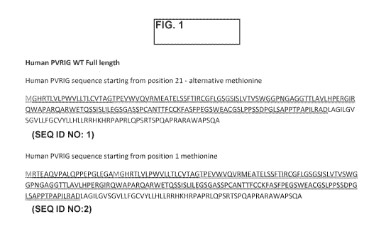
[0030] Figure 1 depicts the full-length sequence of human PVRIG (showing two
different
methionine starting points). The signal peptide is underlined, the ECD is
double underlined.
[0031] Figure 2 depicts the sequence of the human Poliovirus receptor-related
2 protein (PVLR2,
also known as nectin-2, CD112 or herpesvirus entry mediator B, (HVEB)), the
binding partner of
PVRIG. PVLR2 is a human plasma membrane glycoprotein.
[0032] Figure 3A and B depicts the variable heavy and light chains as well as
the vhCDR1,
vhCDR2, vhCDR3, v1CDR1, v1CDR2 and v1CDR3 sequences of each of the enumerated
CHA
antibodies of the invention, CHA.7.518.1.H4(S241P), and
CHA.7.538.1.2.H4(S241P).
[0033] Figure 4A and B PVRIG antibodies increase T cell proliferation in the
MLR. The percentages
of CFSE low cells are shown from MLR assays treated with the indicated PVRIG
antibodies. Each
graph represents one individual CD3 T cell donor. The experiments are
described in Example 23 of
USSN 15/048,967, incorporated by reference herein.
[0034] Figure 5A and B PVRIG hybridoma antibody binding characteristics to HEK
hPVRIG
engineered cell lines, HEK parental cells, and Jurkat cells. HEK OE denotes
HEK hPVRIG cells,
HEK par denotes HEK parental cells. For Jurkat data, gMFIr indicates the fold
difference in
geometric MFI of PVRIG antibody staining relative to their controls.
Concentration indicates that at
which the gMFIr was calculated. No binding indicates antibody does not bind to
the tested cell line.
Highlighted antibodies are the 'top four' antibodies of interest.
[0035] Figure 6A and B PVRIG hybridoma antibody binding characteristics to
primary human
PBMC, cyno over-expressing cells, and cyno primary PBMC. Expi cyno OE denotes
expi cells
transiently transfected with cPVRIG, expi par denotes expi parental cells.
gMFIr indicates the fold
difference in geometric MFI of PVRIG antibody staining relative to their
controls. Concentrations
indicate that at which the gMFIr was calculated. Not tested indicates
antibodies that were not tested
due to an absence of binding to human HEK hPVRIG, expi cPVRIG cells, or not
meeting binding
requirements to PBMC subsets. Highlighted antibodies are the 'top four'
antibodies of interest. The
experiments are described in Example 21 of USSN 15/048,967, incorporated by
reference herein.
[0036] Figure 7A and B Summary of blocking capacity of PVRIG antibodies in the
FACS-based
competition assay. The IC50 of inhibition is indicated. No IC50 indicates that
these antibodies are non-
blockers. Highlighted antibodies are the 'top four' antibodies of interest.
The experiments are
described in Example 21 of USSN 15/048,967, incorporated by reference herein.
7
CA 03032331 2019-01-29
WO 2018/033798
PCT/IB2017/001256
[0037] Figure 8A and B TILs were co-cultured with melanoma cells 624 at 1:1
E:T for 18hr in the
presence of anti-PVRIG Ab (CPA.7.021; lOug/m1) , anti-TIGIT (10A7 clone;
lOug/m1) or in
combination. Supernatant was collected and tested in Thl Th2 Th17 cytometric
bead array assay to
detect secreted cytokines. IFNy (A) and TNF (B) levels were detected.
Treatments were compared by
Student's t-test (*P < 0.05, **P < 0.01) of triplicate samples.
[0038] Figure 9A to F MART-1 or 209 TILs were co-cultured with melanoma cells
624 at 1:1 E:T
for 18hr in the presence of anti-PVRIG Ab (CPA.7.021; 10ug/m1) , anti-DNAM1
(DX11 clone, BD
Biosciences Cat. No. 559787; 10ug/m1) or in in combination. Supernatant was
collected and tested in
Thl Th2 Th17 cytometric bead array assay to detect secreted cytokines. IFNy
(A,D) and TNF (B,E)
levels were detected. TILs were stained for surface expression of CD137 (C,F).
[0039] Figure 10A and B TILs (F4) were co-cultured with melanoma cells 624 at
1:3 E:T for 18hr in
the presence of anti-PVRIG Ab (CPA.7.021; lOug/m1) , anti-TIGIT (10A7 clone;
lOug/m1), anti-PD1
(mAb 1B8, Merck; lOug/m1) or in combination. Supernatant was collected and
tested in Thl Th2
Th17 cytometric bead array assay to detect secreted cytokines. IFNy (A) and
TNF (B) levels were
detected.
[0040] Figure 11A to E depict four humanized sequences for each of CHA.7.518,
CHA.7.524,
CHA.7.530, CHA.7.538_1 and CHA.7.538_2. All humanized antibodies comprise the
H4(5241P)
substitution. Note that the light chain for CHA.7.538_2 is the same as for
CHA.7.538_1. The "Hl"
of each is a "CDR swap" with no changes to the human framework. Subsequent
sequences alter
framework changes shown in larger bold font. CDR sequences are noted in bold.
CDR definitions are
AbM from website bioirtiorg.uk:abs/. Human germline and joining sequences
from IMGTO
the international ImMunoGeneTics0 information system www.imgt.org (founder and
director: Marie-
Paule Lefranc, Montpellier, France). Residue numbering shown as sequential
(seq) or according to
Chothia from website www.bioinf.org.uk/abs/ (AbM). "b" notes buried sidechain;
"p" notes partially
buried; "i" notes sidechain at interface between VH and VL domains. Sequence
differences between
human and murine germlines noted by asterisk (*). Potential additional
mutations in frameworks are
noted below sequence. Potential changes in CDR sequences noted below each CDR
sequence as noted
on the figure (# deamidation substitutions: Q/S/A; these may prevent
asparagine (N) deamidation. @
tryptophan oxidation substitutions: Y/F/H; these may prevent tryptophan
oxidation; @ methionine
oxidation substitutions: L/F/A).
[0041] Figure 12A to E depicts a collation of the humanized sequences of three
CHA antibodies:
CHA.7.518, CHA.7.538.1, and CHA.7.538.2.
8
CA 03032331 2019-01-29
WO 2018/033798
PCT/IB2017/001256
[0042] Figure 13. depicts schemes for combining the humanized VH and VL CHA
antibodies.
The "chimVH" and "chimVL" are the mouse variable heavy and light sequences
attached to a human
IgG constant domain.
[0043] Figure 14. PVRIG hybridoma antibody binding characteristics to primary
human PBMC,
cyno over-expressing cells, and cyno primary PBMC. Expi cyno OE denotes expi
cells transiently
transfected with cPVRIG, expi par denotes expi parental cells. gMFIr indicates
the fold difference in
geometric MFI of PVRIG antibody staining relative to their controls.
Concentrations indicate that at
which the gMFIr was calculated. Not tested indicates antibodies that were not
tested due to an absence
of binding to human HEK hPVRIG, expi cPVRIG cells, or not meeting binding
requirements to
PBMC subsets. Highlighted antibodies are four antibodies for which
humanization was done (See
Figure 24). The experiments are described in Example 21 of USSN 15/048,967,
incorporated by
reference herein.
[0044] Figure 15. Summary of blocking capacity of PVRIG antibodies in the FACS-
based
competition assay. The IC50 of inhibition is indicated. No IC50 indicates that
these antibodies are
non-blockers. Highlighted antibodies are four antibodies for which
humanization was done (See
Figure 24).
[0045] Figure 16. Summary of the activity of select PVRIG antibodies in NK
cell cytotoxicity
assays against Reh and MOLM-13 cells. Fold change in cytotoxicity relative to
control was calculated
by dividing the absolute level of killing (%) in the condition with PVRIG
antibody, by the absolute
level of killing (%) with control antibody. Fold change is calculated from the
5:1 effector to target
ratio.
[0046] Figure 17. Sequence alignment of PVRIG orthologs. Aligned sequences of
the human,
cynomolgus, marmoset, and rhesus PVRIG extra-cellular domain. The differences
between human
and cynomolgus are highlighted in yellow.
[0047] Figure 18. Binding of anti-human PVRIG antibodies to cyno, human,
cyno/human hybrid
PVRIG variants. Binding of antibodies to wild type cyno PVRIG (e), H61R cyno
PVRIG (M), P67S
cyno PVRIG (A), L95R/T97I cyno PVRIG (v), and wild type human PVRIG (#) are
shown. The
ELISA signals are plotted as a function of antibody concentration.
[0048] Figure 19. Correlation of epitope group and cyno cross-reactivity of
anti-human PVRIG
antibodies.
[0049] Figure 20A to B (A) Specificity of CHA.7.518.1.H4(S241P) towards HEK
cells engineered to
overexpress PVRIG and HEK parental cells. Data shows absolute geometric MFI
(gMFI)
9
CA 03032331 2019-01-29
WO 2018/033798
PCT/IB2017/001256
measurements as a function of increasing antibody concentration. (B)
Specificity of
CHA.7.538.1.2.H4(S241P) towards HEK cells engineered to overexpress PVRIG and
HEK parental
cells. Data shows absolute geometric MFI (gMFI) measurements as a function of
increasing antibody
concentration.
[0050] Figure 21 A to B illustrates the ability of CHA.7.518.1.H4(S241P) (A)
and
CHA.7.538.1.2.H4(S241P) (B) to bind Jurkat cells that endogenously express
PVRIG confirmed by
RNA expression. (A) Binding of CHA.7.518.1.H4(5241P) to Jurkat cells. Data
shows absolute
geometric MFI (gMFI) measurements as a function of increasing antibody
concentration. Isotype
staining is shown as a negative control. (B) Binding of
CHA.7.538.1.2.H4(5241P) to Jurkat cells.
Data shows absolute geometric MFI (gMFI) measurements as a function of
increasing antibody
concentration. Isotype staining is shown as a negative control. Both
antibodies are able to bind Jurkat
cells with a comparable affinity to HEK hPVRIG cells.
[0051] Figure 22 illustrates the ability of CHA.7.518.1.H4(5241P) and
CHA.7.538.1.2.H4(5241P) to
bind CD8 T cells that were expanded by exposure to CMV peptide (494-503,
NLVPMVATV) and
endogenously express PVRIG confirmed by RNA expression. Binding of
CHA.7.518.1.H4(S241P)
and CHA.7.538.1.2.H4(5241P) to CMV peptide-expanded CD8 T cells. Data shows
absolute
geometric MFI (gMFI) measurements as a function of increasing antibody
concentration. Isotype
staining is shown as a negative control.
[0052] Figure 23A to B. (A) Specificity of CHA.7.518.1.H4(S241P) towards expi
cells engineered to
overexpress cynomolgus PVRIG and expi parental cells. Data shows absolute
geometric MFI (gMFI)
measurements as a function of increasing antibody concentration. Specificity
of
CHA.7.538.1.2.H4(5241P) towards expi cells engineered to overexpress
cynomolgus PVRIG and
expi parental cells. Data shows absolute geometric MFI (gMFI) measurements as
a function of
increasing antibody concentration.
[0053] Figure 24A to B. (A) Blocking of PVRIG Fc to HEK cells by
CHA.7.518.1.H4(5241P) and
CHA.7.538.1.2.H4(5241P). Data shows the percentage of PVRIG Fc binding to HEK
cells as a
function of increasing antibody concentration relative to maximum PVRIG Fc-
induced signal and
secondary only background. (B) Effect of CHA.7.544 on the binding of PVRIG Fc
to HEK cells.
Data shows the absolute gMFI derived from PVRIG Fc binding to HEK cells in the
presence of
escalating concentrations of CHA.7.544. The amount of PVRIG Fc binding was
detected by an anti-
mouse Fc secondary conjugated to Alexa 647.
[0054] Figure 25A to B. (A) Blocking of PVRL2 Fc to HEK hPVRIG cells by
CHA.7.518.1.H4(S241P), CHA.7.538.1.2.H4(S241P), and CHA.7.530.3. Data shows
the percentage
of PVRL2 Fc binding to HEK hPVRIG cells as a function of increasing antibody
concentration
CA 03032331 2019-01-29
WO 2018/033798
PCT/IB2017/001256
relative to maximum PVRL2 Fc-induced signal and secondary only background. (B)
Effect of
CHA.7.544 on PVRL2 Fc binding to HEK hPVRIG cells. Data shows the percentage
of PVRL2 Fc
binding to HEK hPVRIG cells as a function of increasing antibody concentration
relative to
maximum PVRL2 Fc-induced signal and secondary only background.
[0055] Figure 26. Shows the percentage of Alexa 647 conjugated
CHA.7.518.1.H4(5241P) and
CHA.7.538.1.2.H4(5241P) binding relative to their maximum signal upon pre-
incubation of Jurkat
cells with unconjugated CHA.7.518.1.H4(S241P), CHA.7.538.1.2.H4(S241P) and an
isotype control.
[0056] Figure 27A to B A) Humanized PVRIG antibodies, CHA.7.518.1.H4(5241P)
and
CHA.7.538.1.2.H4(5241P), increase CD4+ T cell proliferation. Representative
data (n>2) shows the
percentage of CFSE low, proliferating CD4+ T cells (mean plus standard
deviation) from a single
human CD4+ T cell donor when co-cultured with the CHO-S OKT3 hPVRL2 cells in
the presence of
an anti-DNAM-1 antibody or different anti-PVRIG antibodies or IgG isotype
controls. The dashed
line indicates the baseline percentage of CFSE low, CD4+ T cells proliferating
after treatment with
the human IgG4 isotype control antibody. The numbers refer to the percent
increase or decrease in
proliferation of the anti-PVRIG or anti-DNAM-1 antibody treatments,
respectively, compared to the
relevant isotype control antibodies (B) Humanized PVRIG antibodies,
CHA.7.518.1.H4(5241P) and
CHA.7.538.1.2.H4(5241P), increase CD4+ T cell proliferation in an hPVRL2-
dependent manner.
Representative data (n>2) shows the percentage of CFSE low, proliferating CD4+
T cells (mean plus
standard deviation) from a single human CD4+ T cell donor in response to co-
culture with the CHO-S
OKT3 parental, or CHO-S OKT3 hPVRL2 cells in the presence of an anti-DNAM-1
antibody or
different anti-PVRIG antibodies or IgG isotype controls. The dashed line
indicates the baseline
percentage of CFSE low CD4+ T cells proliferating after treatment with either
the human IgG4 or the
mouse IgG1 isotype antibodies. The numbers refer to the percent increase or
decrease in proliferation
of the anti-PVRIG or anti-DNAM-1 antibody treatments, respectively, compared
to the relevant
isotype control antibodies.
[0057] Figure 28A to C. (A) Humanized PVRIG antibodies, CHA.7.518.1.H4(5241P)
and
CHA.7.538.1.2.H4(5241P), increase CD8+ T cell proliferation. Representative
data (n>2) shows the
percentage of CFSE low, proliferating CD8+ T cells (mean plus standard
deviation) from a single
human CD8+ T cell donor (Donor 232) when co-cultured with the CHO-S OKT3
hPVRL2 cells in the
presence of an anti-DNAM-1 antibody or different anti-PVRIG antibodies or IgG
isotype controls.
The dashed line indicates the baseline percentage of CFSE low, CD8+ T cells
proliferating after
treatment with the mouse IgG1 or human IgG4 isotype antibodies. The numbers
refer to the percent
increase or decrease in proliferation of the anti-PVRIG or anti-DNAM-1
antibody treatments,
respectively, compared to the relevant isotype control antibodies. (B)
Humanized PVRIG antibodies,
CHA.7.518.1.H4(S241P) and CHA.7.538.1.2.H4(S241P), increase CD8+ T cell
proliferation.
11
CA 03032331 2019-01-29
WO 2018/033798
PCT/IB2017/001256
Representative data (n>2) shows the percentage of CFSE low, proliferating CD8+
T cells (mean plus
standard deviation) from a single human CD8+ T cell donor (Donor 234) when co-
cultured with the
CHO-S OKT3 hPVRL2 cells in the presence of an anti-DNAM-1 antibody or
different anti-PVRIG
antibodies or IgG isotype controls. The dashed line indicates the baseline
percentage of CFSE low,
CD8+ T cells proliferating after treatment with the mouse IgG1 or human IgG4
isotype antibodies.
The numbers refer to the percent increase or decrease in proliferation of the
anti-PVRIG or anti-
DNAM-1 antibody treatments, respectively, compared to the relevant isotype
control antibodies. (C)
Humanized PVRIG antibodies, CHA.7.518.1.H4(S241P) and CHA.7.538.1.2.H4(S241P),
increase
IFNy secretion from CD8+ T cells. Representative data (n>2) shows the pg/ml of
IFNy produced
(mean plus standard deviation) by three different human CD8+ T cell donors
(Donors 231, 232, and
234) when co-cultured with the CHO-S OKT3 hPVRL2 cells in the presence of an
anti-DNAM-1
antibody or different anti-PVRIG antibodies or IgG isotype controls. The
dashed line indicates the
baseline IFNy production following treatment with the human IgG4 isotype
antibody. The numbers
refer to the percent increase in IFNy secretion of the anti-PVRIG antibody
treatments compared to the
IgG4 isotype control.
[0058] Figure 29. Humanized PVRIG antibodies, CHA.7.518.1.H4(S241P) and
CHA.7.538.1.2.H4(S241P), consistently increase CD4+ T cell proliferation
across multiple donors,
while CHA.7.530.3 and CHA.7.544 do not. The percent proliferation relative to
the isotype control
was calculated by dividing the percentage of CFSE low, CD4+ T cells after
PVRIG antibody
treatment over the isotype antibody treatment for each donor. The percent
proliferation for the isotype
antibody treatment was set at zero. Each symbol in the graph represents a
different donor.
[0059] Figure 30A to D. (A) Dose-dependent effect of the humanized PVRIG
antibodies,
CHA.7.518.1.H4(S241P) and CHA.7.538.1.2.H4(S241P), on CD4+ T cell
proliferation.
Representative data (n>2) with 2 different human donors shows the mean
percentage of proliferating
CD4+ T cells following a dose titration of 66nM to 0.726nM with either the
human IgG4 isotype,
CHA.7.518.1.H4(S241P), or CHA.7.538.1.2.H4(S241P) antibodies. The estimated
EC50 is within the
single digit nM range. (B) Dose-dependent effect of the humanized PVRIG
antibodies,
CHA.7.518.1.H4(S241P) and CHA.7.538.1.2.H4(S241P), on CD8+ T cell
proliferation.
Representative data (n>2) with 2 different human donors shows the mean
percentage of proliferating
CD8+ T cells following a dose titration of 66nM to 0.264nM with either the
human IgG4 isotype,
CHA.7.518.1.H4(S241P), CHA.7.38.1.2, or CHA.7.544 antibodies. The estimated
EC50 is within the
single digit nM range.
[0060] Figure 31A to C. (A) Flow cytometry analysis of TIGIT and PVRIG
expression on TILs and
PVR, PVRL2 expression on 624 melanoma cell line. Values represent Mean
fluorescent intensity
(MFI) ratio vs isotype control. (B-C) Representative experiment showing IFNy
(B) and TNF (C)
12
CA 03032331 2019-01-29
WO 2018/033798
PCT/IB2017/001256
secretion by TILs upon co-cultured with melanoma cells 624 at 1:3 E:T for 18hr
in the presence of
isotype control, anti-TIGIT (30 g/m1) or anti- PVRIG Abs (lOug/m1) as mono
treatment (blue
histograms) or in combination with anti-TIGIT (green histograms). Percentage
of Ab mono treatment
effect was compared to isotype control treatment mIgG1 and the percentage of
Ab combo-treatment
effect was compared to anti-TIGIT mono-treatment.
[0061] Figure 32A to H. TILs (209-gp100/463-F4-gp100) were co-cultured with
melanoma cells 624
in 1:3 E:T for 18hr in the presence of anti PVRIG Abs CHA.7.518.1.H4(S241P) or
CHA.7.538 with
or without anti-TIGIT (aTIGIT) combo and tested for cytokine secretion.
Percentage of Ab treatment
effect was compared to isotype control treatment and the mean of 5 experiments
(F4) or 6
experiments (209) were plotted. Paired, two tailed T test was calculated for
each treatment compared
to isotype or in combos- compared to anti-TIGIT alone, p values are indicated.
[0062] Figure 33A to B. (A) Humanized PVRIG antibody, CHA.7.518.1.H4(S241P),
and an anti-
TIGIT antibody increase CD4+ T cell proliferation compared to single antibody
treatments.
Representative data (n>2) shows the percentage of CFSE low, proliferating CD4+
T cells (mean plus
standard deviation) from a single human CD3+ T cell donor (Donor 143) when co-
cultured with the
CHO-S OKT3 hPVRL2 cells. The dashed line indicates the baseline percentage of
CFSE low, CD4+
T cells proliferating after treatment with the human IgG4 isotype control
antibody. (B) Humanized
PVRIG antibody, CHA.7.518.1.H4(S241P), and the anti-TIGIT antibody increase
CD4+ T cell
proliferation compared to single antibody treatments. Representative data
(n>2) shows the percentage
of CFSE low, proliferating CD4+ T cells (mean plus standard deviation) from a
single human CD4+ T
cell donor (Donor 201) when co-cultured with the CHO-S OKT3 hPVRL2 cells. The
dashed line
indicates the baseline percentage of CFSE low, CD4+ T cells proliferating
after treatment with the
human IgG4 isotype control antibody. The numbers refer to the percent increase
or decrease in
proliferation of the anti-PVRIG or anti-DNAM-1 antibody treatments,
respectively, compared to the
relevant isotype control antibodies.
[0063] Figure 34A to B. (A): The combination of the humanized PVRIG antibody,
CHA.7.518.1.H4(S241P), and the anti-TIGIT antibody increases CD8+ T cell
proliferation.
Representative data (n>2) shows the percentage of CFSE low, proliferating CD8+
T cells (mean plus
standard deviation) from a representative human CD8+ T cell donor (Donor 232)
when co-cultured
with the CHO-S OKT3 hPVRL2 cells. The dashed line indicates the baseline
percentage of CFSE
low, CD8+ T cells proliferating after treatment with the human IgG4 isotype
antibody. The numbers
refer to the percent increase or decrease in proliferation of the anti-PVRIG
or anti-DNAM-1 antibody
treatments, respectively, compared to the relevant isotype control antibodies.
(B) The combination of
the humanized PVRIG antibody, CHA.7.518.1.H4(S241P), and the anti-TIGIT
antibody increases
IFNy secretion from CD8+ T cells. Representative data (n>2) shows the pg/ml of
IFNy produced
13
CA 03032331 2019-01-29
WO 2018/033798
PCT/IB2017/001256
(mean plus standard deviation) by a representative human CD8+ T cell donor
(Donor 232) when co-
cultured with the CHO-S OKT3 hPVRL2 cells. The dashed line indicates the
baseline IFNy
production following treatment with the human IgG4 isotype antibody. The
numbers refer to the
percent increase or decrease in IFNy secretion of the anti-PVRIG or anti-DNAM-
1 antibody
treatments, respectively, compared to the relevant isotype control antibodies.
[0064] Figure 35 depicts the design of the experimental system of Example
2(3).
[0065] Figure 36A to C. shows a histogram depicting levels of PVRIG (using
Anti-Human PVRIG
CHA.7.538.AF647), TIGIT (using Anti-Human TIGIT Cat. 17-9500-41 eBioscience)
and DNAM-1
(using Anti-human CD226-APC Cat.338312 biolegend) expression in TILs. Fold of
expression is
compared to isotype (Iso) control.
[0066] Figure 37. Summarized plot of the effect of anti PVRIG antibodies on
the secretion of IFNy
from TILs. TILs were co-cultured with CHO-S HLA-A2/B2M cells over-expressing
PVRL2 in E:T
ratio of 1:3 for 18hr in the presence of anti PVRIG antibodies (c518, c538 and
544) or with anti
TIGIT antibody. Each dot represents an average of data of IFNy secretion from
the same TIL from
different experiments. The percentage indicated is the different between each
antibody treatment
compared to isotype control. Paired, two tailed T-test was calculated for each
treatment compared to
544 or in combos, compared to anti TIGIT alone, p values are indicated. Number
of experiments
preformed per each TILs; 209(N=3), F4 (N=2), F5(N=3) and MART1(N=2).
[0067] Figure 38. Summarized plot of the effect of c518 and c538 dose response
on the secretion of
TNF-a from TILs. TILs were co-cultured with CHO-S HLA-A2/B2M cells over-
expressing PVRL2
in effector-to-target ratio of 1:3 for 18hr in the presence of anti PVRIG
antibodies (c518, c538 or
isotype control) as described in Example 2(3).
[0068] Figure 39A to C. TILs were co-cultured with CHO-S HLA-A2/B2M target
cells over-
expressing PVRL2 in E:T ration of 1:3 for 18hr in the presence of anti PVRIG
antibodies (c518, c538
and 544) or with anti TIGIT antibody. The percentage indicated in the above
tables is the difference in
the effect of cytokine secretion from TILs of each antibody treatment compared
to its isotype control.
The first experiment is represented in Figure A and B, and the second
experiment in Figure C.
[0069] Figure 40. CHO-S OKT3 co-culture assay design. CFSE labeled CD3+ T
cells were co-
cultured with CHO-S-OKT3-PVRL2 or mock transfected cells for 5d. The effect
anti-PVRIG Abs on
T cell proliferation and cytokine secretion was analyzed.
[0070] Figure 41. Effect of anti-PVRIG antibodies on IFNy secretion upon CHO-
OKT3 PVRL2
cells in responder vs. non-responder donor. CD3+ cells from 2 different donors
were co-cultured with
14
CA 03032331 2019-01-29
WO 2018/033798
PCT/IB2017/001256
CHO-S-PVRL2 cells in 5:1 E:T for 5d in the presence of anti PVRIG Abs and
tested for cytokine
secretion and T cells proliferation. (A) 'responder donor' in which we
observed an effect to anti
PVRIG Abs. (B) 'non-responder donor' in which we do not observed effects to
Abs treatment.
[0071] Figure 42. Effect of anti-PVRIG antibodies on CD4 and CD8 proliferation
from responder
donor. CFSE labeled CD3+ T cells were co-cultured with CHO-S-PVRL2 cells in
5:1 E:T for 5d in
the presence of anti PVRIG Abs or anti-TIGIT Abs. The effect on T cells
proliferation gating on CD4
or CD8 was evaluated by flow cytometry. Percentage of proliferating cells
(CFSE low) (A) or total
cells number (B) of CD4+CFSElow or CD8+CFSE low are presented.
[0072] Figure 43. Shows the effect of anti-PVRIG antibodies on IFNy secretion
or CD8
proliferation from responder donor. CD3+ cells were co-cultured with CHO-S-
PVRL2 cells in 5:1
E:T for 5d in the presence of anti PVRIG Abs and tested for (A) cytokine
secretion and (B) T cells
proliferation. Percentage of Ab treatment effect was compared to isotype
control treatment and the
mean of 5 'responders' donors (responders) is presented. (C) IFNy secretion
levels from the same 5
donors upon co-culture with CHOS-OKT3 PVRL2 as described in section A and B
upon treatment
with isotype vs. anti-PVRIG Abs. p value represent ratio paired T test.
[0073] Figure 44 is a summary table of Abs treatment effect across donors
tested (n=10).
Percentages indicated represent the effect of Ab treatment on a specific
readout (indicated in columns
titles) as compared to the relevant isotype control. 'responder' donors
(donors #3, 72,226,345 and
ES_001) considered as 'responder' which some anti-PVRIG Abs (mainly CHA.7.518)
enhanced IFNy
or proliferation vs. isotype controls.
[0074] Figure 45A to B depict the results of experiments with several
antibodies. The affinities
(nM) are shown in A, with the HEK hPVRIG cells being HEK cells transformed
with hPVRIG as
discussed herein and Jurkat cells expressing endogeneous hPVRIG. (B) depicts
the gMFI using 4
different antibodies against Donor 1 primary CD8 T cells and (C) being Donor 2
primary CD8 T cells.
[0075] Figure 46A to B depict interactions of TIGIT with CHO cells. (A) Human
TIGIT Fc protein
binds to CHO cells. Graded concentrations of human TIGIT Fc and synagis IgG1
control were
assessed for their ability to bind to CHO cells in a FACS-based binding assay.
(B) Human PVR is
expressed on activated CD4 T cells. CD4 T cells were co-cultured with CHO
cells expressing the
scFv of the OKT3 antibody and activated for 5 days. On day 5, CD4 T cells were
analysed for
expression of PVR and dilution of CFSE.
100761 Figure 47A to C depict antitumor responses of anti-rnPVR1g and anti-
PDL4 antibodies in
CT26 tumor model. A-B. Groups of 10 BALIB/e mice were subcutaneously injected
with 5x 10⁵ CT26
CA 03032331 2019-01-29
WO 2018/033798
PCT/IB2017/001256
cells. After tumors were measured on day 4, mice were randomized (40 mni3 mean
tumor volume per
group) and then treated with the designated triAb (100 or 200 jig/dose IP)
followed by additional
doses on days 7, 11, 14, 18 and 21. A. Groups were treated with 6 doses of
single agents. Anti-PDL-1
vs control ***p <0.0001. Tumor volumes are represented as the Mean volume +
SEM. B, Tumor
volumes were measured twice weekly. The number of tumor-free (TF) mice per
group is indicated. C.
survival proportions of assigned groups; Anti-PDL-1 vs control **p =0.005.
1007 Figure 48A to C depict antitumor responses of ami-PVRIG and anti-PDL-1
antibodies
combination in cr26 tumor model. A-B. Groups of 10 BALB/c mice were
subcutaneously injected
with 5x10⁵ CT26 cells. After tumors were measured on day 7, mice were
randomized (75 nuri3 mean
tumor volume per group) and then treated with the designated tnAb (300 14/dose
IP) followed by
additional doses on days 11, 14, 18,21 and 25. A. Groups were treated with 6
doses of combined
agents. Anti-PDL-1-i-inAb 407 vs control p = 0.0005; anti-PDL-1 and mAb 406 vs
control p=0.056. B.
Tumor volumes were measured x3 weekly. The number of tumor-free (TF) mice per
group is
indicated. C. survival proportions of assigned groups; Ariti-PDL-1+triAb 407
vs control *p =0.0088.
[0078] Figure 49A to D depict the amino acid sequences and the nucleic acid
sequence for the
variable heavy chain (A and B, respectfully) and the amino acid sequences and
the nucleic acid
sequence for the variable light chain (C and D, respectfully) for AB-407 (B0J-
5G4-F4).
10079] Figure 50 depicts the amino acid sequences of the constant domains of
human IgG1 (with
some useful amino acid substitutions), IgG2, IgG3, IgG4, IgG4 with a hinge
variant that finds
particular use in the present invention, and the constant domains of the kappa
and lambda light chains.
[0080] Figure 51 depicts the sequences of human and cynomolgus macaque
(referred to as cyno)
TIGIT ECD and of the human PVR ECD proteins.
[0081] Figure 52. Shows the flow cytometry binding summary for anti-TIGIT
fabs. All unique
ELISA positive fabs were analyzed by flow cytometry. The mean fluorescence
intensity (MFI) was
measured for the human or cyno TIGIT over-expressing Expi293 cells as well as
the parental Expi293
cells. The MFI ratio for the target-specific vs off-target binding was
calculated. Data for selected
clones is shown.
[0082] Figure 53A and B depict the sequences of anti-TIGIT antibodies. Unless
otherwise noted, the
CDRs utilize the IMGT numbering (including the antibodies of the sequence
listing.
[0083] Figure 54. Shows the FACS KD results of anti-TIGIT mAbs binding to
Expi293 human
TIGIT over-expressing cells as described in Example 12.
16
CA 03032331 2019-01-29
WO 2018/033798
PCT/IB2017/001256
[0084] Figure 55. Shows the FACS KD results of mAbs binding to Expi293 cyno
TIGIT over-
expressing cells.
[0085] Figure 56. Shows the results from Example 14, showing the resulting
kinetic rate constants
and the equilibrium dissociation constants where data were reliable enough to
estimate the binding
constants.
100861 Figure 57A to B show the results of human PVR-Fc variant binding to
Expi293 human
TIGIT over-expressing cells in Example 4. Figure A (left): Binding curve
generated for human PVR-
m2aFc construct titrated with Expi293 human TIGT over-expressing cells. The KD
and 95%
confidence interval are shown. Figure B (right): Binding curve generated for
human PVR-hl Fc
construct titrated with Expi293 human TIGT over-expressing cells. The KD and
95% confidence
interval are shown.
[0087] Figure 58. Shows a table of phage antibodies inhibiting human PVR-m2aFc
binding to human
TIGIT over-expressed on Expi293 cells. mAbs were tested against known blocking
(BM26)
benchmark antibody, and human IgG4 isotype control (Synagis) antibody. A "Yes"
indicates the mAb
inhibited hPVR analogous to BM26.
[0088] Figure 59. Shows a table of IC50 values of anti-TIGIT hybridoma
antibodies inhibiting
binding of human PVR-h1Fc to human TIGIT over-expressed on Expi293 cells.
Values are
representative of one of two independent experiments. The IC50 results for the
two independently
performed experiments showed a range of only 1.2-2-fold differences.
100891 Figure 60. Shows the results of Example 6, that the phage-derived and
BM anti-human
TIGIT antibodies, CPA.9.027, CPA.9.049, CPA.9.059, BM26, and BM29 increase IL-
2 signaling.
BM26 and BM29 are both the human IgG4 (hIgG4 with a S241P variant) isotype.
Representative data
(n>2) shows the RLU (mean +/- standard deviation) of the luciferase signal
from a 6 hour co-culture
of Jurkat IL-2-RE luciferase human TIGIT cells and aAPC CHO-K1 human PVR
cells. The
concentration of each antibody was 10 jig/ml.
[0090] Figure 61. Shows additional results of Example 6, that the phage-
derived and BM hIgG4
anti-human TIGIT antibodies, CPA.9.027, CPA.9.049, CPA.9.059, BM26, and BM29
increase IL-2
signaling in a dose-dependent manner. BM26 and BM29 are both the hIgG4
isotype. Representative
data (n>2) shows the RLU (mean +/- standard deviation) of the luciferase
signal from a 6 hour co-
culture of Jurkat IL-2-RE luciferase human TIGIT cells and aAPC CHO-K1 human
PVR cells. A 10
point, 2-fold dilution series starting at 20 jig/ml was used for each
antibody.
17
CA 03032331 2019-01-29
WO 2018/033798
PCT/IB2017/001256
[0091] Figure 62. Shows the results of Example 6, that the hybridoma-derived
and BM anti-human
TIGIT antibodies, CHA.9.536, CHA.9.541, CHA.9.546, CHA.9.547, CHA.9.560, BM26,
and BM29
increase IL-2 signaling. BM26 and BM29 are both the mIgG1 isotype. The non-
blocking anti-human
TIGIT antibody, CHA.9.543 does not enhance IL-2 signaling. Representative data
(n>2) shows the
RLU (mean +/- standard deviation) of the luciferase signal from a 6 hour co-
culture of Jurkat IL-2-RE
luciferase human TIGIT cells and aAPC CHO-Kl human PVR cells. The
concentration of each
antibody was 10 jig/ml.
10092] Figure 63. Shows the results of Example 6, that the hybridoma-derived
and benchmark
mIgG1 anti-human TIGIT antibodies, CHA.9.536, CHA.9.541, CHA.9.546, CHA.9.547,
CHA.9.560,
and BM26 increase IL-2 signaling in a dose-dependent manner. BM26 is the mIgG1
isotype.
Representative data (n>2) shows the RLU (mean +/- standard deviation) of the
luciferase signal from
a 6 hour co-culture of Jurkat IL-2-RE luciferase human TIGIT cells and aAPC
CHO-Kl human PVR
cells. A 10 point, 2-fold dilution series starting at 20 jig/ml was used for
each antibody.
[0093] Figure 64. Shows that the phage, hybridoma and BM anti-human TIGIT
antibodies,
CPA.9.027, CPA.9.049, CPA.9.059, CHA.9.536, CHA.9.541, CHA.9.546, CHA.9.547,
CHA.9.560,
BM26, and BM29 increase antigen-specific IFNy signaling. BM26 is tested as
both the hIgG4 and
mIgG1 isotypes, while BM29 is only tested as the hIgG4 isotype. Representative
data (n=2) shows the
amount of IFNy (mean +/- standard deviation) in the culture supernatant after
24 hour co-culture of
CMV-specific CD8 T cells with the Me1624 human PVR cells. The concentration of
each antibody
was 10 E g/ml. The Me1624 human PVR used in the assay were pulsed with 0.0033
jig/ml or 0.001
jig/ml peptide.
10094] Figure 65. Shows that the phage, hybridoma and BM anti-human TIGIT
antibodies,
CPA.9.027, CPA.9.049, CPA.9.059, CHA.9.536, CHA.9.541, CHA.9.546, CHA.9.547,
and
CHA.9.560, as well as BM26, increase antigen-specific IFNy signaling either
alone (open bars) or in
combination with an anti-PVRIG antibody, CHA.7.518.1.H4(5241P) (hatched bars).
BM26 is the
mIgG1 isotype. For the isotype antibody control treatments, the open bar
refers to the isotype
antibody alone, and the hatched bar refers to isotype antibody in combination
with
CHA.7.518.1.H4(S241P). Representative data (n=2) shows the amount of IFNy
(mean +/- standard
deviation) in the culture supernatant after a 24 hour co-culture of CMV-
specific CD8+ T cells with
Me1624 cells over-expressing human PVR and human PVRL2. The concentration of
each antibody
was 10 jig/ml. The Me1624 human PVR/human PVRL2 cells used in the assay were
pulsed with
0.0033 jig/ml or 0.001 jig/ml peptide.
18
CA 03032331 2019-01-29
WO 2018/033798
PCT/IB2017/001256
[0095] Figure 66. Shows the percent increase of IFNy secretion with anti-human
TIGIT antibodies,
CHA.7.518.1.H4(5241P), and the combination of anti-human TIGIT antibodies and
CHA.7.518.1.H4(5241P), over the respective isotype control antibodies.
[0096] Figure 67 is the dendrogram for the epitope binning experiments of
Example 7.
[0097] Figure 68 is the grouping of the antibodies from the epitope binning
experiments of
Example 7.
[0098] Figure 69. Shows the high affinity binding to human TIGIT
overexpressing cells in a dose
titration of the affinity matured phage antibodies (CPA.9.083, CPA.9.086),
humanized hybridoma
antibodies (CHA.9.547.7, CHA.9.547.13), benchmark antibodies (BM26, BM29), and
the hIgG4
isotype control (anti-Synagis) on human TIGIT over-expressing Expi293 cells,
as described in
experiments of Example 3. All antibodies were titrated using a serial 2-fold
dilution over 11 points
starting at 10 jig/ml (133.33 nM [binding sitep. AF647-labeled goat anti-human
F(ab') (Jackson
Immunoresearch) was added to the cells to detect binding of anti-TIGIT
antibodies. The gMFI of the
anti-TIGIT antibodies bound to the human TIGIT over-expressing Expi293 cells
(black line), and the
parental Expi293 cells (grey line) are shown. KD values +/- 95% CI, and curve
fits are indicated below
each graph.
[0099] Figure 70. Shows that anti-TIGIT antibodies are cross reactive to cyno
TIGIT in a dose
titration of the affinity matured phage antibodies (CPA.9.083, CPA.9.086),
humanized hybridoma
antibodies (CHA.9.547.7, CHA.9.547.13), benchmark antibodies (BM26, BM29), and
the hIgG4
isotype control (anti-Synagis) on cyno TIGIT over-expressing Expi293 cells, as
described in
experiments of Example 3. All antibodies were titrated using a serial 2-fold
dilution over 11 points
starting at 10 jig/ml (133.33 nM [binding sitep. AF647-labeled goat anti-human
F(ab') (Jackson
Immunoresearch) was added to the cells to detect binding of anti-TIGIT
antibodies. The gMFI of the
anti-TIGIT antibodies bound to the cyno TIGIT over-expressing Expi293 cells
(black line), and the
parental Expi293 cells (grey line) are shown. KD values +/- 95% CI, and curve
fits are indicated below
each graph.
1001001 Figure 71. Shows that affinity matured phage antibodies are cross
reactive to mouse TIGIT
in a dose titration of the affinity matured phage antibodies reformatted as
mouse IgG1 (mIgG1)
(CPA.9.083, CPA.9.086), benchmark anti-mouse TIGIT antibodies (BM27 mIgGl,
BM30 mIgG1),
and the mIgG1 isotype control (anti-Synagis) are shown, as described in
experiments of Example 3.
A) The gMFI of the anti-TIGIT antibodies bound to the mouse TIGIT over-
expressing HEK cells
(black line), and the parental HEK cells (grey line). B) The gMFI of the anti-
TIGIT antibodies (black
line) or Synagis mIgG1 (grey line) bound to regulatory CD4+CD25+Foxp3+ T cells
isolated from s.c.
19
CA 03032331 2019-01-29
WO 2018/033798
PCT/IB2017/001256
implanted Renca tumors in Balb/c mice. Anti-TIGIT antibodies were titrated
using either a serial 2- or
3-fold dilution series starting at 15 jug/m1 (200 nM [binding sitep, or 10
jug/m1 (132 nM [binding
siteD, respectively. AF647-labeled goat anti-mouse IgG-Fc (Southern Biotech)
were added to the cells
to detect binding of the anti-TIGIT antibodies on mouse TIGIT over-expressing
cells. Anti-TIGIT
antibodies were directly conjugated to AF647 for mouse Treg binding. KD values
for each anti-TIGIT
antibody are indicated.
1001011 Figure 72. Shows a dose titration of the affinity matured phage
antibodies (CPA.9.083,
CPA.9.086), humanized hybridoma antibodies (CHA.9.547.7, CHA.9.547.13), and
benchmark
antibodies (BM26, BM29) on human effector memory CD95+CD28-CD8+CD3+ T cells
from 3
healthy donor PBMCs (Donors 321, 322, and 334), as described in experiments of
Example 3.
PBMCs were surface stained with antibodies against the following lineage
markers CD3, CD4, CD8,
CD14, CD16, CD28, CD56, and CD95 (BD Biosciences, BioLegend), as well as
live/dead fixable
aqua dye (Life Technologies). AF647-labeled anti-TIGIT antibodies and hIgG4
isotype control
antibody (anti-Synagis) were then titrated using a serial 3-fold dilution over
12 points starting at 30
jug/m1 (396 nM [binding sitep. The gMFI of the anti-TIGIT antibodies bound to
the effector memory
T cells are shown. KD values for each antibody across the 3 different donors
are reported in the table.
The affinity mature phage antibodies (CPA.9.083 and CPA.9.086) had the highest
binding affinity to
the human effector memory T cells.
1001021 Figure 73. Shows a dose titration of the affinity matured phage
antibodies (CPA.9.083,
CPA.9.086, CPA.9.103), humanized hybridoma antibody (CHA.9.547.1), and
benchmark antibody
(BM26) on cyno effector memory CD95 tD28-CD8 FCD3 T cells from PBMCs isolated
from 2 naïve
cyno monkeys (BioreclamationIVT), as described in experiments of Example 3.
PBMCs were surface
stained with antibodies against the following lineage markers CD3, CD4, CD8,
CD14, CD16, CD28,
CD56, and CD95 (BD Biosciences, BioLegend), as well as live/dead fixable aqua
dye (Life
Technologies). AF647-labeled anti-TIGIT antibodies and hIgG4 isotype control
antibody (anti-
Synagis) were then titrated using a serial 3-fold dilution over 12 points
starting at 30 jug/m1 (396 nM
[binding sitep. The gMFI of the anti-TIGIT antibodies bound to the effector
memory T cells are
shown with the gMFI of the anti-Synagis hIgG4 isotype control antibody
subtracted. KD values for
each antibody across the 2 donors are reported in the table. The affinity
mature phage antibodies
(CPA.9.083 and CPA.9.086) had the highest binding affinity to the cyno
effector memory T cells.
1001031 Figure 74. Shows the SPR kinetics of anti-TIGIT antibody binding to
human, cyno and
mouse TIGIT, as described in experiments of Example 5. The kinetic rate and
equilibrium
dissociation constants for the affinity matured phage antibodies (CPA.9.083,
CPA.9.086, CPA.9.103),
CA 03032331 2019-01-29
WO 2018/033798
PCT/IB2017/001256
humanized hybridoma antibodies (CHA.9.547.1 and CHA.9.547.7), and benchmark
antibodies
(BM26, BM29) were determined by SPR on the ProteOn instrument.
1001041 Figure 75. shows that the anti-TIGIT antibodies block PVR/TIGIT
interactions, as described
in experiments of Example 4. Human TIGIT over-expressing Expi293 cells were
preincubated with
either the affinity matured phage antibodies (CPA.9.083, CPA.9.086), humanized
hybridoma
antibodies (CHA.9.547.7, CHA.9.547.13), benchmark antibodies (BM26, BM29), or
the hIgG4
isotype control (anti-Synagis). All antibodies were titrated using a serial
2.5-fold dilution over 11
points starting at 10 jig/ml (133.33 nM [binding site]). Following antibody
preincubation, human
PVR-m2aFc was added to the cells at 158 nM [binding site] or EC90. AF647-
labeled goat anti-mouse
IgG-Fc (Southern Biotech) was then added to the cells to detect binding of
anti-TIGIT antibodies. The
percent inhibition of PVR-m2aFc binding to the human TIGIT over-expressing
Expi293 cells is
shown for each antibody. IC50 values for each anti-human TIGIT antibody are
reported in the table
(n=2 experiments).
[00105] Figure 76. Show the results of Example 6, that the affinity matured
phage antibodies
(CPA.9.083, CPA.9.086), humanized hybridoma antibodies (CHA.9.547.7,
CHA.9.547.13), and
benchmark antibody (BM26) increase IL-2 signaling in a dose-dependent manner.
Synagis hIgG4 is
the isotype control antibody. Representative data (n>2) shows the RLU (mean +/-
standard deviation)
of the luciferase signal from a 6 hour co-culture of Jurkat IL-2-RE luciferase
human TIGIT cells and
CHO-Kl human PVR cells. A 19 point, 1.5-fold dilution series starting at 20
jig/ml was used for each
antibody.
[00106] Figure 77. Shows that anti-TIGIT antibodies induce IFNy in CMV-
specific CD8+ T
cells. An in vitro co-culture assay with human CMV-specific CD8+ T cells was
utilized to assess the
effect of the affinity matured phage antibodies (CPA.9.083, CPA.9.086),
humanized hybridoma
antibodies (CHA.9.547.7, CHA.9.547.13), and benchmark antibodies (BM26, BM29)
on antigen-
specific cytokine secretion, as described in experiments of Example 6. The
target cell line used in the
assay was the HLA-A2+ pancreatic adenocarcinoma cells, Panc.05.04 that
endogenously expresses
human PVR and PVRL2. Panc.05.04 cells were pulsed with the CMV pp65 peptide at
0.03 jig/ml or
0.01 jig/ml at 37 C for 1 hour. Cells were then washed and plated at 50,000
cells/well in 96-well
round-bottom tissue culture treated plates. Anti-human TIGIT antibodies or the
isotype control hIgG4
antibody (anti-Synagis) were added at a concentration of 0.1 jig/ml. Human CMV-
specific CD8+ T
cells from a single donor were expanded according to the protocol above.
50,000 human CD8+ T cells
were added to each well. Co-cultures were incubated at 37 C with 5% CO2 for
24 hours. The amount
of human interferon gamma (IFNy) in the co-culture supernatant was measured by
flow cytometry
21
CA 03032331 2019-01-29
WO 2018/033798
PCT/IB2017/001256
using a cytometric bead assay (BD Biosciences). The percent increase of IFNy
secretion for each
antibody over the hIgG4 isotype is summarized in the table (n=2 experiments).
1001071 Figure 78. Shows anti-TIGIT antibodies augment IFNy when combined with
a PVRIG
antibody, CHA.7.518.1.H4(5241P). An in vitro co-culture assay with human CMV-
specific CD8+ T
cells was utilized to assess the effect of the affinity matured phage
antibodies (CPA.9.083,
CPA.9.086), humanized hybridoma antibodies (CHA.9.547.7, CHA.9.547.13), and
benchmark
antibodies (BM26, BM29) on antigen-specific cytokine secretion in combination
with an anti-PVRIG
antibody, CHA.7.518.1. The target cell line used in the assay was the HLA-A2+
pancreatic
adenocarcinoma cells, Panc.05.04 that endogenously expresses human PVR and
PVRL2. Panc.05.04
cells were pulsed with the CMV pp65 peptide at 0.03 jig/ml or 0.01 jig/ml at
37 C for 1 hour. Cells
were then washed and plated at 50,000 cells/well in 96-well round-bottom
tissue culture treated
plates. Anti-human TIGIT antibodies or the isotype control hIgG4 antibody
(anti-Synagis) were added
at a concentration of 0.1 jig/ml in combination with CHA.7.518.1 (hatched
bars) or a control hIgG4
isotype antibody at 10 jig/ml (solid bars). Human CMV-specific CD8+ T cells
from a single donor
were expanded according to the protocol above. 50,000 human CD8+ T cells were
added to each well.
Co-cultures were incubated at 37 C with 5% CO2 for 24 hours. The amount of
human IFNy in the
co-culture supernatant was measured by flow cytometry using a cytometric bead
assay (BD
Biosciences). The percent increase of IFNy secretion for each antibody over
the hIgG4 isotype is
summarized in the table (n=2 experiments).
[00108] Figure 79. Shows the correlation analysis of PVRIG and TIGIT
expression on CD4+
and CD8+ T cells from dissociated tumors. For each tumor sample, a mean
flourescence intensity
ratio (MFIr) was calculated, and a Spearman's correlation analysis was
performed, and an r2 and p
value reported.
1001091 Figure 80. Shows the results of tumor growth inhibition and survival
in TIGIT KO mice
treated with an anti-mouse PVRIG antibody. Groups of 7-10 TIGIT KO and C57BL/6
WT mice were
s.c. injected with lx105B16/Db-hmgp100 cells. Mice were treated twice per week
for 3 weeks,
starting at the inoculation day (day 0) with the designated antibody. A) Mean
tumor volumes +/-
standard error of the mean (SEM) are shown in the upper graph, with ***
indicating a p-value <0.001
for TIGIT KO treated with anti-mouse PVRIG antibody (Clone 407) compared to
C57BL/6 WT
treated with the mIgG1 isotype control antibody. Tumor volumes for individual
mice within each
antibody treatment group are shown as spider plots in lower graphs. B) Table
summarizing the TGI as
measured at indicated days compared to control C57BL/6 WT mice treated with
the mIgG1 isotype
control. C) Survival of mice after s.c. injection of B16/Db-hmgp100 cells.
22
CA 03032331 2019-01-29
WO 2018/033798
PCT/IB2017/001256
1001101 Figure 81A to C depicts combination treatments with the indicated
antibodies as compared
to control in Mel-624, Colo205, and Panc.05.04 cells. gp100 or CMVpp65
specific T cells were co-
cultured with Mel-624, Colo205, and Panc.05.04 cells, gp100 or CMVpp65
peptide, and the indicated
antibodies at 10 mg/ml. IFN-y concentration in the conditioned media was
determined at 24 hrs.
Average + Std Dev of triplicates is shown. % change in IFN-y for each
condition relative to hIgG4 is
shown.
10011,1,1 Figure 82A to C depict expression of PD-1/TIGIT/PVRIG on CD8 T cells
and expression of
PD-L1, PVR, PVRL2 on Colo205, Panc.05.04 cells. A) Expression of PVRIG, TIGIT,
and PD-1 on
CMVpp65 reactive T cells expanded with pp65 peptide with IL-2 and IL-7 for 10
days. Expression of
PVRIG, TIGIT, and PD-1 on CMVpp65 reactive T cells is shown. B) Expression of
PD-L1, PVR,
and PVRL2 on Colo205 and Panc.05.04 cells is shown. C) CMVpp65 specific T
cells were co-
cultured with Colo205 and Panc.05.04 cells, CMVpp65 peptide, and the indicated
antibodies at 10
mg/ml. IFN-y concentration in the conditioned media was determined at 24 hrs.
Average + Std Dev
of triplicates is shown. % change in IFN-y for each condition relative to
hIgG4 is shown.
[00112] Figure
83. PVRIG is expressed highest on cytotoxic lymphocyte subsets from human
cancer. A) Expression of PVRIG on leukocyte cell subsets from 5-8 healthy
donor PBMCs is shown.
PVRIG expression is defined as the ratio of PVRIG MFI relative to isotype
control MFI. B)
Expression of PVRIG, TIGIT, CD96, and PD-1 on peripheral blood Tregs as
compared to CD8 T cell
subsets from 5 healthy donor PBMCs is shown. C) CMV pp65 specific T cells from
3 healthy donors
were expanded in vitro with pp65 (495 - 503) peptide, IL-2 and IL-7 for up to
7 days. Expression of
TIGIT (blue) and PVRIG (black) on HLA-A2/pp65 (495 - 503) tetramer positive
cells is shown. D)
Human T cells were cultured with allogeneic DCs and expression of TIGIT and
PVRIG shown on
CD4 T cells on day 0, 1, 2, and 7 post activation. E) Representative FACS
plots showing expression
of PVRIG (blue) compared to isotype control (red) on TILS (CD4 T cells, CD8 T
cells, and NK cells)
from a representative lung and kidney cancer. F) Co-expression of PVRIG,
TIGIT, and PD-1 on CD4
and CD8 TILS from a lung cancer sample is shown. G) Expression of PVRIG on
CD8' and CD4'
TILS from dissociated human tumors of various cancer types is shown. Each dot
represents a distinct
tumor from an individual patient. H) Relative expression on CD8 TILs vs Treg
TILS for PVRIG,
TIGIT, and PD-1 from endometrial, kidney, and lung tumors was assessed. For
each tumor, the fold
expression on CD8 TILS was normalized to fold expression on Treg TILS and
plotted. For A, B, C,
G, and H, mean + SEM is shown by the error bars.
1001131 Figure 84. PVRL2 expression is enhanced in the tumor microenvironment.
A) PVRL2
expression was assessed by IHC on lung, ovarian/endometrial, breast, colon,
kidney, and
melanoma tumors. Bars depict mean + SEM. For each tumor, 2 cores were assessed
by a
23
CA 03032331 2019-01-29
WO 2018/033798
PCT/IB2017/001256
pathologist and scored based on prevalence and intensity of membranous
staining on tumor
cells as described in the supplemental methods. For each tumor, the average
score of 2 cores
is shown. B) A representative melanoma tumor showing PVRL2 expression on tumor
cells
(arrow) and in the immune cells (*) in the stroma is shown. C) PVRL2
expression on a 1og2
scale from dissociated tumors determined by FACS on CD45-, CD14+ TAMs, and Lin-
CD14-
CD33hi mDC cell subsets is shown. Mean + SEM is shown for each cancer type.
Dotted line
represents no staining was observed. For each cell type, at least 100 events
were required in
order to be analyzed. D) Representative FACS plots for PVRL2 expression (blue)
as
compared to IgG (red) are shown for a lung cancer. E) For tumor samples where
we were
able to assess both PVRIG and PVRL2 expression, PVRIG expression on CD8+ T
cells is
plotted versus PVRL2 expression on CD14+ TAMS and CD45- cells for each tumor.
Each
dot represents an individual tumor sample. Red line represents a 2 fold
expression of PVRIG
or PVRL2 compared to IgG. The Table in Figure 84F shows the prevalence of
PVRL2 in
various tumor samples.
1001141 Figure 85. Distinct regulation of PVRL2 and PD-Li on tumor cells. A)
Expression of
PD-Li and PVRL2 was assessed by IHC on serial sections. Tumors samples from
Figure 84
A were grouped based on tissue type and expression of PVRL2 on PD-Li negative
and PD-
Li positive is shown. PD-Li negative tumors were defined as no membranous
staining on
tumor or immune cells from either duplicate cores for a given tumor. PD-Li
positive staining
was defined as membranous staining on at least 1 core of a tumor. Bars depict
mean + SEM
for each group. B, C) Representative expression of a PVRL2+13D-L1- endometrial
(B) tumor
and a PVRL2+13D-L1- lung (C) tumor. D) Immature BM-DCs were cultured with the
indicated stimuli and PVR, PVRL2, and PD-Li expression assessed by FACS on day
2 of
culture. For each condition, expression was normalized to media only control
condition. E)
Expression of PVR, PVRL2, and PD-Li on HT-29 cells treated with IFN-0 or media
alone is
shown. PD-Li or PVRL2 is shown in blue and IgG isotype control staining is
shown in red.
1001151 Figure 86. CHA.7.518.1.H4(S241P) is a high affinity antibody that
enhances T cell
activation. A) Binding of CHA.7.518.1.H4(S241P) or IgG isotype control to
HEK293 PVRIG or
HEK293 parental cells by FACS is shown. FACS KD values are shown for the
binding of
CHA.7.518.1.H4(S241P) to HEK293 hPVRIG, HEK293 cPVRIG, and Jurkat cells. B)
CHA.7.518.1.H4(S241P) disrupts the binding of PVRL2 Fc to HEK293 cells
ectopically expressing
PVRIG. Mean + Std Dev of triplicate values is shown. C) CHA.7.518.1.H4(S241P)
blocks the
24
CA 03032331 2019-01-29
WO 2018/033798
PCT/IB2017/001256
binding of PVRIG Fc to HEK293 cells that endogenously express PVRL2. D) Human
CD4 T cells
were co-cultured with aAPC CHO cells expressing a cell surface bound anti-CD3
antibody and
hPVRL2 in the presence of 10 jig/ml anti-PVRIG antibody and human IgG isotype
control antibodies.
The effect of anti-PVRIG Ab on proliferation of CD4 T cells isolated from 11
different donors is
shown. Bars depicted mean + SEM. E) gp100 specific T cell lines (TIL-209, TIL-
463) were co-
cultured with CHO cells engineered to express HLA-A2 and PVRL2 along with 10
jig/ml anti-PVRIG
or IgG isotype control antibody. IFN-y and TNF-a production was tested at 24
hours post co-culture.
Mean + Std Dev of triplicate values is shown. Percent change in IFN-y and TNF-
a for each condition
relative to isotype control is depicted by the number above each bar F)
Expression of PVR, PVRL2,
and PD-Li (red) relative to IgG (blue) on MEL624, Colo205, and Panc.05.04
cells is shown. For the
T cells, expression of PVRIG, TIGIT, and PD-1 (red) relative to IgG (blue) on
TIL-209 and TIL-463
gp100 specific T cells, and on CMVpp65 specific T cells is shown. To expand
CMVpp65 reactive T
cells, PBMCs were cultured with pp65 (495-503) peptide, IL-2, and IL-7 for 10
days. Expression of
PVRIG, TIGIT, PD-1 is shown on HLA-A2/pp65 tetramer positive cells. G) gp100
specific T cells
(TIL-209, TIL-463) expanded from TILS derived from melanoma tumors were co-
cultured with
MEL624 cells in the presence of 10 E g/ml of the indicated antibodies. IFN-y
concentration in the
conditioned media was determined at 24hrs. H, I) Expanded CMVpp65 specific T
cells were co-
cultured with Colo205 and Panc.05.04 cells, CMVpp65 peptide, and the indicated
antibodies at 10
E g/ml. IFN-y concentration in the conditioned media was determined at 24 hrs.
For E, G, H, I,
average + Std Dev of triplicates is shown. Percent change in IFN-y for each
condition relative to
isotype control is depicted by the number above each bar.
1001161 Figure 87. PVRIG deficient mice have increased T cell function. A) RNA
expression of
PVRIG as measured by qRT-PCR from purified mouse immune cell subsets was
assessed. Relative
expression to housekeeping was determined by E Ct method. B) pmel CD8 TCR
transgenic T cells
were activated with gp100 (25-33) and PVRIG and TIGIT RNA transcript levels
assessed by qRT-
PCR at the indicated time points. Graph shows mean + SEM of results from 5
different experiments.
C) Spleens were harvested from PVRIG-/- and WT littermates and analyzed by
flow cytometry for
expression of PVRIG on NK, CD4' and CD8' T cells ("Resting" cells). In
addition, CD3' T cells
were isolated from splenocytes and activated for 11 days with anti-CD3/anti-
CD28 beads. Following
the activation, PVRIG expression on CD4 + and CD8+ T cells ("activated" cells)
was analyzed by flow
cytometry. Each dot represents cells derived from an individual mouse. D) WT
and PVRIG-/- derived
splenocytes were labeled with Cell Proliferation Dye eFluor450 and were
cultured in the presence of
Control-Fc (mouse IgG2a) or with mouse PVRL2 Fc. After 4 d of culture, cell
division was analyzed
by flow cytometry. Representative FACS plots from an experiment (left) and the
summary of
percentage inhibition by PVRL2 Fc (defined as % proliferation Control-Fc
subtracted from %
proliferation PVRL2 Fc) 3 independent experiments (right) are presented. *
indicate p-value <0.05,
CA 03032331 2019-01-29
WO 2018/033798
PCT/IB2017/001256
paired student's t-test for the change in proliferation in the presence of
PVRL2-FC relative to
proliferation in the presence of protein control in WT versus PVRIG-/- T
cells. E) pmel CD8 T cells
derived from pmel PVRIG-/- or pmel PVRIG WT mice were activated for 11 days
with their cognate
peptide and IL2. Activated pmel CD8' cells were then co-cultured with B16-
Db/gp100 cells for 18
hours and following the co-culture were evaluated for CD107 expression and for
cytokine production.
Four independent experiments are presented as indicated by each paired dot. *
indicate p-value<0.05,
Student's t-test comparing PVRIG-/- versus WT.
1001171 Figure 88. PVRIG deficiency results in reduced tumor growth and
increased CD8+ effector
T cell mechanism. A) C57BL/6 WT or PVRIG -/- mice were subcutaneously injected
with 5x10⁵
MC38 cells. Tumor volumes were measured x2 weekly. n= 10 mice per group, Ave +
SEM
is shown, * Indicate p-value<0.05 by Student's unpaired t-test for WT mice
versus PVRIG'
mice (ANOVA). B) Individual tumor growth curves are shown. n=10 mice per
group, one
representative experiment is shown (n=2). C) C57BL/6 WT or PVRIG -/- mice were
subcutaneously injected with 5x10⁵ MC38 cells. At day 14 post- inoculation,
mice were
treated with anti-PD-L1, x2 weekly for 2 weeks. Tumor volumes were measured x2
weekly.
n=10 mice per group, Ave + SEM is shown, p-value= 0.052 by Student's unpaired
t-test for
WT mice versus PVRIG mice, mice, both treated with anti-PD-Li. D) Individual
tumor growth
curves are shown. One representative experiment is shown (n=2). E-H) In
separate duplicate
experiments, tumors were harvested on day 18 after mice had received 2 doses
of anti-PD-Li
or the relevant isotype control. Dissociated tumors were enriched for CD45+
cells prior to
stimulation for 4 hours with PMA and Ionomycin in the presence of Brefeldin A.
Graphs
illustrate the total numbers per mg tumor tissue of CD45+ immune cells, CD8+ T
cells and
Interferon-y-producing CD8+ T cells from isotype-treated wild-type and PVRIG-/-
mice (E)
and from anti-PD-Li-treated wild-type and PVRIG mice mice (F). G-H) Frequency
of CD8+
IFN- 0+ TNF-0 + effector cells in tumor-draining lymph nodes from isotype- and
anti-PD-L1-
treated PVRIG mice, mice, relative to their corresponding wild-type cohort is
shown. For E-H,
Ave + SEM is shown and p values from a Student's unpaired t-test is shown.
1001181 Figure 89. Antagonistic anti-PVRIG antibodies synergistically inhibit
tumor grown in
combination of PD-1 inhibitors or TIGIT genetic deficiency. A) Binding of
mPVRL2 Fc fusion
protein to mPVRIG HEK293 engineered cells that were pre-incubated with serial
dilutions of anti-
mPVRIG mAb or IgG isotype control Ab is shown. B) BALB/c mice were
subcutaneously injected
with 5x10⁵ CT26 cells. On day 14 post inoculation, mice were sacrificed and
spleen, draining lymph
nodes and tumors were harvested. Cells were analyzed by flow cytometry for
expression of PVRIG
26
CA 03032331 2019-01-29
WO 2018/033798
PCT/IB2017/001256
on CD3 FCD4 T cells, CD3 FCD8' T cells, CD3-CD49b NK cells, CD1lb' Gr-1'
Myeloid-Derived-
Suppressor Cells (MD SC) and CD11b+F4/80+ macrophages. C,D) BALB/c mice were
subcutaneously
injected with 5x 10⁵ CT26 cells. At day 7 post inoculation mice were treated
with anti-PD-Li and/or
anti-PVRIG Ab, 2x weekly for 3 weeks (arrows indicate Ab treatment). C) Tumor
volumes are
shown. *** indicate p-value <0.001 (ANOVA) for aPD-Ll+Rat IgG2b compared to
EPD-
Ll+aPVRIG treated groups. Arrows indicate when antibodies were dosed. D.
Survival analysis of
complete responder's mice. * indicate p value < 0.05 (Log-rank test) for E PD-
L 1 + Rat IgG2b
compared to EPD-L1 + EPVRIG treated groups. One representative study of 3
studies are shown. E.
C57BL/6 or TIGIT-/- mice were subcutaneously injected with 1 x 10⁵ B16/Db-
hmgp100 cells. Mice
were treated 2x weekly for 3 weeks with the designated mAb starting on the day
of inoculation (day
0). E. Tumor volumes were measured 2x weekly and average + SEM is shown shown.
Tumor growth
inhibition as measured at indicated days compared to control WT+mIgG1 isotype
control. ***
indicate p-value<0.001 for TIGIT-/- + aPVRIG compared to WT + mIgG1 isotype
control. Arrows
indicate when antibodies were dosed. F. Individual tumor growth curves for
each mouse is shown.
One representative experiment out of 2 performed is shown.
1001191 Figure 90. PVRIG is expressed on T and NK cells of TILS in human
cancer. A) Expression
of PVRIG, TIGIT, CD96, and PD-1 on CD4 T cell subsets from healthy donor PBMCs
is shown.
Mean + SEM is shown. B) Human T cells were co-cultured with allogeneic PBMCs
and expression of
PVRIG protein on CD4 and CD8 T cells shown (top). C) Tumors were dissociated
and single cells
were activated with anti-CD3 and anti-CD28. Expression of PVRIG (blue)
relative to IgG isotype
control (red) was assessed on day 0 (directly ex vivo) and day 5 post
activation. D) Expression of
PVRIG on NK cells from dissociated human tumors is shown. Each dot represents
a distinct tumor
from an individual patient. Mean + 95% confidence internal is shown. D)
Dissociated tumor cells
were activated with anti-CD3 and anti-CD28 beads for 5 days. Expression of
PVRIG (blue) relative
to IgG control (red) on CD4 and CD8 T cells on day 0 directly ex vivo and on
day 5 post activation is
shown for 2 dissociated tumor samples. E) Expression of PVRIG was assessed on
CD4 and CD8 T
cells from dissociated tumors and from dissociated donor-matched normal
adjacent tissue. Each line
represents matched tissues obtained from an individual patient. A paired
student's t-test was
performed. F) A correlation analysis of the magnitude of PVRIG, TIGIT, and PD-
lfold expression
relative to IgG isotype control on CD4 and CD8 T cells from tumors is shown.
Each dot represents an
individual tumor sample. A Spearman's correlation coefficient and p value are
shown.
1001201 Figure 91. Expression of PVRL2 is enhanced in colon, skin, and breast
cancers. A)
Photomicrographs showing the binding of Sigma anti human PVRL2 antibody to
FFPE sections of
positive cells, CHO-S human PVRL2 (right) compare to negative cells, CHO-S
(left), following
antigen retrieval at pH9. B) Anti-PVRL2 antibody was tested on a panel of
PVRL2 F (HT29, MCF7,
27
CA 03032331 2019-01-29
WO 2018/033798
PCT/IB2017/001256
PC3, PANC1, RT4, NCI-H1573) and PVRL2- (Jurkat, OPM2, Daudi, CA46) cell lines.
C-F)
Example expression of PVRL2 in lung normal and cancer tissues. C) Normal
tissue showing no
staining. D) Lung Adenocarcinoma showing partial positive staining. E) Lung
adenocarcinoma
showing positive staining. F) Lung adenocarcinoma showing strong positive
staining.
[00121] Figure 92. PVRL2 is upregulated on TAMs and CD45- cells in the
tumor as compared
to normal adjacent tissue. Expression of PVRL2 on CD45- cells and TAMs from
donor matched
tumor and normal adjacent tissue is shown. A paired student's t-test p value
is shown.
[00122] Figure 93. PVRIG and PVRL2 are co-expressed in the same tumor
sample. PVRIG
expression on CD4 T cells (A) and NK cells (B) is plotted against PVRL2
expression on TAMS for
an individual tumor.
1001231 Figure 94. Activity of CHA.7.518.1.H4(S241P) on human T cells. A)
Expression of
PVRIG on CD4 T cells activated with CHO cells expressing cell surface bound
anti-CD3 and PVRL2.
B) Expression of HLA-A2, B-2m, and PVRL2 are shown on CHO-S parental and
engineered CHO-S
cell lines. Fold expression relative to isotype is depicted by the number. C)
CHO cells ectopically
expressing cell surface bound anti-CD3 and PVRL2 were co-cultured with
purified CD8 T cells in the
presence of varying concentrations of anti-PVRIG Ab or relevant IgG control. %
Proliferation is
shown. Each dot represents an average of triplicate values. D) CHO cells
ectopically expression
HLA-A2/B2m and PVRL2 were co-cultured with 2 gp100 specific T cell lines (TIL
F4, TIL 209) in
the presence of 1 ug/ml gp100 and varying concentrations of anti-PVRIG
antibody or relevant IgG
control. TNF-a concentrations on day 3 of co-culture is down. Each value
represents an average of
triplicates.
10012411 Figure 95. Characterization of mPVRIG binding interactions and a
surrogate anti-
mPVRIG antibody. A, B) Binding of mPVRIG to mPVRL2 was assessed by surface
plasmon
resonance. C) Soluble receptor Fc or control proteins were incubated in a dose
response with
immobilized mPVRL2 HIS in an ELISA format. Bound receptor Fc is shown. D)
Soluble PVRL2
HIS protein was incubated in a dose response with PVRIG Fc or DNAM Fc coated
plates. E) Binding
of mPVRIG Fc or control Fc fusion protein to B16-F10 cell line transfected
with mPVRL2 siRNA,
mPVRsRNA, or scrambled siRNA transfection is shown. F) Affinity
characterization of rat anti-
mouse PVRIG mAb was performed by examining the binding of anti-mPVRIG to
HEK293 cells
overexpressing mPVRIG. G) Affinity characterization of rat anti-mouse PVRIG
mAb was performed
by examining the of anti-mPVRIG to D10.G4.1 cell line endogenously expressing
mPVRIG vs
isotype control rat IgG is shown. H) Binding of anti-mPVRIG to D10.G4.1 cells
transfected with
mouse PVRIG-siRNA (green histogram) vs scr siRNA (orange histogram). I)
Binding of mPVRIG Fc
pre-incubated with anti-mPVRIG Ab to B16-F10 cells, which endogenously express
PVRL2
28
CA 03032331 2019-01-29
WO 2018/033798
PCT/IB2017/001256
1001251 Figure 96. Generation of transgenic PVRIG and TIGIT knockout mice.
The PVRIG
conditional knockout and Tigit knockout mouse lines were generated by Ozgene
Pty Ltd (Bentley
WA, Australia). A) The targeting construct in which PVRIG exons 1 to 4 were
foxed was
electroporated into a C57BL/6 ES cell line, Bruce4 (Koentgen et al., Int
Immunol 5: 957-964, 1993).
B) The targeting construct in which the coding region of Tigit exon 1
(including the ATG) and exons
2 and 3 were replaced with an FRT-flanked neo cassette was electroporated into
a C57BL/6 ES cell
line, Bruce4. Homologous recombinant ES cell clones were identified by
Southern hybridization and
injected into goGermline blastocysts (Koentgen et al., genesis 54: 326-333,
2016). Male chimeric
mice were obtained and crossed to C57BL/6J females to establish heterozygous
germline offspring
on C57BL/6 background. The germline mice were crossed to a ubiquitous FLP
C57BL/6 mouse line
to remove the FRT flanked selectable marker cassette and generate the
conditional or knockout alleles
(for PVRIG and Tigit, respectively). For PVRIG knockout, mice were further
crossed to a ubiquitous
Cre C57BL/6 mouse line to remove the loxP flanked exons and generate the
knockout allele.
1001261 Figure 97. PVRIG knockout mice are immune-phenotypically similar to
wild-type
mice. Mice (n= 5 per wild-type and PVRIG knockout cohorts) were euthanized
prior to venous blood
being collected in anti-coagulant-coated tubes and harvesting of organs.
Single cells were recovered
from freshly harvested bone marrow, thymus, spleen, cutaneous and mesenteric
lymph nodes. Cells
were stained with fluorochrome-conjugated surface marker antibodies and
acquired on a BD LSR
Fortessa flow cytometer. Panels illustrate comparable frequencies of myeloid
cells (A), dendritic cells
(B), B cells (C), T cells (D), CD4 T cells (E), CD8 T cells (F), and NK cells
(G) across lymphoid
tissue types. (H-I) Whole venous blood was run on a Hemavet 950 veterinary
hematology system to
compare differential counts and frequencies of blood cell subsets from wild-
type and PVRIG deficient
mice.
1001271 Figure 98. Increased T cell effector function in PVRIG-/- mice
treated with anti-PDL1
compared to WT with anti-PD-Li. MC38 tumors were inoculated into WT or PVRIG-/-
mice and
were subsequently treated with anti-PD-Li or rat IgG2b isotype control. On day
18, CD45+ tumor
infiltrating lymphocytes were purified from tumors, RNA extracted, and
transcript profiling
performed. Several T cell related genes are shown, with each dot representing
an individual mouse.
Student's t test p values are shown.
1001281 Figure 99. Anti-TIGIT and anti-PVRIG antibodies induce tumor cell
killing. An in
vitro co-culture assay with human CMV-specific CD8+ T cells expanded was
utilized to assess the
effect of the benchmark anti-TIGIT antibody and CHA.7.518.1.H4(S241P) on
antigen-specific tumor
cell killing. HLA-A2+ target cell lines used in the assay were the Me1624 (A)
and Panc05.04 (B).
Synagis hIgG4 is the isotype control antibody. Luciferase activity in the
target cells was measured
with the Bio-Glo luciferase substrate. Representative data (n>2) shows the
percent specific killing
29
CA 03032331 2019-01-29
WO 2018/033798
PCT/IB2017/001256
(mean +/- standard deviation) of Me1624 or Panc05.04 cells after a 16 hour co-
culture with human
CMV-specific CD8+ T cells from three different donors.
100129] Figure 100. Dose-dependent tumor cell killing of anti-TIGIT
antibodies with
CHA.7.518.1.H4(S241P). An in vitro co-culture assay with human CMV-specific
CD8+ T cells was
utilized to assess the effect of two different anti-TIGIT antibodies, BM26 and
CPA.9.086 when
combined with CHA.7.518.1.H4(S241P) on antigen-specific Me1624 cell killing.
Luciferase activity
in the target cells was measured with the Bio-Glo luciferase substrate.
Representative data (n>2)
shows the percent specific killing (mean +/- standard deviation) of Me1624
cells after a 16 hour co-
culture with human CMV-specific CD8+ T cells from one donor.
[00130] Figure 101. CPA.9.086 CDR sequences, IMGT and Kabat numbering.
[00131] Figure 102. Anti-TIGIT hIgG4 + CHA.7.518.1.H4(S241P) combination
induces
tumor cell killing. Co-culture of CMV-reactive CD8+ T cells with Me1624 PVR,
PVRL2 & luciferase
OE Single dose of 10 jig/ml aTIGIT Ab and 10 jig/ml CHA.7.518.1.H4(5241P) with
CMV-reactive
donor 4, while dose titration starting at 0.5 jig/ml aTIGIT Ab and 10 jig/ml
CHA.7.518.1.H4(5241P)
with CMV-reactive donor 156.
V. DETAILED DESCRIPTION OF THE INVENTION
A. Overview
[00132] The present invention provides a number of useful antibodies, for
use alone or in
combination, for treatment of cancer. Cancer can be considered as an inability
of the patient to
recognize and eliminate cancerous cells. In many instances, these transformed
(e.g. cancerous) cells
counteract immunosurveillance. There are natural control mechanisms that limit
T-cell activation in
the body to prevent unrestrained T-cell activity, which can be exploited by
cancerous cells to evade or
suppress the immune response. Restoring the capacity of immune effector cells-
especially T cells-to
recognize and eliminate cancer is the goal of immunotherapy. The field of
immuno-oncology,
sometimes referred to as "immunotherapy" is rapidly evolving, with several
recent approvals of T cell
checkpoint inhibitory antibodies such as Yervoy, Keytruda and Opdivo. These
antibodies are
generally referred to as "checkpoint inhibitors" because they block normally
negative regulators of T
cell immunity. It is generally understood that a variety of immunomodulatory
signals, both
costimulatory and coinhibitory, can be used to orchestrate an optimal antigen-
specific immune
response. Generally, these antibodies bind to checkpoint inhibitor proteins
such as CTLA-4 or PD-1,
which under normal circumstances prevent or suppress activation of cytotoxic T
cells (CTLs). By
inhibiting the checkpoint protein, for example through the use of antibodies
that bind these proteins,
an increased T cell response against tumors can be achieved. That is, these
cancer checkpoint proteins
CA 03032331 2019-01-29
WO 2018/033798
PCT/IB2017/001256
suppress the immune response; when the proteins are blocked, for example using
antibodies to the
checkpoint protein, the immune system is activated, leading to immune
stimulation, resulting in
treatment of conditions such as cancer and infectious disease.
[00133] The present invention is directed to the use of antibodies to
additional checkpoint
proteins, PVRIG and TIGIT. PVRIG is expressed on the cell surface of NK and T-
cells and shares
several similarities to other known immune checkpoints. The identification and
methods used to
show that PVRIG is a checkpoint receptor are discussed in W02016/134333,
expressly incorporated
herein by reference. Antibodies to human PVRIG that block the interaction
and/or binding of PVLR2
are provided herein. When PVRIG is bound by its ligand (PVRL2), an inhibitory
signal is elicited
which acts to attenuate the immune response of NK and T-cells against a target
cell (i.e. analogous to
PD-1/PDL1). Blocking the binding of PVRL2 to PVRIG shuts-off this inhibitory
signal of PVRIG
and as a result modulates the immune response of NK and T-cells. Utilizing an
antibody against
PVRIG that blocks binding to PVRL2 is a therapeutic approach that enhances the
killing of cancer
cells by NK and T-cells. Blocking antibodies have been generated which bind
PVRIG and block the
binding of its ligand, PVRL2. Anti-PVRIG antibodies in combination with other
checkpoint inhibitor
antibodies such as PD-1 are provided.
[00134] Similarly, TIGIT has been shown to also have attributes of a
checkpoint receptor, and
the present invention provides anti-TIGIT antibodies that block the
interaction and/or binding of
TIGIT to PVR are provided. When TIGIT is bound by its ligand (PVR), an
inhibitory signal is
elicited which acts to attenuate the immune response of NK and T-cells against
a target cell (i.e.
analogous to PD-1/PDL1). Blocking the binding of PVR to TIGIT shuts-off this
inhibitory signal of
TIGIT and as a result modulates the immune response of NK and T-cells.
Utilizing an antibody
against TIGIT that blocks binding to PVR is a therapeutic approach that
enhances the killing of cancer
cells by NK and T-cells. Blocking antibodies have been generated which bind
TIGIT and block the
binding of its ligand, PVR. Anti-TIGIT antibodies in combination with other
checkpoint inhibitor
antibodies such as PD-1 are provided.
[00135] Additionally, the invention provides combinations of anti-PVRIG and
anti-TIGIT
antibodies for use in the treatment of cancer.
B. Definitions
[00136] In order that the application may be more completely understood,
several definitions
are set forth below. Such definitions are meant to encompass grammatical
equivalents.
[00137] By "ablation" herein is meant a decrease or removal of activity. In
some
embodiments, it is useful to remove activity from the constant domains of the
antibodies. Thus, for
example, "ablating FcyR binding" means the Fc region amino acid variant has
less than 50% starting
31
CA 03032331 2019-01-29
WO 2018/033798
PCT/IB2017/001256
binding as compared to an Fc region not containing the specific variant, with
less than 70-80-90-95-
98% loss of activity being preferred, and in general, with the activity being
below the level of
detectable binding in a Biacore assay. As shown in Figure 50, one ablation
variant in the IgG1
constant region is the N297A variant, which removes the native glycosylation
site and significantly
reduces the FcyRIIIa binding and thus reduces the antibody dependent cell-
mediated cytotoxicity
(ADCC).
[00138] By "antigen binding domain" or "ABD" herein is meant a set of six
Complementary
Determining Regions (CDRs) that, when present as part of a polypeptide
sequence, specifically binds
a target antigen as discussed herein. Thus, a "TIGIT antigen binding domain"
binds TIGIT antigen
(the sequence of which is shown in Figure 51) as outlined herein. Similarly, a
"PVRIG antibody
binding domain" binds PVRIG antigen (the sequence of which is shown in Figure
1) as outlined
herein. As is known in the art, these CDRs are generally present as a first
set of variable heavy CDRs
(vhCDRs or VHCDRs) and a second set of variable light CDRs (v1CDRs or VLCDRs),
each
comprising three CDRs: vhCDR1, vhCDR2, vhCDR3 for the heavy chain and v1CDR1,
v1CDR2 and
v1CDR3 for the light. The CDRs are present in the variable heavy and variable
light domains,
respectively, and together form an Fv region. Thus, in some cases, the six
CDRs of the antigen
binding domain are contributed by a variable heavy and variable light chain.
In a "Fab" format, the set
of 6 CDRs are contributed by two different polypeptide sequences, the variable
heavy domain (vh or
VH; containing the vhCDR1, vhCDR2 and vhCDR3) and the variable light domain
(v1 or VL;
containing the v1CDR1, v1CDR2 and v1CDR3), with the C-terminus of the vh
domain being attached
to the N-terminus of the CH1 domain of the heavy chain and the C-terminus of
the vl domain being
attached to the N-terminus of the constant light domain (and thus forming the
light chain).
[00139] By "modification" herein is meant an amino acid substitution,
insertion, and/or
deletion in a polypeptide sequence or an alteration to a moiety chemically
linked to a protein. For
example, a modification may be an altered carbohydrate or PEG structure
attached to a protein. By
"amino acid modification" herein is meant an amino acid substitution,
insertion, and/or deletion in a
polypeptide sequence. For clarity, unless otherwise noted, the amino acid
modification is always to
an amino acid coded for by DNA, e.g. the 20 amino acids that have codons in
DNA and RNA.
[00140] By "amino acid substitution" or "substitution" herein is meant the
replacement of an
amino acid at a particular position in a parent polypeptide sequence with a
different amino acid. In
particular, in some embodiments, the substitution is to an amino acid that is
not naturally occurring at
the particular position, either not naturally occurring within the organism or
in any organism. For
example, the substitution N297A refers to a variant polypeptide, in this case
an Fc variant, in which
the asparagine at position 297 is replaced with alanine. For clarity, a
protein which has been
engineered to change the nucleic acid coding sequence but not change the
starting amino acid (for
32
CA 03032331 2019-01-29
WO 2018/033798
PCT/IB2017/001256
example exchanging CGG (encoding arginine) to CGA (still encoding arginine) to
increase host
organism expression levels) is not an "amino acid substitution"; that is,
despite the creation of a new
gene encoding the same protein, if the protein has the same amino acid at the
particular position that it
started with, it is not an amino acid substitution.
[00141] By "amino acid insertion" or "insertion" as used herein is meant
the addition of an
amino acid sequence at a particular position in a parent polypeptide sequence.
For example, -233E or
233E designates an insertion of glutamic acid after position 233 and before
position 234.
Additionally, -233ADE or A233ADE designates an insertion of AlaAspGlu after
position 233 and
before position 234.
[00142] By "amino acid deletion" or "deletion" as used herein is meant the
removal of an
amino acid sequence at a particular position in a parent polypeptide sequence.
For example, E233- or
E233#, E233() or E233del designates a deletion of glutamic acid at position
233. Additionally,
EDA233- or EDA233# designates a deletion of the sequence GluAspAla that begins
at position 233.
[00143] By "variant protein" or "protein variant", or "variant" as used
herein is meant a
protein that differs from that of a parent protein by virtue of at least one
amino acid modification.
Protein variant may refer to the protein itself, a composition comprising the
protein, or the amino
sequence that encodes it. Preferably, the protein variant has at least one
amino acid modification
compared to the parent protein, e.g. from about one to about seventy amino
acid modifications, and
preferably from about one to about five amino acid modifications compared to
the parent. As
described below, in some embodiments the parent polypeptide, for example an Fc
parent polypeptide,
is a human wild type sequence, such as the Fc region from IgGl, IgG2, IgG3 or
IgG4, although
human sequences with variants can also serve as "parent polypeptides". The
protein variant sequence
herein will preferably possess at least about 80% identity with a parent
protein sequence, and most
preferably at least about 90% identity, more preferably at least about 95-98-
99% identity. Variant
protein can refer to the variant protein itself, compositions comprising the
protein variant, or the DNA
sequence that encodes it. Accordingly, by "antibody variant" or "variant
antibody" as used herein is
meant an antibody that differs from a parent antibody by virtue of at least
one amino acid
modification, "IgG variant" or "variant IgG" as used herein is meant an
antibody that differs from a
parent IgG (again, in many cases, from a human IgG sequence) by virtue of at
least one amino acid
modification, and "immunoglobulin variant" or "variant immunoglobulin" as used
herein is meant an
immunoglobulin sequence that differs from that of a parent immunoglobulin
sequence by virtue of at
least one amino acid modification. "Fc variant" or "variant Fc" as used herein
is meant a protein
comprising an amino acid modification in an Fc domain. The Fc variants of the
present invention are
defined according to the amino acid modifications that compose them. Thus, for
example, S241P or
S228P is a hinge variant with the substitution proline at position 228
relative to the parent IgG4 hinge
33
CA 03032331 2019-01-29
WO 2018/033798
PCT/IB2017/001256
polypeptide, wherein the numbering S228P is according to the EU index and the
S241P is the Kabat
numbering. The EU index or EU index as in Kabat or EU numbering scheme refers
to the numbering
of the EU antibody (Edelman etal., 1969, Proc Nat! Acad Sci USA 63:78-85,
hereby entirely
incorporated by reference.) The modification can be an addition, deletion, or
substitution.
Substitutions can include naturally occurring amino acids and, in some cases,
synthetic amino acids.
Examples include U.S. Pat. No. 6,586,207; WO 98/48032; WO 03/073238; U52004-
0214988A1; WO
05/35727A2; WO 05/74524A2; J. W. Chin et al., (2002), Journal of the American
Chemical Society
124:9026-9027; J. W. Chin, & P. G. Schultz, (2002), ChemBioChem 11:1135-1137;
J. W. Chin, etal.,
(2002), PICAS United States of America 99:11020-11024; and, L. Wang, & P. G.
Schultz, (2002),
Chem. 1-10, all entirely incorporated by reference.
[00144] As used herein, "protein" herein is meant at least two covalently
attached amino
acids, which includes proteins, polypeptides, oligopeptides and peptides. The
peptidyl group may
comprise naturally occurring amino acids and peptide bonds, or synthetic
peptidomimetic structures,
i.e. "analogs", such as peptoids (see Simon et al., PNAS USA 89(20):9367
(1992), entirely
incorporated by reference). The amino acids may either be naturally occurring
or synthetic (e.g. not an
amino acid that is coded for by DNA); as will be appreciated by those in the
art. For example, homo-
phenylalanine, citrulline, ornithine and noreleucine are considered synthetic
amino acids for the
purposes of the invention, and both D- and L- (R or S) configured amino acids
may be utilized. The
variants of the present invention may comprise modifications that include the
use of synthetic amino
acids incorporated using, for example, the technologies developed by Schultz
and colleagues,
including but not limited to methods described by Cropp & Shultz, 2004, Trends
Genet. 20(12):625-
30, Anderson et al., 2004, Proc Nat! Acad Sci USA 101 (2):7566-71, Zhang et
al., 2003,
303(5656):371-3, and Chin etal., 2003, Science 301(5635):964-7, all entirely
incorporated by
reference. In addition, polypeptides may include synthetic derivatization of
one or more side chains or
termini, glycosylation, PEGylation, circular permutation, cyclization, linkers
to other molecules,
fusion to proteins or protein domains, and addition of peptide tags or labels.
[00145] By "residue" as used herein is meant a position in a protein and
its associated amino
acid identity. For example, Asparagine 297 (also referred to as Asn297 or
N297) is a residue at
position 297 in the human antibody IgGl.
[00146] By "Fab" or "Fab region" as used herein is meant the polypeptide
that comprises the
VH, CHL VL, and CL immunoglobulin domains. Fab may refer to this region in
isolation, or this
region in the context of a full length antibody or antibody fragment.
[00147] By "Fv" or "Fv fragment" or "Fv region" as used herein is meant a
polypeptide that
comprises the VL and VH domains of a single antibody. As will be appreciated
by those in the art,
these generally are made up of two chains.
34
CA 03032331 2019-01-29
WO 2018/033798
PCT/IB2017/001256
[00148] By "single chain Fv" or "scFv" herein is meant a variable heavy
domain covalently
attached to a variable light domain, generally using a scFv linker as
discussed herein, to form a scFv
or scFv domain. A scFv domain can be in either orientation from N- to C-
terminus (vh-linker-vl or
vl-linker-vh). In general, the linker is a scFv linker as is generally known
in the art, with the linker
peptide predominantly including the following amino acid residues: Gly, Ser,
Ala, or Thr. The linker
peptide should have a length that is adequate to link two molecules in such a
way that they assume the
correct conformation relative to one another so that they retain the desired
activity. In one
embodiment, the linker is from about 1 to 50 amino acids in length, preferably
about 1 to 30 amino
acids in length. In one embodiment, linkers of 1 to 20 amino acids in length
may be used, with from
about 5 to about 10 amino acids finding use in some embodiments. Useful
linkers include glycine-
serine polymers, including for example (GS)n, (GSGGS)n, (GGGGS)n, and (GGGS)n,
where n is an
integer of at least one (and generally from 3 to 4), glycine-alanine polymers,
alanine-serine polymers,
and other flexible linkers. Alternatively, a variety of nonproteinaceous
polymers, including but not
limited to polyethylene glycol (PEG), polypropylene glycol, polyoxyalkylenes,
or copolymers of
polyethylene glycol and polypropylene glycol, may find use as linkers, that is
may find use as linkers.
[00149] By "IgG subclass modification" or "isotype modification" as used
herein is meant an
amino acid modification that converts one amino acid of one IgG isotype to the
corresponding amino
acid in a different, aligned IgG isotype. For example, because IgG1 comprises
a tyrosine and IgG2 a
phenylalanine at EU position 296, a F296Y substitution in IgG2 is considered
an IgG subclass
modification. Similarly, because IgG1 has a proline at position 241 and IgG4
has a serine there, an
IgG4 molecule with a S241P is considered an IgG subclass modification. Note
that subclass
modifications are considered amino acid substitutions herein.
[00150] By "non-naturally occurring modification" as used herein is meant
an amino acid
modification that is not isotypic. For example, because none of the IgGs
comprise AN asparagine at
position 297, the substitution N297A in IgGl, IgG2, IgG3, or IgG4 (or hybrids
thereof) is considered
a non-naturally occurring modification.
[00151] By "amino acid" and "amino acid identity" as used herein is meant
one of the 20
naturally occurring amino acids that are coded for by DNA and RNA.
[00152] By "effector function" as used herein is meant a biochemical event
that results from
the interaction of an antibody Fc region with an Fc receptor or ligand.
Effector functions include but
are not limited to ADCC, ADCP, and CDC.
[00153] By "IgG Fc ligand" as used herein is meant a molecule, preferably a
polypeptide,
from any organism that binds to the Fc region of an IgG antibody to form an
Fc/Fc ligand complex.
Fc ligands include but are not limited to FcyRls, FcyRIIs, FcyRIIIs, FcRn, Cl
q, C3, mannan binding
CA 03032331 2019-01-29
WO 2018/033798
PCT/IB2017/001256
lectin, mannose receptor, staphylococcal protein A, streptococcal protein G,
and viral FcyR. Fc
ligands also include Fc receptor homologs (FcRH), which are a family of Fc
receptors that are
homologous to the FcyRs (Davis et al., 2002, Immunological Reviews 190:123-
136, entirely
incorporated by reference). Fc ligands may include undiscovered molecules that
bind Fc. Particular
IgG Fc ligands are FcRn and Fc gamma receptors. By "Fc ligand" as used herein
is meant a molecule,
preferably a polypeptide, from any organism that binds to the Fc region of an
antibody to form an
Fc/Fc ligand complex.
[00154] By "parent polypeptide" as used herein is meant a starting
polypeptide that is
subsequently modified to generate a variant. The parent polypeptide may be a
naturally occurring
polypeptide, or a variant or engineered version of a naturally occurring
polypeptide. Parent
polypeptide may refer to the polypeptide itself, compositions that comprise
the parent polypeptide, or
the amino acid sequence that encodes it. Accordingly, by "parent
immunoglobulin" as used herein is
meant an unmodified immunoglobulin polypeptide that is modified to generate a
variant, and by
"parent antibody" as used herein is meant an unmodified antibody that is
modified to generate a
variant antibody. It should be noted that "parent antibody" includes known
commercial,
recombinantly produced antibodies as outlined below.
[00155] By "Fc" or "Fc region" or "Fc domain" as used herein is meant the
polypeptide
comprising the constant region of an antibody excluding the first constant
region immunoglobulin
domain and in some cases, part of the hinge. Thus Fc refers to the last two
constant region
immunoglobulin domains of IgA, IgD, and IgG, the last three constant region
immunoglobulin
domains of IgE and IgM, and the flexible hinge N-terminal to these domains.
For IgA and IgM, Fc
may include the J chain. For IgG, the Fc domain comprises immunoglobulin
domains Cy2 and Cy3
(Cy2 and Cy3) and the lower hinge region between Cyl (Cyl) and Cy2 (Cy2).
Although the
boundaries of the Fc region may vary, the human IgG heavy chain Fc region is
usually defined to
include residues C226 or P230 to its carboxyl-terminus, wherein the numbering
is according to the
EU index as in Kabat. In some embodiments, as is more fully described below,
amino acid
modifications are made to the Fc region, for example to alter binding to one
or more FcyR receptors
or to the FcRn receptor.
[00156] By "heavy constant region" herein is meant the CH1-hinge-CH2-CH3
portion of an
antibody.
[00157] By "position" as used herein is meant a location in the sequence of
a protein.
Positions may be numbered sequentially, or according to an established format,
for example the EU
index for antibody numbering.
36
CA 03032331 2019-01-29
WO 2018/033798
PCT/IB2017/001256
[00158] By "target antigen" as used herein is meant the molecule that is
bound specifically by
the variable region of a given antibody. In the present case, one target
antigen of interest herein is
TIGIT, usually human TIGIT and optionally cyno TIGIT, as defined below.
Another target antigen of
interest is PVRIG, usually human PVRIG and optionally cyno PVRIG, as defined
below.
[00159] By "target cell" as used herein is meant a cell that expresses a
target antigen.
[00160] By "variable region" as used herein is meant the region of an
immunoglobulin that
comprises one or more Ig domains substantially encoded by any of the Vic
(V.kappa), V (V.lamda),
and/or VH genes that make up the kappa, lambda, and heavy chain immunoglobulin
genetic loci
respectively.
[00161] By "wild type or WT" herein is meant an amino acid sequence or a
nucleotide
sequence that is found in nature, including allelic variations. A WT protein
has an amino acid
sequence or a nucleotide sequence that has not been intentionally modified.
[00162] The antibodies of the present invention are generally isolated or
recombinant.
"Isolated," when used to describe the various polypeptides disclosed herein,
means a polypeptide that
has been identified and separated and/or recovered from a cell or cell culture
from which it was
expressed. Ordinarily, an isolated polypeptide will be prepared by at least
one purification step. An
"isolated antibody," refers to an antibody which is substantially free of
other antibodies having
different antigenic specificities. "Recombinant" means the antibodies are
generated using
recombinant nucleic acid techniques in exogeneous host cells.
[00163] "Specific binding" or "specifically binds to" or is "specific for"
a particular antigen
or an epitope means binding that is measurably different from a non-specific
interaction. Specific
binding can be measured, for example, by determining binding of a molecule
compared to binding of
a control molecule, which generally is a molecule of similar structure that
does not have binding
activity. For example, specific binding can be determined by competition with
a control molecule that
is similar to the target.
[00164] Specific binding for a particular antigen or an epitope can be
exhibited, for example,
by an antibody having a KD for an antigen or epitope of at least about 10-9M,
at least about 104 M,
at least about 1041 M, at least about 1042 M, at least about 1043 M, at least
about 1044 M, at least
about 1045 M, where KD refers to a dissociation rate of a particular antibody-
antigen interaction.
Typically, an antibody that specifically binds an antigen will have a KD that
is 20-, 50-, 100-, 500-,
1000-, 5,000-, 10,000- or more times greater for a control molecule relative
to the antigen or epitope.
[00165] Also, specific binding for a particular antigen or an epitope can
be exhibited, for
example, by an antibody having a KA or Ka for an antigen or epitope of at
least 20-, 50-, 100-, 500-,
1000-, 5,000-, 10,000- or more times greater for the epitope relative to a
control, where KA or Ka
37
CA 03032331 2019-01-29
WO 2018/033798
PCT/IB2017/001256
refers to an association rate of a particular antibody-antigen interaction.
Binding affinity is generally
measured using surface plasmon resonance (e.g. Biacore assay) and flow
cytometry with antigen-
expressing cells.
C. Sequences
1001661 The sequence listing provides a number of sequences based on the
Format of Figure 53;
reference is made to Figure 4 of USSN 62/513,916 (hereby expressly
incorporated by reference) as a
guide to the labeling of the sequences. The variable heavy domain is labeled
with the identifier (e.g.
"CPAØ86"), with the next sequence following the format of Figure 53 of the
present specification
(identical to the format of Figure 4, referenced above), in that the next
sequence identifier is to the
vhCDR1, the next to vhCDR2, with vhCDR3, the full length heavy chain, the
variable light domain,
v1CDR1, v1CDR2, v1CDR3 and the full length light chain. Thus an individual
antibody has 10
associated sequence identifiers. ). Included in the sequence listing are the
sequences of BM26 mouse
IgG1 (BM26-M1) (W02016/028656A1, Clone 3106) and BM29 mouse IgG1 (BM29-M1)
(US2016/0176963A1, Clone 22G2). Unless noted, the full length HC sequences of
the TIGIT
antibodies are in the H4(S241P) format.
D. PVRIG Proteins
[00167] The present invention provides antibodies that specifically bind to
PVRIG proteins
and prevent activation by its ligand protein, PVRL2, a human plasma membrane
glycoprotein.
PVRIG, also called Poliovirus Receptor Related Immunoglobulin Domain
Containing Protein,
Q6DKI7 or C7orf15, relates to amino acid and nucleic acid sequences shown in
RefSeq accession
identifier NP 076975, shown in Figure 1. The sequence of human Poliovirus
receptor-related 2
protein (PVLR2, also known as nectin-2, CD112 or herpesvirus entry mediator B,
(HVEB)), the
binding partner of PVRIG (as shown in Example 5 of US Publication
2016/0244521), is shown in
Figure 2. The antibodies of the invention are specific for the PVRIG
extracellular domain such that
the binding of PVRIG and PVLR2 is blocked.
[00168] PVRIG is a transmembrane domain protein of 326 amino acids in
length, with a
signal peptide (spanning from amino acid 1 to 40), an extracellular domain
(spanning from amino acid
41 to 171), a transmembrane domain (spanning from amino acid 172 to 190) and a
cytoplasmic
domain (spanning from amino acid 191 to 326). There are two methionines that
can be start codons,
but the mature proteins are identical.
[00169] Accordingly, as used herein, the term "PVRIG" or "PVRIG protein" or
"PVRIG
polypeptide" may optionally include any such protein, or variants, conjugates,
or fragments thereof,
including but not limited to known or wild type PVRIG, as described herein, as
well as any naturally
38
CA 03032331 2019-01-29
WO 2018/033798
PCT/IB2017/001256
occurring splice variants, amino acid variants or isoforms, and in particular
the ECD fragment of
PVRIG.
[00170] As noted herein and more fully described below, anti-PVRIG
antibodies (including
antigen-binding fragments) that both bind to PVRIG and prevent activation by
PVRL2 (e.g. most
commonly by blocking the interaction of PVRIG and PVLR2), are used to enhance
T cell and/or NK
cell activation and be used in treating diseases such as cancer and pathogen
infection.
E. TIGIT Proteins
[00171] The present invention provides antibodies that specifically bind to
TIGIT proteins and
prevent activation by its ligand protein, PVR, poliovirus receptor (aka CD155)
a human plasma
membrane glycoprotein. TIGIT, or T cell immunoreceptor with Ig and ITIM
domains, is a co-
inhibiotry receptor protein also known as WUCAM, Vstm3 or Vsig9. TIGIT has an
immunoglobulin
variable domain, a transmembrane domain, and an immunoreceptor tyrosine-based
inhibitory motif
(ITIM) and contains signature sequence elements of the PVR protein family. The
extracellular
domain (ECD) sequences of TIGIT and of PVR are shown in Figure 51. The
antibodies of the
invention are specific for the TIGIT ECD such that the binding of TIGIT and
PVR is blocked
[00172] Accordingly, as used herein, the term "TIGIT" or "TIGIT protein" or
"TIGIT
polypeptide" may optionally include any such protein, or variants, conjugates,
or fragments thereof,
including but not limited to known or wild type TIGIT, as described herein, as
well as any naturally
occurring splice variants, amino acid variants or isoforms, and in particular
the ECD fragment of
TIGIT.
[00173] As noted herein and more fully described below, anti-TIGIT
antibodies (including
antigen-binding fragments) that both bind to TIGIT and prevent activation by
PVR (e.g. most
commonly by blocking the interaction of TIGIT and PVR), are used to enhance T
cell and/or NK cell
activation and be used in treating diseases such as cancer and pathogen
infection.
VI. Antibodies
[00174] As is discussed below, the term "antibody" is used generally.
Traditional antibody
structural units typically comprise a tetramer. Each tetramer is typically
composed of two identical
pairs of polypeptide chains, each pair having one "light" (typically having a
molecular weight of
about 25 kDa) and one "heavy" chain (typically having a molecular weight of
about 50-70 kDa).
Human light chains are classified as kappa and lambda light chains. The
present invention is directed
to monoclonal antibodies that generally are based on the IgG class, which has
several subclasses,
including, but not limited to IgGl, IgG2, IgG3, and IgG4. In general, IgGl,
IgG2 and IgG4 are used
more frequently than IgG3. It should be noted that IgG1 has different
allotypes with polymorphisms
at 356 (D or E) and 358 (L or M). The sequences depicted herein use the
356D/358M allotype,
39
CA 03032331 2019-01-29
WO 2018/033798
PCT/IB2017/001256
however the other allotype is included herein. That is, any sequence inclusive
of an IgG1 Fc domain
included herein can have 356E/358L replacing the 356D/358M allotype.
[00175] The amino-terminal portion of each chain includes a variable region
of about 100 to
110 or more amino acids primarily responsible for antigen recognition,
generally referred to in the art
and herein as the "Fv domain" or "Fv region". In the variable region, three
loops are gathered for
each of the V domains of the heavy chain and light chain to form an antigen-
binding site. Each of the
loops is referred to as a complementarity-determining region (hereinafter
referred to as a "CDR"), in
which the variation in the amino acid sequence is most significant. "Variable"
refers to the fact that
certain segments of the variable region differ extensively in sequence among
antibodies. Variability
within the variable region is not evenly distributed. Instead, the V regions
consist of relatively
invariant stretches called framework regions (FRs) of 15-30 amino acids
separated by shorter regions
of extreme variability called "hypervariable regions" that are each 9-15 amino
acids long or longer.
[00176] Each VH and VL is composed of three hypervariable regions
("complementary
determining regions," "CDRs") and four FRs, arranged from amino-terminus to
carboxy-terminus in
the following order: FR1-CDR1-FR2-CDR2-FR3-CDR3-FR4.
[00177] The hypervariable region generally encompasses amino acid residues
from about
amino acid residues 24-34 (LCDR1; "L" denotes light chain), 50-56 (LCDR2) and
89-97 (LCDR3) in
the light chain variable region and around about 31-35B (HCDR1; "H" denotes
heavy chain), 50-65
(HCDR2), and 95-102 (HCDR3) in the heavy chain variable region; Kabat et al.,
SEQUENCES OF
PROTEINS OF IMMUNOLOGICAL INTEREST, 5th Ed. Public Health Service, National
Institutes
of Health, Bethesda, Md. (1991) and/or those residues forming a hypervariable
loop (e.g. residues 26-
32 (LCDR1), 50-52 (LCDR2) and 91-96 (LCDR3) in the light chain variable region
and 26-32
(HCDR1), 53-55 (HCDR2) and 96-101 (HCDR3) in the heavy chain variable region;
Chothia and
Lesk (1987) J. Mol. Biol. 196:901-917. Specific CDRs of the invention are
described below.
[00178] As will be appreciated by those in the art, the exact numbering and
placement of the
CDRs can be different among different numbering systems. However, it should be
understood that
the disclosure of a variable heavy and/or variable light sequence includes the
disclosure of the
associated (inherent) CDRs. Accordingly, the disclosure of each variable heavy
region is a disclosure
of the vhCDRs (e.g. vhCDR1, vhCDR2 and vhCDR3) and the disclosure of each
variable light region
is a disclosure of the v1CDRs (e.g. v1CDR1, v1CDR2 and v1CDR3). A useful
comparison of CDR
numbering is as below, see Lafranc et al., Dev. Comp. Immunol. 27(1):55-77
(2003):
Kabat+Clothia IMGT Kabat AbM Chothia Contact
vhCDR1 26-35 27-38 31-35 26-35 26-32 30-35
CA 03032331 2019-01-29
WO 2018/033798
PCT/IB2017/001256
vhCDR2 50-65 56-65 50-65 50-58 53-55 47-58
vhCDR3 95-102 105-117 95-102 95-102 96-101 93-101
v1CDR1 24-34 27-38 24-34 24-34 26-32 30-36
v1CDR2 50-56 56-65 50-56 50-56 50-52 46-55
v1CDR3 89-97 105-117 89-97 89-97 91-96 89-96
[00179] Throughout the present specification, the Kabat numbering system is
generally used
when referring to a residue in the variable domain (approximately, residues 1-
107 of the light chain
variable region and residues 1-113 of the heavy chain variable region) and the
hinge and the EU
numbering system for Fc regions (e.g, Kabat et al., supra (1991)).
[00180] The present invention provides a large number of different CDR
sets. In this case, a
"full CDR set" comprises the three variable light and three variable heavy
CDRs, e.g. a v1CDR1,
v1CDR2, v1CDR3, vhCDR1, vhCDR2 and vhCDR3. These can be part of a larger
variable light or
variable heavy domain, respectfully. In addition, as more fully outlined
herein, the variable heavy and
variable light domains can be on separate polypeptide chains, when a heavy and
light chain is used, or
on a single polypeptide chain in the case of scFv sequences.
[00181] The CDRs contribute to the formation of the antigen-binding, or
more specifically,
epitope binding site of antibodies. "Epitope" refers to a determinant that
interacts with a specific
antigen binding site in the variable region of an antibody molecule known as a
paratope. Epitopes are
groupings of molecules such as amino acids or sugar side chains and usually
have specific structural
characteristics, as well as specific charge characteristics. A single antigen
may have more than one
epitope.
[00182] The epitope may comprise amino acid residues directly involved in
the binding (also
called immunodominant component of the epitope) and other amino acid residues,
which are not
directly involved in the binding, such as amino acid residues which are
effectively blocked by the
specifically antigen binding peptide; in other words, the amino acid residue
is within the footprint of
the specifically antigen binding peptide.
[00183] Epitopes may be either conformational or linear. A conformational
epitope is
produced by spatially juxtaposed amino acids from different segments of the
linear polypeptide chain.
A linear epitope is one produced by adjacent amino acid residues in a
polypeptide chain.
Conformational and non-conformational epitopes may be distinguished in that
the binding to the
former but not the latter is lost in the presence of denaturing solvents.
41
CA 03032331 2019-01-29
WO 2018/033798
PCT/IB2017/001256
[00184] An epitope typically includes at least 3, and more usually, at
least 5 or 8-10 amino
acids in a unique spatial conformation. Antibodies that recognize the same
epitope can be verified in a
simple immunoassay showing the ability of one antibody to block the binding of
another antibody to a
target antigen, for example "binning." As outlined below, the invention not
only includes the
enumerated antigen binding domains and antibodies herein, but those that
compete for binding with
the epitopes bound by the enumerated antigen binding domains.
[00185] The carboxy-terminal portion of each chain defines a constant
region primarily
responsible for effector function. Kabat et al. collected numerous primary
sequences of the variable
regions of heavy chains and light chains. Based on the degree of conservation
of the sequences, they
classified individual primary sequences into the CDR and the framework and
made a list thereof (see
SEQUENCES OF IMMUNOLOGICAL INTEREST, 5th edition, NIH publication, No. 91-
3242, E.A.
Kabat et al., entirely incorporated by reference).
[00186] In the IgG subclass of immunoglobulins, there are several
immunoglobulin domains
in the heavy chain. By "immunoglobulin (Ig) domain" herein is meant a region
of an immunoglobulin
having a distinct tertiary structure. Of interest in the present invention are
the heavy chain domains,
including, the constant heavy (CH) domains and the hinge domains. In the
context of IgG antibodies,
the IgG isotypes each have three CH regions. Accordingly, "CH" domains in the
context of IgG are as
follows: "CH1" refers to positions 118-220 according to the EU index as in
Kabat. "CH2" refers to
positions 237-340 according to the EU index as in Kabat, and "CH3" refers to
positions 341-447
according to the EU index as in Kabat.
[00187] Another type of Ig domain of the heavy chain is the hinge region.
By "hinge" or
"hinge region" or "antibody hinge region" or "immunoglobulin hinge region"
herein is meant the
flexible polypeptide comprising the amino acids between the first and second
constant domains of an
antibody. Structurally, the IgG CH1 domain ends at EU position 220, and the
IgG CH2 domain begins
at residue EU position 237. Thus for IgG the antibody hinge is herein defined
to include positions 221
(D221 in IgG1) to 236 (G236 in IgG1), wherein the numbering is according to
the EU index as in
Kabat.
[00188] The light chain generally comprises two domains, the variable light
domain
(containing the light chain CDRs and together with the variable heavy domains
forming the Fv
region), and a constant light chain region (often referred to as CL or CIO. In
general, either the
constant lambda or constant kappa domain can be used, with lambda generally
finding use in the
invention.
[00189] Another region of interest for additional substitutions, outlined
below, is the Fc
region.
42
CA 03032331 2019-01-29
WO 2018/033798
PCT/IB2017/001256
A. Chimeric and Humanized Antibodies
[00190] In some embodiments, the antibodies herein can be derived from a
mixture from
different species, e.g. a chimeric antibody and/or a humanized antibody. In
general, both "chimeric
antibodies" and "humanized antibodies" refer to antibodies that combine
regions from more than one
species. For example, "chimeric antibodies" traditionally comprise variable
region(s) from a mouse
(or rat, in some cases) and the constant region(s) from a human. "Humanized
antibodies" generally
refer to non-human antibodies that have had the variable-domain framework
regions swapped for
sequences found in human antibodies. Generally, in a humanized antibody, the
entire antibody, except
the CDRs, is encoded by a polynucleotide of human origin or is identical to
such an antibody except
within its CDRs. The CDRs, some or all of which are encoded by nucleic acids
originating in a non-
human organism, are grafted into the beta-sheet framework of a human antibody
variable region to
create an antibody, the specificity of which is determined by the engrafted
CDRs. The creation of
such antibodies is described in, e.g., WO 92/11018, Jones, 1986, Nature
321:522-525, Verhoeyen et
al., 1988, Science 239:1534-1536, all entirely incorporated by reference.
"Backmutation" of selected
acceptor framework residues to the corresponding donor residues is often
required to regain affinity
that is lost in the initial grafted construct (US 5530101; US 5585089; US
5693761; US 5693762; US
6180370; US 5859205; US 5821337; US 6054297; US 6407213, all entirely
incorporated by
reference). The humanized antibody optimally also will comprise at least a
portion, and usually all, of
an immunoglobulin constant region, typically that of a human immuno globulin,
and thus will
typically comprise a human Fc region. Humanized antibodies can also be
generated using mice with a
genetically engineered immune system. Roque et al., 2004, Biotechnol. Prog.
20:639-654, entirely
incorporated by reference. A variety of techniques and methods for humanizing
and reshaping non-
human antibodies are well known in the art (See Tsurushita & Vasquez, 2004,
Humanization of
Monoclonal Antibodies, Molecular Biology of B Cells, 533-545, Elsevier Science
(USA), and
references cited therein, all entirely incorporated by reference).
Humanization methods include but are
not limited to methods described in Jones et al., 1986, Nature 321:522-525;
Riechmann et al.,1988;
Nature 332:323-329; Verhoeyen et al., 1988, Science, 239:1534-1536; Queen et
al., 1989, Proc Natl
Acad Sci, USA 86:10029-33; He et al., 1998, J. Immunol. 160: 1029-1035; Carter
et al., 1992, Proc
Natl Acad Sci USA 89:4285-9, Presta et al., 1997, Cancer Res. 57(20):4593-9;
Gorman et al., 1991,
Proc. Natl. Acad. Sci. USA 88:4181-4185; O'Connor et al., 1998, Protein Eng
11:321-8, all entirely
incorporated by reference. Humanization or other methods of reducing the
immunogenicity of
nonhuman antibody variable regions may include resurfacing methods, as
described for example in
Roguska et al., 1994, Proc. Natl. Acad. Sci. USA 91:969-973, entirely
incorporated by reference.
[00191] Thus, the vhCDRs and v1CDRs from any of the enumerated antibodies
herein may be
humanized (or "rehumanized", for those that were already humanized).
43
CA 03032331 2019-01-29
WO 2018/033798
PCT/IB2017/001256
[00192] In certain embodiments, the antibodies of the invention comprise a
heavy chain
variable region from a particular germline heavy chain immunoglobulin gene
and/or a light chain
variable region from a particular germline light chain immunoglobulin gene.
For example, such
antibodies may comprise or consist of a human antibody comprising heavy or
light chain variable
regions that are the product of' or "derived from" a particular germline
sequence. A human antibody
that is "the product of' or "derived from" a human germline immunoglobulin
sequence can be
identified as such by comparing the amino acid sequence of the human antibody
to the amino acid
sequences of human germline immunoglobulins and selecting the human germline
immunoglobulin
sequence that is closest in sequence (i.e., greatest % identity) to the
sequence of the human antibody.
A human antibody that is "the product of' or "derived from" a particular human
germline
immunoglobulin sequence may contain amino acid differences as compared to the
germline sequence,
due to, for example, naturally-occurring somatic mutations or intentional
introduction of site-directed
mutation. However, a humanized antibody typically is at least 90% identical in
amino acids sequence
to an amino acid sequence encoded by a human germline immunoglobulin gene and
contains amino
acid residues that identify the antibody as being derived from human sequences
when compared to the
germline immunoglobulin amino acid sequences of other species (e.g., murine
germline sequences).
In certain cases, a humanized antibody may be at least 95, 96, 97, 98 or 99%,
or even at least 96%,
97%, 98%, or 99% identical in amino acid sequence to the amino acid sequence
encoded by the
germline immunoglobulin gene excluding the CDRs. That is, the CDRs may be
murine, but the
framework regions of the variable region (either heavy or light) can be at
least 96%, 97%, 98%, or
99% identical in amino acid sequence to the framework amino acids encoded by
one human germline
immunoglobulin gene.
[00193] Typically, a humanized antibody derived from a particular human
germline sequence
will display no more than 10-20 amino acid differences from the amino acid
sequence encoded by the
human germline immunoglobulin gene. In certain cases, the humanized antibody
may display no
more than 5, or even no more than 4, 3, 2, or 1 amino acid difference from the
amino acid sequence
encoded by the germline immunoglobulin gene (again, prior to the introduction
of any variants herein;
that is, the number of variants is generally low).
[00194] In one embodiment, the parent antibody has been affinity matured,
as is known in the
art. Structure-based methods may be employed for humanization and affinity
maturation, for example
as described in USSN 11/004,590. Selection based methods may be employed to
humanize and/or
affinity mature antibody variable regions, including but not limited to
methods described in Wu et al.,
1999, J. Mol. Biol. 294:151-162; Baca et al., 1997, J. Biol. Chem.
272(16):10678-10684; Rosok et al.,
1996, J. Biol. Chem. 271(37): 22611-22618; Rader et al., 1998, Proc. Natl.
Acad. Sci. USA 95: 8910-
8915; Krauss et al., 2003, Protein Engineering 16(10):753-759, all entirely
incorporated by reference.
Other humanization methods may involve the grafting of only parts of the CDRs,
including but not
44
CA 03032331 2019-01-29
WO 2018/033798
PCT/IB2017/001256
limited to methods described in USSN 09/810,510; Tan et al., 2002, J. Immunol.
169:1119-1125; De
Pascalis et al., 2002, J. Immunol. 169:3076-3084, all entirely incorporated by
reference.
B. Optional Antibody Engineering
[00195] The antibodies of the invention can be modified, or engineered, to
alter the amino
acid sequences by amino acid substitutions. As discussed herein, amino acid
substitutions can be
made to alter the affinity of the CDRs for the protein (e.g. TIGIT or PVRIG,
including both increasing
and decreasing binding), as well as to alter additional functional properties
of the antibodies. For
example, the antibodies may be engineered to include modifications within the
Fc region, typically to
alter one or more functional properties of the antibody, such as serum half-
life, complement fixation,
Fc receptor binding, and/or antigen-dependent cellular cytotoxicity.
Furthermore, an antibody
according to at least some embodiments of the invention may be chemically
modified (e.g., one or
more chemical moieties can be attached to the antibody) or be modified to
alter its glycosylation,
again to alter one or more functional properties of the antibody. Such
embodiments are described
further below. The numbering of residues in the Fc region is that of the EU
index of Kabat.
[00196] In one embodiment, the hinge region of Cm is modified such that the
number of
cysteine residues in the hinge region is altered, e.g., increased or
decreased. This approach is
described further in U.S. Pat. No. 5,677,425 by Bodmer et al. The number of
cysteine residues in the
hinge region of CH1 is altered to, for example, facilitate assembly of the
light and heavy chains or to
increase or decrease the stability of the antibody.
[00197] In still another embodiment, the antibody can be modified to
abrogate in vivo Fab arm
exchange, in particular when IgG4 constant domains are used. Specifically,
this process involves the
exchange of IgG4 half-molecules (one heavy chain plus one light chain) between
other IgG4
antibodies that effectively results in bispecific antibodies which are
functionally monovalent.
Mutations to the hinge region and constant domains of the heavy chain can
abrogate this exchange
(see Aalberse, RC, Schuurman J., 2002, Immunology 105:9-19). As outlined
herein, a mutation that
finds particular use in the present invention is the 5241P in the context of
an IgG4 constant domain.
IgG4 finds use in the present invention as it has no significant effector
function, and is thus used to
block the receptor binding to its ligand without cell depletion (e.g. PVRIG to
PVRL2 or TIGIT to
PVR).
[00198] In some embodiments, amino acid substitutions can be made in the Fc
region, in
general for altering binding to FcyR receptors. By "Fc gamma receptor", "FcyR"
or "FcgammaR" as
used herein is meant any member of the family of proteins that bind the IgG
antibody Fc region and is
encoded by an FcyR gene. In humans this family includes but is not limited to
FcyRI (CD64),
CA 03032331 2019-01-29
WO 2018/033798
PCT/IB2017/001256
including isoforms FcyRIa, FcyRIb, and FcyRIc; FcyRII (CD32), including
isoforms FcyRIIa
(including allotypes H131 and R131), FcyRlIb (including FcyRIIb-1 and FcyRIIb-
2), and FcyRile;
and FcyRIII (CD16), including isoforms FcyRIIIa (including allotypes V158 and
F158) and FcyRIIIb
(including allotypes FcyRIIIb-NA1 and FcyRIIIb-NA2) (Jefferis et al., 2002,
Immunol Lett 82:57-65,
entirely incorporated by reference), as well as any undiscovered human FcyRs
or FcyR isoforms or
allotypes. An FcyR may be from any organism, including but not limited to
humans, mice, rats,
rabbits, and monkeys. Mouse FcyRs include but are not limited to FcyRI (CD64),
FcyRII (CD32),
FcyRIII-1 (CD16), and FcyRIII-2 (CD16-2), as well as any undiscovered mouse
FcyRs or FcyR
isoforms or allotypes.
[00199] There are a number of useful Fc substitutions that can be made to
alter binding to one
or more of the FcyR receptors. Substitutions that result in increased binding
as well as decreased
binding can be useful. For example, it is known that increased binding to
FcyRIIIa generally results in
increased ADCC (antibody dependent cell-mediated cytotoxicity; the cell-
mediated reaction wherein
nonspecific cytotoxic cells that express FcyRs recognize bound antibody on a
target cell and
subsequently cause lysis of the target cell. Similarly, decreased binding to
FcyRlIb (an inhibitory
receptor) can be beneficial as well in some circumstances. Amino acid
substitutions that find use in
the present invention include those listed in U.S. Ser. Nos. 11/124,620
(particularly FIG. 41) and U.S.
Patent No. 6,737,056, both of which are expressly incorporated herein by
reference in their entirety
and specifically for the variants disclosed therein.
[00200] In yet another example, the Fc region is modified to increase the
ability of the
antibody to mediate antibody dependent cellular cytotoxicity (ADCC) and/or to
increase the affinity
of the antibody for an Fcy receptor, and/or increase FcRn binding, by
modifying one or more amino
acids at the following positions: 238, 239, 248, 249, 252, 254, 255, 256, 258,
265, 267, 268, 269, 270,
272, 276, 278, 280, 283, 285, 286, 289, 290, 292, 293, 294, 295, 296, 298,
301, 303, 305, 307, 309,
312, 315, 320, 322, 324, 326, 327, 329, 330, 331, 333, 334, 335, 337, 338,
340, 360, 373, 376, 378,
382, 388, 389, 398, 414, 416, 419, 430, 434, 435, 437, 438 or 439. This
approach is described further
in PCT Publication WO 00/42072 by Presta. Moreover, the binding sites on human
IgG1 for FcyRI,
FcyRII, FcyRIII and FcRn have been mapped and variants with improved binding
have been
described (see Shields, R. L. et al. (2001) J. Biol. Chem. 276:6591-6604).
Specific mutations at
positions 256, 290, 298, 333, 334 and 339 are shown to improve binding to
FcyRIII. Additionally, the
following combination mutants are shown to improve FcyRIII binding:
T256A/5298A,
5298A/E333A, 5298A/K224A and 5298A/E333A/K334A. Furthermore, mutations such as
M252Y/5254T/T256E or M428L/N4345 improve binding to FcRn and increase antibody
circulation
half-life (see Chan CA and Carter PJ (2010) Nature Rev Immunol 10:301-316).
[00201] In addition, the antibodies of the invention are modified to
increase its biological half-
life. Various approaches are possible. For example, one or more of the
following mutations can be
46
CA 03032331 2019-01-29
WO 2018/033798
PCT/IB2017/001256
introduced: T252L, T254S, T256F, as described in U.S. Pat. No. 6,277,375 to
Ward. Alternatively, to
increase the biological half-life, the antibody can be altered within the Cm
or CL region to contain a
salvage receptor binding epitope taken from two loops of a CH2 domain of an Fc
region of an IgG, as
described in U.S. Pat. Nos. 5,869,046 and 6,121,022 by Presta et al.
Additional mutations to increase
serum half-life are disclosed in U.S. Patent Nos. 8,883,973, 6,737,056 and
7,371,826 and include
428L, 434A, 434S, and 428L/4345.
[00202] In still another embodiment, the glycosylation of an antibody is
modified. For
example, an aglycosylated antibody can be made (i.e., the antibody lacks
glycosylation).
Glycosylation can be altered to, for example, increase the affinity of the
antibody for antigen or
reduce effector function such as ADCC. Such carbohydrate modifications can be
accomplished by, for
example, altering one or more sites of glycosylation within the antibody
sequence, for example N297.
For example, one or more amino acid substitutions can be made that result in
elimination of one or
more variable region framework glycosylation sites to thereby eliminate
glycosylation at that site,
with an alanine replacement finding use in some embodiments.
[00203] Additionally or alternatively, an antibody can be made that has an
altered type of
glycosylation, such as a hypofucosylated antibody having reduced amounts of
fucosyl residues or an
antibody having increased bisecting GlcNac structures. Such altered
glycosylation patterns have been
demonstrated to increase the ADCC ability of antibodies. Such carbohydrate
modifications can be
accomplished by, for example, expressing the antibody in a host cell with
altered glycosylation
machinery. Cells with altered glycosylation machinery have been described in
the art and can be used
as host cells in which to express recombinant antibodies according to at least
some embodiments of
the invention to thereby produce an antibody with altered glycosylation. See
for example, U.S. Patent
Publication No. 20040110704 and WO 2003/035835.
[00204] Another modification of the antibodies herein that is contemplated
by the invention is
PEGylation or the addition of other water soluble moieties, typically
polymers, e.g., in order to
enhance half-life. An antibody can be PEGylated to, for example, increase the
biological (e.g., serum)
half-life of the antibody as is known in the art.
[00205] In addition to substitutions made to alter binding affinity to
FcyRs and/or FcRn and/or
increase in vivo serum half-life, additional antibody modifications can be
made, as described in
further detail below.
[00206] In some cases, affinity maturation is done. Amino acid
modifications in the CDRs
are sometimes referred to as "affinity maturation". An "affinity matured"
antibody is one having one
or more alteration(s) in one or more CDRs which results in an improvement in
the affinity of the
47
CA 03032331 2019-01-29
WO 2018/033798
PCT/IB2017/001256
antibody for antigen, compared to a parent antibody which does not possess
those alteration(s). In
some cases, it may be desirable to decrease the affinity of an antibody to its
antigen.
[00207] In some embodiments, one or more amino acid modifications are made
in one or
more of the CDRs of the antibodies of the invention (PVRIG or TIGIT
antibodies). In general, only 1
or 2 or 3-amino acids are substituted in any single CDR, and generally no more
than from 1, 2, 3. 4, 5,
6, 7, 8 9 or 10 changes are made within a set of 6 CDRs (e.g. vhCDR1-3 and
v1CDR1-3). However, it
should be appreciated that any combination of no substitutions, 1, 2 or 3
substitutions in any CDR can
be independently and optionally combined with any other substitution.
[00208] Affinity maturation can be done to increase the binding affinity of
the antibody for
the antigen by at least about 10% to 50-100-150% or more, or from 1 to 5 fold
as compared to the
"parent" antibody. Preferred affinity matured antibodies will have nanomolar
or even picomolar
affinities for the antigen. Affinity matured antibodies are produced by known
procedures. The
correlation of affinity and efficacy is discussed below.
[00209] Alternatively, amino acid modifications can be made in one or more
of the CDRs of
the antibodies of the invention that are "silent", e.g. that do not
significantly alter the affinity of the
antibody for the antigen. These can be made for a number of reasons, including
optimizing expression
(as can be done for the nucleic acids encoding the antibodies of the
invention).
[00210] Thus, included within the definition of the CDRs and antibodies of
the invention are
variant CDRs and antibodies; that is, the antibodies of the invention can
include amino acid
modifications in one or more of the CDRs of the enumerated antibodies of the
invention. In addition,
as outlined below, amino acid modifications can also independently and
optionally be made in any
region outside the CDRs, including framework and constant regions.
a. Generation of Additional Antibodies
[00211] Additional antibodies to human PVRIG can be done as is well known
in the art, using
well known methods such as those outlined in the examples. Thus, additional
anti-PVRIG antibodies
can be generated by traditional methods such as immunizing mice (sometimes
using DNA
immunization, for example, such as is used by Aldevron), followed by screening
against human
PVRIG protein and hybridoma generation, with antibody purification and
recovery.
VII. TIGIT Antibodies of the Invention
[00212] The present invention provides anti-TIGIT antibodies. (For
convenience, "anti-TIGIT
antibodies" and "TIGIT antibodies" are used interchangeably). The anti- TIGIT
antibodies of the
invention specifically bind to human TIGIT, and preferably the ECD of human
TIGIT. The invention
48
CA 03032331 2019-01-29
WO 2018/033798
PCT/IB2017/001256
further provides antigen binding domains, including full length antibodies,
which contain a number of
specific, enumerated sets of 6 CDRs, that bind to TIGIT.
[00213] Specific binding for TIGIT or a TIGIT epitope can be exhibited, for
example, by an
antibody having a KID of at least about 10-4M, at least about 10-5 M, at least
about 10-6M, at least
about 10-7 M, at least about 10-8 M, at least about 10-9 M, alternatively at
least about 104 M, at least
about 1041 M, at least about 1042 M, at least about 1043 M, at least about
1044 M, at least about 1045
M, or greater, where KID refers to the equilibrium dissociation constant of a
particular antibody-
antigen interaction. Typically, an antibody that specifically binds an antigen
will have a KID that is 20-,
50-, 100-, 500-, 1000-, 5,000-, 10,000- or more times greater for a control
molecule relative to the
TIGIT antigen or epitope.
[00214] However, for optimal binding to TIGIT expressed on the surface of NK
and T-cells, the
antibodies preferably have a KID less 50 nM and most preferably less than 1
nM, with less than 0.1
nM and less than 1 pM finding use in the methods of the invention
[00215] Also, specific binding for a particular antigen or an epitope can be
exhibited, for example,
by an antibody having a ka (referring to the association rate constant) for a
TIGIT antigen or epitope
of at least 20-, 50-, 100-, 500-, 1000-, 5,000-, 10,000- or more times greater
for the epitope relative to
a control, where ka refers to the association rate constant of a particular
antibody-antigen interaction.
[00216] In some embodiments, the anti-TIGIT antibodies of the invention bind
to human TIGIT with
a KID of 100 nM or less, 50 nM or less, 10 nM or less, or 1 nM or less (that
is, higher binding affinity),
or 1pM or less, wherein KID is determined by known methods, e.g. surface
plasmon resonance (SPR,
e.g. Biacore assays), ELISA, KINEXA, and most typically SPR at 25 or 37 C.
[00217] The TIGIT antibodies described herein are labeled as follows. The
antibodies have
reference numbers, for example "CPA.9.086". This represents the combination of
the variable heavy
and variable light chains, as depicted in Figure 53, for example, with the
understanding that these
antibodies include two heavy chains and two light chains. "CPA.9.086.VH"
refers to the variable
heavy portion of CPA. 9. 086, while "CPA. 9. 086.VL" is the variable light
chain. "CPA. 9.
086.vhCDR1", "CPA. 9. 086.vhCDR2", "CPA. 9. 086.vhCDR3", "CPA. 9. 086.v1CDR1",
"CPA. 9.
086.v1CDR2", and "CPA. 9. 086.v1CDR3", refers to the CDRs are indicated. "CPA.
9. 086.HC"
refers to the entire heavy chain (e.g. variable and constant domain) of this
molecule, and "CPA. 9.
086 .LC" refers to the entire light chain (e.g. variable and constant domain)
of the same molecule. In
general, the human kappa light chain is used for the constant domain of each
phage (or humanized
hybridoma) antibody herein, although in some embodiments the lambda light
constant domain is used.
"CPA. 9. 086.H1" refers to a full length antibody comprising the variable
heavy and light domains,
including the constant domain of Human IgG1 (hence, the Hl; IgGl, IgG2, IgG3
and IgG4 sequences
49
CA 03032331 2019-01-29
WO 2018/033798
PCT/IB2017/001256
are shown in Figure 50). Accordingly, "CPA. 9. 086.H2" would be the CPA. 9.
086 variable domains
linked to a Human IgG2. "CPA. 9. 086.H3" would be the CPA. 9. 086 variable
domains linked to a
Human IgG3, and "CPA. 9. 086.H4" would be the CPA. 9. 086 variable domains
linked to a Human
IgG4. Note that in some cases, the human IgGs may have additional mutations,
such are described
below, and this can be annotated. For example, in many embodiments, there may
be a S241P mutation
in the human IgG4, and this can be annotated as "CPA.9.086.H4(S241P)" for
example. The human
IgG4 sequence with this S241P hinge variant is shown in Figure 50. Other
potential variants are
IgG1(N297A), (or other variants that ablate glycosylation at this site and
thus many of the effector
functions associated with FcyRIlla binding), and IgG1(D265A), which reduces
binding to FcyR
receptors.
[00218] The invention further provides variable heavy and light domains as
well as full length
heavy and light chains.
[00219] In some embodiments, the invention provides scFvs that bind to
TIGIT comprising a
variable heavy domain and a variable light domain linked by an scFv linker as
outlined above. The
VL and VH domains can be in either orientation, e.g. from N- to C-terminus "VH-
linker-VL" or "VL-
linker"VH". These are named by their component parts; for example, "scFv-CPA.
9.086.VH-linker-
VL" or "scFv-CPA.9.086.VL-linker-VH." Thus, "scFv-CPA.9.086" can be in either
orientation.
[00220] In many embodiments, the antibodies of the invention are human
(derived from
phage) and block binding of TIGIT and PVR. As shown in Figures 58 and 75, the
CPA antibodies
that both bind and block the receptor-ligand interaction are as below, with
their components outlined
as well (as discussed in the "Sequence" section, the sequences of all but the
scFv constructs are in the
sequence listing):
[00221] CPA.9.018, CPA.9.018.VH, CPA.9.018.VL, CPA.9.018.HC, CPA.9.018.LC,
CPA.9.018.H1, CPA.9.018.H2, CPA.9.018.H3, CPA.9.018.H4; CPA.9.018.H4(5241P);
CPA.9.018.vhCDR1, CPA.9.018.vhCDR2, CPA.9.018.vhCDR3, CPA.9.018.v1CDR1,
CPA.9.018.v1CDR2, CPA.9.018.v1CDR3 and scFv-CPA.9.018;
[00222] CPA.9.027, CPA.9.027.VH, CPA.9.027.VL, CPA.9.027.HC, CPA.9.027.LC,
CPA.9.027.H1, CPA.9.027.H2, CPA.9.027.H3, CPA.9.027.H4; CPA.9.018.H4(S241P);
CPA.9.027.vhCDR1, CPA.9.027.vhCDR2, CPA.9.027.vhCDR3, CPA.9.027.v1CDR1,
CPA.9.027.v1CDR2, CPA.9.027.v1CDR3 and scFv-CPA.9.027;
[00223] CPA.9.049, CPA.9.049.VH, CPA.9.049.VL, CPA.9.049.HC, CPA.9.049.LC,
CPA.9.049.H1, CPA.9.049.H2, CPA.9.049.H3; CPA.9.049.H4; CPA.9.049.H4(5241P);
CPA.9.049.vhCDR1, CPA.9.049.vhCDR2, CPA.9.049.vhCDR3, CPA.9.049.v1CDR1,
CPA.9.049.v1CDR2, CPA.9.049.v1CDR3 and scFv-CPA.9.049;
CA 03032331 2019-01-29
WO 2018/033798
PCT/IB2017/001256
[00224] CPA.9.057, CPA.9.057.VH, CPA.9.057.VL, CPA.9.057.HC, CPA.9.057.LC,
CPA.9.057.H1, CPA.9.057.H2, CPA.9.057.H3; CPA.9.057.H4; CPA.9.057.H4(S241P);
CPA.9.057.vhCDR1, CPA.9.057.vhCDR2, CPA.9.057.vhCDR3, CPA.9.057.v1CDR1,
CPA.9.057.v1CDR2, CPA.9.057.v1CDR3 and scFv-CPA.9.057;
[00225] CPA.9.059, CPA.9.059.VH, CPA.9.059.VL, CPA.9.059.HC, CPA.9.059.LC,
CPA.9.059.H1, CPA.9.059.H2, CPA.9.059.H3; CPA.9.059.H4; CPA.9.059.H4(S241P);
CPA.9.059.vhCDR1, CPA.9.059.vhCDR2, CPA.9.059.vhCDR3, CPA.9.059.v1CDR1,
CPA.9.059.v1CDR2, CPA.9.059.v1CDR3 and scFv-CPA.9.059;
[00226] CPA.9.083, CPA.9.083.VH, CPA.9.083.VL, CPA.9.083.HC, CPA.9.083.LC,
CPA.9.083.H1, CPA.9.083.H2, CPA.9.083.H3; CPA.9.083.H4; CPA.9.083.H4(S241P);
CPA.9.083.vhCDR1, CPA.9.083.vhCDR2, CPA.9.083.vhCDR3, CPA.9.083.v1CDR1,
CPA.9.083.v1CDR2, CPA.9.083.v1CDR3 and scFv-CPA.9.083;
[00227] CPA.9.086, CPA.9.086.VH, CPA.9.086.VL, CPA.9.086.HC, CPA.9.086.LC,
CPA.9.086.H1, CPA.9.086.H2, CPA.9.086.H3; CPA.9.086.H4; CPA.9.086.H4(S241P);
CPA.9.086.vhCDR1, CPA.9.086.vhCDR2, CPA.9.086.vhCDR3, CPA.9.086.v1CDR1,
CPA.9.086.v1CDR2, CPA.9.086.v1CDR3 and scFv-CPA.9.086;
[00228] CPA.9.089, CPA.9.089.VH, CPA.9.089.VL, CPA.9.089.HC, CPA.9.089.LC,
CPA.9.089.H1, CPA.9.089.H2, CPA.9.089.H3; CPA.9.089.H4; CPA.9.089.H4(S241P);
CPA.9.089.vhCDR1, CPA.9.089.vhCDR2, CPA.9.089.vhCDR3, CPA.9.089.v1CDR1,
CPA.9.089.v1CDR2, CPA.9.089.v1CDR3 and scFv-CPA.9.089;
[00229] CPA.9.093, CPA.9.093.VH, CPA.9.093.VL, CPA.9.093.HC, CPA.9.093.LC,
CPA.9.093.H1, CPA.9.093.H2, CPA.9.093.H3; CPA.9.093.H4; CPA.9.093.H4(S241P);
CPA.9.093.vhCDR1, CPA.9.093.vhCDR2, CPA.9.093.vhCDR3, CPA.9.093.v1CDR1,
CPA.9.093.v1CDR2, CPA.9.093.v1CDR3 and scFv-CPA.9.093;
[00230] CPA.9.101, CPA.9.101.VH, CPA.9.101.VL, CPA.9.101.HC, CPA.9.101.LC,
CPA.9.101.H1, CPA.9.101.H2, CPA.9.101.H3; CPA.9.101.H4; CPA.9.101.H4(S241P);
CPA.9.101.vhCDR1, CPA.9.101.vhCDR2, CPA.9.101.vhCDR3, CPA.9.101.v1CDR1,
CPA.9.101.v1CDR2, CPA.9.101.v1CDR3 and scFv-CPA.9.101; and
[00231] CPA.9.103, CPA.9.103.VH, CPA.9.103.VL, CPA.9.103.HC, CPA.9.103.LC,
CPA.9.103.H1, CPA.9.103.H2, CPA.9.103.H3; CPA.9.103.H4; CPA.9.103.H4(S241P);
CPA.9.103.vhCDR1, CPA.9.103.vhCDR2, CPA.9.103.vhCDR3, CPA.9.103.v1CDR1,
CPA.9.103.v1CDR2, CPA.9.103.v1CDR3 and scFv-CPA.9.103.
51
CA 03032331 2019-01-29
WO 2018/033798
PCT/IB2017/001256
[00232] Furthermore, the present invention provides a number of CHA
antibodies, which are
murine antibodies generated from hybridomas. As is well known the art, the six
CDRs are useful
when put into either human framework variable heavy and variable light regions
or when the variable
heavy and light domains are humanized.
[00233] Accordingly, the present invention provides antibodies, usually
full length or scFv
domains, that comprise the following sets of CDRs, the sequences of which are
shown in Figure 53
and/or the sequence listing:
[00234] CHA.9.536.1, CHA.9.536.1.VH, CHA.9.536.1.VL, CHA.9.536.1.HC,
CHA.9.536.1.LC, CHA.9.536.1.H1, CHA.9.536.1.H2, CHA.9.536.1.H3;
CHA.9.536.1.H4,
CHA.9.536.1.H4(S241P), CHA.9.536.1.vhCDR1, CHA.9.536.1.vhCDR2,
CHA.9.536.1.vhCDR3,
CHA.9.536.1.v1CDR1, CHA.9.536.1.v1CDR2 and CHA.9.536.1.vhCDR3;
[00235] CHA.9.536.3, CHA.9.536.3.VH, CHA.9.536.3.VL, CHA.9.536.3.HC,
CHA.9.536.3.LC, CHA.9.536.3.H1, CHA.9.536.3.H2, CHA.9.536.3.H3;
CHA.9.536.3.H4,
CHA.9.536.3.H4(S241P); CHA.9.536.3.vhCDR1, CHA.9.536.3.vhCDR2,
CHA.9.536.3.vhCDR3,
CHA.9.536.3.v1CDR1, CHA.9.536.3.v1CDR2 and CHA.9.536.3.vhCDR3;
[00236] CHA.9.536.4, CHA.9.536.4.VH, CHA.9.536.4.VL, CHA.9.536.4.HC,
CHA.9.536.4.LC, CHA.9.536.4.H1, CHA.9.536.4.H2, CHA.9.536.4.H3;
CHA.9.536.4.H4,
CHA.9.536.4.H4(S241P), CHA.9.536.4.vhCDR1, CHA.9.536.4.vhCDR2,
CHA.9.536.4.vhCDR3,
CHA.9.536.4.v1CDR1, CHA.9.536.4.v1CDR2 and CHA.9.536.4.vhCDR3;
[00237] CHA.9.536.5, CHA.9.536.5.VH, CHA.9.536.5.VL, CHA.9.536.5.HC,
CHA.9.536.5.LC, CHA.9.536.5.H1, CHA.9.536.5.H2, CHA.9.536.5.H3;
CHA.9.536.5.H4,
CHA.9.536.5.H4(S241P), CHA.9.536.5.vhCDR1, CHA.9.536.5.vhCDR2,
CHA.9.536.5.vhCDR3,
CHA.9.536.5.v1CDR1, CHA.9.536.5.v1CDR2 and CHA.9.536.5.vhCDR3;
[00238] CHA.9.536.6, CHA.9.536.6.VH, CHA.9.536.6.VL, CHA.9.536.6.HC,
CHA.9.536.6.LC, CHA.9.536.6.H1, CHA.9.536.6.H2, CHA.9.536.6.H3;
CHA.9.536.6.H4,
CHA.9.536.6.vhCDR1, CHA.9.536.6.vhCDR2, CHA.9.536.6.vhCDR3,
CHA.9.536.6.v1CDR1,
CHA.9.536.6.v1CDR2 and CHA.9.536.6.vhCDR3;
[00239] CHA.9.536.7, CHA.9.536.7.VH, CHA.9.536.7.VL, CHA.9.536.7.HC,
CHA.9.536.7.LC, CHA.9.536.7.H1, CHA.9.536.7.H2, CHA.9.536.7.H3;
CHA.9.536.7.H4,
CHA.9.536.5.H4(S241P); CHA.9.536.7.vhCDR1, CHA.9.536.7.vhCDR2,
CHA.9.536.7.vhCDR3,
CHA.9.536.7.v1CDR1, CHA.9.536.7.v1CDR2 and CHA.9.536.7.vhCDR3;
[00240] CHA.9.536.8, CHA.9.536.8.VH, CHA.9.536.8.VL, CHA.9.536.8.HC,
CHA.9.536.8.LC, CHA.9.536.8.H1, CHA.9.536.8.H2, CHA.9.536.8.H3;
CHA.9.536.8.H4,
52
CA 03032331 2019-01-29
WO 2018/033798
PCT/IB2017/001256
CHA.9.536.8.H4(S241P), CHA.9.536.8.vhCDR1, CHA.9.536.8.vhCDR2,
CHA.9.536.8.vhCDR3,
CHA.9.536.8.v1CDR1, CHA.9.536.8.v1CDR2 and CHA.9.536.8.vhCDR3;
[00241] CHA.9.560.1, CHA. 9.560.1VH, CHA. 9.560.1.VL, CHA. 9.560.1.HC, CHA.
9.560.1.LC, CHA. 9.560.1.H1, CHA. 9.560.1.H2, CHA. 9.560.1.H3; CHA.
9.560.1.H4, CHA.
9.560.1.H4(S241P), CHA. 9.560.1.vhCDR1, CHA. 9.560.1.vhCDR2, CHA.
9.560.1.vhCDR3, CHA.
9.560.1.v1CDR1, CHA. 9.560.1.v1CDR2 and CHA. 9.560.1.vhCDR3;
[00242] CHA.9.560.3, CHA. 9.560. 3VH, CHA. 9.560. 3.VL, CHA. 9.560. 3.HC,
CHA.
9.560. 3.LC, CHA. 9.560. 3.H1, CHA. 9.560. 3.H2, CHA. 9.560. 3.H3;
CHA.9.560.3.H4,
CHA.9.560.3.H4(S241P); CHA. 9.560. 3.vhCDR1, CHA. 9.560. 3.vhCDR2, CHA. 9.560.
3.vhCDR3,
CHA. 9.560. 3.v1CDR1, CHA. 9.560. 3.v1CDR2 and CHA. 9.560. 3.vhCDR3;
[00243] CHA.9.560.4, CHA. 9.560. 4VH, CHA. 9.560. 4.VL, CHA. 9.560. 4.HC,
CHA.
9.560. 4.LC, CHA. 9.560. 4.H1, CHA. 9.560. 4.H2, CHA. 9.560. 4.H3;
CHA.9.560.4.H4,
CHA.9.560.4.H4(S241P), CHA. 9.560. 4.vhCDR1, CHA. 9.560. 4.vhCDR2, CHA. 9.560.
4.vhCDR3,
CHA. 9.560. 4.v1CDR1, CHA. 9.560. 4.v1CDR2 and CHA. 9.560. 4.vhCDR3;
[00244] CHA.9.560.5, CHA. 9.560. 5VH, CHA. 9.560. 5.VL, CHA. 9.560. 5.HC,
CHA.
9.560. 5.LC, CHA. 9.560. 5.H1, CHA. 9.560. 5.H2, CHA. 9.560. 5.H3; CHA. 9.560.
5.H4, CHA.
9.560. 5.vhCDR1, CHA. 9.560. 5.vhCDR2, CHA. 9.560. 5.vhCDR3, CHA. 9.560.
5.v1CDR1, CHA.
9.560. 5.v1CDR2 and CHA. 9.560. 5.vhCDR3;
[00245] CHA.9.560.6, CHA. 9.560. 6VH, CHA. 9.560. 6.VL, CHA. 9.560. 6.HC,
CHA.
9.560. 6.LC, CHA. 9.560. 6.H1, CHA. 9.560. 6.H2, CHA. 9.560. 6.H3;
CHA.9.560.6.H4,
CHA.9.560.6.H4(S241P), CHA. 9.560. 6.vhCDR1, CHA. 9.560. 6.vhCDR2, CHA. 9.560.
6.vhCDR3,
CHA. 9.560. 6.v1CDR1, CHA. 9.560. 6.v1CDR2 and CHA. 9.560. 6.vhCDR3;
[00246] CHA.9.560.7, CHA. 9.560. 7VH, CHA. 9.560. 7.VL, CHA. 9.560. 7.HC,
CHA.
9.560. 7.LC, CHA. 9.560. 7.H1, CHA. 9.560. 7.H2, CHA. 9.560. 7.H3;
CHA.9.560.7.H4;
CHA.9.560.7.H4(S241P); CHA. 9.560. 7.vhCDR1, CHA. 9.560. 7.vhCDR2, CHA. 9.560.
7.vhCDR3,
CHA. 9.560. 7.v1CDR1, CHA. 9.560. 7.v1CDR2 and CHA. 9.560. 7.vhCDR3;
[00247] CHA.9.560.8, CHA. 9.560. 8VH, CHA. 9.560. 8.VL, CHA. 9.560. 8.HC,
CHA.
9.560. 8.LC, CHA. 9.560. 8.H1, CHA. 9.560. 8.H2, CHA. 9.560. 8.H3;
CHA.9.560.8.H4,
CHA.9.560.8.H4(S241P); CHA. 9.560. 8.vhCDR1, CHA. 9.560. 8.vhCDR2, CHA. 9.560.
8.vhCDR3,
CHA. 9.560. 8.v1CDR1, CHA. 9.560. 8.v1CDR2 and CHA. 9.560. 8.vhCDR3;
[00248] CHA.9.546.1, CHA. 9. 546.1VH, CHA. 9. 546.1.VL, CHA. 9. 546.1.HC,
CHA. 9.
546.1.LC, CHA. 9. 546.1.H1, CHA. 9. 546.1.H2, CHA. 9. 546.1.H3;
CHA.9.546.1.H4,
53
CA 03032331 2019-01-29
WO 2018/033798
PCT/IB2017/001256
CHA.9.546.1.H4(S241P), CHA. 9. 546.1.vhCDR1, CHA. 9. 546.1.vhCDR2, CHA. 9.
546.1.vhCDR3,
CHA. 9. 546.1.v1CDR1, CHA. 9. 546.1.v1CDR2 and CHA. 9. 546.1.vhCDR3;
[00249] CHA.9.547.1, CHA. 9. 547.1VH, CHA. 9. 547.1.VL, CHA. 9. 547.1.HC,
CHA. 9.
547.1.LC, CHA. 9. 547.1.H1, CHA. 9. 547.1.H2, CHA. 9. 547.1.H3;
CHA.9.547.1.H4,
CHA.9.547.1.H4(S241P), CHA. 9. 547.1.vhCDR1, CHA. 9. 547.1.vhCDR2, CHA. 9.
547.1.vhCDR3,
CHA. 9. 547.1.v1CDR1, CHA. 9. 547.1.v1CDR2 and CHA. 9. 547.1.vhCDR3;
[00250] CHA.9.547.2, CHA. 9. 547. 2VH, CHA. 9. 547. 2.VL, CHA. 9. 547.
2.HC, CHA. 9.
547. 2.LC, CHA. 9. 547. 2.H1, CHA. 9. 547. 2.H2, CHA. 9. 547. 2.H3;
CHA.9.547.2.H4,
CHA.9.547.2.H4(S241P), CHA. 9. 547. 2.vhCDR1, CHA. 9. 547. 2.vhCDR2, CHA. 9.
547.
2.vhCDR3, CHA. 9. 547. 2.v1CDR1, CHA. 9. 547. 2.v1CDR2 and CHA. 9. 547.
2.vhCDR3;
[00251] CHA.9.547.3, CHA. 9. 547. 3VH, CHA. 9. 547. 3.VL, CHA. 9. 547.
3.HC, CHA. 9.
547. 3.LC, CHA. 9. 547. 3.H1, CHA. 9. 547. 3.H2, CHA. 9. 547. 3.H3;
CHA.9.547.3.H4,
CHA.9.547.3.H4(S241P), CHA. 9. 547. 3.vhCDR1, CHA. 9.547. 3.vhCDR2, CHA. 9.
547.
3.vhCDR3, CHA. 9. 547. 3.v1CDR1, CHA. 9. 547. 3.v1CDR2 and CHA. 9. 547.
3.vhCDR3;
[00252] CHA.9.547.4, CHA. 9. 547. 4VH, CHA. 9. 547. 4.VL, CHA. 9. 547.
4.HC, CHA.
9.547. 4.LC, CHA. 9. 547. 4.H1, CHA. 9. 547. 4.H2, CHA. 9. 547. 4.H3;
CHA.9.547.4.H4,
CHA.9.547.4.H4(S241P), CHA. 9. 547. 4.vhCDR1, CHA. 9. 547. 4.vhCDR2, CHA. 9.
547.
4.vhCDR3, CHA. 9. 547. 4.v1CDR1, CHA. 9. 547. 4.v1CDR2 and CHA. 9. 547.
4.vhCDR3;
[00253] CHA.9.547.6, CHA. 9. 547. 6 VH, CHA. 9. 547. 6.VL, CHA. 9. 547.
6.HC, CHA. 9.
547. 6.LC, CHA. 9. 547. 6.H1, CHA. 9. 547. 6.H2, CHA. 9. 547. 6.H3;
CHA.9.547.6.H4,
CHA.9.547.6.H4(S241P), CHA. 9. 547. 6.vhCDR1, CHA. 9. 547. 6.vhCDR2, CHA. 9.
547.
6.vhCDR3, CHA. 9. 547. 6.v1CDR1, CHA. 9. 547. 6.v1CDR2 and CHA. 9. 547.
6.vhCDR3;
[00254] CHA.9.547.7, CHA. 9. 547. 7VH, CHA. 9. 547. 7.VL, CHA. 9. 547.
7.HC, CHA. 9.
547. 7.LC, CHA. 9. 547. 7.H1, CHA. 9. 547. 7.H2, CHA. 9. 547. 7.H3;
CHA.9.547.7.H4,
CHA.9.547.7.H4(S241P), CHA. 9. 547. 7.vhCDR1, CHA. 9. 547. 7.vhCDR2, CHA. 9.
547.
7.vhCDR3, CHA. 9. 547. 7.v1CDR1, CHA. 9. 547. 7.v1CDR2 and CHA. 9. 547.
7.vhCDR3;
[00255] CHA.9.547.8, CHA. 9. 547. 8VH, CHA. 9. 547. 8.VL, CHA. 9. 547.
8.HC,
CHA.9.547.8.LC, CHA. 9. 547. 8.H1, CHA. 9. 547. 8.H2, CHA. 9. 547. 8.H3;
CHA.9.547.8.H4,
CHA.9.547.8.H4(S241P), CHA. 9. 547. 8.vhCDR1, CHA. 9. 547. 8.vhCDR2, CHA. 9.
547.
8.vhCDR3, CHA. 9. 547. 8.v1CDR1, CHA. 9. 547. 8.v1CDR2 and CHA. 9. 547.
8.vhCDR3;
[00256] CHA.9.547.9, CHA.9.547.9, CHA.9.547.9VH, CHA.9.547.9.VL, CHA.9.
547.9.HC,
CHA.9.547.9.LC, CHA.9.547.9.H1, CHA.9.547.9.H2, CHA.9.547.9.H3;
CHA.9.547.9.H4,
CHA.9.547.9.H4, CHA.9.547.9.H4(S241P), CHA.9.547.9.H4(S241P),
CHA.9.547.9.vhCDR1,
54
CA 03032331 2019-01-29
WO 2018/033798
PCT/IB2017/001256
CHA.9.547.9.vhCDR2, CHA.9.547.9.vhCDR3, CHA.9.547.9.v1CDR1, CHA.9.547.9.v1CDR2
and
CHA.9.547.9.vhCDR3;
[00257] CHA.9.547.13, CHA.9.547.13, CHA.9.547. 13VH, CHA.9. 547.13.VL,
CHA.9.
547.13.HC, CHA. 9.547.13.LC, CHA. 9.547.13.H1, CHA.9.547.13.H2, CHA.9.
547.13.H3;
CHA.9.547.13.H4, CHA.9.547.13.H4, CHA.9.547.13.H4(S241P),
CHA.9.547.13.H4(S241P), CHA.
9. 547.13.vhCDR1, CHA.9.547.13.vhCDR2, CHA.9.547. 13.vhCDR3, CHA. 9.
547.13.v1CDR1,
CHA. 9. 547.13.v1CDR2 and CHA. 9. 547. 13.vhCDR3;
[00258] CHA.9.541.1, CHA. 9. 541.1.VH, CHA. 9. 541.1.VL, CHA. 9. 541.1.HC,
CHA. 9.
541.1.LC, CHA. 9. 541.1.H1, CHA. 9. 541.1.H2, CHA. 9. 541.1.H3;
CHA.9.541.1.H4,
CHA.9.541.1.H4(S241P), CHA. 9. 541.1.vhCDR1, CHA. 9. 541.1.vhCDR2, CHA. 9.
541.1.vhCDR3,
CHA. 9. 541.1.v1CDR1, CHA. 9. 541.1.v1CDR2 and CHA. 9.541.1.vhCDR3;
[00259] CHA.9.541.3, CHA. 9. 541. 3.VH, CHA. 9. 541. 3.VL, CHA. 9. 541.
3.HC, CHA. 9.
541. 3.LC, CHA. 9. 541. 3.H1, CHA. 9. 541. 3.H2, CHA. 9. 541. 3.H3;
CHA.9.541.3.H4,
CHA.9.541.3.H4(S241P), CHA. 9. 541. 3.vhCDR1, CHA. 9. 541. 3.vhCDR2, CHA. 9.
541.
3.vhCDR3, CHA. 9. 541. 3.v1CDR1, CHA. 9. 541. 3.v1CDR2 and CHA. 9.541.
3.vhCDR3;
[00260] CHA.9.541.4, CHA. 9. 541.4.VH, CHA. 9. 541. 4.VL, CHA. 9. 541.
4.HC, CHA. 9.
541. 4.LC, CHA. 9. 541. 4.H1, CHA. 9. 541. 4.H2, CHA. 9. 541. 4.H3;
CHA.9.541.4.H4,
CHA.9.541.4.H4(S241P), CHA. 9. 541. 4.vhCDR1, CHA. 9. 541. 4.vhCDR2, CHA. 9.
541.
4.vhCDR3, CHA. 9. 541. 4.v1CDR1, CHA. 9. 541. 4.v1CDR2 and CHA. 9.541.
4.vhCDR3;
[00261] CHA.9.541.5, CHA. 9. 541. 5.VH, CHA. 9. 541. 5.VL, CHA. 9. 541.
5.HC, CHA. 9.
541. 5.LC, CHA. 9. 541. 5.H1, CHA. 9. 541. 5.H2, CHA. 9. 541. 5.H3;
CHA.9.541.5.H4,
CHA.9.541.5.H4(S241P), CHA. 9. 541. 5.vhCDR1, CHA. 9. 541. 5.vhCDR2, CHA. 9.
541.
5.vhCDR3, CHA. 9. 541. 5.v1CDR1, CHA. 9. 541. 5.v1CDR2 and CHA. 9.541.
5.vhCDR3;
[00262] CHA.9.541.6, CHA. 9. 541. 6.VH, CHA. 9. 541. 6.VL, CHA. 9. 541.
6.HC, CHA. 9.
541. 6.LC, CHA. 9. 541. 6.H1, CHA. 9. 541. 6.H2, CHA. 9. 541.6.H3;
CHA.9.541.6.H4,
CHA.9.541.6.H4(S241P), CHA. 9. 541. 6.vhCDR1, CHA. 9. 541. 6.vhCDR2, CHA. 9.
541.
6.vhCDR3, CHA. 9. 541. 6.v1CDR1, CHA. 9. 541. 6.v1CDR2 and CHA. 9.541.
6.vhCDR3;
[00263] CHA.9.541.7, CHA. 9. 541. 7.VH, CHA. 9. 541. 7.VL, CHA. 9. 541.
7.HC, CHA. 9.
541. 7.LC, CHA. 9. 541. 7.H1, CHA. 9. 541. 7.H2, CHA. 9. 541. 7.H3;
CHA.9.541.7.H4,
CHA.9.541.7.H4(S241P), CHA. 9. 541. 7.vhCDR1, CHA. 9. 541. 7.vhCDR2, CHA. 9.
541.
7.vhCDR3, CHA. 9. 541. 7.v1CDR1, CHA. 9. 541. 7.v1CDR2 and CHA. 9.541.
7.vhCDR3; and
[00264] CHA.9.541.8, CHA. 9. 541. 8.VH, CHA. 9. 541. 8.VL, CHA. 9. 541.
8.HC, CHA. 9.
541. 8.LC, CHA. 9. 541. 8.H1, CHA. 9. 541. 8.H2, CHA. 9. 541. 8.H3;
CHA.9.541.8.H4,
CA 03032331 2019-01-29
WO 2018/033798
PCT/IB2017/001256
CHA.9.541.8.H4(S241P); CHA. 9. 541. 8vhCDR1, CHA. 9. 541. 8.vhCDR2, CHA. 9.
541.
8.vhCDR3, CHA. 9. 541. 8.v1CDR1, CHA. 9. 541. 8.v1CDR2 and CHA. 9.541.
8.vhCDR3.
[00265] In the case of scFvs comprising the CDRs of the antibodies above,
these are labeled
as scFvs that include a scFv comprising a variable heavy domain with the
vhCDRs, a linker and a
variable light domain with the v1CDRs, again as above in either orientation.
Thus the invention
includes scFv-CHA.9.536.3.1, scFv-CHA.9.536.3, scFv-CHA.9.536.4, scFv-
CHA.9.536.5, scFv-
CHA.9.536.7, scFv-CHA.9.536.8, scFv-CHA.9.560.1, scFv-CHA.9.560.3, scFv-
CHA.9.560.4, scFv-
CHA.9.560.5, scFv-CHA.9.560.6, scFv-CHA.9.560.7, scFv-CHA.9.560.8, scFv-
CHA.9.546.1, scFv-
CHA.9.547.1, scFv-CHA.9.547.2, scFv-CHA.9.547.3, scFv-CHA.9.547.4, scFv-
CHA.9.547.6, scFv-
CHA.9.547.7, scFv-CHA.9.547.8, scFv-CHA.9.547.9, scFv-CHA.9.547.13, scFv-
CHA.9.541.1, scFv-
CHA.9.541.3, scFv-CHA.9.541.4, scFv-CHA.9.541.5, scFv-CHA.9.541.6, scFv-
CHA.9.541.7 and
scFv-CHA.9.541.8.
[00266] In addition, CHA.9.543 binds to TIGIT but does not block the TIGIT-
PVR
interaction.
[00267] As discussed herein, the invention further provides variants of the
above components
(CPA and CHA), including variants in the CDRs, as outlined above. Thus, the
invention provides
antibodies comprising a set of 6 CDRs as outlined herein that can contain one,
two or three amino
acid differences in the set of CDRs, as long as the antibody still binds to
TIGIT. Suitable assays for
testing whether an anti-TIGIT antibody that contains mutations as compared to
the CDR sequences
outlined herein are known in the art, such as Biacore assays.
[00268] In addition, the invention further provides variants of the above
variable heavy and
light chains. In this case, the variable heavy chains can be 80%, 90%, 95%,
98% or 99% identical to
the "VH" sequences herein, and/or contain from 1, 2, 3, 4, 5, 6, 7, 8, 9, 10
amino acid changes, or
more, when Fc variants are used. Variable light chains are provided that can
be 80%, 90%, 95%, 98%
or 99% identical to the "VL" sequences herein (and in particular CPA.9.086),
and/or contain from 1,
2, 3, 4, 5, 6, 7, 8, 9, 10 amino acid changes, or more, when Fc variants are
used. In these
embodiments, the invention includes these variants as long as the antibody
still binds to TIGIT.
Suitable assays for testing whether an anti-TIGIT antibody that contains
mutations as compared to the
CDR sequences outlined herein are known in the art, such as Biacore assays.
[00269] Similarly, heavy and light chains are provided that are 80%, 90%,
95%, 98% or 99%
identical to the full length "HC" and "LC" sequences herein (and in particular
CPA.9.086), and/or
contain from 1, 2, 3, 4, 5, 6, 7, 8, 9, 10 amino acid changes, or more, when
Fc variants are used. In
these embodiments, the invention includes these variants as long as the
antibody still binds to TIGIT.
56
CA 03032331 2019-01-29
WO 2018/033798
PCT/IB2017/001256
Suitable assays for testing whether an anti-TIGIT antibody that contains
mutations as compared to the
CDR sequences outlined herein are known in the art, such as Biacore assays.
[00270] In addition, the framework regions of the variable heavy and
variable light chains of
either the CPA or CHA antibodies herein can be humanized (or, in the case of
the CHA antibodies,
"rehumanized", to the extent that alternative humanization methods can be
done) as is known in the
art (with occasional variants generated in the CDRs as needed), and thus
humanized variants of the
VH and VL chains of Figure 53 can be generated (and in particular CPA.9.086).
Furthermore, the
humanized variable heavy and light domains can then be fused with human
constant regions, such as
the constant regions from IgGl, IgG2, IgG3 and IgG4 (including IgG4(5241P)).
[00271] In particular, as is known in the art, murine VH and VL chains can
be humanized as
is known in the art, for example, using the IgBLAST program of the NCBI
website, as outlined in Ye
et al. Nucleic Acids Res. 41:W34-W40 (2013), herein incorporated by reference
in its entirety for the
humanization methods. IgBLAST takes a murine VH and/or VL sequence and
compares it to a
library of known human germline sequences. As shown herein, for the humanized
sequences
generated herein, the databases used were IMGT human VH genes (F+ORF, 273
germline sequences)
and IMGT human VL kappa genes (F+ORF, 74 germline sequences). An exemplary
five CHA
sequences were chosen: CHA.9.536, CHA9.560, CHA.9.546, CHA.9.547 and CHA.9.541
(see Figure
53). For this embodiment of the humanization, human germline IGHV1-46(allelel)
was chosen for
all 5 as the acceptor sequence and the human heavy chain IGHJ4(allelel)
joining region (J gene). For
three of four (CHA.7.518, CHA.7.530, CHA.7.538_1 and CHA.7.538_2), human
germline IGKV1-
39(allele 1) was chosen as the acceptor sequence and human light chain
IGKJ2(allelel) (J gene) was
chosen. The J gene was chosen from human joining region sequences compiled at
IMGT the
international ImMunoGeneTics information system as www.imgt.org. CDRs were
defined according
to the AbM definition (see www.bioinfo.org.uk/abs/).
[00272] In some embodiments, the anti-TIGIT antibodies of the present
invention include
anti-TIGIT antibodies wherein the VH and VL sequences of different anti-TIGIT
antibodies can be
"mixed and matched" to create other anti-TIGIT antibodies. TIGIT binding of
such "mixed and
matched" antibodies can be tested using the binding assays described above.
e.g., ELISAs or Biacore
assays). In some embodiments, when VH and VL chains are mixed and matched, a
VH sequence from a
particular VH/VL pairing is replaced with a structurally similar VH sequence.
Likewise, in some
embodiments, a VL sequence from a particular VH/VL pairing is replaced with a
structurally similar VL
sequence. For example, the VH and VL sequences of homologous antibodies are
particularly amenable
for mixing and matching.
[00273] Accordingly, the TIGIT antibodies of the invention comprise CDR
amino acid
sequences selected from the group consisting of (a) sequences as listed
herein; (b) sequences that
57
CA 03032331 2019-01-29
WO 2018/033798
PCT/IB2017/001256
differ from those CDR amino acid sequences specified in (a) by 1, 2, 3, 4, 5,
6, 7, 8, 9, 10 or more
amino acid substitutions; (c) amino acid sequences having 90% or greater, 95%
or greater, 98% or
greater, or 99% or greater sequence identity to the sequences specified in (a)
or (b); (d) a polypeptide
having an amino acid sequence encoded by a polynucleotide having a nucleic
acid sequence encoding
the amino acids as listed herein. In particular, the CPA.9.086 antibody can
have sequences selected
from (a), (b), (c) or (d).
[00274] Additionally included in the definition of TIGIT antibodies are
antibodies that share
identity to the TIGIT antibodies enumerated herein. That is, in certain
embodiments, an anti-TIGIT
antibody according to the invention comprises heavy and light chain variable
regions comprising
amino acid sequences that are identical to all or part of the anti-TIGIT amino
acid sequences of
preferred anti-TIGIT antibodies, respectively, wherein the antibodies retain
the desired functional
properties of the parent anti-TIGIT antibodies. The percent identity between
the two sequences is a
function of the number of identical positions shared by the sequences (i.e., %
homology=# of identical
positions/total # of positions X 100), taking into account the number of gaps,
and the length of each
gap, which need to be introduced for optimal alignment of the two sequences.
The comparison of
sequences and determination of percent identity between two sequences can be
accomplished using a
mathematical algorithm, as described in the non-limiting examples below.
[00275] The percent identity between two amino acid sequences can be
determined using the
algorithm of E. Meyers and W. Miller (Comput. App!. Biosci., 4:11-17 (1988))
which has been
incorporated into the ALIGN program (version 2.0), using a PAM120 weight
residue table, a gap
length penalty of 12 and a gap penalty of 4. In addition, the percent identity
between two amino acid
sequences can be determined using the Needleman and Wunsch (J. Mol. Biol.
48:444-453 (1970))
algorithm which has been incorporated into the GAP program in the GCG software
package
(available commercially), using either a Blossum 62 matrix or a PAM250 matrix,
and a gap weight of
16, 14, 12, 10, 8, 6, or 4 and a length weight of 1, 2, 3, 4, 5, or 6.
[00276] Additionally or alternatively, the protein sequences of the present
invention can
further be used as a "query sequence" to perform a search against public
databases to, for example,
identify related sequences. Such searches can be performed using the XBLAST
program (version 2.0)
of Altschul, et al. (1990) J Mol. Biol. 215:403-10. BLAST protein searches can
be performed with the
XBLAST program, score=50, wordlength=3 to obtain amino acid sequences
homologous to the
antibody molecules according to at least some embodiments of the invention. To
obtain gapped
alignments for comparison purposes, Gapped BLAST can be utilized as described
in Altschul et al.,
(1997) Nucleic Acids Res. 25(17):3389-3402. When utilizing BLAST and Gapped
BLAST programs,
the default parameters of the respective programs (e.g., XBLAST and NBLAST)
can be used.
58
CA 03032331 2019-01-29
WO 2018/033798
PCT/IB2017/001256
[00277] In general, the percentage identity for comparison between TIGIT
antibodies is at
least 75%, at least 80%, at least 90%, with at least about 95, 96, 97, 98 or
99% percent identity being
preferred. The percentage identity may be along the whole amino acid sequence,
for example the
entire heavy or light chain or along a portion of the chains. For example,
included within the
definition of the anti-TIGIT antibodies of the invention are those that share
identity along the entire
variable region (for example, where the identity is 95 or 98% identical along
the variable regions), or
along the entire constant region, or along just the Fc domain. In particular,
the invention provides
TIGIT antibodies that have at least 75%, at least 80%, at least 90%, with at
least about 95, 96, 97, 98
or 99% percent identity being preferred, with the CPA.9.086 antibody.
[00278] In addition, also included are sequences that may have the
identical CDRs but
changes in the framework portions of the variable domain (or entire heavy or
light chain). For
example, TIGIT antibodies include those with CDRs identical to those shown in
Figure 53 but whose
identity along the variable region can be lower, for example 95 or 98% percent
identical. In
particular, the invention provides TIGIT antibodies that have identical CDRs
to CPA.9.086 but with
framework regions that are 95 or 98% identical to CPA.9.086.
A. TIGIT Antibodies That Compete For Binding
[00279] The present invention provides not only the enumerated antibodies
but additional
antibodies that compete with the enumerated antibodies (the CPA numbers
enumerated herein that
specifically bind to TIGIT) to specifically bind to the TIGIT molecule. As is
shown in Example 16,
the TIGIT antibodies of the invention "bin" into different epitope bins. Among
the 44 TIGIT
antibodies in the epitope binning study, there are four communities, each
having related pairwise
blocking patterns, which separate into 12 total discrete bins outlined herein
and shown in Figures 67
and 68. There are twelve discrete bins outlined herein; 1) BM9-H4, CHA.9.525,
CPA.9.081-H4,
CHA.9.538, CHA.9.553, CPA.9.069-H4, CHA.9.543, CHA.9.556, CPA.9.077-H4 and
CHA.9.561; 2)
CHA.9.560 andCHA.9.528; 3) CHA.9.552, CHA.9.521, CHA.9.541, CHA.9.529,
CHA.9.519,
CHA.9.527 and CHA.9.549;4) CPA.9.057-H4 and CHA.9.554; 5) CHA.9.546, CPA.9.012-
H4,
CHA.9.547, CPA.9.013-H4, CPA.9.018-H4, MBSA43-M1, Sino PVR-Fc(ligand),
CHA.9.555, PVR-
Fc M2A(ligand), BM29-H4, CPA.9.027-H4, CPA.9.049-H4 and CPA.9.053-H4; 6)
CPA.9.064-H4;
7) BM26-H4; 8) CPA.9.059-H4; 9) CHA.9.535 and CPA.9.009-H4; 10) CHA.9.536,
CHA.9.522 and
CPA.9.015-H4; 11) CPA.9.011-H4 and BM8-H4 and 12) CPA.9.071-H4.
[00280] Thus, the invention provides anti-TIGIT antibodies that compete for
binding with
antibodies that are in discrete epitope bins 1 to 12. In a particular
embodiment, the invention
provides anti-TIGIT antibodies that compete for binding with CPA.9.086 and are
at least 95, 96, 97,
98 or 99% identical to CPA.9.086.
59
CA 03032331 2019-01-29
WO 2018/033798
PCT/IB2017/001256
[00281] Additional antibodies that compete with the enumerated antibodies
are generated, as
is known in the art and generally outlined below. Competitive binding studies
can be done as is
known in the art, generally using SPR/Biacore0 binding assays, as well as
ELISA and cell-based
assays.
VIII. PVRIG Antibodies
[00282] The present invention provides anti-PVRIG antibodies. (For
convenience, "anti-
PVRIG antibodies" and "PVRIG antibodies" are used interchangeably). The anti-
PVRIG antibodies
of the invention specifically bind to human PVRIG, and preferably the ECD of
human PVRIG.
[00283] Specific binding for PVRIG or a PVRIG epitope can be exhibited, for
example, by an
antibody having a KD of at least about 104M, at least about 10-5 M, at least
about 10-6 M, at least
about 10-7 M, at least about 10-8 M, at least about 10-9 M, alternatively at
least about 104 M, at least
about 1041 M, at least about 10-12 M, or greater, where KD refers to a
dissociation rate of a particular
antibody-antigen interaction. Typically, an antibody that specifically binds
an antigen will have a KD
that is 20-, 50-, 100-, 500-, 1000-, 5,000-, 10,000- or more times greater for
a control molecule
relative to the PVRIG antigen or epitope.
[00284] However, as shown in the Examples of W02016/134333, for optimal
binding to PVRIG
expressed on the surface of NK and T-cells, the antibodies preferably have a
KD less 50 nM and most
preferably less than 1 nM, with less than 0.1 nM and less than 1 pM and 0.1 pM
finding use in the
methods of the invention.
[00285] Also, specific binding for a particular antigen or an epitope can be
exhibited, for example,
by an antibody having a KA or Ka for a PVRIG antigen or epitope of at least 20-
, 50-, 100-, 500-,
1000-, 5,000-, 10,000- or more times greater for the epitope relative to a
control, where KA or Ka
refers to an association rate of a particular antibody-antigen interaction.
[00286] In some embodiments, the anti-PVRIG antibodies of the invention bind
to human PVRIG
with a KD of 100 nM or less, 50 nM or less, 10 nM or less, or 1 nM or less
(that is, higher binding
affinity), or 1pM or less, wherein KD is determined by known methods, e.g.
surface plasmon
resonance (SPR, e.g. Biacore assays), ELISA, KINEXA, and most typically SPR at
25 or 37 C.
[00287] It is important to note that binding affinity for the anti-PVRIG
antibodies is
surprisingly correlated with activity. A cumulative analysis of screening data
shows that the affinity
of the anti-PVRIG antibodies of the invention correlated highly with their
ability to bind to primary
human T cells. More specifically, the antibodies that gave the highest maximum
signal on T cells
were those with affinities in the picomolar range. Antibodies that had
affinities in the low nanomolar
range and above gave relatively weak maximum signals on T cells. Thus, the
data indicates that the
usefulness of anti-PVRIG antibodies for T cell-based immunotherapy can likely
be defined, in part,
CA 03032331 2019-01-29
WO 2018/033798
PCT/IB2017/001256
based on their affinity. Reference is made to antibody sequences from
W02016/134333, hereby
incorporated by reference and in particular for the anti-PVRIG antigen binding
domains outlined in
Figures 38 (depicting sequences that bind PVRIG and block the interaction of
PVRIG and PVRL2),
Figure 39 (depicting sequences that bind PVRIG and do not block the
interaction of PVRIG and
PVRL2), Figure 40 (depicting CDRs and data from these antibodies), and Figure
41 (depicting CDRs
from hybridomas that bind and block). That is, the Figures and Legends as well
as the particular
sequences and SEQ ID NO:s from all CPA.7 and CHA.7 antibodies (including CDRs,
VH and VL
and full length sequences) from W02016/134333 are expressly incorporated
herein.
[00288] Figure 45 illustrates the ability of two anti-PVRIG antibodies of
different affinities to
bind primary CD8 T cells. As shown in Figure 45, CHA.7.518 has approximately
an 8-fold higher
affinity than CPA.7.021 (sequence in W02016/13433) as measured by binding to
HEK cells
engineered to over-express PVRIG (HEK hPVRIG). Consistent with this, CHA.7.518
has
approximately a 13-fold higher affinity than CPA.7.021 as measured by binding
to Jurkat cells. The
higher affinity of CHA.7.518 did correspond to a greater maximum binding
signal from HEK
hPVRIG cells, but not Jurkat cells.
[00289] In contrast, CHA.7.518 consistently gave a higher maximum binding
signal from
primary CD8 T cells, as compared to CPA.7.021. This is illustrated in a
binding titration experiment
where different concentrations of isotype or anti-PVRIG antibodies were added
to primary CD8 T
cells, and the resultant maximum binding signal measured. In the two donors
illustrated (Figure 45),
CHA.7.518 consistently gave a higher maximum signal (geometric mean
fluorescence intensity,
gMFI) than CPA.7.021 in a titration dependent manner. gMFIr= geometric
fluorescence intensity of
the antibody of interest/geometric fluorescence intensity of the control
antibody. The gMFIr measures
the signal the antibody of interest gives relative to an isotype antibody at a
fixed concentration of
both.
[00290] Accordingly, the anti-PVRIG antibodies of the invention have
binding affinities (as
measured using techniques outlined herein) in the picomolar range, e.g. from
0.1 to 9 pM, with from
about 0.2 to about 2 being preferred, and from about 0.2 to about 0.5 being of
particular use.
[00291] As for the TIGIT antibodies, the PVRIG antibodies are similarly
labeled as follows.
The antibodies have reference numbers, for example "CHA.7.518.1". This
represents the
combination of the variable heavy and variable light chains, as depicted in
Figure 3 for example, with
the understanding that these antibodies include two heavy chains and two light
chains. "CPA.
7.518.1.VH" refers to the variable heavy portion of CPA. 7.518.1, while
"CPA.7.518.1.VL" is the
variable light chain. "CPA. 7.518.1.vhCDR1", "CPA.7.518.1.vhCDR2", "CPA.
7.518.1.vhCDR3",
"CPA. 7.518.1.v1CDR1", "CPA. 7.518.1.v1CDR2", and "CPA. 7.518.1.v1CDR3",
refers to the CDRs
are indicated. "CPA. 7.518.1.HC" refers to the entire heavy chain (e.g.
variable and constant domain)
61
CA 03032331 2019-01-29
WO 2018/033798
PCT/IB2017/001256
of this molecule, and "CPA. 7.518.1.LC" refers to the entire light chain (e.g.
variable and constant
domain) of the same molecule. In general, the human kappa light chain is used
for the constant
domain of each phage (or humanized hybridoma) antibody herein, although in
some embodiments the
lambda light constant domain is used. "CPA. 7.518.1.H1" refers to a full-
length antibody comprising
the variable heavy and light domains, including the constant domain of Human
IgG1 (hence, the Hl;
IgGl, IgG2, IgG3 and IgG4 sequences are shown in Figure 50). Accordingly,
"CPA. 7.518.1.H2"
would be the CPA. 7.518.1 variable domains linked to a Human IgG2. "CPA.
7.518.1.H3" would be
the CPA. 7.518.1 variable domains linked to a Human IgG3, and "CPA.
7.518.1.H4" would be the
CPA. 7.518.1 variable domains linked to a Human IgG4. Note that in some cases,
the human IgGs
may have additional mutations, such are described below, and this can be
annotated. For example, in
many embodiments, there may be a S241P mutation in the human IgG4, and this
can be annotated as
"CPA. 7.518.1.H4(S241P)" for example. The human IgG4 sequence with this S241P
hinge variant is
shown in Figure 50. Other potential variants are IgGl(N297A), (or other
variants that ablate
glycosylation at this site and thus many of the effector functions associated
with FcyRIlla binding),
and IgGl(D265A), which reduces binding to FcyR receptors.
[00292] The invention further provides variable heavy and light domains as
well as full length
heavy and light chains.
[00293] In some embodiments, the invention provides scFvs that bind to
PVRIG comprising a
variable heavy domain and a variable light domain linked by an scFv linker as
outlined above. The
VL and VH domains can be in either orientation, e.g. from N- to C-terminus "VH-
linker-VL" or "VL-
linker"VH". These are named by their component parts; for example, "scFv-
CHA.7.518.1VH-linker-
VL" or "scFv-CPA. 7.518.1.VL-linker-VH." Thus, "scFv-CPA. 7.518.1" can be in
either orientation.
IX. Nucleic Acids Encoding Antibodies
[00294] Nucleic acid compositions encoding the antibodies of the invention
are also provided,
as well as expression vectors containing the nucleic acids and host cells
transformed with the nucleic
acid and/or expression vector compositions. As will be appreciated by those in
the art, the protein
sequences depicted herein can be encoded by any number of possible nucleic
acid sequences, due to
the degeneracy of the genetic code.
[00295] The nucleic acid compositions that encode the antibodies will
depend on the format
of the antibody. For traditional, tetrameric antibodies containing two heavy
chains and two light
chains are encoded by two different nucleic acids, one encoding the heavy
chain and one encoding the
light chain. These can be put into a single expression vector or two
expression vectors, as is known in
the art, transformed into host cells, where they are expressed to form the
antibodies of the invention.
In some embodiments, for example when scFv constructs are used, a single
nucleic acid encoding the
62
CA 03032331 2019-01-29
WO 2018/033798
PCT/IB2017/001256
variable heavy chain-linker-variable light chain is generally used, which can
be inserted into an
expression vector for transformation into host cells. The nucleic acids can be
put into expression
vectors that contain the appropriate transcriptional and translational control
sequences, including, but
not limited to, signal and secretion sequences, regulatory sequences,
promoters, origins of replication,
selection genes, etc.
[00296] Preferred mammalian host cells for expressing the recombinant
antibodies according
to at least some embodiments of the invention include Chinese Hamster Ovary
(CHO cells), PER.C6,
HEK293 and others as is known in the art.
[00297] The nucleic acids may be present in whole cells, in a cell lysate,
or in a partially
purified or substantially pure form. A nucleic acid is "isolated" or "rendered
substantially pure" when
purified away from other cellular components or other contaminants, e.g.,
other cellular nucleic acids
or proteins, by standard techniques, including alkaline/SDS treatment, CsC1
banding, column
chromatography, agarose gel electrophoresis and others well known in the art.
[00298] To create a scFv gene, the VH- and VL-encoding DNA fragments are
operatively
linked to another fragment encoding a flexible linker, e.g., encoding the
amino acid sequence (Gly4-
Ser)3 and others discussed herein, such that the VH and VL sequences can be
expressed as a
contiguous single-chain protein, with the VL and VH regions joined by the
flexible linker.
X. Formulations
[00299] The therapeutic compositions used in the practice of the foregoing
methods (and in
particular CHA.7.518.1.H4(S241P) and CPA.9.086) can be formulated into
pharmaceutical
compositions comprising a carrier suitable for the desired delivery method.
Suitable carriers include
any material that when combined with the therapeutic composition retains the
anti-tumor function of
the therapeutic composition and is generally non-reactive with the patient's
immune system. Examples
include, but are not limited to, any of a number of standard pharmaceutical
carriers such as sterile
phosphate buffered saline solutions, bacteriostatic water, and the like (see,
generally, Remington's
Pharmaceutical Sciences 16th Edition, A. Osal., Ed., 1980). Acceptable
carriers, excipients, or
stabilizers are nontoxic to recipients at the dosages and concentrations
employed and may include
buffers.
[00300] In a preferred embodiment, the pharmaceutical composition that
comprises the
antibodies of the invention may be in a water-soluble form, such as being
present as pharmaceutically
acceptable salts, which is meant to include both acid and base addition salts.
"Pharmaceutically
acceptable acid addition salt" refers to those salts that retain the
biological effectiveness of the free
bases and that are not biologically or otherwise undesirable, formed with
inorganic acids and the like.
63
CA 03032331 2019-01-29
WO 2018/033798
PCT/IB2017/001256
"Pharmaceutically acceptable base addition salts" include those derived from
inorganic bases and the
like.
[00301] Administration of the pharmaceutical composition comprising
antibodies of the
present invention, preferably in the form of a sterile aqueous solution, may
be done in a variety of
ways, including, but not limited to subcutaneously and intravenously.
[00302] The dosing amounts and frequencies of administration are, in a
preferred
embodiment, selected to be therapeutically or prophylactically effective. As
is known in the art,
adjustments for protein degradation, systemic versus localized delivery, and
rate of new protease
synthesis, as well as the age, body weight, general health, sex, diet, time of
administration, drug
interaction and the severity of the condition may be necessary, and will be
ascertainable with routine
experimentation by those skilled in the art.
[00303] In order to treat a patient, a therapeutically effective dose of
the Fc variant of the
present invention may be administered. By "therapeutically effective dose"
herein is meant a dose that
produces the effects for which it is administered. The exact dose will depend
on the purpose of the
treatment, and will be ascertainable by one skilled in the art using known
techniques.
XI. Methods for Using Antibodies
[00304] The antibodies of the invention, including both PVRIG and TIGIT
antibodies, can be
used in a number of diagnostic and therapeutic applications. In some cases,
the decision of which
antibody to administer to a patient is done using an evaluation of the
expression levels (either gene
expression levels or protein expression levels, with the latter being
preferred) of sample tumor
biopsies to determine whether the sample is overexpressing either TIGIT or
PVRIG, or both, to
determine what therapeutic antibodies to administer.
A. Diagnostic Uses
[00305] Accordingly, the antibodies of the invention also find use in the
in vitro or in vivo
diagnosis, including imaging, of tumors that over-express either PVRIG or
TIGIT, respectively. It
should be noted, however, that as discussed herein, both TIGIT and PVRIG, as
immuno-oncology
target proteins, are not necessarily overexpressed on cancer cells, but rather
within the immune
infiltrates in the cancer. Thus it is the mechanism of action, e.g. activation
of immune cells such as T
cells and NK cells, that results in cancer diagnosis. Accordingly, these
antibodies can be used to
diagnose cancer. Diagnosis using PVRIG antibodies is also outlined in WO
2016/134333, [0434 to
04591, hereby incorporated by reference.
[00306] Generally, diagnosis can be done in several ways. In one
embodiment, a tissue from
a patient, such as a biopsy sample, is contacted with a TIGIT antibody,
generally labeled, such that the
64
CA 03032331 2019-01-29
WO 2018/033798
PCT/IB2017/001256
antibody binds to the endogenous TIGIT. The level of signal is compared to
that of normal non-
cancerous tissue either from the same patient or a reference sample, to
determine the presence or
absence of cancer. The biopsy sample can be from a solid tumor, a blood sample
(for lymphomas and
leukemias such as ALL, T cell lymphoma, etc).
[00307] In general, in this embodiment, the anti-TIGIT is labeled, for
example with a
fluorophore or other optical label, that is detected using a fluorometer or
other optical detection
system as is well known in the art. In an alternate embodiment, a secondary
labeled antibody is
contacted with the sample, for example using an anti-human IgG antibody from a
different mammal
(mouse, rat, rabbit, goat, etc.) to form a sandwich assay as is known in the
art. Alternatively, the anti-
TIGIT mAb could be directly labeled (i.e. biotin) and detection can be done by
a secondary Ab
directed to the labeling agent in the art.
[00308] Once over-expression of TIGIT is seen, treatment can proceed with
the administration
of an anti-TIGIT antibody according to the invention as outlined herein.
[00309] In other embodiments, in vivo diagnosis is done. Generally, in this
embodiment, the
anti-TIGIT antibody (including antibody fragments) is injected into the
patient and imaging is done.
In this embodiment, for example, the antibody is generally labeled with an
optical label or an MRI
label, such as a gadolinium chelate, radioactive labeling of mAb (including
fragments).
[00310] In some embodiments, the antibodies described herein are used for
both diagnosis and
treatment, or for diagnosis alone. When anti-TIGIT antibodies are used for
both diagnosis and
treatment, some embodiments rely on two different anti-TIGIT antibodies to two
different epitopes,
such that the diagnostic antibody does not compete for binding with the
therapeutic antibody,
although in some cases the same antibody can be used for both. For example,
this can be done using
antibodies that are in different bins, e.g. that bind to different epitopes on
TIGIT, such as outlined
herein. Thus included in the invention are compositions comprising a
diagnostic antibody and a
therapeutic antibody, and in some embodiments, the diagnostic antibody is
labeled as described
herein. In addition, the composition of therapeutic and diagnostic antibodies
can also be co-
administered with other drugs as outlined herein.
[00311] Particularly useful antibodies for use in diagnosis include, but
are not limited to these
enumerated antibodies, or antibodies that utilize the CDRs with variant
sequences, or those that
compete for binding with any of the antibodies in Figure 53.
[00312] In many embodiments, a diagnostic antibody is labeled. By "labeled"
herein is meant
that the antibodies disclosed herein have one or more elements, isotopes, or
chemical compounds
attached to enable the detection in a screen or diagnostic procedure. In
general, labels fall into several
classes: a) immune labels, which may be an epitope incorporated as a fusion
partner that is recognized
CA 03032331 2019-01-29
WO 2018/033798
PCT/IB2017/001256
by an antibody, b) isotopic labels, which may be radioactive or heavy
isotopes, c) small molecule
labels, which may include fluorescent and colorimetric dyes, or molecules such
as biotin that enable
other labeling methods, and d) labels such as particles (including bubbles for
ultrasound labeling) or
paramagnetic labels that allow body imagining. Labels may be incorporated into
the antibodies at any
position and may be incorporated in vitro or in vivo during protein
expression, as is known in the art.
[00313] Diagnosis can be done either in vivo, by administration of a
diagnostic antibody that
allows whole body imaging as described below, or in vitro, on samples removed
from a patient.
"Sample" in this context includes any number of things, including, but not
limited to, bodily fluids
(including, but not limited to, blood, urine, serum, lymph, saliva, anal and
vaginal secretions,
perspiration and semen), as well as tissue samples such as result from
biopsies of relevant tissues.
[00314] In addition, as outlined below and in the Examples and Figures,
information
regarding the protein expression levels of either PVRIG or TIGIT, or both, or
PVRIG and PD-1, or
TIGIT and PD-1, can be used to determine which antibodies should be
administered to a patient.
B. Cancer Treatment
[00315] The antibodies of the invention find particular use in the
treatment of cancer. In
general, the antibodies of the invention are immunomodulatory, in that rather
than directly attack
cancerous cells, the antibodies of the invention stimulate the immune system,
generally by inhibiting
the action of the checkpoint receptor (e.g. PVRIG or TIGIT). Thus, unlike
tumor-targeted therapies,
which are aimed at inhibiting molecular pathways that are crucial for tumor
growth and development,
and/or depleting tumor cells, cancer immunotherapy is aimed to stimulate the
patient's own immune
system to eliminate cancer cells, providing long-lived tumor destruction.
Various approaches can be
used in cancer immunotherapy, among them are therapeutic cancer vaccines to
induce tumor-specific
T cell responses, and immunostimulatory antibodies (i.e. antagonists of
inhibitory receptors = immune
checkpoints) to remove immunosuppressive pathways.
[00316] Clinical responses with targeted therapy or conventional anti-
cancer therapies tend to
be transient as cancer cells develop resistance, and tumor recurrence takes
place. However, the
clinical use of cancer immunotherapy in the past few years has shown that this
type of therapy can
have durable clinical responses, showing dramatic impact on long term
survival. However, although
responses are long term, only a small number of patients respond (as opposed
to conventional or
targeted therapy, where a large number of patients respond, but responses are
transient).
[00317] By the time a tumor is detected clinically, it has already evaded
the immune-defense
system by acquiring immunoresistant and immunosuppressive properties and
creating an
immunosuppressive tumor microenvironment through various mechanisms and a
variety of immune
cells.
66
CA 03032331 2019-01-29
WO 2018/033798
PCT/IB2017/001256
[00318] Accordingly, the antibodies of the invention are useful in treating
cancer. Due to the
nature of an immuno-oncology mechanism of action, the checkpoint receptor
(TIGIT or PVRIG) does
not necessarily need to be overexpressed on or correlated with a particular
cancer type; that is, the
goal is to have the antibodies de-suppress T cell and NK cell activation, such
that the immune system
will go after the cancers.
[00319] "Cancer," as used herein, refers broadly to any neoplastic disease
(whether invasive
or metastatic) characterized by abnormal and uncontrolled cell division
causing malignant growth or
tumor (e.g., unregulated cell growth.) The term "cancer" or "cancerous" as
used herein should be
understood to encompass any neoplastic disease (whether invasive, non-invasive
or metastatic) which
is characterized by abnormal and uncontrolled cell division causing malignant
growth or tumor, non-
limiting examples of which are described herein. This includes any
physiological condition in
mammals that is typically characterized by unregulated cell growth. Examples
of cancer are
exemplified in the working examples and also are described within the
specification.
[00320] Non-limiting examples of cancer that can be treated using the
antibodies of the
invention include, but are not limited to, carcinoma, lymphoma, blastoma,
sarcoma, and leukemia.
More particular examples of such cancers include squamous cell cancer, lung
cancer (including small-
cell lung cancer, non-small cell lung cancer, adenocarcinoma of the lung, and
squamous carcinoma of
the lung), cancer of the peritoneum, hepatocellular cancer, gastric or stomach
cancer (including
gastrointestinal cancer), pancreatic cancer, glioblastoma, cervical cancer,
ovarian cancer, melanoma,
non melanoma skin cancer (squamous and basal cell carcinoma), liver cancer,
bladder cancer,
hepatoma, breast cancer, colon cancer, colorectal cancer, endometrial or
uterine carcinoma, salivary
gland carcinoma, kidney or renal cancer, liver cancer, prostate cancer, vulval
cancer, thyroid cancer,
hepatic carcinoma and various types of head and neck cancer, as well as B-cell
lymphoma (including
low grade/follicular non-Hodgkin's lymphoma (NHL); small lymphocytic (SL) NHL;
intermediate
grade/follicular NHL; intermediate grade diffuse NHL; high grade immunoblastic
NHL; high grade
lymphoblastic NHL; high grade small non-cleaved cell NHL; bulky disease NHL;
mantle cell
lymphoma; AIDS-related lymphoma; and Waldenstrom's Macroglobulinemia); chronic
lymphocytic
leukemia (CLL); acute lymphoblastic leukemia (ALL); Hairy cell leukemia;
chronic myeloblastic
leukemia; multiple myeloma and post-transplant lymphoproliferative disorder
(PTLD).
[00321] As shown in the Examples of W02016/134333, PVRIG is over expressed
and/or
correlates with tumor lymphocyte infiltration (as demonstrated by correlation
to CD3, CD4, CD8 and
PD-1 expression) in a number of different tumors of various origins, and thus
is useful in treating any
cancer, including but not limited to, prostate cancer, liver cancer (HCC),
colorectal cancer, ovarian
cancer, endometrial cancer, breast cancer, pancreatic cancer, stomach cancer,
cervical cancer, head
and neck cancer, thyroid cancer, testis cancer, urothelial cancer, lung
cancer, melanoma, non
67
CA 03032331 2019-01-29
WO 2018/033798
PCT/IB2017/001256
melanoma skin cancer (squamous and basal cell carcinoma), glioma, renal cancer
(RCC), lymphoma
(non-Hodgkins' lymphoma (NHL) and Hodgkin's lymphoma (HD)), Acute myeloid
leukemia (AML),
T cell Acute Lymphoblastic Leukemia (T-ALL), Diffuse Large B cell lymphoma,
testicular germ cell
tumors, mesothelioma, and esophageal cancer.
[00322] In particular, CHA.7.518.1H4(S241P) finds use in treating prostate
cancer, liver
cancer (HCC), colorectal cancer, ovarian cancer, endometrial cancer, breast
cancer, pancreatic cancer,
stomach cancer, cervical cancer, head and neck cancer, thyroid cancer, testis
cancer, urothelial cancer,
lung cancer, melanoma, non melanoma skin cancer (squamous and basal cell
carcinoma), glioma,
renal cancer (RCC), lymphoma (NHL or HL), Acute myeloid leukemia (AML), T cell
Acute
Lymphoblastic Leukemia (T-ALL), Diffuse Large B cell lymphoma, testicular germ
cell tumors,
mesothelioma, bladder cancer and esophageal cancer.
[00323] In particular, CHA.7.538.1.2.H4(S241P) finds use in treating
prostate cancer, liver
cancer (HCC), colorectal cancer, ovarian cancer, endometrial cancer, breast
cancer, pancreatic cancer,
stomach cancer, cervical cancer, head and neck cancer, thyroid cancer, testis
cancer, urothelial cancer,
lung cancer, melanoma, non melanoma skin cancer (squamous and basal cell
carcinoma), glioma,
renal cancer (RCC), lymphoma (NHL or HL), Acute myeloid leukemia (AML), T cell
Acute
Lymphoblastic Leukemia (T-ALL), Diffuse Large B cell lymphoma, testicular germ
cell tumors,
mesothelioma, bladder cancer and esophageal cancer.
[00324] In particular, CPA.9.086H4(S241P) finds use in treating prostate
cancer, liver cancer
(HCC), colorectal cancer, ovarian cancer, endometrial cancer, breast cancer,
pancreatic cancer,
stomach cancer, cervical cancer, head and neck cancer, thyroid cancer, testis
cancer, urothelial cancer,
lung cancer, melanoma, non melanoma skin cancer (squamous and basal cell
carcinoma), glioma,
renal cancer (RCC), lymphoma (NHL or HL), Acute myeloid leukemia (AML), T cell
Acute
Lymphoblastic Leukemia (T-ALL), Diffuse Large B cell lymphoma, testicular germ
cell tumors,
mesothelioma, bladder cancer and esophageal cancer.
[00325] In particular CPA.9.083H4(S241P) finds use in treating prostate
cancer, liver cancer
(HCC), colorectal cancer, ovarian cancer, endometrial cancer, breast cancer,
pancreatic cancer,
stomach cancer, cervical cancer, head and neck cancer, thyroid cancer, testis
cancer, urothelial cancer,
lung cancer, melanoma, non melanoma skin cancer (squamous and basal cell
carcinoma), glioma,
renal cancer (RCC), lymphoma (NHL or HL), Acute myeloid leukemia (AML), T cell
Acute
Lymphoblastic Leukemia (T-ALL), Diffuse Large B cell lymphoma, testicular germ
cell tumors,
mesothelioma, bladder cancer and esophageal cancer.
[00326] In particular CHA.9.547.7.H4(S241P) finds use in treating prostate
cancer, liver
cancer (HCC), colorectal cancer, ovarian cancer, endometrial cancer, breast
cancer, pancreatic cancer,
68
CA 03032331 2019-01-29
WO 2018/033798
PCT/IB2017/001256
stomach cancer, cervical cancer, head and neck cancer, thyroid cancer, testis
cancer, urothelial cancer,
lung cancer, melanoma, non melanoma skin cancer (squamous and basal cell
carcinoma), glioma,
renal cancer (RCC), lymphoma (NHL or HL), Acute myeloid leukemia (AML), T cell
Acute
Lymphoblastic Leukemia (T-ALL), Diffuse Large B cell lymphoma, testicular germ
cell tumors,
mesothelioma, bladder cancer and esophageal cancer.
[00327] In particular CHA.9.547.13.H4(S241P) finds use in treating prostate
cancer, liver
cancer (HCC), colorectal cancer, ovarian cancer, endometrial cancer, breast
cancer, pancreatic cancer,
stomach cancer, cervical cancer, head and neck cancer, thyroid cancer, testis
cancer, urothelial cancer,
lung cancer, melanoma, non melanoma skin cancer (squamous and basal cell
carcinoma), glioma,
renal cancer (RCC), lymphoma (NHL or HL), Acute myeloid leukemia (AML), T cell
Acute
Lymphoblastic Leukemia (T-ALL), Diffuse Large B cell lymphoma, testicular germ
cell tumors,
mesothelioma, bladder cancer and esophageal cancer.
C. TIGIT Antibody Monotherapy
[00328] The TIGIT antibodies of the invention find particular use in the
treatment of cancer as
a monotherapy. Due to the nature of an immuno-oncology mechanism of action,
TIGIT does not
necessarily need to be overexpressed on or correlated with a particular cancer
type; that is, the goal is
to have the anti-TIGIT antibodies de-suppress T cell and NK cell activation,
such that the immune
system will go after the cancers.
[00329] While any anti-TIGIT antibody of Figure 53 find us in the treatment
of cancer
(including the activation of T cells as outlined below), CPA.9.086.H4(S241P) ,
CPA.9.083.H4(S241P), CHA.9.547.7.H4(S241P), and CHA.9.547.13.H4(S241P), find
particular
use in some embodiments.
D. PVRIG Antibody Monotherapy
[00330] The PVRIG antibodies of the invention find particular use in the
treatment of cancer
as a monotherapy. Due to the nature of an immuno-oncology mechanism of action,
TIGIT does not
necessarily need to be overexpressed on or correlated with a particular cancer
type; that is, the goal is
to have the anti-TIGIT antibodies de-suppress T cell and NK cell activation,
such that the immune
system will go after the cancers.
[00331] In particular, CHA.7.518.1H4(S241P) finds use as a monotherapy.
[00332] Similarly, in particular, CHA.7.538.1.2.H4(S241P) finds use as a
monotherapy. in
treating prostate cancer, liver cancer (HCC), colorectal cancer, ovarian
cancer, endometrial cancer,
breast cancer, pancreatic cancer, stomach cancer, cervical cancer, head and
neck cancer, thyroid
cancer, testis cancer, urothelial cancer, lung cancer, melanoma, non melanoma
skin cancer (squamous
69
CA 03032331 2019-01-29
WO 2018/033798
PCT/IB2017/001256
and basal cell carcinoma), glioma, renal cancer (RCC), lymphoma (NHL or HL),
Acute myeloid
leukemia (AML), T cell Acute Lymphoblastic Leukemia (T-ALL), Diffuse Large B
cell lymphoma,
testicular germ cell tumors, mesothelioma, bladder cancer and esophageal
cancer.
E. Combination Therapies
[00333] As is known in the art, combination therapies comprising a
therapeutic antibody
targeting an immunotherapy target and an additional therapeutic agent,
specific for the disease
condition, are showing great promise. For example, in the area of
immunotherapy, there are a number
of promising combination therapies using a chemotherapeutic agent (either a
small molecule drug or
an anti-tumor antibody) or with an immuno-oncology antibody.
[00334] The terms "in combination with and "co-administration" are not
limited to the
administration of said prophylactic or therapeutic agents at exactly the same
time. Instead, it is meant
that the antibody and the other agent or agents are administered in a sequence
and within a time
interval such that they may act together to provide a benefit that is
increased versus treatment with
only either the antibody of the present invention or the other agent or
agents. It is preferred that the
antibody and the other agent or agents act additively, and especially
preferred that they act
synergistically. Such molecules are suitably present in combination in amounts
that are effective for
the purpose intended. The skilled medical practitioner can determine
empirically, or by considering
the pharmacokinetics and modes of action of the agents, the appropriate dose
or doses of each
therapeutic agent, as well as the appropriate timings and methods of
administration.
[00335] Accordingly, the antibodies of the present invention may be
administered
concomitantly with one or more other therapeutic regimens or agents. The
additional therapeutic
regimes or agents may be used to improve the efficacy or safety of the
antibody. Also, the additional
therapeutic regimes or agents may be used to treat the same disease or a
comorbidity rather than to
alter the action of the antibody. For example, an antibody of the present
invention may be
administered to the patient along with chemotherapy, radiation therapy, or
both chemotherapy and
radiation therapy.
[00336]
1. TIGIT Antibodies with Chemotherapeutic Small Molecules
[00337] The TIGIT antibodies of the present invention may be administered
in combination
with one or more other prophylactic or therapeutic agents, including but not
limited to cytotoxic
agents, chemotherapeutic agents, cytokines, growth inhibitory agents, anti-
hormonal agents, kinase
inhibitors, anti-angiogenic agents, cardioprotectants, immunostimulatory
agents, immunosuppressive
agents, agents that promote proliferation of hematological cells, angiogenesis
inhibitors, protein
tyrosine kinase (PTK) inhibitors, or other therapeutic agents.
CA 03032331 2019-01-29
WO 2018/033798
PCT/IB2017/001256
[00338] In this context, a "chemotherapeutic agent" is a chemical compound
useful in the
treatment of cancer. Examples of chemotherapeutic agents include alkylating
agents such as thiotepa
and cyclosphosphamide , alkyl sulfonates such as busulfan, improsulfan and
piposulfan; aziridines
such as benzodopa, carboquone, meturedopa, and uredopa; ethylenimines and
methylamelamines
including altretamine, triethylenemelamine, triethylenephosphoramide,
triethylenethiophosphoramide
and trimethylolomelamine; acetogenins (especially bullatacin and
bullatacinone); delta-9-
tetrahydrocannabinol (dronabinol, MARINOL'); beta-lapachone; lapachol;
colchicines; betulinic acid;
a camptothecin (including the synthetic analogue topotecan (HYCAMTNO), CPT-11
(irinotecan,
CAMPTOSARO), acetylcamptothecin, scopolectin, and 9-aminocamptothecin);
bryostatin;
callystatin; CC-1065 (including its adozelesin, carzelesin and bizelesin
synthetic analogues);
podophyllotoxin; podophyllinic acid; teniposide; cryptophycins (particularly
cryptophycin 1 and
cryptophycin 8); dolastatin; duocarmycin (including the synthetic analogues,
KW-2189 and CB1-
TM1); eleutherobin; pancratistatin; a sarcodictyin; spongistatin; nitrogen
mustards such as
chlorambucil, chlornaphazine, cholophosphamide, estramustine, ifosfamide,
mechlorethamine,
mechlorethamine oxide hydrochloride, melphalan, novembichin, phenesterine,
prednimustine,
trofosfamide, uracil mustard; nitrosoureas such as carmustine, chlorozotocin,
fotemustine, lomustine,
nimustine, and ranimnustine; antibiotics such as the enediyne antibiotics (e.
g., calicheamicin,
especially calicheamicin gammall and calicheamicin omegall (see, e.g., Agnew,
Chem Intl. Ed. Engl.,
33: 183-186 (1994)); dynemicin, including dynemicin A; an esperamicin; as well
as neocarzinostatin
chromophore and related chromoprotein enediyne antiobiotic chromophores),
aclacinomysins,
actinomycin, authramycin, azaserine, bleomycins, cactinomycin, carabicin,
carminomycin,
carzinophilin, chromomycins, dactinomycin, daunorubicin, detorubicin, 6-diazo-
5-oxo-L-norleucine,
doxorubicin (including morpholino-doxorubicin, cyanomorpholino-doxorubicin, 2-
pyrrolino-
doxorubicin and deoxydoxorubicin), epirubicin, esorubicin, idarubicin,
marcellomycin, mitomycins
such as mitomycin C, mycophenolic acid, nogalamycin, olivomycins, peplomycin,
porfiromycin,
puromycin, quelamycin, rodorubicin, streptonigrin, streptozocin, tubercidin,
ubenimex, zinostatin,
zorubicin; anti-metabolites such as methotrexate and 5-fluorouracil (5-FU);
folic acid analogues such
as denopterin, methotrexate, pteropterin, trimetrexate; purine analogs such as
fludarabine, 6-
mercaptopurine, thiamiprine, thioguanine; pyrimidine analogs such as
ancitabine, azacitidine, 6-
azauridine, carmofur, cytarabine, dideoxyuridine, doxifluridine, enocitabine,
floxuridine; androgens
such as calusterone, dromostanolone propionate, epitiostanol, mepitiostane,
testolactone; anti-adrenals
such as aminoglutethimide, mitotane, trilostane; folic acid replenisher such
as frolinic acid;
aceglatone; aldophosphamide glycoside; aminolevulinic acid; eniluracil;
amsacrine; bestrabucil;
bisantrene; edatraxate; defofamine; demecolcine; diaziquone; elfornithine;
elliptinium acetate; an
epothilone; etoglucid; gallium nitrate; hydroxyurea; lentinan; lonidainine;
maytansinoids such as
maytansine and ansamitocins; mitoguazone; mitoxantrone; mopidanmol;
nitraerine; pentostatin;
71
CA 03032331 2019-01-29
WO 2018/033798
PCT/IB2017/001256
phenamet; pirarubicin; losoxantrone; 2-ethylhydrazide; procarbazine; PSK®
polysaccharide
complex (JHS Natural Products, Eugene, Oreg.); razoxane; rhizoxin; sizofiran;
spirogermanium;
tenuazonic acid; triaziquone; 2,2',2"-trichlorotriethylamine; trichothecenes
(especially T-2 toxin,
verracurin A, roridin A and anguidine); urethan; vindesine (ELDISINEO,
FILDESINO); dacarbazine;
mannomustine; mitobronitol; mitolactol; pipobroman; gacytosine; arabinoside
("Ara-C"); thiotepa;
taxoids, e.g., paclitaxel (TAXOLO; Bristol-Myers Squibb Oncology, Princeton,
N.J.),
ABRAXANEO, cremophor-free, albumin-engineered nanoparticle formulation of
paclitaxel
(American Pharmaceutical Partners, Schaumberg, Ill.), and docetaxel
(TAXOTEREO; Rhone-Poulenc
Rorer, Antony, France); chloranbucil; gemcitabine (GEMZARM0); 6-thioguanine;
mercaptopurine;
methotrexate; platinum analogs such as cisplatin and carboplatin; vinblastine
(VELBANO); platinum;
etoposide (VP-16); ifosfamide; mitoxantrone; vincristine (ONCOVINO);
oxaliplatin; leucovovin;
vinorelbine (NAVELBINE0); novantrone; edatrexate; daunomycin; aminopterin;
ibandronate;
topoisomerase inhibitor RFS 2000; difluoromethylornithine (DMF0); retinoids
such as retinoic acid;
capecitabine (XELODA0); pharmaceutically acceptable salts, acids or
derivatives of any of the
above; as well as combinations of two or more of the above such as CHOP, an
abbreviation for a
combined therapy of cyclophosphamide, doxorubicin, vincristine, and
prednisolone; CVP, an
abbreviation for a combined therapy of cyclophosphamide, vincristine, and
prednisolone; and
FOLFOX, an abbreviation for a treatment regimen with oxaliplatin (ELOXATINO)
combined with 5-
FU and leucovorin.
[00339] According to at least some embodiments, the anti TIGIT immune
molecules could be
used in combination with any of the known in the art standard of care cancer
treatment (as can be
found, for example, in http://www.cancer.gov/cancertopics).
[00340] Thus, in some cases, the anti-PVRIG antibodies outlined herein
(particularly
including CHA.7.538.1.2.H4(5241P) or CHA.7.518.1.H4(5241P)) can be combined
with
chemotherapeutic agents. Similarly, the anti-TIGIT antibodies outlined herein
(particularly including
CPA.9.086H4(5241P), CPA.9.083H4(5241P) and CHA.9.547.13.H4(S241P)) can be
combined with
chemotherapeutic agents.
[00341] In addition, the anti-PVRIG and anti-TIGIT antibodies of the
invention can also be
administered with other checkpoint inhibitors or activators.
2. TIGIT and Checkpoint Antibody Combination Therapy
[00342] As shown herein, the TIGIT antibodies of the invention can be
combined with one of
a number of checkpoint receptor antibodies. In some embodiments, a patient's
tumor may be
evaluated for expression of receptors and the results then used to inform a
clinician as to which
72
CA 03032331 2019-01-29
WO 2018/033798
PCT/IB2017/001256
antibodies to administer: PVRIG and PD-1, TIGIT and PD-1 or TIGIT and PVRIG.
These assays are
described below.
a. Anti-TIGIT Antibodies in combination with anti-PD-1 antibodies
[00343] In one embodiment, the invention provides combinations of the anti-
TIGIT antibodies
of the invention and anti-PD-1 antibodies.
[00344] In one embodiment, a biopsy is taken from a tumor from a patient
with cancer, and
dissociated as is known in the art for FACS analysis. The cells are stained
with labeled antibodies to
(1) TIGIT (for example using any described herein or others in the art such as
MBSA43); (2) PD-1
(for example using those known in the art including EH12.2H7, Keytruda0,
Opdivo0, etc.); (3) PD-
Li (for example using those known in the art such as BM-1 outlined herein) and
(4) PVR (for
example using those known in the art such as SKII.4); and (5) an isotype
control antibody. FACS is
done, and for each receptor, the percentage of the cells expressing the
receptor relative to the control
antibody is calculated. If the percentage of positive cells for TIGIT, PD-1,
PD-1 and PVR is > 1% for
all 4 receptors, then the patient is treated with antibodies to TIGIT and PD-1
as outlined herein.
[00345] Accordingly, the invention provides combinations of the anti-TIGIT
antibodies of the
invention and anti-PD-1 antibodies. There are two approved anti-PD-1
antibodies, pembrolizumab
(Keytruda0) and nivolumab (Opdivo0) and many more in development which can be
used in
combination with the anti-TIGIT antibodies of the invention.
[00346] Accordingly, the invention provides the specific combinations of:
CPA.9.083.H4(S241P) (as shown in Figure 53B) with pembrolizumab;
CPA.9.083.H4(S241P) as
shown in Figure 53B with nivolumab; CPA.9.086.H4(S241P) as shown in Figure 53A
with
pembrolizumab; CPA.9.086.H4(S241P) as shown in Figure 53A with nivolumab;
CHA.9.547.7H4(S241P) with pembrolizumab; CHA.9.547.7H4(S241Pwith nivolumab;
CHA.9.547.13.H4(S241P) with pembrolizumab and CHA.9.547.13.H4(S241P) with
nivolumab.
(Reference is made to the sequence listing).
b. Anti-TIGIT antibodies in combination with anti-CTLA-4
antibodies
[00347] In another embodiment, the invention provides combinations of the
anti-TIGIT
antibodies of the invention and anti-CTLA-4 antibodies. There are two approved
anti-CTLA-4
antibodies, ipilimumab (Yervoy0), and tremelimumab, as well as others in
development, which can
be used in combination with the anti-TIGIT antibodies of the invention.
[00348] Accordingly, the invention provides the specific combinations of:
CPA.9.083.H4(S241P) with ipilimumab; CPA.9.083.H4(S241P) with tremelimumab;
CPA.9.086.H4(S241P) with ipilimumab; CPA.9.086.H4(S241P) with tremelimumab;
73
CA 03032331 2019-01-29
WO 2018/033798
PCT/IB2017/001256
CHA.9.547.7H4(S241P) with ipilimumab; CHA.9.547.7H4(S241P) with tremelimumab;
CHA.9.547.13.H4(S241P) with ipilimumab and CHA.9.547.13.H4(S241P) with
tremelimumab.
c. Anti-TIGIT antibodies in combination with anti-PD-Li
antibodies
[00349] In another embodiment, the invention provides combinations of the
anti-TIGIT
antibodies of the invention and anti-PD-Li antibodies. There are three
approved anti-PD-Li
antibodies, atezolizumab (TECENTRIQO), avelumab (BAVENCI00), and durvalumab
(IMFINZIT", as well as other anti-PD-Li antibodies in development, which can
be used in
combination with the anti-TIGIT antibodies of the invention.
[00350] Accordingly, the invention provides the specific combinations of:
CPA.9.083.H4(S241P) with atezolizumab; CPA.9.083.H4(S241P) with avelumab;
CPA.9.083.H4(S241P) with durvalumab; CPA.9.086.H4(S241P) with atezolizumab;
CPA.9.086.H4(S241P) with avelumab; CPA.9.086.H4(S241P) with durvalumab;
CHA.9.547.7H4(S241P) with atezolizumab; CHA.9.547.7H4(S241P) with avelumab;
CHA.9.547.7H4(S241P) with durvalumab; CHA.9.547.13.H4(S241P) with
atezolizumab;
CHA.9.547.13.H4(S241P) with avelumab; and CHA.9.547.13.H4(S241P) with
durvalumab.
d. Anti-TIGIT antibodies in combination with anti-LAG-3
antibodies
[00351] In another embodiment, the invention provides combinations of the
anti-TIGIT
antibodies of the invention and anti-LAG-3 antibodies. There are several anti-
LAG-3 antibodies in
development, including BMS-986016 (see, International Patent Application No.
W02010/019570A2,
incorporated by reference herein in its entirety) GSK2831781 (see, US Patent
Applic. No.
2016/0017037A, incorporated by reference herein in its entirety), and Merck
clones 22D2, 11C9,
4A10, and/or 19E8 (see, W02016/028672A1, incorporated by reference herein in
its entirety) and
GSK2831781 as well as others in development, which can be used in combination
with the anti-
TIGIT antibodies of the invention.
[00352] Accordingly, the invention provides the specific combinations of:
CPA.9.083.H4(S241P) with BMS-986016; CPA.9.083.H4(S241P) with GSK2831781;
CPA.9.086.H4(S241P) with BMS-986016; CPA.9.086.H4(S241P) with GSK2831781;
CHA.9.547.7H4(S241P) with BMS-986016; CHA.9.547.7H4(5241P) with GSK2831781;
CHA.9.547.13.H4(5241P) with BMS-986016 and CHA.9.547.13.H4(5241P) with
GSK2831781.
[00353] Accordingly, the invention also provides the specific combinations
of:
CPA.9.083.H4(S241P) with Merck clones 22D2, 11C9, and/or 4A10;
CPA.9.086.H4(S241P) with
74
CA 03032331 2019-01-29
WO 2018/033798
PCT/IB2017/001256
Merck clones 22D2, 11C9, and/or 4A10; CHA.9.547.7H4(S241P) with Merck clones
22D2, 11C9,
and/or 4A10;; CHA.9.547.13.H4(S241P) with Merck clones 22D2, 11C9, and/or
4A10.
e. Anti-TIGIT antibodies in combination with anti-TIM-3
antibodies
[00354] In another embodiment, the invention provides combinations of the
anti-TIGIT
antibodies of the invention and anti-TIM-3 antibodies. There is at least one
anti-TIM-3 antibody in
development, TSR-022, as well as others in development, which can be used in
combination with the
anti-TIGIT antibodies of the invention.
[00355] Accordingly, the invention provides the specific combinations of:
CPA.9.083.H4(S241P) with TSR-022; CPA.9.086.H4(S241P with TSR-0226;
CHA.9.547.7H4(S241P) with TSR-022; and CHA.9.547.13.H4(S241P) with TSR-022.
f. Anti-TIGIT antibodies in combination with anti-BTLA
antibodies
[00356] In another embodiment, the invention provides combinations of the
anti-TIGIT
antibodies of the invention and anti-BTLA antibodies, see W02011/014438,
hereby incorporated by
reference in its entirety, and particularly for the CDRs and full length
sequences of the anti-BTLA
antibodies disclosed therein. Accordingly, the invention provides the specific
combinations of:
CPA.9.083.H4(S241P) with an anti-BTLA antibody; CPA.9.086.H4(S241P) with an
anti-BTLA
antibody; CHA.9.547.7H4(S241P) with an anti-BTLA antibody; and
CHA.9.547.13.H4(S241P with
an anti-BTLA antibody.
g. TIGIT Antibodies with Anti-Tumor Antibodies
[00357] In some embodiments, the anti-TIGIT antibodies of the invention are
co-administered
with antibodies that, unlike immuno-oncology/checkpoint inhibitors that
generally act on the immune
system to increase a patient's native immune response, instead are directed
against a specific tumor
target antigen (TTA). There are a wide number of anti-TTA antibodies either
approved or in
development that can be combined with the present TIGIT antibodies. Currently
approved antibodies,
include, but are not limited to, cetuximab, panitumumab, nimotuzumab (all to
EGFR), rituximab
(CD20), trastuzumab and pertuzumab (HER2), alemtuzumab (CD52), bevacizumab
(VEGF),
ofatumumab (CD20), denosumab (RANK ligand), brentuximab (CD30), daratumumab
(CD38),
ibritumomab (CD20) and ipilimumab (CTLA-4). Specific target oncology
antibodies in clinical trials
that can be combined with the anti-TIGIT antibodies herein include, but are
not limited to, anti-
CTLA4 mAbs, such as ipilimumab, tremelimumab; anti-PD-1 such as nivolumab BMS-
936558/
MDX-1106/0N0-4538, AMP224, CT-011, MK-3475, anti-PDL-1 antagonists such as BMS-
936559/
MDX-1105, MEDI4736, RG-7446/MPDL3280A; Anti-LAG-3 such as IMP-321), anti-TIM-
3, anti-
CA 03032331 2019-01-29
WO 2018/033798
PCT/IB2017/001256
BTLA, anti-B7-H4, anti-B7-H3, Anti-VISTA; Agonistic antibodies targeting
immunostimulatory
proteins, including anti-CD40 mAbs such as CP-870,893, lucatumumab,
dacetuzumab; anti-CD137
mAbs such as BMS-663513 urelumab (anti-4-1BB; see, for example, US Patent Nos.
7,288,638 and
8,962,804, incorporated by reference herein in their entireties); PF-05082566
utomilumab (see, for
example, US Patent Nos. 8,821,867; 8,337,850; and 9,468,678, as well as
International Patent
Application Publication No. WO 2012/032433, incorporated by reference herein
in their entireties),;
anti-0X40 mAbs, such as anti-0X40 (see, for example, W02006/029879 or
W02010096418,
incorporated by reference herein in their entireties); anti-GITR mAbs such as
TRX518 (see, for
example, US Patent No. 7,812,135, incorporated by reference herein in its
entirety); anti-CD27 mAbs,
such as varlilumab CDX-1127 (see, for example, WO 2016/145085 and U.S. Patent
Publication Nos.
US 2011/0274685 and US 2012/0213771, incorporated by reference herein in their
entireties) anti-
ICOS mAbs (for example, MEDI-570, JTX-2011, and anti-TIM3 antibodies (see, for
example, WO
2013/006490 or U.S. Patent Publication No US 2016/0257758, incorporated by
reference herein in
their entireties), as well as monoclonal antibodies to prostate cancer,
ovarian cancer, breast cancer,
endometrial cancer, multiple myeloma, melanoma, lymphomas, lung cancers
including small cell lung
cancer, kidney cancer, colorectal cancer, pancreatic cancer, gastric cancer,
brain cancer, (see generally
www.clinicaltrials.gov).
3. PVRIG and PD-1 Combination Therapy
[00358] As shown herein, the PVRIG antibodies of the invention can be
combined with one of
a number of checkpoint receptor antibodies.
a. Anti-PVRIG antibodies in combination with anti-PD-1
antibodies
[00359] In another embodiment, the invention provides combinations of the
anti-PVRIG
antibodies of the invention and anti-PD-1 antibodies.
[00360] In one embodiment, a biopsy is taken from a tumor from a patient
with cancer, and
dissociated as is known in the art for FACS analysis. The cells are stained
with labeled antibodies to
(1) PVRIG (generally using CHA.7.518.1H4(S241P), for example, although any
outlined in
W02016/134333 (specifically including any that bind, even if they don't block)
or W02017/041004)
can be used); (2) PD-1 (for example using those known in the art including
EH12.2H7, Keytruda0,
Opdivo0, etc.); (3) PD-Li (for example using those known in the art such as BM-
1 outlined herein)
and (4) PVRL2 (for example using those known in the art such as TX11); and (5)
an isotype control
antibody. FACS is done, and for each receptor, the percentage of the cells
expressing the receptor
relative to the control antibody is calculated. If the percentage of positive
cells for PVRIG, PD-1, PD-
1 and PVRL2 is > 1% for all 4 receptors, then the patient is treated with
antibodies to PVRIG and PD-
1 as outlined herein.
76
CA 03032331 2019-01-29
WO 2018/033798
PCT/IB2017/001256
[00361] There are two approved anti-PD-1 antibodies, pembrolizumab
(Keytruda0) and
nivolumab (Opdivo0) and many more in development which can be used in
combination with the
anti- PVRIG antibodies of the invention.
[00362] Accordingly, the invention provides the specific combinations of:
CHA.7.518.1.H4(S241P) (as shown in Figure 3) with pembrolizumab;
CHA.7.518.1.H4(S241P) as
shown in Figure 3 with nivolumab; CHA.7.538.1.2.H4(S241P) as shown in Figure 3
with
pembrolizumab and CHA.7.538.1.2.H4(S241P) as shown in with nivolumab.
b. Anti- PVRIG antibodies in combination with anti-CTLA-4
antibodies
[00363] In another embodiment, the invention provides combinations of the
anti- PVRIG
antibodies of the invention and anti-CTLA-4 antibodies. There are two approved
anti-CTLA-4
antibodies, ipilimumab (Yervoy0), and tremelimumab, as well as others in
development, which can
be used in combination with the anti-TIGIT antibodies of the invention.
[00364] Accordingly, the invention provides the specific combinations of:
CHA.7.518.1.H4(S241P) with ipilimumab; CHA.7.518.1.H4(S241P) with
tremelimumab;
CHA.7.538.1.2.H4(S241P) with ipilimumab and CHA.7.538.1.2.H4(S241P) with
tremelimumab.
c. Anti- PVRIG antibodies in combination with anti-PD-Li
antibodies
[00365] In another embodiment, the invention provides combinations of the
anti- PVRIG
antibodies of the invention and anti-PD-Li antibodies. There are three
approved anti-PD-Li
antibodies, atezolizumab (TECENTRIQO), avelumab (BAVENCI00), and durvalumab,
as well as
other anti-PD-Li antibodies in development, which can be used in combination
with the anti-TIGIT
antibodies of the invention.
[00366] Accordingly, the invention provides the specific combinations of:
CHA.7.518.1.H4(S241P) with atezolizumab; CPA.7518.1.H4(S241P) with avelumab;
CHA.7.518.1.H4(S241P) with durvalumab; CHA.7.538.1.2.H4(S241P) with
atezolizumab;
CHA.7.538.1.2.H4(S241P) with avelumab and CHA.7.538.1.2.H4(S241P) with
durvalumab.
d. Anti- PVRIG antibodies in combination with anti-LAG-3
antibodies
[00367] In another embodiment, the invention provides combinations of the
anti-PVRIG
antibodies of the invention and anti-LAG-3 antibodies. There are several anti-
LAG-3 antibodies in
development, including BMS-986016 (see, International Patent Application No.
W02010/019570A2,
incorporated by reference herein in its entirety) GSK2831781 (see, US Patent
Applic. No.
2016/0017037A, incorporated by reference herein in its entirety), and Merck
clones 22D2, 11C9,
77
CA 03032331 2019-01-29
WO 2018/033798
PCT/IB2017/001256
4A10, and/or 19E8 (see, W02016/028672A1, incorporated by reference herein in
its entirety) and
GSK2831781 as well as others in development, which can be used in combination
with the anti-
PVRIG antibodies of the invention.
[00368] Accordingly, the invention provides the specific combinations of:
CHA.7.518.1.H4(S241P) with BMS-986016; CHA.7.518.1.H4(S241P) with GSK2831781;
CHA.7.538.1.2.H4(S241P) with BMS-986016 and CHA.7.538.1.2.H4(S241P) with
GSK2831781.
[00369] Accordingly, the invention also provides the specific combinations
of:
CHA.7.518.1.H4(S241P) with Merck clones 22D2, 11C9, and/or 4A10 and
CHA.7.538.1.2.H4(S241P) with Merck clones 22D2, 11C9, and/or 4A10.
e. Anti- PVRIG antibodies in combination with anti-TIM-3
antibodies
[00370] In another embodiment, the invention provides combinations of the
anti- PVRIG
antibodies of the invention and anti-TIM-3 antibodies. There is at least one
anti-TIM-3 antibody in
development, TSR-022, as well as others in development, which can be used in
combination with the
anti- PVRIG antibodies of the invention.
[00371] Accordingly, the invention provides the specific combinations of:
CHA.7.518.1.H4(S241P) with TSR-022 and CHA.7.538.1.2.H4(S241P) with TSR-0226.
f. Anti- PVRIG antibodies in combination with anti-BTLA
antibodies
[00372] In another embodiment, the invention provides combinations of the
anti- PVRIG
antibodies of the invention and anti-BTLA antibodies, see W02011/014438,
hereby incorporated by
reference in its entirety, and particularly for the CDRs and full length
sequences of the anti-BTLA
antibodies disclosed therein. Accordingly, the invention provides the specific
combinations of:
CHA.7.518.1.H4(S241P) with an anti-BTLA antibody and CHA.7.538.1.2.H4(S241P)
with an anti-
BTLA antibody.
g. PVRIG Antibodies with Anti-Tumor Antibodies
[00373] In some embodiments, the anti- PVRIG antibodies of the invention
are co-
administered with antibodies that, unlike immuno-oncology/checkpoint
inhibitors that generally act
on the immune system to increase a patient's native immune response, instead
are directed against a
specific tumor target antigen (TTA). There are a wide number of anti-TTA
antibodies either approved
or in development that can be combined with the present PVRIG antibodies,
including
CHA.7.518.1.H4(S241P) and CHA.7.538.1.2.H4(S241P). Currently approved
antibodies, include,
but are not limited to, cetuximab, panitumumab, nimotuzumab (all to EGFR),
rituximab (CD20),
trastuzumab and pertuzumab (HER2), alemtuzumab (CD52), bevacizumab (VEGF),
ofatumumab
78
CA 03032331 2019-01-29
WO 2018/033798
PCT/IB2017/001256
(CD20), denosumab (RANK ligand), brentuximab (CD30), daratumumab (CD38),
ibritumomab
(CD20) and ipilimumab (CTLA-4). Specific target oncology antibodies in
clinical trials that can be
combined with the anti- PVRIG antibodies herein include, but are not limited
to, anti-CTLA4 mAbs,
such as ipilimumab, tremelimumab; anti-PD-1 such as nivolumab BMS-936558/ MDX-
1106/0NO-
4538, AMP224, CT-011, MK-3475, anti-PDL-1 antagonists such as BMS-936559/ MDX-
1105,
MEDI4736, RG-7446/MPDL3280A; Anti-LAG-3 such as IMP-321), anti-TIM-3, anti-
BTLA, anti-
B7-H4, anti-B7-H3, Anti-VISTA; Agonistic antibodies targeting
immunostimulatory proteins,
including anti-CD40 mAbs such as CP-870,893, lucatumumab, dacetuzumab; anti-CD
i37 mAbs such
as BMS-663513 urelumab (anti-4-1BB; see, for example, US Patent Nos. 7,288,638
and 8,962,804,
incorporated by reference herein in their entireties); PF-05082566 utomilumab
(see, for example, US
Patent Nos. 8,821,867; 8,337,850; and 9,468,678, as well as International
Patent Application
Publication No. WO 2012/032433, incorporated by reference herein in their
entireties),; anti-0X40
mAbs, such as anti-0X40 (see, for example, W02006/029879 or W02010096418,
incorporated by
reference herein in their entireties); anti-GITR mAbs such as TRX518 (see, for
example, US Patent
No. 7,812,135, incorporated by reference herein in its entirety); anti-CD27
mAbs, such as varlilumab
CDX-1127 (see, for example, WO 2016/145085 and U.S. Patent Publication Nos. US
2011/0274685
and US 2012/0213771, incorporated by reference herein in their entireties)
anti-ICOS mAbs (for
example, MEDI-570, JTX-2011, and anti-TIM3 antibodies (see, for example, WO
2013/006490 or
U.S. Patent Publication No US 2016/0257758, incorporated by reference herein
in their entireties), as
well as monoclonal antibodies to prostate cancer, ovarian cancer, breast
cancer, endometrial cancer,
multiple myeloma, melanoma, lymphomas, lung cancers including small cell lung
cancer, kidney
cancer, colorectal cancer, pancreatic cancer, gastric cancer, brain cancer,
(see generally
www.clinicaltrials.gov).
4. PVRIG and TIGIT Combination Therapy
[00374] There are specific combinations of anti-TIGIT and anti-PVRIG
antibodies that find
use in particular embodiments.
[00375] In one embodiment, a biopsy is taken from a tumor from a patient
with cancer, and
dissociated as is known in the art for FACS analysis. The cells are stained
with labeled antibodies to
(1) PVRIG (generally using CHA.7.518.1H4(S241P), for example, although any
outlined in
W02016/134333 (specifically including any that bind, even if they don't block)
or W02017/041004)
can be used); (2) TIGIT (for example using any described herein or others in
the art such as
MBSA43); (3) PVR (for example using those known in the art such as SKII.4) and
(4) PVRL2 (for
example using those known in the art such as TX11); and (5) an isotype control
antibody. FACS is
done, and for each receptor, the percentage of the cells expressing the
receptor relative to the control
antibody is calculated. If the percentage of positive cells for PVRIG, TIGIT,
PVR and PVRL2 is >
79
CA 03032331 2019-01-29
WO 2018/033798
PCT/IB2017/001256
1% for all 4 receptors, then the patient is treated with antibodies to PVRIG
and TIGIT. Preferred
combinations in this regard are CHA.7.518.1.H4(S241P) and CPA.9.086.
[00376] In one embodiment, antibodies containing the CDR sets from the anti-
TIGIT antibody
CPA.9.086 are combined with antibodies containing the CDR sets from the anti-
PVRIG antibody
CHA.7.518.1. In a particular embodiment, antibodies containing the VH and VL
sequences from the
anti-TIGIT antibody CPA.9.086 are combined with antibodies containing the VL
and VL from the
anti-PVRIG antibody CHA.7.518.1. In one embodiment, CPA.9.086.H4(S241P) as
shown in Figure
53 is combined with CHA.7.518.1H4(S241P) as shown in Figure 3.
[00377] In one embodiment, antibodies containing the CDR sets from the anti-
TIGIT antibody
CPA.9.083 are combined with antibodies containing the CDR sets from the anti-
PVRIG antibody
CHA.7.518.1. In a particular embodiment, antibodies containing the VH and VL
sequences from the
anti-TIGIT antibody CPA.9.083 are combined with antibodies containing the VL
and VL from the
anti-PVRIG antibody CHA.7.518.1. In one embodiment, CPA.9.086.H4(S241P) is
combined with
CHA.7.518.1H4(S241P).
[00378] In one embodiment, antibodies containing the CDR sets from the anti-
TIGIT antibody
CPA.9.086 are combined with antibodies containing the CDR sets from the anti-
PVRIG antibody
CHA.7.538.1.2.H4(S241P). In a particular embodiment, antibodies containing the
VH and VL
sequences from the anti-TIGIT antibody CPA.9.086 are combined with antibodies
containing the VL
and VL from the anti-PVRIG antibody CHA.7.538.1.2.H4(S241P). In one
embodiment,
CPA.9.086.H4(S241P) is combined with CHA.7.538.1.2.H4(S241P).
[00379] In one embodiment, CHA.518.1.H4(S241P) is combined with an anti-
TIGIT antibody
as recited in the sequence listing (with reference to all the antibodies
listed in Figure 4 of USSN
62/513,916), specifically CPA.9.018, CPA.9.027, CPA.9.049, CPA.9.057,
CPA.9.059, CPA.9.083,
CPA.9.086, CPA.9.089, CPA.9.093, CPA.9.101, CPA.9.103, CHA.9.536.1,
CHA.9.536.3,
CHA.9.536.4, CHA.9.536.5, CHA.9.536.6, CHA.9.536.7, CHA.9.536.8, CHA.9.560.1,
CHA.9.560.3,
CHA.9.560.4, CHA.9.560.5, CHA.9.560.6, CHA.9.560.7, CHA.9.560.8, CHA.9.546.1,
CHA.9.546.1,
CHA.9.547.2, CHA.9.547.3, CHA.9.547.4, CHA.9.547.6, CHA.9.547.7, CHA.9.547.8,
CHA.9.547.9,
CHA.9.547.13, CHA.9.541.1, CHA.9.541.3. CHA.9.541.4. CHA.9.541.5, CHA.9.541.6.
CHA.9.541.7
and CHA.9.541.8
[00380] In one embodiment, CPA.9.086 is combined with an anti-PVRIG
antibody as
outlined W02017/041004, including, but not limited to, those having a) a HC
sequence SEQ ID NO:5
and LC sequence SEQ ID NO:3 (or the CDR sets contained therein) b) a HC
sequence SEQ ID NO:32
and LC sequence SEQ ID NO:33 (or the CDR sets contained therein); and c) a HC
sequence SEQ ID
NO:32 and LC sequence SEQ ID NO:40 (or the CDR sets contained therein).
CA 03032331 2019-01-29
WO 2018/033798
PCT/IB2017/001256
[00381] In some embodiments, the combination comprises an anti-TIGIT
antibody selected
from the group consisting of CPA.9.086, CPA.9.083, CHA.9.547.7, and
CHA.9.547.13 and the
PVRIG antibody is selected from the group consisting of CHA7.518.1 and
CHA.7.538.1.2. In some
embodiments, the combination comprises an anti-TIGIT antibody selected from
the group consisting
of CPA.9.086, CPA.9.083, CHA.9.547.7, and CHA.9.547.13 and the PVRIG antibody
is
CHA7.518.1. In some embodiments, the combination comprises an anti-TIGIT
antibody selected
from the group consisting of CPA.9.086, CPA.9.083, CHA.9.547.7, and
CHA.9.547.13 and the
PVRIG antibody is CHA7.538.1.2. In some embodiments, the combination comprises
the anti-TIGIT
antibody CPA.9.086 and the PVRIG antibody CHA7.518.1. In some embodiments, the
combination
comprises the anti-TIGIT antibody CPA.9.083 and the PVRIG antibody CHA7.518.1.
In some
embodiments, the combination comprises the anti-TIGIT antibody CHA.9.547.7 and
the PVRIG
antibody CHA7.518. In some embodiments, the combination comprises the anti-
TIGIT antibody
CHA.9.547.13 and the PVRIG antibody CHA7.518.1. In some embodiments, the
combination
comprises the anti-TIGIT antibody CPA.9.086 and the PVRIG antibody
CHA7.538.1.2. In some
embodiments, the combination comprises the anti-TIGIT antibody CPA.9.083 and
the PVRIG
antibody CHA7.538.1.2. In some embodiments, the combination comprises the anti-
TIGIT antibody
CHA.9.547.7 and the PVRIG antibody CHA7.538.1.2. In some embodiments, the
combination
comprises the anti-TIGIT antibody CHA.9.547.13 and the PVRIG antibody
CHA7.538.1.2.
[00382] Figures 20-24 provides PVRIG antibodies, as disclosed in United
States Patent
Application No. 15/277,978, filed September 27, 2016. The TIGIT antibodies of
the present
invention can be used in combination with the PVRIG antibodies as disclosed in
these figures, as well
as those disclosed throughout this application.
5. Assessment of Treatment
[00383] Generally, the antibodies of the invention, alone or in combination
(PVRIG with PD-
1, TIGIT with PD-1 or TIGIT with PVRIG) are administered to patients with
cancer, and efficacy is
assessed, in a number of ways as described herein. Thus, while standard assays
of efficacy can be
run, such as cancer load, size of tumor, evaluation of presence or extent of
metastasis, etc., immuno-
oncology treatments can be assessed on the basis of immune status evaluations
as well. This can be
done in a number of ways, including both in vitro and in vivo assays. For
example, evaluation of
changes in immune status (e.g. presence of ICOS+ CD4+ T cells following ipi
treatment) along with
"old fashioned" measurements such as tumor burden, size, invasiveness, LN
involvement, metastasis,
etc. can be done. Thus, any or all of the following can be evaluated: the
inhibitory effects of PVRIG
or TIGIT on CD4 T cell activation or proliferation, CD8' T (CTL) cell
activation or proliferation,
CD8+ T cell-mediated cytotoxic activity and/or CTL mediated cell depletion, NK
cell activity and NK
mediated cell depletion, the potentiating effects of PVRIG or TIGIT on Treg
cell differentiation and
81
CA 03032331 2019-01-29
WO 2018/033798
PCT/IB2017/001256
proliferation and Treg- or myeloid derived suppressor cell (MDSC)- mediated
immunosuppression or
immune tolerance, and/or the effects of PVRIG or TIGIT on proinflammatory
cytokine production by
immune cells, e.g., IL-2, IFN-y or TNF-a production by T or other immune
cells.
[00384] In some embodiments, assessment of treatment is done by evaluating
immune cell
proliferation, using for example, CFSE dilution method, Ki67 intracellular
staining of immune
effector cells, and 3H-Thymidine incorporation method.
[00385] In some embodiments, assessment of treatment is done by evaluating
the increase in
gene expression or increased protein levels of activation-associated markers,
including one or more
of: CD25, CD69, CD137, ICOS, PD1, GITR, 0X40, and cell degranulation measured
by surface
expression of CD107A.
[00386] In some embodiments, the assessment of treatment is done by
assessing the amount of
T cell proliferation in the absence of treatment, for example prior to
administration of the antibodies
of the invention. If, after administration, the patient has an increase in T
cell proliferation, e.g. a
subset of the patient's T cells are proliferating, this is an indication that
the T cells were activated.
[00387] Similarly, assessment of treatment with the antibodies of the
invention can be done
by measuring the patient's IFNy levels prior to administration and post-
administration to assess
efficacy of treatment. This may be done within hours or days.
[00388] In general, gene expression assays are done as is known in the art.
See for example
Goodkind et al., Computers and Chem. Eng. 29(3):589 (2005), Han et al.,
Bioinform. Biol. Insights
11/15/15 9(Suppl. 1):29-46, Campo et al., Nod. Pathol. 2013 Jan; 26 suppl.
1:597-5110, the gene
expression measurement techniques of which are expressly incorporated by
reference herein.
[00389] In general, protein expression measurements are also similarly done
as is known in
the art, see for example, Wang et al., Recent Advances in Capillary
Electrophoresis-Based Proteomic
Techniques for Biomarker Discovery, Methods. Mol. Biol. 2013:984:1-12; Taylor
et al, BioMed Res.
Volume 2014, Article ID 361590, 8 pages, Becerk et al., Mutat. Res 2011 June
17:722(2): 171-182,
the measurement techniques of which are expressly incorporated herein by
reference.
[00390] In some embodiments, assessment of treatment is done by assessing
cytotoxic activity
measured by target cell viability detection via estimating numerous cell
parameters such as enzyme
activity (including protease activity), cell membrane permeability, cell
adherence, ATP production,
co-enzyme production, and nucleotide uptake activity. Specific examples of
these assays include, but
are not limited to, Trypan Blue or PI staining, 51Cr or 35S release method,
LDH activity, MTT and/or
WST assays, Calcein-AM assay, Luminescent based assay, and others.
82
CA 03032331 2019-01-29
WO 2018/033798
PCT/IB2017/001256
[00391] In some embodiments, assessment of treatment is done by assessing T
cell activity
measured by cytokine production, measure either intracellularly in culture
supernatant using cytokines
including, but not limited to, IFNy, TNFa, GM-CSF, IL2, IL6, IL4, IL5, IL10,
IL13 using well known
techniques.
[00392] Accordingly, assessment of treatment can be done using assays that
evaluate one or
more of the following: (i) increases in immune response, (ii) increases in
activation of c43 and/or y6 T
cells, (iii) increases in cytotoxic T cell activity, (iv) increases in NK
and/or NKT cell activity, (v)
alleviation of c43 and/or y6 T-cell suppression, (vi) increases in pro-
inflammatory cytokine secretion,
(vii) increases in IL-2 secretion; (viii) increases in interferon-y
production, (ix) increases in Thl
response, (x) decreases in Th2 response, (xi) decreases or eliminates cell
number and/or activity of at
least one of regulatory T cells (Tregs).
[00393] Assays to measure efficacy
[00394] In some embodiments, T cell activation is assessed using a Mixed
Lymphocyte
Reaction (MLR) assay as is described in the Examples. An increase in activity
indicates
immunostimulatory activity. Appropriate increases in activity are outlined
below.
[00395] In one embodiment, the signaling pathway assay measures increases
or decreases in
immune response as measured for an example by phosphorylation or de-
phosphorylation of different
factors, or by measuring other post translational modifications. An increase
in activity indicates
immunostimulatory activity. Appropriate increases in activity are outlined
below.
[00396] In one embodiment, the signaling pathway assay measures increases
or decreases in
activation of c43 and/or y T cells as measured for an example by cytokine
secretion or by proliferation
or by changes in expression of activation markers like for an example CD137,
CD107a, PD1, etc. An
increase in activity indicates immunostimulatory activity. Appropriate
increases in activity are
outlined below.
[00397] In one embodiment, the signaling pathway assay measures increases
or decreases in
cytotoxic T cell activity as measured for an example by direct killing of
target cells like for an
example cancer cells or by cytokine secretion or by proliferation or by
changes in expression of
activation markers like for an example CD137, CD107a, PD1, etc. An increase in
activity indicates
immunostimulatory activity. Appropriate increases in activity are outlined
below.
[00398] In one embodiment, the signaling pathway assay measures increases
or decreases in
NK and/or NKT cell activity as measured for an example by direct killing of
target cells like for an
example cancer cells or by cytokine secretion or by changes in expression of
activation markers like
for an example CD107a, etc. An increase in activity indicates
immunostimulatory activity.
Appropriate increases in activity are outlined below.
83
CA 03032331 2019-01-29
WO 2018/033798
PCT/IB2017/001256
[00399] In one embodiment, the signaling pathway assay measures increases
or decreases in
c43 and/or y6 T-cell suppression, as measured for an example by cytokine
secretion or by proliferation
or by changes in expression of activation markers like for an example CD137,
CD107a, PD1, etc. An
increase in activity indicates immunostimulatory activity. Appropriate
increases in activity are
outlined below.
[00400] In one embodiment, the signaling pathway assay measures increases
or decreases in
pro-inflammatory cytokine secretion as measured for example by ELISA or by
Luminex or by
Multiplex bead based methods or by intracellular staining and FACS analysis or
by Alispot etc. An
increase in activity indicates immunostimulatory activity. Appropriate
increases in activity are
outlined below.
[00401] In one embodiment, the signaling pathway assay measures increases
or decreases in
IL-2 secretion as measured for example by ELISA or by Luminex or by Multiplex
bead based
methods or by intracellular staining and FACS analysis or by Alispot etc. An
increase in activity
indicates immunostimulatory activity. Appropriate increases in activity are
outlined below.
[00402] In one embodiment, the signaling pathway assay measures increases
or decreases in
interferon-y production as measured for example by ELISA or by Luminex or by
Multiplex bead
based methods or by intracellular staining and FACS analysis or by Alispot
etc. An increase in
activity indicates immunostimulatory activity. Appropriate increases in
activity are outlined below.
[00403] In one embodiment, the signaling pathway assay measures increases
or decreases in
Thl response as measured for an example by cytokine secretion or by changes in
expression of
activation markers. An increase in activity indicates immunostimulatory
activity. Appropriate
increases in activity are outlined below.
[00404] In one embodiment, the signaling pathway assay measures increases
or decreases in
Th2 response as measured for an example by cytokine secretion or by changes in
expression of
activation markers. An increase in activity indicates immunostimulatory
activity. Appropriate
increases in activity are outlined below.
[00405] In one embodiment, the signaling pathway assay measures increases
or decreases cell
number and/or activity of at least one of regulatory T cells (Tregs), as
measured for example by flow
cytometry or by IHC. A decrease in response indicates immunostimulatory
activity. Appropriate
decreases are the same as for increases, outlined below.
[00406] In one embodiment, the signaling pathway assay measures increases
or decreases in
M2 macrophages cell numbers, as measured for example by flow cytometry or by
IHC. A decrease in
response indicates immunostimulatory activity. Appropriate decreases are the
same as for increases,
outlined below.
84
CA 03032331 2019-01-29
WO 2018/033798
PCT/IB2017/001256
[00407] In one embodiment, the signaling pathway assay measures increases
or decreases in
M2 macrophage pro-tumorigenic activity, as measured for an example by cytokine
secretion or by
changes in expression of activation markers. A decrease in response indicates
immunostimulatory
activity. Appropriate decreases are the same as for increases, outlined below.
[00408] In one embodiment, the signaling pathway assay measures increases
or decreases in
N2 neutrophils increase, as measured for example by flow cytometry or by IHC.
A decrease in
response indicates immunostimulatory activity. Appropriate decreases are the
same as for increases,
outlined below.
[00409] In one embodiment, the signaling pathway assay measures increases
or decreases in
N2 neutrophils pro-tumorigenic activity, as measured for an example by
cytokine secretion or by
changes in expression of activation markers. A decrease in response indicates
immunostimulatory
activity. Appropriate decreases are the same as for increases, outlined below.
[00410] In one embodiment, the signaling pathway assay measures increases
or decreases in
inhibition of T cell activation, as measured for an example by cytokine
secretion or by proliferation or
by changes in expression of activation markers like for an example CD137,
CD107a, PD1, etc. An
increase in activity indicates immunostimulatory activity. Appropriate
increases in activity are
outlined below.
[00411] In one embodiment, the signaling pathway assay measures increases
or decreases in
inhibition of CTL activation as measured for an example by direct killing of
target cells like for an
example cancer cells or by cytokine secretion or by proliferation or by
changes in expression of
activation markers like for an example CD137, CD107a, PD1, etc. An increase in
activity indicates
immunostimulatory activity. Appropriate increases in activity are outlined
below.
[00412] In one embodiment, the signaling pathway assay measures increases
or decreases in
c43 and/or y6 T cell exhaustion as measured for an example by changes in
expression of activation
markers. A decrease in response indicates immunostimulatory activity.
Appropriate decreases are the
same as for increases, outlined below.
[00413] In one embodiment, the signaling pathway assay measures increases
or decreases c43
and/or y6 T cell response as measured for an example by cytokine secretion or
by proliferation or by
changes in expression of activation markers like for an example CD137, CD107a,
PD1, etc. An
increase in activity indicates immunostimulatory activity. Appropriate
increases in activity are
outlined below.
[00414] In one embodiment, the signaling pathway assay measures increases
or decreases in
stimulation of antigen-specific memory responses as measured for an example by
cytokine secretion
or by proliferation or by changes in expression of activation markers like for
an example CD45RA,
CA 03032331 2019-01-29
WO 2018/033798
PCT/IB2017/001256
CCR7 etc. An increase in activity indicates immunostimulatory activity.
Appropriate increases in
activity are outlined below.
[00415] In one embodiment, the signaling pathway assay measures increases
or decreases in
apoptosis or lysis of cancer cells as measured for an example by cytotoxicity
assays such as for an
example MTT, Cr release, Calcine AM, or by flow cytometry based assays like
for an example CFSE
dilution or propidium iodide staining etc. An increase in activity indicates
immunostimulatory
activity. Appropriate increases in activity are outlined below.
[00416] In one embodiment, the signaling pathway assay measures increases
or decreases in
stimulation of cytotoxic or cytostatic effect on cancer cells, as measured for
an example by
cytotoxicity assays such as for an example MTT, Cr release, Calcine AM, or by
flow cytometry based
assays like for an example CFSE dilution or propidium iodide staining etc. An
increase in activity
indicates immunostimulatory activity. Appropriate increases in activity are
outlined below.
[00417] In one embodiment, the signaling pathway assay measures increases
or decreases
direct killing of cancer cells as measured for an example by cytotoxicity
assays such as for an
example MTT, Cr release, Calcine AM, or by flow cytometry based assays like
for an example CFSE
dilution or propidium iodide staining etc. An increase in activity indicates
immunostimulatory
activity. Appropriate increases in activity are outlined below.
[00418] In one embodiment, the signaling pathway assay measures increases
or decreases
Th17 activity as measured for an example by cytokine secretion or by
proliferation or by changes in
expression of activation markers. An increase in activity indicates
immunostimulatory activity.
Appropriate increases in activity are outlined below.
[00419] In one embodiment, the signaling pathway assay measures increases
or decreases in
induction of complement dependent cytotoxicity and/or antibody dependent cell-
mediated
cytotoxicity, as measured for an example by cytotoxicity assays such as for an
example MTT, Cr
release, Calcine AM, or by flow cytometry based assays like for an example
CFSE dilution or
propidium iodide staining etc. An increase in activity indicates
immunostimulatory activity.
Appropriate increases in activity are outlined below.
[00420] In one embodiment, T cell activation is measured for an example by
direct killing of
target cells like for an example cancer cells or by cytokine secretion or by
proliferation or by changes
in expression of activation markers like for an example CD137, CD107a, PD1,
etc. For T-cells,
increases in proliferation, cell surface markers of activation (e.g. CD25,
CD69, CD137, PD1),
cytotoxicity (ability to kill target cells), and cytokine production (e.g. IL-
2, IL-4, IL-6, IFNy, TNF-a,
IL-10, IL-17A) would be indicative of immune modulation that would be
consistent with enhanced
killing of cancer cells.
86
CA 03032331 2019-01-29
WO 2018/033798
PCT/IB2017/001256
[00421] In one embodiment, NK cell activation is measured for example by
direct killing of
target cells like for an example cancer cells or by cytokine secretion or by
changes in expression of
activation markers like for an example CD107a, etc. For NK cells, increases in
proliferation,
cytotoxicity (ability to kill target cells and increases CD107a, granzyme, and
perforin expression),
cytokine production (e.g. IFNy and TNF ), and cell surface receptor expression
(e.g. CD25) would be
indicative of immune modulation that would be consistent with enhanced killing
of cancer cells.
[00422] In one embodiment, y6 T cell activation is measured for example by
cytokine
secretion or by proliferation or by changes in expression of activation
markers.
[00423] In one embodiment, Thl cell activation is measured for example by
cytokine
secretion or by changes in expression of activation markers.
[00424] Appropriate increases in activity or response (or decreases, as
appropriate as outlined
above), are increases of at least about 5%, 10%, 20%, 30%, 40%, 50%, 60%, 70%,
80%, 90%, 95% or
98 to 99% percent over the signal in either a reference sample or in control
samples, for example test
samples that do not contain an anti-PVRIG antibody of the invention. Specific
increases in activity
are depicted in Figure 27 to 34. For example, with regard to increases in T
cell proliferation,
CHA.7.518.1.H4(5241P) shows an increase of about 60% and
CHA.7.538.1.2.H4(5241P) shows an
increase of 47%; relevant increases are shown in either T cell proliferation
or IFN-y of from about 10
to 70% with from about 20 to 60% also finding use.
[00425] Similarly, increases of at least one-, two-, three-, four- or five-
fold as compared to
reference or control samples show efficacy.
XII. LIST OF EMBODIMENTS
1. A composition comprising an antigen binding domain that binds to human
TIGIT (SEQ ID
NO:97) comprising:
a) a variable heavy domain comprising SEQ ID NO:160; and
b) a variable light domain comprising SEQ ID NO:165.
2. A composition according to claim 1 wherein said composition is an antibody
comprising:
a) a heavy chain comprising VH-CH1-hinge-CH2-CH3, wherein said VH comprises
SEQ ID NO:160; and
b) a light chain comprising VL-VC, wherein said VL comprising SEQ ID NO:165
and VC is either kappa or lambda.
3. A composition according to claim 2 wherein the sequence said CH1-hinge-CH2-
CH3 is
selected from human IgGl, IgG2 and IgG4, and variants thereof
4. A composition according to claim 2 or 3 wherein said heavy chain has SEQ ID
NO:164
and said light chain has SEQ ID NO:169.
87
CA 03032331 2019-01-29
WO 2018/033798
PCT/IB2017/001256
5. A composition according to any of claims 2 to 4 further comprising a second
antibody that
binds to a human checkpoint receptor protein.
6. A composition according to claim 5 wherein said second antibody binds human
PD-1.
7. A composition according to claim 5 wherein said second antibody binds human
PVRIG
(SEQ ID NO:2).
8. A composition according to claim 7 wherein said second antibody comprises
an antigen
binding domain comprising a variable heavy domain comprising SEQ ID NO:5 and a
variable
light domain comprising SEQ ID NO:10.
9. A composition according to claim 7 wherein the heavy chain of said second
antibody has
SEQ ID NO:9 and the light chain of said second antibody has SEQ ID NO: i4.
10. A nucleic acid composition comprising:
a) a first nucleic acid encoding a variable heavy domain comprising SEQ ID NO:
i60;
and
b) a second nucleic acid encoding a variable light domain comprising SEQ ID
NO:165.
11. A nucleic acid composition according to claim 10 wherein said first
nucleic acid encodes
a heavy chain comprising VH-CH1-hinge-CH2-CH3, wherein said VH comprises SEQ
ID
NO:160; and said second nucleic acid encodes a light chain comprising VL-VC,
wherein said
VL comprising SEQ ID NO: i65 and VC is the lambda domain.
12. An expression vector composition comprising a first expression vector
comprising said
first nucleic acid according to claim 10 or 11 and a second expression vector
comprising said
second nucleic acid according to claim 10 or 11, respectively.
13. An expression vector composition comprising a expression vector comprising
said first
nucleic acid according to claim 10 or 11 and said second nucleic acid
according to claim 10 or
11, respectively.
14. A host cell comprising said expression vector composition according to
claim 12 or 13.
is. A method of making an anti-TIGIT antibody comprising:
a) culturing said host cell of claim 14 under conditions wherein said antibody
is
expressed; and
b) recovering said antibody.
16. A method of treating cancer by activating T cells comprising administering
an
composition according to any of claims 1 to 9.
17. A composition comprising an antigen binding domain that binds to human
TIGIT (SEQ
ID NO:97) comprising:
a) a variable heavy domain comprising SEQ ID NO:150; and
b) a variable light domain comprising SEQ ID NO:155.
88
CA 03032331 2019-01-29
WO 2018/033798
PCT/IB2017/001256
18. A composition according to claim 17 wherein said composition is an
antibody
comprising:
a) a heavy chain comprising VH-CH1-hinge-CH2-CH3, wherein said VH comprises
SEQ ID NO:150; and
b) a light chain comprising VL-VC, wherein said VL comprising SEQ ID NO:159
and VC is either kappa or lambda.
19. A composition according to claim 18 wherein the sequence said CH1-hinge-
CH2-CH3 is
selected from human IgGl, IgG2 and IgG4, and variants thereof
20. A composition according to claim 17 or 18 wherein said heavy chain has SEQ
ID
NO:154 and said light chain has SEQ ID NO:159.
21. A composition according to any of claims 17 to 20 further comprising a
second antibody
that binds to a human checkpoint receptor protein.
22. A composition according to claim 21 wherein said second antibody binds
human PD-1.
23. A composition according to claim 21 wherein said second antibody binds
human PVRIG
(SEQ ID NO:2).
24. A composition according to claim 23 wherein said second antibody comprises
an antigen
binding domain comprising a variable heavy domain comprising SEQ ID NO:5 and a
variable
light domain comprising SEQ ID NO:10.
25. A composition according to claim 23 wherein the heavy chain of said second
antibody
has SEQ ID NO:9 and the light chain of said second antibody has SEQ ID NO:14.
26. A nucleic acid composition comprising:
a) a first nucleic acid encoding a variable heavy domain comprising SEQ ID
NO:150;
and
b) a second nucleic acid encoding a variable light domain comprising SEQ ID
NO:155.
27. A nucleic acid composition according to claim 26 wherein said first
nucleic acid encodes
a heavy chain comprising VH-CH1-hinge-CH2-CH3, wherein said VH comprises SEQ
ID
NO:150; and said second nucleic acid encodes a light chain comprising VL-VC,
wherein said
VL comprising SEQ ID NO:155 and VC is the lambda domain.
28. An expression vector composition comprising a first expression vector
comprising said
first nucleic acid according to claim 26 or 27 and a second expression vector
comprising said
second nucleic acid according to claim 26 or 27, respectively.
29. An expression vector composition comprising a expression vector comprising
said first
nucleic acid according to claim 26 or 27 and said second nucleic acid
according to claim 26 or
27, respectively.
30. A host cell comprising said expression vector composition according to
claim 27 or 28.
31. A method of making an anti-TIGIT antibody comprising:
89
CA 03032331 2019-01-29
WO 2018/033798
PCT/IB2017/001256
a) culturing said host cell of claim 30 under conditions wherein said antibody
is
expressed; and
b) recovering said antibody.
32. A method of treating cancer by activating T cells comprising administering
an
composition according to any of claims 17 to 25.
33. An antibody comprising:
a) a heavy chain having SEQ ID NO:9; and
b) a light chain having SEQ ID NO:14.
34. An antibody according to claim 33 further comprising a second antibody
that binds to a
human checkpoint receptor protein.
35. A composition according to claim 34 wherein said second antibody binds
human PD-1.
36. A composition according to claim 34 wherein said second antibody binds
human TIGIT
(SEQ ID NO:97).
37. A composition according to claim 36 wherein said second antibody comprises
an antigen
binding domain comprising a variable heavy domain comprising SEQ ID NO:160 and
a
variable light domain comprising SEQ ID NO:165.
38. A composition according to claim 36 wherein the heavy chain of said second
antibody
has SEQ ID NO:164 and the light chain of said second antibody has SEQ ID
NO:169.
39. A nucleic acid composition comprising:
a) a first nucleic acid encoding SEQ ID NO:9; and
b) a second nucleic acid encoding SEQ ID NO:14.
40. An expression vector composition comprising a first expression vector
comprising said
first nucleic acid according to claim 39 and a second expression vector
comprising said
second nucleic acid according to claim 39.
41. An expression vector composition comprising an expression vector
comprising said first
nucleic acid according to claim 39 and said second nucleic acid according to
claim 39.
42. A host cell comprising said expression vector composition according to
claim 41.
43. A method of making an anti-PVRIG antibody comprising:
a) culturing said host cell of claim 42 under conditions wherein said antibody
is
expressed; and
b) recovering said antibody.
44. A method of treating cancer by activating T cells comprising administering
the antibody
of claim 33.
45. An antibody comprising:
CA 03032331 2019-01-29
WO 2018/033798
PCT/IB2017/001256
a) a heavy chain having SEQ ID NO:19; and
b) a light chain having SEQ ID NO:24.
46. An antibody according to claim 45 further comprising a second antibody
that binds to a
human checkpoint receptor protein.
47. A composition according to claim 46 wherein said second antibody binds
human PD-1.
48. A composition according to claim 46 wherein said second antibody binds
human TIGIT
(SEQ ID NO:97).
49. A composition according to claim 48 wherein said second antibody comprises
an antigen
binding domain comprising a variable heavy domain comprising SEQ ID NO:160 and
a
variable light domain comprising SEQ ID NO:165.
50. A composition according to claim 49 wherein the heavy chain of said second
antibody
has SEQ ID NO:164 and the light chain of said second antibody has SEQ ID
NO:169.
51. A nucleic acid composition comprising:
a) a first nucleic acid encoding SEQ ID NO:19; and
b) a second nucleic acid encoding SEQ ID NO:24.
52. An expression vector composition comprising a first expression vector
comprising said
first nucleic acid according to claim 51 and a second expression vector
comprising said
second nucleic acid according to claim 51.
53. An expression vector composition comprising an expression vector
comprising said first
nucleic acid according to claim 51 and said second nucleic acid according to
claim 51.
54. A host cell comprising said expression vector composition according to
claim 53.
55. A method of making an anti-TIGIT antibody comprising:
a) culturing said host cell of claim 54 under conditions wherein said antibody
is
expressed; and
b) recovering said antibody.
56. A method of treating cancer by activating T cells comprising administering
the antibody
of claim 45.
57. A method comprising:
a) providing a cell population from a tumor sample from a patient;
b) staining said population with labeled antibodies that bind:
i) TIGIT protein;
ii) PVR protein;
iii) PD-1 protein;
91
CA 03032331 2019-01-29
WO 2018/033798
PCT/IB2017/001256
iv) PD-Li protein; and
v) a relevant isotype control for the antibodies in i)-iv);
c) running fluorescence activated cell sorting (FACS);
d) for each of TIGIT, PVR, PD-1 and PD-L1, determining the percentage of cells
in
said population that express the protein relative to said isotype control
antibody;
wherein if the percentage of positive cells is > 1% for all 4 receptors,
e) administering antibodies to TIGIT and PD-1 to said patient.
58. A method according to claim 57, wherein said TIGIT antibody is CPA.9.086.
59. A method according to claim 57 or 58, wherein said PD-1 antibody is
selected from
pembrolizumab and nivolumab.
60. A method comprising:
a) providing a cell population from a tumor sample from a patient;
b) staining said population with labeled antibodies that bind:
i) PVRIG protein;
ii) PVRL2 protein;
iii) PD-1 protein;
iv) PD-Li protein; and
v) a relevant isotype control for the antibodies in i)-iv);
c) running fluorescence activated cell sorting (FACS);
d) for each of PVRIG, PVRL2, PD-1 and PD-L1, determining the percentage of
cells
in said population that express the protein relative to said isotype control
antibody;
wherein if the percentage of positive cells is > 1% for all 4 receptors,
e) administering antibodies to PVRIG and PD-1 to said patient.
61. A method according to claim 60, wherein said PVRIG antibody is
CHA.7.518.1.H4(S241P).
62. A method according to claim 60 or 61, wherein said PD-1 antibody is
selected from
pembrolizumab and nivolumab.
63. A method comprising:
a) providing a cell population from a tumor sample from a patient;
b) staining said population with labeled antibodies that bind:
i) PVRIG protein;
92
CA 03032331 2019-01-29
WO 2018/033798
PCT/IB2017/001256
ii) PVRL2 protein;
iii) TIGIT protein;
iv) PVR protein; and
v) an isotype control;
c) running fluorescence activated cell sorting (FACS);
d) for each of PVRIG, PVRL2, TIGIT and PVR, determining the percentage of
cells
in said population that express the protein relative to said isotype control
antibody;
wherein if the percentage of positive cells is > 1% for all 4 receptors,
e) administering antibodies to PVRIG and TIGIT to said patient.
64. A method according to claim 63, wherein said PVRIG antibody is
CHA.7.518.1.H4(S241P).
65. A method according to claim 63 or 64, wherein said TIGIT antibody is
CPA9.086.
66. A composition comprising an antigen binding domain that binds to human
TIGIT (SEQ
ID NO:97) comprising:
a) a variable heavy domain comprising SEQ ID NO:560; and
b) a variable light domain comprising SEQ ID NO:565.
67. A composition according to claim 66 wherein said composition is an
antibody
comprising:
a) a heavy chain comprising VH-CH1-hinge-CH2-CH3, wherein said VH comprises
SEQ ID NO:560; and
b) a light chain comprising VL-VC, wherein said VL comprising SEQ ID NO:565
and VC is either kappa or lambda.
68. A composition according to claim 67 wherein the sequence said CH1-hinge-
CH2-CH3 is
selected from human IgGl, IgG2 and IgG4, and variants thereof.
69. A composition according to claim 67 or 68 wherein said heavy chain has SEQ
ID
NO:564 and said light chain has SEQ ID NO:569.
70. A composition according to any of claims 67 to 69 further comprising a
second antibody
that binds to a human checkpoint receptor protein.
71. A composition according to claim 70 wherein said second antibody binds
human PD-1.
72. A composition according to claim 70 wherein said second antibody binds
human PVRIG
(SEQ ID NO:2).
73. A composition according to claim 72 wherein said second antibody comprises
an antigen
binding domain comprising a variable heavy domain comprising SEQ ID NO:5 and a
variable
light domain comprising SEQ ID NO:10.
93
CA 03032331 2019-01-29
WO 2018/033798
PCT/IB2017/001256
74. A composition according to claim 72 wherein the heavy chain of said second
antibody
has SEQ ID NO:9 and the light chain of said second antibody has SEQ ID NO:14.
75. A nucleic acid composition comprising:
a) a first nucleic acid encoding a variable heavy domain comprising SEQ ID
NO:560;
and
b) a second nucleic acid encoding a variable light domain comprising SEQ ID
NO: 565.
76. A nucleic acid composition according to claim 75 wherein said first
nucleic acid encodes
a heavy chain comprising VH-CH1-hinge-CH2-CH3, wherein said VH comprises SEQ
ID
NO:560; and said second nucleic acid encodes a light chain comprising VL-VC,
wherein said
VL comprising SEQ ID NO:565 and VC is the lambda domain.
77. An expression vector composition comprising a first expression vector
comprising said
first nucleic acid according to claim 75 or 76 and a second expression vector
comprising said
second nucleic acid according to claim 75 or 76, respectively.
78. An expression vector composition comprising an expression vector
comprising said first
nucleic acid according to claim 75 or 76 and said second nucleic acid
according to claim 75 or
76, respectively.
79. A host cell comprising said expression vector composition according to
claim 77 or 78.
80. A method of making an anti-TIGIT antibody comprising:
a) culturing said host cell of claim 79 under conditions wherein said antibody
is
expressed; and
b) recovering said antibody.
81. A method of treating cancer by activating T cells comprising administering
a composition
according to any of claims 66 to 74.
XIII. EXAMPLES
[00426] Reference is made to PCT/U52016/18809, filed February 19, 2016,
entitled "PVRIG
ANTIBODIES AND METHODS OF TREATMENT", expressly incorporated herein by
reference in
its entirety, and in particular for the incorporation of Examples 1 -5, 7-8,
11-13, 16-20 and 26-28, and
the accompanying figures.
A. EXAMPLE 1: SURFACE PLASMON RESONANCE STUDIES OF PVR,
PVRL2, AND PVRL3 BINDING TO PVRIG, DNAM, AND TIGIT
Materials and Methods
[00427] All experiments were performed using a ProteOn XPR 36 instrument at 22
C.
[00428] Step 1: A high density goat anti-human fc polyclonal antibody surface
(Invitrogen H10500)
was prepared over all six lanes of a GLC chip using a ProteOn XPR 36
biosensor. The activation step
94
CA 03032331 2019-01-29
WO 2018/033798
PCT/IB2017/001256
for the anti-human fc surface occurred in the horizontal flow direction while
the immobilization step
for the high density pAb occurred in the vertical flow direction. The blocking
step occurred in both
the vertical and horizontal positions so that the horizontal "interspots"
could be used as reference
surfaces. An average of ¨4400 RU of goat anti-human pAb was immobilized on
each lane.
[00429] Step 2: For each cycle, three different lots of human PVRIG fusion
protein (human fc,
GenScript lots 451, 448, 125), human DNAM-1 fusion protein (human fc, R&D
Systems), human
TIGIT fusion protein (human fc, R&D Systems), and a control human IgG
(Synagis) were each
captured over a different vertical lane for two minutes at a concentration of
2 jtg/mL. PVR, two lots
of PVRL2, and PVRL3 were each injected in the horizontal flow direction at six
different
concentrations over all six captured ligands at different ligand capture
cycles. The injections were
two minutes followed by 10 minutes of dissociation at a flow rate of 50 L/min.
The PVR
concentration range was 1.4nM-332nM in a 3-fold dilution series, both lots of
PVRL2 were injected
at a concentration range of 1.3nM-322nM in a 3-fold dilution series, and PVRL3
was injected at a
concentration range of 1.4nM-334nM in a 3-fold dilution series. All protein
reagents were prepared
in running buffer which was degassed PBS buffer with 0.05% Tween 20 and 0.01%
BSA added. The
anti-human fc capture surfaces were regenerated with two 30-second pulses of
146 mM phosphoric
acid after each cycle.
[00430] Step 3: Sensorgram data of the analytes binding to each captured
ligand were processed
and double-referenced using ProteOn Manager version 3.1Ø6 making use of
interspot referencing
and a pre-blank injection identical to the analyte injections.
Results
[00431] a) PVR: Binds weakly to captured DNAM-1 and TIGIT and shows no binding
to all three
lots of PVRIG and the control IgG. Not enough information was generated to
estimate the KD of the
PVR interactions with DNAM-1 and TIGIT (data not shown).
[00432] b) PVRL2: Both lots of PVRL2 showed binding to all three lots of PVRIG
and to DNAM-
1 but minimal or no binding to TIGIT and no binding to the control IgG.
Sensorgrams showed
complex kinetics, therefore binding constants could not be estimated (data not
shown).
[00433] c) PVRL3: Showed minimal binding to TIGIT and did not bind the other
proteins (data not
shown).
B. EXAMPLE 2: EFFECT OF PVRIG KNOCK DOWN (KD) AND ANTI-
PVRIG ANTIBODY ON HUMAN MELANOMA SPECIFIC TILS
FUNCTION
CA 03032331 2019-01-29
WO 2018/033798 PCT/IB2017/001256
[00434] The aim of these assays is to evaluate the functional capacity of
PVRIG in human derived
TILs, as measured by activation markers and cytokine secretion, upon co-
culture with melanoma
target cells.
1. Example 2(1):
[00435] The effect of anti-PVRIG antibody (CPA.7.021), which has been shown to
block the
interaction of PVRIG and PVRL2, alone or in combination with other antibodies
(e.g anti-TIGIT,
Anti-DNAM1) was evaluated. PD1 was used as a benchmark immune-checkpoint for
the knock
down (siRNA) studies.
[00436] Materials and Methods: TILs: Tumor-infiltrating lymphocytes (TILs)
from three melanoma
patients were used (1) TIL-412- HLA-A2-Marti specific, (2) TIL-F4- HLA-A2-
gp100 specific, and
(3) TIL-209- HLA-A2-gp100 specific. TILs were thawed in IMDM (BI, 01-058-1A)
full medium
supplemented with 10% human serum (Sigma, H3667) + 1% Glutamax (Life
technologies, 35050-
038) + 1% Na-Pyruvate (Biological Industries, 03-042-1B) + 1% non-essential
amino acids
(Biological Industries, 01-340-1B) + 1% Pen-Strep (Biological Industries, 03-
031-1B) + 300 U/ml of
rhIL2 (Biolegend, 509129).
[00437] Tumor cell lines: Human melanoma cells Mel-624 express MART-1 and gp-
100 antigens in
the context of MHC-I haplotype HLA-A2. Cells were cultured in complete DMEM
medium
supplemented with 10%, 25 mM HEPES buffer, 1%, and 1% Pen-Strep.
[00438] Knock down in TILs: Knock-down (KD) of human PVRIG and human PD1 in
TILs was
done using 100pmol of Dharmacon ON-TARGETplus human PVRIG siRNA - SMARTpool (L-
032703-02) or Human PD1 siRNA - SMARTpool (L-004435) or non-targeting siRNA (D-
001810-
01-05). siRNA were electroporated to TILs (AMAXA, program X-005).
Electroporation was done on
resting TILs cultured in full IMDM supplemented with IL-2 24hr post thawing.
After the
electroporation TILs were seeded in 96 well TC plate to recover for 24hr.
After 24 hr, cells were
harvested and stained with viability dye (BD Horizon; Cat# 562247, BD
biosciences), washed with
PBS and stained with anti-human PVRIG ¨ CPA.7.021 (CPA.7.021 IgG2 A647,
7.5ug/m1) or with
anti-human PD-1 (Biolegend, #329910 AF647, 5ug/m1) in room temperature for
30min. isotype
control used are synagis (IgG2 A647, 7.5ug/m1) and mouse IgG1 (Biolegend
#400130 A647, 5ug/m1)
respectively. All samples were run on a MACSQuant analyzer (Miltenyi) and data
was analyzed using
FlowJo software (v10Ø8).
[00439] Co-culture of TILs with 624 melanoma cells: siRNA electroporated TILs
were harvested
and seeded in 96 TC plate 5x10⁴/well. Mel-624 cells were harvested as well and
seeded in 1:1 / 1:3
E:T ratios in co-culture. The plate was incubated overnight (18hr) in 37 C, 5%
CO2.
96
CA 03032331 2019-01-29
WO 2018/033798
PCT/IB2017/001256
[00440] To assess the effect of anti-PVRIG antibody (CPA.7.021), anti-TIGIT
(Clone 10A7; from
Genentech US Patent Application No. US 2009/0258013) and anti-DNAM1 (clone
DX11, first
described in Shibuya et al Immunity Volume 4, Issue 6, 1 June 1996, Pages 573-
581; BD
Biosciences; Mouse anti-human DNAM-1 Clone DX11, Cat No. 559787) on melanoma
specific TIL
activity, TILs (1x10⁵ cells/well) were pre-incubated with tested antibodies or
relevant isotype controls
in mono-treatment (10itg/mL) or in combination-treatment (final 10itg/mL for
each) prior to the
addition of 624 Melanoma target cells at a 1:1 Effector:Target ratio. The
plate was incubated
overnight (18hr) in 37 C, 5% CO2.
[00441] Assessment of TILs activation: 16 hours post co-culture, cells were
stained with viability
dye (BD Horizon; Cat# 562247, BD biosciences), washed with PBS and exposed to
Fc blocking
solution (cat# 309804, Biolegend), followed by surface staining with anti-CD8a
(Cat #301048,
Biolegend) and anti-CD137 (Cat #309804, Biolegend) in 4 C for 30min. All
samples were run on a
MACSQuant analyzer (Miltenyi) and data was analyzed using FlowJo software
(v10Ø8). Culture
supernatants were collected and analyzed for cytokine secretion by CBA kit
(Cat #560484, BD).
Results
[00442] PVRIG Knock-Down in TILs: TIL MART-1 and TIL F4 were cultured 24 hr
with IL-2. 100
pmol of ON-TARGETplus human PVRIG siRNA - SMART pool (L-032703-02) or Human
PD1
siRNA - SMARTpool (L-004435) or non-targeting siRNA (D-001810-01-05) were
electroporated to
TILs (AMAXA, program X-005). Detection of PVRIG or PD-1 was performed 24 hr
post
electroporation (and prior to co-culture). Cells were stained for viability
dye followed by 30min RT
incubation with anti PVRIG or anti PD-1. The percentage of KD population is
indicated in Figure 82
of USSN 15/048,967, incorporated by reference herein.
[00443] Functional assay using knocked down TILs: Human TILs, cultured for 24
hours with IL2
were electroporated with siRNA encoding for human PVRIG or PD-1 or scrambled
sequence as
control. TILs were tested for PVRIG and PD-1 expression 24 hr post
electroporation. ¨80% knock
down of PVRIG and ¨50% knock down of PD-1 compared to scrambled-electroporated
TILs was
observed as demonstrated in Figure 82 of USSN 15/048,967, incorporated by
reference herein.
[00444] KD TILs were cultured with Mel-624 cells in 1:1 or 1:3 E:T for 18hr
and were stained for
the expression of CD137. Elevated levels of activation marker CD137 were shown
in TIL MART-1
electroporated with PVRIG siRNA, similarly to TILs that were electroporated
with PD-1 siRNA,
compared to control scrambled siRNA (as demonstrated in Figure 83A of USSN
15/048,967,
incorporated by reference herein). Co-culture supernatant was collected and
tested for the presence of
secreted cytokines. TILs that were electroporated with PVRIG siRNA show a
significant increase in
IFNy and TNF levels compared to control SCR siRNA. A similar effect was shown
in TILs that were
97
CA 03032331 2019-01-29
WO 2018/033798
PCT/IB2017/001256
electroporated with PD-1 siRNA (as demonstrated in Figure 83B-C of USSN
15/048,967,
incorporated by reference herein).
[00445] The same trend of increase in activation levels was observed in TIL
F4. Co-culture of
PVRIG siRNA electroporated TIL F4 with Mel-624 in 1:3 E:T led to increased
levels of CD137
surface expression as well as increased secretion of IFNy in co-culture
supernatant as shown in
Figure 84A and 84B of USSN 15/048,967, incorporated by reference herein.
Similar trends were
observed in TILs that were electroporated with PD-1 siRNA.
[00446] Functional assay using blocking Abs:
[00447] In vitro monotherapy and combo therapy of anti-PVRIG and anti-TIGIT:
209 TILs were
cultured with Mel-624 cells in 1:1 E:T for 18hr. Co-culture supernatant was
collected and tested for
the presence of secreted cytokines. Treatment with anti TIGIT did not affect
IFNy or TNF secretion
levels. However, an increase in IFNy and TNF levels was observed when anti
TIGIT and anti PVRIG
were added to co-culture in combination (Figure 8A-B).
[00448] In vitro monotherapy and combo therapy of anti-PVRIG and anti-TIGIT:
209 TILs were
cultured with Mel-624 cells in 1:1 E:T for 18hr. TILs were stained for surface
expression of activation
marker CD137 and showed reduced level of expression upon treatment with anti
DNAM-1. Co-
culture supernatant was collected and tested for presence of secreted
cytokines. Treatment of anti
DNAM-1 mediated a trend to increase secreted cytokines IFNy and TNF. Treatment
with anti
DNAM-1 and anti PVRIG in combination partially reversed the effect on CD137
expression (Figure
9C) and enhanced the effect on cytokine secretion IFNy and TNF (Figure 9A-B).
MART-1 TILs were
cultured with Mel-624 cells in 1:1 E:T for 18hr. Co-culture supernatant was
collected and tested for
the presence of secreted cytokines. Treatment with anti DNAM-1 reduced CD137
surface expression
on TILs and also the secreted cytokines IFNy and TNF. Treatment with anti DNAM-
1 and anti
PVRIG in combination partially reversed these effects (Figure 9D-F).
[00449] Summary and conclusions: PD1 KD improved TIL activity, as measured by
IFNy and
secretion in F4 and MART-1 TILs. An increase (-20%) of IFNy and TNF secretion
was observed
upon PVRIG KD in MART-1 TILs compared to control siRNA. The same trend was
observed in
CD137 expression upon co-culture with 624 Melanoma cells on F4 TILs.
[00450] Treatment of anti-TIGIT did not affect IFNy or TNF secretion levels
from TILs co-cultured
with 624 Mels, however, an increase in IFNy and TNF levels was observed when
anti TIGIT and anti
PVRIG (CPA.7.021) were added to co-culture in combination.
[00451] Anti DNAM-1 treatment reduced TIL-MART-1 activation manifested by
reduced CD137
and cytokine secretion and anti-PVRIG (CPA.7.021) could partially reverse this
effect in combo
treatment with DNAM-1 Ab. In TIL 209, IFNy and TNF secretion levels were
slightly elevated
98
CA 03032331 2019-01-29
WO 2018/033798 PCT/IB2017/001256
(-10%) with anti DNAM-1, and an increase in IFNy and TNF levels (-40% and 30%,
respectively)
was observed when anti DNAM1 and anti PVRIG (CPA.7.021) were added to co-
culture in
combination. Collectively, our results showed that PVRIG is a new co-
inhibitory receptor for PVRL2.
2. Example 2(2):
[00452] The effect of additional anti-PVRIG antibodies (CHA.7.518.1.H4(S241P);
CHA.7.524;
CHA.7.530; CHA.7.538), which have been shown block the interaction of PVRIG
and PVRL2, alone
or in combination with other antibodies (e.g anti-TIGIT, PD1) on TIL-209, TIL-
412 and TIL-463-F4
activity upon co-culture with 624 melanoma cell line was evaluated.
[00453] Functional antibodies used in this assay were anti hPVRIG hybridoma
Abs (mIgG1
backbone) - CHA.7.518.1.H4(S241P); CHA.7.524; CHA.7.530; CHA.7.538 (M1
lot#30816); anti
hTIGIT (mIgG1 backbone) - clone 10A7 (Genescript), anti-TIGIT clone MBSA43 (e-
biosciences)
and mIgG1 (cat#400166, MOPC-21 clone, Biolegend)
[00454] Co-culture of TIL and 624 mels: TILs were thawed and cultivated as
described in 2.1 24 hr
prior to co-culture. Abs tested were added in mono-treatment (lOug/mL) or in
combination with anti
TIGIT (20ug/mL) to seeded TILs and incubated (in total 100uL) for thr in 37 C,
5% CO2. Mel-624
cells were harvested and seeded in 1:3 Effector: Target ratio in co-culture
with TILs. Plate was
incubated overnight (18hr) in 37 C, 5% CO2.
[00455] Assessment of TILs functional capacity: T cell activity was assessed
based on detection of
IFNy in co-culture supernatants. Culture supernatants were collected and
tested for cytokines by CBA
kit (Cat #560484, BD) or by MAGPIX human IFNy/TNFa kit. Two tailed unpaired T-
tests were
calculated. P<0.05 was referred to as statistically significant.
Results
[00456] Functional assay using TILs and melanoma cells in the presence of anti
PVRIG hybridoma
Abs: Human TILs, cultured for 24 hours with IL2 were co-cultured with Mel-624
cells in 1:3 E:T for
18hr and tested for cytokine secretion. Figure 31 described a representative
experiment out of 5-6
performed. TILs were co-cultured with melanoma cells 624 in the presence of
anti-TIGIT or anti-
PVRIG Abs (blue) or in combination of anti-TIGIT and anti PVRIG (green) and
tested for IFNy/TNF
secretion. In this experiment, all 4 anti-PVRIG Abs mono treatments increased
(20-30%) IFNy
secretion in 2 out of 3 TILs tested (TIL-209 and TIL463-F4) while in
combination with anti-TIGIT all
anti-PVRIG Abs CHA.7.518.1.H4(S241P), CHA. 7.530, CHA. 7.538 increased IFNy
secretion
compared to anti-TIGIT treatment alone.
[00457] The effect of Ab CHA.7.518.1.H4(S241P) was found statistically
significant across
experiments in TIL 463-F4-gp100 across 5 experiments as mono and in
combination with anti-TIGIT
99
CA 03032331 2019-01-29
WO 2018/033798 PCT/IB2017/001256
(Figure 9E, G). Combo treatment of anti-PVRIG Ab CHA.7.518.1.H4(S241P) effect
was also
statistically significant in TIL 209 (Figure 9C). Combo treatment effect of
anti PVRIG Ab CHA.7.538
was found statistically significant in TIL 463-F4-gp100 (Figure 9F).
[00458] Summary and conclusions: In the experimental systems described herein
we observed an
effect of anti PVRIG on TILs in response to target melanoma cells as seen by
changes in IFNy
secretion. Anti PVRIG Hybridoma Abs tested mediated an increase in IFNy
secretion compared to
relevant isotype control. Ab CHA.7.518.1.H4(5241P) seems to have an advantage
in mediating an
increase in IFNy secretion as a mono-treatment and compared to other aPVRIG
Abs tested however
the magnitude of this effects varies between different TILs. This effect is
enhanced in combination
with anti-TIGIT treatment.
3. Example 2(3):
[00459] The aim is to evaluate the functional activity of anti-human PVRIG
antibodies
(CHA.7.518.1.H4(5241P); CHA.7.544; or CHA.7.538) on human TILs activity upon
co-culture with
peptide-pulsed CHO-S cells stably co-expressing HLA-A2, b2 microglobulin (B2M)
and PVRL2.
[00460] TILs from resected metastases of three melanoma patients were used:
TIL-412- HLA-A2-
Mart1(26-35) specific, TIL-463-F4- HLA-A2-gp100(209-217) specific, TIL-463-F5-
HLA-A2-
gp100(209-217) specific, and TIL-209- HLA-A2-gp100(209-217) specific.
[00461] TILs were thawed in IMDM full medium supplemented with 10% human serum
+ 1%
Glutamax + 1% Na-Pyruvate + 1% non-essential amino acids + 1% Pen-Strep + 300
U/ml of rhIL2
(Biole gend, 589106).
[00462] CHO-S cells (target cells) were stably transduced with a lentivirus
expressing HLA-
A2/B2M (lentivirus vector cat# CD515B-1-SBI, system biosciences) and grown
under 600ug/m1 of
hygromycin B selection in CD CHO medium (Cat#10743-011) supplemented with 8mM
GlutaMax
1% and 1% Pen/Strep. HLA-A2/B2M expressing cells were then cloned by limiting
dilution. The 3E8
clone with high HLA-A2 and B2M expression was then transduced with a
lentivirus expressing
human PVRL2 (lentivirus vector cat# CD510B-1-SBI, system biosciences), and
grown under 6ug/m1
puromycin selection.
[00463] In the experimental system described herein (depicted in Figure 35),
gp100 or MART-1-
reactive TILs that endogenously express TIGIT, DNAM-1 and PVRIG Figure 37)
were co-cultured
with peptide-pulsed CHO-S HLA-A2/B2M/PVRL2 cells.
[00464] Functional antibodies used in this assay were anti human PVRIG; Ab 461
(Aldeveron) -
referred to 544 in this example, anti human PVRIG chimera Ab (hIgG4 back bone)
- CHA.7.538;
CHA.7.518 (referred to c538 and c518 in this example, meaning that the
variable heavy and light
100
CA 03032331 2019-01-29
WO 2018/033798
PCT/IB2017/001256
regions from 7.538 and 7.518 were fused to human IgG4 constant regions anti
human TIGIT (mIgG1
backbone) clone MBSA43 (e-biosciences), mIgG1 (biolegend) and hIgG4
(biolegend).
[00465] TILs were thawed and cultured as described herein for 24hr prior to co-
culture with target
cells. The tested antibodies were added in mono-treatment (lOug/mL) or in
combination with anti
TIGIT (total 20ug/mL) to seeded TILs and incubated (in total 100uL) for 30min
in 37 C, 5% CO2.
CHO-S cells were harvested and pulsed with 0.1 or 0.5ug/m1 of gp-100 (gp100209-
217) or with
20ug/m1 of MART-126-35 peptides, for 1 hour at 37 C in Opti-MEMTm reduced
serum media.
Following tree washes with Opti-MEMTm reduced serum media, peptide-pulsed
target cells were
over-night (18hr) co-cultured with TILs at Effector:Target ratio of 1:3 (33k:
100k).
[00466] Assessment of TILs functional capacity: The effect of anti PVRIG
antibodies (lOug/m1) as
mono treatment or as combo treatment with anti TIGIT on TILs activity was
assessed using
measurement of cytokines secretion from over-night co-culture supernatants
using Combined Bead
Array (CBA) kit (Cat #560484, BD). All samples were acquired in MACSQuant
analyzer (Miltenyi)
and data was analyzed using FlowJo software (v10Ø8).
[00467] Dose response of anti PVRIG antibodies: The effect of anti PVRIG
antibodies c518, c538
(or hIgG4 isotype control) dose response was tested on the described assay in
an antibodies
concentration of 30, 10, 3, 1, 0.3, 0.1 and 0.03ug/ml. Two tailed unpaired T-
tests were calculated.
P<0.05 was referred to as statistically significant.
Results
[00468] Effect of anti PVRIG antibodies on TILs activity upon co-culture with
CHO-S HLA-
A2/B2M cells expressing PVRL2: The effect of three anti PVRIG antibodies (544,
c538 and c518) on
the activity of four different TILs (412, 463, 462 and 209) from two different
experiments is
summarized in Figure 37. Ab served as non-blocker Ab control. The detailed
results of the
experiments are presented in Figure 39. Treatment with 544, c538 and c518
antibodies increased the
levels of IFN secretion from TILs (on average of 6%, 28% and 23%,
respectively) compared to
treatment with isotype antibody. Increased IFN secretion was detected in TILs
treated with c538 or
c518 compared to 544, the non-blocker control. No significant difference was
found between
treatments with c538 to c518 Abs. Treatment with anti TIGIT increased IFN
secretion from TILs (on
average of 49%) compared to isotype. The combo treatment of c518 and c538 with
anti TIGIT
induced additive effect in IFN secretion from TILs, but the combo effect was
not statistically
significant compare to treatment with mono treatment of TIGIT.
[00469] Effect of anti-PVRIG antibodies dose response on TILs functional
capacity: The effect of
adding anti PVRIG antibodies (c538 and c518) in dose response on the activity
of TILs F4 and 209
101
CA 03032331 2019-01-29
WO 2018/033798 PCT/IB2017/001256
was evaluated (figure 80). The EC50 of c518 and c538 antibodies is in the
single digit nM compared
to isotype control as measured by the effect of TNFa secretion from the TILs.
[00470] Summary and conclusions: In the experimental system described herein
we observed effect
of anti PVRIG antibodies on TILs activity in response to co-culture with
peptide-pulsed CHO-S
HLA-A2/B2M target cells over-expressing PVRL2. The anti PVRIG antibodies that
were tested
mediated an increased secretion of IFN and TNF from TILs compared to the
relevant isotype control.
Antibodies c518 and c538 have statistical significant advantage (p-0.0063 and
p-0.0034 respectively)
on TIL activity, as manifested by IFN secretion, as compared to 544, which is
a non-blocker antibody
of PVRIG (based on competition experiment done on PVRIG expressing cells).
Both c518 and c538
antibodies had an additive effect with anti TIGIT antibody (no statistical
significant).
4. Example 2(4)
[00471] The aim of this example was to evaluate the functional capacity of
PVRIG in human derived
TILs as measured by cytokine secretion upon co-culture with melanoma target
cells. The effect of
anti-PVRIG antibodies (CHA.7.518.1.H4(S241P); CHA.7.524; CHA.7.530;
CHA.7.538), which have
been shown block the interaction of PVRIG and PVRL2, alone or in combination
with other
antibodies (e.g anti-TIGIT, PD1) was evaluated.
[00472] Purified CD3+ T cells were obtained using Rossetesep human T cell
enrichment cocktail kit
(Stem cell technologies) on buffy coat blood samples. Cells were thawed and
labeled with CFSE
(Moleculare probes) to be able to track proliferation in co-culture.
[00473] CHO-S-OKT3 cells: CHO-S cells were transduced with CD5L-OKT3-scFv-CD14
in
CD710B-1 (SBI, cat# C5965A-1, lot# 151014-005, 1.40x10⁸ ifus/ml). Cells were
cultured in the
presence of CD CHO (Gibco, life technologies Cat#10743-011) with addition of
8mM GlutaMax and
6ps/m1puromycin. Surface OKT3 levels were evaluated by flow cytometry using PE-
goat anti-mouse
IgG F(ab)'2 at 1:200 dilution (Jackson Immunoresearch, cat# 115-116-146). CHO-
S-OKT3 cells were
then transiently transfected with human PVRL2 (delta isoform) or empty vector
using Amaxa
electroporation system (Lonza, Walkersville, MD, USA) according to the
manufacturer's instructions.
5ug of pcDNA3.1 plasmid (empty vector or hPVRL2) per 2X10⁶ cells in IngenioTM
Electroporation
Solution (Mirus, Cat #MC-MIR-50115) and pulse-program U-024 were used.
Expression of PVRL2
on transfected CHOS-S-OKT3 cells was evaluated by flow cytometry using anti-
PVRL2 Ab (cat#
337412, Biolegend).
[00474] The functional antibodies used in this assay were Anti hPVRIG
hybridoma Abs (mIgG1
backbone) - CHA.7.518.1.H4(5241P); CHA.7.524; CHA.7.530; CHA.7.538, anti-TIGIT
clone
MBSA43 (e-biosciences) and mIgG1 (cat#400166, MOPC-21 clone, Biolegend).
102
CA 03032331 2019-01-29
WO 2018/033798
PCT/IB2017/001256
[00475] Co-culture of CD3 T cells and CHO-OKT3 cells: CD3+ T cells were thawed
and
immediately labeled with CFSE. In parallel CHO-S-OKT3-PVRL2 cells were
harvested and treated
with Mitomycin-C for 1 hr in 37 C, washed and added to co-culture with T cells
in 1:5 E:T (1x10⁵ T
cells and 2x10⁴ CHO-OKT3-PVRL2 or mock) . Abs were added in mono-treatment
(lOug/mL) or in
combination with anti TIGIT (1 Oug/mL) and co-culture plates were incubated 37
C, 5% CO2 for 5
days. After 5 days cells were harvested and T cell proliferation wad analyzed
by FACS gating on CD4
and CD8 sub-populations.
[00476] Effect of anti-PVRIG antibodies in CHOS-OKT3 co-culture assay: CFSE-
labeled T cells
were stimulated with stimulator cells (CHO cells expressing membrane-bound
anti-CD3 mAb
fragments). CHOS-stimulator cells expressing human PVRL2 and control
stimulator cells (empty
vector) treated with mitomycin C (50ug/m1 for 1h) before co-cultured with CFSE-
labeled human T
cells at the ratio of 1:5. After 5 days at 37 C and 5.0% CO2, the effect of
anti-PVRIG antibodies
(1 Oug/ml) on T cell proliferation (CFSE dilution) and cytokine secretion
(ELISA or TH1/2/17 CBA
kits) in culture supernatants was assessed. All samples were acquired in
MACSQuant analyzer
(Miltenyi) and data was analyzed using FlowJo software (v10Ø8). Culture
supernatants were
collected and analyzed for cytokine secretion by CBA kit (Cat #560484, BD).
Results
[00477] Effect of anti-PVRIG antibodies on PVRL2 over-expression in CHOS-OKT3
assay: CHOS-
OKT3 overexpressing PVRL2 or mock (empty vector) cells were co-cultured with
CD3+ cells and the
effect of anti-PVRIG antibodies as mono treatment or in combination with anti-
TIGIT on T cell
proliferation and cytokine secretion was tested (figure 40). After 5 days
cells were harvested and
analyzed for CFSE dilution. In parallel co-culture supernatant was collected
and tested for cytokine
secretion. Figure 41 shows the effect of anti-PVRIG Abs in responder vs. non
responder donor. The
effect of various anti-PVRIG Abs on T cell proliferation as mono treatment in
combination with anti-
TIGIT were evaluated. While some anti-PVRIG Ab enhance T cell proliferation,
no additive effect
with anti-TIGIT antibody was observed in this system (Figure 42). These
effects were not seen when
the Abs were tested in co-culture of CD3+ cells with mock (empty vector
transfected) CHO-S cells
(data not shown).
[00478] Total of 10 donors were tested and 5 out of 10 donors responded to
anti-PVRIG Abs.
Treatment of Ab CHA.7.518.1.H4(S241P) consistently resulted in enhanced IFNy
secretion ranging
between 20-50% across 5 responder donors tested while treatment with other Abs
did not demonstrate
a clear trend (Figure 43). Similar effects were observed in CD8+ cells
proliferation. Effect of Abs
treatment are summarized in Figure 44.
103
CA 03032331 2019-01-29
WO 2018/033798
PCT/IB2017/001256
C. EXAMPLE 3: EFFECT OF ANTI-PVRIG ANTIBODY ON HUMAN
MELANOMA SPECIFIC TILS FUNCTION IN COMBINATION WITH
ANTI-TIGIT AND ANTI-PD1 ANTIBODIES
1. Example 3(1):
Materials and Methods
[00479] TILs: Tumor-infiltrating lymphocytes (TILs) from three melanoma
patients were used: (1)
TIL-412- HLA-A2-Marti specific, (2) TIL-F4- HLA-A2-gp100 specific and (3) TIL-
209- HLA-A2-
gp100 specific.
[00480] TILs were thawed in IMDM (BI, 01-058-1A) full medium supplemented with
10% human
serum (Sigma, H3667) + 1% Glutamax (Life technologies, 35050-038) + 1% Na-
Pyruvate (Biological
Industries, 03-042-1B) + 1% non-essential amino acids (Biological Industries,
01-340-1B) + 1% Pen-
Strep (Biological Industries, 03-031-1B) + 300 U/ml of rhIL2 (Biolegend,
509129).
[00481] Tumor cell lines: Human melanoma cells Mel-624 express MART-1 and gp-
100 antigens in
the context of MHC-I haplotype HLA-A2. Cells were cultured in complete DMEM
medium
(Biological Industries, 01-055-1A) supplemented with 10% FBS (BI, 04-127-1A),
25 mM HEPES
buffer (BI, 03-025-1B), 1% Glutamax (Life technologies, 35050-038), and 1% Pen-
Strep (Biological
Industries, 03-031-1B).
[00482] Co-culture of TILs with 624 melanoma cells in the presense of anti-
PVRIG, anti-TIGIT and
PD1 blocking antibodies: To assess the effect of anti-PVRIG antibody
(CPA.7.021), anti-TIGIT
(Clone 10A7) and anti-PD1 (mAb 1B8, Merck) on melanoma specific TIL activity,
TILs
(3x10⁴ce115/well) were pre-incubated with tested antibodies or relevant
isotype controls in mono-
treatment (10m/mL) or in combination-treatment (final 10 g/mL for each) prior
to addition of 624
Melanoma target cells at 1:3 Effector:target ratio. Plate was incubated
overnight (18hr) in 37 C, 5%
CO2.
[00483] Assessment of TILs activation: Culture supernatants were collected and
analyzed for
cytokine secretion by CBA kit (Cat #560484, BD).
[00484] In vitro monotherapy anti-PVRIG and combo-therapy of with anti-TIGIT
and PD1 blocking
antibodies: F4 TILs (gp100 sepecific) were cultured with Mel-624 cells in 1:3
E:T for 18hr. Co-
culture supernatant was collected and tested for presence of secreted
cytokines. Treatment of anti-
TIGIT or anti-PD1 did not affect IFNy or TNF secretion levels. However, an
increase in IFNy and
TNF levels was observed when anti TIGIT or anti-PD1 in combination with anti
PVRIG were added
to co-culture in combination (Figure 10A-B).
104
CA 03032331 2019-01-29
WO 2018/033798 PCT/IB2017/001256
[00485] Treatment of anti-PVRIG, anti-TIGIT and PD1 alone did not affect IFNy
or TNF secretion
levels from TILs co-culture with 624 Mels, however, an increase in IFNy and
TNF levels was
observed when anti-TIGIT or anti-PD1 antibodies were added in combination with
anti PVRIG
(CPA.7.021). The presented data suggest that there is synergestic effect for
combinatory therapy with
anti-TIGIT or anti-PD1 antibodies.
2. Example 3(2):
[00486] Again, the ability of anti-PVRIG antibodies to enhance CD4+ and CD8+ T
cell function in
combination with an anti-TIGIT antibody in a primary in vitro cell-based assay
was assessed.
[00487] CHO-S OKT3 assay: The CHO-S OKT3 assay was utilized to determine
whether the
combination of a humanized PVRIG antibody, CHA.7.518.1.H4(S241P), and a
commercially
available anti-TIGIT antibody could increase T cell proliferation, and
cytokine secretion greater than a
single anti-PVRIG or anti-TIGIT antibody treatment. The target cells used in
the co-culture assay
were the Chinese hamster ovary cell line, CHO-S (ATCC), stably overexpressing
the single chain
variable fragment of the anti-human CD3 antibody Clone OKT3 (abbreviated as
OKT3), and human
PVRL2 (abbreviated as hPVRL2). CHO-S OKT3 parental cells were grown in serum-
free CD-CHO
medium supplemented with 40mM glutamax, penicillin/streptomycin, and 6
g/m1puromycin. CHO-S
OKT3 hPVRL2 cells were grown in serum-free CD-CHO medium supplemented with
40mM
glutamax, penicillin/streptomycin, 6 g/m1puromycin, and 600 g/m1hygromycin B.
[00488] Primary CD3+ and CD8+ T cells were isolated from healthy human donors
using the
RosetteSepTM human CD3+ T cell enrichment cocktail (Stemcell Technologies),
and the human CD8+
microbeads (Miltenyi Biotec), respectively, and frozen in liquid nitrogen. On
the day of the co-culture
assay, CD3+ or CD8+ T cells were thawed, counted, and labeled with liaM CFSE
(Life Technologies)
for 10 minutes at 37 C. Following this incubation, T cells were washed and
resuspended in complete
medium containing RPMI, supplemented with 10% heat-inactivated FBS, glutamax,
penicillin/streptomycin, non-essential amino acids, sodium pyruvate, and 50 M
E -mercaptoethanol.
CHO-S OKT3 hPVRL2 cells were harvested from culture, and treated with
mitomycin C for 1 hour at
37 C with periodic mixing. After the incubation, the target cells were
thoroughly washed, counted,
and resuspended in complete RPMI medium. The assay was set up with a 5:1 ratio
of T cells
(100,000) to target cells (20,000). The target cells, T cells, and 10 g/m1 of
each antibody treatment
were added together in a 96-well U-bottom plate (Costar), and incubated for
either 3 days (CD8+ T
cells), or 5 days (CD4+ T cells) at 37 C. The antibody treatments included
human
CHA.7.518.1.H4(S241P) IgG4 alone, a human IgG4 isotype control combined with
the mouse anti-
human TIGIT (Clone MBSA43, eBioscience), and a combination of
CHA.7.518.1.H4(S241P) and
anti-TIGIT (Clone MBSA43). In addition, the activity of the mouse anti-human
DNAM-1 IgG1
105
CA 03032331 2019-01-29
WO 2018/033798
PCT/IB2017/001256
(Clone DX11, BioLegend), mouse IgG1 isotype control (Clone MOPC21, BioLegend),
and a human
IgG4 isotype control was also assessed.
[00489] After the 3 or 5-day incubation period, co-culture supernatants were
analyzed for secreted
cytokines, including IL-2, IL-4, IL-5, IL-6, IL-9, IL-10, IL-13, IL-17A, IL-
17F, IL-21, IL-22, TNFa,
and IFNy, with the cytometric bead array (CBA) human Th1/Th2/Th17 cytokine kit
(BD
Biosciences), or with the LEGENDplexTM Human Th cytokine kit (BioLegend). T
cell proliferation
was measured by staining CD4+ or CD8+ T cells with the LIVE/DEAD fixable aqua
dead cell stain
kit (ThermoFisher Scientific), anti-CD4 antibody (Clone RPA-T4, BioLegend),
and anti-CD8
antibody (Clone HIT8a, BioLegend), and gating on the percentage of live, CFSE
low proliferating
CD4+ or CD8+ T cells. Data was acquired using a FACS Canto II (BD
Biosciences), and analyzed
using FlowJo (Treestar) and Prism (Graphpad) software.
[00490] Results: Combination of CHA.7.518.1.H4(S241P) and an anti-TIGIT
antibody augments
CD4+ T cell proliferation compared to single antibody treatments: The ability
of
CHA.7.518.1.H4(5241P) humanized hybridoma-derived PVRIG antibody to enhance
primary CD4+
T cell proliferation in vitro when combined with an anti-TIGIT antibody was
assessed with the CHO-
S OKT3 assay.
[00491] Figure 33A and B show the percentage of proliferating CD4+ T cells
from two different
donors in response to co-culture with the CHO-S OKT3 hPVRL2 target cells, and
treated with anti-
PVRIG and anti-TIGIT antibodies either alone or in combination. In these two
representative human
CD3+ T cell donors, the combination of CHA.7.518.1.H4(5241P) and the anti-
TIGIT antibody
increases CD4+ T cell proliferation compared to CHA.7.518.1.H4(S241P) alone,
or the combination
of IgG4 isotype and the anti-TIGIT antibody. The anti-DNAM-1 antibody reduces
CD4+ T cell
proliferation compared to the IgG1 isotype control in both donors.
[00492] CHA.7.518.1.H4(5241P) and an anti-TIGIT antibody enhances CD8+ T cell
proliferation
and IFN-g secretion Figure 34A illustrates the ability of the humanized PVRIG
antibody,
CHA.7.518.1.H4(5241P), to increase CD8+ T cell proliferation in combination
with the anti-TIGIT
antibody in the CHO-S OKT3 assay. In a representative human CD8+ T cell donor,
the combination
of CHA.7.518.1.H4(S241P) and the anti-TIGIT antibody increases CD8+ T cell
proliferation when T
cells are co-cultured with the CHO-S OKT3 hPVRL2 cells. The combination of
anti-PVRIG and anti-
TIGIT antibodies increases proliferation greater than CHA.7.518.1.H4(5241P)
alone, or the hIgG4
isotype plus anti-TIGIT antibody treatment. Figure 34B shows that in the same
representative human
CD8+ T cell donor as described above, the humanized PVRIG antibody,
CHA.7.518.1.H4(5241P), in
combination with the anti-TIGIT antibody also enhances IFNy secretion in the
CHO-S OKT3 assay.
The combination of anti-PVRIG and anti-TIGIT antibodies increases IFNy
secretion greater than
CHA.7.518.1.H4(5241P) alone, or the hIgG4 isotype plus anti-TIGIT antibody
treatment. The anti-
106
CA 03032331 2019-01-29
WO 2018/033798
PCT/IB2017/001256
DNAM-1 antibody reduces both CD8+ T cell proliferation and IFNy production
compared to the
IgG1 isotype control antibody.
Summary and Conclusions
[00493] Together, the humanized PVRIG antibody, CHA.7.518.1.H4(S241P) and the
anti-TIGIT
antibody had in vitro functional activity in the primary cell-based CHO-S OKT3
assay. The
combination of CHA.7.518.1.H4(S241P) and the anti-TIGIT antibody led to
increased CD4+ and
CD8+ T cell proliferation, as well as IFNy secretion from CD8+ T cells
compared to treatment with
either CHA.7.518.1.H4(S241P) or the anti-TIGIT antibody alone. Together, these
data demonstrate
that co-blockade of the two checkpoint receptors, PVRIG and TIGIT, increased T
cell function
compared to single receptor blockade.
[00494] It should be noted that TIGIT does not interact with CD112 (PVRL2; see
Figures 4E and 4F
of Zhu et. al., J. Exp. Med. (2016):1-10); rather, it interacts with PVR, a
different ligand. PVR is
expressed in the CHO/CD112 system of Zhu et al. Accordingly, our
interpretation of the combination
effect of the aCD112R (anti-PVRIG antibody) and anti TIGIT is that the
aCD112R/aPVRIG is
blocking the interaction of human CD112R with human CD112, but the anti TIGIT
antibody is
blocking the interaction of human TIGIT with human or hamster PVR (on T cells
or CHO cells), Zhu
et al do not really give a hypothesis as to why the anti CD112R/anti TIGIT
combination effect is
occurring in the CHO CD112 assay. That is, the combination effect is not
through the
PVRL2/CD112 ligand alone.
D. EXAMPLE 4: EPITOPE MAPPING OF ANTI-HUMAN PVRIG
ANTIBODIES BASED ON CYNOMOLGUS CROSS-REACTIVITY
Rationale and Objectives
[00495] The objective of this study is to identify the epitopes on the PVRIG
protein that determine
cross-reactivity of anti-human PVRIG antibodies against the cynomolgus monkey
(cyno) orthologue.
Many of the antibodies against human PVRIG target show varied degrees of cyno
cross-reactivity
despite the fact that many of these antibodies belong to the same epitope bin.
To shed light on the
molecular basis of human/cyno cross-reactivity (or lack thereof), several cyno-
to-human mutations of
the PVRIG recombinant proteins were designed, expressed and purified, and
tested for binding to a
panel of anti-human PVRIG antibodies in ELISA.
Methods
[00496] Design of cyno-to-human PVRIG variants: Sequence alignment of human
and PVRIG
ECDs shows 90% sequence identity and 93% sequence homology between human and
cyno
orthologs. Based on the nature of the mutations (conserved vs non-conserved)
and the secondary
structure prediction (coil vs extended) of the mutation region, three site-
directed mutants of the cyno
107
CA 03032331 2019-01-29
WO 2018/033798
PCT/IB2017/001256
PVRIG were designed to probe the cyno-cross reactivity focused epitope
mapping. These mutants
include H61R, P67S, and L95R/T97I cyno PVRIG. Wild type cyno and human PVRIG
were also
generated.
[00497] Expression and purification of cyno, human, and hybrid PVRIG variants:
All the PVRIG
variants were expressed as ECD fusions with a C-terminal 6XHis tag in
mammalian cells. The
proteins were purified by affinity purification, ion-exchange chromatography,
and size-exclusion
chromatography. The purified proteins were buffer-exchanged into PBS buffer
(pH 7.4) and stored at
4 C.
[00498] ELISA to determine PVRIG-antibody interaction: The functional ELISA
was performed as
follows: cyno, human, and cyno/human hybrid PVRIG (His-tagged) recombinant
proteins were
adsorbed on an IA plate overnight at 4 C. Coated plate wells were rinsed twice
with PBS and
incubated with 300 IaL blocking buffer (5% skim milk powder in PBS pH 7.4) at
room temperature
(RT) for 1 hr. Blocking buffer was removed and plates were rinsed twice more
with PBS. Plate-bound
PVRIG variants were incubated with anti-human PVRIG mAbs (human IgG1 isotype)
in solution
(linear range of 0.1 ps/mL to 8 p,g/mL in a 50 pi/well volume) at RT for 1 hr.
Plates were washed
three times with PBS-T (PBS 7.4, 0.05% Tween20), then three times with PBS and
50 L/well of a
HRP-conjugated secondary antibody was added (Human IgG Fc domain specific,
Jackson
ImmunoResearch). This was incubated at RT for lhr and plates were washed
again. ELISA signals
were developed in all wells by adding 50 IaL of Sureblue TMB substrate (KPL
Inc) and incubating for
5-20 mins. The HRP reaction was stopped by adding 50 lut 2N H2504 (VWR) and
absorbance
signals at 450 nm were read on a SpectraMax (Molecular Devices) or EnVision
(PerkinElmer)
spectrophotometer. The data were exported to Excel (Microsoft) and plotted in
GraphPad Prism
(GraphPad Software, Inc.).
Results
[00499] S67, R95, and 197 residues as determinants of cyno cross-reactivity:
The binding data
shown in Figure 18 clearly shows that the S67, R95, and 197 residues affect
the cyno cross-reactivity
of various antibodies. While the P67S cyno-to-human mutation negatively
impacts the binding of
CPA.7.002 and CPA.7.041, the L95R/T97I cyno-to-human mutation significantly
improves the
binding of CPA.7.002, CPA.7.021, CPA.7.028, and CPA.7.041. On the other hand,
H61R cyno-to-
human mutation does not affect the binding of any of the antibodies tested.
[00500] Relative binding to cyno-to-human variants suggests three epitope
groups: The relative
binding of the antibodies to cyno, human and hybrid PVRIG variants suggests 3
distinct epitope
groups: Group 1 binds to R95/I97 residues (CPA.7.021 and CPA.7.028). Group 2
binds to S67 and
R95/I97 residues (CPA.7.002 and CPA.7.041). Group 3 does not bind to S67 or
R95/I97 residues
108
CA 03032331 2019-01-29
WO 2018/033798
PCT/IB2017/001256
(CPA.7.024 and CPA.7.050). The epitope groups show strong correlation to the
degree of cyno cross-
reactivity of these antibodies (Figure 19).
[00501] Summary and Conclusions: The restricted epitope mapping based on cyno-
to-human
variations in the PVRIG ECD identified S67, R95, and 197 residues as
determinants of cyno cross-
reactivity of anti-human PVRIG antibodies. The complete restoration of binding
to L95R/T97I cyno
PVRIG for CPA.7.021 and CPA.7.028 antibodies and improved binding of CPA.7.002
to this mutant
strongly suggests that R95 and 197 residues are critical human PVRIG epitopes
for these antibodies.
These findings also suggest a possible way to predict cross-reactivity to non-
human primate PVRIG
orthologs based on their primary amino acid sequence.
E. EXAMPLE 5: HUMANIZED ANTIBODIES: BINDING AND RECEPTOR-
LIGAND BLOCKING ANALYSIS OF HUMANIZED ANTI-PVRIG
HYBRIDOMA-DERIVED ANTIBODIES, CHA.7.518.1.H4(S241P) AND
CHA.7.538.1.2.H4(S241P)
[00502] This experiment was run to characterize the binding of
CHA.7.518.1.H4(5241P) and
CHA.7.538.1.2.H4(5241P) to human and cynomolgus PVRIG protein on cell lines
and primary
leukocytes, to characterize the capacity of CHA.7.518.1.H4(S241P) and
CHA.7.538.1.2.H4(S241P) to
block the interaction between PVRIG and PVRL2 and to characterize the epitope
space of
CHA.7.518.1.H4(5241P) and CHA.7.538.1.2.H4(5241P) relative to each other, by
assessing
competition for binding to PVRIG antigen expressed on Jurkat cells.
[00503] FACS analysis of hPVRIG over-expressing cells: The following cell
lines were used to
assess the specificity of CHA.7.518.1.H4(5241P) and CHA.7.538.1.2.H4(5241P):
HEK parental and
HEK hPVRIG over-expressing cells. These cells were cultured in DMEM (Gibco) +
10% fetal calf
serum (Gibco) + glutamax (Gibco). For the HEK hPVRIG over-expressing cells,
0.5ug/m1 puromycin
(Gibco) was also added to the media for positive selection. For FACS analysis,
all cell lines were
harvested in log phase growth and 50,000-100,000 cells per well were seeded in
96 well plates.
Binding of unconjugated CHA.7.518.1.H4(5241P) and CHA.7.538.1.2.H4(S241P)
(hIgG4) and their
respective controls were assessed in an 8-point titration series starting at
lOug/m1 on ice for 30 mins-1
hr. The titration series was conducted as 3 fold serial dilutions.
Unconjugated primary antibodies were
detected with an anti-human Fc Alexa 647 conjugated antibody (Jackson
Laboratories). Data was
acquired using a FACS Canto II (BD Biosciences), FACS LSR Fortessa X-20 (BD
Biosciences), or
IntelliCyt (IntelliCyt Corporation) and analyzed using FlowJo (Treestar) and
Prism (Graphpad)
software.
[00504] FACS analysis of human cell lines for hPVRIG: The following cell lines
were used to assess
the expression and specificity of CHA.7.518.1.H4(5241P) and
CHA.7.538.1.2.H4(5241P): Jurkat and
HepG2. Jurkat cells were cultured in RPMI media + 10% fetal calf serum,
glutamax, non-essential
109
CA 03032331 2019-01-29
WO 2018/033798
PCT/IB2017/001256
amino acids (Gibco), sodium pyruvate (Gibco), and penicillin/streptomycin
(Gibco). HepG2 cells
were cultured in DMEM + 10% fetal calf serum + glutamax. For FACS analysis,
all cell lines were
harvested in log phase growth and 50,000-100,000 cells per well were seeded in
96 well plates.
Binding of unconjugated CHA.7.518.1.H4(S241P) and CHA.7.538.1.2.H4(S241P)
(hIgG4) and their
respective controls were assessed in an 8-point titration series starting at
lOug/m1 on ice for 30 mins-1
hr. Unconjugated primary antibodies were detected with an anti-human Fc Alexa
647 conjugated
antibody. The titration series were conducted as 3-fold serial dilutions. Data
was acquired using a
FACS Canto II or IntelliCyte and analyzed using FlowJo and Prism software.
[00505] FACS analysis of PVRIG on CMV-expanded CD8 T cells: CMV reactive
donors were
purchased from Cellular Technology Limited (CTL). Supplied PBMC were pulsed
for 2 hours with
10uM CMV peptide 494-503 (NLVPMVATV, Anaspec). The PBMC were subsequently
washed
three times after which they were plated in 24 well plates for 9 days in RPMI
+ 10% human AB
serum (Sigma), glutamax, penicillin/streptomycin, and a cytokine growth
cocktail consisting of
2ng/m1 IL-2 (R&D systems) and lOng/m1 IL-7 (R&D systems). After 9 days, non-
adherent cells were
harvested, phenotyped for CD8 T cell enrichment, and banked in liquid
nitrogen.
[00506] To assess expression on CMV-expanded CD8 T cells, binding of
unconjugated
CHA.7.518.1.H4(S241P) and CHA.7.538.1.2.H4(S241P) (hIgG4) and their respective
controls was
assessed in an 8-point titration series starting at 666nM on ice for 30 mins-1
hr. The titration series
was conducted as a 4-fold serial dilution series. Unconjugated primary
antibodies were detected with
an anti-human Fc Alexa 647 conjugated antibody. Data was analysed using FlowJo
and Prism
software and collected on a BD LSR Fortessa X-20.
[00507] FACS analysis of cynomolgus PVRIG engineered over-expressing cells:
The following cell
lines were used to assess the cross-reactivity of CHA.7.518.1.H4(5241P) and
CHA.7.538.1.2.H4(5241P) with cynomolgus PVRIG (cPVRIG): expi parental and expi
cPVRIG
over-expressing cells. These cells were cultured in DMEM + 10% fetal calf
serum + glutamax. expi
cPVRIG transient over-expressing cells were generated by electroporating
cPVRIG DNA into
parental expi cells using the Neon transfection system. For FACS analysis,
expi cPVRIG cells were
used between 1-3 days-post transfection. Parental expi cells were harvested
from log growth phase.
50,000-100,000 cells of per well of each type were seeded in 96 well plates.
Binding of unconjugated
CHA.7.518.1.H4(S241P) and CHA.7.538.1.2.H4(S241P) (hIgG4) and their respective
controls were
assessed in an 8-point titration series starting at lOug/m1 on ice for 30 mins-
1 hr. The titration series
were conducted as a 3-fold dilution series. Unconjugated primary antibodies
were detected with an
anti-human Fc Alexa 647 conjugated antibody. Data was acquired using a FACS
Canto II or
IntelliCyte and analyzed using FlowJo and Prism software.
110
CA 03032331 2019-01-29
WO 2018/033798
PCT/IB2017/001256
[00508] Cellular-based receptor-ligand blocking assays: The ability of
CHA.7.518.1.H4(S241P) and
CHA.7.538.1.2.H4(S241P) to inhibit the interaction of PVRIG with its ligand
PVRL2 was assessed in
a cellular competition assay conducted in two orientations.
[00509] In the first orientation, PVRL2 is endogenously expressed on un-
manipulated HEK cells,
and the ability of CHA.7.518.1.H4(S241P) and CHA.7.538.1.2.H4(S241P) to block
soluble
biotinylated PVRIG Fc binding to HEK cells was measured. More specifically,
biotinylated PVRIG
Fc protein (33nM) and CHA.7.518.1.H4(S241P) and CHA.7.538.1.2.H4(S241P) (1.03-
198 nM,
hIgG4) were concominantly added to 100,000 HEK cells and incubated for 1 hour
on ice. The extent
of biotinylated PVRIG Fc binding was subsequently detected by the addition of
streptavidin Alexa
647 (Jackson Laboratories) for 20-30 minutes on ice. Cells were washed twice
in PBS for acquisition
using a FACS Canto II. Data was analyzed using FlowJo, Excel (Microsoft), and
Prism.
[00510] In the second orientation, HEK cells were engineered to express human
PVRIG (HEK
hPVRIG) and the ability of CHA.7.518.1.H4(5241P) and CHA.7.538.1.2.H4(5241P)
(hIgG4) to
inhibit soluble human PVRL2 Fc was assessed. More specifically, HEK hPVRIG
cells were pre-
incubated with CHA.7.518.1.H4(5241P) and CHA.7.538.1.2.H4(5241P) (0.66-66nM)
for 30 mins on
ice, after which PVRL2 mFc (human PVRL2 with a mouse Fc) was added (for 1 hr
on ice) and its
ability to bind HEK hPVRIG was measured. The extent of PVRL2 mFc binding was
detected by the
subsequent addition of goat anti-mouse Fc A647 (Jackson Laboratories) for 20-
30 mins on ice. Cells
were washed twice in PBS for acquisition using a FACS Canto II. Data was
analyzed using FlowJo,
Excel and Prism.
[00511] Cellular-based epitope space analysis: Epitope space for
CHA.7.518.1.H4(5241P) and
CHA.7.538.1.2.H4(5241P) was assessed on their ability to compete with another
for binding to Jurkat
cells. Briefly, Jurkat cells were harvested in log growth phase and stained
with EEE g/ml unlabeled
CHA.7.518.1.H4(5241P) and CHA.7.538.1.2.H4(5241P) for 30 mins on ice. Jurkat
cells were
subsequently spun down, washed, and counterstained with EE g/m1 Alexa 647-
labelled
CHA.7.518.1.H4(5241P) and CHA.7.538.1.2.H4(5241P) for 30 mins on ice. The
competition of
labelled antibodies for PVRIG binding with unlabeled antibodies on Jurkat
cells was assessed by the
magnitude of Alexa 647 signal by flow cytometry. Data was acquired using a
FACS Canto II and
analysed using FlowJo, Excel, and Prism.
Results
[00512] CHA.7.518.1.H4(5241P) and CHA.7.538.1.2.H4(5241P) recognize PVRIG on
overexpressing cells, Jurkat cells, and human T cells: The ability of
CHA.7.518.1.H4(5241P) and
CHA.7.538.1.2.H4(5241P) humanized hybridoma-derived PVRIG antibodies to bind
to human
PVRIG was assessed using HEK cells that overexpress human PVRIG, Jurkat cells,
and primary T
111
CA 03032331 2019-01-29
WO 2018/033798
PCT/IB2017/001256
cells. Figure 20 illustrates the specificity of both CHA.7.518.1.H4(S241P) (A)
and
CHA.7.538.1.2.H4(S241P) (B). Both antibodies bind highly specifically to HEK
hPVRIG cells, and
do not bind to HEK parental cells.
[00513] Binding affinities: Both CHA.7.518.1.H4(S241P) and
CHA.7.538.1.2.H4(S241P) also
display binding to HEK hPVRIG cells with high affinity with their associated
Kd values: 0.29 nM for
CHA.7.518.1.H4(S241P) and 0.86 nM for CHA7.538.1.2 for binding to HEK hPVRIG
cells.
[00514] Figure 21 illustrates the ability
of CHA.7.518.1.H4(S241P)(A) and
CHA.7.538.1.2.H4(S241P) (B) to bind Jurkat cells that endogenously express
PVRIG. Both are able
to bind Jurkat cells with a comparable affinity to HEK hPVRIG cells.
[00515] The affinity of these antibodies to Jurkat cells are 0.15 nM for
CHA.7.518.1.H4(S241P) and
0.59 nM for CHA.7.538.1.2.H4(S241P).
[00516] Figure 22 illustrates the ability of CHA.7.518.1.H4(S241P) and
CHA.7.538.1.2.H4(S241P)
to bind CD8 T cells that were expanded by exposure to CMV peptide (494-503,
NLVPMVATV) and
endogenously express PVRIG.
[00517] CHA.7.518.1.H4(S241P) and CHA.7.538.1.2.H4(S241P) detect cynomolgus
PVRIG
(cPVRIG) expressed on expi cells: The ability of CHA.7.518.1.H4(S241P) and
CHA.7.538.1.2.H4(S241P) to bind to cPVRIG was assessed using expi cells that
overexpress
cPVRIG. Figure 23 illustrates the specificity of both CHA.7.518.1.H4(S241P)
(A) and
CHA.7.538.1.2.H4(S241P) (B). Both antibodies bind highly specifically to expi
cPVRIG cells, and do
not bind to expi parental cells. Both CHA.7.518.1.H4(S241P) and
CHA.7.538.1.2.H4(S241P) also
display binding to expi cPVRIG cells with high affinity with their associated
Kd values of 0.24 nM
for CHA.7.518.1.H4(S241P) and 0.58 nM for CHA7.538.1.2.
[00518] Cellular-based receptor-ligand blocking assays: The ability of
CHA.7.518.1.H4(S241P) and
CHA.7.538.1.2.H4(S241P) to inhibit the interaction of PVRIG with PVRL2 was
assessed in two
orientations, as outlined in the protocols section. In the first permutation,
both
CHA.7.518.1.H4(S241P) and CHA.7.538.1.2.H4(S241P) were able to completely
inhibit the binding
of PVRIG Fc to HEK cells (Figure 24A). The IC50 values associated with this
blocking capacity are
15 nM for CHA.7.518.1.H4(S241P) and 16.1 nM for CHA.7.538.1.2.H4(S241P).
Importantly, not all
the antibodies derived from the hybridoma campaign confirmed to bind to PVRIG
were able to block
the binding of PVRIG Fc to HEK cells. As shown in Figure 24B, an antibody
clone designated
CHA.7.544 is unable to block the binding of PVRIG Fc to HEK cells.
[00519] In the second permutation, both CHA.7.518.1.H4(S241P) and
CHA.7.538.1.2.H4(S241P)
were also able to completely inhibit the binding of PVRL2 Fc to HEK hPVRIG
cells (Figure 25A).
The IC50 values associated with this inhibition are 1.8 nM for
CHA.7.518.1.H4(S241P) and 2.53 nM
112
CA 03032331 2019-01-29
WO 2018/033798
PCT/IB2017/001256
for CHA.7.538.1.2.H4(S241P). Although the ability of CHA.7.518.1.H4(S241P) and
CHA.7.538.1.2.H4(S241P) were able to completely inhibit PVRL2 Fc binding in
this permutation,
consistent with their ability to inhibit PVRIG Fc binding in the first
permutation, other antibodies did
not show this same trend. More specifically, another humanized hybridoma-
derived antibody,
CHA.7.530.3, that was able to completely inhibit binding of PVRIG Fc to HEK
cells (first
permutation, data not shown), was not able to completely inhibit binding of
PVRL2 Fc binding to
HEK hPVRIG cells (Figure 25A). Collectively, this data indicates that the
second permutation of the
cellular-based receptor ligand blocking assay is able to distinguish potency
of receptor-ligand
blocking antibodies with more sensitivity compared to the first permutation.
Importantly, CHA.7.544
was shown to be unable to block the binding of PVRL2 Fc to HEK hPVRIG cells
(Figure 25B)
consistent with its inability to block PVRIG Fc binding to HEK cells.
[00520] Cellular-based epitope space analysis: As outlined in the protocols
section, an analysis of
the epitope space of CHA.7.518.1.H4(S241P) and CHA.7.538.1.2.H4(S241P) was
conducted by
assessing their ability to compete for PVRIG binding. Figure 26 shows the
ability of unconjugated
versions of the antibodies to inhibit binding of the Alexa 647 (A647)
conjugated versions of the same
antibodies. The data in the Figure 26 depicts the percentage binding of A647
conjugated antibodies
relative to the maximum signal they yield with no competition. The signal
yielded from
CHA.7.518.1.H4(S241P) A647 and CHA.7.538.1.2.H4(S241P) A647 was not affected
by pre-
incubation of the Jurkat cells with isotype control (data not shown). As
expected, the signal yielded
from CHA.7.518.1.H4(S241P) A647 and CHA.7.538.1.2.H4(S241P) A647 was
significantly reduced
when in competition with unconjugated versions of themselves (data not shown).
Interestingly, upon
analysis of A647 signal from CHA.7.518.1.H4(S241P) and CHA.7.538.1.2.H4(S241P)
in the context
of pre-incubation with the unconjugated version of the opposite antibody,
there was also significant
reduction. This indicates that CHA.7.518.1.H4(S241P) and
CHA.7.538.1.2.H4(S241P) may share a
similar epitope space on endogenously expressed PVRIG.
[00521] Summary and Conclusions: Mouse versions of anti-PVRIG antibodies
designated
CHA.7.518 and CHA.7.538 were successfully humanized into a human IgG4 isotype
(CHA.7.518.1.H4(S241P) and CHA.7.538.1.2.H4(S241P)) which retained binding
properties towards
the human PVRIG antigen. Using engineered over-expressing cells, Jurkat, and
CMV expanded
primary CD8 T cells, CHA.7.518.1.H4(S241P) and CHA.7.538.1.2.H4(S241P) were
shown to be
highly specific to endogenous human PVRIG and bound with high affinity.
Furthermore,
CHA.7.518.1.H4(S241P) and CHA.7.538.1.2.H4(S241P) also showed reactivity to
cyno PVRIG
antigen and bound to over-expressing cells with high affinity. Functionally,
CHA.7.518.1.H4(5241P)
and CHA.7.538.1.2.H4(S241P) were able to inhibit the interaction of PVRIG with
PVRL2 in FACS-
based assays. Lastly, CHA.7.518.1.H4(5241P) and CHA.7.538.1.2.H4(5241P) were
shown to
113
CA 03032331 2019-01-29
WO 2018/033798
PCT/IB2017/001256
potentially share epitope space on endogenous human PVRIG due to their ability
to compete with one
another for binding to Jurkat cells.
F. EXAMPLE 6: HUMANIZED ANTIBODIES: FUNCTIONAL ANALYSIS
OF HUMANIZED ANTIBODIES
[00522] The functional activity of several humanized antibodies of the
invention was validated.
[00523] CHO-S OKT3 assay: The CHO-S OKT3 assay was utilized to determine
whether the
humanized PVRIG antibodies, CHA.7.518.1.H4(S241P) and CHA.7.538.1.2.H4(S241P),
could
enhance CD4+ and CD8+ T cell proliferation, and cytokine secretion. The target
cells used in the co-
culture assay were the Chinese hamster ovary cell line, CHO-S (ATCC), either
stably overexpressing
the single chain variable fragment of the anti-human CD3 antibody Clone OKT3
(abbreviated as
OKT3), or stably overexpressing both OKT3 and human PVRL2 (abbreviated as
hPVRL2). CHO-S
OKT3 parental cells were grown in serum-free CD-CHO medium (Gibco)
supplemented with 40mM
glutamax (Gibco), penicillin/streptomycin (Gibco), and 6p.g/m1 puromycin
(Gibco). CHO-S OKT3
hPVRL2 cells were grown in the same CD-CHO medium as the parental cells, but
also supplemented
with 600p.g/m1hygromycin B (Gibco).
[00524] Primary CD4+ and CD8+ T cells were isolated from healthy human donors
using the
RosetteSepTM human CD4+ T cell enrichment cocktail (Stemcell Technologies),
and the human CD8+
microbeads (Miltenyi Biotec), respectively, and frozen in liquid nitrogen. On
the day of the co-culture
assay, CD4+ or CD8+ T cells were thawed, counted, and labeled with 1p.M CFSE
(Life Technologies)
for 10 minutes at 37 C. Following this incubation, T cells were washed and
resuspended in complete
medium containing RPMI (Gibco), supplemented with 10% heat-inactivated FBS,
glutamax,
penicillin/streptomycin, non-essential amino acids (Gibco), sodium pyruvate
(Gibco), and 50p.M 13-
mercaptoethanol (Gibco). CHO-S OKT3 and CHO-S OKT3 hPVRL2 cells were harvested
from
culture, and treated with mitomycin C (Sigma-Aldrich) for 1 hour at 37 C with
periodic mixing. After
the incubation, the target cells were thoroughly washed, counted, and
resuspended in complete RPMI
medium. The assay was set up with a 5:1 ratio of T cells (100,000) to target
cells (20,000). The target
cells, T cells, and 10p.g/m1 of each antibody treatment were added together in
a 96-well U-bottom
plate (Costar), and incubated for either 3 days (CD8+ T cells), or 5 days
(CD4+ T cells) at 37 C. The
PVRIG antibody treatments included human CHA.7.518.1.H4(S241P) IgG4, human
CHA.7.538.1.2.H4(S241P) IgG4, human CHA.7.530.3 IgG4 (partial receptor/ligand
blocking
antibody), and mouse CHA.7.544 IgG1 (non-receptor/ligand blocking antibody).
In addition to the
PVRIG antibodies, the activity the mouse anti-human DNAM-1 IgG1 (Clone DX11,
BioLegend),
mouse IgG1 isotype control (Clone MOPC21, BioLegend), and a human IgG4 isotype
control was
also assessed. For antibody dose-titrations, 3-fold dilutions from 66nM to
0.264nM of the PVRIG
antibodies, and the respective isotype control antibody were utilized.
114
CA 03032331 2019-01-29
WO 2018/033798
PCT/IB2017/001256
[00525] After the 3 or 5-day incubation period, co-culture supernatants were
analyzed for secreted
cytokines, including IL-2, IL-4, IL-5, IL-6, IL-9, IL-10, IL-13, IL-17A, IL-
17F, IL-21, IL-22, TNFa,
and IFNy, with the cytometric bead array (CBA) human Th1/Th2/Th17 cytokine kit
(BD
Biosciences), or with the LEGENDplexTM Human Th cytokine kit (BioLegend). T
cell proliferation
was measured by staining CD4+ or CD8+ T cells with the LIVE/DEAD fixable aqua
dead cell stain
kit (ThermoFisher Scientific), anti-CD4 antibody (Clone RPA-T4, BioLegend),
and anti-CD8
antibody (Clone HIT8a, BioLegend), and gating on the percentage of live, CFSE
low proliferating
CD4+ or CD8+ T cells. Data was acquired using a FACS Canto II (BD
Biosciences), and analyzed
using FlowJo (Treestar) and Prism (Graphpad) software.
Results
[00526] CHA.7.518.1.H4(S241P) and CHA.7.538.1.2.H4(S241P) enhance CD4+ T cell
proliferation
in a hPVRL2-dependent manner: The ability of the CHA.7.518.1.H4(5241P) and
CHA.7.538.1.2.H4(5241P) humanized hybridoma-derived PVRIG antibodies to
enhance primary
CD4+ T cell proliferation in vitro was assessed with the CHO-S OKT3 assay.
Figure 27A shows the
percentage proliferating CD4+ T cells from a representative donor in response
to co-culture with the
CHO-S OKT3 hPVRL2 target cells and different PVRIG antibodies. In this donor,
humanized
CHA.7.518.1.H4(5241P) and CHA.7.538.1.2.H4(5241P) antibodies increase CD4+ T
cell
proliferation compared to the human IgG4 isotype control (dashed line). The
partial receptor/ligand
blocking antibody, human CHA.7.530.3 IgG4 only weakly enhances T cell
proliferation, while the
non-receptor/ligand blocking antibody, mouse CHA.7.544 IgG1 has no effect
compared to the isotype
control antibodies. The anti-DNAM-1 antibody reduces CD4+ T cell
proliferation. Figure 27B
demonstrates that the effects of the humanized CHA.7.518.1.H4(5241P) and
CHA.7.538.1.2.H4(5241P) PVRIG antibodies, and the anti-DNAM-1 antibody are
dependent on
hPVRL2 overexpression on the target cells. Following CHA.7.518.1.H4(5241P) and
CHA.7.538.1.1
antibody treatment, a greater increase in CD4+ T cell proliferation is
observed when the CD4+ T cells
are co-cultured with the CHO-S OKT3 hPVRL2 cells, compared to co-culture with
the CHO-S OKT3
parental cells. Similarly, the anti-DNAM-1 antibody only decreases CD4+ T cell
proliferation when T
cells are co-cultured with the hPVRL2-expressing CHO-S OKT3 cells.
[00527] CHA.7.518.1.H4(5241P) and CHA.7.538.1.2.H4(5241P) enhance CD8+ T cell
proliferation
and IFN-g secretion: Figure 28A-B illustrate the ability of humanized PVRIG
antibodies,
CHA.7.518.1.H4(5241P) and CHA.7.538.1.2.H4(5241P), to increase CD8+ T cell
proliferation in the
CHO-S OKT3 assay. In two different human CD8+ T cell donors,
CHA.7.518.1.H4(5241P) and
CHA.7.538.1.2.H4(5241P) antibodies increase CD8+ T cell proliferation compared
to the human
IgG4 isotype control when T cells are co-cultured with the CHO-S OKT3 hPVRL2
cells. However,
the mouse CHA.7.544 IgG1 has little to no effect. As observed with the CD4+ T
cells, the anti-
115
CA 03032331 2019-01-29
WO 2018/033798
PCT/IB2017/001256
DNAM-1 antibody reduces CD8+ T cell proliferation. Figure 28C shows that the
humanized PVRIG
antibodies, CHA.7.518.1.H4(S241P) and CHA.7.538.1.2.H4(S241P) also enhance
IFNy secretion in
the CHO-S OKT3 assay. In three different human CD8+ T cell donors,
CHA.7.518.1.H4(S241P) and
CHA.7.538.1.2.H4(S241P) antibodies increase IFNy production compared to the
human IgG4 isotype
control (dashed line). Increases in IL-10, IL22 and TNFa were also observed
following
CHA.7.518.1.H4(S241P) and CHA.7.538.1.2.H4(S241P) antibody treatment (data not
shown).
[00528] CHA.7.518.1.H4(S241P) and CHA.7.538.1.2.H4(S241P) consistently enhance
CD4+ T cell
proliferation across multiple human donors: Next, to demonstrate that the
CHA.7.518.1.H4(S241P)
and CHA.7.538.1.2.H4(S241P) antibodies could reproducibly enhance T cell
function, the effects of
the humanized PVRIG antibodies on CD4+ T cell proliferation were examined
across 11 different
donors in the CHO-S OKT3 assay. Figure 29 demonstrates that both
CHA.7.518.1.H4(S241P) and
CHA.7.538.1.2.H4(S241P) consistently increased CD4+ T cell proliferation in
the majority of the
tested donors compared to the human IgG4 isotype control antibody when T cells
were co-cultured
with the CHO-S OKT3 hPVRL2 cells. Furthermore, the partial receptor/ligand
blocking antibody,
CHA.7.530.3, and the non-receptor/ligand blocking antibody, CHA.7.544, do not
consistently
enhance CD4+ T cell proliferation across the same donors.
[00529] CHA.7.518.1.H4(S241P) and CHA.7.538.1.2.H4(S241P) have a dose-
dependent effect on
CD4+ and CD8+ T cell proliferation: Finally, the dose-dependent effect of the
humanized PVRIG
antibodies, CHA.7.518.1.H4(S241P) and CHA.7.538.1.2.H4(S241P) was measured in
the CHO-S
OKT3 assay. Decreasing the dose of the CHA.7.518.1.H4(S241P) and
CHA.7.538.1.2.H4(S241P)
antibodies lowers the percent of CD4+ T cell (Figure 30A), and CD8+ T cell
(Figure 30B)
proliferation when the T cells are co-cultured with the CHO-S OKT3 hPVRL2
cells. This dose-
dependent effect on T cell proliferation is not observed with the CHA.7.544
antibody, nor the IgG4
isotype control. Furthermore, no biphasic effect with the dose titration was
observed, suggesting a
lack of agonist activity of the humanized PVRIG antibodies.
Summary and Conclusions
[00530] Humanized PVRIG antibodies, CHA.7.518.1.H4(S241P) and
CHA.7.538.1.2.H4(S241P),
had in vitro functional activity in the primary cell-based CHO-S OKT3 assay.
CHA.7.518.1.H4(S241P) and CHA.7.538.1.2.H4(S241P) both increased CD4+ and CD8+
T cell
proliferation in a dose-dependent manner. CHA.7.518.1.H4(S241P) and
CHA.7.538.1.2.H4(S241P)
were also capable of augmenting IFNy secretion in the CHO-S OKT3 assay. It was
shown that the
activity of the CHA.7.518.1.H4(S241P) and CHA.7.538.1.2.H4(S241P) antibodies
was dependent on
overexpression of hPVRL2 on target cells. CHA.7.518.1.H4(5241P) and
CHA.7.538.1.2.H4(5241P)
consistently enhanced T cell activity across multiple human donors, while the
non-blocking
CHA.7.544 antibody had little to no effect.
116
CA 03032331 2019-01-29
WO 2018/033798
PCT/IB2017/001256
G. EXAMPLE 7: DEVELOPMENT OF RAT MONOCLONAL ANTIBODIES
FOR MOUSE PVRIG
[00531] Development of rat monoclonal antibodies (mAbs) was performed at
Aldevron Freiburg
(Germany). Antibodies against mouse PVRIG protein were raised by using DNA
immunization
technology. Immunization vector expressing mouse PVRIG introduced into the
host organism (rat).
Mouse PVRIG was expressed, and an immunization response was generated.
Positive antisera
identification and hybridomas screening were analyzed on cells transiently
express mouse PVRIG.
Rat anti-mouse PVRIG pAb generation
[00532] Development of rat polyclonal antibodies against mouse PVRIG protein
included cloning of
mouse PVRIG extracellular domain into Aldevron proprietary immunization
vector, and cloning of
the full length and the extracellular domain into Aldevron proprietary
screening vectors. The various
expression vectors used for immunizations and for screening were confirmed by
FACS on cells
transiently express mouse PVRIG. Three rats were then immunized with the
immunization vector.
Immune sera were taken and diluted sera were tested by FACS using cells
transiently transfected with
the screening vectors. Production bleeds from each rat were collected, and
purification using protein-
A was performed.
Rat anti-mouse PVRIG mAb generation
[00533] Fusion of rat lymphocytes and selection using Aldevron's test systems
were performed. This
included: Fusion of 20 x 96-well plates followed by initial screening by Cell
Based ELISA (cELISA),
on transiently transfected cells with mouse PVRIG ECD (extracellular domain)
or FL (full length).
108 positive clones (bound to cells expressing mouse PVRIG ECD\ FL) were
further propagated and
retested. 30 positives clones were propagated into T-25 flasks and the
supernatants were tested in cell
based ELISA. 23 hybridoma clones were selected for further subcloning. Serum
free supernatant was
tested by cELISA and by FACS. Total of 21 clones were generated and binding
was confirmed on
cells over expressing the mouse PVRIG protein.
Abs Characterization
[00534] Binding of the rat anti mouse PVRIG test bleeds, purified pAb, pre-
clonal and clonal
supernatants as well as the purified mAbs, was analyzed by Flow Cytometry,
using stable HEK293
cells over expressing the mouse PVRIG. The binding of the antibodies to
D10.G4.1 cells
endogenously expressing mouse PVRIG was also tested. Specific cell surface
expression of mouse
PVRIG was confirmed. Cells (1-2x10⁵) were stained with Fixable viability stain
diluted 1:1000 in
PBS, for 10 min at R.T. followed by cells washing with PBS. The Abs were then
added to cells
(diluted in FACS buffer) followed by staining with goat anti rat-PE (diluted
1:100 in FACS buffer).
117
CA 03032331 2019-01-29
WO 2018/033798
PCT/IB2017/001256
[00535] mAbs specificity was tested by siRNA for PVRIG transfection of
D10.G4.1 cell line
endogenously expressing mouse PVRIG. Reduction in cell surface was observed
following mouse
PVRIG knockdown.
mAbs binding to NK cells was also tested by FACS.
Binning assay was performed to demonstrate mAbs diversity.
[00536] Affinity of the purified mAbs (Kd) was determined by FACS titration on
stable cells over
expressing the mouse PVRIG versus empty vector transduced cells, and on
D10.G4.1 cell line. Cells
(1x10⁵) were stained with Fixable viability stain diluted 1:1000 in PBS, for
10 min at RT followed by
cells washing with PBS. The Abs were then added to cells (8 concentrations-
series dilution 1:3, 10-
0.01 g/m1 diluted in FACS buffer) followed by staining with Goat Anti rat-PE
(diluted 1:100 in
FACS buffer).
mAbs characterization -Summary Table
[00537] Table 7 (columns 1-10) summarize the data generated for the
characterization of the anti-
mouse PVRIG antibodies.
= Column 1 represent the Ab code ID
= Column 2 represent the Ab name provided by Aldevron
= Column 3 represent FACS data as MFI ratio on stable over expressing cells
over empty
vector transduced cells at 10 1/m1 mAb concentration
= Column 4 represent affinity (nM) on the over expressing HEK cells
= Coolum 5 represent binding to NK cells at lOug\ml mAb concentration
= Column 6 represent MFI ratio of binding of D10.G4.1 cell line over
isotype control
= Column 7 represent affinity (nM) to D10.G4.1 cell line
= Column 8 represent the various bins in the epitope binning assay
= Column 9 represent % Receptor-Ligand blocking assay (mouse PVRIG-Fc
fusion protein
binding to mouse PVRL2 over expressing cells) and IC50 (nM)
= Column 10 represent % Receptor-Ligand blocking assay (mouse PVRL2-Fc
fusion protein
binding to mouse PVRIG over expressing cells) and IC50 (nM)
[00538] AB-406 and AB-407 demonstrated blocking activity in both Receptor-
Ligand binding
assays have relative high affinity, binds to NK and to D10.G4.1 cells.
[0001] These Abs were selected for TME expression and for in vivo studies.
118
CA 03032331 2019-01-29
WO 2018/033798 PCT/IB2017/001256
[00539] Table 7. Anti-mouse PVRIG monoclonal Abs characterization.
HMS @ Ab s3arne M Ek {HE( Kci {pfelj Expression
iEnxppliesaselz ,70034±
rime % % R L
blociung
OX\EVI Orr OX hi K Oo
ltlogirni Cells MEllr kt.kt. D1.4164.1 blacking
(rniPVRL2-Fc)
lAbliso) Celis ill@c50291c)
WW1
(Abkiapj
.....
.. . . .... 2 .4 :_;;; ...
, 5,7
'
. . ..... r
.... 5.7 43;:5,' .. \\\SVM6
2Ar.::_ltr,
05'7' NT = = = = ..
4E4,35 g0J-5"4-:" 135 3.5 i
.8 8
AS-4,37 Er, :c.sstent 1'1.9 24-8 O.155 NT
11'NT OflS
............
3B1-7 .. .. .. . . . . . . ....... .. IN X
.. 9,5404)% 1.188
,.3. NT
"'ss
AU-411 :801:42E2-F..S.... 36 5.3405 NT
a 5 2.1 12
AR-4a 3* IBC.-1583 'E 3 1-8-3
.. 995:::::: :
... .. .
AS-425 '60.1-1a7-.GE 24 :1.-7:F3 .... 5 NT 5
3
sZ%
2.5 " NT 4 72X l.3.45-..;
-
¨.2,80.2ta ED14TC:7-3,15 - '
3 \
130,1..1-11:4c2-F5 s. 5 .µs3-
I 57-54
H. EXAMPLE 8: COMBINATION TESTING WITH ADDITIONAL IMMUNE
CHECKPOINT INHIBITORS
Background
[00540] While antibody blockade of the CTLA4 and PD1 pathways has emerged as
an effective
treatment modality for cancer, the majority of patien ts do not derive long
term benefit, suggesting a
need for targeting of additional immune checkpoints. Employing our unique
computational algorithms
to define new members of the B7/CD28 family we identified PVRIG, which is
expressed by multiple
subsets of T and NK cells. We report here its expression pattern, functional
characterization, and anti-
tumor activity of blocking antibodies targeting this molecule.
Methods
[00541] Utilizing the Predictive Discovery platform PVRIG was identified as a
potential novel
immune checkpoint, after which a retroviral cell screening library was used to
identify its cognate
binding counterpart. Target effects on T-cell modulationx were assessed
with primary and tumor-
derived T-cell assays, taking advantage of target overe pre ssio for their
ability to enhance T-celln, knockdown, and antagonist antibody
approaches.Antibodies against the human protein were screened 119
CA 03032331 2019-01-29
WO 2018/033798
PCT/IB2017/001256
activation in vitro, while antibodies targeting the mouse orthologue were
assessed in vivo for effects
on tumor growth inhibition in syngeneic models
Results
[00542] A PVRIG-Fc-fusion protein was found to bind PVRL2, with binding
specificity confirmed
both by ELISA and flow cytometry analysis. PVRIG demonstrated unique
expression kinetics upon
T-cell activation, with detection of the target on memory T-cells, as well as
on NK cells and y6 T-
cells. A panel of high affinity human antibodies with the ability to block
interaction of PVRIG with
PVRL2 were generated, which when tested in vitro were shown to enhance
activation of both primary
CD4+ and tumor-derived CD8+ T-cells through a PVRL2-dependent mechanism.
[00543] Since CHA.7.518.1.H4(5241P) is not mouse cross-reactive, in vivo
studies were conducted
with a surrogate blocking anti-mouse PVRIG antibody. When combined with anti-
PDL-1 blockade,
anti-mouse PVRIG inhibits growth of established tumors in both the CT26 and
MC38 colorectal
cancer models. Combination testing with additional immune checkpoint
inhibitors, as well as in
PVRIG knockout mice, is ongoing
Conclusions
[00544] High affinity antagonistic antibody, is able to enhance human T-cell
activation, and a
surrogate antibody with similar characteristics shows synergy with PD-Li in
vivo in multiple
syngeneic models. Overall, our data demonstrates the utility of targeting
PVRIG in addition to other
B7 family checkpoints for the treatment of cancer
I. EXAMPLE 9: IN VIVO POC STUDY: EFFICACY OF ANTI MPVRIG
MABS IN CT26 TUMOR MODEL
1005451 This example describes the efficacy of anti mPVRIg mAbs treatment in
CT26 murine colon
carcinoma model as mono-therapy or in combination with anti-PDL-I treatment.
Materials and Methods
Tumor Challenge Experiments:
[00546] CT26 colon carcinoma was purchased from ATCC (CRL-2638). Cells were
cultured in
RPMI 1640 (Biological Industries, 01-100-1A) with 10% FBS (Biological
Industries, 04-127-1A),
and 100 litg/mL penicillin/streptomycin (Biological Industries, 03-031-1B).
For tumor implantation,
cells were harvested and washed, counted and suspended to 10' cells/ml in cold
RPMI 1640 and
placed in ice. BALB/c mice ((female, 8wk) Envigo), were anesthetized with 10%
Ketamine
(Clorketam; SAGARPA Q-7090-053) and 10% Xylazine (Sedaxylan ;BE-V254834)
mixture injected
intraperitoneal. Next, the back of the mice was shaved and disinfected with a
70% Ethanol solution.
Tumor cells were injected as 50p.1 of 5x 10⁵ CT26 cells subcutaneously into
the back right flank of
120
CA 03032331 2019-01-29
WO 2018/033798
PCT/IB2017/001256
mice. The mAb administration started at day 4 (Mono treatment) or day 7 (Combo
treatment) post
tumor inoculation when tumors were at volume of 30-50mm3 (Mono treatment) or
reached the volume
of 60-90mm3 (Combo treatment); and was given intra-peritoneal (i.p.) in a
final volume/injection of
200u1, for 3wks for a total of 6 administrations. Tumor growth was measured
with electronic caliper
every 2-3 days and was reported as 0.5 X W2X L mm3. Mice were sacrificed with
CO2 at either study
termination or any of the following clinical endpoints: tumor volume >2250
mm3, tumor ulceration,
body weight loss >20%, or moribund appearance
Antibodies:
[00547] The chimeric anti-mouse PVRIg antibodies (mAb 406 and mAb 407) used in
this study,
engineered as a Rat IgG2b isotype monoclonal antibody (mAb) were shown to bind
to 293HEK
transfectants expressing mPVRIg and block binding of mPVRL2 to these cells.
The mIgG1 anti-
mouse PDL-1 inhibitor used in this study was mAb YW243.55.S70. The
YW243.55.S70 antibody is
an anti-PD-Ll described in WO 2010/077634 (heavy and light chain variable
region sequences shown
in SEQ ID Nos.20 and 21, respectively, of WO 2010/077634), and having a
sequence disclosed
therein.
[00548] All mAbs were formulated in sterile PBS and were low in endotoxin
(<0.05 EU/mg).
[00549] Table 8. Tested mAbs.
1 Mouse IgGI, k isotype Ctrl. (MOPC-21) BP0083 BioXcell
2 Rat IgG2b, k Isotype Ctrl. BP0090 B ioXcell
(LTV-2,)
3 Benchmark anti PDL-1 YW243.55.S70 Compugen inc.
(mIgG1)
4 Anti CGEN PVRIG tnAb 406 BM-5C 7-B3 ALDEVERON
(Rat IgG2b)
Anti CGEN PVRIG niAb 407 BOI-5G4-E4 AL DE VERON
(Rat IgG2b)
Study Desi2n
Mono Treatment
100550] Eight weeks old BALM female mice were purchased from Envigo and
maintained in an
SPF animal facility for 1 week prior to beginning the experiment. Mice were
anesthetized, shaved
and inoculated subcutaneously with .50 1 of 5X10⁵ CT26 tumor cells. At day 4
post tumor inoculation,
mice were randomly assigned into treatment groups of n=10 (as described
below). Mice were treated
with mAbs (as detailed below) injected on day 4, 7, 11, 14, 18 and 21 post
inoculation, Tumor growth
was measured with caliper eveiy 2-3 days.
[00551] Table 9. Treatment groups.
121
CA 03032331 2019-01-29
WO 2018/033798 PCT/IB2017/001256
# Vol/Dose
Group Treat ment/mA b Dose (mg/Kg) # Dose (id)
1 Vehicle 6 200
2 mIgG1 iso CIA 5 6 200
3 Rat IgG2b iso Cal 10 6 200
4 Anti-PDL-1 migG1 5 6 200
Anti-mPVRIR mAb 406 :rIgG2b 10 6 200
6 Anti-mPVRIg inAb 407 r1gG2b 10 6 200
Combo Treatment
[005521 For Combo of anti-mP NTRIg and anti-ruPDL-1 mAbs treatments. Mice were
treated as
described in the Mono treatment. At day 7 post tumor inoculation, mice were
randomly assigned into
treatment groups of 11=- 10 as described below. Mice were treated with inAbs
(as detailed below)
injected on day 7, 11, 14, 18, 21 and 25 post tumor inoculation.
[00553] Table 10. Treatment dosages.
4 Dose Dose Vol/Dose
Group TreatmentimAb 1 (mg/Kg) Treatment/mAb 2 (mg/Kg) # Dose (Al)
7 inigGI iso Oil 5 Rat IgG2b iso Ctrl 10 6 200
8 Anti-PDL-1 inIRGI 5 Rat IgG2b iso CO 10 6 200
9 Anti-PDL-1 migG1 5 Anti-rnPVR1g inAb 10 6 200
406 :rIgG2h
Anti-PDL-1 inigGi 5 Anti-rnPVRIg InAb 10 6 200
407 rIgG2b
Statistical Analysis:
[00554] Two-way ANOVA with repeated measures, followed by two way ANOVA with
repeated
measures for selected pairs of groups using JUMP (Statistical Discoveries TM)
software. Analyses of
tumor growth measurements were performed by comparing tumor volumes measured
on the last day
on which all study animals were alive. Statistical differences in percentage
of mice tumor free were
determined by a Log Rank Mantel¨Cox test. Values of P <0.05 were considered
significant.
122
CA 03032331 2019-01-29
WO 2018/033798
PCT/IB2017/001256
[00555] * p<0.05; ** p<0.01; *** p<0.001. For each experiment, the number of
replicates performed
and the number of animals per group are described in the corresponding figure
legend(s) (Figures 47-
48).
Results
Monotherapy activity of anti-mPVRIg and anti-mPDL-1 in syngeneic CT26 tumor
model
100556] We began preclinical assessment of anti-mPVR1g and anti-mPDL-1
monotherapy in mouse
syngeneic CT26 tumor model. We treated mice with a mIgG1 isotype anti-PDL-I
antibody
(YW243.55.S70) or with rigG2b isotype anti-inPVRIg (mAbs 406 and 407).
[00557] In a semi-therapeutic treatment model of CT26 colon carcinoma,
monotherapy with anti-
PDL-1 was significantly efficacious (P<0.0001), eliciting a 70% of TGI
compared to control inigGI
isotype, greater rates of tumor rejection with rapid tumor rejection and
durable antitumor immunity
observed in a majority of mice (Fig. 63A+B).
[00558] Groups treated with either anti-mPVRIg mAb 406, and anti-nIPVRIg mAb
407 showed
similar tumor growth rates with no 1G1 over rIgG2b isotype (Fig. 63A+B).
Accordingly, anti- PDL-1
migGI treatment prolonged the survival of mice (P<0.01, Fig. 63C), with 5 out
of 10 individuals
demonstrating a complete tumor clearance (Fig. 63B). No effect of anti-mPVRIg
mAbs on survival
rates was observed.
Activity of Anti-PVR12 and anti-PDL-1 Combination in Syn2eneic Mouse Tumor
Model
[00559] Next, we assessed the activity of anti-PVRIg and anti-PDL4 combination
therapy in mouse
syngeneic tumor model.
[00560] In a therapeutic treatment model of CT26 colon carcinoma,
administration of anti-PDL-1
with control rIgG2b isotype treatment, initiated on day 7 post inoculation,
was not efficacious, while
combination of anti-PVRIg mAb 407 with anti-PDL1 elicited significant Tcil
(56%, P=0.0005),
higher rates of tumor rejection with 4 out of 10 individuals demonstrating a
complete tumor clearance
(Fig, 64A+B) and promoted better antitumor activity, with durable antitumor
immunity detected
(P<0.01, Fig. 64C). Combination of anti-PVRIg mAb 406 with anti-PE/L-1 was
partially efficacious,
resulting a 33% of TG1, however, the anti-tumor response recorded was
transient and no effect on
survival rate was observed.
Conclusions
[00561] The mPVRIg was predicted to play a role as a novel B7-like molecule
and thus as a
potential target for antibody based cancer immunotherapy. Several human in
vitro experimental
systems have demonstrated an immune-modulatory effect for mPVRIg. In the
studies presented in this
report we have evaluated the in vivo anti-cancer effect of mAbs directed
against mPVRIg. In our
123
CA 03032331 2019-01-29
WO 2018/033798
PCT/IB2017/001256
study, treatment with 10mg/kg (200ug/mouse) of anti-mPVRIg as monotherapy in a
minimal disease
set-up, i.e. treatment initiation on day 4 (tumor mean of 40mm3), did not
result in TGI or survival
advantage while positive control anti-PDL-1 mAb exhibited significant TGI and
resulted prolonged
survival.
[00562] Anti-mPVRIg mAbs were tested also in combination with anti-PDL-1
treatment. Treatment
with 10mg/kg (200ug/mouse) was initiated on day 7, when tumors reach an
average size of 75mm3.
Combination therapy of Anti-mPVRIg mAb 407 with anti-PDL-1 in therapeutic CT26
model
exhibited tumor growth inhibition and prolonged survival of treated mice. The
effect on tumor growth
varied between individual mice with some individuals demonstrating a complete
tumor clearance
while other individuals exhibiting partial response (transient TGI) and some
individuals were not
responsive. An in vivo effect of anti-mPVRIg and anti-mPDL-1 combination
treatment was also
shown in MC38 and B16-Db/gp100 tumor models (data not shown).
[00563] Additional in vivo studies are planned to assess dose dependencies and
efficacy in additional
syngeneic models or in combination with additional treatment compounds or
regimens.
J. EXAMPLE 10: TIGIT THERAPEUTIC ANTIBODY DISCOVERY BY
PHAGE DISPLAY
1. Introduction
[00564] A phage display antibody discovery campaign was conducted to
isolate human TIGIT
binders from a naïve human fab library using recombinant human TIGIT extra-
cellular domain as
target antigen. Forty-five novel human TIGIT-specific antibodies were isolated
and generated as
human IgG4, inclusive of an optional S241P in the hinge region as discussed
herein. The resulting
antibodies were screened for their ability to block the TIGIT-PVR interaction
and for cross-reactivity
with cell-expressed cynomolgus TIGIT by flow cytometry. Two of these
antibodies were further
optimized for higher human and cynomolgus TIGIT binding affinity.
2. Protocols
[00565] Antigens for antibody discovery by phage display: Two formats of
human TIGIT
protein were used as antigens in phage display. The first comprised of the
human TIGIT ECD (Met22
- Pro141) fused to a C-terminal polyhistidine tag (hTIGIT-HIS) and was either
generated in-house or
sourced commercially from Sino Biological Inc. The second antigen format
comprised of the human
TIGIT ECD fused to a human IgG1 Fc domain at the C-terminus (hTIGIT-hFc) and
was either
generated in-house or sourced commercially from R&D Systems.
[00566] Functional QC of antigens: The recombinant TIGIT antigens used for
biopanning
were functionally validated by their ability to bind to human PVR, the ligand
of human TIGIT.
Biotinylated antigens were tested for PVR binding, either by ELISA or flow
cytometry. Biotinylated
124
CA 03032331 2019-01-29
WO 2018/033798
PCT/IB2017/001256
hTIGIT-HIS was validated by its ability to bind hPVR-hFc (Sino Biological
Inc.) by ELISA.
Biotinylated hTIGIT-hFc was validated by flow cytometry for its ability to
bind endogenously surface
expressed PVR on Expi293 cells.
[00567] Phage panning of human antibody library: Two phage campaigns,
utilizing either
human TIGIT-HIS (campaign 1) or human TIGIT-hFc (campaign 2) as antigens, were
executed.
Panning reactions were carried out in solution, using streptavidin-coated
magnetic beads to display
the biotinylated TIGIT antigens. Both campaigns used a human fab antibody
phage display library for
initial discovery. Three rounds of panning were carried out using the
respective human TIGIT
antigens, with higher wash stringency and lower antigen concentrations in each
successive round of
panning. Antibody CPA.9.002, generated in campaign 1, was optimized for
improved human TIGIT
binding by generating a phage library by saturation mutagenesis of L-CDR3 and
panning the resulting
library against human TIGIT-HIS (campaign 3). Two antibodies, CPA.9.059 and
CPA.9.027,
generated in campaigns 2 and 3, respectively, were also optimized for improved
human TIGIT
affinity and cyno TIGIT cross-reactivity (campaign 4). For each antibody, a
phage library was
generated by saturation mutagenesis of two CDRs (any combination of H-CDR1, H-
CDR2, H-CDR3,
L-CDR1, or L-CDR3). The resulting phage libraries were panned for four rounds
against human
TIGIT-HIS and C-terminal HIS-tagged cyno TIGIT ECD recombinant protein in
alternating rounds of
panning. The panning antigens used were as follows: 1 nM human TIGIT-HIS in
round 1, 1 nM cyno
TIGIT-HIS in round 2, 0.1 nM human TIGIT-HIS in round 3, and 0.1 nM cyno TIGIT-
HIS in round
4.
[00568] Binding screens using antibodies expressed as fab fragments: The
phagemid construct
contains an amber stop codon that allows it to function as a fab expression
vector. Transformation of
these vectors into E. coli and induction with isopropyl 13-D-1-
thiogalactopyranoside (IPTG) results in
periplasmic expression of soluble fab molecules. Fab proteins secreted into
the E. coli periplasm were
extracted by osmotic shock for binding screens.
[00569] Primary screen by ELISA: The fab PPE extracts were tested for
binding to the
panning antigen hTIGIT-HIS or hTIGIT-hFc by ELISA. The positive hits from the
ELISA screen
were sequenced using heavy chain and light chain-specific primers. The
sequences were assembled
and analyzed. Clones were deemed sequence-unique if there were more than one
non-conservative
differences in heavy chain CDR3.
[00570] Secondary screen by flow cytometry: The sequence-unique ELISA-
positive fab
clones were selected and analyzed for their ability to bind human TIGIT over-
expressing Expi293
cells by flow cytometry. Parental Expi293 cells were used as a negative
control for each fab sample.
125
CA 03032331 2019-01-29
WO 2018/033798
PCT/IB2017/001256
[00571] Re-formatting of fab hits and production as human IgG4 molecules:
Potential human
TIGIT binding fabs were converted to full length human IgG4 (including a S241P
hinge mutant, see
Aalberse et al., Immunology 202 105:9-19, hereby incorporated by reference in
its entirety, and in
particular for the discussion of S241P and references 1, 2 and 3 cited
therein) for further
characterization. Protein expression constructs were derived by PCR-
amplification of variable heavy
and light chain sequences, which were sub-cloned into pUNO3 vector
(Invivogen).
3. Results
[00572] Functional QC of the human TIGIT recombinant proteins: The hTIGIT-
HIS and
hTIGIT-hFc recombinant proteins, either generated in-house or sourced
commercially, were
functionally validated by their ability to bind to human PVR. Human PVR (Fc-
conjugated) showed a
dose-dependent binding to biotinylated hTIGIT-HIS in ELISA (data not shown).
Similar binding was
observed in the reverse orientation where PVR was immobilized on the ELISA
plate and hTIGIT-HIS
was in solution (data not shown).
[00573] The hTIGIT-hFc protein was functionally validated by binding to PVR
in a flow
cytometry assay. In this assay, the hTIGIT-hFc protein was titrated against
Expi293 cells that
endogenously express human PVR. The interaction was detected using an anti-hFc
secondary
antibody conjugated to AF647 fluorescence label. An irrelevant Fc protein was
used as a control (data
not shown).
[00574] Functional assays were done on a number of the candidates as is
described in the
Examples below.
[00575] Affinity maturation binding screens using antibodies expressed as
fab fragments:
Eight 96-well plates of periplasmic extracted fab clones were analyzed for the
de novo campaigns (1
and 2). Seventy-three unique clones were identified in campaign 1 using the
hTIGIT-HIS protein as
target antigen. Secondary screening of the 73 ELISA positive clones by flow
cytometry identified 21
positive for binding to human TIGIT over-expressing Expi293 cells. A similar
screen for campaign 2
(hTIGIT-hFc as target antigen) yielded 37 ELISA-positive clones, 24 of which
were also positive for
binding to human TIGIT over-expressing Expi293 cells, by flow cytometry
(Figure 52).
[00576] Two 96-well plates of fab clones (as PPEs) were screened for the
optimization/affinity maturation campaigns (3 and 4). The ELISA-positive
unique variants were
screened for binding to human and/or cynomolgus TIGIT over-expressing Expi293
cells in flow
cytometry. The binding affinities of the top clones to the hTIGIT-HIS protein
was also evaluated by
Surface Plasmon Resonance (SPR). The first cycle of affinity maturation of
CPA.9.002 antibody
yielded 5 new antibodies, CPA.9.021, CPA.9.027, CPA.9.044, CPA.9.048, and
CPA.9.049, with
mutations in the L-CDR3 and at least 3-fold improvement in the binding
affinity for recombinant
126
CA 03032331 2019-01-29
WO 2018/033798
PCT/IB2017/001256
human TIGIT. A second cycle of optimization of CPA.9.027 antibody yielded 4
new antibodies with
at least 25-fold improvement in binding to recombinant human TIGIT. The new
variants showed
mutations in the H-CDR2 and L-CDR3 (CPA.9.083 and CPA.9.086) and additionally
in the L-FR4
for CPA.9.089 and CPA.9.093. Optimization of CPA.9.059 resulted in two new
antibodies,
CPA.9.101 and CPA.9.103, with significantly improved binding to cynomolgus
TIGIT as well as a
significant improvement in the human TIGIT binding for CPA.9.103. The
mutations were observed in
H-CDR3 and L-CDR1 for both the new variants. Additionally, minor changes in L-
FR1 were
observed for CPA.9.101.
[00577] Reformatting of the ELISA and FACS positive fabs into hIgG4: Forty-
five unique
fabs positive for ELISA and flow cytometry human TIGIT binding were
reformatted for expression as
human IgG4 molecules, inclusive of an optional S241P hinge variant as
discussed herein. In addition,
11 affinity optimized variants were also reformatted as IgG4. The sequences of
selected phage-
derived antibodies are shown in Figure 53. The sequences of two benchmark
antibodies, BM26
(W02016/028656, Clone 3106) and BM29 (US2016/0176963, Clone 22G2) are also
shown in Figure
53 for comparison. The reformatted antibodies were evaluated for binding to
human TIGIT over-
expressing Expi293 cells and a binding curve was generated to calculate the
equilibrium binding
constant (KD). These antibodies were also evaluated for binding to cyno TIGIT
over-expressing
Expi293 cells as well as their ability to block the interaction between human
TIGIT and human PVR
in cell-based assays. Based on these characterization, a subset of these
antibodies were selected for in
vitro functional assays as more fully described below.
K. EXAMPLE 11: TIGIT THERAPEUTIC ANTIBODY DISCOVERY BY
HYBRIDOMA
1. Rationale and Objectives
[00578] Hybridoma technology using known and standard methods in the field
was used to
generate murine antibodies that bind to human TIGIT with high affinity, are
cross-reactive with non-
human primate (cynomolgus macaque, Macaca fascicularis, referred to as cyno)
TIGIT, and block the
interaction of TIGIT with its ligand, PVR (CD155).
2. Summary
[00579] Balb/c mice were immunized with recombinant forms of human and cyno
TIGIT
extra-cellular domain proteins. Cells isolated from the spleen and lymph nodes
of immunized mice
were fused with the Sp2/0 myeloma cell line to generate hybridomas that
secrete murine antibodies.
Supernatants from polyclonal and sub-cloned monoclonal hybridomas were
screened for binding to
human and cyno TIGIT-overexpressing Expi293 cells and for binding affinity for
human and cyno
TIGIT recombinant proteins using standard SPR methods. Murine antibodies from
selected
127
CA 03032331 2019-01-29
WO 2018/033798
PCT/IB2017/001256
hybridomas were purified and characterized extensively in binding and
functional assays. Five
functional and cyno cross-reactive murine antibodies were humanized to contain
a hIgG4 framework
(inclusive of an optional hinge variant as outlined herein) and isotype. The
sequences are shown in
Figure 53.
L. EXAMPLE 12: FACS KD MEASUREMENTS OF PHAGE AND
HYBRIDOMA-DERIVED ANTIBODIES BINDING TO CELLS OVER-
EXPRESSING HUMAN AND CYNO TIGIT
1. Protocols
[00580] The following cell lines were prepared to estimate the binding
affinities of human
phage and mouse anti-TIGIT antibodies: Expi293 Parental, Expi293 human TIGIT
over-expressing,
and Expi293 cyno TIGIT over-expressing. The following hybridoma and phage
antibodies were each
prepared in an 11-point 2-fold serial dilution series at a binding site
concentration range of 195pM-
200nM:
[00581] Phage generated antibodies: CPA.9.027, CPA.9.049, CPA.9.059.
[00582] Hybridoma generated antibodies (pre-humanization): CHA.9.536,
CHA.9.541,
CHA.9.543, CHA.9.546, CHA.9.547 and CHA.9.560. Included were two different
benchmark
antibodies, BM26 (W02016/028656A1, Clone 3106 as mouse IgG1) and BM29
(US2016/0176963A1, Clone 22G2 as mouse IgG1).
[00583] The 12th well of each titration contained buffer only to serve as
background. Each
cell type was incubated with an anti-human TIGIT mAb for 60 minutes at 4 C.
After washing,
AF647-tagged goat anti-human F(ab') (Jackson Immunoresearch) and AF647-tagged
goat anti-mouse
IgG-Fc (Southern Biotech #1030-30) were added to cells incubated with human
and mouse mAbs,
respectively. A FACS Canto II HTS instrument then recorded the Geometric Mean
Fluorescence
Intensity (gMFI) of 5000-10,000 events for each well. A plot of the gMFI as a
function of the human
PVR molecular concentration was fit using Graphpad Prism's "one site, specific
binding" model to
estimate the KD and the 95% confidence intervals of each nonlinear fit.
2. Results
[00584] The two independent FACS KDs measured for each mAb differed by no
more than 2-
fold on average. A single representative measurement for KD along with the 95%
confidence interval
of the binding isotherm fit is listed for each mAb for human and cyno over-
expressing cells in Figure
54 and Figure 55, respectively. CPA.9.059 did not show binding to the cyno
over-expressing cells. It
should be noted that the binding site concentrations (2X the molecular
concentration) for all mAbs are
used for the nonlinear curve-fitting, which means the assumption is made that
this FACS KD method
128
CA 03032331 2019-01-29
WO 2018/033798
PCT/IB2017/001256
is measuring the binding site constant (kD) rather than the molecular or
stoichiometric binding
constant.
M. EXAMPLE 13: FACS BLOCKING ASSAY OF PHAGE AND
HYBRIDOMA-DERIVED ANTI-HUMAN TIGIT MABS INHIBITING
PVR-FC BINDING TO TIGIT
1. Introduction
[00585] The purpose of this assay is to characterize phage and hybridoma-
derived anti-human
TIGIT antibodies' ability to inhibit the binding of human PVR to human TIGIT
over-expressed on a
cell surface. First, the human TIGIT-human PVR binding affinity will be
determined by FACS. The
binding isotherms showed the saturating concentration of human PVR which was
used for the
blocking assays. Next, cells over-expressing human TIGIT cells were titrated
with phage and
hybridoma-produced anti-TIGIT mAbs, followed by adding a saturating
concentration of human
PVR. Anti-human TIGIT antibody binding on the over-expressing cells were then
measured using
FACS.
2. Protocols
[00586] FACS KD Assay: Various human PVR-Fc isotypes were tested via FACS
for optimal
binding and it was determined human PVR-h1Fc (Sino Biological #10109-H20H) and
human PVR-
m2aFc (Compugen) showed the highest binding levels to human TIGIT over-
expressing cells. The
two PVR isotypes were each 2-fold serially diluted over an 11-point titration
series at a final
molecular concentration range of 98pM-100nM. The 12th well of each titration
contained buffer only
to serve as background. Each cell type was incubated with mAb for 60 minutes
at 4 C while. After
washing, AF647-tagged F(ab')2 fragment goat-anti human Fc (Jackson
Immunoresearch #109-606-
098) and AF647-tagged goat anti-mouse IgG (SouthernBiotech #1033-31) were
added to wells
titrated with human and mouse anti-TIGIT mAbs, respectively. A FACS Canto II
HTS instrument
then recorded the Geometric Mean Fluorescence Intensity (gMFI) of 5000-10,000
events for each
well. A plot of the gMFI as a function of the human PVR molecular
concentration was fit using
Graphpad Prism's "one site, specific binding" model to estimate the KD and the
95% confidence
intervals of each nonlinear fit. Results of human PVR-m2aFc and human PVR-h1Fc
are shown in
Figure 57A and B, respectively.
[00587] Phage MAbs Blocking Assay: The following phage-derived hIgG4
antibodies and
benchmark mAbs were each prepared in a three-point 5-fold serial dilution
series at a binding site
concentration range of 267pM - 6.7 nM: CPA.9.027, CPA.9.049 and CPA.9.059, as
well as BM26
(W02016/028656A1, Clone 3106 as hIgG4) and Synagis hIgG4 (negative isotype
control).
129
CA 03032331 2019-01-29
WO 2018/033798
PCT/IB2017/001256
[00588] The 4th well of each titration contained buffer only to serve as a
background. Cells
were incubated with mAb for 15 minutes at 4 C. Human PVR-m2aFc (Compugen) was
then
incubated for 1 hour at 4 C. After washing, AF647-tagged goat anti-mouse IgG
(SouthernBiotech
#1033-31) was added. A FACS Canto II HTS instrument then recorded the
Geometric Mean
Fluorescence Intensity (gMFI) of 5000-10,000 events for each well. The gMFI
values of bound
human PVR for the cells pre-incubated with the mAbs were compared to gMFI
values of cells pre-
incubated with the blocking benchmark mAb and non-blocking control mAb. If a
phage antibody
reduced the human PVR-m2aFc binding signal compared to the signal from the
titration with the
known non-blocking mAb, the antibody was characterized as blocking PVR binding
at that
concentration of phage mAb. The blocking trends of the phage mAbs were similar
to the PVR
blocking with the BM26 benchmark (Figure 58).
[00589] Hybridoma MAbs Blocking Assay: The following hybridoma antibodies
were each
prepared in an 11-point 2.5-fold dilution series at a binding site
concentration range of 14pM-133nM:
CHA.9.536, CHA.9.541, CHA.9.546, CHA.9.547, CHA.9.560, BM26 (W02016/028656A1,
Clone
3106 as mouse IgG1) and BM29 (US2016/0176963A1, Clone 22G2 as mouse IgG1).
[00590] The 12th well of each titration contained buffer only to serve as
background. Cells
were incubated with mAb for 15 minutes at 4 C. Human PVR-h1Fc (Sino Biological
#10109-H2OH)
was then added, andthe cells were then incubated for 1 hour at 4 C. After
washing, AF647-tagged
F(ab')2 fragment goat-anti human Fc (Jackson Immunoresearch) was added. A FACS
Canto II HTS
instrument then recorded the Geometric Mean Fluorescence Intensity (gMFI) of
5000-10,000 events
for each well. A plot of the gMFI as a function of the mAb binding site
concentration was fit
nonlinearly using Graphpad Prism's "log(inhibitor) vs. response - Variable
slope (four parameters)"
model to estimate the IC50 of each nonlinear fit. This experiment was repeated
twice over two days.
3. Results
[00591] Figures 58 and 59 demonstrate that both the phage and hybridoma
antibodies potently
block the binding of human PVR-Fc to human TIGIT over-expressed on the cell-
surface of Expi293
cells. The blocking activity of the phage and hybridoma antibodies is
comparable to the two
benchmark antibodies tested, BM26 and BM29.
N. EXAMPLE 14: SURFACE PLASMON RESONANCE (SPR) KINETICS
STUDIES OF NINE PHAGE- AND HYBRIDOMA-DERIVED MABS
BINDING TO HUMAN, CYNO, AND MOUSE TIGIT
1. Protocols
[00592] All experiments were performed using a ProteOn XPR 36 instrument at
22 C. First,
high density capture surfaces were prepared with goat anti human Fc polyclonal
antibody (Thermo
130
CA 03032331 2019-01-29
WO 2018/033798
PCT/IB2017/001256
#H10500) and rabbit anti mouse antibodies (GE Healthcare #BR100838),
respectively, immobilized
over all vertical capture lanes and horizontal interspots on separate GLC
chips using standard amine
coupling. Typical immobilization levels for the anti-human capture pAb and the
anti-mouse capture
antibody for each GLC chip were around 5000RU. Human TIGIT was obtained from
Sino
Biologicals while mouse TIGIT monomer and cyno TIGIT monomer were prepared in-
house. The
purified mAbs studied for binding to human, mouse, and cyno TIGIT are listed
below:
[00593] Phage antibodies: CPA.9.027, CPA.9.049 and CPA.9.059
[00594] Hybridoma antibodies: CHA.9.536, CHA.9.541, CHA.9.543, CHA.9.546,
CHA.9.547 and CHA.9.560
[00595] Benchmark comparisons: BM26 (W02016/028656A1, Clone 3106 as hIgG4)
and
BM29 (US2016/0176963A1, Clone 22G2 as hIgG4).
[00596] Each mAb was diluted to ¨0.5 jtg/mL in running buffer which was
1xPBST with
filtered BSA added to a final concentration of 100 jtg/mL. For each "single-
shot kinetics" cycle on
the ProteOn instrument, a different mAb was captured over one of the six
unique vertical capture
lanes for approximately 1.5-2.5 minutes. After switching the buffer flow of
the ProteOn to the
horizontal direction, capture surfaces were stabilized for approximately 15-20
minutes. Six
concentrations of a 3-fold dilution series of human TIGIT (346pM - 841M), cyno
TIGIT (371pM-
90.2nM), or mouse TIGIT (382pM - 92.9nM) were injected for 2 minutes followed
by 20 minutes of
dissociation at a flow rate of 50 L/min. An identical buffer injection
preceded each series of injected
antigen for double-referencing. Anti-human antibody surfaces were regenerated
with two 30-second
pulses of 146mM phosphoric acid and anti-mouse antibody capture surfaces were
regenerated with
two 30-second pulses of 10mM glycine, pH 1.7. The sensorgrams of TIGIT antigen
injected over
captured mAbs were processed using a ProteOn version of Scrubber and were fit
to a 1:1 kinetic
binding model including a term for mass transport.
[00597] Figure 56 shows the resulting kinetic rate constants and the
equilibrium dissociation
constants where data were reliable enough to estimate the binding constants
(sensogram data not
shown). The asterisks indicate the kd values that had to be held constant at
1.00x10⁻⁵ /sec. In cases
such as clone CHA.9.560 binding to human TIGIT, the kinetic model was able to
estimate a Kd, but it
is it virtually impossible to accurately estimate a Kd on the order 1x10⁻⁶/sec
after only 20 minutes of
dissociation data given the sensitivity of the instrumentation.
0. EXAMPLE 15: FUNCTIONAL ANALYSES OF ANTI-TIGIT
ANTIBODIES
1. Rationale and Objectives
131
CA 03032331 2019-01-29
WO 2018/033798
PCT/IB2017/001256
[00598] To functionally characterize the ability of anti-human TIGIT
antibodies to inhibit the
interaction of TIGIT and its ligand PVR, and to consequently enhance human T
cell activation either
as a monotherapy or in combination with an anti-human PVRIG antibody,
CHA.7.518.1.H4(S241P).
2. Protocols
[00599] Human TIGIT/CD155 Jurkat IL-2 Luciferase Reporter Assay: The human
TIGIT/PVR Jurkat IL-2 luciferase reporter bioassay kit (Promega) was utilized
to assess the effect of
anti-human TIGIT antibody treatment on T cell activation. Jurkat T cells were
stably transfected with
recombinant human TIGIT and a luciferase reporter gene driven by the IL-2
response element (IL-2-
RE). The stimulator cells were artificial APC (aAPC) CHO-Kl cells expressing
recombinant human
PVR, and an engineered cell surface protein designed to activate TCR-mediated
signaling in an
antigen-independent manner. Following co-culture of these cells, the human
TIGIT/human PVR
interaction inhibits TCR signaling and IL-2-RE-mediated luminescence. Addition
of an anti-human
TIGIT antibody that blocks the human TIGIT/human PVR interaction releases the
inhibitory signal,
resulting in T cell activation and IL-2-RE-mediated luminescence. The assay
was carried out
according to the manufacturer's instructions. Briefly, aAPC CHO-Kl human PVR
cells were thawed
in a 37 C water bath and diluted in F-12 medium supplemented with 10% FBS
(Promega). 25,000
cells/well were plated on white, flat-bottom tissue culture treated 96 well
plates (Costar). Plates were
then incubated overnight at 37 C. The next day, hybridoma and phage-derived
anti-human TIGIT
antibodies, mouse IgG1 (mIgG1) and hIgG4 isotype control antibodies, or
benchmark (BM) anti-
human TIGIT antibodies were added either as a single dose at 10 jig/ml, or in
a 10 point, 2-fold
dilution series starting at 20 jig/ml. Jurkat IL-2-RE luciferase human TIGIT
cells were thawed in a
37 C water bath and diluted in RPMI medium supplemented with 10% FBS
(Promega). 125,000
Jurkat cells were added to each well. Plates were then incubated at 37 C with
5% CO2 for 6 hours.
After the incubation, plates were removed from the incubator and allowed to
equilibrate to room
temperature for 30 minutes. 80 p.1 of Bio-Glo luciferase substrate (Promega)
was added to each well
and the mixture was allowed to equilibrate for 10 minutes at room temperature
protected from light.
Luminesce was quantified on an EnVision multi-label reader (Perkin Elmer) with
an ultra-sensitive
luminescence detector. Luminesce signal was reported in relative light units
(RLU).
[00600] Human CMV-Specific CD8 T Cell Expansion: Human CMV-reactive
peripheral
blood mononuclear cells (PBMCs) (CTL) were thawed, resuspended at 2x10⁶
cells/ml, and stimulated
with 1 jig/ml of the CMV pp65 peptide (Anaspec) in complete RPMI medium
supplemented with 2
ng/ml recombinant human IL-2 (R&D systems) and 10 ng/ml recombinant human IL-7
(R&D
systems) at 37 C with. After 9 days, cells were split 1:2 and rested with low
dose human IL-2 (100
IU/ml). The frequency of CMV-specific CD8' T cells was determined with the CMV
pp65/HLA-A2
132
CA 03032331 2019-01-29
WO 2018/033798
PCT/IB2017/001256
tetramer (MBL). CMV-specific CD8 T cells that were 65-98% tetramer positive
were utilized in
assays between days 12 and 16 following CMV peptide stimulation.
[00601] Human CMV-Specific CD8+ T cell Co-Culture Assay with Human PVR-
Expressing
Melanoma Cell Lines: An in vitro co-culture assay with human CMV-specific CD8+
T cells was
utilized to assess the effect of anti-human TIGIT antibodies on antigen-
specific cytokine secretion.
The target cell line used in the assay was the HLA-A2 melanoma cell line,
Me1624 stably transduced
with a lentivirus containing human PVR DNA (System Biosciences). A stable pool
of Me1624 human
PVR over-expressing cells were pulsed with the CMV pp65 peptide at 0.0033
jig/ml or 0.001 jig/ml at
37 C for 1 hour. Cells were then washed and plated at 50,000 cells/well.
Hybridoma and phage
derived anti-human TIGIT antibodies, control mIgG1 or hIgG4 isotype
antibodies, or BM anti-human
TIGIT antibodies were added at a concentration of 10 jig/ml. Human CMV-
specific CD8' T cells
from three different donors, specified as Donor 2, Donor 4, and Donor 210 were
expanded according
to the protocol above. 50,000 human CD8+ T cells were added to each well. Co-
cultures were
incubated at 37 C with 5% CO2 for 24 hours. After the incubation, plates were
centrifuged at 1200
rpm for 1 minute and the supernatant was collected. The amount of human
interferon gamma (IFNy)
in the co-culture supernatant was measured by flow cytometry using a
cytometric bead assay (BD).
[00602] Human CMV-Specific CD8+ T cell Co-Culture Assay with Human PVR- and
Human
PVRL2 (CD112)-Expressing Melanoma Cell Lines: The combined effect of anti-
human TIGIT
antibodies and CHA.7.518.1.H4(5241P), an anti-human PVRIG antibody, on antigen-
specific
cytokine secretion was assessed by an in vitro co-culture assay with human CMV-
specific CD8+ T
cells similar to the assay described above. The target cell line used in the
assay was the HLA-A2
melanoma cell line, Me1624, which stably expressed human PVR and human PVRL2,
the ligands for
TIGIT and PVRIG, respectively, through lentiviral transduction (System
Biosciences). The human
PVR and human PVRL2 overexpressing Me1624 cells were pulsed with the CMV pp65
peptide at
0.0033 jig/ml or 0.001 jig/ml at 37 C for 1 hour. Cells were then washed and
plated at 50,000
cells/well. Hybridoma and phage derived anti-human TIGIT antibodies, or a BM
anti-human TIGIT
antibody, were added to the culture in combination with CHA.7.518.1.H4(5241P)
or a control hIgG4
isotype antibody at 10 jig/ml. Human CMV-specific CD8+ T cells from three
different donors,
specified as Donor 4, Donor 25, and Donor 210 were expanded, according to the
protocol above.
50,000 human CD8+ T cells were added to each well. Co-cultures were incubated
at 37 C for 24
hours. After the incubation, plates were centrifuged at 1200 rpm for 1 minute
and the supernatant was
collected. The amount of human interferon gamma (IFNy) in the co-culture
supernatant was measured
by flow cytometry using a cytometric bead assay (BD).
3. Results
133
CA 03032331 2019-01-29
WO 2018/033798
PCT/IB2017/001256
[00603] Anti-Human TIGIT Antibodies Enhance IL-2 Signaling: The ability of
hybridoma
and phage-derived anti-human TIGIT antibodies to enhance IL-2 signaling was
assessed with the
human TIGIT/human PVR Jurkat luciferase reporter assay. Figure 60 and Figure
62 demonstrate the
effect of 10 p.g/mlphage or hybridoma-derived anti-human TIGIT antibodies on
IL-2 signaling,
respectively. Three phage-derived antibodies, CPA.9.027, CPA.9.049, and
CPA.9.059 robustly
enhanced IL-2 signaling compared to the hIgG4 isotype control. In addition,
all three phage
antibodies induced more IL-2 signaling compared to the BM anti-human TIGIT
antibodies, BM26
and BM29. The five hybridoma-derived antibodies, CHA.9.536, CHA.9.541,
CHA.9.546, CHA.9.547
and CHA.9.560 also induced IL-2 signaling compared to the mIgG1 isotype
control. Of note, the five
hybridoma antibodies induced similar IL-2 signaling compared to BM26 and BM29.
The anti-human
TIGIT non-blocking antibody, CHA.9.543 did not significantly increase IL-2
signaling. To determine
whether the effect of anti-TIGIT antibodies was dose-dependent, the assay was
carried out with a 10
point, 2-fold dilution series for each antibody starting at 20 jig/ml (Figures
61 and 63). IL-2 signaling
decreased in a dose-dependent manner with all eight anti-human TIGIT
antibodies, as well as BM26
and BM29.
[00604] Anti-Human TIGIT Antibodies Increase IFNy Secretion from Human CMV-
Specific
CD8+ T Cells: The ability of hybridoma and phage-derived anti-human TIGIT
antibodies to modulate
IFNy secretion was assessed with the CMV-specific T cell/Me1624 co-culture
assay. Figure 64 shows
the effect of the anti-human TIGIT antibodies on IFNy secretion. Three phage-
derived antibodies,
CPA.9.027, CPA.9.049, and CPA.9.059 enhanced IFNy secretion compared to the
media alone and
hIgG4 isotype control antibody. Additionally, five hybridoma derived
antibodies, CHA.9.536,
CHA.9.541, CHA.9.546, CHA.9.547 and CHA.9.560 also increased IFNy production
compared to the
mIgG1 isotype control antibody. The phage and hybridoma-derived TIGIT
antibodies induced IFNy
in a similar manner to BM26 and BM29. As expected, the anti-human TIGIT non-
blocking antibody,
CHA.9.543 did not significantly effect IFNy secretion.
[00605] Figure 65 shows the combined effect of the anti-human TIGIT
antibodies and
CHA.7.518.1.H4(5241P) on IFNy secretion. Three phage-derived antibodies,
CPA.9.027, CPA.9.049,
and CPA.9.059, and five hybridoma derived antibodies, CHA.9.536, CHA.9.541,
CHA.9.546,
CHA.9.547 and CHA.9.560, including BM26, all enhanced IFNy secretion compared
to their
respective isotype control antibodies, when either treated alone, or in
combination with
CHA.7.518.1.H4(5241P). The anti-human TIGIT non-blocking antibody, CHA.9.543
resulted in less
IFNy secretion compared to other anti-human TIGIT antibodies. The percent
increase of IFNy
secretion in each antibody over respective isotype control antibodies is
summarized in Figure 66. A
syergistic effect is observed in the combined treatment of anti-human TIGIT
antibodies and
CHA.7.518.1.H4(S241P).
134
CA 03032331 2019-01-29
WO 2018/033798
PCT/IB2017/001256
4. Summary and Conclusions
[00606] Addition of anti-human TIGIT antibodies to the human TIGIT/human
PVR Jurkat
reporter assay induced a robust, dose-dependent increase in IL-2 signaling.
Additionally, the anti-
human TIGIT antibodies increased IFNy secretion from human CMV-specific CD8+ T
cells when co-
cultured with Me1624 human PVR cells. The secretion of IFNy was further
increased by anti-human
TIGIT antibodies in combination with an anti-human PVRIG antibody. Taken
together, these data
demonstrate that the anti-human TIGIT antibodies can block TIGIT-mediated
suppression of human T
cell activation, and T cell activation is enhanced by co-blockade of both
TIGIT and PVRIG.
P. EXAMPLE 16: BINNING ANALYSIS OF ANTI-TIGIT ANTIBODIES
1. Protocols
[00607] Experiments were performed by Wasatch Microfluidics Inc. (Salt Lake
City, UT)
using a Continuous Flow Microspotter (CFM) and an IBIS MX96 SPR Imager (MX96
SPRi). The
following anti-human TIGIT mAbs and human PVR-Fc variants were each diluted to
¨10p.g/mL in
10mM sodium acetate, pH 5.0 and covalently immobilized using standard amine
coupling on
independent spots of a Xantec 200M biosensor prism chip for 7-minute cycles
using the CFM:
1 CPA.9.009-H4 17 CPA.9.081-H4 32 CHA.9.555
2 CPA.9.011-H4 18 CHA.9.519 33 CHA.9.560
3 CPA.9.012-H4 19 CHA.9.521 34 CHA.9.525
4 CPA.9.013-H4 20 CHA.9.522 35 CHA.9.538
CPA.9.014-H4 21 CHA.9.527 36 CHA.9.543
6 CPA.9.015-H4 22 CHA.9.528 37 CHA.9.553
7 CPA.9.018-H4 23 CHA.9.529 38 CHA.9.556
8 CPA.9.027-H4 24 CHA.9.535 39 CHA.9.561
9 CPA.9.049-H4 25 CHA.9.536 40 BM8-H4
CPA.9.053-H4 26 CHA.9.541 41 BM9-H4
11 CPA.9.057-H4 27 CHA.9.546 42 BM26-H4
12 CPA.9.059-H4 28 CHA.9.547 43 BM29-H4
13 CPA.9.064-H4 29 CHA.9.549 44 MBSA43-M1
14 CPA.9.069-H4 30 CHA.9.552 45 PVR-Fc M2A
CPA.9.071-H4 31 CHA.9.554 46 Sino PVR-Fc
135
CA 03032331 2019-01-29
WO 2018/033798
PCT/IB2017/001256
16 CPA.9.077-H4
[00608] BM8-H4 and BM9-H4 refer to (US2015/0216970A1, Clones 10A7 and 1F4
reformatted as hIgG4), respectively. MBSA43-M1 is a mouseanti-human TIGIT IgG1
from
eBioscience. The prism chip was then rinsed with 1X PBST for 3 minutes and
then directly loaded
into the MX96 SPRi imager where excess NHS esters were quenched with a 5-
minute injection of 1
M ethanolamine. Preliminary experiments included several cycles of injecting
100 nM monomeric
human TIGIT (Sino Biologicals, Cat#10917-H08H) over all immobilized mAbs for
four minutes
followed by regeneration in order to test the binding activity of the
antibodies and to best determine
the regeneration conditions by assessing reproducibility of the TIGIT binding.
These preliminary
experiments showed that the best reagent to reproducibly regenerate most of
the immobilized mAbs
was a 30-second pulse of 1/500 phosphoric acid. The immobilized PVR, however,
did not retain
activity and therefore their blocking patterns were generated and "binned" as
analytes in solution
only. In these preliminary experiments and the binning experiments described
below, all protein
samples were prepared in the running buffer which was degassed HBST. A
"sandwich" epitope
binning protocol was performed where each mAb and PVR was injected over TIGIT
pre-complexed
to each immobilized mAb to determine whether or not the immobilized mAb blocks
the mAb in
solution from binding to TIGIT. For each cycle 100 nM of TIGIT was first
injected over all
immobilized mAbs for 4 minutes followed immediately by a 4-minute injection of
a competitor mAb
or ligand at 274nM (binding site concentration). This was repeated with each
mAb and PVR acting as
the competitor analyte. Control cycles with running buffer instead of
competitor protein were
performed after every 12 cycles for double-referencing. All surfaces were
regenerated after each
cycle with a 30 second pulse of 1/500 phosphoric acid. Sensorgram data were
processed and
referenced using Wasatch's proprietary software. An antibody pair was
classified as having a shared
TIGIT-binding epitope if no binding was observed from the injection of
competitor over TIGIT pre-
complexed to immobilized mAb. An antibody pair was classified as binding to
different epitopes on
TIGIT, or "sandwiching", if the injection of competitor mAb showed binding to
the pre-complexed
TIGIT. Low or minimal binding of competitor was classified as an
"intermediate" blocker.
Hierarchical clustering of the pair-wise TIGIT blocking patterns for each mAb
and ligand was
performed using Wasatch's proprietary software.
2. Results
[00609] Both PVR-Fc proteins and 13 of the mAbs either lost activity or
could not be
regenerated as ligands so their blocking patterns were determined as analytes
in solution only. MAb
CPA.9.014-H4 was not binned because it showed no binding to TIGIT. Figure 67
shows the
dendrogram clustering based on the pair-wise blocking patterns for each mAb
and two PVR proteins.
The vertical axis represents the statistical similarity factor in the blocking
patterns. Wasatch
136
CA 03032331 2019-01-29
WO 2018/033798
PCT/IB2017/001256
Microfluidics applied a cut-off factor of 5 to cluster the mAbs which is
indicated by the line in Figure
67. For the strictest definition of an epitope "bin" where only those mAbs
(and PVRs) which show
identical blocking patterns bin together, there are a total of 12 discrete
bins. If blocking patterns that
show only minimal differences are clustered together, there are four closely
related "communities" of
mAbs and PVRs. These "communities" are indicated with different shaded blocks
on the bottom of
Figure 67. Figure 68 groups together the mAbs and PVRs that populate each
discrete, unique bin with
each bin indicated by a black box. Gray boxes surround all the unique bins
that make up each
community" of related blocking patterns. The mAbs and PVRs in Figure 68 are
listed with the
number key which represents each protein in the dendrogram in Figure 67.
Q. EXAMPLE 17: ADMINISTRATION OF ANTI-PVRIG ANTIBODIES TO
TIGIT KNOCK OUT MICE
Rationale and Objectives
[00610] To examine whether TIGIT deletion in combination with mouse PVRIG
blockade can
enhance tumor growth inhibition and survival in a syngeneic mouse tumor model.
Protocols
Animals
[00611] TIGIT knockout (KO) mice were generated at Ozgene Pty LTD
(Australia). C57BL/6
wild type (WT) mice (Ozgene) served as controls. Eight to eleven weeks old
female TIGIT KO and
C57BL/6 mice were used. All studies were approved by the Institutional Animal
Care and Use
Committee at the Tel-Aviv University (Tel-Aviv, Israel).
In vivo tumor models
[00612] lx 105 B16/Db-hmgp100 melanoma cells were inoculated subcutaneously
(s.c.) into
the right flank of C57BL/6 WT or TIGIT KO mice. Antibody treatment was
initiated on the same day
as tumor inoculation (day 0), with 7-10 mice per treatment group. Antibodies
used were the mouse
IgG1 isotype control (Clone MOPC-21 BioXcell), and mouse IgG1 anti-mouse PVRIG
(Clone 407,
Compugen LTD). Antibodies were administrated at 10mg/kg by intra-peritoneal
injection, twice per
week for 3 weeks. Tumor growth was measured with electronic caliper every 2-3
days and was
reported as 0.5 X W2 X L mm3 (L is length and W is width of the tumor).
Animals reaching
2250mm3 tumor size were anesthetized.
Statistical Analysis
[00613] Two-way ANOVA with repeated measures, followed by two-way ANOVA
with
repeated measures for selected pairs of groups was performed using JUMP
software (Statistical
Discoveries TM). Analyses of tumor growth measurements were performed by
comparing tumor
volumes measured on the last day on which all study animals were alive.
Statistical differences in
137
CA 03032331 2019-01-29
WO 2018/033798
PCT/IB2017/001256
percentage of mice tumor free were determined by a Log Rank Mantel¨Cox test.
Values of P <0.05
were considered significant. * p<0.05; ** p<0.01; *** p<0.001.
Results
In vivo tumor growth inhibition following treatment with anti-mouse PVRIG
blocking antibody
in TIGIT KO mice
We tested the in vivo efficacy of TIGIT deletion in combination with mouse
PVRIG blockade in a
syngeneic mouse B16/Db-hmgp100 subcutaneous melanoma tumor model. Treating
tumor bearing
C57BL/6 WT mice with an anti-mouse PVRIG blocking antibody had a minor effect
on tumor growth
inhibition (TGI) compared to the isotype treatment (17% TGI at day 11 and 8%
TGI at endpoint, day
18). The effect of TIGIT deletion on tumor growth was minor compared to
C57BL/6 WT control
group (17% TGI at day 11 and 13% TGI at endpoint). However, when TIGIT
deletion was combined
with anti-mouse PVRIG antibody (Clone 407) treatment, significant TGI was
evident (63% at day 11
and 49% TGI at endpoint) (Figure 80A and 80B). In accordance to TGI, TIGIT KO
mice treated with
the anti-mouse PVRIG antibody (Clone 407) exhibited increased survival
compared to the C57BL/6
WT control group, however, statistical significance was not achieved (Figure
80C).
Summary and Conclusions
[00614] The combination of TIGIT deletion and PVRIG blockade significantly
reduced tumor
growth in vivo, indicating that both TIGIT and PVRIG play an inhibitory role
in this melanoma tumor
model. These data suggest that co-targeting TIGIT and PVRIG could be another
combination therapy
that significantly enhances anti-tumor responses.
R. EXAMPLE 18: PVRIG ANTAGONISM ENHANCES T CELL EFFECTOR
FUNCTION AND REDUCES TUMOR GROWTH
Abstract
[00615] Despite recent advances, the majority of patients do not derive
long term benefit from
checkpoint inhibitors. PVRIG is a novel immune suppressive receptor of the
DNAM/TIGIT family
and we demonstrate here a role of PVRIG in regulating anti-tumor responses.
PVRIG binds to PVRL2
and displays significantly enhanced expression on tumor infiltrating
lymphocytes as compared to
lymphocytes from normal tissues. PVRIG antagonism enhanced human T cell
activation and
combination of PVRIG with either PD-1 or TIGIT inhibitors further
synergistically increased
lymphocyte function. We next addressed the role fo PVRIG in preclinical tumor
models. PVRIG
mice displayed significantly increased T cell activation in vitro and reduced
MC38 tumor growth that
was mediated by increased CD8 effector function. Antagonistic anti-PVRIG
antibody significantly
reduced tumor growth in combination with anti-PD-Li or when tested in TIGIT-/-
mice. In summary,
we demonstrate that PVRIG-PVRL2 pathway was induced in human cancers and that
antagonizing
PVRIG-PVRL2 interactions resulted in increased T cell function and reduced
tumor growth.
138
CA 03032331 2019-01-29
WO 2018/033798
PCT/IB2017/001256
State of Significance
[00616] These
data demonstrate that PVRIG is a promising target for the treatment of cancer
and provide the rationale for testing a PVRIG inhibitor,
CHA.7.518.1.H4(S241P), as a novel cancer
immunotherapy agent either as monotherapy or in combination with either TIGIT
or PD1 blockade.
Introduction
[00617]
Increasing evidence demonstrate that endogenous immune responses are critical
in
sculpting the initiation, progression, and suppression of cancer (1) (2). The
immune status of patients
as well as the content of tumor-infiltrating leukocytes (TILs) within the
tumor microenvironment
(TME) are key prognostic indicators of not only cancer survival rates, but
also how patients respond
to therapy (3) (4). T cells are a key component of TILs that can invoke an
anti-tumor response, and
most anti-tumor immune responses ultimately rely on the functionality of
effector lymphocytes cells.
An enrichment of CD8 T cells in the TME of a patient's tumor, as well as other
factors that bias an
immune response towards an effective CD8 T cell response such as mutational
load and a Thl
polarized TME, are all key prognostic indicators for a favorable anti-tumor
immune response (5) (6).
[00618] A key
observation across many solid tumors is that effector T cells have an
activated
or 'exhausted' phenotype within the TME (7). This indicates that although T
cells within the TME
have initially seen cognate antigen, been activated, and trafficked to the
tumor, they are subsequently
not capable of invoking an effective anti-tumor response. Pre-activated or
exhausted T cells are
defined by increased surface expression of co-inhibitory receptors, such as PD-
1 and CTLA-4 (8).
Therapeutically targeting these co-inhibitory receptors with antibodies that
inhibit interactions with
their cognate ligands have shown remarkable clinical efficacy in patients with
multiple advanced
cancers (9). Mechanistically, it has been shown that targeting these co-
inhibitory receptors leads to the
expansion of already tumor-reactive T cells that pre-exist in the TME and to
the production of T cell
pools with widened T cell receptor diversity (10) (11) (12). Although
checkpoint inhibitors currently
in the clinic have revolutionized cancer treatment and demonstrated the power
of the immune system
in combating cancer, many patients still relapse and/or do not respond to
treatment. Consequently,
increased understanding of the immune response in cancer and targeting
additional immune-based
pathways will lead to additional therapeutic treatments.
[00619] Among
these novel pathways, a group of receptors and ligands within the nectin and
nectin-like family are currently under investigation as potential novel cancer
immunotherapies.
Receptors within this family include DNAM-1 (CD226), CD96 (TACTILE), TIGIT,
and more
recently, PVRIG (CD112R) (13) (14) (15). Of these molecules, DNAM is an
activating receptor
within this subfamily, binding to 2 ligands, PVR (CD155) and PVRL2 (CD112), to
deliver an
activating signal to lymphocytes (16). Two receptors in this family have been
shown to inhibit human
lymphocyte function, TIGIT, and more recently, PVRIG (17) (18). TIGIT is
reported to have a high
139
CA 03032331 2019-01-29
WO 2018/033798
PCT/IB2017/001256
affinity interaction with PVR, a much weaker affinity to PVRL2, and has been
shown to inhibit both
T cell and NK cell responses by delivering an inhibitory signal into
lymphocytes through its ITSM
motif (19) (20). More recently, PVRIG was shown to bind with high affinity to
PVRL2 and to deliver
an inhibitory signal through its ITIM motif (15). In both cases, the affinity
of TIGIT to PVR and of
PVRIG to PVRL2 is higher than the affinity of DNAM to either PVR or PVRL2,
suggesting TIGIT
and PVRIG can outcompete PVR and PVRL2 from DNAM, providing an indirect
mechanism by
which TIGIT and PVRIG can reduce T cell function. Within this family, PVR is
also a ligand for
CD96. The function of CD96 has been reported to be inhibitory on mouse
lymphocytes (21) but
activating on human lymphocytes (22). Based on these data, we postulate on
human lymphocytes that
2 receptors, TIGIT and PVRIG, bind with high affinity to PVR and PVRL2,
respectively, to deliver
inhibitory signals to dampen T cell function.
[00620] Although
human PVRIG has been shown to inhibit T cells response in one recent
report, the role of PVRIG and PVRL2 in cancer immune surveillance is not well
understood. In
particular, the expression profile of this pathway in cancers and the role of
PVRIG in regulating CD8
T cell anti-tumor responses has not been reported. Furthermore, functional
characterization of the
mouse PVRIG gene and the effect of disrupting PVRIG-PVRL2 interaction in vivo
in pre-clinical
tumor models has not been reported. Herein, we elucidated the role of PVRIG in
a cancer setting by
reporting on PVRIG and PVRL2 expression profile in cancer and the effect of
PVRIG antagonism in
tumor cell co-culture assays and in preclinical tumor models. We
demonstrate that PVRIG has a
differentiated expression profile on T cell subsets compared to TIGIT or CD96
and that PVRIG and
PVRL2 expression were induced in cancer compared to normal adjacent tissues.
In multiple human
in vitro assay systems, a high-affinity PVRIG antagonistic monoclonal antibody
(CHA.7.518.1.H4(S241P)) enhanced T cell function, in particular when combined
with anti-TIGIT or
anti-PD1 antibody. In addition, we report the novel characterization of mouse
PVRIG using
antagonistic antibodies or PVRIG deficient mice and demonstrate that
inhibition of PVRIG-PVRL2
interaction reduced tumor growth, with most potent effects in combination with
PD-1 inhibition or
TIGIT genetic deficiency. Collectively, this data shows that PVRIG is a
critical inhibitory receptor in
regulating T cell anti-tumor responses and support the development of
CHA.7.518.1.H4(S241P), for
clinical testing in cancer patients.
Materials and Methods
Human peripheral blood and tumor expression studies
1006211 Healthy
donor human PBMCs were obtained from Stanford University in accordance
with the Declaration of Helsinki. Human tissues were provided by the
Cooperative Human Tissue
Network, a National Cancer Institute supported resource. Human cancer tissue
and matched normal
adjacent tissues were dissociated into single cells as per manufacturer's
protocol (Miltenyi Biotec).
Dissociated cells were analyzed by flow cytornetry for expression of various
targets on different cell
140
CA 03032331 2019-01-29
WO 2018/033798
PCT/IB2017/001256
subsets. For each target expression on an individual cell subset, a fold
expression value was
calculated by taking the WI value of target divided by the MFI value of the
isotype control. Other
investigators may have received samples from these same tissue specimens. The
tumor type was
determined based on reviewing the pathology report for each sample. For -IH.0
studies, anti-PVRL2
antibody (HPA-012759, Sigma) and PD-L1 (Sp142, SpringBio) were used to stain
tumor micro-arrays
(Biochain institute) using conditions as described in the supplemental
methods. Scoring was
performed by 2 independent reviewers on duplicate cores from the same tumor.
PVRIG Antibody generation and characterization
[006221 Anti-
human PVRIG and anti-mouse PVRIG antibodies were generated as detailed in
the supplemental methods. Briefly, antibody binding specificity and affmity
were assessed by
selective binding to PVRIG engineered cells with no detectable binding to
cells that do no express the
gene. Antagonistic activity of these anti-PVRIG antibodies was determined
using EL1SA and FACS
based assays in which the interaction of .PVRIG with PVRL2 was disrupted. For
characterization in
cell based assays, antibodies were tested in several T cell-target cell co-
culture assay systems
consisting of target cells that express PVRL2 in culture with PBMC or tumor-
derived T cells. gp100
specific T cells lines were expanded from melanoma tumors as previously
described (23). CMVpp65
reactive T cells were expanded from healthy donor PBMCs (CTL irnmunospot) with
CMVpp65 (495-
503), 1L-2, and IL-7 for 10 days. For combination studies, antibodies to PD-I,
TIGIT, and PVRIG
were used at 10 pg/rtil. Cytokine concentrations in conditioned media was
determined using
Cytornetric Bead Array (CBA) and FACS staining was performed as described in
the supplemental
methods.
Characterization of mouse PVRIG expression and function
1006231 Binding
interactions of mouse PVRIG with mPVRL2 and InPVR were assessed by
SPR and ELISA using recombinant PVRIG, PVRL2, and PVR proteins and by FACS
using
ectopically engineered PVRIG and PVRL2 overexpressing cell lines or PVR or
PVRL2 siRNA
transfeeted cell lines. PVRIG and TIGIT deficient mice were generated as
described in the
supplemental methods. Expression analysis was performed to examine expression
of PVRIG in
spleen, lymph node, and tumor in various cell subsets. Cell functional assays
demonstrating a T cell
modulatory activity for mouse PVRIG were established using WT and PVRIG-/- T
cells and PVRL2
Fe or PVRL2 ectopically expressed target cells as detailed in the supplemental
materials and methods.
CT26, MC38, and B16/Db-hmgp100 tumor models were performed as described in the
supplemental
methods. All studies were approved by the Institutional Animal Care and Use
committee at the Tel-
Aviv University (Tel-aviv, Israel) or Johns Hopkins University (Baltimore,
USA).
141
CA 03032331 2019-01-29
WO 2018/033798
PCT/IB2017/001256
Results
PVRIG expression is highest on effector T cells of peripheral blood and tumors
[00624] The Ig
superfamily (IgSF) consists of hundreds of proteins but only a few of them are
T cell inhibitory receptors. Proteins of the IgSF tend to evolve quickly (24)
and therefore sequence
similarity among these proteins is generally low and is not optimal for
identifying novel immune
receptors. To identify novel immune checkpoints, we developed bioinformatic
algorithms based on
shared genomic and proteomic characteristics among known immune checkpoints,
such as gene
structure, protein domains, predicted cellular localization and expression
pattern. Using these
algorithms, PVRIG was identified as a novel immune receptor. A report has
recently also
demonstrated that human PVRIG (CD112R) binds to PVRL2 and inhibits T cell
function (15).
However, the relevance of this pathway in regulating tumor immune surveillance
has not been
reported. Here, we have elucidated the expression and function of PVRIG and
PVRL2 in human
cancers and preclinical tumor models. In peripheral blood from healthy donors,
PVRIG was
expressed exclusively on lymphocytes, with highest expression on CD8 T cells
and NK cells (Figure
83A). Further subset analysis of T cells showed highest PVRIG expression on
CD8 or CD4
memory/effector T cell subsets in comparison with Treg subset (Figure 83B,
Figure 90A). The
predominantly memory T-cell expression pattern differentiates PVRIG from other
receptors in the
family (TIGIT, CD96) which tend to have equal or higher expression on Tregs
compared to
memory/effector T cells. We further compared the expression kinetics of PVRIG
and TIGIT post T
cell activation in 2 assay systems (CMV recall response Figure 83C, DC-MLR
Figure 83D, Figure
90B) and show that PVRIG has delayed kinetics of induction and more sustained
expression at the
late timepoint as compared to TIGIT. The preferential expression of PVRIG on
memory/effector cells
as compared to TIGIT suggests a unique role for PVRIG in regulating T cell
responses.
[00625] The
delayed and sustained induction of PVRIG expression on T cells after
activation
suggested that it could be expressed in the tumor microenvironment. Next, we
analyzed the
expression of PVRIG on leukocytes from dissociated human tumors directly ex
vivo by FACS.
Expression of PVRIG was detected on CD8 T cells, CD4 T cells, and NK cells
from multiple tumor
types (Figure 83E-G, Figure 90C). PVRIG was co-expressed with PD-1 and TIGIT
on CD4 and CD8
T cells (Figure 83F). On average, higher expression was detected on CD4 and
CD8' TILs from
breast, endometrial, head and neck, lung, kidney, and ovarian tumors as
compared to bladder,
colorectal, and prostate. In tumor samples in which PVRIG expression was
low/not present ex vivo,
activation with anti-CD3 and anti-CD28 enhanced the expression of PVRIG,
suggesting that TIL
expression of PVRIG can be further induced upon re-activation (Figure 90D).
For colon, lung,
kidney, endometrial, and ovarian tumors, we were able to obtain normal
adjacent tissue from the same
patient and perform a comparison of PVRIG expression on lymphocytes isolated
from the tumor vs
normal tissue. TILS showed a significant induction of PVRIG on CD4 and CD8 T
cells as compared
142
CA 03032331 2019-01-29
WO 2018/033798
PCT/IB2017/001256
to cells isolated from matching normal adjacent tissues (NAT) (Figure 90E). As
with PBMCs, we
further compared PVRIG, TIGIT, and PD1 expression on Tregs vs CD8 T cells from
lung,
endometrial, and kidney tumors. On TILS, TIGIT expression was higher on Tregs
compared to CD8
T cells whereas for PVRIG and PD1, similar or higher expression was observed
on CD8 T cells
compared to Tregs (Figure 83H). Next, we examined the co-regulation of PVRIG,
TIGIT, and PD-1
on T cell populations by correlation analysis of either the magnitude of
expression on TILS ex vivo or
the magnitude of the fold change in expression between tumor and NAT. In both
analyses, CD4 and
CD8 T cells displayed a positive and significant correlation between PVRIG and
PD1 or TIGIT on
(Figure 90F). Taken together, these data demonstrate that PVRIG is expressed
on T cells and NK
cells from multiple human cancers, placing PVRIG as a novel inhibitory
receptor target that may be
critical in regulating T cell function in the tumor.
PVRL2 Expression is Enhanced in Tumors Tissue Compared to Normal Adjacent
Tissue
[00626] As PD-Li
expression has been demonstrated to help predict responses to PD-1
inhibitors, we examined whether the expression of PVRL2 was concomitant with
expression of its
cognate receptor, PVRIG, in human cancer tissues. Using an anti-PVRL2 antibody
that we
validated for staining FFPE samples (Figure 91A), we stained tumor microarrays
(TMA)
composed of lung, colon, skin, breast, ovarian/endometrial, and kidney cancer
tissues and
scored each core based on prevalence and intensity of PVRL2 expression. PVRL2
expression was not present or minimally expressed in the majority of normal
tissue samples
from these organs. In tumor tissues, PVRL2 expression on tumor cells was
detected in ¨50-
70% of lung, colon, breast, and ovarian/endometrial cancers (Figure 84A, 84F).
Expression in
kidney cancer samples ranged from 20-40% whereas expression in melanoma was
the lowest
(-10%) (Figure 84A, 84F). PVRL2 expression was detected on tumor cells and
immune cells
at the invasive front (Figure 84B). To determine the specific immune cell
subsets expressing
PVRL2, we performed flow cytometry on freshly dissociated tumors. Expression
of PVRL2
was detected on CD45+ immune cells, particularly myeloid cells (e.g. CD14+
tumor
associated macrophages (TAMs) and myeloid DCs) and on CD45- non-immune cells
from
multiple tumor types (Figure 84C, D). No
expression of PVRL2 was detected on
lymphocytes (data not shown). Comparison of PVRL2 expression on CD45- cells
and TAMs
isolated from colon, lung, kidney, endometrial, and ovarian tumors showed a
significant
induction of PVRL2 on cells isolated from the tumor as compared to cells
isolated from
matching NAT of the same donor (Figure 92). For samples where we obtained
PVRIG and
PVRL2 expression, we examined expression of PVRIG on lymphocytes compared with
PVRL2 on myeloid and on CD45- cells from multiple tumor types. Of the cancer
types
143
CA 03032331 2019-01-29
WO 2018/033798
PCT/IB2017/001256
examined, endometrial, lung, and kidney cancers had the highest prevalence of
PVRIGhi
lymphocytes and PVRL2h1 TAMs or CD45- non-immune cells (Figure 842E, Figure
93).
Integrating data the TMA and dissociated tumor studies, we demonstrate that
breast,
endometrial, lung, head and neck, kidney, and ovarian tumors may
representative a
responsive tumor type for PVRIG antagonism.
Compared to PD-L1, PVRL2 expression is differentially regulated and present in
PD-L1-
tumors
[00627] As PVRIG
and PD-1 can be co-expressed on tumor-infiltrating lymphocytes (TILs),
we also examined the co-expression of PVRL2 and PD-Li on the same tumor by
staining serial
sections of the same TMA. PVRL2 expression on tumor cells was clearly detected
in PD-L1-
tumor samples (as defined by no membranous PD-Li staining on tumor cells or
immune
cells) at similar frequency and average score compared to PD-L1+ samples.
(Figure 85A,
Figure 84F). On immune cells, 3 of 5 tumors in which PVRL2 expression was
detected on
immune cells also expressed PD-Li (data not shown), but the small numbers of
samples
makes it difficult to conclude on immune cell co-expression of PD-Li and
PVRL2. The
expression of PVRL2 on tumor cells in PD-Li negative tumors suggested that
PVRL2
expression was more prevalent than PD-Li in some tumors types and that
targeting this
pathway may be particularly effective in PD-L1- tumors. Whereas PD-Li is
induced
primarily by IFN-0 as a mechanism of adaptive resistance (28), PVRL2 is
modulated by
genomic stress, DNA damage, and tumor suppressor genes (29,30). To further
understand
the distinct regulation of PD-Li and PVR/PVRL2, we next assessed the
regulation of PVR,
PVRL2 and PD-Li expression in tumor cell lines and in monocyte-derived DCs by
exposure
to various inflammatory stimuli (Figure 85D). Treatment of DCs with pro-
inflammatory
signals generally lead to an increase in PVR, PVRL2, and PD-Li expression,
demonstrating
that PVR, PVRL2, and PD-Li expression are increased upon DC maturation. In
contrast,
treatment of epithelial cells with IFN-0 increased expression of PD-Li but had
no effect on
the high baseline expression of PVRL2 (Figure 85E), supporting differential
regulation of
PVRL2 expression in comparison with PD-Li by IFN-0. In summary, these findings
indicate that PD-Li and PVRL2 can be co-regulated on antigen presenting cells
(APCs) such
as DCs but can be differentially regulated on epithelial cells. The presence
of PVRL2 in PD-
Li -negative tumors suggests that targeting this pathway may be of potential
benefit in
patients that are non-responsive to or progress on PD-1 inhibitors.
144
CA 03032331 2019-01-29
WO 2018/033798
PCT/IB2017/001256
CHA.7.518.1.114(S241P) is a high affinity humanized monoclonal antibody to
PVRIG that
disrupts the interaction of PVRIG to PVRL2
[00628] To
examine the functional consequences of antagonizing human PVRIG-PVRL2
interactions, we generated a high affinity, antagonistic anti-PVRIG antibody,
CHA.7.518.1.H4(S241P), which blocks the interaction of PVRIG and PVRL2. This
antibody
selectively bound HEK293 cells ectopically expressing human PVRIG or
cynomolgus macaque
PVRIG and also bound Jurkat cells that endogenously express PVRIG with sub-
nanomolar affinity
(Figure 86A). In biochemical assays, CHA.7.518.1.H4(S241P) blocked the
interaction of PVRIG Fc
with PVRL2 HEK293 cells (Figure 86B) and also blocked PVRL2 Fc binding to
PVRIG F HEK293
cells (Figure 86C). Using this antibody, we observed a functional effect of an
antagonistic anti-
PVRIG in several T cell assays. Artificial antigen-presenting cells (aAPC)
ectopically expressing a
cell surface anti-CD3 antibody and human PVRL2 were generated and co-cultured
with primary
human CD4 T cells, either in the presence of anti-PVRIG
(CHA.7.518.1.H4(S241P)) or isotype
control. PVRIG expression was induced on proliferating CD4 T cells upon co-
culture with the CHO
anti-CD3 aAPC (Figure 94A). Antagonism of PVRIG with CHA.7.518.1.H4(S241P)
enhanced
proliferation of CD4 T cells from multiple donors (Figure 86D). We also tested
the effect of anti-
PVRIG on 2 human gp100 reactive CD8 T cell lines that were derived from
melanoma tumors. These
T cell lines were individually co-cultured with aAPCs expressing HLA-A2 and
PVRL2 (Figure 94B)
in the presence of isotype control IgG or anti-PVRIG antibodies. As observed
in both lines, anti-
PVRIG increased IFN-y and TNF-a production by ¨20-50%. In a dose response
assessment,
CHA.7.518.1.H4(S241P) displayed single digit nano-molar EC50 values in
multiple assays (Figure
94C, D). These data collectively demonstrate that antagonizing PVRIG-PVRL2
interactions with
CHA.7.518.1.H4(S241P) resulted in increased T cell activation.
CHA.7.518.1.114(S241P) in combination with TIGIT or PD-1 inhibitors resulted
in synergistic
enhancement of T cell function.
[00629]
Combination of PVRIG and TIGIT blockade synergistically increased CD4 T cell
function in a T cell-dendritic cell co-culture assay (15), suggesting a role
for this pathway in
regulating T cell-APC interactions. The effects of PVRIG and TIGIT blockade on
CD8 T cells in a
tumor cell co-culture setting has not been reported. As our tumor expression
profiling demonstrated
expression of PVRL2 on CD45- immune cells, we further explored the effect of
targeting this pathway
in T cell ¨ tumor cell co-cultures using 2 T cell assay systems. We first
performed a co-culture of 2
gp100 tumor antigen specific CD8 T cell lines with a melanoma cell line,
MEL624, in the presence of
anti-PVRIG, anti-TIGIT, or isotype control antibodies, either individually or
in combination. MEL624
cells express both PVR and PVLR2 and both TIL-209 and TIL-463 expressed PVRIG,
TIGIT, and
PD-1 (Figure 86F). On TIL-209, we observed that anti-PVRIG or anti-TIGIT alone
did not increase
IFN-y and that the combination of anti-PVRIG and anti-TIGIT synergistically
increased IFN-y
145
CA 03032331 2019-01-29
WO 2018/033798
PCT/IB2017/001256
production (Figure 86G). On TIL-463, we observed that anti-PVRIG or anti-TIGIT
modestly
increased IFN-y production, and that combination of anti-PVRIG and anti-TIGIT
additively increased
IFN-y (Figure 86G). In an additional assay system, we utilized CMVpp65-
reactive CD8 T cells as a
model system to study human T cell responses. HLA-A2+ CMVpp65 CD8 T cells were
expanded in
the presence of CMVpp65 (495-503) and expression of PVRIG, TIGIT, and PD-1 was
observed on
day 10 (Figure 86F). PVRIG was expressed on CMVpp65 specific CD8 T cells at
similar magnitude
to what was observed in human cancer samples (Figure 83). As target cells, we
identified a PD-L1111
(Panc05.04) and a PD-L110 (Co1 205) HLA-A2+ cancer cell line that both
expressed similar amounts
of PVR and PVRL2 (Figure 86F). We next performed a co-culture of the CMVpp65
reactive T cells
with HLA-A2+ tumor cell lines pulsed with pp65 (495-503) peptide in the
presence of blocking
antibodies to PVRIG, TIGIT, and/or PD-1. We observed that anti-PVRIG Ab
increased IFN-y by
¨50% in the co-culture with Panc05.04 cells and minimally in the co-culture
with Co1 205 (Figure
861). Combination of anti-TIGIT with anti-PVRIG Ab synergistically increased
IFN-y production on
both target cell lines, resulting in a greater increase in IFN-y compared to
PD-1 antibody alone (Figure
86H).
Combination of anti-PVRIG and anti-PD-1 also led to synergistic increases in
IFN-y
production as compared to individual antibody (Figure 861). Taken together,
these data suggest a
potent synergy of combining PVRIG and TIGIT or PVRIG and PD1 blockade in
increasing activation
of human CD8 T cells upon interaction with tumor cells.
PVRIG deficiency resulted in increased T cell proliferation and reduced tumor
growth
1006301 Although
the sequence for mouse PVRIG and its interaction with mouse PVRL2 has
been reported, the expression profile and immune modulatory activity of mouse
PVRIG is not well
understood. We first analyzed mPVRIG RNA expression and transcript in NK, NKT
and T cells
(Figure 87A). Activated mouse CD8 T cells had elevated PVRIG transcripts with
delayed induction
kinetics compared to TIGIT (Figure 87B). We confirmed that that recombinant
mPVRIG bound to
rnPVRL2 protein by surface plasmou resonance (SPR) and ELISA performed in
several assay
orientations (Figure 95A-D). We also observed an interaction between mPVRIG
and inPVR, although
the affmity was approximately 10x less than the interaction with tnPVR_L2
(Figure 95E). To
determine whether PVR or PVRL2 is the dominant ligand for mPVRIG, we tested
the binding of
mouse PVRIG Fc to Bl6F10 cells which express PVR and PVRL2 (data not shown).
PVRIG Fc
showed a dose dependent binding to Bl6F10 cells that was completely abolished
upon PVRL2 siRNA
knockdown in Bl6F10 cells (Figure 95F). In comparison, the binding of PVRIG Fc
fusion protein
was slightly, but consistently, reduced following PVR knockdown (Figure 95F)
suggesting that a very
weak interaction occurs between mPVRIG and mPVR. Taken together, these results
demonstrate that
in mice, PVRL2 is the primary ligand for PVRIG, as is the case in human.
To delineate the role of PVRIG in immune responses, we generated PVRIG
deficient (4) mice (Figure
96). PVRIG-1" mice were born at the expected Mendelian ratios, displayed no
overt phenotype up to 10
146
CA 03032331 2019-01-29
WO 2018/033798
PCT/IB2017/001256
months of age, and at 8 weeks of age had similar leukocyte cellularity
(peripheral and lymphoid
tissue) when compared to wild type mice (Figure 97). Wild-type (WT) CD8 T
cells and NK cells
express PVRIG and no expression of PVRIG was detected on PVRIG cells (Figure
87C). To
examine the role of PVRIG in regulating mouse T cell responses, we examined
the proliferation of
WT and PVRIG' T cells in 2 assay systems. WI or PVRIG" T cells were activated
with
immobilized anti-CD3 in the presence of soluble PVRL2 Fe or control Fe
protein, Soluble PVRL2 Fe
significantly inhibited WT CD4+ T cell proliferation but not PVRIG-I- CD4+ T
cell proliferation
(Figure 87D), suggesting that PVRIG-/- cells lack an inhibitory signal. To
evaluate the role of mouse
PVRIG in CD8+ T cell interaction with tumor cells, PVRIG-1- mice were bred to
pinel TCR transgenie
mice, which express a transgenic TCR specific to gp10025_33 (28). Activated
PVRIG -l- or WT Pmel
CD8+ T cells were co-cultured with B16-Db/gp100 melanoma tumor cells that
endogenously express
PVRL2 (data not shown) and activation and effector function evaluated. PVRIG-1-
pmel CD8+I cells
showed enhanced degranulation and production of effector cytokines (IFN-y and
TNF-a) compared to
WT cells (Figure 87E). These data indicate that mouse PVRIG inhibits
activation and effector
function of tumor-specific T cells upon co-culture with PVRL2 + tumor target
cells.
[00631] We next
studied the effects of PVRIG deficiency on tumor growth in the MC38
syngeneic model. PVRIG-/-inice displayed significantly reduced tumor gowth
compared to WT mice
<0.05; Figure 88A-B). Moreover, PD-Li blockade, begun on day 14, further
amplified
anti-tumor responses and reduced tumor growth in PVRIG -/- mice compared to
anti-PD-L1-
treated WT mice (p=0.052) (Figure 88C-D). To assess the functional effects of
PD-Li
blockade on PVRIG -/- and WT tumor micro-environments, we harvested tumors and
tumor-
draining lymph nodes from each of the four experimental cohorts on day 18,
when groups
had received 2 doses of either isotype or anti-PD-Li but no differences in
tumor volume were
observed, and performed flow cytometry for immune subset composition and
intracellular
cytokines. Immune cell (CD45+) trafficking into PVRIG -/- tumors was enhanced
moderately
(88% relative to WT tumors) as were CD8+ T cells (92% compared to WT tumors)
and IFN-
y-producing CD8+ T cells (110% increase over WT tumors; Figure 88E). In
combination with
PD-Li blockade, infiltration of CD45+ cells was increased significantly in
PVRIG-/- tumors
(160% relative to tumors from anti-PD-Li-treated WT mice; p = 0.032; Figure
88F). Anti-
PD-Li-treated PVRIG" tumors also had greater numbers of total CD8+ T cells per
tumor
weight (252% increase) and IFN-y-producing CD8+ T cells (297% increase),
compared to
treated anti-PD-Li treated WT tumors (Figure 88F). We also observed that PVRIG
mice
mice
had unaltered effector tumor-infiltrating CD4+ T cell and Foxp3+ Treg numbers
regardless of
PD-Li blockade (data not shown). The rescue of immune dysfunction in PVRIG-/-
tumors,
147
CA 03032331 2019-01-29
WO 2018/033798
PCT/IB2017/001256
particularly following PD-Li blockade, was mirrored in the tumor-draining
lymph nodes that
had increased frequencies of IFN-0+TNF-0+ effector CD8+ T cells relative to
anti-PD-Li-
treated WT mice (Figure 88G-H). Taken together, these data demonstrate that
PVRIG
ablation, results in reduced tumor growth associated with an increased anti-
tumor immune
response, in particular when combined with anti-PD-Li antibody treatment.
Anti-mPVRIG antibody inhibited tumor growth in combination with PD-1 antibody
or TIGIT
deficiency
1006321 After
demonstrating that genetic deficiency of PVRIG resulted in reduced tumor
growth, we next aimed to demonstrate that antibody-mediated inhibition of
PVRIG-PVRL2
interaction could improve anti-tumor immunity, in particular in combination
with PD1 or TIGIT
inhibitors as our human in vitro data has demonstrated. To assess this, we
generated a high affinity,
antagonistic anti-mPVRIG antibody. Affinity assessments of anti-mPVRIG mAb
determined by
FACS showed sub-nano-molar Kd (0.33 nM on HEK293 mPVRIG, 0.39 nM on D10.G4.1
cells),
similar to CHA.7.518.1.H4(S241P) (Figure 95G-H). The specificity of this
antibody was further
confirmed as the majority of binding to D10.G4.1 cells was abrogated upon
mPVRIG knockdown
(Figure 951). Anti-mPVRIG was tested for disrupting mPVRIG-mPVRL2 interaction
by inhibiting the
binding of mPVRIG Fc to B16F10 and the binding of mPVRL2 Fc to mPVRIG-
overexpressing
HEK293 cells (Figure 89A). Complete blocking of PVRIG-PVRL2 interaction by
anti-mPVRIG
antibody was observed in both assay formats (Figure 89A, Figure 95J),
demonstrating an antagonistic
anti-mPVRIG antibody. Next, we tested the in vivo efficacy of tnPVR1G blockade
in a syngeneic
CT26 subcutaneous colon tumor model. PVRIG expression was elevated on NK and T
cells in the
tumor microenvironment, compared to corresponding splenic or draining lymph
node subsets (Figure
89B). Treating tumor bearing mice with anti-m PVRIG blocking mAb as
monothcrapy failed to reduce
tumor growth (data not shown). However, combination of anti-PVRIG and anti-PD-
L1 mAbs
effectively delayed CT26 tumor growth (Figure 89C) and increased significantly
the survival of
treated mice with 40% rate of complete responders (Figure 89D). Consistent
with our human T cell
assay data, these data demonstrate that combination of PD-1 and PVRIG
inhibitors can reduce tumor
growth.
[006331 We also
tested the effect of ablating both PVRIG and TIGIT signaling in regulating
anti-tumor responses. For these studies, we tested the efficacy of anti-mPVRIG
antibody in either WT
or TIGIT-/- mice inoculated with B 16F10/Db-hmgp 100 melanoma cells. Treatment
of tumor bearing
WT mice with anti-inPVRTG blocking mAb had minor effect compared to isotype
treatment (17%
TGI at day 11 and 8% TGI at endpoint, day 18). The effect of TIGIT deletion on
tumor growth was
minor as well, compared to WT control group (17% TGI at day 11 and 13% TGI at
endpoint).
148
CA 03032331 2019-01-29
WO 2018/033798
PCT/IB2017/001256
However, when TIGIT deletion was combined with anti-PVRIG niAb treatment, a
significant tumor
growth inhibition was observed (63% at day 11 and 49% TGI at endpoint (Figure
89E, F). In
accordance to tumor growth inhibition, TIGIT¨ mice treated with anti-PVRIG mAb
407 exhibited
increased survival compared to WT control group, however, statistical
significance was not achieved
in this aggressive rapidly growing tumor model (data not shown). Taken
together, these data
demonstrate synergistic activity of PVRIG inhibitors with P1)1 or TIGIT
inhibitors and are in
accordance with our human functional data providing the rationale for clinical
testing of
CHA.7.518.1.H4(S241P) with PD I or TIGIT inhibitors.
Discussion
[00634] Although
antibodies targeting immune T cell checkpoints such as CTLA4 and PD-1
have increased cancer patient survival, the majority of cancer patients still
do not display clinical
benefit. One possible reason for this is the presence of additional T cell
regulators that inhibit T cell
anti-tumor immunity. Here, we elucidated the role of PVRIG in regulating
effector T cell function
and demonstrate that PVRIG antagonism increases T cell anti-tumor responses
and reduces tumor
growth.
[00635] PVRIG is
a novel member of the nectin and nectin like family, placing it among
several known immunoregulatory receptors in the family. Understanding the
interplay of the
receptors within this family is crucial to understanding the relevance and
mechanism of action of
PVRIG. Of these receptors, DNAM, TIGIT, and CD96 are most closely related to
PVRIG in terms of
sharing the same ligands, PVR and PVRL2. DNAM binds to both PVR and PVRL2 and
delivers a
costimulatory signal to lymphocytes. TIGIT is reported to bind to PVR and
weakly to PVRL2. We
were unable to detect an interaction between TIGIT and PVRL2 using ELISA or
SPR (data not
shown), suggesting that PVR is the dominant ligand for TIGIT. Using similar
methods, we and a
recent report detected a high affinity interaction between PVRL2 and PVRIG,
suggesting that PVRIG
is the dominant inhibitory receptor to PVRL2. These data suggest that TIGIT
and PVRIG comprise
dual signaling nodes in this axis and that blocking both is needed for maximal
increase of T cell
activation within this family. In addition to interacting with different
ligands, we observed that
PVRIG has the highest expression on effector or memory T cells, similar to PD-
1 whereas TIGIT has
the highest expression on regulatory T cells. Furthermore, we observed that
PVRIG displayed late
induction after T cell activation as compared to TIGIT. These data suggest
that PVRIG has a unique
role within this family, interacting with high affinity to PVRL2 and having a
differentiated expression
on memory cells and a late induction profile to TIGIT.
[00636] Reported
here is the novel role of PVRIG in regulating anti-tumor T cell responses
using PVRIG deficient mice and antagonistic anti-PVRIG antibodies. It was
demonstrated here that
mouse PVRIG was expressed on T cells and NK cells, induced upon lymphocyte
activation, and is
149
CA 03032331 2019-01-29
WO 2018/033798
PCT/IB2017/001256
highest in the TME as compared to the periphery. Furthermore, we show that
PVRIG deficiency led
to increased T cell function in-vitro and reduced tumor growth in-vivo. An
antagonistic antibody to
PVRIG reduced tumor growth when combined with anti-PD-Li or genetic deficiency
of TIGIT,
demonstrating a necessary role of PVRIG in regulating T cell responses. These
novel data provide in
vivo proof of concept using preclinical tumor models that targeting PVRIG in
combination with PD1
or TIGIT antagonism is a potential novel therapy for the treatment of cancers.
[00637] Reported
here on a high affinity anti-human PVRIG antibody that disrupts the
interaction of PVRIG and PVRL2 which we are pursuing for testing in clinical
trials. To determine
potential cancer indications that could inform on patient selection in
clinical trials, we examined the
expression profile of this axis in human cancers by FACS and IHC. For PVRIG,
we observed that the
mean expression of PVRIG on CD4 and CD8 T cells by FACS highest in
endometrial, lung, kidney,
and ovarian cancers, although this difference did not achieve statistical
difference with other cancer
types as determined by ANOVA with a Tukey's multiple comparison test with the
current number of
samples. As PVRIG is induced upon T cell activation and given that the
majority of tumor infiltrating
T cells are antigen experienced, it is perhaps not surprising that the median
PVRIG expression was
similar across tumor samples and cancer types. We observed that PVRIG
expression was correlated
with PD-1 and TIGIT expression, suggesting that the interplay of these 3
inhibitory receptors will be
important in regulating the anti-tumor response. In this report, we observed a
synergistic increase in
T cell function when PVRIG antibodies were combined with TIGIT antibodies in a
CD8 T cell tumor
cell co-culture, better than PD-1 in combination with PVRIG or TIGIT
inhibitors. These data, along
with a previous study demonstrating a role for PVRIG and TIGIT in regulating
DC-T cell
interactions, show that this pathway could be involved in regulating T cell-
APC and T cell-tumor cell
interactions, and provide multiple mechanisms by which targeting PVRIG could
increase the anti-
tumor immune response.
[00638] As
expression of PD-Li has been correlated with clinical response to PD-1
inhibitors,
we also analyzed PVRL2 expression in tumors by FACS and IHC to assess whether
certain cancer
types have higher expression. Assessing dissociated tumor cells, we observed
that mean PVRL2
expression on macrophages from endometrial, head & neck, kidney, lung, and
ovarian samples were
higher when compared to other tumor types. Mean PVRL2 expression on CD45- non
immune cells
was higher on breast, colorectal, endometrial, lung, ovarian, and prostate
cancers compared to other
cancers. Based on the PVRIG and PVRL2 expression, we determined that
endometrial, head & neck,
lung, kidney, and ovarian cancers have a greater incidence of tumors with high
PVRIG and PVRL2
expression and that these are potential cancers that could response to
inhibitors of this pathway.
[00639] It was
observed here that PVRL2 expression can be modulated on antigen producing
cells in vitro by inflammatory mediators whereas PVRL2 expression on cancer
cells was not altered.
150
CA 03032331 2019-01-29
WO 2018/033798
PCT/IB2017/001256
These data suggest that PVRL2 expression on antigen presenting cells can be
regulated by
inflammation and could be an indicator of an inflamed tumor. Indeed, we did
observe that all PD-L1+
tumors also express PVRL2, both on the tumor cells and in the immune
compartment. Expression of
PVRL2 on myeloid cells could help predict responses to PVRIG inhibitors in a
combination setting
with PD-1 or TIGIT to further enhance the anti-tumor effect. Interestingly, a
portion of PD-Li
negative tumors also expressed PVRL2, primarily on the tumor cells and not on
the immune cells.
PVR and PVRL2 expression on epithelial cells is reported to be induced in
tumorigenesis, as well as
in response to stress and DNA damage. These data are consistent with in vitro
findings that the
regulation of PVRL2 expression on tumor cells is not dependent on IFN-g. As PD-
Li is induced in
an adaptive resistance setting in response to IFN-g and is associated with an
inflammatory response,
the expression of PVRL2 in the absence of PD-Li suggests that PVRL2 expression
is more prevalent
than PD-Li and that PVRL2 is expressed in non-inflamed tumors. Based on the
above, it is possible
that the presence of PVR and PVRL2 contribute to suppressing immune responses
independently of
PD-Li and that inhibitors of PVRIG and TIGIT could be of particular importance
in patients that are
PD-Li negative or non-responders/progressors to PD-1 inhibitors.
[00640] In
summary, this report provides several novel insights into PVRIG biology,
including characterizing the expression of this axis in human cancers,
demonstrating a prominent role
for PVRIG/TIGIT in regulating the CD8-tumor cell interaction, and showing that
PVRIG antagonism
in combination with PD-1 inhibition or TIGIT deficiency lead to a synergistic
reduction in tumor
growth. These data extend our current understanding of PVRIG biology and
provide rationale for
clinical testing of CHA.7.518.1.H4(S241P), a high affinity anti-PVRIG
antibody, in patients with
cancer.
References
1. Hanahan D, Weinberg RA. The hallmarks of cancer. Cell 2000;100(1):57-70.
2. Hanahan D, Weinberg RA. Hallmarks of cancer: the next generation. Cell
2011;144(5):646-
74 doi 10.1016/j.ce11.2011.02.013.
3. Galon J, Mlecnik B, Bindea G, Angell HK, Berger A, Lagorce C, et al.
Towards the
introduction of the 'Immunoscore' in the classification of malignant tumours.
J Pathol
2014;232(2):199-209 doi 10.1002/path.4287.
4. Zitvogel L, Galluzzi L, Smyth MJ, Kroemer G. Mechanism of action of
conventional and
targeted anticancer therapies: reinstating immunosurveillance. Immunity
2013;39(1):74-88
doi 10.1016/j . immuni.2013.06.014.
5. Danilova L, Wang H, Sunshine J, Kaunitz GJ, Cottrell TR, Xu H, et al.
Association of PD-
1/PD-L axis expression with cytolytic activity, mutational load, and prognosis
in melanoma
and other solid tumors. Proc Natl Acad Sci U S A 2016;113(48):E7769-E77 doi
10.1073/pnas.1607836113.
6. Topalian SL, Taube JM, Anders RA, Pardo11 DM. Mechanism-driven
biomarkers to guide
immune checkpoint blockade in cancer therapy. Nat Rev Cancer 2016;16(5):275-87
doi
10.1038/nrc.2016.36.
7. Zarour HM. Reversing T-cell Dysfunction and Exhaustion in Cancer. Clin
Cancer Res
2016;22(8):1856-64 doi 10.1158/1078-0432.CCR-15-1849.
151
CA 03032331 2019-01-29
WO 2018/033798
PCT/IB2017/001256
8. Pardo11 DM. The blockade of immune checkpoints in cancer immunotherapy.
Nat Rev Cancer
2012;12(4):252-64 doi 10.1038/nrc3239.
9. Sharma P, Allison JP. Immune checkpoint targeting in cancer therapy:
toward combination
strategies with curative potential. Cell 2015;161(2):205-14 doi 10.1016/j
.ce11.2015.03.030.
10. Cha E, Klinger M, Hou Y, Cummings C, Ribas A, Faham M, et al. Improved
survival with T
cell clonotype stability after anti-CTLA-4 treatment in cancer patients. Sci
Transl Med
2014;6(238):238ra70 doi 10.1126/scitranslmed.3008211.
11. Robert L, Tsoi J, Wang X, Emerson R, Homet B, Chodon T, et al. CTLA4
blockade broadens
the peripheral T-cell receptor repertoire. Clin Cancer Res 2014;20(9):2424-32
doi
10.1158/1078-0432.CCR-13-2648.
12. Tumeh PC, Harview CL, Yearley JH, Shintaku IP, Taylor EJ, Robert L, et
al. PD-1 blockade
induces responses by inhibiting adaptive immune resistance. Nature
2014;515(7528):568-71
doi 10.1038/nature13954.
13. Chan CJ, Andrews DM, Smyth MJ. Receptors that interact with nectin and
nectin-like
proteins in the immunosurveillance and immunotherapy of cancer. Curr Opin
Immunol
2012;24(2):246-51 doi 10.1016/j.coi.2012.01.009.
14. Martinet L, Smyth MJ. Balancing natural killer cell activation through
paired receptors. Nat
Rev Immunol 2015;15(4) :243-54 doi 10.1038/nri3799.
15. Zhu Y, Paniccia A, Schulick AC, Chen W, Koenig MR, Byers JT, et al.
Identification of
CD112R as a novel checkpoint for human T cells. J Exp Med 2016;213(2):167-76
doi
10.1084/jem.20150785.
16. Bottino C, Castriconi R, Pende D, Rivera P, Nanni M, Carnemolla B, et
al. Identification of
PVR (CD155) and Nectin-2 (CD112) as cell surface ligands for the human DNAM-1
(CD226) activating molecule. J Exp Med 2003;198(4):557-67 doi
10.1084/jem.20030788.
17. Yu X, Harden K, Gonzalez LC, Francesco M, Chiang E, Irving B, et al.
The surface protein
TIGIT suppresses T cell activation by promoting the generation of mature
immunoregulatory
dendritic cells. Nat Immunol 2009;10(1):48-57 doi 10.1038/ni.1674.
18. Stanietsky N, Simic H, Arapovic J, Toporik A, Levy 0, Novik A, et al.
The interaction of
TIGIT with PVR and PVRL2 inhibits human NK cell cytotoxicity. Proc Natl Acad
Sci U S A
2009;106(42):17858-63 doi 10.1073/pnas.0903474106.
19. Johnston RJ, Comps-Agrar L, Hackney J, Yu X, Huseni M, Yang Y, et al.
The
immunoreceptor TIGIT regulates antitumor and antiviral CD8(+) T cell effector
function.
Cancer Cell 2014;26(6):923-37 doi 10.1016/j.cce11.2014.10.018.
20. Zhang B, Zhao W, Li H, Chen Y, Tian H, Li L, et al. Immunoreceptor
TIGIT inhibits the
cytotoxicity of human cytokine-induced killer cells by interacting with CD155.
Cancer
Immunol Immunother 2016;65(3):305-14 doi 10.1007/s00262-016-1799-4.
21. Chan CJ, Martinet L, Gilfillan S, Souza-Fonseca-Guimaraes F, Chow MT,
Town L, et al. The
receptors CD96 and CD226 oppose each other in the regulation of natural killer
cell
functions. Nat Immunol 2014;15(5):431-8 doi 10.1038/ni.2850.
22. Fuchs A, Cella M, Giurisato E, Shaw AS, Colonna M. Cutting edge: CD96
(tactile) promotes
NK cell-target cell adhesion by interacting with the poliovirus receptor
(CD155). J Immunol
2004;172(7):3994-8.
23. Machlenkin A, Uzana R, Frankenburg S, Eisenberg G, Eisenbach L,
Pitcovski J, et al.
Capture of tumor cell membranes by trogocytosis facilitates detection and
isolation of tumor-
specific functional CTLs. Cancer Res 2008;68(6):2006-13 doi 10.1158/0008-
5472.CAN-07-
3119.
24. Ohtani H, Nakajima T, Akari H, Ishida T, Kimura A. Molecular evolution
of immunoglobulin
superfamily genes in primates. Immuno genetic s 2011; 63 (7) : 417-28 doi
10.1007/s 00251 -011 -
0519-7.
25. Taube JM, Anders RA, Young GD, Xu H, Sharma R, McMiller TL, et al.
Colocalization of
inflammatory response with B7-hl expression in human melanocytic lesions
supports an
adaptive resistance mechanism of immune escape. Sci Transl Med
2012;4(127):127ra37 doi
10.1126/scitranslmed.3003689.
152
CA 03032331 2019-01-29
WO 2018/033798
PCT/IB2017/001256
26. Cerboni C, Fionda C, Soriani A, Zingoni A, Doria M, Cippitelli M, et
al. The DNA Damage
Response: A Common Pathway in the Regulation of NKG2D and DNAM-1 Ligand
Expression in Normal, Infected, and Cancer Cells. Front Immunol 2014;4:508 doi
10.3389/fimmu.2013.00508.
27. de Andrade LF, Smyth MJ, Martinet L. DNAM-1 control of natural killer
cells functions
through nectin and nectin-like proteins. Immunol Cell Biol 2014;92(3):237-44
doi
10.1038/icb.2013.95.
28. Overwijk WW, Tsung A, Irvine KR, Parkhurst MR, Goletz TJ, Tsung K,
etal. gp100/pmel 17
is a murine tumor rejection antigen: induction of "self'-reactive, tumoricidal
T cells using
high-affinity, altered peptide ligand. J Exp Med 1998;188(2):277-86.
S. EXAMPLE 19: TUMOR CELL KILLING ASSAY
[00641] The effect of an anti-human TIGIT antibody and
CHA.7.518.1.H4(S241P), either
alone or in combination, on tumor cell killing was assessed by an in vitro co-
culture assay with human
CMV-specific CD8 T cells. The HLA-A2F target cell lines used in the assay were
the melanoma cell
line, Me1624, which stably expresses human PVR and PVRL2, and the pancreatic
adenocarcinoma
cell line, Panc05.04, which expresses endogenous levels of human PVR and
PVRL2. Both tumor cell
lines were stably transduced with a luciferase reporter gene through
lentiviral transduction (System
Biosciences). Me1624 and Panc05.04 cells were pulsed with the CMV pp65 peptide
at 0.0033 lag/m1
or 0.01 lag/m1 at 37 C for 1 hour, respectively. Cells were then washed and
plated at 20,000
cells/well. A benchmark anti-human TIGIT antibody and CHA.7.518.1.H4(5241P)
were added to the
culture in combination, or with a control hIgG4 isotype antibody at 10 lag/ml.
Human CMV-specific
CD8' T cells from three different donors, specified as Donor 4, Donor 72, and
Donor 234 were added
at 100,000 cells/well. Co-cultures were incubated at 37 C for 16 hours. After
the incubation, plates
were removed from the incubator and allowed to equilibrate to room temperature
for 30 minutes. Bio-
Glo luciferase substrate (Promega) was added to each well and the mixture
equilibrated for 10
minutes at room temperature protected from light. Luminesce or relative light
units (RLU) was
quantified on an EnVision multi-label reader (Perkin Elmer) with an ultra-
sensitive luminescence
detector. Percent specific killing was calculated by [(RLU for treatment
antibody ¨ RLU for medium
alone)/RLU for medium alone] x 100.
Results
[00642] Figure 99A and B show the effect of the anti-TIGIT antibody and
CHA.7.518.1.H4(5241P) treatment on killing of the Me1624 and Panc05.04 cells,
respectively. When
added to the co-culture alone, both the anti-TIGIT antibody and
CHA.7.518.1.H4(5241P) induced
signficant T cell killing of the tumor cell lines compared to the isotype
control antibody. For the anti-
TIGIT antibody the percent specific killing ranged from 19-41% for the Me1624
cells, and 3-44% for
the Panc05.04 cells across the 3 different CMV-reactive donors tested. For
CHA.7.518.1.H4(5241P),
the percent specific killing ranged from 16-20% for the Me1624 cells, and 0.21-
29% for the Panc05.04
153
CA 03032331 2019-01-29
WO 2018/033798
PCT/IB2017/001256
cells. In some cases, an additive effect on tumor cell killing was observed in
the combined treatment
of the anti-TIGIT antibody and CHA.7.518.1.H4(S241P).
[00643] To determine whether the effect of an anti-TIGIT antibody and
CHA.7.518.1.H4(S241P) on tumor cell killing was dose-dependent, the assay was
carried out with a
point, 2-fold dilution series for each antibody starting at 0.5 jig/ml for the
anti-TIGIT antibodies,
and 10 jig/ml for CHA.7.518.1.H4(S241P) (Figure 100). Me1624 killing decreased
in a dose-
dependent manner when either anti-TIGIT antibody, BM26 or CPA.9.086, were
combined with
CHA.7.518.1.H4(S241P). More potent killing was observed for the CPA.9.086 and
CHA.7.518.1.H4(S241P) combination with an EC50 of 0.40 0.49 nM, compared to
the BM26 and
CHA.7.518.1.H4(S241P) combination with an EC50 of 2.6 1.7 nM.
T. EXAMPLE 20: BIOPHYSICAL MEASUREMENT OF KD
[00644] KinExA equilibrium experiments were performed using a KinExA 3200
instrument
(Sapidyne Instruments, Boise, ID, USA) at 22 C. Recombinant His-tagged human
TIGIT was
obtained from Sino Biologicals (Beijing, China) and reconstituted into 1XPBS.
All antigen and
antibody samples for KinExA analyses were prepared in degassed PBST buffer
(PBS with 0.05%
tween 20) with 100 jig/mL filtered BSA and 0.02% sodium azide. The secondary
detection antibody
used was Alexa Flour 647-labeled goat anti-human IgG (H+L) (Jackson
ImmunoResearch
Laboratories) diluted 400- to 700-fold in the PBST buffer (with BSA and azide)
described above
from a 0.5 mg/mL stock in 1X PBS, pH 7.4. For each KinExA experiment, ¨20 lag
of human TIGIT
was diluted into 1 mL of 50 mM sodium carbonate, pH 9.2 which was added
directly to 50 mg of
azlactone beads (Ultralink Support, Thermo Scientific, Rockford, IL, USA) and
rocked overnight at
4 C. After rocking, the beads were rinsed once with 1 M Tris buffer, pH 8.5,
containing 10 mg/mL
BSA and rocked for one hour at room temperature in the same buffer. Coupled
beads were added to
the bead reservoir in the KinExA instrument and diluted to ¨30 mL with 1X HBS-
N (0.01 M Hepes,
0.15M NaCl, GE Healthcare) containing 0.02% sodium azide which was also the
running buffer for
the KinExA instrument. All antigen-coupled beads were used immediately after
preparation.
[00645] For two replicate measurements of KD for CPA.9.086 (Table 1), 14
concentrations of
TIGIT ranging from 957 aM ¨ 212 pM were equilibrated at room temperature for
¨72 hours with 2.5
pM CPA.9.086 binding sites and 1.8pM CPA.9.086 binding sites. For CPA.9.083,
14 concentrations
of TIGIT ranging from 478 aM ¨ 196pM were equilibrated for ¨72 hours with
1.8pM CPA.9.083
binding sites. For duplicate measurements of the benchmark antibody, BM26
hIgG4, 14
concentrations of TIGIT ranging from 9.6fM ¨ 3.53nM were equilibrated for ¨72
hours with 20pM
BM26 binding sites and 8.0 pM BM26 binding sites. For CHA.9.547.13, 14
concentrations of TIGIT
154
CA 03032331 2019-01-29
WO 2018/033798
PCT/IB2017/001256
ranging from 10.5fM ¨ 2.2nM were equilibrated for ¨72 hours with 8pM mAb
CHA.9.547.13 binding
sites. The volume flowed through the bead pack for each equilibrated sample
for all experiments
ranged from 4mL to 11mL at a flow rate of 0.25 mL/min. Data were fit with a
1:1 "standard
equilibrium" binding model using KinExA Pro software (Version 4.2.10; Sapidyne
Instruments) to
estimate KD and generate the 95% confidence interval (CI) of the curve fit.
Results
[00646] Both CPA.9.083 and CPA.9.086 bound to human TIGIT with femtomolar
binding
affinity, while CHA.9.547.13 and BM26 bound with picmolar affinity. Thus,
CPA.9.083 and
CPA.9.086 bound to human TIGIT with the highest affinity of the four different
antibodies tested.
[00647] Table 1: KD measurements of anti-human TIGIT hIgG4 antibodies
determined by
KinExA
Antibody KD 95% CI (n=1) KD 95% CI (n=2)
CHA.9.547.13 18.8 5.8pM Not determined
CPA.9.083 694 277fM Not determined
CPA.9.086 553 230fM 665 378fM
BM26 8.2 2.8pM 11.2 3.6pM
U. EXAMPLE 21: DEVELOPMENT AND FUNCTIONAL
CHARACTERIZATION OF CPA.9.086, A NOVEL THERAPEUTIC
ANTIBODY TARGETING THE IMMUNE CHECKPOINT TIGIT
[00648] Background: TIGIT is a coinhibitory receptor that is highly
expressed on
lymphocytes, including effector and regulatory CD4+ T cells (Tregs), effector
CD8+ T cells, and NK
cells, that infiltrate different types of tumors. Engagement of TIGIT with its
reported ligands,
poliovirus receptor (PVR) and PVR-like proteins (PVRL2 and PVRL3) directly
suppresses
lymphocyte activation. PVR is also broadly expressed in tumors, suggesting
that the TIGIT-PVR
signaling axis may be a dominant immune escape mechanism for cancer. We report
here the
biophysical and functional characterization of CPA.9.086, a therapeutic
antibody targeting TIGIT. We
also demonstrate that co-blockade of TIGIT and a new checkpoint inhibitor,
PVRIG, augments T cell
responses.
[00649] Materials and Methods: Human phage display and mouse hybridoma
antibody
discovery campaigns were conducted to generate therapeutic anti-TIGIT
antibodies. The resulting
antibodies were evaluated for their ability to bind to recombinant and cell
surface-expressed human
TIGIT with high affinity. Cross-reactivity of the antibodies to cynomolgus
macaque and mouse
TIGIT was also examined. A subset of antibodies that bound with high affinity
to human TIGIT, and
155
CA 03032331 2019-01-29
WO 2018/033798
PCT/IB2017/001256
cross-reactive to cynomolgus TIGIT were further characterized for their
ability to block the
interaction between TIGIT and PVR. Blocking antibodies were screened for their
ability to enhance
antigen-specific T cell activation in vitro either alone, or in combination
with an anti-PVRIG
antibody, CHA.7.518.1.H4(S241P).
[00650] Results:
A lead antibody, CPA.9.086, was identified that binds to human TIGIT with
high femtomolar affinity. This antibody bound to TIGIT endogenously expressed
on human CD8+ T
cells with higher affinity than tested benchmark antibodies, and was also
cross-reactive to both
cynomolgus and mouse TIGIT. When tested for in vitro activity, CPA.9.086
augmented cytokine
secretion and tumor cell killing by CMV-specific CD8+ T cells with superior or
equivalent potency to
the tested benchmark antibodies. Combination of CPA.9.086 with an anti-PD1
antibody or
CHA.7.518.1.H4(S241P) resulted in enhanced CMV-specific CD8+ T cell activity.
Furthermore, we
demonstrated that TIGIT is predominantly expressed on Tregs and effector CD8+
T cells from solid
tumors compared to peripheral blood, suggesting that these populations will
likely be preferentially
targeted by CPA.9.086.
[00651]
Conclusion: The development of a very high affinity antagonistic TIGIT
antibody,
CPA.9.086, that is currently in preclinical development is described. We
postulate that the femtomolar
affinity of CPA.9.086 could result in lower and less frequent dosing in
patients. CPA.9.086 can
enhance human T cell activation either alone or in combination with other
checkpoint antibodies.
Thus, this data demonstrates the utility of targeting TIGIT, PD1, and PVRIG
for the treatment of
cancer.
156