Note: Descriptions are shown in the official language in which they were submitted.
CA 03032343 2019-01-29
WO 2018/033596 1 PCT/EP2017/070838
Marker for Neural Stem Cells
Field of invention
The present invention relates to a marker for identification and isolation of
mammalian
neural stem cells and neural progenitor cells, as well as uses thereof for
preparing
enriched cellular populations of neural stem cells and neural progenitor
cells. The
invention further relates to use of neural stem cells, neural progenitor cells
and
mesenchymal stem cells for treating disease and damage, as well as preventing
and
protecting from damage of the nervous system. Furthermore the invention
relates to
use the marker to detect and diagnose the damage and as a prognostic marker.
Background of invention
The complex, delicate structures that make up the nervous system ¨ the brain,
spinal
cord and peripheral nerves ¨ are susceptible to various types of injury
ranging from
trauma to neurodegenerative diseases that cause progressive deterioration.
Unfortunately, very little spontaneous regeneration, repair or healing occurs
after
injuries. Therefore, brain damage, paralysis due to spinal cord injury and
peripheral
nerve damage are often permanent and incapacitating. Patients with serious
nervous
system injuries or stroke often require lifelong assistance. Stroke is among
the most
frequent causes of death and adult disability, especially in highly developed
countries.
However, treatment options to date are very limited. Innovative, new
strategies are thus
required to advance treatment of neurological injury and stroke.
Neural stem cells (NSCs) are cells in the central nervous system (CNS) with
the
capacity to proliferate, self-renew and generate a large number of progenitors
of both
neurons and glia. During the process of adult neurogenesis, NSCs undergo
numerous
stages, including NSCs self-renewal, transient amplifying progenitors,
neuroblasts, and
terminally mature neurons, astrocytes, and oligodendrocytes (Gage F. H. and
Temple
S., 2013). NSCs have been identified in nearly all regions of the embryonic
mouse, rat
and human CNS. In the adult CNS, neural stem cells and neural progenitor cells
have
been shown to contribute to neurogenesis in specialized stem cell niches. NSCs
are
thus useful for regeneration of nervous tissue following disease and damage.
CA 03032343 2019-01-29
WO 2018/033596 2 PCT/EP2017/070838
Neural stem cells (NSC) and progenitor cells (NPC) have the ability to form
all the
major cell types of the central nervous system making them candidates for cell-
based
therapies in brain injuries and neurodegenerative disorders. However, two
reasons
have hampered the development of cell therapies for clinical applications. One
is the
difficulty to isolate NSCs from human tissues and expand the cells in
sufficient
quantities for therapy. The other is the inability to purify the NSCs from
more
differentiated cell types and other contaminating cells. Specific cellular
markers of
neural stem cells/neural progenitor cells (NSC) are therefore critical for the
identification, isolation and selection of human NSCs for therapeutic
applications.
Several markers have been used over the years to identify different cell types
and
differentiation stages:
5ox2 (SRY box 2), a member of the Sox family of transcription factors, and
Nestin, an
intracellular filament protein, are frequently utilized as markers of NSCs in
both the
embryonic and adult brains. However, since both markers are found
intracellularly, they
cannot readily be used for isolating and selecting functional NSCs.
PSA-NCAM, polysialylated neural cell adhesion molecule, is highly expressed in
neural
progenitor cells during brain development. However, it is also found in
differentiated
neural cells (Kim HS et al. 2014; Butenschon J et al. 2016).
GFAP, glial fibrillary acidic protein, an intermediate filament protein, is
found on neural
progenitor cells, but is also primarily seen as the major intermediate
filament protein in
mature astrocytes (Zhang QB et al. 2006).
CD133 (prominin-1) is present in different types of stem cells including NSCs,
but is
also expressed on differentiated cells (Kania G, Corbeil D, Fuchs J et al.
2005; Zhang
QB et al. 2006).
PDGFR-a is found throughout the adult CNS and is evenly distributed along the
ventricular wall (Chojnacki et al. 2011). Within the adult subventricular zone
(SVZ),
PDGFR-a has been found to label a specific population of cells that are type B
neural
stem cells (Jackson et al. 2006) and that generate primarily oligodendrocytes
(Chojnacki et al. 2011). Studies suggest that PDGFR-a regulates the balance
between
CA 03032343 2019-01-29
WO 2018/033596 3 PCT/EP2017/070838
neuronal and oligodendrocytic production (Farahani and Xaymardan 2015). PDGFR-
a
is thus expressed by some differentiated neural cells but is mainly found on
oligodendrocyte progenitors.
Musashi-1 (Msi-1), an RNA-binding protein, is putatively expressed in CNS stem
and
progenitor cells where it regulates proliferation and maintenance (for a
review, see
MacNicol et al. 2008). However, it is localised in the cytoplasm and nucleus
of cells, i.e.
intracellularly, and can thus not readily be used for isolating and selecting
functional
NSCs.
0D24 is a cell adhesion molecule that is found on immune cells and cells of
the CNS.
In the brain, 0D24 is found within neurogenic zones in the young adult mouse
and co-
localizes with PSA-NCAM, hence being recognized as a neuroblast (type A NSC)
marker (Calaora et al. 1996). It has been used to isolate NSCs from the mouse
brain
by flow cytometry (Rietze et al. 2001; Panchision et al. 2009) and in
combination with
other cell surface markers (such as CD15 and 0D29) is used to enrich neuronal
cultures (Pruszak et al. 2009). However, since levels of 0D24 increase upon
maturation of neuronal cells, it is also considered a marker of early neuronal
differentiation (Pruszak et al. 2009).
LeX, also known as SSEA-1 or CD15 is a marker of immature neuroepithelial
cells
lining the ventricles in the adult CNS and of differentiating postmitotic
neurons (Calaora
et al. 1996; Capela and Temple, 2002). While its expression overlaps with
GFAP, only
few (4%) isolated cells from the SVZ are LeX-positive. The fact that LeX is
shed by
SVZ cells in the extracellular niche can explain such low levels of free LeX.
Furthermore, LeX is expressed by both types B and C cells of the adult mouse
SVZ,
but not type A (neuroblasts) cells (Capela and Temple, 2006).
Vimentin is one of the main intermediate filament proteins that is mainly
expressed by
radial-glia and immature astrocytes during early brain development.
13-III tubulin or Tuj1 is an intracellular filament marker of immature
neurons. It is
involved in axon guidance and maintenance.
CA 03032343 2019-01-29
WO 2018/033596 4 PCT/EP2017/070838
Finally, some members of the integrin family have been suggested as markers of
human neural stem cells, including some integrins of the 131 subfamily which
consists of
12 different integrins where different a-subunits are combined with integrin
131 to form
unique receptors with different functions. The integrin subunit 131 has been
used to
select NSC from fetal brain tissue, however, selection based on the 131
subunit is not
specific because integrins in the 131 subfamily are present on most cells
including
differentiated cells. It is unclear which of the a-integrins on NSCs that
partners with 131.
Expression of a2, a3, a5, a6 and a7 has all been detected on NSCs (Flanagan et
al.,
2006) but these integrins are also present on a variety of cell types. Hall PE
et al.
(2006) suggested using the dimer integrin a661 as a possible marker of human
NSCs.
This integrin is a receptor for vascular laminins and known to play a role in
platelet
adhesion, activation and arterial thrombosis.
In conclusion, there is an unmet need for specific markers suitable for the
identification
and isolation of neural stem cells and neural progenitor cells.
Summary of invention
The integrin a10131 is a collagen type II binding receptor found on
chondrocytes
(Camper et al., 1998). lntegrin al 0131 is a major collagen-binding integrin
on
chondrocytes that it is highly expressed in cartilage, both during development
and in
adult tissues (Camper et al., 2001). lntegrin a10131 is also expressed by
mesenchymal
stem cells (MSCs) (Varas et al., 2007). It has furthermore been shown that
fibroblast
growth factor -2 (FGF-2) upregulates expression of integrin a10131 and
improves
chondrogenic potential of MSCs (Varas et al., 2007). Bengtsson et al (2005)
demonstrated that mice lacking the integrin a10131 have defects in the
cartilaginous
growth plate and, as a consequence, develop growth retardation of the long
bones. A
recent study revealed that a naturally occurring mutation in the canine al 0
integrin
gene is responsible for chondrodysplasia in hunting dog breeds (Kyoostila et
al., 2013),
supporting a critical role for al 0131 in skeletal development.
The present inventors have surprisingly detected expression of integrin
heterodimer
al 0131 on neural stem cells and neural progenitor cells derived from neural
tissue, and
disclose herein its use as a selective marker for identification and isolation
of
CA 03032343 2019-01-29
WO 2018/033596 5 PCT/EP2017/070838
mammalian neural stem cells and neural progenitor cells. In contrast to the
above-
mentioned NSC surface markers, integrin a10131 is present on all three NSC/NPC
cell
types of the adult mouse SVZ stem cell niche (see table 1 below) as shown by
colocalisation studies (Figures3, 6, 7, 8, 9,10, 11 and 12) suggesting that
integrin
a1031 is a broader stem and progenitor cell marker, compared to other known
markers, covering the different subtypes of the brain stem cell niche.
Table 1. Cell types of the adult mouse SVZ identified by NSC surface markers.
Type A: neuroblasts, which
will give rise to mature neurons; Type B: stem cells, astroglial cells -
adjacent to a layer of ependymal cells
(E cells); Type C: transit amplifying cells.
Surface marker SVZ cell type
A
PDGFR-a
CD24
LeX
lntegrin a10131
One aspect of the present disclosure relates to the use of a marker comprising
an
integrin alpha 10 subunit expressed by a neural stem cell and/or a neural
progenitor
cell as a marker for mammalian neural stem cells and mammalian neural
progenitor
cells.
Another aspect of the present disclosure relates to a method for identifying a
mammalian neural stem cell and/or a mammalian neural progenitor cell, the
method
comprising the steps of:
a) providing a sample comprising neural tissue e.g. neural cells,
b) detecting expression of an integrin alphal 0 subunit by a cell comprised in
the
sample of a),
c) scoring the integrin alphal 0 subunit expression of b), and
d) identifying the mammalian neural stem cell and/or the neural progenitor
cell
according to the scoring in c).
Another aspect of the present disclosure relates to a method for isolating a
mammalian
neural stem cell and/or a mammalian neural progenitor cell, the method
comprising the
steps of:
CA 03032343 2019-01-29
WO 2018/033596 6 PCT/EP2017/070838
a) providing a sample comprising neural tissue,
b) detecting expression of an integrin alpha10 subunit by a cell comprised in
the
sample of a),
c) scoring the integrin alpha10 subunit expression of b), and
d) selecting the mammalian neural stem cell and/or the mammalian neural
progenitor
cell according to the scoring in c).
Another aspect of the present disclosure relates to a method for determining
whether a
test compound modulates a mammalian neural stem cell and/or a mammalian neural
progenitor cell differentiation in vitro, the method comprising the steps of
a) providing a neural stem cell and/or a neural progenitor cell that expresses
integrin
alpha10 subunit,
b) contacting the neural stem cell and/or the neural progenitor cell with a
test
compound, and
c) detecting a change in rate or pattern of differentiation of the neural stem
cell and/or
neural progenitor cell as an indication of that the test compound modulates a
neural
stem cell and/or a neural progenitor cell differentiation,
wherein the rate or pattern of differentiation is determined by detecting
integrin alpha10
expression by the cell according to the method described herein.
Another aspect of the present disclosure relates to a method for manufacturing
an
isolated population of mammalian cells in vitro which are enriched for neural
stem cells
and/or neural progenitor cells relative to a reference population, the method
comprising
the steps of
a) providing at least a portion of a population of cells from the CNS, or a
portion of a
reference population, comprising a neural stem cell and/or a neural progenitor
cell,
b) introducing into the population of cells in a) above a compound identifying
an integrin
alpha10 subunit expressed by the neural stem cell and/or neural progenitor
cell,
c) selecting and isolating from the population of cells in b) above the neural
stem cells
and/or the neural progenitor cells,
thereby producing a population of cells enriched for neural stem cells and/or
neural
progenitor cells.
CA 03032343 2019-01-29
WO 2018/033596 7 PCT/EP2017/070838
Another aspect of the present disclosure relates to an in vitro cell culture
of
undifferentiated mammalian cells expressing an integrin alpha10 subunit,
wherein the
cells are derived from neural tissue and wherein
a) cells in the culture have the capacity to differentiate into neurons and/or
oligodendrocytes and/or astrocytes when differentiated in a culture medium
substantially free of both serum and a proliferation-inducing growth factor;
b) the cell in culture proliferates in a culture medium containing a serum
replacement
such as B27 and at least one proliferation-inducing growth factor;
c) cells in the culture differentiate into neurons and/or oligodendrocytes
and/or
astrocytes upon withdrawal of both serum replacement B27 and the proliferation
inducing growth factor.
Another aspect of the present disclosure relates to an in vitro cell culture
comprising
a) a culture medium containing a serum replacement such as B27 and at least
one
proliferation-inducing growth factor; and
b)undifferentiated mammalian cells derived from the central nervous system of
a
mammal, wherein at least 30%, e.g. at least 40%, e.g. at least 50%, e.g. at
least 60%,
e.g. at least 70%, e.g. at least 80%, e.g. at least 90%, e.g. at least 95% of
the cells
express an integrin alpha10 subunit.
Another aspect of the present disclosure relates to a suspension culture of
mammalian
undifferentiated cells expressing an integrin alpha10 subunit, wherein said
cells are
substantially formed into cell aggregates, and wherein the cell aggregates are
maintained in a culture medium containing a proliferation-inducing growth
factor.
Another aspect of the present disclosure relates to a method of treating
disease or
damage of the nervous system and/or preventing and protecting from CNS damage
in
a subject in need thereof, the method comprising:
a) providing a composition comprising an enriched population of mammalian
neural stem cells and/or mammalian neural progenitor cells, wherein the cells
express
an integrin alpha10 subunit;
b) administering a therapeutically effective amount of the isolated
population
of mammalian neural stem cells and/or neural progenitor cells to the subject,
CA 03032343 2019-01-29
WO 2018/033596 8 PCT/EP2017/070838
thereby treating the disease or damage and/or preventing and protecting from
damage
of the central nervous system.
Another aspect of the present disclosure relates to a method of treating a
mental and
behavioral disorder in a subject in need thereof, the method comprising:
a) providing a composition comprising an enriched population of mammalian
neural stem cells and/or mammalian neural progenitor cells, wherein the cells
express
an integrin alpha10 subunit;
b) administering a therapeutically effective amount of the isolated
population
of mammalian neural stem cells and/or neural progenitor cells to the subject,
thereby treating the neurologic disorders with psychiatric symptoms.
Another aspect of the present disclosure relates to a method of treating
disease or
damage and/ or preventing and protecting from damage of the nervous system in
a
subject in need thereof, the method comprising:
a) providing a composition comprising an enriched population of mammalian
mesenchymal stem cells, wherein the cells express integrin alpha10 subunit;
b) administering a therapeutically effective amount of the isolated
population
of mammalian mesenchymal stem cells to the subject,
thereby treating the disease or damage and/or preventing and protecting from
damage
of the central nervous system.
Another aspect of the present disclosure relates to a method of treating a
mental and
behavioural disorder in a subject in need thereof, the method comprising:
a) providing a composition comprising an enriched population of mammalian
mesenchymal stem cells, wherein the cells express integrin alpha10 subunit;
b) administering a therapeutically effective amount of the isolated
population
of mammalian mesenchymal stem cells to the subject,
thereby treating the neurologic disorders with psychiatric symptoms.
Another aspect of the present disclosure relates to a marker for mammalian
neural
stem cells and/or mammalian neural progenitor cells, comprising an integrin
alpha10
CA 03032343 2019-01-29
WO 2018/033596 9 PCT/EP2017/070838
chain subunit expressed by the neural stem cell and/or neural progenitor
cells, for use
in a method of treating a disease or damage and/or preventing and protecting
from
damage of the nervous system and/or treating a mental and behavioral disorder.
Another aspect of the present disclosure relates to a composition comprising
an
isolated population of mammalian neural stem cells and/or mammalian neural
progenitor cells expressing an integrin alphal 0 subunit for use in a method
of treatment
of disease or damage of the nervous system and/or mental and behavioral
disorder.
Description of Drawings
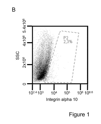
Figure 1. lntegrin al 0131 is expressed by a subpopulation of cells isolated
from the
subventricular zone of adult mouse brain.
The figure shows expression of integrin a10131 on cells isolated from the
subventricular
zone of the adult mouse brain, as assessed by flow cytometry. Analysis of
unstained
cells (A). Cells stained with a monoclonal antibody directed to the integrin
alphal 0
subunit (B) and with a control mouse IgG2a antibody (C) and the percentage of
positive
cells were subsequently analyzed. The results show that a subpopulation of the
isolated cells expresses integrin al 0131.
Figure 2. lntegrin al 0131 is expressed by a subpopulation of cells isolated
from the
subventricular zone and cultured as neurospheres or as a monolayer.
The figure shows expression of integrin a10131, PDFGRa, Lex and CD24 by cells
cultured as a monolayer (A-D) and neurospheres (E-H) as assessed by flow
cytometry.
Cells were stained with a monoclonal antibody directed to the integrin alphal
0 subunit
(A, E), a monoclonal antibody directed to PDFGRa (B, F), an antibody directed
to
CD24 (C,G) and an antibody directed to Lex (D,H) and the percentage of
positive cells
was analyzed by flow cytometry. The results show that a subpopulation of the
cells
cultured in monolayer or as neurospheres expresses integrin a10131, PDGFRa,
Lex
and CD24.
Figure 3. Neurospheres express integrin al 0131 and other neural
stem/progenitor stem
cell markers.
The figure shows the double immunolabeling of cells in neurospheres, isolated
from the
SVZ of mice and immunostained for integrin al 0131 and other neural
stem/progenitor
CA 03032343 2019-01-29
WO 2018/033596 10 PCT/EP2017/070838
cell markers. Stainings were visualized and images acquired by confocal
microscopy.
Whole neurosphere shows expression of both al 0 and neural stem/progenitor
cell
markers Nestin (A), PDGFRa (B), GFAP (C), PSA-NCAM (D), Vimentin (E). In
agreement with the cellular composition of the SVZ in vivo (Doetsch et al.
1997),
neurospheres in vitro also show low abundance of type C cells, as demonstrated
by
01ig2 staining (F).
Figure 4. Neural stem/progenitor cells isolated from the SVZ and cultured
under stem
cell conditions retain their potential to differentiate into neuronal and
glial cells. Neural
stem/progenitor cells were differentiated and expression for neuronal (Map2,
13- III Tub)
and glial (Gfap, 04) genes were measured. Gapdh was used as a housekeeping
gene
for normalization. Gene expression is expressed as mean standard error of
the mean
(SEM) of triplicate samples.
Figure 5. lntegrin al 0131 is expressed by cells of the subventricular zone
(SVZ) of the
adult mouse brain.
The figure shows expression of integrin a10131 in the SVZ of adult mouse
tissue as
assessed by immunofluorescence staining and confocal microscopy. Brain tissue
was
stained with a rabbit polyclonal antibody directed to the integrin alphal 0
subunit (A)
and DAPI to visualize the cell nucleus DNA (B). A composite image of A and B
is
shown in C.
Figure 6. lntegrin al 0131 and the neural stem/progenitor cell marker nestin
partially co-
localize in the subventricular zone (SVZ) of the adult mouse brain.
The figure shows expression of integrin a10131 and nestin in the SVZ of adult
mouse
tissue as assessed by immunofluorescence staining and confocal microscopy.
Brain
tissue was stained with a rabbit polyclonal antibody directed to the integrin
alphal 0
subunit (A), a mouse monoclonal antibody directed to nestin (B) and DAPI to
visualize
the cell nucleus DNA (C).
Figure 7. lntegrin al 0131 and the neuroblast marker PSA-NCAM partially co-
localize on
cells in the subventricular zone (SVZ) of the adult mouse brain.
The figure shows expression of integrin a10131 and PSA-NCAM in the SVZ of
adult
mouse brain tissue as assessed by immunofluorescence staining and confocal
microscopy. Brain tissue was stained with a rabbit polyclonal antibody
directed to the
CA 03032343 2019-01-29
WO 2018/033596 11 PCT/EP2017/070838
integrin alphal 0 subunit (C), a mouse monoclonal antibody directed to PSA-
NCAM (B)
and DAPI to visualize the cell nucleus DNA (A). A composite image of A, B and
C is
shown in D.
Figure 8. lntegrin a10131 and the glial cells marker GFAP partially co-
localize on cells in
the subventricular zone (SVZ) in the adult mouse brain.
The figure shows expression of integrin a10131 and GFAP in the SVZ of adult
mouse
brain tissue as assessed by immunofluorescence staining and confocal
microscopy.
Brain tissue was stained with a rabbit polyclonal antibody directed to the
integrin
alphal 0 subunit (A), a goat polyclonal antibody direct to GFAP (B). A
composite image
of A and B is shown in C.
Figure 9. lntegrin al 0131 and the neural stem cell marker SOX2 partially co-
localize on
cells in the subventricular zone (SVZ) in the of newborn mouse brain.
The figure shows expression of integrin a10131 and the neural stem cell marker
SOX2
in the SVZ of newborn mouse brain tissue as assessed by immunofluorescence
staining and epifluorescence microscopy. Brain tissue was stained with DAPI to
visualize the cell nucleus DNA (A), a mouse monoclonal antibody direct to SOX2
(B)
and a rabbit polyclonal antibody directed to the integrin alpha 10 subunit
(C). A
composite image of A, B and C is shown in D.
Figure 10. lntegrin al 0131 and PDFGRa co-localize on a subpopulation of cells
isolated
from the subventricular zone in the adult mouse brain.
The figure shows expression of integrin a10131 and PDFGRa by cells isolated
from the
subventricular zone of adult mouse brain as assessed by flow cytometry. Cells
were
stained with a monoclonal antibody directed to the integrin alphal 0 subunit
(A) and
with a monoclonal antibody directed to PDFGRa (B) and the percentage of
positive
cells was analyzed.Flow cytometry analysis demonstrates that a subpopulation
of the
isolated cells expresses both integrin a10131 and PDFGRa (C).
Figure 11. lntegrin a10131 is expressed in the subgranular zone (SGZ) of the
hippocampus in the newborn mouse brain and partially co-localizes with the
neural
stem cell marker SOX2.
The figure shows expression of integrin a10131 and neural stem cell marker
SOX2 in
the SGZ in newborn mouse brain tissue, as assessed by immunofluorescence
staining
CA 03032343 2019-01-29
WO 2018/033596 12 PCT/EP2017/070838
and epifluorescence microscopy. Brain tissue was stained with DAPI to
visualize the
cell nucleus DNA (A), a mouse monoclonal antibody directed to SOX2 (B) and a
rabbit
polyclonal antibody directed to the integrin alphal 0 subunit (C). A composite
image of
A, B and C is shown in D.
Figure 12. lntegrin a10131 is expressed in the meninges of the newborn mouse
brain
and partially co-localizes with PDGFRa, a marker of glial cells /
oligodendrocyte
progenitors.
The figure shows expression of integrin a10131 and PDGFRa in the meninges of
the
newborn mouse brain tissue, as assessed by immunofluorescence staining and
epifluorescence microscopy. Brain tissue was stained with DAPI to visualize
the cell
nucleus DNA (A), a goat polyclonal antibody direct to PDGFRa (B) and a rabbit
polyclonal antibody directed to the integrin alphal 0 subunit (C). A composite
image of
A, B and C is shown in D.
Figure 13. lntegrin a10131 is upregulated in mouse brain following stroke.
The figure shows higher expression of integrin a10131 in the stroke area
versus intact
area in mouse brain, as assessed by immunofluorescence staining and
epifluorescence microscopy. The stroke area is the area to the right of the
dashed line.
Figure 14. Expression of lntegrin a10131, neuronal marker NeuN, and astrocytic
marker
GFAP.
The figure shows expression of integrin a10131, NeuN, GFAP and lbal in the
mouse
brain assessed by immunofluorescence staining and confocal microscopy. Brain
tissue
was stained with a rabbit polyclonal antibody directed to the integrin alphal
0 subunit, a
guinea pig polyclonal antibody direct to NeuN, a goat polyclonal antibodies
direct to
GFAP and lbal (microglial marker). Composite images of control mouse brain is
shown
in A, D and composite image of stroke brain with regard to expression of
integrin a10131
and NeuN (B and C), expression of integrin a10131 and GFAP (E and F) and
expression of integrin a10131 and lbal (H and l), Colocalization is shown by
arrows.
The results show that integrin a10131 colocalizes with an increased expression
of NeuN
and GFAP on neurons and astrocytes respectively (regenerative response) but
not with
the microglial marker lbal .
CA 03032343 2019-01-29
WO 2018/033596 13 PCT/EP2017/070838
Definitions
As used herein, the terms "rodent" and "rodents" refer to all members of the
phylogenetic order Rodentia.
"Integrin alpha 10" or "Integrin alpha 10 subunit" as used herein refers to
the alpha 10
subunit of the heterodimeric protein integrin alpha 10 beta 1. This denotation
does not
exclude the presence of the beta 1 subunit bound to the alpha 10 subunit thus
forming
the quaternary structure of integrin alpha 10 beta 1 heterodimer.
"Anti-integrin alpha 10 antibody" or "Anti-integrin alpha 10 subunit antibody"
as used
herein refers to an antibody capable of recognizing and binding to at least
the alpha 10
subunit of the heterodimeric protein integrin alpha 10 beta 1. These
antibodies may be
antibodies that recognize an epitope of the heterodimeric protein integrin
alpha10
beta1, wherein the epitope comprises amino acid residues of both the alpha10
and the
beta1 subunit.
The term "identifying" as used herein refers to the action of recognizing a
cell as being
a certain type of cell, e.g. a neural stem cell or a neural progenitor cell.
An alternative
term to identifying is "detecting", which is used herein with the same
meaning. A cell is
identified as a neural stem cell or a neural progenitor cell for example by
detecting
expression of specific markers by the cell.
The terms "isolating", "sorting" and "selecting" as used herein refer to the
action of
identifying a cell as being a certain type of cell and separating it from
cells that do not
belong to the same cell type or to a differentiation state.
The term "scoring" as used herein refers to scoring of the integrin alpha10
subunit
expression. The scoring may be measuring integrin alpha10 subunit expression
via for
example immunoassay, flow cytometry, immunofluorescence, western blot or
immunoprecipitation in the sample population and comparing the measurement
with a
measurement done in a reference cell population expressing the integrin
alpha10
subunit, as well as to a cell population not expressing the integrin alpha10
subunit.
Examples of reference cells expressing the integrin alpha10 are 02012 cells,
HEK293
cells transfected with integrin sequences. Examples of reference cells not
expressing
alpha10 are non-transfected 02012 cells and HEK293 cells.
CA 03032343 2019-01-29
WO 2018/033596 14 PCT/EP2017/070838
The term "murine" refers to any and all members of the family Muridae,
including rats
and mice.
The term "substantially free from" is herein intended to mean below detection
limits of
the assay used thereby appearing negative, i.e. free from.
The term "committed" is herein intended to mean dedicated to, or focused on.
Thus, a
committed cell is a cell that is dedicated to, or focused on a specific
differentiation
pathway. From this it will follow that an uncommitted cell is not dedicated
to, or focused
on, any specific differentiation pathway and has several options.
The term "subject" used herein is taken to mean any mammal to which neural
stem
cells and/or neural progenitor cells and/or mesenchymal stem cell identified
and/or
isolated according to the methods disclosed herein, which are based on the
detection
of integrin alphal 0 expression on the surface of the cells or intracellular
in the cells,
may be administered. Subjects specifically intended for treatment with the
method of
the disclosure include humans, as well as nonhuman primates, sheep, horses,
cattle,
goats, pigs, dogs, cats, rabbits, guinea pigs, hamsters, gerbils, rats and
mice, as well
as the organs, tumors, and cells derived or originating from these hosts.
Detailed description of the invention
Integrin alphal 0 as a marker for neural stem cells and neural progenitor
cells
The present inventors have surprisingly found that the integrin alphal Obetal
is present
on human neural stem cells (NSCs) and/or neural progenitor cells (NPCs). Thus,
this
integrin can be used to identify, select and specifically isolate neural stem
cells and
neural progenitor cells from a mixed cell population and will be a useful tool
in cell
therapy to repair damaged tissue, for example as consequence of injuries of
the
nervous system or for treatment of neurodegenerative diseases. Compared to the
above-mentioned NSC surface markers, integrin al 0131 identifies all three
cell types of
the adult mouse SVZ (see Table 1 above) suggesting that integrin a10131 is a
broader
stem and progenitor cell marker, compared to other known markers, covering the
different cellular subtypes of the brain stem cell niche.
CA 03032343 2019-01-29
WO 2018/033596 15 PCT/EP2017/070838
The human integrin alpha10 chain sequence is known and publicly available at
GenBank Tm/EBI Data Bank accession number AF074015. Thus, new uses and
methods of the integrin alpha10 chain are disclosed in the present invention.
As revealed above, one aspect of the present disclosure relates to the use of
a marker
comprising an integrin alpha10 subunit expressed by a neural stem cell and/or
a neural
progenitor cell as a marker for mammalian neural stem cells and mammalian
progenitor cells.
In one embodiment, the integrin alpha10 subunit is expressed as a heterodimer
in
combination with an integrin beta1 chain.
In one embodiment, the integrin alpha10 subunit is expressed on the cell
surface of the
mammalian NSC and/or NPC.
In one embodiment, the integrin alpha10 subunit is expressed intracellularly
in a
mammalian NSC and/or NPC.
A method for identifying neural stem cells (NSCs) and neural progenitor cells
(NPCs)
One aspect of the present disclosure relates to a method for identifying a
mammalian
neural stem cell and/or a mammalian neural progenitor cell. The method
comprises the
steps of
a) providing a sample comprising neural tissue,
b) detecting expression of an integrin alpha10 subunit by a cell comprised in
the
sample of a),
c) scoring the integrin alpha10 subunit expression of b), and
d) identifying the mammalian neural stem cell and/or the neural progenitor
cell
according to the scoring in c).
A further aspect of the present disclosure relates to a method for isolating a
mammalian neural stem cell and/or a mammalian neural progenitor cell. The
method
comprises the steps of:
CA 03032343 2019-01-29
WO 2018/033596 16
PCT/EP2017/070838
a) providing a sample comprising neural tissue,
b) detecting expression of an integrin alpha10 subunit by a cell comprised in
the
sample of a),
c) scoring the integrin alpha10 subunit expression of b), and
d) selecting the mammalian neural stem cell and/or the mammalian neural
progenitor cell according to the scoring in c).
In some embodiments, the method of identifying a mammalian NSC and/or NPC and
the method of isolating a mammalian NSC and/or NPC, further comprises a step
of
contacting the sample with an antibody which specifically binds integrin
alpha10
subunit, prior to detecting integrin alpha10 subunit expression of b).
In more detail, the methods for identifying and/or isolating a mammalian
neural stem
cells and/or a mammalian neural progenitor cells according to the disclosure
may
further comprise the steps of:
e) providing a cell suspension comprising NSCs and/or NPCs,
f) contacting the cell suspension in e) with a monoclonal antibody or
fragments
thereof binding to the integrin alpha1Obeta1, under conditions wherein said
monoclonal antibody or fragments thereof form an antibody-antigen complex
with the extracellular domain of integrin alpha1Obeta1,
g) separating cells binding to said monoclonal antibody or fragments thereof
in
f),
thereby producing a pure population of mammalian NSCs and/or NPCs
In some embodiments of the present disclosure, the methods for identifying
and/or
isolating a mammalian neural stem cells and/or a mammalian neural progenitor
cells
according to the disclosure may further comprise recovering the cells binding
to the
monoclonal antibody or fragments thereof from said antibody or fragments
thereof.
The cell suspension provided in e) above, comprising mammalian NSCs and/or
NPCs
may be isolated from neural tissue as described in details in the section
below "Neural
tissue".
CA 03032343 2019-01-29
WO 2018/033596 17 PCT/EP2017/070838
In some embodiments of the present disclosure, the methods for identifying
and/or
isolating a mammalian neural stem cells and/or a mammalian neural progenitor
cells
according to the disclosure is performed in vitro.
Neural tissue
The main source of mammalian NSCs and NPCs is neural tissue. In fact, NSCs and
NPCs are found in niches in fetal or adult mammalian central nervous system
(CNS)
and from fetal or adult mammalian brain or spinal cord where neurogenesis
takes
place.
The neural stem cell niche in brain is a tissue microenvironment capable of
hosting and
maintaining neural progenitor cells. Until recently, only two brain niches
were
recognized in mammals, the subventricular zone (SVZ) of the anterolateral
ventricle
and the subgranular zone (SGZ) of the hippocampal dentate gyrus. Increasing
evidences show neurogenesis and gliogenesis also in other parts of the adult
brain,
particularly after injury, suggesting that additional stem cell niches are
present in the
adult brain (Lin and lacovitti, 2015). It was recently shown that
leptomeninges host a
subset of cells expressing markers of undifferentiated, proliferating and
differentiating
neural precursors presenting nestin and SOX2 and this set of cells persists in
adulthood. Thus, meninges may represent another functional niche for
progenitors
during embryonic development and in adulthood.
Accordingly, in some embodiments of the present disclosure the neural tissue
comprises NSCs and NPCs.
In some embodiments, the neural tissue is obtained or derived from brain
tissue.
In some embodiments, the neural tissue is adult brain tissue.
In some embodiments, the neural tissue is fetal brain tissue.
In some embodiments the neural tissue is derived or obtained from the
subventricular
zone (SVZ), the subgranular zone (SGZ) or the meninges of a mammal.
CA 03032343 2019-01-29
WO 2018/033596 18 PCT/EP2017/070838
All mammals are suitable for obtaining neural tissue. In some embodiments, the
neural
tissue is selected from the group consisting of human, canine, equine, bovine,
feline,
murine, ovine or swine neural tissue. Other mammalian neural tissues may be
obtained
if there is a need for that.
In some embodiments, the mammalian NSCs and NPCs are human NSCs and NPCs.
In one further embodiment, the mammalian NSCs and NPCs are murine NSCs and
NPCs.
In some embodiments, the neural tissue does not derive from a human embryo.
Detection of integrin alpha 10 expression
A key step in the method for identification of a mammalian neural stem cell
and/or a
mammalian neural progenitor cell, as well as for their isolation, is the
detection of
integrin alpha10 protein expression.
In one embodiment, the detection of expression of an integrin alpha10 subunit
by a cell
is determined by flow cytometry. For example, the expression of alpha10 may be
analyzed by flow cytometry, or any other methodology having high specificity.
Multi-
color analyses may be employed with the flow cytometry, which is particularly
convenient. NSCs and/or NPCs may, thus, be separated from other cells on the
basis
of the level of staining for the particular antigens.
In one embodiment, the detection of expression of an integrin alpha10 subunit
by a cell
is determined by measuring integrin alpha10 protein expression. For example,
flow
cytometry, immunofluorescence, immunoprecipitation and/or western blotting may
be
used.
In one embodiment, the detection of expression of an integrin alpha10 subunit
by a cell
is determined by measuring integrin alpha10 mRNA expression. Detection of mRNA
expression of a specific protein is well known to the skilled man in the art,
and is
generally done by probing the mRNA with a DNA or RNA probe specific for the
mRNA
of interest, under hybridization conditions where the probe is not hybridizing
to other
CA 03032343 2019-01-29
WO 2018/033596 19 PCT/EP2017/070838
mRNA molecules. Different polymerase chain reactions (PCR) may also be used,
which is obvious to the skilled man in the art.
A suitable PCR-method is given below. In brief, polymerase chain reaction
(PCR) may
be used.
RNA may be prepared from human neural stem cells or neural progenitor cells by
standard methods, for example by the use of RNeasy Mini Kit (Qiagen Germany).
cDNA may be produced by reverse transcriptase reaction, Superscript II
(lnvitrogen,
USA) according to manufacturer's recommendation with oligo d(T)-primers or
gene
specific primers.
PCR is thereafter performed to amplify the cDNA. Specific primers for al 0,
forward
5'GCT CCA GGA AGG CCC CAT TTG TG 3' and reverse 5'GTG TTT TOT TGA AGG
GTG CCA TTT 3 are added to the cDNA and the specific product is amplified by
Platinum Taq DNA polymerase (Invitrogen, USA) according to manufacturer's
recommendations at 65 for 40 cycles.
Several methods are known to the person skilled in the art for detection of
expression
of markers. Accordingly, in one embodiments of the present disclosure the
detection of
expression of an integrin alphal 0 subunit by a cell is determined by a method
selected
from the group consisting of immunoassay, flow cytometry, immunofluorescence,
immunoprecipitation and western blot.
In still a further embodiment, the integrin chain alphal 0 expression is
detected on the
cell surface of a NSC and/or of a NPC or intracellular in a NSC and/or in a
NPC in the
method according to the invention. Methods given above, e.g. flow cytometry
and
immunoprecipitation may be used. Preferably flow cytometry is used.
In still a further embodiment, the expression of the integrin alphal 0 subunit
is detected
by any immunoassay, such as the methods described in lmmunochemical protocols
(Methods in molecular biology, Humana Press Inc). The detection may be
performed
by various methods, e.g. any immune method known to the skilled man in the
art, such
CA 03032343 2019-01-29
WO 2018/033596 20 PCT/EP2017/070838
as immunoprecipitation, western blotting, magnetic-activated cell sorting
(MACS) or
flow cytometry methods, e.g. fluorescence activated cell sorting (FACS).
Accordingly, in some embodiments, the expression of an integrin alpha10
subunit by a
cell is detected via an antibody, wherein the antibody is a monoclonal
antibody,
polyclonal antibody, a chimeric antibody, a single chain antibody or fragment
thereof.
Antibodies, such as monoclonal antibodies or fragments thereof, are
particularly useful
for identifying markers, cell surface proteins as well as intracellular
markers, associated
with particular cell lineages and/or stages of differentiation. Thus, it is
suitable for the
identification of integrin alpha10.
In one embodiment, the antibody is a monoclonal antibody or a polyclonal
antibody.
In a further embodiment, the antibody is a non-human antibody, a chimeric
antibody, a
bispecific antibody, a humanized antibody or a human antibody.
Still, identification may as well be performed by any specific molecule, such
as a
protein or peptide, binding specifically to the integrin alpha10 molecule.
Examples of
such proteins or peptides are natural ligands, binding to the integrin alpha10
molecule.
Such natural ligands may be made recombinant, chemically synthesized, or
purified
from a natural source.
The detection is facilitated when the antibody, protein or peptide, binding
specifically to
the integrin alpha10 molecule is bound to a detectable moiety.
In some embodiments, an antibody is used for detection of the expression of
integrin
alpha10 subunit and the antibody is covalently bound to a detectable moiety,
such as a
detectable moiety selected from the group consisting of a fluorophore, an
enzyme or a
radioactive tracer or radioisotope.
In some embodiments, the antibody is bound to a fluorophore and the
fluorophore is
selected from the group consisting of fluorescein isothiocyanate,
phycoerythrin and
phycoerythrin conjugates, allophycocyanin and allophycocyanin conjugates,
Texas Red
CA 03032343 2019-01-29
WO 2018/033596 21 PCT/EP2017/070838
and Texas Red conjugates, Alexa series of fluorochromes, Brilliant Violet and
Brilliant
Blue series of fluorochromes.
Many other fluorophores may be used and the skilled person will choose the
most
suitable one according to the specific detection method and also according to
the
characteristics of the antibody used.
In further embodiments, the antibody has an isotype selected from the group
consisting
of IgA, IgD, IgG and IgM.
In even further embodiments, the antibody is:
a) a monoclonal antibody, produced by the hybridoma cell line deposited at the
Deutsche Sammlung von Microorganismen und Zellkulturen GmbH under the
accession number DSM A002583; or
b) an antibody which competes for binding to the same epitope as the epitope
bound
by the monoclonal antibody produced by the hybridoma deposited at the Deutsche
Sammlung von Microorganismen und Zellkulturen GmbH under the accession number
DSM A002583; or
c) a fragment of a) or b), wherein said fragment is capable of binding
specifically to the
extracellular I-domain of the integrin alpha 10 subunit chain.
The detection may also be facilitated when the antibody, protein or peptide,
binding
specifically to the integrin alpha10 molecule is bound to a solid support.
Accordingly, in
one embodiment an antibody is used for detection of the expression of integrin
alpha10
subunit and the antibody is attached to a solid support.
Isolation and cultivation of NSCs and NPCs
The isolation of a mammalian neural stem cell (NSC) and/or a mammalian neural
progenitor cell (NPC) according to the method of the present disclosure may be
based
on the cells capacity to adhere to plastic culture dishes and form colonies
under
specific culture conditions. Suitable protocol for isolation of mammalian
neural stem
cells and neural progenitor cells, without including the marker according to
the
invention, is further given in detail in Giachino et al. 2009, Guo W et al.,
(2012) and
CA 03032343 2019-01-29
WO 2018/033596 22 PCT/EP2017/070838
Oliver-De la Cruz and Ayuso-Sacido (2012). Thus, known methods may be used,
but
with the introduction of the marker according to the invention.
Procedures for separation may include magnetic separation, using e.g. antibody-
coated magnetic beads, affinity chromatography, agents joined to a monoclonal
antibody or used in conjunction with a monoclonal antibody, e.g., complement
and
cytotoxins, and "panning" with antibody attached to a solid matrix,
e.g., a plate, or other convenient techniques. Techniques providing accurate
separation
include fluorescence activated cell sorters, which can have varying degrees of
sophistication, e.g., a plurality of color channels, light scattering
detecting channels,
impedance channels, etc. known to the skilled man in the art.
Further protocols for separation methods suitable to be used in the method
according
to the invention are described by Orfao, A and Ruiz-Arguelles, A ((1996)
General
Concepts about Cell Sorting Techniques. Clin Biochem. 29(1):5-9), and by
Herzenberg,
LA, De Rose, SC and Herzenberg, LA ((2000) Monoclonal Antibodies and FACS:
complementary tools for immunobiology and medicine. lmmunol. Today. 21(8):383-
390).
Mammalian NSCs and NPCs are first purified from the mixture of cells collected
from
nervous tissue. Generally, negative markers are used to separate NSCs and NPCs
from differentiated cells.
The isolation of NSCs and NPCs from other cells, for example committed neural
cells,
present in neural tissue comprises a selection and sorting step where the NSCs
and
NPCs are first identified and then separated from the other cells. Various
techniques
known to the skilled artisan may be employed to separate the cells by
initially removing
cells dedicated to other lineages than NSCs and NPCs
In a first separation, antibodies for other markers may be used labelled with
one or
more fluorochrome(s).
The NSCs and the NPCs may be selected against dead cells, by employing dyes
associated with dead cells (propidium iodide, LDS). The cells may be collected
in their
culture medium, or in any physiological saline solution, preferably buffered,
such as
CA 03032343 2019-01-29
WO 2018/033596 23 PCT/EP2017/070838
phosphate buffer saline (PBS), optionally with fetal calf serum (1% FCS) or
bovine
serum albumin (1% BSA) present
Markers that are not expressed on NSCs and NPCs are, e.g. 0D326, 0D34 and 0D45
and their expression, or lack of expression, may in certain embodiments be
used for as
markers for negative selection of cells that are not NSCs or NPCs.
If further lineages or cell populations not being NSCs or NPCs are to be
removed in
one step, various antibodies to such lineage-specific markers may be included.
In some embodiments of the present disclosure, other less specific and non-
unique
mammalian NSCs and/or NPCs markers may be analyzed in parallel with the marker
according to the invention. In fact, NSCs and NPCs subsets detected at
different
stages of CNS development have been shown to express markers such as nestin,
GFAP, CD15, 0D24, 5ox2, Musashi, 0D133, EGFR, PDGFRa, Doublecortin (DCX),
Pax6, FABP7 and GLAST. These markers are co-expressed by some of the cells
expressing integrin alpha10 subunit, as shown in the examples. However, none
of
these markers are uniquely expressed by NSCs and/or by NPCs, many are indeed
expressed by neural differentiated cells and other non-neural cell types.
In contrast, the use of the integrin alpha10 marker according to the invention
in the
isolation and expansion protocols will give a homogenous population of neural
stem
cells and/or neural progenitor cells with the ability to differentiate to
different cells of the
brain and spinal cord thus being useful for regenerating brain or spinal cord
tissue.
Thus, including the marker according to the invention, comprising an integrin
alpha10
subunit in known isolation and expansion protocols, as well as using the
marker(s)
alone, will be highly valuable for further evaluation and enrichment of the
neural stem
cells and neural progenitor cell populations. Particularly, no other specific
and unique
marker as the marker according to the invention for mammalian neural stem
cells and
neural progenitor cells is known.
NSCs and NPCs may as well be selected based on light-scatter properties and
their
expression of various cell surface antigens, in combination with the
identification using
the method according to the invention.
CA 03032343 2019-01-29
WO 2018/033596 24 PCT/EP2017/070838
Generally, markers present on the cell surface or intracellularly are detected
with the
help of fluorochromes. Fluorochromes, which may find use in a multi-color
analysis,
include phycobiliproteins, e.g., phycoerythrin and allophycocyanins,
fluorescein, Texas
red, and many others, all well known to the skilled man in the art and
commercially
available.
Commonly used techniques for detection and selection of cells based on the use
of
markers present on their cell surface are flow cytometry and
immunoprecipitation.
In some embodiments, NSCs and NPCs may be analyzed by flow cytometry or
immunoprecipitation thereby detecting and identifying integrin alpha10 subunit
expression. The person skilled in the art is familiar with suitable protocols
for flow
cytometry and immunoprecipitation to be used for detection and/or selection of
cells.
If a population of cells is collected from whole mouse brain, only a minor
fraction, for
example less than 2% of the total number of cells are NSCs or NPCs.
If an antibody or fragments thereof is used it may be attached to a solid
support to
allow for a highly specific separation. The particular procedure for
separation
employed, e.g. centrifugation, mechanical separation, such as columns,
membranes or
magnetic separation, should maximize the viability of the fraction to be
collected.
Various techniques of different efficacy may be employed known to a person
skilled in
the art. The particular technique employed will depend upon efficiency of
separation,
cytotoxicity of the methodology, ease and speed of performance, and necessity
for
sophisticated equipment and/or technical skill.
Procedures for separation of NSCs and/or NPCs from a cell suspension aided by
the
method according to the invention may include magnetic separation, using e.g.
antibody-coated magnetic beads, affinity chromatography based on the antibody
or
fragments thereof according to the invention, and "panning" with an antibody
or
fragments thereof attached to a solid matrix, e.g., a plate, or other
convenient
techniques.
CA 03032343 2019-01-29
WO 2018/033596 25 PCT/EP2017/070838
Techniques for providing accurate separation include fluorescence activated
cell
sorters, magnetic bead sorting and any suitable method known by those of skill
in the
art.
In a further embodiment, the first enrichment step of the methods of the
present
disclosure, is a positive selection of the NSCs or of the NPCs that may be
repeated
until the desired purity of the NSCs or of the NPCs is achieved.
A method for producing an isolated population of cells enriched for mammalian
NSCs and/or NPCs
One aspect of the present disclosure relates to a method for manufacturing an
isolated
population of mammalian cells in vitro which are enriched for neural stem
cells and/or
neural progenitor cells relative to a reference population, the method
comprising the
steps of
a) providing at least a portion of a population of cells, or a portion of a
reference population, comprising a neural stem cell and/or a neural progenitor
cell,
b) introducing into the population of cells in a) above a compound identifying
an integrin alpha10 subunit expressed by the neural stem cell and/or neural
progenitor
cell,
c) selecting and isolating from the population of cells in b) above the neural
stem cells and/or the neural progenitor cells,
thereby producing a population of cells enriched for neural stem cells and/or
neural
progenitor cells.
Providing a population may be performed in a similar way as in the method for
identification of NSCs and NPCs described in detailed above. The population of
cells
may comprise at least one NSC and/or at least one NPC, or may comprise only
cells
that are neither NSCs nor NPC. For example a population of cells obtained or
derived
from neural tissue may be provided, or a reference population of cells, such
as HEK
cells or 02012 cells transfected with integrin alpha10 may be provided.
The compound introduced to identify the NSCs and/or the NPCs may be a protein,
peptide, monoclonal antibody, or part thereof, or polyclonal antibody
identifying the
NSCs and/or NPCs.
CA 03032343 2019-01-29
WO 2018/033596 26 PCT/EP2017/070838
The compound introduced to identify the NSCs and/or the NPCs may be a protein,
peptide, monoclonal antibody, or part thereof, or polyclonal antibody that is
able to
detect integrin alpha10.
In one embodiment, the NSCs and/or NPCs is identified as a NSC and/or as a NPC
by
detecting expression of integrin chain alpha10 on the cell surface of said NSC
and/or of
said NPC according to the method for identifying NSCs and/or NPCs described
above.
Monoclonal or polyclonal antibodies, or parts thereof, are particularly useful
for
identifying markers, e.g. markers expressed on the cell surface of intact
viable cells.
The compound used to identify the NSC and/or NPC may also be used for the
separation step. Thus, said compound(s), such as antibodies, or parts thereof,
may be
attached to a solid support to allow for a first crude separation. Examples of
solid
supports are beads e.g. magnetic beads, agarose or other similar types of
beads
known to the skilled man in the art. Any means suitable for separation of
cells may be
employed on the condition that the separation is not unduly detrimental to the
viability
of a cell.
The separation techniques employed should maximize the retention of viability
of the
fraction to be collected. The assessment of viability is described below.
In brief, assessment of cell viability may be performed using e.g. flow
cytometry. After
staining of the appropriate cell viability dye can be added to discriminate
between
viable and non-viable cells. A number of such dyes exist, of which examples
and
typical methods for using them are described. The principle is the same for
most of
these dyes: these dyes enter the cells if the cell membrane is compromised; as
such,
cells that stain with these dyes are non-viable, and cells that do not stain
are
considered viable.
Examples of dyes are Propidium Iodide (PI), 7-Aminoactinomycin D (7 AAD), To-
Pro3,
and Ethidium Monoazide (EMA). Other methods are described in Flow Cytometry
and
Cell Sorting (Springer Lab Manual) by A. Radbruch (Springer Verlag, 2nd
edition,
January 2000).
CA 03032343 2019-01-29
WO 2018/033596 27 PCT/EP2017/070838
The cell viability of the fraction collected is typically > 90%, preferably
95%, 96%, 97%,
98%, 99%, 99.9%, or even 100%.
The particular technique employed for separation of cells in the method
according to
the invention will depend upon efficiency of separation, cytotoxicity of the
methodology,
ease and speed of performance, and necessity for sophisticated equipment
and/or
technical skill.
In one embodiment of the method according to the invention, at least one
enrichment
step of mammalian NSCs and/or NPCs is included.
In one embodiment of the method according to the invention, at least one
enrichment
step of mammalian NSCs is included.
In one embodiment of the method according to the invention, at least one
enrichment
step of mammalian NPCs is included.
In still a further embodiment, the first enrichment step of NSCs and/or NPCs
is a
negative selection of the NSCs and/or NPCs, i.e. other lineage-committed cells
are
depleted, or removed, from the initial population of cells. For example, cells
expressing
markers such as 0D34, 0D45 and 0D326 are negatively selected.
In still a further embodiment, the methods of the present disclosure comprise
a further
step a negative selection based on detection of the expression of integrin
alpha11
subunit, for example, cells expressing the marker integrin alpha11 are
depleted, or
removed, from the population of cells.
In still a further embodiment, the first enrichment is a positive selection of
NSC and/or NPCs that may be repeated till the desired purity of the NSC and/or
of the
NPC is achieved. For a positive or a negative selection, proteins, peptides,
monoclonal
or polyclonal antibodies may be used as a compound to identify the integrin
alpha10
molecule as described above. The compound may be conjugated with means for
separation, such as magnetic beads, which allow for direct separation; biotin,
which
can be removed with avidin; or streptavidin bound to a support; fluorochromes,
which
can be used with a fluorescence activated cell sorter; or the like, to allow
for ease of
CA 03032343 2019-01-29
WO 2018/033596 28 PCT/EP2017/070838
separation of the particular cell type as exemplified in the paragraphs above.
Any
technique may be employed which is not unduly detrimental to the viability of
the cells
of interest, i.e. the NSCs and the NPCs.
In one embodiment, the selection is performed by fluorescent cell sorting, by
using e.g.
a flow cytometry based cell sorter such as a FACS , or any other similar
methodology
having high specificity. Multi-color analyses may be employed with flow
cytometry
which is particularly convenient and the technique well known to person
skilled in the
art of flow cytometry. The cells may be separated on the basis of the level of
staining
for the particular antigens. In a first separation, antibodies for other
markers may be
used labelled with one fluorochrome, while the antibodies for the dedicated
lineages,
i.e. the integrin alpha10, may be conjugated to (a) different fluorochrome(s).
Other
markers may in further embodiments be nestin, GFAP, CD15, 0D24, Sox2, Musashi,
0D133, EGFR, PDGFRa, Doublecortin (DCX), Pax6, FABP7 and GLAST that NSC
and/or NPC may express. Examples of markers that are not expressed on NSC and
NPC are 0D326, 0D34, 0D45 and integrin alpha11 and their expression, or lack
of,
may in further embodiments also be evaluated together with the marker
according to
the invention, e.g. integrin alpha10 expression.
If further lineages or cell populations are to be removed in this step,
various antibodies
to such lineage specific markers may be included. Fluorochromes which may find
use
in a multi-color analysis include phycobiliproteins, e.g., phycoerythrin and
allophycocyanins, fluorescein, Texas red, and any suitable fluorochrome known
by
those of skill in the art.
The cells may be selected against dead cells, by employing dyes associated
with dead
cells such as propidium iodide or LDS-75 1 (Laser Dye Styry1-75 1 (6-
dimethylamino-2-
14-[4-(dimethylamino)pheny1]-1 ,3-butadieny11-1-ethyl quinoliniurn
perchlorate) ). The
cells may be collected in any suitable cell culturing media, such as lscove's
modified
Dulbecco's medium (IMDM), or in any physiological saline solution, preferably
buffered,
such as phosphate buffer saline (PBS), optionally with fetal calf serum (FCS)
or bovine
serum albumin (BSA) present. Other techniques for positive or negative
selection may
be employed, which permit accurate separation, such as affinity columns, and
the like,
further described by Silvestri F, Wunder E, Soyalat H, Henon P, Serke S in
Positive
selection of CD34+ cells: a short review of the immunoadsorption methods
currently
CA 03032343 2019-01-29
WO 2018/033596 29 PCT/EP2017/070838
available for experimental and clinical use (Report on the "2nd European
Workshop on
stem Cell Methodology", Mulhouse, France, May 3-7, 1993. J Hematother. 1993
Winter;2(4):473-81) and by Basch RS, Berman JW, Lakow E.in Cell separation
using
positive immunoselective techniques (J Immunol Methods. 1983 Feb
11;56(3):269-80).
Cells may be selected based on light-scatter properties as well as their
expression of
various cell surface antigens.
While it is believed that the particular order of separation is not critical
to this invention,
the order indicated is one way of performing the invention that is known to
work. Thus,
suggestively, cells are initially separated by a crude separation, preferably
a negative
selection removing cells not committed for NSCs and NPCs using negative cell
markers such as CD326, CD34 and CD45. The negative selection is typically
followed
by a positive selection, wherein the positive selection is of a marker
associated with
NSC and/or the NPCs and negative selection for markers associated with lineage
committed cells, and other stem cell populations not being NSC and/or the
NPCs. This
separation is then followed by selection for a cellular population, or a
cellular
composition comprising said population, having multi-lineage potential as a
NSC and/or
a NPC and enhanced self-regeneration capability. The composition is further
described
below.
In further embodiments, the enrichment of such a population is about 70, 80,
90, 95,
98, 99, 99.9, or even 100%. The viability of such cells is discussed in detail
above.
The enriched population may further be expanded and then induced with defined
factors, such as epidermal growth factor (EGF) and fibroblast growth factor-2
(FGF-2),
to differentiate into specific neural cells. For example, FGF-2 is generally
used in
combination with heparin, which mediates the binding of the growth factor to
its
receptor. Other growth factors that have been reported to support cell culture
are
Transforming growth factor alpha (TGF-a), Leukemia inhibitory factor (LIF) and
its
equivalent Ciliary neurotropic factor (CNTF), or Brain-derived neurotrophic
factor
(BDNF). PDGFa is frequently used in the maintenance media for oligodendrocyte
progenitor cells. As alternative or in addition to using specific growth
factors, NPCs and
NSCs may be co-cultured in presence of other supportive cells like astrocytes,
that
CA 03032343 2019-01-29
WO 2018/033596 30 PCT/EP2017/070838
seem to favor the NPCs and NSCs growth by physical contact, or endothelial
cells, that
may enhance cell proliferation via vascular endothelial growth factor (VEGF)
production.
Modulation of NSC and/or NPC
A further aspect of the present disclosure relates to a method for determining
whether
a test compound modulates a mammalian NSC and/or NPC differentiation in vitro.
Such a method comprises the steps of
a) providing a neural stem cell and/or a neural progenitor cell that expresses
integrin alpha10 subunit,
b) contacting the neural stem cell and/or the neural progenitor cell with a
test
compound, and
c) detecting a change in rate or pattern of differentiation of the neural stem
cell
and/or neural progenitor cell as an indication of that the test compound
modulates a
neural stem cell and/or a neural progenitor cell differentiation,
wherein the rate or pattern of differentiation is determined by detecting
integrin alpha10
expression by the cell according to the method disclosed herein, see also the
section
above "Detection of integrin alpha10 expression".
The NSCs and the NPCs provided may be an enriched cell population achieved
according to any of the methods disclosed herein, the isolated NSC and/or NPC
according to the invention, or the cellular composition according to the
invention.
The test compound may be any compound known to affect or suspected to affect
NSC
and/or NPC, e.g. pharmaceutical compositions, drugs, polyclonal or monoclonal
antibodies, or parts thereof, such as antibodies binding to integrin alpha10
or any other
molecule on the NSC and/or on the NPC, factors used to promote growth of NSC
and/or of NPC, e.g. PDGFRa, EGF and FGF-2.
The detection of a change in rate or pattern of e.g. differentiation of the
NSC and/or of
the NPC as an indication that the test compound modulates NSC and/or NPC
differentiation may be done by detecting integrin alpha10 expression on the
cell surface
of said neural stem cell and/or a neural progenitor cell or intracellular in a
neural stem
cell and/or a neural progenitor cell according to the methods disclosed
herein.
CA 03032343 2019-01-29
WO 2018/033596 31 PCT/EP2017/070838
The detection of a change in rate or pattern of e.g. differentiation of the
NSC and/or of
the NPC as an indication that the test compound modulates NSC and/or NPC
differentiation may be done via flow cytometry or any other suitable method,
such as
any immuno-method, known to a person skilled in the art. The change in rate or
pattern
of differentiation may be detected via kinetic, functional or phenotypical
studies of the
NSC and/or NPC modulated with the test compound, relative to an untreated, or
mock
treated, NSCs and/or NPCs population.
A cellular composition
One aspect of the present disclosure relates to an in vitro cell culture of
undifferentiated
mammalian cells expressing an integrin alpha10 subunit, wherein the cells are
derived
from neural tissue and wherein
a) cells in the culture have the capacity to differentiate into neurons
and/or
oligodendrocytes and/or astrocytes when differentiated in a culture medium
substantially free of both serum and a proliferation-inducing growth factor as
defined in
(b) to produce a cell culture of at least 10% neurons and/or oligodendrocytes
and/or
astrocytes;
b) the cell culture divides in a culture medium containing a serum
replacement such as B27 and at least one proliferation-inducing growth factor;
c) cells in the culture differentiate into neurons and/or oligodendrocytes
and/or astrocytes upon withdrawal of both serum replacement and the
proliferation
inducing growth factors.
In one embodiment, step c) comprises addition of fetal calf serum.
A further aspect of the present disclosure relates to an in vitro cell culture
comprising
a) a culture medium containing a serum replacement such as B27 and at
least one proliferation-inducing growth factor; and
b) undifferentiated mammalian cells derived from the central nervous system
of a mammal, wherein at least 10%, preferably at least 20%, preferably at
least 30%,
preferably at least 40%, preferably at least 50%, preferably at least 60%,
preferably at
least 70%, preferably at least 80%, preferably at least 90% of the cells
express an
integrin alpha10 subunit.
CA 03032343 2019-01-29
WO 2018/033596 32 PCT/EP2017/070838
An even further aspect of the present disclosure relates to a suspension
culture of
mammalian undifferentiated cells expressing an integrin alpha10 subunit,
wherein said
cells are substantially formed into cell aggregates, and wherein the cell
aggregates are
maintained in a culture medium containing a proliferation-inducing growth
factor.
In some embodiments, integrin alpha10 expression is as described in the
section
above "Integrin alpha10 as a marker for neural stem cells and neural
progenitor cells".
In some embodiments, at least 10%, preferably at least 20%, preferably at
least 30%,
preferably at least 40%, preferably at least 50%, preferably at least 60%,
preferably at
least 70%, preferably at least 80%, preferably at least 90% of the
undifferentiated
mammalian cells are mammalian neural stem cells expressing an integrin alpha10
subunit.
In some embodiments, the undifferentiated mammalian cell is a neural stem cell
or a
neural progenitor cell.
In some embodiments, the undifferentiated mammalian cell is obtained or
derived from
adult or fetal mammalian, for example human or murine, neural tissue, which is
described in detail in the section above "Neural tissue".
In some embodiments, the undifferentiated mammalian cell is not obtained or
derived
from human embryonic cells or from a human embryo.
In some embodiments, some of the undifferentiated mammalian cells further
express at
least one marker selected from the group consisting of nestin, PSA-NCAM, GFAP,
SOX2 and PDGFRa.
Compositions having greater than 50%, such as greater than 60%, e.g. greater
than
70%, such as greater than 80%, e.g. greater than 90%, such as greater than
95%,
such as 96%, 97%, 98%, or 99.9%, of human NSC or NPC cells may be achieved
according to the disclosed methods for enrichment of NSC or NPC. Such NSC
and/or
NPC are able to provide for cell regeneration and development of members of
all of the
various lineages of NSC, such as neurons, astrocytes, and oligodendrocytes and
other
cells.
CA 03032343 2019-01-29
WO 2018/033596 33 PCT/EP2017/070838
Ultimately, a single cell type may be obtained from a NSC or a NPC composition
and
used for reconstitution or regeneration of a mammalian neural tissue. The NSC
or NPC
composition should preferably be administered in a therapeutically effective
dosage,
wherein the dosage is a specific cell number able to repopulate said mammal,
such as
a human being. The cell number may be different from donor to donor and may be
determined empirically from case to case by a person skilled in the art.
Cell proliferation-inducing factors are specific for each cell type and are
known to the
person of skill in the art. In some embodiments of the present disclosure, the
at least
one proliferation-inducing growth factor is selected from the group consisting
of
epidermal growth factor (EGF), fibroblast growth factor-2 (FGF-2),
Transforming growth
factor alpha (TGF-a), Leukemia inhibitory factor (LIF), Ciliary neurotropic
factor
(CNTF), Brain-derived neurotrophic factor (BDNF), PDGFa, and combinations
thereof.
Treatment of injuries of the nervous system and neurodegenerative diseases
Mammalian NSC and/or NPC, such as human or mouse NSC and/or NPC identified
and isolated according to the methods disclosed herein can be used for
treating
damage and injuries and/or preventing and protecting from damage of the
nervous
system and/or treating neurodegenerative disease in a subject in need thereof.
As for
neurodegenerative disorders, this applies in particular to neurodegenerative
diseases
which are associated with loss or damage of neuronal tissue. The present
invention
treats such neurodegenerative diseases by regenerating the lost or damaged
tissue.
Furthermore, mesenchymal stem cells (MSCs) identified and isolated according
to
methods disclosed e.g. in WO 03/106492, can also be used for regenerating lost
or
damaged tissue of the central nervous system as evidenced by Steinberg G.K. et
al
(2016) Stroke 47:1817-1824. Thus, in one aspect the present disclosure relates
to a
methods of regenerating lost or damaged tissue of the central nervous system,
and to
methods of treating disease or damage and/or preventing and protecting from
damage
of the central nervous system in a subject in need thereof, the method
comprising:
a) providing a composition comprising an enriched population of mammalian
neural stem cells and/or mammalian neural progenitor cells, wherein the cells
express
an integrin alpha10 subunit;
b) administering a therapeutically effective amount of the isolated
population
of mammalian neural stem cells and/or neural progenitor cells to the subject,
CA 03032343 2019-01-29
WO 2018/033596 34 PCT/EP2017/070838
thereby treating the disease or damage and/or preventing and protecting from
damage
of the central nervous system.
A further aspect of the present disclosure relates to a method of treating a
mental and
behavioral disorder in a subject in need thereof, the method comprising:
a) providing a composition comprising an enriched population of mammalian
neural stem cells and/or mammalian neural progenitor cells, wherein the cells
express
an integrin alpha10 subunit;
b) administering a therapeutically effective amount of the isolated
population
of mammalian neural stem cells and/or neural progenitor cells to the subject,
thereby treating the neurologic disorders with psychiatric symptoms.
A further aspect of the present disclosure relates to a method of treating
disease or
damage and/or preventing and protecting from damage of the nervous system in a
subject in need thereof, the method comprising:
a) providing a composition comprising an enriched population of mammalian
mesenchymal stem cells, wherein the cells express integrin alpha10 subunit;
b) administering a therapeutically effective amount of the isolated
population
of mammalian mesenchymal stem cells to the subject,
thereby treating the disease or damage and/or preventing and protecting from
damage
of the central nervous system.
A further aspect of the present disclosure relates to a method of treating a
mental and
behavioral disorder in a subject in need thereof, the method comprising:
a) providing a composition comprising an enriched population of mammalian
mesenchymal stem cells, wherein the cells express integrin alpha10 subunit;
b) administering a therapeutically effective amount of the
isolated population
of mammalian mesenchymal stem cells to the subject,
thereby treating the neurologic disorders with psychiatric symptoms.
Said mesenchymal stem cells may be isolated e.g. from bone marrow, adipose
tissue,
cord blood, Wharton's jelly, dental pulp, amniotic fluid, amniotic membrane,
dental
tissues, endometrium, limb bud, blood, placenta and fetal membrane, salivary
gland,
skin and foreskin, sub-amniotic umbilical cord lining membrane or synovial
membrane.
CA 03032343 2019-01-29
WO 2018/033596 35 PCT/EP2017/070838
A further aspect of the present disclosure relates to a marker for mammalian
neural
stem cells and/or mammalian neural progenitor cells, comprising an integrin
alpha10
chain subunit expressed by the neural stem cell and/or neural progenitor
cells, for use
in a method of treating a disease or damage and/or preventing and protecting
from
damage of the nervous system and/or of treating a mental and behavioural
disorder.
A further aspect of the present disclosure relates to a composition comprising
an
isolated population of mammalian neural stem cells and/or mammalian neural
progenitor cells expressing an integrin alpha10 subunit for use in a method of
treatment
of disease or damage of the nervous system and/or mental and behavioral
disorder.
In some embodiments, integrin alpha10 expression is as described in the
section
above "Integrin alpha10 as a marker for neural stem cells and neural
progenitor cells".
In some embodiments, the population of cells is enriched for mammalian neural
stem
cells and/or mammalian neural progenitor cells expressing an integrin alpha10
subunit,
for example as described in the section above "A cellular composition" and "A
method
for producing an isolated population of cells enriched for mammalian NSCs
and/or
NPCs".
Neural stem cells, such as a neural progenitor cell, such as mesenchymal stem
cells
isolated by detecting integrin alpha10 subunit expression on the cell surface
of said
cells according to the methods disclosed herein may be implanted in a damaged
area
of the nervous system of a subject and may so promote growth of neural tissues
and
repair injuries of the nervous system and neurodegenerative diseases.
The neural stem cells, such as the neural progenitor cell, such as the
mesenchymal
stem cells isolated by detecting integrin alpha10 subunit expression on the
cell surface
of said cells according to the methods disclosed herein and implanted or
transplanted
to a subject in need thereof may promote neural stem cell migration and
differentiation
and production of extracellular matrix factors that provide trophic support
for damaged
cells.
In some embodiments, the neural stem cells, such as the neural progenitor
cell, such
as the mesenchymal stem cells isolated by detecting integrin alpha10 subunit
CA 03032343 2019-01-29
WO 2018/033596 36 PCT/EP2017/070838
expression on the cell surface of said cells according to the methods
disclosed herein
are administered to a subject in need thereof via intracerebral, intra-
arterial,
intravenous, or intracerebroventricular routes.
The cells may be administered to said subject in one or more occasions.
Once they have been administered or transplanted into a subject, the cells
will behave
differently according to the environment they are transferred to and may
differentiate
into all type of neural cells.
Diseases, damages and disorders to be characterized and treated
Several diseases and disorders can be characterized and treated using the
markers
and/or cells and methods of the present disclosure.
In one embodiments of the present disclosure, the disease or damage of the
nervous
system is an injury of the central or peripheral nervous system or a
neurodegenerative
disease. Said disease, damage or injury may involve lost or damaged neural
cells.
In some embodiments of the disclosure, the injury of the nervous system
involves injury
to the brain, brain stem, the spinal cord, and/or peripheral nerves, resulting
in
conditions such as stroke, traumatic brain injury (TI31), spinal cord injury
(SCI), diffuse
axonal injury (DA!), epilepsy, neuropathy, peripheral neuropathy, and
associated pain
and other symptoms that these syndromes may cause.
Stroke is a medical condition in which poor blood flow to the brain results in
cell death.
There are two main types of stroke: ischemic and hemorrhagic. Brain ischemia
(a.k.a.
cerebral ischemia, cerebrovascular ischemia) is a condition in which there is
insufficient
blood flow to the brain to meet metabolic demand. This leads to poor oxygen
supply or
cerebral hypoxia and thus to the death of brain tissue or cerebral infarction
/ ischemic
stroke. Focal brain ischemia reduces blood flow to a specific brain region,
increasing
the risk of cell death to that particular area, whereas global brain ischemia
affects the
brain globally. Rodent models of focal cerebral ischaemia is frequently
employed in
experimental stroke research.
CA 03032343 2019-01-29
WO 2018/033596 37 PCT/EP2017/070838
As described in (Fairbairn et al, 2015), NSCs can be implanted into peripheral
nerve
injury sites in consequences of an acute peripheral nerve injury. NSCs can
also be
implanted into chronically denervated nerve to improve morphological and
electrophysiological recovery.
The present inventors have demonstrated (e.g. figures 13-14) that neural stem
cells
expressing integrin alpha 10 beta 1 are recruited in areas afflicted by
stroke. In one
embodiment, the present invention is thus useful for characterizing size and
location of
an area afflicted by ischemic damage such as stroke. The invention is thus
useful for
the medical professional, as a diagnostic and prognostic tool subsequent to
disease or
damage to the CNS. See e.g. Panagiotou et al (2015) Frontiers in Neuroscience
Vol. 9,
Article 182 which demonstrates use of nanoparticle-conjugated antibodies for
assessing characterization of stroke. Furthermore, neural stem cells (e.g.
autologous
neural stem cells or progenitor cells) can be obtained from a donor, such as
the patient
himself, and characterized as neural stem or progenitor cells, expanded and
transplanted into the damaged or diseased area of the CNS. Thus, the invention
is
useful for treating a multitude of diseases and disorders of the CNS, as well
as injuries
and trauma involving loss or damage of neural tissue.
In other embodiments, the disease or damage of the nervous system is a
neurodegenerative disease that involves the degeneration of neurons and their
processes in the brain, brain stem, the spinal cord, and/or peripheral nerves,
such as
neurodegenerative disorders including but not limited to Parkinson's Disease,
Alzheimer's Disease, senile dementia, Huntington's Disease, amyotrophic
lateral
sclerosis (ALS), neuronal/axonal injury associated with Multiple Sclerosis
(MS), and
associated symptoms.
In other embodiments, the injury of the nervous system and/or the
neurodegenerative
disease involves dysfunction, and/or loss of neurons in the brain, brain stem,
the spinal
cord, and/or peripheral nerves, such as dysfunction and/or loss caused by
metabolic
diseases, nutritional deficiency, toxic injury, malignancy, and/or genetic or
idiopathic
conditions, including but not limited to diabetes, renal dysfunction,
alcoholism,
chemotherapy, chemical agents, drug abuse, vitamin deficiencies, infection,
and
associated symptoms.
CA 03032343 2019-01-29
WO 2018/033596 38 PCT/EP2017/070838
In other embodiment, the injury of the nervous system and/or the
neurodegenerative
disease involves the degeneration or sclerosis of glia such as
oligodendrocytes,
astrocytes, and Schwann cells in the brain, brain stem, the spinal cord, and
peripheral
nervous system, including but not limited to Multiple Sclerosis (MS), optic
neuritis,
cerebral sclerosis, post-infectious encephalomyelitis, and epilepsy, and
associated
symptoms.
In other embodiments, the injury of the nervous system and/or the
neurodegenerative
disease involves the retina, photoreceptors, and associated nerves including
but not
limited to retinitis pigmentosa, macular degeneration, glaucoma, and
associated
symptoms.
In other embodiment, the injury of the nervous system and/or the
neurodegenerative
disease involves the sensory epithelium and associated ganglia of the
vestibuloacoustic complex, including but not limited to noise induced hearing
loss,
deafness, tinnitus, otitis, labyrintitis, hereditary and cochleovestibular
atrophies,
Meniere's Disease, and associated symptoms.
In some embodiments the injury of the nervous system is selected from a group
consisting of spinal cord injuries (SCI), traumatic brain injuries (TB!),
stroke and brain
cancer. In a preferred embodiment, the injury of the nervous system is stroke.
In some embodiments mental and behavioral disorders involving damage of neural
tissue, can be treated by the NCSs or NPCs of the present invention. See e.g.
Ladran et al (2013) lnterdiscip Rev Syst Biol Med. 5(6): 701-715 and Kalman et
al
(2016) Stem Cells Int. 7909176.
In certain embodiments the mental and behavioral disorders are selected from
the
group consisting of Rett syndrome, schizophrenia, depression, autism spectrum
disorders (ASD) and bipolar disorder (BPD). In fact, some mental and
behavioral
disorders are associated with neural cells having altered morphology and/or
function.
Therefore, transplantation of healthy NSCs and/or NPCs can result in the
generation of
morphologically and functionally healthy committed neural cells and in a
regression of
the mental and behavioral disorder.
CA 03032343 2019-01-29
WO 2018/033596 39 PCT/EP2017/070838
The cellular composition according to the invention may also be used for
treatment of
genetic diseases. Genetic diseases associated with NSC and/or NPCs may be
treated
by genetic modification of autologous or allogeneic NSC to correct the genetic
defect.
For example, Huntington's disease (HD) is a hereditary disease and children
with an
affected parent have a 50% chance of inheriting the genetic fault that causes
the
disease. This fault occurs in the gene that holds the code for a protein
called
Huntingtin. The defective gene causes the body to make a faulty, toxic version
of the
Huntingtin protein and this eventually results in the loss of Medium spiny
neuron
(MSNs) and other neurons. Administration of a cellular composition comprising
healthy
NSCs and/or NPSc to a subject whose cells comprise the defective gene may
result in
the subject being able to produce healthy Huntingtin.
With allogeneic NSCs and/or NPCs, normal cells form a mammal of the same
species
without the genetic defect can be used as a therapy.
Various procedures can be contemplated for transferring and immobilizing the
NSCs
and/or the NPCs and/or the MSCs, and the composition comprising NSCs and/or
NPCs and/or the MSCs, including injecting the isolated cells into the site of
defect e.g.
damage to brain or bone marrow, incubating isolated cells in suitable gel and
implanting, incubating with bioresorbable scaffold, or by systemically
infusing etc.
Different procedures are known by the person skilled in the art.
Optionally NSCs and/or NPCs and/or the MSCs can be incubated with an antibody
to
the integrin alpha10 in order to hold the cells in place. Thus antibodies can
be
conjugated to a bioresorbable scaffold allowing immobilization of the cells
before
implantation into the damaged or defect site, e.g. into the site of a neural
defect. The
scaffold allows 3D immobilization of NSCs and/or NPCs and/or the MSCs.
Suitable
biomaterial scaffolds are exemplified below. The examples given are not
limiting the
use of other suitable scaffolds obvious to a skilled artisan to choose if more
suitable for
the particular application.
Types of scaffold include, bioresorbable poly(a-hydroxy esters) scaffolds such
as
polylactic acid (PLLA), polyglycolic acid (PGA) and copolymer (PLGA).
CA 03032343 2019-01-29
WO 2018/033596 40 PCT/EP2017/070838
Further embodiments include scaffolds derived from polymeric gels such as
hyaluronic
acid, collagen, alginate and chitosan.
Examples
Example 1: Isolation of cells from mouse subventricular zone (SVZ) and flow
cytometry analysis
SVZ isolation
Neural stem/progenitor cells were isolated from the SVZ of mouse pups and
adults.
Mice were decapitated, brains were removed, placed in ice-cold PBS with
antibiotics
and sections (1mm thick) containing SVZ were collected. The SVZ was dissected
from
the lateral wall of the anterior horn of the lateral ventricle. The tissue was
digested in
StemPro Accutase (ThermoFisher Scientific) solution for 20 min at 37 C. After
trituration with P200 and P20 tips, the cell suspension was filtered (50 m
filter, BD
Biosciences) and plated in NSP medium: DMEM/F12 w/Glutamax and Neurobasal
media (1:1) (Gibco) supplemented with lx B27 (Gibco), lx N2 (Gibco), 100 U/mL
Antibiotic-Antimycotic (Gibco), bFGF (20 ng/ml) (Gibco) and EGF (20 ng/ml)
(Gibco) at
37 C with 5% 002. Spheres started to appear after approximately 5 days. Cell
were
passaged every 5-7 days using Accutase.
Flow cytometry procedure
1. Antibodies: a monoclonal antibody directed to the integrin alphal 0 subunit
was
used. The cells were centrifuged and an aliquot of 100 L was added per
Eppendorf tubes. 1 pg/mL control mouse IgG2a antibody was used as control
from 0.2 mg/mL stock.
2. The cells were incubated with primary antibodies for 60 min on ice.
3. The cells were washed twice in 500 I staining buffer (PBS+/+ with 2% FBS).
4. The secondary antibodies were added and the cells were incubated on ice for
45 minutes.
5. Cells were washed in 500 L staining buffer 2-3 times.
6. 500 L staining buffer was added to the final pellet and the cells were
resuspended.
7. The cells were analyzed by flow cytometry.
Conclusion: This Example shows that cells isolated from the subventricular
zone of
adult mouse brain express integrin al 0131, see Figure 1B.
CA 03032343 2019-01-29
WO 2018/033596 41 PCT/EP2017/070838
Example 2: Analysis of co-expression of integrin a10131 and other markers in
mouse brain tissue by immunofluorescence
Fresh frozen mouse brain tissues were embedded in TissueTek (Sakura, Japan).
Sections, lOpm (made in a MICROM HM 500 OM Cryostat), were collected on
SuperFrost Plus slides (Menzel-Glaser, GmbH). Sections were used for
immunolabeling, after post-fixation with acetone (100% at - 20 C, for 10
min).
Cryo-sections were air-dried in 37 C, for about 20 min. When the sections had
reached
room temperature they were rinsed twice in PBS (phosphate buffered saline, 0.1
M, pH
7.4) for 5 min. A silicone barrier ("PAP pen") was applied around the
sections. The
sections were incubated in PBS containing 0.05% (0.1-0.001) Triton X-100 and
1%
BSA, for 30 min, at RT and then rinsed in PBS 1 x 2 min. The sections were
incubated
with a mix of primary antibodies made in different species against different
antigens
(first evaluated individually for the specificity and optimal working
dilution). Incubations
were performed for 16-18 hours at 4-8 C, with al 0 pAb 1.2 pg/ml (0.6-1.2
g/ml)
(diluted in PBS containing 0.05% Triton X-100 and 1% BSA). For simultaneous
fluorescence visualization of two epitopes, the primary and secondary
antibodies
respectively, were applied as a mixture ("cocktail"). The sections were rinsed
in PBS, 1
min followed by 2 x 5 min and incubated with fluorophore conjugated secondary
Ab/Abs in a mixture, diluted 1:150, for 30-45min, at RT.
Secondary antibodies for multiple labelling (highly affinity purified, mainly
Fab2
fragments) were made in donkey or in goat against rabbit, mouse, or goat IgG's
or
against chicken IgY (Jackson, USA or lnvitrogen, USA) and diluted in PBS
containing
1% BSA. For simultaneous fluorescence visualization of two epitopes, the
primary and
secondary antibodies were applied as a mixture, a "cocktail". The sections
were rinsed
first in PBS- Triton X100 for 2 min, and then in PBS 1 x 5 min. The sections
were
incubated in organelle (nuclear) stain DAPI, 0.1 pM, diluted in PBS, for 15
min and
rinsed in PBS, 2 x 5 min. The sections were mounted and cover slipped in the
"anti-
fade solution" ProLong Gold (Invitrogen, USA). The antibodies used in the
present
study are given in Table 2.
CA 03032343 2019-01-29
WO 2018/033596 42 PCT/EP2017/070838
Table 2. Antibodies used in the present study.
Host
Antigen species Supplier Product no.
Rabbit pAb
Nestin IgG AbD Serotec AHP1739
PSA-NCAM Mouse IgM Millipore MAB5324
Goat pAb
GFAP IgG Abcam ab53554
Goat pAb
PDFGRa IgG R&D Systems AF-307-NA
SOX-2 Mouse mAb Abcam ab75485
lntegrin alpha10 Mouse mAb Xintela AB mAb365
lntegrin Rabbit pAb Xintela AB pAb129
Chicken
Vimentin pAb Abcam ab24525
01ig2 Mouse mAb Merck MABN50
CD24 mouse BD Horizon 564521
LeX (CD15/
SSEA-1) mouse BioSite 125609
Guinea pig
NeuN pAb SYSY 266004
Goat pAb
lba1 IgG Abcam Ab5076
Analysis:
The analysis was conducted by confocal laser scanning microscopy and
epifluorescence.
Specimens were examined in a Zeiss LSM 510 META confocal microscope, utilizing
lasers for excitation between 305-633 nm and detection of emission between 420-
650
nm. Images were acquired with a 20x/0,8 Plan Apochromate and a 40x/1,3 oil
immersion Plan Apochromate objective, with three immunofluorescence channels,
one
DAPI channels and one bright-field DIC channel.
Z-stacks (no DIC) of consecutive confocal planes were obtained with the
40x/1,3
objective, either with 1024x1024 px frame size (pixel width 0,22 pm), or with
"zoom"
(scanning a smaller area) and Nyquist optimal sampling frequency (pixel width
0,115
pm) for maximal resolution. Step size between consecutive confocal planes were
according to Nyquist optimal sampling frequency (0.48 pm).
CA 03032343 2019-01-29
WO 2018/033596 43 PCT/EP2017/070838
Results: Images from immunofluorescence staining and confocal microscopy
indicate
expression of integrin a10131 in the SVZ of adult mouse tissue (Figure 5),
expression
and co-localization of integrin a10131 and nestin in the SVZ of adult mouse
brain tissue
(Figure 6), expression and partial co-localization of integrin a10131 and PSA-
NCAM in
the SVZ of adult mouse brain tissue (Figure 7); expression and partial co-
localization of
integrin a10131 and GFAP in the SVZ of adult mouse brain tissue (Figure 8), as
well as
expression and partial co-localization of integrin a10131 and SOX2 in the SVZ
of
newborn mouse brain tissue (Figure 9). Partial co-localization of integrin
a10131 and
PDFGRa in the SVZ of adult mouse brain tissue was also seen using flow
cytometry,
as shown in Figure 10.
The images from immunofluorescence staining, confocal microscopy and
epifluorescence indicate expression and partial co-localization of integrin
a10131 and
SOX2 in the subgranular zone of newborn mouse brain tissue (Figure 11) and
expression and partial co-localization of integrin a10131 and PDFGRa in the
meninges
of newborn mouse brain tissue (Figure 12).
Conclusion: In this Example, various markers are shown to be expressed and
partially
co-localized with integrin a10131 in the SVZ of adult mouse brain tissue.
Example 3: Differentiation of isolated mouse NSP/NSP cells and gene expression
analysis
To confirm specific neural cell lineages, quantitative PCR assay was used to
measure
gene expression.
RNA extraction and quantitative PCR
Total RNA was extracted from cells or tissue using an RNeasy Plus micro kit
(Qiagen),
and then reversed to cDNA using a SuperScript cDNA Synthesis Kit (Life
Technologies). For quantitative PCR, TaqMan Gene expression master mix (Life
Technologies) and TaqMan probes (ThermoFisher Scientific) were used. Cycle
threshold values of target genes were normalized to geometric mean of
housekeeping
gene Gapdh to get ACt. 2 to the power of -.8.Ct (2-Act) was calculated for
final analysis,
see Figure 4. The Taqman probes used for qPCR analysis are given in Table 3.
CA 03032343 2019-01-29
WO 2018/033596 44 PCT/EP2017/070838
Table 3. Taq man probes used for qPCR analysis.
Gene Name Gene Function TaqMan probe Number
GAPDH Housekeeping gene Mm99999915 gl
Map2 Microtubule Associated Mm00485231 ml
Protein
Neuronal marker
13111 Tub Neuronal marker Mm00727586 sl
GFAP Neural progenitor marker Mm01253033 m 1
Mature astrocyte
04 Transcription factor Mm00840140 gl
oligodendrocyte lineage
gene
Conclusion: The results showed upregulation of the gene expression for Map2,
13-
Tubulin Ill, Gfap and 04 (Figure 4). This indicates the effective
differentiation of
isolated NSCs/NSPs into neurons, astrocytes and oligodendrocytes.
Example 4: Integrin alphal 0 subunit positive neural stem cells are cultured
as
neurospheres
Neural stem/progenitor cells were isolated from the SVZ of postnatal and adult
mice
(057131/6). Brains were isolated, the lateral ventricular walls dissected and
digested in
StemPro Accutase (ThermoFisher Scientific) for 20 min at 37 C. The tissue was
triturated with P200 and p20 tips and the cell suspension filtered (50pm
filter, BD
Biosciences) and plated in NSP medium (DMEM/F12 w/Glutamax and Neurobasal
media (1:1) (Gibco) supplemented with B27 (Gibco), N2 (Gibco), 100 U/mL
Antibiotic-
Antimycotic (Gibco), bFGF (20 ng/ml) (Gibco) and EGF (20 ng/ml) (Gibco). Cells
were
passaged every 5 days using Accutase.
Results: Flow cytometry and images from immunofluorescence staining followed
by
confocal microscopy show that a subpopulation of cells cultured as
neurospheres
express integrin al 0131 together with other stem cell markers (Figure 2 and
Figure 3).
CA 03032343 2019-01-29
WO 2018/033596 45 PCT/EP2017/070838
Example 5: Treatment of stroke in mouse by administering an enriched
population of NSCs identified and isolated by detection of integrin alphal0
subunit expression
Isolation and culture of mouse NSCs
Neural stem cells expressing an integrin alphal 0 subunit are isolated from
whole
mouse brain according to the procedure described in Example 1.
After isolation, the neural stem cells are cultured in neurosphere form as
described in
Example 4.
Passaging is performed every 4-6 days at a split ratio of 1:3. Normally, the
cells at
Passage 3 are readily usable for experiments. NSCs at Passage 3 are also used
for
characterization. Neural stem cell markers 0D133 and PDFGRa, as well as
integrin
a10131 are examined by flow cytometry.
Permanent middle cerebral artery occlusion (pMCAO)
pMCAO in mice is surgically generated, for example following the procedure
described
in Engel et al. (2011).
Transplantation of different number of the NSCs stable expressing a10131 cells
into
stroke afflicted mouse brain
Mice receive single doses of 2.5x10⁶, 5x10⁶ or 10x106 NSCs - al 0 cells.
lntegrin
a10131 positive NSCs are implanted by using magnetic resonance imaging
stereotactic
techniques to define the target sites surrounding the residual stroke volume.
In conclusion, this study demonstrates that intracerebral stem cell transplant
with
NSCs- al 0 expressing cells for treatment of stoke is generally safe and well
tolerated
by mice and results in an improved neurological function.
Isolation and culture of mouse BM-MSCs
Mice aged 4 weeks or 8 weeks are terminated by cervical dislocation and placed
in a
100 mm cell culture dish (Becton Dickinson, Franklin Lakes, NJ, USA), where
the
whole body is soaked in 70% (v/v) ethanol for 2 minutes, and then the mouse is
transferred to a new dish. Four claws are dissected at the ankle and carpal
joints, and
incisions made around the connection between hind limbs and trunk, forelimbs,
and
CA 03032343 2019-01-29
WO 2018/033596 46 PCT/EP2017/070838
trunk. The whole skin is subsequently removed from the hind limbs and
forelimbs by
pulling toward the cutting site of the claw. Muscles, ligaments, and tendons
are
carefully disassociated from tibias, femurs, and humeri using microdissecting
scissors
and surgical scalpel. Tibias, femurs, and humeri are dissected by cutting at
the joints,
and the bones are transferred onto sterile gauze. Bones are carefully scrubbed
to
remove the residual soft tissues, and transferred to a 100-mm sterile culture
dish with
mL complete a-MEM medium on ice. All samples are processed within 30 minutes
following animal death to ensure high cell viability. The soft tissues are
completely
dissociated from the bones to avoid contamination.
In a biosafety cabinet, the bones are washed twice with PBS containing 1% PSN
to
flush away the blood cells and the residual soft tissues, then bones are
transferred into
a new 100-mm sterile culture dish with 10 mL complete a-MEM medium. The bone
is
held with forceps and the two ends excised just below the end of the marrow
cavity
using microdissecting scissors. A 23-gauge needle attached to a 5 mL syringe
is used
to draw 5 mL complete a-MEM medium from the dish; then the needle is inserted
into
the bone cavity. The marrow out is slowly flushed and the bone cavities washed
twice
again until the bones become pale. All the bone pieces are removed from the
dish
using forceps, leaving the solid mass in the medium, and the dish is incubated
at 37 C
in a 5% CO2 incubator for 5 days. In order to obtain enough marrow cells, the
bone
cavities are flushed repeatedly until the bones appear to be pale.
The initial spindle-shaped cells appear on Day 3 in phase-contrast microscopy,
and
then culture becomes more confluent and reaches 70-90% confluence within only
2
days. Cells are washed with PBS twice, and digested with 2.5 mL of 0.25%
trypsin for 2
minutes at 37 C, then the trypsin neutralised with 7.5 mL complete a-MEM
medium.
The bottom of the plate is flushed using pipet-aid and the cells transferred
to a 15 mL
Falcon tube (Becton Dickinson), which is centrifuged at 800g for 5 minutes,
and the
cells resuspended in a 75 cm2 cell culture flask (Corning Inc, Corning, NY,
USA) at a
split ratio of 1:3.
Passaging are performed every 4-6 days at a split ratio of 1:3. Normally, the
cells at
Passage 3 contain fewer macrophages and blood cells, and less fat than those
at
Passages 1 and 2, and are thus readily usable for experiments.
CA 03032343 2019-01-29
WO 2018/033596 47 PCT/EP2017/070838
BM-MSCs at Passage 3 are used for characterization. Mesenchymal stem cell
markers
0D44 and CD90, endothelial cell marker CD31 and haematopoietic marker 0D45 are
examined by flow cytometry
Lentiviral Transduction of integrin a10131 into BM-MSCs
Day 1: Add 1.6 x 10⁴ BM-MSCs cells in fresh media to the number of wells
needed for
each construct in a 96-well plate. Duplicate or triplicate wells for each
lentiviral
construct and control should be used. Incubate 18-20 hours at 37 C in a
humidified
incubator in an atmosphere of 5-7% 002.
Day 2: Remove medium from wells. Add 110 pl medium and Hexadimethrine bromide
(final concentration 8 pg/ml) to each well. Gently swirl the plate to mix. Add
2-15 pl of
a10131 or control lentiviral particles to appropriate wells. Gently swirl the
plate to mix.
Incubate 18-20 hours at 37 C in a humidified incubator in an atmosphere of 5-
7% 002.
Day 3: Remove the medium containing lentiviral particles from wells. Add fresh
medium
to a volume of 120 pl to each well.
Day 4: Remove medium from wells. Add fresh media containing puromycin.
Day 5 and onwards: Replace medium with fresh puromycin containing medium every
3-4 days until resistant colonies can be identified.
Permanent middle cerebral artery occlusion (pMCAO)
pMCAO in mice is surgically generated, for example following the procedure
described
in Engel et al. (2011).
Transplantation of different number of the BM-MSCs stable overexpressing
a10131 cells
into stroke area of mouse brain
Mice receive single doses of 2.5x10⁶, 5x10⁶ or 10x106 BM-MSCs- al 0 cells. The
NSCs- a10131 are implanted by using magnetic resonance imaging stereotactic
technique to define the target sites surrounding the residual stroke volume.
Conclusion: In conclusion this study demonstrates that intracerebral stem cell
transplant with BM-MSCs - al 0 expressing cells for treatment of stoke is
generally safe
and well tolerated by mice and results in an improved neurological function.
CA 03032343 2019-01-29
WO 2018/033596 48 PCT/EP2017/070838
Example 6: Expression of integrin a10131 in a stroke mouse model
Stroke model
Procedures were carried out on C57BU6 mice (25-30g, Charles River, Germany).
In
brief, permanent focal ischemia model by photothrombosis (PT) was used. Body
temperature during surgery was kept at 37 C. Mice were anesthetized with
isoflurane
(2% in 02 under spontaneous ventilation). The photosensitive Rose Bengal dye
(10
mg/ml, Sigma) was injected intravenously in the tail vein. The skin above the
skull was
incised. The brain was illuminated through the exposed skull with cold light
(KI 1500
LCD, Schott) and green filter (510 nm) for 20 min at a stereotactically
defined position
(1 mm laterally and +2/-2 mm anterior/posterior to Bregma). As a control the
mouse
brain was removed and frozen.
In response to stroke, expression of integrin a10131 were noticeably increased
by day
seven (Figure 13). Confocal analysis shows that NeuN expression on neurons was
also increased in the stroke area and there was partial colocalization of
integrin al 0131
and NeuN (Figure 14 B and C). In addition expression of GFAP, on mature
astrocytes,
was increased in the stroke area (Figure 14 E and F) and found to colocalize
with
integrin a10131. The microglial marker lbal was also found within the stroke
area
(Figure 14 H and I) but not in the control brain tissue (Figure 14G)).
However, it did not
colocalize with integrin al 0131. This suggests that integrin al 0131
identifies cell types
that can be involved in regenerating the brain tissue after stroke.
Conclusion: As a response to stroke, the increased expression of integrin
a10131, alone
or along with other markers (NeuN, GFAP and lbal) can be used as a biomarker
to
determine stroke area and to predict severity and outcome of the injury in
stroke
patients.
References
Bengtsson et al (2005) J Cell Sci. 118(Pt 5):929-36.
Butenschon et al (2016) Stem Cell Research & Therapy 7(11):1-17.
Calaora et al (1996) Neuroscience 73(2): 581-94.
Camper et al (1998) J Biol Chem. 273(32):20383-9.
CA 03032343 2019-01-29
WO 2018/033596 49 PCT/EP2017/070838
Camper et al (2001) Cell Tissue Res. 306(1):107-16.
Capaela et al (2002) Neuron 35(5): 865-75.
Capela et al (2006) Developmental Biol. 291(2): 300-313
Chojnacki et al (2011) J. Neurosci. 31(26): 9503-12.
Decimo et al (2012) Curr Pharm 18(13):1755-83.
Decimo et al (2012) Am J Stem Cells. 1(2):92-105.
Doetsch et al (1997) J. Neurosci. 17(13): 5046-61.
Engel et al (2011) J. Vis. Exp. 2011(47): e2423.
Fairbairn et al (2015) World J. Stem Cells 7(1): 11-26.
Farahani et al (2015) Stem Cells Int. 2015.
Flanagan et al (2006) J. Neurosci. Res. 83(5):845-56.
Gage et al (2013) Neuron 80(3): 588-601.
Giachino et al (2009) Stem Cells in Regenerative Medicine, Volume 482 of the
series
Methods in Molecular Biology pp 143-58.
Guo et al (2012) Nature Protoc. 7(11): 2005-2012.
Hall et al (2006) Stem Cells. 24(9):2078-84.
Jackson et al (2006) Neuron 51(2): 187-99.
Kania et al (2005) Stem Cells 23(6):791-804.
Kim et al (2014) Stem Cell Rev. 10(6):761-71.
Kyostila et al (2013) PLoS One. 8(9):e75621.
Lin et al (2015) Brain Res. 1628(Pt B): 327-42.
MacNicol et al (2008) Bioschem. Soc. Trans. 36(Pt 3):528-30.
Engel et al (2011) J Vis Exp. 2011; (47): 2423.
Oliver-De la Cruz et al (2012) Neural Stem Cells and Therapy, book edited by
Tao Sun,
ISBN 978-953-307-958-5.
Panagiotou et al (2015) Frontiers in Neuroscience Vol. 9, Article 182
Prowse et al (2011) Stem Cell Research 6(1):1-12.
Pruszak et al (2009) Stem Cells 27(12): 2928-40.
Rietze et al (2001) Nature 16;412(6848):736-9.
Steinberg et al (2016) Stroke 47:1817-1824
Varas et al (2007) Stem Cells Dev. 16(6):965-78.
Zhang et al (2006) Cell Research 16: 909-15.