Note: Descriptions are shown in the official language in which they were submitted.
CA 03032790 2019-02-01
WO 2017/221128
PCT/IB2017/053625
METHODS OF TREATING DRY EYE DISEASE USING TNFa ANTAGONISTS
TECHNICAL FIELD
The disclosure is directed to predictive methods, personalized therapies,
kits,
transmittable forms of information and methods for treating patients having
dry eye disease.
BACKGROUND OF THE DISCLOSURE
Dry eye disease (DED) is a common and multifactorial disease of the tears and
ocular
surface characterized by ocular surface inflammation and increased osmolarity
of the tear film
that result in symptoms of discomfort, visual disturbance and tear film
instability (Lienert JP,
Tarko L, Uchino M, Christen WG, Schaumberg DA. (2016). Long-term Natural
History of Dry
Eye Disease from the Patient's Perspective. Ophthalmology. 123(2):425-33). The
only available
pharmacological treatment is topical cyclosporine that is an anti-inflammatory
agent and
approved for increasing tear production. Steroids are also used to treat DED
but contraindicated
for long-term use because of side effects. For more severe forms of DED, serum
tears and scleral
contact lenses are recommended. However, none of these treatments fully
addresses the
underlying causes of DED (Marshall LL, Roach JIM. (2016). Treatment of Dry Eye
Disease.
Consult Pharm. 2016;31(2): 96-106).
Dry eye, also referred to as keratoconjunctivitis sicca, is a common
ophthalmological
disorder affecting millions of persons each year. The condition is
particularly widespread among
post-menopausal women due to hormonal changes following the cessation of
fertility. Dry eye
may afflict an individual with varying severity. In mild cases, a patient may
experience burning,
a feeling of dryness, and persistent irritation such as is often caused by
small bodies lodging
between the eye lid and the eye surface. In severe cases, vision may be
substantially impaired.
.. Other diseases, such as Sjogren's syndrome and cicatricial pemphigoid, may
also lead to dry eye
conditions. Transient symptoms of dry eye associated with refractive surgery
have been reported
to last in some cases from six weeks to six months or more following surgery.
Although it appears that dry eye may result from a number of unrelated
pathogenic
causes, all presentations of the complication share a common effect, that is
the breakdown of the
- 1 -
CA 03032790 2019-02-01
WO 2017/221128
PCT/IB2017/053625
pre-ocular tear film, which results in exposure of the ocular surface,
dehydration, and cytokine
production resulting in many of the symptoms outlined above (Lemp, Report of
the National Eye
Institute/Industry Workshop on Clinical Trials in Dry Eyes, The CLAO Journal,
volume 21,
number 4, pages 221-231 (1995)).
Practitioners have taken several approaches to the treatment of dry eye. One
common
approach has been to supplement and stabilize the ocular tear film using so-
called artificial tears
instilled throughout the day. Other approaches include the use of ocular
inserts that promote
retention of tears (e.g., punctal plugs) or the stimulation of endogenous tear
production.
Examples of the tear substitution approach include the use of buffered,
isotonic saline
solutions, aqueous solutions containing water soluble polymers that render the
solutions more
viscous and thus less easily shed by the eye. Tear film stabilization is also
attempted by
providing one or more components of the tear film such as phospholipids and
oils. Phospholipid
compositions have been shown to be useful in treating dry eye; see, e.g.,
McCulley and Shine,
Tear film structure and dry eye, Contactologia, volume 20(4), pages 145-49
(1998); and Shine
and McCulley, Keratoconjunctivitis sicca associated with meibomian secretion
polar lipid
abnormality, Archives of Ophthalmology, volume 116(7), pages 849-52 (1998).
Another approach involves the provision of lubricating substances in lieu of
artificial
tears. For example, U.S. Patent No. 4,818,537 (Guo) discloses the use of a
lubricating, liposome-
based composition, and U.S. Patent No. 5,800,807 (Hu et al.) discloses
compositions containing
glycerin and propylene glycol for treating dry eye.
Although these approaches have met with some success, problems in the
treatment of dry
eye nevertheless remain, since the use of tear substitutes, while temporarily
effective, generally
requires repeated application over the course of a patient's waking hours. It
is not uncommon for
a patient to have to apply artificial tear solution ten to twenty times over
the course of the day.
Such an undertaking is not only cumbersome and time consuming, but is also
potentially very
expensive.
Aside from efforts described above, which are directed primarily to the
palliative
alleviation of symptoms associated with dry eye, methods and compositions
directed to treatment
of the physiological conditions that cause such symptoms have also been
pursued. For example,
- 2 -
CA 03032790 2019-02-01
WO 2017/221128
PCT/IB2017/053625
U.S. Patent No. 5,041,434 (Lubkin) discloses the use of sex steroids, such as
conjugated
estrogens, to treat dry eye conditions in post-menopausal women; U.S. Patent
No. 5,290,572
(MacKeen) discloses the use of finely divided calcium ion compositions to
stimulate pre-ocular
tear film production.
Such efforts to treat the underlying causes of dry eye have focused on
treating
inflammation of the relevant ocular tissues and meibomian gland dysfunction.
The use of
various types of agents for such treatment of dry eye patients has been
disclosed, including
steroids (e.g., U.S. Patent No. 5,958,912; Marsh et al., Topical nonpreserved
methylprednisolone
therapy for keratoconjunctivitis sicca in Sjogren's syndrome, Ophthalmology,
106(4): 811-816
(1999); and Pflugfelder et al., U.S. Patent No. 6,153,607), cytokine release
inhibitors (Yanni, J.
M.; et. al. WO 00/03705 Al), cyclosporine A (Tauber, J. Adv. Exp. Med. Biol.
1998, 438
(Lacrimal Gland, Tear Film, and Dry Eye Syndromes 2), 969), and
mucosecretatogues, such as
15-HETE (Yanni et. al., U.S. Patent No. 5,696,166).
Elevation of inflammatory cytokines, including tumor necrosis factor a (TNF-
a), in
affected tissues has been reported in many studies (Massingale ML, Li X,
Vallabhajosyula M,
Chen D, Wei Y, et al. (2009). Analysis of inflammatory cytokines in the tears
of dry eye patients.
Cornea. 28(9):1023-7; Chen Y, Zhang X, Yang L, Li M, Li B, et al. (2014).
Decreased PPAR-y
expression in the conjunctiva and increased expression of TNF-a and IL-1(3 in
the conjunctiva
and tear fluid of dry eye mice. Mol Med Rep. 9(5):2015-23). A correlation
between TNFa levels
in tears or conjunctival tissue and clinical severity of DED was also observed
(Lee SY, Han SJ,
Nam SM, Yoon SC, Ahn JIM, et al. (2013). Analysis of tear cytokines and
clinical correlations in
Sjogren syndrome dry eye patients and non-Sjogren syndrome dry eye patients.
Am J
Ophthalmol. 156(2):247-253). TNF-a is a pleiotropic cytokine and involved in
regulation of cell
trafficking, activation, and host defenses against various pathogens upon
binding to its receptors.
Anti-TNF agents have demonstrated clinical efficacy in treating human
autoimmune diseases
including rheumatoid arthritis and Crohn's disease However, topical anti-TNF
therapy for DED
has not been evaluated despite evidence of TNF-a involvement in DED (Ji YVV,
Byun YJ, Choi
W, Jeong E, Kim JS, et al. (2013). Neutralization of ocular surface TNF-a
reduces ocular surface
and lacrimal gland inflammation induced by in vivo dry eye. Invest Ophthalmol
Vis Sci.
54(12):7557-66).
- 3 -
CA 03032790 2019-02-01
WO 2017/221128
PCT/IB2017/053625
A recent study by Hallak et al. showed that Va166Met in the brain-derived
neurotrophic
factor (BDNF) gene and two SNPs, Fokl and Aped, in the vitamin D receptor
(VDR) gene may
potentially be associated with DED (Haflak et al., Investigative Ophthalmology
& Visual
Science September 2015, Vol.56, 5990-5996). However, there is no known SNP
that can
identify DED patients most likely to benefit from INFa. antagonism.
BRIEF SUMMARY OF THE DISCLOSURE
Provided herein are novel predictive methods and personalized therapies for
patients
having dry eye disease (DED) that maximize the benefit and minimize the risk
of TNFa
antagonism in these populations by identifying those patients likely to
respond favorably prior to
treatment with a TNFa antagonist. This finding is based, in part, on the
determinations that DED
patients carrying DED response marker selected from an rs1800693 response
allele display
improved response to LME636 relative to DED patients that do not carry the
allele.
We thus contemplate that testing subjects for the presence of an rs1800693
response
allele will be useful in a variety of pharmaceutical products and methods that
involve identifying
DED patients who are more likely to respond to TNFa antagonsim and in helping
physicians
decide whether to prescribe TNFa antagonists (e.g., LME636) to those patients
or whether to
prescribe an alternative therapeutic regimen.
Accordingly, it is one object of the disclosure to provide methods of treating
a patient
having DED by administering the patient a therapeutically effective amount of
a TNFa
antagonist, e.g., a TNFa antibody, such as LME636, based on certain aspects of
the patient's
biochemical profile. It is another object of the disclosure to provide methods
of identifying a
patient having DED who is more likely to respond to treatment with a TNFa
antagonist, e.g., a
TNFa antibody, such as LME636, based on certain aspects of the patient's
biochemical profile.
It is another object of the disclosure to provide methods of determining the
likelihood that a
patient having DED will respond to treatment with a TNFa antagonist, e.g., a
TNFa antibody,
such as LME636, based on certain aspects of the patient's biochemical profile.
- 4 -
CA 03032790 2019-02-01
WO 2017/221128
PCT/IB2017/053625
Disclosed herein are various methods of selectively treating a patient having
DED. In
some embodiments, these methods comprise assaying a biological sample from the
patient for
the disclosed DED response marker; and thereafter selectively administering a
therapeutically
effective amount of a TNFa antagonist, e.g., a TNFa antibody, such as LME636,
to the patient if
the patient has the response marker.
Disclosed herein are also various methods of predicting the likelihood that a
patient having
DED will respond to treatment with a TNFa antagonist, e.g., a TNFa antibody,
such as LME636.
In some embodiments, these methods comprise detecting the disclosed DED
response markers in
a biological sample from the patient, wherein the presence of the response
marker is indicative of
an increased likelihood that the patient will respond to treatment with the
TNFa antagonist.
In preferred embodiments, the TNFa antagonist is a TNFa binding molecule,
preferably
an antibody or antigen-binding portion thereof, most preferably LME636. In
some embodiments,
the DED response marker is at least one DED response marker as shown in Table
1.
Additional methods, uses, and kits are provided in the the following
description and
appended claims. Further features, advantages and aspects of the present
disclosure will become
apparent to those skilled in the art from the following description and
appended claims.
BRIEF DESCRIPTION OF THE DRAWINGS
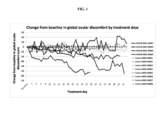
Figure 1 illustrates the symptomatic changes for twelve patients having the
rs1800693
CC genotype, 4 patients treated with LME636 and 8 treated with vehicle, from
baseline to day
43.
Figure 2 is a waterfall plot showing change from baseline in global ocular
discomfort
score at day 26 for all patients treated with LME636 or vehicle to allow
visualization of the
symptomatic changes by treatment and rs1800693 genotype.
Figure 3 is a waterfall plot showing change from baseline in global ocular
discomfort
score at day 27 for all patients treated with LME636 or vehicle to allow
visualization of the
symptomatic changes by treatment and rs1800693 genotype.
- 5 -
CA 03032790 2019-02-01
WO 2017/221128
PCT/IB2017/053625
Figure 4 is a waterfall plot showing change from baseline in global ocular
discomfort
score at day 28 for all patients treated with LME636 or vehicle to allow
visualization of the
symptomatic changes by treatment and rs1800693 genotype.
Figure 5 is a waterfall plot showing change from baseline in global ocular
discomfort
score at day 29 for all patients treated with LME636 or vehicle to allow
visualization of the
symptomatic changes by treatment and rs1800693 genotype.
DETAILED DESCRIPTION OF THE DISCLOSURE
The term "comprising" encompasses "including" as well as "consisting," e.g. a
composition "comprising" X may consist exclusively of X or may include
something additional,
e.g., X + Y.
The term "about" in relation to a numerical value x means +/-10% unless the
context
dictates otherwise.
As used herein, the terms "subject" and "patient" include any human or
nonhuman animal.
The term "nonhuman animal" includes all vertebrates, e.g., mammals and non-
mammals, such as
nonhuman primates, sheep, dogs, cats, horses, cows, chickens, amphibians,
reptiles, etc.
The term "assaying" is used to refer to the act of identifying, screening,
probing, testing
measuring or determining, which act may be performed by any conventional
means. For
example, a sample may be assayed for the presence of a particular genetic or
protein marker by
using an ELISA assay, a Northern blot, imaging, serotyping, cellular typing,
gene sequencing,
phenotyping, haplotyping, immunohistochemistry, western blot, mass
spectrometry, etc. The
term "detecting" (and the like) means the act of extracting particular
information from a given
source, which may be direct or indirect. In some embodiments of the predictive
methods
disclosed herein, the presence of a given thing (e.g., allele, level of
protein, etc.) is detected in a
biological sample indirectly, e.g., by querying a database. The terms
"assaying" and
"determining" contemplate a transformation of matter, e.g., a transformation
of a biological
sample, e.g., a blood sample or other tissue sample, from one state to another
by means of
subjecting that sample to physical testing.
- 6 -
CA 03032790 2019-02-01
WO 2017/221128
PCT/IB2017/053625
The term "obtaining" means to procure, e.g., to acquire possession of in any
way, e.g., by
physical intervention (e.g., biopsy, blood draw) or non-physical intervention
(e.g, transmittal of
information via a server), etc.
The phrase "assaying a biological sample ..." and the like, is used to mean
that a sample
may be tested (either directly or indirectly) for either the presence of a
given DED response
marker. It will be understood that, in a situation where the presence of a
substance denotes one
probability and the absence of a substance denotes a different probabiltity,
then either the
presence or the absence of such substance may be used to guide a therapeutic
decision. For
example, one may determine if a patient has DED response marker by determining
the actual
existence of particular response allele in the patient or by determining the
absence of the
particular response allele in the patient. In both such cases, one has
determined whether the
patient has the presence of the DED response marker. The disclosed methods
involve, inter alia,
determining whether a particular individual has a DED response marker. This
determination is
undertaken by identifying whether the patient has a DED response marker in
Table 1. Each of
these determinations (i.e., presence or absence), on its own, provides the
allelic status of the
patient and thus each of these deteriminations equally provide an indication
of whether a
particular individual would or would not respond more favorably to TNFa
antagonism.
Table 1
Gene SNP Location Response
Allele
(copies for
response)
TNFRI rs1800693 Intronic C (One or
two)
TNFRI rs1800693 Intronic T (One or
two)
- 7 -
CA 03032790 2019-02-01
WO 2017/221128
PCT/IB2017/053625
Table 1 shows the various response alleles of the disclosure. Column 1
provides the
gene in which the SNP of column 2 resides, and column 3 provides the general
location of that
SNP in that gene.
While the SNP listed in Table 1 has predictive value for TNFa antagonism if
there is any
.. of the given response alleles (i.e., the patient is either homozygous or
heterozygous for the given
response allele), as discussed in the Examples below, patients with the CC
genotype tended to
have a larger improvement than those with the CT or TT genotypes in response
to LME636.
The term "dry eye," also known as conjunctivitis sicca or keratoconjunctivitis
sicca, is a
common ophthalmological disorder involving breakdown of the pre-ocular tear
film, resulting in
dehydration of the exposed outer surface of the eye. In certain embodiments,
the "dry eye" is
characterized as moderate to severe, severity being determined by one of skill
in the art, for
example based on global ocular discomfort score as described herein. Methods
for determining
severity of dry eye are also described, for example, in the DEWS definition
and classification
guidelines from the 2007 International Dry Eye Workshop (see "The Ocular
Surface" April 2007,
Vol. 5, No. 2, pages 75-92) or the methods described by Sullivan et al.
(Investigative
Ophthalmology & Visual Science, December 2010, Vol. 51, pg. 6125-6130).
The term "TNFa" refers to tumour necrosis factor alpha (also known as
cachectin), which
is a naturally occurring mammalian cytokine produced by numerous cell types,
including
monocytes and macrophages in response to endotoxin or other stimuli. TNFa is a
major mediator
of inflammatory, immunological, and pathophysiological reactions (Grell, M.,
et al. (1995) Cell,
83: 793-802). "TNFa" includes wild-type TNFa from various species (e.g.,
human, mouse, and
monkey), polymorphic variants of TNFa, and functional equivalents of TNFa.
Functional
equivalents of TNFa according to the present disclosure preferably have at
least about 85%, 95%,
96%, 97%, 98%, or even 99% overall sequence identity with a wild-type TNFa
(e.g., human
.. TNFa), and substantially retain the ability to mediate inflammatory,
immunological, and
pathophysiological reactions.
"TNFa antagonist" as used herein refers to a molecule capable of antagonizing
(e.g.,
reducing, inhibiting, decreasing, delaying) TNFa function, expression and/or
signalling (e.g., by
blocking the binding of TNFa to the TNFa receptor). Non-limiting examples of
TNFa
antagonists include TNFa binding molecules and TNFa receptor binding
molecules. In some
- 8 -
CA 03032790 2019-02-01
WO 2017/221128
PCT/IB2017/053625
embodiments of the disclosed methods, regimens, kits, processes, uses and
compositions, an
TNFa antagonist is employed.
By "TNFa binding molecule" is meant any molecule capable of binding to the
human
TNFa antigen either alone or associated with other molecules. The binding
reaction may be
shown by standard methods (qualitative assays) including, for example, a
binding assay,
competition assay or a bioassay for determining the inhibition of TNFa binding
to its receptor or
any kind of binding assays, with reference to a negative control test in which
an antibody of
unrelated specificity, but ideally of the same isotype, e.g., an anti-CD25
antibody, is used. Non-
limiting examples of TNFa binding molecules include small molecules, TNFa
receptor decoys,
and antibodies that bind to TNFa as produced by B-cells or hybridomas and
chimeric, CDR-
grafted or human antibodies or any fragment thereof, e.g., F(ab')2 and Fab
fragments, as well as
single chain or single domain antibodies. Preferably the TNFa binding molecule
antagonizes
(e.g., reduces, inhibits, decreases, delays) TNFa function, expression and/or
signalling. In some
embodiments of the disclosed methods, regimens, kits, processes, uses and
compositions, an
TNFa binding molecule is employed.
By "TNFa receptor binding molecule" is meant any molecule capable of binding
to the
human TNFa receptor either alone or associated with other molecules. The
binding reaction may
be shown by standard methods (qualitative assays) including, for example, a
binding assay,
competition assay or a bioassay for determining the inhibition of TNFa
receptor binding to
TNFa or any kind of binding assays, with reference to a negative control test
in which an
antibody of unrelated specificity, but ideally of the same isotype is used.
Non-limiting examples
of TNFa receptor binding molecules include small molecules, TNFa decoys, and
antibodies to
the TNFa receptor as produced by B-cells or hybridomas and chimeric, CDR-
grafted or human
antibodies or any fragment thereof, e.g., F(ab')2 and Fab fragments, as well
as single chain or
single domain antibodies. Preferably the TNFa receptor binding molecule
antagonizes (e.g.,
reduces, inhibits, decreases, delays) TNFa function, expression and/or
signalling. In some
embodiments of the disclosed methods, regimens, kits, processes, uses and
compositions, a
TNFa receptor binding molecule is employed.
The term "antibody" as referred to herein includes whole antibodies and any
antigen-
binding portion or single chains thereof. A naturally occurring "antibody" is
a glycoprotein
- 9 -
CA 03032790 2019-02-01
WO 2017/221128
PCT/IB2017/053625
comprising at least two heavy (H) chains and two light (L) chains inter-
connected by disulfide
bonds. Each heavy chain is comprised of a heavy chain variable region
(abbreviated herein as VH)
and a heavy chain constant region. The heavy chain constant region is
comprised of three
domains, CH1, CH2 and CH3. Each light chain is comprised of a light chain
variable region
(abbreviated herein as VL) and a light chain constant region. The light chain
constant region is
comprised of one domain, CL. The VH and VL regions can be further subdivided
into regions of
hypervariability, termed hypervariable regions or complementarity determining
regions (CDR),
interspersed with regions that are more conserved, termed framework regions
(FR). Each VH and
VL is composed of three CDRs and four FRs arranged from amino-terminus to
carboxy-terminus
in the following order: FR1, CDR1, FR2, CDR2, FR3, CDR3, FR4. The variable
regions of the
heavy and light chains contain a binding domain that interacts with an
antigen. The constant
regions of the antibodies may mediate the binding of the immunoglobulin to
host tissues or
factors, including various cells of the immune system (e.g., effector cells)
and the first
component (Cl q) of the classical complement system. In some embodiments of
the disclosed
methods, regimens, kits, processes, uses and compositions, an antibody to TNFa
or the TNFa
receptor is employed, preferably an antibody to TNFa, e.g., LME636.
The term "antigen-binding portion" of an antibody as used herein, refers to
fragments of an
antibody that retain the ability to specifically bind to an antigen (e.g.,
TNFa). It has been shown
that the antigen-binding function of an antibody can be performed by fragments
of a full-length
antibody. Examples of binding fragments encompassed within the term "antigen-
binding
portion" of an antibody include a Fab fragment, a monovalent fragment
consisting of the VL, VH,
CL and CH1 domains; a F(ab)2 fragment, a bivalent fragment comprising two Fab
fragments
linked by a disulfide bridge at the hinge region; a Fd fragment consisting of
the VH and CH1
domains; a Fv fragment consisting of the VL and VH domains of a single arm of
an antibody; a
dAb fragment (Ward et al., 1989 Nature 341:544-546), which consists of a VH
domain; and an
isolated CDR. Exemplary antigen binding sites include the CDRs of LME636 as
set forth in
SEQ ID NOs:1-6 (Table 2), preferably the heavy chain CDR3. Furthermore,
although the two
domains of the Fv fragment, VL and VH, are coded for by separate genes, they
can be joined,
using recombinant methods, by a synthetic linker that enables them to be made
as a single
protein chain in which the VL and VH regions pair to form monovalent molecules
(known as
single chain Fv (scFv); see, e.g., Bird et al., 1988 Science 242:423-426; and
Huston et al., 1988
- 10 -
CA 03032790 2019-02-01
WO 2017/221128
PCT/IB2017/053625
Proc. Natl. Acad. Sci. 85:5879-5883). Such single chain antibodies are also
intended to be
encompassed within the term "antibody". Single chain antibodies and antigen-
binding portions
are obtained using conventional techniques known to those of skill in the art.
In some
embodiments of the disclosed methods, regimens, kits, processes, uses and
compositions, a
single chain antibody or an antigen-binding portion of an antibody against
TNFa (e.g., LME636)
or the TNFa receptor is employed.
An "isolated antibody", as used herein, refers to an antibody that is
substantially free of
other antibodies having different antigenic specificities (e.g., an isolated
antibody that
specifically binds TNFa is substantially free of antibodies that specifically
bind antigens other
.. than TNFa). The term "monoclonal antibody" or "monoclonal antibody
composition" as used
herein refer to a preparation of antibody molecules of single molecular
composition. The term
"human antibody", as used herein, is intended to include antibodies having
variable regions in
which both the framework and CDR regions are derived from sequences of human
origin. A
"human antibody" need not be produced by a human, human tissue or human cell.
The human
.. antibodies of the disclosure may include amino acid residues not encoded by
human sequences
(e.g., mutations introduced by random or site-specific mutagenesis in vitro,
by N-nucleotide
addition at junctions in vivo during recombination of antibody genes, or by
somatic mutation in
vivo). In some embodiments of the disclosed methods, regimens, kits,
processes, uses and
compositions, the TNFa antagonist is a human antibody, an isolated antibody,
and/or a
.. monoclonal antibody. In other embodiments of the disclosed methods,
regimens, kits, processes,
uses and compositions, the TNFa antagonist is a recombinant single-chain
(scFv) antibody.
The term "KD" is intended to refer to the dissociation rate of a particular
antibody-antigen
interaction. The term "KD", as used herein, is intended to refer to the
dissociation constant, which
is obtained from the ratio of Kd to Ka (i.e. Kd/Ka) and is expressed as a
molar concentration (M).
.. KD values for antibodies can be determined using methods well established
in the art. Methods
for determining the KD of an antibody are known in the art, for instance using
a biosensor system
such as a Biacore system. In some embodiments, the TNFa antagonist, e.g.,
TNFa binding
molecule (e.g., TNFa antibody or antigen-binding portion thereof, e.g.,
LME636) or TNFa
receptor binding molecule (e.g., TNFa receptor antibody or antigen-binding
portion thereof)
binds human TNFa with a KD of about 5-250 pM.
-11 -
CA 03032790 2019-02-01
WO 2017/221128
PCT/IB2017/053625
The term "affinity" refers to the strength of interaction between antibody and
antigen at
single antigenic sites. Within each antigenic site, the variable region of the
antibody "arm"
interacts through weak non-covalent forces with antigen at numerous sites; the
more interactions,
the stronger the affinity. Standard assays to evaluate the binding affinity of
the antibodies
toward TNFa of various species are known in the art, including for example,
ELISAs, western
blots and RIAs. The binding kinetics (e.g., binding affinity) of the
antibodies also can be
assessed by standard assays known in the art, such as by Biacore analysis.
An antibody that "inhibits" one or more of these TNFa functional properties
(e.g.,
biochemical, immunochemical, cellular, physiological or other biological
activities, or the like)
as determined according to methodologies known to the art and described
herein, will be
understood to relate to a statistically significant decrease in the particular
activity relative to that
seen in the absence of the antibody (or when a control antibody of irrelevant
specificity is
present). An antibody that inhibits TNFa activity affects a statistically
significant decrease, e.g.,
by at least about 10% of the measured parameter, by at least 50%, 80% or 90%,
and in certain
embodiments of the disclosed methods, uses, processes, kits and compositions,
the TNFa
antibody used may inhibit greater than 95%, 98% or 99% of TNFa functional
activity.
The term "derivative", unless otherwise indicated, is used to define amino
acid sequence
variants, and covalent modifications (e.g., pegylation, deamidation,
hydroxylation,
phosphorylation, methylation, etc.) of an TNFa antagonist, e.g., TNFa binding
molecule (e.g.,
TNFa antibody or antigen-binding portion thereof, e.g., LME636) or TNFa
receptor binding
molecule (e.g., TNFa receptor antibody or antigen-binding portion thereof)
according to the
present disclosure, e.g., of a specified sequence (e.g., a variable domain). A
"functional
derivative" includes a molecule having a qualitative biological activity in
common with the
disclosed TNFa antagonists, e.g., TNFa binding molecules. A functional
derivative includes
fragments and peptide analogs of a TNFa antagonist as disclosed herein.
Fragments comprise
regions within the sequence of a polypeptide according to the present
disclosure, e.g., of a
specified sequence. Functional derivatives of the TNFa antagonists disclosed
herein (e.g.,
functional derivatives of LME636) preferably comprise VH and/or VL domains
that have at least
about 65%, 75%, 85%, 95%, 96%, 97%, 98%, or even 99% overall sequence identity
with the
- 12 -
CA 03032790 2019-02-01
WO 2017/221128
PCT/IB2017/053625
VH and/or VL sequences of the TNFa binding molecules disclosed herein (e.g.,
the VH and/or VL
sequences of Table 2), and substantially retain the ability to bind human
TNFa.
The phrase "substantially identical" means that the relevant amino acid or
nucleotide
sequence (e.g., VH or VL domain) will be identical to or have insubstantial
differences (e.g.,
through conserved amino acid substitutions) in comparison to a particular
reference sequence.
Insubstantial differences include minor amino acid changes, such as 1 or 2
substitutions (e.g.,
conservative substitutions, such as swapping a serine for a threonine, or
substitutions at positions
not involved in antibody activity, structural integrity, complement fixation,
etc.) in a 5 amino
acid sequence of a specified region (e.g., VH or VL domain). In the case of
antibodies, the
second antibody has the same specificity and has at least 50% of the affinity
of the same.
Sequences substantially identical (e.g., at least about 85% sequence identity)
to the sequences
disclosed herein are also part of this disclosure. In some embodiments, the
sequence identity of a
derivative TNFa antibody (e.g., a derivative of LME636, e.g., an LME636
biosimilar antibody)
can be about 90% or greater, e.g., 90%, 91%, 92%, 93%, 94%, 95%, 96%, 97%,
98%, 99% or
higher relative to the disclosed sequences.
"Identity" with respect to a native polypeptide and its functional derivative
is defined
herein as the percentage of amino acid residues in the candidate sequence that
are identical with
the residues of a corresponding native polypeptide, after aligning the
sequences and introducing
gaps, if necessary, to achieve the maximum percent identity, and not
considering any
conservative substitutions as part of the sequence identity. Neither N- or C-
terminal extensions
nor insertions shall be construed as reducing identity. Methods and computer
programs for the
alignment are well known. The percent identity can be determined by standard
alignment
algorithms, for example, the Basic Local Alignment Search Tool (BLAST)
described by Altshul
et al. ((1990) J. Mol. Biol., 215: 403 410); the algorithm of Needleman et al.
((1970) J. Mol.
Biol., 48: 444 453); or the algorithm of Meyers et al. ((1988) Comput. Appl.
Biosci., 4: 1117).
A set of parameters may be the Blosum 62 scoring matrix with a gap penalty of
12, a gap extend
penalty of 4, and a frameshift gap penalty of 5. The percent identity between
two amino acid or
nucleotide sequences can also be determined using the algorithm of E. Meyers
and W. Miller
((1989) CABIOS, 4:11-17) which has been incorporated into the ALIGN program
(version 2.0),
using a PAM120 weight residue table, a gap length penalty of 12 and a gap
penalty of 4.
- 13 -
CA 03032790 2019-02-01
WO 2017/221128
PCT/IB2017/053625
"Amino acid(s)" refer to all naturally occurring L-a-amino acids, e.g., and
include D-
amino acids. The phrase "amino acid sequence variant" refers to molecules with
some
differences in their amino acid sequences as compared to the sequences
according to the present
disclosure. Amino acid sequence variants of a polypeptide according to the
present disclosure,
e.g., of a specified sequence, still have the ability to bind the human TNFa.
Amino acid
sequence variants include substitutional variants (those that have at least
one amino acid residue
removed and a different amino acid inserted in its place at the same position
in a polypeptide
according to the present disclosure), insertional variants (those with one or
more amino acids
inserted immediately adjacent to an amino acid at a particular position in a
polypeptide according
to the present disclosure) and deletional variants (those with one or more
amino acids removed in
a polypeptide according to the present disclosure).
The term "pharmaceutically acceptable" means a nontoxic material that does not
interfere
with the effectiveness of the biological activity of the active ingredient(s).
The term "administering" in relation to a compound, e.g., an TNFa binding
molecule or
another agent, is used to refer to delivery of that compound to a patient by
any route.
As used herein, a "therapeutically effective amount" refers to an amount of an
TNFa
antagonist, e.g., TNFa binding molecule (e.g., TNFa antibody or antigen-
binding portion thereof,
e.g., LME636) or TNFa receptor binding molecule (e.g., TNFa receptor antibody
or antigen-
binding portion thereof) that is effective, upon single or multiple dose
administration to a patient
.. (such as a human) for treating, preventing, preventing the onset of,
curing, delaying, reducing the
severity of, ameliorating at least one symptom of a disorder or recurring
disorder, or prolonging
the survival of the patient beyond that expected in the absence of such
treatment. When applied
to an individual active ingredient (e.g., a TNFa antagonist, e.g., LME636)
administered alone,
the term refers to that ingredient alone. When applied to a combination, the
term refers to
combined amounts of the active ingredients that result in the therapeutic
effect, whether
administered in combination, serially or simultaneously.
The term "treatment" or "treat" refer to both prophylactic or preventative
treatment (as
the case may be) as well as curative or disease modifying treatment, including
treatment of a
patient at risk of contracting the disease or suspected to have contracted the
disease as well as
patients who are ill or have been diagnosed as suffering from a disease or
medical condition, and
- 14 -
CA 03032790 2019-02-01
WO 2017/221128
PCT/IB2017/053625
includes suppression of clinical relapse. The treatment may be administered to
a patient having a
medical disorder or who ultimately may acquire the disorder, in order to
prevent, cure, delay the
onset of, reduce the severity of, or ameliorate one or more symptoms of a
disorder or recurring
disorder, or in order to prolong the survival of a patient beyond that
expected in the absence of
such treatment.
The phrase "respond to treatment" is used to mean that a patient, upon being
delivered a
particular treatment, e.g., a TNFa binding molecule (e.g., LME636) shows a
clinically
meaningful benefit from said treatment. In the case of DED, such criteria
include, e.g., an
improvement in global ocular discomfort score. All such criteria are
acceptable measures of
whether a patient is responding to a given treatment. The phrase "respond to
treatment" is meant
to be construed comparatively, rather than as an absolute response. For
example, a DED patient
having an DED response marker is predicted to have more benefit from treatment
with a TNFa
antagonist than a DED patient who does not have the DED response marker. These
carriers of
DED response markers respond more favorably to treatment with the TNFa
antagonist, and may
simply be said to "respond to treatment" with a TNFa antagonist.
The phrase "receiving data" is used to mean obtaining possession of
information by any
available means, e.g., orally, electronically (e.g., by electronic mail,
encoded on diskette or other
media), written, etc.
As used herein, "selecting" and "selected" in reference to a patient is used
to mean that a
particular patient is specifically chosen from a larger group of patients on
the basis of (due to)
the particular patient having a predetermined criteria, e.g., the patient has
a DED response
marker. Similarly, "selectively treating" refers to providing treatment to a
patient having DED,
where that patient is specifically chosen from a larger group of patients on
the basis of the
particular patient having predetermined criteria, e.g., a DED patient
specifically chosen for
treatment due to the patient having a DED response marker. Similarly,
"selectively
administering" refers to administering a drug to a patient that is
specifically chosen from a larger
group of patients on the basis of (due to) the particular patient having
predetermined criteria, e.g.,
a particular genetic or other biological marker. By selecting, selectively
treating and selectively
administering, it is meant that a patient is delivered a personalized therapy
based on the patient's
particular biology, rather than being delivered a standard treatment regimen
based solely on the
- 15 -
CA 03032790 2019-02-01
WO 2017/221128
PCT/IB2017/053625
patient having DED. Selecting, in reference to a method of treatment as used
herein, does not
refer to fortuitous treatment of a patient that has a DED response marker, but
rather refers to the
deliberate choice to administer a TNFa antagonist to a patient based on the
patient having a
DEDI response marker. Thus, selective treatment differs from standard
treatment, which
delivers a particular drug to all patients, regardless of their allelic
status.
As used herein, "predicting" indicates that the methods described herein
provide
information to enable a health care provider to determine the likelihood that
an individual having
DED will respond to or will respond more favorably to treatment with a TNFa
binding molecule.
It does not refer to the ability to predict response with 100% accuracy.
Instead, the skilled
artisan will understand that it refers to an increased probability.
As used herein, "likelihood" and "likely" is a measurement of how probable an
event is to
occur. It may be used interchangably with "probability". Likelihood refers to
a probability that
is more than speculation, but less than certainty. Thus, an event is likely if
a reasonable person
using common sense, training or experience concludes that, given the
circumstances, an event is
probable. In some embodiments, once likelihood has been ascertained, the
patient may be
treated (or treatment continued, or treatment proceed with a dosage increase)
with the TNFa
binding molecule or the patient may not be treated (or treatment discontinued,
or treatment
proceed with a lowered dose) with the TNFa binding molecule.
The phrase "increased likelihood" refers to an increase in the probability
that an event will
occur. For example, some methods herein allow prediction of whether a patient
will display an
increased likelihood of responding to treatment with an TNFa binding molecule
or an increased
likelihood of responding better to treatment with a TNFa binding molecule in
comparison to a
patient having DED who does not have a DED response marker.
As used herein "SNP" refers to "single nucleotide polymorphism". A single
nucleotide
polymorphism is a DNA sequence variation occurring when a single nucleotide in
the genome
(or other shared sequence) differs between members of a biological species or
paired
chromosomes in an individual. Most SNPs have only two alleles, and one is
usually more
common in the population. A SNP may be present in an exon or an intron of a
gene, an upstream
or downstream untranslated region of a gene, or in a purely genomic location
(i.e., non-
transcribed). When a SNP occurs in the coding region of a gene, the SNP may be
silent (i.e., a
- 16 -
CA 03032790 2019-02-01
WO 2017/221128
PCT/IB2017/053625
synonymous polymorphism) due to the redundancy of the genetic code, or the SNP
may result in
a change in the sequence of the encoded polypeptide (i.e., a non-synonymous
polymorphism).
In the instant disclosure, SNPs are identified by their Single Nucleotide
Polymorphism Database
(dbSNP) rs number, e.g., "rs1800693". The dbSNP is a free public archive for
genetic variation
within and across different species developed and hosted by the National
Center for
Biotechnology Information (NCBI) in collaboration with the National Human
Genome Research
Institute (NHGRI).
A polymorphic site, such as a SNP, is usually preceded by and followed by
conserved
sequences in the genome of the population of interest and thus the location of
a polymorphic site
can often be made in reference to a consensus nucleic acid sequence (e.g., of
thirty to sixty
nucleotides) that bracket the polymorphic site, which in the case of a SNP is
commonly referred
to as the "SNP context sequence". Context sequences for the SNPs disclosed
herein may be
found in the NCBI SNP database available at: www.ncbi.nlm.nih.gov/snp.
Alternatively, the
location of the polymorphic site may be identified by its location in a
reference sequence (e.g.,
GeneBank deposit) relative to the start of the gene, mRNA transcript, BAC
clone or even relative
to the initiation codon (ATG) for protein translation. The skilled artisan
understands that the
location of a particular polymorphic site may not occur at precisely the same
position in a
reference or context sequence in each individual in a population of interest
due to the presence of
one or more insertions or deletions in that individual's genome as compared to
the consensus or
reference sequence. It is routine for the skilled artisan to design robust,
specific and accurate
assays for detecting the alternative alleles at a polymorphic site in any
given individual, when the
skilled artisan is provided with the identity of the alternative alleles at
the polymorphic site to be
detected and one or both of a reference sequence or context sequence in which
the polymorphic
site occurs. Thus, the skilled artisan will understand that specifying the
location of any
polymorphic site described herein by reference to a particular position in a
reference or context
sequence (or with respect to an initiation codon in such a sequence) is merely
for convenience
and that any specifically enumerated nucleotide position literally includes
whatever nucleotide
position the same polymorphic site is actually located at in the same locus in
any individual
being tested for the genetic marker of the invention using any of the
genotyping methods
described herein or other genotyping methods known in the art.
- 17 -
CA 03032790 2019-02-01
WO 2017/221128
PCT/IB2017/053625
In addition to SNPs, genetic polymorphisms include translocations, insertions,
substitutions, deletions, etc., that occur in gene enhancers, exons, introns,
promoters, 5' UTR,
3'UTR, etc.
As used herein "rs1800693" refers to a T/C SNP within the sixth intron of the
human
tumor necrosis factor receptor superfamily, member 1A (TNFRSF IA) gene
(GenBank Accession
No. NM 001065.3) that is also known as tumor necrosis factor receptor 1
(TNFR1). The
TNFRSF1A protein is one of the major receptors for TNFa, and is involved in
the NF-kappaB
pathway, mediates apoptosis, and regulates inflammation. The rs1800693
polymorphic site is
located at Chromosome 12:6330843. The phrase "rs1800693 response allele" as
used herein
refers to the "C" allele (G allele, in the case of the noncoding strand) or
the "T" allele (A allele,
in the case of the noncoding strand) at the rs1800693 polymorphic site. In
some embodiments of
the disclosed methods, uses, and kits, the patient has at least one rs1800693
response allele.
The aforementioned response alleles are useful for the prediction of a DED
patient's
response to TNFa antagonism. In some embodiments, a DED patient having the CC,
CT, or TT
genotype is considered likely to respond to treatment with a TNFa antagonist,
e.g., a TNFa
antibody, such as LME636.
As recognized by the skilled artisan, nucleic acid samples containing a
particular SNP
may be complementary double stranded molecules and thus reference to a
particular site on the
sense strand refers as well to the corresponding site on the complementary
antisense strand.
Similarly, reference to a particular genotype obtained for a SNP on both
copies of one strand of a
chromosome is equivalent to the complementary genotype obtained for the same
SNP on both
copies of the other strand. Thus, for example, a T/C genotype for the
rs1800693 polymorphic
site on the coding strand is equivalent to an A/G genotype for that
polymorphic site on the
noncoding strand.
As used herein, "genomic sequence" refers to a DNA sequence present in a
genome, and
includes a region within an allele, an allele itself, or a larger DNA sequence
of a chromsome
containing an allele of interest.
Products of the DED response markers include nucleic acid products and
polypeptide
products. "Polypeptide product" refers to a polypeptide encoded by a DED
response marke and
- 18 -
CA 03032790 2019-02-01
WO 2017/221128
PCT/IB2017/053625
fragments thereof. "Nucleic acid product" refers to any DNA (e.g., genomic,
cDNA, etc.) or
RNA (e.g., pre-mRNA, mRNA, miRNA, etc.) products of a DED response markers and
fragments thereof.
An "equivalent genetic marker" refers to a genetic marker that is correlated
to an allele of
interest, e.g., it displays linkage disequilibrium (LD) or is in genetic
linkage with the allele of
interest. Equivalent genetic markers may be used to determine if a patient has
a DED response
marker, rather than directly interrogating a biological sample from the
patient for the allele per se.
Various programs exist to help determine LD for particular SNPs, e.g,
HaploBlock (available at
bioinfo. cs.technion.ac. il/haploblock/), HapMap, WGA Viewer.
The term "probe" refers to any composition of matter that is useful for
specifically
detecting another substance, e.g., a substance related to a DED response
marker. A probe can be
an oligonucleotide (including a conjugated oligonucleotide) that specifically
hybridizes to a
genomic sequence of a DED response marker, or a nucleic acid product of a DED
response
marker. A conjugated oligonucleotide refers to an oligonucleotide covalently
bound to
chromophore or molecules containing a ligand (e.g., an antigen), which is
highly specific to a
receptor molecule (e.g., an antibody specific to the antigen). The probe can
also be a PCR
primer, e.g., together with another primer, for amplifying a particular region
within a DED
response marker. Further, the probe can be an antibody that specifically binds
to polypeptide
products of these alleles. Further, the probe can be any composition of matter
capable of
detecting (e.g., binding or hybridizing) an equivalent genetic marker of a DED
response marker.
In preferred embodiments, the probe specifically hybridizes to a nucleic acid
sequence
(preferably genomic DNA) or specifically binds to a polypeptide sequence of an
allele of interest.
The phrase "specifically hybridizes" is used to refer to hybrization under
stringent
hybridization conditions. Stringent conditions are known to those skilled in
the art and can be
found in Current Protocols in Molecular Biology, John Wiley & Sons, N.Y.
(1989), 6.3.1-6.3.6.
Aqueous and nonaqueous methods are described in that reference and either can
be used. One
example of stringent hybridization conditions is hybridization in 6X sodium
chloride/sodium
citrate (SSC) at about 45 C, followed by at least one wash in 0.2X SSC, 0.1%
SDS at 50 C. A
second example of stringent hybridization conditions is hybridization in 6X
SSC at about 45 C,
followed by at least one wash in 0.2X SSC, 0.1% SDS at 55 C. Another example
of stringent
- 19 -
CA 03032790 2019-02-01
WO 2017/221128
PCT/IB2017/053625
hybridization conditions is hybridization in 6X SSC at about 45 C, followed by
at least one wash
in 0.2X SSC, 0.1% SDS at 60 C. A further example of stringent hybridization
conditions is
hybridization in 6X SSC at about 45 C, followed by at least one wash in 0.2X
SSC, 0.1% SDS at
65 C. High stringent conditions include hybridization in 0.5 M sodium
phosphate, 7% SDS at
65 C, followed by at least one wash at 0.2X SSC, 1% SDS at 65 C.
The phrase "a region of a nucleic acid" is used to indicate a smaller sequence
within a
larger sequence of nucleic acids. For example, a gene is a region of a
chromosome, an exon is a
region of a gene, etc.
The term "specifically binds" in the context of polypeptides is used to mean
that a probe
binds a given polypeptide target (e.g., a polypeptide product a DED response
marker) rather than
randomly binding undesireable polypeptides. However, "specifically binds" does
not exclude
some cross reactivity with undesireable polypeptides, as long as that cross
reactivity does not
interfere with the capability of the probe to provide a a useful measure of
the presence of the
given polypeptide target.
The term "capable" is used to mean that ability to achieve a given result,
e.g., a probe that
is capable of detecting the presence of a particular substance means that the
probe may be used
to detect the particular substance.
An "oliogonucelotide" refers to a short sequence of nucleotides, e.g., 2-100
bases.
The term "biological sample" as used herein refers to a sample from a patient,
which may
be used for the purpose of identification, diagnosis, prediction, or
monitoring. Preferred samples
include synovial fluid, blood, blood-derived product (such as buffy coat,
serum, and plasma),
lymph, urine, tear, saliva, hair bulb cells, cerebrospinal fluid, buccal
swabs, feces, synovial fluid,
synovial cells, sputum, or tissue samples (e.g., cartilage samples). In
addition, one of skill in the
art would realize that some samples would be more readily analyzed following a
fractionation or
purification procedure, for example, isolation of DNA from whole blood.
TNFa Antagonists
The various disclosed pharmaceutical compositions, regimens, processes, uses,
methods
and kits utilze an TNFa antagonist, e.g., TNFa binding molecule (e.g., TNFa
antibody or
- 20 -
CA 03032790 2019-02-01
WO 2017/221128
PCT/IB2017/053625
antigen-binding portion thereof, e.g., L1V1E636) or TNFa receptor binding
molecule (e.g., TNFa
receptor antibody or antigen-binding portion thereof).
In one embodiment, the TNFa antagonist, e.g., TNFa binding molecule (e.g.,
TNFa
antibody or antigen-binding portion thereof, e.g., LME636) comprises at least
one heavy chain
variable domain (VH) comprising hypervariable regions CDRH1, CDRH2 and CDRH3,
said
CDRH1 having the amino acid sequence SEQ ID NO:1, said CDRH2 having the amino
acid
sequence SEQ ID NO:2, and said CDRH3 having the amino acid sequence SEQ ID
NO:3. In
one embodiment, the TNFa antagonist, e.g., TNFa binding molecule (e.g., TNFa
antibody or
antigen-binding portion thereof, e.g., LME636) comprises at least one light
chain variable
domain (VI) comprising hypervariable regions CDRL1, CDRL2 and CDRL3, said
CDRL1
having the amino acid sequence SEQ ID NO:4, said CDRL2 having the amino acid
sequence
SEQ ID NO:5 and said CDRL3 having the amino acid sequence SEQ ID NO:6.
In one embodiment, the TNFa antagonist, e.g., TNFa binding molecule (e.g.,
TNFa
antibody or antigen-binding portion thereof, e.g., LME636) comprises a VH
domain and a Vt,
domain, wherein: a) the VH domain comprises (e.g., in sequence): i)
hypervariable regions
CDRH1, CDRH2 and CDRH3, said CDRH1 having the amino acid sequence SEQ ID NO:1,
said
CDRH2 having the amino acid sequence SEQ ID NO:2, and said CDRH3 having the
amino acid
sequence SEQ ID NO:3; and b) the VL domain comprises (e.g., in sequence)
hypervariable
regions CDRL1, CDRL2 and CDRL3, said CDRL1 having the amino acid sequence SEQ
ID
NO:4, said CDRL2 having the amino acid sequence SEQ ID NO: 5, and said CDRL3
having the
amino acid sequence SEQ ID NO:6.
In one embodiment, the TNFa antagonist, e.g., TNFa binding molecule (e.g.,
TNFa
antibody or antigen-binding portion thereof, e.g., LME636) comprises: a) a
heavy chain variable
domain (VH) comprising the amino acid sequence set forth as SEQ ID NO: 8; b) a
light chain
variable domain (VI) comprising the amino acid sequence set forth as SEQ ID
NO:10; c) a VH
domain comprising the amino acid sequence set forth as SEQ ID NO:8 and a VL
domain
comprising the amino acid sequence set forth as SEQ ID NO:10; d) a VH domain
comprising the
hypervariable regions set forth as SEQ ID NO:1, SEQ ID NO:2, and SEQ ID NO:3;
e) a VL
domain comprising the hypervariable regions set forth as SEQ ID NO:4, SEQ ID
NO:5 and SEQ
ID NO:6; or f) a VH domain comprising the hypervariable regions set forth as
SEQ ID NO:1,
- 21 -
CA 03032790 2019-02-01
WO 2017/221128
PCT/IB2017/053625
SEQ ID NO:2, and SEQ ID NO:3 and a VL domain comprising the hypervariable
regions set
forth as SEQ ID NO:4, SEQ ID NO:5 and SEQ ID NO:6.
For ease of reference the amino acid sequences of the hypervariable regions of
the
LME636 scFv antibody is provided in Table 2, below.
Table 2
Light-Chain
CDRL1 QSSQSVYGNIWMA (SEQ ID NO:4)
CDRL2 QASKLAS (SEQ ID NO:5)
CDRL3 QGNFNTGDRYA (SEQ ID NO:6)
Variable EIVMTQSPSTLSASVGDRVIITCQSSQSVYGNIWMAWYQQKPGRAPKL
Light Chain LIYQASKLASGVPSRFSGSGSGAEFTLTISSLQPDDFATYYCQGNFNT
GDRYAFGQGTKLTVLG (SEQ ID NO: 7)
Heavy-Chain
CDRH1 GFTISRSYWIC (SEQ ID NO:1)
CDRH2 CIYGDNDITPLYANWAKG (SEQ ID NO:2)
CDRH3 LGYADYAYDL (SEQ ID NO:3)
Variable EVQLVESGGGSVQPGGSLRLSCTASGFTISRSYWICWVRQAPGKGLEW
Heavy Chain VGCIYGDNDITPLYANWAKGRFTISRDTSKNTVYLQMNSLRAEDTATY
YCARLGYADYAYDLWGQGTTVTVSS (SEQ ID NO:8)
In some embodiments, the TNFa antagonist, e.g., TNFa binding molecule (e.g.,
TNFa
antibody or antigen-binding portion thereof, e.g., LME636) comprises the light
chain of SEQ ID
NO: 7. In other embodiments, the TNFa antagonist comprises the heavy chain of
SEQ ID NO: 8.
In other embodiments, the TNFa 7 antagonist comprises the light chain of SEQ
ID NO: 7 and the
heavy chain of SEQ ID NO: 8. In some embodiments, the TNFa antagonist
comprises the three
CDRs of SEQ ID NO: 7. In other embodiments, the TNFa antagonist comprises the
three CDRs
of SEQ ID NO: 8. In other embodiments, the TNFa antagonist comprises the three
CDRs of
SEQ ID NO: 7 and the three CDRs of SEQ ID NO: 8. CDRs of SEQ ID NO: 7 and SEQ
ID NO:
8, are shown in Table 2. In other embodiments, the TNFa antagonist comprises
the sequence of
SEQ ID NO: 9:
E IVMTQS PS TLSASVGDRVI I TCQS S QSVYGNIWMAWYQQKPGRAPKLL I YQASKLAS GV
PSRFS GS GS GAE FTL TISS LQPDDFAT YYCQGNFNTGDRYAFGQGTKLTVLGGGGGS GGG
GS GGGGS GGGGSEVQLVE S GGGSVQPGGS LRLS CTAS GFT I SRSYWICWVRQAPGKGLEW
- 22 -
CA 03032790 2019-02-01
WO 2017/221128
PCT/IB2017/053625
VGC I YGDND I TPLYANWAKGRFT I SRDTSKNTVYLQMNSLRAEDTATYYCARLGYADYAY
DLWGQGTTVTVSS (SEQ ID NO: 9 ) .
Hypervariable regions may be associated with any kind of framework regions,
though
preferably are of human origin. Suitable framework regions are described in
Kabat E.A. et al,
ibid. The preferred heavy chain framework is the heavy chain framework of the
LME636
antibody as shown in SEQ ID NO: .10:
EVQLVESGGGLVQPGGSLRLSCTAS (X) n=3-50WVRQAPGKGLEWVG (X) n=3-50
RFT I SRDTSKNTVYLQMNS LRAEDTAVYYCAR ( X ) n=3-5 o WGQGTLVTVS S ( SEQ ID
NO: 10) .
The preferred light chain framework is the light chain framework of the LME636
antibody
as shown in SEQ ID NO: .11:
EIVMTQSPSTLSASVGDRVI ITC (X ) n=3-50 WYQQKPGKAPKLLIY (X) n=3-50
GVPSRFSGSGSGTEFTLTISSLQPDDFATYYC (X) n=3-50 FGQGTKLTVLG (SEQ ID NO:
11).
As used in the sequences of SEQ ID NO: 10 and SEQ ID NO: 11, (X) n=3-50
represents a
CDR
In one embodiment, the TNFa antagonist, e.g., TNFa binding molecule (e.g.,
TNFa
antibody or antigen-binding portion thereof, e.g., LME636) is selected from a
single chain
binding molecule which comprises an antigen binding site comprising: a) a
first domain
comprising in sequence the hypervariable regions CDRH1, CDRH2 and CDRH3, said
CDRH1
having the amino acid sequence SEQ ID NO:1, said CDRH2 having the amino acid
sequence
SEQ ID NO:2, and said CDRH3 having the amino acid sequence SEQ ID NO:3; and b)
a second
domain comprising the hypervariable regions CDRL1, CDRL2 and CDRL3, said CDRL1
having
the amino acid sequence SEQ ID NO:4, said CDRL2 having the amino acid sequence
SEQ ID
NO:5, and said CDRL3 having the amino acid sequence SEQ ID NO:6; and c) a
peptide linker
which is bound either to the N-terminal extremity of the first domain and to
the C-terminal
- 23 -
CA 03032790 2019-02-01
WO 2017/221128
PCT/IB2017/053625
extremity of the second domain or to the C-terminal extremity of the first
domain and to the
N-terminal extremity of the second domain.
Alternatively, a TNFa antagonist, e.g., TNFa binding molecule (e.g., TNFa
antibody or
antigen-binding portion thereof) for use in the disclosed methods may comprise
a derivative of
the TNFa binding molecules set forth herein by sequnence (e.g., a pegylated
version of
L1V1E636). Alternatively, the VH or VL domain of a TNFa antagonist, e.g., TNFa
binding
molecule (e.g., TNFa antibody or antigen-binding portion thereof) for use in
the disclosed
methods may have VH or VL domains that are substantially identical to the the
VH or VL domains
set forth herein (e.g., those set forth in SEQ ID NO:8 and 7). An anti-TNFa
antibody disclosed
herein may comprise a heavy chain that is substantially identical to that set
forth as SEQ ID NO:
8 and/or a light chain that is substantially identical to that set forth as
SEQ ID NO: 7. An anti-
TNFa antibody disclosed herein may comprise a heavy chain that comprises SEQ
ID NO: 8 and
a light chain that comprises SEQ ID NO: 7. An anti-TNFa antibody disclosed
herein may
comprise: a) one heavy chain which comprises a variable domain having an amino
acid sequence
substantially identical to that shown in SEQ ID NO: 8 and the constant part of
a human heavy
chain; and b) one light chain which comprises a variable domain having an
amino acid sequence
substantially identical to that shown in SEQ ID NO: 7 and the constant part of
a human light
chain. Alternatively, a TNFa antagonist, e.g., TNFa binding molecule (e.g.,
TNFa antibody or
antigen-binding portion thereof) for use in the disclosed methods may be an
amino acid sequence
variant of the reference TNFa binding molecules set forth herein. In all such
cases of derivative
and variants, the TNFa antagonist is capable of inhibiting the activity of
about 1 nM (= 30 ng/ml)
human TNFa at a concentration of about 50 nM or less, about 20 nM or less,
about 10 nM or less,
about 5 nM or less, or more preferably of about 3 nM or less of said molecule
by 50%, said
inhibitory activity being measured, for example, by assaying for
neutralization of TNFa
cytotoxicity of L929 cells as described in Chiu et al., 2011, PLoS ONE, Vol 6,
issue 1, e16373.
The disclosure also includes TNFa antagonists, e.g., TNFa binding molecules
(e.g., TNFa
antibody or antigen-binding portion thereof, e.g., L1V1E636) in which one or
more of the amino
acid residues of CDRH1, CDRH2, CDRH3, CDRL1, CDRL2, CDRL3, or the frameworks,
typically only a few (e.g., 1-4), are changed; for instance by mutation, e.g.,
site directed
- 24 -
CA 03032790 2019-02-01
WO 2017/221128
PCT/IB2017/053625
mutagenesis of the corresponding DNA sequences. The disclosure includes the
DNA sequences
coding for such changed TNFa antagonists.
The disclosure also includes TNFa antagonists, e.g., TNFa binding molecules
(e.g.,
TNFa antibody or antigen-binding portion thereof, e.g., LME636) that have
binding specificity
for human TNFa, in particular TNFa antibodies capable of inhibiting the
binding of TNFa to its
receptor and TNFa antibodies capable of inhibiting the activity of 1 nM (= 30
ng/ml) human
TNFa at a concentration of about 50 nM or less, about 20 nM or less, about 10
nM or less, about
5 nM or less, or more preferably of about 3 nM or less of said molecule by 50%
(said inhibitory
activity being measured by assaying for neutralization of TNFa cytotoxicity of
L929 cells).
In a preferred embodiment, the anti-TNFa antibodies for use in the disclosed
methods,
uses, kits, etc. is LME636, which comprises the sequence of SEQ ID NO: 9.
LME636 is a
humanized monoclonal scFv antibody fragment consisting of 254 amino acids
(molecular mass:
26.7 kDa) that inhibits human TNFa, and is recombinantly produced in E. coli
by standard
expression technology.
The molecule was genetically engineered by grafting the
complementarity determining regions (CDRs) and specific framework residues
from light and
heavy chain variable region sequences of a monoclonal rabbit anti-human TNFa
antibody to
human light and heavy chain variable region frameworks, covalently linked by a
flexible amino
acid sequence consisting of glycine and serine.
In one embodiment, a methionine derived from the start codon in an expression
vector is
present in the final protein in cases where it has not been cleaved
posttranslationally. In that case,
L1V1E636 has the sequence of SEQ ID NO: 12:
ME IVMTQS PS TL SASVGDRVI I TCQSSQSVYGNIWMAWYQQKPGRAPKLL I YQASKLAS
GVPSRFS GS GS GAE FTLT I SSLQPDDFATYYCQGNFNTGDRYAFGQGTKLTVLGGGGGS
GGGGSGGGGSGGGGSEVQLVESGGGSVQPGGSLRLSCTASGFT I SRSYWI CWVRQAPGK
GLEWVGC I YGDND I TPLYANWAKGRFT I SRDT SKNTVYLQMNSLRAEDTATYYCARLGY
ADYAYDLWGQGTTVTVSS ( SEQ ID NO: 12) .
Other preferred TNFa antibodies for use in the disclosed methods, kits and
uses are those
set forth in WO 2009/155723 and WO 2012/051734.
- 25 -
CA 03032790 2019-02-01
WO 2017/221128
PCT/IB2017/053625
Techniques for Assaying, Diagnostic Methods and Methods of Producing a
Transmittable
Form of Information
The disclosed methods are useful for the treatment or amelioration of DED, as
well as
predicting the likelihood of a DED patient's response to treatment with a TNFa
antagonist, e.g.,
.. LME636. These methods employ, inter alia, determining whether a patient has
a DED response
marker in a sample from the patient.
A biological sample from the patient may be assayed for the presence of a DED
response
marker by any applicable conventional means, which will be selected depending
on whether the
particular marker falls within an exon, an intron, a non-coding portion of
mRNA or a non-
conding genomic sequence.
Numerous biological samples may be used to identify the presence of alleles or
proteins,
the level of expression of genes or proteins, and the activity of a protein,
e.g., blood, synovial
fluid, buffy coat, serum, plasma, lymph, feces, urine, tear, saliva,
cerebrospinal fluid, buccal
swabs, sputum, or tissue. Various sources within a biological sample may be
used in the
disclosed methods, e.g., one may assay genomic DNA obtained from a biological
sample to
detect a DED response marker, or one may assay products of a DED response
marker, e.g.,
nucleic acid products (e.g., DNA, pre-mRNA, mRNA, micro RNAs, etc.) and
polypeptide
products (e.g., expressed proteins) obtained from a biological sample.
We have determined that the various SNP alleles of Table 1 are useful for
predicting
certain patient's response to treatment by TNFa antagonism (e.g., using
LME636). In preferred
embodiments, a genomic sequence of a DED response marker is analyzed to
determine whether
a subject has a DED response marker.
As described in the Examples, our most recent findings lead to the conclusion
that the
presence of a genotype associated with the SNP rs1800693 may be useful to
predict improved
response to TNFa antagonism (e.g., LME636) for DED. The presence of a DED
response
marker may be detected by a variety of genotyping techniques. Typically, such
genotyping
techniques employ one or more oligonucleotides that are complementary to a
region containing,
or adjacent to, the polymorphic site (e.g., SNP) of interest. The sequence of
an oligonucleotide
- 26 -
CA 03032790 2019-02-01
WO 2017/221128
PCT/IB2017/053625
used for genotyping a particular polymorphic site of interest is typically
designed based on a
context sequence or a reference sequence.
Numerous methods and devices are available to identify the presence of a DED
response
marker. DNA (genomic and cDNA) for SNP detection can be prepared from a
biological sample
by methods well known in the art, e.g., phenol/chloroform extraction, PUREGENE
DNA
purification system from GentAS Systems (Qiagen, CA). Detection of a DNA
sequence may
include examining the nucleotide(s) located at either the sense or the anti-
sense strand within that
region. The presence of polymorphisms in a patient may be detected from DNA
(genomic or
cDNA) obtained from PCR using sequence-specific probes, e.g., hydrolysis
probes from Taqman,
Beacons, Scorpions; or hybridization probes that detect the marker or
polymorphism. For the
detection of the polymorphism, sequence specific probes may be designed such
that they
specifically hybridize to the genomic DNA for the alleles of interest or, in
some cases, an RNA
of interest. Primers and probes for polymorphic sites (e.g., SNP) may be
designed based on
context sequences found in the NCBI SNP database available at:
www.ncbi.nlm.nih.gov/snp.
These probes may be labeled for direct detection or contacted by a second,
detectable molecule
that specifically binds to the probe. The PCR products also can be detected by
DNA-binding
agents. Said PCR products can then be subsequently sequenced by any DNA
sequencing method
available in the art. Alternatively the presence of allele can be detected by
sequencing using any
sequencing methods such as, but not limited to, Sanger-based sequencing,
pyrosequencing or
next generation sequencing (Shendure J. and Ji, H., Nature Biotechnology
(1998), Vol. 26, Nr 10,
pages 1135-1145). Optimised allelic discrimination assays for SNPs may be
purchased from
Applied Biosystems (Foster City, California, USA).
Various techniques can be applied to interrogate a particular polymorphism
(e.g., SNP),
including, e.g., hybridization-based methods, such as dynamic allele-specific
hybridization
(DASH) genotyping, polymorphic site (e.g., SNP) detection through molecular
beacons
(Abravaya K., et al. (2003) Clin Chem Lab Med. 41:468-474), Luminex xMAP
technology ,
Illumina Golden Gate technology and commercially available high-density
oligonucleotide
SNP arrays (e.g., the Affymetrix Human SNP 5.0 GeneChip performs a genome-
wide assay
that can genotype over 500,000 human SNPs), BeadChip kits from Illumina, e.g,
Human660W-Quad and Human 1.2M-Duo); enzyme-based methods, such as restriction
- 27 -
CA 03032790 2019-02-01
WO 2017/221128
PCT/IB2017/053625
fragment length polymorphism (RFLP), PCR-based methods (e.g., Tetra-primer
ARMS-PCR),
Invader assays (Olivier M. (2005) Mutat Res. 573(1-2):103-10), various primer
extension assays
(incorporated into detection formats, e.g., MALDI-TOF Mass spectrometry,
electrophoresis,
blotting, and ELISA-like methods), TaqMan assays, and oligonucleotide ligase
assays; and
other post-amplification methods, e.g., analysis of single strand conformation
polymorphism
(Costabile et al. (2006) Hum. Mutat. 27(12):1163-73), temperaure gradient gel
electrophoresis
(TGGE), denaturing high performance liquid chromatography, high-resolution
melting analysis,
DNA mismatch-binding protein assays (e.g., MutS protein from Thermus aquaticus
binds
different single nucleotide mismatches with different affinities and can be
used in capillary
electrophoresis to differentiate all six sets of mismatches), SNPLex
(proprietary SNP detecting
system available from Applied Biosystems), capillary electrophoresis, mass
spectrometry, and
various sequencing methods, e.g., pyrosequencing and next generation
sequencing, etc.
Commercial kits for SNP genotyping include, e.g., Fluidigm Dynamic Array IFCs
(Fluidigm),
TaqMan SNP Genotyping Assay (Applied Biosystems), MassARRAY iPLEX Gold
(Sequenom), Type-it Fast SNP Probe PCR Kit (Quiagen), etc.
In some embodiments, the presence of a polymorphic site (e.g., SNP) in a
patient is
detected using a hybridization assay. In a hybridization assay, the presence
of the genetic marker
is determined based on the ability of the nucleic acid from the sample to
hybridize to a
complementary nucleic acid molecule, e.g., an oligonucleotide probe. A variety
of hybridization
assays are available. In some, hybridization of a probe to the sequence of
interest is detected
directly by visualizing a bound probe, e.g., a Northern or Southern assay. In
these assays, DNA
(Southern) or RNA (Northern) is isolated. The DNA or RNA is then cleaved with
a series of
restriction enzymes that cleave infrequently in the genome and not near any of
the markers being
assayed. The DNA or RNA is then separated, e.g., on an agarose gel, and
transferred to a
membrane. A labeled probe or probes, e.g., by incorporating a radionucleotide
or binding agent
(e.g., SYBR Green), is allowed to contact the membrane under low-, medium- or
high-
stringency conditions. Unbound probe is removed and the presence of binding is
detected by
visualizing the labeled probe. In some embodiments, arrays, e.g., the
MassARRAY system
(Sequenom, San Diego, California, USA) may be used to genotype a subject.
- 28 -
CA 03032790 2019-02-01
WO 2017/221128
PCT/IB2017/053625
Traditional genotyping methods may also be modified for use in genotyping.
Such
traditional methods include, e.g., DNA amplification techniques such as PCR
and variants
thereof, direct sequencing, SSO hybridization coupled with the Luminex )(MAP
technology,
SSP typing, and SBT.
Sequence-Specific Oligonucleotide (SSO) typing uses PCR target amplification,
hybridization of PCR products to a panel of immobilized sequence-specific
oligonucleotides on
the beads, detection of probe-bound amplified product by color formation
followed by data
analysis. Those skilled in the art would understand that the described
Sequence-Specific
Oligonucleotide (SSO) hybridization may be performed using various
commercially available
kits, such as those provided by One Lambda, Inc. (Canoga Park, CA) or
Lifecodes EILA Typing
Kits (Tepnel Life Sciences Corp.) coupled with Luminex technology (Luminex,
Corporation,
TX). LABType SSO is a reverse SSO (rSSO) DNA typing solution that uses
sequence¨specific
oligonucleotide (SSO) probes and color-coded microspheres to identify EILA
alleles. The target
DNA is amplified by polymerase chain reactions (PCR) and then hybridized with
the bead probe
array. The assay takes place in a single well of a 96-well PCR plate; thus, 96
samples can be
processed at one time.
Sequence Specific Primers (SSP) typing is a PCR based technique which uses
sequence
specific primers for DNA based typing. The SSP method is based on the
principle that only
primers with completely matched sequences to the target sequences result in
amplified products
under controlled PCR conditions. Allele sequence-specific primer pairs are
designed to
selectively amplify target sequences which are specific to a single allele or
group of alleles. PCR
products can be visualized on agarose gel. Control primer pairs that matches
non-allelic
sequences present in all samples act as an internal PCR control to verify the
efficiency of the
PCR amplification. Those skilled in the art would understand that low, medium
and high
resolution genotyping with the described sequence-specific primer typing may
be performed
using various commercially available kits, such as the Olerup 55TM kits
(Olerup, PA) or
(Invitrogen) or Allset and TmGold DQA1 Low resolution SSP (Invitrogen).
Sequence Based Typing (SBT) is based on PCR target amplification, followed by
sequencing of the PCR products and data analysis.
- 29 -
CA 03032790 2019-02-01
WO 2017/221128
PCT/IB2017/053625
In some cases, RNA, e.g., mature mRNA, pre-mRNA, can also be used to determine
the
presence of particular polymorphisms (see Table 1). Analysis of the sequence
of mRNA
transcribed from a given gene can be performed using any known method in the
art including,
but not limited, to Northern blot analysis, nuclease protection assays (NPA),
in situ hybridization,
reverse transcription-polymerase chain reaction (RT-PCR), RT-PCR ELISA, TaqMan-
based
quantitative RT-PCR (probe-based quantitative RT-PCR) and SYBR green-based
quantitative
RT-PCR. In one example, detection of mRNA levels involves contacting the
isolated mRNA
with an oligonucleotide that can hybridize to mRNA encoded by a DED response
marker. The
nucleic acid probe can typically be, for example, a full-length cDNA, or a
portion thereof, such
as an oligonucleotide of at least 7, 15, 30, 50, or 100 nucleotides in length
and sufficient to
specifically hybridize under stringent conditions to the mRNA. Hybridization
of an mRNA with
the probe indicates that the marker in question is being expressed. In one
format, the RNA is
immobilized on a solid surface and contacted with a probe, for example by
running the isolated
RNA on an agarose gel and transferring the mRNA from the gel to a membrane,
such as
nitrocellulose. Amplification primers are defined as being a pair of nucleic
acid molecules that
can anneal to 5' or 3' regions of a gene (plus and minus strands,
respectively, or vice-versa) and
contain a short region in between. In general, amplification primers are from
about 10 to 30
nucleotides in length and flank a region from about 50 to 200 nucleotides in
length. Under
appropriate conditions and with appropriate reagents, such primers permit the
amplification of a
nucleic acid molecule comprising the nucleotide sequence flanked by the
primers. PCR products
can be detected by any suitable method including, but not limited to, gel
electrophoresis and
staining with a DNA-specific stain or hybridization to a labeled probe.
In some cases, the presence of a polymorphism in a patient can be determined
by
analyzing polypeptide products of the DED response markers (see Table 1).
Detection of
polypeptide products can be performed using any known method in the art
including, but not
limited, to immunocytochemical staining, ELISA, flow cytometry, Western blot,
spectrophotometry, HPLC, and mass spectrometry.
One method for detecting polypeptide products in a sample is by means of a
probe that is
a binding protein capable of interacting specifically with a marker protein
(e.g., an antibody).
Preferably, labeled antibodies, binding portions thereof, or other binding
partners can be used.
- 30 -
CA 03032790 2019-02-01
WO 2017/221128
PCT/IB2017/053625
The antibodies can be monoclonal or polyclonal in origin, or may be
biosynthetically produced.
The binding partners may also be naturally occurring molecules or
synthetically produced. The
amount of complexed proteins is determined using standard protein detection
methodologies
described in the art. A detailed review of immunological assay design, theory
and protocols can
be found in numerous texts in the art, including Practical Immunology, Butt,
W. R., ed., Marcel
Dekker, New York, 1984. A variety of assays are available for detecting
proteins with labeled
antibodies. Direct labels include fluorescent or luminescent tags, metals,
dyes, radionucleides,
and the like, attached to the antibody. Indirect labels include various
enzymes well known in the
art, such as alkaline phosphatase, hydrogen peroxidase and the like. In a one-
step assay,
polypeptide products, if present, are immobilized and incubated with a labeled
antibody. The
labeled antibody binds to the immobilized target molecule. After washing to
remove unbound
molecules, the sample is assayed for the label.
The use of immobilized antibodies specific for the proteins or polypeptides is
also
contemplated by the present disclosure. The antibodies can be immobilized onto
a variety of
solid supports, such as magnetic or chromatographic matrix particles, the
surface of an assay
place (such as microtiter wells), pieces of a solid substrate material (such
as plastic, nylon, paper),
and the like. An assay strip can be prepared by coating the antibody or a
plurality of antibodies
in an array on solid support. This strip can then be dipped into the test
sample and then
processed quickly through washes and detection steps to generate a measurable
signal, such as a
colored spot.
In a two-step assay, immobilized polypeptide products of a DED response marker
may be
incubated with an unlabeled antibody. The unlabeled antibody complex, if
present, is then bound
to a second, labeled antibody that is specific for the unlabeled antibody. The
sample is washed
and assayed for the presence of the label. The choice of marker used to label
the antibodies will
vary depending upon the application. However, the choice of the marker is
readily determinable
to one skilled in the art. The antibodies may be labeled with a radioactive
atom, an enzyme, a
chromophoric or fluorescent moiety, or a colorimetric tag. The choice of
tagging label also will
depend on the detection limitations desired. Enzyme assays (ELISAs) typically
allow detection
of a colored product formed by interaction of the enzyme-tagged complex with
an enzyme
substrate. Some examples of radioactive atoms include 32p, 125-%
H, and "P. Some examples of
-31 -
CA 03032790 2019-02-01
WO 2017/221128
PCT/IB2017/053625
enzymes include horseradish peroxidase, alkaline phosphatase, beta-
galactosidase, and glucose-
6-phosphate dehydrogenase. Some examples of chromophoric moieties include
fluorescein and
rhodamine. The antibodies may be conjugated to these labels by methods known
in the art. For
example, enzymes and chromophoric molecules may be conjugated to the
antibodies by means
of coupling agents, such as dialdehydes, carbodiimides, dimaleimides, and the
like. Alternatively,
conjugation may occur through a ligand-receptor pair. Some suitable ligand-
receptor pairs
include, for example, biotin-avidin or -streptavidin, and antibody-antigen.
In one aspect, the present disclosure contemplates the use of a sandwich
technique for
detecting polypeptide products in biological samples. The technique requires
two antibodies
capable of binding the protein of interest: e.g., one immobilized onto a solid
support and one free
in solution, but labeled with some easily detectable chemical compound.
Examples of chemical
labels that may be used for the second antibody include but are not limited to
radioisotopes,
fluorescent compounds, and enzymes or other molecules which generate colored
or
electrochemically active products when exposed to a reactant or enzyme
substrate. When
samples containing polypeptide products are placed in this system, the
polypeptide products
binds to both the immobilized antibody and the labeled antibody. The result is
a "sandwich"
immune complex on the support's surface. The complexed protein is detected by
washing away
nonbound sample components and excess labeled antibody, and measuring the
amount of labeled
antibody complexed to protein on the support's surface. The sandwich
immunoassay is highly
specific and very sensitive, provided that labels with good limits of
detection are used.
Preferably, the presence of polypeptide products in a sample is detected by
radioimmunoassays or enzyme-linked immunoassays, competitive binding enzyme-
linked
immunoassays, dot blot, Western blot, chromatography, preferably high
performance liquid
chromatography (1-IPLC), or other assays known in the art. Specific
immunological binding of
the antibody to the protein or polypeptide can be detected directly or
indirectly.
Dot blotting is routinely practiced by the skilled artisan to detect a desired
protein using
an antibody as a probe (Promega Protocols and Applications Guide, Second
Edition, 1991, Page
263, Promega Corporation). Samples are applied to a membrane using a dot blot
apparatus. A
labeled probe is incubated with the membrane, and the presence of the protein
is detected.
- 32 -
CA 03032790 2019-02-01
WO 2017/221128
PCT/IB2017/053625
Western blot analysis is well known to the skilled artisan (Sambrook et al.,
Molecular
Cloning, A Laboratory Manual, 1989, Vol. 3, Chapter 18, Cold Spring Harbor
Laboratory). In
Western blot, the sample is separated by SDS-PAGE. The gel is transferred to a
membrane. The
membrane is incubated with labeled antibody for detection of the desired
protein.
The assays described above involve steps such as but not limited to,
immunoblotting,
immunodiffusion, immunoelectrophoresis, or immunoprecipitation. In some
embodiments, an
automatic analyzer is used to determine the presence of a DED response marker.
Disclosed herein are methods of predicting the likelihood that a patient
having DED will
respond to treatment with a TNFa antagonist, comprising detecting the presence
or absence of a
DED response marker in a biological sample from the patient, wherein: a) the
presence of the
DED response marker is indicative of an increased likelihood that the patient
will respond to
treatment with the TNFa antagonist; and b) the absence of the DED response
marker is
indicative of a decreased likelihood that the patient will respond to
treatment with the TNFa
antagonist.
In some embodiments, the method further comprises the step of obtaining the
biological
sample from the patient, wherein the step of obtaining is performed prior to
the step of assaying.
In some embodiments, the DED response marker is detected by assaying the
biological
sample for a nucleic acid product of the DED response marker, a polypeptide
product of the
DED response marker, or an equivalent genetic marker of the DED response
marker. In some
embodiments, the DED response marker is detected by assaying the biological
sample for a
genomic sequence of the DED response marker. In some embodiments, the
biological sample is
selected from the group consisting of synovial fluid, blood, serum, feces,
plasma, urine, tear,
saliva, cerebrospinal fluid, a leukocyte sample and a tissue sample.
In some embodiments, the presence of the DED response marker is detected by a
technique selected from the group consisting of Northern blot analysis,
polymerase chain
reaction (PCR), reverse transcription-polymerase chain reaction (RT-PCR),
TaqMan-based
assays, direct sequencing, dynamic allele-specific hybridization, high-density
oligonucleotide
SNP arrays, restriction fragment length polymorphism (RFLP) assays, primer
extension assays,
oligonucleotide ligase assays, analysis of single strand conformation
polymorphism, temperature
- 33 -
CA 03032790 2019-02-01
WO 2017/221128
PCT/IB2017/053625
gradient gel electrophoresis (TGGE), denaturing high performance liquid
chromatography, high-
resolution melting analysis, DNA mismatch-binding protein assays, SNPLex ,
capillary
electrophoresis, Southern Blot, immunoassays, immunohistochemistry, ELISA,
flow cytometry,
Western blot, EIPLC, and mass spectrometry.
In some embodiments of the disclosed methods and uses, the TNFa antagonist is
an
TNFa binding molecule or an TNFa receptor binding molecule. In some
embodiments, the
TNFa binding molecule or an TNFa receptor binding molecule is an TNFa binding
molecule. In
some embodiments, the TNFa binding molecule is a TNFa antibody or antigen-
binding portion
thereof.
In some embodiments of the disclosed methods and uses, the TNFa antibody is a
recombinant humanized antibody. In some embodiments of the disclosed methods
and uses, the
recombinant humanized TNFa antibody is LME636.
Methods of Treatment and Uses of TNFa Antagonists
The disclosed methods allow clinicians to provide a personalized therapy for
DED
patients, i.e., they allow determination of whether to selectively treat the
patient with a TNFa
antagonist (e.g., LME636) or whether to selectively treat the patient with an
over the counter
treatment or topical cyclosporine. In this way, a clinician can maximize the
benefit and
minimize the risk of TNFa antagnoism in the entire population of patients
afflicted with DED. It
will be understood that TNFa antagonists, e.g., TNFa binding molecules (e.g.,
TNFa antibody or
antigen-binding portion thereof, e.g., LME636) or TNFa receptor binding
molecules (e.g., TNFa
receptor antibody or antigen-binding portion thereof) are useful for the
treatment, prevention, or
amelioration of DED (e.g., signs and symptoms & structural changes, improving
ocular
discomfort, etc.) as disclosed herein, particularly in patients that have a
DED response marker.
The TNFa antagonists, e.g., TNFa binding molecules (e.g., TNFa antibody or
antigen-
binding portion thereof, e.g., LME636) or TNFa receptor binding molecules
(e.g., TNFa
receptor antibody or antigen-binding portion thereof), may be used in vitro,
ex vivo, or
incorporated into pharmaceutical compositions and administered to individuals
(e.g., human
patients) in vivo to treat, ameliorate, or prevent DED, e.g., in patients who
have a DED response
- 34 -
CA 03032790 2019-02-01
WO 2017/221128
PCT/IB2017/053625
marker. A pharmaceutical composition will be formulated to be compatible with
its intended
route of administration (e.g., oral compositions generally include an inert
diluent or an edible
carrier). Other nonlimiting examples of routes of administration include
parenteral (e.g.,
intravenous), intradermal, subcutaneous, oral (e.g., inhalation), transdermal
(topical),
.. transmucosal, and rectal administration. The pharmaceutical compositions
compatible with each
intended route are well known in the art.
The TNFa antagonists, e.g., TNFa binding molecules (e.g., TNFa antibody or
antigen-
binding portion thereof, e.g., LME636) or TNFa receptor binding molecules
(e.g., TNFa
receptor antibody or antigen-binding portion thereof), may be used as a
pharmaceutical
composition when combined with a pharmaceutically acceptable carrier. Such a
composition
may contain, in addition to a TNFa antagonist, carriers, various diluents,
fillers, salts, buffers,
stabilizers, solubilizers, and other materials well known in the art. The
characteristics of the
carrier will depend on the route of administration. The pharmaceutical
compositions for use in
the disclosed methods may also contain additional therapeutic agents for
treatment of the
particular targeted disorder. For example, a pharmaceutical composition may
also include other
anti-inflammatory agents. Such additional factors and/or agents may be
included in the
pharmaceutical composition to produce a synergistic effect with the TNFa
binding molecules, or
to minimize side effects caused by the TNFa antagonists, e.g., TNFa binding
molecules (e.g.,
TNFa antibody or antigen-binding portion thereof, e.g., LME636) or TNFa
receptor binding
molecules (e.g., TNFa receptor antibody or antigen-binding portion thereof).
Pharmaceutical compositions for use in the disclosed methods may be
manufactured in
conventional manner. In one embodiment, the pharmaceutical composition is
provided in
lyophilized form. For immediate administration it is dissolved in a suitable
aqueous carrier, for
example sterile water for injection or sterile buffered physiological saline.
If it is considered
desirable to make up a solution of larger volume for administration by
infusion rather than a
bolus injection, may be advantageous to incorporate human serum albumin or the
patient's own
heparinised blood into the saline at the time of formulation. The presence of
an excess of such
physiologically inert protein prevents loss of antibody by adsorption onto the
walls of the
container and tubing used with the infusion solution. If albumin is used, a
suitable concentration
- 35 -
CA 03032790 2019-02-01
WO 2017/221128
PCT/IB2017/053625
is from 0.5 to 4.5% by weight of the saline solution. Other formulations
comprise liquid or
lyophilized formulation.
Antibodies, e.g., antibodies to TNFa, are typically formulated either in
aqueous form
ready for parenteral administration or as lyophilisates for reconstitution
with a suitable diluent
prior to administration. In some embodiments of the disclosed methods and
uses, the TNFa
antagonist, e.g., TNFa antibody, e.g., LME636, is formulated as a
lyophilisate. Suitable
lyophilisate formulations can be reconstituted in a small liquid volume (e.g.,
2 ml or less) to
allow subcutaneous administration and can provide solutions with low levels of
antibody
aggregation.
The appropriate dosage will, of course, vary depending upon, for example, the
particular
TNFa antagonists, e.g., TNFa binding molecules (e.g., TNFa antibody or antigen-
binding
portion thereof, e.g., LME636) or TNFa receptor binding molecules (e.g., TNFa
receptor
antibody or antigen-binding portion thereof) to be employed, the host, the
mode of
administration and the nature and severity of the condition being treated, and
on the nature of
prior treatments that the patient has undergone. Ultimately, the attending
health care provider
will decide the amount of the TNFa antagonist with which to treat each
individual patient. In
some embodiments, the attending health care provider may administer low doses
of the TNFa
antagonist and observe the patient's response. In other embodiments, the
initial dose(s) of TNFa
antagonist administered to a patient are high, and then are titrated downward
until signs of
relapse occur. Larger doses of the TNFa antagonist may be administered until
the optimal
therapeutic effect is obtained for the patient, and the dosage is not
generally increased further.
In practicing some of the methods of treatment or uses of the present
disclosure, a
therapeutically effective amount of a TNFa antagonists, e.g., TNFa binding
molecules (e.g.,
TNFa antibody or antigen-binding portion thereof, e.g., LME636) or TNFa
receptor binding
molecule (e.g., TNFa antibody or antigen-binding portion thereof) is
administered to a patient,
e.g., a mammal (e.g., a human). While it is understood that the disclosed
methods provide for
selective treatment of patients (i.e., patients having DED) depending on the
presence of a DED
response marker, this does not preclude that, if the patient is ultimately
treated with a TNFa
antagonist, such TNFa antagonist therapy is necessarily a monotherapy. Indeed,
if a patient is
selected for treatment with a TNFa antagonist, then the TNFa antagonist (e.g.,
LME636) may be
- 36 -
CA 03032790 2019-02-01
WO 2017/221128
PCT/IB2017/053625
administered in accordance with the method of the disclosure either alone or
in combination with
other therapeutics for treating DED in patients. When coadministered with one
or more
additional therapeutics, a TNFa antagonist may be administered either
simultaneously with the
other therapeutic, or sequentially. If administered sequentially, the
attending physician will
decide on the appropriate sequence of administering the TNFa antagonist in
combination with
other therapeutics, as well as the appropriate dosages for co-delivery.
A TNFa antagonist can be conveniently administered parenterally,
intravenously, e.g.,
into the antecubital or other peripheral vein, intramuscularly, or
subcutaneously. The duration of
intravenous (i.v.) therapy using a pharmaceutical composition of the present
disclosure will vary,
depending on the severity of the disease being treated and the condition and
personal response of
each individual patient. Also contemplated is subcutaneous (s.c.) therapy
using a pharmaceutical
composition of the present disclosure. The health care provider will decide on
the appropriate
duration of i.v. or s.c. therapy and the timing of administration of the
therapy, using the
pharmaceutical composition of the present disclosure.
In certain embodiments, a TNFa antagonist, e.g., LME636, can be delivered
directly to
the eye by ocular tissue injection such as periocular, conjunctival, subtenon,
intracameral,
intravitreal, intraocular, subretinal, subconjunctival, retrobulbar, or
intracanalicular injections; by
direct application to the eye using a catheter or other placement device such
as a retinal pellet,
intraocular insert, suppository or an implant comprising a porous, non-porous,
or gelatinous
material; by topical ocular drops or ointments; or by a slow release device in
the cul-de-sac or
implanted adjacent to the sclera (transscleral) or in the sclera
(intrascleral) or within the eye.
Intracanalicular injection may be into the venous collector channels draining
Schlemm's canal or
into Schlemm's canal.
For ophthalmic delivery, an antibody of the invention may be combined with
ophthalmologically acceptable preservatives, co-solvents, surfactants,
viscosity enhancers,
penetration enhancers, buffers, sodium chloride, or water to form an aqueous,
sterile ophthalmic
suspension or solution. Topical ophthalmic products may be packaged, for
example, in
multidose form. Preservatives may thus be required to prevent microbial
contamination during
use. Suitable preservatives include: chlorobutanol, methyl paraben, propyl
paraben, phenylethyl
alcohol, edetate disodium, sorbic acid, polyquaternium-1, or other agents
known to those skilled
- 37 -
CA 03032790 2019-02-01
WO 2017/221128
PCT/IB2017/053625
in the art. Such preservatives are typically employed at a level of from 0.001
to 1.0% w/v. Unit
dose compositions of the present invention will be sterile, but typically
unpreserved. Such
compositions, therefore, generally will not contain preservatives.
In certain embodiments, compositions intended to be administered topically to
the eye are
formulated as eye drops or eye ointments, wherein the total amount of antibody
will be about 0.1
to 10.0% (w/w). Preferably, the amount of TNFa antagonist, e.g., LME636, is
about 5.0 to about
10.0% (w/w), most preferably about 6.0% (w/w).
Compositions of the invention in certain circumstances will be administered as
solutions
for topical administration. Aqueous solutions are generally preferred, based
on ease of
formulation, as well as a patient's ability to easily administer such
compositions by means of
instilling one to two drops of the solutions in the affected eyes. However,
the compositions may
also be suspensions, viscous or semi-viscous gels, or other types of solid or
semi-solid
compositions.
The therapeutically effective amount of an antibody present in the formulation
is
determined by taking into account the desired dose volumes and mode(s) of
administration, for
example. From about 1.0 mg/ml to about 100 mg/ml, preferably from about 5.0
mg/ml to about
80 mg/ml and most preferably from about 10.0 mg/ml to about 60 mg/ml is an
exemplary
antibody concentration in the formulation.
As a general proposition for systemic administration, the therapeutically
effective amount
of the TNFa antagonist, e.g., LME636, administered will be in the range of
about 0.1 to about
100 mg/kg of patient body weight whether by one or more administrations, with
the typical range
of an antibody used being about 0.3 to about 20 mg/kg, more preferably about
0.3 to about 15
mg/kg, administered daily, for example. However, other dosage regimens may be
useful. The
progress of this therapy is easily monitored by conventional techniques.
Disclosed herein are methods of selectively treating a patient having DED,
comprising
either: a) selectively administering a therapeutically effective amount of a
TNFa antagonist to
the patient on the basis of the patient having a DED response marker wherein
the DED response
marker an rs1800693 response allele.
- 38 -
CA 03032790 2019-02-01
WO 2017/221128
PCT/IB2017/053625
In some embodiments, the method further comprises the step of obtaining the
biological
sample from the patient, wherein the step of obtaining is performed prior to
the step of assaying.
In some embodiments of the disclosed methods and uses, a DED response marker
is
detected by assaying the biological sample for a nucleic acid product of the
DED response
marker, a polypeptide product of the DED response marker, or an equivalent
genetic marker of
the DED response marker.
In some embodiments of the disclosed methods and uses, the DED response marker
is
detected by assaying the biological sample for a genomic sequence of the DED
response marker.
In some embodiments of the disclosed methods and uses, the biological sample
is
selected from the group consisting of synovial fluid, blood, serum, feces,
plasma, urine, tear,
saliva, cerebrospinal fluid, a leukocyte sample and a tissue sample.
In some embodiments of the disclosed methods and uses, the DED response marker
is
detected by a technique selected from the group consisting of Northern blot
analysis, polymerase
chain reaction (PCR), reverse transcription-polymerase chain reaction (RT-
PCR), TaqMan-based
assays, direct sequencing, dynamic allele-specific hybridization, high-density
oligonucleotide
SNP arrays, restriction fragment length polymorphism (RFLP) assays, primer
extension assays,
oligonucleotide ligase assays, analysis of single strand conformation
polymorphism, temperature
gradient gel electrophoresis (TGGE), denaturing high performance liquid
chromatography, high-
resolution melting analysis, DNA mismatch-binding protein assays, SNPLex ,
capillary
electrophoresis, Southern Blot, immunoassays, immunohistochemistry, ELISA,
flow cytometry,
Western blot, EIPLC, and mass spectrometry.
Kits
The invention also encompasses kits for detecting a DED response marker in a
biological
sample (a test sample) from a patient. Such kits can be used to predict if a
patient having DED is
likely to respond (or have a higher response) to treatment with a TNFa
antagonist, e.g., TNFa
binding molecule (e.g., TNFa antibody or antigen-binding portion thereof,
e.g., LME636) or
TNFa receptor binding molecule (e.g., TNFa antibody or antigen-binding portion
thereof). For
example, the kit can comprise a probe (e.g., an oligonucleotode, antibody,
labeled compound or
- 39 -
CA 03032790 2019-02-01
WO 2017/221128
PCT/IB2017/053625
other agent) capable of detecting a DED response marker, products of those
alleles and/or an
equivalent genetic marker of those alleles in a biological sample. The kit may
also comprise
instructions for providing a prediction of the likelihood that the patient
will respond to treatment
with the TNFa antagonist.
Probes may specifically hybridize to genomic sequences, nucleic acid products,
or
polypeptide products. Exemplary probes are oligonucleotides or conjugated
oligonucleotides
that specifically hybridizes to the response alleles of Table 1 (e.g., from
DNA, cDNA, mRNA,
etc.); primer-extension oligonucleotides, allele-specific primers, a
combination of allele-specific
primers, allele-specfic probes, and primer extension primers, etc. Optionally,
the kit can contain
a probe that targets an internal control allele, which can be any allele
presented in the general
population. Detection of an internal control allele is designed to assure the
performance of the
kit. The disclosed kits can also comprise, e.g., a buffering agent, a
preservative, or a protein
stabilizing agent. The kit can also comprise components necessary for
detecting the detectable
agent (e.g., an enzyme or a substrate). The kit can also contain a control
sample or a series of
control samples that can be assayed and compared to the test sample contained.
Each component
of the kit is usually enclosed within an individual container, and all of the
various containers are
within a single package along with instructions for use.
Such kits may also comprise a TNFa antagonist, e.g., TNFa binding molecule
(e.g.,
TNFa antibody or antigen-binding portion thereof, e.g., LME636) or TNFa
receptor binding
molecule (e.g., TNFa antibody or antigen-binding portion thereof) (e.g., in
liquid or lyophilized
form) or a pharmaceutical composition comprising the TNFa antagonist
(described supra). In
this way, such kits are useful in the selective treatment of DED using a TNFa
antagonist (e.g.,
LME636). Additionally, such kits may comprise means for administering the TNFa
antagonist
(e.g., a syringe and vial, a prefilled syringe, a prefilled pen) and
instructions for use. These kits
may contain additional therapeutic agents (described supra) for treating DED,
e.g., for delivery
in combination with the enclosed TNFa antagonist, e.g., LME636.
The phrase "means for administering" is used to indicate any available
implement for
systemically administering a drug top a patient, including, but not limited
to, a pre-filled syringe,
a vial and syringe, an injection pen, an autoinjector, an i.v. drip and bag, a
pump, etc. With such
- 40 -
CA 03032790 2019-02-01
WO 2017/221128
PCT/IB2017/053625
items, a patient may self-administer the drug (i.e., administer the drug on
their own behalf) or a
physician may administer the drug.
General
The details of one or more embodiments of the disclosure are set forth in the
accompanying description above. Although any methods and materials similar or
equivalent to
those described herein can be used in the practice or testing of the present
disclosure, the
preferred methods and materials are now described. Other features, objects,
and advantages of
the disclosure will be apparent from the description and from the claims. In
the specification and
the appended claims, the singular forms include plural referents unless the
context clearly
dictates otherwise. Unless defined otherwise, all technical and scientific
terms used herein have
the same meaning as commonly understood by one of ordinary skill in the art to
which this
disclosure belongs. All patents and publications cited in this specification
are incorporated by
reference. The following Examples are presented in order to more fully
illustrate the preferred
embodiments of the disclosure. These examples should in no way be construed as
limiting the
scope of the disclosed patient matter, as defined by the appended claims.
EXAMPLES
Example 1 - CLME636X2202A Study: LME636 Improves Signs and Symptoms of Dry Eye
Disease
CLME636X2202A was a proof of concept (PoC) study to evaluate efficacy and
safety of
topical ocular treatment with LME636 in patients with severe dry eye disease
(DED). The
primary objective of the PoC study was to demonstrate the efficacy of
topically administered
LME636 over LME636 vehicle in reduction of ocular symptoms as determined by
global ocular
discomfort score at treatment day 29. The key secondary objective was to
evaluate the
percentage of patients with improvement in global ocular discomfort score >20
(responder
analysis). DNA samples were collected from the patients with written informed
consent, and an
exploratory pharmacogenetics analysis was conducted to identify genetic
factors that may
- 41 -
CA 03032790 2019-02-01
WO 2017/221128
PCT/IB2017/053625
influence the response to LME636 treatment. The pharmacogenetics analysis is
described in
Example 2.
During the treatment phase, 69 patients were randomized to receive LME636 and
65 to
receive LME636 vehicle. 67 and 64 patients on LME636 and LME636 vehicle,
respectively,
completed the study and were part of the Per protocol analysis. Demographics
and baseline
characteristics were well balanced between the two groups including parameters
(LME636 vs.
L1V1E636 vehicle): Mean age 61.7 vs. 58.8 years; Female patients 61 vs. 54;
Mean global ocular
discomfort at baseline 77.9 vs. 80.3.
For the primary efficacy endpoint, the change from baseline in global ocular
discomfort
score was (LME636 vs. LME636 vehicle): -7.9 [1.45 SE] vs. -3.6 [1.49 SE] at
Day 29 (primary
timepoint and per protocol analysis set) and -10.5 [1.74 SE] vs. -5.4 [1.77
SE] at Day 43
(exploratory timepoint with full analysis set). The 90% confidence interval
for the difference in
change from baseline at Day 29 was -7.7 to -0.8.
For LME636 vs. LME636 vehicle, the number of patients achieving an improvement
from baseline in global ocular discomfort of 20 units or more at Day 29 were
12 (17.9 %) vs. 3
(4.7 %) with a p-value based on chi-square test and Wald interval of 0.018
(responder analysis).
In conclusion, the results met the prespecified criteria for the primary
analysis and were
further supported by the responder analysis. The improvements were sustained
from Day 29
through Day 43.
Example 2: Materials and Method for Pharmacogenetic (PG) Analysis
Example 2.1: Samples and Processing
A total of 127 patient samples were collected. The patients who met all of the
following
criteria were included in the pharmacogenetics analysis:
= Provided written informed consent for pharmacogenetic study.
= DNA was successfully extracted and genotyped.
= Included in the analysis of overall clinical study.
- 42 -
CA 03032790 2019-02-01
WO 2017/221128
PCT/IB2017/053625
Blood samples from consenting patients were collected at the individual trial
sites and
then shipped to Pharmaceutical Product Development (Wilmington, NC 28401). The
genomic
DNA of each patient was extracted from the blood by Covance (Indianapolis, IN
46214), and
genotyping data was generated by Clinical Reference Laboratory (Lenexa, KS
66215) using
TaqMan technology. Of the nine SNPs, eight were successfully developed, and
one
(rsl 15575857) failed to be developed due to complexity of local DNA sequence.
Of the 127
DNA samples, 126 were successfully genotyped for the eight SNPs. One patient
sample failed to
be genotyped due to DNA quality issue. Two duplicated samples were included as
quality
control.
Several candidate genes and single nucleotide polymorphisms (SNPs) were
selected for
the PG analysis based on being in the drug target (TNF-a) or its receptor
(TNFR1), or being
genetic variants that were highly associated with Sjogren's syndrome (see
Table 1).
Table 1. Candidate genes and SNPs
Mechanism of action Sjogren's syndrome
Gene SNP Gene SNP
TNF-a rs1800629 EILA-DPB1 rs4282438
TNF-a rs361525 EILA-DQA1 rs116232857
TNF-a rs1799724 EILA-DRA rs3135394
TNFR1 rs1800693 HLA-DQB 1 rs115575857
IRF5 rs17339836
Example 2.2: Statistical analysis
Primary efficacy endpoint
The primary efficacy endpoint was change from baseline in global ocular
discomfort
score at treatment day 29. The per-protocol set was used for the efficacy
analysis.
To test genotype effect in patients treated with LME636: A mixed model
repeated
measures analysis was performed with terms of baseline global discomfort
score, genotype,
treatment day, genotype by treatment day interaction, age and race. Estimates
of the difference in
- 43 -
CA 03032790 2019-02-01
WO 2017/221128
PCT/IB2017/053625
mean change from baseline in global discomfort between genotype groups and
associated 90%
confidence interval were presented.
To test interaction between genotype and treatment at treatment day 29: An
ANCOVA
model was used, and baseline global discomfort score, age and race were
included as covariates.
Estimates of the difference in mean change from baseline in global discomfort
between genotype
groups and associated 90% confidence interval were presented.
All statistical tests were 2-sided. A Bonferroni correction was applied to
adjust for
multiple testing. For any genetic effects attaining p<0.1 after adjustment for
multiple testing, a
scatter plot or waterfall plot will be presented displaying individual patient
data.
Key secondary efficacy endpoint
The key secondary efficacy endpoint was percentage of patients with
improvement in
global ocular discomfort score > 20 (determined as responder) from baseline to
treatment day 29.
The per-protocol set was used for the efficacy analysis.
Fisher's exact test was used. Only the genotypes that have been associated
with the
primary endpoint were analyzed. The percentage of patients with improvement in
global ocular
discomfort score > 20 between genotype groups was presented.
Example 3: Results for PG analysis
Example 3.1: Comparison for PG and overall study populations
A total of 126 patient samples were successfully genotyped for 8 SNPs. The two
duplicates that were introduced for quality control showed 100% concordance of
the 8 SNPs
genotypes. Of the 126 patients with genotype data, 88 entered the treatment.
Of the 88 patients
who had genotype data and entered the treatment, 86 were in the per protocol
analysis set. In
contrast, the overall study had 131 patients in the per protocol analysis set.
Table 3 summarizes
the demographic features, global ocular discomfort score at baseline, and the
response rate by
treatment in the PG population and overall study, respectively. The PG
population looked similar
- 44 -
CA 03032790 2019-02-01
WO 2017/221128
PCT/IB2017/053625
to the overall study as determined by these variables, especially global
ocular discomfort score at
baseline.
Table 3. Comparison for PG and overall study population
PG population Overall study
Variable LME636 (n=43) Vehicle (n=43) LME636 (n=67) Vehicle
(n=64)
Age, yrs 62.18 (12.11) 59.26 (14.01) 61.61
(13.22) 58.83 (14.60)
Race, % Caucasian 74.42% 79.07% 74.63% 79.69%
Gender, % female 83.72% 79.07% 88.06% 82.81%
Baseline GDS 77.23 (13.53) 80.84 (12.00) 77.91
(13.89) 80.33 (12.56)
Responder 16.28% 4.65% 17.91% 4.69%
Values are mean (SD). Per protocol analysis set (PPS).
Example 3.2: Primary efficacy endpoint analysis - Initial association test
Similar to the overall clinical study, a mixed model repeated measures
analysis was used
to test the association between the genotypes and change from baseline in
global ocular
discomfort score at treatment day 29. Among the 8 SNPs tested, only rs1800693
showed
significant effect on the response to LME636 after Bonferroni correction
(p<0.0001) (Table 4).
The SNP rs1800629 showed a nominal significant association with the clinical
endpoint
(p=0.0159), but the significance disappeared after Bonferroni correction
(p=0.1272).
Table 4. Initial screening for association between SNPs and response to LME636
LS Mean Nominal
SNP Genotype SE (90% CI)
change p-value
CC (n=32) -3.50 3.58 (-9.52, 2.53)
rs1799724
CT (n=10) -5.85 4.85 (-13.99, 2.30) 0.3922
(TNFa)
TT (n=1) 0.09 12.6 (-21.07, 21.25)
rs1800629 AG (n=14) -5.43 4.51 (-13.00,2.13)
0.0159
(TNFa) GG (n=29) -3.64 3.47 (-9.47, 2.20)
rs361525 AG (n=5) 1.32 6.31 (-9.26, 11.893)
0.5193
(TNFa) GG (n=38) -4.69 3.39 (-10.39, 1.01)
rs1800693 CC (n=4) -29.48 6.52 (-40.34, -18.61) <0.0001*
- 45 -
CA 03032790 2019-02-01
WO 2017/221128
PCT/IB2017/053625
(TNFR1) CT (n=25) -0.09 3.52 (-6.01, 5.83)
TT (n=14) -3.90 3.51 (-9.79, 1.99)
AA (n=11) -1.61 4.30 (-8.82, 5.60)
rs116232857
AG (n=23) -5.75 3.97 (-12.43,0.93) 0.9943
(DQA1)
GG (n=9) -4.57 4.98 (-12.93, 3.78)
rs3135394 AA (n=37) -3.69 3.40 (-9.40, 2.02)
0.9649
(DRA1) AG (n=6) -5.37 6.41 (-16.14, 5.39)
GG (n=1) N/A N/A N/A
rs4282438
GT (n=2) N/A N/A N/A 1.0000
(DPB1)
TT (n=40) N/A N/A N/A
rs17339836 CC (n=37) -4.60 3.42 (-10.34,1.15)
(IRF5) CT (n=6) -0.93 5.86 (-10.75, 8.89) 0.9770
Patients treated with LME636 for 29 day. Per protocol analysis set (PPS).
Mixed model repeated measure to test the interaction between genotype and
visit.
* Passed Bonferroni correction.
As shown in Table 5, the genotype effect of rsl 800693 on symptomatic
improvement
only existed in the patients who were treated with LME636 but not in those who
were treated
with vehicle. The patients with CC genotype tended to have larger improvement
than those with
CT or TT genotypes.
Table 5. Association between rs1800693 and response to LME636
LS Mean Nominal
Treatment Genotype SE (90% CI)
change p-value
CC (n=4) -29.48 6.52 (-40.34, -18.61)
LME636 CT (n=25) -0.09 3.52 (-6.01, 5.83) <0.0001
TT (n=14) -3.90 3.51 (-9.79, 1.99)
CC (n=8) -1.08 3.74 (-7.32, 5.15)
Vehicle CT (n=19) -4.05 2.82 (-8.77, 0.67) 0.9863
TT (n=16) -4.03 2.80 (-8.71, 0.65)
Patients treated with LME636 or vehicle for 29 day. Per protocol analysis set
(PPS).
Mixed model repeated measure to test the interaction between genotype and
visit.
- 46 -
CA 03032790 2019-02-01
WO 2017/221128
PCT/IB2017/053625
Example 3.3: Primary efficacy endpoint analysis - Interaction between
treatment
and genotype
Next, we tested if there was an interaction between treatment and genotypes
using an
analysis of covariance (ANCOVA) model. Similar to the initial mixed model
analysis, only
rs1800693 showed a significant interaction at treatment day 29 (p=0.0076)
(Table 6). The
interaction remained significant after Bonferroni correction (p=0.0608).
Table 6. Interaction between treatment and SNPs at treatment day 29
LS Mean (90% CI Nominal
SNP Treatment Genotype SE . .
change p-value
CC (n=30) -1.96 3.46 (-7.73, 3.81)
LME636 CT (n=10) -5.29 4.60 (-12.96, 2.38)
rs1799724 TT (n=1) -0.43 11.07 (-18.88,18.02)
0.6211
(TNFa) CC (n=32) -1.35 3.45 (-7.10, 4.41)
Vehicle CT (n=9) 0.43 4.78 (-7.53, 8.39)
TT (n=1) 6.93 11.59 (-12.39,26.26)
AG (n=13) -1.55 4.33 (-8.77, 5.68)
LME636
GG (n=28) -2.59 3.54 (-8.49, 3.30)
rs1800629
(TNFa) AA (n=1) 3.95 11.08 (-14.52,22.41)
0.5038
Vehicle AG (n=8) -2.66 4.44 (-10.06, 4.73)
GG (n=33) 0.039 3.73 (-6.18, 6.25)
AG (n=5) 1.51 5.55 (-7.73, 10.76)
LME636
rs361525 GG (n=36) -3.38 3.32 (-8.91,2.16)
0.1522
(TNFa) AG (n=4) -5.96 5.98 (-15.92, 3.99)
Vehicle
GG (n=38) -0.22 3.37 (-5.83, 5.39)
CC (n=2) -22.90 7.63 (-35.61, -10.19)
LME636 CT (n=25) -0.27 3.59 (-6.25, 5.70)
rs1800693 TT (n=14) -4.10 3.59 (-10.08,1.88)
0.0076*
(TNER1) CC (n=8) 2.31 4.68 (-5.49, 10.12)
Vehicle CT (n=18) -2.26 3.84 (-8.66, 4.15)
TT (n=16) -0.71 3.61 (-6.72, 5.30)
AA (n=10) -1.25 4.19 (-8.23, 5.73)
LME636 AG (n=22) -4.02 3.93 (-10.57, 2.53)
rs116232857
(DQA1) GG (n=9) -4.47 4.76 (-12.41, 3.47) 0.6382
AA (n=15) -2.86 4.25 (-9.95, 4.22)
Vehicle
AG (n=17) -0.20 3.86 (-6.64, 6.24)
- 47 -
CA 03032790 2019-02-01
WO 2017/221128
PCT/IB2017/053625
GG (n=10) -2.01 4.56 (-9.62, 5.59)
AA (n=35) -2.58 3.36 (-8.18, 3.01)
LME636
rs3135394 AG (n=6) -5.80 5.41 (-14.82,3.22)
0.8775
(DRA1) AA (n=36) -0.93 3.41 (-6.61, 4.74)
Vehicle
AG (n=6) -3.14 5.36 (-12.06, 5.79)
GG (n=1) N/A N/A N/A
LME636 GT (n=2) N/A N/A N/A
rs4282438
(DPB1) TT (n=38) N/A N/A N/A
0.7174
GT (n=2) N/A N/A N/A
Vehicle
TT (n=40) N/A N/A N/A
CC (n=35) -3.52 3.28 (-8.99, 1.95)
LME636
rs17339836 CT (n=6) -1.08 5.25 (-9.82, 7.67)
0.0815
(IRF5) CC (n=36) -0.01 3.31 (-5.53, 5.51)
Vehicle
CT (n=6) -8.77 5.24 (-17.50, -0.04)
ANCOVA model to test the interaction between treatment and genotype at day 29.
Per
protocol analysis set (PPS).
* Passed Bonferroni correction.
The interaction test was only for treatment day 29. However, there were
missing values at
individual treatment days. To improve the understanding of the interaction
between treatment
and rs1800693, we further tested the interaction from day 23 to day 28. As
shown in Table 7, a
similar trend of the interaction was observed from day 23 to 29.
Table 7. Interaction between treatment and rs1800693 from treatment day 23 to
29
Treatment LS Mean
day Treatment Genotype change SE (90% CI limits) p-value
CC (n=4) -21.21 6.18 (-31.51, -10.91)
LME636 CT (n=25) 0.81 3.78 (-5.49, 7.11)
TT (n=14) -3.01 3.79 (-9.32, 3.30)
Day 23 0.0023
CC (n=8) 2.27 4.93 (-5.95, 10.48)
Vehicle CT (n=18) -1.96 4.04 (-8.70, 4.78)
TT (n=14) -4.27 3.86 (-10.70, 2.16)
CC (n=3) -6.02 6.36 (-16.62, 4.58)
LME636 CT (n=24) 2.37 3.49 (-3.45, 8.19)
TT (n=14) -5.20 3.47 (-10.99, 0.58)
Day 24 0.0190
CC (n=7) 1.60 4.73 (-6.28, 9.48)
Vehicle CT (n=18) -4.96 3.74 (-11.19, 1.27)
TT (n=16) -0.32 3.49 (-6.13, 5.49)
Day 25 LME636 CC (n=4) -22.2 6.94 (-33.76, -10.63)
0.0030
- 48 -
CA 03032790 2019-02-01
WO 2017/221128
PCT/IB2017/053625
CT (n=23) 1.28 4.29 (-5.87, 8.43)
TT (n=13) -5.27 4.36 (-12.53, 2.00)
CC (n=7) 2.93 5.78 (-6.70, 12.56)
Vehicle CT (n=18) -3.69 4.54 (-11.25, 3.88)
TT (n=16) -3.88 4.26 (-10.98, 3.23)
CC (n=4) -20.04 6.53 (-30.93, -9.15)
LME636 CT (n=24) 0.70 4.04 (-6.04, 7.43)
D 26 TT (n=14) -2.99 4.01 (-9.67, 3.68)
0.0031 ay
CC (n=8) 3.00 5.25 (-5.75, 11.75)
Vehicle CT (n=16) -3.50 4.33 (-10.71, 3.72)
TT (n=15) -5.53 4.15 (-12.46, 1.40)
CC (n=4) -15.98 7.38 (-28.27, -3.68)
LME636 CT (n=24) 0.37 4.52 (-7.17, 7.91)
D 27 TT (n=14) -4.84 4.51 (-12.36, 2.68)
0.0679 ay
CC (n=8) 2.58 5.88 (-7.23,12.38)
Vehicle CT (n=16) -2.08 4.90 (-10.25. 6.09)
TT (n=16) -2.69 4.53 (-10.25, 4.86)
CC (n=2) -15.86 8.77 (-30.48,-1.24)
LME636 CT (n=24) 1.50 4.15 (-5.42, 8.41)
D 28 TT (n=14) -3.40 4.12 (-10.28, 3.48)
0.0881 ay
CC (n=7) 2.33 5.64 (-7.07, 11.73)
Vehicle CT (n=18) -2.23 4.44 (-9.64, 5.18)
TT (n=14) -2.89 4.22 (-9.93, 4.16)
CC (n=2) -22.90 7.63 (-35.61, -10.19)
LME636 CT (n=25) -0.27 3.59 (-6.25, 5.70)
D 29 TT (n=14) -4.10 3.59 (-10.08, 1.88)
0.0076 ay
CC (n=8) 2.31 4.68 (-5.49, 10.12)
Vehicle CT (n=18) -2.26 3.84 (-8.66, 4.15)
TT (n=16) -0.71 3.61 (-6.72, 5.30)
ANCOVA model to test interaction between treatment and genotype. Per protocol
analysis
set (PPS).
Example 3.4: Key secondary efficacy endpoint analysis
The secondary efficacy endpoint analysis focused on the response rate between
genotype
groups. Only rs1800693 was analyzed because it is the only SNP associated with
the primary
efficacy endpoint. As shown by the Fisher's exact test, the response rate was
much greater in
patients with CC genotype than those with CT or TT genotype (Table 8). The
results are
- 49 -
CA 03032790 2019-02-01
WO 2017/221128 PCT/IB2017/053625
supportive to the primary efficacy endpoint analysis. However, the number of
responders is
small, so cautious interpretation is recommended.
Table 8. Response rate from treatment day 23 to 29
Treatment day Treatment Genotype Responder (%) p-value*
CC (n=4) 3 (75.00%)
LME636 CT (n=25) 3 (12.00%) 0.0137
D 23 TT (n=14) 1(7.14%)
ay
CC (n=8) 0 (0.00%)
Vehicle CT (n=18) 2(11.11%) 0.6769
TT (n=14) 0 (0.00%)
CC (n=3) 2 (66.67%)
LME636 CT (n=24) 2 (8.33%) 0.0414
D 24 TT (n=14) 1(7.14%)
ay
CC (n=7) 0 (0.00%)
Vehicle CT (n=18) 2(11.11%) 0.6488
TT (n=16) 0 (0.00%)
CC (n=4) 3 (75.00%)
LME636 CT (n=23) 3 (13.04%) 0.0160
D 25 TT (n=13) 1 (7.69%)
ay
CC (n=7) 0 (0.00%)
Vehicle CT (n=18) 2(11.11%) 0.6488
TT (n=16) 0 (0.00%)
CC (n=4) 3 (75.00%)
LME636 CT (n=24) 3 (12.50%) 0.0149
D 26 TT (n=14) 1(7.14%)
ay
CC (n=8) 0 (0.00%)
Vehicle CT (n=16) 2 (12.50%) 0.5034
TT (n=15) 0 (0.00%)
CC (n=4) 3 (75.00%)
LME636 CT (n=24) 3 (12.50%) 0.0149
D 27 TT (n=14) 1(7.14%)
ay
CC (n=8) 0 (0.00%)
Vehicle CT (n=16) 2 (12.50%) 0.3436
TT (n=16) 0 (0.00%)
CC (n=2) 2 (100.00%)
LME636 CT (n=24) 3(12.50%) 0.0211
D 28 TT (n=14) 1(7.14%)
ay
CC (n=7) 0 (0.00%)
Vehicle CT (n=18) 2(11.11%) 0.6599
TT (n=14) 0 (0.00%)
Day 29 LME636 CC (n=2) 2 (100.00%) 0.0199
- 50 -
CA 03032790 2019-02-01
WO 2017/221128
PCT/IB2017/053625
CT (n=25) 3 (12.00%)
TT (n=14) 1(7.14%)
CC (n=8) 0 (0.00%)
Vehicle CT (n=18) 2(11.11%) 0.6655
TT (n=16) 0 (0.00%)
*Fisher's exact test. Per protocol analysis set (PPS).
Example 3.5: Visualization of individual patient data
There were 12 patients with CC genotype in this pharmacogenetics analysis. To
visualize
and compare the symptomatic changes between the treatments, the change from
baseline in
global ocular discomfort score of the 12 patients with CC genotype were
plotted against the
treatment days from baseline to day 43. As shown by Figure 1, 3 of 4 patients
treated with
LME636 had large symptomatic improvements over time. In contrast, none of the
eight (8)
patients treated with vehicle showed consistently greater than 10 units of
symptomatic
improvement. The difference in symptomatic improvement between LME636 and
vehicle at
treatment day 29 was significant by an ANCOVA analysis (p<0.0001).
The change from baseline in global ocular discomfort score of all patients was
further
illustrated by waterfall plots to allow visualization of the symptomatic
changes by treatment and
genotype (Figures 2-5). Interestingly, only minimal vehicle response was seen
in the patients
with CC genotype, whereas vehicle responses of >10 occurred within CT and TT
groups. The
data suggested that the patients with CC genotype may have a type of dry eye
disease that is less
likely to be alleviated simply with vehicle.
Conclusion
This exploratory pharmacogenetic analysis used a candidate gene approach and
focused
on the genes relevant to mechanism of action of LME636 and the genes
associated with
Sjogren's syndrome. Due to small sample size, only 8 SNPs were included in the
analysis to
reduce the burden of adjustment for multiple testing. The three SNPs in the
TNF-a gene were
reported to associate with response to anti-TNF agents in patients with
autoimmune diseases
such as rheumatoid arthritis (Julia A, Fernandez-Nebro A, Blanco F, Ortiz A,
Callete JD, et al.
- 51 -
CA 03032790 2019-02-01
WO 2017/221128
PCT/IB2017/053625
(2016). A genome-wide association study identifies a new locus associated with
the response to
anti-TNF therapy in rheumatoid arthritis. Pharmacogenomics J. 16(2):147-50).
The one SNP in
the TNFR1 gene that was primarily associated with multiple sclerosis (MS)
causes exon 6
skipping and results in production of soluble TNFR1 (sTNFR1) (De Jager PL, Jia
X, Wang J, de
Bakker PI, Ottoboni L, et al. (2009). Meta-analysis of genome scans and
replication identify
CD6, IRF8 and TNFRSF1A as new multiple sclerosis susceptibility loci. Nat
Genet. 41(7):776-
82; Gregory AP, Dendrou CA, Attfield KE, Haghikia A, Xifara DK, et al. (2012).
TNF receptor
1 genetic risk mirrors outcome of anti-TNF therapy in multiple sclerosis.
Nature.
23;488(7412):508-11). To explore potential impact of the Sjogren's syndrome
risk alleles on the
response to LE636 in patients with DED, four SNPs strongly associated with
Sjogren's
syndrome were included in this analysis (Lessard CJ, Li H, Adrianto I, Ice JA,
Rasmussen A,
Grundahl KM, et al. (2013). Variants at multiple loci implicated in both
innate and adaptive
immune responses are associated with Sjogren's syndrome. Nat Genet.
45(11):1284-92).
sTNFR1 is constitutively released from the cell membrane by TNF-a-converting
enzyme,
and its level increases in the course of various human diseases including
Sjogren's syndrome,
uveitis and glaucoma (Touchard E, Bloquel C, Bigey P, Kowalczuk L, Jonet L, et
al. (2009).
Local ocular immunomodulation resulting from electrotransfer of plasmid
encoding soluble TNF
receptors in the ciliary muscle. Gene Ther. 16(7):862-73; Sakimoto T, Yamada
A, Sawa M.
(2009). Release of soluble tumor necrosis factor receptor 1 from corneal
epithelium by TNF-
alpha-converting enzyme-dependent ectodomain shedding. Invest Ophthalmol Vis
Sci.
50(10):4618-21; Sakimoto T, Ohnishi T, Ishimori A. (2014). Significance of
ectodomain
shedding of TNF receptor 1 in ocular surface. Invest Ophthalmol Vis Sci.
55(4):2419-23). In
contrast, mutations that impair the process of sTNFR1 production cause
periodic syndrome that
is an auto-inflammatory disorder (Magnotti F, Vitale A, Rigante D, Lucherini
OM, Cimaz R, et
al. (2013).The most recent advances in pathophysiology and management of
tumour necrosis
factor receptor-associated periodic syndrome (TRAPS): personal experience and
literature
review. Clin Exp Rheumatol. 31(3 Suppl 77):141-9). Therefore, an imbalance of
sTNFR1
production may contribute to pathogenesis of human diseases. Some mechanistic
studies
suggested that sTNFR1 may act as physiological attenuators of TNF-a activity,
or may function
as a buffer system to enhance the effect of TNF-a (Aderka D, Engelmann H, Maor
Y,
Brakebusch C, Wallach D. (1992). Stabilization of the bioactivity of tumor
necrosis factor by its
- 52 -
CA 03032790 2019-02-01
WO 2017/221128
PCT/IB2017/053625
soluble receptors. J Exp Med. 175(2):323-9; Gregory AP, Dendrou CA, Attfield
KE, Haghikia A,
Xifara DK, et al. (2012). TNF receptor 1 genetic risk mirrors outcome of anti-
TNF therapy in
multiple sclerosis. Nature. 23;488(7412):508-11). rs1800693 is considered as a
novel mechanism
of sTNFR1 production. A recent study demonstrated that the rs1800693 CC
genotype was
correlated with increased signaling to TNF-a and that this altered signaling
may be due to altered
localization of sTNFR1 within the cells (Housley WJ, Fernandez SD, Vera K,
Murikinati SR,
Grutzendler J, et al. (2015). Genetic variants associated with autoimmunity
drive NFicl3 signaling
and responses to inflammatory stimuli. Sci Transl Med. 10;7(291):291ra93).
The prevalence of CC is 19.9% and 12.7% in Caucasian and African,
respectively, as
reported in 1000 human genome database. In contract, the prevalence of CC is
11.5% (11/96)
and 12.7% (2/19) in Caucasian and African, respectively, in the LME636X2202
study. No
genome-wide genetic association study for dry eye disease has been reported in
literature. It is
not clear if rs1800693 has any impact on the risk for dry-eye disease.
We have identified, inter alia, a particular genetic variant that is
predictive of response to
TNFa antagonism, e.g., a TNFa antibody, e.g., LME636, in DED. The findings
disclosed herein
could not have been predicted based solely on the fact that a certain SNP may
be associated with
an increased likelihood of a patient developing DED. The findings disclosed
herein could not
have been predicted based solely on the particular genes in which the SNPs
were studied.
Indeed, of the eight SNPs tested, four were known to be associated with the
TNFa pathway and
four were known to be associated with Sjogren's syndrome, but only rs1800693
located in the
TNFR1 gene showed a significant effect on the response to LME636 treatment.
The patients
with CC genotype tended to have much greater improvement on the symptoms than
those with
CT or TT genotype. As such, one cannot predict how a patient will respond to a
drug based
solely on whether that patient carries an allele associated with a particular
disease state or
whether the patient carries a SNP in a particular gene.
We conclude that the predictive methods and personalized therapies disclosed
herein are
useful to maximize the benefit and minimize the risk of TNFa antagonism in
patients having
DED by identifying those patients likely to respond prior to treatment with a
TNFa antagonist,
such as LME636.
- 53 -
CA 03032790 2019-02-01
WO 2017/221128
PCT/IB2017/053625
SEQUENCE LISTING
<110> Novartis AG
He, Yunsheng
Leisner, Christian
Wald, Michael
Weissgerber, Georges
<120> METHODS OF TREATING DRY EYE DISEASE USING TNFalpha ANTAGONISTS
<130> RAT057361
<160> 12
<170> PatentIn version 3.5
<210> 1
<211> 11
<212> PRT
<213> Artificial Sequence
<220>
<223> synthetic peptide - Heavy chain CDR1
<400> 1
Gly Phe Thr Ile Ser Arg Ser Tyr Trp Ile Cys
1 5 10
<210> 2
<211> 18
<212> PRT
.. <213> Artificial Sequence
<220>
<223> Synthetic peptide - Heavy chain CDR2
<400> 2
Cys Ile Tyr Gly Asp Asn Asp Ile Thr Pro Leu Tyr Ala Asn Trp Ala
1 5 10 15
Lys Gly
<210> 3
<211> 10
<212> PRT
<213> Artificial Sequence
<220>
<223> Synthetic peptide - heavy chain CDR3
- 54 -
CA 03032790 2019-02-01
WO 2017/221128
PCT/IB2017/053625
<400> 3
Leu Gly Tyr Ala Asp Tyr Ala Tyr Asp Leu
1 5 10
<210> 4
<211> 13
<212> PRT
<213> Artificial Sequence
<220>
<223> Synthetic peptide - light chain CDR1
<400> 4
Gln Ser Ser Gln Ser Val Tyr Gly Asn Ile Trp Met Ala
1 5 10
<210> 5
<211> 7
<212> PRT
<213> Artificial Sequence
<220>
<223> Synthetic peptide - light chain CDR2
<400> 5
Gln Ala Ser Lys Leu Ala Ser
1 5
<210> 6
<211> 11
<212> PRT
<213> Artificial Sequence
<220>
<223> Synthetic peptide - light chain CDR3
<400> 6
Gln Gly Asn Phe Asn Thr Gly Asp Arg Tyr Ala
1 5 10
<210> 7
<211> 112
<212> PRT
<213> Artificial Sequence
<220>
<223> Synthetic peptide
- 55 -
CA 03032790 2019-02-01
WO 2017/221128
PCT/IB2017/053625
<400> 7
Glu Ile Val Met Thr Gin Ser Pro Ser Thr Leu Ser Ala Ser Val Gly
1 5 10 15
Asp Arg Val Ile Ile Thr Cys Gin Ser Ser Gin Ser Val Tyr Gly Asn
20 25 30
Ile Trp Met Ala Trp Tyr Gin Gin Lys Pro Gly Arg Ala Pro Lys Leu
35 40 45
Leu Ile Tyr Gin Ala Ser Lys Leu Ala Ser Gly Val Pro Ser Arg Phe
50 55 60
Ser Gly Ser Gly Ser Gly Ala Glu Phe Thr Leu Thr Ile Ser Ser Leu
65 70 75 80
Gin Pro Asp Asp Phe Ala Thr Tyr Tyr Cys Gin Gly Asn Phe Asn Thr
85 90 95
Gly Asp Arg Tyr Ala Phe Gly Gin Gly Thr Lys Leu Thr Val Leu Gly
100 105 110
<210> 8
<211> 121
<212> PRT
<213> Artificial Sequence
<220>
<223> Synthetic peptide
<400> 8
Glu Val Gin Leu Val Glu Ser Gly Gly Gly Ser Val Gin Pro Gly Gly
1 5 10 15
Ser Leu Arg Leu Ser Cys Thr Ala Ser Gly Phe Thr Ile Ser Arg Ser
20 25 30
Tyr Trp Ile Cys Trp Val Arg Gin Ala Pro Gly Lys Gly Leu Glu Trp
35 40 45
Val Gly Cys Ile Tyr Gly Asp Asn Asp Ile Thr Pro Leu Tyr Ala Asn
50 55 60
- 56 -
CA 03032790 2019-02-01
WO 2017/221128
PCT/IB2017/053625
Trp Ala Lys Gly Arg Phe Thr Ile Ser Arg Asp Thr Ser Lys Asn Thr
65 70 75 80
Val Tyr Leu Gln Met Asn Ser Leu Arg Ala Glu Asp Thr Ala Thr Tyr
85 90 95
Tyr Cys Ala Arg Leu Gly Tyr Ala Asp Tyr Ala Tyr Asp Leu Trp Gly
100 105 110
Gln Gly Thr Thr Val Thr Val Ser Ser
115 120
<210> 9
<211> 253
<212> PRT
<213> Artificial Sequence
<220>
<223> Synthetic peptide
<400> 9
Glu Ile Val Met Thr Gln Ser Pro Ser Thr Leu Ser Ala Ser Val Gly
1 5 10 15
Asp Arg Val Ile Ile Thr Cys Gln Ser Ser Gln Ser Val Tyr Gly Asn
20 25 30
Ile Trp Met Ala Trp Tyr Gln Gln Lys Pro Gly Arg Ala Pro Lys Leu
35 40 45
Leu Ile Tyr Gln Ala Ser Lys Leu Ala Ser Gly Val Pro Ser Arg Phe
55 60
Ser Gly Ser Gly Ser Gly Ala Glu Phe Thr Leu Thr Ile Ser Ser Leu
45 65 70 75 80
Gln Pro Asp Asp Phe Ala Thr Tyr Tyr Cys Gln Gly Asn Phe Asn Thr
85 90 95
Gly Asp Arg Tyr Ala Phe Gly Gln Gly Thr Lys Leu Thr Val Leu Gly
100 105 110
Gly Gly Gly Gly Ser Gly Gly Gly Gly Ser Gly Gly Gly Gly Ser Gly
- 57 -
CA 03032790 2019-02-01
WO 2017/221128
PCT/IB2017/053625
115 120 125
Gly Gly Gly Ser Glu Val Gin Leu Val Glu Ser Gly Gly Gly Ser Val
130 135 140
Gin Pro Gly Gly Ser Leu Arg Leu Ser Cys Thr Ala Ser Gly Phe Thr
145 150 155 160
Ile Ser Arg Ser Tyr Trp Ile Cys Trp Val Arg Gin Ala Pro Gly Lys
165 170 175
Gly Leu Glu Trp Val Gly Cys Ile Tyr Gly Asp Asn Asp Ile Thr Pro
180 185 190
.. Leu Tyr Ala Asn Trp Ala Lys Gly Arg Phe Thr Ile Ser Arg Asp Thr
195 200 205
Ser Lys Asn Thr Val Tyr Leu Gin Met Asn Ser Leu Arg Ala Glu Asp
210 215 220
Thr Ala Thr Tyr Tyr Cys Ala Arg Leu Gly Tyr Ala Asp Tyr Ala Tyr
225 230 235 240
Asp Leu Trp Gly Gin Gly Thr Thr Val Thr Val Ser Ser
245 250
<210> 10
<211> 232
<212> PRT
<213> Artificial Sequence
<220>
<223> Synthetic peptide
<220>
<221> MISC_FEATURE
<222> (26)..(75)
<223> CDR1 - X can be any naturally occurring amino acid. at least
three and up to 50 amino acids can be present
<220>
<221> MISC_FEATURE
<222> (90)..(139)
<223> CDR2 - X can be any naturally occurring amino acid. at least
three and up to 50 amino acids can be present
- 58 -
CA 03032790 2019-02-01
WO 2017/221128
PCT/IB2017/053625
<220>
<221> MISC_FEATURE
<222> (172)..(221)
<223> CDR3 - X can be any naturally occurring amino acid. at least
three and up to 50 amino acids can be present
<400> 10
Glu Val Gln Leu Val Glu Ser Gly Gly Gly Leu Val Gln Pro Gly Gly
1 5 10 15
Ser Leu Arg Leu Ser Cys Thr Ala Ser Xaa Xaa Xaa Xaa Xaa Xaa Xaa
25 30
Xaa Xaa Xaa Xaa Xaa Xaa Xaa Xaa Xaa Xaa Xaa Xaa Xaa Xaa Xaa Xaa
35 40 45
Xaa Xaa Xaa Xaa Xaa Xaa Xaa Xaa Xaa Xaa Xaa Xaa Xaa Xaa Xaa Xaa
50 55 60
Xaa Xaa Xaa Xaa Xaa Xaa Xaa Xaa Xaa Xaa Xaa Trp Val Arg Gln Ala
65 70 75 80
Pro Gly Lys Gly Leu Glu Trp Val Gly Xaa Xaa Xaa Xaa Xaa Xaa Xaa
85 90 95
Xaa Xaa Xaa Xaa Xaa Xaa Xaa Xaa Xaa Xaa Xaa Xaa Xaa Xaa Xaa Xaa
100 105 110
Xaa Xaa Xaa Xaa Xaa Xaa Xaa Xaa Xaa Xaa Xaa Xaa Xaa Xaa Xaa Xaa
115 120 125
Xaa Xaa Xaa Xaa Xaa Xaa Xaa Xaa Xaa Xaa Xaa Arg Phe Thr Ile Ser
130 135 140
Arg Asp Thr Ser Lys Asn Thr Val Tyr Leu Gln Met Asn Ser Leu Arg
145 150 155 160
Ala Glu Asp Thr Ala Val Tyr Tyr Cys Ala Arg Xaa Xaa Xaa Xaa Xaa
165 170 175
Xaa Xaa Xaa Xaa Xaa Xaa Xaa Xaa Xaa Xaa Xaa Xaa Xaa Xaa Xaa Xaa
180 185 190
- 59 -
CA 03032790 2019-02-01
WO 2017/221128
PCT/IB2017/053625
Xaa Xaa Xaa Xaa Xaa Xaa Xaa Xaa Xaa Xaa Xaa Xaa Xaa Xaa Xaa Xaa
195 200 205
Xaa Xaa Xaa Xaa Xaa Xaa Xaa Xaa Xaa Xaa Xaa Xaa Xaa Trp Gly Gin
210 215 220
Gly Thr Leu Val Thr Val Ser Ser
225 230
<210> 11
<211> 231
<212> PRT
<213> Artificial Sequence
<220>
<223> Synthetic peptide
<220>
<221> MISC_FEATURE
<222> (24)..(73)
<223> CDR1 - X can be any naturally occurring amino acid. at least
three and up to 50 amino acids can be presen
<220>
<221> MISC_FEATURE
<222> (89)..(138)
<223> CDR2 - X can be any naturally occurring amino acid. at least
three and up to 50 amino acids can be presen
<220>
<221> MISC_FEATURE
<222> (171)..(220)
<223> CDR3 - X can be any naturally occurring amino acid. at least
three and up to 50 amino acids can be presen
<400> 11
Glu Ile Val Met Thr Gin Ser Pro Ser Thr Leu Ser Ala Ser Val Gly
1 5 10 15
Asp Arg Val Ile Ile Thr Cys Xaa Xaa Xaa Xaa Xaa Xaa Xaa Xaa Xaa
20 25 30
Xaa Xaa Xaa Xaa Xaa Xaa Xaa Xaa Xaa Xaa Xaa Xaa Xaa Xaa Xaa Xaa
35 40 45
Xaa Xaa Xaa Xaa Xaa Xaa Xaa Xaa Xaa Xaa Xaa Xaa Xaa Xaa Xaa Xaa
50 55 60
- 60 -
CA 03032790 2019-02-01
WO 2017/221128
PCT/IB2017/053625
Xaa Xaa Xaa Xaa Xaa Xaa Xaa Xaa Xaa Trp Tyr Gin Gin Lys Pro Gly
65 70 75 80
Lys Ala Pro Lys Leu Leu Ile Tyr Xaa Xaa Xaa Xaa Xaa Xaa Xaa Xaa
85 90 95
Xaa Xaa Xaa Xaa Xaa Xaa Xaa Xaa Xaa Xaa Xaa Xaa Xaa Xaa Xaa Xaa
100 105 110
Xaa Xaa Xaa Xaa Xaa Xaa Xaa Xaa Xaa Xaa Xaa Xaa Xaa Xaa Xaa Xaa
115 120 125
Xaa Xaa Xaa Xaa Xaa Xaa Xaa Xaa Xaa Xaa Gly Val Pro Ser Arg Phe
130 135 140
Ser Gly Ser Gly Ser Gly Thr Glu Phe Thr Leu Thr Ile Ser Ser Leu
145 150 155 160
Gin Pro Asp Asp Phe Ala Thr Tyr Tyr Cys Xaa Xaa Xaa Xaa Xaa Xaa
165 170 175
Xaa Xaa Xaa Xaa Xaa Xaa Xaa Xaa Xaa Xaa Xaa Xaa Xaa Xaa Xaa Xaa
180 185 190
Xaa Xaa Xaa Xaa Xaa Xaa Xaa Xaa Xaa Xaa Xaa Xaa Xaa Xaa Xaa Xaa
195 200 205
Xaa Xaa Xaa Xaa Xaa Xaa Xaa Xaa Xaa Xaa Xaa Xaa Phe Gly Gin Gly
210 215 220
Thr Lys Leu Thr Val Leu Gly
225 230
<210> 12
<211> 254
<212> PRT
<213> Artificial Sequence
<220>
<223> Synthetic peptide
<400> 12
Met Glu Ile Val Met Thr Gin Ser Pro Ser Thr Leu Ser Ala Ser Val
- 61 -
CA 03032790 2019-02-01
WO 2017/221128
PCT/IB2017/053625
1 5 10 15
Gly Asp Arg Val Ile Ile Thr Cys Gin Ser Ser Gin Ser Val Tyr Gly
20 25 30
Asn Ile Trp Met Ala Trp Tyr Gin Gin Lys Pro Gly Arg Ala Pro Lys
35 40 45
Leu Leu Ile Tyr Gin Ala Ser Lys Leu Ala Ser Gly Val Pro Ser Arg
50 55 60
Phe Ser Gly Ser Gly Ser Gly Ala Glu Phe Thr Leu Thr Ile Ser Ser
65 70 75 80
Leu Gin Pro Asp Asp Phe Ala Thr Tyr Tyr Cys Gin Gly Asn Phe Asn
85 90 95
Thr Gly Asp Arg Tyr Ala Phe Gly Gin Gly Thr Lys Leu Thr Val Leu
100 105 110
Gly Gly Gly Gly Gly Ser Gly Gly Gly Gly Ser Gly Gly Gly Gly Ser
115 120 125
Gly Gly Gly Gly Ser Glu Val Gin Leu Val Glu Ser Gly Gly Gly Ser
130 135 140
Val Gin Pro Gly Gly Ser Leu Arg Leu Ser Cys Thr Ala Ser Gly Phe
145 150 155 160
Thr Ile Ser Arg Ser Tyr Trp Ile Cys Trp Val Arg Gin Ala Pro Gly
165 170 175
Lys Gly Leu Glu Trp Val Gly Cys Ile Tyr Gly Asp Asn Asp Ile Thr
180 185 190
Pro Leu Tyr Ala Asn Trp Ala Lys Gly Arg Phe Thr Ile Ser Arg Asp
195 200 205
Thr Ser Lys Asn Thr Val Tyr Leu Gin Met Asn Ser Leu Arg Ala Glu
210 215 220
Asp Thr Ala Thr Tyr Tyr Cys Ala Arg Leu Gly Tyr Ala Asp Tyr Ala
- 62 -
CA 03032790 2019-02-01
WO 2017/221128
PCT/IB2017/053625
225 230 235 240
Tyr Asp Leu Trp Gly Gin Gly Thr Thr Val Thr Val Ser Ser
245 250
- 63 -