Note: Descriptions are shown in the official language in which they were submitted.
CA 03032805 2019-02-01
WO 2018/026712
PCT/US2017/044676
ULTRASONIC SURGICAL DEVICE
WITH REDUCTION IN ELECTRICAL INTERFERENCE
BACKGROUND OF THE INVENTION
This invention relates to ultrasonic surgical devices. The invention also
relates
to an associated surgical method. The invention is particularly useful in
reducing
electrical interference between the electromechanical energization system of
an
ultrasonic surgical device and intraoperative neurophysiological monitoring
devices
(IONM).
The ultrasonic removal of tissue as a result of direct instrument-target
tissue
contact is performed with the help of a hand-held device including a handpiece
and
any of a multitude of handpiece attachments or probes.
In the handpiece, a high voltage signal of a frequency equal to that of the
resonant frequency of the handpiece-probe assembly is converted into
mechanical
vibratory motion. The electromechanical conversion is achieved by using either
a
magnetostrictive or piezoelectric stack. Typically a handpiece is fitted with
a
piezoelectric stack.
A piezoelectric stack can be built using one or more piezo-ceramic disks. The
ceramic disks are sandwiched between electrodes which ensure an electrical
connection to an ultrasonic generator via a handpiece cable.
Ultrasonic systems used for the removal of tissue located in the close
proximity of critical structures that are part of the body's nervous system
may use an
electrical scheme where the piezoelectric stack is electrically isolated from
the probe,
the applied part. This is called a floating output and is done in order to
minimize
unwanted leakage currents that could negatively impact the nervous system.
However it is noted that leakage current levels that are well within the
limits defined
by safety standards may create electrical interference with other devices
within the
surgical field. Such other devices include Intraoperative Neurophysiological
Monitoring devices or IONM devices.
Intraoperative neurophysiological monitoring has been utilized in attempts to
minimize neurological morbidity from operative manipulations. The goal of such
monitoring is to identify changes in brain, spinal cord and peripheral nerve
function
prior to irreversible damage. Intraoperative monitoring also has been
effective in
CA 03032805 2019-02-01
WO 2018/026712
PCT/US2017/044676
- 2 -
localizing anatomical structures, including peripheral nerves and sensorimotor
cortex,
which helps guide the surgeon during dissection.
During the ultrasonic removal of tissue via direct probe-tissue contact,
leakage
currents, even when below the safe operating levels current could interfere
with and
prevent the proper operation of an IONM.
SUMMARY OF THE INVENTION
The present invention aims to provide an improved ultrasonic surgical device
which reduces or eliminates electrical interference with other electrical
devices within
the surgical field. More specifically, the present invention aims to provide
such a
surgical device which reduces or eliminates undesired leakage currents. The
invention enables a method for ultrasonic surgery wherein electrical
interference
between an ultrasonic surgical instrument and other electrical devices at or
near the
operating site is reduced if not eliminated.
The present invention provides a solution to unwanted interference in
ultrasonic surgery. The invention basically consists of connecting the probe,
which is
the part of the instrument that is applied to or placed into contact with a
patient's
tissues, to earth ground. Comparative measurements between (a) a piezoelectric
handpiece using a floating stack and a non-grounded probe and (b) a floating
stack
with a grounded probe have showed a substantial reduction, by approximately
one
order of magnitude, in leakage current flowing through the applied part. When
tested
with an IOMN system, previously unacceptable interference was replaced by a
normal
operating condition.
An ultrasonic surgical device in accordance with the present invention
comprises a hand piece, a probe, and an electromechanical transducer assembly
disposed inside the hand piece, the transducer assembly being configured for
converting electrical waveform energy of an ultrasonic frequency into
ultrasonic
vibratory energy. The probe is mounted to a distal end of the hand piece and
is
operatively connected to the transducer assembly. An electrical connector is
mounted
at least indirectly to the hand piece, while an electrical circuit
electrically or
operatively connects the probe to the electrical connector. A wire or cable is
operatively coupled at one end to the electrical connector and at an opposite
end to
electrical ground.
Where the transducer assembly includes a front driver, a stack of
piezoelectric
disks, a rear driver, and a bolt connecting the rear driver to the front
driver, the
CA 03032805 2019-02-01
WO 2018/026712
PCT/US2017/044676
- 3 -
electrical circuit includes the front driver, the bolt and the rear driver.
Generally, all
device components except the circuits for energizing the transducer elements
may be
connected to the grounding circuit.
A method for using the above-described ultrasonic surgical device comprises
providing the wire or cable, operatively coupling one end of the wire or cable
to the
electrical connector and an opposite end to electrical ground, and placing an
operating
tip of the probe in contact with organic tissue of a patient. While the
operating tip is
in contact with the organic tissue, one conducts an alternating voltage to the
transducer assembly to induce same to generate a mechanical standing wave of
ultrasonic frequency in the probe. Simultaneously therewith, one conducts
leakage
current away from the probe to ground via the electrical circuit, the
connector and the
wire or cable.
The method further contemplates providing an intraoperative
neurophysiological monitoring device, operatively connecting the
intraoperative
neurophysiological monitoring device to the patient proximate a point of
contact of
the operating tip of the probe with the organic tissue of the patient, and
operating the
intraoperative neurophysiological monitoring device to detect neuron
activation.
BRIEF DESCRIPTION OF THE DRAWINGS
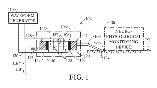
FIG. 1 is a diagram of an ultrasonic surgical system in accordance with the
present invention, showing a blue monopolar cable connecting an ultrasonic
surgical
instrument or device to ground..
FIG. 2 is a partial view of a rear panel of a power console for the ultrasonic
surgical system of FIG. 1, showing an end of the blue monopolar cable of FIG.1
connected to a grounding terminal on the rear panel.
FIG. 3 is an isometric front and side view of an ultrasonic aspirator
instrument
in accordance with the invention, with a middle portion of a housing removed
to show
operative components.
FIG. 4 is a detail view of the instrument of Fig. 3, on a larger scale.
FIG. 5 is a detail view, similar to FIG. 4 but from another side of the
instrument.
FIG. 6 is partially an isometric view and partially a longitudinal or axis
cross-
sectional view of the instrument of FIGS. 3-5.
FIG. 7 is an isometric rear and side view of the instrument of FIGS. 3-6.
CA 03032805 2019-02-01
WO 2018/026712
PCT/US2017/044676
- 4 -
DETAILED DESCRIPTION
As depicted in FIG.1, an ultrasonic surgical system comprises an ultrasonic
surgical device 102 comprising a hand piece 104, a probe 106, and an
electromechanical transducer assembly 108. The transducer assembly 108 is
disposed
inside the hand piece 104 and is configured for converting electrical waveform
energy
of an ultrasonic frequency from a waveform generator 110 into ultrasonic
vibratory
energy.
Probe 106 is mounted to a distal end of the hand piece 104 and is operatively
connected to the transducer assembly 108. An electrical connector or spud,
schematically indicated at 112, is provided at a rear or proximal end of the
hand piece
104. An electrical circuit 114, including selected parts of the transducer
assembly 108,
electrically or operatively connects the probe 106 to the electrical connector
112. A
wire or cable 116 is operatively coupled at one end to the electrical
connector 112 and
at an opposite end to electrical ground 118.
As described in U.S. Patent No. 5,371,429, the disclosure of which is hereby
incorporated by reference, transducer assembly 108 includes a front driver
120, a
stack of piezoelectric disks 122, a rear driver 124, and a bolt 126 connecting
the rear
driver to the front driver. Electrical circuit 114 includes front driver 120,
bolt 126 and
rear driver 124. Generally, various components of device 102 may be included
in or
connected to the grounding circuit 114 except for the circuit elements that
energize
piezoelectric disks 122. Those circuit elements include leads 128, 130 and
electrodes
132, some of which are located between adjacent pieozoelectric disks 122, and
two of
which are located between respective piezoelectric disks 122 and insulator
disks 138
and 140 respectively.
Insulator disks 138 and 140 serve to electrically isolate the stack of
piezoelectric disks 122 from the probe 106, rendering the stack a floating
output.
A method for using the above-described ultrasonic surgical device comprises
providing the wire or cable 116, operatively coupling one end of the wire or
cable to
the electrical connector 112 and an opposite end to electrical ground 118, and
placing
an operating tip 134 of the probe 106 in contact with organic tissue OT of a
patient PT.
While the operating tip 134 is in contact with the organic tissue OT, one
conducts an
alternating voltage to the transducer assembly 108 from waveform generator 110
to
induce the transducer assembly to generate a mechanical standing wave of
ultrasonic
frequency in the probe 106. Simultaneously therewith, one conducts leakage
current
CA 03032805 2019-02-01
WO 2018/026712
PCT/US2017/044676
- 5 -
away from the probe 106 to ground 118 via the electrical circuit 114, the
connector
112 and the wire or cable 116.
The method further contemplates providing an intraoperative
neurophysiological monitoring device 136, operatively connecting the
intraoperative
neurophysiological monitoring device to the patient PT proximate a point of
contact
of the operating tip 134 of the probe 106 with the organic tissue OT of the
patient PT,
and operating the intraoperative neurophysiological monitoring device 136 to
detect
activation or stimulation of the nervous system of the patient.
Transducer assembly 108 includes the two insulating disks 138 and 140 which
are provided between the stack of piezoelectric or piezo ceramic disks 122 and
the
front driver 120 and the rear drive 124, respectively. Insulating disks 138
and 140
serve to isolate the metal probe 106 from the disks 122 and the energization
or
voltage-application circuit elements 128, 130, 132. The present invention
reduces or
eliminates leakage currents that may nevertheless enter the patient through
the probe
106 from the transducer assembly 108.
Metal connector or spud 112 in the rear of the handpiece 104 is provided in
prior art instruments to enable coupling of an RF cautery device so that probe
106
becomes a carrier for RF current. Wire or cable 116 may take the form of a
blue
monopolar cautery cable (part No. CFSM6-C130). Cable 116 is typically
connected
to ground 118 via a screw terminal 142 (FIG. 2) on a rear panel 144 of a power
console or cabinet 146. In that case, an adapter or shunt member 148 is
provided.
Adapter or shunt member 148 is a pin insertable at one end into cable 116 and
provided at an opposite end with an eyelet or loop 150 for insertion around
the
grounding screw terminal 142.
FIGS. 3-7 depict a particular embodiment of device 102 that is an ultrasonic
aspirator particularly useful in neurological operations. The Sonastar FLV-
12801 of
Misonix Incorporated is such an aspirator. Parts illustrated in FIGS. 3-7 and
discussed above bear the same reference numerals as above. Handpiece 104
includes
a housing 152 with an end cap 154 that exhibits an electrical socket 156
through
which connections are made to leads 128 and 130 (FIGS. 1, 4, and 5). End cap
154
also has an outlet port or coupling 158 for the aspiration of disrupted
tissue. A distal
end of the instrument includes a sheath 160 having a port or connector 162 for
attachment of an irrigation tube (not shown).
CA 03032805 2019-02-01
WO 2018/026712
PCT/US2017/044676
-6 -
In an alternative embodiment (not illustrated), the connection to ground is
established through the hand piece cable (connected at 156) via a conductor
that is
electrically connected to the applied circuit. In that case, electrical socket
156 forms
the connector that enables grounding of the probe 106, as well as the
generation of the
.. ultrasonic standing wave in the probe. The wire or cable 116 is then
connected to the
instrument via electrical socket 156.
FIG. 6 shows end cap 154 slightly removed in a proximal direction. End cap
154 is provided internally with a metal liner 164 that is mechanically and
electrically
connected to an extension of bolt 126. Liner 164 fits over and may engage rear
driver 124. Liner 164 has a rearwardly extending metal finger 166 that serves
as
connector or spud 112 (see also FIG. 7) at its outer end.
Although the invention has been described in terms of particular embodiments
and applications, one of ordinary skill in the art, in light of this teaching,
can generate
additional embodiments and modifications without departing from the spirit of
or
exceeding the scope of the claimed invention. Accordingly, it is to be
understood
that the drawings and descriptions herein are proffered by way of example to
facilitate
comprehension of the invention and should not be construed to limit the scope
thereof.
