Note: Descriptions are shown in the official language in which they were submitted.
CA 03032997 2019-02-04
WO 2018/013718
PCT/US2017/041762
COMPOSITIONS AND METHODS OF CELL ATTACHMENT
Field of the Invention
The present invention contemplates compositions, devices and methods of
improving
adhesion, attachment, and/or differentiation of cells in a microfluidic device
or chip. In one
embodiment, ECM protein(s) is covalently coupled to the surface of a
microchannel of a
microfluidic device. The microfluidic devices can be either stored, or
immediately used for
culture and/or support of living cells such as mammalian cells, and/or for
simulating a function
of a tissue, e.g., a liver tissue, muscle tissue, etc. Extended adhesion and
viability with sustained
function over time is observed.
Background
Cell adhesion is a central mechanism that ensures the structural integrity of
tissue and is
often a requirement for its biological function. The most prominent cell-
matrix adhesion
.. structures are so-called focal contacts. Focal contacts consist of large
patches of transmembrane
adhesion receptors from the integrin-family. These integrin patches in the
cell membrane can
reach lateral sizes of several micrometers. On the extracellular side,
integrin binds to ligands
such as the ECM proteins collagen, fibronectin and vitronectin. On the
intracellular side, the
receptors are linked to the actin cytoskeleton via a cytoplasmic plaque
composed of many
different proteins, including talin, vinculin, paxillin, and a-actinin. See
Zamir and Geiger,
"Molecular complexity and dynamics of cell-matrix adhesions," J. Cell Science
114: 3583
(2001). This connection to the cytoskeleton, which is often organized in the
form of stress
fibers, allows transmitting forces between cells and the ECM through focal
contacts. Adhesions
between cells and the extracellular matrix (ECM) are known to modulate
numerous cellular
events.
Cell adhesion is also important in cell culture. Researchers have attempted to
optimize
the culture conditions of cells by extracellular matrix (ECM) coating of the
culture dish, culture
well, or culture channel. However, these ECMs are often applied generically
with mixed results,
depending on the cell type and culture conditions. What is needed is a more
specific use of
ECMs in the context of specific cell types and culture conditions.
1
CA 03032997 2019-02-04
WO 2018/013718
PCT/US2017/041762
Summary of the Invention
The present invention contemplates compositions, devices and methods of
improving
adhesion, attachment, differentiation, longevity, quiescence, or biological
function of cells in a
microfluidic device or chip. In one embodiment, one or more proteins (e.g. ECM
proteins) or
peptides (e.g. RGD) are covalently coupled to the surface of a microfluidic
device, whether
within a microchannel or an open structure. The microfluidic devices can
stored and used later,
or they can be immediately used for culture and/or support of living cells
such as mammalian
cells, and/or for simulating a function of a tissue, e.g., a liver tissue,
muscle tissue, etc., or
simulating a function of an organ, e.g., a Liver-Chip, a Lung-Chip, etc.
Extended adhesion and
viability with sustained function over time is observed. In one embodiment,
the microchannel
comprises a surface comprising a silicone polymer. In one embodiment, the
silicone polymer is
polydimethylsiloxane or "PDMS." In one embodiment, the ECM protein is
covalently coupled
to a PDMS surface using a crosslinker, such as the heterobifunctional linker N-
sulphosuccinimidy1-6-(4'-azido-2'-nitrophenylamino) hexanoate (Sulfo-SANPAH).
In one
embodiment, the living cells are exposed to fluid flow, the fluid flow
providing shear stress.
In one embodiment, the present invention contemplates a method of culturing
cells,
comprising: a) providing a microfluidic device comprising a microchannel
comprising a surface,
said microchannel in fluidic communication with a fluid source comprising
fluid; b) covalently
attaching one or more proteins or peptides to said microchannel surface so as
to create a treated
.. surface; c) seeding viable cells on said treated surface so as to create
attached cells; c) flowing
fluid from said fluid source through said microchannel so as to create flowing
conditions; and d)
culturing said attached cells under said flow conditions such that said cells
remain attached and
viable (e.g. viable for at least 14 days). It is not intended that the present
invention be limited to
any particular cell type; a variety of cell types are contemplates (including
more than one cell
.. type). In one embodiment, said cells are hepatocytes. It is not intended
that the present
invention be limited to any particular protein or peptide; a variety are
contemplated, including
mixtures. For example, in one embodiment, the covalently attached protein is
collagen. In
another embodiment, a mixture of proteins are covalently attached, e,g. a
mixture of collagen
type I, fibronectin and collagen type IV. In yet another embodiment, the RGD
peptide is attached
(or a peptide comprising the RGD motif is attached). In one embodiment, the
microchannel
further comprises a membrane. In one embodiment, the membrane comprises PDMS.
In one
2
CA 03032997 2019-02-04
WO 2018/013718
PCT/US2017/041762
embodiment, the membrane comprises a crosslinker (e.g. covalently bound to the
membrane). In
one embodiment, the crosslinker is a bifunctional crosslinker. In one
embodiment, the
crosslinker is Sulfo-SANPAH (which is light activated with UV irradiation). In
one
embodiment, an extracellular matrix protein (e.g. laminin) is attached to the
crosslinker (e.g.
covalently bound) so as to provide an ECM-coated membrane. In one embodiment,
the
irradiated membrane is washed before the ECM attachment step. In one
embodiment, the viable
cells are further seeded onto the ECM-coated membrane. In one embodiment, the
viable cells
are neurons. In one embodiment, the viable cells are motor neurons. In one
embodiment, the
viable cells are hepatocyte. In one embodiment, the viable cells are muscle
cells. In one
embodiment, the viable cells are skeletal muscle cells. In one embodiment, the
skeletal muscle
cells are human.
In one embodiment, the microchannel further comprises a micropatterned
membrane. In
one embodiment, the micropatterned membrane comprises PDMS. In one embodiment,
a
bifunctional crosslinker is attached to the micropatterned membrane. In one
embodiment, the
micropatterned membrane is in the flow channel of a microfluidic device. In
one embodiment,
the micropattern is parallel to the fluid flow. In one embodiment, the
micropattern is
perpendicular to the fluid flow. In one embodiment, an extracellular matrix
protein (e.g. laminin)
is attached to the crosslinker (e.g. covalently bound) so as to provide an ECM-
coated
micropatterned membrane. In one embodiment, the viable cells are further
seeded onto the
ECM-coated micropatterned membrane. In one embodiment, the viable cells are
neurons. In
one embodiment, the viable cells are motor neurons. In one embodiment, the
viable cells are
hepatocyte. In one embodiment, the viable cells are muscle cells. In one
embodiment, the viable
cells are skeletal muscle cell. In one embodiment, the skeletal muscle cells
are human
("hSKMCs"). In one embodiment, the skeletal muscle cells elongate in the
grooves of the
micropatterned membrane.
In one embodiment, the crosslinker is only attached to a portion of the
membrane or the
micropatterned membrane. In one embodiment, the portion where the crosslinker
is not attached
is covered with a mask (e.g. the crosslinker is light activated and the mask
blocks the light) and
the portion where the crosslinker is attached is unmasked. The mask may be
adhesive material
(e.g. tape) or non-adhesive material (e.g. metal or metal foil such as
aluminum foil). In one
embodiment, an extracellular matrix protein (e.g. laminin) is attached to the
crosslinker (e.g.
3
CA 03032997 2019-02-04
WO 2018/013718
PCT/US2017/041762
covalently bound) in the unmasked portion so as to provide an ECM-coated
membrane or
micropatterned membrane. In one embodiment, the viable cells are further
seeded onto the
ECM-coated portion of the membrane or micropatterned membrane. In one
embodiment, the
viable cells are neurons. In one embodiment, the viable cells are motor
neurons. In one
embodiment, the viable cells are hepatocyte. In one embodiment, the viable
cells are muscle
cells. In one embodiment, the viable cells are skeletal muscle cell. In one
embodiment, the
skeletal muscle cells are human.
It is not intended that the present invention be limited to the method by
which
micropatterns are introduced into the membrane. In one embodiment, the
micropatterns are
introduced into the membrane through the use of an existing micropatterned
silicon wafer mold;
PDMS material can be spin coated on the mold and cured (the membrane is
thereafter removed
from the mold and used to assemble a microfluidic device or chip). In one
embodiment, the
micropattern is embossed using an existing micropatterned silicon wafer using
both heat and
pressure.
The present invention contemplates that in certain embodiments, the surface
can be
treated prior to step b). A variety of surface treatments (e.g. chemical vapor
deposition, plasma
oxidation, Corona, RF plasma, etc.) are possible. For example, in one
embodiment, the present
invention contemplates wherein said microchannel surface is PDMS and wherein
said PDMS is
plasma treated prior to step b).
It is not intended that the present invention be limited by the manner in
which the
proteins or peptides are covalently attached. In one embodiment, a crosslinker
is used. In
another embodiment, a bifunctional crosslinker is used. In a preferred
embodiment, the protein or
peptide is covalently attached to said microchannel surface using N-
sulphosuccinimidy1-6-(4'-
azido-2'-nitrophenylamino) hexanoate.
The present invention contemplates that the cells will be remain viable and
can be
tested to confirm this. However, it is not intended that the present invention
be limited to a
particular viability test. In one embodiment, the method further comprises e)
assessing viability
by measuring the level of activity of one or more cellular enzymes. A variety
of enzymes can be
used for this purpose, including but not limited to, a CYP enzyme, a
transaminase and the like.
In another embodiment, the present invention contemplates that the method
further comprises e)
assessing viability by measuring the level of expression of one or more
cellular proteins.
4
CA 03032997 2019-02-04
WO 2018/013718
PCT/US2017/041762
In yet another embodiment, the present invention contemplates a method of
culturing
specific cells, comprising: a) providing a microfluidic device comprising a
microchannel
comprising a surface, said microchannel in fluidic communication with a fluid
source comprising
fluid; b) covalently attaching one or more proteins or peptides to said
microchannel surface so as
to create a treated surface; c) seeding viable hepatocytes on said treated
surface so as to create
attached cells; c) flowing fluid from said fluid source through said
microchannel so as to create
flowing conditions; and d) culturing said attached cells under said flow
conditions such that said
cells remain attached and viable (e.g. viable for at least 14 days). It is not
intended that the
present invention be limited by the nature or species of hepatocytes. In one
embodiment, said
hepatocytes are dog hepatocytes. It is not intended that the present invention
be limited to any
particular protein or peptide; a variety are contemplated, including mixtures.
For example, in
one embodiment, the covalently attached protein is collagen. In another
embodiment, a mixture
of proteins are covalently attached, e,g. a mixture of collagen type I,
fibronectin and collagen
type IV. In yet another embodiment, the RGD peptide is attached (or a peptide
comprising the
RGD motif is attached). In one embodiment, the microchannel further comprises
a membrane.
In one embodiment, the membrane comprises PDMS. In one embodiment, the
membrane
comprises a crosslinker (e.g. covalently bound to the membrane). In one
embodiment, the
crosslinker is a bifunctional crosslinker. In one embodiment, the crosslinker
is Sulfo-SANPAH
(which is light activated with UV irradiation). In one embodiment, an
extracellular matrix
protein (e.g. laminin) is attached to the crosslinker (e.g. covalently bound)
so as to provide an
ECM-coated membrane. In one embodiment, the irradiated membrane is washed
before the
ECM attachment step. In one embodiment, the viable cells are further seeded
onto the ECM-
coated membrane. In one embodiment, the viable cells are neurons. In one
embodiment, the
viable cells are motor neurons. In one embodiment, the viable cells are
hepatocyte. In one
.. embodiment, the viable cells are muscle cells. In one embodiment, the
viable cells are skeletal
muscle cell. In one embodiment, the skeletal muscle cells are human.
In one embodiment, the microchannel further comprises a micropatterned
membrane. In
one embodiment, the micropatterned membrane comprises PDMS. In one embodiment,
a
bifunctional crosslinker is attached to the micropatterned membrane. In one
embodiment, the
micropatterned membrane is in the flow channel of a microfluidic device. In
one embodiment,
the micropattern is parallel to the fluid flow. In one embodiment, the
micropattern is
5
CA 03032997 2019-02-04
WO 2018/013718
PCT/US2017/041762
perpendicular to the fluid flow. In one embodiment, an extracellular matrix
protein (e.g. laminin)
is attached to the crosslinker (e.g. covalently bound) so as to provide an ECM-
coated
micropatterned membrane. In one embodiment, the viable cells are further
seeded onto the
ECM-coated micropatterned membrane. In one embodiment, the viable cells are
neurons. In
one embodiment, the viable cells are motor neurons. In one embodiment, the
viable cells are
hepatocyte. In one embodiment, the viable cells are muscle cells. In one
embodiment, the viable
cells are skeletal muscle cell. In one embodiment, the skeletal muscle cells
are human. In one
embodiment, the skeletal muscle cells elongate in the grooves of the
micropatterned membrane.
In one embodiment, the crosslinker is only attached to a portion of the
membrane or the
micropatterned membrane. In one embodiment, the portion where the crosslinker
is not attached
is covered with a mask (e.g. the crosslinker is light activated and the mask
blocks the light) and
the portion where the crosslinker is attached is unmasked. The mask may be
adhesive material
or non-adhesive material (e.g. metal or metal foil such as aluminum foil). In
one embodiment,
an extracellular matrix protein (e.g. laminin) is attached to the crosslinker
(e.g. covalently bound)
in the unmasked portion so as to provide an ECM-coated membrane or
micropatterned
membrane. In one embodiment, the viable cells are further seeded onto the ECM-
coated portion
of the membrane or micropatterned membrane. In one embodiment, the viable
cells are neurons.
In one embodiment, the viable cells are motor neurons. In one embodiment, the
viable cells are
hepatocyte. In one embodiment, the viable cells are muscle cells. In one
embodiment, the viable
cells are skeletal muscle cell. In one embodiment, the skeletal muscle cells
are human.
It is not intended that the present invention be limited to the method by
which
micropatterns are introduced into the membrane. In one embodiment, the
micropatterns are
introduced into the membrane through the use of an existing micropatterned
silicon wafer mold;
PDMS material can be spin coated on the mold and cured (the membrane is
thereafter removed
from the mold and used to assemble a microfluidic device or chip). In one
embodiment, the
micropattern is embossed using an existing micropatterned silicon wafer using
both heat and
pressure.
It is not intended that the present invention be limited by the manner in
which the
proteins or peptides are covalently attached. In one embodiment, a crosslinker
is used. In
another embodiment, a bifunctional crosslinker is used. In a preferred
embodiment, the protein or
6
CA 03032997 2019-02-04
WO 2018/013718
PCT/US2017/041762
peptide is covalently attached to said microchannel surface using N-
sulphosuccinimidy1-6-(4'-
azido-2'-nitrophenylamino) hexanoate.
The present invention contemplates that the cells will be remain viable and
can be
tested to confirm this. However, it is not intended that the present invention
be limited to a
particular viability test. In one embodiment, the method further comprises e)
assessing viability
by measuring the level of activity of one or more cellular enzymes. A variety
of enzymes can be
used for this purpose, including but not limited to, a CYP enzyme, a
transaminase and the like.
In another embodiment, the present invention contemplates that the method
further comprises e)
assessing viability by measuring the level of expression of one or more
cellular proteins.
The present invention contemplates that in certain embodiments, the surface
can be
treated prior to step b). A variety of surface treatments (e.g. chemical vapor
deposition, plasma
oxidation, Corona, RF plasma, etc.) are possible. For example, in one
embodiment, the present
invention contemplates wherein said microchannel surface is PDMS and wherein
said PDMS is
plasma treated prior to step b).
As noted above, cells need not be immediately cultured in the device, i.e. the
device can
be stored with the covalently attached protein(s). In one embodiment, the
present invention
contemplates a method of treating a microfluidic device, comprising: a)
providing a microfluidic
device comprising a microchannel comprising a surface, said microchannel in
fluidic
communication with a fluid source comprising fluid; b) covalently attaching
one or more
proteins or peptides to said microchannel surface so as to create a treated
surface; and c) storing
said microfluidic device. It is not intended that the present invention be
limited to the precise
storage conditions. However, the storing is typically done at a controlled
temperature below
room temperature, e.g. between 2 and 10 C, in a refrigerator or other cooling
device. It is not
intended that the present invention be limited to dry or wet storage. In one
embodiment, said one
or more covalently attached proteins is collagen I and it is stored wet or dry
(more preferred). In
one embodiment, said one or more covalently attached proteins is laminin and
it is stored wet
(preferred) or dry. Laminin and/or Matrigel are contemplated for a variety of
chips, including
Intestine-on-Chip, Blood Brain Barrier (BBB)-on-Chip, and NeuroMuscular
Junction (NMJ)-on-
Chip. In one embodiment, the microchannel further comprises a membrane. In one
embodiment, the membrane comprises PDMS. In one embodiment, the membrane
comprises a
crosslinker (e.g. covalently bound to the membrane). In one embodiment, the
crosslinker is a
7
CA 03032997 2019-02-04
WO 2018/013718
PCT/US2017/041762
bifunctional crosslinker. In one embodiment, the crosslinker is Sulfo-SANPAH
(which is light
activated with UV irradiation). In one embodiment, an extracellular matrix
protein (e.g. laminin)
is attached to the crosslinker (e.g. covalently bound) so as to provide an ECM-
coated membrane.
In one embodiment, the irradiated membrane is washed before the ECM attachment
step. In one
embodiment, the viable cells are further seeded onto the ECM-coated membrane.
In one
embodiment, the viable cells are neurons. In one embodiment, the viable cells
are motor
neurons. In one embodiment, the viable cells are hepatocyte. In one
embodiment, the viable
cells are skeletal muscle cell. In one embodiment, the skeletal muscle cells
are human.
In one embodiment, the microchannel further comprises a micropatterned
membrane. In
one embodiment, the micropatterned membrane comprises PDMS. In one embodiment,
a
bifunctional crosslinker is attached to the micropatterned membrane. In one
embodiment, the
micropatterned membrane is in the flow channel of a microfluidic device. In
one embodiment,
the micropattern is parallel to the fluid flow. In one embodiment, the
micropattern is
perpendicular to the fluid flow. In one embodiment, an extracellular matrix
protein (e.g. laminin)
is attached to the crosslinker (e.g. covalently bound) so as to provide an ECM-
coated
micropatterned membrane. In one embodiment, the viable cells are further
seeded onto the
ECM-coated micropatterned membrane. In one embodiment, the viable cells are
neurons. In
one embodiment, the viable cells are motor neurons. In one embodiment, the
viable cells are
hepatocyte. In one embodiment, the viable cells are muscle cells. In one
embodiment, the viable
cells are skeletal muscle cell. In one embodiment, the skeletal muscle cells
are human. In one
embodiment, the skeletal muscle cells elongate in the grooves of the
micropatterned membrane.
In one embodiment, the crosslinker is only attached to a portion of the
membrane or the
micropatterned membrane. In one embodiment, the portion where the crosslinker
is not attached
is covered with a mask (e.g. the crosslinker is light activated and the mask
blocks the light) and
the portion where the crosslinker is attached is unmasked. The mask may be
adhesive material
or non-adhesive material (e.g. metal or metal foil such as aluminum foil). In
one embodiment,
an extracellular matrix protein (e.g. laminin) is attached to the crosslinker
(e.g. covalently bound)
in the unmasked portion so as to provide an ECM-coated membrane or
micropatterned
membrane. In one embodiment, the viable cells are further seeded onto the ECM-
coated portion
of the membrane or micropatterned membrane. In one embodiment, the viable
cells are neurons.
In one embodiment, the viable cells are motor neurons. In one embodiment, the
viable cells are
8
CA 03032997 2019-02-04
WO 2018/013718
PCT/US2017/041762
hepatocyte. In one embodiment, the viable cells are muscle cells. In one
embodiment, the viable
cells are skeletal muscle cell. In one embodiment, the skeletal muscle cells
are human.
It is not intended that the present invention be limited to the method by
which
micropatterns are introduced into the membrane. In one embodiment, the
micropatterns are
introduced into the membrane through the use of an existing micropatterned
silicon wafer mold;
PDMS material can be spin coated on the mold and cured (the membrane is
thereafter removed
from the mold and used to assemble a microfluidic device or chip). In one
embodiment, the
micropattern is embossed using an existing micropatterned silicon wafer using
both heat and
pressure.
The storage may be for a time period of 1, 2, 3, 4, 5, 6, 7, 8, 9, 10, 11, 12,
13, 14 or more
days ¨ and even may be done for 3, 4 or 5 weeks. After storage, it is
contemplated that cells will
be added. For example, in one embodiment, the present invention contemplates
that the method
further comprises: d) seeding viable cells on said treated surface so as to
create attached cells; e)
flowing fluid from said fluid source through said microchannel so as to create
flowing
conditions; and f) culturing said attached cells under said flow conditions
such that said cells
remain attached and viable (e.g. viable for at least 14 days).
Again, any type of cell (or combination of cells) can be seeded. Cells may be
of any cell
type from a multicellular structure, including nematodes, amoebas, up to
mammals such as
humans. The cell types seeded on the device may simply depend on the type of
organ (lung,
liver, intestine, brain, kidney etc.) or organ function one wishes to mimic,
and the tissues that
comprise those organs. One can also co-culture various stem cells, such as
bone marrow cells,
induced adult stem cells, embryonal stem cells or stem cells isolated from
adult tissues. In one
embodiment said cells are hepatocytes. In one embodiment, said cells are Human
Umbilical
Vein Endothelial Cells (HUVECs). In one embodiment, said cells are intestinal
cells. In one
embodiment, said cells are from patients with a disease, i.e. diseased cells.
In one embodiment,
said cells are from healthy controls.
The present invention contemplates that in certain embodiments, the surface
can be
treated prior to step b). A variety of surface treatments (e.g. chemical vapor
deposition, plasma
oxidation, Corona, RF plasma, etc.) are possible. For example, in one
embodiment, the present
invention contemplates wherein said microchannel surface is PDMS and wherein
said PDMS is
plasma treated prior to step b).
9
CA 03032997 2019-02-04
WO 2018/013718
PCT/US2017/041762
A noted above, the cells may be tested for viability. On the other hand, the
cells may be
put to other tests, such as tests directed at basic biological science, life
science research, drug
discovery and development, drug safety testing, chemical and biological
assays, as well as tissue
and organ engineering. In an embodiment, the organ mimic device can be used as
microvascular
network structures for basic research in cardiovascular, cancer, and organ-
specific disease
biology. Furthermore, one or more embodiments of the device find application
in organ assist
devices for liver, kidney, lung, intestine, bone marrow, and other organs and
tissues, as well as in
organ replacement structures.
It is not intended that the devices be limited in their use. In one
embodiment, there are
used for: the identification of markers of disease; assessing efficacy of anti-
cancer therapeutics;
testing gene therapy vectors; drug development; screening; studies of cells,
especially stem cells
and bone marrow cells; studies on biotransformation, absorption, clearance,
metabolism, and
activation of xenobiotics; studies on bioavailability and transport of
chemical or biological
agents across epithelial or endothelial layers; studies on transport of
biological or chemical
agents across the blood-brain barrier; studies on transport of biological or
chemical agents across
the intestinal epithelial barrier; studies on acute basal toxicity of chemical
agents; studies on
acute local or acute organ-specific toxicity of chemical agents; studies on
chronic basal toxicity
of chemical agents; studies on chronic local or chronic organ-specific
toxicity of chemical
agents; studies on teratogenicity of chemical agents; studies on genotoxicity,
carcinogenicity,
and mutagenicity of chemical agents; detection of infectious biological agents
and biological
weapons; detection of harmful chemical agents and chemical weapons; studies on
infectious
diseases; studies on the efficacy of chemical or biological agents to treat
disease; studies on the
optimal dose range of agents to treat disease; prediction of the response of
organs in vivo to
biological agents; prediction of the pharmacokinetics of chemical or
biological agents; prediction
of the pharmacodynamics of chemical or biological agents; studies concerning
the impact of
genetic content on response to agents; studies on gene transcription in
response to chemical or
biological agents; studies on protein expression in response to chemical or
biological agents;
studies on changes in metabolism in response to chemical or biological agents.
The organ mimic
device can also be used to screen on the cells, for an effect of the cells on
the materials (for
example, in a manner equivalent to tissue metabolism of a drug).
CA 03032997 2019-02-04
WO 2018/013718
PCT/US2017/041762
In one embodiment, the present invention contemplates a method of culturing
cells,
comprising: a) providing a microfluidic device comprising a microchannel
comprising a surface,
said microchannel in fluidic communication with a fluid source comprising
fluid; b) covalently
attaching a bifunctional crosslinker to said surface to create attached
crosslinker, c) covalently
attaching one or more proteins or peptides to said attached crosslinker as to
create a treated
surface; d) seeding viable cells on said treated surface so as to create
attached cells; e) flowing
fluid from said fluid source through said microchannel so as to create flowing
conditions; and f)
culturing said attached cells under said flow conditions such that said cells
remain attached and
viable for at least 7 days. In one embodiment, said surface is a membrane and
said membrane is
micropatterned. In one embodiment, said cells are muscle cells that align with
said micropattern
(of the micropatterned membrane). In one embodiment, said crosslinker is
activated with UV
light in the presence of a mask (so that light is blocked from contacting a
portion of said surface).
In one embodiment, the present invention contemplates a method of culturing
cells,
comprising: a) providing a microfluidic device comprising a surface; b)
covalently attaching one
or more proteins or peptides to said surface using a crosslinker so as to
create a treated surface;
c) seeding viable cells on said treated surface so as to create attached
cells; and d) culturing said
attached cells such that said cells remain attached and viable for at least 7
days. In one
embodiment, the microfluidic device further comprises a microchannel, said
surface disposed
within said microchannel, and wherein said microchannel is in fluidic
communication with a
fluidic source comprising fluid, the method further comprising the step of
flowing fluid from
said fluid source through said microchannel so as to create flow conditions,
and wherein said
culturing in d) further comprises culturing said attached cells under said
flow conditions. In one
embodiment, the attached cells further remain viable for at least 14 days. In
one embodiment,
the attached cells further remain functional for at least 7 days. In one
embodiment, the attached
cells further remain functional for at least 14 days. In one embodiment, the
crosslinker
comprises at least one light-reactive portion and at least one chemically
reactive portion. In one
embodiment, the crosslinker further comprises at least one spacer portion. In
one embodiment,
the at least one light-reactive portion is selected from the group consisting
of a nitrophenyl, a
diazirine and an azide. In one embodiment, the at least one chemically
reactive portion is
selected from the group consisting of an NHS-ester, a sulfo-NHS-ester,
isocyanate,
isothiocyanate, imidoester, maleimide, pyridyldithiol, and hydrazide. In one
embodiment, the
11
CA 03032997 2019-02-04
WO 2018/013718
PCT/US2017/041762
crosslinker is selected from the group consisting of sulfo-SANPAH, SANPAH,
SDA, sulfo-
SDA, LC-SDA, sulfo-LC-SDA, ANB-NOS, SDAD, sulfo-SDAD. In one embodiment, the
surface comprises PDMS. In one embodiment, the surface is plasma treated prior
to step b). In
one embodiment, the cells are hepatocytes. In one embodiment, the method
further comprises
step e) assessing viability by measuring the level of activity of one or more
cellular enzymes. In
one embodiment, the cellular enzyme is a CYP enzyme. In one embodiment, the
cellular
enzyme is a transaminase. In one embodiment, the method further comprises step
e) assessing
viability by measuring the level of expression of one or more cellular
proteins. In one
embodiment, the one or more proteins comprises collagen. In one embodiment,
the one or more
proteins comprises a mixture of collagen type I, fibronectin and collagen type
IV. In one
embodiment, the one or more peptides comprises ROD or a peptide comprising the
ROD motif.
In one embodiment, wherein RGD is covalently attached to said surface using N-
sulphosuccinimidy1-6-(4'-azido-2'-nitrophenylamino) hexanoate. In one
embodiment, the
covalently attaching one or more proteins or peptides in step b) further
comprises: i) introducing
said crosslinker or a solution containing said crosslinker to contact said
surface and permitting
said crosslinker or said solution containing said crosslinker to react with
said surface; and ii)
introducing at least one protein or peptide, or a solution containing at least
one protein or peptide
to contact said surface. In one embodiment, the covalently attaching one or
more proteins or
peptides in b) further comprises exposing at least a portion of said surface
to light. In one
embodiment, the light comprises UV light. In one embodiment, the exposing
comprises
exposing a selected area or pattern for the covalent attachment of at least a
portion of said one or
more proteins or peptides. In one embodiment, the exposing comprises masking
said light so as
to select said selected area or pattern. In one embodiment, the exposing
comprises projecting a
light pattern so as to select said selected area or pattern. In one
embodiment, the exposing
comprises rastering light so as to select said selected area or pattern. In
one embodiment, the
selected area or pattern comprises a linear pattern. In one embodiment, the
cells comprise
muscle cells or muscle-like cells that align with respect to said selected
area of pattern. In one
embodiment, the covalently attaching one or more proteins or peptides in b)
further comprises
introducing said crosslinker to contact only one or more selected areas of
said microfluidic
device. In one embodiment, the microfluidic device further comprises a porous
membrane. In
one embodiment, the porous membrane comprises said surface.
12
CA 03032997 2019-02-04
WO 2018/013718
PCT/US2017/041762
In one embodiment, the present invention contemplates a method of treating a
microfluidic device, comprising: a) providing a microfluidic device comprising
a surface; b)
covalently attaching one or more proteins or peptides to said surface using a
crosslinker so as to
create a treated surface; and c) storing said microfluidic device. In one
embodiment, the
microfluidic device comprises a microchannel, said surface disposed within
said microchannel.
In one embodiment, the storing in step c) comprises storing said surface dry.
In one
embodiment, the storing in step c) comprises storing said surface wet. In one
embodiment, the
crosslinker comprises at least one light-reactive portion, at least one
chemically reactive portion.
In one embodiment, the crosslinker further comprises at least one spacer
portion. In one
embodiment, the at least one light-reactive portion is selected from the group
consisting of a
nitrophenyl, a diazirine and an azides. In one embodiment, the at least one
chemically reactive
portion is selected from the group consising of NHS-ester, sulfo-NHS-ester,
isocyanate,
isothiocyanate, imidoester, maleimide, pyridyldithiol, and hydrazide. In one
embodiment, the
crosslinker is selected from the list comprising: sulfo-SANPAH, SANPAH, SDA,
sulfo-SDA,
LC-SDA, sulfo-LC-SDA, ANB-NOS, SDAD, sulfo-SDAD. In one embodiment, the
storing is
done at a controlled temperature below room temperature. In one embodiment,
the storing is
done at between 2 and 10 C. In one embodiment, the one or more covalently
attached proteins is
collagen I. In one embodiment, the covalently attached collagen I is stored
dry. In one
embodiment, the one or more covalently attached proteins is laminin. In one
embodiment, the
covalently attached laminin is stored wet. In one embodiment, the method
further comprises step
d) seeding viable cells on said treated surface so as to create attached
cells; and f) culturing said
attached cells such that said cells remain attached and viable for at least 7
days. In one
embodiment, the microfluidic device further comprises a microchannel, said
surface disposed
within said microchannel, and wherein said microchannel is in fluidic
communication with a
fluidic source comprising fluid, the method further comprising flowing fluid
from said fluid
source through said microchannel so as to create flow conditions, and wherein
culturing in f)
further comprises culturing said attached cells under said flow conditions. In
one embodiment,
the attached cells further remain viable for at least 14 days. In one
embodiment, the attached
cells further remain functional for at least 7 days. In one embodiment, the
attached cells further
remain functional for at least 14 days. In one embodiment, the attached cells
are hepatocytes. In
one embodiment, the surface comprises PDMS. In one embodiment, the surface is
plasma
13
CA 03032997 2019-02-04
WO 2018/013718
PCT/US2017/041762
treated prior to step b). In one embodiment, the covalently attaching one or
more proteins or
peptides in b) further comprises: i) introducing said crosslinker or a
solution containing said
crosslinker to contact said surface and permitting said crosslinker or said
solution containing said
crosslinker to react with said surface; and ii) introducing at least one
protein or peptide, or a
solution containing at least one protein or peptide to contact said surface.
In one embodiment,
the covalently attaching one or more proteins or peptides in b) further
comprises exposing at
least a portion of said surface to light. In one embodiment, the light
comprises UV light. In one
embodiment, the exposing comprises exposing a selected area or pattern for the
covalent
attachment of at least a portion of said one or more proteins or peptides. In
one embodiment, the
exposing comprises masking said light so as to select said selected area or
pattern. In one
embodiment, the exposing comprises projecting a light pattern so as to select
said selected area
or pattern. In one embodiment, the exposing comprises rastering light so as to
select said
selected area or pattern. In one embodiment, the selected area or pattern
comprises a linear
pattern. In one embodiment, the cells comprise muscle cells or muscle-like
cells that align with
respect to said selected area or pattern. In one embodiment, the covalently
attaching one or more
proteins or peptides in b) further comprises introducing said crosslinker to
contact one or more
selected areas of said microfluidic device. In one embodiment, the
microfluidic device further
comprises a porous membrane. In one embodiment, the membrane comprises said
surface.
In one embodiment, a method of culturing cells, comprising: a) providing a
microfluidic
device comprising a microchannel comprising a surface, said microchannel in
fluidic
communication with a fluid source comprising fluid; b) covalently attaching a
crosslinker to said
surface to create attached crosslinker, c) covalently attaching one or more
proteins or peptides to
said attached crosslinker as to create a treated surface; d) seeding viable
cells on said treated
surface so as to create attached cells; e) flowing fluid from said fluid
source through said
microchannel so as to create flowing conditions; and 0 culturing said attached
cells under said
flow conditions such that said cells remain attached and viable for at least 7
days. In one
embodiment, the surface is a membrane and said membrane is micropatterned. In
one
embodiment, the cells are muscle cells that align with said micropattern. In
one embodiment, the
crosslinker is activated with UV light in the presence of a mask.
In one embodiment, the present ivention contemplates a kit comprising: a) a
microfluidic
device comprising a surface; b) a crosslinker comprising at least one light-
reactive portion, and
14
CA 03032997 2019-02-04
WO 2018/013718
PCT/US2017/041762
at least one chemically reactive portion; c) at least one protein or peptide;
and d) a set of
instructions. In one embodiment, the kit further comprises cells.
In one embodiment, a method of culturing cells, comprising: a) providing a
microfluidic
device comprising a surface; b) covalently attaching one or more proteins or
peptides to said
surface at a selected area or pattern using a crosslinker so as to create a
treated surface; c)
seeding viable cells on said treated surface so as to create attached cells;
and d) culturing said
attached cells. In one embodiment, the microfluidic device comprises a
microchannel, said
surface disposed within said microchannel, and wherein said microchannel is in
fluidic
communication with a fluidic source comprising fluid, the method further
comprising flowing
fluid from said fluid source through said microchannel so as to create flow
conditions, and
wherein culturing in d) further comprises culturing said attached cells under
said flow conditions.
In one embodiment, the crosslinker comprises at least one light-reactive
portion, at least one
chemically reactive portion. In one embodiment, the crosslinker further
comprises at least one
spacer portion. In one embodiment, the at least one light-reactive portion is
selected from the
group consisting of a nitrophenyl, a diazirine and an azides. In one
embodiment, the at least one
chemically reactive portion is selected from the group consisting of NHS-
ester, sulfo-NHS-ester,
isocyanate, isothiocyanate, imidoester, maleimide, pyridyldithiol, and
hydrazide. In one
embodiment, the crosslinker is selected from the group consisting of sulfo-
SANPAH, SANPAH,
SDA, sulfo-SDA, LC-SDA, sulfo-LC-SDA, ANB-NOS, SDAD, and sulfo-SDAD. In one
embodiment, the surface comprises PDMS. In one embodiment, the surface is
plasma treated
prior to step b). In one embodiment, the attached cells further remain viable
for at least 7 days.
In one embodiment, the attached cells further remain functional for at least 7
days. In one
embodiment, the attached cells further remain functional for at least 14 days.
In one
embodiment, the method further comprises storing said microfluidic device
before step c). In
one embodiment, the covalently attaching one or more proteins or peptides in
b) further
comprises: i) introducing said crosslinker or a solution containing said
crosslinker to contact said
surface and permitting said crosslinker or said solution containing said
crosslinker to react with
said surface; and ii) introducing at least one protein or peptide, or a
solution containing at least
one protein or peptide to contact said surface. In one embodiment, the
covalently attaching one
or more proteins or peptides in b) further comprises exposing at least a
portion of said surface to
light. In one embodiment, the light comprises UV light. In one embodiment, the
exposing
CA 03032997 2019-02-04
WO 2018/013718
PCT/US2017/041762
comprises masking said light so as to select said selected area or pattern. In
one embodiment,
the exposing comprises projecting a light pattern so as to select said
selected area or pattern. In
one embodiment, the exposing comprises rastering light so as to select said
selected area or
pattern. In one embodiment, the selected area or pattern comprises a linear
pattern. In one
embodiment, the cells comprise muscle cells or muscle-like cells that align
with respect to said
selected area or pattern. In one embodiment, the microfluidic device further
comprises a porous
membrane. In one embodiment, the membrane comprises said surface.
In one embodiment, the present invention contemplates a microfluidic device
for
culturing cells, comprising a) a surface; b) one or more proteins or peptides
attached to at least
one portion of said surface by a crosslinker, said crosslinker comprising a
light-reactive portion
and a chemically reactive portion; wherein at least one chemical moiety of
said light-reactive
portion is covalently attached to said surface, and at least one chemical
moiety of said chemically
reactive portion is covalently attached to said one or more proteins or
peptides. In one
embodiment, the device further comprises a microchannel, said surface disposed
within said
microchannel, and wherein said microchannel is in fluidic communication with a
fluidic source.
In one embodiment, the at least one chemical moiety of said light-reactive
portion is selected
from the group consisting of a reacted nitrophenyl, a reacted diazirine and a
reacted azide. In
one embodiment, the at least one chemical moiety of said chemically reactive
portion is selected
from the group consisting of a reacted NHS-ester, a reacted sulfo-NHS-ester, a
reacted
isocyanate, a reacted isothiocyanate, a reacted imidoester, a reacted
maleimide, a reacted
pyridyldithiol, and reacted hydrazide. In one embodiment, the crosslinker is
selected from the
group consisting of sulfo-SANPAH, SANPAH, SDA, sulfo-SDA, LC-SDA, sulfo-LC-
SDA,
ANB-NOS, SDAD and sulfo-SDAD. In one embodiment, the surface comprises PDMS.
In one
embodiment, the at least one portion of said surface comprises a selected
pattern. In one
embodiment, the selected pattern comprises a linear pattern. In one
embodiment, the device
further comprises cells disposed in contact with said one or more proteins or
peptides. In one
embodiment, the cells comprise muscle cells or muscle-like cells that align
with respect to said
selected pattern. In one embodiment, the microfluidic device further comprises
a porous
membrane. In one embodiment, the membrane comprises said at least one portion
of said
.. surface.
16
CA 03032997 2019-02-04
WO 2018/013718
PCT/US2017/041762
Definitions
The term "channels" refer to pathways (whether straight, curved, single,
multiple, in a
network, etc.) through a medium (e.g., silicon, glass, polymer, etc.) that
allow for movement of
liquids and gasses. Channels thus can connect other components, i.e., keep
components "in
communication" and more particularly, "in fluidic communication" and still
more particularly,
"in liquid communication." Such components include, but are not limited to,
liquid-intake ports
and gas vents. Microchannels are channels with dimensions less than 1
millimeter and greater
than 1 micron. It is not intended that the present invention be limited to
only certain
microchannel geometries. In one embodiment, a four-sided microchannel is
contemplated. In
another embodiment, the microchannel is circular (in the manner of a tube)
with curved walls. In
yet another embodiment, a combination of circular or straight walls is used.
The term "microfluidic" as used herein relates to components where moving
fluid is
constrained in or directed through one or more channels wherein one or more
dimensions are 1
mm or smaller (microscale). Microfluidic channels may be larger than
microscale in one or
more directions, though the channel(s) will be on the microscale in at least
one direction. In some
instances the geometry of a microfluidic channel may be configured to control
the fluid flow rate
through the channel (e.g. increase channel height to reduce shear).
Microfluidic channels can be
formed of various geometries to facilitate a wide range of flow rates through
the channels.
The term "arginylglycylaspartic acid" or "RGD" refers to a tripeptide composed
of L-
arginine, glycine and L-aspartic acid. RGD and RGD-peptides (i.e. peptides
that are more than 3
amino acids in length that contain the RGD motif), such as GRGDSP, are
implicated in cellular
attachment via integrins.
The term "microfluidic device" refers to a substrate comprising at least one
channel that
is configured to support fluid flow. Such a device may be constructed out of a
variety of
materials including, but not limited to, quartz, glass, plastic and/or PDMS or
other polymer(s).
For example, some microfluidic devices may comprise a microchip.
The term " seed" or "seeding" as used herein, refers to the attachment and
growth of cells
on a surface of a microfluidic device, for example, within a channel or on a
membrane of the
microfludic device.
The term "viable" as used herein, refers to any cell or group of cells that
have
demonstrated the capability of growing, dividing, developing and/or
differentiating. Further,
17
CA 03032997 2019-02-04
WO 2018/013718
PCT/US2017/041762
viability may be demonstrated by the identification of specific biomarkers
known in the art for
certain cell types and/or organs.
The term, "surface" as used herein refers to any substrate as well as solid
substrates
which may comprise an array, microarray or microdevice. In some cases, the
substrate is solid
and may comprise PDMS.
The term, "muscle" as used herein refers to any group of cells or tissue
having contractile
capability including, but not limited to, skeletal muscle, smooth muscle,
cardiac muscle,
myofibroblasts, pericytes, muscle cells and muscle-like cells.
Brief Description of the Figures
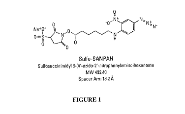
Figure 1 shows the chemical structure of the commercially available
heterobifunctional
linker N-sulphosuccinimidy1-6-(4'-azido-2'-nitrophenylamino) hexanoate (Sulfo-
SANPAH).
Figure 2 shows the chemical surface modification to bind EMC protein to native
PDMS
via linker molecule.
Figure 3A-D show photographs of hepatocytes six (6) days after being seeded on
a
PDMS surface that was either plasma treated (Figure 3 A & B) or that was Sulfo-
SANPAH
treated (i.e. ECM protein(s) covalently attached to the surface with this
crosslinker) (Figure 3 C
& D). The cells were cultured under flow conditions for two (2) days.
Figure 4A-D show photographs of hepatocytes nine (9) days after being seeded
on a
PDMS surface that was either plasma treated (Figure 4A & B) or that was Sulfo-
SANPAH
treated (i.e. ECM protein(s) covalently attached to the surface with this
crosslinker) (Figure 4C
& D). The cells were cultured under flow conditions for 5 days.
Figure 5 A&B show photographs of hepatocytes fourteen (14) days after being
seeded on
a PDMS surface that was either plasma treated (Figure 5A) or that was Sulfo-
SANPAH treated
(i.e. ECM protein(s) covalently attached to the surface with this crosslinker)
(Figure 5B). The
cells were cultured under flow conditions for 10 days. ECM : Collagen type
1100 ug/ml + FN 50
ug/ml + Collagen type IV 50 ug/ml. Cells on the Sulfo-SANPAH treated surface
(right)
maintained monolayer over 14 days in culture. Cells on the plasma treated
surface (left) started
to detach (see arrow).
Figure 6 is a drawing showing the extracellular matrix (ECM) next to primary
hepatocytes and endothelial cells.
18
CA 03032997 2019-02-04
WO 2018/013718
PCT/US2017/041762
Figure 7A-C are photographs showing examples of liver cells (hepatic cells) on
ECM
coated chips under various conditions. Chips were coated with collagen I and
fibronectin and
stored either dry or wet for one week. Cells were then added to the chips and
cultured for 14
days. As a control, a chip was coated fresh (no storage) and cultured with
cells for 14 days. No
differences in cell attachment were observed in Liver sinusoidal endothelial
cells (LSECs) or
Hepatic cells (Hep). No differences in morphology were observed (LSEC and
Hep). Figure 7A is
the control (fresh ECM coat) after 14 days of cell culture. Figure 7B shows
the results after 1
week wet storage and cell culture for 14 days. Figure 7C shows the results
after 1 week dry
storage and cell culture for 14 days.
Figure 8A&B are photographs showing the results from a one month storage
study. Chips
were coated with collagen I and fibronectin and stored dry for one month.
Liver cells were then
added to the chips and cultured for 13 days (Figure 8B). As a control, a chip
was coated fresh (no
storage) and cultured with cells for 13 days (Figure 8A). No differences in
cell attachment were
observed (LSEC and Hep). No differences in morphology were observed (LSEC and
Hep)
Figure 9A&B are bar graphs showing comparisons of different liver on chip
examples.
Chips were coated with collagen I and fibronectin and stored dry for one
month. Liver cells
(Hepaptocytes) were then added to the chips and cultured (grey bars). As a
control, a chip was
coated fresh (no storage) and cultured with cells (blue bars). Albumin was
measured in the
culture fluid after 6 and 13 days of culture (Figure 9A). LDH was measured in
the culture fluid
after 6 and 13 days (Figure 9B).
Figure 10A-C are photographs showing the results from a 1 week gut-on-chip
storage
study. Chips were ECM coated (Matrigel and collagen I) and stored wet for 1
week. Thereafter,
Human Umbilical Vein Endothelial Cells (HUVEC) and Caco-2 cells were cultured
on the chip
for 11 days. Caco-2 cells and HUVEC were on each chip with the Caco-2 cells
are on the top
side of the membrane and the HUVEC are on the bottom. Figure 10A-C images were
taken at the
point of the chip where the two channels join, the wall of the channel is the
dark separator in the
images. This is a top-down view. The gut function is assessed via barrier
function (pApp, the
system's permeability coefficient) and response to stimulation (using an
inflammatory stimulus).
Figure 11A&B are photographs showing small amounts of air observed in bottom
channel inlets after wet storage because PDMS is vapor permeable. Figure 11A
shows wet
storage of 3 weeks. Figure 11B shows a close up of air observed in the bottom
channel inlets
19
CA 03032997 2019-02-04
WO 2018/013718
PCT/US2017/041762
after wet storage for 4 weeks. Small amounts of air observed in bottom channel
inlets after 12
days of storage. Air volume increased after 3 weeks (Figure 11A). By 30 days,
most ECM
solution has evaporated from the chip.
Figure 12 is a graph showing the increasing barrier function of cells in the
gut-on-chip.
Chips were coated with Matrigel and collagen I and stored wet for one week.
Thereafter, the gut
cells (Caco-2 and HUVEC cells) were added to the chip and cultured. The
results demonstrate
healthy and functional cell populations.
Figure 13 presents exemplary data showing a concentration dependent effect of
laminin
binding subsequent to 500[1g/m1 Sulfo-SANPAH (IV) treated channels.
Figure 13A: 10 [tg/m1Laminin treated channels.
Figure 13B: 50 ng/ml Laminin treated channels.
Figure 13C: 100 [ig/m1 Laminin treated channels.
Figure 14 presents exemplary data showing a concentration dependent effect of
laminin
on the development and differentiation of motor neuron cells cultured on
chips.
Figure 14A: Motor neurons cultured in channels treated with 501.1.g/m1
Laminin.
Figure 14B: Motor neurons cultured in channels treated with 100 lag/m1
Laminin.
Figure 15 presents alternative embodiments of PDMS membrane micropatterning.
Figure 15A: Micropatterning perpendicular to channel fluid flow.
Figure 15B: Micropatterning parallel to channel fluid flow.
Figure 16 presents exemplary data showing nuclei development within human
skeletal
muscle cell (hSKMC) myotubes.
Figure 16A: Phase contrast photomicrograph of skeletal muscle cell
alignment along a micropatterned PDMS membrane.
Figure 16B: Photomicrograph (10X) of hSKMC myotube nuclei development
on Day 3 of culture on a micropatterned PDMS membrane.
Figure 16C: Photomicrograph (10X) of hSKMC myotube nuclei development
on Day 11 of culture on a micropatterned PDMS membrane.
Figure 16D: Photomicrograph (20X) of hSKMC myotube nuclei development
on Day 11 of culture on a micropatterned PDMS membrane.
CA 03032997 2019-02-04
WO 2018/013718
PCT/US2017/041762
Figure 17 presents exemplary data showing actin growth and development within
human
skeletal muscle cell (hSKMC) myotubes.
Figure 17A: Photomicrograph (10X) of actin development on a non-
micropatterned PDMS membrane on culture Day 7.
Figure 17B: Photomicrograph (20X) of actin development on a non-
micropatterned PDMS membrane on culture Day 7.
Figure 17C: Photomicrograph (10X) of actin development on a
micropatterned PDMS membrane on culture Day 7.
Figure 17D: Photomicrograph (20X) of actin development on a
micropatterned PDMS membrane on culture Day 7.
Figure 18 presents exemplary data in a bar graph showing hSKMC morphology
(e.g.,
elongated or round) as measured by a cell shape index.
Figure 19A and 19B show exemplary data showing greater hSI(MC attachment to
PDMS
membranes in unmasked regions versus masked regions due to UV-activated
crosslinkers.
Figure 20 presents exemplary data showing the fabrication and use of an
embossed
PDMS membrane.
Figure 20A: An embossed PDMS membrane before cell attachment.
Figure 20B: An embossed PDMS membrane subsequent to one day of cell
attachment and culture.
Figure 20C: An embossed PDMS membrane subsequent to six days of cell
attachment and culture.
Figure 21 shows generic examples of potential crosslinkers with the formula A-
B-C,
wherein A represents light-reactive portion, B represents a linker, and C
represents modifier-
reactive portion. The formula on the left represents a linear crosslinker, the
formula in the center
and the left represent where the liker portion is multivalent.
Figure 22 shows a specific example of a crosslinker, Sulfo-SANPAH which is
diagrammed according to the A-B-C crosslinker formula described above.
Detailed Description of the Invention
Silicone elastomers, such as PDMS, are used in microfluidics. However,
silicone
polymers are hydrophobic and do not promote cell adhesion. Surface treatments
(e.g. chemical
21
CA 03032997 2019-02-04
WO 2018/013718
PCT/US2017/041762
vapor deposition, plasma oxidation, Corona, RF plasma, etc.) have been used to
make such
polymers more useful. See e.g. Hong et al., "Hydrophilic Surface Modification
of PDMS Using
Atmospheric RF Plasma," Journal of Physics: Conference Series 34 (2006) 656-
661 (Institute of
Physics Publishing). A microfluidic device (or portion thereof) made of a
naturally hydrophobic
material becomes hydrophilic upon such surface treatment. Nonetheless, cell
attachment
remains a problem, both in the short term and the long term. That is to say,
some cells do not
adhere well to surface treated PDMS at the outset; they exhibit low seeding
levels. In the long
term, cells (and even monolayers) can detach from the surface treated PDMS.
The present invention contemplates compositions, devices and methods of
improving
adhesion, attachment, and/or differentiation of cells in a microfluidic device
or chip, and in
particular, cells on a PDMS surface. In one embodiment, one or more proteins
(e.g. ECM
proteins) or peptides (e.g. RGD) are covalently coupled to the surface of a
microchannel of a
microfluidic device. The microfluidic devices can be stored and later used, or
they can be
immediately used for culture and/or support of living cells such as mammalian
cells, and/or for
simulating a function of a tissue, e.g., a liver tissue, muscle tissue, etc.
Even under flow
conditions, extended adhesion and viability with sustained function over time
is observed.
The present invention contemplates microfluidic devices (or "chips")
containing living
cells recreate the physiological tissue-tissue interfaces and permit fluid
flow. See U.S. Patent
No. 8647861, hereby incorporated by reference. Such devices subject the cells
to shear stress.
In contrast to static 2D culture, microchannels allow the perfusion of cell
culture medium
throughout the cell culture during in vitro studies and as such offer a more
in vivo-like physical
environment. In simple terms, an inlet port allows injection of fluids such as
cell culture medium
(and the like) into a microfluidic channel or chamber (with or without cells).
In one
embodiment, the present invention contemplates introduction of fluid into a
cell-laden
microfluidic channel or chamber. In a preferred embodiment, the cells are
attached to one or
more ECM proteins (e.g. laminin), which are in turn covalently attached to the
microchannel
surface. An outlet port then permits the exit of remaining fluid as well as
harmful metabolic by-
products.
The surface over which the fluid flows and to which the cells are attached
(using the
methods described herein) can be a surface of any material that is compatible
to the fluid sample
22
CA 03032997 2019-02-04
WO 2018/013718
PCT/US2017/041762
and cells. Exemplary materials for the fluid-contact surface can comprise
glass, synthetic
polymers (e.g., PDMS, polysulfonate, and polycarbonate), hydrogels, and a
combination thereof.
One portion of a microchannel can be a membrane. For example, the floor of a
microchannel can comprise a membrane, including a porous membrane. The
microchannel (or
portion thereof) or membrane can be coated with substances such as various
cell adhesion
promoting substances or ECM proteins, such as fibronectin, laminin or various
collagen types or
combinations thereof. For example, endothelial cells can attach to a collagen
coated
microchannel. While non-covalent coating can be used, it is preferred that
such proteins and
peptides be covalently attached, e.g. by use of a crosslinker or other
chemistry.
It is not intended that the present invention be limited to the method by
which one or
more ECM proteins are covalently attached to the microchannel surface. In one
embodiment,
bifunctional crosslinkers are used. A variety of such crosslinkers are
available commercially,
including (but not limited to) the following compounds:
ANB-NOS (N-5-azido-2-nitrobenzoyloxysuccinimide) having the formula of:
11,
0 H1.
(I)
0
NV-
.
(I)
Sulfo-SAND (sulfosuccinimidyl 24m-azido-o-nitrobenzamido]ethy1-1, 3'-
dithiopropionate) having the formula of:
1.
Na _ 0
N
0 ¨ s
//
os
0 0 0 N+
0
N
(H)
23
CA 03032997 2019-02-04
WO 2018/013718
PCT/US2017/041762
SANPAH (N-succinimidy1-644'-azido-2--nitrophenylamino]hexanoate) having the
formula of:
N
0
N+
N
Sulfo-SANPAH (sulfosuccinimidy1-644--azido-2'-nitrophenylamino]hexanoate)
having
the formula of:
Na0 - 0
0 N
ID= H
0 N ..0/=\f\/''N 40/
0
I I
I I
N
(IV)
By way of example, sulfosuccinimidyl 6-(4'-azido-2'-nitrophenyl-amino)
hexanoate or "Sulfo-
SANPAH" (commercially available from Pierce) is a long-arm (18.2 angstrom)
crosslinker that
contains an amine-reactive N-hydroxysuccinimide (NHS) ester and a
photoactivatable
nitrophenyl azide. NHS esters react efficiently with primary amino groups (-
NIT)) in pH 7-9
buffers to form stable amide bonds. The reaction results in the release of N-
hydroxy-
succinimide. When exposed to UV light, nitrophenyl azides form a nitrene group
that can initiate
.. addition reactions with double bonds, insertion into C-H and N-H sites, or
subsequent ring
expansion to react with a nucleophi1e (e.g., primary amines). The latter
reaction path dominates
when primary amines are present.
24
CA 03032997 2019-02-04
WO 2018/013718
PCT/US2017/041762
Sulfo-SANPAH should be used with non-amine-containing buffers at pH 7-9 such
as
20mM sodium phosphate, 0.15M NaCl; 20mM HEPES; 100mM carbonate/bicarbonate; or
50mM borate. Tris, glycine or sulfhydryl-containing buffers should not be
used. Tris and glycine
will compete with the intended reaction and thiols can reduce the azido group.
The present invention is not to be limited to any particular crosslinker. In
one
embodiment, the crosslinkers of the current invention comprise three parts: a
light-reactive
portion, a linker, and a modifier-reactive portion. In one embodiment, the
bifunctional
crosslinkers are represented by the formula A-B-C, wherein A represents light-
reactive portion,
B represents a linker, and C represents modifier-reactive portion. The present
invention is not to
be limited to linear crosslinkers. In one embodiment, B can also be branched
it multivalent. i.e. it
can link one A to two Cs, 3As to 4Cs, etc, see Figure 13. As a non-limiting
example, sulfo-
SANPAH uses a nitrophenyl azide group as the light-reactive portion,
aminohexanoate as the
linker, and sulfo-NHS ester as the modifier-reactive portion (in this case
reacting with an amine
group on the modifier), see Figure 14. In one embodiment, light reactive
portions may be
selected from the group consisting of nitrophenyl, diazirine, and azides. The
present invention is
not to be limited to any particular linker. In one embodiment, the linker (B)
are connected to
light-reactive portion (A) through an amine bond and modifier-reactive portion
(C) through an
ester bond. In one embodiment, the linkers may be selected from the group
consisting of
polyethyleneglycols, alkanes, and olefins. In one embodiment, the modifier-
reactive chemistry
portion may be selected from the group consisting of NHS-ester (amine
reactive), Sulfo-NHS-
ester (amine reactive), Isocyanate (amine reactive), Isothiocyanate (amine
reactive), Imidoester
(amine reactive), Maleimide (sulfhydryl reactive), Pyridyldithiol (sulfhydryl
reactive), and
Hydrazide (aldehyde and ketone reactive). Specific examples of commercially
available
crosslinkers that fit this description include ANB-NOS, SDA, sulfo-SDA, LC-
SDA, sulfo-LC-
SDA, SDAD, sulfo-SDAD, and more (see Table 1). ANB-NOS is a short-arm (7.7
angstrom)
crosslinker that contains an amine-reactive N-hydroxysuccinimide (NHS) ester
and a
photoactivatable nitrophenyl azide, also called N-5-azido-2-
nitrobenzoyloxysuccinimide. Sulfo-
SANPAH is a long-arm (18.2 angstrom) crosslinker that contains an amine-
reactive N-
hydroxysuccinimide (NHS) ester and a photoactivatable nitrophenyl azide, also
called
sulfosuccinimidyl 6-(4'-azido-2'-nitrophenylamino)hexanoate. SDA (NHS-
Diazirine) combines
proven NHS-ester and diazirine-based photoreaction chemistries with conjugate
amine-
CA 03032997 2019-02-04
WO 2018/013718
PCT/US2017/041762
containing molecules with nearly any other functional group via long-wave UV-
light activation.
SDA (Sulfo-NHS-Diazirine) is an amine and photoreactive, membrane impermeable,
heterobifunctional crosslinker with a 3.9 Angstrom spacer arm. Also called
Sulfosuccinimidyl
4,4'-azipentanoate. LC-SDA (NHS-LC-Diazirine) is an amine and photoreactive,
membrane
permeable, heterobifunctional crosslinker with a 12.5 Angstrom spacer arm.
Also called
Succinimidyl 6-(4,4'-azipentanamido)hexanoate. Sulfo-LC-SDA (Sulfo-NHS-LC-
Diazirine) is a
sulfo-NHS-diazirine based photoreactive crosslinker. Membrane impermeable with
a 12.5
Angstrom spacer arm. Also called Sulfosuccinimidyl 6-(4,4'-
azipentanamido)hexanoate.
Table 1: Examples of commercially available crosslinkers
Reactive Spacer Cleavable Membrane
Products Water-soluble?
Groups Arm (A' ) by?
permeable?
NHS ester/ ANB-NOS 7.7 Short No No
No
Sulfo-SANPAH 18.2
aryl azide Long No Yes No
SDA 3.9 Short No No
Yes
Sulfo-SDA 3.9 Short No Yes
No
NHS ester/ LC-SDA 12.5 Mid No No
Yes
diazirine Sulfo-LC-SDA 12.5 Mid No
Yes No
SDAD 13.5 Mid Thiols No
Yes
Sulfo-SDAD 13.5 Mid Thiols Yes
No
By way of example, sulfosuccinimidyl 6-(4'-azido-2'-nitrophenyl-amino)
hexanoate or "Sulfo-
SANPAH" (commercially available from Pierce) is a long-arm (18.2 angstrom)
crosslinker that
contains an amine-reactive N-hydroxysuccinimide (NHS) ester and a
photoactivatable
nitrophenyl azide. NHS esters react efficiently with primary amino groups (-
NH2) in pH 7-9
buffers to form stable amide bonds. The reaction results in the release of N-
hydroxy-
succinimide. When exposed to UV light, nitrophenyl azides form a nitrene group
that can initiate
addition reactions with double bonds, insertion into C-H and N-H sites, or
subsequent ring
expansion to react with a nucleophile (e.g., primary amines). The latter
reaction path dominates
when primary amines are present.
Sulfo-SANPAH should be used with non-amine-containing buffers at pH 7-9 such
as
20mM sodium phosphate, 0.15M NaCl; 20mM HEPES; 100mM carbonate/bicarbonate; or
26
CA 03032997 2019-02-04
WO 2018/013718
PCT/US2017/041762
50mM borate. Tris, glycine or sulfhydryl-containing buffers should not be
used. Tris and glycine
will compete with the intended reaction and thiols can reduce the azido group.
For photolysis, one should use a UV lamp that irradiates at 300-460nm. High
wattage
lamps are more effective and require shorter exposure times than low wattage
lamps. UV lamps
that emit light at 254nm should be avoided; this wavelength causes proteins to
photodestruct.
Filters that remove light at wavelengths below 300nm are ideal. Using a second
filter that
removes wavelengths above 370 nm could be beneficial but is not essential.
Calfskin type I collagen has been covalently attached to a polyacrylamide
surface using
sulfo-SANPAH. See Gaudet, C., "Influence of type I collagen surface density on
Fibroblast
Spreading, Motility, and Contractility" Biophys J. 85(5): 3329-3335 (2003).
Collagen I was
coupled to other surfaces using Sulfo-SANPAH in order to avoid potential
differences in ECM
remodeling on different substrates. See Trappman et al. "Extracellular-matrix
tethering regulates
stem-cell fate," Nature Materials (2012) (on-line publication). RGD has been
covalently
attached to a PDMS surface using sulfo-SANPAH. See Li et al., "RGD peptide-
conjugated
poly(dimethylsiloxane) promotes adhesion, proliferation, and collagen
secretion of human
fibroblasts," J. Biomed Mat Res A. 79(4):989-98 (2006).
It is not intended that the present invention be limited by the number or
nature of
channels in the microfluidic device. In some embodiments, the surface can be a
surface of a
fluid-flowing conduit or passageway disposed in a solid substrate. In some
embodiments, the
surface can be a solid surface. For example, in one embodiment, the solid
surface can be a wall
surface of a fluid channel, e.g., a microfluidic channel.
In one embodiment, the present invention contemplates a co-culture of liver
sinusoidal
endothelial cells in one chamber with hepatocytes in other chamber(s) to
establish hepatic
function in vitro. In one embodiment, the chambers are first and second
microchannels aligned
(e.g., vertically) with each other with one or more membranes separating them
from each other
("liver-on-a-chip"). The liver-on-a-chip devices have been developed and
optimized based on the
basic design of an organ-on-a-chip as described in the U.S. Patent No.
8,647,861, and the
International Patent App. No. PCT/US2014/071611, the contents of each of which
are
incorporated herein by reference in their entireties. In some aspects, the
inventors have optimized
the design of the liver-on-a chip devices and culture conditions to provide
long-term hepatic
27
CA 03032997 2019-02-04
WO 2018/013718
PCT/US2017/041762
culture with physiologically relevant hepatic function (e.g., albumin and/or
urea secretion, and/or
CYP 450 metabolic capacity) for different animal models, e.g., human, rats,
and dogs.
In a preferred embodiment, the present invention contemplates a microfluidic
device
comprising a microchannel, said microchannel comprising a monolayer of viable
hepatocytes
adhered to a coating, said coating comprising at least one extracellular
matrix protein covalently
coupled to a microchannel surface. The viable hepatocytes can be derived from
different
mammalian sources, including, e.g., but not limited to humans, rats, mice, and
dogs.
In one embodiment, the present invention contemplates covalently attaching one
or more
proteins or peptides to a surface in the microfluidic device (e.g. to the
membrane and/or one or
more microchannel), and storing the microfluidic device for a week or more (a
month or more)
prior to attaching cells. In one embodiment, the present invention
contemplates dry storage.
Experimental results have shown that chips for liver samples may be stored
dry. That is
to say, the extracellular matrix protein can be attached to the chip and can
be stored dry (prior to
any cell culture). However, not all ECM proteins can be stored dry;
empirically, it was found
that only some ECMs can be stored dry (collagens, fibronectin (FN)). Chips
were stored at 4 C
and then compared to freshly coated chips.
Figure 7A-C are photographs showing examples of liver cells (hepatic cells) on
ECM
coated chips under various conditions. Chips were coated with collagen I and
fibronectin and
stored either dry or wet for one week. Cells were then added to the chips and
cultured for 14
days. As a control, a chip was coated fresh (no storage) and cultured with
cells for 14 days. No
differences in cell attachment were observed in Liver sinusoidal endothelial
cells (LSECs) or
Hepatic cells (Hep). No differences in morphology were observed (LSEC and
Hep). Figure 7A is
the control (fresh ECM coat) after 14 days of cell culture. Figure 7B shows
the results after 1
week wet storage and cell culture for 14 days. Figure 7C shows the results
after 1 week dry
storage and cell culture for 14 days.
Figure 8A&B are photographs showing the results from a one month storage
study. Chips
were coated with collagen I and fibronectin and stored dry for one month.
Liver cells were then
added to the chips and cultured for 13 days (Figure 8B). As a control, a chip
was coated fresh (no
storage) and cultured with cells for 13 days (Figure 8A). No differences in
cell attachment were
observed (LSEC and Hep). No differences in morphology were observed (LSEC and
Hep).
28
CA 03032997 2019-02-04
WO 2018/013718
PCT/US2017/041762
There was also an evaluation of biomarkers in order to compare between freshly
coated
(control) chips and chips dry-stored for 1 month with 5 chips tested per
condition with liver.
Figure 9A&B are bar graphs showing comparisons of different liver on chip
examples. Chips
were coated with collagen I and fibronectin and stored dry for one month.
Liver cells
(Hepaptocytes) were then added to the chips and cultured (second bars). As a
control, a chip was
coated fresh (no storage) and cultured with cells (first bars). Albumin was
measured in the
culture fluid after 6 and 13 days of culture (Figure 9A). LDH was measured in
the culture fluid
after 6 and 13 days (Figure 9B). Chips stored dry for 1 month showed higher
albumin production
and lower LHD release over a 2-week observation period. The LDH release is not
desirable.
Therefore, dry storage of coll/FN-coated chips is a viable platform for use
with the human
Liver-on-Chip.
The above results indicate that ECMs for Liver-on-Chip can be stored dry.
Chips can be
coated with collagen I and Fibronectin and put in a 1 week dry storage, and
even a 1 month dry
storage.
In one embodiment, the present invention contemplates covalently attaching one
or more
proteins or peptides to a surface in the microfluidic device (e.g. to the
membrane and/or one or
more microchannel), and storing the microfluidic device for a week or more (a
month or more)
prior to attaching cells. In one embodiment, the present invention
contemplates wet storage. In
one embodiment, the present invention contemplates vapor proof packaging.
In one embodiment, the present invention contemplates a membrane comprising a
pattern. In one embodiment, the pattern is a line and groove pattern. Although
it is not
necessary to understand the mechanism of an invention, it is believed that a
line and groove
pattern provides alignment for cells such as muscle cells. It is further
believed that it is
microgrooves on the surface of the membrane that guides such cell alignment.
In one
embodiment, the membrane is a PDMS membrane.
In a preliminary experiment, PDMS membrane grooves were made using an existing
micropatterned silicon wafer (e.g., 10 [.tm x 10 pm x 2 [tm) as a mold. In one
embodiment, a
bifunctional crosslinker is attached to the micropatterned membrane. In one
embodiment, the
micropatterned membrane is in the flow channel of a microfluidic device. In
one embodiment,
the micropattern is parallel to the fluid flow. In one embodiment, the
micropattern is
perpendicular to the fluid flow. In one embodiment, an extracellular matrix
protein (e.g. laminin)
29
CA 03032997 2019-02-04
WO 2018/013718
PCT/US2017/041762
is attached to the crosslinker (e.g. covalently bound) so as to provide an ECM-
coated
micropatterned membrane. Human primary muscle cells were then cultured on the
micropatterned PDMS membrane in a static culture condition. Cell alignment was
measured by
F-actin stain and image analysis. When micropatterned membranes were
fabricated according to
the methods described herein, cell alignment measurements demonstrated cell
elongation. See,
Figures 17 and 18.
In one embodiment, the PDMS membrane or micropatterned membrane may be
selectively coated with a crosslinker using a mask. In one embodiment, the
present invention
contemplates the crosslinker used is Sulfo-SANPAH and the masking is done to
control surface
coating of this light activated crosslinker in a closed chip system (i.e. UV
light can be used
without opening the chip). For example, Sulfo-SANPAH may be applied via
channels and
ultraviolet light can be shined over the chip with a mask (to block the light
from striking a
specific portion or portions of the chip) covering a portion of an area, or
alternatively a patterned
mask to create a surface pattern. In one embodiment, the mask is layered,
deposited or simply
positioned on top of a portion of the membrane and subsequently exposed to UV
light. Once the
mask is removed, an ECM protein (or proteins) can be attached to the
bifunctional crosslinker.
In one embodiment, the irradiated membrane is washed before the ECM attachment
step.
Thereafter, cells may be attached to the resultant pattern generated by the
mask and cultured in a
static condition. Although it is not necessary to understand the mechanism of
an invention, it is
believed that most cells attach to ultraviolet light-exposed membrane areas
(where the
crosslinker was activated to bind) and very few cells attach in the masked
areas (i.e., where there
was no ultraviolet light exposure).
In one embodiment, the membrane micropatterning may be achieved by embossing.
A
preliminary experiment was performed using a pre-manufactured thin membrane
layered on top
of a conventional micropatterned silicon wafer which was then exposed to a
high heat (e.g.,
about 80-90 C) and a weight (to create pressure) for 24-48hrs to create the
embossed pattern.
Subsequent to the embossing, the micropatterned membrane was removed from the
silicon
wafer. In one embodiment, the micropatterned membrane comprises PDMS. In one
embodiment, the micropatterned membrane is in the flow channel of a
microfluidic device. In
one embodiment, the micropattern is parallel to the fluid flow. In one
embodiment, the
micropattern is perpendicular to the fluid flow. In one embodiment, a
bifunctional crosslinker is
CA 03032997 2019-02-04
WO 2018/013718
PCT/US2017/041762
attached to the micropatterned membrane. In one embodiment, an extracellular
matrix protein
(e.g. laminin) is attached to the crosslinker (e.g. covalently bound) so as to
provide an ECM-
coated micropatterned membrane. In one embodiment, the viable cells are
further seeded onto
the ECM-coated micropatterned membrane. In one embodiment, human primary
muscle cells
were attached to the membrane and cultured in a static condition. The data
showed that the
hSKMCs were bound to the membrane and observed to grow along the
micropatterned grooves.
After approximately six days of culture the hSKMCs were aligned along the
microgrooves.
Experimental
The following are examples that further illustrate embodiments contemplated by
the
present invention. It is not intended that these examples provide any
limitations on the present
invention.
Example 1
Cellular Crosslinking To Improve Cell Attachment To Channels
In one embodiment, the present invention contemplates using a crosslinker to
covalently
attach proteins or peptides that enhance cell attachment. In this example, a
protocol for using
Sulfo-SANPAH as the crosslinker is provided as one embodiment of a method.
First, fresh 0.5mM Sulfo-SANPAH (492.4g/mol) solution in 50mM HEPES (0.22um
sterile filtered, pH 7.4) (protect from light) is prepared. Then, an ECM
solution is prepared (e.g.
50ug/mL Laminin in PBS or media without FBS) on ice.
The microfluidic device ("chip") comprising a microchannel is then plasma
treated.
Plasma ¨ 15 sccm 02, 60 sec, 100W.
The channels are then washed with 200uL of 50mM HEPES. Excess 50mM HEPES is
removed from the channel.
Sulfo-SANPAH is introduced into the microchannel by inserting a pipet tip
reservoir in a
port of the chip. 100uL of Sulfo-SANPAH solution is added to the top channel,
ejecting tip into
inlet port. 50uL of Sulfo-SANPAH solution is added to the bottom channel,
ejecting tip into inlet
port. The channels are then inspected to be sure no bubbles are present. At
this point, one
carefully removes and discards the pipet tips reservoirs without spilling
excess reagent on
surface of chip.
31
CA 03032997 2019-02-04
WO 2018/013718
PCT/US2017/041762
At this point, the chips are ready for light treatment. The chips are
incubated in the UV
lamp chamber. Decrease distance to UV lamp and chips with lmL tip boxes or lab-
jack. The
incubation is for 20min at 0.72 joules/cm2. After UV treatment, Sulfo-SANPAH
is removed
from the channels and each channel is washed twice with 200uL 50m1V1 HEPES.
The channel is
dried by removing or aspirating remaining HEPES buffer.
At this point, a protein or peptide can be attached. A solution containing a
protein (e.g.
ECM solution) or peptide (to enhance cell binding) can be loaded into the
channels with one of
the following conditions: Overnight at 4 C or Minimum of 1.5 hours at 37 C.
Before seeding with cells, the solution (e.g. ECM solution) should be removed
from the
channels and the channels washed with 200uL of desired media or PBS.
Example 2
Channel Surface Modification To Improve Cell Attachment
In this example, PDMS surfaces treated with plasma were compared with PDMS
surfaces
.. modified by covalent attachment ECM proteins. Figures 3A-B show photographs
of hepatocytes
six (6) days after being seeded on a PDMS surface that was either plasma
treated (Figure 3 A &
B) or that was Sulfo-SANPAH treated (i.e. ECM protein(s) covalently attached
to the surface
with this crosslinker) (Figure 3 C & D). The cells were cultured under flow
conditions for two
(2) days.
Figures 4A-D show photographs of hepatocytes nine (9) days after being seeded
on a
PDMS surface that was either plasma treated (Figure 4A & B) or that was Sulfo-
SANPAH
treated (i.e. ECM protein(s) covalently attached to the surface with this
crosslinker) (Figure 4C
& D). The cells were cultured under flow conditions for 5 days.
Figures 5A-B show photographs of hepatocytes fourteen (14) days after being
seeded on
a PDMS surface that was either plasma treated (Figure 5A) or that was Sulfo-
SANPAH treated
(i.e. ECM protein(s) covalently attached to the surface with this crosslinker)
(Figure 5B). The
cells were cultured under flow conditions for 10 days. ECM : Collagen type
1100 ug/ml + FN 50
ug/ml + Collagen type IV 50 ug/ml. Cells on the Sulfo-SANPAH treated surface
(right)
maintained monolayer over 14 days in culture. Cells on the plasma treated
surface (left) started
to detach (see arrow). Clearly, the Sulfo-SANPAH treatment was an improvement
over plasma
treatment.
32
CA 03032997 2019-02-04
WO 2018/013718
PCT/US2017/041762
Example 3
Shelf-Life Study Of ECM-Coated Chips
This example evaluates conditions to avoid ECM (i.e., for example, laminin,
Matrigel)
inactivation found during dry storage. All tested chips were stored at 4 C
then compared to
freshly coated chips. Results indicate that ECM for Gut-on-Chip chips are best
stored in solution
(e.g., wet).
Experimental Design
The ECM for Gut-on Chip chips comprised Matrigel and collagen 1. Gut-on-Chip
chips
were chosen as test platform due to its robustness to varying culture
conditions. In particular,
test Chips were treated with Sulfo-SANPAH and ECM (10Oug/mL Martigel and
25ug/mL
collagen 1). All conditions were compared to freshly coated chips.
Results
Twenty-eight (28 chips) were stored for 1 week. No differences in Caco-2 and
HUVEC
cell attachment was observed. No differences in Caco-2 and HUVEC cell
morphology was
observed. The chips were maintained for 8 days prior to exposure to TNF-a and
IL-1(3. The
experiment also included some chips with lamina propria cells. Samples for
barrier function and
lactate dehydrogenase (LDH) were collected, along with cytokine profiles.
It appears that a 1 week storage does not impact cell attachment or morphology
in the
Gut-on-Chip configuration. Figure 10A-C show photographs demonstrating the
results from a 1
week gut-on-chip storage study. Chips were ECM coated (Matrigel and collagen
I) and stored
wet for 1 week. Thereafter, Human Umbilical Vein Endothelial Cells (HUVEC) and
Caco-2
cells were cultured on the chip for 11 days. Caco-2 cells and HUVEC were on
each chip with the
Caco-2 cells on the top side of the membrane and the HUVEC on the bottom side
of the
membrane. Figure 10A-C show examples of gut on chips where the chips have been
stored for 1
week. Figure 10A-C images were taken at the point of the chip where the two
channels join, the
wall of the channel is the dark separator in the images. This is a top-down
view. The gut
function is assessed via barrier function (pApp, the system's permeability
coefficient) and
response to stimulation (using an inflammatory stimulus). The increasing
barrier function (see
Figure 12) demonstrates healthy and functional cell populations and correlates
with previous
33
CA 03032997 2019-02-04
WO 2018/013718
PCT/US2017/041762
data. Caco-2 and HUVEC cells were used.Wet storage (1 week) of Matrigel/Coll
coated chips
are a viable platform for use with the human Gut-on-Chip.
Figure 12 shows the increasing barrier function which demonstrates healthy and
functional cell populations. The Figure 12 graph shows the increasing barrier
function of cells in
the gut-on-chip. Chips were coated with Matrigel and collagen I and stored wet
for one week.
Thereafter, the gut cells (Caco-2 and HUVEC cells) were added to the chip and
cultured. The
results demonstrate healthy and functional cell populations.
In conclusion, the data shows that the storage of Gut-on-Chip coated with
extracellular
protein should be stored wet, such that more than 1 week leads to evaporation
issues. However,
with a vapor proof packaging, the wet storage lifespan would be expected to
improve.
The present invention contemplates other chips, such as those that use Laminin
and/or
Matrigel and may include chips that could be used as Intestine-on-Chip, Blood
Brain Barrier
(BBB)-on-Chip, and neruromuscular junction (NMJ)-on-Chip.
Example 4
Enhanced Cell Attachment In Presence of Crosslinker and Laminin
This example shows that the coating of a channel with crosslinker and laminin
improves
the binding of cells to chip channels. The experimental design coated chip
channels with one of
several concentrations (0.1 mg/ml, 0.5 mg/ml and 1 mg/ml) of Sulfo-SANPAH
(IV), followed by
one of several concentrations (10 ug/ml, 50 ug/m1 and 100iug/m1) of
fluorescently labeled
laminin (red).
In general the protocol was as follows:
1. Flush the chip with 70% ethanol briefly and wash with 50 mM HEPES
buffer.
2. Add 50 ul of Sulfo-SANPAH (IV) and incubate under an ultraviolet light
for
approximately 20 min.
3. Wash with 200 ul of 50 mM HEPES buffer three (3) times.
4. Wash with 200 ul of DPBS twice.
5. Add 50 ul of Laminin and incubate at 4 C overnight.
6. Next day, transfer the chip at 37 C for at least 1 h.
7. Seed the chip with motor neuron cells and culture.
34
CA 03032997 2019-02-04
WO 2018/013718
PCT/US2017/041762
Data collected before seeding with motor neuron cells shows iucreased laminin
binding as the
laminin concentration increases. See, Figure 13A-C. Data collected after
seeding with motor
neuron cells shows that the cells grown on higher concentrations of laminin
were differentiated
and developed thicker axons. See, Figure 14A,B.
Example 5
Micropatterning Of A Chip Membrane
This example shows one method of micropatterning a PDMS membrane to facilitate
cell
alignment in a chip. In general the protocol was as follows:
1. Prepare silicon wafer mold comprising 10 x 21AM grooves.
2. Spin coat PDMS on top of the silicone mold.
3. Cure at 60 C for over 6 hours.
4. Delaminate the micropatterned PDMS membrane from the silicon wafer mold
and assemble the micropattern PDMS membrane into a channel chip in either a
perpendicular or parallel orientation relative to fluid flow. See, Figure
15A and 15B,
respectively.
5. Surface treat with Sulfo-SANPAH and coat chips with Laminin.
6. Seed human skeletal muscle cells (hSKMC)
7. Subsequent to culturing, observe cell morphology and cell alignment
relative
to the micropatterning.
8. Stain for muscle markers to show viability, differentiation and
development.
9. Assess the hSKMCs for spontaneous contraction, cholinergic stimulation
and IHC
The data show: i) hSKMC alignment within the PDMS groove micropattern (Figure
16A); ii) progressive nuclei development within hSKMC myotubes between Day 3 ¨
Day 11 of
culture (Figures 16B-C); iii) greater actin development within hSKMCs cultured
on
micropatterned membranes by Day 7 (Figures 17A-D); iv) greater proportion of
elongated versus
round hSKMC morphology by Day 7 (Figure 18).
CA 03032997 2019-02-04
WO 2018/013718
PCT/US2017/041762
Example 6
Membrane Micropatterning Using A Mask
This example creates a membrane surface pattern using Sulfo-SANPH and a mask.
In general the protocol was as follows:
1. Sulfo-SANPAH (0.5mg/m1) was added onto PDMS membrane.
2. A mask (e.g. a piece of aluminum foil) was overlayed on top of
a portion of the
PDMS membrane.
3. The membrane with mask was transferred under an ultraviolet
light and
illuminated for 20 min
4. The membrane was washed and Laminin was added and incubated
for 2h at 37C incubator
5. Next day, hSKMC cells were seeded on to the membrane
6. The seeded hSKMC cells were then cultured for 7 days.
The data show that the regions where Sulfo-SANPAH was activated by exposure to
ultraviolet light have greater sSKMC cell attachment than those masked regions
(e.g., no Sulfo-
SANPAH activation). See, Figure 19A,B.
Example 7
Membrane Micropatterning Using Embossing
This example creates a membrane surface pattern using embossing with heat a
pressure.
In general the protocol was as follows:
1. A PDMS membrane was embossed onto silicon wafer at 80 C for 2
days with
pressure.
2. At day 2, the embossed PDMS membrane was delaminated from the wafer.
3. Sulfo-SANPAH (0.5mg/m1) was added onto PDMS membrane and
treated
under ultraviolet light for 20 min.
4. The irradiated membrane was washed and Laminin was added and
incubated for
2h at 37 C.
5. Next day, hSKMC cells were seeded onto the membrane.
6. The seeded hSKMC cells were then cultured for 7 days
36
CA 03032997 2019-02-04
WO 2018/013718
PCT/US2017/041762
A representative embossed PDMS membrane patterns is shown in Figure 20A. The
growth of hSKMC cells was observed on the embossed PDMS membrane patterns
after Day 1
and Day 6 of culture. See, Figure 20B and 20C, respectively.
While embodiments and applications have been shown and described, it would be
apparent to those skilled in the art having the benefit of this disclosure
that many more
modifications than mentioned above are possible without departing from the
inventive concepts
disclosed herein. It is specifically contemplated that some of the features
described above can be
combined. For example, the membrane can be micropatterned to create a first
pattern (e.g. by
molding or embossing features such as lines, grooves, etc.) and then a
crosslinker can be used
with a mask to create a second pattern. The embodiment(s), therefore, are not
to be restricted
except in the spirit of the appended claims.
37