Note: Descriptions are shown in the official language in which they were submitted.
CA 03033102 2019-02-06
WO 2018/029213 PCT/EP2017/070111
Histones and/or proADM as markers indicating organ dysfunction
The present invention relates to the diagnosis, prognosis, risk assessment,
risk stratification,
monitoring, therapy guidance and/or therapy control of organ dysfunction in a
subject. The
invention relates to a method that comprises determining a level of at least
one histone,
particularly H2B, H4, H2A and/or H3, in a sample of said subject and wherein
said level of at
least one histone is indicative of said organ dysfunction; and/or determining
a level of
proadrenomedullin (proADM), particularly midregional proadrenomedullin (MR-
proADM), in a
sample of said subject and wherein said level of proADM is indicative of said
organ dysfunction.
The invention further relates to kits for carrying out the methods of the
invention.
Background of the invention
Critically ill patients are commonly admitted to the intensive care unit (ICU)
with several signs of
organ dysfunction and/or organ failure (OF). In order to stratify and treat
these patients quickly
and effectively, clinicians need to assess the overall status and severity of
illness of the patient
as accurately as possible. This assessment and its resulting consequences are
of high
importance, since the multiple organ dysfunction syndrome (MODS) is the major
cause of death
on ICUs (Vincent 2006; Ferreira and Sakr 2011). Only few people die due to a
single OF in the
ICU (Mayr, Dunser et al. 2006). Various clinical studies support these
findings and confirm a
strong correlation between organ dysfunction or organ failure, morbidity and
mortality of such
subjects (Ferreira and Sakr 2011, Mayr et al. 2006, Vincent et al. 1998,
Vincent et al. 2006).
In the 1980ies and 1990ies, many of the current gold standard ICU scoring
systems were
developed. Among these are outcome predictive scores, such as Acute Physiology
and Chronic
Health Evaluation II (APACHE II) and simplified acute physiology score
(SAPSII), that are
based on physiological assessment and criteria of the patient. Further scores
are morbidity
diagnosing scores, such as the SOFA score, that is based on organ specific
parameters (Bouch
and Thompson 2008). All three scores are based on at least six parameters and
are determined
within the first 24 hours after ICU admission. APACHE II consists of 14
variables (12 physiology
parameters, age and chronic health status), each weighted from 0 to 4 with
increasing
abnormality of the parameter (Knaus, Draper et al. 1985). SAPS II is composed
of 17 variables
including 12 physiology parameters, age of the patient, type of admission and
three underlying
disease variables (Le Gall, Lemeshow et al. 1993). As for the SOFA score, six
organs are
CA 03033102 2019-02-06
WO 2018/029213 PCT/EP2017/070111
2
evaluated and weighted according to their degree of dysfunction, each scoring
from 0 to 4
(Vincent, Moreno et al. 1996).
Although the SOFA score was not designed to predict outcome, but rather, to
display the
sequential complications of ICU patients, there is a clear correlation with
the mortality rate due
to the cumulative organ failure in a patient (Vincent 2006). Especially, in
case of a high-organ-
failure group, such as patients with four or more OFs, the mortality rate was
65% or 83% in two
ICU focused clinical studies compared to 6% or 22% for patients without any
organ failure
(Vincent, de Mendonca et al. 1998; Vincent, Sakr et al. 2006). The ability to
identify subjects
suffering from organ dysfunction or organ failure, and hence having a high
mortality rate, is still
challenging and time consuming as described in the following, but of
tremendous importance.
Apart from their ability to prognosticate or diagnose critically ill patients
with a certain probability,
the ICU scores disclosed in the prior art are used in only 10 to 15 % of non-
randomly selected
ICUs (often hospitals with focus on clinical studies and/or stringent quality
regulations) and have
several drawbacks (Breslow and Badawi 2012). For example, one disadvantage is
that the
scores are vulnerable to the subjectivity of every clinician performing the
assessment. Hardly
any hospital has either specialized scoring experts or, at least, regularly
trained medical staff
focusing on a reliable assessment of all parameters following strict and
similar guidelines for the
evaluation of the different variables of a score (Polderman, Girbes et al.
2001). Additionally, the
number of parameters to assess and the resulting time to conclusion (at least
one day after
admission) is critical in many patients to quickly address the required
treatment. Moreover, the
repeated (potentially daily) score assessment is time-consuming (Le Gall,
Lemeshow et al.
1993). Furthermore, the prior art scores are determined based on cumulative
data with
aggregated results for several parameters of a patient's status. Therefore,
such score could be
misinterpreted and could mislead the physician because patients with the same
total score may
have very distinct and divergent outcomes (Vincent, de Mendonca et al. 1998).
A further
unfavorable effect is the lead time bias, which reflects spontaneous
variability of physiological
data or effects of treatment prior to score assessment, especially when
analyzing many variable
parameters for calculating a total score number, which may lead to an
inaccurate or biased
score for outcome prediction or diagnosis (Bouch and Thompson 2008).
Accordingly, there is a need of simple, fast and objective criteria for the
diagnosis, prognosis,
risk assessment, risk stratification, monitoring, therapy guidance and/or
therapy control of organ
dysfunction in a subject, particularly in a critical ill subject.
Therefore, the technical problem underlying the invention is the provision of
means and
methods to provide a fast and reliable way to predict organ dysfunction in a
subject.
The technical problem is solved by provision of the embodiments provided
herein below and as
characterized in the appended claims.
CA 03033102 2019-02-06
WO 2018/029213 PCT/EP2017/070111
3
Description of the invention
The invention relates to a method for the diagnosis, prognosis, risk
assessment, risk
stratification, monitoring, therapy guidance and/or therapy control of organ
dysfunction in a
subject, wherein said method comprises determining a level of at least one
histone or a
fragment thereof in a sample of said subject and wherein said level of at
least one histone or
said fragment thereof is indicative of said organ dysfunction.
Further, the invention relates to a method for the diagnosis, prognosis, risk
assessment, risk
stratification, monitoring, therapy guidance and/or therapy control of organ
dysfunction in a
subject, wherein said method comprises determining a level of
proadrenomedullin (proADM) or
a fragment thereof, particularly MR-proADM, in a sample of said subject, and
wherein said level
of proADM or said fragment thereof is indicative of said organ dysfunction.
Further, the invention relates to a method for the diagnosis, prognosis, risk
assessment, risk
stratification, monitoring, therapy guidance and/or therapy control of organ
dysfunction in a
subject, wherein said method comprises determining a level of at least one
histone and a level
of proADM and/or fragment(s) thereof in a sample of said subject, and wherein
said level of at
least one histone and said level of proADM and/or said fragment(s) thereof are
indicative of said
organ dysfunction.
The present invention solves the above identified technical problem. As
documented herein
below and in the appended examples, it was unexpectedly found in a clinical
study that the
levels of at least one histone, particularly histone H2B, H4, H2A and H3,
and/or the level of
proADM demonstrate(s) a strong statistical relationship with organ dysfunction
in the subjects;
see illustrative Example 1. Accordingly, it is documented herein that said at
least one histone
protein and/or said proADM, particularly MR-proADM, can be used as a surrogate
for organ
dysfunction(s), particularly organ failure(s).
In the appended examples, it is also surprisingly demonstrated that an
increase in the level of at
least one histone, particularly H2B, H4, H2A and H3, in the sample of the
subject indicates the
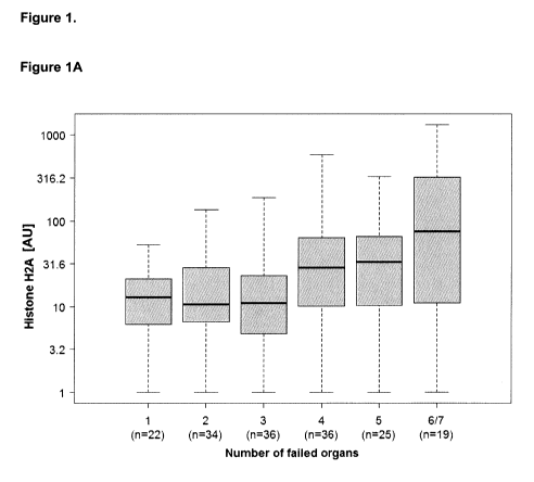
organ dysfunction; see e.g. Figures 1A to 1D. In addition, it is surprisingly
demonstrated that an
increase in the level of the proADM, particularly of the fragment MR-proADM,
in the sample of
the subject indicates the organ dysfunction; see e.g. Figure 1E.
Moreover, it is documented in the appended examples that the increase of the
levels of the
markers increases with the number of organ dysfunctions; see e.g. Example 1.
For example, it
is unexpectedly shown herein below that the levels of the histones,
particularly H2B, H4, H2A
CA 03033102 2019-02-06
WO 2018/029213 PCT/EP2017/070111
4
and H3, further increase in case four or more organ dysfunctions occur in the
subject compared
to the levels of subjects suffering from one to three organ dysfunctions.
Accordingly, the level of
the at least one histone protein is particularly indicative of four or more
organ dysfunctions.
Therefore, the level of the histone(s) is also particularly indicative to
determine whether the
subject suffers from three organ dysfunctions or four organ dysfunctions; see
Table 1.
Additionally, the appended examples unexpectedly demonstrate that the level of
proADM,
particularly of the fragment MR-proADM, increases stepwise from one organ
dysfunction, two
organ dysfunctions, three organ dysfunctions to four organ dysfunctions; see
e.g. Figure 1E,
and Table 1. Accordingly, the level of proADM, particularly of MR-proADM, is
particularly
indicative of one organ dysfunction, two organ dysfunctions, three organ
dysfunctions or four
organ dysfunctions. Accordingly, the level of proADM, particularly of MR-
proADM, is also
indicative to determine whether the subject suffers from one organ
dysfunction, two organ
dysfunctions, three organ dysfunctions or four organ dysfunctions.
Moreover, the appended Examples demonstrate that the level of at least one
histone,
particularly the level of histone H2B, is indicative whether the subject
suffers from one organ
dysfunction, two organ dysfunctions to three organ dysfunctions or whether the
subject suffers
from four or more organ dysfunctions; see e.g. Figure 1 and Table 2A. In
addition, it is
documented herein below that the level of proADM, particularly the level of MR-
proADM, is
indicative whether the subject suffers from one organ dysfunction, two organ
dysfunctions, three
organ dysfunctions, or four or more organ dysfunctions; see e.g. Figure 1,
Table 1 and Table
2A.
In addition, it is documented in the appended examples that the determination
of a level of a
further marker or parameter in addition to the level of at least one histone
or of proADM further
improves the prediction of organ dysfunction; see e.g. Table 2B. For example,
the clinical
scores, such as the SAPS II or the SOFA score, further improve the diagnosis.
In addition, it is
exemplified herein that the combination of makers further improves the
diagnosis of organ
dysfunction. For example, determining the level of proADM, particularly MR-
proADM, and the
level of at least one histone improve the prediction of organ dysfunction in
the subject compared
to the determination of only one marker; see illustrative Table 2B. It is
further demonstrated that
further markers, such as Aldolase B, procalcitonin, or lactate, improve the
prediction of organ
dysfunction in the subject. For example, determining the level of Aldolase B
or lactate in
addition to proADM increases the statistical relationship with the event of
organ dysfunction in
the subject. Accordingly, the invention also relates to a method comprising
determining the level
of a further marker and/or parameter, i.e. the use of marker panels.
The present invention has, inter alia, the following advantages over the
conventional methods:
the inventive methods and the kits are fast, objective, easy to use and
precise for the prediction
of organ dysfunction(s). The methods and kits of the invention relate to
markers that are easily
CA 03033102 2019-02-06
WO 2018/029213 PCT/EP2017/070111
measurable in routine in hospitals because the levels of histones and of
proADM can be
determined in routinely obtained blood samples or further biological fluids
obtained from a
subject. In addition, the determination of the levels of the histones or
proADM is very fast.
Therefore, the methods and the kits of the invention are suitable for a quick
assessment, and
diagnosis and prognosis of organ dysfunction(s). Accordingly, the quick
determination also is
suitable for a fast treatment decision. Furthermore, due to the simple outcome
of a biomarker
measurement as one specific value, there is no subjective bias of medical
staff when using this
method or the kits of the invention. The reproducibility is thus higher
compared to subjective
scoring for physiological parameters as, for example, employed in the SOFA
score. The level of
the histone or proADM can also be combined with further marker(s) and/or
parameter(s) as
add-on to existing assessments or scores in order to further improve the
prediction and to adapt
the analysis to specific sensitivities and specificities for evaluating the
overall status of critical ill
patients. When looking at patients with multiorgan failure, biomarkers may
help to stratify
patients according to their number of failed organs and support the diagnosis
and treatment
decision in this important field of ICU care.
As documented herein above and in the appended examples, the level of histones
and the level
of proADM were surprisingly found to correlate with organ dysfunction(s) in
subjects.
Accordingly, the present invention relates to methods and kits to determine
organ dysfunction in
a subject by determining the level of proADM and/or (a) histone(s) in a sample
of the subject.
Further, the invention relates to methods and kits for the diagnosis,
prognosis, risk assessment,
risk stratification, and/or monitoring of organ dysfunction in a subject.
Further, the invention
relates to methods and kits for therapy guidance and/or therapy control of
subjects suffering
from an organ dysfunction.
Accordingly, the invention relates to a method for the diagnosis, prognosis,
risk assessment,
risk stratification, monitoring, therapy guidance and/or therapy control of
organ dysfunction in a
subject, wherein said method comprises
(i) determining a level of at least one histone in a sample of said
subject, and wherein said
level of at least one histone is indicative of said organ dysfunction; and/or
(ii) determining a level of proadrenomedullin (proADM) in a sample of said
subject, and
wherein said level of proADM is indicative of said organ dysfunction.
As used herein, the term "determining the level of at least one histone" or
the like refers to
determining a level of a histone or a fragment thereof in a sample of the
subject or determining
a level of more than one histones or fragments thereof in the sample of the
subject. Particularly,
"determining the level of at least one histone" may refer to determining a
level of a histone in the
sample of the subject, wherein preferably the histone is selected from the
group consisting of
CA 03033102 2019-02-06
WO 2018/029213 PCT/EP2017/070111
6
histone H2B, H4, H2A and H3. Particularly, the level of the histone H2B is
determined. Further,
"determining the level of at least one histone" may refer to determining a
level of a histone in the
sample of the subject, wherein particularly the level of the histone H4 is
determined. Further,
"determining the level of at least one histone" may refer to determining a
level of two histones in
the sample of the subject, wherein preferably the levels of the histones H2B
and H4 are
determined. Further, "determining the level of at least one histone" may refer
to determining a
level of three histones in the sample of the subject, wherein preferably the
levels of the histones
H2B, H4, and H2A are determined. Further, "determining the level of at least
one histone" may
refer to determining a level of four histones in the sample of the subject,
wherein preferably the
levels of the histones H2B, H4, H2A and H3 are determined.
Accordingly, in the context of the present invention, "determining the level
of at least one
histone" or the like may refer to determining a level of histone H2B, a level
of histone H4, a level
of histone H2A and/or a level of histone H3.
In particular, the term "determining the level of at least one histone" or the
like may refer to
determining a level of a histone in the sample of the subject. Accordingly,
the invention also
relates to a method for the diagnosis, prognosis, risk assessment, risk
stratification, monitoring,
therapy guidance and/or therapy control of organ dysfunction in a subject,
wherein said method
comprises determining a level of a histone or a fragment thereof in a sample
of said subject and
wherein said level of a histone or said fragment thereof is indicative of said
organ dysfunction.
As used herein, "histone" or "histone protein", or "histones" or "histone
proteins" refers to the
canonical histone(s), such as H1, H2A, H2B, H3 or H4, as well as histone
variant(s), such as
H3.3, H2A.Z etc. or fragment(s) thereof. Histones form the octamer around
which DNA is
wrapped in order to assemble the chromatin structure (Luger, Nature. 1997 Sep
18;
389(6648):251-60). For example, the histone proteins H2A, H2B, H3, and H4 (two
of each) form
an octamer, which is wrapped by 165 base pairs of DNA to form the fundamental
subunit of
chromatin, the nucleosome. Histones are also detected outside the nucleus in
multiple
pathophysiological processes (WO 2009/061918). The presence of extracellular
histones has
been described in the blood of patients suffering from different etiologies
involving inflammatory
processes. Histone release from activated immune cells can be mediated by
extracellular traps.
Activated neutrophils, as an ultimate mechanism of controlling and clearing an
infection, can
release extracellular fibers, so called neutrophile extracellular traps (NETs)
(Brinkmann V., et al.
Science 2004; 303(5663): p. 1532-5). Other mechanisms by which histones may be
released
into a patient's blood stream include apoptosis, necrosis, pyroptosis or
necroptosis of cells.
In particular, the at least one histone is selected from the group consisting
of H2B, H4, H2A and
H3. Accordingly, the level of the histone to be determined in the methods and
kits of the
invention is particularly a level of the histones(s) H2B, H4, H2A and/or H3.
The sequences of
CA 03033102 2019-02-06
WO 2018/029213 PCT/EP2017/070111
7
the histones are known to the skilled person. Exemplary sequences of the
histones are given in
SEQ ID NOs: 1 to 4. The exemplary amino acid sequence of histone H4 is given
in SEQ ID NO:
1. The exemplary amino acid sequence of histone H2A is given in SEQ ID NO: 2.
The
exemplary amino acid sequence of histone H3 is given in SEQ ID NO: 3. The
exemplary amino
acid sequence of histone H2B is given in SEQ ID NO: 4. Particularly, the at
least one histone is
selected from the group consisting of H2B, H4, H2A and H3. More particularly,
the at least one
histone is selected from the group consisting of H2B, H4 and H2A. More
particularly, the at least
one histone is H2B and H4. More particularly, the at least one histone is H2B
or H4.
It is understood that "determining the level of at least one histone" or the
like refers to
determining the level of at least one histone or a fragment of the at least
one histone in the
sample. In particular, the level of the histone H2B, H3, H2A, and/or H4 is
determined in the
sample. Accordingly, the at least one histone determined in the sample can be
a free histone or
the at least one histone determined in the sample can occur and can be
assembled in a
macromolecular complex, for example, in the octamer, nucleosome and/or NETs.
Therefore, the
level of at least one histone in the sample can comprise the level of free
histone protein and/or
histone protein assembled in a macromolecular complex.
In particular aspects of the invention, a level of a histone or a fragment
thereof can be
determined in the sample that is not assembled in a macromolecular complex,
such as a
nucleosome, octamer or a neutrophil extracellular trap (NET). Such histone(s)
are herein
referred to as "free histone(s)". Accordingly, the level of the at least one
histone may particularly
be a level of at least one free histone.
The level of such free histones can be determined by the detection of amino
acid sequences or
structural epitopes of histones that are not accessible in an assembled
stoichiometric
macromolecular complex, like a mono-nucleosome or an octamer. In such
structures, particular
regions of the histones are covered and are thus sterically inaccessible as
shown for the
neutrophil extracellular traps ("NETs"), (Brinkmann V., et al. Science
303(5663): p. 1532-5,
2004). In addition, in the octamer or nucleosome, regions of histones also
participate in
intramolecular interactions, such as between the individual histones.
Accordingly, the
region/peptide/epitope of the histone that is determined in the context of the
invention may
determine whether the histone is a free histone or a histone that is assembled
in a
macromolecular complex. For example, in an immunoassay based method, the
utilized
antibodies may not detect histones, e.g. H4, when they are part of the
octameric core of
nucleosomes as the epitopes are structurally inaccessible. Herein below,
regions/peptides/epitopes of the histone are exemplified that could be
employed to determine a
free histone. For example, regions/peptides/epitopes of the N-terminal or C-
terminal tail of the
histones can be employed to determine histones independent of whether they are
assembled in
the macromolecular complex or are free histones according to the present
invention.
CA 03033102 2019-02-06
WO 2018/029213 PCT/EP2017/070111
8
It is understood that the determination of histones may include post-
translational modified
histone proteins. Accordingly, the post-translational modifications can
comprise deacetylation or
acetylation, phosphorylation, methylation, ubiquitylation and citrullination
of amino acids.
"Stoichiometric" in this context relates to intact complexes, e.g. a
mononucleosome or an
octamer. "Free histone proteins" can also comprise non-chromatin-bound
histones. For
example, "free histone proteins" may also comprise individual histone proteins
or non-octameric
histone complexes. Free histones may (e.g. transiently) be bound to individual
histones, for
instance, histones may form homo- or hetero-dimers. The free histones may also
form homo- or
hetero-tetramers. The homo- or heterotetramer may consist of four molecules of
histones, e.g.
H2A, H2B, H3 and/or H4. A typical heterotetramer is formed by two
heterodimers, wherein each
heterodimer consists of H3 and H4. It is also understood herein that a
heterotetramer may be
formed by H2A and H2B. It is also envisaged herein that a heterotetramer may
be formed by
one heterodimer consisting of H3 and H4, and one heterodimer consisting of H2A
and H2B.
Free histones are thus herein referred to as and can be monomeric,
heterodimeric or tetrameric
histone proteins, which are not assembled in a ("stoichiometric")
macromolecular complex
consisting of the histone octamer bound to nucleic acid, e.g. a nucleosome. In
addition, free
histones may also be bound to nucleic acids, and wherein said free histones
are not assembled
in a ("stoichiometric") macromolecular complex, e.g. an intact nucleosome.
Preferably, the free
histone(s) is/are essentially free of nucleic acids.
The fragment of the at least one histone can have any length, e.g. at least
about 5, 10, 20, 30,
40, 50 or 100 amino acids, so long as the fragment allows the unambiguous
determination of
the level of the particular histone. The fragment of the at least one histone
refers to an
independent fragment of the histones, e.g. of the histones H2B, H4, H2A and
H3. Various
exemplary fragments of the histones are disclosed herein below that are
suitable to determine
the level of the histone in the sample of the subject. It is also herein
understood that the level of
the histones can be determined by determining a fragment spanning the N-
terminal or C-
terminal tail of the histones. In addition, the histone or the fragment
thereof to be determined in
the context of the present invention may also be modified, e.g. by post-
translational
modification. Exemplary post translational modifications can be acetylation,
citrullination,
deacetylation, methylation, demethylation, deimination, isomerization,
phosphorylation and
ubiquitination.
As used herein, the term "proadrenomedullin" or "proADM" refers to
proadrenomedullin or a
fragment thereof, particularly MR-proADM. It is understood that "determining
the level of
proADM" or the like refers to determining the level of proADM or a fragment
thereof in the
CA 03033102 2019-02-06
WO 2018/029213 PCT/EP2017/070111
9
sample. The fragment can have any length, e.g. at least about 5, 10, 20, 30,
40, 50 or 100
amino acids, so long as the fragment allows the unambiguous determination of
the level of the
proADM. In particular preferred aspects of the invention, "determining the
level of proADM"
refers to determining the level of midregional proadrenomedullin (MR-proADM).
MR-proADM is
a fragment of proADM. The peptide adrenomedullin (ADM) was discovered as a
hypotensive
peptide comprising 52 amino acids, which had been isolated from a human
phenochromocytomeby (Kitamura et al., 1993). Adrenomedullin (ADM) is encoded
as a
precursor peptide comprising 185 amino acids ("preproadrenomedullin" or "pre-
proADM"). An
exemplary amino acid sequence of ADM is given in SEQ ID NO: 5. ADM comprises
the
positions 95-146 of the pre-proADM amino acid sequence and is a splice product
thereof.
"Proadrenomedullin" ("proADM") refers to pre-proADM without the signal
sequence (amino
acids 1 to 21), i.e. to amino acid residues 22 to 285 of pre-proADM.
"Midregional
proadrenomedullin" ("MR-proADM") refers to the amino acids 42-95 of pre-
proADM. An
exemplary amino acid sequence of MR-proADM is given in SEQ ID NO: 6. It is
also envisaged
herein that a peptide and fragment thereof of pre-proADM or MR-proADM can be
used for the
herein described methods. For example, the peptide or the fragment thereof can
comprise the
amino acids 22-41 of pre-proADM (PAMP peptide) or amino acids 95-146 of pre-
proADM
(mature adrenomedullin). A C-terminal fragment of proADM (amino acids 153 to
185 of
preproADM) is called adrenotensin. Fragments of the proADM peptides or
fragments of the MR-
proADM can comprise, for example, at least about 5, 10, 20, 30 or more amino
acids.
Accordingly, the fragment of proADM may, for example, be selected from the
group consisting
of MR-proADM, PAMP, adrenotensin and mature adrenomedullin, preferably herein
the
fragment is MR-proADM.
It is also envisaged herein that polypeptides can be determined, which have a
sequence identity
to proADM or to the at least one histone. For example, polypeptides can be
determined in the
methods and kits of the invention that have at least 75%, 80%, 85%, 90%, 91%,
92%, 93%,
94%, 95%, 96%, 97%, 98% or 99% sequence identity to SEQ ID NO: 5 or 6, or
respectively to
SEQ ID NO: 1, SEQ ID NO: 2, SEQ ID NO: 3, or SEQ ID NO: 4, wherein the higher
values of
sequence identity are preferred. In accordance with the present invention, the
terms "sequence
identity", "homology" or "percent homology" or "identical" or "percent
identity" or "percentage
identity" in the context of two or more amino acid sequences refers to two or
more sequences or
subsequences that are the same, or that have a specified percentage of amino
acids that are
the same, when compared and aligned for maximum correspondence over the window
of
comparison (preferably over the full length), or over a designated region as
measured using a
sequence comparison algorithm as known in the art, or by manual alignment and
visual
inspection. Sequences having, for example, 70% to 90% or greater (preferably
95% or greater)
sequence identity may be considered to be substantially identical. Such a
definition also applies
CA 03033102 2019-02-06
WO 2018/029213 PCT/EP2017/070111
to the complement of a test sequence. Preferably, the described identity
exists over a region
that is at least about 10 to about 15 amino acids in length, more preferably,
over a region that is
at least about 20 to about 35 amino acids in length, most preferably, over the
full length. Those
having skill in the art will know how to determine percent identity
between/among sequences
using, for example, algorithms such as those based on CLUSTALW computer
program
(Thompson Nucl. Acids Res. 2 (1994), 4673-4680) or FASTDB (Brutlag Comp. App.
Biosci. 6
(1990), 237-245), as known in the art.
As used herein, the "level" of the marker refers to the quantity of the
molecular entity of the
marker in the sample. In other words, the concentration of the marker is
determined in the
sample. For example, the concentration of proADM or a fragment thereof,
preferably MR-
proADM, and/or the concentration of the histone(s) H2B, H4, H3 and/or H2A or
(a) fragment(s)
thereof is determined in the sample of the subject.
As used herein, the term "level of at least one histone" refers to the
quantity of the molecular
entity of the at least histone, e.g. the quantity of H2B, H4, H2A and/or H3,
or a fragment thereof
in a sample that is obtained from the subject. In other words, the
concentration of the at least
one histone protein or the fragment thereof is determined in the sample.
As used herein, the term "level of the marker proadrenomedullin (proADM)" or
the "level of the
marker proadrenomedullin (proADM) or a fragment thereof" refers to the
quantity of the
molecular entity of the marker proadrenomedullin or fragments thereof in a
sample that is
obtained from a subject. In other words, the concentration of the marker is
determined in the
sample. Hence, the term "level of the marker midregional proadrenomedullin (MR-
proADM)"
refers to the quantity of the molecular entity of the marker midregional
proadrenomedullin (MR-
proADM) in the sample that is obtained from a subject. As described above, it
is also envisaged
herein that a fragment of proadrenomedullin (proADM), preferably MR-proADM,
can be
detected and quantified. Also, fragments of MR-proADM can be detected and
quantified.
Suitable methods to determine the level of proADM or a fragment thereof
(preferably MR-
proADM) or to determine the level of the at least one histone or a fragment
thereof are
described herein below.
An organ is a collection of tissues joined in a structural unit to serve a
common function. As
used herein, the general term "organ dysfunction" can also mean that more than
one organ has
a dysfunction, i.e. it can also relate to organ dysfunctions unless stated
otherwise. The term
"organ dysfunction" or "organ dysfunctions" relates to a condition in the
subject where an organ
or more than one organ do(es) not perform its/their normal function compared
to an unaffected
organ, such for example the organ(s) of at least one healthy subject. For
example, the organ(s)
CA 03033102 2019-02-06
WO 2018/029213 PCT/EP2017/070111
11
may have a reduced activity or the organ(s) may be abnormally active in the
subject with the
organ dysfunction in comparison to (an) organ(s) of at least one healthy
subject. Preferably, the
organ(s) with the organ dysfunction(s) may have an impaired (reduction or an
increase) activity
of at least about 10%, 20%, 30%, 50%, 70%, 90%, 100% or 200% compared to
unaffected
organ(s), e.g. of at least one healthy subject.
In particular, organ dysfunction(s) can result in organ failure(s).
Accordingly, "organ
dysfunction(s)" can preferably also refer to organ failure(s). "Organ
failure(s)" refers to (an)
organ dysfunction(s) to such a degree that normal homeostasis cannot be
maintained, e.g.
without external clinical intervention. "Organ failure(s)" may also refer to
(an) organ
dysfunction(s) to such a degree that normal homeostasis of the organ(s) cannot
be maintained,
e.g. without external clinical intervention. "Organ failure(s)" may also refer
to (an) organ
dysfunction(s) to such a degree that normal homeostasis of the subject cannot
be maintained,
e.g. without external clinical intervention.
In particular aspects of the invention, the organ dysfunction is an organ
failure or at least one
organ failure. The general term "organ failure" can also mean that more than
one organ has a
failure, i.e. it can also relate to organ failures unless stated otherwise. It
is herein understood
that organ dysfunctions can also be referred to as multiple organ dysfunction.
It is herein
understood that organ failures can also be referred to as multiple organ
failure.
Exemplary organ dysfunctions or organ failures are circulatory shock,
hematologic failure, liver
failure, neurologic failure, renal failure, respiratory failure and metabolic
acidosis. Accordingly, in
the context of the invention, the organ dysfunction or the at least one
dysfunction can preferably
be selected from the group consisting of circulatory shock, hematologic
failure, liver failure,
neurologic failure, renal failure, respiratory failure and metabolic acidosis.
It is herein
understood that the subject can also have more than one organ dysfunctions or
failures that are
e.g. a combination of two, three, four organ dysfunctions selected from the
group consisting of
circulatory shock, hematologic failure, liver failure, neurologic failure,
renal failure, respiratory
failure and metabolic acidosis. For example, the methods and kits of the
invention can
determine whether the subject suffers from two organ dysfunction, e.g. a
circulatory shock and
a respiratory failure.
As used herein, a subject with a "circulatory shock" may refer to a subject
that has a systolic
arterial pressure lower than about 90 mmHg with e.g. signs of peripheral
hypoperfusion.
Accordingly, such a subject can have the need for infusion of vasopressor
and/or inotropic
agents.
CA 03033102 2019-02-06
WO 2018/029213 PCT/EP2017/070111
12
As used herein, a subject with a "hematologic failure" may refer to a subject
that has a
thrombocythemia lower than about 100,000/mm3.
As used herein, a subject with a "liver failure" may refer to a subject that
has bilirubinemia
greater than about 2 mg/dL and/or enzyme levels of aspartate or alanine
transaminase greater
than about 500 international units per liter.
As used herein, a subject with a "neurologic failure" may refer to a subject
that has Glasgow
coma scale below 13.
As used herein, a subject with a "renal failure" may refer to a subject that
has urine output less
than about 0.5 ml/kg/h for at least about 3 hours and/or creatinemia rising
more than about 50
% as compared to previous values.
As used herein, a subject with a "respiratory failure" may refer to a subject
that has a ratio of the
partial pressure of arterial oxygen to the fraction of inspired oxygen (Pa
02/Fi 02) lower than
about 300 mmHg regardless of the chosen ventilatory support.
As used herein, a subject with a "metabolic acidosis" may refer to a subject
with a lactate level
below about 2.5 mmol/L, as base excess (base level below -2 mEquivalent/L) or
bicarbonate
levels (H003-) below about 24, preferably below about 18 mmol/L.
The term "indicative of said organ dysfunction" means that the subject has or
will likely
have/suffer from an organ dysfunction or at least one organ dysfunction.
Therefore, the level of
the at least one histone and/or the level of proADM of the subject indicate(s)
organ dysfunction
in the subject.
The method of the invention also relates to a method, wherein a level of at
least one histone is
determined in a sample of a subject, wherein said level of at least one
histone is compared to a
reference level of at least one histone and wherein said level of at least one
histone is indicative
of said organ dysfunction.
The invention also relates to a method, wherein a level of proadrenomedullin
(proADM),
particularly MR-proADM, is determined in a sample of a subject, wherein said
level of proADM,
particularly MR-proADM, is compared to a reference level of proADM and wherein
said level of
proADM, particularly MR-proADM, is indicative of said organ dysfunction.
The method also relates to a method, wherein a level of at least one histone
is determined and
wherein a level of proadrenomedullin (proADM), particularly MR-proADM, is
determined in a
sample of a subject, wherein said level of at least one histone is compared to
a reference level
CA 03033102 2019-02-06
WO 2018/029213 PCT/EP2017/070111
13
of at least one histone, and wherein said level of proADM is compared to a
reference level of
proADM, and wherein said organ dysfunction in said subject is identified based
on the
comparison step.
As used herein, the term "is compared to a reference level of at least one
histone" or
grammatical variants thereof means that the level of the at least one histone
of the subject is
compared to a reference level of the at least one histone. Thus, a level of
the histone of the
subject is compared to a corresponding reference level of the same histone.
For example, the
level of the histone H2B determined in the sample of the subject is compared
to a reference
level of histone H2B. This applies mutatis mutandis to the other histones. The
reference level of
at least one histone is particularly a level of histone H2B, a level of
histone H4, a level of histone
H2A and/or a level of histone H3.
As used herein, the term "is compared to a reference level of proADM" or
grammatical variants
thereof means that the level of the proADM of the subject is compared to a
reference level of
the proADM. If a level of (a) fragment(s) of the at least one histone and/or
of the proADM is
determined the reference level may also be a level of (the) corresponding
fragment(s).
As used herein, the "reference level" may reflect a normal level of the
corresponding marker
that is indicative of no organ dysfunction or organ failure in preferred
aspects of the invention.
Thus, the reference level can represent the level of the at least one histone
and/or the level of
proADM of a group of healthy subjects (e.g. a cohort). A healthy subject is a
subject with no
diagnosed (and confirmed) disease(s) and/or medical disorder(s). The healthy
subjects may
preferably have normally functioning organs, i.e. no organ dysfunction(s) or
no organ failure(s).
Accordingly, the reference level(s) can be a level of at least one histone
and/or a level of
proADM that is determined in samples of healthy subjects. The reference
subjects or healthy
subjects are herein preferably defined as a group of subjects or a group of
healthy subjects, e.g.
a cohort of subjects. The healthy reference subjects preferably have no organ
dysfunction or no
organ failure. Accordingly, the reference level is preferably a level of the
at least one histone
and/or a level of proADM of subjects having no organ dysfunction or no organ
failure.
Accordingly, the reference level is preferably a level indicating no organ
dysfunction or no organ
failure.
The reference level may be a level of at least one histone and/or a level of
proADM of at least
one reference subject, wherein said reference subject(s) has/have no organ
dysfunction or no
organ failure.
Further, the reference level can also be a level of at least one histone
and/or a level of proADM
of at least one reference subject, wherein said reference subject(s) suffer(s)
from a disease
CA 03033102 2019-02-06
WO 2018/029213 PCT/EP2017/070111
14
and/or medical disorder, and wherein said subject(s) has/have no organ
dysfunction or no organ
failure.
Further, the reference level can also be a level of at least one histone
and/or a level of proADM
of at least one reference subject, wherein said reference subject(s) suffer(s)
from a disease
and/or medical disorder and an infection (such as sepsis or septic disorders),
and wherein said
subject(s) has/have no organ dysfunction or no organ failure.
Further, the reference level can also be a level of at least one histone
and/or a level of proADM
of at least one reference subject, wherein said reference subject(s) suffer(s)
from a disease
and/or medical disorder and not from an infection, and wherein said subject(s)
has/have no
organ dysfunction or no organ failure.
Further, the reference level can also be a level of at least one histone
and/or a level of proADM
of at least one reference subject, and wherein said reference subject(s)
suffer(s) from a disease
and/or medical disorder including systemic inflammatory response syndrome
(SIRS), wherein
said subject(s) do(es) not suffer from an infection, and wherein said
subject(s) has/have no
organ dysfunction or no organ failure.
As used herein, the at least one reference subject refers to more than one
reference subject.
Particularly, the at least one reference subject is a group or a cohort of
reference subjects. As
described herein below, means and methods are described to determine the
levels of the
markers e.g. in reference subjects and exemplary reference levels are also
provided.
The reference level as used herein is typically a predetermined level, i.e. it
has been determined
in advance as a reference for later use at the point-of-care, e.g. ICU.
As documented herein, an increased level of at least one histone (or an
increase in the level of
at least one histone) and/or an increased level of proADM (or an increase in
the level of
proADM), particularly MR-proADM, as compared to the reference level is
indicative of organ
dysfunction. Accordingly, the method of the invention includes a method that
comprises
determining a level of at least one histone in a sample of said subject, and
wherein an
increased level of said at least one histone of said subject as compared to a
reference level of
at least one histone is indicative of said organ dysfunction in said subject.
Further, the invention includes a method that comprises determining a level of
proADM,
particularly MR-proADM, in a sample of said subject, and wherein an increased
level of said
proADM, particularly MR-proADM, of said subject as compared to a reference
level of said
proADM, particularly MR-proADM is indicative of said organ dysfunction in said
subject.
Further, the invention includes a method that comprises determining a level of
at least one
histone in a sample of said subject and determining a level of proADM,
particularly MR-proADM,
in a sample of said subject, and wherein an increased level of said at least
one histone of said
subject and an increased level of said proADM, particularly MR-proADM, of said
subject as
CA 03033102 2019-02-06
WO 2018/029213 PCT/EP2017/070111
compared to a reference level of at least one histone and a reference level of
said proADM,
particularly MR-proADM are indicative of said organ dysfunction in said
subject.
The invention also relates to a method comprising
(i) determining a level of at least one histone in a sample of a subject,
wherein said level of the at least one histone is compared to a reference
level of
the at least one histone,
and wherein an increased level of the at least one histone of said subject as
compared to said reference level of the at least one histone is indicative of
organ
dysfunction in said subject; and/or
(ii) determining a level of proadrenomedullin (proADM) in a sample of said
subject,
wherein the level of proADM is compared to a reference level of proADM,
and wherein an increased level of the proADM of said subject as compared to
the
reference level of proADM is indicative of organ dysfunction in said subject.
In other words, the invention also relates to a method comprising
(i) determining a level of at least one histone in a sample of a subject,
wherein said level of the at least one histone is compared to a reference
level of
the at least one histone,
and wherein an increase in the level of the at least one histone of said
subject as
compared to said reference level of the at least one histone is indicative of
organ
dysfunction in said subject; and/or
(ii) determining a level of proadrenomedullin (proADM) in a sample of said
subject,
wherein the level of proADM is compared to a reference level of proADM,
and wherein an increase in the level of proADM of said subject as compared to
the
reference level of proADM is indicative of organ dysfunction in said subject.
As used herein, the term "increase in the level of (the) marker" means that
the level of the
marker is increased, i.e. it refers to an increased level of the marker.
Accordingly, the term
"increase in the level of (the) marker" is used interchangeably herein with
the term "increased
level of (the) marker". An increased level of the marker or an increase in the
level of the marker
of the subject means that the level of the marker is at least about 15%,
preferably at least about
20%, more preferably at least about 25%, or even more preferably at least
about 30% higher
than the reference level of the marker.
In the context of the invention, the term "increase in the level of at least
one histone as
compared to the reference level" or the like is used interchangeably with the
term "increased
level of the at least one histone of said subject as compared to the reference
level" or the term
CA 03033102 2019-02-06
WO 2018/029213 PCT/EP2017/070111
16
"increased level of the at least one histone as compared to the reference
level" or the like. Such
terms mean that the level of the at least one histone, e.g. the level of H2B,
the level of H4, the
level of H2A and/or the level of H3, of the subject is at least about 15%,
preferably at least
about 20%, more preferably at least about 25%, or even more preferably at
least about 30%
higher than the reference level of the at least one histone.
As used herein, the term "increase in the level of proADM as compared to the
reference level"
or the like is used interchangeably herein with the term "increased level of
the proADM of said
subject as compared to said reference level" or the term "increased level of
the proADM as
compared to said reference level" or the like. Such terms mean that the level
of proADM,
particularly the level of MR-proADM, of the subject is at least about 15%,
preferably at least
about 20%, more preferably at least about 25%, or even more preferably at
least about 30%
higher than the reference level of proADM, particularly MR-proADM.
The invention may also relate to a method comprising
(i) determining a level of at least one histone in a sample of said
subject,
wherein said level of at least one histone is compared to a reference level of
at
least one histone of the same subject obtained from prior analysis,
and wherein an increase in the level of at least one histone as compared to
said
reference level of at least one histone is indicative of organ dysfunction or
a further
organ dysfunction in said subject; and/or
(ii) determining a level of proadrenomedullin (proADM) in a sample of said
subject,
wherein said level of proADM is compared to a reference level of proADM of the
same subject obtained from prior analysis,
and wherein an increase in the level of proADM as compared to said reference
level of proADM is indicative of organ dysfunction or a further organ
dysfunction in
said subject.
As used herein, "obtained from prior analysis" refers to a determination of
the marker level in
the sample of the same subject at a pervious time, e.g. 28 days, 7 days, 6
days, 5 days, 4 days,
3 days, 2 days or 1 day prior to the next analysis, and wherein in such
aspects said previously
determined level of the marker is considered as the reference level. As used
herein, a further
organ dysfunction may mean that a further organ shows/has a dysfunction or
failure. For
example, if the subject suffers from one organ dysfunction, a further organ
dysfunction means
that the subject suffers from two or more organ dysfunctions or failures.
The increased level of at least one histone of a subject as compared to a
reference level of at
least one histone can be indicative of at least one organ dysfunction, at
least two organ
dysfunctions, or at least three organ dysfunctions. Particularly, the
increased level of at least
one histone of said subject as compared to the reference level of the at least
one histone can
CA 03033102 2019-02-06
WO 2018/029213 PCT/EP2017/070111
17
be indicative of at least five organ dysfunctions, or at least six organ
dysfunctions. More
particularly, the increased level of at least one histone of said subject as
compared to the
reference level of the at least one histone can be indicative of at least four
organ dysfunctions in
said subject.
The increased level of proADM of a subject as compared to a reference level of
proADM can be
indicative of at least one organ dysfunction. Particularly, the increased
level of said proADM of
said subject as compared to said reference level of proADM can be indicative
of at least two
organ dysfunctions. More particularly, the increased level of said proADM of
said subject as
compared to said reference level of proADM can be indicative of at least three
organ
dysfunctions. More particularly, the increased level of said proADM of said
subject as compared
to said reference level of proADM can be indicative of at least four organ
dysfunctions.
As used herein, the term "one organ dysfunction" means that the subject has
one organ
dysfunction, i.e. one organ of the subject has a dysfunction. As used herein,
the term "two organ
dysfunctions" means that the subject has two organ dysfunctions, i.e. two
organs of the subject
have a dysfunction. This applies mutatis mutandis to the terms three
dysfunctions, four
dysfunctions, five dysfunctions or six organ dysfunctions.
Further, it is documented in the appended examples that the levels of the
histones, particularly
H2B, H4, H2A and H3, increase in case four or more organ dysfunctions occur in
the subject
compared to the levels of subjects suffering from one to three organ
dysfunctions; see e.g.
Figure 1 and Table 1. Therefore, the methods and kits of the invention can
determine how many
organ dysfunctions the subject suffers from or will likely suffer from.
Accordingly, in the context of the herein provided methods and kits an
increased level of said at
least one histone of said subject as compared to a reference level is
indicative of at least four
organ dysfunctions in said subject, wherein said reference level indicates one
to three organ
dysfunctions.
Further, an increased level of at least one histone of a subject as compared
to a reference level
of the at least one histone is indicative of at least five organ dysfunctions
in said subject,
wherein said reference level indicates four organ dysfunctions.
Further, an increased level of at least one histone of a subject as compared
to a reference level
of the least one histone is indicative of at least six or seven organ
dysfunctions in said subject,
wherein said reference level indicates five organ dysfunctions.
Additionally, the appended examples demonstrate that the level of proADM,
particularly of the
fragment MR-proADM, increases stepwise from one organ dysfunction, two
dysfunctions, three
CA 03033102 2019-02-06
WO 2018/029213 PCT/EP2017/070111
18
dysfunctions, four organ dysfunctions to five organ dysfunctions; see e.g.
Figure 1E, and Table
1.
Accordingly, in the context of the herein provided methods and kits an
increased level of
proADM, particularly MR-proADM, of a subject as compared to a reference level
of proADM is
indicative of at least one organ dysfunction in said subject, wherein said
reference level
indicates no organ dysfunction.
Further, an increased level of proADM, particularly MR-proADM, of a subject as
compared to a
reference level of proADM is indicative of at least two organ dysfunctions in
said subject,
wherein said reference level indicates one organ dysfunction.
Further, an increased level of proADM, particularly MR-proADM, of a subject as
compared to a
reference level of proADM is indicative of at least three organ dysfunctions
in said subject,
wherein said reference level indicates two organ dysfunctions.
Further, an increased level of proADM, particularly MR-proADM, of a subject as
compared to a
reference level of proADM is indicative of at least four organ dysfunctions in
said subject,
wherein said reference level indicates three organ dysfunctions.
Further, an increased level of proADM, particularly MR-proADM, of a subject as
compared to a
reference level of proADM is indicative of at least five organ dysfunctions in
said subject,
wherein said reference level indicates four organ dysfunctions.
Further, an increased level of proADM, particularly MR-proADM, of a subject as
compared to a
reference level of proADM is indicative of at least six or seven organ
dysfunctions in said
subject, wherein said reference level indicates five organ dysfunctions.
Further, an identical or similar level, e.g. about +/-10%, +/-15%, or +/-20%,
of proADM,
particularly MR-proADM, and/or of at least one histone of a subject as
compared to a reference
level of proADM and/or the at least on histone is indicative of multiple organ
dysfunctions in said
subject, wherein said reference level indicates multiple organ dysfunctions.
As used herein, the term "reference level indicates number of organ
dysfunction" refers to a
level reflecting the number of organ dysfunctions. For example, such a
(reference) level can be
determined in at least one reference subject (typically in a group of
subjects) that suffers from
said particular number of organ dysfunctions. Such levels are documented in
the appended
examples, e.g. levels of markers of subjects suffering from one to six/seven
organ dysfunctions.
Accordingly, the reference level indicating the number of organ dysfunctions
can be a level of at
least one histone of at least one reference subject that suffers from the
number of organ
dysfunctions. Mutatis mutandis, the reference level indicating the number of
dysfunctions can
be a level of said proADM, particularly MR-proADM, of at least one reference
subject that
suffers the number of organ dysfunctions or does not suffer from an organ
dysfunction.
CA 03033102 2019-02-06
WO 2018/029213 PCT/EP2017/070111
19
The present invention also relates to methods and kits of the invention,
wherein at least one
histone is determined in the sample of the subject and wherein the level of
the at least one
histone is indicative whether the subject suffers from one organ dysfunction
to three organ
dysfunctions or whether the subject suffers from four or more organ
dysfunctions. Particularly,
the present invention also relates to methods and kits of the invention,
wherein at least one
histone is determined in the sample of the subject and wherein the level of
the at least one
histone is indicative whether the subject suffers from one organ dysfunction
or whether the
subject suffers from four or more organ dysfunctions.
Particularly, the present invention also relates to methods and kits of the
invention, wherein at
least one histone is determined in the sample of the subject and wherein the
level of the at least
one histone is indicative whether the subject suffers from one organ
dysfunction to three organ
dysfunctions or whether the subject suffers from four organ dysfunctions, or
five organ
dysfunctions or six and more organ dysfunctions.
In addition, it is documented herein below that the level of proADM,
particularly the level of MR-
proADM, is indicative whether the subject suffers from one organ dysfunction
to three organ
dysfunctions or whether the subject suffers from four or more organ
dysfunctions; see e.g.
Table 1 and Table 2A.
Accordingly, the present invention also relates to methods and kits of the
invention, wherein
proADM, particularly MR-proADM is determined in the sample of the subject and
wherein the
level of proADM, particularly MR-proADM, is indicative whether the subject
suffers from one
organ dysfunction or whether the subject suffers two or more organ
dysfunctions.
Further, the present invention also relates to methods and kits of the
invention, wherein
proADM, particularly MR-proADM is determined in the sample of the subject and
wherein the
level of proADM, particularly MR-proADM, is indicative whether the subject
suffers from two
organ dysfunctions or whether the subject suffers from three or more organ
dysfunctions.
Further, the present invention also relates to methods and kits of the
invention, wherein
proADM, particularly MR-proADM is determined in the sample of the subject and
wherein the
level of proADM, particularly MR-proADM, is indicative whether the subject
suffers from three
organ dysfunctions or whether the subject suffers from four or more organ
dysfunctions.
The reference level as used herein is typically a predetermined level, i.e. it
has been determined
in advance as a reference for later use at the point-of-care, e.g. ICU. It is
herein understood that
the reference levels and the determined marker levels (i.e. the levels that
are determined for the
individual subject at the point-of-care, e.g. ICU) can vary depending on the
assay/method by
which the levels are determined as also exemplified in table 4. For example,
the reference level
and the determined marker level determined by mass spectrometry based methods
can be
different from respective levels determined by immunoassays. The appended
examples
demonstrate that the levels of the markers can be determined by several
methods, e.g.
CA 03033102 2019-02-06
WO 2018/029213 PCT/EP2017/070111
immunoassays and mass spectrometry based methods, e.g. table 4, and that the
reference
levels can additionally be optimized by statistical methods, such as the
Youden's index (e.g.
Rota et al., BMC Medical Research Methodology (2015) 15:24; and Ruopp et al.,
Biom J. 2008
June; 50(3): 419-430)). Accordingly, the skilled person is aware how to
determine reference
levels. For example, the levels (including reference levels) can be determined
by an
immunoassay, e.g. by determining the level of at least one histone and/or the
level of proADM,
e.g. MR-proADM, in samples of subjects (or reference subjects) that do not
suffer from (an)
organ dysfunctions or organ failure, or alternatively that do suffer from (an)
organ dysfunctions
or organ failure.
In one embodiment, the reference levels of the at least one histone were
determined by an
immunoassay. The reference level of the at least one histone may be about 100
ng/ml, more
preferably about 90 ng/ml, more preferably about 80 ng/ml, more preferably
about 70 ng/ml,
more preferably about 60 ng/ml, more preferably about 50 ng/ml, more
preferably about 45
ng/ml, more preferably about 40 ng/ml, or most preferably about 35 ng/ml. The
reference level
of the at least one histone determined by an immunoassay may be about 10
ng/ml, more
preferably about 15 ng/ml, more preferably about 20 ng/ml, more preferably
about 25 ng/ml,
more preferably about 30 ng/ml, or most preferably about 35 ng/ml.
The reference level of the at least one histone may be about 10 ng/ml to about
100 ng/ml, about
10 ng/ml to about 90 ng/ml, more preferably about 10 ng/ml to about 60 ng/ml,
more preferably
about 10 ng/ml to about 40 ng/ml, more preferably about 15 ng/ml to about 40
ng/ml, or most
preferably about 20 ng/ml to about 40 ng/ml.
The reference level of the histone H4 determined by an immunoassay may be
about 100 ng/ml,
more preferably about 90 ng/ml, more preferably about 80 ng/ml, more
preferably about 70
ng/ml, more preferably about 60 ng/ml, more preferably about 50 ng/ml, more
preferably about
45 ng/ml, more preferably about 40 ng/ml, and most preferably about 35 ng/ml.
The reference
level of the histone H4 determined by an immunoassay may be about 10 ng/ml,
more preferably
about 15 ng/ml, more preferably about 20 ng/ml, more preferably about 25
ng/ml, more
preferably about 30 ng/ml or most preferably about 35 ng/ml.
The reference level of the histone H4 may be about 10 ng/ml to about 100
ng/ml, about 10
ng/ml to about 90 ng/ml, more preferably about 10 ng/ml to about 60 ng/ml,
more preferably
about 10 ng/ml to about 40 ng/ml, more preferably about 15 ng/ml to about 40
ng/ml, or most
more preferably about 20 ng/ml to about 40 ng/ml.
The exemplary reference levels of proADM were determined by an immunoassay as
described
in the appended examples. For example, the reference level of proADM may be
about 4 nmol/L,
more preferably about 5 nmol/L, more preferably about 7 nmol/L, more
preferably about 8
nmol/L, more preferably about 9 nmol/L or particular preferably about 6
nmol/L.
CA 03033102 2019-02-06
WO 2018/029213 PCT/EP2017/070111
21
An increase of the level of the marker compared to the exemplary reference
level can be
indicative of one organ dysfunction, two organ dysfunctions, three organ
dysfunctions, four
organ dysfunctions, five organ dysfunctions, or six or more organ dysfunctions
in the subject.
Moreover, in another embodiment, the levels (including reference levels) can
be determined by
mass spectrometric based methods, such as methods determining the relative
quantification or
determining the absolute quantification of the protein or fragment thereof of
interest.
Relative quantification "rSRM" may be achieved by:
1. Determining increased or decreased presence of the target protein by
comparing the SRM
(selected reaction monitoring) signature peak area from a given target
fragment peptide
detected in the sample to the same SRM signature peak area of the target
fragment peptide in
at least a second, third, fourth or more biological samples.
2. Determining increased or decreased presence of target protein by comparing
the SRM
signature peak area from a given target peptide detected in the sample to SRM
signature peak
areas developed from fragment peptides from other proteins, in other samples
derived from
different and separate biological sources, where the SRM signature peak area
comparison
between the two samples for a peptide fragment are normalized for e.g to
amount of protein
analyzed in each sample.
3. Determining increased or decreased presence of the target protein by
comparing the SRM
signature peak area for a given target peptide to the SRM signature peak areas
from other
fragment peptides derived from different proteins within the same biological
sample in order to
normalize changing levels of histones protein to levels of other proteins that
do not change their
levels of expression under various cellular conditions.
Such assays can be applied to both unmodified fragment peptides and to
modified fragment
peptides of the target proteins, where the modifications include, but are not
limited to
phosphorylation and/or glycosylation, acetylation, methylation (mono, di,
tri), citrullination,
ubiquitinylation and where the relative levels of modified peptides are
determined in the same
manner as determining relative amounts of unmodified peptides.
Absolute quantification of a given peptide may be achieved by:
1. Comparing the SRM/MRM (multiple reaction monitoring) signature peak area
for a given
fragment peptide from the target proteins in an individual biological sample
to the SRM/MRM
signature peak area of an internal fragment peptide standard spiked into the
protein lysate from
the biological sample. The internal standard may be a labeled synthetic
version of the fragment
peptide from the target protein that is being interrogated or the labeled
recombinant protein.
This standard is spiked into a sample in known amounts before (mandatory for
the recombinant
protein) or after digestion, and the SRM/MRM signature peak area can be
determined for both
the internal fragment peptide standard and the native fragment peptide in the
biological sample
CA 03033102 2019-02-06
WO 2018/029213 PCT/EP2017/070111
22
separately, followed by comparison of both peak areas. This can be applied to
unmodified
fragment peptides and modified fragment peptides, where the modifications
include but are not
limited to phosphorylation and/or glycosylation, acetylation, methylation
(e.g. mono-, di-, or tri-
methylation), citrullination, ubiquitinylation, and where the absolute levels
of modified peptides
can be determined in the same manner as determining absolute levels of
unmodified peptides.
2. Peptides can also be quantified using external calibration curves. The
normal curve approach
uses a constant amount of a heavy peptide as an internal standard and a
varying amount of
light synthetic peptide spiked into the sample. A representative matrix
similar to that of the test
samples needs to be used to construct standard curves to account for a matrix
effect. Besides,
reverse curve method circumvents the issue of endogenous analyte in the
matrix, where a
constant amount of light peptide is spiked on top of the endogenous analyte to
create an
internal standard and varying amounts of heavy peptide are spiked to create a
set of
concentration standards. Test samples to be compared with either the normal or
reverse curves
are spiked with the same amount of standard peptide as the internal standard
spiked into the
matrix used to create the calibration curve.
Accordingly, the skilled person is aware how to determine marker levels and in
particular
appropriate reference levels as is also exemplified in the appended examples.
As described above, reference levels can be determined by determining the
level of at least one
histone and/or the level of proADM in samples of subjects suffering from e.g.
one organ
dysfunction, two organ dysfunctions, three organ dysfunctions, or four or more
organ
dysfunctions. An increased level of the target protein(s) in the sample of the
subject compared
to a reference level of each target protein(s) representing one or more organ
dysfunctions is
then indicative of the number of organ dysfunctions wherein the number of
organ dysfunction in
the sample of the subject is higher than the number of organ dysfunction
represented by the
reference levels. For example, the reference levels of at least one histone
and/or of proADM
being indicative of at least four organ dysfunctions are exemplified in tables
3 and 4.
Accordingly, the reference level of the at least one histone being indicative
of at least four organ
dysfunctions may be about 100 ng/ml, more preferably about 90 ng/ml, more
preferably about
80 ng/ml, more preferably about 70 ng/ml, more preferably about 60 ng/ml, more
preferably
about 50 ng/ml, more preferably about 45 ng/ml, more preferably about 40
ng/ml, or most
preferably about 35 ng/ml. The reference level of the at least one histone
being indicative of at
least four organ dysfunctions may be about 10 ng/ml, more preferably about 15
ng/ml, more
preferably about 20 ng/ml, more preferably about 25 ng/ml, more preferably
about 30 ng/ml, or
most preferably about 35 ng/ml.
The reference level of the at least one histone being indicative of at least
four organ
dysfunctions may be about 10 ng/ml to about 100 ng/ml, about about 10 ng/ml to
about 90
ng/ml, more preferably about 10 ng/ml to about 60 ng/ml, more preferably about
10 ng/ml to
CA 03033102 2019-02-06
WO 2018/029213 PCT/EP2017/070111
23
about 40 ng/ml, more preferably about 15 ng/ml to about 40 ng/ml, or most more
preferably
about 20 ng/ml to about 40 ng/ml.
The reference level of proADM being indicative of at least four organ
dysfunctions may be about
4 nmol/L, more preferably about 5 nmol/L, more preferably about 7 nmol/L, more
preferably
about 8 nmol/L, more preferably about 9 nmol/L or particular preferably about
6 nmol/L. An
increase of the level of the marker compared to such exemplary reference
levels can be
indicative of at least four organ dysfunctions in the subject.
The sensitivity and specificity of the provided methods depend on more than
just the analytical
quality of the test. Sensitivity and specificity also depend on the definition
of what constitutes an
abnormal (e.g. organ dysfunction) or normal result. The specificity and
sensitivity of the
provided methods may also be dependent on the employed reference level as
exemplified in
table 4. The distribution of levels of the at least one histone and/or of
proADM, preferably the
level of MR-proADM, for subjects with and without an organ dysfunction may
overlap. Under
such conditions, a test does not absolutely distinguish normal from a
dysfunctioning state with
100% accuracy. The skilled person is aware of the fact that the condition per
se of a subject or
at least one further maker and/or parameter of the subject can assist in the
interpretation of the
data and that this further information allows a more reliable prognosis in the
areas of overlap.
Accordingly, the level(s) of at least one further marker and/or parameter is
determined. The
levels of at least one further marker and/or parameter can also be compared to
reference levels,
wherein similar or identical values/levels of said at least one further marker
and/or parameter of
the subject compared to the corresponding levels of said at least one further
marker and/or
parameter of said reference levels indicate that the risk of the subject to
have (an) organ
dysfunction(s) is decreased, and/or wherein higher or lower levels/values of
said at least one
further marker and/or parameter compared to the corresponding levels of said
at least one
further marker and/or parameter of said reference levels indicate that the
risk to have (an) organ
dysfunction(s) is increased.
Accordingly, the methods and kits of the present invention can also comprise
determining at
least one further marker and/or parameter in addition to the at least one
histone and/or proADM.
As used herein, a parameter is a characteristic, feature, or measurable factor
that can help in
defining a particular system. A parameter is an important element for health-
and physiology-
related assessments, such as a disease/disorder/clinical condition risk,
preferably organ
dysfunction(s). Furthermore, a parameter is defined as a characteristic that
is objectively
measured and evaluated as an indicator of normal biological processes,
pathogenic processes,
or pharmacologic responses to a therapeutic intervention. An exemplary
parameter can be
CA 03033102 2019-02-06
WO 2018/029213 PCT/EP2017/070111
24
selected from the group consisting of Acute Physiology and Chronic Health
Evaluation score
(APACHE scores I-IV), APACHE II, the simplified acute physiology score (SAPS I-
Ill score),
sequential organ failure assessment score (SOFA score), simplified acute
physiology score II
(SAPSII score) , mortality probability model (MPM
multiple organ dysfunction score
(MODS), therapeutic intervention scoring system (TISS), nine equivalents of
nursing manpower
use score (NEMS), World Federation of Neurosurgical Societies (WFNS) grading,
and Glasgow
Coma Scale (GCS), age, gender, family history, ethnicity, body weight, body
mass index (BMI),
cystoscopy report, white blood cell count, CT scan, blood pressure, heart
rate, antihypertensive
treatment, liquid intake, wheezing, body temperature, presence of diabetes
mellitus and current
smoking habits.
As used herein, terms such as "marker", "surrogate", "prognostic marker",
"factor" or "biomarker"
or "biological marker" are used interchangeably and relate to measurable and
quantifiable
biological markers (e.g., specific protein or enzyme concentration or a
fragment thereof, specific
hormone concentration or a fragment thereof, or presence of biological
substances or a
fragment thereof) which serve as indices for health- and physiology-related
assessments, such
as a disease/disorder/clinical condition risk, preferably an adverse event. A
marker is defined as
a characteristic that can be objectively measured and evaluated as an
indicator of normal
biological processes, pathogenic processes, or pharmacologic responses to a
therapeutic
intervention. Biomarkers may be measured in a sample (as a blood, plasma,
urine, or tissue
test). The at least one further marker of said subject can be selected from
the group consisting
of procalcitonin, calcitonin, Endothelin-1 (ET-1), Arginine Vasopressin (AVP),
Atrial Natriuretic
Peptide (ANP), Neutrophil Gelatinase-Associated Lipocalin (NGAL), Troponin,
Brain Natriuretic
Peptide (BNP), C-Reactive Protein (CRP), Pancreatic Stone Protein (PSP),
Triggering Receptor
Expressed on Myeloid Cells 1 (TREM1), Interleukin-6 (IL-6), Interleukin-1,
Interleukin-24 (IL-24)
other ILs, Presepsin (sCD14-ST), Lipopolysaccharide Binding Protein (LBP),
Alpha-1-
Antitrypsin, Matrix Metalloproteinase 2 (MMP2), Matrix Metalloproteinase 9
(MMP9), Matrix
Metalloproteinase 7 (MMP9), Chromogranin A, S100A protein, S100B protein and
Tumor
Necrosis Factor a (TNFa) or a fragment thereof.
In particular aspects of the invention, the at least one further marker and/or
parameter of said
subject can be selected from the group consisting of aldolase B, copeptin,
proendothelin-1,
lactate, procalcitonin (PCT), the sequential organ failure assessment score
(SOFA score), the
simplified acute physiology score (SAPSII), heparin binding protein (HBP), the
Acute Physiology
and Chronic Health Evaluation II (APACHE II) score, and soluble fms-like
tyrosine kinase-1
(sFlt-1). It is herein understood that particularly level(s) of the further
marker(s) are determined
in the sample of the subject.
CA 03033102 2019-02-06
WO 2018/029213 PCT/EP2017/070111
In certain aspects of the invention the at least one further marker and/or
parameter of said
subject can be selected from the group consisting of procalcitonin,
calcitonin, Endothelin-1 (ET-
1), Arginine Vasopressin (AVP), Atrial Natriuretic Peptide (ANP), Neutrophil
Gelatinase-
Associated Lipocalin (NGAL), Troponin, Brain Natriuretic Peptide (BNP), C-
Reactive Protein
(CRP), Pancreatic Stone Protein (PSP), Triggering Receptor Expressed on
Myeloid Cells 1
(TREM1), Interleukin-6 (IL-6), Interleukin-1, Interleukin-24 (IL-24) other
ILs, Presepsin (sCD14-
ST), Lipopolysaccharide Binding Protein (LBP), Alpha-1-Antitrypsin, Matrix
Metalloproteinase 2
(MMP2), Matrix Metalloproteinase 9 (MMP9), Matrix Metalloproteinase 7 (MMP9),
Chromogranin A, S100A protein, S100B protein, Tumor Necrosis Factor a (TNFa),
age, gender,
family history, ethnicity, body weight, body mass index (BMI), cystoscopy
report, white blood cell
count, CT scan, blood pressure, heart rate, antihypertensive treatment, liquid
intake, wheezing,
, body temperature, presence of diabetes mellitus, current smoking habits,
Acute Physiology
and Chronic Health Evaluation score (APACHE scores I-IV), the simplified acute
physiology
score (SAPS I-Ill score), sequential organ failure assessment score (SOFA
score), IGS II,
mortality probability model (MPM I-III), multiple organ dysfunction score
(MODS), therapeutic
intervention scoring system (TISS), nine equivalents of nursing manpower use
score (NEMS),
World Federation of Neurosurgical Societies (WFNS) grading, and Glasgow Coma
Scale
(GCS).
As used herein, "aldolase B" refers to fructose-bisphosphate aldolase B or
liver-type aldolase
that is one of three isoenzymes (A, B, and C) of the class I fructose 1,6-
bisphosphate aldolase
enzyme. The level of Aldolase B in the sample of the subject can be determined
by mass
spectrometry based methods.
"Copeptin" is also referred to as "CT-proAVP" or "C-terminal portion of
vasopressin".
Vasopressin is a powerful vasoconstrictor. The level of CT-proAVP can be
measured in the
plasma or serum of a subject by immunoassays as described below.
As used herein, "lactate" refers to the maximal lactate concentration measured
in the blood.
Normally, the lactate concentration is assessed daily or even more often. The
lactate
concentration in the blood can be determined by lactate oxidase
spectrophotometric methods.
As used herein, "procalcitonin" or "PCT" relates to a peptide spanning amino
acid residues 1-
116, 2-116, 3-116 or fragments thereof. Thus the length of procalcitonin
fragments is at least 12
amino acids, preferably more than 50 amino acids, more preferably more than
110 amino acids.
PCT may comprise post-translational modifications such as glycosylation,
liposidation or
derivatisation. Procalcitonin is a precursor of calcitonin and katacalcin.
Thus, under normal
CA 03033102 2019-02-06
WO 2018/029213 PCT/EP2017/070111
26
conditions the PCT levels in the circulation are very low (< about 0.05
ng/ml). The level of PCT
in the sample of the subject can be determined by immunoassays as described
below.
As used herein, the "sequential organ failure assessment score" or "SOFA
score" is one score
used to track a patient's status during the stay in an intensive care unit
(ICU). The SOFA score
is a scoring system to determine the extent of a person's organ function or
rate of failure. The
score is based on six different scores, one each for the respiratory,
cardiovascular, hepatic,
coagulation, renal and neurological systems. Both the mean and highest SOFA
scores being
predictors of outcome. An increase in SOFA score during the first 24 to 48
hours in the ICU
predicts a mortality rate of at least 50% up to 95%. Scores less than 9 give
predictive mortality
at 33% while above 14 can be close to or above 95%.
As used herein, "SAPS II" or "Simplified Acute Physiology Score II" relates to
a system for
classifying the severity of a disease or disorder (see Le Gall JR et al., A
new Simplified Acute
Physiology Score (SAPS II) based on a European/North American multicenter
study. JAMA.
1993;270(24):2957-63.). The SAPS ll score is mainly made of 12 physiological
variables and 3
disease-related variables. The point score is calculated from 12 routine
physiological
measurements, information about previous health status and some information
obtained at
admission to the ICU. The SAPS II score can be determined at any time,
preferably, at day 2
after admission. The "worst" measurement is defined as the measure that
correlates to the
highest number of points. The SAPS ll score ranges from 0 to 163 points. The
classification
system includes the followings parameters: Age, Heart Rate, Systolic Blood
Pressure,
Temperature, Glasgow Coma Scale, Mechanical Ventilation or CPAP, Pa02, Fi02,
Urine
Output, Blood Urea Nitrogen, Sodium, Potassium, Bicarbonate, Bilirubin, White
Blood Cell,
Chronic diseases and Type of admission. There is a sigmoidal relationship
between mortality
and the total SAPS ll score. The mortality of a subject is 10% at a SAPSII
score of 29 points,
the mortality is 25% at a SAPSII score of 40 points, the mortality is 50% at a
SAPSII score of 52
points, the mortality is 75% at a SAPSII score of 64 points, the mortality is
90% at a SAPS!!
score of 77 points (Le Gall loc. cit.).
As used herein, "heparin-binding protein" or "HBP", refers to a protein
released from activated
neutrophils, which is a potent inducer of vascular leakage. The level of HBP
in the sample of the
subject can be determined by mass spectrometer based assays as described
herein below.
As used herein, "APACHE II" or "Acute Physiology and Chronic Health Evaluation
II" is a
severity-of-disease classification scoring system (Knaus et al., 1985). It can
be applied within 24
hours of admission of a patient to an intensive care unit (ICU) and may be
determined based on
e.g. 12 or 14 different physiologic parameters.
CA 03033102 2019-02-06
WO 2018/029213 PCT/EP2017/070111
27
As used herein, "soluble fms-like tyrosine kinase-1" or "sFlt-1" is a tyrosine
kinase protein that
disables proteins that cause blood vessel growth. Soluble Flt-1 (sFlt-1) is a
splice variant of
VEGF receptor 1 (Flt-1) which is produced by a variety of tissues. The level
of sFLT1 in the
sample of the subject can be determined by mass spectrometry based assays or
by an
immunoassay.
The methods or the kits of the invention can comprise determining the level of
at least one
histone and/or the level of proADM and a level of a further marker and/or
parameter as
described above.
For example, the method can comprise determining the level of at least one
histone and/or the
level of proADM in the sample of the subject and the SOFA score of the
subject.
Further, the method can comprise determining the level of at least one histone
and/or the level
of proADM in the sample of the subject and the SAPS II score of the subject.
Further, the method can comprise determining the level of at least one histone
and/or the level
of proADM in the sample of the subject and the level of aldolase B in the
sample of the subject.
Further, the method can comprise determining the level of at least one histone
and/or the level
of proADM in the sample of the subject and the level of PCT in the sample of
the subject.
Further, the method can comprise determining the level of at least one histone
and/or the level
of proADM in the sample of the subject and the level of lactate in the sample
of the subject.
Further, the method can comprise determining the level of at least one histone
and/or the level
of proADM in the sample of the subject and the level of copeptin in the sample
of the subject.
Particularly, the method can comprise determining the level of histone H2B in
said sample and
the SOFA score of said subject. Particularly, the method can comprise
determining the level of
histone H4 in said sample and the SOFA score of said subject.
Particularly, the method can comprise determining the level of histone H2A in
said sample and
the SOFA score of said subject. Particularly, the method can comprise
determining the level of
histone H2A in said sample and the SAPSII score of said subject. Particularly,
the method can
comprise determining the level of histone H4 in said sample and the SAPSII
score of said
subject. Particularly, the method can comprise determining the level of
histone H2B in said
sample and the SAPSII score of said subject.
Particularly, the method can comprise determining the level of proADM in said
sample and said
level of aldolase B in said sample. Particularly, the method can comprise
determining the level
of proADM in said sample and said level of H2B in said sample. Particularly,
the method can
comprise determining the level of proADM in said sample and said level of H2A
in said sample.
Particularly, the method can comprise determining the level of proADM in said
sample and said
level of H4 in said sample. Particularly, the method can comprise determining
the level of
CA 03033102 2019-02-06
WO 2018/029213 PCT/EP2017/070111
28
proADM in said sample and said level of lactate in said sample. Particularly,
the method can
comprise determining the level of proADM in said sample and said level of H3
in said sample.
As used herein, the "subject" (or "patient") may be a vertebrate. In the
context of the present
invention, the term "subject" includes both humans and animals, particularly
mammals, and
other organisms. Thus, the herein provided methods are applicable to both
human and animal
subjects. Accordingly, said subject may be an animal such as a mouse, rat,
hamster, rabbit,
guinea pig, ferret, cat, dog, chicken, sheep, bovine species, horse, camel, or
primate.
Preferably, the subject is a mammal. Most preferably, the subject is human.
As it is shown in the appended examples, the organ dysfunction of subjects
suffering from
various disorders or diseases is predicted. Therefore, the method provided
herein can be used
on any subject that is a healthy subject or a subject that suffers from any
disease or disorder. In
preferred aspects, the sub!Pr_st from a ciise..,so, dori1r or medical
condition. The subject
to be tested can be a critical ill patient, preferably wherein said subject is
admitted to an
emergency department or wherein said subject is admitted to an intensive care
unit.
The subject to be tested can be a subject that suffers from a disease or
medical condition and
wherein said disease or medical condition is selected from the group
consisting of
cardiovascular disease, diabetes mellitus, malignancy, respiratory disease,
liver disease, renal
disease immunodepression, an inflammatory response related to infective and
non-infective
etiologies, systemic inflammatory response syndrome (SIRS), sepsis, severe
sepsis, and/or
septic shock.
Particularly, the subject suffers from sepsis. More particularly, the subject
suffers from severe
sepsis and/or septic shock.
"Systematic inflammation" in the context of the invention preferably relates
to a condition
characterized by a release of pro-inflammatory cytokines and an activated
innate immune
system which can be caused by biological factors, chemical factors or by
genetic factors.
Severe "Systemic Inflammation" can lead to organ failure and death.
"SIRS" in the context of the invention is a systemic inflammatory response
syndrome with no
signs of infection. It includes, but is not limited to more than one of the
following clinical
manifestations: (1) a body temperature greater than 38 C or less than 36 C;
(2) a heart rate
greater than 90 beats per minute; (3) tachypnea, manifested by a respiratory
rate greater than
20 breaths per minute, or hyperventilation, as indicated by a PaCO2 of less
than 32 mm Hg; and
(4) an alteration in the white blood cell count such as a count greater than
12,000/mm3, a count
less than 4,000/mm3, or the presence of more than 10% immature neutrophiles
(Bone et al.,
CHEST 101(6): 1644-55, 1992).
CA 03033102 2019-02-06
WO 2018/029213 PCT/EP2017/070111
29
"Sepsis" in the context of the invention refers to a systemic response to
infection. Alternatively,
sepsis may be seen as the combination of SIRS with a confirmed infectious
process or an
infection. Sepsis may be characterized as clinical syndrome defined by the
presence of both
infection and a systemic inflammatory response (Levy MM et al. 2001
SCCM/ESICM/ACCP/ATS/SIS International Sepsis Definitions Conference. Crit Care
Med.
2003 Apr;31(4):1250-6). The term "sepsis" used herein includes, but is not
limited to, sepsis,
severe sepsis, septic shock. Severe sepsis in this context means sepsis
associated with organ
dysfunction, hypoperfusion abnormality, or sepsis-induced hypotension.
Hypoperfusion
abnormalities include lactic acidosis, oliguria and acute alteration of mental
status. Sepsis-
induced hypotension is defined by the presence of a systolic blood pressure of
less than about
90 mm Hg or its reduction by about 40 mm Hg or more from baseline in the
absence of other
causes for hypotension (e.g. cardiogenic shock). Septic shock is defined as
severe sepsis with
sepsis-induced hypotension persisting despite adequate fluid resuscitation,
along with the
presence of hypoperfusion abnormalities or organ dysfunction (Bone et al.,
CHEST 101(6):
1644-55, 1992).
The term "sepsis" used herein relates to all possible stages in the
development of sepsis.
As used herein, "infection" within the scope of the invention means a
pathological process
caused by the invasion of normally sterile tissue or fluid by pathogenic or
potentially pathogenic
microorganisms and relates to infection(s) by bacteria, viruses, fungi, and/or
parasites.
Accordingly, the infection can be a bacterial infection, viral infection,
and/or fungal infection. The
infection can be a local or systemic infection. Further, the subject suffering
from an infection can
suffer from more than one source(s) of infection simultaneously. For example,
the subject
suffering from an infection can suffer from a bacterial infection and viral
infection; from a viral
infection and fungal infection; from a bacterial and fungal infection, and
from a bacterial
infection, fungal infection and viral infection.
As used herein, the term "sample" is a biological sample that is obtained from
the subject. The
term "sample of the subject" and "sample from the subject" can be used
interchangeably herein.
"Sample" as used herein may, e.g., refer to a sample of bodily fluid or tissue
obtained for the
purpose of diagnosis, prognosis, or evaluation of a subject of interest, such
as a patient.
Preferably herein, the sample is a sample of a bodily fluid, such as blood,
serum, plasma,
cerebrospinal fluid, urine, saliva, sputum, and pleural effusions.
Particularly, the sample is
blood, blood plasma, blood serum, or urine. The samples could be processed
(pre-treated),
such as by fractionation or purification procedures, for example, separation
of whole blood into
serum or plasma components. Such pre-treatments can also include, but are not
limited to
dilution, filtration, centrifugation, concentration, sedimentation,
precipitation or dialysis. Pre-
treatments may also include the addition of chemical or biochemical substances
to the solution,
CA 03033102 2019-02-06
WO 2018/029213 PCT/EP2017/070111
such as acids, bases, buffers, salts, solvents, reactive dyes, detergents,
emulsifiers, chelators.
Preferably, the sample is a blood sample, more preferably a serum sample or a
plasma sample.
"Plasma" in the context of the present invention is the virtually cell-free
supernatant of blood
containing anticoagulant obtained after centrifugation. Exemplary
anticoagulants include
calcium ion binding compounds such as EDTA or citrate and thrombin inhibitors
such as
heparinates or hirudin. Cell-free plasma can be obtained by centrifugation of
the anticoagulated
blood (e.g. citrated, EDTA or heparinized blood), for example for at least 15
minutes at 2000 to
3000 g.
"Serum" in the context of the present invention is the liquid fraction of
whole blood that is
collected after the blood is allowed to clot. When coagulated blood (clotted
blood) is centrifuged
serum can be obtained as supernatant.
As used herein, "urine" is a liquid product of the body secreted by the
kidneys through a process
called urination (or micturition) and excreted through the urethra.
As described above, the level of at least one histone and/or of proADM is
determined in the
sample of the subject. The skilled person is aware of methods/assay that can
be employed to
determine the level of biomarkers in a sample.
As described above, the level of at least one histone is determined,
particularly, the level(s) of
the histones H2B, H4, H2A and/or H3 is/are determined. Particularly, the
histone or the histone
fragment can be determined. Such fragments are herein exemplified below.
Such a histone or fragment thereof may also represent a free histone. For
example, the
methods, kits and antibodies of the present invention may particularly detect
peptides or
epitopes of free histones. Such stretches of amino acids are also referred
herein as central
regions or parts of the histones. In the following, peptides or epitopes are
described that may
also be employed to detect free histones using the methods herein provided.
In particular, the at least one histone can be histone H4 and wherein at least
a peptide of the
sequence spanning amino acid residues 22 to 102 of histone H4 according to SEQ
ID NO:1 is
determined. Particularly, the least one histone is histone H4 and wherein at
least a peptide of
the sequence is determined selected from the group consisting of an amino acid
sequence
spanning residues 46 to 56 of SEQ ID NO:1, residues 67 to 78 of SEQ ID NO: 1,
residues 60 to
67 of SEQ ID NO: 1, residues 22 to 30 of SEQ ID NO: 1, residues 67 to 78 of
SEQ ID NO: 1,
residues 92 to 102 of SEQ ID NO: 1, residues 22 to 34 of SEQ ID NO: 1,
residues 46 to 102 of
SEQ ID NO: 1, residues 46 to 55 of SEQ ID NO: 1, residues 80 to 91 of SEQ ID
NO: 1, residues
CA 03033102 2019-02-06
WO 2018/029213 PCT/EP2017/070111
31
24 to 35 of SEQ ID NO: 1, and residues 68 to 77 of SEQ ID NO: 1. For example,
two peptides
may be determined.
Particularly, the least one histone is histone H4 and wherein the peptides are
determined of an
amino acid sequence spanning residues 46 to 56 of SEQ ID NO:1 and residues 67
to 78 of
SEQ ID NO: 1.
Further, the least one histone is histone H2A and wherein at least a peptide
of the sequence
spanning amino acid residues 20 to 118 of histone H2A according to SEQ ID NO:
2 is
determined. In particular, the least one histone is histone H2A and wherein at
least a peptide of
the sequence is determined selected from the group consisting of an amino acid
sequence
spanning residues 21 to 53 of SEQ ID NO: 2, residues 21 to 29 of SEQ ID NO: 2,
residues 30 to
53 of SEQ ID NO: 2, residues 120 to 129 of SEQ ID NO: 2, residues 21 to 29 of
SEQ ID NO: 2,
residues 82 to 88 of SEQ ID NO: 2, residues 89 to 95 of SEQ ID NO: 2, and
residues 100 to
118 of SEQ ID NO: 2.
Further, the least one histone is histone H3 and wherein at least a peptide of
the sequence
spanning amino acid residues 27 to 62 of histone H3 according to SEQ ID NO: 3
is determined.
Further, the least one histone is histone H3 and wherein at least a peptide of
the sequence
spanning amino acid residues 27 to 37 of SEQ ID NO: 3 and/or spanning amino
acid residues
52 to 62 of SEQ ID NO: 3 is determined.
Further, the least one histone is histone H2B and wherein at least a peptide
of the sequence
spanning amino acid residues 41 to 69 of histone H2B according to SEQ ID NO: 4
is
determined.
Further, the least one histone is histone H2A and wherein at least a peptide
or a fragment
thereof is determined selected from the group consisting of SEQ ID NOs: 7, 8,
9 and 10 is
determined.
Further, the least one histone is histone H4 and wherein at least a peptide or
a fragment thereof
selected from the group consisting of SEQ ID NOs: 11, 12, 13, 14, 15 and 16 is
determined.
It is herein understood that one, two three, four or more peptides can be
determined.
The level of the markers, e.g. the at least one histone or the fragment
thereof and/or the
proADM or the fragment thereof, can be determined by any assay that reliably
determines the
concentration of the marker. Particularly, mass spectrometry (MS) and/or
immunoassays can be
employed as exemplified in the appended examples. As used herein, an
immunoassay is a
CA 03033102 2019-02-06
WO 2018/029213 PCT/EP2017/070111
32
biochemical test that measures the presence or concentration of a
macromolecule/polypeptide
in a solution through the use of an antibody or antibody binding fragment or
immunoglobulin.
Alternatively, instead of antibodies, other capture molecules or molecular
scaffolds that
specifically and/or selectively recognize histone sequences, histone epitopes,
and structural
conformations of histones may be encompassed by the scope of the present
invention. Herein,
the term "capture molecules" or "molecular scaffolds" comprises molecules
which may be used
to bind target molecules or molecules of interest, i.e. analytes (e.g. the
histone(s) and/or
proADM), from a sample. Capture molecules must thus be shaped adequately, both
spatially
and in terms of surface features, such as surface charge, hydrophobicity,
hydrophilicity,
presence or absence of lewis donors and/or acceptors, to specifically bind the
target molecules
or molecules of interest. Hereby, the binding may, for instance, be mediated
by ionic, van-der-
Waals, pi-pi, sigma-pi, hydrophobic or hydrogen bond interactions or a
combination of two or
more of the aforementioned interactions or covalent interactions between the
capture molecules
or molecular scaffold and the target molecules or molecules of interest. In
the context of the
present invention, capture molecules or molecular scaffolds may for instance
be selected from
the group consisting of a nucleic acid molecule, a carbohydrate molecule, a
PNA molecule, a
protein, a peptide and a glycoprotein. Capture molecules or molecular
scaffolds include, for
example, aptamers, DARpins (Designed Ankyrin Repeat Proteins). Affimers and
the like.
As used herein, the term, "antibody" refers to immunoglobulin molecules and
immunologically
active portions of immunoglobulin (Ig) molecules, i.e., molecules that contain
an antigen binding
site that specifically binds (immuno reacts with) an antigen. According to the
invention, the
antibodies may be monoclonal as well as polyclonal antibodies. Particularly,
antibodies that are
specifically binding to the at least one histone and/or that bind specifically
to proADM are used.
An antibody is considered to be specific, if its affinity towards the molecule
of interest, e.g. the at
least one histone and/or proADM, or the fragment thereof is at least 50-fold
higher, preferably
100-fold higher, most preferably at least 1000-fold higher than towards other
molecules
comprised in a sample containing the molecule of interest. It is well known in
the art how to
develop and to select antibodies with a given specificity. In the context of
the invention,
monoclonal antibodies are preferred. The antibody or the antibody binding
fragment binds
specifically to the herein defined markers or fragments thereof. In
particular, the antibody or the
antibody binding fragment binds to the herein defined peptides of the at least
one histone
protein. Thus, the herein defined peptides can also be epitopes to which the
antibodies
specifically bind to. Further, an antibody or an antibody binding fragment is
used in the methods
and kits of the invention that binds specifically to proADM, particularly to
MR-proADM.
Exemplary immunoassays can be luminescence immunoassay (LIA), radioimmunoassay
(RIA),
chemiluminescence- and fluorescence- immunoassay, enzyme immunoassay (EIA),
Enzyme-
CA 03033102 2019-02-06
WO 2018/029213 PCT/EP2017/070111
33
linked immunosorbent assays (ELISA), luminescence-based bead assay, magnetic
beads
based assay, protein microarray assay, rapid test formats or rare cryptate
assay. Further,
assays suitable for point-of-care testing and rapid test formats such as for
instance immune-
chromatographic strip tests can be employed.
In certain aspects of the invention, the method is a method for the diagnosis,
prognosis, risk
assessment, risk stratification, monitoring, therapy guidance and/or therapy
control of organ
dysfunction in a subject, wherein said method comprises
(i) obtaining a sample of the subject;
(ii) detecting a level of at least one histone and/or a level of
proadrenomedullin
(proADM) in the sample of said subject by contacting the sample with (an)
antibody(ies) or (an) antigen-binding fragment(s) or derivative(s) thereof
specific
for a epitope of said at least one histone and/or of said proADM and detecting
binding between the antibody(ies) or the antigen-binding fragment(s) or
derivative(s) thereof and said at least one histone and/or said proADM; and
(iii) wherein said level of at least one histone and/or said level of
proadrenomedullin
(proADM) is/are indicative of said organ dysfunction.
In certain aspects of the invention, the method is an immunoassay comprising
the steps of:
a) contacting the sample with
(i) a first antibody or an antigen-binding fragment or derivative thereof
specific for a first
epitope of a histone or of proADM, and
(ii) a second antibody or an antigen-binding fragment or derivative thereof
specific for a second
epitope of the histone or the proADM; and
b) detecting the binding of the first and second antibodies or antigen-binding
fragments or
derivates thereof to the histone or to proADM.
Preferably, one of the antibodies can be labeled and the other antibody can be
bound to a solid
phase or can be bound selectively to a solid phase. In a particularly
preferred aspect of the
assay, one of the antibodies is labeled while the other is either bound to a
solid phase or can be
bound selectively to a solid phase. The first antibody and the second antibody
can be present
dispersed in a liquid reaction mixture, and wherein a first labelling
component which is part of a
labelling system based on fluorescence or chemiluminescence extinction or
amplification is
bound to the first antibody, and a second labelling component of said
labelling system is bound
to the second antibody so that, after binding of both antibodies to said at
least one histone
and/or to said proADM to be detected, a measurable signal which permits
detection of the
resulting sandwich complexes in the measuring solution is generated. The
labelling system can
CA 03033102 2019-02-06
WO 2018/029213 PCT/EP2017/070111
34
comprise a rare earth cryptate or chelate in combination with a fluorescent or
chemiluminescent
dye, in particular of the cyanine type.
In a preferred embodiment, the method is executed as heterogeneous sandwich
immunoassay,
wherein one of the antibodies is immobilized on an arbitrarily chosen solid
phase, for example,
the walls of coated test tubes (e.g. polystyrol test tubes; coated tubes; CT)
or microtiter plates,
for example composed of polystyrol, or to particles, such as for instance
magnetic particles,
whereby the other antibody has a group resembling a detectable label or
enabling for selective
attachment to a label, and which serves the detection of the formed sandwich
structures. A
temporarily delayed or subsequent immobilization using suitable solid phases
is also possible.
The method according to the present invention can furthermore be embodied as a
homogeneous method, wherein the sandwich complexes formed by the
antibody/antibodies and
the marker, e.g., the histone or the proADM or a fragment thereof, which is to
be detected
remains suspended in the liquid phase. In this case it is preferred, that when
two antibodies are
used, both antibodies are labeled with parts of a detection system, which
leads to generation of
a signal or triggering of a signal if both antibodies are integrated into a
single sandwich. Such
techniques are to be embodied in particular as fluorescence enhancing or
fluorescence
quenching detection methods. A particularly preferred aspect relates to the
use of detection
reagents which are to be used pair-wise, such as for example the ones which
are described in
US 4 882 733 A, EP-B1 0 180 492 or EP-B1 0 539 477 and the prior art cited
therein. In this
way, measurements in which only reaction products comprising both labeling
components in a
single immune-complex directly in the reaction mixture are detected, become
possible. For
example, such technologies are offered under the brand names TRACE (Time
Resolved
Amplified Cryptate Emission) or KRYPTOR , implementing the teachings of the
above-cited
applications. Therefore, in particular preferred aspects, a diagnostic device
is used to carry out
the herein provided method. For example, the level of the histone or proADM or
a fragment
thereof, and/or the level of any further marker of the herein provided method
is determined. In
particular preferred aspects, the diagnostic device is KRYPTOR .
Further, the immunoassay methods of the present invention may preferably
utilize a first
antibody and/or a second antibody or antigen-binding fragment(s) or
derivative(s) thereof being
specific for (an) epitope(s) of at least one histone and/or of proADM.
Exemplary, peptides are
described herein below and above that can be suitable for the determination of
the level of
proADM and/or of the at least one histone.
For example, the immunoassay methods of the present invention may preferably
utilize a first
antibody and/or a second antibody or antigen-binding fragment(s) or
derivative(s) thereof being
specific for (an) epitope(s) of histone H4, wherein the first epitope and/or
second epitope are
CA 03033102 2019-02-06
WO 2018/029213 PCT/EP2017/070111
epitopes of histone H4 present in the sequence spanning amino acid residues 22
to 102 of SEQ
ID NO:1.
Further, the immunoassay methods of the present invention may utilize a first
antibody, antigen-
binding fragment or derivative thereof that is specific for an epitope of
histone H4 present in the
sequence spanning amino acid residues 46 to 56 of SEQ ID NO:1, and a second
antibody,
antigen-binding fragment or derivative thereof that is specific for an epitope
of histone H4
present in the sequence spanning amino acid residues 67 to 78 of SEQ ID NO: 1.
For example, the immunoassay methods of the present invention may preferably
utilize a first
antibody and/or a second antibody or antigen-binding fragment(s) or
derivative(s) thereof being
specific for (an) epitope(s) of histone H2B, wherein the first epitope and/or
second epitope are
epitopes of histone H2B present in the sequence spanning amino acid residues
41 to 69 of SEQ
ID NO:4.
Further, the immunoassay methods of the present invention may preferably
utilize a first
antibody and/or a second antibody or antigen-binding fragment(s) or
derivative(s) thereof being
specific for (an) epitope(s) of histone H2A, wherein the first epitope and/or
second epitope are
epitopes of histone H2A present in the sequence spanning amino acid residues
20 to 118 of
SEQ ID NO:2.
Further, the immunoassay methods of the present invention may utilize a first
antibody and/or a
second antibody or antigen-binding fragment(s) or derivative(s) thereof being
specific for (an)
epitope(s) of histone H2A, wherein the first epitope and/or second epitope are
epitopes of
histone H2A present in the sequence spanning amino acid residues 21 to 53, 20
to 118 or 120
to 129 of SEQ ID NO:2.
Further, the immunoassay methods of the present invention may utilize a first
antibody and/or a
second antibody or antigen-binding fragment(s) or derivative(s) thereof being
specific for (an)
epitope(s) of histone H3, wherein the first epitope and/or second epitope are
epitopes of histone
H3 present in the sequence spanning amino acid residues 27 to 62 of SEQ ID
NO:3.
More preferably, the immunoassay methods of the present invention may utilize
a first antibody
and/or a second antibody or antigen-binding fragment(s) or derivative(s)
thereof being specific
for (an) epitope(s) of histone H4, wherein the epitope(s) is/are selected from
the group
consisting of an amino acid sequence spanning residues 22 to 30 of SEQ ID
NO:1, residues 46
to 56 of SEQ ID NO:1, residues 67 to 78 of SEQ ID NO:1, residues 92 to 102 of
SEQ ID NO:1,
residues 22 to 34 of SEQ ID NO: 1, and residues 46 to 102 of SEQ ID NO: 1.
Further, the immunoassay methods of the present invention may utilize a first
antibody and/or a
second antibody or antigen-binding fragment(s) or derivative(s) thereof being
specific for (an)
epitope(s) of histone H2A, wherein the epitope(s) is/are selected from the
group consisting of an
amino acid sequence spanning residues 21 to 53 of SEQ ID NO:2, residues 21 to
29 of SEQ ID
NO:2, residues 30 to 53 of SEQ ID NO:2, and residues 120 to 129 of SEQ ID NO:
2.
CA 03033102 2019-02-06
WO 2018/029213 PCT/EP2017/070111
36
Further, the immunoassay methods of the present invention may utilize a first
antibody and/or a
second antibody or antigen-binding fragment(s) or derivative(s) thereof being
specific for (an)
epitope(s) of histone H2B spanning residues 41 to 69 of SEQ ID NO: 4.
Further, the immunoassay methods of the present invention may utilize a first
antibody and/or a
second antibody or antigen-binding fragment(s) or derivative(s) thereof being
specific for (an)
epitope(s) of histone H3 spanning residues 27 to 37 of SEQ ID NO: 3 and/or
spanning residues
52 to 62 of SEQ ID NO: 3.
Further, the immunoassay methods of the present invention may utilize a first
antibody and/or
the second antibody or the antigen-binding fragment or derivative thereof
which are specific for
an epitope of histone H2A present in the sequence spanning amino acid residues
21 to 53
and/or 120 to 129 of the histone H2A sequence represented by SEQ ID NO:2.
Further, the immunoassay methods of the present invention may utilize a first
antibody, antigen-
binding fragment or derivative thereof that is specific for an epitope of
histone H4 present in the
sequence spanning amino acid residues 22 to 102 of SEQ ID NO:1, and a second
antibody,
antigen-binding fragment or derivative thereof that is specific for an epitope
of a free histone
H2A, H2B, or preferably H3.
Further, the immunoassay methods of the present invention may utilize a first
antibody, antigen-
binding fragment or derivative thereof that is specific for an epitope of
histone H2B present in
the sequence spanning amino acid residues 41 to 69 of SEQ ID NO:4, and a
second antibody,
antigen-binding fragment or derivative thereof that is specific for an epitope
of a free histone
H2A, H4, or H3.
Further, the immunoassay methods of the present invention may utilize a first
antibody, antigen-
binding fragment or derivative thereof that is specific for an epitope of
histone H2B present in
the sequence spanning amino acid residues 20 to 118 of SEQ ID NO:2, and a
second antibody,
antigen-binding fragment or derivative thereof that is specific for an epitope
of a free histone
H2B, H4, or H3.
Further, the immunoassay methods of the present invention may utilize a first
antibody, antigen-
binding fragment or derivative thereof that is specific for an epitope of
histone H2B present in
the sequence spanning amino acid residues 27 to 62 of SEQ ID NO:3, and a
second antibody,
antigen-binding fragment or derivative thereof that is specific for an epitope
of a free histone
H2B, H4, or H2A.
Further, the immunoassay methods of the present invention may utilize a first
antibody, antigen-
binding fragment or derivative thereof that is specific for an epitope of
histone H2A present in
the sequence spanning amino acid residues 21 to 53, 120 to 129, or 20 to 118
of SEQ ID NO:2,
and a second antibody, antigen-binding fragment or derivative thereof that is
specific for an
epitope of a free histone H3, H4 or preferably H2B.
The invention further relates to an antibody or an antigen-binding fragment or
derivative thereof
which is specific for an epitope of a histone protein or fragment thereof as
detailed above.
CA 03033102 2019-02-06
WO 2018/029213 PCT/EP2017/070111
37
Exemplary antibodies that are successfully employed to detect histones or
proADM, preferably
MR-proADM are shown in the appended examples. The present invention thus
relates to an
antibody(ies), (an) antigen-binding fragment(s) or derivative(s) thereof that
is/are specific for an
epitope of histone H2B, H4, H2A, H3 and/or proADM, preferably MR-proADM.
Exemplary
epitopes or peptides to which the antibodies are specifically binding to are
herein documented
above and below.
The level of the marker, e.g. the at least one histone and/or proADM, can also
be determined by
a mass spectrometric (MS) based analysis as described in the appended
examples. Such a
method may comprise detecting the presence, amount or concentration of one or
more modified
or unmodified fragment peptides of e.g. proADM and/or the histone in said
biological sample or
a protein digest (e.g. tryptic digest) from said sample, and optionally
separating the sample with
chromatographic methods, and subjecting the prepared and optionally separated
sample to MS
analysis. For example, selected reaction monitoring (SRM), multiple reaction
monitoring (MRM)
or parallel reaction monitoring (PRM) mass spectrometry may be used in the MS
analysis,
particularly to determine the amounts of at least one histone peptide. Herein,
the term "mass
spectrometry" or "MS" refers to an analytical technique to identify compounds
by their mass. In
order to enhance the mass resolving and mass determining capabilities of mass
spectrometry,
the samples can be processed prior to MS analysis. Accordingly, the invention
relates to MS
detection methods that can be combined with immuno-enrichment technologies,
methods
related to sample preparation and/or chromatographic methods, preferably with
liquid
chromatography (LC), more preferably with high performance liquid
chromatography (HPLC) or
ultra high performance liquid chromatography (UHPLC). Sample preparation
methods comprise
techniques for lysis, fractionation, digestion of the sample into peptides,
depletion, enrichment,
dialysis, desalting, alkylation and/or peptide reduction. However, these steps
are optional. The
selective detection of analyte ions may be conducted with tandem mass
spectrometry (MS/MS).
Tandem mass spectrometry is characterized by mass selection step (as used
herein, the term
"mass selection" denotes isolation of ions having a specified m/z or narrow
range of rn/z's),
followed by fragmentation of the selected ions and mass analysis of the
resultant product
(fragment) ions.
The skilled person is aware how quantify the level of a marker in the sample
by mass
spectrometric methods. For example, relative quantification "rSRM" or absolute
quantification
can be employed as described above.
As used herein, "diagnosis" in the context of the present invention relates to
the recognition and
(early) detection of organ dysfunction and/o organ failure in a subject and
may also comprise
differential diagnosis. Also the assessment of the severity of the organ
dysfunction may be
CA 03033102 2019-02-06
WO 2018/029213 PCT/EP2017/070111
38
encompassed by the term "diagnosis". For example, the assessment of how many
organ
dysfunctions and/or organ failures the subject may suffer from.
"Prognosis" relates to the prediction of an outcome or a specific risk for a
subject to suffer from
an organ dysfunction and/or organ failure. This may also include an estimation
of the chance of
recovery or the chance of an adverse outcome for said subject.
The methods of the invention may also be used for monitoring. "Monitoring"
relates to keeping
track of an already diagnosed organ dysfunction and/or organ failure, disease,
disorder,
complication or risk, e.g. to analyze the progression of the disease or the
influence of a
particular treatment on the progression of the organ dysfunction and/or organ
failure, disease or
disorder.
The term "therapy control" in the context of the present invention refers to
the monitoring and/or
adjustment of a therapeutic treatment of a subject.
In the present invention, the terms "risk assessment" and "risk
stratification" relate to the
grouping of subjects into different risk groups according to their further
prognosis. Risk
assessment also relates to stratification for applying preventive and/or
therapeutic measures.
As used herein, the term "therapy guidance" refers to application of certain
therapies or medical
interventions based on the value of one or more biomarkers and/or clinical
parameter and/or
clinical scores.
The invention further relates to kits, the use of the kits and methods wherein
such kits are used.
The invention relates to kits for carrying out the herein above and below
provided methods. The
herein provided definitions, e.g. provided in relation to the methods, also
apply to the kits of the
invention. In particular, the invention relates to kits for the diagnosis,
prognosis, risk
assessment, risk stratification, monitoring, therapy guidance and/or therapy
control of organ
dysfunction in a subject, wherein said kit comprises
(i) detection reagents for determining said level of at least one histone
in a sample
from said subject, and/or
reference data including a reference level of at least one histone, and
wherein an
increased level of the at least one histone in the sample as compared to said
reference level of the at least one histone is indicative of organ dysfunction
in said
subject; and/or
(ii) detection reagents for determining said level of proADM in said
sample, and/or
reference data including a reference level of proADM, and
CA 03033102 2019-02-06
WO 2018/029213 PCT/EP2017/070111
39
wherein an increased level of proADM in the sample as compared to the
reference
level of proADM is indicative of organ dysfunction in said subject.
The invention also relates to a kit for and its use in the diagnosis,
prognosis, risk assessment,
risk stratification, monitoring, therapy guidance and/or therapy control of
organ dysfunction in a
subject,
(i) wherein a level of at least one histone is determined in a sample from
said subject,
wherein the level of at least one histone is compared to a reference level of
the at
least one histone, and
wherein organ dysfunction is identified based on a comparison of the level of
at
least one histone determined in said sample and the reference level of at
least one
histone; and/or
(ii) wherein a level of proADM is determined in said sample of said
subject,
wherein the level of proADM is compared to a reference level of proADM, and
wherein organ dysfunction is identified based on a comparison of said level of
proADM determined in said sample and the reference level of proADM.
The invention also relates to the kit and its use in the diagnosis, prognosis,
risk assessment,
risk stratification, monitoring, therapy guidance and/or therapy control of
organ dysfunction in a
subject,
(i) wherein said kit comprises detection reagents for determining a level
of at least
one histone, and
wherein the level of at least one histone is indicative of organ dysfunction
in said
subject; and/or
(ii) wherein said kit comprises detection reagents for determining a level of
proADM in
a sample of a subject, and
wherein the level of proADM is indicative of organ dysfunction in the subject.
As used herein, "reference data" comprise reference level(s) of at least one
histone and/or of
proADM, particularly MR-proADM. The levels of the at least one histone and/or
of proADM in
the sample of the subject can be compared to the reference levels comprised in
the reference
data of the kit. An increased level of the marker(s) determined is indicative
of organ dysfunction.
The reference levels are herein described above and are exemplified also in
the appended
examples. The reference data can also include a reference sample to which the
level of the at
least one histone and/or the level of proADM is compared to. The reference
data can also
include an instruction manual how to use the kits of the invention.
As used herein, the "detection reagent" or the like are reagents that are
suitable to determine
CA 03033102 2019-02-06
WO 2018/029213 PCT/EP2017/070111
the herein described marker(s), e.g. the at least one histone and/or the
proADM. Such
exemplary detection reagents are, for example, ligands, e.g. antibodies or
fragments thereof,
which specifically bind to the peptide or epitopes of the herein described
marker(s). Such
ligands might be used in immunoassays as described above. Further reagents
that are
employed in the immunoassays to determine the level of the marker(s) may also
be comprised
in the kit and are herein considered as detection reagents. Detection reagents
can also relate to
reagents that are employed to detect the markers or fragments thereof by MS
based methods.
Such detection reagent can thus also be reagents, e.g. enzymes, chemicals,
buffers, etc, that
are used to prepare the sample for the MS analysis. A mass spectrometer can
also be
considered as a detection reagent. Detection reagents according to the
invention can also be
calibration solution(s), e.g. that can be employed to determine and compare
the level of the
marker(s).
The given definitions and explanations also apply mutatis mutandis to the
following items. The
present invention also relates to the following items in certain embodiments.
1. A method for the diagnosis, prognosis, risk assessment, risk
stratification, monitoring,
therapy guidance and/or therapy control of organ dysfunction in a subject,
wherein said
method comprises
(i) determining a level of at least one histone in a sample of said
subject, and wherein
said level of at least one histone is indicative of organ dysfunction; and/or
(ii) determining a level of proadrenomedullin (proADM) in a sample of said
subject,
and wherein said level of proADM is indicative of organ dysfunction.
2. The method of item 1, wherein
(i1) said level of at least one histone is compared to a reference level of
the at least
one histone; and/or
(iii) said level of proADM is compared to a reference level of proADM; and
(iii) wherein organ dysfunction in said subject is identified based on the
comparison in
step (i1) and/or (iii), respectively.
3. The method of item 1 or 2, wherein
(i) an increase in the level of at least one histone as compared to the
reference level
of at least one histone is indicative of organ dysfunction in said subject;
and/or
(ii) an increase in the level of proADM as compared to the reference level
of proADM
is indicative of organ dysfunction in said subject.
CA 03033102 2019-02-06
WO 2018/029213 PCT/EP2017/070111
41
4. The method of any one of items 1 to 3, wherein the increase in the level
of at least one
histone as compared to the reference level of at least one histone is
indicative of at least
four organ dysfunctions in said subject.
5. The method of any one of items 1 to 3, wherein the increase in the level
of proADM of
said subject as compared to said reference level of proADM is indicative of at
least one
organ dysfunction.
6. The method of any of items 2 to 5, wherein the reference level of at
least one histone
and/or the reference level of proADM is a level of at least one histone and/or
proADM,
as the case may be, from at least one reference subject, and preferably
wherein each of
said at least one reference subject is healthy, or preferably wherein said
reference
subject has no organ dysfunction or no organ failure.
7. The method of any one of items 1 to 6, wherein said proADM is
midregional
proadrenomedullin (MR-proADM).
8. The method of any one of items 1 to 7, wherein said at least one histone
is histone H2B,
histone H2A, histone H3 and/or histone H4.
9. The method of any one of items 1 to 8, wherein said at least one histone
is histone H28.
10. The method of any one of items 1 to 9, wherein said reference level of
at least one
histone is about 10 ng/ml to about 100 ng/ml and/or said reference level of
proADM is
about 6 nmol/L.
11. The method of any one of items 1 to 10, wherein said method further
comprises
determining at least one marker in said sample selected from the group
consisting of a
level of aldolase B, a level of copeptin, a level of lactate, a level of
procalcitonin (PCT), a
level of the heparin binding protein (HBP), and a level of soluble fms-like
tyrosine kinase-
1 (sFlt-1), and/or determining at least one parameter of said subject selected
from the
group consisting of the Acute Physiology and Chronic Health Evaluation ll
(APACHE II)
score, the sequential organ failure assessment score (SOFA score), and the
simplified
acute physiology score (SAPSII).
12. The method of any one of items 1 to 11, wherein said method comprises
determining
(i) the level of histone H2B in said sample and the SOFA score of said
subject,
(ii) the level of histone H4 in said sample and the SOFA score of said
subject,
CA 03033102 2019-02-06
WO 2018/029213 PCT/EP2017/070111
42
(iii) the level of histone H2A in said sample and the SOFA score of said
subject,
(iv) the level of histone H2A in said sample and the SAPS!! score of said
subject,
(v) the level of histone H4 in said sample and the SAPSII score of said
subject,
(vi) the level of histone H2B in said sample and the SAPSII score of said
subject,
(vii) the level of proADM in said sample and said level of aldolase B in said
sample,
(viii) the level of proADM in said sample and said level of histone H2B in
said sample,
and/or
(ix) the level of proADM in said sample and said level of histone H2A in said
sample.
13. The method of any one of items 1 to 12, wherein said organ dysfunction
is at least one
organ dysfunction selected from the group consisting of circulatory shock,
hematologic
failure, liver failure, neurologic failure, renal failure, respiratory failure
and metabolic
acidosis.
14. The method of any one of items 1 to 13, wherein said organ dysfunction
is an organ
failure or at least one organ failure.
15. The method of any one of items 1 to 14, wherein said subject suffers
from a disease or
medical condition.
16. The method of any one of items 1 to 15, wherein said subject is a
critically ill patient,
preferably wherein said subject is admitted to an intensive care unit.
17. The method of any one of items 1 to 16, wherein said subject suffers
from a disease or
medical condition and wherein said disease or medical condition is selected
from the
group consisting of cardiovascular disease, diabetes mellitus, malignancy,
respiratory
disease, liver disease, renal disease immunodepression, an inflammatory
response
related to infective and non-infective etiologies, systemic inflammatory
response
syndrome (SIRS), sepsis, severe sepsis, and/or septic shock.
18. The method of any one of items 1 to 17, wherein said sample is a body
fluid blood, blood
plasma, blood serum, or urine.
19. The method of any one of items 1 to 18, wherein said level of at least
one histone and/or
of proADM is/are determined using a method selected from the group consisting
of mass
spectrometry (MS), luminescence immunoassay (LIA), radioimmunoassay (RIA),
chemiluminescence- and fluorescence- immunoassays, enzyme immunoassay (EIA),
Enzyme-linked immunoassays (ELISA), luminescence-based bead arrays, magnetic
CA 03033102 2019-02-06
WO 2018/029213 PCT/EP2017/070111
43
beads based arrays, protein microarray assays, rapid test formats, and rare
cryptate
assay.
20. The method of item 19, wherein the method is an immunoassay and wherein
the assay is
performed in homogeneous phase or in heterogeneous phase.
21. The method of item 19, wherein the method is an immunoassay comprising
the steps of:
a) contacting the sample with
(i) a first antibody or an antigen-binding fragment or derivative thereof
specific for a first epitope of a first histone or proADM, and
(ii) a second antibody or an antigen-binding fragment or derivative thereof
specific for a second epitope of the first histone or proADM; and
b) detecting the binding of the first and second antibodies or antigen-
binding
fragments or derivates thereof to said first histone or to proADM.
22. The method of item 21, wherein one of the first or second antibodies is
labeled and the
other antibody is bound to, or is capable of being selectively bound to a
solid phase.
23. The method of item 21 or 22, wherein the first antibody and the second
antibody are
present dispersed in a liquid reaction mixture, and wherein a first labelling
component
which is part of a labelling system based on fluorescence or chemiluminescence
extinction or amplification is bound to the first antibody, and a second
labelling component
of said labelling system is bound to the second antibody so that, after
binding of both
antibodies to said at least one histone or to said proADM to be detected, a
measurable
signal which permits detection of the resulting sandwich complexes in the
measuring
solution is generated.
24. The method of item 23, wherein the labelling system comprises a rare earth
cryptate or
chelate in combination with a fluorescent or chemiluminescent dye, in
particular of the
cyanine type.
25.
The method of item 19, wherein the MS analysis method is reaction monitoring
(SRM),
multiple reaction monitoring (MRM) or parallel reaction monitoring (PRM).
26.
The method of any one of items 1 to 25, wherein said at least one histone is
histone H4
and wherein at least a peptide of the sequence spanning amino acid residues 22
to 102
of histone H4 according to SEQ ID NO:1 is determined.
CA 03033102 2019-02-06
WO 2018/029213 PCT/EP2017/070111
44
27. The method of any one of items 1 to 26, wherein said at least one
histone is histone H4
and wherein at least a peptide of the sequence is determined selected from the
group
consisting of an amino acid sequence spanning residues 47 to 59 of SEQ ID
NO:1,
residues 68 to 79 of SEQ ID NO: 1, residues 60 to 67 of SEQ ID NO: 1, residues
22 to
30 of SEQ ID NO: 1, residues 67 to 78 of SEQ ID NO: 1, residues 92 to 102 of
SEQ ID
NO: 1, residues 22 to 34 of SEQ ID NO: 1, residues 46 to 102 of SEQ ID NO: 1,
residues 46 to 55 of SEQ ID NO: 1, residues 80 to 91 of SEQ ID NO: 1, residues
24 to
35 of SEQ ID NO: 1, and residues 68 to 77 of SEQ ID NO: 1.
28. The method of any one of items 1 to 27, wherein said at least one
histone is histone H2A
and wherein at least a peptide of the sequence spanning amino acid residues 20
to 118
of histone H2A according to SEQ ID NO: 2 is determined.
29. The method of any one of items 1 to 28, wherein said at least one
histone is histone H2A
and wherein at least a peptide of the sequence is determined selected from the
group
consisting of an amino acid sequence spanning residues 21 to 53 of SEQ ID NO:
2,
residues 21 to 29 of SEQ ID NO: 2, residues 30 to 53 of SEQ ID NO: 2, residues
120 to
129 of SEQ ID NO: 2, residues 21 to 29 of SEQ ID NO: 2, residues 82 to 88 of
SEQ ID
NO: 2, residues 89 to 95 of SEQ ID NO: 2, and residues 100 to 118 of SEQ ID
NO: 2.
30. The method of any one of items 1 to 29, wherein said at least one
histone is histone H3
and wherein at least a peptide of the sequence spanning amino acid residues 27
to 62 of
histone H3 according to SEQ ID NO: 3 is determined.
31. The method of any one of items 1 to 30, wherein said at least one
histone is histone H3
and wherein at least a peptide of the sequence spanning amino acid residues 27
to 37 of
SEQ ID NO: 3 and/or spanning amino acid residues 52 to 62 of SEQ ID NO: 3 is
determined.
32. The method of any one of items 1 to 31, wherein said at least one
histone is histone H2B
and wherein at least a peptide of the sequence spanning amino acid residues 41
to 69 of
histone H2B according to SEQ ID NO: 4 is determined.
33. The method of any one of items 1 to 32, wherein said at least one
histone is histone H2A
and wherein at least a peptide or a fragment thereof is determined selected
from the
group consisting of SEQ ID NOs: 7, 8, 9 and 10 is determined.
CA 03033102 2019-02-06
WO 2018/029213 PCT/EP2017/070111
34. The method of any one of items 1 to 33, wherein said at least one
histone is histone H4
and wherein at least a peptide or a fragment thereof selected from the group
consisting
of SEQ ID NOs: 11, 12, 13, 14, 15 and 16 is determined.
35. A kit for carrying out the method according to any one of items 1 to
34, wherein said kit
comprises
(i) detection reagents for determining said level of at least one histone
in said
sample, and
reference data including the reference level of at least one histone, and
wherein
an increase in the level of at least one histone in said sample as compared to
the
reference level of at least one histone is indicative of organ dysfunction in
said
subject; and/or
(ii) detection reagents for determining said level of proADM in said
sample, and
reference data including the reference level of proADM, and
wherein an increase in the level of proADM in said sample as compared to the
reference level of proADM is indicative of organ dysfunction in said subject.
36. Use of the kit according to item 35 in the method of any one of the
items 1 to 35.
31. Use of the kit according to item 30 for the diagnosis, prognosis, risk
assessment, risk
stratification, monitoring, therapy guidance and/or therapy control of organ
dysfunction in
said subject,
(i) wherein said level of at least one histone is determined in said sample
of said
subject,
wherein said level of said at least one histone is compared to said reference
level
of at least one histone, and
wherein said organ dysfunction is identified based on the comparison of said
level
of at least one histone determined in said sample and said reference level of
at
least one histone; and/or
(ii) wherein said level of proADM is determined in said sample of said
subject,
wherein said level of proADM is compared to said reference level of proADM,
and
wherein said organ dysfunction is identified based on the comparison of said
level
of proADM determined in said sample and said reference level of proADM.
32. Use of a kit for the diagnosis, prognosis, risk assessment, risk
stratification, monitoring,
therapy guidance and/or therapy control of organ dysfunction in said subject,
(i) wherein said kit comprises detection reagents for determining a
level of at least
one histone, and
CA 03033102 2019-02-06
WO 2018/029213 PCT/EP2017/070111
46
wherein said level of said at least one histone is indicative of said organ
dysfunction; and/or
(ii) wherein said kit comprises detection reagents for determining a level of
proADM in
a sample of a subject, and
wherein said level of said proADM is indicative of said organ dysfunction.
As used herein, the terms "comprising" and "including" or grammatical variants
thereof are to be
taken as specifying at least the stated features, integers, steps or
components but do not
preclude the addition of one or more additional features, integers, steps,
components or groups
thereof. This term encompasses the terms "consisting of' and "consisting
essentially of' that are
understood to specify only the stated feature, integers, steps or components
to the exclusion of
any additional features.
Thus, the terms "comprising"/"including"/"having" mean that any further
component (or likewise
features, integers, steps and the like) can/may be present.
The term "consisting of' means that no further component (or likewise
features, integers, steps
and the like) is present.
The term "consisting essentially of" or grammatical variants thereof when used
herein are to be
taken as specifying the stated features, integers, steps or components but do
not preclude the
addition of one or more additional features, integers, steps, components or
groups thereof but
only if the additional features, integers, steps, components or groups thereof
do not materially
alter the basic and novel characteristics of the claimed composition, device
or method.
Thus, the term "consisting essentially of' means those specific further
components (or likewise
features, integers, steps and the like) can be present, namely those not
materially affecting the
essential characteristics of the composition, device or method. In other
words, the term
"consisting essentially of" (which can be interchangeably used herein with the
term "comprising
substantially"), allows the presence of other components in the composition,
device or method
in addition to the mandatory components (or likewise features, integers, steps
and the like),
provided that the essential characteristics of the device or method are not
materially affected by
the presence of other components.
The term "method" refers to manners, means, techniques and procedures for
accomplishing a
given task including, but not limited to, those manners, means, techniques and
procedures
either known to, or readily developed from known manners, means, techniques
and procedures
by practitioners of the chemical, biological and biophysical arts.
CA 03033102 2019-02-06
WO 2018/029213 PCT/EP2017/070111
47
The term "about" preferably refers to 10% of the indicated numerical value,
more preferably to
5% of the indicated numerical value, and in particular to the exact numerical
value indicated.
As used herein, the term "about" refers to 10% of the indicated numerical
value, and in
particular to 5% of the indicated numerical value. Whenever the term "about"
is used, a specific
reference to the exact numerical value indicated is also included. If the term
"about" is used in
connection with a parameter that is quantified in integers, such as the number
of nucleotides in
a given nucleic acid, the numbers corresponding to 10% or 5% of the
indicated numerical
value are to be rounded to the nearest integer. For example, the expression
"about 25 amino
acids" refers to the range of 23 to 28 amino acids, in particular the range of
24 to 26 amino
acids, and preferably refers to the specific value of 25 amino acids.
The sensitivity and specificity of a diagnostic and/or prognostic test depends
on more than just
the analytical "quality" of the test. Sensitivity and specificity also depend
on the definition of
what constitutes an abnormal result. In practice, Receiver Operating
Characteristic curves (ROC
curves), are typically calculated by plotting the value of a variable versus
its relative frequency
in "normal" (i.e. apparently healthy individuals not having a prenatal
disorder or condition) and
"disease" populations, e.g. subjects having one or more organ dysfunction(s)
or failure(s). For
any particular marker (like MR-proADM), a distribution of marker levels for
subjects with and
without a disease/condition will likely overlap. Under such conditions, a test
does not absolutely
distinguish normal from disease with 100% accuracy, and the area of overlap
might indicate
where the test cannot distinguish normal from disease. A threshold is
selected, below which the
test is considered to be abnormal and above which the test is considered to be
normal or below
or above which the test indicates a specific condition, e.g. organ
dysfunction. The area under
the ROC curve is a measure of the probability that the perceived measurement
will allow correct
identification of a condition. ROC curves can be used even when test results
do not necessarily
give an accurate number. As long as one can rank results, one can create a ROC
curve. For
example, results of a test on "disease" samples might be ranked according to
degree (e.g.
1=low, 2=normal, and 3=high). This ranking can be correlated to results in the
"normal"
population, and a ROC curve created. These methods are well known in the art;
see, e.g.,
Hanley et al. 1982. Radiology 143: 29-36. Preferably, a threshold is selected
to provide a ROC
curve area of greater than about 0.5, more preferably greater than about 0.7,
still more
preferably greater than about 0.8, even more preferably greater than about
0.85, and most
preferably greater than about 0.9. The term "about" in this context refers to
+/- 5% of a given
measurement.
The horizontal axis of the ROC curve represents (1-specificity), which
increases with the rate of
false positives. The vertical axis of the curve represents sensitivity, which
increases with the
CA 03033102 2019-02-06
WO 2018/029213 PCT/EP2017/070111
48
rate of true positives. Thus, for a particular cut-off selected, the value of
(1-specificity) may be
determined, and a corresponding sensitivity may be obtained. The area under
the ROC curve is
a measure of the probability that the measured marker level will allow correct
identification of a
disease or condition. Thus, the area under the ROC curve can be used to
determine the
effectiveness of the test.
In other embodiments, a positive likelihood ratio, negative likelihood ratio,
odds ratio, or hazard
ratio is used as a measure of a test's ability to predict risk or diagnose a
disorder or condition
("diseased group"). In the case of a positive likelihood ratio, a value of 1
indicates that a positive
result is equally likely among subjects in both the "diseased" and "control"
groups; a value
greater than 1 indicates that a positive result is more likely in the diseased
group; and a value
less than 1 indicates that a positive result is more likely in the control
group. In the case of a
negative likelihood ratio, a value of 1 indicates that a negative result is
equally likely among
subjects in both the "diseased" and "control" groups; a value greater than 1
indicates that a
negative result is more likely in the test group; and a value less than 1
indicates that a negative
result is more likely in the control group.
In the case of an odds ratio, a value of 1 indicates that a positive result is
equally likely among
subjects in both the "diseased" and "control" groups; a value greater than 1
indicates that a
positive result is more likely in the diseased group; and a value less than 1
indicates that a
positive result is more likely in the control group.
In the case of a hazard ratio, a value of 1 indicates that the relative risk
of an endpoint (e.g.,
death) is equal in both the "diseased" and "control" groups; a value greater
than 1 indicates that
the risk is greater in the diseased group; and a value less than 1 indicates
that the risk is greater
in the control group.
The skilled artisan will understand that associating a diagnostic or
prognostic indicator, with a
diagnosis or with a prognostic risk of a future clinical outcome is a
statistical analysis. For
example, a marker level of lower than X may signal that a patient is more
likely to suffer from an
adverse outcome than patients with a level more than or equal to X, as
determined by a level of
statistical significance. Additionally, a change in marker concentration from
baseline levels may
be reflective of patient prognosis, and the degree of change in marker level
may be related to
the severity of adverse events. Statistical significance is often determined
by comparing two or
more populations, and determining a confidence interval and/or a p value; see,
e.g., Dowdy and
Wearden, Statistics for Research, John Wiley & Sons, New York, 1983. Preferred
confidence
intervals of the invention are 90%, 95%, 97.5%, 98%, 99%, 99.5%, 99.9% and
99.99%, while
preferred p values are 0.1, 0.05, 0.025, 0.02, 0.01, 0.005, 0.001, and 0.0001.
CA 03033102 2019-02-06
WO 2018/029213 PCT/EP2017/070111
49
Unless otherwise indicated, established methods of recombinant gene technology
were used as
described, for example, in Sambrook, Russell "Molecular Cloning, A Laboratory
Manual", Cold
Spring Harbor Laboratory, N.Y. (2001) ) which is incorporated herein by
reference in its entirety.
The present invention is further described by reference to the following non-
limiting figures and
examples.
Description of the Figure
Figure 1: Distribution of marker levels and clinical scores with respect to
the number of
OFs. Boxplots illustrate the marker level (histone H2A (A); histone H2B (B);
histone H3 (C);
histone H4 (D); and MR-proADM (E)) or illustrate the score ((F): SOFA score)
for each group
represented by the number of OFs. The number of patients in each group is
indicated in
brackets (n).
Example 1
METHODS
Study population
Two hundred and thirty-seven critically ill patients admitted to the medical
intensive care unit
(ICU) of 'centre hospitalier universitaire (CHU) de Dijon Bourgogne' from the
1st of June 2013 to
the 14th of June 2014 were consecutively enrolled in the clinical study.
Patients younger than 18
years were excluded. The study was approved by the local institutional review
board. Before
enrollment, written informed consent was obtained from patients themselves or
from the
patient's next of kin. All patients showed a broad spectrum of diseases
including cardiovascular
disease, diabetes mellitus, malignancy, respiratory disease, liver disease,
renal disease and
immunodepression and were monitored until discharge or death in the hospital.
Based on
retrospective review of medical records, imaging and microbiology results two
independent
physicians classified the patients on the day of admission as either non-
sepsis (systemic
inflammatory response syndrome (SIRS) or no SIRS, severe sepsis or septic
shock according
to international standardized criteria (Bone, Balk et al. 1992). A blood
sample was taken on the
day of admission, i.e. during the first 24 hours. Baseline demographics and
clinical data
including medical history, results from physical examination, routine blood
analyses (e.g. blood
cultures), non-laboratory diagnostic investigations (e.g. SIRS criteria, organ
failure (OF) criteria),
therapeutic interventions (e.g. mechanical ventilation (MV), vasopressors and
renal replacement
therapy (RRT)) as well as outcome parameters (e.g. length of stay, all cause
mortality) were
CA 03033102 2019-02-06
WO 2018/029213 PCT/EP2017/070111
recorded. The sequential organ failure assessment (SOFA) score, based on six
organ
parameters, and the simplified acute physiology score (SAPS II), based on 17
mainly
physiology variables, were calculated on admission (Le Gall, Lemeshow et al.
1993; Vincent,
Moreno et al. 1996).
Seven types of acute organ failures were recorded at admission, for example
circulatory shock
(i), hematologic failure (ii), liver failure (iii), neurologic failure (iv),
renal failure (v) as well as
respiratory failure (vi) and metabolic acidosis (vii). The criteria for each
single organ failure were
employed as follows. For example, circulatory shock was defined as systolic
arterial pressure
lower than 90 mmHg with signs of peripheral hypoperfusion or the need for
continuous infusion
of vasopressor or inotropic agents. Hematologic failure was defined as
thrombocythemia lower
than 100,000/mm3. Liver failure was defined as bilirubinemia greater than 2
mg/dL and/or
enzyme levels of aspartate or alanine transaminase greater than 500
international units per liter.
Neurologic failure was defined as the Glasgow coma scale below 13. Renal
failure was defined
as urine output less than 0.5 ml/kg/h for at least 3 hours and/or creatinemia
rising more than 50
% as compared to previous values. Respiratory failure was defined as the ratio
of the partial
pressure of arterial oxygen to the fraction of inspired oxygen (Pa 02/Fi 02)
lower than 300
mmHg regardless of the chosen ventilatory support. Metabolic acidosis was
defined as lactate
levels below 2.5 mmol/L, as base excess (base level below -2 mEquivalent/L) or
bicarbonate
levels (HCO3-) below 18 mmol/L.
Biomarkers
MR-proADM (midregional proadrenomedullin), copeptin and PCT (procalcitonin)
levels were
determined in plasma samples using ultrasensitive assays, such as those
developed for use
with the KRYPTOR random access analyser (Thermo Scientific B.R.A-H.M.S). The
levels of
histone H2A, H2B, H3 and H4 as well as the level of Aldolase B were determined
in the plasma
samples by e.g. selected reaction monitoring or multiple reaction monitoring
(SRM/MRM)
assays as described in the following. Specific peptides derived from the
markers were
measured by LC-MS/MS technology (TSQ Quantiva mass spectrometer (MS);
ThermoFisher
Scientific). Identified peptide sequences and fragmentation ions thereof, so-
called Transitions,
for each peptide were found to be useful surrogates for monitoring marker
proteins levels in a
blood sample. Optimization was done on synthetic peptides which can be
isotopically heavy
labeled. Best peptides regarding signal to noise were selected. Optimal
retention time and at
least 4 best transitions were set up for each peptide.
Exemplary MS Quantification and Choice of Peptides and Transitions
5uL of each clinical plasma sample was added to 20uL of 8M Urea/2.5% n-
propano1/300mM
Tris/10mM DTT pH 8.5 and incubated at 370 for one hour. 500mM iodoacetic acid
prepared in
CA 03033102 2019-02-06
WO 2018/029213 PCT/EP2017/070111
51
1M ammonium bicarbonate was added to each sample well and incubated in the
dark at room
temperature for one hour. 113uL of 50mM Tris/5mM CaCl2 pH 8.0 were added to
each well.
Trypsin (Thermo Fisher Scientific) was rehydrated with 150uL of 25mM acetic
acid is added with
a ratio 1:10 (total protein content:protease) and incubated at 37C for 20
hours. Digestion was
finally quenched with the additional of 2uL of formic acid. Glucagon (1ng/uL)
and standard
heavy peptides were then added before injection.
SRM assays were developed on a triple quadrupole mass spectrometer TSQ
Quantiva coupled
with HPLC Ultimate 3000 (Thermo Fisher Scientific). Reverse phase separations
were carried
out in a 20 min linear gradient from 5 to 40% B, with a total run time of 40
min (Solvent A :
Water 0.2% FA, Solvent B : ACN 0.2% FA). The flow rate during the linear
gradient was set to
240 pL/min. The total injection volume was 160 pL for all samples and points
on the curve. A
150 mm x2.1 mm Accucore aQ column (ThermoFisher Scientific) was run at a
temperature of
50 C.
Optimization was performed on heavy labeled synthetic peptides, incorporating
13C- and 15N-
labeled arginine or lysine (ThermoFisher Scientific or New England Peptide).
Individual
instrument parameters such as collision energy, tube lens, and dwell time were
automatically
tested for every transition. After multiple iterations, the optimized list of
peptides and transitions
(i.e. highest intensity signal and least overlap with other transitions), and
corresponding
retention times were finalized with at least four fragment transitions per
peptide chosen.
Peptides were identified by co-eluting light and heavy-labeled transitions in
the chromatographic
separation. Pinpoint (Thermo Fisher Scientific) and Skyline (MacCoss Lab)
softwares were used
for time alignment, relative quantification of the transitions and targeted
protein quantification.
Relative and absolute quantifications of the markers were performed by
employing the
exemplary methods as described in the following.
Relative quantification:
1. Determining increased or decreased presence of the marker by comparing the
SRM
signature peak area from a given peptide detected in biological sample to the
same SRM
signature peak area of the same fragment peptide in at least a second, third,
fourth or more
biological samples.
2. Determining increased or decreased presence of the marker by comparing the
SRM
signature peak area from a given peptide detected in a biological sample to
SRM signature
peak areas developed from fragment peptides from other proteins, in other
samples derived
from different and separate biological sources, where the SRM signature peak
area comparison
CA 03033102 2019-02-06
WO 2018/029213 PCT/EP2017/070111
52
between the 2 samples for a peptide fragment are normalized to amount of
protein analyzed in
each sample.
3. Determining increased or decreased presence of the marker by comparing the
SRM
signature peak area for a given peptide to the SRM signature peak areas from
other fragment
peptides derived from different proteins within the same biological sample in
order to normalize
changing levels of the maker to levels of other proteins that do not change
their levels of
expression under various cellular conditions.
4. These assays were applied to both unmodified fragment peptides and for
modified fragment
peptides, e.g. the histones protein, where the modifications included, but
were not limited to
phosphorylation and/or glycosylation, acetylation, methylation (mono, di,
tri), citrullination,
ubiquitinization and where the relative levels of modified peptides were
determined in the same
manner as determining relative amounts of unmodified peptides.
Absolute quantification of a given peptide:
Comparing the SRM/MRM signature peak area for a given fragment peptide from
the marker in
an individual biological sample to the SRM/MRM signature peak area of an
internal fragment
peptide standard spiked into the protein lysate from the biological sample.
The internal standard was a labeled synthetic version of the fragment peptide
from the marker
protein that was being interrogated or the labeled recombinant protein. This
standard was
spiked into a sample in known amounts before or after digestion, and the
SRM/MRM signature
peak area was determined for both the internal fragment peptide standard and
the native
fragment peptide in the biological sample separately, followed by comparison
of both peak
areas.
Such an assay was applied to unmodified fragment peptides and modified
fragment peptides,
where the modifications included but are not limited to phosphorylation and/or
glycosylation,
acetylation, methylation (mono, di, tri), citrullination, ubiquitinization,
and where the absolute
levels of modified peptides were determined in the same manner as determining
absolute levels
of unmodified peptides.
Levels of histone H4 were also measured by immunoassay methods. Accordingly,
the Histone
H4 lmmuno-Assay (H4 IA) consist of a mouse monoclonal antibody (mAb) raised
against a
synthetic peptide (amino acids 46-56 of SEQ ID NO: 1) coupled to MagPlex-C
Micropheres
(Luminex, Austin Texas), and a biotinylated sheep polyclonal antibody (pAb)
raised against a
synthetic peptide (amino acids 67-78 of SEQ ID NO 1). A synthetic peptide
(amino acids 46-102
of SEQ ID NO: 1) was used as standard material. Samples were measured on a
MAgPix with
CA 03033102 2019-02-06
WO 2018/029213 PCT/EP2017/070111
53
xPonent 4.2 System (Luminex, Austin Texas). Data was analyzed using 5
parameter logistic
regression from JMP-12(SAS statistical software, UK).
Serum lactate levels were measured by colorimetric assay using the e501 module
analyzer
from Roche Diagnostics (Meylan, France). Reference limit for lactate was 0.5 -
2.2 mmol/L.
Statistical analysis
All analyses were performed using the software R 3Ø2.The data is expressed
as median and
interquartile range [IQR] in brackets.As all analyzed biomarkers show highly
right-skewed
distributions, values were 10g10-transformed prior to inclusion into
regression models in order to
decrease the impact of extreme values on the model fit.
Values below limit of quantification (LoQ) were replaced by a small value
below LoQ. Missing
values were not replaced. Each model includes all patients with complete data
on all variables
in the model. P-values <0.05 were considered as significant.Boxes in boxplots
show the lower
and upper quartile and the median (bold line). The whiskers extend to the most
extreme data
point which is no more than 1.5 times the interquartile range (IQR) from the
box. All data points
beyond the hinges are displayed as outliers. Between-group comparisons are
performed using
ANOVA on 10g10-transformed biomarker values (clinical scores are not
transformed). Post-hoc
comparisons are analyzed using Tukey's honest significance distances. For
dichotomous
outcome variables, logistic regression models are used. Displayed results are
the nested
Likelihood-Ratio-X2 test (L.R. X' and p-value) and the C index (Harrel).
Exemplary cut-offs are optimized via maximization of Youden's index, which is
defined as
'Sensitivity + Specificity - 1'. The cut-off optimization was performed using
the R library
'OptimalCutpoints'.
RESULTS
The study population comprised 237 patients. Two patients (one patient without
SIRS, one
sepsis patient) had to be excluded from analyses due to conflicting
documentation of mortality
data. One hundred and seventy-two patients (73 %) presented with severe sepsis
or septic
shock, 15 patients (6 %) with SIRS and 49 patients (21 %) without SIRS. Median
age was 67
[59-77 years] years. The majority of patients were male (60 %). Most frequent
underlying
conditions were cardiovascular diseases (35 %), diabetes mellitus (31 %) and
malignancies (27
%) followed by respiratory disease (16 %), liver disease (12 %), renal disease
(12 %) and
immunodepression (7 %). Most frequent site of infection was the lower
respiratory tract (46 %)
and urinary tract (45 %). The SAPS II score (e.g. median 56 [40-69] points)
and SOFA score
(e.g. median 9 [6-12] points) were increased on admission. The observed organ
failures were
most frequently respiratory failure (61 %), circulatory shock (56 %) and renal
failure (41 %).
CA 03033102 2019-02-06
WO 2018/029213 PCT/EP2017/070111
54
Accordingly, many patients required mechanical ventilation (MV) (78 %),
vasopressors (68 %)
and renal replacement therapy (RRT) (37 %) during ICU stay. All cause ICU
mortality was 32 %,
median length of ICU stay was 5.4 [2.5-10.6 days] days.
The following analysis is limited to patients with organ failures, hence to
the 172 patients with
severe sepsis and septic shock as described above. Patients are grouped
according to their
number of acute organ failures (1, 2, 3, 4, 5 or 6/7) in all Figures and
Tables.
Different selected biomarkers and clinical scores correlate differently with
the increasing number
of organ failures in a critical ill patient. The levels of the histones
increased significantly in
subjects demonstrating four or more organ failures (Figure 1A to 1D).
Accordingly, there is a
correlation between the number of organ failures in a subject and the level of
the histones. The
histones H2A, H2B, H3 and H4, as well as aldolase B, are inferior to the other
biomarkers
tested in differentiating subjects having one, two or three organ failures
compared (see below).
Moreover, the level of MR-proADM (Figure 1E) or the level of copeptin
increased with the rising
number of organ failures (e.g. from 1 to 4 organ failures). Accordingly, there
is a correlation
between the number of organ failures in a subject and the level of MR-proADM.
However, MR-
proADM and copeptin are inferior in differentiating between subjects having
multiple failures,
e.g. 4, 5 or 6/7 organs compared to the histones (Figure 1E). For further
biomarkers as lactate
and the two clinical scores SOFA and SAPS II, their median level for each
assigned organ
failure group correlates positively with the whole range from 1 to 6/7 organ
failures (Figure 1F).
When comparing groups of organ failures, PCT and copeptin are capable in
differentiating
patients with low amounts of organ failures with statistical significance,
represented by a p-value
<0.05, for example 1 organ failure versus 3 or 4 organ failures (Table 1).
MR-proADM, lactate, SAPS II and SOFA score have the broadest range and
capability in
distinguishing patients with different numbers of organ failures (Table 1).
Histones H2A, H2B,
H3 and H4 as well as aldolase B are inferior when comparing 1 or 2 organ
failures with up to 5
organ failures. The histones significant value in dividing patients into the
group of 3 versus 4
organ failures, where other biomarkers, such as copeptin, lactate, MR-proADM
and PCT are
inferior (Table 1).
Table 1: Pairwise group comparison between the number of organ failures for
selected
biomarkers and clinical scores.
P-values for each biomarker/score are given for the comparison of 1, 2 or 3
organ failure(s)
(0F(s)) versus 2, 3, 4, 5 or 6/7 OFs, as indicated (post hoc analysis using
Tukey's test).
Statistically significant biomarkers/scores with p-values <0.05 are
highlighted in grey for the
according group comparison.
CA 03033102 2019-02-06
WO 2018/029213 PCT/EP2017/070111
Table 1
1 OF 2 OFs 3 OFs
versus versus versus
2 OFs 3 OFs I 4 OFs 3 OFs I 4 OFs I 5 OFs 4 OFs 5 OFs I 6/7 OFs
Aldolase B 0.999 1.000 0.012 0.993
0.001 0.143 0.004 0.369 0.010
Copeptin 0.682
0.034 <0.001 0.467 0.004 0.059 0.390 0.834 0.066
Histone H2A 0.999 0.999 0.113 0.968 0.135 0.224 0.014
0.036 <0.001
Histone H2B 0.996 1.000 0.048 0.986 0.077 0.195 0.010
0.042 <0.001
Histone H3 1.000 1.000 0.154 1.000 0.044
0.383 0.043 0.389 0.006
Histone H4 1.000 1.000 0.071 1.000 0.037
0.171 0.016 0.099 <0.001
Lactate 0.788
0.255 0.003 0.903 0.030 <0.001 0.285 0.002 <0.001
MR-proADM 0.542 0.001 <0.001 0.063 <0.001 <0.001 0.483 0.014 0.028
PCT 0.032
0.024 0.002 1.000 0.930 0.035 0.948 0.038 0.757
SAPS ll 0.835
0.034 <0.001 0.323 <0.001 <0.001 0.007 <0.001 <0.001
SOFA 0.002
<0.001 <0.001 0.239 <0.001 <0.001 0.007 <0.001 <0.001
The predictive value of a biomarker/score to distinguish patients with up to 3
OFs from those
with higher number of OFs was calculated by logistic regression models. Aside
the clinical
scores, MR-proADM, lactate and histone H2B showed the best predictive results
(Table 2A).
In addition, the determination of a further marker (add-on marker) could
further improve the
prediction of organ failures in the subjects (Table 2B). For example, the SOFA
score or the
SAPS ll score improved the prediction of the employed single markers, such as
histones or MR-
proADM. In addition, the combination of MR-proADM and the histones (H2A, H2B,
H3 or H4)
further improved the prediction of organ failures in comparison to the
prediction of these
markers used as a single marker, i.e. the determination of only, e.g. H2B or
MR-proADM. The
analysis also demonstrated that the best add-on-markers to the three best
performing single
markers/scores (SOFA, SAPS II, MR-proADM) include histones H2A, H2B, H4 and
aldolase B
(Table 2B).
The specificity for the prediction of a patient with
OFs is also highest for histones H2A, H2B
and aldolase B (92.39 %, 88.04 %, 91.30 %, respectively), which is accompanied
by a
sensitivity of 45.00 %, 51.25 % and 45.00 /0, respectively, as shown in Table
3 for the given
cut-offs (reference levels). Such cut-off levels or reference levels were
determined by mass
spectrometry. In addition, exemplary reference levels of histone H4 were also
determined by an
immunoassay (IA) (Table 4). The specificity as well as the sensitivity of
those levels determined
using an immunoassay were comparable high as the reference levels determined
by mass
spectrometry. For example, a reference level of 35 ng/ml revealed a
specificity of >80% and
sensitivity of 53% (Table 4). Highest sensitivity is achieved by using the
clinical scores SOFA
CA 03033102 2019-02-06
WO 2018/029213 PCT/EP2017/070111
56
and SAPS II or copeptin (87.50 %, 85.00 %, 78.21 %, respectively) with a
corresponding
specificity of 76.09 %, 72.83 % and 58.62 %, respectively (Table 3).
Table 2: Logistic regression for outcome of aft OFs for single biomarker/score
models
(2A) as well as combinations of two biomarkers/scores (2B).
For each listed biomarker/score, the following model characteristics are
indicated: the number
of patients for the analysis, the number of patients with outcome ...et OFs,
the likelihood ratio X2
as well as the C index. The list is sorted by the likelihood ratio, ranging
from the highest to the
lowest value. The p-value for all models in these tables is <0.001. For
bivariate models (Table
2B) with a combination of two biomarkers/scores, all models with an add-on
(increase of L.R. X2
>7) to the corresponding univariate model are shown. The table is limited to
the three best
single models from Table 2A (SOFA, SAPS II and MR-proADM). For each of these
markers/scores, the three best add-on-models are highlighted in grey.
Table 2A
N of N of patients with L.R. C
patients N of OFs A X2 index
SOFA 172 80 95.40 0.893
SAPS II 172 80 68.90 0.852
MR-proADM 172 80 46.00 0.780
Lactate 158 75 41.22 0.774
Histone H2B 172 80 29.00 0.713
Aldolase B 172 80 28.00 0.712
Histone H4 172 80 27.17 0.716
Copeptin 165 78 26.88 0.719
Histone H2A 172 80 26.70 0.710
Histone H3 172 80 19.90 0.689
Histone H4
(IA) 165 77 21,47 0.725
PCT 171 80 15.25 0.674
CA 03033102 2019-02-06
WO 2018/029213 PCT/EP2017/070111
57
Table 2B
N of N of patients with L.R. C
patients N of OFs A X2 index
SOFA + Histone H2B 172 80 110.92 0.914
SOFA + Histone H4 172 80 110.70 0.914
SOFA + Histone H2A 172 80 110.59 0.914
SOFA + Aldolase B 172 80 110.06 0.914
SOFA + Histone H3 172 80 105.19 0.906
SOFA + MR-proADM 172 80 104.04 0.906
SOFA + SAPS II 172 80 102.67 0.906
SOFA+Histone H4 (IA) 165 77 99.69 0.905
SAPS II + Histone H2A 172 80 88.93 0.881
SAPS II + Histone H4 172 80 88.88 0.882
SAPS II + Histone H2B 172 80 87.31 0.881
SAPS II + Aldolase B 172 80 85.71 0.876
SAPS II + Histone H3 172 80 84.35 0.875
SAPS II + MR-proADM 172 80 82.79 0.875
SAPS II + PCT 171 80 77.90 0.866
SAPS II + Lactate 158 75 75.94 0.874
MR-proADM + Aldolase B 172 80 68.50 0.834
MR-proADM + Histone H2B 172 80 60.02 0.820
MR-proADM + Histone H2A 172 80 58.92 0.817
MR-proADM + Histone H4 172 80 58.87 0.819
MR-proADM + Lactate 158 75 57.71 0.820
MR-proADM + Histone H3 172 80 55.57 0.809
Table 3: Cut-offs for biomarkers and clinical scores for the prediction of A
OFs.
Biomarker levels/score values equal or above the given cut-off are determined
to predict __LI OFs
for a patient with the indicated sensitivity and specificity (PPV: positive
predictive value; NPV:
negative predictive value). The unit of the reference levels (cut-offs) is
indicated. The reference
levels determined by mass spectrometry are provided as arbitrary units (AU).
CA 03033102 2019-02-06
WO 2018/029213
PCT/EP2017/070111
58
Table 3
Sensitivity Specificity PPV NPV
Cut-off IN [0/0] [ok] IN
Aldolase B 42.1 AU 45.00 91.30 81.82 65.63
Copeptin 79.71 pmol/L 78.21 58.62 62.89
75.00
Histone H2A 42.4 AU 45.00 92.39 83.72 65.89
Histone H2B 32.2 AU 51.25 88.04 78.85 67.50
Histone H3 45.3 AU 51.25 85.87 75.93 66.95
Lactate 3 mmol/L 62.67 81.93 75.81 70.83
MR-proADM 5.97 nmol/L 61.25 82.61 75.38
71.03
PCT 7.12 pg/L 53.75 75.82 66.15
65.09
SAPS II 56 85.00 72.83 73.12 84.81
SOFA 10 87.50 76.09
76.09 87.50
Table 4: Cut-offs (reference levels) for Histone H4 for the prediction of ..4
OFs.
Histone H4 levels/score values equal or above the given cut-off are determined
to predict A
OFs for a patient with the indicated sensitivity and specificity (PPV:
positive predictive value;
NPV: negative predictive value). Histone H4 levels were generated by measuring
samples from
patients using mass spectrometry (MS) or immunoassay (Luminex) techniques. The
reference
levels determined by MS were provided as arbitrary units (AU) and the
reference levels
determined by the immunoassay (Luminex or IA) are provided as ng/ml.
CA 03033102 2019-02-06
WO 2018/029213
PCT/EP2017/070111
59
Table 4:
cut off (<) specificity. sensitivity
AU % %
MS 62 >95% 33,8%
41 93,0% 45,0%
37 >90% 45,0%
28 >85% 48,8%
23 81,5% 57,5%
22 >80% 57,5%
18 >75% 60,0%
16 >70% 62,5%
9 55,4% 78,8%
ng/m L % ok
Luminex 92 >95% 29,9%
43 92,2% 52,0%
40 >90% 52,0%
35 >85% 53,3%
31 >80% 53,3%
25 >75% 57,1%
23 73,9% 61,0%
19 >70% 64,9%
16 62,8% 74,0%
11 51,6% 79,2%
CA 03033102 2019-02-06
WO 2018/029213 PCT/EP2017/070111
All references cited herein are fully incorporated by reference.
Albrich, W. C. and S. Harbarth (2015). Intensive Care Med 41(10): 1739-1751.
Bone, R. C., R. A. Balk, et al. (1992). Chest 101(6): 1644-1655.
Bouch, D. C. and J. P. Thompson (2008). Continuing Education in Anaesthesia,
Critical Care &
Pain 8(5): 181-185.
Breslow, M. J. and 0. Badawi (2012). Chest 141(1): 245-252.
Ferreira, A. M. and Y. Sakr (2011). Semin Respir Crit Care Med 32(5): 543-551.
Knaus, W. A., E. A. Draper, et al. (1985). Crit Care Med 13(10): 818-829.
Le Gall, J. R., S. Lemeshow, et al. (1993). Jama 270(24): 2957-2963.
Mayr, V. D., M. W. Dunser, et al. (2006). Crit Care 10(6): R154.
Polderman, K. H., A. R. Girbes, et al. (2001). Anaesthesia 56(1): 47-50.
Vincent, J. L. (2006). Sum Infect (Larchmt) 7 Suppl 2: S69-72.
Vincent, J. L., A. de Mendonca, et al. (1998). of the European Society of
Intensive Care
Medicine. Crit Care Med 26(11): 1793-1800.
Vincent, J. L. and R. Moreno (2010). Crit Care 14(2): 207.
Vincent, J. L., R. Moreno, et al. (1996). Intensive Care Med 22(7): 707-710.
Vincent, J. L., Y. Sakr, et al. (2006). Crit Care Med 34(2): 344-353.