Note: Descriptions are shown in the official language in which they were submitted.
CA 03033125 2019-02-05
WO 2018/035213
PCT/US2017/047123
METHODS AND COMPOSITIONS FOR TARGETED GENE TRANSFER
STATEMENT OF PRIORITY
This application claims the benefit, under 35 U.S.C. 119(e), of U.S.
Provisional
.. Application No. 62/375,666, filed August 16, 2016, the entire contents of
which are
incorporated by reference herein.
STATEMENT OF GOVERNMENT SUPPORT
This invention was made with government support under Grant No. 5103757
awarded
by the National Institutes of Health. The government has certain rights in the
invention.
FIELD OF THE INVENTION
The present invention relates to modified capsid proteins from adeno-
associated virus
(AAV) and virus capsids and virus vectors comprising the same. In particular,
the invention
relates to modified AAV capsid proteins and capsids comprising the same that
can be
incorporated into virus vectors to confer a desirable transduction profile
with respect to a
target tissue of interest.
BACKGROUND OF THE INVENTION
New adeno-associated virus (AAV) strains isolated from animal tissues and
adenoviral stocks have expanded the panel of AAV vectors available for
therapeutic gene
transfer applications. Comprehensive efforts to map tissue tropisms of these
AAV isolates in
animal models are currently underway. The ability to direct homing of AAV
vectors to
selective organs is useful for gene therapy and other therapeutic
applications.
Adeno-associated virus (AAV) has become the vector of choice for viral gene
transfer
and has shown great promise in clinical trials. Of importance is the
successful treatment of
the retina by subretinal delivery. Development of a less invasive injection
route is met by
intravitreal delivery, but delivery of AAV by this route results in poor
transduction outcomes.
The inner limiting membrane (ILM) creates a barrier separating the vitreous
and the retina.
The present invention addresses a need in the art for nucleic acid delivery
vectors with
desirable targeting features.
1
CA 03033125 2019-02-05
WO 2018/035213
PCT/US2017/047123
SUMMARY OF THE INVENTION
The present invention provides a method of introducing a nucleic acid molecule
into a
cell of a retina and/or retinal pigment epithelium of a subject, comprising
intravitreally
administering an adeno-associated virus (AAV) serotype 4 (AAV4) vector
comprising an
AAV4 capsid protein, wherein the AAV4 capsid protein comprises a substitution
at amino
acid residue K530 and/or further comprises a substitution at one or more of
amino acid
residues S584, N585, S586 and N587 in any combination, wherein the numbering
of the
residues is based on the amino acid sequence of SEQ ID NO:4 (amino acid
sequence of
AAV4 capsid protein).
The present invention also provides a method of introducing a nucleic acid
molecule
into a cell of a retina and/or retinal pigment epithelium of a subject,
comprising intravitreally
administering an adeno-associated virus (AAV) serotype 5 (AAV5) vector
comprising an
AAV5 capsid protein, wherein the AAV5 capsid protein comprises a substitution
at amino
acid residue K517 and/or further comprises a substitution at one or more of
amino acid
residues S575, S576, T577 and T578 in any combination, wherein the numbering
of the
residues is based on the amino acid sequence of SEQ ID NO:5 (amino acid
sequence of
AAV5 capsid protein).
Additionally provided herein is a method of introducing a nucleic acid
molecule into a
cell of a retina and/or retinal pigment epithelium of a subject, comprising
intravitreally
administering an adeno-associated virus (AAV) serotype 7 (AAV7) vector
comprising an
AAV7 capsid protein, wherein the AAV7 capsid protein comprises a substitution
at amino
acid residue K533 and/or further comprises a substitution at one or more of
amino acid
residues A587, A588, N589 and R590 in any combination, wherein the numbering
of the
residues is based on the amino acid sequence of SEQ ID NO:7 (amino acid
sequence of
AAV7 capsid protein).
The present invention further provides a method of introducing a nucleic acid
molecule into a cell of a retina and/or retinal pigment epithelium of a
subject, comprising
intravitreally administering an adeno-associated virus (AAV) serotype 8 (AAV8)
vector
comprising an AAV8 capsid protein, wherein the AAV8 capsid protein comprises a
substitution at amino acid residue K533 and/or further comprises a
substitution at one or
more of amino acid residues Q587, Q588, N589 and T590 in any combination,
wherein the
numbering of the residues is based on the amino acid sequence of SEQ ID NO:8
(amino acid
sequence of AAV8 capsid protein).
2
CA 03033125 2019-02-05
WO 2018/035213
PCT/US2017/047123
Also provided herein is a method of introducing a nucleic acid molecule into a
cell of
a retina and/or retinal pigment epithelium of a subject, comprising
intravitreally
administering an adeno-associated virus (AAV) serotype 9 (AAV9) vector
comprising an
AAV9 capsid protein, wherein the AAV9 capsid protein comprises a substitution
at amino
acid residue K531 and/or further comprises a substitution at one or more of
amino acid
residues Q587, A588, N589 and T 590 in any combination, wherein the numbering
of the
residues is based on the amino acid sequence of SEQ ID NO:9 (amino acid
sequence of
AAV9 capsid protein).
Furthermore, the present invention provides a method of treating a disorder or
defect
of the eye in a subject, comprising intravitreally administering to the
subject the virus vector
of this invention, wherein the virus vector comprises a nucleic acid molecule
that encodes a
therapeutic protein or therapeutic DNA effective in treating the disorder or
defect of the eye
in the subject.
These and other aspects of the invention are addressed in more detail in the
description of the invention set forth below.
BRIEF DESCRIPTION OF THE DRAWINGS
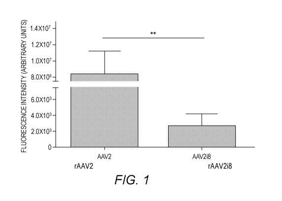
Figure 1. Green fluorescence protein (GFP) fluorescence following intravitreal
delivery of rAAV2 vector and its HS-binding deficient variants twelve weeks
post-
injection. Quantification of fundus images showed a 300-fold decrease in
expression
between rAAV2 (HS-binding) and rAAV2i8 (ablated HS-binding).
Immunohistochemistry
(IHC) of rAAV2-inj ected retinas shows fluorescence mainly in the RGC with
fewer GFP-
positive somas in the INL. Graph is shown with error bars indicating the
standard error mean
(SEM) and significance is detected by a non-parametric t-test (**p<0.01).
Figure 2. Schematic of the retina depicts the trafficking of rAAV following
intravitreal delivery.
Figure 3. qPCR analysis of viral binding to human retinas ex vivo. Results are
quantified as vector genomes per cell genome. rAAV2 (HS-binding) vector shows
the
greatest presence at the retina with few transgenes found elsewhere. The
presence of
transgenes delivered by rAAV2i8 (ablated HS-binding) were low in all collected
tissues but
showed a significant increase compared to rAAV2-delivered transgenes in both
the choroid
and sclera. Error bars indicated standard deviation.
Figure 4. GFP fluorescence following intravitreal delivery of HS-binding
variants of rAAV1 eight weeks post-injection. Quantification of fundus images
shows a 3-
3
CA 03033125 2019-02-05
WO 2018/035213
PCT/US2017/047123
fold increase with rAAV1-E531K (HS-binding) capsid compared to rAAV1 (non HS-
binding) capsid. Graph is shown with error bars indicating the SEM and
significance by a
non-parametric t-test (*p<0.05).
Figure 5. Chimeric capsids suggest tropism is influenced by other motifs other
than HS binding. Elements of rAAV1 were applied to rAAV2 using the chimeric
rAAV2.5
capsid and imaged for intravitreal delivery. Quantification of the fundus
fluorescence for the
collection of capsids. Error bars represent the SEM.
Figure 6. In vitro competition assay using soluble heparin to block the
transduction of rAAV of 11EK293 cells. Viruses were incubated with increasing
doses of
soluble heparin and applied to cell culture at a multiplicity of infection of
10,000 vg per cell.
rAAV2 displayed a dose-dependent decrease in transduction which was not
observed with
either rAAV1 or rAAV1-E531K. The amount of transduction of rAAV1-E531K was
lower
than rAAV1 in all conditions. Error bars shown as SEM.
DETAILED DESCRIPTION OF THE INVENTION
The present invention will now be described with reference to the accompanying
drawings, in which representative embodiments of the invention are shown. This
invention
may, however, be embodied in different forms and should not be construed as
limited to the
embodiments set forth herein. Rather, these embodiments are provided so that
this disclosure
will be thorough and complete, and will fully convey the scope of the
invention to those
skilled in the art.
Unless otherwise defined, all technical and scientific terms used herein have
the same
meaning as commonly understood by one of ordinary skill in the art to which
this invention
belongs. The terminology used in the description of the invention herein is
for the purpose of
describing particular embodiments only and is not intended to be limiting of
the invention.
The present invention is based on the unexpected discovery that intravitreal
transduction of cells of the retina and/or retinal pigment epithelium can be
enhanced by the
addition of the heparan sulfate binding motif on the AAV capsid. Thus, in one
embodiment,
the present invention provides a method of introducing a nucleic acid molecule
into a cell of a
retina and/or retinal pigment epithelium of a subject, comprising
intravitreally administering
an adeno-associated virus (AAV) serotype 4 (AAV4) vector comprising an AAV4
capsid
protein, wherein the AAV4 capsid protein comprises a substitution at amino
acid residue
K530 and/or further comprises a substitution at one or more of amino acid
residues S584,
4
CA 03033125 2019-02-05
WO 2018/035213
PCT/US2017/047123
N585, S586 and N587 in any combination, wherein the numbering of the residues
is based on
the amino acid sequence of SEQ ID NO:4 (amino acid sequence of AAV4 capsid
protein).
The present invention also provides a method of introducing a nucleic acid
molecule
into a cell of a retina and/or retinal pigment epithelium of a subject,
comprising intravitreally
administering an adeno-associated virus (AAV) serotype 5 (AAV5) vector
comprising an
AAV5 capsid protein, wherein the AAV5 capsid protein comprises a substitution
at amino
acid residue K517 and/or further comprises a substitution at one or more of
amino acid
residues S575, S576, T577 and T578 in any combination, wherein the numbering
of the
residues is based on the amino acid sequence of SEQ ID NO:5 (amino acid
sequence of
AAV5 capsid protein).
Additionally provided herein is a method of introducing a nucleic acid
molecule into a
cell of a retina and/or retinal pigment epithelium of a subject, comprising
intravitreally
administering an adeno-associated virus (AAV) serotype 7 (AAV7) vector
comprising an
AAV7 capsid protein, wherein the AAV7 capsid protein comprises a substitution
at amino
acid residue K533 and/or further comprises a substitution at one or more of
amino acid
residues A587, A588, N589 and R590 in any combination, wherein the numbering
of the
residues is based on the amino acid sequence of SEQ ID NO:7 (amino acid
sequence of
AAV7 capsid protein).
The present invention further provides a method of introducing a nucleic acid
molecule into a cell of a retina and/or retinal pigment epithelium of a
subject, comprising
intravitreally administering an adeno-associated virus (AAV) serotype 8 (AAV8)
vector
comprising an AAV8 capsid protein, wherein the AAV8 capsid protein comprises a
substitution at amino acid residue K533 and/or further comprises a
substitution at one or
more of amino acid residues Q587, Q588, N589 and T590 in any combination,
wherein the
numbering of the residues is based on the amino acid sequence of SEQ ID NO:8
(amino acid
sequence of AAV8 capsid protein).
Also provided herein is a method of introducing a nucleic acid molecule into a
cell of
a retina and/or retinal pigment epithelium of a subject, comprising
intravitreally
administering an adeno-associated virus (AAV) serotype 9 (AAV9) vector
comprising an
AAV9 capsid protein, wherein the AAV9 capsid protein comprises a substitution
at amino
acid residue K531 and/or further comprises a substitution at one or more of
amino acid
residues Q587, A588, N589 and T 590 in any combination, wherein the numbering
of the
residues is based on the amino acid sequence of SEQ ID NO:9 (amino acid
sequence of
AAV9 capsid protein).
5
CA 03033125 2019-02-05
WO 2018/035213
PCT/US2017/047123
Furthermore, the present invention provides a method of treating a disorder or
defect
of the eye in a subject, comprising intravitreally administering to the
subject the virus vector
of this invention, wherein the virus vector comprises a nucleic acid molecule
that encodes a
therapeutic protein or therapeutic DNA effective in treating the disorder or
defect of the eye
in the subject.
In further embodiments, the methods of this invention can be carried out with
an
AAV vector comprising a capsid protein that has been modified as described
below.
Specifically, in some embodiments, the heparan sulfate binding motif (AAV2
numbering:
484R, 485Q, 486Q, 487R, 488V, 489S, 490K, 491T, 527K, 528D, 529D, 530E, D531E,
532K, 5585R, S586G, S587N, T588R) can be graphed onto an AAV1 capsid protein
and/or
the AAV1 capsid protein can comprise Tyr mutations (AAV2 numbering: Y252F,
Y272F,
Y444F, Y500F, Y700F, Y704F, Y730F,), galactose motif (AAV2 numbering: G469N,
470M,
5471A, 472V, P474G, 500F) insertion of peptide with the amino acid
abbreviation
LALGETTRPA (AAV2 numbering: 587), or deletion mutation (AAV2 numbering: T265),
in
any combination. Amino acid residue numbering is based on the amino acid
sequence of
AAV2 (SEQ ID NO:2).
In some embodiments, the heparan sulfate binding motif (AAV2 numbering: 484R,
485Q, 486 Q, 487 R, 488 V, 489 S, 490 K, 491T, 527K, 528D, 529D, 530E, 531E,
532K,
585R, 586G, 587N, 588R) can be graphed onto an AAV2 capsid protein and/or the
AAV2
capsid protein can comprise Tyr mutations (AAV2 numbering: Y252F, Y272F,
Y444F,
Y500F, Y700F, Y704F, Y730F), galactose motif (AAV2 numbering: D469N, 1470M,
R471A, D472V, 5474G, Y500F), insertion of peptide with the amino acid
abbreviation
LALGETTRPA (AAV2 numbering: 587), or insertion mutation (AAV2 numbering: 265),
in
any combination. Amino acid residue numbering is based on the amino acid
sequence of
AAV2 (SEQ ID NO:2).
In some embodiments, the heparan sulfate binding motif (AAV2 numbering: 484R,
485Q, 486Q, 487R, L488V, 489S, 490K, 491T, 527K, 528D, 529D, 530E, 531E, 532K,
5585R, 5586G, N587N, T588R) can be graphed onto an AAV3B capsid protein and/or
the
AAV3B capsid protein can comprise Tyr mutations (AAV2 numbering: Y252F, Y272F,
Y444F, Y500F, Y700F, Y704F, Y730F,), galactose motif (AAV2 numbering: 5469N,
470M,
5471A, N472V, A474G, 500F) insertion of peptide with the amino acid
abbreviation
LALGETTRPA (AAV2 numbering: 587), or insertion mutation (AAV2 numbering: 265),
in
any combination. Amino acid residue numbering is based on the amino acid
sequence of
AAV2 (SEQ ID NO:2).
6
CA 03033125 2019-02-05
WO 2018/035213
PCT/US2017/047123
In some embodiments, the heparan sulfate binding motif (AAV2 numbering: K484R,
485Q, 486Q, G487R, F488V, 489S, 490K, 491T, G527K, P528D, A529D, D530E, S531E,
532K, S585R, N586G, S587N, N588R) can be graphed onto an AAV4 capsid protein
and/or
the AAV4 capsid protein can comprise Tyr mutations (AAV2 numbering: Y252F,
Y272F,
Y444F, Y500F, Y700F, Y704F, Y730F), galactose motif (AAV2 numbering: 469N,
F470M,
S471A, N472V, K474G, S500F), insertion of peptide with the amino acid
abbreviation
LALGETTRPA (AAV2 numbering: 587), or insertion mutation (AAV2 numbering: 265),
in
any combination. Amino acid residue numbering is based on the amino acid
sequence of
AAV2 (SEQ ID NO:2).
In some embodiments, the heparan sulfate binding motif (AAV2 numbering: 484R,
T485Q, G487R, W488V, N489S, L490K, G491T, L527K, Q528D, G529D, 5530E, N531E,
T532K, S585R, S586G, T587N, T588R) can be graphed onto an AAV5 capsid protein
and/or
the AAV5 capsid protein can comprise Tyr mutations (AAV2 numbering: Y252F,
Y272F,
Y444F, Y500F, Y700F, Y704F, Y730F), galactose motif (AAV2 numbering: R469N,
Y470M, 471A, N472V,Y474G, S500F), insertion of peptide with the amino acid
abbreviation
LALGETTRPA (AAV2 numbering: 587), or insertion mutation (AAV2 numbering: 265),
in
any combination. Amino acid residue numbering is based on the amino acid
sequence of
AAV2 (SEQ ID NO:2).
In some embodiments, the heparan sulfate binding motif (AAV2 numbering: 484R,
485Q, 486Q, 487R, 488V, 489S, 490K, 491T, 527K, 528D, 529D, 530E, 531E, R532K,
A585R, A586G, 587N, T588R) can be graphed onto an AAV7 capsid protein and/or
the
AAV7 capsid protein can comprise Tyr mutations (AAV2 numbering: Y252F, Y272F,
Y444F, Y500F, Y700F, Y704F, Y730F,), galactose motif (AAV2 numbering: T469N,
470M,
471A, E472V, A474G, 500F) insertion of peptide with the amino acid
abbreviation
LALGETTRPA (AAV2 numbering: 587), or insertion mutation (AAV2 numbering: 265),
in
any combination. Amino acid residue numbering is based on the amino acid
sequence of
AAV2 (SEQ ID NO:2).
In some embodiments, the heparan sulfate binding motif (AAV2 numbering: 484R,
485Q, 486Q, 487R, 488V, 489S, T490K, 491T, 527K, 528D, 529D, 530E, 531E,
R532K,
Q585R, Q586G, N587N, T588R) can be graphed onto an AAV8 capsid protein and/or
the
AAV 8 capsid protein can comprise Tyr mutations (AAV2 numbering: Y252F, Y272F,
Y444F, Y500F, Y700F, Y704F, Y730F,), galactose motif (AAV2 numbering: T469N,
470M,
471A, N472V, A474G, 500F) insertion of peptide with the amino acid
abbreviation
LALGETTRPA (AAV2 numbering: 587), or substitution mutation (AAV2 numbering:
7
CA 03033125 2019-02-05
WO 2018/035213
PCT/US2017/047123
S265), in any combination. Amino acid residue numbering is based on the amino
acid
sequence of AAV2 (SEQ ID NO:2).
In some embodiments, the heparan sulfate binding motif (AAV2 numbering: 484R,
485Q, 486Q, 487R, 488V, 489S, T490K, 491T, 527K, E528D, G529D, 530E, D531E,
R532K, S585R, A586G, Q587N, A588R) can be graphed onto an AAV9 capsid protein
for
and/or the AAV9 capsid protein can comprise Tyr mutations (AAV2 numbering:
Y252F,
Y272F, Y444F, Y500F, Y700F, Y704F, Y730F), galactose motif (AAV2 numbering:
469N,
470M, 471A, 472V, 474G, 500F) insertion of peptide with the amino acid
abbreviation
LALGETTRPA (AAV2 numbering: 587), or substitution mutation (AAV2 numbering:
S265), in any combination. Amino acid residue numbering is based on the amino
acid
sequence of AAV2 (SEQ ID NO:2).
In some embodiments, the heparan sulfate binding motif (AAV2 numbering: 484R,
485Q, 486Q, 487R, 488V, 489S, T490K, 491T, 527 K, 528D, 529D, 530E, 531E,
R532K,
Q585R, A586G, 587N, T588R) can be graphed onto an AAV10 capsid protein and/or
the
AAV10 capsid protein can comprise Tyr mutations (AAV2 numbering: Y252F, Y272F,
Y444F, Y500F, Y700F, Y704F, Y730F,), galactose motif (AAV2 numbering: 469N,
470M,
S471A, A472V, A474G, 500F) insertion of peptide with the amino acid
abbreviation
LALGETTRPA (AAV2 numbering: 587), or substitution mutation (AAV2 numbering:
5265), in any combination. Amino acid residue numbering is based on the amino
acid
sequence of AAV2 (SEQ ID NO:2).
In some embodiments, the heparan sulfate binding motif (AAV2 numbering: K484R,
485Q, 486Q, 487R, F488V, 489S, 490K, 491T, G527K, P528D, 5529D, D530E, G531E,
D532K, N585R, A586G, T587N, T588R) can be graphed onto an AAV11 capsid protein
in
any combination and/or the AAV11 capsid protein can comprise Tyr mutations
(AAV2
numbering: Y252F, Y272F, Y444F, Y500F, Y700F, Y704F, Y730F), galactose motif
(AAV2
numbering: D469N, F470M, 471A, F472V, R474G, A500F) insertion of peptide with
the
amino acid abbreviation LALGETTRPA (AAV2 numbering: 587). or insertion
mutation
(AAV2 numbering: 265), in any combination. Amino acid residue numbering is
based on the
amino acid sequence of AAV2 (SEQ ID NO:2).
In some embodiments, the heparan sulfate binding motif (AAV2 numbering: K484R,
485Q, 486Q, K487R, F488V, 489S, 490K, N491T, G527K, A528D, G529D, D530E,
5531E,
D532K, N585R, A586G, T587N, T588R) can be graphed onto an AAV12 capsid protein
and/or the AAV12 capsid protein can comprise Tyr mutations (AAV2 numbering:
Y252F,
Y272F, Y444F, Y500F, Y700F, Y704F, Y730F), galactose motif (AAV2 numbering:
8
CA 03033125 2019-02-05
WO 2018/035213
PCT/US2017/047123
D469N, F470M, 471A, F472V, R474G, A500F) insertion of peptide with the amino
acid
abbreviation LALGETTRPA (AAV2 numbering: 587), or insertion mutation (AAV2
numbering: 265), in any combination. Amino acid residue numbering is based on
the amino
acid sequence of AAV2 (SEQ ID NO:2).
In the methods of this invention, the viral vector can comprise a nucleic acid
molecule
that encodes a therapeutic protein and/or therapeutic DNA.
The present invention further provides a method of treating a disorder or
defect of the
eye in a subject, comprising the intravitreally administering viral vector of
this invention to
the subject receiving a therapeutic protein or therapeutic DNA effective in
treating the
.. disorder or defect of the eye in the subject.
Nonlimiting examples of a disorder or defect of the eye that can be treated
according
to the methods of this invention include age-related macular degeneration,
Lebers congenital
amarousis type 1, Lebers, congenital amarousis type 2, retinitis pigmentosa,
retinoschosis,
achromatopsia, color blindness, congenital stationary night blindness or any
combination
thereof.
Definitions.
The following terms are used in the description herein and the appended
claims:
The singular forms "a," "an" and "the" are intended to include the plural
forms as
well, unless the context clearly indicates otherwise.
Furthermore, the term "about," as used herein when referring to a measurable
value
such as an amount of the length of a polyrmcleotide or polypeptide sequence,
dose, time,
temperature, and the like, is meant to encompass variations of 20%, 10%,
5%, 1%,
0.5%, or even 0.1% of the specified amount.
Also as used herein, "and/or" refers to and encompasses any and all possible
combinations of one or more of the associated listed items, as well as the
lack of
combinations when interpreted in the alternative ("or").
As used herein, the transitional phrase "consisting essentially of" (and
grammatical
variants) means that the scope of a claim is to be interpreted to encompass
the specified
materials or steps recited in the claim, "and those that do not materially
affect the basic and
novel characteristic(s)" of the claimed invention. Thus, the term "consisting
essentially of'
when used in a claim of this invention is not intended to be interpreted to be
equivalent to
"comprising."
9
CA 03033125 2019-02-05
WO 2018/035213
PCT/US2017/047123
Unless the context indicates otherwise, it is specifically intended that the
various
features of the invention described herein can be used in any combination.
Moreover, the present invention also contemplates that in some embodiments of
the
invention, any feature or combination of features set forth herein can be
excluded or omitted.
To illustrate further, if, for example, the specification indicates that a
particular amino
acid can be selected from A, G, I, L and/or V, this language also indicates
that the amino acid
can be selected from any subset of these amino acid(s) for example A, G, I or
L; A, G, I or V;
A or G; only L; etc. as if each such subcombination is expressly set forth
herein. Moreover,
such language also indicates that one or more of the specified amino acids can
be disclaimed.
For example, in particular embodiments the amino acid is not A, G or I; is not
A; is not G or
V; etc. as if each such possible disclaimer is expressly set forth herein.
As used herein, the terms "reduce," "reduces," "reduction" and similar terms
mean a
decrease of at least about 25%, 35%, 50%, 75%, 80%, 85%, 90%, 95%, 97% or
more.
As used herein, the terms "enhance," "enhances," "enhancement" and similar
terms
indicate an increase of at least about 5%, 10%, 20%, 25%, 50%, 75%, 100%,
150%, 200%,
300%, 400%, 500% or more. These terms can also be used in reference to fold
increases,
e.g., one-fold, two-fold, three-fold, four-fold, five-fold, six-fold, seven-
fold, eight-fold, nine-
fold, ten-fold, etc.
The term "parvovirus" as used herein encompasses the family Parvoviridae,
including
autonomously replicating parvoviruses and dependoviruses. The autonomous
parvoviruses
include members of the genera Parvovirus, Erythrovirus, Densovirus,
Iteravirus, and
Contravirus. Exemplary autonomous parvoviruses include, but are not limited
to, minute
virus of mouse, bovine parvovirus, canine parvovirus, chicken parvovirus,
feline
panleukopenia virus, feline parvovirus, goose parvovirus, H1 parvovirus,
muscovy duck
.. parvovirus, B19 virus, and any other autonomous parvovirus now known or
later discovered.
Other autonomous parvoviruses are known to those skilled in the art. See,
e.g., BERNARD
N. FIELDS et al., VIROLOGY, volume 2, chapter 69 (4th ed., Lippincott-Raven
Publishers).
As used herein, the term "adeno-associated virus" (AAV), includes but is not
limited
to, AAV type 1, AAV type 2, AAV type 3 (including types 3A and 3B), AAV type
4, AAV
.. type 5, AAV type 6, AAV type 7, AAV type 8, AAV type 9, AAV type 10, AAV
type 11,
AAV type 12, avian AAV, bovine AAV, canine AAV, equine AAV, ovine AAV, and any
other AAV now known or later discovered. See, e.g., BERNARD N. FIELDS et al.,
VIROLOGY, volume 2, chapter 69 (4th ed., Lippincott-Raven Publishers). A
number of
CA 03033125 2019-02-05
WO 2018/035213
PCT/US2017/047123
relatively new AAV serotypes and clades have been identified (see, e.g., Gao
et al. (2004)1
Virology 78:6381-6388; Moris et al. (2004) Virology 33-:375-383; and Table 1).
The genomic sequences of various serotypes of AAV and the autonomous
parvoviruses, as well as the sequences of the native terminal repeats (TRs),
Rep proteins, and
capsid subunits are known in the art. Such sequences may be found in the
literature or in
public databases such as the GenBanke Database. See, e.g., GenBank Accession
Numbers
NC 044927, NC 002077, NC 001401, NC 001729, NC 001863, NC 001829, NC 001862,
NC 000883, NC 001701, NC 001510, NC 006152, NC 006261, AF063497, U89790,
AF043303, AF028705, AF028704, J02275, J01901, J02275, X01457, AF288061,
AH009962, AY028226, AY028223, NC_001358, NC_001540, AF513851, AF513852,
AY530579; the disclosures of which are incorporated by reference herein for
teaching
parvovirus and AAV nucleic acid and amino acid sequences. See also, e.g.,
Srivistava et al.
(1983) J. Virology 45:555; Chiorini et al. (1998) J. Virology 71:6823;
Chiorini et al. (1999)
J. Virology 73:1309; Bantel-Schaal et al. (1999) J. Virology 73:939; Xiao et
al. (1999) J.
Virology 73:3994; Muramatsu et al. (1996) Virology 221:208; Shade et al.
(1986) J ViroL
58:921; Gao et al. (2002) Proc. Nat. Acad. Sci. USA 99:11854; Moris etal.
(2004) Virology
33-:375-383; international patent publications WO 00/28061, WO 99/61601, WO
98/11244;
and U.S. Patent No. 6,156,303; the disclosures of which are incorporated by
reference herein
for teaching parvovirus and AAV nucleic acid and amino acid sequences. See
also Table 1.
The capsid structures of autonomous parvoviruses and AAV are described in more
detail in BERNARD N. FIELDS et al., VIROLOGY, volume 2, chapters 69 & 70 (4th
ed.,
Lippincott-Raven Publishers). See also, description of the crystal structure
of AAV2 (Xie et
al. (2002) Proc. Nat. Acad. Sci. 99:10405-10), AAV4 (Padron et al. (2005) J.
ViroL 79: 5047-
58), AAV5 (Walters et al. (2004)1 ViroL 78: 3361-71) and CPV (Xie et al.
(1996)1 MoL
Biol. 6:497-520 and Tsao et al. (1991) Science 251: 1456-64).
The term "tropism" as used herein refers to preferential entry of the virus
into certain
cells or tissues, optionally followed by expression (e.g., transcription and,
optionally,
translation) of a sequence(s) carried by the viral genome in the cell, e.g.,
for a recombinant
virus, expression of a heterologous nucleic acid(s) of interest. Those skilled
in the art will
appreciate that transcription of a heterologous nucleic acid sequence from the
viral genome
may not be initiated in the absence of trans- acting factors, e.g., for an
inducible promoter or
otherwise regulated nucleic acid sequence. In the case of a rAAV genome, gene
expression
from the viral genome may be from a stably integrated provirus, from a non-
integrated
episome, as well as any other form the virus may take within the cell.
11
CA 03033125 2019-02-05
WO 2018/035213
PCT/US2017/047123
Unless indicated otherwise, "efficient transduction" or "efficient tropism,"
or similar
terms, can be determined by reference to a suitable control (e.g., at least
about 50%, 60%,
70%, 80%, 85%, 90%, 95%, 98%, 99%, 100% or more of the transduction or
tropism,
respectively, of the control). Suitable controls will depend on a variety of
factors including
the desired tropism profile.
As used herein, the term "polypeptide" encompasses both peptides and proteins,
unless indicated otherwise.
A "polynucleotide" is a sequence of nucleotide bases, and may be RNA, DNA or
DNA-RNA hybrid sequences (including both naturally occurring and non-naturally
occurring
nucleotides), but in representative embodiments are either single or double
stranded DNA
sequences.
As used herein, an "isolated" polynucleotide (e.g., an "isolated DNA" or an
"isolated
RNA") means a polynucleotide at least partially separated from at least some
of the other
components of the naturally occurring organism or virus, for example, the cell
or viral
structural components or other polyp eptides or nucleic acids commonly found
associated
with the polynucleotide. In representative embodiments an "isolated"
nucleotide is enriched
by at least about 10-fold, 100-fold, 1000-fold, 10,000-fold or more as
compared with the
starting material.
Likewise, an "isolated" polypeptide means a polypeptide that is at least
partially
separated from at least some of the other components of the naturally
occurring organism or
virus, for example, the cell or viral structural components or other
polypeptides or nucleic
acids commonly found associated with the polypeptide. In representative
embodiments an
"isolated" polypeptide is enriched by at least about 10-fold, 100-fold, 1000-
fold, 10,000-fold
or more as compared with the starting material.
As used herein, by "isolate" or "purify" (or grammatical equivalents) a virus
vector, it
is meant that the virus vector is at least partially separated from at least
some of the other
components in the starting material. In representative embodiments an
"isolated" or
"purified" virus vector is enriched by at least about 10-fold, 100-fold, 1000-
fold, 10,000-fold
or more as compared with the starting material.
A "therapeutic protein" is a protein that can alleviate, reduce, prevent,
delay and/or
stabilize symptoms that result from an absence or defect in a protein in a
cell or subject
and/or is a protein that otherwise confers a benefit to a subject.
A "therapeutic RNA molecule" or "functional RNA molecule" as used herein can
be
an antisense nucleic acid, a ribozyme (e.g., as described in U.S. Patent No.
5,877,022), an
12
CA 03033125 2019-02-05
WO 2018/035213
PCT/US2017/047123
RNA that effects spliceosome-mediated trans-splicing (see, Puttaraju et al.
(1999) Nature
Biotech. 17:246; U.S. Patent No. 6,013,487; U.S. Patent No. 6,083,702), an
interfering RNA
(RNAi) including siRNA, shRNA or miRNA, which mediate gene silencing (see,
Sharp et al.,
(2000) Science 287:2431), and any other non-translated RNA, such as a "guide"
RNA
.. (Gorman et al. (1998) Proc. Nat. Acad. Sci. USA 95:4929; U.S. Patent No.
5,869,248 to Yuan
et al.) and the like as are known in the art.
By the terms "treat," "treating" or "treatment of" (and grammatical variations
thereof)
it is meant that the severity of the subject's condition is reduced, at least
partially improved or
stabilized and/or that some alleviation, mitigation, decrease or stabilization
in at least one
clinical symptom is achieved and/or there is a delay in the progression of the
disease or
disorder.
The terms "prevent," "preventing" and "prevention" (and grammatical variations
thereof) refer to prevention and/or delay of the onset of a disease, disorder
and/or a clinical
symptom(s) in a subject and/or a reduction in the severity of the onset of the
disease, disorder
and/or clinical symptom(s) relative to what would occur in the absence of the
methods of the
invention. The prevention can be complete, e.g., the total absence of the
disease, disorder
and/or clinical symptom(s). The prevention can also be partial, such that the
occurrence of
the disease, disorder and/or clinical symptom(s) in the subject and/or the
severity of onset is
less than what would occur in the absence of the present invention.
A "treatment effective" amount as used herein is an amount that is sufficient
to
provide some improvement or benefit to the subject. Alternatively stated, a
"treatment
effective" amount is an amount that will provide some alleviation, mitigation,
decrease or
stabilization in at least one clinical symptom in the subject. Those skilled
in the art will
appreciate that the therapeutic effects need not be complete or curative, as
long as some
benefit is provided to the subject.
A "prevention effective" amount as used herein is an amount that is sufficient
to
prevent and/or delay the onset of a disease, disorder and/or clinical symptoms
in a subject
and/or to reduce and/or delay the severity of the onset of a disease, disorder
and/or clinical
symptoms in a subject relative to what would occur in the absence of the
methods of the
invention. Those skilled in the art will appreciate that the level of
prevention need not be
complete, as long as some benefit is provided to the subject.
The terms "heterologous nucleotide sequence" and "heterologous nucleic acid
molecule" are used interchangeably herein and refer to a nucleic acid molecule
and/or
nucleotide sequence that is not naturally occurring in the virus. Generally,
the heterologous
13
CA 03033125 2019-02-05
WO 2018/035213
PCT/US2017/047123
nucleic acid comprises an open reading frame that encodes a protein, protein
fragment,
peptide or nontranslated RNA of interest (e.g., for delivery to a cell or
subject).
As used herein, the terms "virus vector," "vector" or "gene delivery vector"
refer to a
virus (e.g., AAV) particle that functions as a nucleic acid delivery vehicle,
and which
comprises the vector genome (e.g., viral DNA [vDNA]) packaged within a virion.
Alternatively, in some contexts, the term "vector" may be used to refer to the
vector
genome/vDNA alone.
A "rAAV vector genome" or "rAAV genome" is an AAV genome (i.e., vDNA) that
comprises one or more heterologous nucleic acid sequences. rAAV vectors
generally require
only the terminal repeat(s) (TR(s)) in cis to generate virus. All other viral
sequences are
dispensable and may be supplied in trans (Muzyczka (1992) Curr. Topics
Microbiol.
Immunol. 158:97). Typically, the rAAV vector genome will only retain the one
or more TR
sequence so as to maximize the size of the transgene that can be efficiently
packaged by the
vector. The structural and non-structural protein coding sequences may be
provided in trans
(e.g., from a vector, such as a plasmid, or by stably integrating the
sequences into a
packaging cell). In embodiments of the invention, the rAAV vector genome
comprises at
least one terminal repeat (TR) sequence (e.g., AAV TR sequence), optionally
two TRs (e.g.,
two AAV TRs), which typically will be at the 5' and 3' ends of the vector
genome and flank
the heterologous nucleic acid sequence, but need not be contiguous thereto.
The TRs can be
the same or different from each other.
The term "terminal repeat" or "TR" includes any viral terminal repeat or
synthetic
sequence that forms a hairpin structure and functions as an inverted terminal
repeat (i.e.,
mediates the desired functions such as replication, virus packaging,
integration and/or
provirus rescue, and the like). The TR can be an AAV TR or a non-AAV TR. For
example,
a non-AAV TR sequence such as those of other parvoviruses (e.g., canine
parvovirus (CPV),
mouse parvovirus (MVM), human parvovirus B-19) or any other suitable virus
sequence
(e.g., the SV40 hairpin that serves as the origin of SV40 replication) can be
used as a TR,
which can further be modified by truncation, substitution, deletion, insertion
and/or addition.
Further, the TR can be partially or completely synthetic, such as the "double-
D sequence" as
described in US Patent No. 5,478,745 to Samulski et al.
An "AAV terminal repeat" or "AAV TR" may be from any AAV, including but not
limited to serotypes 1, 2, 3, 4, 5, 6, 7, 8, 9, 10 or 11 or any other AAV now
known or later
discovered (see, e.g., Table 1). An AAV terminal repeat need not have the
native terminal
repeat sequence (e.g., a native AAV TR sequence may be altered by insertion,
deletion,
14
CA 03033125 2019-02-05
WO 2018/035213
PCT/US2017/047123
truncation and/or missense mutations), as long as the terminal repeat mediates
the desired
functions, e.g., replication, virus packaging, integration, and/or provirus
rescue, and the like.
The virus vectors of the invention can further be "targeted" virus vectors
(e.g., having
a directed tropism) and/or a "hybrid" parvovirus (i.e., in which the viral TRs
and viral capsid
are from different parvoviruses) as described in international patent
publication WO
00/28004 and Chao et al. (2000) Molecular Therapy 2:619.
The virus vectors of the invention can further be duplexed parvovirus
particles as
described in international patent publication WO 01/92551 (the disclosure of
which is
incorporated herein by reference in its entirety). Thus, in some embodiments,
double
stranded (duplex) genomes can be packaged into the virus capsids of the
invention.
Further, the viral capsid or genomic elements can contain other modifications,
including insertions, deletions and/or substitutions.
As used herein, the term "amino acid" encompasses any naturally occurring
amino
acid, modified forms thereof, and synthetic amino acids.
Naturally occurring, levorotatory (L-) amino acids are shown in Table 2.
Alternatively, the amino acid can be a modified amino acid residue
(nonlimiting
examples are shown in Table 3) and/or can be an amino acid that is modified by
post-
translation modification (e.g., acetylation, amidation, formylation,
hydroxylation,
methylation, phosphorylation or sulfatation).
Further, the non-naturally occurring amino acid can be an "unnatural" amino
acid as
described by Wang et al. Annu Rev Biophys Biomol Struct. 35:225-49 (2006)).
These
unnatural amino acids can advantageously be used to chemically link molecules
of interest to
the AAV capsid protein.
Modified AAV Capsid Proteins and Virus Capsids and Virus Vectors Comprising
the
Same.
The present invention provides AAV capsid proteins comprising a mutation
(i.e., a
modification) in the amino acid sequence and virus capsids and virus vectors
comprising the
modified AAV capsid protein. The inventors have discovered that modifications
such as
substitutions at the amino acid positions described herein can confer one or
more desirable
properties to virus vectors comprising the modified AAV capsid protein
including without
limitation selective transduction of cells having heparin sulfate on the
surface and enhanced
transduction of cells of the retina and/or retinal pigment epithelium.
In particular embodiments, the modified AAV capsid protein of the invention
comprises one or more mutations (e.g., substitutions) in the amino acid
sequence of the native
CA 03033125 2019-02-05
WO 2018/035213
PCT/US2017/047123
AAV4 capsid protein or the corresponding region of a capsid protein from
another AAV,
including but not limited to AAV5. AAV7, AAV8 and AAV9.
As used herein, a "mutation" or "modification" in an amino acid sequence
includes
substitutions, insertions and/or deletions, each of which can involve one,
two, three, four,
five, six, seven, eight, nine, ten or more amino acids. In particular
embodiments, the
modification is a substitution. For example, in particular embodiments, the
AAV4 capsid
protein sequence is modified at amino acid positions 530, 584, 585, 586 and/or
587, in any
combination.
In certain embodiments as described herein, amino acid residue numbering is
based
on the amino acid sequence of an AAV4 capsid protein having GenBank Accession
No.
NP 044927 (SEQ ID NO:4):
MTDGYLPDWLEDNLSEGVREWWALQPGAPKPKANQQHQDNARGLVLPGYKYLGP
GNGLDKGEPVNAADAAALEHDKAYDQQLKAGDNPYLKYNHADAEFQQRLQGDTS
FGGNLGRAVF QAKKRVLEP LGLVEQAGETAP GKKRP LIE SP QQPD SSTGIGKKGKQP
AKKKLVFEDETGAGD GPPEG S T S GAMS DD SEMRAAAGGAAVEGGQGAD GVGNAS
GDWHCD STWSEGHVTTTSTRTWVLPTYNNHLYKRLGESLQSNTYNGFSTPWGYFD
FNRFHCHFSPRDWQRLINNNWGMRPKAMRVKIFNIQVKEVTTSNGETTVANNLTST
VQIFADS SYELPYVMDAGQEGSLPPFPNDVFMVPQYGYCGLVTGNTSQQQTDRNAF
YCLEYFP SQMLRTGNNFEITYSFEKVPFHSMYAHS Q S LDRLMNPLID QYLWGLQ S TT
TGTTLNAGTATTNFTKLRPTNF SNFKKNWLP GP SIKQQGFSKTANQNYKIPATGSDSL
IKYETHSTLDGRWSALTPGPPMATAGPADSKFSNS QLIFAGPKQNGNTATVPGTLIFT
S EEELAATNATDTDMWGNLP GGD Q SN SNLP TVDRLTALGAVP GMVWQNRDIYYQG
PIVVAKIPHTDGHFHP SP LIGGFGLKHPPP QIFIKNTPVPANPATTF S S TPVN S FITQYS TG
QVSVQIDWEIQKERSKRWNPEVQFTSNYGQQNSLLWAPDAAGKYTEPRAIGTRYLT
HHL.
In certain embodiments as described herein, amino acid residue numbering is
based
on the amino acid sequence of an AAV2 capsid protein having GenBank Accession
No.
YP 680426 (SEQ ID NO:2):
MAADGYLPDWLEDTLSEGIRQWWKLKPGPPPPKPAERHKDDSRGLVLPGYKYLGPF
NGLDKGEPVNEADAAALEHDKAYDRQLDSGDNPYLKYNHADAEFQERLKEDTSFG
GNLGRAVFQAKKRVLEPLGLVEEPVKTAP GKKRPVEHSPVEPD SSSGTGKAGQQPA
RKRLNFGQTGDADSVPDPQPLGQPPAAP SGLGTNTMATGSGAPMADNNEGADGVG
NSSGNWHCDSTWMGDRVITTSTRTWALPTYNNHLYKQISSQSGASNDNHYFGYSTP
WGYFDFNRFHCHFSPRDWQRLINNNWGFRPKRLNFKLFNIQVKEVTQNDGTTTIAN
16
CA 03033125 2019-02-05
WO 2018/035213
PCT/US2017/047123
NLTSTVQVFTDSEYQLPYVLGSAHQGCLPPFPADVFMVPQYGYLTLNNGS QAVGRS
SFYCLEYFP SQMLRTGNNFTF SYTFEDVPFHS SYAHSQSLDRLMNPLIDQYLYYLSRT
NTP SGTTTQSRLQF SQAGASDIRDQSRNWLP GP CYRQQRVSKT SADNNN S EYSWTG
ATKYH LNGRD S LVNP GP AMAS HKDDEEKFFP Q S GVLIF GKQGS EKTNVD IEKVMITD
EEEIRTTNPVATEQYG SVS TNLQRGNRQAATADVNTQGVLP GMVWQDRDVYLQGP I
WAKIPHTDGHFHP SPLMGGFGLKHPPPQILIKNTPVPANP STTFSAAKFASFITQYSTG
QVSVEIEWELQKENSKRWNPEIQYTSNYNKSVNVDFTVDTNGVYSEPRPIGTRYLTR
NL
In certain embodiments as described herein, amino acid residue numbering is
based
on the amino acid sequence of an AAV9 capsid protein having GenBank Accession
No.
AAS99264 (SEQ ID NO:9):
MAADGYLPDLEDNLSEGIEWWALKPGAQPKANQQHQNARGLVLPGKYLGPGNGL
DKGEPVNAADAALEHDKAYQQLKAGDNPLKYNHADAEQERLKEDTSGGNLGRAV
FQAKKRLLEPLLVEEAAKTAGKKRPVEQS QEPDS SAGIKSGAQPAKKLNFGQTGDTE
SVPDPQPIGPPAAP SGVGLTMASGGGAVADNNEGADVGS SSGNWHDS QWLGDRVIT
TSTRTWALTYNNHLYKQSNSTSGGS SDNAYFGYSTWGYFDFNRFCHFSPRDWQRLI
NNNWGFRKRLNFKLFNQVKEVTDNNVKTIANNLTTVQVFTD SD QLPYVLGSAHEGC
LPPFPAVFMIP QYGYTLNDGSQAVRS SFYCLEYP S QMLRTGNFQF S YEFENVPFH S SY
AHS SLDRLMNPLDQYLYYLSKINGSGQNQQLKFSVAGP SMAVQGRNYIP GP SYRQQ
RVTTVTQNNN S FAWP GAS SWLNGRNS LMNGPAMASHKEEDRFFPLS G S LIFGKQGT
GDNVDADKVMTNEEEIKTTPVATESYGQATNHQSAQAAQTGWVQNQGILPGMVW
QDDVYLQGP IWKIPHTDGNFP S P LMGGF GKHPPP QILINTPVPADPP TAFNKDKLNS IT
QYSTGQVVEIEWELQKNSKRWNPEIYTSNYYKSNVEFAVNTEGVYSEPRPIGTYLTR
NL
In certain embodiments as described herein, amino acid residue numbering is
based
on the amino acid sequence of an AAV5 capsid protein having GenBank Accession
No.
YP 068409 (SEQ ID NO:5):
MSFVDHPPDWLEEVGEGLREFLGLEAGPPKPKPNQQHQDQARGLVLPGYNYLGPGN
GLDRGEPVNRADEVAREHDISYNEQLEAGDNPYLKYNHADAEFQEKLADDTSFGGN
LGKAVF QAKKRVLEPFGLVEEGAKTAP TGKRIDDHFPKRKKARTEED SKP ST S SDAE
AGP SGS QQLQIPAQPAS SLGADTMSAGGGGPLGDNNQGADGVGNASGDWHCDSTW
MGDRVVTKSTRTWVLP SYNNHQYREIKSGSVDGSNANAYFGYSTPWGYFDFNRFHS
HWSPRDWQRLINNYWGFRPRSLRVKIFNIQVKEVTVQDSTTTIANNLTSTVQVFTDD
DYQLP YVVGNGTEG C LPAFPP QVFTLP QYGYATLNRDNTENP TER S SFFCLEYFP SK
17
CA 03033125 2019-02-05
WO 2018/035213
PCT/US2017/047123
MLRTGNNFEFTYNFEEVPFHSSFAP S QNLFKLANPLVDQYLYRFVSTNNTGGVQFNK
NLAGRYANTYKNWFP GPMGRTQGWNLGS GVNRASVSAFATTNRMELEGASYQVPP
QPNGMTNNLQGSNTYALENTMIFNSQPANPGTTATYLEGNMLITSESETQPVNRVAY
NVGGQMATNNQSSTTAPATGTYNLQEIVP GSVWMERDVYLQGPIWAKIPETGAHFH
P SPAMGGFGLKHPPPMMLIKNTPVPGNITSF SDVPVS SFITQYSTGQVTVEMEWELKK
EN S KRWNPEIQYTNNYNDP QFVDFAPD S TGEYRTTRP IGTRYLTRPL
In certain embodiments as described herein, amino acid numbering is based on
the
amino acid sequence of an AAV1 capsid protein having GenBank Accession No.
NP_049542
(SEQ ID NO:1):
MAAD GYLPDWLEDNLS EGIREWWD LKP GAPKPKANQQKQDD GRGLVLP GYKYLG
PFNGLDKGEPVNAADAAALEHDKAYDQQLKAGDNPYLRYNHADAEFQERLQEDTS
FGGNLGRAVFQAKKRVLEP LGLVEEGAKTAP GKKRPVEQ S P QEPD S S SGIGKTGQQP
AKKRLNF GQTGD S ES VPDP QP LGEPP ATP AAVGPTTMAS GGGAPMADNNEGAD GV
GNAS GNWHCD STWLGDRVITTS TRTWALPTYNNHLYKQIS SAS TGASNDNHYFGYS
TPWGYFDFNRFHCHF SPRDWQRLINNNWGFRPKRLNFKLFNIQVKEVTTNDGVTTIA
NNLT S TVQVF SD S EYQLPYVLG S AHQGC LP PFPADVFMIP QYGYLTLNNGS QAVGRS
SFYCLEYFP S QMLRTGNNFTFSYTFEEVPFHSSYAHSQSLDRLMNPLID QYLYYLNRT
QNQ S GSAQNKD LLF S RGS PAGMS VQPKNWLP GP CYRQQRVSKTKTDNNNSNFTWT
GASKYNLNGRESIINP GTAMASHKDDEDKFFPMSGVMIFGKESAGASNTALDNVMIT
DEEEIKATNPVATERFGTVAVNFQS SSTDPATGDVHAMGALPGMVWQDRDVYLQG
PIWAKIPHTDGHFHP SPLMGGFGLKNPPPQILIKNTPVPANPPAEFSATKFASFITQYST
GQVSVEIEWELQKENSKRWNPEVQYTSNYAKSANVDFTVDNNGLYTEPRPIGTRYL
TRPL
In certain embodiments as described herein, amino acid residue numbering is
based
on the amino acid sequence of an AAV7 capsid protein having GenBank Accession
No.
YP 077178 (SEQ ID NO:7):
MAADGYLPDWLEDNLSEGIREWWDLKP GAPKPKANQQKQDNGRGLVLPGYKYLG
PFNGLDKGEPVNAADAAALEHDKAYDQ QLKAGDNPYLRYNHADAEF QERLQEDT S
FGGNLGRAVFQAKKRVLEPLGLVEEGAKTAPAKKRPVEP SPQRSPDS STGIGKKGQQ
PARKRLNFGQTGDSESVPDPQPLGEPPAAP SSVGSGTVAAGGGAPMADNNEGADGV
GNASGNWHCDSTWLGDRVITTSTRTWALPTYNNHLYKQISSETAGSTNDNTYFGYS
TPWGYFDFNRFHCHF SPRDWQRLINNNWGFRPKKLRFKLFNIQVKEVTTNDGVTTIA
NNLTSTIQVF SD SEYQLPYVLGSAHQGCLPP FPADVFMIP QYGYLTLNNGS QSVGRSS
FYCLEYFP S QMLRTGNNFEF SY SFEDVPFH S SYAHS Q S LDRLMNP LID QYLYYLARTQ
18
CA 03033125 2019-02-05
WO 2018/035213
PCT/US2017/047123
SNP GGTAGNRELQFYQGGP STMAEQAKNWLP GP CFRQQRV S KTLD QNNN SNFAWT
GATKYHLNGRNSLVNPGVAMATHKDDEDRFFP S SGVLIFGKTGATNKTTLENVLMT
NEEEIRPTNPVATEEYGIVS SNLQAANTAAQTQVVNNQGALPGMVWQNRDVYLQGP
IWAKIPHTDGNFHP S PLMGGFGLKHPPP QILIKNTPVPANPPEVFTPAKFASFITQY S TG
QV SVEIEWELQKEN S KRWNP EIQYT SNFEKQTGVDFAVD S QGVYS EPRPIGTRYLTR
NL
In certain embodiments as described herein, amino acid residue numbering is
based
on the amino acid sequence of an AAV8 capsid protein having GenBank Accession
No.
YP 077180 (SEQ ID NO:8):
.. MAAD GYLPDWLEDNLS EGIREWWALKP GAPKPKANQQKQDD GRGLVLP GYKYLG
PFNGLDKGEPVNAADAAALEHDKAYDQQLQAGDNPYLRYNHADAEFQERLQEDTS
FGGNLGRAVFQAKKRVLEPLGLVEEGAKTAPGKKRPVEP SPQRSPD SSTGIGKKGQQ
PARKRLNFGQTGDSESVPDPQPLGEPPAAP SGVGPNTMAAGGGAPMADNNEGADG
VGS S SGNWHCDSTWLGDRVITTSTRTWALPTYNNHLYKQISNGT SGGATNDNTYFG
.. YS TPWGYFDFNRFHCHF S PRDWQRLINNNWGFRPKRLSFKLFNIQVKEVTQNEGTKT
IANNLTSTIQVFTDSEYQLPYVLGSAHQGCLPPFPADVFMIPQYGYLTLNNGS QAVGR
SSFYCLEYFP S QMLRTGNNFQFTYTFEDVPFHS SYAH S QS LDRLMNP LID QYLYYLSR
TQTTGGTANTQTLGF S Q GGPNTMANQAKNWLP GP CYRQQRVS TTTGQNNNSNFAW
TAGTKYHLNGRNSLANPGIAMATHKDDEERFFP SNGILIFGKQNAARDNADYSDVM
LTSEEEIKTTNPVATEEYGIVADNLQQQNTAP QIGTVNS QGALP GMVWQNRDVYLQ
GPIWAKIPHTDGNFHP SP LMGGF GLKHPPP QILIKNTPVPADPP TTFNQ S KLN S FITQY S
TGQVSVEIEWELQKENSKRWNPEIQYTSNYYKSTSVDFAVNTEGVYSEPRPIGTRYL
TRNL.
In certain embodiments as described herein, amino acid residue numbering is
based
on the amino acid sequence of an AAV10 capsid protein having GenBank Accession
No.
AAT46337 (SEQ ID NO:10):
MAAD GYLPDWLEDNLS EGIREWWD LKP GAPKPKANQQKQDD GRGLVLP GYKYLG
PFNGLD61KGEPVNAADAAALEHDKAYDQQLKAGDNPYLRYNHADAEFQERLQED
TSFGGNLGRAVFQ121AKKRVLEPLGLVEEAAKTAPGKKRPVEP SPQRSPDSSTGIGK
KGQQPAKKRLNFGQTGES181ESVPDPQPIGEPPAGP SGLGSGTMAAGGGAPMADNN
EGAD GVGS SSGNWHCDSTWLGDRV241ITTSTRTWALPTYNNHLYKQISNGTSGGST
NDNTYFGYSTPWGYFDFNRFHCHF SPRDWQ301RLINNNWGFRPKRLSFKLFNIQVK
EVTQNEGTKTIANNLTSTIQVFTDSEYQLPYVLGSA361HQGCLPPFPADVFMIPQYGY
LTLNNGSQAVGRS SFYCLEYFP S QMLRTGNNFEFSYTFED421VPFHS SYAHS QSLDR
19
CA 03033125 2019-02-05
WO 2018/035213
PCT/US2017/047123
LMNP LID QYLYYLSRTQSTGGTQGTQQLLF S QAGP ANM SAQAKNW481LP GP CYRQ
QRVSTTLSQNNNSNFAWTGATKYHLNGRD SLVNPGVAMATHKDDEERFFP S S541G
VLMFGKQGAGRDNVDYS SVMLTSEEEIKTTNPVATEQYGVVADNLQQANTGPIVGN
VN S 601 QGALP GMVWQNRDVYLQGPIWAKIPHTDGNFHP SPLMGGFGLKHPPPQILI
.. KNTPVP ADP 661P TTF S QAKLASFITQYSTGQVSVEIEWELQKENSKRWNPEIQYTSNY
YKSTNVDFAVNTE721GTYSEPRPIGTRYLTRNL.
In certain embodiments as described herein, amino acid residue numbering is
based
on the amino acid sequence of an AAV11 capsid protein having GenBank Accession
No.
AAT46339 (SEQ ID NO:11):
.. MAADGYLPDWLEDNLSEGIREWWDLKP GAPKPKANQQKQDDGRGLVLP GYKYLG
PFNGLDKGEPVNAADAAALEHDKAYDQQLKAGDNPYLRYNHADAEFQERLQEDTS
FGGNLGRAVF QAKKRVLEP LGLVEEGAKTAP GKKRPLE SP QEPD S S SGIGKKGKQPA
RKRLNFEEDTGAGD GPPEG SDT SAM S SD IEMRAAP GGNAVDAGQG S D GVGNAS GD
WHCDSTWSEGKVTTTSTRTWVLPTYNNHLYLRLGTTS S SNTYNGFSTPWGYFDFNR
FHCHF SPRDWQRLINNNWGLRPKAMRVKIFNIQVKEVTTSNGETTVANNLTS TVQIF
ADS SYELPYVMDAGQEG S LPPFPNDVFMVP QYGYC GIVTGENQNQTDRNAFYCLEY
FP SQMLRTGNNFEMAYNFEKVPFHSMYAHS Q S LDRLMNP LLD QYLWHLQ S TT S GET
LNQGNAATTFGKIRSGDFAFYRKNWLP GP CVKQQRF SKTASQNYKIPASGGNALLK
YDTHYTLNNRWSNIAPGPPMATAGP S D GDFSNAQLIFP GP SVTGNTTTSANNLLFTSE
EEIAATNPRDTDMFGQIADNNQNATTAPITGNVTAMGVLPGMVWQNRDIYYQGPIW
AKIPHADGHFHP SPLIGGFGLKHPPPQIFIKNTPVPANPATTFTAARVDSFITQYSTGQV
AVQIEWEIEKERSKRWNPEVQFTSNYGNQS SMLWAPDTTGKYTEPRVIGSRYLTNHL
In certain embodiments as described herein, amino acid residue numbering is
based
on the amino acid sequence of an AAV3B capsid protein having GenBank Accession
No.
NC 001863 (SEQ ID NO:3):
MAADGYLPDWLEDNLSEGIREWWALKP GVPQPKANQQHQDNRRGLVLPGYKYLG
PGNGLDKGEPVNEADAAALEHDKAYDQQLKAGDNPYLKYNHADAEFQERLQEDTS
F GGNLGRAVFQAKKRILEP LGLVEEAAKTAP GKKRPVD Q SP QEPD S S SGVGKSGKQP
ARKRLNFGQTGD SE SVPDP QP LGEPPAAPTS LGSNTMAS GGGAPMADNNEGAD GVG
.. NS SGNWHCDS QWLGDRVITTSTRTWALPTYNNHLYKQIS SQSGASNDNHYFGYSTP
WGYFDFNRFHCHFSPRDWQRLINNNWGFRPKKLSFKLFNIQVKEVTQNDGTTTIAN
NLTSTVQVFTDSEYQLPYVLGSAHQGCLPPFPADVFMVPQYGYLTLNNGSQAVGRS
SFYCLEYFP S QMLRTGNNFQFSYTFEDVPFHS SYAH S Q S LDRLMNP LID QYLYYLNRT
QGTTSGTTNQSRLLFS QAGPQ SMS LQARNWLP GP CYRQQRLSKTANDNNN SNFPWT
CA 03033125 2019-02-05
WO 2018/035213
PCT/US2017/047123
AASKYHLNGRDSLVNPGPAMASHKDDEEKFFPMHGNLIFGKEGTTASNAELDNVMI
TDEEEIRTTNPVATEQYGTVANNLQS SNTAPTTRTVNDQGALPGMVWQDRDVYLQ
GP IWAKIPHTD GHFHP S P LMG GFGLKHP PP QIMIKNTPVPANPP TTF S PAKFAS F ITQY
STGQVSVEIEWELQKENSKRWNPEIQYTSNYNKSVNVDFTVDTNGVYSEPRPIGTRY
LTRNL.
In certain embodiments as described herein, amino acid residue numbering is
based
on the amino acid sequence of an AAV12 capsid protein having GenBank Accession
No.
ABI16639 (SEQ ID NO:12):
MAADGYLPDW LEDNLSEGIR EWWALKPGAP QPKANQQHQD NGRGLVLPGY
KYLGPFNGLD KGEPVNEADA AALEHDKAYD KQLEQGDNPY LKYNHADAEF
QQRLATDTSF GGNLGRAVFQ AKKRILEPLG LVEEGVKTAP GKKRPLEKTP
NRPTNPDSGK APAKKKQKDG EPAD SARRTL DFEDSGAGDG PPEGS S S GEM
SHDAEMRAAP GGNAVEAGQG ADGVGNASGD WHCDSTWSEG RVTTTSTRTW
VLPTYNNHLY LRIGTTANSN TYNGFSTPWG YFDFNRFHCH FSPRDWQRLI
NNNWGLRPKS MRVKIFNIQV KEVTTSNGET TVANNLTSTV QIFADSTYEL
PYVMDAGQEG SFPPFPNDVF MVPQYGYCGV VTGKNQNQTD RNAFYCLEYF
PSQMLRTGNN FEVSYQFEKV PFHSMYAHSQ SLDRMMNPLL DQYLWHLQST
TTGNSLNQGT ATTTYGKITT GDFAYYRKNW LPGACIKQQK FSKNANQNYK
IPASGGDALL KYDTHTTLNG RWSNMAPGPP MATAGAGDSD FSNSQLIFAG
PNP SGNTTTS SNNLLFTSEE EIATTNPRDT DMFGQIADNN QNATTAPHIA
NLDAMGIVPG MVWQNRDIYY QGPIWAKVPH TDGHFHPSPL MGGFGLKHPP
PQIFIKNTPV PANPNTTFSA ARINSFLTQY STGQVAVQID WEIQKEHSKR
WNPEVQFTSN YGTQNSMLWA PDNAGNYHEL RAIGSRFLTH HL.
The modified virus capsid proteins of the invention can be but are not limited
to AAV
capsid proteins in which amino acids from one AAV capsid protein are
substituted into
another AAV capsid protein, and the substituted and/or inserted amino acids
can be from any
source, and can further be naturally occurring or partially or completely
synthetic.
Furthermore, the AAV capsid proteins of this invention can have a native amino
acid
sequence or a synthetic amino acid sequence.
As described herein, the nucleic acid and amino acid sequences of the capsid
proteins
from a number of AAVs are known in the art. Thus, for example, the amino
acid(s)
"corresponding" to amino acid positions 530, 584, 585, 586 and 587 of the
reference AAV4
capsid protein can be readily determined for any other AAV capsid protein,
including, for
example, AAV5, AAV7, AAV8 and AAV9 (e.g., by using sequence alignments as are
well
21
CA 03033125 2019-02-05
WO 2018/035213
PCT/US2017/047123
known in the art). The amino acid positions in other AAV serotypes or modified
AAV
capsids that "correspond to" these positions in the native AAV4 capsid protein
will be
apparent to those skilled in the art and can be readily determined using
sequence alignment
techniques (see, e.g., Figure 7 of WO 2006/066066) and/or crystal structure
analysis (Padron
et al. (2005) J. Virol. 79:5047-58). Examples of amino acid residues that can
be substituted
for the native amino acid at these respective positions in other AAV serotype
are set forth in
Tables 2 and 3.
The invention contemplates that the modified capsid proteins of the invention
can be
produced by modifying the capsid protein of any AAV now known or later
discovered.
Further, the AAV capsid protein that is to be modified can be a naturally
occurring AAV
capsid protein (e.g., an AAV4, AAV5, AAV7, AAV8, or AAV9 capsid protein or any
of the
AAV shown in Table 1) but is not so limited. Those skilled in the art will
understand that a
variety of manipulations to the AAV capsid proteins are known in the art and
the invention is
not limited to modifications of naturally occurring AAV capsid proteins. For
example, the
capsid protein to be modified may already have alterations as compared with
naturally
occurring AAV (e.g., is derived from a naturally occurring AAV capsid protein,
e.g., AAV1,
AAV2, AAV3a, AAV3b, AAV4, AAV5, AAV6, AAV7, AAV8, AAV9, AAV10, AAV11
and/or AAV12 or any other AAV serotype now known or later discovered). Such
AAV
capsid proteins are also within the scope of the present invention.
Thus, in particular embodiments, the AAV capsid protein to be modified can be
derived from a naturally occurring AAV but further comprise one or more
foreign sequences
(e.g., that are exogenous to the native virus) that are inserted and/or
substituted into the
capsid protein and/or has been altered by deletion of one or more amino acids.
Accordingly, when referring herein to a specific AAV capsid protein (e.g., an
AAV1,
AAV2, AAV3, AAV4, AAV5, AAV6, AAV7, AAV8, AAV9, AAV10, AAV11 or AAV12
capsid protein or a capsid protein from any of the AAV shown in Table 1,
etc.), it is intended
to encompass the native capsid protein as well as capsid proteins that have
alterations other
than the modifications of the invention. Such alterations include
substitutions, insertions
and/or deletions. In particular embodiments, the capsid protein comprises 1,
2, 3, 4, 5, 6, 7,
8, 9, 10, 11, 12, 13, 14, 15, 16, 17, 18, 19 or 20, less than 20, less than
30, less than 40 less
than 50, less than 60, or less than 70 amino acids inserted therein (other
than the insertions of
the present invention) as compared with the native AAV capsid protein
sequence. In
embodiments of the invention, the capsid protein comprises 1,2, 3,4, 5, 6, 7,
8, 9, 10, 11, 12,
13, 14, 15, 16, 17, 18, 19 or 20, less than 20, less than 30, less than 40
less than 50, less than
22
CA 03033125 2019-02-05
WO 2018/035213
PCT/US2017/047123
60, or less than 70 amino acid substitutions (other than the amino acid
substitutions according
to the present invention) as compared with the native AAV capsid protein
sequence. In
embodiments of the invention, the capsid protein comprises a deletion of 1, 2,
3, 4, 5, 6, 7, 8,
9, 10, 11, 12, 13, 14, 15, 16, 17, 18, 19 or 20, less than 20, less than 30,
less than 40 less than
50, less than 60, or less than 70 amino acids (other than the amino acid
deletions of the
invention) as compared with the native AAV capsid protein sequence.
Thus, for example, the term "AAV4 capsid protein" includes AAV capsid proteins
having the native AAV4 capsid protein sequence (see GenBank Accession No.
NC_044927)
as well as those comprising substitutions, insertions and/or deletions (as
described herein) in
the native AAV4 capsid protein sequence.
In particular embodiments, the AAV capsid protein has the native AAV capsid
protein sequence or has an amino acid sequence that is at least about 90%,
95%, 97%, 98% or
99% similar or identical to a native AAV capsid protein sequence. For example,
in particular
embodiments, an "AAV4" capsid protein encompasses the native AAV4 capsid
protein
sequence as well as sequences that are at least about 90%, 95%, 97%, 98% or
99% similar or
identical to the native AAV4 capsid protein sequence.
Methods of determining sequence similarity or identity between two or more
amino
acid sequences are known in the art. Sequence similarity or identity may be
determined using
standard techniques known in the art, including, but not limited to, the local
sequence identity
algorithm of Smith & Waterman, Adv. AppL Math. 2, 482 (1981), by the sequence
identity
alignment algorithm of Needleman & Wunsch J. MoL Biol. 48,443 (1970), by the
search for
similarity method of Pearson & Lipman, Proc. NatL Acad. Sci. USA 85,2444
(1988), by
computerized implementations of these algorithms (GAP, BESTFIT, FASTA, and
TFASTA
in the Wisconsin Genetics Software Package, Genetics Computer Group, 575
Science Drive,
Madison, WI), the Best Fit sequence program described by Devereux et al. Nucl.
Acid Res.
12, 387-395 (1984), or by inspection.
Another suitable algorithm is the BLAST algorithm, described in Altschul et
al. J.
MoL Biol. 215, 403-410, (1990) and Karlin et al. Proc. NatL Acad. Sci. USA 90,
5873-5787
(1993). A particularly useful BLAST program is the WU-BLAST-2 program which
was
obtained from Altschul et al. Methods in Enzymology, 266, 460-480 (1996);
blast.wustl/edu/blast/ README.html. WU-BLAST-2 uses several search parameters,
which
are optionally set to the default values. The parameters are dynamic values
and are
established by the program itself depending upon the composition of the
particular sequence
23
CA 03033125 2019-02-05
WO 2018/035213
PCT/US2017/047123
and composition of the particular database against which the sequence of
interest is being
searched; however, the values may be adjusted to increase sensitivity.
Further, an additional useful algorithm is gapped BLAST as reported by
Altschul et
al., (1997) Nucleic Acids Res. 25, 3389-3402.
The modified virus capsids can be used as "capsid vehicles," as has been
described,
for example, in US Patent No. 5,863,541. Molecules that can be packaged by the
modified
virus capsid and transferred into a cell include heterologous DNA, RNA,
polypeptides, small
organic molecules, metals, or combinations of the same.
Heterologous molecules are defined as those that are not naturally found in an
AAV
infection, e.g., those not encoded by a wild-type AAV genome. Further,
therapeutically
useful molecules can be associated with the outside of the chimeric virus
capsid for transfer
of the molecules into host target cells. Such associated molecules can include
DNA, RNA,
small organic molecules, metals, carbohydrates, lipids and/or polypeptides. In
one
embodiment of the invention the therapeutically useful molecule is covalently
linked (i.e.,
conjugated or chemically coupled) to the capsid proteins. Methods of
covalently linking
molecules are known by those skilled in the art.
In other embodiments, the virus capsids can be administered to block certain
cellular
sites prior to and/or concurrently with (e.g., within minutes or hours of each
other)
administration of a virus vector delivering a nucleic acid encoding a
polypeptide or functional
RNA of interest. For example, the inventive capsids can be delivered to block
cellular
receptors on particular cells and a delivery vector can be administered
subsequently or
concurrently, which may reduce transduction of the blocked cells, and enhance
transduction
of other targets (e.g., CNS progenitor cells and/or neuroblasts).
According to representative embodiments, modified virus capsids can be
administered
to a subject prior to and/or concurrently with a modified virus vector
according to the present
invention. Further, the invention provides compositions and pharmaceutical
formulations
comprising the inventive modified virus capsids; optionally, the composition
also comprises a
modified virus vector of the invention.
Further provided herein is an AAV capsid comprising the AAV capsid protein
described herein as well as a virus vector comprising the AAV capsid. Also
provided herein
is a composition comprising the virus vector of this invention in a
pharmaceutically
acceptable carrier.
24
CA 03033125 2019-02-05
WO 2018/035213
PCT/US2017/047123
The invention also provides nucleic acid molecules (optionally, isolated
nucleic acid
molecules) encoding the modified virus capsids and capsid proteins of the
invention. Further
provided are vectors comprising the nucleic acid molecules and cells (in vivo
or in culture)
comprising the nucleic acid molecules and/or vectors of the invention.
Suitable vectors
include without limitation viral vectors (e.g., adenovirus, AAV, herpesvirus,
vaccinia,
poxviruses, baculoviruses, and the like), plasmids, phage, YACs, BACs, and the
like. Such
nucleic acid molecules, vectors and cells can be used, for example, as
reagents (e.g., helper
packaging constructs or packaging cells) for the production of modified virus
capsids or virus
vectors as described herein.
Virus capsids according to the invention can be produced using any method
known in
the art, e.g., by expression from a baculovirus (Brown et al. (1994) Virology
198:477-488).
The modifications to the AAV capsid protein according to the present invention
are
"selective" modifications. This approach is in contrast to previous work with
whole subunit
or large domain swaps between AAV serotypes (see, e.g., international patent
publication
WO 00/28004 and Hauck et al. (2003) J. Virology 77:2768-2774). In particular
embodiments, a "selective" modification results in the insertion and/or
substitution and/or
deletion of less than about 20, 18, 15, 12, 10, 9, 8, 7, 6, 5, 4, 3, 2 or 1
contiguous amino acids.
The modified capsid proteins and capsids of the invention can further comprise
any
other modification, now known or later identified.
For example, the AAV capsid proteins and virus capsids of the invention can be
chimeric in that they can comprise all or a portion of a capsid subunit from
another virus,
optionally another parvovirus or AAV, e.g., as described in international
patent publication
WO 00/28004.
The virus capsid can be a targeted virus capsid comprising a targeting
sequence (e.g.,
substituted or inserted in the viral capsid) that directs the virus capsid to
interact with cell-
surface molecules present on a desired target tissue(s) (see, e.g.,
international patent
publication WO 00/28004 and Hauck et al. (2003)1 Virology 77:2768-2774); Shi
et al.
Human Gene Therapy 17:353-361 (2006) [describing insertion of the integrin
receptor
binding motif RGD at positions 520 and/or 584 of the AAV capsid subunit]; and
US Patent
No. 7,314,912 [describing insertion of the P1 peptide containing an RGD motif
following
amino acid positions 447, 534, 573 and 587 of the AAV2 capsid subunit]). Other
positions
within the AAV capsid subunit that tolerate insertions are known in the art
(e.g., positions
449 and 588 described by Grifman et al. Molecular Therapy 3:964-975 (2001)).
CA 03033125 2019-02-05
WO 2018/035213
PCT/US2017/047123
In representative embodiments, the targeting sequence may be a virus capsid
sequence
(e.g., an autonomous parvovirus capsid sequence, AAV capsid sequence, or any
other viral
capsid sequence) that directs infection to a particular cell type(s).
As another nonlimiting example, a heparin binding domain (e.g., the
respiratory
syncytial virus heparin binding domain) may be inserted or substituted into a
capsid subunit
that does not typically bind HS receptors (e.g., AAV 4, AAV5) to confer
heparin binding to
the resulting mutant.
In representative embodiments, the exogenous targeting sequence may be any
amino
acid sequence encoding a peptide that alters the tropism of a virus capsid or
virus vector
comprising the modified AAV capsid protein. In particular embodiments, the
targeting
peptide or protein may be naturally occurring or, alternately, completely or
partially
synthetic. Exemplary targeting sequences include ligands and other peptides
that bind to cell
surface receptors and glycoproteins, such as RGD peptide sequences,
bradykinin, hormones,
peptide growth factors (e.g., epidermal growth factor, nerve growth factor,
fibroblast growth
factor, platelet-derived growth factor, insulin-like growth factors I and II,
etc.), cytokines,
melanocyte stimulating hormone (e.g., a, 13 or y), neuropeptides and
endorphins, and the like,
and fragments thereof that retain the ability to target cells to their cognate
receptors. Other
illustrative peptides and proteins include substance P, keratinocyte growth
factor,
neuropeptide Y, gastrin releasing peptide, interleukin 2, hen egg white
lysozyme,
erythropoietin, gonadoliberin, corticostatin, 13-endorphin, leu-enkephalin,
rimorphin, a-neo-
enkephalin, angiotensin, pneumadin, vasoactive intestinal peptide,
neurotensin, motilin, and
fragments thereof as described above. As yet a further alternative, the
binding domain from a
toxin (e.g., tetanus toxin or snake toxins, such as a-bungarotoxin, and the
like) can be
substituted into the capsid protein as a targeting sequence. In a yet further
representative
embodiment, the AAV capsid protein can be modified by substitution of a
"nonclassical"
import/export signal peptide (e.g., fibroblast growth factor-1 and ¨2,
interleukin 1, HIV-1 Tat
protein, herpes virus VP22 protein, and the like) as described by Cleves
(Current Biology
7:R318 (1997)) into the AAV capsid protein. Also encompassed are peptide
motifs that
direct uptake by specific cells, e.g., a FVFLP peptide motif triggers uptake
by liver cells.
Phage display techniques, as well as other techniques known in the art, may be
used
to identify peptides that recognize any cell type of interest.
The targeting sequence may encode any peptide that targets to a cell surface
binding
site, including receptors (e.g., protein, carbohydrate, glycoprotein or
proteoglycan).
26
CA 03033125 2019-02-05
WO 2018/035213
PCT/US2017/047123
Examples of cell surface binding sites include, but are not limited to,
heparan sulfate,
chondroitin sulfate, and other glycosaminoglycans, sialic acid moieties,
polysialic acid
moieties, glycoproteins, and gangliosides, MHC I glycoproteins, carbohydrate
components
found on membrane glycoproteins, including, mannose, N-acetyl-galactosamine, N-
acetyl-
glucosamine, fucose, galactose, and the like.
As yet a further alternative, the targeting sequence may be a peptide that can
be used
for chemical coupling (e.g., can comprise arginine and/or lysine residues that
can be
chemically coupled through their R groups) to another molecule that targets
entry into a cell.
The foregoing embodiments of the invention can be used to deliver a
heterologous
nucleic acid to a cell or subject as described herein. Thus, in one
embodiment, the present
invention provides a method of introducing a nucleic acid molecule into a
cell, comprising
contacting the cell with the virus vector and/or composition of this
invention.
Further provided herein is a method of delivering a nucleic acid molecule to a
subject,
comprising administering to the subject the virus vector of this invention
and/or the
composition of this invention. In some embodiments, the virus vector or
composition is
administered to the central nervous system of the subject.
Additionally provided herein is a method of selectively transducing a cell
having
heparan sulfate on the surface, comprising contacting the cell with the virus
vector of this
invention and/or the composition of this invention.
The present invention further provides a method of delivering a nucleic acid
molecule
of interest to a cell of a retina and/or retinal pigment epithelium,
comprising contacting the
cell with the virus vector of this invention, wherein the virus vector
comprises the nucleic
acid molecule of interest. In some embodiments of this method, the nucleic
acid molecule of
interest encodes a therapeutic protein or therapeutic RNA.
In some embodiments of the methods described above, the cell of a retina
and/or
retinal pigment epithelium can be in a subject and in some embodiments, the
subject can be a
human subject.
The present invention further provides a method of treating a disorder or
defect in the
eye of a subject, comprising intravitreally administering to the subject the
virus vector of this
invention, wherein the virus vector comprises a nucleic acid molecule that
encodes a
therapeutic protein or therapeutic RNA effective in treating the disorder or
defect in the eye
of the subject.
27
CA 03033125 2019-02-05
WO 2018/035213
PCT/US2017/047123
In further embodiments, the present invention provides a method of selectively
transducing a cell of a retina and/or retinal pigment epithelium, comprising
contacting the cell
with a virus vector comprising an AAV capsid protein as described herein.
Those skilled in the art will appreciate that for some AAV capsid proteins the
corresponding modification will be an insertion and/or a substitution,
depending on whether
the corresponding amino acid positions are partially or completely present in
the virus or,
alternatively, are completely absent. Likewise, when modifying AAV other than
AAV4, the
specific amino acid position(s) may be different than the position in AAV4
(see, e.g., Table
4, which shows a representative example of amino acid residues corresponding
to S257 in
AAV4). As discussed elsewhere herein, the corresponding amino acid position(s)
will be
readily apparent to those skilled in the art using well-known techniques.
The modifications described above can be incorporated into the capsid proteins
or
capsids of the invention in combination with each other and/or with any other
modification
now known or later discovered.
The invention also encompasses virus vectors comprising the modified capsid
proteins and capsids of the invention. In particular embodiments, the virus
vector is a
parvovirus vector (e.g., comprising a parvovirus capsid and/or vector genome),
for example,
an AAV vector (e.g., comprising an AAV capsid and/or vector genome). In
representative
embodiments, the virus vector comprises a modified AAV capsid comprising a
modified
capsid subunit of the invention and a vector genome.
For example, in representative embodiments, the virus vector comprises: (a) a
modified virus capsid (e.g., a modified AAV capsid) comprising a modified
capsid protein of
the invention; and (b) a nucleic acid comprising a terminal repeat sequence
(e.g., an AAV
TR), wherein the nucleic acid comprising the terminal repeat sequence is
encapsidated by the
modified virus capsid. The nucleic acid can optionally comprise two terminal
repeats (e.g.,
two AAV TRs).
In representative embodiments, the virus vector is a recombinant virus vector
comprising a heterologous nucleic acid molecule encoding a protein, peptide
and/or
functional RNA of interest.
It will be understood by those skilled in the art that the modified capsid
proteins, virus
capsids and virus vectors of the invention exclude those capsid proteins,
capsids and virus
vectors that have the indicated amino acids at the specified positions in
their native state (i.e.,
are not mutants).
28
CA 03033125 2019-02-05
WO 2018/035213
PCT/US2017/047123
Recombinant Virus Vectors.
The virus vectors of the present invention are useful for the delivery of
nucleic acids
to cells in vitro, ex vivo, and in vivo. In particular, the virus vectors can
be advantageously
employed to deliver or transfer nucleic acids to animal, including mammalian,
cells.
Any heterologous nucleic acid sequence(s) of interest may be delivered in the
virus
vectors of the present invention. Nucleic acids of interest include nucleic
acids encoding
polypeptides, including therapeutic (e.g., for medical or veterinary uses) or
immunogenic
(e.g., for vaccines) proteins and/or functional or therapeutic RNA molecules.
Heterologous nucleic acid sequences encoding polypeptides include those
encoding
reporter polypeptides (e.g., an enzyme). Reporter polypeptides are known in
the art and
include, but are not limited to, green fluorescent protein (GFP), P-
galactosidase, alkaline
phosphatase, luciferase, and chloramphenicol acetyltransferase gene.
Optionally, the heterologous nucleic acid encodes a secreted polypeptide
(e.g., a
polypeptide that is a secreted polypeptide in its native state or that has
been engineered to be
secreted, for example, by operable association with a secretory signal
sequence as is known
in the art).
Alternatively, in particular embodiments of this invention, the heterologous
nucleic
acid may encode an antisense nucleic acid, a ribozyme (e.g., as described in
U.S. Patent No.
5,877,022), RNAs that effect spliceosome-mediated trans-splicing (see,
Puttaraju et al.
(1999) Nature Biotech. 17:246; U.S. Patent No. 6,013,487; U.S. Patent No.
6,083,702),
interfering RNAs (RNAi) including siRNA, shRNA or miRNA that mediate gene
silencing
(see, Sharp et al. (2000) Science 287:2431), and other non-translated RNAs,
such as "guide"
RNAs (Gorman et al. (1998) Proc. Nat. Acad. Sci. USA 95:4929; U.S. Patent No.
5,869,248
to Yuan et al.), and the like. Exemplary untranslated RNAs include RNAi
against a multiple
drug resistance (MDR) gene product (e.g., to treat and/or prevent tumors
and/or for
administration to the heart to prevent damage by chemotherapy), RNAi against
myostatin
(e.g., for Duchenne muscular dystrophy), RNAi against VEGF (e.g., to treat
and/or prevent
tumors), RNAi against phospholamban (e.g., to treat cardiovascular disease,
see, e.g., Andino
et al. I Gene Med. 10:132-142 (2008) and Li et al. Acta Pharrnacol Sin. 26:51-
55 (2005));
phospholamban inhibitory or dominant-negative molecules such as phospholamban
516E
(e.g., to treat cardiovascular disease, see, e.g., Hoshijima et al. Nat. Med.
8:864-871 (2002)),
RNAi to adenosine kinase (e.g., for epilepsy), and RNAi directed against
pathogenic
29
CA 03033125 2019-02-05
WO 2018/035213
PCT/US2017/047123
organisms and viruses (e.g., hepatitis B and/or C virus, human
immunodeficiency virus,
CMV, herpes simplex virus, human papilloma virus, etc.).
Further, a nucleic acid sequence that directs alternative splicing can be
delivered. To
illustrate, an antisense sequence (or other inhibitory sequence) complementary
to the 5'
.. and/or 3' splice site of dystrophin exon 51 can be delivered in conjunction
with a Ul or U7
small nuclear (sn) RNA promoter to induce skipping of this exon. For example,
a DNA
sequence comprising a Ul or U7 snRNA promoter located 5' to the
antisense/inhibitory
sequence(s) can be packaged and delivered in a modified capsid of the
invention.
The virus vector may also comprise a heterologous nucleic acid that shares
homology
with and recombines with a locus on a host chromosome. This approach can be
utilized, for
example, to correct a genetic defect in the host cell.
As a further alternative, the heterologous nucleic acid can encode any
polypeptide that
is desirably produced in a cell in vitro, ex vivo, or in vivo. For example,
the virus vectors may
be introduced into cultured cells and the expressed gene product isolated
therefrom.
It will be understood by those skilled in the art that the heterologous
nucleic acid(s) of
interest can be operably associated with appropriate control sequences. For
example, the
heterologous nucleic acid can be operably associated with expression control
elements, such
as transcription/translation control signals, origins of replication,
polyadenylation signals,
internal ribosome entry sites (IRES), promoters, and/or enhancers, and the
like.
Further, regulated expression of the heterologous nucleic acid(s) of interest
can be
achieved at the post-transcriptional level, e.g., by regulating selective
splicing of different
introns by the presence or absence of an oligonucleotide, small molecule
and/or other
compound that selectively blocks splicing activity at specific sites (e.g., as
described in WO
2006/119137).
Those skilled in the art will appreciate that a variety of promoter/enhancer
elements
can be used depending on the level and tissue-specific expression desired. The
promoter/enhancer can be constitutive or inducible, depending on the pattern
of expression
desired. The promoter/enhancer can be native or foreign and can be a natural
or a synthetic
sequence. By foreign, it is intended that the transcriptional initiation
region is not found in the
wild-type host into which the transcriptional initiation region is introduced.
In particular embodiments, the promoter/enhancer elements can be native to the
target
cell or subject to be treated. In representative embodiments, the
promoters/enhancer element
can be native to the heterologous nucleic acid sequence. The promoter/enhancer
element is
generally chosen so that it functions in the target cell(s) of interest.
Further, in particular
CA 03033125 2019-02-05
WO 2018/035213
PCT/US2017/047123
embodiments the promoter/enhancer element is a mammalian promoter/enhancer
element.
The promoter/enhancer element may be constitutive or inducible.
Inducible expression control elements are typically advantageous in those
applications
in which it is desirable to provide regulation over expression of the
heterologous nucleic acid
sequence(s). Inducible promoters/enhancer elements for gene delivery can be
tissue-specific
or ¨preferred promoter/enhancer elements, and include muscle specific or
preferred
(including cardiac, skeletal and/or smooth muscle specific or preferred),
neural tissue specific
or preferred (including brain-specific or preferred), eye specific or
preferred (including
retina-specific and cornea-specific), liver specific or preferred, bone marrow
specific or
preferred, pancreatic specific or preferred, spleen specific or preferred, and
lung specific or
preferred promoter/enhancer elements. Other inducible promoter/enhancer
elements include
hormone-inducible and metal-inducible elements. Exemplary inducible
promoters/enhancer
elements include, but are not limited to, a Tet on/off element, a RU486-
inducible promoter,
an ecdysone-inducible promoter, a rapamycin-inducible promoter, and a
metallothionein
promoter.
In embodiments wherein the heterologous nucleic acid sequence(s) is
transcribed and
then translated in the target cells, specific initiation signals are generally
included for efficient
translation of inserted protein coding sequences. These exogenous
translational control
sequences, which may include the ATG initiation codon and adjacent sequences,
can be of a
variety of origins, both natural and synthetic.
The virus vectors according to the present invention provide a means for
delivering
heterologous nucleic acids into a broad range of cells, including dividing and
non-dividing
cells. The virus vectors can be employed to deliver a nucleic acid of interest
to a cell in vitro,
e.g., to produce a polypeptide in vitro or for ex vivo gene therapy. The virus
vectors are
additionally useful in a method of delivering a nucleic acid to a subject in
need thereof, e.g.,
to express an immunogenic or therapeutic polypeptide or a functional RNA. In
this manner,
the polypeptide or functional RNA can be produced in vivo in the subject. The
subject can be
in need of the polypeptide because the subject has a deficiency of the
polypeptide. Further,
the method can be practiced because the production of the polypeptide or
functional RNA in
the subject may impart some beneficial effect.
The virus vectors can also be used to produce a polypeptide of interest or
functional
RNA in cultured cells or in a subject (e.g., using the subject as a bioreactor
to produce the
polypeptide or to observe the effects of the functional RNA on the subject,
for example, in
connection with screening methods).
31
CA 03033125 2019-02-05
WO 2018/035213
PCT/US2017/047123
In general, the virus vectors of the present invention can be employed to
deliver a
heterologous nucleic acid encoding a polypeptide or functional RNA to treat
and/or prevent
any disease state for which it is beneficial to deliver a therapeutic
polypeptide or functional
RNA. Illustrative disease states of this invention include, but are not
limited to, age-related
macular degeneration, Lebers congenital amarousis type 1, Lebers, congenital
amarousis type
2, retinitis pigmentosa, retinoschosis, achromatopsia, color blindness,
congenital stationary
night blindness or any combination thereof
Gene transfer has substantial potential use for understanding and providing
therapy
for disease states. There are a number of inherited diseases in which
defective genes are
known and have been cloned. In general, the above disease states fall into two
classes:
deficiency states, usually of enzymes, which are generally inherited in a
recessive manner,
and unbalanced states, which may involve regulatory or structural proteins,
and which are
typically inherited in a dominant manner. For deficiency state diseases, gene
transfer can be
used to bring a normal gene into affected tissues for replacement therapy, as
well as to create
animal models for the disease using antisense mutations. For unbalanced
disease states, gene
transfer can be used to create a disease state in a model system, which can
then be used in
efforts to counteract the disease state. Thus, virus vectors according to the
present invention
permit the treatment and/or prevention of genetic diseases.
The virus vectors according to the present invention may also be employed to
provide
a functional RNA to a cell in vitro or in vivo. Expression of the functional
RNA in the cell,
for example, can diminish expression of a particular target protein by the
cell. Accordingly,
functional RNA can be administered to decrease expression of a particular
protein in a
subject in need thereof. Functional RNA can also be administered to cells in
vitro to regulate
gene expression and/or cell physiology, e.g., to optimize cell or tissue
culture systems or in
screening methods.
In addition, virus vectors according to the instant invention find use in
diagnostic and
screening methods, whereby a nucleic acid of interest is transiently or stably
expressed in a
cell culture system, or alternatively, a transgenic animal model.
In some embodiments, the virus vectors of the present invention can be used to
induce
an immune response in a subject and the virus vector can comprise a nucleotide
sequence that
encodes an immunogen.
The virus vectors of the present invention can also be used for various non-
therapeutic
purposes, including but not limited to use in protocols to assess gene
targeting, clearance,
transcription, translation, etc., as would be apparent to one skilled in the
art. The virus
32
CA 03033125 2019-02-05
WO 2018/035213
PCT/US2017/047123
vectors can also be used for the purpose of evaluating safety (spread,
toxicity,
immunogenicity, etc.). Such data, for example, are considered by the United
States Food and
Drug Administration as part of the regulatory approval process prior to
evaluation of clinical
efficacy.
Alternatively, the virus vector may be administered to a cell ex vivo and the
altered
cell is administered to the subject. The virus vector comprising the
heterologous nucleic acid
is introduced into the cell, and the cell is administered to the subject,
where the heterologous
nucleic acid can be expressed in the subject.
Subjects, Pharmaceutical Formulations, and Modes of Administration.
Virus vectors and capsids according to the present invention find use in both
veterinary and medical applications. Suitable subjects include both avians and
mammals.
The term "avian" as used herein includes, but is not limited to, chickens,
ducks, geese, quail,
turkeys, pheasant, parrots, parakeets, and the like. The term "mammal" as used
herein
includes, but is not limited to, humans, non-human primates, rodents, bovines,
ovines,
caprines, equines, felines, canines, lagomorphs, etc. Human subjects include
neonates,
infants, juveniles, adults and geriatric subjects.
In representative embodiments, the subject is "in need of' the methods of the
invention.
In particular embodiments, the present invention provides a pharmaceutical
composition comprising a virus vector and/or capsid of the invention in a
pharmaceutically
acceptable carrier and, optionally, other medicinal agents, pharmaceutical
agents, stabilizing
agents, buffers, carriers, adjuvants, diluents, etc. For injection, the
carrier will typically be a
liquid. For other methods of administration, the carrier may be either solid
or liquid. For
inhalation administration, the carrier will be respirable, and optionally can
be in solid or
liquid particulate form.
By "pharmaceutically acceptable" it is meant a material that is not toxic or
otherwise
undesirable, i.e., the material may be administered to a subject without
causing any
undesirable biological effects.
One aspect of the present invention is a method of transferring a nucleic acid
to a cell
in vitro. The virus vector may be introduced into the cells at the appropriate
multiplicity of
infection according to standard transduction methods suitable for the
particular target cells.
Titers of virus vector to administer can vary, depending upon the target cell
type and number,
and the particular virus vector, and can be determined by those of skill in
the art without
33
CA 03033125 2019-02-05
WO 2018/035213
PCT/US2017/047123
undue experimentation. In representative embodiments, at least about 103
infectious units,
optionally at least about 105 infectious units are introduced to the cell.
The cell(s) into which the virus vector is introduced can be of any type,
including but
not limited to cells of the eye (including retinal cells, retinal pigment
epithelium, and corneal
cells).
The virus vector can be introduced into cells in vitro for the purpose of
administering
the modified cell to a subject. In particular embodiments, the cells have been
removed from a
subject, the virus vector is introduced therein, and the cells are then
administered back into
the subject. Methods of removing cells from subject for manipulation ex vivo,
followed by
introduction back into the subject are known in the art (see, e.g., U.S.
patent No. 5,399,346).
Alternatively, the recombinant virus vector can be introduced into cells from
a donor subject,
into cultured cells, or into cells from any other suitable source, and the
cells are administered
to a subject in need thereof (i.e., a "recipient" subject).
Suitable cells for ex vivo nucleic acid delivery are as described above.
Dosages of the
cells to administer to a subject will vary upon the age, condition and species
of the subject,
the type of cell, the nucleic acid being expressed by the cell, the mode of
administration, and
the like. Typically, at least about 102 to about 108 cells or at least about
103 to about 106 cells
will be administered per dose in a pharmaceutically acceptable carrier. In
particular
embodiments, the cells transduced with the virus vector are administered to
the subject in a
treatment effective or prevention effective amount in combination with a
pharmaceutical
carrier.
A further aspect of the invention is a method of administering the virus
vector and/or
virus capsid to a subject. Administration of the virus vectors and/or capsids
according to the
present invention to a human subject or an animal in need thereof can be by
any means
known in the art, but in particular embodiments, administration is
intravitreal. Optionally,
the virus vector and/or capsid is delivered in a treatment effective or
prevention effective
dose in a pharmaceutically acceptable carrier.
Dosages of the virus vector and/or capsid to be administered to a subject
depend upon
the mode of administration, the disease or condition to be treated and/or
prevented, the
individual subject's condition, the particular virus vector or capsid, and the
nucleic acid to be
delivered, and the like, and can be determined in a routine manner. Exemplary
doses for
achieving therapeutic effects are titers of at least about 105, 106, 107, 108,
109, 101 , 1011, 1012,
103, 1014, 1015 transducing units, optionally about 108¨ 1013 transducing
units.
34
CA 03033125 2019-02-05
WO 2018/035213
PCT/US2017/047123
In particular embodiments, more than one administration (e.g., two, three,
four or
more administrations) may be employed to achieve the desired level of gene
expression over
a period of various intervals, e.g., daily, weekly, monthly, yearly, etc.
Injectables can be prepared in conventional forms, either as liquid solutions
or
suspensions, solid forms suitable for solution or suspension in liquid prior
to injection, or as
emulsions. Alternatively, one may administer the virus vector and/or virus
capsids of the
invention in a local rather than systemic manner, for example, in a depot or
sustained-release
formulation. Further, the virus vector and/or virus capsid can be delivered
adhered to a
surgically implantable matrix (e.g., as described in U.S. Patent Publication
No. US-2004-
0013645-A1).
Disorders of the CNS include ophthalmic disorders involving the retina,
posterior
tract, and optic nerve (e.g., retinitis pigmentosa, diabetic retinopathy and
other retinal
degenerative diseases, uveitis, age-related macular degeneration, glaucoma).
Most, if not all, ophthalmic diseases and disorders are associated with one or
more of
three types of indications: (1) angiogenesis, (2) inflammation, and (3)
degeneration. The
delivery vectors of the present invention can be employed to deliver anti-
angiogenic factors;
anti-inflammatory factors; factors that retard cell degeneration, promote cell
sparing, or
promote cell growth and combinations of the foregoing.
Diabetic retinopathy, for example, is characterized by angiogenesis. Diabetic
retinopathy can be treated by delivering one or more anti-angiogenic factors
either
intraocularly (e.g., in the vitreous) or periocularly (e.g., in the sub-
Tenon's region). One or
more neurotrophic factors may also be co-delivered, either intraocularly
(e.g., intravitreally)
or periocularly.
Uveitis involves inflammation. One or more anti-inflammatory factors can be
administered by intraocular (e.g., vitreous or anterior chamber)
administration of a delivery
vector of the invention.
Retinitis pigmentosa, by comparison, is characterized by retinal degeneration.
In
representative embodiments, retinitis pigmentosa can be treated by intraocular
(e.g., vitreal
administration) of a delivery vector encoding one or more neurotrophic
factors.
Age-related macular degeneration involves both angiogenesis and retinal
degeneration. This disorder can be treated by administering the inventive
deliver vectors
encoding one or more neurotrophic factors intraocularly (e.g., vitreous)
and/or one or more
anti-angiogenic factors intraocularly or periocularly (e.g., in the sub-
Tenon's region).
CA 03033125 2019-02-05
WO 2018/035213
PCT/US2017/047123
Glaucoma is characterized by increased ocular pressure and loss of retinal
ganglion
cells. Treatments for glaucoma include administration of one or more
neuroprotective agents
that protect cells from excitotoxic damage using the inventive delivery
vectors. Such agents
include N-methyl-D-aspartate (NMDA) antagonists, cytokines, and neurotrophic
factors,
delivered intraocularly, optionally intravitreally.
In particular embodiments, the vector can comprise a secretory signal as
described in
U.S. Patent No. 7,071,172.
The virus vector and/or capsid may also be administered to different regions
of the
eye such as the retina, cornea and/or optic nerve.
In particular embodiments, the virus vector and/or capsid is administered in a
liquid
formulation by direct injection (e.g., stereotactic injection) to the desired
region or
compartment in the CNS. In other embodiments, the virus vector and/or capsid
may be
provided by topical application to the desired region or by intra-nasal
administration of an
aerosol formulation. Administration to the eye, may be by topical application
of liquid
droplets. As a further alternative, the virus vector and/or capsid may be
administered as a
solid, slow-release formulation (see, e.g., U.S. Patent No. 7,201,898).
Having described the present invention, the same will be explained in greater
detail in
the following examples, which are included herein for illustration purposes
only, and which
are not intended to be limiting to the invention.
EXAMPLES
EXAMPLE 1.
Recombinant adeno-associated virus (rAAV) has become the preferred vector for
retinal gene transfer. Delivery of rAAV to the retina through the vitreous
results in few
serotypes efficiently transducing the retina. These few serotypes have capsid
proteins which
bind to heparan sulfate proteoglycan (HSPG). The interaction between capsid
and receptor
was evaluated using rAAV capsid with modified receptor interactions. Viruses
were
delivered intravitreally in adult mice and evaluated eight weeks later.
Mutations in heparan
sulfate (HS) binding residues of rAAV2 led to a dramatic decrease in the
transduction of the
inner retina. Elements of the non-HS binding rAAV1 were added to rAAV2 using
the
designer chimeric capsid, rAAV2.5. rAAV2.5 transduced along retinal vessels
and showed
greater expression in Muller glia cells. The addition of HS binding to rAAV1
showed an
increase in fluorescence which resembled the expression pattern observed with
rAAV2.5.
The dual receptor interaction of HS and galactose was evaluated using a
recently designed
36
CA 03033125 2019-02-05
WO 2018/035213
PCT/US2017/047123
chimeric capsid, rAAV2G9, to determine if the addition of galactose binding
would enhance
capsid transduction. rAAV2G9 revealed an altered fluorescence pattern by
fundus compared
to rAAV2. The transduction profile of rAAV2G9 showed a shift in tropism to
Muller glia.
Taken together, HS binding is essential for successful intravitreal
transduction, and this
transduction can be skewed to specific retinal cells by the addition of
galactose binding.
EXAMPLE 2. Heparan Sulfate Binding Promotes Accumulation of Intravitreally-
Delivered Adeno-Associated Viral Vectors at the Retina for Enhanced
Transduction
Many adeno-associated virus (AAV) serotypes efficiently transduce the retina
when
delivered to the subretinal space, but show limited success when delivered to
the vitreous due
to the inner limiting membrane (ILM). Subretinal delivery of rAAV2 and its HS-
binding-
deficient capsid indicated rAAV2 transduction of the outer retina occurred by
HS-
independent mechanisms. However, intravitreal delivery of HS-ablated rAAV2
lead to a 300-
fold decrease in transduction compared to rAAV2. Fluorescence in situ
hybridization (FISH)
of AAV retinal trafficking revealed a mechanism of AAV2 accumulation at the
ILM was
influenced by HS binding. This mechanism was tested on human ex vivo retinas
and showed
similar accumulation only with HS-binding AAV2 capsid. To evaluate if HS
binding could
be applied to other AAV serotypes to enhance their transduction, AAV1 and AAV8
were
modified to bind HS with a single amino acid mutation and tested in mice. Both
HS-binding
mutants of AAV1 and AAV8 had higher intravitreal transduction over their non-
HS-binding
parent capsid. To understand the influence HS binding has on AAV2 tropism,
chimeric
capsids with dual glycan usage were tested intravitreally in mice. Compared to
HS binding
alone, these chimeric capsids displayed enhanced transduction that was
correlated to a change
in tropism. Taken together, this indicates that HS-binding serves to sequester
AAV capsids
from the vitreous to the ILM, but does not influence tropism. The enhanced
retinal
transduction of HS-binding capsids provides a rational design strategy for
engineering
capsids across species for intravitreal delivery.
The present studies show that HSPG binding is correlated in greater
accumulation and
transduction in the retina. We validated that this accumulation is conserved
in mouse and
human retinas. The addition of HSPG binding to any AAV capsid can increase the
number
of serotypes which show efficient intravitreal transduction.
Adeno-associated virus (AAV) is a small (25 urn), non-pathogenic virus that
has been
extensively studied as a vector for gene transfer applications. The virus
consists of two parts:
the viral genome and the protein capsid. The viral genome can be largely
replaced with a
37
CA 03033125 2019-02-05
WO 2018/035213
PCT/US2017/047123
desired transgene to create recombinant AAV (rAAV) vectors used for gene
delivery. The
protein capsid is responsible for cell attachment and entry via a variety of
glycans and cell
surface receptors. There exist eleven naturally-occurring serotypes of AAV,
denoted as
AAV1 to AAV11. Glycans and receptors have been elucidated for several AAV
serotypes.
Heparan sulfate proteoglycan (HSPG) has been shown to be used for both rAAV2
and
rAAV3 cell entry. rAAV6 displays dual glycan interaction with HSPG and sialic
acid;
however, HSPG binding alone is insufficient for cellular entry. Various
linkages of sialic
acid are important for the transduction of rAAV1, rAAV4, and rAAV5 serotypes.
N-linked
galactose is used for the transduction of rAAV9 serotype. Glycans expressed on
the cell
surface dictate the tissue and cellular tropism observed with the various AAV
capsids. In
addition to the attachment to these glycans, AAV serotypes interact with cell
receptors for
entry, including human growth factor receptor, integrins, and laminin
receptors.
rAAV has shown promise for retinal gene transfer. In addition to the influence
of the
capsid as discussed above, the route of administration to the retina
determines the transgene
expression profile and efficacy. Subretinal (SR) delivery deposits vector
between the outer
nuclear layer (ONL) and retinal pigment epithelium (RPE), which causes a
detachment of
these two layers to accommodate the injected solution. Many serotypes display
transduction
in the ONL and RPE layers with some serotypes showing restricted tropism. Of
the natural
serotypes, rAAV8 is one of the best for SR delivery based on its rapid
transgene expression
.. and transduction of all retinal layers. In addition to SR delivery, vectors
can be administered
to the vitreous. Intravitreal delivery of rAAV vectors has become the
preferred route to
subretinal for several reasons, including 1) technical ease of injection, 2)
the potential to
deliver vector to a greater area of the retina, and 3) it's less damaging to
the retina. For
clinical applications, intravitreal delivery could be performed as an
outpatient procedure and
circumvent the retinal disruption may exclude applicable to patients with
severe retinal
degeneration. However, few serotypes exhibit efficient transduction by this
route. rAAV2 is
one of the few serotypes tested in multiple animal models, typically resulting
in the
transduction of retinal ganglion cells (RGC). In rodent models, transduction
with this
serotype has been seen in occasional Muller glia, amacrine, and horizontal
cells. In addition,
rAAV6 expression in the RGC and inner nuclear layer (INL) has been seen in
rodent models.
Understanding viral trafficking and barriers to efficient intravitreal
transduction provides
opportunities to rationally design capsids to overcome current limitations.
At the vitreoretinal junction, the inner limiting membrane (ILM) has been
implicated
as the barrier responsible for the inefficiency of most rAAV vectors to
transduce the retina.
38
CA 03033125 2019-02-05
WO 2018/035213
PCT/US2017/047123
Despite the limited transduction, several AAV serotypes are capable of
accumulating at the
vitreoretinal junction following delivery. Injections of fluorescently-labeled
capsids (rAAV1,
2, 5, 8, and 9) into the vitreous of adult rodents showed rAAV2, rAAV8, and
rAAV9
accumulated at the ILM, but only rAAV2 resulted in transduction. With a
degenerated ILM,
all of these AAV serotypes were capable of transducing the retina. The ILM is
composed of
the extracellular matrix of the Muller glial endfeet which displays an array
of glycans similar
to other basement membranes and prevents access to cells needed for AAV
transduction.
The binding of rAAV2, rAAV8, and rAAV9 is likely explained by laminin
interaction;
however, accumulation by laminin is not sufficient for transduction of rAAV8
and rAAV9.
HSPG seems to explain the rAAV2 transduction, but enzymatic digestion of HSPG
increases
transduction and penetration of rAAV2 in the retina. Because rAAV2 has shown
HSPG-
independent transduction in other tissue, it is possible that rAAV2 does not
need HSPG
binding for retinal transduction and that HSPG may prevent the spread of rAAV2
particles to
the outer retina. To this end, rAAV2 capsid interactions with HSPG at the ILM
pose the rate-
limiting step to efficient intravitreal transduction of the retina and
understanding these
interactions will help guide rational design of vectors for more efficient
intravitreal delivery.
We used a self-complementary CBh-GFP cassette for optimized transgene
performance. The CBh promoter has shown exceptional activity in other neuronal
tissue
compared to CMV or CBA promoter activity without potential silencing issues,
and its small
size is beneficial for maximizing the limited transgene capacity of rAAV. The
self-
complementary form of the transgene facilitates faster expression that is more
robust than the
classic single-stranded form. This self-complementary form can also facilitate
production of
transgene product in cells that do not provide second-strand synthesis. In
addition to
optimizing GFP production, we used FISH to track rAAV capsids following
intravitreal
delivery and obtain an accurate picture of the trafficking. Genetic capsid
mutations were
used to understand the role of HS binding to rAAV transduction of the mouse
retina without
modifying the ILM structure. We used known capsids mutations in the HS-binding
footprint
of rAAV2 to ablate HS binding. The motif on the rAAV2 capsid consists of a
basic patch of
residues (R484, R487, K532, R585, and R588) at the base of the three-fold
spike. Capsid
mutants, like rAAV2i8, replace residues 585Q and 588T to alter tropism away
from HS-rich
liver tissue and become more systemic when delivered intravenously. Using this
capsid, we
investigated the unilateral necessity for HS binding for retinal transduction.
Vector production and purification. Self-complementary rAAV carrying the GFP
gene under the control of the ubiquitous CBh promoter was produced by a triple
transfection
39
CA 03033125 2019-02-05
WO 2018/035213
PCT/US2017/047123
method using polyethylenimine. Viruses were harvested. Lysate was clarified by
centrifugation at 6200xg and purified by iodixanol gradient
ultracentrifugation at 402,000xg
for 1 hour. Viruses were pulled from the 40%/60% interface, purified by ion-
exchange
chromatography on a 1-ml Q HyperD F column (Pall) and eluted with 200 mM NaCl,
25 mM
Tris [pH 9.0]. AAV8-E533K vector was difficult to produce in significant yield
by
iodixanol. Therefore, AAV8 and AAV8-E533K were purified by CsC1 and then by
sucrose
to obtain pure vector. Viruses were dialyzed against 350mM NaCl, 5% sorbitol
in 1xPBS
before being aliquoted and frozen at -80 C. Viral titer was determined by qPCR
against
wild-type ITR of DNase-resistant vector genomes relative to a virus standard.
Viruses
underwent electrophoresis on a 1% Bis-Tris gel (Novex) and silver stain (Life
Technologies)
to assess purity.
Animal injections. Adult C57BL/6 mice were used for this study. All animals
were
housed under 12/12 hours light/dark cycle in the University of North Carolina
Division of
Laboratory Animal Medicine facilities and were handled in accordance within
the guidelines
of the Institutional Animal Care and Use Committee at the University of North
Carolina.
Prior to vector delivery, animals were anesthetized with ketamine (75 mg/kg),
xylazine (10
mg/kg), acepromazine (1.5 mg/kg), and dilated with 1% tropicamide and 2.5%
phenylephrine. Proparacaine-HC1 was applied to eyes as a local anesthetic.
Intraocular
needles were constructed using 32G canula connected to a Hamilton syringe via
tubing filled
with water. An air bubble separated the water from the viral suspension.
Freshly thawed
viruses were diluted to working stock and incubated in the intraocular needle
at room
temperature for 10 minutes prior to injection. Needles were evacuated and
loaded with fresh
suspension. Viral suspension was mixed with fluorescein sodium salt (Sigma) to
confirm
successful injection. All injections were carried out by the same surgeon. For
intravitreal
injections, a pilot hole was made with the tip of a beveled 30G needle in the
superior portion
of the eye approximately 0.5 mm posterior to the limbus. The intraocular
needle was inserted
through this hole into the vitreous under direct observation through the
microscope. A
volume of 1 microliter was delivered at a constant rate over 30 seconds using
a syringe pump.
The needle was held in place for 20 seconds to allow for intraocular pressure
equilibration
before removal. For subretinal injection, the intraocular needle was inserted
tangential to the
eye. Delivery of fluid was immediate and characterized for success by optical
coherence
tomography (OCT) and fundoscopy using the Micron IV (Phoenix Research
Laboratories).
CA 03033125 2019-02-05
WO 2018/035213
PCT/US2017/047123
GENTEAL eye drops were applied to eyes to prevent corneal drying, and mice
were allowed
to recover on heating pads.
In vivo imaging. Fundus images and OCT were carried out by dilating and
sedating
animals as described herein. All fluorescence images were taken under the same
settings and
similar retinal position using the Micron IV. The green channel of the
fluorescent fundus
image was isolated, converted to grayscale, and quantified by integrated
density
measurements using ImageJ software (National Institutes of Health).
Enucleation and histology. Animals were sedated and perfused with PBS
containing
1 unit heparin per ml, followed by 4% paraformaldehyde (PFA) in PBS. Eyes were
enucleated and a puncture was made anterior to the limbus using an 18G needle
before
incubating in 4% PFA for 10 minutes. The anterior segment, musculature, and
lens were
removed and eyecups were placed in 10% sucrose at 4 C overnight followed by
20% and
30% sucrose incubations. Eyecups were embedded in OCT cutting media (Sakura),
frozen at
-20 C, and stored at -80 C. Ten micron transverse sections were collected on
precleaned
.. Superfrost Plus slides (Fisher) and stored at -80 C until further
processing.
Immunohistochemistry and analysis. Sections were washed in TBS containing
0.3% Tween-20 (TBS-T) and incubated in blocking buffer (10% normal goat serum,
0.1%
Triton-x 100 in PBS) for 1 hour in a humid chamber. Slides were incubated in
antibody
solution (3% NGS, 0.1% TRITON-X100 in PBS) with primary antibodies in a humid
chamber overnight at room temperature. Primary antibodies used were rabbit
anti-GFP
(1:500; Millipore), mouse anti-glutamine synthetase (1:100; Abcam), mouse anti-
heparan
sulfate 10E4 epitope (1:70; Amsbio), mouse anti-rhodopsin (1:100; Rockland),
and mouse
anti-PKC alpha [H-7](1:250; Santa Cruz). After three washes with TBS-T,
secondary
fluorescent antibodies were applied in antibody solution for 2 hours in a
humid chamber.
Secondary antibodies were Alexa-Fluor 488 goat anti-rabbit (1:1000; Molecular
Probes),
Alexa-Fluor 568 goat anti-mouse (1:1000; Molecular Probes), or Alexa Fluor 568
rabbit anti-
goat (1:1000; Molecular Probes). Slides were mounted in Prolong Gold Antifade
with DAPI
(Molecular Probes) as stated by manufacturer's protocol. Images were taken on
a LeicaSP2
AOBS Upright Laser Scanning Confocal microscope or Olympus IX83 fluorescence
microscope.
Soluble HS analog assays. For in vitro studies, HEK293 cells were plated in a
24-
well dish at a density of 105 cells per well and allowed to adhere overnight
at 37 C, 5% CO2.
Viruses were pre-incubated with soluble heparin at the specified
concentrations for 1 hour
41
CA 03033125 2019-02-05
WO 2018/035213
PCT/US2017/047123
prior to the addition to cells at a concentration of 10,000 vg per cell. Cells
were harvested 48
hours later and quantified by flow cytometry.
Fluorescence in situ hybridization. The GFP gene was cloned into the pSPT18
vector (Roche RNA in vitro transcription kit) at the HindIII and EcoRI sites
and sequenced
for confirmation. Plasmids were linearized with these restriction enzymes and
purified by
phenol-chloroform extraction/ethanol precipitation and resuspended in water.
Linearized
plasmids were quantified by spectrophotometry and verified by gel
electrophoresis before in
vitro transcription of antisense and sense riboprobes were carried as
described by the
manufacturer (Roche). Aliquots of riboprobes were frozen in water and were
quantified as
described by the manufacturer and analyzed by gel electrophoresis and SYBR
Gold staining
(Invitrogen). Riboprobe functionality was assayed for sensitivity and
selectivity by dot blot
of virus controls to a positively-charged nitrocellulose membrane (Roche).
Both sense and
antisense probe were able to detect viral GFP transgene equally.
Frozen slides were heated to 55 C for 10 minutes and pre-treated. Slides were
then
incubated in hybridization buffer (50% formamide, 10 mM Tris [pH 7.6], 200
ig/ml yeast
tRNA, lx Denhardt's solution, 10% dextran sulfate, 600mM NaCl, 0.25% SDS, 1mM
EDTA
[pH 8]) without probe at hybridization temperature of 65 C for 2 to 4 hours.
Slides were
transferred to prehybridization buffer containing 50 ng/ml of sense riboprobe
to specifically
detect DNA and not mRNA transcripts. Slides were heated to 80 C for 20
minutes, snap
chilled on ice, and incubated overnight at 65 C. Slides were washed in 50%
formamide/2x
SSC at 65 C for 30 minutes, 2x SSC at 55 C for 20 minutes, and two washes of
0.2x SSC at
55 C for 20 minutes. Slides were washed in lx Washing Buffer (Roche) followed
by
incubation of 10% sheep serum in lx Blocking Buffer for 1 hour in a humid
chamber. Sheep
anti-DIG-AP antibody (1:1000; Roche) was applied and incubated 2 to 3 hours in
a humid
chamber. Slides were washed three times in Washing Buffer with gentle
agitation for 10
minutes followed by two incubations in Detection Buffer (100 mM Tris, 100 mM
NaCl, 10
mM MgCl2 [pH 8.0]) for 10 minutes. HNPP/Fast Red detection substrate was
prepared and
applied as directed by the manufacturer (Roche) for two to three applications.
Following the
detection reaction, slides were rinsed in distilled water and coverslipped
using Prolong Gold
antifade reagent with DAPI. Images were taken on a LeicaSP2 AOBS Upright Laser
Scanning Confocal microscope or an Olympus IX83 fluorescence microscope.
Ex vivo human retinal binding assay. Human whole globes were procured
immediately after death and placed in a moist chamber on ice for 4 days. The
anterior
42
CA 03033125 2019-02-05
WO 2018/035213
PCT/US2017/047123
chamber, iris, and lens were removed and the globe was quartered with some
vitreous
remaining attached to the retina. Ten 1 of vector was applied into the
vitreous at a titer of
2x10⁹ vg/ 1 and allowed to bind to the retina for 2 hours at 4 C. The
quartered retinas were
kept out of media to prevent the dispersion of vector solution. Following the
incubation, PBS
was washed over the tissue, collected, and stored at -80 C. Vitreous, retina,
choroid, and
sclera were collected separately and stored at -80 C. Tissue samples were
digested and
purified using the DNeasy Blood & Tissue kit (Qiagen). Virus in the collected
samples was
quantified by qPCR using primers against GFP and hGAPDH housekeeping genes.
HS binding on rAAV2 is not required for subretinal transduction. A variety of
AAV
serotypes, both HS binding and non-HS binding, work effectively in retinal
transduction
when delivered subretinally. To determine if rAAV2 requires HS binding in the
transduction
of the outer retina, HS-deficient rAAV2i8 and rAAV2 were subretinally
delivered.
Transduction between them was similar by fundoscopy. The strongest signal of
the GFP
fluorescence with rAAV2i8 was seen within the detached area but expression
could be seen
outside the bleb area. Similarly, rAAV2-injected eyes showed that transduction
occurred
mainly in the area of the detachment. Both vectors resulted in large areas of
transduction that
appeared to be the RPE. For rAAV2, transduction of ganglion cells was evident
by the
fluorescent axons leading to the optic head from the site of injection.
Immunohistochemistry (IHC) was used to evaluate the cell tropism of both
vectors.
The RPE and ONL were the major cell layers transduced by both vectors. Areas
could be
seen where high RPE transduction but low ONL transduction occurred, indicating
that RPE
may be the predominant cell type to be transduced. Transduction of the ONL
occurred
predominately in rods for both rAAV2 and rAAV2i8 capsids. rAAV2i8 transduction
of cells
in the INL was identified as rod bipolar cells and Muller glia. These results
confirm that HS-
deficient rAAV2 capsid is infectious in the retina by subretinal delivery.
HS binding of rAAV2 is required for intravitreal transduction. To assess
whether HS-
binding is necessary for intravitreal transduction, AAV2 and AAV2i8 capsids
were delivered
to adult mice at a titer of 108 vg. rAAV2-injected eyes were fluorescent at
the first imaging
time point of two weeks whereas rAAV2i8 showed no expression. Eyes were
evaluated for
up to twelve weeks for the possibility of slower expression kinetics. During
that time,
rAAV2 fluorescence continued to increase, but no fluorescence was detected
with rAAV2i8.
By twelve weeks, rAAV2 capsid leads to a diffuse pattern of fluorescence over
the neural
retina as seen by fundus imaging. The rAAV2i8 capsid did not yield observable
GFP
43
CA 03033125 2019-02-05
WO 2018/035213
PCT/US2017/047123
fluorescence by fundoscopy and resulted in a 300-fold reduction in GFP
fluorescence (Fig.
1). We confirmed that the vitreous was not inhibitory to the transduction of
the HS-ablated
rAAV2 capsid by mixing vitreous and virus before injecting subretinally.
Again, rAAV2 and
rAAV2i8 had intense expression throughout most of the retina observed on
fundoscopy. The
.. transduced cells for both capsids appeared to be RPE, but the addition of
RGCs can be seen
with rAAV2.
Eyes injected intravitreally with rAAV were further evaluated by IHC. rAAV2
transduction was mainly detected in the RGC and INL, and in some sections,
transduction of
photoreceptors could be observed. The histology of HS-ablated rAAV2 capsids
revealed
very few GFP-positive rods, but most of the retina remained negative. We tried
a higher titer
of 2x 10⁹ vg for both rAAV2 and its HS-binding mutant to maximize the chance
to observe
expression. The expression was much greater with higher titered rAAV2 virus
compared to
what was observed with the lower titer-injected eyes, but the pattern of
transduction was
unchanged.
Fluorescence in situ hybridization (FISH) was used to determine the
distribution of
transgenes between the two capsids after intravitreal delivery. Studies of
subretinally-
injected virus have shown that virus distribution and transgene expression are
not
synonymous; therefore, we wanted to determine how HS binding affected rAAV2
distribution irrespective of expression. Similar to the IHC expression, FISH
signal for
.. rAAV2-delivered transgenes was detected mainly in the RGC and INL, with
fewer
transgenes in the ONL. rAAV2i8-injected eyes showed GFP transgenes present in
the ONL,
but none were detected in the RGC or INL. Because histology was performed
months after
injection, these transgenes most likely represent episomes that are stable
following entry into
the retina by intravitreal delivery.
HS binding is necessary for the vitreal accumulation of rAilV2 at the ILM in
mice. To
better understand the trafficking differences between the capsids following
intravitreal
delivery, eyes were enucleated soon after injection for FISH analysis. Because
we have
established that exposure of charged residues on the capsid surface are
important in
transduction, we used FISH as an alternative to modifying the capsid with
fluorescent
.. particles for trafficking experiments. That AAV particles accumulate at the
ILM 24 hours
post-injection was confirmed using our FISH protocol and this indicated we
could detect
transgenes can-led by capsids that were still intact. The time point was
extended to three days
post-injection to allow sufficient time for capsid accumulation at the ILM and
observe
trafficking differences between the rAAV2 capsid and its HS-binding mutants. A
range of
44
CA 03033125 2019-02-05
WO 2018/035213
PCT/US2017/047123
doses was used to capture any concentration effect in accumulation and the
enzymatic time
for FISH signal was shortened to give a more dynamic range. PBS-injected eyes
served as
the negative control which had minimal background labeling. With the shortened
detection
time, a dose of lx108 vg had only weak signal in retinas for both rAAV2 and HS-
deficient
.. rAAV2-R585E capsids. At a dose of 5x10⁸ vg, transgenes delivered by rAAV2
showed an
accumulation at the ILM, as well as being present in all retinal layers.
Without HS binding,
rAAV2-R585E had only minimal signal. At the highest dose tested of 2x10⁹ vg,
rAAV2
resulted in even greater signal intensity at the ILM with sporadic transgenes
detected in
multiple retinal layers. At the same dose, few rAAV2-R585E-delivered
transgenes were
detected in the retina but did not result in any accumulation at the ILM.
Taken together, these results indicate 1) that HS binding on rAAV2 helps to
accumulate vectors at the ILM, 2) that this accumulation increases the number
of transgenes
residing in the retina, 3) that capsids can penetrate the retina from the
vitreous without
binding to HS but to a far less extent, and 4) that HS binding is not required
for rAAV2
transduction of the retina when vector is delivered subretinally. The FISH
data indicate that
of the capsids that quickly pass through the ILM barrier, they seem capable of
trafficking
rapidly to distal layers of the outer retina (Fig. 2). This highlights that
rAAV's rate-limiting
step to efficient intravitreal transduction of the retina lies with the
interaction between capsid
and ILM.
HS binding is necessary for the vitreal accumulation of rAAV2 on human
retinas.
The abundant HSPG staining at the ILM is present in many animal models,
including
humans. This suggests that this mechanism may translate across species for
human clinical
applications. A viral binding assay was done on human retinas ex vivo by
quartering the eye
and leaving a small amount of vitreous attached to the retina to maintain the
ILM. Vectors
were applied into the vitreous and then the various retinal layers were
harvested. Transgenes
carried by rAAV2 were bound to the retina, unlike those of the rAAV2i8 capsid.
The HS-
deficient rAAV2i8 had relatively low vector binding in any of the collected
tissue, but did
show a significant increase in binding to the choroid and sclera compared to
rAAV2 (Fig. 3).
Together with the mouse data, these results corroborate the mechanism of HS
binding
promoting the accumulation of AAV vector out of the vitreous and onto the ILM
and later
serves to enhance the transduction in the retina.
HS binding increases the intravitreal transduction of other rAAV serotypes.
Intravitreal transduction of other serotypes may benefit from the addition of
a HS-binding
motif and carried out this selection in mice. The rAAV1 and rAAV6 serotypes
differ by only
CA 03033125 2019-02-05
WO 2018/035213
PCT/US2017/047123
six amino acids, with a single residue responsible for their difference in
HSPG binding. To
evaluate the influence of HS binding between rAAV1 and rAAV6 retinal
transduction for
intravitreal transduction, the single residue mutant capsids were tested
intravitreally.
Although rAAV1 and the HS binding rAAV1-E531K had similar patterns of
expression,
rAAV1-E531K had 3-fold greater GFP fluorescence compared to rAAV1 (Fig. 4).
The
removal of HS-binding in rAAV6 using the rAAV6-K531E capsid led to a reduction
in
retinal fluorescence by fundoscopy. Both rAAV1 and rAAV6 capsids displayed a
punctate
expression pattern around the retinal vessels. Because of the homology between
rAAV1 and
rAAV6, only rAAV1 and rAAV1-E531K were further evaluated for possible
differences in
cell tropism. Immunohistochemistry showed the transduction of mainly Muller
glia for both
capsids by the colocalization of GFP and glutamine s3mthetase, although
additional cells of
the INL appear to be transduced. In addition, both rAAV1 and rAAV1-E531K
showed
transduction of a few RGC and photoreceptors. The similar transduction
patterns of rAAV1
and rAAV1-E531K indicate that HS-binding has not altered the tropism of rAAV1.
To confirm that the HS-binding mutation on rAAV1 does not convey use of HSPG
for
transduction, soluble heparin was mixed with capsids and applied to cells for
an in vitro
competition assay. rAAV2 requires HSPG for in vitro transduction and showed a
dose
dependent decrease in transduction. Neither rAAV1 nor rAAV1-E531K transduction
were
affected at any heparin dose, indicating that the rAAV1-E531K capsid does not
depend on
HS binding for transduction (Fig. 6). On average, transduction with rAAV1-
E531K led to
fewer GFP-positive cells compared to rAAV1, indicating that the single amino
acid change
alone does not provide an enhancement in transduction. A single amino acid
mutant has been
identified on rAAV8 providing HS-binding ability. Titers of rAAV8 and rAAV8-
E533K
were matched to lx108 vg and injected intravitreally. At eight weeks post-
administration,
fundus images were taken of the injected eyes. Transduction of rAAV8 was very
low. Eyes
injected with rAAV8-E533K resulted in hazy fluorescence over the retina, which
when
quantified, was higher compared to the non-binding serotype. We used FISH
analysis to
observe trafficking differences soon after injection and found again that HS-
binding
promoted the accumulation of vector at the ILM and within the retina. These
results suggest
that HS binding alone is sufficient to enhance the transduction of
intravitreally-delivered
AAV capsids by increasing the amount of vector that accumulate onto the
retina.
A double chimera of a rAAV2.5G9 capsid was used to determine which of rAAV1 or
rAAV9 capsid elements were dominant. The intravitreal delivery of rAAV2.5G9
led to
expression similar to the rAAV2.5 parent when imaged by fundoscopy. The
fluorescence
46
CA 03033125 2019-02-05
WO 2018/035213
PCT/US2017/047123
around the retinal vessels was very evident with punctate expression found
around the
vessels. Quantification of this expression when compared all together showed
rAAV2.5 and
rAAV2.5G9 to have the highest transduction and non-HS-binding capsids showing
the lowest
transduction (Fig. 5).
All together, the binding to HSPG at the ILM promotes rAAV accumulation from
the
vitreous onto the retina, but motifs from other non-HS-binding capsids can be
used to
influence retinal transduction patterns. This would explain how rAAV1-E531K
maintained
the transduction profile of rAAV1 despite having HS-binding. It would be
difficult to
accurately compare rAAV2 to rAAV1-E531K as these two capsids differ in residue
composition and affinity to HS. Therefore, to assess the influence that rAAV1
can have on
intravitreal transduction, we used the rAAV2.5 chimeric capsid. This capsid
showed greater
transduction than either parent and can be skewed to transduce Muller glia by
the addition of
galactose binding. Although the retinal glycan staining does not readily
suggest the tropism,
these chimeric capsids are reagents for targeted transduction in the retina.
Because HSPG is
abundant at the ILM in several animal models, this mechanism could be applied
across
multiple species, including humans. Once at the ILM, other glycan interactions
with the
rAAV capsid promote the tropism profile observed with the various capsid
mutants.
The interaction of rAAV capsids with the ILM poses the rate-limiting step to
retinal
trafficking. Through the use of FISH, a small number of capsids pass through
the ILM and
.. traffic to the ONL and outer segments rapidly. Once in the outer retina,
these vectors can
transduce the photoreceptors but this transduction is very rare. Perhaps the
majority of the
capsids that enter the retina do not successfully traffic to the nucleus once
in the cell to
establish latency. Increasing the viral concentration is more likely to evoke
an immune
response as the vitreal space is not immune privileged like the subretinal
space. The number
of transgenes making it to the nucleus could be increased by using a higher
titer. This
enhanced intracellular trafficking may help to increase the transgene
expression of
intravitreally-delivered rAAV2i8 and rAAV2-R585E capsids. Accumulation at the
ILM can
be a function of vector concentration in that high doses of vector can lead to
the increased
transgenes found at the ILM and in the retina. To take advantage of the
retinal structure,
binding to the ILM can be used to accumulate vectors at the retina for
transduction. This is
likely why rAAV2 has been successful at intravitreal retinal transduction.
The charged sulfate groups facilitate the interaction of HSPG with the rAAV2
capsid
for transduction. The transgenes observed by FISH indicate that HS-deficient
rAAV2
particles are able to traffic to distal layers of the retina, which indicate
that these charged
47
CA 03033125 2019-02-05
WO 2018/035213
PCT/US2017/047123
residue changes on the capsid do not prevent the vector from entering the
retina, but just limit
the number of vectors accumulating on the retina. This is also demonstrated by
the rare
transduction of rods with HS-deficient capsid. To test if the capsid
interaction with the
sulfate is necessary, we mixed rAAV2 with sulfated and unsulfated HS and
tested
intravitreally. We found both forms could inhibit transduction with the
sulfated heparin
leading to greater inhibition. This experiment only suggests the importance of
the interaction
with the heparan chain to intravitreal transduction. The genetic capsid
mutants seem to
validate this chemical inhibition assay, but again, the single and double
amino acid
modifications that disrupt the basic patch of residues involved in HSPG
binding rely on a
charge interaction. Nanoparticles coated with specific charged formulations
indicated
cationic charges (basic) to be successful in promoting penetration in the
retina from the
vitreous. This may help to explain how glycans present at the ONL can
influence the
transduction of intravitreally delivered rAAV vectors.
The similar transduction profiles of SR-delivered rAAV2 and rAAV2i8 indicate
that
.. rAAV2 does not require HS binding for retinal transduction. It may be that
the subretinal
delivery effectively concentrates the vector, thereby stoichiometrically
skewing capsids
towards expression. In addition to transduction by receptor-mediated
endocytosis, SR
provides abundant rAAV vectors to the phagocytic RPE and may explain why the
RPE
appears to be the primary cell target by both rAAV2 and rAAV2i8. Regardless of
the affinity
towards any particular cell type, rAAV2 and rAAV2i8 vectors lead to
transduction of RPE,
rods, cones, rod bipolar cells, and Muller glia. The majority of these
transduced cells are
located within the injection bleb, but transduction of the RPE can be seen far
outside the
detached area. FISH could be used to map the trafficking of SR-delivered
vector.
The ILM structure is found across multiple species and could serve to attract
and
.. concentrate rAAV capsids out of the vitreous. Indeed, HS binding led to a
greater presence
of transgenes in the retina compared to the parent capsid when assessed by
FISH soon after
injection. Transgenes of non-HS-binding rAAV1 and rAAV8 could still be
detected in the
retina, but to a less extent, similar to the data observed between rAAV2 and
rAAV2-R585E
capsids. The lack of expression with HS-binding rAAV3 when injected
intravitreally was
expected because this serotype is inefficient in the transduction of most cell
types and may
encounter additional barriers for efficient transduction of cells. rAAV6 is a
serotype of
interest for retinal transduction and has been modified to increase the
specific transduction of
Muller glia using the ShH10 capsid. Without HS binding, the ShH10 vector may
reveal a
much weaker fluorescence. Because both rAAV6 and rAAV1-E531K do not rely on
HSPG-
48
CA 03033125 2019-02-05
WO 2018/035213
PCT/US2017/047123
mediated transduction, simply adding the ability to bind to HS to any capsid
serotype could
enhance its transgene expression. We tested this by using rAAV8-E533K mutant
capsid.
The addition of HS binding could be applied to other serotypes for a greater
breath of AAV
used for intravitreal delivery.
Although HSPG is abundant at the ILM, other receptors can play a role in the
transduction of the retina from the vitreous. The transduction by rAAV1 and
rAAV6 suggest
the presence of 2,3- or 2,6- N-linked sialic acid at the ILM despite the lack
of staining in that
region. The pattern of transduction observed by fundus may indicate a distinct
pattern of this
sialic acid in the retina that is not visible by histology by could be seen by
flat mount. Other
forms of sialic acid known to interact with rAAV4 and rAAV5 may not be
expressed
abundantly at the ILM or could be masked by other glycans. This would explain
the lack of
transduction by these serotypes when compared in a normal mouse retina. In
addition to
sialic acid, laminin staining is abundant at the ILM and restricted to the
blood vessels.
Laminin receptor is known to interact with rAAV2, rAAV3, rAAV8, and rAAV9
serotypes.
Although these capsids can interact with laminin receptors at the
vitreoretinal junction, this
interaction seems insufficient to promote efficient intravitreal retinal
transduction.
It is important to remember that the transduction observed in a mouse model
may not
be indicative to other models. While the mouse has become a standard model for
retinal gene
transfer, certain size and anatomical differences exist between them and
primates. Other
models, such as rabbit or pig, have similarly sized globes and vitreal volumes
compared to
primates. In addition, the thickness differences of the ILM between mouse and
primates may
lead to selection of capsids that are not efficient across species. The over
abundance of
HSPG at the ILM in multiple animal species, including avian, rodent, rabbit,
primate, and
human makes studying the influence of capsid interaction with HS important for
the rational
design of efficient vectors for intravitreal retinal gene transfer.
The foregoing is illustrative of the present invention, and is not to be
construed as
limiting thereof. The invention is defined by the following claims, with
equivalents of the
claims to be included therein.
All publications, patent applications, patents, sequences and other references
mentioned herein are incorporated by reference herein in their entirety.
49
CA 03033125 2019-02-05
WO 2018/035213
PCT/US2017/047123
Table 1
GenBank GenBank GenBank
Accession Accession Accession
Number Number Number
Complete Genomes Hu S17 AY695376 Hu66 AY530626
Adeno-associated NC_002077, Hu T88 AY695375 Hu42 AY530605
virus 1 AF063497
Adeno-associated NC 001401 Hu T71 AY695374 Hu67 AY530627
virus 2
Adeno-associated NC 001729 Hu T70 AY695373 Hu40 AY530603
virus 3
Adeno-associated NC 001863 Hu T40 AY695372 Hu41 AY530604
virus 3B
Adeno-associated NC 001829 Hu T32 AY695371 Hu37 AY530600
virus 4
Adeno-associated Y18065, Hu T17 AY695370 Rh40 AY530559
virus 5 AF085716
Adeno-associated NC 001862 Hu LG15 AY695377 Rh2 AY243007
virus 6
Avian AAV ATCC AY186198, Clade C Bbl AY243023
VR-865 AY629583,
NC 004828
Avian AAV strain NC 006263, Hu9 AY530629 Bb2 AY243022
DA-1 AY629583
Bovine AAV NC_005889, Hul0 AY530576
AY388617,
AAR26465
AAV11 AAT46339, Hull AY530577 Rh10 AY243015
AY631966
AAV12 ABI16639, Hu17 AY530582
DQ813647
Clade A Hu53 AY530615 Hu6 AY530621
AAV1 NC_002077, Hu55 AY530617 Rh25 AY530557
AF063497
- AAV6 NC 001862 Hu54 AY530616 Pi2 AY530554
Hu.48 AY530611 Hu7 AY530628 Pil AY530553
Hu 43 AY530606 Hul 8 AY530583 Pi3 AY530555
Hu 44 AY530607 Hu15 AY530580 Rh57 AY530569
Hu 46 AY530609 Hul 6 AY530581 Rh50 AY530563
Clade B Hu25 AY530591 Rh49 AY530562
Hu. 19 AY530584 Hu60 AY530622 Hu39 AY530601
Hu. 20 AY530586 Ch5 AY243021 Rh58 AY530570
Hu 23 AY530589 Hu3 AY530595 Rh61 AY530572 -
_
CA 03033125 2019-02-05
WO 2018/035213
PCT/US2017/047123
Hu22 AY530588 Hul AY530575 Rh52 AY530565
Hu24 AY530590 Hu4 AY530602 Rh53 AY530566
Hu21 AY530587 Hu2 AY530585 Rh51 AY530564
Hu27 AY530592 Hu61 AY530623 Rh64 AY530574
Hu28 AY530593 Clade D Rh43 AY530560
Hu 29 AY530594 Rh62 AY530573 AAV8 AF513852
Hu63 AY530624 Rh48 AY530561 Rh8 AY242997
Hu64 AY530625 Rh54 AY530567 Rhl AY530556
Hu13 AY530578 1Th55 AY530568 Clade F
Hu56 AY530618 Cy2 AY243020 Hu 1 4 AY530579
(AAV9)
Hu57 AY530619 AAV7 AF513851 Hu31 AY530596
Hu49 AY530612 Rh35 AY243000 Hu32 AY530597
Hu58 AY530620 Rh37 AY242998 Clonal
Isolate
Hu34 AY530598 Rh36 AY242999 AAV5 Y18065,
AF085716
Hu35 AY530599 Cy6 AY243016 AAV 3 NC 001729
AAV2 NC_001401 Cy4 AY243018 AAV 3B NC 001863
_
Hu45 AY530608 Cy3 AY243019 AAV4 NC 001829
_
Hu47 AY530610 Cy5 AY243017 Rh34 AY243001
Hu51 AY530613 Rh13 AY243013 Rh33 AY243002
Hu52 AY530614 Clade E Rh32 AY243003
Hu T41 AY695378 1Th38 AY530558
51
CA 03033125 2019-02-05
WO 2018/035213
PCT/US2017/047123
TABLE 2
Abbreviation
Amino Acid Residue
Three-Letter Code One-Letter Code
Alanine Ala A
Arginine Arg
Asparagine Asn
Aspartic acid (Aspartate) Asp
Cysteine Cys
Glutamine Gin
Glutamic acid (Glutamate) Glu
Glycine Gly
Hi stidine His
Isoleucine Ile
Leucine Leu
Lysine Lys
Methionine Met
Phenylalanine Phe
Proline Pro
S erine Ser
Threonine Thr
Tryptophan Trp
Tyrosine Tyr
Valine Val V
52
CA 03033125 2019-02-05
WO 2018/035213
PCT/US2017/047123
TABLE 3
Modified Amino Acid Residue Abbreviation
Amino Acid Residue Derivatives
2-Aminoadipic acid Aad
3-Aminoadipic acid bAad
beta-Alanine, beta-Aminoproprionic acid bAla
2-Aminobutyric acid Abu
4-Aminobutyric acid, Piperidinic acid 4Abu
6-Aminocaproic acid Acp
2-Aminoheptanoic acid Ahe
2-Aminoisobutyric acid Aib
3-Aminoisobutyric acid bAib
2-Aminopimelic acid Apm
t-butylalanine t-BuA
Citrulline Cit
Cyclohexylalanine Cha
2,4-Diaminobutyric acid Dbu
Desmosine Des
2,2'-Diaminopimelic acid Dpm
2,3-Diaminoproprionic acid Dpr
N-Ethylglycine EtGly
N-Ethylasparagine EtAsn
Homoarginine hArg
Homocysteine hCys
Homo serine hSer
Hydroxylysine Hyl
Allo-Hydroxylysine aHyl
3 -Hydroxyproline 3Hyp
4-Hydroxyproline 4Hyp
Isodesmo sine Ide
allo-Isoleucine aIle
Methionine sulfoxide MSO
N-Methylglycine, sarcosine MeGly
53
CA 03033125 2019-02-05
WO 2018/035213
PCT/US2017/047123
N-Methylisoleucine MeIle
6-N-Methyllysine MeLys
N-Methylvaline MeVal
2-Naphthylalanine 2-Nal
Norvaline Nva
Norleucine Nle
Ornithine Orn
4-Chlorophenylalanine Phe(4-C1)
2-Fluorophenylalanine Phe(2-F)
3-Fluorophenylalanine Phe(3-F)
4-Fluorophenylalanine Phe(4-F)
Phenylglycine Phg
Beta-2-thienylalanine Thi
Table 4
Serotype Position 1 Position 2
AAV1 A263X T265X
AAV2 Q263X -265X
AAV3a Q263X -265X
AAV3b Q263X -265X
AAV4 S257X -259X
AAV5 G253X V255X
AAV6 A263X T265X
AAV7 E264X A266X
AAV8 G264X S266X
AAV9 S263X S265X
Where, (X) 4 mutation to any amino acid
(-) 4 insertion of any amino acid
Note: Position 2 inserts are indicated by the site of insertion
54