Note: Descriptions are shown in the official language in which they were submitted.
CA 03033269 2019-02-06
WO 2018/053254
PCT/US2017/051765
INTRAMEDULLARY IMPLANT WITH PROXIMAL PLATE
AND METHOD FOR ITS USE
FIELD OF THE INVENTION
[0001] The present
invention relates to an orthopedic intramedullary implant,
which is configured for a minimally invasive procedure for the fixation of a
bone
or bones of the foot, hand or wrist in particular following a procedure for
fusion of
adjacent segments or reconstruction such as various osteotomy procedures.
The invention also relates to a method for the use of the orthopedic implant
and
instruments for use with the implant in surgical procedures.
BACKGROUND OF THE INVENTION
[0002] There are several conditions, such as hallux valgus, or more
commonly "bunions", which result from congenital deformation or which arise as
a result of repeated use type injuries. Surgical intervention that includes
surgical
sectioning of bone or an "osteotomy" is often used to restructure the bones as
a
treatment for such conditions. For example, the chevron translational
osteotomy,
of the first metatarsal with which the present invention might be used, is
typically
used to treat a condition that is the result of adult acquired metatarsal
deformity.
The present invention is likewise useful for other conditions of the foot or
hand
that result from prior trauma, surgical intervention or defects from birth or
that
develop with age (such as rheumatoid arthritis).
[0003] Examples of
some of the procedures with which the present invention
could be used include hallus valgus and hallus rigidus corrections, and
bunionectomies. Other applications which could use the present invention
include first and fifth metatarsal chevrons, translational osteotomies,
closing
wedge osteotomies, pediatric femoral osteotomies, metacarpal and calcaneal
rotational osteotomies, intraarticular osteotomies and hand and wrist
realignment
osteotom ies.
SUMMARY OF THE INVENTION
[0004] In accordance
with the present invention an orthopedic intramedullary
implant (as well as a surgical method which uses the implant) is provided
which
CA 03033269 2019-02-06
WO 2018/053254
PCT/US2017/051765
2
can be used to good advantage in an osteotomy. In a first embodiment, the
implant has a body having an exterior continuous curved surface formed around
a long axis and configured to lodge within the intramedullary channel of the
associated bone so as to avoid rotation within the channel. The body
preferably
has a basic shape (meaning an outline exclusive of the optional flange,
striation
or groove meant to further stabilize the body in the intramedullary channel)
which
preferably forms a closed rounded cross-section including a circle or oval
which
can be the same dimension along the axis or not, to form a three dimensional
figure such as a cylindrical, ellipsoid, torpedo, or egg shape. The body
portion
also has a first end that is tapered or sharpened for insertion, and a
longitudinally
opposing second end that either cooperates with or extends into a plate
portion
so as to form an integral substantially exclusive (meaning that the implant
has
only the body and the plate, and no other arms, or plate members) two member
implant that further includes means for fastening to the cortical section of a
bone.
This first end (of the body member) includes a rounded or tapered chamfered
edge such as a counterbore that promotes insertion into the bone. Between the
first end and the second end, the implant includes at least one through hole
so
that the implant can be fixed through an extramedullary portion of a co-axial
bone
segment. Likewise, the plate portion includes one or more, and preferably two,
offset through holes that receive cortical bone screws which are optionally
locking bone screws (i.e,. including threads on the head which cooperate with
internal threads on the through holes of the plate portion).
[0005] The invention can be used in a variety of indications including for
example, calcaneal osteotomies Dwyer osteotomy, cotton osteotomy, isolated
TMT fusion, Navicular fracture, Evans osteotomy and metacarpal rotational
osteotomies, or intraarticular osteotomies or hand and wrist realignment
osteotomies. Other applications which could use the present invention include
first and fifth metatarsal chevrons, translational osteotomies, and closing
wedge
osteotomies.
BRIEF DESCRIPTION OF THE DRAWINGS
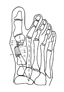
[0006] Figure 1(a) is posterior view of a foot with a hallux valgus
deformity
CA 03033269 2019-02-06
WO 2018/053254
PCT/US2017/051765
3
[0007] Figure 1(b) is a posterior view of a foot with the implant of the
present
invention following a translational chevron osteotomy procedure;
[0008] Figure 2 is a side view of the orthopedic implant of Figures 1(b);
[0009] Figure 3 is a side view of the plate shown in Figure 2;
[0010] Figure 4 is an end view of the plate shown in Figure 3;
[0011] Figure 5 is a side detail of the plate portion of a second
embodiment of
the plate shown in Figure 2;
[0012] Figure 6 is a side detail of the plate portion of a third embodiment
of
the plate shown in Figure 2
[0013] Figure 7 is a side detail of the plate portion of a fourth
embodiment of
the plate shown in Figure 2;
[0014] Figure 8 is a side detail of the plate portion of a fifth embodiment
of the
plate shown in Figure 2;
[0015] Figure 9 is a side view of a first metatarsal illustrating the cuts
for a
method of the present invention;
[0016] Figure 10 is a side view of the metatarsal of Figure 9 following the
osteotomy and a counter-bore procedure to prepare for the implant of the
present
invention.
DETAILED DESCRIPTION OF THE INVENTION
[0017] Figure 1(a) shows a skeletal version of a foot from the top side
illustrating a deformity with which the present implant could be used, and
Figure
1(b) shows the same version of the foot following the method of the present
invention with an embodiment of the implant 10 of the present invention in
place
inserted into the intramedullary channel of the first metatarsal following an
osteotomy and the translation of the distal portion of the metatarsal. Thus,
Figure 1(b) illustrates the implant used in a translational osteotomy of the
first
metatarsal for treatment of hallux valgus syndrome. Similarly, it can be used
for
fixation of other bone segments following osteotomies as previously mentioned.
[0018] As viewed from the top in Figure 1(b), it can be seen that the first
embodiment of the plate 10 has a body member 11 shaped for insertion into the
CA 03033269 2019-02-06
WO 2018/053254
PCT/US2017/051765
4
intramedullary portion of the bone following the osteotomy with a first end 12
that
extends into a plate portion 13 and an opposing cutting end 14 aligned along
the
longitudinal axis of the plate which further includes a cannulation 16 which
extends through the body member to allow for the insertion or use of a k-wire.
The body of the implant further includes a through hole 17 closer to the
second
end than the first which again is optionally threaded, for a screw 18 which is
optionally a locking screw. The screw is intended to inhibit rotation of the
implant
in the channel. The body portion is sized and shaped for insertion into the
intramedullary channel of a bone, for example, it may be cylindrical or
ellipsoid,
or have an elongate shape that is relatively round in cross-section but which
is
less regular than a cylinder or ellipse. The outer surface may be smooth, or
may
include one or more raised portions15 (illustrated in Figure 4), such as
grooves,
flutes or flanges which extend parallel to the cannulation part of all of the
way
down the length of the body member or which spiral around the body member) in
order to inhibit the implant from rotating within the inner portion of the
bone.
[0019] The plate
portion 13 has a profile when viewed straight on in the widest
dimension which is sized to accommodate the specific application, in this case
so
that it will best conform to the remaining head of the metatarsal, and further
includes at least a first through hole 20 (optionally threaded) for a screw 24
(optionally locking) and preferably a second through hole 22 (also optionally
threaded) for a second screw 26 (again optionally locking). Specifically, in
the
illustrated application for a bunionectomy, the plate portion has an outline
viewed
from the outer surface of a tab or partial egg shape, and the bone facing
surface
is curved so as to form a section of a cylinder (corresponding to an idealized
shape of the head of the associated metatarsal).
[0020] Figures 5
through 8 illustrate various embodiments of the implant 10, in
which the plate portion is offset to accommodate particular anatomical
variations,
of 1/3 of the distance across the diameter at the top surface of the implant
in
Figure 5, of 2/3 of the distance across the diameter at the top surface of the
implant in Figure 6, and of 7/8 of the distance across the diameter at the top
surface of the implant in Figure 7, and in Figure 8, the plate portion is
angled at
CA 03033269 2019-02-06
WO 2018/05325-1
PCT/US2017/051765
from 2 to 15 , and preferably from 7 to 12 relative to the axis of the body
member to inhibit a Varus tilt. Further this drawing illustrates the body and
plate
as an assembly, as compared to the other integrally formed plate and body
members. In Figure 8, the plate portion 413 extends from a threaded tap
member 30 that is received in a threaded screw hole 32 in the body member. In
this embodiment, various plate portions with varying degrees and/ or angles of
offset can be used in the body member to simplify the inventory of the system.
[0021] Figures 9 and
10 illustrate steps of the surgical procedure of the
present invention where in Figure 9 multiple incisions are made to the head
and
at about 1 cm down the neck of the metatarsal. Next, a tool is placed to
translate
the head of the metatarsal laterally and the remaining proximal portion of the
bone is reamed in an Eichhorn shape as is shown in Figure 10. Next with
pushing or drilling, a k-wire is placed in the metatarsal to the base, and the
bone
is reamed by power or hand. The guide wire is left in place. Then the implant
is
pushed into position over the guide wire and into the intramedullary channel
using a cross-jig which is radiographically located. The screws are inserted
into
the plate portion, and the implant is pushed into the proximal portion of the
bone
to compress. The cross screw is inserted, the jigs are disassembled and the
guide wire is removed, the medial ledge is removed, including a saw is used or
a
burr if a mini-incision is used. Graft is added as needed and the incision is
closed.
[0022] The screws
useful with the plate of the present invention are self-
starting, self-tapping screws including the option of partial or full
cannulation.
The screws include a cutting end having multiple flutes, and preferably 2 or 3
flutes about a conical recess. The screws further include a partial taper of
the
inner diameter in the proximal end over the first several thread turns, for
example
over 2-8, and preferably over 3-5 turns in order to increase the fatigue life
of the
screw as well as providing potential physiological advantages in use. The
screws further include a torque driving recess. The screws have a threaded
distal end and a head including a torque driving recess. The head of the
locking
screw includes locking means, such as a variable locking mechanism, which
CA 03033269 2019-02-06
WO 2018/053254
PCT/US2017/051765
6
could be a bushing that mates with the screw head so as to lock the screw
relative to the plate at a desired angle, or could include external screw
threads
that mate with internal threads in the locking screw hole at a pre-selected
angle,
in this instance, the screw axis is perpendicular to the longitudinal axis of
the
plate. The screw used in the anti-rotation slot has a rounded rear shoulder
(such
as a hemisphere, or a torroid) which mates with the edges of the slot.
[0023] The implant is
formed of a biocompatible material, and preferably a
metal such as surgical grade stainless steel, titanium or a titanium alloy or
a
cobalt chromium alloy. Preferably, the plate portion has a thickness of
between
about 1.0 and about 2.5 millimeters, more preferably between about 1.5 and
about 2 millimeters, and most preferably between about 1.5 and about 1.8
millimeters. The body portion of the implant has a curved cross-section having
a
diameter defining a total thickness of about 4 and 8 millimeters, or roughly
four
times the width of the plate portion of the implant.
[0024] In addition,
the implant can include additional small through holes
sized to receive a K-wire or other similar guide wire.
[0025] During the
surgery the joints are first prepped which may include de-
articulation between the bones to be fused and removal of any bone as part of
the osteotomy, and as necessary, the plate is bent to contour to the bone
surface. A pilot hole or preferably, an Eichhorn shaped recess may be drilled
into
the bone into which the implant will be inserted. The implant is inserted into
the
implant recess in the driver and secured by tightening the implant upward in
the
holder using the holder. The implant is tamped into the cancellous portion of
the
bone fragment optionally by tapping the implant driver with a one pound mallet
as
is necessary to insert the body of the implant. The implant should be driven
until
it is fully seated. Once the implant is sunk, a drill guide is mated to the
driver,
and a hole is drilled for the transverse screw. The implant can be held in
position
using k-wires or olive wires (thru the non-locking hole and into the bone).
The
plate portion is located such that all of the screws are aimed into the
targeted
bones and away from the joint, fracture, or bone interface. The olive wire is
removed if used, and a pilot hole is drilled at the end of the plate that
includes the
CA 03033269 2019-02-06
WO 2018/053254
PCT/US2017/051765
7
first hole and this hole is pinned or screwed. A second pilot hole may be
drilled
for the transverse. The plate is viewed radiographically, and the soft tissues
are
closed in the usual manner.
[0026] This invention
has been described in detail with reference to specific
embodiments thereof, including the respective best modes for carrying out each
embodiment. It shall be understood that these illustrations are by way of
example
and not by way of limitation.
