Note: Descriptions are shown in the official language in which they were submitted.
CA 03033475 2019-02-08
Heterodimeric Fc-fused cytokine and
pharmaceutical composition comprising the same
TECHNICAL FIELD
The present invention relates to a heterodimeric Fc-fused protein comprising a
first Fc
region and a second Fc region of an immunoglobulin Fc pair and a
physiologically active
protein, wherein one or more subunits of the physiologically active protein
are linked to one
or more ends of the N-terminus or C-terminus of the first Fc region and/or the
second Fc
region, and CH3 domains of the first Fc region and the second Fc region are
mutated so as to
promote Fc heterodimer formation, and a pharmaceutical composition comprising
the
heterodimeric Fc-fused protein.
The heterodimeric Fc-fused protein according to the present invention has an
advantage in that it can retain the activity of a naturally occurring
physiologically active
protein, which is composed of two or more different subunit proteins and
thereby exhibit
the intact biological activity by forming a assembled protein, because each
subunit of the
protein can be separately fused to each chain of heterodimeric Fc of
immunoglobulin such
that the fused protein can maintain the naturally occurring form and structure
to the highest
possible degree.
When the heterodimeric Fc-fused protein according to the present invention is
used,
there is an advantage in that the in vivo half-life of the physiologically
active protein
contained in the heterodimeric Fc-fused protein can be significantly increased
due to the Fc-
mediated long half-life such that the physiological activities thereof in vivo
can be long-
lasting.
- -
CA 03033475 2019-02-08
In addition, the heterodimeric Fe-fused protein according to the present
invention has
a structure in which one or more subunits of the physiologically active
protein are fused to
the N-terminus or C-terminus of an immunoglobulin heterodimeric Fe, and the
heterodimeric Fe-fused protein is easily purified after its expression,
compared to a wild-
type Fe-based fusion protein.
BACKGROUND ART
Naturally occurring human antibodies (immunoglobulin G (IgG), IgM, IgD, IgE,
and
IgA) are each present as an assembly of two heavy chains having the same amino
acid
sequence and two light chains having the same sequence. In this regard,
homodimerization
between the two identical heavy chains is induced by the non-covalent
interactions between
the constant region terminal domains (CH3 domains in IgG, IgD and IgA, CH4
domains in
IgM, and CH2 and CH4 domains in IgE) and the disulfide bond between hinge
domains.
Antibody heterodimeric Fe technology is a technology that makes heterodimeric
fragment crystallizable (Fe) of immunoglobulin heavy chain constant regions by
modifications to the CH3 domain interface, with different mutations on each
domain such
that the engineered Fe fragments, carrying the CH3 variant pair,
preferentially form Fe
heterodimers in naturally occurring antibodies (IgG, IgM, IgA, IgD, and IgE)
rather than the
Fe homodimers. More specifically, it is a technology that induces mutations in
two different
CH3 domains of Fe by genetic engineering, such that the two Fe fragments form
a
heterodimer with minimal sequence variations while they have tertiary
structures very
similar to those of naturally occurring antibodies (US Patent No. 7,695,936;
and Korean
Patent No. 1,522,954). The heterodimeric Fe technology is a platform
technology for
making bispecific antibodies, and CH3 domain mutants that induce Fe
heterodimer
- 2 -
CA 03033475 2019-02-08
formation known so far were mostly generated by introducing an asymmetric
mutation pair
into the CH3 domain interface by the structure-based rational design of
antibody (Spreter
Von Kreudenstein et al., 2014). Pioneering studies include knob-into-hole
technology
(Ridgway et al., 1996) from Genentech, and many multinational pharmaceutical
companies,
including Zymeworks (ZW1; Von Kreudenstein et al., 2013), Xencor (HA-TF; Moore
GL et
al., 2011) and EMD Serono (SEEDbody; Davis JH et al., 2010), have developed
and
reported the platform technology.
Above all, the A107 variant used in the present invention is a high-yield Fc
heterodimer screened from a human antibody heterodimeric Fc library
constructed using a
yeast cell surface display system, and is a heterodimeric Fc variant which
promotes the
heterodimeric formation by inducing mutations at charged amino acids to form
sterically
complementary hydrophobic interactions (K409WcH3A-D399V/F405TcH3B) and forming
hydrogen bonds (K370EcH3A-E357NcH3B), while retaining hydrophobic core
integrity at the
CH3 domain interface (Choi et al. 2016; Korean Patent Application No. 2015-
0142181).
Heterodimeric Fc variants reported so far, including the A107 variant, are all
based on
IgG1 occupying the largest proportion of human antibody isotypes, and variants
of isotypes
(IgG2, IgG3, IgG4, IgA, IgM, and IgE) other than IgG1 have not been reported
yet.
This is because therapeutic antibodies that are being marketed under approval
of the
U.S. Food and Drug Administration (FDA) mostly adopt the IgG1 isotype (Irani
et al. 2015).
In recent years, for immune-modulating antibodies or receptor agonist fusion
proteins that do
not need to have great antibody effector functions such as antibody-dependent
cellular
cytotoxicity (ADCC) or complement-dependent cellular cytotoxicity (CDC), the
development of therapeutic proteins based on IgG2 or IgG4 whose effector
functions are
significantly lower than those of IgG1 have been made.
- 3 -
CA 03033475 2019-02-08
Meanwhile, physiologically active proteins mostly have small sizes, and thus
have the
disadvantage of having a short in vivo half-life. In order to solve this
disadvantage, there has
been an attempt to conjugate PEG (polyethylene glycol) or the like, or fusion
to an antibody
Fc (crystallizable fragment) region. However, it has not yet been possible to
develop
physiologically active proteins whose activity is efficiently and sufficiently
maintained for a
long period of time.
In particular, for proteins composed of two or more different subunits,
wherein the two
or more different subunits form a protein complex to exhibit physiological
activity, it has
never been possible to develop Fc-fused proteins which are formed to have
naturally
occurring original protein complex structures with wild type Fc because wild
type Fc-fused
protein forms homodimer due to the homodimeric nature of Fc. Thus, wild type
Fc is not
suitable for Fc fusion for heterodimeric or heterooligomeric proteins to
properly exhibit the
activity of the original proteins and sufficiently maintain their activity for
a long period of
time.
Under this technical background, the present inventors have constructed
heterodimer
variants comprising Fc regions derived not only from IgG I , but also from
other isotype
antibodies such as IgG2, IgG3 and IgG4, which were previously not reported,
and have used
these heterodimer variants to develop a novel therapeutic fusion protein in
the form of a
heterodimeric Fc-fused protein wherein one or more subunits of a protein,
which is
composed of two or more different subunits and in which two or more subunits
exhibit
physiological activity by forming a protein complex, are genetically fused to
the terminus of
the Fc region, thereby completing the present invention.
- 4 -
CA 03033475 2019-02-08
In particular, in the present invention, preferably, interleukin-12 (IL-12)
can be used as
the protein which is composed of two different subunits, p35 and p40, wherein
the two
subunits exhibit physiological activity by forming the IL-12 protein.
IL-12 can directly kill tumors by increasing the activity of immune cells such
as
cytotoxic T lymphocytes (CTLs) or natural killer cells (NKs) among immune
cells, or can
inhibit tumorigenesis by activating immune responses through secretion of pro-
inflammatory
cytokines such as interferon-gamma (TEN-7) in tumor microenvironments where
the immune
responses are inhibited. Thus, IL-12 has been much studied as an anti-cancer
cytokine
(Lasek et al., 2014). However, in the development of therapeutic methods using
IL-12, the
short half-life of the cytokine itself necessitates frequent administration
which can lead to
toxicity. For this reason, studies have been conducted to fuse IL-12 with an
antibody or Fc
in order to use it as long-acting IL-12 (Tugues et al., 2015). However, in
these studies, a
problem arises in that, due to the fusion of a wild-type Fc-based antibody
that forms a
homodimer by the interaction between CH3 domains, the fused IL12 protein is
bivalent,
unlike an endogenous monovalent form of IL-12, and for this reason, the wild
type Fc-based
antibody fused IL-12 shows poor physiological activity than endogenous IL-12,
or unwanted
localization appears due to avidity-driven increased binding of IL-12 to
immune cells (Tzeng
et al., 2015; Dumont et al., 2006).
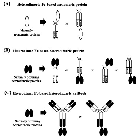
Therefore, in an effort to make a monovalent fusion protein using a wild-type
antibody
or an Fc region, as shown in FIGS. 1(A) to 1(C), there has been used a method
of
constructing a fusion protein through a strategy such as fusing a selective
tag for additional
purification only to the C-terminus of a single Fc region or fusing an Fc
region and a protein
to each other after separately purifying them with high purity. However, this
method is not
-5-
CA 03033475 2019-02-08
only very costly to produce a large amount of protein, but also requires
research to optimize
an additional purification process.
However, the use of a heterodimeric Fc-fused protein according to the present
invention makes it possible to easily produce a monovalent heterodimeric Fc-
fused protein
as shown in FIG. 2 without needing to optimize an additional purification
process.
DISCLOSURE OF INVENTION
TECHNICAL PROBLEM
It is an object of the present invention to provide a novel heterodimeric Fe-
fused
protein, the protein of which is composed of one, two, or more different
subunits and
thereby exhibits the intact biological activity by forming the assembled
protein, and thus
can maintain the natural physiological activity of the fused protein thereof
in vivo for a long
period of time.
In particular, the heterodimeric Fe-fused protein according to the present
invention
is formed such that it can retain the activity of a naturally occurring
physiologically active
protein, in which two or more subunits assemble together to form a protein to
exhibit
physiological activity, such that the fused protein can maintain the naturally
occurring form
and structure to the highest possible degree.
Further, the heterodimeric Fe-fused protein according to the present invention
has
an advantage in that the in vivo half-life of the physiologically active
protein contained in
the heterodimeric Fe-fused protein can be significantly increased due to the
Fe-mediated
long half-life such that the physiological activities thereof in vivo can be
long-lasting.
- 6 -
CA 03033475 2019-02-08
Another object of the present invention is to provide a pharmaceutical
composition
comprising the above-described heterodimeric Fc-fused protein, and a
composition and a
therapeutic method for treating diseases, particularly cancer, using the same.
TECHNICAL SOLUTION
To achieve the above object, the present invention provides a heterodimeric Fc-
fused protein comprising a first Fc region and a second Fc region of an
immunoglobulin Fc
pair and a physiologically active protein,
wherein the physiologically active protein is composed of two or more
different
subunits, wherein the two or more different subunits exhibit physiological
activity by
forming a protein complex,
wherein the subunits of a physiologically active protein are linked or
genetically
fused to one or more ends of the N-terminus or C-terminus of the first Fc
region and/or the
second Fc region,
wherein the CH3 domains of the first Fc region and the second Fc region are
mutated so as to promote Fc heterodimer formation.
The present invention also provides a pharmaceutical composition comprising
the
above-described heterodimeric Fc-fused protein, and a composition and a
therapeutic
method for treating diseases, particularly cancer, using the same.
BRIEF DESCRIPTION OF THE DRAWINGS
FIGS. 1(A) to 1(C) illustrate conventional strategies for obtaining monomeric
and
heterodimeric fusion proteins using wild-type Fc of human IgG antibody. (A)
Wild-type Fc-
based Epo-Fc dimer vs. Epo-Fc monomer. (B) Aglycosylated Fc-fused
- 7 -
CA 03033475 2019-02-08
GLP-1/GCG monomeric peptide, generated by the LAPScovery technology. (C) Wild-
type
Fc-based Fc-FSH tandem homodimer vs. Fc-FSH heterodimer. Epo, erythropoietin;
GLP-
1/GCG, glucagon-like peptide- l/glucagon; FSH, Follicle-stimulating hormone.
FIG. 1(D) shows an example of constructing an antibody-cytokine
(immunocytokine)
by fusing a monomeric cytokine (IL2) to an IgG type antibody comprising a knob-
into-hole
(KiH) heterodimeric Fc variant according to previous literature.
FIGS. 2(A) and 2(B) illustrate monomeric and heterodimeric fusion protein
forms
which may be constructed using a heterodimeric Fc. Potential use of
heterodimeric Fc for the
generation of Fc-fused monomeric or heterodimeric proteins to present the
fusion partner in
its naturally occurring form. The Fc-fused monomer can easily be generated by
the fusion of
monomeric protein to the N- or C-terminus of one heterodimeric Fc chain.
FIG. 2(C) illustrates a fusion protein formed by fusing a heterodimer to an
IgG type
human antibody comprising a heterodimeric Fc. The Fc-fused heterodimer can be
generated
by separate fusion of the two subunits of heterodimeric proteins to each chain
of the
heterodimeric Fc at the N- or C-terminus.
FIG. 3 shows the sequence alignment of CH3 domain of human IgG isotype
antibodies (hIgG 1 , hIgG2, hIgG3, hIgG4) with highlights of the mutated
residues in A107
heterodimeric Fc variant (K370E/K409WcH3A¨E357N/D399V/F405TcH3B).
FIG. 4 shows the results of performing structural modeling of heterodimeric Fc
variants for each isotype by use of sequences having induced mutations at the
positions
selected in FIG. 3 and analyzing the resulting modeling structures
comparatively with wild-
type IgGl-based A107 variants.
FIG. 5 is a schematic view of a vector for expressing a heterodimeric Fc for
each
isotype, constructed by sequence and structure analysis, in animal cells. The
heterodimeric
- 8 -
CA 03033475 2019-02-08
Fc variant for each isotype, which comprises a mutated hinge region, was
cloned into the
vector by use of restriction enzymes (NotI/HindIII).
FIG. 6 schematically shows a scFv-FecH3A/FecH3B expression system for
evaluating
the ability of heterodimeric Fc variants to form a heterodimer, by the dimer
size difference
between expressed proteins.
FIG. 7 is a schematic view for cloning scFv-Fc fused to a single-chain
variable
fragment (scFv), constructed to evaluate the heterodimerization formation
yield of an
antibody Fc by a CH3 mutant pairs as shown in FIG. 6, into a pcDNA3.1 vector
which is an
animal cell expression vector.
FIG. 8 show the results of co-transfecting CH3 mutant pairs-introduced animal
cell
expression vectors, constructed according to the expression systems shown in
FIGS. 5 and 7,
into HEK293F cells in order to evaluate the heterodimerization formation as
shown in FIG. 6,
transiently expressing and purifying the vectors, and then separating 5 n of
protein on SDS-
PAGE under non-reducing conditions in order to evaluate the heterodimerization
formation,
and analyzing the protein according to size and combination by Coomassie blue
staining. As
a negative control, a wild-type Fc with wild-type CH3 was used.
FIG. 9 shows the results of separating protein by SDS-PAGE according to the
method shown in FIG. 8, and then performing Western blotting using anti-human
IgG-AP
conjugated antibody.
FIG. 10(A) is a schematic view showing the form of endogenous IL-12 cytokine
to
which Fc was not fused and which is used as a control in the present
invention.
FIG. 10(B) is a schematic view showing the form of a bi-1L-12-Fc fusion
protein
which was obtained by fusing IL-12 cytokine to wild-type IgG4 Fc by an amino
acid linker
and which is used as a comparative example in the present invention.
- 9 -
CA 03033475 2019-02-08
FIG. 10(C) is a schematic view showing the form of a mono-IL-12-Fc fusion
protein
obtained by fusing IL-12 cytokine to an IgG4-based 74-A107 variant among
heterodimeric
Fc variants for each isotype according to the present invention.
FIGS. 11(A) and 11(B) are schematic views of vectors for expressing and
purifying a
fusion protein of an example of the present invention (FIG. 10 (C)) in animal
cells.
FIG. 12 is a schematic view of a vector for expressing and purifying a fusion
protein
of an example of the present invention (FIG. 10 (B)) in animal cells.
FIG. 13 shows the results of co-transfecting the animal cell expression
vectors of
FIGS. 11(A) and 11(B), constructed using human and mouse interleukin genes,
into
HEK293F cells, transiently expressing and purifying the genes, and then
separating 5 [ig of
protein on SDS-PAGE under non-reducing conditions, and analyzing the protein
according
to size and combination by Coomassie blue staining.
FIG. 14 shows the results of analyzing the fusion proteins of FIG. 13 by size-
exclusion chromatography (SEC).
FIG. 15 shows the results of FACS analysis performed to analyze the binding
affinities of mono-hIL-12-Fc and wild-type bi-hIL-12-Fc on normal PMBCs having
no IL-
12 receptor and PHA-activated PBMCs in which the IL-12 receptor was induced by
treatment with the mitogen PHA (phytohaemagglutinin).
FIG. 16 shows the results of a WST-1 cell proliferation assay performed to
measure
the effect of various concentrations of Fc (A107), recombinant human IL-12
(rhIL-12), bi-
h1L-12-Fc and mono-hIL-12-Fc on the proliferation of PHA-activated PBMCs in
which the
IL-12 receptor was induced by treatment with the mitogen PHA.
FIG. 17 shows the results of an ELISA performed to measure the concentration
of
1FN-7 in culture supernatants obtained as shown in FIG. 16.
- 10
CA 03033475 2019-02-08
FIG. 18 shows the results of flow cytometry analysis performed to measure the
binding affinities of mono-m1L-12-Fc and bi-mIL-12-Fc on normal PMBCs having
no IL-12
receptor and PHA-activated PBMCs in which the IL-12 receptor was induced by
treatment
with the mitogen PHA, because mIL-12 cross-reacts with human IL-12R on
activated human
.. T cells and NK cells.
FIG. 19 shows the results of a WST-1 cell proliferation assay performed to
measure
the effect of various concentrations of Fe (A107), recombinant mouse IL-
12(rmIL-12), bi-
mIL-12-Fc and mono-mIL-12-Fc on the proliferation of PHA-activated PBMCs in
which the
IL-12 receptor was induced by treatment with the mitogen PHA.
FIG. 20(A) shows the changes of tumor volume in Balb/c mice transplanted with
CT26HER2/Neu tumors during the intraperitoneally administration of Fe (A107),
rmIL-12, bi-
mIL-12-Fc and mono-mIL-12-Fc, and picture of the tumor-bearing mice after
sacrifice at the
end of administration. Injection of mIL12-Fc proteins was initiated 11 days
after tumor cell
inoculation when the tumors volume reached 100 mm3).
FIG. 20(B) is a graph showing the changes of mouse body weight measured at
indicated time points in the experimental procedure shown in FIG. 20(A).
FIG. 21(A) shows the results of measuring mouse tumor volume changes measured
while intraperitoneally administering various concentrations of bi-mIL-12-Fc
and mono-
m1L-12-Fc, twice a week, when the tumor volume in Balb/c mice transplanted
with
.. CT26HER2/Neu reached 300 mm3.
FIG. 21(B) is a graph showing the changes of individual mouse tumor volume
treated
with m1L12-Fc proteins at indicated time points in the experimental procedure
shown in FIG.
21(A).
-11-
CA 03033475 2019-02-08
FIG. 21(C) shows the picture of tumors taken from tumor-bearing mice on 3 days
after the last administration in FIG. 21(A).
Fig. 21(D) is a graph showing the changes of mouse body weight measured at
indicated time points in the experimental procedure shown in FIG. 21(A).
FIG. 21(E) is a graph showing the results of measuring alanine
aminotransferase
(ALT) (which is a hepatotoxicity marker) in the blood which was collected from
mouse
facial veins on 1 day after the last administration in FIG. 21(A).
FIG. 22(A) is a graph showing the results of measuring increases in the number
of
CD4+ T cells, CD8+ T cells and NK cells in the spleens of mice sacrificed on 3
days after the
last administration in FIG. 21(A).
FIG. 22(B) is a graph showing the number of total immune cells, CD4+ T cells
and
CD8+ T cells that infiltrated the tumor in mice sacrificed on 3 days after the
third
administration in FIG. 21(A).
FIG. 23(A) shows the results of an ELISA performed to measure the serum levels
of
IFN-y in CT26HER2/neu tumor bearing mouse treated with mIL-12-Fc proteins.
Mouse serum
was separated after clotting blood collected from mouse facial veins at 24
hours after the last
administration in FIG. 21(A).
FIG. 23(B) is a graph showing the results of an ELISA performed to measure the
concentration of IFN-y in serum separated from blood collected from mouse
facial veins on
1, 3 and 5 days after intraperitoneally administering bi-mIL-12-Fc and mono-
mIL-12-Fc at a
concentration equimolar to 1 lig rmIL-12 when the tumor volume in Balb/c mice
transplanted with CT26HER2/Ne1I cancer cells reached 300 mm3.
-12-
CA 03033475 2019-02-08
FIG. 23(C) is a graph showing the results of measuring the cytotoxic effect of
cytotoxic T cells, isolated from the spleen of mice sacrificed on 3 days after
the last
administration in FIG. 21(A), against CT26HER2/Neu cancer cells.
FIG. 23(D) shows the cytotoxic activity of splenic CD8+ T cells isolated from
CT26-
.. HER2/neu tumor-bearing mouse treated with mIL-12-Fc proteins, analyzed on 3
days after
the third administration in Fig. 21(A), followed by 4 h of culture with
CT26HER2/Neu cancer
cells expressing tumor antigen and 4T1 cells not expressing tumor antigen.
FIG. 23(E) is a graph showing the results of measuring the cytotoxic effect of
natural
killer cells, isolated from the spleen of mice sacrificed on 3 days after the
third
.. administration in FIG. 21(A), against CT26HER2/Neu cancer cells.
FIG. 24(A) is a graph showing the results of measuring the number of CD8+
effector
T cells isolated from in the spleen isolated from tumor-bearing mice
sacrificed on 3 days
after the last administration in FIG. 21(A).
FIG. 24(B) is a graph showing the results of measuring the number of CD8+
effector
.. memory T cells in the spleen isolated from tumor-bearing mice sacrificed on
3 days after the
last administration in FIG. 21(A).
FIG. 24(C) is a graph showing the results of measuring the number of CD8+
central
memory T cells in the spleen isolated from tumor-bearing mice sacrificed on 3
days after the
last administration in FIG. 21(A).
FIG. 24(D) shows the results obtained by re-transplanting CT26HER2/Neu cancer
cells
into survived Balb/c mice on 120 days after administration of 1 g mono-IL-12-
Fc in FIG.
21(A), and measuring tumor volume changes in the mice.
FIG. 24(E) shows the results of flow cytometry performed to analyze the
proportion
of memory precursor effector cells (KLRGIIL-7R+) and short-lived effector
cells
- 13 -
CA 03033475 2019-02-08
(KLRG1 IL-71Z-) among CD8+ T cells in the spleen isolated from tumor-bearing
mice
sacrificed on 3 days after the third administration in FIG. 21(A).
FIG. 25(A) is a graph showing the results of flow cytometry analysis performed
to
measure the proportion of CD8+ T cells (which showed high expression of the
transcription
factor T-bet that inhibits memory cell differentiation) in spleen cells
isolated from mice
sacrificed on 3 days after the third administration in FIG. 21(A).
FIG. 25(B) is a graph showing the results of flow cytometry analysis performed
to
measure the proportion of CD8+ T cells (which showed high expression of Eomes
and low
expression of T-bet) in spleen cells isolated from mice sacrificed on 3 days
after the third
administration in FIG. 21(A).
FIG. 25(C) is a graph showing the results of flow cytometry analysis performed
to
measure the expression level of phosphorylated STAT4 in CD8+ T cells isolated
from tumor
draining (inguinal) lymph nodes at 24 hours after intraperitoneally
administering bi-mIL-12-
Fc and mono-mIL-12-Fc once at a concentration equimolar to 1 Kg rmIL-12 when
the tumor
volume in Balb/c mice transplanted with CT26HER2/Neu cancer cells reached 300
mm3.
FIG. 25(D) is a graph showing the results of flow cytometry analysis performed
to
measure the proportion of CD8+ T cells (which expressed T-bet that inhibits
memory cell
differentiation) in tumor draining (inguinal) lymph nodes at 72 hours after
the single
intraperitoneal administration in FIG. 25(C).
FIG. 25(E) is a graph showing the results of flow cytometry analysis performed
to
measure the expression level of pSTAT4 when CD8+ T cells isolated from the
spleen and
inguinal lymph node of normal Balb/c mice were stimulated with the mono-mIL-12-
Fc and
bi-mIL-12-Fc that cross-reacted with anti-Fc antibody.
- 14 -
CA 03033475 2019-02-08
FIG. 25(F) is a graph showing the results of flow cytometry analysis performed
to
measure the proportion of T-bet-expressing CD8+ T cells when CD8+ T cells
isolated from
the spleen and groin lymph node of normal Balb/c mice were stimulated with the
mono-
mIL-12-Fc and bi-mIL-12-Fc that cross-reacted with anti-Fc antibody.
FIG. 26 is an overall schematic view showing a mechanism that induces
differentiation of memory precursor effector cells by mono-mIL-12-Fc and a
mechanism that
induces differentiation of short-lived effector cells by bi-mIL-12-Fc.
BEST MODE FOR CARRYING OUT THE INVENTION
Unless defined otherwise, all the technical and scientific terms used herein
have the
same meaning as those generally understood by one of ordinary skill in the art
to which the
invention pertains. Generally, the nomenclature used herein and the experiment
methods,
which will be described below, are those well-known and commonly employed in
the art.
In one aspect, the present invention relates to a heterodimeric Fc-fused
protein
comprising a first Fc region and a second Fc region of an immunoglobulin Fc
pair and a
physiologically active protein,
wherein the physiologically active protein is composed of two or more
different
subunits, wherein the two or more different subunits exhibit physiological
activity by
forming a protein complex, wherein one or more subunits of a physiologically
active protein
are linked to one or more ends of the N-terminus or C-terminus of the first Fc
region and/or
the second Fc region,
wherein C113 domains of the first Fc region and the second Fc region are
mutated so
as to promote heterodimer formation.
- 15 -
CA 03033475 2019-02-08
As used herein, the term "Fe region" or "heavy chain constant region" means a
region
comprising an immunoglobulin CH2 domain, a CH3 domain and a hinge domain.
However,
for IgE, the term means a region comprising a CH2 domain, a CH3 domain, a CH4
domain
and a hinge domain.
As used herein, the expression "the first Fc region and the second Fc region
are
mutated so as to promote heterodimer formation" means that a naturally
occurring antibody
has a homodimeric form in which two Fc regions have the same sequence, and a
portion of
these Fc region sequences is mutated, so that heterodimer formation can be
promoted
through a specific non-covalent interaction between the first Fc region and
the second Fc
region, or homodimer formation can be reduced, or preferably can hardly occur.
Preferably, "the first Fc region and the second Fc region are mutated so as to
promote
heterodimer formation" may include "each of CH3 domains contained in the first
Fc region
and second Fc region from immunoglobulin is mutated so as to promoter Fc
heterodimer
formation".
In the present invention, "heterodimeric Fc or Fc heterodimer" comprises the
first Fc
region and the second Fc region, and the first Fc region and the second Fc
region mean
heterodimers in which CH3 domains of the first Fc region and the second Fc
region are
mutated so as to promote Fc heterodimer formation.
In the present invention, each of the first Fc region and the second Fc region
may be
derived from an Fc region selected from the group consisting of human IgG1 ,
IgG2, IgG3,
IgG4, IgM, IgA, IgD and IgE, and preferably each of the first Fc region and
the second Fc
region is derived from IgG 1 , IgG2, IgG3 or IgG4.
In addition, the first Fc region and the second Fc region may be derived from
an
isotype antibody.
-16-
CA 03033475 2019-02-08
In another aspect, the mutation of CH3 domain may include one or more
mutations
selected from the following group (wherein all mutation positions in the
present invention
are numbered according to the EU index):
(1) substitution of the amino acid residue at position K370 in the CH3 domain
of the
.. first Fc region; and substitution of the amino acid residue at position(s)
E357 and/or S364 in
the CH3 domain of the second Fc region; and/or
(2) substitution of the amino acid residue at position K409 in the CH3 domain
of the
first Fc region; and substitution of the amino acid residue at position(s)
F405 and/or D399
in the CH3 domain of the second Fc region.
Preferably, the substitution of amino acid residue at position K370 in the CH3
domain
of the first Fc region may be K370E, K370R, K370M, K370D or K370H,
substitution of
the amino acid residue at position E357 in the CH3 domain of the second Fc
region may be
E357N, E357D, E357A, E3571, E357G or E357M, and substitution of the amino acid
residue at position S364 in the CH3 domain of the second Fc region may be
S364T or
S364W.
In addition, substitution of the amino acid residue at position K409 in the
CH3
domain of the first Fc region may be K409W, substitution of the amino acid
residue at
position F405 in the CH3 domain of the second Fc region may be F405T, and
substitution
of the amino acid residue at position D399 in the CH3 domain of the second Fc
region may
be D399V.
The amino acid residue mutation such as K370E means that K at position 370 is
mutated to E, and the mutation of all amino acid residues in the present
invention is used as
the same meaning as described above.
-17-
CA 03033475 2019-02-08
Most preferably, the mutation of the CH3 domain of the first Fe region or the
second
Fe region may include one or more mutations selected from the following group
(wherein
mutation positions are numbered according to the EU index.):
(1) a substitution K370E, K370R, K370M, K370D or K370H of the amino acid
residue at position K370 in the CH3 domain of the first Fe region;
(2) a substitution E357N, E357D, E357A, E3571, E357G or E357M of the amino
acid ,
residue at position E357 in the CH3 domain of the second Fe region, and
substitution S364T
or S364W of the amino acid residue at position S364 in the CH3 domain of the
second Fe
region;
(3) a substitution K409W of the amino acid residue at position K409 in the CH3
domain of the first Fe region; and
(4) a substitution F405T of the amino acid residue at position F405 in the CH3
domain of the second Fe region, and substitution D399V of the amino acid
residue at
position D399 in the CH3 domain of the second Fe region.
The CH3 domains in the first Fe region and the second Fe region may further
include
the following residue:
(i) cysteine (C) substituted at position Y349 in the CH3 domain of the first
Fe region;
and
(ii) cysteine (C) substituted at position S354 in the CH3 domain of the second
Fe
region.
In still another aspect, mutation of the CH3 domain may include one or more
mutations selected from the following group:
-18-
CA 03033475 2019-02-08
(1) a substitution of the amino acid residue at position K360 in the CH3
domain of the
first Fc region; and substitution of the amino acid residue at position E347
in the CH3
domain of the second Fc region; and/or
(2) a substitution of the amino acid residue at position K409 in the CH3
domain of the
first Fc region; and substitution of the amino acid residue at position(s)
F405 and D399 in
the CH3 domain of the second Fc region.
Preferably, the substitution of the amino acid residue at position K360 in the
CH3
domain of the first Fc region may be K360E, and substitution of the amino acid
residue at
position E347 in the CH3 domain of the second Fc region may be E347R.
Substitution of the amino acid residue at position K409 in the CH3 domain of
the first
Fc region may be K409W, substitution of the amino acid residue at position
F405 in the
CH3 domain of the second Fc region may be F405T, and substitution of the amino
acid
residue at position D399 in the CH3 domain of the second Fc region may be
D399V.
Most preferably, the mutation of the CH3 domain of the first Fc region or the
second
Fc region may include one or more mutations selected from the following group
(wherein
mutation positions are numbered according to the EU index):
(1) a substitution K360E of the amino acid residue at position K360 in the CH3
domain of the first Fc region;
(2) a substitution E347R of the amino acid residue at position E347 in the CH3
domain
of the second Fc region;
(3) a substitution K409W of the amino acid residue at position K409 in the CH3
domain of the first Fc region; and
-19-
CA 03033475 2019-02-08
(4) a substitution F405T of the amino acid residue at position F405 in the CH3
domain of the second Fc region, and substitution D399V of the amino acid
residue at
position D399 in the CH3 domain of the second Fc region.
The CH3 domains in the first Fc region and the second Fc region may further
include
the following residue:
(i) cysteine (C) substituted at position Y349 in the CH3 domain of the first
Fc region;
and
(ii) cysteine (C) substituted at position S354 in the CH3 domain of the second
Fc
region.
Preferably, each of the CH3 domains contained in the first Fc region and the
second
Fc region from immunoglobulin according to the present invention may have an
amino acid
sequence selected from the group consisting of the amino acid sequences
represented by the
following SEQ ID NOS:
(1) SEQ ID NO: 1 and SEQ ID NO: 2;
(2) SEQ 1D NO: 3 and SEQ ID NO: 4;
(3) SEQ ID NO: 5 and SEQ ID NO: 6;
(4) SEQ ID NO: 8 and SEQ ID NO: 9;
(5) SEQ ID NO: 11 and SEQ 1D NO: 12; and
(6) SEQ ID NO: 14 and SEQ ID NO: 15.
In particular, the first Fc region and second Fc region from immunoglobulin
according to the present invention preferably have the sequences of IgG4 CH3
domains
shown in Table 1 below.
Table 1
- 20 -
CA 03033475 2019-02-08
CH3 sequence of first Fe region CH3
sequence of second Fe region
configuration
(EU number 341 to 447) (EU number 341 to 447)
GQPREPQVYTLPPSQEEMTEN
GQPREPRVYTLPPSQEEMTKNQ
QVSLTCLVKGFYPSDIAVEWE
VSLTCLVKGFYPSDIAVEWESNG
SNGQPENNYKTTPPVLDSDGS
QPENNYKTTPPVLVSDGSFTLYS
y4-EWRVT FFLYSWLTVDKSRWQEGNVFS
RLTVDKSRWQEGNVFSCSVMHE
CSVMHEALHNHYTQKSLSLSL
ALHNHYTQKSLSLSLGK
GK
(SEQ ff) NO: 2)
(SEQ ID NO: 1)
GQPREPQVCTLPPSQEEMTEN
GQPREPRVYTLPPCQEEMTKNQ
QVSLTCLVKGFYPSDIAVEWE
VSLTCLVKGFYPSDIAVEWESNG
SNGQPENNYKTTPPVLDSDGS
QPENNYKTTPPVLVSDGSFTLYS
y4-EWRVT,-, FFLYSWLTVDKSRWQEGNVFS
RLTVDKSRWQEGNVFSCS VMHE
CSVMHEALHNHYTQKSLSLSL
ALHNHYTQKSLSLSLGK
GK
(SEQ ID NO: 4)
(SEQ ID NO: 3)
GQPREPQVYTLPPSQEEMTKN
GQPREPQVYTLPPSQENMTKNQ
QVSLTCLVEGFYPSDIAVEWE
VSLTCLVKGFYPSDIAVEWESNG
SNGQPENNYKTTPPVLDSDGS
QPENNYKTTPPVLVSDGSFTLYS
y4-A107 FFLYSWLTVDKSRWQEGNVFS
RLTVDKSRWQEGNVFSCS VMHE
CSVMHEALHNHYTQKSLSLSL
ALHNHYTQKSLSLSLGK
GK
(SEQ ID NO: 6)
(SEQ ID NO: 5)
In the heterodimeric Fe-fused protein according to the present invention, a
subunit of
the physiologically active protein may be linked only to any one end of the N-
terminus or
-21-
CA 03033475 2019-02-08
C-terminus of the first Fc region or the second Fc region, and one or more
different subunits
of a single physiologically active protein may be linked to each of the N-
terminus and C-
terminus of each of the first Fc region and the second Fc region (see FIGS.
2(B) and 2(C)).
"A subunit of the physiologically active protein is linked only to any one end
of the
N-terminus or C-terminus of the first Fc region or the second Fc region" means
that one of
the subunits of the physiologically active protein is linked only to any one
of four ends of
the N-terminus or C-terminus of the first Fc region or the second Fc region,
and the
remaining subunit(s) of the physiologically active protein is(are) linked by a
linker to the
subunit of physiologically active protein, which is linked to any one end of
the N-terminus
or C-terminus of the first Fc region or the second Fc region. The linker is
preferably an
amino acid linker, but is not limited thereto.
In addition, "one or more different subunits of a single physiologically
active protein
are linked to each of the N-terminus and C-terminus of each of the first Fc
region and the
second Fc region" means that one or more different subunits of a single
physiologically
active protein are linked to the N-terminus of each of the first Fc region and
the second Fc
region, one or more different subunits of a single physiologically active
protein are linked to
the C-terminus of each of the first Fc region and the second Fc region, or one
or more
different subunits of a single physiologically active protein are respectively
linked to the N-
terminus and C-terminus of each of the first Fc region and the second Fc
region.
In the heterodimeric Fc-fused protein according to the present invention, the
subunit
of the physiologically active protein may be linked to the N-terminus and/or C-
terminus of
the first Fc region and/or the second Fc region by genetic fusion.
- 22 -
CA 03033475 2019-02-08
In still another aspect, the subunit of the physiologically active protein may
be linked
to the first Fc region and the second Fc region through a linker. The linker
is preferably an
amino acid linker, but is not limited thereto.
In yet another aspect, in the heterodimeric Fc-fused protein according to the
present
invention, the physiologically active protein is characterized in that it is
composed of two or
more different subunits, wherein the two or more different subunits exhibit
physiological
activity by forming a protein complex.
"The physiologically active protein is composed of two or more different
subunits,
wherein the two or more different subunits exhibit physiological activity by
forming a
protein complex" means that the physiologically active protein exhibits
desired
physiological activity when two or more subunits form a protein complex.
The physiologically active protein is selected from the group consisting of
interleukin-12 (IL-12), interleukin-23 (IL-23), interleukin-27 (IL-27),
interleukin-35 (IL-35),
and follicle stimulating hormone (FSH), but is not limited thereto. Besides,
it will be
obvious to those skilled in the art that any physiologically active protein
suitable for the
purpose of the present invention may be used in the present invention.
Most preferably, the physiologically active protein according to the present
invention
is IL-12.
A protein which is composed of two or more two different subunits, wherein the
two
or more different subunits exhibit physiological activity by forming a protein
complex
according to the present invention will now be described in detail by way of
example of IL-
12 which is a preferred physiologically active protein.
IL-12 is composed of two subunits, p35 (IL-12A) and p40 (IL-12B), and the
physiologically active form of IL-12 is p70 which is a heterodimer of p35 and
p40. IL-12
- 23 -
CA 03033475 2019-02-08
should be present in the form of p70 which is the heterodimer of p35 and p40
in order for
IL-12 to exhibit the activity thereof in nature systems.
In the present invention, in order to mimic the form of naturally occurring IL-
12 to
the greatest possible extent, the form of a heterodimeric Fc-fused protein
according to the
present invention was embodied.
Specifically, as described above, in the heterodimeric Fc-fused protein
comprising a
first Fc region and a second Fc region according to the present invention,
wherein one or
more subunits of a physiologically active protein are linked to one or more
ends of the N-
terminus or C-terminus of the first Fc region and the second Fc region,
(i) one or more subunits constituting a physiologically active protein may be
linked
only to any one end of the N-terminus or C-terminus of the first Fc region or
the second Fc
region, and the remaining subunit(s) of the physiologically active protein may
be linked by
a linker, or
(ii) one or more different subunits of a single physiologically active protein
may be
respectively linked to the N-terminus and/or C-terminus of each of the first
Fc region and
the second Fc region".
In the above case, an example of IL-12 will be described hereinafter.
In the case of (i), the p35 or p40 subunit of IL-12 may be linked only to any
one end
of the N-terminus or C-terminus of the first Fc region or the second Fc
region, and the
remaining subunit may be linked by a linker to the p35 or p40 subunit linked
to any one end
of the N-terminus or C-terminus of the first Fc region or the second Fc region
to form the
heterodimeric Fc-fused protein (see FIGS. 2(B) and 2(C)).
In the case of (ii), any one selected from the p35 and p40 subunits of IL-12
may be
linked only to the N-terminus or C-terminus of the first Fc region, and the
other subunit
- 24 -
CA 03033475 2019-02-08
may be linked only to the N-terminus or C-terminus of the second Fc region to
form the
heterodimeric Fe-fused protein (see FIGS. 2(B) and 2(C)).
It was found that this form showed in vitro physiological activity similar to
that of a
conventional recombinant IL-12 protein while maintaining the naturally
occurring original
heterodimeric form (see FIGS. 2(B), 2(C) and 10(C)).
Accordingly, a preferable immunoglobulin heterodimeric Fe-fused protein
according
to the present invention is characterized in that the physiologically active
protein is IL-12,
and in that the p35 or p40 subunit of IL-12 is linked only to any one end of
the N-terminus
or C-terminus of the first Fe region or the second Fe region, and the
remaining subunit is
linked by a linker to the subunit linked to any one end of the N-terminus or C-
terminus of
the first Fe region or the second Fe region, or in that the p35 and p4-0
subunits of IL-12 are
linked to each of the N-terminus and C-terminus of each of the first Fe region
and the
second Fe region.
In another aspect, in the heterodimeric Fe-fused protein according to the
present
.. invention, the hinge domain included in the N-terminus of each of the first
Fe region and
the second Fe region may be characterized in that the cysteine residues
contained in the
hinge domain is mutated.
Preferably, mutation of the cysteine residues in the hinge domain may be
characterized in that cysteine residues in an upper hinge region, other than
cysteine residues
in a core hinge domain for heterodimer formation, are all substituted with
serine residues,
but the scope of the present invention is not limited thereto.
In addition, on the present invention, the first Fe region and the second Fe
region may
be included in a whole antibody form consisting of human IgG 1 , IgG2, IgG3,
IgG4, IgM,
IgA, IgD and IgE.
- 25 -
CA 03033475 2019-02-08
In the present invention, the term "whole antibody form" means an intact
antibody
further comprising a CH1 domain, a VH domain, a CL domain and a VL domain, in
addition to the CH2 domain, CH3 domain and hinge domain (also comprising CH4
domain
for IgE) in the Fc region for IgG, IgA and IgD.
In still another aspect, the present invention relates to a pharmaceutical
composition
comprising the heterodimeric Fc-fused protein according to the present
invention. The use
of the pharmaceutical composition according to the present invention may
depend on the
use of a physiologically active protein contained in the heterodimeric Fc-
fused protein.
Preferably, the physiologically active protein contained in the heterodimeric
Fc-fused
protein according to the present invention may be IL-12 or one or more
subunits thereof.
Therefore, the present invention provides a pharmaceutical composition for
treating cancer,
which comprises a heterodimeric Fc-fused protein comprising IL-12 as a
physiologically
active protein.
A cancer that can be treated with the pharmaceutical composition for treating
cancer,
which comprises a heterodimeric Fc-fused protein comprising IL-12 or one or
more
subunits as the physiologically active protein may be selected from the group
consisting of
colorectal cancer, melanoma, breast cancer, pancreatic cancer, kidney cancer,
prostate
cancer, ovarian cancer, small intestine cancer, esophageal cancer, cervical
cancer, lung
cancer, lymphoma, and blood cancer, but not limited thereto.
A pharmaceutical composition according to the present invention may further
comprise a pharmaceutically acceptable carrier. The term "pharmaceutically
acceptable
carrier" refers to a substance which can be added to the active ingredient to
help formulate
or stabilize the preparation and causes no significant adverse toxicological
effects to the
patient.
- 26 -
CA 03033475 2019-02-08
As used herein, the term "pharmaceutically acceptable carrier" refers to a
carrier or
diluent that does not impair the biological activity and characteristics of a
heterodimeric Fc-
fused protein according to the present invention without irritating a patient.
As a
pharmaceutically acceptable carrier in a composition that is formulated as a
liquid solution,
a sterile and biocompatible carrier is used. The pharmaceutically acceptable
carrier may be
physiological saline, sterile water, Ringer's solution, buffered saline,
albumin injection
solution, dextrose solution, maltodextrin solution, glycerol, ethanol, or a
mixture of two or
more thereof. In addition, the composition of the present invention may, if
necessary,
comprise other conventional additives, including antioxidants, buffers, and
bacteriostatic
agents. Further, the composition of the present invention may be formulated as
injectable
forms such as aqueous solutions, suspensions or emulsions with the aid of
diluents,
dispersants, surfactants, binders and lubricants. In addition, the composition
according to
the present invention may be formulated in the form of pills, capsules,
granules, or tablets.
Other carriers are described in a literature [Remington's Pharmaceutical
Sciences (E. W.
Martin)].
Pharmaceutically acceptable carriers include sterile aqueous solutions or
dispersions
and sterile powders for the extemporaneous preparation of sterile injectable
solutions or
dispersions. The use of such media and agents for pharmaceutically active
substances is
known in the art. The composition is preferably formulated for parenteral
injection. The
composition can be formulated as a solid, a solution, a microemulsion, a
liposome, or other
ordered structures suitable to high drug concentration. The carrier may be a
solvent or
dispersion medium containing, for example, water, ethanol, polyol (e.g.,
glycerol, propylene
glycol and liquid polyethylene glycol), and suitable mixtures thereof. In some
cases, the
composition may contain an isotonic agent, for example, sugar, polyalcohol,
for example,
- 27 -
CA 03033475 2019-02-08
sorbitol or sodium chloride.
Sterile injectable solutions can be prepared by the
heterodimeric Fc-fused protein in the required amount in an appropriate
solvent with one or
a combination of ingredients enumerated above, as required, followed by
sterile
microfiltration. Generally, dispersions are prepared by incorporating an
active compound
into a sterile vehicle, which contains a basic dispersion medium and the
required other
ingredients from those enumerated above. In the case of sterile powders for
the preparation
of sterile injectable solutions, the preferred methods of preparation are
vacuum drying and
freeze-drying, which yield a powder of the active ingredient and any
additional desired
ingredient from a previously sterile-filtered solution thereof.
In addition, the pharmaceutical composition according to the present invention
may
be orally or parenterally administered to suffering patients at a dosage and
frequency that
may vary with the severity of the suffering patients. The compositions may be
administered
to patients in need as a bolus or by continuous infusion.
In another example, the
pharmaceutical composition according to the present invention may be
administered rectally,
intravenously, subcutaneously, intrauterinely, or intracerebrovascularly, but
is not limited
thereto.
In addition, a pharmaceutical composition for cancer treatment, comprising an
immunoglobulin heterodimeric Fc-fused protein including IL-12 can be used for
combination therapy with other anticancer drugs. Other anticancer drugs are
preferably
cytotoxic T cells and/or natural killer (NK) cells, but not limited thereto,
and all the
anticancer drugs that can be used in the art to which the present invention
pertains can be
used for the combination therapy.
- 28 -
CA 03033475 2019-02-08
In particular, when a pharmaceutical composition for cancer treatment,
comprising an
immunoglobulin heterodimeric Fc-fused protein including IL-12, is used for
combination
therapy with cytotoxic T cells and/or natural killer (NK) cells, it may
induce:
(1) an increase in cytokine secretion by stimulation of the T cells or natural
killer (NK)
.. cells;
(2) an increase in antibody-dependent cell-mediated cytotoxicity (ADCC) or
cytotoxic T lymphocyte (CTL) response;
(3) an increase in the number of cytotoxic T lymphocytes (CTLs) and/or natural
killer
cells;
1 0 (3) an increase in lymphocyte introduction around a tumor; or
(4) an increase in the IL-12R betal and IL-12R beta2 signaling of lymphocytes
in
vivo.
In yet another aspect, the present invention relates to a method for treating
or
preventing diseases, comprising administering, to a patient in need of
treatment, a
pharmaceutical composition comprising the heterodimeric Fc-fused protein
according to the
present invention.
Similar to the case of the composition, a disease that can be treated or
prevented
depends on the use of a physiologically active protein contained in the
heterodimeric Fc-
fused protein. Preferably, when one or more subunits of a physiologically
active protein
contained in the heterodimeric Fc-fused protein according to the present
invention are one
or more subunits of IL-12, the present invention provides a cancer treatment
or prevention
method for a patient suffering from a cancer, particularly a cancer selected
from the group
consisting of colorectal cancer, melanoma, breast cancer, pancreatic cancer,
kidney cancer,
- 29 -
CA 03033475 2019-02-08
prostate cancer, ovarian cancer, small intestine cancer, esophageal cancer,
cervical cancer,
lung cancer, lymphoma, and blood cancer.
EXAMPLES
Hereinafter, the present invention will be described in further detail with
reference to
examples. It will be obvious to a person having ordinary skill in the art that
these examples
are for illustrative purposes only and are not to be construed to limit the
scope of the present
invention.
Example 1: Design of Antibody Fc CH3 Domain Variants for Heterodimer
Formation for Each Human Immunoglobulin Isotype (Sequencing)
In order to make heterodimeric Fc fragments for each human immunoglobulin
isotype
by introducing CH3 domain mutations that flavor heterodimer formation, the
amino acid
sequence similarity between CH3 domains playing a major role in interactions
for
heterodimer formation was first analyzed as described below. In this regard,
the
heterodimeric Fc variant (A107) was generated by introducing asymmetric
mutations into
the CH3 homodimeric interface to generate heterodimeric CH3A:CH3B pair (in the
present
invention, CH3A and CH3B mean the CH3 domain of the first Fc region and the
CH3
region of the second Fc region, respectively) by a strategy as published in
previous
literature or patent documents (Choi et al. 2016; Korean Patent Application
No. 2015-
0142181), such that the heterodimerization of CH3A:CH3B drive the Fc variant
to form the
heterodimer in high yield. FIG. 3 aligns and compares the sequences of CH3
domains for
each human antibody immunoglobulin G (IgG) isotype. Each amino acid sequence
was
obtained from the International ImMunoGeneTics information system ("MGT; URL:
- 30 -
CA 03033475 2019-02-08
http://www.imgt.org/). In particular, among various allotypes, the sequence of
G3m(s,t)
whose serum half-life was reported to be maintained at levels similar to those
of other IgG
isotypes was used for IgG3 (Stapleton NM et al., 2011).
The results of sequencing indicated that IgG4 has a sequence conserved in all
.. isotypes, except that the amino acid at position 409 among positions into
which the A107
mutation is introduced is arginine, unlike those in IgG 1 , IgG2 and IgG3.
Accordingly,
positions having the same amino acid sequence number were selected as
positions for
introducing the A107 mutation pair into isotypes other than IgG I. All amino
acid positions
in the present invention are numbered according to the EU index.
Example 2: Design of Immunoglobulin Fc CH3 Domain Variants for
Heterodimer Formation for Each Human Immunoglobulin Isotype (Structural
Modeling)
Before CH3 domain variants for each isotype were actually constructed, whether
the
A107 mutation pair could be stably introduced into the positions selected in
Example 1 so
as to form heterodimers was predicted through structural modeling using the
variant
sequences introduced with each mutation as shown in FIG. 3. Structural
modeling was
predicted through an online modeling server (URL:
https://swissmodel.expasy.org/; Biasini
M et al., 2014) using an already known immunoglobulin Fc heterodimer variant
structure
.. (PDB ID: 4X98) as a template. Each of the obtained structures was
overlapped using the
Pymol software, which could visualize protein structures, in order to observe
the structural
change of the CH3 domain and the position of the A107 mutation after
introduction of the
mutation. On the overlapping structure, it was seen that, even when the A107
mutation was
introduced into each isotype, the structures were maintained without great
changes,
-31 -
CA 03033475 2019-02-08
compared to the modeled structure of conventional A107 variants constructed
based on
IgG1 isotypes and forming CH3A:CH3B Fc heterodimers. In particular, it was
shown that
the directions of the introduced A107 mutation amino acid residues were almost
consistent
and that the distances for interaction between the mutated amino acids were
also maintained
at similar levels (see FIG. 4).
Example 3: Construction of A107 Heterodimeric Fc Isotype Variants for Each
Human Immunoglobulin Isotype
The A107 heterodimeric Fc isotype variants designed through the sequencing in
Example 1 and the structural modeling in Example 2 were cloned in-frame into
the animal
cell expression vector pcDNA3.1(+)(Invitrogen, USA) to have signal sequence-
hinge-CH2-
CH3 using NotI/HindIII restriction enzymes and synthetic oligonucleotides
(Macrogen,
Korea) by a site-directed mutagenesis method which is performed by those
skilled in the art
(see FIG. 5).
In the hinge domain used, the cysteine residues in the upper hinge region,
other than
the cysteine residues in the core hinge region for heterodimer formation, were
substituted
with serine residues in order to prevent disulfide bonds from being produced
during protein
fusion. In particular, for IgG3, it was found in the literature that the high
antibody effector
functions (ADCC and CDC) of IgG3 are maintained even by only the C-terminal 15
amino
acids of the core hinge domain among the 47 amino acids of the hinge domain of
the
G3m(s,t) allotype (Dall'Acqua WF et al., 2006). Accordingly, only the C-
terminal 15
amino acids of the sequence shown in FIG. 5 were used.
Table 2 below shows the amino acid sequence information of the CH3 regions in
the
wild-type and A107 heterodimeric Fc variant pairs of the present invention.
- 32 -
CA 03033475 2019-02-08
Table 2
CH3B chain
CH3A chain
(CH3 sequence of second Fc
configuration (CH3 sequence of first Fc region)
region)
(EU number 341 to 447)
(EU number 341 to 447)
GQPREPQVYTLPPSRDELTKN
QVSLTCLVKGFYPSDIAVEWE
SNGQPENNYKTTPPVLDSDGS
IgG1
FFLYSKLTVDKSRWQQGNVFS Same as SEQ ID NO: 7
Wild type CSVMHEALHNHYTQKSLSLSP
GK
(SEQ ID NO: 7)
GQPREPQVYTLPPSRDELTKN
GQPREPQVYTLPPSRDNLTKNQ
QVSLTCLVEGFYPSDIAVEWE
VSLTCLVKGFYPSDIAVEWESN
SNGQPENNYKTTPPVLDSDGS
GQPENNYK'TTPPVLVSDGSFTL
yl-A107 FFLYSWLTVDKSRWQQGNVF
YSKLTVDKSRWQQGNVFSCS V
SCSVMHEALHNHYTQKSLSLS
MHEALHNHYTQKSLSLSPGK
PGK
(SEQ ID NO: 9)
(SEQ ID NO: 8)
GQPREPQVYTLPPSREEMTKN
QVSLTCLVKGFYPSDIAVEWE
IgG2 SNGQPENNYKTTPPMLDSDGS
FFLYSKLTVDKSRWQQGNVFS Same as SEQ ID NO: 10
Wild type CS VMHEALHNHYTQKSLSLSP
GK
(SEQ ID NO: 10)
GQPREPQVYTLPPSREEMTKN GQPREPQVYTLPPSRENMTKN
QVSLTCLVEGFYPSDIAVEWE QVSLTCLVKGFYPSDIAVEWES
SNGQPENNYKTTPPMLDSDGS NGQPENNYKTTPPMLVSDGSF
y2-A107 FFLYSWLTVDKSRWQQGNVF TLYSKLTVDKSRWQQGNVFSC
SCSVMHEALHNHYTQKSLSLS SVMHEALHNHYTQKSLSLSPG
PGK
(SEQ ID NO: 11) (SEQ ID NO: 12)
- 33 -
CA 03033475 2019-02-08
GQPREPQVYTLPPSREEMTKN
QVSLTCLVKGFYPSDIAMEWE
SSGQPENNYKTTPPVLDSDGSF
IgG3 Same as SEQ ID NO: 13
FLYSKLTVDKSRWQQGNLFSC
Wild type SVMHEALHNHYTQKSLSLSPG
(SEQ ID NO: 13)
GQPREPQVYTLPPSREEMTKN
GQPREPQVYTLPPSRENMTKN
QVSLTCLVEGFYPSDIAMEWE
Qs GVQSLpTECNLNVyKKGTFTYpPpvSDLIAvsMDEGWsFETS
SSGQPENNYKTTPPVLDSDGSF
73-A107 FLYSWLTVDKSRWQQGNIFSC
LYSKLTVDKSRWQQGNIFSCS V
SVMHEALHNHYTQKSLSLSPG
MHEALHNHYTQKSLSLSPGK
(SEQ ID NO: 15)
(SEQ ID NO: 14)
GQPREPQVYTLPPSQEEMTKN
QVSLTCLVKGFYPSDIAVEWE
SNGQPENNYKTTPPVLDSDGS
IgG4 Same as SEQ ID NO: 16
FFLYSRLTVDKSRWQEGNVFS
Wild type CSVMHEALHNHYTQKSLSLSL
GK
(SEQ ID NO: 16)
GQPREPQVYTLPPSQEEMTKN
GQPREPQVYTLPPSQENMTKN
QVSLTCLVEGFYPSDIAVEWE
QVSLTCLVKGFYPSDIAVEWES
SNGQPENNYKTTPPVLDSDGS
NGQPENNYKTTPPVLVSDGSFT
74-A107 FFLYSWLTVDKSRWQEGNVFS
LYSRLTVDKSRWQEGNVFSCS
CSVMHEALHNHYTQKSLSLSL
VMHEALHNHYTQKSLSLSLGK
GK
(SEQ ID NO: 6)
(SEQ ID NO: 5)
Example 4: Evaluation of the Heterodimerization Ability of A107 Heterodimeric
Fc Variants for Each Human Immunoglobulin Isotype
In order to examine whether the A107 heterodimeric Fc isotype variants
constructed
in Example 3 actually have heterodimerization ability similar to those of wild-
type A107
- 34 -
CA 03033475 2019-02-08
variants, a scFv-FccH3A/FccH3B expression system which is mainly used to
evaluate
heterodimerization ability of Fc variants in the same kind of studies was used
(Choi et al.,
2013). FIG. 6 is a schematic view showing the scFv-FccH3A/FccH3B expression
system.
Since antibodies purified in the scFv-FccH3A/FccH3B expression system show
molecular
weights different between an scFv-FccH3A homodimer (103 kDa), an scFv-
FccH3A/FccH3B
heterodimer (78 kDa) and an FCCH3B homodimer (53 kDa), the degree of formation
of the
heterodimer can be compared on SDS-PAGE.
As the FCCH3B vector, the vector constructed in Example 3 was used.
Additionally, a
vector was cloned by introducing scFy only into the N-terminus of FccH3A, that
is, providing
a format of pcDNA3.1(+)-scFv-hinge-CH2-CH3A (scFv-FccH3A). FIG. 7 is a
schematic
view of the animal cell expression vector pcDNA3.1(+)-scFv-hinge-CH2-CH3A
(scFv-
FccH3A) used in the scFv-FccH3A/FccH3B expression system. The scFv antibody
used is an
antibody obtained by linking the VH and VL regions of hAY4a which is an
affinity-
enhanced version of the humanized antibody hAY4 that binds specifically to DR4
(Lee,
Park et al. 2010). Cloning was performed using Noll restriction enzyme and the
BsiWI
restriction enzyme located immediately before the hinge domain. As a control
for the
variant, wild-type Fc was constructed in the same format (scFv-Fc/Fc).
Example 5: Expression and Purification of Antibodies Comprising A107
Heterodimeric Fc Variants for Each Human Immunoglobulin Isotype
Co-expression of the constructed scFv-FccH3A and FCCH3B was performed by
transiently transfecting a mixture of expression vectors (1:1 ratio) and
polyethylenimine
(PEI) (Polyscience) into HEK293-F cells (Invitrogen) and culturing the cells
in a shake
- 35 -
CA 03033475 2019-02-08
flask containing serum-free FreeStyle 293 expression medium. The detailed
method is as
follows.
For 200 mL transfection in a shake flask (Corning), HEK293-F cells were seeded
into
100 ml of medium at a density of 2.0 x 10⁶ cells/ml, and cultured at 150 rpm
under 8% CO2.
To produce each humanized antibody, heavy chain and light-chain plasmids for
each
antibody were diluted in 10 ml of FreeStyle 293 expression medium (Invitrogen)
in a total
amount of 250 g (2.5 g/ml) (125 Kg for the light chain and 125 pg for the
heavy chain),
and the medium was mixed with 10 ml of medium (7.5 gimp containing 750 1.1g of
PEI
diluted therein. The medium mixture was incubated at room temperature for 10
minutes.
Next, the incubated medium mixture was added to 100 ml of the seeded cells and
incubated
for 4 hours at 150 rpm under 8% CO2, after which the remaining 100 ml of
FreeStyle 293
expression medium was added thereto, followed by incubation for 5 to 7 days.
During this
incubation, the protein produced by the cells, that is, an antibody comprising
the Fe variant,
was secreted out of the cells and accumulated in the medium. For this reason,
the protein
was purified using the protein A Sepharose column (GE Healthcare) from the
cell culture
supernatant collected by 20 minutes of centrifugation at 2500 rpm after cell
culturing. In
this case, the purification process was performed with reference to the
standard protocol
provided by the protein A column company. The purified protein was quantified
by
measuring the absorbance at a wavelength of 562 nm using the solution in the
BCA protein
assay kit (Thermo) and determining the amount thereof according to the
prepared standard
curve.
Example 6: Evaluation of the Heterodimerization Ability of A107 Heterodimeric
Fc Variants for Each Human Immunoglobulin Isotype
- 36 -
CA 03033475 2019-02-08
lig of the antibody, purified in Example 5 and comprising the A107
heterodimeric
Fc variant for each isotype, was analyzed on 12% SDS-PAGE under non-reducing
conditions (FIG. 8). A homodimer of the CH3A variant was observed at 103 kD; a
homodimer of the CH3B variant was observed at 53kD; a monomer of the CH3B
variant
5 was observed at 25 kD; and a heterodimer of the CH3A variant and the CH3B
variant was
observed at 78 kD. To more accurately examine the degree of homodimerization,
Western
blotting was also performed. Western blotting was performed by isolating 0.1
ps of protein,
which was smaller than that in 12% SDS-PAGE analysis, under non-reducing
conditions,
and then treating the protein with anti-human IgG-AP conjugated antibody
(Sigma)
according to a conventional method known in the art (FIG. 9).
As can be seen in FIGS. 8 and 9, for the IgG1 heterodimers introduced with the
wild-
type CH3 domain which is a control, a homodimer of each of CH3A and CH3B and a
CH3A:CH3B heterodimer were all observed on SDS-PAGE, whereas the A107
heterodimeric Fc variants for each human immunoglobulin isotype, obtained by
introducing
the A107 heterodimerization mutation into IgG2, IgG3 and IgG4, except for IgG
1 , all
formed heterodimers in yields similar to or higher than those of previously
reported IgG 1-
based A107 variants. At this time, for the IgG4 variant, an Fc monomer (half
Fc)
comprising CH3A or CH3B was also observed, which is one of the properties of
naturally
occurring IgG4 and results from the property of forming half Fc with respect
to the hinge
2 0 domain (particularly, serine at position 228 in the core hinge region)
before the occurrence
of Fab-arm exchange in blood (Liu H et al., 2012).
Example 7: Construction of Human/Mouse IL-12 Fusion Protein
- 37 -
CA 03033475 2019-02-08
The isotype variants in Examples 1 to 6 were found to retain their
heterodimerization
ability at a level similar to that of the previously reported IgG1 -based A107
heterodimeric
Fc variant. Among these isotype variants, the IgG4-based variant (y4-A107) was
used to
construct a long-lasting IL-12 fusion protein. Naturally occurring IL-12 is
composed of two
subunits, a p35 subunit (p35; IL-12A) and a p40 subunit (p40; IL-12B), and the
two
subunits interact to form a heterodimer having activity. Formation of this
heterodimer is
achieved because the two subunits are more strongly and stably coupled by a
single
disulfide bond present between the two subunits. Accordingly, the two subunits
(p35 and
p40) of IL12 were genetically fused to the N-terminus of each heterodimeric Fc
chain in
order to maintain the heterodimeric form of the naturally occurring cytokine.
As a heterodimeric Fc variant for construction of a fusion protein, y4-A107
was used,
which was based on IgG4 and would form a heterodimer by introduction of the
A107
mutation. As previously reported, in construction of an immunocytokine which
is a fusion
of an antibody and a cytokine, the antibody effector function (such as
ADCC/CDC) of IgG1
rather promotes in vivo clearance. For this reason, a fusion protein was
constructed using
an IgG4 isotype which hardly shows the ADCC/CDC function, compared to IgG1
(Gillies
SD et al., 1999).
FIG. 10 shows schematic views of an IL-12 recombinant protein, a monovalent 1L-
12
fusion protein (mono-IL-12-Fc) obtained using y4-A107, and a bivalent IL-12
fusion
protein (bi-IL-12-Fc) obtained using wild-type Fc in the present invention. In
particular,
FIG. 10(C) shows a fusion protein constructed by introducing the CH3 variant
pair in the
present invention. The DNA sequence of each of human IL-12 (hIL-12, Uniprot
entry name
P29460, P29459; SEQ ID NO: 17-18) and mouse IL-12 (mIL-12, Uniprot entry name
P43432, P43431; SEQ ID NO: 19-20), which encodes a mature form excluding a
signal
- 38 -
CA 03033475 2019-02-08
sequence, was amplified, and each amplification product was cloned in-frame
into an
animal cell expression vector containing the y4-A107 variant by use of
Notl/BsiWI
restriction enzymes as shown in FIGS. 11(A) and 11(B). The resulting proteins
were named
mono-hIL-12-Fc and mono-m1L-12-Fc, respectively. In particular, a flexible
peptide linker
consisting of 15 amino acids was added between the p35 subunit and the hinge
domain so
that the human/mouse p35 subunit could sufficiently interact with the p40
subunit (flexible
(G4S)3 Linker). As comparative examples for the protein shown in FIG. 10(C),
bi-hIL-12-
Fc and bi-mIL-12-Fc were constructed by fusing each of human IL-12 (hIL-12)
and mouse
IL-12 (mIL-12) to wild-type IgG4 Fc (wt IgG4). In order to fuse a single Fc
with IL-12
which have activity only in a heterodimeric form, the two subunits of IL-12
were linked to
each other by the 15-amino-acid peptide linker, and then cloned in-frame into
an animal cell
expression vector containing the y4-A107 variant by use of NotI/BsiWI
restriction enzymes
as shown in FIG. 12. The comparative examples are fusion proteins used in
previous
studies to make IL-12 fusion proteins (Lisan S. Peng et al., 1999).
Table 3 below shows the amino acid sequences for a mature form of the subunits
of
the human and mouse IL-12 used for construction of the fusion proteins.
Table 3
CH3A chain CH3B chain
configuration
(p40 subunit) (p35 subunit)
- 39 -
CA 03033475 2019-02-08
IWELKKDVYVVELDWYPDA
PGEMVVLTCDTPEEDGITWT
LDQS SEVLGSGKTLTIRVKEF
GDAGQYTCHKGGEVLSHSLL
RNLPVATPDPGMFPCLHHSQNLL
LLHKKEDGIWSTDILKDQKE
RAVSNMLQKARQTLEFYPCTSE
PKNKTFLRCEAKNYSGRFTC
EIDHVDITKDKTSTVEACLPLELT
WWLTTISTDLTFS VKS SRGSS
KNESCLNSRETSFITNGSCLASRK
DPQGVTCGAATLSAERVRGD
Mature human TS FMMALCLS S IYEDLKMYQVE
NKEYEYS VECQEDSACPAAE
IL-12
FKTMNAKLLMDPKRQIFLDQNM
ES LPIEVMVDAVHKLKYENY
LAVIDELMQALNFNSETVPQKSS
TS SFFIRDIIKPDPPKNLQLKP
LEEPDFYKTKIKLCILLHAFRIRA
LKNSRQVEVSWEYPDTWSTP
VTIDRVMSYLNAS
HS YFSLTFCVQVQGKSKREK
KDRVFTD KTS ATVICRKNAS I (SEQ ID NO: 18)
SVRAQDRYYSSSWSEWASVP
CS
(SEQ ID NO: 17)
MWELEKDVYVVEVDWTPD
RVIPVSGPARCLSQSRNLLKTTD
APGETVNLTCDTPEEDDITW
DMVKTAREKLKHYSCTAEDIDH
TSDQRHGVIGSGKTLTITVKE
EDITRDQTSTLKTCLPLELHKNES
FLDAGQ YTCHKGGETLS HS H
CLATRETS STTRGSCLPPQKTSL
LLLHKKENGIWSTEILKNFKN
Mature mouse
MMTLCLGSIYEDLKMYQTEFQA
KTFLKCEAPNYSGRFTCSWL
IL-12
INAALQNHNHQQIILDKGMLVAI
VQRNMDLKFNIKSSSSPPDSR
DELMQSLNHNGETLRQKPPVGE
AVTC GMA S LS AEKVTLD QR
ADPYRVKMKLCILLHAFSTRVV
DYEKYSVSCQEDVTCPTAEE
TINRVMGYLS SA
TLPIELALEARQQNKYENYST
(SEQ ID NO: 20)
SFFIRDIIKPDPPKNLQMKPLK
- 40 -
CA 03033475 2019-02-08
NS QVEVSWEYPDSWSTPHSY
FSLKFFVRIQRKKEKMKETK
EGCNQKGAFLVEKTSTEVQC
KGGNVCVQAQDRYYNSSCS
KWACVPCRVRS
(SEQ ID NO: 19)
Example 8: Expression/Purification of IL-12 Fusion Protein
The mono-IL-12-Fc fusion protein in FIG. 10(C) was expressed/purified from the
human/mouse IL-12.p40-74-A107A and human/mouse IL-12.p35-74-A107B expression
vectors (1:1 ratio) according to the method described in Example 5. The bi-IL-
12-Fc fusion
protein in FIG. 10(B) was expressed/purified through single transfection of
the
human/mouse scIL-12-IgG4 Fc(wt) expression vector. All the fusion proteins
were
expressed/purified in an amount of 12 to 13 mg per 100 ml of the HEK293F cell
culture.
5 g of each of the purified mono-IL-12-Fc and bi-IL-12-Fc fusion proteins was
analyzed on 12% SDS-PAGE under non-reducing conditions (FIG. 13). A monomer of
the
IL-12.p40-CH3A variant was observed at 60 kD; a homodimer of the IL-12.p40-
CH3A
variant was observed at 120 kD; a monomer of the IL-12.p35-CH3B variant was
observed
at 50 kD; a homodimer of the IL-12.p35-CH3B variant was observed at 100 kD;
and a
heterodimer of the IL-12.p4O-CH3A variant and the IL-12.p35-CH3B variant was
observed
.. at 110 kD. However, for the proteins obtained by linking the human and
mouse interleukin
subunits, bands were observed at slightly different sizes, and it was found in
the literature
that these bands result from different glycosylation patterns (Lo et al.,
2007). In addition, in
the same manner as described in Example 6 above, a monomer was observed in all
the IL-
12 fusion proteins based on IgG4. Similar to the previous report that the p35
subunit is not
-41-
CA 03033475 2019-02-08
naturally expressed in a monomeric form without the aid of the p40 subunit,
only a p40
subunit-linked CH3A monomer was observed in the mono-IL-12-Fc fusion protein
obtained
using the heterodimeric Fc variant (Gillies et al., 1998b).
FIG. 14 shows the results of analyzing the fusion proteins by size-exclusion
chromatography (SEC). An oligomer was partially observed from the Mono-hIL-12-
Fc
fusion protein.
Example 9: Evaluation of the Binding Affinity of Mono-hIL-12-Fc Fusion
Protein for IL-12 Receptor
The binding affinity of the mono-hIL-12-Fc, expressed and purified in Example
8, for
the IL-12 receptor, was analyzed comparatively with that of bi-hIL-12-Fc.
FIG. 15 shows the results of FACS-Calibur (BD Biosciences) analysis performed
to
determine that the constructed mono-hIL-12-Fc would show binding affinity for
the IL-12
receptor, in comparison with bi-hIL-12-Fc.
Specifically, in order to isolate immune cells (PBMCs) from human peripheral
blood,
5 ml of Ficoll (GE Healthcare) was filled in a 15-ml test tube. Sampled blood
was mixed
with PBS (pH 7.4) at 1:1 and shaken, and then 10 ml of the blood was taken and
centrifuged
in the Ficoll-containing test tube in a "no break" state at 750 g for 20
minutes so as not to
mix with Ficoll. Next, the buffy coat formed on the Ficoll was recovered and
washed twice
with PBS (pH 7.4), and then PBMCs, including T cells, B cells, NK cells and
monocytes,
were obtained. The isolated normal PBMCs did not express IL-12R in such large
amounts
that the binding of IL-12 could be observed. For this reason, the cells were
stimulated by
treatment with the mitogen PHA (Sigma-Aldrich) for 72 hours so that T cells
and NK cells
could be activated. It was reported that when cells are treated with PHA, the
IL-12 receptor
-42 -
CA 03033475 2019-02-08
is expressed in T cells and NK cells while immune cells divide. PBMCs were
added to 10%
FBS-containing RPM11640 medium at a density of 1 x 10⁶ cells/ml, and the
mitogen PHA
was added thereto at a concentration of 10 1.tg/ml, after which the cells were
cultured in a 5%
CO2 incubator at 37 C for 72 hours. Normal PBMCs and PHA-activated PBMCs were
washed with cold PBS (pH 7.4), and 5 x 10⁵ cells per sample were prepared.
Each of Pc
(A107), bi-hIL-12-Fc and mono-hIL-12-Fc was added to each sample at a
concentration of
1 M, incubated at 4 C for 30 minutes, and then washed with cold PBS (pH 7.4).
Each
sample was incubated with FITC-conjugated human anti-IgG4 secondary antibody
(Sigma-
Aldrich) at 4 C for 30 minutes, washed with PBS (pH 7.4), and then analyzed by
flow
cytometry (FACS Calibur, BD Bioscience). After analysis, a histogram graph for
each
sample was obtained, and the binding affinity of mono-hIL-12-Fc for the IL-12
receptor
was evaluated.
The results of the analysis indicated that bi-hIL-12-Fc and mono-hIL-12-Fc did
not
bind to normal PBMCs expressing no IL-12 receptor and did bind only to PHA-
activated
PBMCs expressing the IL-12 receptor. Thus, it was found that the binding
affinity of
mono-hIL-12-Fc for the IL-12 receptor was equal to that of bi-hIL-12-Fc.
Example 10: Evaluation of the Ability of Mono-hIL-12-Fc Fusion Protein to
Induce PBMC Proliferation
Whether the IL-12 moiety in the IL-12 fusion protein would retain
physiological
activity comparable to that actual recombinant IL-12 (rIL-12) by its binding
to the IL-12
receptor was examined using recombinant human IL-12 (rhIL-12, Thermo Fisher
Scientific)
as a control.
- 43 -
CA 03033475 2019-02-08
FIG. 16 shows the results of a WST-1 cell proliferation assay performed to
examine
the cell proliferation abilities of Fc (A107), rhIL-12, bi-hIL-12-Fc and mono-
hIL-12-Fc in
PHA-activated PBMCs.
Specifically, PBMCs (2 x 10⁴ cells, 50 I) activated by PHA in the same manner
as
described in Example 9 were added to a 96-well plate (SPL, Korea), followed by
addition of
50 IA of each of 50-0.4 pM Fc (A107), rhIL-12, bi-h1L-12-Fc and mono-hIL-12-Fc
diluted
serially with 10% FBS-containing RPMI1640 medium. Next, the cells were
cultured at
37 C under 5% CO2 for 72 hours. For a cell proliferation assay, 10 A of WST-1
(Water-
soluble Tetrazolium salts, Sigma-aldrich) reagent was then added to each well
and
incubated at 37 C for 4 hours, and the absorbance at 570 nm was measured using
a
microplate reader (Molecular Devices).
As a result, it was shown that mono-hIL-12-Fc had a PBMC proliferation ability
similar to or higher than that of rhIL-12.
Example 11: Evaluation of the Ability of Mono-hIL-12-Fc Fusion Protein to
Induce IFN-y Secretion from PBMCs
FIG. 17 shows the results of an ELISA performed to measure the amount of IFN-y
secreted from PHA-activated PBMCs by Fc (A107), rhIL-12, bi-hIL-12-Fc and mono-
hIL-
12-Fc.
Specifically, in order to measure the concentration of IFN-y in the culture
supernatant
cultured for 72 hours in Example 10, a 96-well plate (Thermo Fisher
Scientific, Korea) for
ELISA was coated with human IFN-y capture antibody (Thermo Fisher Scientific)
for 12
hours, washed with PBST, and then blocked with 1% BSA (PBS with 1% bovine
serum
albumin) at room temperature for 1 hour. After washing with PBST (PBS with
0.1%
-44 -
CA 03033475 2019-02-08
Tween-20), the culture supernatant obtained in Example 2 was diluted 5-fold
with 1% BSA,
and 100 1 of the dilution was added to each well and incubated at room
temperature for 2
hour. After washing with PBST, each well was incubated with biotin-conjugated
IFN-y
detection antibody (Thermo Fisher Scientific) at room temperature for 1 hour.
After
washing with PBST (PBS with 0.1% Tween-20), each well was incubated with
avidin-
conjugated horse radish peroxidase (HRP) (Thermo Fisher Scientific) at room
temperature
for 30 minutes, washed with PBST (PBS with 0.1% Tween-20), and then treated
with
3,3',5,5'-tetramethylbenzidine substrate (TMB, sigma-aldrich). The absorbance
at 405 nm
was measured using a microplate reader.
As a result, it was shown that the ability of mono-h1L-12-Fc to induce IFN-y
secretion from PBMCs was similar to or higher than that of rhIL-12.
Example 12: Evaluation of the Binding Affinity of Mono-mIL-12-Fc for IL-12
Receptor
The binding affinity of mono-mIL-12-Fc, expressed/purified in Example 8, for
the
IL-12 receptor, was analyzed comparatively with that of bi-mIL-12-Fc.
FIG. 18 shows the results of flow cytometry performed to determine that the
constructed mono-mIL-12-Fc shows a binding affinity for the IL-12 receptor, in
comparison
with bi-mIL-12-Fc.
Specifically, it was reported that mouse IL-12 binds not only to the mouse IL-
12
receptor, but also to the human IL-12 receptor. Thus, analysis was performed
in the same
manner as described in Example 9. The results of the analysis indicated that
bi-mIL-12-Fc
and mono-mIL-12-Fc did not bind to normal PBMCs expressing no IL-12 receptor
and did
bind only to PHA-activated PBMCs expressing the IL-12 receptor. Thus, it was
shown that
- 45 -
CA 03033475 2019-02-08
the binding affinity of mono-mIL-12-Fc for the IL-12 receptor was the same as
that of bi-
mlL-12-Fc.
Example 13: Evaluation of the Ability of Mono-mIL-12-Fc to Induce PBMC
Proliferation
FIG. 19 shows the results of a WST-1 cell proliferation assay performed to
examine
effects of abilities of Fc (A107), recombinant mouse IL-12(rmIL-12), bi-mlL-12-
Fc, and
mono-mIL-12-Fc on the cell proliferation of PHA-activated PBMCs.
Specifically, PBMCs (2 x 10⁴ cells, 50 111) activated by PHA in the same
manner as
described in Example 9 were added to a 96-well plate, followed by addition of
50 tl of each
of 50-0.4 pM Fc (A107), rmIL-12, bi-mIL-12-Fc and mono-mIL-12-Fc diluted
serially with
10% FBS-containing RPMI1640 medium. Next, the cells were cultured at 37 C
under 5%
CO2 for 72 hours, and then a WST assay was performed in the same manner as
described in
Example 10. As a result, it was shown that mono-mIL-12-Fc had the ability to
induce
PBMC proliferation, similar to rmIL-12.
Example 14: Evaluation of the Ability of Mono-mIL-12-Fc to Inhibit In Vivo
Tumor Growth
In Example 13, the ability of mono-mIL-12-Fc to induce the proliferation of
PHA-
activated PBMCs was evaluated. Whether the same effect of mono-mIL-12-Fc would
also
appear in vivo was examined.
FIGS. 20(A) and 20(B) show the results of measuring the tumor growth
inhibitory
activity of mono-mIL-12-Fc on 100 mm3 tumors in living mice.
- 46 -
CA 03033475 2019-02-08
Specifically, 4-week-old female Balb/c mice (NARA Biotech, Korea) were shaved,
and CT26HER2/Neu colorectal cancer cells (1 x 10⁶ cells/mouse) diluted in 150
tit of PBS
were transplanted subcutaneously into the mice. Mice having similar tumor
volumes
(average volume: 100 to 120 mm3) were randomly grouped, and each of Fc (A107),
rmIL-
12 (Thermo Fisher Scientific) , bi-mIL-12-Fc and mono-mIL-12-Fc was
intraperitoneally
injected a total of six times (twice a week) into each mouse at the dose
corresponding to an
equivalent molar amount of 1 1.1g IL-12. The tumor was measured twice a week,
and the
tumor volume (V) was calculated using the following equation: V=length x
width2/2.
As shown in FIG. 20(A), in comparison with the control, administration of 1
lig of
rmIL-12 had no effect on the inhibition of tumor growth, but the equimolar
concentrations
of mono-mIL-12-Fc and bi-m1L-12-Fc inhibited tumor growth. In addition, as
shown in
FIG. 20(B), administration of mono-mIL-12-Fc and bi-m1L-12-Fc showed little or
no
changes of mouse body weight compared to the control, indicating that mono-mIL-
12-Fc
and bi-mIL-12-Fc are not toxic.
FIGS. 21(A), 21(B) and 21(C) show the results of measuring the tumor growth
inhibitory activity of various concentrations of mono-mIL-12-Fc on 300 mm3
tumors in
living mice.
Specifically, 4-week-old female Balb/c mice (NARA Biotech, Korea) were shaved,
and CT26HER2/Neu colorectal cancer cells (1 x 10⁶ cells/mouse) diluted in 150
pL of PBS
were transplanted subcutaneously into the mice. Mice having similar tumor
volumes
(average volume: 300 mm3) were randomly grouped, and each of bi-mIL-12-Fc and
mono-
mIL-12-Fc was intraperitoneally injected a total of 6 times (twice a week)
into each mouse
at a concentration equimolar to 0.1-2 lag rmIL-12. The tumor was measured
twice a week,
and the tumor volume (V) was calculated using the following equation: V=length
x width2/2.
-47 -
CA 03033475 2019-02-08
As shown in FIGS. 21(A), 21(B) and 21(C), at a dose corresponding to an
equivalent
molar amount of 1 jig IL-12 or less, mono-mIL-12-Fc showed a high effect of
inhibiting the
growth of large tumors, compared to bi-IL-12-Fc. At a concentration
corresponding to an
equivalent molar amount of 0.25 jig IL-12, bi-mIL-12-Fc showed the effect of
inhibiting
tumor growth, but did not remove the tumor. However, under the identical
dosing regimen,
mono-mIL-12-Fc showed the effect of removing the tumor in 40% of the mice. In
addition,
at a concentration corresponding to an equivalent molar amount of 0.5 jig IL-
12 at which bi-
mIL-12-Fc failed to remove the tumor, mono-mIL-12-Fc removed the tumor in 73%
of the
mice even when it was administered only five times.
Example 15: Evaluation of In Vivo Toxicity of Mono-mIL-12-Fc
FIG. 21(D) shows the results of measuring body weight changes to determine the
in
vivo toxicity of mono-mIL-12-Fc administered at various concentrations.
Specifically, as shown in FIG. 21(A), whether the body weight would be reduced
was
observed by measuring the body weight of mice, administered with mono-mIL-12-
Fc, twice
a week. It was shown that the body weight increased as the tumor volume
increased in the
control group, but the mice administered with all concentrations of bi-mIL-12-
Fc and
mono-mIL-12-Fc showed no decrease in the body weight compared to before
administration. Thus, it was determined that mono-mIL-12-Fc does not induce a
reduction
in body weight, and thus has no significant in vivo toxicity.
FIG. 21(D) shows the results of measuring alanine aminotransferase (ALT) that
is a
hepatotoxicity marker.
Specifically, blood was sampled from the facial veins of the mice of FIG.
21(A) at 24
hours after the last administration. The blood was allowed to stand at room
temperature for
- 48 -
CA 03033475 2019-02-08
2 hours so as to induce blood coagulation, and then centrifuged at 8000 rpm
for 10 minutes,
and the supernatant serum was collected. To measure the concentration of ALT
in serum,
blood was sampled from the mouse facial veins at 24 hours after the last
administration of
the IL-12-Fc fusion protein. The blood was allowed to stand at room
temperature for 2
hours so as to induce blood coagulation, and then centrifuged at 8000 rpm for
10 minutes,
and the supernatant serum was collected. To measure the concentration of ALT
in the
serum, a substrate solution for ALT measurement (a mixture of alanine and a-
ketoglutarate)
was taken in a 15-ml test tube and incubated in a constant-temperature water
bath at 37 C
for 5 minutes. Serum isolated from the blood of tumor-transplanted mice
administered with
each of bi-mIL-12-Fc and mono-mIL-12-Fc was diluted 10-fold, and 200 tl of the
dilution
was added to the substrate solution, shaken, and incubated in a constant-
temperature water
bath at 37 C for 30 minutes. 1 ml of a color development reagent (2,4-
dinitropheny1-1-
hydrazone) was added to the test tube taken out from the constant-temperature
water bath,
and the test tube was allowed to stand at room temperature for 20 minutes.
Next, 10 ml of
0.4 N sodium hydroxide solution was added to the test tube and mixed, and then
the test
tube was allowed to stand at room temperature for 10 minutes. The absorbance
at 505 nm
was measured using a photoelectric spectrophotometer (GeneQuant100, GE
Healthcare).
Using a standard curve prepared by adding a standard curve reagent instead of
serum, ALT
was converted into units. It was shown that the serum from the blood sampled
from mice
administered with Bi-mIL-12-Fc or mono-mIL-12-Fc showed ALT activity similar
to that
of the serum separated from the blood sample of the control or normal Balb/c
mice. This
suggests that when bi-mIL-12-Fc or mono-mIL-12-Fc is administered to tumor-
transplanted
mice at a concentration equimolar to 0.5 g or 1 pg IL-12, it induces no
hepatotoxicity.
- 49 -
CA 03033475 2019-02-08
Example 16: Evaluation of the Ability of mono-mIL-12-Fc to Induce Immune
Cell Proliferation In vivo
As shown in Example 15, when bi-mIL-12-Fc and mono-mIL-12-Fc were
administered at a concentration corresponding to an equivalent molar amount of
21.tg IL-12,
bi-mIL-12-Fc and mono-mIL-12-Fc all removed the tumor, but when they were
administered at a molar concentration lower than 1 jig IL-12, the tumor growth
inhibitory
effect of mono-mIL-12-Fc was significantly higher than that of bi-mIL-12-Fc.
In fact,
analysis was performed to determine whether the high tumor growth inhibitory
effect of
mono-m1L-12-Fc would be associated with an increase in the number of intrinsic
effector
cells such as NK cells, CD4+ T cells and CD8+ T cells, which have the IL-12
receptor.
FIG. 22(A) shows the results of measuring increases in the number of CD4+ T
cells,
CD8+ T cells and NK cells in the spleen of mice sacrificed on 3 days after the
last
administration in FIG. 21(A).
Specifically, after treatment as shown in FIG. 21(A), the mouse spleen was
dissected
on 34 days after tumor transplantation, crushed using a wide mesh in a Petri
dish, and then
washed with 10 ml of 2% FBS-containing medium. Next, 1 ml of red blood cell
lysis buffer
was added thereto to lyse red blood cells, and the resulting cells were washed
with PBS to
prepare a spleen cell suspension, and the number of the cells was countered
with a
hemocytometer. APC, FITC, PE or PE-cy5-conjugated anti-CD45, anti-CD3, anti-
CD4,
anti-CD8 and anti-CD49b antibodies were added to the spleen lymphocytes which
were
then stained at 4 C for 30 minutes, washed with cold PBS (pH 7.4), and then
analyzed by
flow cytometry (FACS Calibur, BD Bioscience) and Flow jo (Thermo Fisher
Scientific).
Each sample was analyzed by dot plots, and the CD45+CD3+CD4+ cell population,
the
CD45+CD3+CD8+ cell population and the CD45+CD3-CD49b+ cell population were
defined
- 50 -
CA 03033475 2019-02-08
as CD4+ T cells, CD8+ T cells and NK cells, respectively, and the proportions
thereof
relative to the total spleen cells were calculated and multiplied by the cell
number counted
with a hemocytometer, and the number of CD4+ T cells, CD8+ T cells and NK
cells which
increased after administration of mono-mIL-12-Fc was analyzed.
As a result, it could be seen that, in comparison with the control, mono-mIL-
12-Fc
increased the number of CD4+ T cells and CD8+ T cells in the tumor-
transplanted mice in a
concentration-dependent manner. However, bi-mIL-12-Fc increased the number of
CD8+ T
cells only in the group administered with the same at a concentration
corresponding to an
equivalent molar amount of 0.5 jAg IL-12, and it did not increase the number
of CD4+ T
cells and CD8+ T cells in the group administered with the same at a
concentration
corresponding to an equivalent molar amount of 1 j.g IL-12. Consistent with
the previous
study results (Cerwenka and Lanier, 2016; Schreiber et al., 2011) that NK
cells do not form
memory cells in tumor-transplanted mice, it was observed that on 34 days after
tumor
transplantation, the number of NK cells in the groups administered with mono-
mIL-12-Fc
and bi-mIL-12-Fc was similar to that in the control group. As a result, it was
shown that
mono-mIL-12-Fc caused greater expansion of CD4+ T cells and CD8+ T cells,
accounting
for the stronger tumor growth inhibition, compared to bi-mIL12-Fc.
Based on the report (Schreiber et al., 2011) that an increase in the number of
adaptive
immune dells (CD4+ T cells and CD8+ T cells) that infiltrated tumors is
important in
inhibiting tumor growth, whether mono-m1L-12-Fc would increase the number of
adaptive
immune cells that infiltrated tumors was analyzed. When mono-mlL-12-Fc was
administered 6 times, there were many mice having no tumor. For this reason,
mono-mIL-
12-Fc was administered 3 times, and then the number of immune cells that
infiltrated the
mouse tumor was analyzed.
-51-
CA 03033475 2019-02-08
FIG. 22(B) shows the results of measuring the number of total immune cells,
CD4+ T
cells and CD8+ T cells that infiltrated the tumor in the mice sacrificed on 3
days after the
third administration in FIG. 21(A).
Specifically, after treatment as shown in FIG. 21(A), the mouse tumor was
dissected
on 24 days after tumor transplantation and weighed. Then, the tumor was
crushed using a
wire mesh and collagenase (100 pLg/m1) in a Petri dish and centrifuged in 10
ml of 2% FBS-
containing medium at 50 g for 5 minutes to remove the parenchymal tissue.
Next, 1 ml of
red blood cell lysis buffer was added thereto to lyse red blood cells, and the
resulting cells
were washed with PBS to prepare a cell suspension, and the number of the cells
was
countered with a hemocytometer. APC, FITC, or PE-cy5-conjugated anti-CD45,
anti-CD3,
anti-CD4, and anti-CD8 antibodies were added to the cells isolated from tumor
which were
then stained at 4 C for 30 minutes, washed with cold PBS (pH 7.4), and then
analyzed by
flow cytometry (FACS Calibur,BD Bioscience) and Flow jo (Thermo Fisher
Scientific).
Each sample was analyzed by dot plots, and then the CD45+ cell population, the
CD45+CD3 CD4+ cell population and the CD45 CD3+CD8+ cell population and the
CD45+CD3-CD49b+ cell population were defined as total tumor infiltrating
immune cells,
tumor infiltrating CD4+ T cells and tumor infiltrating CD8+ T cells,
respectively. The
proportions of these cells relative to the cells isolated from the whole tumor
were calculated
and multiplied by the cell number counted with a hemocytometer, and then the
number of
total tumor infiltrating immune cells, tumor infiltrating CD4+ T cells and
tumor infiltrating
CD8+ T cells that increased after administration of mono-m1L-12-Fc was
analyzed.
As a result, it could be seen that, in comparison with the control, bi-mIL-12-
Fc and
mono-mIL-12-Fc concentration-dependently increased the number of total immune
cells,
CD4+ T cells and CD8+ T cells that infiltrated the tumor. At the equimolar
concentration,
-52-
CA 03033475 2019-02-08
mono-mIL-12-Fc significantly increased the total immune cells, CD4+ T cells
and CD8+ T
cells that infiltrated the tumor, compared to bi-mIL-12-Fc. As a result, it
was shown that
mono-mIL-12-Fc caused greater infiltration of CD4+ T cells and CD8+ T cells in
tumor,
accounting for the stronger tumor growth inhibition, compared to bi-mIL12-Fc.
Example 17: Evaluation of the Effects of Mono-mIL-12-Fc on Cytokine
Secretion from Immune Cells In Vivo and Increase in Cytotoxicity
IL-12 is known to inhibit the growth of cancer cells by increasing the
secretion of
lFN-y from T cells and NK cells (Trinchieri, 2003). In addition, IL-12
exhibits anticancer
effects by enhancing the direct cytotoxic effects of cytotoxic T cells and
natural killer cells
against cancer cells. Thus, analysis was performed to determine whether the
high
anticancer effect of mono-IL-12-Fc would be attributable to an increase in the
serum IFN-y
concentration of tumor-transplanted mice and to the enhancement of the direct
cytotoxic
effect of cytotoxic T cells and natural killer cells against cancer cells.
FIG. 23(A) shows the results of an ELISA performed to measure the
concentration of
IFN-y in the serum separated from the blood sampled from mouse facial veins at
24 hours
after the last administration in FIG. 21(A).
Specifically, at 24 hours after the last administration of the mIL-12-Fc
fusion protein
in FIG. 20(A), blood was sampled from the facial veins of the mice. The blood
was allowed
to stand at room temperature for 2 hours so as to induce blood coagulation,
and then
centrifuged at 8000 rpm for 10 minutes, and the supernatant serum was
collected. In order
to measure the concentration of IFN-y in the serum, a 96-well plate (Thermo
Fisher
Scientific) for ELISA was coated with mouse IFN-y capture antibody for 12
hours, washed
with PBST (PBS with 0.1% Tween-20), and then blocked with 1% BSA (PBS with 1%
-53-
CA 03033475 2019-02-08
bovine serum albumin) at room temperature for 1 hour. After washing with PBST
(PBS
with 0.1% Tween-20), the serum was diluted 10-fold with 1% BSA, and incubated
at room
temperature for 2 hour. After washing with PBST (PBS with 0.1% Tween-20), each
well
was incubated with biotin-conjugated mouse IFN-y detection antibody (Thermo
Fisher
Scientific) at room temperature for 1 hour. After washing with PBST (PBS with
0.1%
Tween-20), each well was incubated with avidin-conjugated horseradish
peroxidase (HRP)
(Thermo Fisher Scientific) at room temperature for 30 minutes, washed with
PBST (PBS
with 0.1% Tween-20), and then treated with 3,3',5,5'-tetramethylbenzidine
substrate (TMB,
sigma-aldrich). The absorbance at 450 nm was measured using a microplate
reader. As
shown in FIG. 23(A), the serum IFN-y concentration of the mice administered
with bi-mIL-
12-Fc did not increased compared to that of the control group. However, it was
observed
that the serum IFN-y levels were increased in the mice receiving the mono-
mIL12-Fc
treatment in proportion to the dose up to an equivalent molar amount of 1 mg
rmIL12
compared to that of the control group. In addition, it was shown that the
tumor formation
.. inhibitory effect of mono-mIL-12-Fc was because mono-mIL-12-Fc increased
the secretion
of IFN-y known to have the effect of inhibiting the proliferation of some
cancer cells.
In the tumor-transplanted mice treated with bi-mIL12-Fc, serum levels of IFN-y
were low (FIG. 23(A)). Thus, in order to determine whether bi-mIL-12-Fe had a
low ability
to induce 1FN-y secretion from NK cells and T cells, the serum IFN-y
concentration was
measured at indicated time points after single administration of mono-m1L-12-
Fc and bi-
mIL-12-Fc.
FIG. 23(B) shows the results of an ELISA performed to measure the
concentration of
lFN-1 in serum at various indicated time points after single intraperitoneal
administration of
- 54 -
CA 03033475 2019-02-08
bi-mIL-12-Fc and mono-mIL-12-Fc to Balb/c mice transplanted with CT26HER2/Neu
colorectal cancer cells.
Specifically, when the tumor volume in the Balb/c mice transplanted with
c,r26HER2nveu colorectal cancer cells reached 300 mm3, bi-mIL-12-Fc and mono-
mIL-12-Fc
was administered intraperitoneally at a concentration equimolar to 1 Ag rmIL-
12. After 1, 3
and 5 days, blood was sampled from the facial veins of the mice. The blood was
allowed to
stand at room temperature for 2 hours so as to induce blood coagulation and
centrifuged at
8000 rpm for 10 minutes, and the supernatant serum was collected. In order to
measure the
concentration of IFN-y in the serum, a 96-well plate (Thermo Fisher
Scientific) for ELISA
was coated with mouse IFN-y capture antibody for 12 hours, washed with PBST
(PBS with
0.1% Tween-20), and then blocked with 1% BSA (PBS with 1% bovine serum
albumin) at
room temperature for 1 hour. After washing with PBST (PBS with 0.1% Tween-20),
the
serum was diluted 10-fold with 1% BSA, and incubated at room temperature for 2
hour.
After washing with PBST (PBS with 0.1% Tween-20), each well was incubated with
biotin-
conjugated mouse IFN-y detection antibody (Thermo Fisher Scientific) at room
temperature
for 1 hour. After washing with PBST (PBS with 0.1% Tween-20), each well was
incubated
with avidin-conjugated horseradish peroxidase (HRP) (Thermo Fisher Scientific)
at room
temperature for 30 minutes, washed with PBST (PBS with 0.1% Tween-20), and
then
treated with 3,3',5,5'-tetramethylbenzidine substrate (TMB, sigma-aldrich).
The absorbance
at 450 nm was measured using a microplate reader. As shown in FIG. 23(B), in
the tumor-
transplanted mice, the group administered with bi-mIL-12-Fc showed a serum 1FN-
y
concentration similar to that of the mono-mIL-12-Fc group up to 5 days,
suggesting that bi-
mIL-12-Fc has no intrinsic defect in the ability to induce 1FN-y secretion
from effector cells.
- 55 -
CA 03033475 2019-02-08
FIG. 23(C) is a graph showing the results of measuring the cytotoxic effect of
cytotoxic T cells, isolated from the spleen of mice sacrificed on 3 days after
the last
administration in FIG. 21(A), against CT26HER2/Neu cancer cells.
Specifically, 72 hours after the last administration of the cytokine in FIG.
21(A), the
mice were sacrificed, and the spleen was dissected therefrom and crushed in a
60 mm dish
containing a 70-micron mesh and PBS. To the cells obtained by centrifugation,
red blood
cell lysis buffer was added to lyse red blood cells. Next, the cells were
washed with PBS
and incubated with APC-conjugated anti-CD3 antibody (Thermo Fisher Scientific)
and PE-
conjugated anti-CD8 antibody at 4 C for 30 minutes. After the cells were
washed with PBS,
cytotoxic T cells (CD3 CD84) were isolated using FACS Aria III (BD
biosciences, Korea).
To measure the cytotoxic effect of the cytotoxic T cells against target
CT26HER2/Ne0 cancer
cells, the CT26HER2iNeu cancer cells were stained with calcein AM (Thermo
Fisher Scientific
Inc., 10 .tM). CT26HER2/Neu cancer cells (2 x 10⁶) were suspended in 2 ml of
DPBS, and
mixed with 2 1 of calcein AM (10 mM), and then incubated at 37 C under 5% CO2
for 45
minutes. After washing with 10 ml of 10% FBS-containing RPMI1640, the cells
were
added to each well of a 96-well plate at a density of 2 x 10⁴ cells per well,
and cytotoxic T
cells (1 x 10⁵/100 ill/well) were added to each well and incubated at 37 C
under 5% CO2 for
4 hours. Living CT26HER2/Neu cancer cells showing green fluorescence and dead
CT26HER2/Neu cancer cells showing no green fluorescence were analyzed by flow
cytometry,
and the cytotoxic effect of the cytotoxic T cells was expressed as percentage.
It was shown
that the cytotoxic T cells isolated from the tumor-transplanted mice
administered with
mono-mIL-12-Fc showed a higher cytotoxic effect against target CT26HER2/Neu
cancer cells
compared to the cytotoxic T cells isolated from the tumor-transplanted mice
administered
with bi-mlL-12-Fc or the cytotoxic T cells isolated from the control group. In
addition, it
-56-
CA 03033475 2019-02-08
was shown that the tumor formation inhibitory effect of mono-mIL-12-Fc was
attributed to
the direct cytotoxic effect of some cytotoxic T cells against cancer cells.
FIG. 23(D) shows the results of measuring the cytotoxic effect of cytotoxic T
cells,
isolated from the spleen of mice sacrificed on 3 days after the third
administration in FIG.
21(A), using CT26HER2/Neu cancer cells expressing tumor antigen and 4T1 cells
expressing
no tumor antigen, in order to determine whether the cytotoxic effect of
cytotoxic T cells that
was enhanced by administration of mono-IL-12-Fc to the tumor-transplanted mice
would be
tumor antigen-specific.
Specifically, 72 hours after the third administration of mono-IL-12-Fc in FIG.
20(A),
the mice were sacrificed, and the spleen was dissected therefrom and crushed
in a 60-mm
dish containing a 70-micron mesh and PBS. In order to measure the cytotoxic
effect of
cytotoxic T cells against target CT26HER2/Neu cancer cells and non-target 4T1
cells, the
c1,26HER2/Neu cancer cells and the 4T1 cancer cells were stained with calcein
AM (Thermo
Fisher Scientific Inc., 10 111\4) according to the method used for FIG. 21(C).
After washing
three times with 10 ml of 10% PBS-containing RPMI1640, the cells were added to
each
well of a 96-well plate at a density of 2 x 10⁴ cells per well, and cytotoxic
T cells (1 x
105/100 pi/well) were added to each well and incubated in a 37 C incubator
under 5% CO2
for 4 hours. Living CT26HER2/Neu cancer cells showing green fluorescence and
dead
ci,26HER2/Neu
cancer cells showing no green fluorescence or 4T1 cancer cells were analyzed
by flow cytometry, and the cytotoxic effect of the cytotoxic T cells was
expressed as
percentage. As a result, it was shown that the cytotoxic effect of cytotoxic T
cells that was
enhanced by administration of mono-mIL-12-Fc was target cell-specific.
- 57 -
CA 03033475 2019-02-08
FIG. 23(E) shows the results of measuring the cytotoxic effect of natural
killer cells,
isolated from the spleen of mice sacrificed on 3 days after the third
administration in FIG.
21(A), against CT26HER2Neu cancer cells.
Specifically, 3 days after the third administration of the cytokine in FIG.
21(A), the
mice were sacrificed, and the spleen was dissected therefrom and crushed in a
70-mm dish
containing a 70-micron mesh and PBS. To the cells obtained by centrifugation,
red blood
cell lysis buffer was added to lyse red blood cells. Next, the cells were
washed with PBS
and incubated with APC-conjugated anti-CD3 antibody (Thermo Fisher Scientific)
and PE-
conjugated anti-CD49b antibody at 4 C for 30 minutes. After the cells were
washed with
PBS, natural killer cells (CD3-CD49b+) were isolated using FACS Aria III (BD
biosciences,
Korea). To measure the cytotoxic effect of the natural killer cells against
target
cT26HER2iiNe
- cancer cells, the CT26HER2/Neu cancer cells were stained with calcein AM
(Thermo Fisher Scientific Inc., 10 ?AM). CT26HER2/Neu cancer cells (2 x 10⁶)
were suspended
in 2 ml of DPBS, and mixed with 2 111 of calcein AM (10 mM), and then
incubated at 37 C
under 5% CO2 for 45 minutes. After washing with 10 ml of 10% FBS-containing
RPMI1640, the cells were added to each well of a 96-well plate at a density of
2 x 10⁴ cells
per well, and natural killer cells (1 x 10⁵/100 l/well) were added to each
well and incubated
at 37 C under 5% CO2 for 4 hours. Living CT26HER2/Neu cancer cells showing
green
fluorescence and dead CT26HER2/Neu cancer cells showing no green fluorescence
were
analyzed by flow cytometry, and the cytotoxic effect of the natural killer
cells was
expressed as percentage. It was shown that the natural killer cells isolated
from the tumor-
transplanted mice administered with mono-mIL-12-Fc showed a higher cytotoxic
effect
against target CT26HER2/Neu cancer cells compared to the natural killer cells
isolated from the
tumor-transplanted mice administered with bi-mIL-12-Fc or the cytotoxic T
cells isolated
- 58 -
CA 03033475 2019-02-08
from the control group. In addition, it was shown that the tumor formation
inhibitory effect
of mono-mIL-12-Fc was attributed to the direct cytotoxic effect of some
natural killer cells
against cancer cells.
Example 18: Evaluation of the Ability of mono-mIL-12-Fc to Form Effector
CD8+ T Cells and Memory CD8+ T Cells In Vivo
The production of adaptive immunity in tumor-transplanted mice is evaluated by
whether effector memory CD8+ T cells and memory CD8+ T cells are generated.
Whether
the tumor removal effect of mono-mIL-12-Fc would be attributable to the
generation of
effector memory CD8+ T cells and memory CD8+ T cells was measured.
FIGS. 24(A), 24(B) and 24(C) shows the results of measuring the number of
effector
CD8+ T cells, effector memory CD8+ T cells and memory CD8+ T cells produced
when
mono-mIL-12-Fc was administered to tumor-bearing mice.
Specifically, after treatment as shown in FIG. 21(A), the mouse spleen was
dissected
on 34 days after tumor transplantation, crushed using a wide mesh in a Petri
dish, and then
washed with 10 ml of 2% PBS-containing medium. Next, 1 ml of red blood cell
lysis buffer
was added thereto to lyse red blood cells, and the resulting cells were washed
with PBS to
prepare a spleen cell suspension, and the number of the cells was countered
with a
hemocytometer. APC, FITC, PE or PE-cy5-conjugated anti-CD3, anti-CD8, anti-
CD62L,
.. and anti-IL-7 receptor (IL-7R) antibodies were added to the spleen cells
which were then
stained at 4 C for 30 minutes, washed with cold PBS (pH 7.4), and then
analyzed by flow
cytometry (FACS Calibur, BD Bioscience) and Flow jo (Thermo Fisher
Scientific). Each
sample was analyzed by dot plots, and the CD3+CD8+CD62Ll'IL-7R1' cell
population, the
CD3+CD8+CD62Ll'IL-7R'l cell population and the CD3+CD8+CD62Lh1lL-7Rhi cell
- 59 -
CA 03033475 2019-02-08
population were defined as effector CD8+ T cells, effector memory CD8+ T cells
and
memory CD8+ T cells, respectively, and the proportions thereof relative to the
total spleen
cells were calculated and multiplied by the cell number counted with a
hemocytometer, and
the number of effector CD8+ T cells, effector memory CD8+ T cells and memory
CD8+ T
cells which increased after administration of mono-m1L-12-Fc was analyzed.
As a result, it could be seen that, in comparison with the control, mono-mIL-
12-Fc
concentration-dependently increased the number of effector memory CD8+ T cells
and
memory CD8+ T cells in tumor-transplanted mice. However, bi-mIL-12-Fc
increased the
number of effector memory CD8+ T cells and memory CD8+ T cells only in the
group
administered at a concentration corresponding to an equivalent molar amount of
0.5 1.tg IL-
12, and did not increase the number of effector memory CD8+ T cells and memory
CD8+ T
cells in the group administered with the same at a concentration corresponding
to an
equivalent molar amount of 1 1.1g IL-12. Thus, it was found that the higher
tumor formation
inhibitory effect of mono-mIL-12-Fc was attributed to the increased number of
effector
memory CDS+ T cells and memory CD8+ T cells, compared to bi-mIL-12-Fc.
FIG. 24(D) shows the results obtained by re-transplanting CT2611ER2iNeu cancer
cells
into the survived mice on 120 days after administration of 1 ptg mono-IL-12-Fc
in FIG.
21(A) and measuring tumor volume changes in the mice.
Specifically, on 120 days after the last administration of 1 ptg mono-IL-12-Fc
to the
female Balb/c mice (NARA Biotech, Korea) in FIG. 21(A), the survived mice were
shaved,
and CT26HER2/Neu cells (1 x 10⁶ cells/mouse) diluted in 150 jiL of PBS were
transplanted
subcutaneously into the mice. Next, the tumor was measured twice a week
without
additional administration of 1 lig mono-IL-12-Fc, and the tumor volume (V) was
calculated
using the following equation: V=length x width2/2. As a result, it could be
seen that, in
- 60 -
CA 03033475 2019-02-08
comparison with the control group, the tumor in the mice that survived after
administration
of 1 jig mono-mIL-12-Fc started to decrease from 11 days. Thus, it was found
that when
mono-mIL-12-Fc was administered to the tumor-transplanted mice, it produced
effector
memory CD8+ T cells and memory CD8+ T cells, and thus even when a tumor was
transplanted again into the mice, it would be removed.
Example 19: Evaluation of the Ability of mono-mIL-12-Fc to Form Memory
Precursor Effector CD8+ T Cells In Vivo
In Examples 16 and 18, it was observed that the effect of bi-mIL-12-Fc on
increasing
the number of CD8+ T cells, effector memory CD8+ T cells and central memory
CD8+ T
cells in tumor-transplanted mice was lower than that of mono-mIL-12-Fc. It was
reported
that after the effector phase in which activated CD8+ T cells directly destroy
tumor cells,
effector CD8+ T cells partially differentiate into memory precursor effector
cells (MPECs)
and then into memory CD8+ T cells, and mostly differentiate into short-lived
effector cells
(SLECs). Thus, analysis was performed to determine whether CD8+ T cells
activated by
administration of bi-mIL-12-Fc would differentiate into short-lived effector
cells, and thus
the number of memory CD8+ T cells produced was small so that they could not
remove
tumors.
FIG. 24(E) shows the results of analyzing the proportions of memory precursor
effector cells (KLRGFIL-7R+) and short-lived effector cells (KLRG1+IL-710 in
the CD8+
T cells present in the spleen of mice sacrificed on 3 days after the third
administration in
FIG. 21(A).
Specifically, after treatment as shown in FIG. 21(A), the mouse spleen was
dissected
on 24 days after tumor transplantation, crushed using a wide mesh in a Petri
dish, and then
- 61 -
CA 03033475 2019-02-08
washed with 10 ml of 2% FBS-containing medium. Next, 1 ml of red blood cell
lysis buffer
was added thereto to lyse red blood cells, and the resulting cells were washed
with PBS to
prepare a cell suspension. APC, FITC, PE or PE-cy5-conjugated anti-CD3, anti-
CD8, anti-
KLRG1, and anti-IL-7 receptor (IL-7R) antibodies were added to the spleen
cells which
were then stained at 4 C for 30 minutes, washed with cold PBS (pH 7.4), and
then analyzed
by flow cytometry (FACS Calibur, BD Bioscience) and Flow jo (Thermo Fisher
Scientific).
Each sample was analyzed by dot plots, and the CD3+CD8+KLRGI -IL-7R+ cell
population
and the CD3 CD8+KLRG1:1L-7R- cell population were defined as memory precursor
effector cells and short-lived effector cells, respectively, and the
proportions thereof relative
to the total spleen cells were analyzed.
As a result, it could be seen that, in comparison with the control, mono-mIL-
12-Fc
concentration-dependently increased the proportion of memory precursor
effector cells in
the tumor-transplanted mice. However, administration of bi-mIL-12-Fc did not
increase the
proportion of memory precursor effector cells compared to control, but rather
increase the
number of short-lived effector cells. Thus, it was found that mono-mIL-12-Fc
significantly
increased the number of effector memory CD8+ T cells and memory CD8+ T cells,
compared to bi-m1L-12-Fc by promoting production of memory precursor effector
cells,
indicating that it has a higher effect on tumor removal.
Example 20: Evaluation of the Effect of mono-mIL-12-Fc on Expression of
Transcription Factors Involved in Induction of Memory Cell Differentiation
It was reported that when CD8+ T cells were administered with high
concentrations of
IL-12 or were activated by administering IL-12 frequently for 2 days or more,
expression of
the transcription factor T-bet that allows CD8+ T cells to differentiate into
short-lived
- 62 -
CA 03033475 2019-02-08
effector cells increases and expression of the transcription factor
eomesodermin (Eomes)
that allows CD8+ T cells to differentiate into memory precursor effector cells
decreases.
Thus, analysis was performed to determine whether mono-mIL-12-Fc and bi-mIL-12-
Fc
would differentially regulate the expression of T-bet and Eomes in CD8+ T
cells so as to
change the proportion of CD8+ T cells that differentiate into short-lived
effector cells.
FIGS. 25(A) and 25(B) show the results of flow cytometry analysis performed to
measure the proportions of CD8+ T cells (which show high expression of T-bet
that inhibits
memory cell differentiation) and CD8+ T cells (which show low expression of
Eomes that
promotes memory cell differentiation) in the spleen of mice sacrificed on 3
days after the
third administration in FIG. 21(A).
Specifically, after treatment as shown in FIG. 21(A), the mouse spleen was
dissected
on 24 days after tumor transplantation, crushed using a wire mesh in a Petri
dish, and then
washed with 10 ml of 2% FBS-containing medium. Next, 1 ml of red blood cell
lysis buffer
was added thereto to lyse red blood cells, and the resulting cells were washed
with PBS to
prepare a cell suspension. The spleen cells were stained with PE-cy5- or FITC-
conjugated
anti-CD3 and anti-CD8 antibodies at 4 C for 30 minutes and washed with cold
PBS (pH
7.4). Then, the cells were fixed with Foxp3/Transcription Factor Staining
Buffer Set
(Thermo Fisher Scientific) (which is an intranuclear transcription factor
staining reagent),
and permeabilized. Next, the cells were stained with PE- or efluor 660-
conjugated anti-T-
bet or anti-Eomes antibody at 4 C for 30 minutes, and then analyzed by flow
cytometry
(FACS Calibur,BD Bioscience) in permeabilization buffer and Flow jo (Thermo
Fisher
Scientific) for flow cytometry data analysis. Each sample was analyzed by dot
plots, and
the proportions of the CD3+CD8+T-bethigh cell population and the
CD3+CD8+Eomes+T-
beti0w cell population were analyzed. As a result, it could be seen that, in
comparison with
- 63 -
CA 03033475 2019-02-08
the control, mono-mIL-12-Fc concentration-dependently reduced the proportion
of the
CD3 CD8+T-bet''gh cell population and increased the proportion of the
CD3+CD8+Eomes+T-betl' cell population. However, bi-mIL-12-Fc reduced the
proportion
of the CD3+CD8+T-bethigh cell population only in the group administered with
the same at a
concentration corresponding to an equivalent molar amount of 0.5 [tg IL-12 and
increased
the proportion of the CD3+CD8 Eomes+T-beti0w cell population in the group. In
addition, in
the group administered with bi-mIL-12-Fc at a concentration corresponding to
an equivalent
molar amount of 1 pg IL-12, bi-mIL-12-Fc did not show the effect of reducing
the
proportion of the CD3+CD8+T-bethigh cell population or increasing the
proportion of the
CD3+CD8+Eomes+T-betl' cell population. Thus, it was found that, in comparison
with bi-
mIL-12-Fe, mono-mIL-12-Fc had a higher effect of removing tumors by reducing
the
proportion of the CD3 CD8 T-bethigh cell population and increasing the
proportion of the
CD3+CD8+Eomes+T-betl' cell population so as to significantly increase the
number of
effector memory CD8+ T cells and memory CD8+ T cells.
It is known that when CD8+ T cells are stimulated with inflammatory cytokines
such
as IL-12 in the presence of a T cell receptor signal and a co-stimulatory
signal, the
phosphorylation of STAT4 increases and the phosphorylated STAT4 (pSTAT4)
migrates
into the nucleus and binds to the T-bet enhancer, thereby increasing the
expression of T-bet.
Thus, analysis was performed to determine whether the differentiation of CD8+
T cells into
short-lived effector cells, which occurred when bi-mIL-12-Fc was administered
at a
concentration corresponding to an equivalent molar amount of 1 ps IL-12, would
be
because administration of bi-mIL-12-Fc at a concentration corresponding to an
equivalent
molar amount of 1 i.tg IL-12 increased the expression of pSTAT4 and T-bet when
T cells
- 64 -
CA 03033475 2019-02-08
were activated in the tumor draining lymph nodes of the tumor-transplanted
mice, compared
to mono-mIL-12-Fc.
FIG. 25(C) shows the results of flow cytometry analysis performed to measure
the
expression level of phosphorylated STAT4 in CD8+ T cells isolated from the
tumor draining
lymph node on 24 hours after intraperitoneally administrating bi-mIL-12-Fc and
mono-
mIL-12-Fc once at a concentration corresponding to equivalent molar amount ofl
jig rmIL-
12 when the tumor volume in the Balb/c mice transplanted with CT26HER2/Neu
reached 300
MM3 .
Specifically, as described with respect to FIG. 23(B), when the tumor volume
in the
Balb/c mice transplanted with CT26HER2iNeu colorectal cancer cells reached 300
mm3, bi-
mIL-12-Fc and mono-mIL-12-Fc were administered intraperitoneally into the mice
at a
concentration equimolar to 1 jig rmIL-12. After 24 hours, the tumor draining
lymph node
of the mice was dissected, crushed using a wire mesh in a Petri dish, and then
washed with
10 ml of 2% FBS-containing medium. Next, 1 ml of red blood cell lysis buffer
was added
thereto to lyse red blood cells, and the resulting cells were washed with PBS,
thus preparing
a cell suspension. The draining lymph node cells were stained with PE-cy5- or
FITC-
conjugated anti-CD3 and anti-CD8 antibodies at 4 C for 30 minutes, washed with
PBS (pH
7.4), and then fixed in cold methanol. Next, the draining lymph node cells
were washed
with cold PBS (pH 7.4), stained with APC-conjugated anti-pSTAT4 antibody at 4
C for 30
minutes, washed with cold PBS (pH 7.4), and then analyzed by flow cytometry
(FACS
Calibur, BD Bioscience) and Flow jo (Thermo Fisher Scientific). Each sample
was
analyzed by dot plots, and the expression levels of pSTAT4 in CD3+CD8+T cells
were
compared. As a result, in comparison with mono-mIL-12-Fc, bi-mIL-12-Fc showed
the
- 65 -
CA 03033475 2019-02-08
effect of increasing the expression of pSTAT4 when CD8 T cells were activated
in the
tumor draining lymph nodes of the tumor-transplanted mice.
FIG. 25(D) shows the results of flow cytometry performed to measure the
proportion
of CD8+ T cells (which express T-bet that inhibits memory cell
differentiation) in the tumor
draining lymph node on 72 hours after single intraperitoneal administration in
FIG. 25(C).
Specifically, as described with respect to FIG. 23(B), when the tumor volume
in the
Balb/c mice transplanted with CT26HER2fNeu colorectal cancer cells reached 300
mm3, bi-
m1L-12-Fc and mono-mIL-12-Fc were administered intraperitoneally into the mice
at a
concentration corresponding to equivalent molar amount ofl jig rmIL-12. After
72 hours,
the tumor draining lymph node of the mice was dissected, crushed using a wire
mesh in a
Petri dish, and then washed with 10 ml of 2% FBS-containing medium. Next, 1 ml
of red
blood cell lysis buffer was added thereto to lyse red blood cells, and the
resulting cells were
washed with PBS, thus preparing a cell suspension. The draining lymph node
cells were
stained with PE-cy5- or FITC-conjugated anti-CD3 and anti-CD8 antibodies at 4
C for 30
minutes, washed with PBS (pH 7.4), fixed using Foxp3/Transcription Factor
Staining
Buffer Set (Thermo Fisher Scientific) (which is an intranuclear transcription
factor staining
reagent), and then permeabilized. Next, the cells were stained with PE- or APC-
conjugated
anti-T-bet antibody at 4 C for 30 minutes, and then analyzed by flow cytometry
(FACS
Calibur (BD Bioscience) in permeabilization buffer and Flow jo (Thermo Fisher
Scientific)
analysis. Each sample was analyzed by dot plots, and the proportion of
CD3+CD8+T cells
expressing T-bet was compared. As a result, in comparison with mono-mIL-12-Fc,
bi-mIL-
12-Fc showed the effect of increasing the expression of T-bet when CD8+T cells
were
activated in the draining lymph lodes of the tumor-transplanted mice. Thus, it
was found
that the differentiation of CD8+ T cells into short-lived effector cells,
which occurred when
- 66 -
CA 03033475 2019-02-08
bi-mIL-12-Fc was administered at a concentration corresponding to equivalent
molar
amount 1 g IL-12, was because administration of bi-mIL-12-Fc increased the
expression of
pSTAT4 and T-bet, compared to mono-mIL-12-Fc, when T cells in the tumor
draining
lymph nodes of the tumor-transplanted mice were activated.
FIGS. 25(E) and 25(F) show the result of measuring whether when mono-mIL-12-Fc
was cross-reacted with anti-Fc antibody, like bi-mIEL-12-Fc expressing two L-
12 molecules,
so that CD84 T cells could be stimulated by two L-12 molecules, the expression
of pSTAT4
and T-bet in the cells would be increased to a level similar to the level
shown when the cells
were treated with bi-mIL-12-Fc.
Specifically, spleens and tumor draining lymph nodes were dissected from
normal
Balb/c mice, crushed using a wire mesh in a Petri dish, and then washed with
10 ml of 2%
FBS-containing medium. Next, 1 ml of red blood cell lysis buffer was added
thereto to lyse
red blood cells, and the resulting cells were washed with PBS, thus preparing
a cell
suspension. The lymph node cells were stained with PE-conjugated anti-CD8
antibody at
4 C for 30 minutes, washed with cold PBS (pH 7.4), and incubated with anti-PE
microbeads (Miltenyi Biotec) for 15 minutes, and CD84 T cells were separated
therefrom
using a MACS separator and an LS column (Miltenyi Biotec). 100 I of 0.5 g/m1
of anti-
CD3 antibody was added to each well of a 96-well round bottom plate which was
then
incubated at 4 C for 12 hours and washed with PBS to remove anti-CD3 antibody
not
attached to the plate, and 50 I of 2 g/m1 of anti-CD28 antibody was added to
each well.
Next, mono-mlL-12-Fc and bi-mIL-12-Fc were reacted with various concentrations
of anti-
Pc antibody at 4 C for 30 minutes, and then added to each well at a
concentration equimolar
to 20 pM IL-12. Next, CD84 T cells (4 x 10⁴/well) were added to each well and
incubated
in a 37 C incubator for 3 hours in order to measure the expression of pSTAT4
and for 3
- 67 -
CA 03033475 2019-02-08
days in order to measure the expression of T-bet. To measure the expression of
pSTAT4
and T-bet, the cells were stained according to the method described with
respect to FIGS.
25(C) and 25(D), and were then analyzed by flow cytometry. Each sample was
analyzed by
dot plots, and the expression levels of pSTAT4 or T-bet in the CD8+ T cells
were compared.
As a result, it was shown that when mono-mIL-12-Fc was cross-reacted with anti-
Fc
antibody so that CD8+ T cells could be stimulated by two IL-12 molecules, the
expression
levels of pSTAT4 and T-bet in the cells increased to the levels shown when the
cells were
treated with bi-mIL-12-Fc.
In conclusion, as shown in FIG. 26, in comparison with bi-mIL-12-Fc, mono-m1L-
12-
Fc induces low expression of pSTAT4 and T-bet in CD8+ T cells so that the CD8+
T cells
can differentiate into memory precursor effector cells and then into effector
memory cells
and central memory cells. Thus, mono-mIL-12-Fc can remove tumors from tumor-
transplanted mice even at low concentration (corresponding to equivalent molar
amount 0.5
ptg IL-12), thus prolonging the life-span of the mice. However, bi-mIL-12-Fc
induces high
expression of pSTAT4 and T-bet in CD8+ T cells so that the cells can
differentiate into
short-livered effector cells precluding the development of memory cells. Thus,
when bi-
mIL-12-Fc is administered at the same molar concentration as that of mono-m1L-
12-Fc, it
cannot completely remove tumors from tumor-transplanted mice. Thus, only when
bi-mIL-
12-Fc is administered at higher concentration (corresponding to equivalent
molar amount
.. of2 lag IL-12) and cytotoxic CD8+ T cells are expanded in the effector
phase that directly
destroys tumor cells, bi-mIL-12-Fc can remove tumors.
INDUSTRIAL APPLICABILITY
- 68 -
CA 03033475 2019-02-08
The heterodimeric Fc-fused protein according to the present invention has an
advantage in that it can retain the activity of a naturally occurring
physiologically active
protein, which is composed of two or more different subunit proteins and
thereby exhibit
the physiological activity by forming an assembled protein, because each
subunit of the
protein can be separately fused to each chain of heterodimeric Fc of
immunoglobulin such
that the fused protein can maintain the naturally occurring form and structure
to the highest
possible degree.. In addition, the in vivo half-life of the physiologically
active protein
contained in the heterodimeric Fc-fused protein can be significantly increased
due to the
heterodimeric Fc-mediated long half-life such that the physiological
activities thereof in
vivo can be long-lasting.
Further, the heterodimeric Fc-fused protein according to the present invention
has an
advantage in that it is possible to easily produce a heterodimeric Fc-fused
protein in the
native configuration without need to optimize an additional purification
process.
Although the present invention has been described in detail with reference to
the
specific features, it will be apparent to those skilled in the art that this
description is only for
a preferred embodiment and does not limit the scope of the present invention.
Thus, the
substantial scope of the present invention will be defined by the appended
claims and
equivalents thereof.
- 69 -