Note: Descriptions are shown in the official language in which they were submitted.
CA 03033506 2019-02-08
WO 2018/031631
PCT/US2017/046060
METHODS OF DE NOVO ASSEMBLY OF
BARCODED GENOMIC DNA FRAGMENTS
RELATED APPLICATION DATA
This application claims priority to U.S. Provisional Application No.
62/373,057 filed
on August 10, 2016 which is hereby incorporated herein by reference in its
entirety for all
purposes.
STATEMENT OF GOVERNMENT INTERESTS
This invention was made with government support under 5DP1CA186693 from the
National Institutes of Health. The Government has certain rights in the
invention.
BACKGROUND
Field of the Invention
Embodiments of the present invention relate in general to methods and
compositions
for the de novo assembly of genomic nucleic acids, such as DNA from a single
cell.
Description of Related Art
De novo genome assembly is the process of assembling individual short
sequencing
reads into longer sequences without the aid of a reference sequence.
Currently, most high
throughput sequences generate sequence lengths of only a few hundred base
pairs. The short
fragments are then reconstructed together by determining where these fragments
overlap.
However, there are a great number of repetitive sequences in the genome of a
complex
organism like a human being. Many of those repetitive regions are longer than
the read length
of a DNA sequencer, which makes it difficult to assemble the whole genome
without gaps.
The capability to perform single-cell genome sequencing is important in
studies
where cell-to-cell variation and population heterogeneity play a key role,
such as tumor
growth, stem cell reprogramming, embryonic development, etc. Single cell
genome
1
CA 03033506 2019-02-08
WO 2018/031631
PCT/US2017/046060
sequencing is also important when the cell samples subject to sequencing are
precious or rare
or in minute amounts. Important to accurate single-cell genome sequencing is
the initial
amplification of the genomic DNA which can be in minute amounts.
De novo genome assembly after amplification and sequencing is an important
aspect
of many methods that are used with whole genome sequencing. Whole genome
amplification
methods include multiple displacement amplification (MDA) which is a common
method
used in the art with genomic DNA from a single cell prior to sequencing and
other analysis.
In this method, random primer annealing is followed by extension taking
advantage of a
DNA polymerase with a strong strand displacement activity. The original
genomic DNA
from a single cell is amplified exponentially in a cascade-like manner to form
hyperbranched
DNA structures. Another method of amplifying genomic DNA from a single cell is
described in Zong, C., Lu, S., Chapman, A.R., and Xie, X.S. (2012), Genome-
wide detection
of single-nucleotide and copy-number variations of a single human cell,
Science 338, 1622-
1626 which describes Multiple Annealing and Looping-Based Amplification Cycles
(MALBAC). Another method known in the art is degenerate oligonucleotide primed
PCR or
DOP-PCR. Several other methods used with single cell genomic DNA include
Cheung, V.G.
and S.F. Nelson, Whole genome amplification using a degenerate oligonucleotide
primer
allows hundreds of genotypes to be performed on less than one nanogram of
genomic DNA,
Proceedings of the National Academy of Sciences of the United States of
America, 1996.
93(25): p. 14676-9; Telenius, H., et al., Degenerate oligonucleotide-primed
PCR: general
amplification of target DNA by a single degenerate primer, Genomics, 1992.
13(3): p. 718-
25; Zhang, L., et al., Whole genome amplification from a single cell:
implications for genetic
analysis. Proceedings of the National Academy of Sciences of the United States
of America,
1992, 89(13): p. 5847-51; Lao, K., N.L. Xu, and N.A. Straus, Whole genome
amplification
using single-primer PCR, Biotechnology Journal, 2008, 3(3): p. 378-82; Dean,
F.B., et al.,
2
CA 03033506 2019-02-08
WO 2018/031631
PCT/US2017/046060
Comprehensive human genome amplification using multiple displacement
amplification,
Proceedings of the National Academy of Sciences of the United States of
America, 2002.
99(8): p. 5261-6; Lage, J.M., et al., Whole genome analysis of genetic
alterations in small
DNA samples using hyperbranched strand displacement amplification and array-
CGH,
Genome Research, 2003, 13(2): p. 294-307; Spits, C., et al., Optimization and
evaluation of
single-cell whole-genome multiple displacement amplification, Human Mutation,
2006,
27(5): p. 496-503; Gole, J., et al., Massively parallel polymerase cloning and
genome
sequencing of single cells using nanoliter microwells, Nature Biotechnology,
2013. 31(12): p.
1126-32; Jiang, Z., et al., Genome amplification of single sperm using
multiple displacement
amplification, Nucleic Acids Research, 2005, 33(10): p. e91; Wang, J., et al.,
Genome-wide
Single-Cell Analysis of Recombination Activity and De Novo Mutation Rates in
Human
Sperm, Cell, 2012. 150(2): p. 402-12; Ni, X., Reproducible copy number
variation patterns
among single circulating tumor cells of lung cancer patients, PNAS, 2013, 110,
21082-21088;
Navin, N., Tumor evolution inferred by single cell sequencing, Nature, 2011,
472 (7341):90-
94; Evrony, G.D., et al., Single-neuron sequencing analysis of 11
retrotransposition and
somatic mutation in the human brain, Cell, 2012. 151(3): p. 483-96; and
McLean, J.S., et al.,
Genome of the pathogen Porphyromonas gingivalis recovered from a biofilm in a
hospital
sink using a high-throughput single-cell genomics platform, Genome Research,
2013. 23(5):
p. 867-77. Methods directed to aspects of whole genome amplification are
reported in WO
2012/166425, US 7,718,403, US 2003/0108870 and US 7,402,386.
However, a need exists for further methods of amplifying small amounts of
genomic
DNA, such as from a single cell or a small group of cells where the amplicons
can be de novo
assembled into the genomic DNA.
SUMMARY
3
CA 03033506 2019-02-08
WO 2018/031631
PCT/US2017/046060
The present disclosure provides a method for genomic DNA fragmentation where
adjoining ends of fragments are barcoded with the same unique end barcode
sequence during
the fragmentation process such that the sequenced fragments can be later
computationally
assembled into larger sequences by linking the fragments having the same
unique end
barcode sequences. According to one aspect, a transposome library is used to
make
fragments of genomic DNA in aqueous media where a unique barcode sequence is
inserted or
attached to each end of the genomic DNA at a site which has been cut by the
transposase of
the transposome. The present disclosure contemplates fragmenting genomic DNA
into a
plurality of fragments, such as 5 or more fragments, 10 or more fragments, 100
or more
fragments, 1000 or more fragments, 10,000 or more fragments, 100,000 or more
fragments,
1,000,000 or more fragments, or 10,000,000 or more fragments using a
transposome library
as described herein. According to one aspect, a transposome library includes 5
to 10
transposome members, 10 to 100 transposome members, 100 or more transposome
members,
1000 or more transposome members, 10,000 or more transposome members, 100,000
or more
transposome members, 1,000,000 or more transposome members, or 10,000,00 or
more
transposome members.
According to one aspect, each transposome includes two
transposases and two transposon DNA. The transposon DNA includes a transposase
binding
site, a barcode and a primer binding site. According to one aspect, the
transposon DNA
includes a single transposase binding site, a barcode and a primer binding
site. Each
transposon DNA is a separate nucleic acid bound to a transposase at the
transposase binding
site. The transposome is a dimer of two separate transposases each bound to
its own
transposon DNA. According to one aspect, the transposome includes two separate
and
individual transposon DNA, each bound to its own corresponding transposase.
According to
one aspect, the transposome includes only two transposases and only two
transposon DNA.
According to one aspect, the two transposon DNA as part of the transposome are
separate,
4
CA 03033506 2019-02-08
WO 2018/031631
PCT/US2017/046060
individual or non-linked transposon DNA, each bound to its own corresponding
transposase.
As an example, separate and individual transposon DNA as described herein
having a single
transposon binding site, a barcode and a primer binding site allow for the
making of millions
of transposomes using a microdroplet approach as the transposome can be
assembled by its
individual parts of a transposase binding to a corresponding transposon DNA
and with two
transposases dimerizing to form a transposome and with the two transposon DNA
of the
transposome having the same barcode sequence.
According to one aspect, each transposome member of the library includes a
unique
barcode of the same sequence on each transposon DNA of the transposome. In
this manner,
each transposome includes a pair of unique barcode sequences that are
different from the
barcode sequence of any other transposome in the transposome library.
According to one
aspect, the transposome library may include transposome members that have the
same
barcode, although the number of members having the same barcode is relatively
small or
insignificant. In this manner, the transposome library may be considered to be
a subset of the
prepared collection of transposomes, where the subset includes only
transposomes with a
unique barcode sequence, as the objective is to fragment genomic DNA where
each fragment
cut site is represented by a unique barcode sequence. It is to be understood
that an
insignificant number of cut sites may share the same barcode sequence due to
transposome
library preparation. For example, for a given library preparation method, it
is mathematically
possible that multiple molecules of transposome with the same barcode pair
exist, but the
library is prepared such that the number of different barcode sequences
significantly exceeds
the number of transposome molecules that will actually be inserted into the
target genome.
For example, for a single human cell whole genome which is 6,000,000,000 base
pairs long,
1,000,000 transposomes need to be inserted into the whole genome to get an
average
fragment length of 6,000 bp. To reach this 6000bp insertion density, at least
3,000,000,000
CA 03033506 2019-02-08
WO 2018/031631
PCT/US2017/046060
molecules of transposome are added into the reaction mixture. For a 14bp
randomly
synthesized barcode, there are 4^14 = 268,435,456 different barcode sequences,
which means
for each specific barcode there are 3,000,000,000/268,435,456 = 11.2 copies of
molecules.
But no matter how many copies of molecules share the same barcode sequence,
the chance of
having two molecules of transposome with the same barcode sequence inserted
into the
genome to create fragments is 1,000,000/268,435,456=0.0037. Using this
example, on
average, 268 fragments may be linked by barcodes before encountering two
different
genomic DNA fragments having the same barcode tag or sequence. Methods exist
to ensure
that each barcode sequence in a transposome library is unique, i.e. beginning
with more than
3,000,000,000 barcode sequences.
According to one aspect, for genomes of other sizes, the number of barcodes to
be
used can be scaled accordingly and is determined by the total number of base
pairs in the
genome divided by the desired fragment size. For example, for a small genome
such as that
of a lambda phage, having around 50,000 base pairs, only 9 barcodes are needed
for insertion
into the genome if having an average fragment length of 6,000bp, so only 9
transposomes
each with its uniquely associated barcode are needed for insertion into the
genome.
According to one aspect, the average fragment length can also be tuned to be
smaller or
larger by using more orm fewer number of transposomes, which can be
accomplished by
using more or less concentrated transposome solution, respectively; when the
targeted
average fragment length is smaller so that the number of total fragments is
expectedly larger,
the number or barcodes to be used may be tuned to be larger to achieve unique
barcoding,
and vice versa.
Therefore, according to one aspect, substantially all of the cut sites are
represented by a
unique barcode sequence, and accordingly, substantially all of the fragments
may be de novo
assembled. According to one aspect, more than 90% of the cut sites are
represented by a
6
CA 03033506 2019-02-08
WO 2018/031631
PCT/US2017/046060
unique barcode sequence, more than 95% of the cut sites are represented by a
unique barcode
sequence, 96% of the cut sites are represented by a unique barcode sequence,
97% of the cut
sites are represented by a unique barcode sequence, 98% of the cut sites are
represented by a
unique barcode sequence, 99% of the cut sites are represented by a unique
barcode sequence,
99.5% of the cut sites are represented by a unique barcode sequence, or 100%
of the cut sites
are represented by a unique barcode sequence.
The transposome library is then used to cut the genomic DNA and each
transposome
inserts or attaches its barcode sequence, such as a unique barcode sequence,
in the transposon
DNA at both ends of the cut site. In this manner, adjoining ends of a cut site
may be later
identified by matching barcode sequences and the adjoining ends may be
computationally
joined together. According to one aspect, fragments produced by the
transposome library
have one member of a barcode sequence pair, such as a unique barcode sequence
pair, on
each end of the fragment. According to one aspect, fragments produced by the
transposome
library each have one member of a barcode sequence pair, such as a unique
barcode sequence
pair, on each end of the fragment. After the fragments are amplified and
sequenced, the ends
of fragments can be computationally linked together by matching barcodes so as
to de novo
assemble the genomic DNA. Accordingly, methods are provided for the linking of
nucleic
acid fragments by matching barcode sequences which have been attached to the
fragments
using a transposase.
According to one aspect, the transposon DNA of the transposome can include
sequences facilitating amplification methods, such as specific primer
sequences or
transcription sequences which can be attached to the fragments so that the
fragments can be
amplified prior to sequencing, such as by PCR or RNA transcription using
methods known to
those of skill in the art. It is to be understood that the present disclosure
contemplates
different amplification methods for amplifying the fragments and different
sequencing
7
CA 03033506 2019-02-08
WO 2018/031631
PCT/US2017/046060
methods for sequencing the amplicons and the methods for de novo genome
assembly are not
limited to any particular amplification or sequencing method.
Embodiments of the present disclosure are directed to a method of de novo
assembly
of DNA such as a small amount of genomic DNA or a limited amount of DNA such
as a
genomic sequence or genomic sequences obtained from a single cell or a
plurality of cells of
the same cell type or from a tissue, fluid or blood sample obtained from an
individual or a
substrate. According to certain aspects of the present disclosure, the methods
described
herein can be performed in a single tube with a single reaction mixture.
According to certain
aspects of the present disclosure, the nucleic acid sample can be within an
unpurified or
unprocessed lysate from a single cell. Nucleic acids to be subjected to the
methods disclosed
herein need not be purified, such as by column purification, prior to being
contacted with the
various reagents and under the various conditions as described herein. The
barcode methods
described herein aid in the de novo assembly of fragmented DNA so as to assist
in providing
substantial and uniform coverage of the entire genome of a single cell
producing amplified
DNA for high-throughput sequencing.
Embodiments of the present invention relate in general to methods and
compositions
for making DNA fragments, for example, DNA fragments from the whole genome of
a single
cell which may then be subjected to amplification and sequencing methods known
to those of
skill in the art and as described herein. According to certain aspects,
methods of making
nucleic acid fragments described herein utilize a transposome library.
According to one
aspect, a transposase as part of a transposome is used to create a set of
double stranded
genomic DNA fragments. According to certain aspects, the transposases have the
capability
to bind to transposon DNA and dimerize when contacted together, such as when
being placed
within a reaction vessel or reaction volume, forming a transposase/transposon
DNA complex
dimer called a transposome. Each transposon DNA of the transposome includes a
double
8
CA 03033506 2019-02-08
WO 2018/031631
PCT/US2017/046060
stranded transposase binding site and a first nucleic acid sequence including
a barcode
sequence unique to the transposome and an amplification promoting sequence,
such as a
specific priming site ("primer binding site") or a transcription promoter
site. The first nucleic
acid sequence may be in the form of a single stranded extension. Each
transposome of the
transposome library includes a unique barcode sequence that is different from
the barcode
sequence of each remaining member of the transposome library.
The transposomes have the capability to randomly bind to target locations
along
double stranded nucleic acids, such as double stranded genomic DNA, forming a
complex
including the transposome and the double stranded genomic DNA. The
transposases in the
transposome cleave the double stranded genomic DNA, with one transposase
cleaving the
upper strand and one transposase cleaving the lower strand. Each of the
transposon DNA in
the transposome is attached to the double stranded genomic DNA at each end of
the cut site,
i.e. one transposon DNA of the transposome is attached to the left hand cut
site and the other
transposon DNA of the transposome is attached to the right hand cut site. In
this manner, the
left hand cut site and the right hand cut site are barcoded with the same
barcode sequence
which is unique to the cut site. Accordingly, the barcode sequence identifies
the left hand cut
site and the right had cut site as being directly adjoining to each other for
de novo genome
assembly.
According to certain aspects, a plurality of transposase/transposon DNA
complex
dimers, i.e. transposomes, bind to a corresponding plurality of target
locations along a double
stranded genomic DNA, for example, and then cleave the double stranded genomic
DNA into
a plurality of double stranded fragments with each fragment having transposon
DNA with a
different barcode sequence attached at each end of the double stranded
fragment. In this
manner and consistent with the above description, each fragment can be
computationally
9
CA 03033506 2019-02-08
WO 2018/031631
PCT/US2017/046060
placed in sequence by identifying corresponding ends of fragments having the
same barcode
sequence and computationally linking the ends of the fragments together.
According to one aspect, the transposon DNA is attached to the double stranded
genomic DNA and a single stranded gap exists between one strand of the genomic
DNA and
one strand of the transposon DNA. According to one aspect, gap extension is
carried out to
fill the gap and create a double stranded connection between the double
stranded genomic
DNA and the double stranded transposon DNA. According to one aspect, a nucleic
acid
sequence including the transposase binding site, the barcode sequence and the
amplification
promoting sequence of the transposon DNA is attached at each end of the double
stranded
fragment. According to certain aspects, the transposase is attached to the
transposon DNA
which is attached at each end of the double stranded fragment. According to
one aspect, the
transposases are removed from the transposon DNA which is attached at each end
of the
double stranded genomic DNA fragments.
According to one aspect of the present disclosure, the double stranded genomic
DNA
fragments produced by the transposases which have the transposon DNA with
different
barcode sequences attached at each end of the double stranded genomic DNA
fragments are
then gap filled and extended using the transposon DNA as a template.
Accordingly, a double
stranded nucleic acid extension product is produced which includes the double
stranded
genomic DNA fragment and a double stranded transposon DNA including a
different barcode
sequence and an amplification promoting sequence at each end of the double
stranded
genomic DNA.
At this stage, the double stranded nucleic acid extension products including
the
genomic DNA fragment, the different barcodes at each end and the amplification
promoting
sequence can be amplified using methods known to those of skill in the art to
produce
amplicons of the genomic DNA fragment and the different barcodes at each end.
The
CA 03033506 2019-02-08
WO 2018/031631
PCT/US2017/046060
amplification promoting sequence can be a specific primer binding site at each
end of the
double stranded genomic DNA. The reference to a "specific" primer binding site
indicates
that the two primer binding sites have the same sequence and so a primer of a
common
sequence can be used for amplification of all fragments. PCR primer sequences
and reagents
can be used for amplification. The amplification promoting sequence can be an
RNA
polymerase binding site for production of RNA transcripts which may then be
reverse
transcribed into cDNA for linear amplification. The double stranded nucleic
acid extension
products including the genomic DNA fragment, the different barcodes at each
end and the
amplification promoting sequence can be combined with amplification reagents
and the
double stranded genomic nucleic acid fragment may then be amplified using
methods known
to those of skill in the art to produce amplicons of the double stranded
genomic nucleic acid
fragment.
The amplicons can then be collected and/or purified prior to further analysis.
The
amplicons can be sequenced using methods known to those of skill in the art.
Once
sequenced, the sequences can be computationally analyzed to identify fragment
ends having
the same barcode sequence and the fragment ends can be computationally joined
to one
another to create longer sequences for de novo assembly of the genomic DNA. In
one
embodiment, when the genomic DNA is from a single cell with more than one
ploidy, de
novo assembly of the genome can achieve a haplotype-resolved de novo assembly,
when
unique barcode sequences are inserted into each fragment end of each fragment
of two
alleles.
Embodiments of the present disclosure are directed to a method of amplifying
DNA
using a barcoded fragments as described herein, wherein the DNA is a small
amount of
genomic DNA or a limited amount of DNA such as a genomic sequence or genomic
sequences obtained from a single cell or a plurality of cells of the same cell
type or from a
11
CA 03033506 2019-02-08
WO 2018/031631
PCT/US2017/046060
tissue, fluid or blood sample obtained from an individual or a substrate.
According to certain
aspects of the present disclosure, the methods described herein can be
performed in a single
tube to create the barcoded fragments which are then amplified and sequenced
using high
throughput sequencing platforms known to those of skill in the art and then
computationally
joined end to end, using methods and software known to those of skill in the
art, by matching
barcode sequences which designate cut or fragmentation sites between adjoining
fragments of
the original nucleic acid sequence.
The transposome fragmentation and barcoding method described herein is useful
for
amplifying, sequencing and de novo assembling of small or limited amounts of
DNA.
Methods described herein have particular application in biological systems or
tissue samples
characterized by highly heterogeneous cell populations such as tumor and
neural masses.
Methods described herein to amplify and sequence barcoded genomic DNA
fragments
facilitate the analysis and de novo assembly of such amplified DNA using next
generation
sequencing techniques known to those of skill in the art and described herein.
The methods
described herein can utilize varied sources of DNA materials, including
genetically
heterogeneous tissues (e.g. cancers), rare and precious samples (e.g.
embryonic stem cells),
and non-dividing cells (e.g. neurons) and the like, as well as, sequencing
platforms and
genotyping methods known to those of skill in the art.
Further features and advantages of certain embodiments of the present
disclosure will
become more fully apparent in the following description of the embodiments and
drawings
thereof, and from the claims.
BRIEF DESCRIPTION OF THE DRAWINGS
12
CA 03033506 2019-02-08
WO 2018/031631
PCT/US2017/046060
The foregoing and other features and advantages of the present invention will
be more
fully understood from the following detailed description of illustrative
embodiments taken in
conjunction with the accompanying drawings in which:
Fig. 1 depicts in schematic a structure of a transposon DNA with a 5 extension
being
linear, where T is the double stranded transposase binding site, P is a
priming site at one end
of the extension and B is a barcode sequence.
Fig. 2 is a schematic of a general embodiment of transposase and transposon
DNA
spontaneously forming a transposome, which may occur within a droplet or other
formation
media.
Fig. 3 is a schematic of transposome binding to genomic DNA, cutting into
fragments
and addition or insertion of transposon DNA including a primer binding site
(purple), a
transposase binding site (light blue) and a unique barcode sequence
represented in each
transposome by different colors.
Fig. 4 is a schematic of transposase removal, gap filling and extension to
form nucleic
acid extension products including genomic DNA, primer binding site, barcode
sequence and
transposase binding site.
Fig. 5 is a schematic of the use of barcodes to chain short sequencing reads
into a
longer continuous sequence.
Fig. 6 depicts a microparticle or bead having a plurality of transposon DNA
attached
thereto by a linker and having a cleavage site for cleavage of the transposon
DNA from the
microparticle or bead.
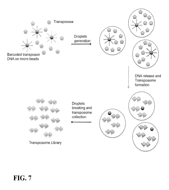
Fig. 7 is a schematic of using microdroplets to isolate microparticles
containing
transposon DNA with specific barcodes and the creation of transposomes having
the same
barcode pair within each microdroplet.
13
CA 03033506 2019-02-08
WO 2018/031631
PCT/US2017/046060
Fig. 8 is a schematic of microfluidic circuits for use in preparing barcoded
transposomes.
Fig. 9 is a schematic of insertion of transposomes carrying different pairs of
barcodes
to two alleles of a diploid genome and haplotyping of the genome.
DETAILED DESCRIPTION
The practice of certain embodiments or features of certain embodiments may
employ,
unless otherwise indicated, conventional techniques of molecular biology,
microbiology,
recombinant DNA, and so forth which are within ordinary skill in the art. Such
techniques are
explained fully in the literature. See e.g., Sambrook, Fritsch, and Maniatis,
MOLECULAR
CLONING: A LABORATORY MANUAL, Second Edition (1989), OLIGONUCLEOTIDE
SYNTHESIS (M. J. Gait Ed., 1984), ANIMAL CELL CULTURE (R. I. Freshney, Ed.,
1987), the series METHODS IN ENZYMOLOGY (Academic Press, Inc.); GENE
TRANSFER VECTORS FOR MAMMALIAN CELLS (J. M. Miller and M. P. Cabs eds.
1987), HANDBOOK OF EXPERIMENTAL IMMUNOLOGY, (D. M. Weir and C. C.
Blackwell, Eds.), CURRENT PROTOCOLS IN MOLECULAR BIOLOGY (F. M. Ausubel,
R. Brent, R. E. Kingston, D. D. Moore, J. G. Siedman, J. A. Smith, and K.
Struhl, eds., 1987),
CURRENT PROTOCOLS IN IMMUNOLOGY (J. E. coligan, A. M. Kruisbeek, D. H.
Margulies, E. M. Shevach and W. Strober, eds., 1991); ANNUAL REVIEW OF
IMMUNOLOGY; as well as monographs in journals such as ADVANCES IN
IMMUNOLOGY. All patents, patent applications, and publications mentioned
herein, both
supra and infra, are hereby incorporated herein by reference.
Terms and symbols of nucleic acid chemistry, biochemistry, genetics, and
molecular
biology used herein follow those of standard treatises and texts in the field,
e.g., Komberg
and Baker, DNA Replication, Second Edition (W.H. Freeman, New York, 1992);
Lehninger,
14
CA 03033506 2019-02-08
WO 2018/031631
PCT/US2017/046060
Biochemistry, Second Edition (Worth Publishers, New York, 1975); Strachan and
Read,
Human Molecular Genetics, Second Edition (Wiley-Liss, New York, 1999);
Eckstein, editor,
Oligonucleotides and Analogs: A Practical Approach (Oxford University Press,
New York,
1991); Gait, editor, Oligonucleotide Synthesis: A Practical Approach (IRL
Press, Oxford,
1984); and the like.
The present invention is based in part on the discovery of methods for making
nucleic
acid fragment templates, such as from DNA or genomic DNA, using a transposase
or
transposome to fragment the original or starting nucleic acid sequence, such
as genomic
DNA, and to attach a barcode sequence to each end of a cut or fragmentation
site to facilitate
the later computational rejoining of fragment sequences as part of a de novo
assembly
process. The method described herein may be referred to as "chaine annotation
via
transposon insertion" or "CHIANTI." The barcoded nucleic acid fragment
templates are
amplified to produce amplicons. The amplicons of the nucleic acid fragment
templates may
be collected and sequenced. The collected amplicons form a library of
amplicons of the
fragments of the original nucleic acid, such as genomic DNA.
According to one aspect, a genomic DNA, such as genomic nucleic acid obtained
from a lysed single cell, is obtained. A plurality or library of transposomes
is used to cut the
genomic DNA into double stranded fragments. Each transposome of the plurality
or library
is a dimer of a transposase bound to a transposon DNA, i.e. each transposome
includes two
separate transposon DNA. Each transposon DNA of a transposome includes a
transposase
binding site, a barcode sequence unique to the transposome and an
amplification facilitating
sequence, such as a specific primer binding site.
The barcode sequence of each transposon DNA of a transposome is the same
sequence and is unique to the transposome. Each transposome of the plurality
or library of
transposomes has its own unique representative barcode sequence which is
different from the
CA 03033506 2019-02-08
WO 2018/031631
PCT/US2017/046060
remaining members of the transposome plurality or library. The transposon DNA
becomes
attached to the upper and lower strands of each double stranded fragment at
each cut or
fragmentation site. Since the barcode sequence is the same for each transposon
DNA, the cut
or fragmentation site is tagged with the same barcode sequence which can be
later identified
to computationally rejoin the cut or fragmentation site. Since each
transposome has its own
unique barcode sequence, and a library of transposomes are used to create many
cut or
fragmentation sites, each cut or fragmentation site will have its own unique
barcode
sequence. Accordingly, many fragments from the original nucleic acid sequence
are created
by the library of transposomes with each fragment having a dissimilar barcode
at each end of
the fragment. The double stranded fragments are then processed to fill gaps.
The fragments
are amplified using suitable amplification reagents, such as a specific primer
sequence, DNA
polymerase and nucleotides for PCR amplification and are sequenced using
methods known
to those of skill in the art. Matching barcodes are identified which indicate
cut or
fragmentation sites and the matching barcodes are used to computationally
rejoin fragments
to recreate the original nucleic acid sequence.
DNA fragment templates made using the transposase methods described herein can
be
amplified within microdroplets using methods known to those of skill in the
art.
Microdroplets may be formed as an emulsion of an oil phase and an aqueous
phase. An
emulsion may include aqueous droplets or isolated aqueous volumes within a
continuous oil
phase Emulsion whole genome amplification methods are described using small
volume
aqueous droplets in oil to isolate each fragment for uniform amplification of
a single cell's
genome. By distributing each fragment into its own droplet or isolated aqueous
reaction
volume, each droplet is allowed to reach saturation of DNA amplification. The
amplicons
within each droplet are then merged by demulsification resulting in an even
amplification of
all of the fragments of the whole genome of the single cell.
16
CA 03033506 2019-02-08
WO 2018/031631
PCT/US2017/046060
In certain aspects, amplification is achieved using PCR. PCR is a reaction in
which
replicate copies are made of a target polynucleotide using a pair of primers
or a set of primers
consisting of an upstream and a downstream primer, and a catalyst of
polymerization, such as
a DNA polymerase, and typically a thermally-stable polymerase enzyme. Methods
for PCR
are well known in the art, and taught, for example in MacPherson et al. (1991)
PCR 1: A
Practical Approach (IRL Press at Oxford University Press). The term
"polymerase chain
reaction" ("PCR") of Mullis (U.S. Pat. Nos. 4,683,195, 4,683,202, and
4,965,188) refers to a
method for increasing the concentration of a segment of a target sequence
without cloning or
purification. This
process for amplifying the target sequence includes providing
oligonucleotide primers with the desired target sequence and amplification
reagents, followed
by a precise sequence of thermal cycling in the presence of a polymerase
(e.g., DNA
polymerase). The primers are complementary to their respective strands
("primer binding
sequences") of the double stranded target sequence. To effect amplification,
the double
stranded target sequence is denatured and the primers then annealed to their
complementary
sequences within the target molecule. Following annealing, the primers are
extended with a
polymerase so as to form a new pair of complementary strands. The steps of
denaturation,
primer annealing, and polymerase extension can be repeated many times (i.e.,
denaturation,
annealing and extension constitute one "cycle:" there can be numerous
"cycles") to obtain a
high concentration of an amplified segment of the desired target sequence. The
length of the
amplified segment of the desired target sequence is determined by the relative
positions of the
primers with respect to each other, and therefore, this length is a
controllable parameter. By
virtue of the repeating aspect of the process, the method is referred to as
the "polymerase
chain reaction" (hereinafter "PCR") and the target sequence is said to be "PCR
amplified."
The PCR amplification reaches saturation when the double stranded DNA
amplification
product accumulates to a certain amount that the activity of DNA polymerase is
inhibited.
17
CA 03033506 2019-02-08
WO 2018/031631
PCT/US2017/046060
Once saturated, the PCR amplification reaches a plateau where the
amplification product
does not increase with more PCR cycles.
With PCR, it is possible to amplify a single copy of a specific target
sequence in
genomic DNA to a level detectable by several different methodologies (e.g.,
hybridization
with a labeled probe; incorporation of biotinylated primers followed by avidin-
enzyme
conjugate detection; incorporation of 32P-labeled deoxynucleotide
triphosphates, such as
dCTP or dATP, into the amplified segment). In addition to genomic DNA, any
oligonucleotide or polynucleotide sequence can be amplified with the
appropriate set of
primer molecules. In particular, the amplified segments created by the PCR
process itself
within each microdroplet are, themselves, efficient templates for subsequent
PCR
amplifications. Methods and kits for performing PCR are well known in the art.
All
processes of producing replicate copies of a polynucleotide, such as PCR or
gene cloning, are
collectively referred to herein as replication. A primer can also be used as a
probe in
hybridization reactions, such as Southern or Northern blot analyses.
The expression "amplification" or "amplifying" refers to a process by which
extra or
multiple copies of a particular polynucleotide are formed. Amplification
includes methods
such as PCR, ligation amplification (or ligase chain reaction, LCR) and other
amplification
methods. These methods are known and widely practiced in the art. See, e.g.,
U.S. Patent
Nos. 4,683,195 and 4,683,202 and Innis et al., "PCR protocols: a guide to
method and
applications" Academic Press, Incorporated (1990) (for PCR); and Wu et al.
(1989)
Genomics 4:560-569 (for LCR). In general, the PCR procedure describes a method
of gene
amplification which is comprised of (i) sequence-specific hybridization of
primers to specific
genes within a DNA sample (or library), (ii) subsequent amplification
involving multiple
rounds of annealing, elongation, and denaturation using a DNA polymerase, and
(iii)
screening the PCR products for a band of the correct size. The primers used
are
18
CA 03033506 2019-02-08
WO 2018/031631
PCT/US2017/046060
oligonucleotides of sufficient length and appropriate sequence to provide
initiation of
polymerization, i.e. each primer is specifically designed to be complementary
to each strand
of the genomic locus to be amplified.
Reagents and hardware for conducting amplification reactions are commercially
available. Primers useful to amplify sequences from a particular gene region
are preferably
complementary to, and hybridize specifically to sequences in the target region
or in its
flanking regions and can be prepared using methods known to those of skill in
the art.
Nucleic acid sequences generated by amplification can be sequenced directly.
When hybridization occurs in an antiparallel configuration between two single-
stranded polynucleotides, the reaction is called "annealing" and those
polynucleotides are
described as "complementary". A double-stranded polynucleotide can be
complementary or
homologous to another polynucleotide, if hybridization can occur between one
of the strands
of the first polynucleotide and the second. Complementarity or homology (the
degree that one
polynucleotide is complementary with another) is quantifiable in terms of the
proportion of
bases in opposing strands that are expected to form hydrogen bonding with each
other,
according to generally accepted base-pairing rules.
The terms "PCR product," "PCR fragment," and "amplification product" refer to
the
resultant mixture of compounds after two or more cycles of the PCR steps of
denaturation,
annealing and extension are complete. These terms encompass the case where
there has been
amplification of one or more segments of one or more target sequences.
According to one
aspect of the present disclosure, each microdroplet includes PCR product of a
single template
DNA fragment.
The term "amplification reagents" may refer to those reagents
(deoxyribonucleotide
triphosphates, buffer, etc.), needed for amplification except for primers,
nucleic acid
template, and the amplification enzyme. Typically, amplification reagents
along with other
19
CA 03033506 2019-02-08
WO 2018/031631
PCT/US2017/046060
reaction components are placed and contained in a reaction vessel (test tube,
microwell, etc.).
Amplification methods include PCR methods known to those of skill in the art
and also
include rolling circle amplification (Blanco et al., J. Biol. Chem., 264, 8935-
8940, 1989),
hyperbranched rolling circle amplification (Lizard et al., Nat. Genetics, 19,
225-232, 1998),
and loop-mediated isothermal amplification (Notomi et al., Nuc. Acids Res.,
28, e63, 2000)
each of which are hereby incorporated by reference in their entireties.
For emulsion PCR, an emulsion PCR reaction is created by vigorously shaking or
stirring a "water in oil" mix to generate millions of micron-sized aqueous
compartments.
Microfluidic chips may be equipped with a device to create an emulsion by
shaking or
stirring an oil phase and a water phase. Alternatively, aqueous droplets may
be
spontaneously formed by combining a certain oil with an aqueous phase or
introducing an
aqueous phase into an oil phase. The DNA library to be amplified is mixed in a
limiting
dilution prior to emulsification. The combination of compartment size, i.e.
microdroplet size,
and amount of microdroplets created limiting dilution of the DNA fragment
library to be
amplified is used to generate compartments containing, on average, just one
DNA molecule.
Depending on the size of the aqueous compartments generated during the
microdroplet
formation or emulsification step, up to 3x10⁹ individual PCR reactions per pl
can be
conducted simultaneously in the same tube. Essentially each little aqueous
compartment
microdroplet in the emulsion forms a micro PCR reactor. The average size of a
compartment
in an emulsion ranges from sub- micron in diameter to over a 100 microns, or
from 1 picoliter
to 1000 picoliters or from 1 nanoliter to 1000 nanoliters or from 1 picoliter
to 1 nanoliter or
from 1 picoliter to 1000 nanoliters depending on the emulsification
conditions.
Other amplification methods, as described in British Patent Application No. GB
2,202,328, and in PCT Patent Application No. PCT/US89/01025, each incorporated
herein by
reference, may be used in accordance with the present disclosure. In the
former application,
CA 03033506 2019-02-08
WO 2018/031631
PCT/US2017/046060
"modified" primers are used in a PCR-like template and enzyme dependent
synthesis. The
primers may be modified by labeling with a capture moiety (e.g., biotin)
and/or a detector
moiety (e.g., enzyme). In the latter application, an excess of labeled probes
are added to a
sample. In the presence of the target sequence, the probe binds and is cleaved
catalytically.
After cleavage, the target sequence is released intact to be bound by excess
probe. Cleavage
of the labeled probe signals the presence of the target sequence.
Other suitable amplification methods include "race and "one-sided PCR.".
(Frohman,
In: PCR Protocols: A Guide To Methods And Applications, Academic Press, N.Y.,
1990,
each herein incorporated by reference). Methods based on ligation of two (or
more)
oligonucleotides in the presence of nucleic acid having the sequence of the
resulting "di-
oligonucleotide," thereby amplifying the di-oligonucleotide, also may be used
to amplify
DNA in accordance with the present disclosure (Wu et al., Genomics 4:560-569,
1989,
incorporated herein by reference).
According to certain aspects, an exemplary transposon system includes Tn5
transposase, Mu transposase, Tn7 transposase or IS5 transposase and the like.
Other useful
transposon systems are known to those of skill in the art and include Tn3
transposon system
(see Maekawa, T., Yanagihara, K., and Ohtsubo, E. (1996), A cell-free system
of Tn3
transposition and transposition immunity, Genes Cells 1, 1007-1016), Tn7
transposon system
(see Craig, N.L. (1991), Tn7: a target site-specific transposon, MoL MicrobioL
5, 2569-
2573), Tn10 tranposon system (see Chalmers, R., Sewitz, S., Lipkow, K., and
Crellin, P.
(2000), Complete nucleotide sequence of Tn10, J. Bacteriol 182, 2970-2972),
Piggybac
transposon system (see Li, X., Burnight, E.R., Cooney, A.L., Malani, N.,
Brady, T., Sander,
J.D., Staber, J., Wheelan, S.J., Joung, J.K., McCray, P.B., Jr., et al.
(2013), PiggyBac
transposase tools for genome engineering, Proc. Natl. Acad. Sci. USA 110,
E2279-2287),
Sleeping beauty transposon system (see Ivics, Z., Hackett, P.B., Plasterk,
R.H., and Izsvak, Z.
21
CA 03033506 2019-02-08
WO 2018/031631
PCT/US2017/046060
(1997), Molecular reconstruction of Sleeping Beauty, a Tcl-like transposon
from fish, and its
transposition in human cells, Cell 91, 501-510), To12 transposon system
(seeKawakami, K.
(2007), To12: a versatile gene transfer vector in vertebrates, Genome Biol. 8
Suppl. 1, S7.)
DNA to be amplified may be obtained from a single cell or a small population
of
cells. Methods described herein allow DNA to be amplified from any species or
organism in
a reaction mixture, such as a single reaction mixture carried out in a single
reaction vessel. In
one aspect, methods described herein include sequence independent
amplification of DNA
from any source including but not limited to human, animal, plant, yeast,
viral, eukaryotic
and prokaryotic DNA.
According to one aspect, a method of single cell whole genome amplification,
sequencing and de novo assembly is provided which includes contacting double
stranded
genomic DNA from a single cell with Tn5 transposases each bound to a
transposon DNA,
wherein the transposon DNA includes a double-stranded 19 bp transposase (Tnp)
binding site
and a first nucleic acid sequence including one or more of a barcode sequence
and a primer
binding site to form a transposase/transposon DNA complex dimer called a
transposome.
The first nucleic acid sequence may be in the form of a single stranded
extension. According
to one aspect, the first nucleic acid sequence may be an overhang, such as a
5' overhang,
wherein the overhang includes a barcode region and a priming site. The
overhang can be of
any length suitable to include a barcode region and a priming site as desired.
The
transposome bind to target locations along the double stranded genomic DNA and
cleave the
double stranded genomic DNA into a plurality of double stranded fragments,
with each
double stranded fragment having a first complex attached to an upper strand by
the Tnp
binding site and a second complex attached to a lower strand by the Tnp
binding site. The
transposon binding site, and therefore the transposon DNA, is attached to each
5' end of the
double stranded fragment. According to one aspect, the Tn5 transposases are
removed from
22
CA 03033506 2019-02-08
WO 2018/031631
PCT/US2017/046060
the complex. The double stranded fragments are extended along the transposon
DNA to
make a double stranded extension product having dissimilar barcode sequences
and specific
primer binding sites at each end of the double stranded extension product.
According to one
aspect, a gap which may result from attachment of the Tn5 transposase binding
site to the
double stranded genomic DNA fragment may be filled. The gap filled double
stranded
extension product is mixed with amplification reagents, and the double
stranded genomic
DNA fragment is amplified. The amplicons, which include a dissimilar barcode
sequence at
each end, are sequenced using, for example, high-throughput sequencing methods
known to
those of skill in the art.
In a particular aspect, embodiments are directed to methods for the
amplification,
sequencing and de novo assembly of substantially the entire genome without
loss of
representation of specific sites (herein defined as "whole genome
amplification"). In a
specific embodiment, whole genome amplification comprises amplification of
substantially
all fragments or all fragments of a genomic library. In a further specific
embodiment,
"substantially entire" or "substantially all refers to about 80%, about 85%,
about 90%, about
95%, about 97%, or about 99% of all sequences in a genome.
According to one aspect, the DNA sample is genomic DNA, micro dissected
chromosome DNA, yeast artificial chromosome (YAC) DNA, plasmid DNA, cosmid
DNA,
phage DNA, PI derived artificial chromosome (PAC) DNA, or bacterial artificial
chromosome (BAC) DNA, mitochondrial DNA, chloroplast DNA, forensic sample DNA,
or
other DNA from natural or artificial sources to be tested. In another
preferred embodiment,
the DNA sample is mammalian DNA, plant DNA, yeast DNA, viral DNA, or
prokaryotic
DNA. In yet another preferred embodiment, the DNA sample is obtained from a
human,
bovine, porcine, ovine, equine, rodent, avian, fish, shrimp, plant, yeast,
virus, or bacteria.
Preferably the DNA sample is genomic DNA.
23
CA 03033506 2019-02-08
WO 2018/031631
PCT/US2017/046060
According to certain exemplary aspects, a transposition system is used to make
nucleic acid fragments for amplification, sequencing and de novo assembly as
desired.
According to one aspect, a transposition system is used to fragment genomic
DNA into
double stranded genomic DNA fragments with the transposon DNA having the same
barcode
inserted therein. As illustrated in Fig. 1, a transposon DNA includes a double
stranded
transposase binding site, a barcode sequence B and a priming site P. The
double stranded
transposase binding site may be a double-stranded 19 bp Tn5 transposase (Tnp)
binding site
which is linked or connected, such as by covalent bond, to a single-stranded
overhang
including a barcode region and a priming site at one end of the overhang. The
transposon
DNA is inserted into the genomic DNA of a single cell while creating millions
of small
fragments using a transposase. After transposase removal and gap fill-in, the
genomic DNA
fragments having dissimilar barcode sequences and a specific primer sequence
at each end of
the fragment are amplified using specific primers together with a DNA
polymerase,
nucleotides and amplification reagents to PCR amplify the whole genome of the
single cell.
According to certain aspects when amplifying small amounts of DNA such as DNA
from a single cell, a DNA column purification step is not carried out so as to
maximize the
small amount (-6 pg) of genomic DNA that can be obtained from within a single
cell prior to
amplification. The DNA can be amplified directly from a cell lysate or other
impure
condition. Accordingly, the DNA sample may be impure, unpurified, or not
isolated.
Accordingly, aspects of the present method allow one to maximize genomic DNA
for
amplification and reduce loss due to purification. According to an additional
aspect, methods
described herein may utilize amplification methods other than PCR.
According to one aspect and as illustrated in general in Fig. 2, transposase
(Tnp) and
the transposon DNA are combined, such as within a microdroplet and the Tnp and
the
transposon DNA bind to each other and dimerize to form transposomes.
24
CA 03033506 2019-02-08
WO 2018/031631
PCT/US2017/046060
As shown in Fig. 3, the transposomes of the transposome library randomly
capture or
otherwise bind to the target single-cell genomic DNA as dimers.
Representative
transposomes are numbered 1, 2 and 3, though the number of transposomes can be
in the
thousands, ten-thousands, hundred-thousands, millions, etc. Each transposome
is represented
by a unique barcode sequence, for example barcode sequence 1, barcode sequence
2, barcode
sequence 3, etc. The unique barcode sequence is within each transposon DNA of
the
transposome. Since there are two transposon DNAs per transposome, the two
transposon
DNAs can be considered a homo dimer, which means one transposon DNA dimer
carries two
DNA sequences with the same barcode information. Each transposome (and
transposon
DNA dimer) of the transposome library has a different barcode unique to the
transposome.
The transposases in the transposome cut the genomic DNA with one transposase
cutting an
upper strand and one transposase cutting a lower strand to create a genomic
DNA fragment.
The plurality of transposomes creates a plurality of genomic DNA fragments.
One
transposon DNA from the transposon DNA dimer is thus attached to each end of
the cut site
or fragmentation site, i.e., one transposon DNA from transposome 1 is attached
to the left
hand cut site and the other transposon DNA from transposome 1 is attached to
the right hand
cut site. Since the transposome library cuts the nucleic acid into fragments,
each fragment
will have a dissimilar barcode sequence at each end of the fragment, i.e. each
fragment is
produced by two different cut sites cut by two different transposomes of the
transposome
library including different barcode sequences. This is represented by the two
exemplary
fragments where the upper fragment has barcode sequence 1 on one end and
barcode
sequence 2 on the other end. Likewise, the lower fragment has barcode sequence
2 on one
end and barcode sequence 3 on the other end. As illustrated, the cut site
between the two
fragments is produced by transposome 2 and the left hand cut site (i.e.
viewing the right side
of the upper fragment in Fig. 3) includes the one transposon with barcode
sequence 2 while
CA 03033506 2019-02-08
WO 2018/031631
PCT/US2017/046060
the right hand cut site (i.e. viewing the left side of the lower fragment in
Fig. 3) includes the
other transposon with barcode sequence 2.
As illustrated in Fig. 4, the fragmentation of the genomic DNA leaves a gap on
both
ends of the transposition/insertion site. The gap may have any length but a 9
base gap is
exemplary. The result is a genomic DNA fragment with a transposon DNA Tnp
binding site
attached to the 5' position of an upper strand and a transposon DNA Tnp
binding site
attached to the 5' position of a lower strand. Gaps resulting from the
attachment or insertion
of the transposon DNA are shown. After transposition, the transposase is
removed and gap
extension is performed to fill the gap and complement the single-stranded
overhang originally
designed in the transposon DNA as shown in Fig. 4.
As further illustrated in Fig. 5, a plurality of transposomes n with
corresponding
barcode sequences Bn are used to create a plurality of fragments and the
barcode sequences
are used to chain short sequencing reads into longer continuous sequences. A
library of
transposomes (on the order of millions for example) with each transposome
carrying two
transposon DNA with the same barcodes B(n) are inserted into the genomic DNA
and cut the
genomic DNA into millions of different fragments (F1, F2, F3...). After whole
genome
amplification and sequencing, the fragments tagged with the same barcodes can
be
computationally linked together to achieve longer fragment length.
Particular Tn5 transposition systems are described and are available to those
of skill
in the art. See Goryshin, I.Y. and W.S. Reznikoff, Tn5 in vitro transposition.
The Journal of
biological chemistry, 1998. 273(13): p. 7367-74; Davies, D.R., et al., Three-
dimensional
structure of the Tn5 synaptic complex transposition intermediate. Science,
2000. 289(5476):
p. 77-85; Goryshin, I.Y., et al., Insertional transposon mutagenesis by
electroporation of
released Tn5 transposition complexes. Nature biotechnology, 2000. 18(1): p. 97-
100 and
Steiniger-White, M., I. Rayment, and W.S. Reznikoff, Structure/function
insights into Tn5
26
CA 03033506 2019-02-08
WO 2018/031631
PCT/US2017/046060
transposition. Current opinion in structural biology, 2004. 14(1): p. 50-7
each of which are
hereby incorporated by reference in their entireties for all purposes. Kits
utilizing a Tn5
transposition system for DNA library preparation and other uses are known. See
Adey, A., et
al., Rapid, low-input, low-bias construction of shotgun fragment libraries by
high-density in
vitro transposition. Genome biology, 2010. 11(12): p. R119; Marine, R., et
al., Evaluation of
a transposase protocol for rapid generation of shotgun high-throughput
sequencing libraries
from nanogram quantities of DNA. Applied and environmental microbiology, 2011.
77(22):
p. 8071-9; Parkinson, N.J., et al., Preparation of high-quality next-
generation sequencing
libraries from picogram quantities of target DNA. Genome research, 2012.
22(1): p. 125-33;
Adey, A. and J. Shendure, Ultra-low-input, tagmentation-based whole-genome
bisulfite
sequencing. Genome research, 2012. 22(6): p. 1139-43; Picelli, S., et al.,
Full-length RNA-
seq from single cells using Smart-seq2. Nature protocols, 2014. 9(1): p. 171-
81 and
Buenrostro, J.D., et al., Transposition of native chromatin for fast and
sensitive epigenomic
profiling of open chromatin, DNA-binding proteins and nucleosome position.
Nature
methods, 2013, each of which is hereby incorporated by reference in its
entirety for all
purposes. See also WO 98/10077, EP 2527438 and EP 2376517 each of which is
hereby
incorporated by reference in its entirety. A commercially available
transposition kit is
marketed under the name NEXTERA and is available from Illumina.
The term "genome" as used herein is defined as the collective gene set carried
by an
individual, cell, or organelle. The term "genomic DNA" as used herein is
defined as DNA
material comprising the partial or full collective gene set carried by an
individual, cell, or
organelle.
As used herein, the term "nucleoside" refers to a molecule having a purine or
pyrimidine base covalently linked to a ribose or deoxyribose sugar. Exemplary
nucleosides
include adenosine, guanosine, cytidine, uridine and thymidine. Additional
exemplary
27
CA 03033506 2019-02-08
WO 2018/031631
PCT/US2017/046060
nucleosides include inosine, 1-methyl inosine, pseudouridine, 5 , 6-
dihydrouridine,
ribothymidine, 2N-methylguanosine and 2,2N,N-dimethylguanosine (also referred
to as
"rare" nucleosides). The term "nucleotide" refers to a nucleoside having one
or more
phosphate groups joined in ester linkages to the sugar moiety. Exemplary
nucleotides include
nucleoside monophosphates, diphosphates and triphosphates. The terms
"polynucleotide,"
"oligonucleotide" and "nucleic acid molecule" are used interchangeably herein
and refer to a
polymer of nucleotides, either deoxyribonucleotides or ribonucleotides, of any
length joined
together by a phosphodiester linkage between 5 and 3' carbon atoms.
Polynucleotides can
have any three-dimensional structure and can perform any function, known or
unknown. The
following are non-limiting examples of polynucleotides: a gene or gene
fragment (for
example, a probe, primer, EST or SAGE tag), exons, introns, messenger RNA
(mRNA),
transfer RNA, ribosomal RNA, ribozymes, cDNA, recombinant polynucleotides,
branched
polynucleotides, plasmids, vectors, isolated DNA of any sequence, isolated RNA
of any
sequence, nucleic acid probes and primers. A polynucleotide can comprise
modified
nucleotides, such as methylated nucleotides and nucleotide analogs. The term
also refers to
both double- and single-stranded molecules. Unless otherwise specified or
required, any
embodiment of this invention that comprises a polynucleotide encompasses both
the double-
stranded form and each of two complementary single-stranded forms known or
predicted to
make up the double-stranded form. A polynucleotide is composed of a specific
sequence of
four nucleotide bases: adenine (A); cytosine (C); guanine (G); thymine (T);
and uracil (U) for
thymine when the polynucleotide is RNA. Thus, the term polynucleotide sequence
is the
alphabetical representation of a polynucleotide molecule. This alphabetical
representation can
be input into databases in a computer having a central processing unit and
used for
bioinformatics applications such as functional genomics and homology
searching.
28
CA 03033506 2019-02-08
WO 2018/031631
PCT/US2017/046060
The terms "DNA," "DNA molecule" and "deoxyribonucleic acid molecule" refer to
a
polymer of deoxyribonucleotides. DNA can be synthesized naturally (e.g., by
DNA
replication). RNA can be post-transcriptionally modified. DNA can also be
chemically
synthesized. DNA can be single-stranded (i.e., ssDNA) or multi-stranded (e.g.,
double
stranded, i.e., dsDNA).
The terms "nucleotide analog," "altered nucleotide" and "modified nucleotide"
refer
to a non-standard nucleotide, including non-naturally occurring
ribonucleotides or
deoxyribonucleotides. In certain exemplary embodiments, nucleotide analogs are
modified at
any position so as to alter certain chemical properties of the nucleotide yet
retain the ability of
the nucleotide analog to perform its intended function. Examples of positions
of the
nucleotide which may be derivitized include the 5 position, e.g., 5-(2-
amino)propyl uridine,
5-bromo uridine, 5-propyne uridine, 5-propenyl uridine, etc.; the 6 position,
e.g., 6-(2-amino)
propyl uridine; the 8-position for adenosine and/or guanosines, e.g., 8-bromo
guanosine, 8-
chloro guanosine, 8-fluoroguanosine, etc. Nucleotide analogs also include
deaza nucleotides,
e.g., 7-deaza-adenosine; 0- and N-modified (e.g., alkylated, e.g., N6-methyl
adenosine, or as
otherwise known in the art) nucleotides; and other heterocyclically modified
nucleotide
analogs such as those described in Herdewijn, Antisense Nucleic Acid Drug
Dev., 2000 Aug.
10(4):297-310.
Nucleotide analogs may also comprise modifications to the sugar portion of the
nucleotides. For example the 2 OH-group may be replaced by a group selected
from H, OR,
R, F, Cl, Br, I, SH, SR, NH2, NHR, NR2, COOR, or OR, wherein R is substituted
or
unsubstituted Ci-C6 alkyl, alkenyl, alkynyl, aryl, etc.Other possible
modifications include
those described in U.S. Pat. Nos. 5,858,988, and 6,291,438.
The phosphate group of the nucleotide may also be modified, e.g., by
substituting one
or more of the oxygens of the phosphate group with sulfur (e.g.,
phosphorothioates), or by
29
CA 03033506 2019-02-08
WO 2018/031631
PCT/US2017/046060
making other substitutions which allow the nucleotide to perform its intended
function such
as described in, for example, Eckstein, Antisense Nucleic Acid Drug Dev. 2000
Apr.
10(2):117-21, Rusckowski et al. Antisense Nucleic Acid Drug Dev. 2000 Oct.
10(5):333-45,
Stein, Antisense Nucleic Acid Drug Dev. 2001 Oct. 11(5): 317-25, Vorobjev et
al. Antisense
Nucleic Acid Drug Dev. 2001 Apr. 11(2):77-85, and U.S. Pat. No. 5,684,143.
Certain of the
above-referenced modifications (e.g., phosphate group modifications) decrease
the rate of
hydrolysis of, for example, polynucleotides comprising said analogs in vivo or
in vitro.
The term "in vitro" has its art recognized meaning, e.g., involving purified
reagents or
extracts, e.g., cell extracts. The term "in vivo" also has its art recognized
meaning, e.g.,
involving living cells, e.g., immortalized cells, primary cells, cell lines,
and/or cells in an
organism.
As used herein, the terms "complementary" and "complementarity" are used in
reference to nucleotide sequences related by the base-pairing rules. For
example, the
sequence 5'-AGT-3 is complementary to the sequence 5'-ACT-3'. Complementarity
can be
partial or total. Partial complementarity occurs when one or more nucleic acid
bases is not
matched according to the base pairing rules. Total or complete complementarity
between
nucleic acids occurs when each and every nucleic acid base is matched with
another base
under the base pairing rules. The degree of complementarity between nucleic
acid strands
has significant effects on the efficiency and strength of hybridization
between nucleic acid
strands.
The term "hybridization" refers to the pairing of complementary nucleic acids.
Hybridization and the strength of hybridization (i.e., the strength of the
association between
the nucleic acids) is impacted by such factors as the degree of complementary
between the
nucleic acids, stringency of the conditions involved, the Tri, of the formed
hybrid, and the G:C
CA 03033506 2019-02-08
WO 2018/031631
PCT/US2017/046060
ratio within the nucleic acids. A single molecule that contains pairing of
complementary
nucleic acids within its structure is said to be "self-hybridized."
The term "T." refers to the melting temperature of a nucleic acid. The melting
temperature is the temperature at which a population of double-stranded
nucleic acid
molecules becomes half dissociated into single strands. The equation for
calculating the T.
of nucleic acids is well known in the art. As indicated by standard
references, a simple
estimate of the T. value may be calculated by the equation: T. = 81.5 + 0.41
(% G + C),
when a nucleic acid is in aqueous solution at 1 M NaCl (See, e.g., Anderson
and Young,
Quantitative Filter Hybridization, in Nucleic Acid Hybridization (1985)).
Other references
include more sophisticated computations that take structural as well as
sequence
characteristics into account for the calculation of T..
The term "stringency" refers to the conditions of temperature, ionic strength,
and the
presence of other compounds such as organic solvents, under which nucleic acid
hybridizations are conducted.
"Low stringency conditions," when used in reference to nucleic acid
hybridization,
comprise conditions equivalent to binding or hybridization at 42 C in a
solution consisting of
5x SSPE (43.8 g/1 NaCl, 6.9 g/1 NaH2PO4(H20) and 1.85 g/1 EDTA, pH adjusted to
7.4 with
NaOH), 0.1% SDS, 5x Denhardt's reagent (50x Denhardt's contains per 500 ml: 5
g Ficoll
(Type 400, Pharmacia), 5 g BSA (Fraction V; Sigma)) and 100 mg/ml denatured
salmon
sperm DNA followed by washing in a solution comprising 5x SSPE, 0.1% SDS at 42
C
when a probe of about 500 nucleotides in length is employed.
"Medium stringency conditions," when used in reference to nucleic acid
hybridization, comprise conditions equivalent to binding or hybridization at
42 C in a
solution consisting of 5x SSPE (43.8 g/1 NaCl, 6.9 g/1 NaH2PO4(H20) and 1.85
g/1 EDTA,
pH adjusted to 7.4 with NaOH), 0.5% SDS, 5x Denhardt's reagent and 100 mg/ml
denatured
31
CA 03033506 2019-02-08
WO 2018/031631
PCT/US2017/046060
salmon sperm DNA followed by washing in a solution comprising 1.0x SSPE, 1.0%
SDS at
42 C when a probe of about 500 nucleotides in length is employed.
"High stringency conditions," when used in reference to nucleic acid
hybridization,
comprise conditions equivalent to binding or hybridization at 42 C in a
solution consisting of
5x SSPE (43.8 g/1 NaCl, 6.9 g/1 NaH2PO4(H20) and 1.85 g/1 EDTA, pH adjusted to
7.4 with
NaOH), 0.5% SDS, 5x Denhardt's reagent and 100 mg/ml denatured salmon sperm
DNA
followed by washing in a solution comprising 0.1x SSPE, 1.0% SDS at 42 C when
a probe
of about 500 nucleotides in length is employed.
In certain exemplary embodiments, cells are identified and then a single cell
or a
plurality of cells is isolated. Cells within the scope of the present
disclosure include any type
of cell where understanding the DNA content is considered by those of skill in
the art to be
useful. A cell according to the present disclosure includes a cancer cell of
any type,
hepatocyte, oocyte, embryo, stem cell, iPS cell, ES cell, neuron, erythrocyte,
melanocyte,
astrocyte, germ cell, oligodendrocyte, kidney cell and the like. According to
one aspect, the
methods of the present invention are practiced with the cellular DNA from a
single cell. A
plurality of cells includes from about 2 to about 1,000,000 cells, about 2 to
about 10 cells,
about 2 to about 100 cells, about 2 to about 1,000 cells, about 2 to about
10,000 cells, about 2
to about 100,000 cells, about 2 to about 10 cells or about 2 to about 5 cells.
Nucleic acids processed by methods described herein may be DNA and they may be
obtained from any useful source, such as, for example, a human sample. In
specific
embodiments, a double stranded DNA molecule is further defined as comprising a
genome,
such as, for example, one obtained from a sample from a human. The sample may
be any
sample from a human, such as blood, serum, plasma, cerebrospinal fluid, cheek
scrapings,
nipple aspirate, biopsy, semen (which may be referred to as ejaculate), urine,
feces, hair
follicle, saliva, sweat, immunoprecipitated or physically isolated chromatin,
and so forth. In
32
CA 03033506 2019-02-08
WO 2018/031631
PCT/US2017/046060
specific embodiments, the sample comprises a single cell. In specific
embodiments, the
sample includes only a single cell.
In particular embodiments, the amplified and de novo assembled nucleic acid
molecule from the sample provides diagnostic or prognostic information. For
example, the
prepared nucleic acid molecule from the sample may provide genomic copy number
and/or
sequence information, allelic variation information, cancer diagnosis,
prenatal diagnosis,
paternity information, disease diagnosis, detection, monitoring, and/or
treatment information,
sequence information, and so forth.
As used herein, a "single cell" refers to one cell. Single cells useful in the
methods
described herein can be obtained from a tissue of interest, or from a biopsy,
blood sample, or
cell culture. Additionally, cells from specific organs, tissues, tumors,
neoplasms, or the like
can be obtained and used in the methods described herein. Furthermore, in
general, cells from
any population can be used in the methods, such as a population of prokaryotic
or eukaryotic
single celled organisms including bacteria or yeast. A single cell suspension
can be obtained
using standard methods known in the art including, for example, enzymatically
using trypsin
or papain to digest proteins connecting cells in tissue samples or releasing
adherent cells in
culture, or mechanically separating cells in a sample. Single cells can be
placed in any
suitable reaction vessel in which single cells can be treated individually.
For example a 96-
well plate, such that each single cell is placed in a single well.
Methods for manipulating single cells are known in the art and include
fluorescence
activated cell sorting (FACS), flow cytometry (Herzenberg., PNAS USA 76:1453-
55 1979),
micromanipulation and the use of semi-automated cell pickers (e.g. the Quixell
cell
transfer system from Stoelting Co.). Individual cells can, for example, be
individually
selected based on features detectable by microscopic observation, such as
location,
33
CA 03033506 2019-02-08
WO 2018/031631
PCT/US2017/046060
morphology, or reporter gene expression. Additionally, a combination of
gradient
centrifugation and flow cytometry can also be used to increase isolation or
sorting efficiency.
Once a desired cell has been identified, the cell is lysed to release cellular
contents
including DNA, using methods known to those of skill in the art. The cellular
contents are
contained within a vessel or a collection volume. In some aspects of the
invention, cellular
contents, such as genomic DNA, can be released from the cells by lysing the
cells. Lysis can
be achieved by, for example, heating the cells, or by the use of detergents or
other chemical
methods, or by a combination of these. However, any suitable lysis method
known in the art
can be used. For example, heating the cells at 72 C for 2 minutes in the
presence of Tween-
20 is sufficient to lyse the cells. Alternatively, cells can be heated to 65 C
for 10 minutes in
water (Esumi et al., Neurosci Res 60(4):439-51 (2008)); or 70 C for 90 seconds
in PCR
buffer II (Applied Biosystems) supplemented with 0.5% NP-40 (Kurimoto et al.,
Nucleic
Acids Res 34(5):e42 (2006)); or lysis can be achieved with a protease such as
Proteinase K or
by the use of chaotropic salts such as guanidine isothiocyanate (U.S.
Publication No.
2007/0281313). Amplification of genomic DNA according to methods described
herein can
be performed directly on cell lysates, such that a reaction mix can be added
to the cell lysates.
Alternatively, the cell lysate can be separated into two or more volumes such
as into two or
more containers, tubes or regions using methods known to those of skill in the
art with a
portion of the cell lysate contained in each volume container, tube or region.
Genomic DNA
contained in each container, tube or region may then be amplified by methods
described
herein or methods known to those of skill in the art.
A nucleic acid used in the invention can also include native or non-native
bases. In
this regard a native deoxyribonucleic acid can have one or more bases selected
from the
group consisting of adenine, thymine, cytosine or guanine and a ribonucleic
acid can have
one or more bases selected from the group consisting of uracil, adenine,
cytosine or guanine.
34
CA 03033506 2019-02-08
WO 2018/031631
PCT/US2017/046060
Exemplary non-native bases that can be included in a nucleic acid, whether
having a native
backbone or analog structure, include, without limitation, inosine, xathanine,
hypoxathanine,
isocytosine, isoguanine, 5-methylcytosine, 5-hydroxymethyl cytosine, 2-
aminoadenine, 6-
methyl adenine, 6-methyl guanine, 2-propyl guanine, 2-propyl adenine, 2-
thioLiracil, 2-
thiothymine, 2- thiocytosine, 15 -halouracil, 15 -halocytosine, 5-propynyl
uracil, 5-propynyl
cytosine, 6-azo uracil, 6-azo cytosine, 6-azo thymine, 5-uracil, 4-thiouracil,
8-halo adenine or
guanine, 8- amino adenine or guanine, 8-thiol adenine or guanine, 8-thioalkyl
adenine or
guanine, 8- hydroxyl adenine or guanine, 5-halo substituted uracil or
cytosine, 7-
methylguanine, 7- methyladenine, 8-azaguanine, 8-azaadenine, 7-deazaguanine, 7-
deazaadenine, 3-deazaguanine, 3-deazaadenine or the like. A particular
embodiment can
utilize isocytosine and isoguanine in a nucleic acid in order to reduce non-
specific
hybridization, as generally described in U.S. Pat. No.5,681,702.
As used herein, the term "primer" generally includes an oligonucleotide,
either natural
or synthetic, that is capable, upon forming a duplex with a polynucleotide
template, of acting
as a point of initiation of nucleic acid synthesis, such as a sequencing
primer, and being
extended from its 3 end along the template so that an extended duplex is
formed. The
sequence of nucleotides added during the extension process is determined by
the sequence of
the template polynucleotide. Usually primers are extended by a DNA polymerase.
Primers
usually have a length in the range of between 3 to 36 nucleotides, also 5 to
24 nucleotides,
also from 14 to 36 nucleotides. Primers within the scope of the invention
include orthogonal
primers, amplification primers, constructions primers and the like. Pairs of
primers can flank
a sequence of interest or a set of sequences of interest. Primers and probes
can be degenerate
or quasi-degenerate in sequence. Primers within the scope of the present
invention bind
adjacent to a target sequence. A "primer" may be considered a short
polynucleotide,
generally with a free 3' -OH group that binds to a target or template
potentially present in a
CA 03033506 2019-02-08
WO 2018/031631
PCT/US2017/046060
sample of interest by hybridizing with the target, and thereafter promoting
polymerization of
a polynucleotide complementary to the target. Primers of the instant invention
are comprised
of nucleotides ranging from 17 to 30 nucleotides. In one aspect, the primer is
at least 17
nucleotides, or alternatively, at least 18 nucleotides, or alternatively, at
least 19 nucleotides,
or alternatively, at least 20 nucleotides, or alternatively, at least 21
nucleotides, or
alternatively, at least 22 nucleotides, or alternatively, at least 23
nucleotides, or alternatively,
at least 24 nucleotides, or alternatively, at least 25 nucleotides, or
alternatively, at least 26
nucleotides, or alternatively, at least 27 nucleotides, or alternatively, at
least 28 nucleotides,
or alternatively, at least 29 nucleotides, or alternatively, at least 30
nucleotides, or
alternatively at least 50 nucleotides, or alternatively at least 75
nucleotides or alternatively at
least 100 nucleotides.
The expression "amplification" or "amplifying" refers to a process by which
extra or
multiple copies of a particular polynucleotide are formed.
The DNA amplified according to the methods described herein may be sequenced
and
analyzed using methods known to those of skill in the art. Determination of
the sequence of a
nucleic acid sequence of interest can be performed using a variety of
sequencing methods
known in the art including, but not limited to, sequencing by hybridization
(SBH),
sequencing by ligation (SBL) (Shendure et al. (2005) Science 309:1728),
quantitative
incremental fluorescent nucleotide addition sequencing (QIFNAS), stepwise
ligation and
cleavage, fluorescence resonance energy transfer (FRET), molecular beacons,
TaqMan
reporter probe digestion, pyrosequencing, fluorescent in situ sequencing
(FISSEQ), FISSEQ
beads (U.S. Pat. No. 7,425,431), wobble sequencing (PCT/US05/27695), multiplex
sequencing (U.S. Serial No. 12/027,039, filed February 6, 2008; Porreca et al
(2007) Nat.
Methods 4:931), polymerized colony (POLONY) sequencing (U.S. Patent Nos.
6,432,360,
6,485,944 and 6,511,803, and PCT/US05/06425); nanogrid rolling circle
sequencing
36
CA 03033506 2019-02-08
WO 2018/031631
PCT/US2017/046060
(ROLONY) (U.S. Serial No. 12/120,541, filed May 14, 2008), allele-specific
oligo ligation
assays (e.g., oligo ligation assay (OLA), single template molecule OLA using a
ligated linear
probe and a rolling circle amplification (RCA) readout, ligated padlock
probes, and/or single
template molecule OLA using a ligated circular padlock probe and a rolling
circle
amplification (RCA) readout) and the like. High-throughput sequencing methods,
e.g., using
platforms such as Roche 454, Illumina Solexa, AB-SOLiD, Helicos, Polonator
platforms and
the like, can also be utilized. A variety of light-based sequencing
technologies are known in
the art (Landegren et al. (1998) Genome Res. 8:769-76; Kwok (2000)
Pharmacogenomics
1:95-100; and Shi (2001) Clin. Chem. 47:164-172).
The amplified DNA can be sequenced by any suitable method. In particular, the
amplified DNA can be sequenced using a high-throughput screening method, such
as Applied
Biosystems' SOLiD sequencing technology, or Illumina's Genome Analyzer. In one
aspect of
the invention, the amplified DNA can be shotgun sequenced. The number of reads
can be at
least 10,000, at least 1 million, at least 10 million, at least 100 million,
or at least 1000
million. In another aspect, the number of reads can be from 10,000 to 100,000,
or
alternatively from 100,000 to 1 million, or alternatively from 1 million to 10
million, or
alternatively from 10 million to 100 million, or alternatively from 100
million to 1000
million. A "read" is a length of continuous nucleic acid sequence obtained by
a sequencing
reaction.
"Shotgun sequencing" refers to a method used to sequence very large amount of
DNA
(such as the entire genome). In this method, the DNA to be sequenced is first
shredded into
smaller fragments which can be sequenced individually. The sequences of these
fragments
are then reassembled into their original order based on their overlapping
sequences, thus
yielding a complete sequence. "Shredding" of the DNA can be done using a
number of
difference techniques including restriction enzyme digestion or mechanical
shearing.
37
CA 03033506 2019-02-08
WO 2018/031631
PCT/US2017/046060
Overlapping sequences are typically aligned by a computer suitably programmed.
Methods
and programs for shotgun sequencing a cDNA library are well known in the art.
The amplification and sequencing methods are useful in the field of predictive
medicine in which diagnostic assays, prognostic assays, pharmacogenomics, and
monitoring
clinical trials are used for prognostic (predictive) purposes to thereby treat
an individual
prophylactically. Accordingly, one aspect of the present invention relates to
diagnostic
assays for determining the genomic DNA in order to determine whether an
individual is at
risk of developing a disorder and/or disease. Such assays can be used for
prognostic or
predictive purposes to thereby prophylactically treat an individual prior to
the onset of the
disorder and/or disease. Accordingly, in certain exemplary embodiments,
methods of
diagnosing and/or prognosing one or more diseases and/or disorders using one
or more of
expression profiling methods described herein are provided.
As used herein, the term "biological sample" is intended to include, but is
not limited
to, tissues, cells, biological fluids and isolates thereof, isolated from a
subject, as well as
tissues, cells and fluids present within a subject.
In certain exemplary embodiments, electronic apparatus readable media
comprising
one or more genomic DNA sequences described herein is provided. As used
herein,
"electronic apparatus readable media" refers to any suitable medium for
storing, holding or
containing data or information that can be read and accessed directly by an
electronic
apparatus. Such media can include, but are not limited to: magnetic storage
media, such as
floppy discs, hard disc storage medium, and magnetic tape; optical storage
media such as
compact disc; electronic storage media such as RAM, ROM, EPROM, EEPROM and the
like; general hard disks and hybrids of these categories such as
magnetic/optical storage
media. The medium is adapted or configured for having recorded thereon one or
more
expression profiles described herein.
38
CA 03033506 2019-02-08
WO 2018/031631
PCT/US2017/046060
As used herein, the term "electronic apparatus" is intended to include any
suitable
computing or processing apparatus or other device configured or adapted for
storing data or
information. Examples of electronic apparatuses suitable for use with the
present invention
include stand-alone computing apparatus; networks, including a local area
network (LAN), a
wide area network (WAN) Internet, Intranet, and Extranet; electronic
appliances such as a
personal digital assistants (PDAs), cellular phone, pager and the like; and
local and
distributed processing systems.
As used herein, "recorded" refers to a process for storing or encoding
information on
the electronic apparatus readable medium. Those skilled in the art can readily
adopt any of
the presently known methods for recording information on known media to
generate
manufactures comprising one or more expression profiles described herein.
A variety of software programs and formats can be used to store the genomic
DNA
information of the present invention on the electronic apparatus readable
medium. For
example, the nucleic acid sequence can be represented in a word processing
text file,
formatted in commercially-available software such as WordPerfect and MicroSoft
Word, or
represented in the form of an ASCII file, stored in a database application,
such as DB2,
Sybase, Oracle, or the like, as well as in other forms. Any number of data
processor
structuring formats (e.g., text file or database) may be employed in order to
obtain or create a
medium having recorded thereon one or more expression profiles described
herein.
It is to be understood that the embodiments of the present invention which
have been
described are merely illustrative of some of the applications of the
principles of the present
invention. Numerous modifications may be made by those skilled in the art
based upon the
teachings presented herein without departing from the true spirit and scope of
the invention.
The contents of all references, patents and published patent applications
cited throughout this
application are hereby incorporated by reference in their entirety for all
purposes.
39
CA 03033506 2019-02-08
WO 2018/031631
PCT/US2017/046060
The following examples are set forth as being representative of the present
invention.
These examples are not to be construed as limiting the scope of the invention
as these and
other equivalent embodiments will be apparent in view of the present
disclosure, figures and
accompanying claims.
EXAMPLE I
General Protocol
The following general protocol is useful for whole genome amplification. A
single
cell is lysed in lysis buffer. The transposome library including transposome
with a specific
barcode pair, such as a unique barcode pair, and transposition buffer are
added to the cell
lysis which is mixed well and is incubated at 55 C for 10 minutes. lmg/m1
protease is added
after the tranposition to remove the transpoase from binding to the single
cell genomic DNA.
Deepvent exo- DNA polymerase, dNTP, PCR reaction buffer and primers are added
to the
reaction mixture which is heated to 72 C for 10min to fill in the gap
generated from the
transposon insertion. The reaction mixture is loaded to the microfluidic
device to form micro
droplets. The droplets containing single cell genomic DNA template, DNA
polymerase,
dNTP, reaction buffer and primer are collected into PCR tubes. 40 to 60 cycles
of PCR
reaction are performed to amplify the single cell genomic DNA. The number of
cycles is
selected to drive the amplification reaction in the droplets to saturation.
The droplets are
lysed and the amplification products are purified for further analysis like
high through put
deep sequencing.
EXAMPLE II
Making a Transposome with Transposon DNA Homo Dimers
To make a transposomes with transposon DNA homodimers, (i.e. a transposome
with
CA 03033506 2019-02-08
WO 2018/031631
PCT/US2017/046060
the same barcode sequence on each transposon DNA), and accordingly, a library
of
transposomes with uniquely associated barcodes, a plurality of the transposon
DNA
containing a cleavage site (for example, a DNA nuclease cutting site), a
priming site, a
unique barcode sequence and a transposase binding site are linked to a single
microparticle or
bead, such that a single microparticle includes a plurality of transposon DNA
with the same
unique barcode sequence and no other barcode sequence.
As illustrated in Figure 6, a plurality of barcoded transposon DNA as shown in
Fig. 1
is attached to a microparticle, such as a bead, via a linker. A cleavage
moiety or site is also
provided so that the transposon DNA may be cleaved or otherwise removed from
the
microparticle.
As illustrated in exemplary Fig. 7, a library of microparticles is created
with each
microparticle in the library having linked thereto a plurality of transposon
DNA with its own
unique barcode sequence. Millions
of microparticles are contemplated with each
microparticle having its own unique associated barcode sequence. The methods
described
herein provide for the making of millions of symmetrically indexed
transposomes
simultaneously and not separately, i.e. each transposome has its own unique
associated
barcode sequence because each transposon DNA of the transposome is identical
and the
number of transposomes produced in a single reaction volume is on the order of
millions.
Methods of making barcoded transposomes are described in W02012/2061832,
however
such materials and methods are different from those described herein and
result in a limited
in the number of transposomes that can be made. According to one aspect, all
of the
transposon DNA on the same single micro particle of the library have the same
barcode
sequence, while each microparticle or substantially each microparticle in the
library has its
own unique associated barcode sequence, i.e. each microparticle includes
transposon DNA
with a barcode sequence that is different from each remaining microparticle in
the library.
41
CA 03033506 2019-02-08
WO 2018/031631
PCT/US2017/046060
According to one aspect, the number of transposon DNA molecules on a
particular
microparticle exceeds the number of transposase molecules which are to come
into contact
with the transposon DNA molecules to form transposomes. In this manner, each
transposome will have two identical transposon DNA molecules, and so will also
have the
same barcode sequence in each of the two transposon DNA molecules. Having more
transposon DNA molecules than there are transposase molecules ensures that no
transposome
lacks a transposon DNA molecule during formation of the transposomes within a
microdroplet, for example. Accordingly, the presence of a transposome complex
with two
different transposon DNA molecules (and accordingly two different barcode
sequences) is
reduced or eliminated.
The beads are then loaded into micro droplets together with transposase and
nuclease
such that each microdroplet includes only one bead and, therefore, only one
unique barcode.
Within the microdroplet, the transposon DNAs are cleaved from the bead and
transposomes
having the same unique barcode sequence (i.e., transposon DNA homo dimers) are
formed.
The transposomes with homo dimeric transposon DNA are then collected after
lysing or
breaking the droplets to form the library of transposomes.
In particular, to make more than 1,000 transposomes each carrying its own
uniquely
associated barcode sequence, microparticles or beads and droplet microfluidics
are utilized.
M number of microparticles or beads that each carries DNA strands with a
unique barcode
are synthesized according to the methods described in Macosko et al. Cell 161
(5), 2015
hereby incorporated by reference in its entirety, such that there are on
average n number of
transposon DNA strands on each microparticle or bead that share the same
barcode
specifically associated with the microparticle or bead, and that each
microparticle or bead has
its own unique barcode sequence that differs from other microparticles or
beads. Every
transposon DNA strand is linked to the microparticle or bead via a linker
molecule, and its
42
CA 03033506 2019-02-08
WO 2018/031631
PCT/US2017/046060
sequence contains a cleavage site (for example, a single uracil nucleotide
that can be cut by
the USER Tm Enzyme from New England Biolabs), a priming site, a unique barcode
sequence
and a transposase binding site, and all DNA strands on all beads or
microparticles share the
same sequence for cleavage site, the same sequence for priming site and the
same sequence
for transposase binding site. All microparticles or beads are then mixed with
single-stranded
DNA molecules of the same sequence that is complementary to the transposase
binding site
on DNA strands on beads or microparticles, so that partially double-stranded
and partially
single-stranded DNA molecules can be created on beads or microparticles as
depicted in Fig.
6. Because the transposome inserts more efficiently to double stranded DNA
than single
stranded DNA, this partially single stranded DNA structure can prevent
insertions between
transposome molecules.
To make uniquely barcoded transposomes, each microparticle or bead is co-
encapsulated into a droplet with a mixture of transposase enzyme and cutting
enzyme (such
as the USERTM Enzyme from New England Biolabs) using a flow-focusing
microfluidic
device such as the devices described in Macosko et al. Cell, 2015, 161 (5): p.
1202-14 and
Klein et al. Cell, 2015, 161(5): p. 1187-1201 each of which is hereby
incorporated by
reference in its entirety, such that each droplet contains zero to one bead or
microparticle. An
exemplary flow circuit is illustrated in Fig. 8 which includes in fluid
communication via
microchannels an aqueous phase enzyme mix inlet, an aqueous phase bead inlet,
a
hydrophobic liquid inlet (referred to as an oil inlet), a combination zone for
combining the
enzyme mix with the beads, and a combination zone for combining the aqueous
phase with
the oil phase which is in further fluid communication by a microchannel to an
emulsion
droplet outlet region. The enzyme mix is combined with the beads and the
combination is
then formed into microdroplets with one bead per microdroplet.
A suitable hydrophobic phase is one that generates aqueous droplets when an
aqueous
43
CA 03033506 2019-02-08
WO 2018/031631
PCT/US2017/046060
media is introduced into the hydrophobic phase. Suitable oil phases are known
to those of
skill in the art in which an aqueous phase spontaneously results in aqueous
droplets or
isolated volumes or compartments surrounded by the oil phase. An exemplary
hydrophobic
phase includes a hydrophobic liquid, such as an oil, such as a fluorinated
oil, such as 3-
ethoxyperfluoro(2-methylhexane), and a surfactant. Surfactants are well known
to those of
skill in the art. An exemplary hydrophobic phase including a suitable oil and
a surfactant is
commercially available as QX200TM Droplet Generation Oil for Evagreen (Bio-
Rad), a
hydrophobic surfactant-containing liquid that does not mix with aqueous
solution or
adversely affect biochemical reactions in aqueous solution, 008-
FluoroSurfactant in HFE
7500 (RAN Biotechnologies), PicoSurfTM 1 (Dolomite Microfluidics), Proprietary
Oil
Surfactants (RainDance Technologies), fluorosurfactants described in
fluorinated oils
discussed in Mazutis, L., et al. Single-cell analysis and sorting using
droplet-based
microfluidics, Nature Protocols, 2013, 8, p. 870-891, and other surfactants
described in Baret,
J.-C., Lab on a Chip, 2012, 12, p. 422-433 each of which is hereby
incorporated by reference
in its entirety.
When the oil phase and the aqueous phase are combined in the combination
region or
the emulsion droplet outlet region, the aqueous phase will spontaneously form
droplets
surrounded by the oil phase. According to one aspect, a flush volume of a
hydrophobic fluid,
such as an oil which may not contain a surfactant as none is needed for a
flush volume,
upstream of the aqueous phase either within the microfluidic design or within
a syringe or
injector used to input the aqueous bead phase or aqueous enzyme mix phase into
the
microfluidic design is used to displace any aqueous phase that may otherwise
occupy a dead
volume to minimize loss of original aqueous phase introduced into the
microfluidic chip
design. Useful microfluidic chip designs can be created using AutoCAD software
(Autodesk
Inc.) and can be printed by CAD Art Services Inc. into a photomask for
microfluidic
44
CA 03033506 2019-02-08
WO 2018/031631
PCT/US2017/046060
fabrication. Molds or masters can be created using conventional techniques as
described in
Mazutis et al. Nature Protocols 8 (5), 2013 hereby incorporated by reference
in its entirety.
Microfluidic chips can be made from the master by curing uncured polydimethyl
siloxane
(PDMS) (Dow Corning Sylgard 184) poured onto the master and heated to curing
to create a
surface with trenches or circuits. Inlet and outlet holes are created and the
cured surface with
the circuits is placed against a glass slide and secured to create the
microchannels and the
microfluidic chip. Before use, the interior of the microfluidic chip can be
treated with a
compound for improving the hydrophobicity of the interior of the microfluidic
chip and
washed to remove potential contamination.
According to one aspect, general methods known to those of skill in the art
are used to
create droplets where each droplet includes a single bead or no bead. The
enzyme mix in
aqueous media and the beads in aqueous media are combined and the combination
is
introduced into oil which results in droplets where the number of droplets
exceeds the
number of beads such that a single bead is isolated within a single droplet
along with
sufficient enzymes.
Within each droplet, the n number of transposon DNA molecules attached to the
microparticle are cut from the microparticle or bead by the cutting enzyme,
and
spontaneously assemble with transposase monomers within the microparticle into
around n/2
number of transposomes, each of which is composed of two transposase monomers
and two
transposon DNA molecules with the same barcode, as depicted in Fig. 7. The
number of
barcodes, which is the number of encapsulated microparticles or beads (i.e.
M), and the
average number of transposomes in a droplet, which is half of the average
number of DNA
strands on each microparticle or bead (i.e. half of n), are scaled such that
transposomes with
statistically unique barcodes can be obtained for cutting and insertion or
addition of
transposon DNA at the cut site, i.e. to each end of an adjacent genomic DNA
fragment.
CA 03033506 2019-02-08
WO 2018/031631
PCT/US2017/046060
Transposomes with effectively unique barcodes are obtained by pooling all the
M
times n/2 number of transposomes by lysing the droplets, i.e. demulsification,
and collecting
the transposomes, and having a small portion, significantly less than 1/(n/2)
of the total
amount, of the pool of transposomes (having M barcodes and on average n/2
copies of each
barcode) insert into the genome, so that the chance of having two or more
transposomes with
the same barcode insert into the genome is statistically minute. Lysing of the
droplets or
demulsification can be accomplished by adding perfluorooctanol (TCI Chemicals)
to the
droplets and after shaking by hand or vortexing and centrifugation, all
droplets are lysed and
aqueous solution containing the transposomes is collected.
As a non-limiting example, to assemble a human genome with around
6,000,000,000
base pairs, 1,000,000 transposomes with unique barcodes are needed for
insertion into the
genome, assuming an average insertion length of 6000bp, so M is at least 106,
which can be
107, for example. Given that a typical microparticle or bead can bear around
108 DNA
molecules as explained in Macosko et al. Cell 161 (5), 2015, n=108 is a
reasonable estimate.
As a result, if M=107 microparticles or beads are used to make 5 x 10¹⁴ (M
times n/2) number
of barcoded transposomes, 1/166667 of the total pool of transposomes can be
taken and
added to the genomic DNA, and around 1/3000 of the added transposomes can
insert into the
genome, so the final number of transposomes that insert into the genome is
estimated to be 5
x 10'4 x 1/166667 x 1/3000, which is approximately 1,000,000. In this example,
the
transposomes that insert into the genome is approximately 1/500,000,000
(1/166667 times
1/3000), which is significantly less than 1/(n/2), so the chance of having two
identical
barcodes that insert into the genome is statistically minute. In short, to
assemble a human
genome using an average insertion length of 6000bp, 10 million uniquely
barcoded beads can
be used for making barcoded transposomes, and ion this example 1/166667 of the
total
transposomes need to be added to the genomic DNA for insertion.
46
CA 03033506 2019-02-08
WO 2018/031631
PCT/US2017/046060
In one embodiment, the cutting site for each DNA strand on a microparticle or
bead
can be a site that can be cleaved upon UV light exposure, such as the cleavage
site described
in Klein, A.M., et al. Droplet barcoding for single-cell transcriptomics
applied to embryonic
stem cells. Cell, 2015. 161(5): p. 1187-1201, which is hereby incorporated by
reference in its
entirety. The aqueous phase for enzyme mix in this example may not contain
cutting enzymes
for cleaving DNA strands from the microparticles or beads.
In another embodiment, the barcoded beads (or particles or microparticles) may
be
porous beads in such a way that DNA molecules can bind on the material or
within the
porous network of the material. The buffer for the enzyme mix can be chosen so
that once a
bead is co-encapsulated into a droplet with the enzyme mix, the DNA bound on
the bead or
within the pores of the bead can be released from the bead and subsequently
assemble with
transposase monomers into transposomes within the droplet. Examples of
materials and
methods that can carry and release DNA in a controlled manner depending on
buffer
conditions include the GemCodeTM particles (10X Genomics), include the spin
columns in
nucleic acid purification kits such as the DNA Clean & ConcentratorTM5 (Zymo
Research),
Monarch Nucleic Acid Purification Kits (New England Biolabs), and QIAquick PCR
Purification Kit (Qiagen), and include the materials and methods described in
Boom, R. et al.
Rapid and simple method for purification of nucleic acids. Journal of Clinical
Microbiology,
1990 , 28(3), p. 495-503; Chen, C.W. and Thomas Jr., C.A. Recovery of DNA
segments from
agarose gels. Analytical Biochemistry, 1980, 101(2), p. 339-341; and Tian, H.,
et al.
Evaluation of silica resins for direct and efficient extraction of DNA from
complex biological
matrices in a miniaturized format. Analytical Biochemistry, 2000, 283, p. 175-
191 each of
which is hereby incorporated by reference in its entirety.
In some aspect, the barcoded particles may be replaced by barcoded droplets
which
have been exemplified and described in Lan, F., et al. Droplet barcoding for
massively
47
CA 03033506 2019-02-08
WO 2018/031631
PCT/US2017/046060
parallel single-molecule deep sequencing. Nature Communications, 2016, 7:11784
which is
hereby incorporated by reference in its entirety. The enzyme mix can then be
introduced into
the barcoded droplets using pico-injection or droplet merging methods
described in Abate,
A., et al. High-throughput injection with microfluidics using picoinjectors.
Proceedings of the
National Academy of Sciences of the united States of America, 2010, 107(45),
p. 19163-
19166; Lan, F., et al. Droplet barcoding for massively parallel single-
molecule deep
sequencing. Nature Communications, 2016, 7:11784; and Rhee, M., et al.
Pressure stabilizer
for reproducible picoinjection in droplet microfluidic systems. Lab on a Chip,
2014, 14(23),
p. 4533-4539 each of which is hereby incorporated by reference in its
entirety. Within each
droplet, the introduced transposase monomers can then be assemble with the
transposon
DNA molecules with droplet-specific barcode into transposomes. All the
droplets can then be
lysed so that barcoded transposomes can be pooled for insertion into genomic
DNA with
barcoded annotation.
According to one aspect, the transposomes with the transposon DNA sequences
described herein may be synthesized in separate compartments that are not
created using
droplet microfluidics; examples of such platforms, instruments, materials or
methods include
multi-well plates, high-throughput synthesizers, microarrays, microwells,
microreactors or
other compartmentalization methods such as those described in Sims, P.A., et
al., Fluorogenic
DNA sequencing in PDMS microreactors. Nature Methods, 2011, 8(7), p. 575-580;
Gole, J.,
et al., Massively parallel polymerase cloning and genome sequencing of single
cells using
nanoliter microwells. Nature Biotechnology, 2013, 31(12), p. 1126-1132; Leung
K., et al.,
Robust high-performance nanoliter-volume single-cell multiple displacement
amplification
on planar substrates. Proceedings of the National Academy of Sciences of the
United States of
America. 2016, 113(30), p. 8484-8489; and Zarzar, L.D., et al., Dynamically
reconfigurable
complex emulsions via tunable interface tensions. Nature, 2015, 518, p. 520-
524 each of
48
CA 03033506 2019-02-08
WO 2018/031631
PCT/US2017/046060
which is hereby incorporated by reference in its entirety.
EXAMPLE III
Cell Lysis
A cell is selected, cut from a culture dish, and dispensed in a tube using a
laser
dissection microscope (LMD-6500, Leica) as follows. The cells are plated onto
a membrane-
coated culture dish and observed using bright field microscopy with a 10x
objective (Leica).
A UV laser is then used to cut the membrane around an individually selected
cell such that it
falls into the cap of a PCR tube. The tube is briefly centrifuged to bring the
cell down to the
bottom of the tube. 3 - 5 pl lysis buffer (30mM Tris-Cl PH 7.8, 2mM EDTA, 20mM
KC1,
0.2% Triton X-100, 500 pg/ml Qiagen Protease) is added to the side of the PCR
tube and
span down. The captured cell is then thermally lysed using the using following
temperature
schedule on PCR machine: 50 C 3 hours, 75 C 30 minutes. Alternatively, mouth
pipette a
single cell into a low salt lysis buffer containing EDTA and protease such as
QIAGEN
protease (QIAGEN) at a concentration of 10 - 5000 ug/mL. The incubation
condition varies
based on the protease that is used. In the case of QIAGEN protease, the
incubation would be
37-55 C for 1 ¨ 4 hrs. The protease is then heat inactivated up to 80 C and
further
inactivated by specific protease inhibitors such as 4-(2-Aminoethyl)
benzenesulfonyl fluoride
hydrochloride (AEBSF) or phenylmethanesulfonyl fluoride (PMSF) (Sigma
Aldrich). The
cell lysis is preserved at -80 C.
EXAMPLE IV
Transposition
49
CA 03033506 2019-02-08
WO 2018/031631
PCT/US2017/046060
The single cell lysis and the transposome library are mixed in a buffer system
containing 1 ¨ 100 mM Mg' and optionally 1 ¨ 100 mM Mn' or Co' or Ca' as well
and
incubate at 37 - 55 C for 5 - 240 minutes. The reaction volume varies
depending on the cell
lysis volume. The amount of transposome library added in the reaction could be
readily
tuned depending on the desired fragmentation size. The transposition reaction
is stopped by
chelating Mg" using EDTA and optionally EGTA or other chelating agents for
ions.
Optionally, short double stranded DNA could be added to the mixture as a spike-
in. The
residue transposome is inactivated by protease digestion such as QIAGEN
protease at a final
concentration 1 - 500 ug/mL at 37 - 55 C for 10 - 60 minutes. The protease is
then
inactivated by heat and/or protease inhibitor, such as AEBSF.
EXAMPLE V
Gap Filling
After transposition and transposase removal, a PCR reaction mixture including
Mg",
dNTP mix, primers and a thermal stable DNA polymerase such as Deepvent exo-DNA
polymerase (New England Biolabs) is added to the solution at a suitable
temperature and for
a suitable time period to fill the 9 bp gap left by the transposition
reaction. The gap filling
incubation temperature and time depends on the specific DNA polymerase used.
After the
reaction, the DNA polymerase is optionally inactivated by heating and/or
protease treatment
such as QIAGEN protease. The protease, if used, is then inactivated by heat
and/or protease
inhibitor.
EXAMPLE VI
DNA Fragment Amplification
CA 03033506 2019-02-08
WO 2018/031631
PCT/US2017/046060
According to one aspect, general methods known to those of skill in the art
are used to
amplify a DNA fragment. The gap filled double stranded products from the above
example
including the DNA fragments with primer binding sites are added to PCR
reaction reagents in
an aqueous medium. The aqueous medium is then subject to PCR conditions to PCR
amplify
each DNA fragment.
EXAMPLE VII
Sequencing of DNA Fragment Amplicons and
De Novo Genome Assembly Using Barcodes
According to one aspect, the fragments are sequenced using methods known to
those
of skill in the art and the sequences are stored in computer readable memory.
The sequences
then can be compared and fragments having matching barcode sequences can be
identified.
Fragments having matching barcode sequences are then identified as having been
sequences
that were adjacent to each other in the original genomic DNA sequence. Two or
more
adjacent sequences can then be computationally linked together, i.e. in silico
using computer
software, to create longer sequence fragments of the original genomic DNA. In
this aspect,
the disclosure provides methods of de novo assembly of fragments of genomic
DNA created
using transposome barcodes to create longer fragments.
According to one aspect, each end of every genomic DNA fragment has a gap-
filled
sequence in addition to the transposase binding site sequence, barcode
sequence and the
priming sequence. The gap filled sequence can serve as a second set of
barcodes for chaining
different fragments into longer genomic sequences because it is a duplicated
sequence shared
by two fragments cut by a transposome. For example, it is known that when a
Tn5
transposome inserts into the double-stranded genomic DNA template, it leaves a
single
stranded 9bp gap at each of the two ends of the insertion site, as shown in
Fig. 3, and both
51
CA 03033506 2019-02-08
WO 2018/031631
PCT/US2017/046060
9bp gaps across the same insertion site will share the same sequence after the
gap filling step
(also known as a gap extension step) that is shown in Fig. 4. Such a 9bp
sequence that is
duplicated across the insertion site can serve as an additional barcode for
chaining fragments
for de novo assembly, which is very helpful when insertions of two
transposomes carrying
the same barcode sequence happen.
According to one aspect, fragments are de novo assembled in silico by matching
barcode sequences to recreate the original genomic DNA sequence, such as whole
genomic
DNA. After chaining all the fragments using the barcode information, the
chained, linked or
assembled continuous or contiguous genomic sequence made up of fragments, also
known as
a "contig", may be compared with or matched to another contig that share
similar or identical
sequence from a homologous chromosome, and by matching contigs from homologous
chromosomes, the genomic sequences or contigs can be further linked into
longer sequences
or contigs that are ultimately assembled into the entire genome. The de novo
assembly
methods known to those of skill in the art include the overlap-layout-
consensus (OLC), de
Bruijn, the string graph approaches and other assembly algorithms reviewed in
Chaisson,
M.J.P. et al., Genetic variation and the de novo assembly of human genomes.
Nature Review
Genetics, 2015. 16: p. 627-640 which is hereby incorporated by reference in
its entirety for
all purposes.
According to one aspect, genomes from two, three, four or more daughter cells
or
identical cells can be individually fragmented and amplified with barcoded
annotation,
sequenced, separately assembled using the aforementioned methods to
effectively provide
substantial homologous chromosome pairs for cross-referencing in order to
arrive at a unique
de novo assembled genome map. These methods may be combined with the de novo
assembly approaches that utilize overlapping regions between homologs such as
SSAKE,
SHARCGS, VCAKE, Newbler, Celera Assembler, Euler, Velvet, ABySS, AllPaths, and
52
CA 03033506 2019-02-08
WO 2018/031631
PCT/US2017/046060
SOAPdenovo reviewed in Miller et al. Genomics, 95(6), 2010; and the algorithms
described
in Chaisson et al. Nature Reviews Genetics, 16, 2015, each of which is hereby
incorporated
by reference in its entirety, to provide substantial homologous overlaps for
high-quality
whole genome de novo assembly.
When the target genomic DNA is from a single cell with more than one ploidy,
the de
novo assembly of the genome can also achieve haplotyping as illustrated in
Fig. 9. Ploidy is
the number of sets of chromosomes in a cell. For example, human somatic cells
have two
sets of homologous copies of each chromosome. The two copies, or alleles, are
from the
father and the mother separately and are two physically separate DNA molecules
in the cell.
Because the two copies are not joined together and use of transposomes for
transposition, i.e.
insertion of the transposon DNA and production of fragments, happens
independently for
each separate copy, any part of one copy does not share the same insertion
site with any part
of the other copy, so fragments from one copy do not contain barcodes that can
be matched to
any barcode on fragments of the other copy, and so fragments from one copy
will not be
linked or chained to those from the other copy. For example, as illustrated in
Fig. 9,
transposomes 1 and 2 insert transposon DNA into the first allele and
transposomes 3 and 4
insert transposon DNA into the second allele. After the independent
transposome initiated
insertion of transposon DNA for each separate allele and after amplification,
sequencing, and
de novo assembly using the methods described herein, the two alleles are
assembled
separately and the final assembled product is a haplotype-resolved genome.
This is because
the fragments of Allele 1 do not share the same barcode with any fragment from
Allele 2. So
fragments from Allele 1 will not be linked or chained to those from Allele 2,
and fragments
within each allele can be linked or chained independent of any information
from Allele 2, and
vice versa. Accordingly, the resulting de novo assembly will result in longer
chains in
chained sequence and a whole chromosome assembly from the same allele and
therefore, the
53
CA 03033506 2019-02-08
WO 2018/031631
PCT/US2017/046060
genomic DNA is haplotype resolved. In contrast, when a human genome is
assembled by
shot-gun sequencing, it is taken as a haploid genome because the two alleles
are almost
identical and cannot be distinguished. Using the transposome method described
herein, the
two sets of chromosomes are assembled separately as illustrated in Fig. 9
because of the
unique barcode sequences associated with each allele. The method allows
distinguishing
allele 1 from allele 2 by linking all the allele 1 fragments one by one by
matching barcodes,
and by linking all the allele 2 fragments one by one by matching barcodes. The
assembling
of the unique barcodes results in the de novo assembly of the separate alleles
resulting in
haplotype resolution.
EXAMPLE XI
Kits
The materials and reagents required for the disclosed amplification method may
be
assembled together in a kit. The kits of the present disclosure generally will
include at least
the transposome (consists of transposase enzyme and transposon DNA),
nucleotides, and
DNA polymerase necessary to carry out the claimed method along with primer
sets as
needed. In a preferred embodiment, the kit will also contain directions for
amplifying DNA
from DNA samples. Exemplary kits are those suitable for use in amplifying
whole genomic
DNA. In each case, the kits will preferably have distinct containers for each
individual
reagent, enzyme or reactant. Each agent will generally be suitably aliquoted
in their
respective containers. The container means of the kits will generally include
at least one vial
or test tube. Flasks, bottles, and other container means into which the
reagents are placed and
aliquoted are also possible. The individual containers of the kit will
preferably be maintained
in close confinement for commercial sale. Suitable larger containers may
include injection or
54
CA 03033506 2019-02-08
WO 2018/031631
PCT/US2017/046060
blow-molded plastic containers into which the desired vials are retained.
Instructions are
preferably provided with the kit.
EXAMPLE XII
Embodiments
The disclosure provides a method of making a transposome library including the
steps
of attaching a plurality of transposon DNA to each of a plurality of
microparticles, wherein
all transposon DNA attached to a single microparticle includes a common unique
barcode
sequence associated with the single microparticle, such that each
microparticle of the
plurality has a unique associated barcode sequence, combining the plurality of
microparticles
with the transposon DNA attached thereto with transposase and a cleavage
enzyme to form
an aqueous mixture, combining the aqueous mixture with an oil phase such that
a plurality of
microdroplets are formed wherein each microparticle of the plurality is
isolated within a
corresponding single microdroplet along with the transposase and the cleavage
enzyme, for
each corresponding single microdroplet, cleaving the plurality of transposon
DNA from the
microparticle within the corresponding single microdroplet and forming a
plurality of
transposomes within the microdroplet with each transposome within the
microdroplet having
two transposon DNA with the common unique barcode sequence, lysing each
microdroplet of
the plurality of microdroplets, and collecting the transposomes to create the
transposome
library. According to one aspect, the transposome library includes greater
than 1,000
transposomes. According to one aspect, the transposome library includes
greater than 10,000
transposomes. According to one aspect, the transposome library includes
greater than
100,000 transposomes. According to one aspect, the transposome library
includes greater
than 1,000,000 transposomes. According to one aspect, the transposome library
includes
greater than 2,000,000 transposomes. According to one aspect, the transposome
library
includes greater than 3,000,000 transposomes. According to one aspect, the
transposome
CA 03033506 2019-02-08
WO 2018/031631
PCT/US2017/046060
library includes greater than 4,000,000 transposomes. According to one aspect,
the
transposome library includes greater than 5,000,000 transposomes. According to
one aspect,
the transposome library includes greater than 10,000,000 transposomes.
According to one
aspect, the method further includes taking a portion of the transposome
library to form a
reagent transposome library wherein each transposome of the reagent
transposome library has
a unique associated barcode sequence. According to one aspect, the method
further includes
taking a portion of the transposome library to form a reagent transposome
library wherein
substantially all transposomes within the reagent transposome library have a
unique
associated barcode sequence. According to one aspect, each transposon DNA
includes a
specific primer binding site and a double stranded transposase binding site.
According to one
aspect, the transposon DNA includes a double-stranded transposase binding site
and an
overhang, wherein the overhang includes a barcode sequence and a primer
binding site at the
5' end of the overhang. According to one aspect, each transposon DNA is
attached to a
corresponding microparticle by a linker and a cleavage site. According to one
aspect, each
transposon DNA includes a 5 overhang and is attached at its corresponding 5'
end to a
corresponding microparticle by a linker and a cleavage site. According to one
aspect, the
transposase is Tn5 transposase, Mu transposase, Tn7 transposase or IS5
transposase.
According to one aspect, the oil phase includes a surfactant. According to one
aspect, the
plurality of microdroplets within the oil phase are created by combining the
aqueous mixture
with the oil phase in a manner to create more microdroplets than there are
microparticles.
According to one aspect, the plurality of microdroplets within the oil phase
are created by
combining the aqueous mixture with the oil phase in a manner to create more
microdroplets
than there are microparticles and wherein the plurality of microdroplets are
spontaneously
created. According to one aspect, the plurality of microdroplets within the
oil phase are
56
CA 03033506 2019-02-08
WO 2018/031631
PCT/US2017/046060
created by combining the oil phase and the aqueous media within a microfluidic
chip.
According to one aspect, the plurality of microdroplets are lysed by a
demulsification agent.
The disclosure provides amethod of de novo genomic DNA assembly including the
steps ofcontacting genomic DNA with a library of transposomes with each
transposome of
the library having its own unique associated barcode sequence, wherein each
transposome of
the library includes a transposase and a transposon DNA homo dimer, wherein
each
transposon DNA of the homo dimer includes a transposase binding site, a unique
barcode
sequence and a primer binding site, wherein the library of transposomes bind
to target
locations along the genomic DNA and the transposase cleaves the genomic DNA
into a
plurality of double stranded genomic DNA fragments representing a genomic DNA
fragment
library, with each double stranded genomic DNA fragment includes one member of
a unique
barcode sequence pair on each end of the genomic DNA fragment, gap filling a
gap between
the transposon DNA and the genomic DNA fragment to form a library of double
stranded
genomic DNA fragment extension products having primer binding sites at each
end,
amplifying the double stranded genomic DNA fragment extension products to
produce
amplicons, sequencing the amplicons, and computationally linking together the
amplicons by
matching barcodes so as to de novo assemble the genomic DNA. According to one
aspect,
the genomic DNA is whole genomic DNA obtained from a single cell. According to
one
aspect, the transposase is Tn5 transposase, Mu transposase, Tn7 transposase or
IS5
transposase. According to one aspect, the transposon DNA includes a double-
stranded 19 bp
Tnp binding site and an overhang, wherein the overhang includes a barcode
sequence and a
primer binding site at the 5' end of the overhang. According to one aspect,
bound
transposases are removed from the double stranded fragments before gap filling
and
extending of the double stranded genomic DNA fragments. According to one
aspect, the
transposases are Tn5 transposases each complexed with a transposon DNA,
wherein the
57
CA 03033506 2019-02-08
WO 2018/031631
PCT/US2017/046060
transposon DNA includes a double-stranded 19 bp Tnp binding site and an
overhang, wherein
the overhang includes a barcode sequence and a primer binding site. According
to one
aspect, the genomic DNA is from a prenatal cell. According to one aspect, the
genomic DNA
is from a cancer cell. According to one aspect, the genomic DNA is from a
circulating tumor
cell. According to one aspect, the genomic DNA is from a single prenatal cell.
According to
one aspect, the genomic DNA is from a single cancer cell. According to one
aspect, the
genomic DNA is from a single circulating tumor cell. According to one aspect,
the primer
binding site is a specific PCR primer binding site. According to one aspect,
the de novo
assembly is a haplotype-resolved de novo assembly. According to one aspect,
the haplotype-
resolved de novo assembly is on a human leukocyte antigen region, V(D)J
recombination
region or other regions of human single cells.
The disclosure provides a method of de novo genomic DNA assembly including the
steps of creating a plurality of aqueous microdroplets within a nonaqueous
phase, wherein
each microdroplet includes a plurality of transposomes formed within the
microdroplet, with
all transposomes having two transposases and two identical transposon DNA,
with each
transposon DNA having a transposase binding site, a barcode sequence and a
primer binding
site, releasing the plurality of transposomes from each microdroplet and
collecting the
released transposomes into a transposome library, forming a reagent
transposome library
within a reaction volume wherein substantially all or all transposomes within
the reagent
transposome library have a unique associated barcode sequence, contacting
genomic DNA
with the reagent transposome library within the reaction volume wherein the
transposomes
bind to target locations along the genomic DNA and the transposase cleaves the
genomic
DNA into a plurality of double stranded genomic DNA fragments representing a
genomic
DNA fragment library, with each double stranded genomic DNA fragment including
one
member of a unique barcode sequence pair on each end of the genomic DNA
fragment, gap
58
CA 03033506 2019-02-08
WO 2018/031631
PCT/US2017/046060
filling a gap between the transposon DNA and the genomic DNA fragment to form
a library
of double stranded genomic DNA fragment extension products having primer
binding sites at
each end within the reaction volume, amplifying the double stranded genomic
DNA fragment
extension products to produce amplicons within the reaction volume, sequencing
the
amplicons within the reaction volume, and computationally linking together the
amplicons by
matching barcodes so as to de novo assemble the genomic DNA. According to one
aspect,
the reagent transposome library includes greater than 1,000 transposomes.
According to one
aspect, the reagent transposome library includes greater than 10,000
transposomes.
According to one aspect, the reagent transposome library includes greater than
100,000
transposomes. According to one aspect, the reagent transposome library
includes greater than
1,000,000 transposomes. According to one aspect, the reagent transposome
library includes
greater than 2,000,000 transposomes. According to one aspect, the reagent
transposome
library includes greater than 3,000,000 transposomes. According to one aspect,
the reagent
transposome library includes greater than 4,000,000 transposomes. According to
one aspect,
the reagent transposome library includes greater than 5,000,000 transposomes.
According to
one aspect, the reagent transposome library includes greater than 10,000,000
transposomes.
According to one aspect, the genomic DNA is whole genomic DNA obtained from a
single
cell. According to one aspect, the transposase is Tn5 transposase, Mu
transposase, Tn7
transposase or IS5 transposase. According to one aspect, the transposon DNA
includes a
double-stranded 19 bp Tnp binding site and an overhang, wherein the overhang
includes a
barcode sequence and a primer binding site at the 5' end of the overhang.
According to one
aspect, bound transposases are removed from the double stranded fragments
before gap
filling and extending of the double stranded genomic DNA fragments. According
to one
aspect,the transposases are Tn5 transposases each complexed with a transposon
DNA,
wherein the transposon DNA includes a double-stranded 19 bp Tnp binding site
and an
59
CA 03033506 2019-02-08
WO 2018/031631
PCT/US2017/046060
overhang, wherein the overhang includes a barcode sequence and a primer
binding site.
According to one aspect, the genomic DNA is from a prenatal cell. According to
one aspect,
the genomic DNA is from a cancer cell. According to one aspect, the genomic
DNA is from
a circulating tumor cell. According to one aspect, the genomic DNA is from a
single prenatal
cell. According to one aspect, the genomic DNA is from a single cancer cell.
According to
one aspect, the genomic DNA is from a single circulating tumor cell. According
to one
aspect, the primer binding site is a specific PCR primer binding site.
The disclosure provides a method of de novo genomic DNA assembly including the
steps of contacting transposases with a plurality of transposon DNA within
physically
separated reaction chambers to form transposomes within each physically
separated reaction
chamber, wherein each transposon DNA includes a common transposase binding
site, a
common primer binding site and a barcode sequence, wherein the barcode
sequence is the
same for all transposon DNA within the same reaction chamber, but different
from
transposon DNA within other reaction chambers, collecting the transposomes
from each
reaction chamber and mixing all the transposomes to form a transposome
library, forming a
reagent transposome library within a reaction volume wherein substantially all
or all
transposomes within the reagent transposome library have a unique associated
barcode
sequence,
contacting genomic DNA with the reagent transposome library within the
reaction volume wherein the transposomes bind to target locations along the
genomic DNA
and the transposase cleaves the genomic DNA into a plurality of double
stranded genomic
DNA fragments representing a genomic DNA fragment library, with each double
stranded
genomic DNA fragment including one member of a unique barcode sequence pair on
each
end of the genomic DNA fragment, gap filling a gap between the transposon DNA
and the
genomic DNA fragment to form a library of double stranded genomic DNA fragment
extension products having primer binding sites at each end within the reaction
volume,
CA 03033506 2019-02-08
WO 2018/031631
PCT/US2017/046060
amplifying the double stranded genomic DNA fragment extension products to
produce
amplicons within the reaction volume, sequencing the amplicons within the
reaction volume,
and computationally linking together the amplicons by matching barcodes so as
to de novo
assemble the genomic DNA. According to one aspect, the reaction chambers are
tubes,
multi-well plates, micro-array chips, micro-wells, micro-reactors, micro-
droplets, micro-
particles hydrogel or other compartmentalization methods.
61