Note: Descriptions are shown in the official language in which they were submitted.
CA 03033728 2019-02-12
WO 2018/035001
PCT/US2017/046668
TREATMENT AND SUSTAINED VIROLOGIC REMISSION OF HIV INFECTION
BY ANTIBODIES TO CD4 IN HAART STABILIZED PATIENTS
The present application is a PCT International Application that claims the
benefit of
U.S. Provisional Application Serial No. 62/374,752, filed August 13, 2016,
which is
incorporated herein by reference in its entirety.
FIELD OF THE INVENTION
The invention relates to antibodies directed against CD4 for treatment and
sustained
virologic remission of HIV infection in Highly Active Antiretroviral Therapy
(HAART)
stabilized patients as a replacement for HAART.
BACKGROUND OF THE INVENTION
The HIV/AIDS pandemic represents the most important global health challenge in
modern history. Combination antiretroviral therapy (cART), when used
optimally, can
effectively control HIV replication, prevent the development of AIDS, prolong
life and reduce
the risk of transmission. Despite this remarkable success, the current
antiretroviral therapy has
its limitations because it is not curative and infected patients must continue
treatment
indefinitely. Given the challenges in providing lifelong therapy to a global
population of more
than 35 million people living with HIV, there is intense interest in
developing a cure for HIV
infection. A recent review article "Global Scientific Strategy: Towards an HIV
Cure 2016"
describes the crucial knowledge gaps and research questions in the field and
is referenced as
the background for the state of the art of of this field (Deeks, S.G., et al.,
2016). The
information disclosed in this review article is incorporated herein by
reference in its entirety.
The ideal outcome in treating any viral infection is the complete eradication
of all
replication-competent virion within the treated patient, i.e., a cure. Such a
sterilizing cure can
be challenging to achieve and/or difficult to prove for certain viral
infections, such as HIV. A
more pragmatic, yet clinically successful, treatment outcome for complicated
viral infections
would be the achievement of a sustained, long-term virologic remission.
Remission is likely
to be a necessary precursor for the development of a true HIV cure, and is
increasingly utilized
in the field to indicate the goal of long-term undetectable viremia for an as-
yet-undefined period
(probably of several years) in the absence of ART.
1
CA 03033728 2019-02-12
WO 2018/035001
PCT/US2017/046668
In view of the current state of the art, there is a need for products and
treatment methods
that can achieve a sustained, long-term virologic remission of HIV as a
replacement for
HAART.
REFERENCES:
1. Deek, S.G., Lewin, SR., Ross, A.L. et al. "International AIDS Society
global scientific
strategy: towards an HIV cure 2016." Nature Medicine.2016. 22:839-850.
2. Hunt, P.W., LAnday, A.L., Sinclair, E., et al. A low T regulartory Cell
Response May
Contribute to Both Viral Control and Generalized Immune Activation in HIV
Controllers.
PLoS One. 2011. 6: e15924.
.. 3. Yuan R, Qi J, Zhang Z, et al. Anti-CD4: An Alternative Way to Inhibit
HIV Infection. J
HIV Retrovirus. 2016, 2:1.
4. Celada F, Cambiaggi C, Maccari J, Burastero S, Gregory T, Patzer E, Porter
J, McDanal C,
Matthews T. Antibody raised against soluble CD4-rgp120 complex recognizes the
CD4
moiety and blocks membrane fusion without inhibiting CD4-gp120 binding. J Exp
Med.
1990 Oct 1;172(4):1143-1150
5. Moore JP, Sattentau QJ, Klasse PJ, Burkly LC. A monoclonal antibody to CD4
domain 2
blocks soluble CD4-induced conformational changes in the envelope
glycoproteins of
human immunodeficiency virus type 1 (HIV-1) and HIV-1 infection of CD4+ cells.
Journal
of Virology. 1992;66(8):4784-4793.
6. Wang, C.Y. "Antibodies against a host cell antigen complex for pre and post
exposure
protection from infection by HIV." US Patent No. 5,912,176, 1999.
7. Wang, C.Y., Sawyer, L. S.W., Murthy, K.K., et al. "Postexposure
immunoprophylaxis of
primary isolates by an antibody to HIV receptor complex." Proc. Nat. Acad.
Sci. USA.
1999, 96, 10367-10372.
8. Lynn, S. and Wang, C.Y. "Designed deimmunized monoclonal antibodies for
protection
against HIV exposure and treatment of HIV infection." US Patent No. 7,501,494
(Issued
March 10, 2009).
9. Chiba, Y., "Leu3A Binding Peptides." US Patent 5,171,838 (1992).
10. Jameson, B.D., Rao, P.E., Kong, L.L. et al. Location and chemical
synthesis of a binding
site for HIV-1 on the CD4 protein. Science. 1988, 240, 1335-1339.
2
CA 03033728 2019-02-12
WO 2018/035001
PCT/US2017/046668
11. Kuritzkes, D.R., Jacobson, J.L., Powderly, W.G., et al. "Antiretroviral
activity of the anti-
CD4 monoclonal antibody TNX-355 in patients infected with HIV type I." J.
Infect. Dis.
2004. 189:286-291.
12. Yan-Mei Jiao et al. CD4+CD25+CD127 regulatory cells play multiple roles in
maintaining
HIV-1 p24 production in patients on long-term treatment: HIV-1 p24-producing
cells and
suppression of anti-HIV immunity. International Journal of Infectious
Diseases. 2015;
37:42-49.
13. Konig R, Shen X, Germain RN: Involvement of both major histocomputibility
complex
class 11 alpha and beta chains in CD4 function indicates a role for ordered
oligomerization
in T cell activation. J Exp Med 1995;182:779-787.
BRIEF SUMMARY OF THE INVENTION
The present disclosure is directed to antibodies, compositions, and methods
for the
treatment and sustained virologic remission of HIV infection in HAART
stabilized patients as
a replacement for HAART. One aspect of the present disclosure relates to
antibodies directed
against CD4, compositions thereof, and methods of making and employing such
compositions
for the treatment and sustained virologic remission of HIV infection in HAART
stabilizied
patients in the subsequent absence of other treatments, including cART.
In certain embodiments, the antibodies specifically binds to extracellular
sites on CD4.
In specific embodiments, the antibodies specifically bind to CD4 on sites at
or near the CDR2
region in domain 1. The disclosed antibodies exert competitive HIV entry
inhibition through
its binding to CD4 in both cell-free and cell-to-cell systems. The disclosed
antibodies also
have the ability to reactivate resting CD4 positive T cells with or without
crosslinking, which
can lead to an increase in TNF-a production. Such antibodies include, but are
not limited to
monoclonal antibodies (Mabs) including B4, M2, and dB4C7 (e.g., Wang, C.Y.
1999; Lynn.
S. and Wang, C.Y. 2009); Leu3a (Than, S. et al., 1997), 5T4 (Briant, L,1999);
and polyclonal
antibodies including anti-HIV RC, CDR2 region of domain 1 in CD4 (Wang, C.Y.,
W02016/043788).
In certain embodiments, the antibodies are directed against, and specifically
bind to,
CD4 and, functionally, have the ability to (1) block HIV entry, in both cell-
free and cell-to-cell
transmission modes and (2) reactivate HIV infected resting CD4 T-cells. In
specific
embodiments, the antibodies reactivate HIV infected resting CD4 T-cells, in
vitro with or
without crosslinking, as manifested in an increase in TNF production, HIV p24
release, or T-
3
CA 03033728 2019-02-12
WO 2018/035001
PCT/US2017/046668
cell activation via Lck Kinsase phosphorylation. In certain embodiments,
treating HIV patients
with the disclosed antibodies results in (1) a reduction in regulatory T cells
(Tregs); (2) an
increase in blood CD8+ cell counts; (3) an expansion of HIV specific CD8+
cells upon in vitro
stimulation by HIV specific antigen(s); and/or (4) a reduction in HIV DNA
level in blood cells.
The present disclosure is also directed to pharmaceutical compositions
comprising the
anti-CD4 antibodies (e.g., monoclonal human, humanized, chimeric, etc.) having
the above
described functional properties as well as methods employing such
pharmaceutical
compositions for the treatment and sustained virologic remission of HIV
infection. Specific
embodiments relate to methods of making and/or using the pharmaceutical
compositions for
the treatment and sustained virologic remission of HIV infection in HAART
stabilizied patients
in the subsequent absence of cART.
In certain embodiments, the disclosed pharmaceutical compositions comprising
the
disclosed antibodies are prepared and administered to a patient to reduce the
viral load to a
non-detectable level with no viral load rebound. In some embodiments,
pharmaceutical
compositions are prepared and administered to a patient at a dose of about 10
mg/kg or higher
on a weekly, biweekly, or even longer schedule. In some embodiments, the
pharmaceutical
compositions are administered as a monotherapy or in combination with another
therapy, such
as HAART. In some embodiments, the viral load is reduced to a non-detectable
level with no
viral load rebound when the serum antibody level in the treated patient is
about 10 [tg/mL or
higher. In certain embodiments, the pharmaceutical composition is given at a
dose of about 10
mg/kg or higher on a weekly or biweekly or even longer schedule, as a
monotherapy, which
leads to a reduction in viral load down to non-detectable level in treated
subjects with no viral
load rebound as long as the serum antibody level is higher than 10 [tg/mL.
The disclosed pharmaceutical compositions comprising antibodies directed
against
CD4 can be used in HIV treatment as (1) a monotherapy, when administered
alone; (2) a
combinatorial therapy, when administered as an adjunct to other treatment
methods (e.g.,
cART); or (3) a monotherapy in drug substitution treatment cycles with other
treatment
methods (e.g., cART) given intermitantly.
The cellular and immunological characteristics acheived with the disclosed
antibodies
and treatment methods resemble those of an elite controller or long-term
nonprogressor
(LTNP). That is, the disclosed antibodies and treatment methods are capable of
achieving a
4
CA 03033728 2019-02-12
WO 2018/035001
PCT/US2017/046668
sustained virologic remission of HIV infection in the subsequent absence of
cART, a revolution
in the treatment of HIV infection.
BRIEF DESCRIPTION OF THE DRAWINGS
Figure 1A. A graph illustrating a competitive HIV entry inhibition mechanism.
The
graph shows theoretical results obtained in a competitive HIV entry inhibition
model, where
HIV envelope protein gp120 and an inhibitor (e.g., antibody drug) compete for
binding on the
same portion of a common target surface molecule (i.e., CDR2 of CD4 domain 1).
In this
model, 100% inhibition of HIV binding/entry can be achieved when the
concentration of the
inhibitor reaches a certain threshold.
Figure 1B. HIV-1 entry inhibition from a panel of over 850 Env pseudotype HIV
viruses collected over a 10 year period using mAb B4. MAb B4 offers both
breadth and
potency in HIV entry inhibition with nearly 100% maximum percent inhibition
(MPI) in all
850 Env pseudotype viruses with ICso clustered around two concentrations one
between 0.01
to 1 g/mL and the second one around 10 pg/mL.
Figure 2A. A graph illustrating a non-competitive HIV entry inhibition
mechanism.
The graph shows theoretical results obtained in a non-competitive HIV entry
inhibition model,
where HIV and an inhibitor bind to different sites on the same target molecule
(e.g. domain 2
of CD4 for TMB-355). In this non-competitive inhibition model, HIV
binding/entry can be
reduced by the inhibitor, but complete inhibition is not achieved regardless
of the concentration
of the inhibitor. Resistance of HIV to the antibody drug is reflected as a
"plateau" in %
inhibition regardless of drug concentration.
Figure 2B. HIV-1 entry inhibition results from a panel of 118 diverse HIV-1
Env
pseudovirus strains covering 11 clades using TMB-355 (Pace, G., et al., 2011).
For each virus,
black lines indicate maximum percent inhibition (MPI) when treated at TMB-355
concentrations up to 10 g/mL (left Y axis); and grey lines indicate the
corresponding ICso
(right Y axis). TMB-355 neutralized 92% of the viral strains with? 50%
inhibition and only
neutralized 31% of the viral strains with? 95% inhibition.
Figure 3. Bar graph showing virus reactivation in resting PBMCs (as measured
by
HIV-1 p24 gag production) induced by the following stimuli: unstimulated (lane
1), PHA (lane
2), inactivated HIV (iHIV) lysate (lane 3), monoclonal antibody directed at
CDR2 region of
CD4 domain 1 (lane 4), monoclonal antibody directed at CDR3 region of CD4
domain (lane
5), monoclonal antibody directed at CD4 domains 1/2 (lane 6), iHIV in the
presence of soluble
5
CA 03033728 2019-02-12
WO 2018/035001
PCT/US2017/046668
CD4 (lane 7), monoclonal antibody directed at CDR2 region of CD4 domain 1 in
the presence
of soluble CD4 (lane 8), monoclonal antibody directed at CDR3 region of CD4
domain 1 in
the presence of soluble CD4 (lane 9), and monoclonal antibody directed at CD4
domains 1/2
in the presence of soluble CD4 (lane 10), as depicted in the figure legend
(adapted from Briant
L., et al., 1999).
Figure 4. Antibody B4 recognizes conformational epitopes covering CDR2 region
of
CD4 domain 1 bound by antibody Leu3a. Competitive binding inhibition to CD4
positive cells
was found by monoclonal antibody B4 and Leu3a (directed against CDR2 region of
CD4
domain 1). Chimp PBMC cells from two subjects (X282 and X301) were used for
the study.
Monoclonal antibody B4 was labeled by FITC. Antibody Leu3a was labeled by PE.
Cytofluorograph analysis of PBMC cells indicated a positive binding by Leu3a-
PE as shown
in the second panel from the left (Panel 2), by antibody B4-FITC as shown in
the third panel
from the left (Panel 3), and a double stained population as shown in the
fourth panel from the
left when the PBMCs were first stained by Leu3a-PE followed by staining with
antibody B4-
FITC (Panel 4); whereas prior binding by antibody B4-FITC would block the
sequential
binding by Leu3a-PE as shown in the fifth panel from the left leaving only B4-
FITC binding
(Panel 5). This sequential binding inhibition study indicated a one way
inhibition by antibody
B4-FITC against Leu3a-PE indicating antibody B4 recognizes a larger surface
contact area
with CD4 positive cells around the region of CDR2 in CD4 domain 1 which is
recognized by
Leu3 in a shorter stretch of peptides from AA47-64 within domain 1.
Figure 5. Graph showing competitive inhibition of biotinylated-B4 binding to
rsCD4
by anti-HIV RC polyclonal antibodies, as measured by ELISA.
Figure 6. Graph showing antibody titration of mAb dB4 and anti-HIV RC
polyclonal
antibodies to surface CD4 on PBMCs. The antibody titration was determined as %
CD4
binding vs antibody concentration in [tg/mL.
Figures 7A to 7G. Analysis of mAb dB4 and anti-HIV RC polyclonal antibody
inhibition of superantigen SEB induced production of cytokines IL2 and IFN-y
by proliferating
CD4+ and CD8+ T cells in treatment naive HIV positive and HIV negative
subjects. MAb
dB4 and anti-HIV RC polyclonal antibody inhibition of IL2 production by
superantigen
induced proliferating CD4+ T cells for HIV negative (Figure 7A) and HIV
positive (Figure
7B) subjects are shown. MAb dB4 and anti-HIV RC polyclonal antibody inhibition
of IL2
production by superantigen induced proliferating CD8+ T cells for HIV negative
subjects and
6
CA 03033728 2019-02-12
WO 2018/035001
PCT/US2017/046668
age-matched HIV positive subjects (Figure 7C) are also shown. MAb dB4 and anti-
HIV RC
polyclonal antibody inhibition of IFN-y production by superantigen induced
proliferating
CD4+ T cells for HIV negative (Figure 7D) and HIV positive (Figure 7E)
subjects are shown.
MAb dB4 and anti-HIV RC polyclonal antibody inhibition of IFN-y production by
superantigen induced proliferating CD8+ T cells for HIV negative (Figure 7F)
and HIV
positive (Figure 7G) subjects are also shown.
Figures 8A to 8D. Graphs showing the clinical efficacy of UB-421 treatment, as
measured by viral load reduction (upper panels), and pharmacokinetics of UB-
421, as
measured by g/mL serum concentration (lower panels), over the course of a
Phase ha clinical
trial. The relevant data are provided for the following representative
patients: Patient 1-1-01
receiving 10 mg/kg weekly administrations of UB-421 (Figure 8A); Patient 1-1-
02 receiving
10 mg/kg weekly administrations of UB-421 (Figure 8B); Patient 1-2-03
receiving 25 mg/kg
bi-weekly administrations of UB-421 (Figure 8C); and Patient 1-2-06 receiving
25 mg/kg bi-
weekly administrations of UB-421 (Figure 8D). Duration of UB-421 binding on
PBMC CD4+
cells indicative of full coating of the cells is shaded in grey.
Figures 9A and 9B. Figure 9A are graphs showing relatively stable CD4 T cell
counts
(mean and STD) in the per-protocol (PP) population who received all
administrations of either
a 10 mg/kg of the study drug UB-421 (top) or 25 mg/kg of the study drug UB-421
(bottom)
with a valid baseline. Figure 9B shows the proliferative percentage of CD3+,
CD3+/CD4+
cells from patents before (W1), at the end (W8) and after the monitoring
period (W16) of the
treatment when PBMC are obtained from patients receiving UB421 and stimulated
by antigens
including superantigen SEB (upper panel) or CMV pp65 (lower panel).
Figures 10A and 10B. Graphs showing a theoretical comparison of viral load
reduction observed in a Phase ha clinical trial using UB-421 against the viral
load reduction
observed in similar studies for TMB-355 (ibalizumab, formerly TNX-355)
performed by others
(Jacobson, J.L., et al., 2009; Toma, J., et al., 2011; and Pace, CS., et al.,
2013). Figure 10A
summarizes the viral load changes observed in subjects treated with 10 mg/kg
and 25 mg/kg
of UB-421, while Figure 10B summarizes the viral load changes observed in
subjects treated
with the same dosage levels of TMB-355.
Figure 11. The proliferative percentage of CD3+, CD3+/CD4+ and CD3+CD8+ cells
from patents before (W1), at the end of (W8), and post (W16), UB421 treatment
when PBMC
were obtained from patients receiving UB421 and stimulated by HIV Gag motif
peptides with
7
CA 03033728 2019-02-12
WO 2018/035001
PCT/US2017/046668
consensus B sequences. There is a statistically significant increase in
proliferation percentage
in CD3+ (p<0.01) T cells which is attributed mainly to CD3+/CD8+ (p<0.01) T
cell population.
Figure 12. Schematic showing the protocol design from both cohort 1 and cohort
2 for
a treatment modality employing UB-421 monotherapy as a substitute for
antiretroviral therapy
in HIV-1 infected adults.
Figure 13. Graph showing relatively stable CD4 T cell counts (mean and STD) in
patients who received all administrations of either a 10 mg/kg of the study
drug UB-421 (cohort
1) or 25 mg/kg of the study drug UB-421 (cohort 2) at the beginning (V1) or
end (V12) of the
treatment. There is no statistically significant difference before and after
the treatment for both
cohort 1 (P=0.331) and cohort 2 (P=0.905).
Figure 14. Graph showing CD8 T cell counts (mean and STD) in patients who
received
all administrations of either a 10 mg/kg of the study drug UB-421 (cohort 1)
or 25 mg/kg of
the study drug UB-421 (cohort 2) at the beginning (V1) or end (V12) of the
treatment. There
is statistically significant difference before and after the treatment for
both cohort 1 (P<0.001)
and cohort 2 (P=0.004).
Figure 15A and 15B. Graphs showing the clinical efficacy of UB-421 treatment,
as
measured by mean viral load reduction (HIV RNA copies/mL), and
pharmacokinetics of UB-
421, as measured by mean percentage of UB-421-a1exa488 bound cells over the
course of a
Phase ha clinical trial employing UB-421 monotherapy as a substitute for
antiretroviral therapy
in HIV-1 infected adults. Figure 15A for cohort 1 and 15B for cohort 2.
Figure 16A and 16B. Graphs showing the clinical efficacy of UB-421 treatment
as
measured by individual patient viral load reduction (HIV RNA copies/mL) over
the course of
a Phase II clinical trial employing UB-421 monotherapy as a substitute for
antiretroviral
therapy in HIV-1 infected adults. Figure 16A for cohort 1 and 16B for cohort
2. Viral rebound
is defined by viral load exceeding 400 RNA copies/mL in two consecutive visits
(dashed line).
Figure 17A and 17B. Graphs showing CD4+CD25+FoxP3+ T cell % out of total CD4+
cells repersneting the % Treg cells (mean and STD) for each time point (days
of visit) in
patients who received all administrations of either a 10 mg/kg of the study
drug UB-421 (cohort
1, Figure 17A) or 25 mg/kg of the study drug UB-421 (cohort 2, Figure 17B)
throughout the
trial.
Figure 18. PBMC HIV Proviral DNA content for individual patients who received
all
administrations of either a 10 mg/kg or 25mg/kg of the study drug UB-421
measured at either
8
CA 03033728 2019-02-12
WO 2018/035001
PCT/US2017/046668
the beginning (V1) or end (V8) of the treatment period, or at the end of the
monitoring period
when patients from V8 to V12 returned back to the original HAART treatment.
Each line
represents the results obtained from an individual patient.
Figure 19. Graphs for 9 patients are shown for levels of plasma viremia (solid
triangle,
HIV RNA copies/mL) and VRCO1 antibody plasma concentration (solid circle,
ug/ml) in
HAART stabilized patients employing anti HIV gp120 broadly neutralizing
antibody VRCO1
monotherapy as a substitute for antiretroviral therapy. This NIH trial was
terminated ahead of
schedule as HIV suppression was not achieved despite of the presence of high
VRCO1 antibody
plasma concentration serum. The upside down triangle above the x-axis
represents a UB-421
infusion time point.
Figure 20. Comparison of efficacy in maintenance of HIV viral suppression for
monotherapies employing either (1) HIV drugs (HAART) on the market or
monoclonal
antibodies (2) VRCO1, (3) Pro140 targeting HIV co-receptor CCR5 or (4) UB421
for a period
from 4 to 16 weeks.
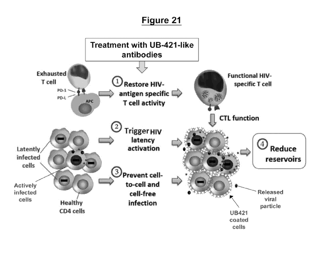
Figure 21. Cellular mechanisms mediated by UB421-like antibodies upon
treatment
include: (1) restoration of HIV antigen specific T cell activity by reduction
of % Treg cells, (2)
activation of HIV latency in infected cells upon antibody binding, and (3)
prevention of cell-
to-cell and cell-free infection to halt new HIV infection; all of which
results in (4) the reduction
or elimination of viral reservoirs leading to sustained virological remission
of HIV-1 infection.
Figures 22A to 22D. Western blot analysis of Lck phosphorylation on tyrosine
394
(Y394) and tyrosine 505 (Y505) in Jurkat T cells. Figure 22A are Western blot
images of Lck
Y394 phosphorylation (top) and Y505 phosphorylation (middle), and total Lck
protein level
after stimulation (bottom) with anti-CD3 (OKT3) antibody as a positive
control. Figure 22B
are graphs showing the Lck Y394 and Y505 phosphorylation level normalized with
total Lck
of each time point shown in Figure 22A. Figure 22C are Western blot images of
Lck Y394
phosphorylation (top), Y505 phosphorylation (middle), and total Lck protein
level (bottom)
with UB-421 stimulation with or without crosslinking. Figure 22D are graphs
showing Lck
Y394 and Y505 phosphorylation levels normalized with total Lck of each time
point shown in
Figure 22C, where dashed lines represent data obtained under crosslinking
condition and solid
lines represent data obtained under conditions without crosslinking.
Figures 23A to 23B. Western blot analysis of Lck phosphorylation with anti-CD3
(OKT-3) stimulation in primary CD4+ T cells from normal blood Donor 3. Figure
23A are
9
CA 03033728 2019-02-12
WO 2018/035001
PCT/US2017/046668
Western blot images of Lck Y394 phosphorylation (top), Y505 phosphorylation
(middle), and
total Lck protein level (bottom) with anti-CD3 (OKT3) antibody stimulation.
Figure 23B are
graphs showing Lck Y394 and Y505 phosphorylation levels normalized with total
Lck of each
time point in Figure 23A.
Figures 24A to 24D. Western blot analysis of Lck phosphorylation with UB-421
stimulation in primary CD4+ T cells from normal blood Donors 1, 2, 4, 5, 6,
and 7. Figure
24A are Western blot images of Lck Y394 phosphorylation (top), Y505
phosphorylation
(middle), and total Lck protein level (bottom) with UB-421 stimulation with or
without
crosslinking in healthy Donors 1 and 2. Figure 24B are Western blot images of
Lck Y394
phosphorylation (top), Y505 phosphorylation (middle), and total Lck protein
level (bottom)
with UB-421 stimulation without crosslinking in healthy Donors 4, 5, 6, and 7.
Figure 24C
and Figure 24D are graphs showing Lck Y394 and Y505 phosphorylation levels
normalized
with total Lck of each time point in Figures 24A and 24B, where the dashed
lines represent
data obtained under crosslinking condition and solid lines represent data
obtained under
conditions without crosslinking.
Figures 25A to 25B. Flow cytometry analysis of Lck phosphorylation in primary
CD4+
T cells from normal blood Donors 8 and 9. Figure 25A shows the MFI of PE-anti-
Lck pY394
(left) and Alexa647-anti-LckpY505 (right) with either anti-CD3 stimulation
(dashed line) as a
positive control or without any treatment (solid line) as a negative control.
Figure 25B shows
the MFI of PE-anti-Lck pY394 (left) and Alexa647-anti-LckpY505 (right) of
primary CD4+ T
cells from normal blood Donors 8 and 9 stimulated with UB-421 under
crosslinking conditions
(dashed line) or under conditions without crosslinking (solid line).
DETAILED DESCRIPTION OF THE INVENTION
The present disclosure is directed to antibodies, compositions, and methods
for the
treatment and sustained virologic remission of HIV infection in HAART
stabilized patients.
One aspect of the present disclosure relates to antibodies directed against
CD4, compositions
thereof, and methods of making and employing such compositions for the
treatment and
sustained virologic remission of HIV infection in HAART stabilizied patients
in the subsequent
absence of other treatments, including cART.
The section headings used herein are for organizational purposes only and are
not to be
construed as limiting the subject matter described. All references or portions
of references cited
CA 03033728 2019-02-12
WO 2018/035001
PCT/US2017/046668
in this application are expressly incorporated by reference herein in their
entirety for any
purpose.
CD4
Human CD4 (cluster of differentiation 4) is a 458 amino acid glycoprotein
(UniProtKB/Swiss-Prot: P01730.1) found on the surface of immune cells such as
T helper cells,
monocytes, macrophages, and dendritic cells (website:
en.wikipedia.org/wiki/CD4). The
amino acid sequence of CD4 is shown as SEQ ID NO: 22 in the Sequence Listing.
CD4+ T
helper cells are white blood cells that are an essential part of the human
immune system. They
are often referred to as CD4 cells, T-helper cells or T4 cells. They are
helper cells because they
send signals to other types of immune cells, including CD8 killer cells, which
destroy infectious
particles. If CD4 cells become depleted, for example in untreated HIV
infection, or following
immune suppression prior to a transplant, the body is left vulnerable to a
wide range of
infections that it would otherwise have been able to fight.
CD4 is a co-receptor that assists the T cell receptor (TCR) in communicating
with an
antigen-presenting cell. CD4 interacts directly with Major Histocompatibility
Complex (MHC)
class II molecules on the surface of the antigen-presenting cell using its
extracellular domain.
The extracellular domain adopts an immunoglobulin-like beta-sandwich with
seven strands in
2 beta sheets. Using its intracellular domain, CD4 amplifies the signal
generated by the TCR
by recruiting the tyrosine kinase Lck, which is essential for activating many
molecular
components of the signaling cascade of an activated T cell. Various types of T
helper cells are
thereby produced.
The major structural features of CD4 are shown in the Sequence Listing and
discussed
in further detail below.
CD4 is a member of the immunoglobulin superfamily and has four immunoglobulin
domains (D1 to D4) that are exposed on the extracellular surface of the cell.
CD4 domains D1
and D3 resemble immunoglobulin variable (IgV) domains; whereas D2 and D4
resemble
immunoglobulin constant (IgC) domains.
CD4 Domain 1 (D1)
The D1 core domain (approx. aa 26-125) consists of two 13-sheets formed by 13-
strands
that are linked by a disulfide bond bridge. The amino acid sequence of D1
shares homologies
with immunoglobulin at three complimentarily determining regions (CDRs)
similar to that of
11
CA 03033728 2019-02-12
WO 2018/035001
PCT/US2017/046668
immunoglobulin chains. The CDR1-, CDR2-, and CDR3-like regions are located in
the D1
domain of CD4.
The D1 domain of CD4 interacts directly with MHC class II molecules on the
surface
of antigen presenting cells and recruits lck to facilitate the activation of
helper T cells, thus
.. modulating the adaptive immune response. Both domain 1 and domain 2 of the
extracellular
region of the CD4 molecule were found to contribute to the binding sites for
class II MHC
molecules; however, domain 1 alone was found to be involved with HIV binding
and syncytia
formation. In particular, the binding site for the HIV envelope glycoprotein
gp120 was found
to be localized to the CDR2-like loop of Dl.
Several anti-CD4 antibodies have been produced that recognize the D1 domain of
CD4.
For example, HIV RC, B4, M2, and dB4C7 (e.g., Wang, C.Y. 1999; Lynn. S. and
Wang, C.Y.
2009; and Wang, C.Y., W02016/043788); Leu3a (Chiba, Y. 1992); OKT4A (Jameson,
B.D.,
et al., 1988); 5T4 and 13B8.2 (Briant, L,1999); 6H10 (e.g., Moore, et al.,
1992); 15A7, 2D5,
and 2F2 (e.g., Yuan R, et al., 2016); and F91-55 and BL4, which recognize the
region between
D1 and D2 (Briant, L, et al., 1999; Celada F, et al., 1990; and Moore, et al.,
1992);
CD4 Domain 2 (D2)
The D2 domain of CD4 (approx. aa 126-203) connects with D1 through its
hydrophobic
interface. D2 contributes to the binding sites for class II MHC molecules.
Several anti-CD4
antibodies have been produced that recognize the D2 domain of CD4. For
example, ibalizumab
.. (TMB-355; formerly known as TNX-355 or Hu5A8; e.g., Kuritzkes, D.R., et
al., 2004); M-
T441 (Konig R, et al., 1995).
CD4 Domain 3 (D3)
The D3 domain of CD4 is located at approx. aa 204-317. D3 connects to D4
through
its hydrophobic interface, similar to the way D2 interacts with Dl. The
antibody OKT4
recognizes D3 (e.g., Yuan R, et al., 2016; Moore, et al., 1992).
CD4 Domain 4 (D4)
The D4 domain (approx. aa 318-374) is the last extracellular domain on the CD4
molecule before the transmembrane domain. D4, structurally resembling D2, is
widely
believed to activate T cells and CD4 function through the dimerization of CD4
molecules. The
antibody OKT4 and L120 recognize D4 (e.g., Yuan R, et al., 2016; Moore, et
al., 1992).
CD4 ¨ Transmembrane region and cytoplasmic region
12
CA 03033728 2019-02-12
WO 2018/035001
PCT/US2017/046668
The transmembrane region (approx. aa 397-418) is hydrophobic whereas the
intracellular/cytoplasmic region (approx. 419-458) comprises three serine
residues (S433,
S440 and S456) that are phosphorylated to mediate signal transduction. These
serine residues
connect directly with the Src Tyrosine Kinase (TK) family member P561ck, which
can increase
the level of P561ck tyrosine phosphorylation and regulate signal transduction.
CD4 ¨ The Role in HIV Infection
HIV-1 uses CD4 to gain entry into host T-cells and achieves this through its
viral
envelope protein known as gp120. Gp120 is one of the two domains of the
maturing HIV-1
membrane envelope glycoprotein precursor gp160; the other is gp41. The binding
of gp120 to
CD4 constitutes the first step in HIV-1 attachment and the CD4¨gp120
interaction creates a
shift in the conformation of gp120 allowing it to bind to chemokine receptors
CCR5 or CXCR4
expressed on the host cell. This secondary binding allows the gp41 (fusion
peptide) molecule
of HIV-1 to insert into the host cell membrane, eventually mediating membrane
fusion of the
virus with the host. HIV infection leads to a progressive reduction in the
number of T cells
.. expressing CD4.
CD4 thus has a key role in the initiation of HIV-1 infection. Comparing bound
and
unbound crystal structures of gp120 with CD4 shows that a "bridging sheet" ¨ a
four-stranded
13-sheet formed by two 13-hairpins¨fixes the relative orientations of the two
closely associated
"inner" and "outer" domains of the gp120 core during CD4 binding. The CD4 D1
domain
interacts with these inner and outer domains as well as the bridging sheet,
which leads to the
rearrangements of the gp120 inner domain. Furthermore, with additional
interactions with the
gp120 V3 variable loop, the bridging sheet exposes the co-receptor binding
site (e.g., Yuan R,
et al., 2016).
Antibody
One aspect of the present disclosure relates to an antibody directed against
CD4,
compositions thereof, and methods employing such compositions for the
treatment and
sustained virologic remission of HIV infection.
The antibody of the present disclosure broadly encompasses intact antibody
molecules,
which include intact polyclonal, monoclonal, monospecific, polyspecific,
chimeric,
deimmunized, humanized, human, primatized, single-chain, single-domain,
synthetic and
recombinant antibodies. The present disclosure also includes portions of
intact antibodies that
have a desired activity or function (e.g., immunological fragments of
antibodies).
13
CA 03033728 2019-02-12
WO 2018/035001
PCT/US2017/046668
The antibody of the present disclosure is directed against CD4. In some
embodiments,
the antibody specifically binds to the extracellular region of CD4. In certain
embodiments,
antibody specifically recognizes and binds to at least one of the
immunoglobulin domains (D1
to D4) of CD4. In certain embodiments, the antibody binds to only one of the
immunoglobulin
domains of CD4 (i.e., D1, D2, D3, or D4). In specific embodiments, the
antibody binds to the
D1 domain of CD4. In some embodiments, the antibody binds to CD4 at or nearby
a
complimentarily determining region (CDR1, 2, or 3) in the D1 domain. In
specific
embodiments, the antibody binds to CD4 at or nearby the CDR2 region of the D1
domain.
The antibody of the present disclosure can be produced by any standard method.
In
some embodiments, the disclosed antibody is produced by immunizing an animal
(e.g., mouse,
dog, guinea pig, pig, goat, horse, etc.) with a recombinant CD4 protein,
fragments of the CD4
protein, fusion proteins containing immunological portions of CD4, and/or
analogues or
homologues of CD4. In other embodiments, the antibody can be produced by
immunizing an
animal with cells that express CD4 on the surface. In yet other embodiments,
the antibody can
be chemically synthesized.
In certain embodiments, the antibody is produced by immunizing an animal with
a CD4
protein, fragments of the CD4 protein, fusion proteins containing
immunological portions of
CD4, and/or analogues or homologues of CD4. In some embodiments, the antibody
is
produced by immunizing an animal with a peptide containing the full-length CD4
protein. In
other embodiments, the antibody is produced by immunizing an animal with a
peptide
containing a portion of the CD4 protein. For example, the peptide can contain
a portion of the
CD4 protein representing the extracellular region (e.g., D1 to D4), an
immunoglobulin domain
(D1, D2, D3, and/or D4), a complimentarily determining region (CDR1, 2, or 3)
within the D1
domain, etc. The antibody can be produced by immunizing an animal with a
single peptide
comprising at least a portion of CD4 or a combination of peptides containing
the amino acid
sequence of CD4. In some embodiments, the peptide immunogen contains aa39-66
of CD4,
which is also known to as the HIV receptor complex ("HIV RC"), as HIV binds to
this portion
of CD4. In a specific embodiment, the HIV RC peptide is made cyclic through a
disulfide
bond. In some embodiments, polyclonal antibodies are produced by immunizing an
animal
with the cyclic HIV RC peptide. The term "anti-HIV RC polyclonal antibodies",
as used
herein, refers to immune sera directed against a cyclic peptide containing
aa39-66 of the CDR2
region of CD4 domain 1.
14
CA 03033728 2019-02-12
WO 2018/035001
PCT/US2017/046668
In other embodiments, the antibody is produced by immunizing an animal with
CD4
positive cells. The cell lines can be any cell line that expresses CD4, such
as Jurkat cells, HPB-
ALL cells, U87MG cells, NIH-3T3 cells, HOS cells, CCRF-CEM cells (ATCCO CCL-
119Tm),
HuT 78 (ATCCO TIB-161Tm), MJ (G11) (ATCCO CRL-8294Tm), and the like. In
certain
embodiments, the antibody can be produced by immunizing BALB/c mice with
intact,
uninfected CD4+ human HPB-ALL cells, a T-acute lymphoblastic leukemia cell
line or
purified peripheral blood mononuclear T cells (PBL T cells). Such antibodies
are discussed in
further detail in US Patent Numbers 5,912,176, 6,090,388 and WO/2016/043788 by
Wang and
the journal article by Wang et al., 1999, all of which are incorporated by
reference in their
entireties.
In certain embodiments, the antibody of the present disclosure is tagged or
labeled with
a chemical. For example, the antibody can be labeled with biotin, spacer arms,
probes (e.g.,
FITC, PE, TRITC, DyLight Fluors, Alexa, GFP, R-Phycoerythrin, quantum dots,
etc.), enzyme
conjugates, and combinations thereof In a specific embodiment, the antibody is
labeled with
a biotin or fluorescent probe.
In specific embodiments, the antibody can be modified through a process known
as
deimmunization. The term "deimmunization", as used herein, generally refers to
a process for
modifying portions of an antibody so that it can be administered to an animal
without triggering
an immune response within the animal. Specifically, deimmunization involves a
process for
locating and removing portions of the amino acid sequence of the antibody that
would be
immunogenic (e.g., T-cell epitopes) in the particular animal that is being
administered the
antibody. This process can be accomplished through the combined use of
immunological and
molecular biology techniques. This process has been described previously
(e.g., Jones, T.D.,
et al. 2009). In the case of deimmunization of antibodies, mutations to remove
T-cell epitopes
can generally be introduced without significantly reducing the binding
affinity of the antibody.
The term "humanized", as used herein, refers to an antibody that was
originally
produced by a non-human species whose protein sequence has been modified
(deimmunized),
in a manner that removes the immunogenicity of the antibody when it is
administered to a
human. In certain embodiments, the disclosed antibody is deimmunized for human
use by
replacing the constant regions with human constant regions and/or by
expression of genes
encoding these antibodies in mammalian cells.
CA 03033728 2019-02-12
WO 2018/035001
PCT/US2017/046668
The term "mAb B4" or "B4" or "murine B4" as used herein, refers to a murine
monoclonal antibody which has been shown to recognize CD4 and can inhibit HIV
entry. The
structural and functional characteristics of this antibody are discussed in
further detailed in the
Examples that follow.
The term "mAb dB4" or "dB4", as used herein, refers to the human deimmunized
antibody derived from mAb B4. In one embodiment, mAb B4 is deimmunized for
human use
according to the method described in U.S. Patent Nos. 7,501,494 and 7,872,110,
which are
incorporated by references in their entireties. In a particular embodiment,
the human
deimmunized mAb dB4 is produced by removing and replacing the constant regions
of the
murine antibody (CH and CIO of mAb B4 and with the constant regions of human
IgGl. MAb
dB4 encompasses the dB4 produced by any suitable cellular clone. In a specific
embodiment,
mAb dB4 is produced by clone 7.
The term "mAb dB4C7" or "dB4C7", as used herein, refers to mAb dB4 expressed
by
clone 7 containing the recombinant genes B4DIVHv1NK1CHO#7 that was described
previously in U.S. Patent Nos. 7,501,494 and 7,872,110, and WO/2016/043788 by
Wang which
are incorporated by references in their entireties. The C7 clone has been
shown to produce
high quantities of mAb dB4 antibody. Additionally, the Asn (N) residue at
position 298 in
mAb dB4C7 has been substituted with His (H), to remove the N-glycosylation
site, thus
eliminating the IgG mediated complement dependent cytotoxicity (CdC) to
prevent depletion
of CD4 positive T cells in the presence of antibody B4.
The term "UB-421", as used herein, refers to the mAb dB4C7 that is used in a
suitable
form to be administered to human subjects.
The antibody can contain post-translational modifications, including sites for
glycosylation, methylation, and/or phosphorylation. In certain embodiments,
the antibody has
a sugar binding residue. In specific embodiments, the antibody contains an
asparagine (Asn)
residue that serves as a glycosylation site. In particular embodiments, the
Asn residue is on the
heavy chain, and in specific embodiments, the Asn is in the Fv region and/or
in a CDR.
The antibody of the present disclosure can also be described by its
interesting and
unique functional characteristics.
For example, the disclosed antibody exerts potent competitive HIV entry
inhibition
through its binding to domain 1 of CD4. In particular, the disclosed antibody
has nearly 100%
maximum percent inhibition (MPI) in all Env pseudotype viruses tested, with
IC5(is clustered
16
CA 03033728 2019-02-12
WO 2018/035001
PCT/US2017/046668
around two concentrations; one between 0.01 to 1 g/mL and the second one
around 10 g/mL.
The binding activity of the disclosed antibody is about two logs higher (i.e.
100X tighter
binding) than the CD4 binding affinity exhibited by HIV gp120 envelope
protein.
Additionally, the mean Kd of the disclosed antibody was estimated to be 5.6 x
10-11M (range:
3.1 to 8.1 x 10⁻¹¹M), and the Bmax was estimated to be 1.2 x 10⁶ Ab per cell
(range: 0.93 - 1.4
x 106).
The competitive inhibition property for the disclosed antibody has been shown
in both
cell-free and cell-to-cell systems. The disclosed antibody binds to CD4
receptors with an
affinity at least 50-fold higher than that for HIV-1 envelope protein gp120
MN. Also, the
disclosed antibody binds to CD4 with greater affinity and specificity compared
to other
commercially available antibodies, such as Leu3a.
The disclosed antibody can also inhibit antigen induced T cell proliferation
and
cytokine production (IL2 and IFN-gamma) of CD4 positive T cells, which is
implicated in the
pathogenic cycle of pyroptosis. Such high affinity monoclonal antibodies to
CD4 inhibit
antigen such as superantigen SEB (staphylococcal enterotoxin B, SEB) induced
CD4 positive
T cell activation and cytokine (e.g. IL2 and IFN-y) production. Such antigen
induced activation
leading to cytokine production in quiescent CD4+ T cells having abortive HIV
infection would
lead to pyroptosis of these quiescent CD4+ T cells and nearby normal resting
CD4 positive
cells resulting in ensuing mass depletion of CD4 + T cells, thus AIDS.
The disclosed antibody also has the ability to reactivate resting CD4 positive
T cells.
This property is particularly useful for reactivating latent reservoirs of HIV
in resting T cells
to make these cells susceptible to treatment with antiretroviral agents. Such
high affinity
antibodies to CD4 are capable of activating resting HIV infected cells for the
release of HIV.
Reactivation of HIV infected resting CD4+ T cells allows combinational
treatment
incorporating antibody of the current invention with HAART in HIV infected
patients leading
to functional cure.
Additional structural and functional characteristics of the disclosed
antibodies are
provided in the Examples that follow.
Formulation
The present disclosure is also directed to pharmaceutical formulations that
can be used
for the prevention, treatment, and/or functional cure of HIV infection. In
certain embodiments,
the formulations contain antibodies directed against CD4. In specific
embodiments, the present
17
CA 03033728 2019-02-12
WO 2018/035001
PCT/US2017/046668
disclosure relates to pharmaceutical compositions comprising high affinity
monoclonal
antibodies to CD4 that are directed to sites within or nearby CDR2 region of
CD4 domain 1.
The binding activity (EC50) of such antibodies is about two logs higher (i.e.
100X tighter
binding) than the CD4 binding affinity exhibited by HIV gp120 envelope protein
(EC50 for
gp120=97nM).
Pharmaceutical formulations of the antibody proteins disclosed can be prepared
by
mixing an antibody protein with optional pharmaceutically acceptable carriers.
Pharmaceutically acceptable carriers include solvents, dispersion media,
isotonic agents and
the like. The carrier can be liquid, semi-solid, e.g. pastes, or solid
carriers. Examples of carriers
include water, saline solutions or other buffers (such as phosphate, citrate
buffers), oil, alcohol,
proteins (such as serum albumin, gelatin), carbohydrates (such as
monosaccharides,
disaccharides, and other carbohydrates including glucose, sucrose, trehalose,
mannose,
mannitol, sorbitol or dextrins), gel, lipids, liposomes, resins, porous
matrices, binders, fillers,
coatings, stabilizers, preservatives, antioxidants including ascorbic acid and
methionine,
chelating agents such as EDTA; salt forming counter-ions such as sodium; non-
ionic
surfactants such as TWEENTm, PLURONICSTM or polyethylene glycol (PEG), or
combinations thereof
The formulation can contain more than one active compound. For example, the
formulation can contain one or more antibody and/or one or more additional
beneficial
compound for preventing and treating HIV infections. The active ingredients
can be combined
with the carrier in any convenient and practical manner, e.g., by admixture,
solution,
suspension, emulsification, encapsulation, absorption and the like, and can be
made in
formulations such as tablets, capsules, powder (including lyophilized powder),
syrup,
suspensions that are suitable for injections, ingestions, infusion, or the
like. Sustained-release
preparations can also be prepared.
In certain embodiments, the pharmaceutical formulation contains mAb dB4C7 for
human use. The pharmaceutical formulation containing mAb dB4C7 can be prepared
in an
appropriate buffer including, but not limited to, citrate, phosphate, Tris,
BIS-Tris, etc. at a pH
between 6.0 to 7.0 and can also contain excipients such as sugars (50 mM to
500 mM of
sucrose, trehalose, mannitol, or mixtures thereof), surfactants (e.g., 0.025% -
0.5% of Tween
20 or Tween 80), and/or other reagents. In a specific embodiment, the
formulation contains
mAb dB4C7 in 20 mM glycine, and 0.05% (v/v) Tween (polysorbate 20) in
phosphate buffer
saline (PBS), pH 6.5. In another specific embodiment, high concentration
formulations of mAb
18
CA 03033728 2019-02-12
WO 2018/035001
PCT/US2017/046668
dB4 were also prepared for use in certain applications including subcutaneous
injections, which
included 10 mM histidine.
The formulation can be prepared to contain various amounts of antibody. In
general,
formulations for administration to a subject contain between about 0.1 mg/mL
to about 200
mg/mL. In certain embodiments, the formulations can contain between about 0.5
mg/mL to
about 50 mg/mL; between about 1.0 mg/mL to about 50 mg/mL; between about 1
mg/mL to
about 25 mg/mL; or between about 10 mg/mL to about 25 mg/mL of antibody. In
specific
embodiments, the formulations contain about 1.0 mg/mL, about 5.0 mg/mL, about
10.0
mg/mL, or about 25.0 mg/mL of antibody.
In specific embodiments, the present invention relates to pharmaceutical
compositions
comprising human, humanized or chimeric, monoclonal anti-CD4 antibodies with
the above
described binding characteristics which exhibit competitive HIV entry
inhibition as well as
activation of CD4+ T cells, as an immunotherapy in patients with HIV
infection.
In another embodiment, the present invention relates to pharmaceutical
compositions
comprising monoclonal human, humanized or chimeric, anti-CD4 antibodies with
the above
described binding characteristics that serve as a monotherapy that can reduce
viral load down
to non-detectable level in treated subjects at a serum antibody level higher
than 10 [tg/mL.
In another embodiment, the present invention relates to pharmaceutical
compositions
comprising monoclonal human, humanized or chimeric, anti-CD4 antibodies with
the above
described binding characteristics that serve as a monotherapy that can reduce
viral load down
to non-detectable level in treated subjects at a serum antibody level higher
than 10 [tg/mL and
maintained stable CD4 T cell counts during a 12-weeks treatment period.
In certain embodiments, the present invention relates to pharmaceutical
compositions
comprising monoclonal human, humanized or chimeric) anti-CD4 antibodies with
the above
described binding characteristics that when given, at a dose of about 10 mg/kg
or higher on a
weekly or biweekly schedule, as a monotherapy, such treatment can reduce viral
load down to
non-detectable level in treated subjects during a 12-weeks treatment period.
In yet another preferred embodiment, the present invention relates to
pharmaceutical
compositions comprising a monoclonal humanized anti-CD4 antibody with the
above
described binding characteristics as the key ingredient in an adjunct therapy
with HAART, that
when given, at about 10 mg/kg or higher on a weekly or biweekly schedule, to
treatment naive
HIV patients, will lead to functional cure of the patients.
19
CA 03033728 2019-02-12
WO 2018/035001
PCT/US2017/046668
In yet another preferred embodiment, the present invention relates to
pharmaceutical
compositions comprising a monoclonal humanized anti-CD4 antibody with the
above
described binding characteristics as the key ingredient in an adjunct therapy
with HAART, that
when given, at about 10 mg/kg or higher on a weekly or biweekly schedule, to
patients with
stabilized viral load under HAART, will lead to functional cure of the
patients.
Antiviral A2ents
The present disclosure also includes antiviral agents that can be used in the
methods for
the treatment, prevention, and functional cure of HIV infection.
Antiviral agents include any agent (compound or biological) that is effective
to inhibit
the formation and/or replication of HIV in a mammal. Examples of antiviral
agents include,
but are not limited to, entry/fusion inhibitors (e.g., maraviroc,
enfuvirtide); nucleoside reverse
transcriptase inhibitors (NRTI) and nucleotide reverse transcriptase
inhibitors (NtRTI) (e.g.,
zidovudine, abacavir, didanosine, lamivudine, emtricitabine, stavudine, and
tenofovir); non-
nucleoside reverse transcriptase inhibitors (NNRTI) (e.g., nevirapine,
efavirenz, etravirine, and
rilpivirine); integrase inhibitors also known as integrase nuclear strand
transfer inhibitors or
INSTIs (e.g., raltegravir, dolutegravir, elvitegravir); protease inhibitors
(e.g., saquinavir,
saquinavir mesylate, fosamprenavir, tipranavir, lopinavir, indinavir,
nelfinavir, amprenavir,
ritonavir, darunavir, atazanavir, bevirimat, vivecon); viral maturation
inhibitors; agents
targeting the expression of HIV genes; agents targeting key host cell genes
and gene products
involved in HIV replication; and other anti-HIV agents; iRNA agents; antisense
RNA; vectors
expressing iRNA agents or antisense RNA; PNA and antiviral antibodies; and
combinations
thereof
The antiviral agents can be used individually or in combination. Use of
antiviral agents
in combination is known as anti-retroviral therapy (ART), combination anti-
retroviral therapy
(cART) or highly active anti-retroviral therapy (HAART). Anti-retroviral (ARV)
drugs are
broadly classified by the phase of the retrovirus life-cycle that the drug
inhibits. Typical
combinations include 2 NRTIs as a "backbone" along with 1 NNRTI, PI or INSTI
as a "base".
In certain embodiments combinations of antiviral agents are used, such as
Combivir, Trizivir,
Kaletra, Epzicom, Truvada, Atripla, Complera, Stribild, Triumeq.
CA 03033728 2019-02-12
WO 2018/035001
PCT/US2017/046668
Methods of Treatment and Sustained Virolo2ic Remission of HIV Infection
The present disclosure is also directed to methods for the treatment,
prevention, and
functional cure of HIV infection. In certain embodiments, the formulations
contain antibodies
directed against CD4.
In a further aspect, the antibody disclosed herein, optionally provided in
pharmaceutically acceptable carrier, can be employed for the treatment,
prevention, and/or
functional cure of HIV infection in a subject, as well as prevention of HIV
transmission.
The term "treatment" of HIV infection refers to effective inhibition of the
HIV infection
so as to delay the onset, slow down the progression, reduce viral load, and/or
ameliorate the
symptoms caused by HIV infection. Treatment include both pre- and post-
exposure to HIV.
The term "prevention" of HIV infection means the onset of HIV infection is
delayed,
and/or the incidence or likelihood of HIV infection is reduced or eliminated.
The term
"prevention" of HIV transmission means the incidence or likelihood of HIV
being transmitted
from one individual to another (e.g., from an HIV-positive woman to the child
during
pregnancy, labor or delivery, or breastfeeding) is reduced or eliminated.
The term "subject" refers to any primate subject, including human, rhesus,
baboon, and
chimpanzee subjects.
To treat and/or prevent HIV infection, a therapeutic amount of an antibody
disclosed
herein is administered to a subject in need.
The term "therapeutically effective amount" means the dosage required to
effect an
inhibition of HIV infection so as to treat and/or prevent HIV infection. The
dosage of an
antibody depends on the disease state and other clinical factors, such as
weight and condition
of the subject, the subject's response to the therapy, the type of
formulations and the route of
administration. The precise dosage to be therapeutically effective and non-
detrimental can be
determined by those skilled in the art.
Generally, a suitable dose of an antibody for the administration to adult
humans is in
the range of about 3 to 50 mg/kg of the subject's body weight, with the
typical initial range
used being in the range of about 5 to 25 mg/kg of the subject's body weight.
Suitable dosages
also include about 5.0 mg/kg, about 10.0 mg/kg, or about 25.0 mg/kg of the
patient's body
weight.
21
CA 03033728 2019-02-12
WO 2018/035001
PCT/US2017/046668
The therapeutic compositions containing a human monoclonal antibody of this
invention are conventionally administered intravenously, as by injection of a
unit dose, for
example. A unit dose generally refers to a therapeutic composition of the
present invention
which further refers to physically discrete units suitable as unitary dosage
for the subject, each
unit containing a predetermined quantity of active material calculated to
produce the desired
therapeutic effect in association with the required diluent; i.e., carrier, or
vehicle.
The compositions are administered in a manner compatible with the dosage
formulation, and in a therapeutically effective amount. The quantity to be
administered depends
on the subject to be treated, capacity of the subject's system to utilize the
active ingredient, and
degree of therapeutic effect desired. Precise amounts of active ingredient
required to be
administered depend on the judgment of the practitioner and are peculiar to
each individual.
However, suitable dosage ranges for systemic application are disclosed herein
and depend on
the route of administration. Suitable regimes for administration are also
variable, but are
typified by an initial administration followed by repeated doses at one or
more hour intervals
by a subsequent injection or other administration. Alternatively, continuous
intravenous
infusion sufficient to maintain concentrations in the blood in the ranges
specified for in vivo
therapies are contemplated.
The method for the treatment, prevention, and/or functional cure of HIV
infection in a
subject includes administering to the subject an effective amount of a
formulation containing
the antibody. In certain embodiments, the formulation is provided to the
subject in a single
administration. In other embodiments, the formulation is provided to the
subject in multiple
administrations. When the formulation is provided in multiple administrations,
the formulation
can be administered once per day, once a week, bi-weekly (every other week),
or once a month.
In a specific embodiment, when the treatment schedule is once a week, the
formulation is
administered to the subject in a dosage of about 5.0 mg/kg of the subject's
body weight. In
another embodiment, when the treatment schedule is bi-weekly, the formulation
is
administered to the subject in a dosage of about 25.0 mg/kg of the subject's
body weight.
In certain embodiments, formulations containing the monoclonal antibody show
high
safety factor and was well tolerated when subjects were given repeatedly on a
weekly basis at
5 mg/kg or 25 mg/kg for a total of 8 weeks. In specific embodiments, the
monoclonal antibody
can be given to subjects within hours of HIV infection at 5 mg/kg to provide
sterilizing cure of
HIV infection. In other embodiments, the monoclonal antibody can be given to a
subject within
days after HIV infection at 5 mg/kg to provide a functional cure of HIV
infection.
22
CA 03033728 2019-02-12
WO 2018/035001
PCT/US2017/046668
In certain embodiments, the present invention relates to pharmaceutical
compositions
comprising monoclonal human, humanized or chimeric, anti-CD4 antibodies with
the above
described binding characteristics that can be administered to HIV patients
through intravenous
(IV), intramuscular (IM) or subcutaneous (SC) route as an immunotherapy for
reduction of
viral load. In specific embodiments, the present invention relates to
pharmaceutical
compositions comprising human, humanized or chimeric, monoclonal anti-CD4
antibodies,
with the above described binding characteristics which exhibit competitive HIV
entry
inhibition as well as activation of CD4+ T cells, as an immunotherapy in
patients with HIV
infection.
In other certain embodiments, the present invention relates to pharmaceutical
compositions comprising monoclonal human, humanized or chimeric, anti-CD4
antibodies
with the above described binding characteristics that can be administered to
HIV patients
through IV, IM or SC route as an immunotherapy for reduction of viral load at
a dose of about
10 mg/kg or higher on a weekly or biweekly schedule.
In another embodiment, the present invention relates to pharmaceutical
compositions
comprising monoclonal human, humanized or chimeric, anti-CD4 antibodies with
the above
described binding characteristics that serve as a monotherapy that can reduce
viral load down
to non-detectable level in treated subjects at a serum antibody level higher
than 10 pg/mL.
In another embodiment, the present invention relates to pharmaceutical
compositions
comprising monoclonal human, humanized or chimeric, anti-CD4 antibodies with
the above
described binding characteristics that serve as a monotherapy that can reduce
viral load down
to non-detectable level in treated subjects at a serum antibody level higher
than 10 pg/mL and
maintained stable CD4 T cell counts during a 12-weeks treatment period.
In another embodiment, the present invention relates to pharmaceutical
compositions
comprising monoclonal human, humanized or chimeric) anti-CD4 antibodies with
the above
described binding characteristics that when given, at a dose of about 10 mg/kg
or higher on a
weekly or biweekly schedule, as a monotherapy, such treatment can reduce viral
load down to
non-detectable level in treated subjects during a 12-weeks treatment period.
In another embodiment, the present invention relates to pharmaceutical
compositions
comprising monoclonal human, humanized or chimeric, anti-CD4 antibodies with
the above
described binding characteristics that when given, at a dose of about 10 mg/kg
or higher on a
weekly or biweekly schedule, as a monotherapy, such treatment can reduce viral
load down to
23
CA 03033728 2019-02-12
WO 2018/035001
PCT/US2017/046668
non-detectable level in treated subjects with no viral load rebound as long
the serum antibody
level is higher than 10 [tg/mL.
In another embodiment, the present invention relates to pharmaceutical
compositions
comprising a monoclonal humanized anti-CD4 antibody with the above described
binding
characteristics as the key ingredient in an adjunct therapy with HAART, that
when given, at
about 10 mg/kg or higher on a weekly or biweekly schedule, to treatment naïve
HIV patients,
will lead to functional cure of the patients.
In another embodiment, the present invention relates to pharmaceutical
compositions
comprising a monoclonal humanized anti-CD4 antibody with the above described
binding
characteristics as the key ingredient in an adjunct therapy with HAART, that
when given, at
about 10 mg/kg or higher on a weekly or biweekly schedule, to patients with
stabilized viral
load under HAART, will lead to functional cure of the patients.
In another embodiment, the present invention relates to pharmaceutical
compositions
comprising a monoclonal humanized anti-CD4 antibody with the above described
binding
characteristics that can be administered, in either IV, IM or SC route, as the
key ingredient in
an HAART replacement therapy, whereby each treatment cycle begins with anti-
CD4 antibody
treatment for 2 to 4 months as a treatment holiday for patients experiencing
stabilized
undetectable viral load under HAART followed by HAART treatment over one to
four or more
cycles leading to functional cure.
In another embodiment, the present invention relates to pharmaceutical
compositions
comprising a monoclonal humanized anti-CD4 antibody with the above described
binding
characteristics that can be administered, in either IV, IM or SC route, as the
key ingredient in
an HAART replacement therapy, whereby each treatment cycle begins with anti-
CD4 antibody
treatment for 2 to 4 months for treatment naïve HIV patients followed by 2 to
4 months of
HAART treatment over one to four or more cycles leading to functional cure.
In another embodiment, the present invention relates to pharmaceutical
compositions
comprising a monoclonal humanized anti-CD4 antibody with the above described
binding
characteristics that can be administered, in either IV, IM or SC route, as the
key ingredient in
an HAART replacement therapy, whereby each treatment cycle begins with anti-
CD4 antibody
treatment for 2 to 4 months as a treatment holiday for patients experiencing
stabilized
undetectable viral load under HAART followed by HAART treatment over one to
four or more
24
CA 03033728 2019-02-12
WO 2018/035001
PCT/US2017/046668
cycles at a dose of about 5 mg/kg or higher on a weekly or biweekly schedule,
leading to
functional cure.
In another embodiment, the present invention relates to pharmaceutical
compositions
comprising a monoclonal humanized anti-CD4 antibody with the above described
binding
characteristics that can be administered, in either IV, IM or SC route, as the
key ingredient in
an HAART replacement therapy, whereby each treatment cycle begins with anti-
CD4 antibody
treatment for 2 to 4 months for treatment naïve HIV patients followed by 2 to
4 months of
HAART treatment over one to four or more cycles at a dose of about 5 mg/kg or
higher on a
weekly or biweekly schedule, leading to functional cure.
In another embodiment, the present invention relates to pharmaceutical
compositions
comprising a monoclonal humanized anti-CD4 antibody with the above described
binding
characteristics as the key ingredient in an adjunct therapy with HAART, that
when given, at
about 10 mg/kg or higher on a weekly or biweekly schedule, to treatment naïve
HIV patients,
will lead to functional cure of the patients.
In another embodiment, the present invention relates to pharmaceutical
compositions
comprising a monoclonal humanized anti-CD4 antibody with the above described
binding
characteristics as the key ingredient in an adjunct therapy with HAART, that
when given, at
about 10 mg/kg or higher on a weekly or biweekly schedule, to patients with
stabilized viral
load under HAART, will lead to functional cure of the patients.
In another embodiment, the present invention relates to pharmaceutical
compositions
comprising a monoclonal humanized anti-CD4 antibody with the above described
binding
characteristics that can be administered in either IV, IM or SC route, to
patients who failed
HAART treatment in an adjunct therapy to HAART at a dose of about 10 mg/kg or
higher on
a weekly or biweekly schedule, leading to further viral reduction.
In another embodiment, the present invention relates to pharmaceutical
compositions
comprising a monoclonal humanized anti-CD4 antibody with the above described
binding
characteristics that can be administered, in either IV, IM or SC route, as the
key ingredient in
an adjunct therapy with HAART, in an intermittent mode beginning with a
treatment period
for 2 to 4 months and a treatment holiday for 1 to 2 months per cycle over one
to four or more
cycles, to treatment naïve HIV patients as an adjunct therapy in an intensive
HAART treatment
mode, leading to functional cure of the patients.
CA 03033728 2019-02-12
WO 2018/035001
PCT/US2017/046668
In another embodiment, the present invention relates to pharmaceutical
compositions
comprising a monoclonal humanized anti-CD4 antibody with the above described
binding
characteristics that can be administered, in either IV, IM or SC route, as the
key ingredient in
an adjunct therapy with HAART, in an intermittent mode beginning with a
treatment period
for 2 to 4 months and a treatment holiday for 1 to 2 months per cycle over one
to four or more
cycles, at a dose of about 5 mg/kg or higher on a weekly or biweekly schedule,
to treatment
naïve HIV patients as an adjunct therapy in an intensive HAART treatment mode,
leading to
functional cure of the patients.
In another embodiment, the present invention relates to pharmaceutical
compositions
comprising a monoclonal humanized anti-CD4 antibody with the above described
binding
characteristics that can be administered, in either IV, IM or SC route, as the
key ingredient in
an adjunct therapy with HAART, in an intermittent mode beginning with a
treatment period
for 2 to 4 months and a treatment holiday for 1 to 2 months per cycle over one
to four or more
cycles, at a dose of about 5 mg/kg or higher on a weekly or biweekly schedule,
to patients
experiencing stabilized undetectable viral load under HAART, as an adjunct
therapy in an
intensive HAART treatment mode, leading to functional cure of the patients.
In another embodiment, the present invention relates to pharmaceutical
compositions
comprising monoclonal human, humanized or chimeric, anti-CD4 antibodies with
the above
described binding characteristics that can be administered to HIV patients
through IV, IM or
SC route as an immunotherapy for reduction of viral load.
In another embodiment, the present invention relates to pharmaceutical
compositions
comprising monoclonal human, humanized, or chimeric anti-CD4 antibodies with
the above
described binding characteristics that can be administered to HIV patients
through IV, IM or
SC route as an immunotherapy for reduction of viral load at a dose of about 5
mg/kg or higher
on a weekly or biweekly schedule.
Specific Embodiments
(1) An antibody directed against a CD4 molecule, wherein
the antibody specifically binds to an extracellular region of the CD4
molecule, and
wherein
when the antibody is bound to the CD4 molecule on the surface of a CD4+ cell,
the
antibody:
26
CA 03033728 2019-02-12
WO 2018/035001
PCT/US2017/046668
a) competitively inhibits HIV entry into the CD4+ cell;
b) activates latent HIV reservoirs in a resting CD4+ cell infected with HIV;
d) reduces levels of cellular HIV DNA; and
e) provides sustained virologic remission of HIV infection without viral load
rebound.
(2) The antibody according to (1), wherein the antibody competitively
inhibits cell-free
and cell-to-cell transmission of HIV.
(3) The antibody according to (1), wherein the antibody reduces the
percentage of
regulatory T cells when administered to a subject.
(4) The antibody according to (1), wherein the antibody increases the
amount of CD8+
cells when administered to a subject.
(5) The antibody according to (1), wherein the antibody increases CD8+
proliferating
cells in response to HIV gag motif peptide stimulation when administered to a
subject.
(6) The antibody according to (1), wherein the antibody enhances functional
HIV specific
CD8+ CTL cells that target an HIV infected CD4+ cell when administered to a
subject.
(7) The antibody according to (1), wherein the antibody enhances TNF-alpha
production
in CD4+ cell.
(8) The antibody according to (1)wherein the antibody activates a resting
CD4+ cells with
or without crosslinking.
(9) The antibody according to (1), wherein the antibody reduces HIV viral
load in an HIV
positive patient to less than 50 copies per milliliter of blood without viral
load rebound.
(10) The antibody of (1), wherein the antibody binds to a region around domain
1 of the
CD4 molecule.
(11) The antibody of (1), wherein the antibody binds to a region around the
CDR2 region
in domain 1 of CD4.
(12) The antibody of (1), wherein the antibody comprises
a heavy chain variable region amino acid sequence comprising:
CDR1 of SEQ ID NO: 1,
CDR2 of SEQ ID NO: 2, and
CDR3 of SEQ ID NO: 3; and
a light chain variable region amino acid sequence comprising:
27
CA 03033728 2019-02-12
WO 2018/035001
PCT/US2017/046668
CDR1 of SEQ ID NO: 4,
CDR2 of SEQ ID NO: 5, and
CDR3 of SEQ ID NO: 6.
(13) The antibody of (1), wherein the antibody is a monoclonal antibody.
(14) The antibody of (1), wherein the antibody is a humanized monoclonal
antibody.
(15) The antibody of (1), wherein the antibody is a humanized monoclonal
antibody
comprising:
a heavy chain variable region comprising an amino acid sequence of SEQ ID NO:
11; and
a light chain variable region comprising an amino acid sequence of SEQ ID NO:
13.
(16) The antibody of (1), wherein the antibody is a humanized monoclonal
antibody
comprising:
a heavy chain comprising an amino acid sequence of SEQ ID NO: 10; and
a light chain comprising an amino acid sequence of SEQ ID NO: 8.
(17) The antibody of (1), wherein the antibody is a humanized monoclonal
antibody
comprising:
a heavy chain comprising an amino acid sequence of SEQ ID NO: 9; and
a light chain comprising an amino acid sequence of SEQ ID NO: 8.
(18) The antibody of (1), wherein the antibody is a humanized monoclonal
antibody
comprising:
a heavy chain comprising an amino acid sequence of SEQ ID NO: 7; and
a light chain comprising an amino acid sequence of SEQ ID NO: 8.
(19) The antibody of (1) having an absolute binding affinity (Kd) to membrane-
bound
CD4 on HPB-ALL cells between about 3.1 x 10⁻¹¹ M to about 8.1 x 10⁻¹¹ M.
(20) The antibody of (1) bound to a CD4 molecule.
(21) A composition comprising the antibody of (1).
(22) A pharmaceutical composition comprising the antibody of (1) and a
pharmaceutically
acceptable carrier.
(23) A pharmaceutical composition comprising the antibody of (1) in phosphate
buffer
saline (PBS), 20 mM glycine, and 0.05% (v/v) polysorbate 20.
(24) A pharmaceutical composition comprising the antibody of (1) in phosphate
buffer
saline (PBS), 20 mM glycine, 0.05% (v/v) polysorbate 20, and 10 mM histidine.
28
CA 03033728 2019-02-12
WO 2018/035001
PCT/US2017/046668
(25) A pharmaceutical composition comprising about 1.0 mg/mL to about 200.0
mg/mL of
the antibody of (1) in phosphate buffer saline (PBS), 20 mM glycine, and 0.05%
(v/v)
polysorbate 20.
(26) A pharmaceutical composition comprising about 1.0 mg/mL to about 200.0
mg/mL of
the antibody of (1) in phosphate buffer saline (PBS), 20 mM glycine, 0.05%
(v/v) polysorbate
20, and 10 mM histidine.
(27) A pharmaceutical composition comprising about 10.0 mg/mL of the antibody
of (1) in
phosphate buffer saline (PBS), 20 mM glycine, and 0.05% (v/v) polysorbate 20.
(28) A pharmaceutical composition comprising about 10.0 mg/mL of the antibody
of (1) in
phosphate buffer saline (PBS), 20 mM glycine, 0.05% (v/v) polysorbate 20, and
10 mM
histidine.
(29) A pharmaceutical composition comprising the antibody of (12) and a
pharmaceutically acceptable carrier.
(30) A pharmaceutical composition comprising the antibody of (16) and a
pharmaceutically acceptable carrier.
(31) A method for treating a subject exposed to HIV comprising:
administering to the subject a pharmacologically effective amount of the
antibody of (1).
(32) The method of (31), wherein the antibody is administered to the subject
prior to
exposure to HIV.
(33) The method according to (31), wherein the antibody is administered to the
subject
after exposure to HIV.
(34) The method according to (31), wherein the antibody is administered within
48 hours
after exposure to HIV.
(35) The method according to (31), wherein the antibody is administered to the
subject at a
dosage of at least about 5 mg/kg body weight.
(36) The method according to (35), wherein the antibody is administered to the
subject
multiple times.
(37) The method according to (36), wherein the antibody is administered to the
subject in a
weekly, bi-weekly, or monthly interval.
(38) The method according to (36), further comprising a step of administering
an antiviral
agent to the subject.
(39) The method according to (38), wherein the antiviral agent is a highly
active
antiretroviral therapy (HAART).
29
CA 03033728 2019-02-12
WO 2018/035001
PCT/US2017/046668
(40) The method according to (39), wherein HAART comprises a nucleoside
analogue
reverse transcriptase inhibitor in combination with a protease inhibitor or a
non-nucleoside
reverse transcriptase inhibitor.
(41) The method according to (39), wherein the antibody is administered
concurrently with
HAART.
(42) The method according to (39), wherein the antibody and HAART are
administered to
the subject over the course of a cycle, wherein the cycle comprises:
i) administering the antibody to the subject for a first period of time
followed by a
treatment holiday for a second period of time; and
ii) administering HAART to the subject continuously during the first period of
time
and the second period of time in (i).
(43) The method according to (39), wherein the antibody and HAART are
administered to
the subject over the course of a cycle, wherein the cycle comprises:
i) administering the antibody to the subject for a period of four months in a
weekly,
bi-weekly, or monthly interval followed by a two month treatment holiday; and
ii) administering HAART to the subject continuously during the six-month
period in
(i).
(44) The method according to (42), wherein the subject is treated over the
course of two
cycles.
(45) The method according to (43), wherein the subject is treated over the
course of two
cycles.
(46) The method according to (39), wherein the antibody is administered at a
time that is
not concurrent with HAART.
(47) The method according to (39), wherein the antibody and HAART are
administered to
the subject over the course of a cycle, wherein the cycle comprises:
i) administering the antibody to the subject for a first period of time
followed by a
treatment holiday for a second period of time; and
ii) administering HAART to the subject during the second period of time and
not
during the first period of time.
(48) The method according to (47), wherein the antibody is administered in
regular
intervals during the first time period.
(49) The method according to (47), wherein the antibody is administered in
weekly, bi-
weekly, or monthly intervals during the first time period.
(50) A method for treating a subject with HIV infection, comprising
administering to the
CA 03033728 2019-02-12
WO 2018/035001
PCT/US2017/046668
subject a treatment regimen comprising:
a) a pharmacologically effective amount of the antibody of (1); and
b) a highly active antiretroviral therapy (HAART).
(51) The method of (50), wherein the antibody is administered to the subject
at a dosage of
at least about 5 mg/kg body weight.
(52) The method according to (50), wherein the antibody and HAART are
administered to
the subject over the course of a cycle, wherein the cycle comprises:
i) administering the antibody to the subject for a first period of time
followed by a
treatment holiday for a second period of time; and
ii) administering HAART to the subject continuously during the first period of
time
and the second period of time in (i).
(53) The method according to (50), wherein the antibody and HAART are
administered to
the subject over the course of a cycle, wherein the cycle comprises:
i) administering the antibody to the subject for a period of four months in a
weekly,
bi-weekly, or monthly interval followed by a two-month treatment holiday; and
ii) administering HAART to the subject continuously during the six-month
period in
(i).
(54) The method according to (52), wherein the subject is treated over the
course of two or
more cycles.
(55) The method according to (53), wherein the subject is treated over the
course of two or
more cycles.
(56) The method according to (53), wherein the antibody and HAART are
administered to
the subject over the course of a cycle, wherein the cycle comprises:
i) administering the antibody to the subject for a period of four months in a
weekly,
bi-weekly, or monthly interval followed by a two-month treatment holiday; and
ii) administering HAART to the subject continuously during the six-month
period in
(i).
(57) The method according to (50), wherein the antibody in (a) is administered
at a time
that is not concurrent with HAART in (b).
(58) The method according to (50), wherein the antibody in (a) and HAART in
(b) are
administered to the subject over the course of a cycle, wherein the cycle
comprises:
i) administering the antibody to the subject for a first period of time
followed by a
treatment holiday for a second period of time; and
31
CA 03033728 2019-02-12
WO 2018/035001
PCT/US2017/046668
ii) administering HAART to the subject during the second period of time and
not
during the first period of time.
(59) The method according to (58), wherein the antibody is administered in
regular
intervals during the first time period.
.. (60) The method according to (58), wherein the antibody is administered in
weekly, bi-
weekly, or monthly intervals during the first time period.
(61) A method for inhibiting HIV entry into a CD4+ cell, comprising
exposing the antibody of (1) to the cell.
(62) A method for inhibiting gp120 binding to a CD4+ cell, comprising
exposing the antibody of (1) to the cell.
(63) A method for activating a resting CD4+ T cell, comprising
exposing the antibody of (1) to the cell.
(64) A method for activating a latent reservoir of HIV in a resting T cell,
comprising
exposing the antibody of (1) to the cell.
.. (65) A method for reducing latent HIV reservoirs in a sample of cells
infected with HIV,
comprising
a) exposing the antibody of (1) to the sample of cells; and
b) exposing HAART to the sample of cells.
Unless otherwise explained, all technical and scientific terms used herein
have the same
meaning as commonly understood by one of ordinary skill in the art to which
this invention
belongs. The singular terms "a," "an," and "the" include plural referents
unless context clearly
indicates otherwise. Similarly, the word "or" is intended to include "and"
unless the context
clearly indicates otherwise. Hence "comprising A or B" means including A, or
B, or A and B.
It is further to be understood that all amino acid sizes, and all molecular
weight or molecular
mass values, given for polypeptides are approximate, and are provided for
description.
Although methods and materials similar or equivalent to those described herein
can be used in
the practice or testing of the disclosed method, suitable methods and
materials are described
below. All publications, patent applications, patents, and other references
mentioned herein are
incorporated by reference in their entirety. In case of conflict, the present
specification,
including explanations of terms, will control. In addition, the materials,
methods, and examples
are illustrative only and not intended to be limiting.
32
CA 03033728 2019-02-12
WO 2018/035001
PCT/US2017/046668
The following illustrative explanations of the figures and related examples
are provided
to facilitate understanding of certain terms used frequently herein,
particularly in the examples.
The explanations are provided as a convenience and are not limitative of the
invention.
EXAMPLE 1
IMMUNOLOGICAL AND FUNCTIONAL PROPERTIES OF MAB B4
Monoclonal antibody B4 (mAb B4) or M2 (mAb M2) is a monoclonal antibody that
recognizes a complex HIV receptor site on the T cell surface (CD4). MAb B4 or
M2 can
influence and interfere with CD4's interaction with HIV co-receptors. MAb B4
or M2
preferentially neutralized primary HIV-1 isolates (both antibodies were
discussed in further
detail in US Patent Numbers 5,912,176, 6,090,388).
The information below summarizes the discovery and preliminary
characterization
studies of murine mAb B4 including data excerpted from two US patents (US
Patent Numbers
5,912,176 and 6,090,388 by Wang) and the journal article by Wang etal., 1999,
all of which
are incorporated by reference in their entireties.
1. Murine Monoclonal Antibodies Derived from immunization with HPB ALL cells
or
purified PBL T cells
MAb B4 was obtained by immunizing BALB/c mice with intact, uninfected CD4+
human HPB-ALL cells, a T-acute lymphoblastic leukemia cell line.
MAb M2 was obtained by immunizing BALB/c mice with intact, uninfected CD4+
cells isolated from PBL.
A novel class of anti-CD4 antibodies, represented by mAb B4 or M2, were
obtained
having specificity for CD4 on the cell surface and with broad neutralizing
activity against
primary isolates of HIV-1. In the subsequence discussion and examples, only
Mab B4 will be
further illustrated and discussed for purpose of focusing on important
property disclosure.
2. Characterization of the mAb B4 Reco2nition Site
MAb B4 has been found to preferentially recognize membrane-bound CD4 on the
surface of cells compared to recombinant soluble CD4 (rsCD4).
MAb B4 binding to membrane-bound CD4 prior to exposure of HIV has been shown
to block subsequent attachment of gp120 and whole virus to CD4. However,
membrane-bound
33
CA 03033728 2019-02-12
WO 2018/035001
PCT/US2017/046668
CD4 that has been bound to gp120 prior to exposure to the antibody can still
bind mAb B4.
Thus, mAb B4 can affect the binding of gp120 to membrane-bound CD4, but gp120
does not
affect the binding of mAb B4 to CD4.
3. In Vitro Neutralization Activity of mAb B4
Murine mAb B4 is not, by common definition, a neutralizing antibody. Instead,
mAb
B4 inhibits viral entry by coating the host cell receptor rather than by
attaching to the virus.
MAb B4' s effect on HIV infection can be readily observed by viral
neutralization assays used
in the field (e.g., MT-2 Microplaque Neutralization Assay (Sawyer et al.,
1994)). The
neutralization activity of murine mAb B4 was evaluated by our collaborator Dr.
Carl Hanson
(California Department of Health Services) and was also independently
evaluated in the
laboratories of Dr. John Mascola, (Henry Jackson Foundation, WRAIR), Dr. David
Montefiori
(Duke University) and Dr. Malcolm Martin (NIAID). The following HIV
neutralizing features,
extensively characterized from 1995 to 2010, are associated with mAb B4:
1. PBMC-grown primary isolates are more sensitive to neutralization by mAb B4
than T cell
line-adapted isolates HIV-1111B and HIV-1 MN.
2. mAb B4 neutralizes infection by primary isolates of co-receptor usage
CCR5/CXCR4 (dual)
and CCR5.
3. mAb B4 has low activity against T cell line-adapted HIV-1 isolates of
CXCR4 co-receptor
usage.
4. mAb B4 neutralizes a diverse range of Syncytial Inducing (SI) and Non-
Syncytial Inducing
(NSI) primary isolates representing HIV-1 subtypes A-G, to 90% endpoints and
up to 3
logs of infectivity.
5. mAb B4 neutralizes HIV-2, Sly, and SHIV having a dual co-receptor HIV-1
envelope.
6. In the tonsil histoculture system, mAb B4 reduces the infectivity of HIV-
1 primary isolate
VL135 (HIV-1-v-L135) by two logs. As little as 12.5 pg/mL of mAb B4 completely
neutralizes >100 TID5o (50% tonsil infectious doses) of the monocytotropic
isolate JR-CSF
in the presence of active human complement, which is a condition under which
many anti-
viral antibodies show antibody-dependent enhancement.
7. mAb B4 exerts neutralizing activity on HIV-1vm35 when added up to 48 hours
post-
infection, with significant anti-viral effect when added up to 72 hours later.
a. it is equally effective whether pre-incubated with cells or virus.
34
CA 03033728 2019-02-12
WO 2018/035001
PCT/US2017/046668
b. it acts by blocking foci of infection from spreading to new cells rather
than by a post-
entry mechanism.
c. in these assays, mAb B4 did not contribute to cytotoxicity.
EXAMPLE 2
HIV-1 NEUTRALIZATION AND RESISTANCE ASSAYS
The following viral neutralization and resistance assays were performed at the
laboratories of Dr. Carl Hanson and Monogram Biosciences, Inc. for multiple
HIV isolates of
various clades during the period 1998 to 2011. Detailed descriptions of the
assays are described
below.
1. HIV-1 Neutralization Assays.
Blood or antibody samples were collected as indicated in each of the studies.
Serum or
antibody samples were evaluated on a multi-clade panel of HIV-1 isolates using
either MT-2
microplaque assay or mitogen (PHA)-stimulated PBMC assay.
1.1. MT-2 microplaque assay
The MT-2 microplaque assay was limited to syncytium-inducing isolates of HIV.
The
assay was performed in 96-well plates, in which up to 25 small plaques per
well could be
enumerated by fluorescence staining of the syncytia on the microplaques. In
this assay, infected
MT-2 cells formed into monolayers by centrifugation through molten agarose,
which gels
during centrifugation. The assay was found to be sensitive and has a dynamic
range extending
over many orders of magnitude. The assay has also been found to be uniquely
efficient for
processing large number of specimens. The use of computerized statistical
analysis, made
possible by the large number of replicate wells, was found to provide a degree
of quality control
and standardization that has been difficult to achieve using other formats.
1.2 The PBMC assay
The PBMC assay is a standard antigen-reduction assay in which expression of
p24
antigen in PBMCs is quantified by antigen-capture ELISA following growth of
infected cells
in 96-well microtiter plates. An advantage of this assay is its applicability
to all HIV strains
and isolates.
1.3 Virus Stocks.
CA 03033728 2019-02-12
WO 2018/035001
PCT/US2017/046668
HIV-1 stocks for neutralization, ex vivo and in vivo studies are listed in
Tables 3,5, and
6 as well as in Figures la, lb, 3, 22 and 23. Primary HIV-1 viruses from
subtypes A to G and
H were: (a) isolated from homosexual men participating in the San Francisco
Men's Health
Study of the California Department of Health Services, Viral and Rickettsia'
Disease
Laboratory, VRDL; (b) acquired from the World Health Organization Network for
HIV
Isolation and Characterization, (c) supplied by the U.S. Military HIV Research
Program, and
(d) as gifts from National Institute of Allergy and Infectious Diseases AIDS
Research and
Reference Reagent Program. DH-12, a patient isolate passaged in chimpanzee
peripheral blood
mononuclear cells (PBMCs) was also supplied by the National Institute of
Allergy and
Infectious Diseases AIDS Research and Reference Reagent Program.
1.4. B4 or dB4 neutralizing activity
B4 or dB4 neutralizing activity was defined as the antibody concentration that
provided
the indicated percentage of reduction (50-95%) in virus when compared to
controls containing
no antibody. Antibody concentrations for the 50% and 90% endpoints were
derived by
interpolation between antibody dilutions.
2. The PhenoSense HIV Entry Assay
The PhenoSense HIV Entry Assay for determination of drug resistance was
performed
at Monogram Biosciences, Inc. (South San Francisco, California).
Recombinant virus generated from vector pools was used to infect cells in the
presence
of varying concentrations of a drug or antibody (e.g. B4 or dB4). The amount
of drug needed
to inhibit viral replication of the test vector by 50% (IC50) or 90% (IC90)
was determined.
2.1 Generation of recombinant viruses used in the PhenoSense HIV Assay
Recombinant viruses used in the PhenoSense HIV Assay were generated from
samples
collected from patients screened in longitudinal studies of HIV infection and
identified as HIV
seropositive. For individuals with incident HIV infection, clinical and plasma
samples were
collected for laboratory assessment including HIV viral load and CD4 cell
counts. For
individuals who were initially seronegative, but became seropositive after
approximately 1 year
of follow-up, HIV infection was confirmed by two enzyme immunoassays with
western blot
confirmation.
Samples from participants who had subtype A, BF, C, D, E, EA, F, G, or J at
the time
of seroconversion (based on previous HIV subtyping using a multiple
hybridization assay)
were collected for construction of recombinant viruses, as shown in Table 3.
The HIV env, pot
36
CA 03033728 2019-02-12
WO 2018/035001
PCT/US2017/046668
regions were amplified from a test sample and the amplified DNAs were cloned
into a test
vector. In the GeneSeq HIV, vector pools were sequenced to determine the HIV
genotype. In
the PhenoSense HIV assay, recombinant virus generated from the vector pools
was used to
infect cells in the presence of varying concentrations of a drug.
EXAMPLE 3
NEUTRALIZING ACTIVITIES OF MAB B4 BY MONOGRAM BIOSCIENCE
PHENOSENSE ASSAY AGAINST HIV ISOLATES OF ALL CLADES
It has been well documented that mAb B4 neutralizes all HIV viruses of the B
clade.
In one study a total of 73 representative non-B clade HIV isolates from clades
A (n=8), BF
(n=1), C (n=18), D (n=18), E (n=4), EA (n=10), F (n=8), G (n=4), J (N=2), plus
three control
viruses 92HT594, JRCSF, JRFL were made into recombinant viruses and tested in
a
PhenoSense HIV assay for their sensitivity to mAb B4 (Table 3). It was found
that all of the
recombinant viruses were highly sensitive to mAb B4 with an unprecedented low
IC50 and IC90
concentrations, with an average IC50=0.018 g/mL and IC90=0.062 [i.g/mL. It
was noteworthy
to find that many of these HIV isolates were derived from multi-drug resistant
patients, a clear
indication that mAb B4 or its human counterpart would be highly efficient in
treating patients
who are already HIV drug resistant.
EXAMPLE 4
MONOCLONAL ANTIBODY B4 MEDIATES COMPETITIVE HIV ENTRY
INHIBITION: AN UNEXPECTED FEATURE WHICH PREDICTS THE
PREVENTION OF HIV RESISTANT MUTANTS UPON TREATMENT
Competitive inhibition studies can evaluate the ability and efficacy of an
inhibitor (e.g.,
entry inhibitor antibody) to compete with HIV envelope proteins for the same
receptor binding
site on CD4, thereby, inhibiting entry of HIV into the cell. In a theoretical
study, mAb B4
competes with HIV envelope protein (gp120) for binding of CD4. Figure lA shows
the
predicted results of this study, where each line represents a different viral
isolate. Specifically,
the expected results from this theoretical study demonstrate that, although
different viral
isolates would have different sensitivities (IC50) to mAb B4, entry of all
viral isolates would be
inhibited by 100% as long as mAb B4 was present in a sufficient concentration.
37
CA 03033728 2019-02-12
WO 2018/035001
PCT/US2017/046668
By comparison, noncompetitive inhibition studies can evaluate the ability and
efficacy
of an inhibitor (e.g., co-receptor antagonist or antibody that binds to a
different portion of CD4)
to inhibit or reduce the ability of HIV envelope proteins to bind to CD4,
thereby, inhibiting
entry of HIV into the cell. In a theoretical study, the ability of a
noncompetitive inhibitor (e.g.,
TMB-355) to inhibit HIV envelope protein (gp120) from binding CD4 is analyzed.
Figure 2A
shows the predicted results of this study, where each line represents a
different viral isolate.
Specifically, the expected results from this theoretical study demonstrate
that different viral
isolates would have different sensitivities (IC50) to TMB-355 and at least
some portion of the
viral isolates would enter the cell regardless of the amount of TMB-355
present. Based on this
theoretical study, it would be expected that HIV resistance would be observed
as a "plateau"
in maximal percent inhibition regardless of IC50.
TMB-355 (formerly TNX-355, also called Ibalizumab) is a humanized IgG4
monoclonal antibody that was designed to bind to extracellular domain 2 of
rhesus and human
CD4 to prevent post-binding entry of HIV into CD4+ cells (e.g., Burkly, LC, et
al., 1992; and
Kurizkes, DR, et al., 2004). The TMB-355 antibody binding site on CD4 is
distinct from the
site required for the binding of HIV-1 envelope gp120 and is distinct from the
site needed for
interaction with major histocompatibility complex proteins. Accordingly, TMB-
355 mediates
non-competitive HIV entry inhibition.
TMB-355 has been shown to have a strong neutralization activity against some
HIV-1
viruses but its inhibitory activity is inconsistent when a broad panel of HIV
strains is evaluated.
Figure 2B shows that the MPI of TMB-355 ranges between 100% to 15% (left Y
axis), coupled
with an increasing IC50 from 0.01 ug/mL to 10 ug/mL (right Y axis), against a
panel of 118
Env pseudotype HIV viruses with each bar representing one virus isolate (Song,
R., et al.,
2013). Of all clades analyzed, clade A and E viruses were significantly more
susceptible to
TMB-355 than non-clade A and E viruses. In addition, viral resistant mutants
were found with
mutations identified in the V5 region of gp120 from patients receiving TMB-355
treatment for
viral load reduction (Toma, J., et al., 2011; Pace, CS., et al., 2013). The
non-competitive
inhibitory effect demonstrated by TMB-355 (Ibalizumab) suggests that there
would be a high
likelihood for development of resistant HIV mutants during the antibody
treatment period
because viral replication will take place for isolates that have less than
100% inhibition.
In contrast, data collected over a 10 year period from a panel of over 850 Env
pseudotype HIV viruses shows that mAb B4 offers an unexpected breadth and
potency in HIV
entry inhibition (Figure 1B). From this collection of data, it can be seen
that mAb B4 has
38
CA 03033728 2019-02-12
WO 2018/035001
PCT/US2017/046668
nearly 100% MPI with an IC50 clustered around two concentrations, one between
0.01 to 1
pg/mL, and the other around 10 pg/mL. The HIV entry inhibition profile for mAb
B4 has the
typical characteristics of a competitive inhibition mechanism with an MPI for
each of the HIV
viruses at ¨100% regardless of IC50. In view of mAb B4's notably strong
competitive HIV
entry inhibition characteristics, viral resistant mutants are unlikely to
develop during the mAb
B4 treatment period. Such tight competitive inhibition, as exerted by mAb B4,
has never been
observed with any other HIV inhibitor tested thus far.
The MPI and IC50 data from this Example, combined with the data showing that
many
of the HIV isolates derived from multi-drug resistant patients were highly
sensitive to mAb B4
discussed in Example 3, suggested that mAb B4 or its human counterpart would
be highly
efficient in treating drug resistant HIV patients who are failing HAART
treatment. The mode
of neutralization mediated by mAb B4 offers a unique HIV drug that would
prevent the
generation of drug resistant viral mutants in HIV patients receiving treatment
with mAb B4 or
its human counterpart analogues carrying similar Fv regions. Competitive HIV
binding
inhibition is a unique property that would allow anti CD4 antibodies to exert
the clinical
efficacy in treatment of HIV patients as described in this invention.
EXAMPLE 5
ANTIBODY B4 INHIBITS EFFECTIVELY BOTH CELL-FREE AND CELL-TO-
CELL TRANSMISSION OF HIV
HIV particles classically spread throughout the body by cell-free
transmission, where
the virus diffuses in the bloodstream and local environment to infect cells.
The virus also has
the ability to transfer from infected to uninfected cells directly by a
mechanism that requires
intimate cell-to-cell contact. Such spread occurs when an infected cell forms
a stable point of
contact with an uninfected cell and transmits HIV particles directly to the
uninfected cell. Cell-
to-cell spread is more efficient, quicker, and does not require diffusion in
the bloodstream,
compared to cell-free spread.
Sigal, A., et al., 2011 reported that infections originating from cell-free
virus decrease
strongly in the presence of the antiretroviral drug tenofovir whereas
infections involving cell-
to-cell spread are markedly less sensitive to the drug in a co-culture assay.
The reduction in
sensitivity was sufficient to keep multiple rounds of infection from
terminating in the presence
of drug. The authors examined replication from cell-to-cell spread in the
presence of clinical
39
CA 03033728 2019-02-12
WO 2018/035001
PCT/US2017/046668
drug concentrations using a stochastic infection model and found that
replication was
intermittent, without substantial accumulation of mutations. If cell-to-cell
spread has the same
properties in vivo, it may have adverse consequences for the immune system,
leading to therapy
failure in individuals with risk factors, and potentially contribute to viral
persistence and,
.. hence, be a barrier to curing HIV infection.
It is therefore important to assess the ability and potency of mAb B4 and mAb
dB4
related antibodies to inhibit cell-to-cell transmission of HIV for assessment
of its potential
effect in treatment.
1. Assay to measure antibody mediated inhibition of cell-to-cell transmission
of HIV
1.1 Materials And Methods
1.1.1 Cells and viruses. The Jurkat-inGLuc clone (NIH AIDS Research and
Reagents
Program) with a reporter gene luciferase engineered into HIV-1 genome was
selected as donor
cells due to low expression of surface CD4 to minimize donor-to-donor
infection in co-culture
experiments with target primary CD4+ T cells. The reporter gene luciferase can
be expressed
in infected cells and used as a marker for viral infection. These virally
expressed reporters in
the infected cells can be measured to quantify HIV-1 infection. Primary CD4' T
cells were
used as the target cells. Viruses UG266 and IJG046 of clade D were used in the
study.
1.1.2 Viral cell-to-cell transmission assay. In this assay, donors were
preincubated
with the antibody B4 in serial dilutions prior to mixing with the indicated
HIV-1 strains and
used a few days later, when -10-75% of the cells were Gag+. Donor and CD4
positive PBMC
target cells were then mixed at a 1:2 ratio in 96-well plates at a final
concentration of 1.5 x 10⁶
cells/ml in 200 IA After 48 hrs, cells were stained for intracellular Gag and
analyzed by flow
cytometry. GLuc accumulated in the culture supernatant was detected using the
BioLux
Gaussia Luciferase Assay Kit (New England Biolabs) and a Berthold Technologies
luminometer.
1.1.3 Calculation of ICso and IC90.Dose-response inhibition curves were drawn
by
fitting data to sigmoid dose-response curves (variable slope). Percentage of
inhibition was
defined as (percent signal in nontreated target cells - percent signal in
antibody-treated
cells)/(percent signal in nontreated target cells) x 100. The IC50 and IC90
were calculated
accordingly.
CA 03033728 2019-02-12
WO 2018/035001
PCT/US2017/046668
2. Results and discussion
Table 4 shows that antibody B4 was able to inhibit cell-to-cell and cell-free
transmission of HIV (viral strains UG266 and UG046 of clade C) equivalently
when measured
by a stringent 90% entry inhibition criteria. Specifically, the fusion
inhibition titers were found
to be 1:140 and 1:245 for UG266 and UG046 viral strains in cell-to-cell
transmission assays,
which was comparable to the neutralization titers of 1:136 and 1:234 in cell-
free transmission
neutralization assays, respectively. Higher fusion inhibition titers for the
two strains were
observed for cell-to-cell transmission compared to the corresponding cell-free
transmission
when measured by a 50% entry inhibition criteria.
These results demonstrate that antibody B4 has an unusual property in its
capability to
inhibit both cell-to-cell and cell-free transmission of HIV when compared to
all other
neutralizing monoclonal antibodies targeting HIV Env proteins and other ART-
drugs measured
thus far. These results suggest that mAb B4 and mAb dB4 related antibodies are
uniquely
qualified to prevent cell-free and cell-to-cell spread of HIV virus in an
individual.
EXAMPLE 6
ANTIBODY UB-421 (DB4C7 OR DB4) MEDIATES REACTIVATION OF RESTING
PBMCS FOR ENHANCED VIRAL REPLICATION IN HIV INFECTED
INDIVIDUALS
1. Background
HIV-1 infects resting peripheral blood mononuclear cells (PBMCs) but remains
inactive until subsequent cell activation. An in vitro model using cell
culture condition and a
protocol that allows nonproductive infection of resting T cells mimicking
latent HIV-1
harbored in quiescent PBMCs was used to investigate the stimulation effect of
heat-inactivated
HIV-1 (iHIV-1) or gp120-anti-gp120 immune complexes on these resting PBMCs
(Briant, L.,
.. et al., 1996).
It was demonstrated that CD4 engagement with the envelope glycoproteins of
heat-
inactivated HIV-1 (iHIV-1) or gp120-anti-gp120 immune complexes was
sufficient, through
crosslinking, to stimulate a signal transduction pathway controlling
activation of NF-kB (i.e.
nuclear translocation) and AP-1 which in turn involves extracellular domain 1
(D1) and the
intracytoplasmic domain of CD4 and several kinases (Lck, Raf-1, MEK and ERK)
to induce
41
CA 03033728 2019-02-12
WO 2018/035001
PCT/US2017/046668
cell cycle progression, promote cell-surface expression of activation marker
CD25, and
stimulate provirus integration and commit cells to produce virus.
A separate scientific finding by Than, et al. (Than, et al., 1997) further
confirmed that
crosslinking of CD4 molecules at the gp120 binding site by anti-CD4 monoclonal
antibody
induces latently infected PBMCs from HIV infected patients to promote virus
replication. The
anti-CD4 mAb used in this study was Leu3a which binds the CDR2-loop of D1 of
CD4.
Specifically, Leu3a is directed to a linear epitope represented by peptide
with aa47-64 within
domain 1 of CD4 (Chiba, Y. 1992).
Additionally, virus reactivation in resting PBMCs was found to be specifically
induced
by monoclonal antibodies directed against the CDR2-loop in domain 1 (Dl) of
CD4 and not by
antibodies directed against other epitopes, such as CDR3 in D1 or the nearby
D1/D2 junction
region (Briant, L., et al., 1999) (Figure 3, compare lane 4 with lanes 5 and
6). Such virus
reactivation can be prevented by prior absorption of CDR2-loop ligands with
soluble CD4
(sCD4) (Figure 3, compare lane 4 and lane 8).
It was, therefore, important to assess whether antibody dB4C7 (UB-421) with
high
binding affinity with CD4 around domain 1 region can mediate reactivation of
resting PBMCs
for enhanced viral replication in HIV infected individuals.
2. Refinement of B4/dB4 conformational binding site around D1 of CD4
2.1 Competitive sequential binding inhibition of Leu3a binding to chimp CD4
positive
PBMCs by mAb B4 but not in the reverse order
Chimp PBMC cells isolated from two subjects (X282 and X301) were used in this
study
as well as mAb B4 (labeled by FITC) and Leu3a (labeled by PE). PBMCs were
sequentially
stained with the respective antibodies and analyzed by cytofluorography. The
data obtained
from this experiment is reported in Table 5 and Figure 4 and discussed below.
In the single label control samples, cells stained with Leu3a only tested
positive for
Leu3a-PE binding (Figure 4, Panel 2); and cells stained with mAb B4 only
tested positive for
B4-FITC binding (Figure 4, Panel 3). Specifically, CD4+ cells (as detected by
Leu3a) in non-
infected chimp samples (X282 and X301) were 25.5% and 44.0% respectively,
similar to those
detected by mAb B4 (26.1% and 45.5%) (Table 5).
Prior binding of Leu3a followed by exposure to mAb B4 led to double stained
(Leu3a+/B4+) PBMC cell counts (Figure 4, Panel 4) similar to the single label
control cells
42
CA 03033728 2019-02-12
WO 2018/035001
PCT/US2017/046668
stained with Leu3a or B4 alone (i.e., 24.5% and 46.7% for X282 and X301,
respectively)
(Table 5).
In contrast, prior binding of mAb B4 followed by exposure to Leu3a led to only
mAb
B4 stained PBMCs with no Leu3a positive staining in either single or double
staining procedure
(Figure 4, Panel 5; Table 5).
Collectively, these results demonstrate a one way inhibition by antibody B4-
FITC
against Leu3a-PE. That is, B4 binding is not blocked by prior Leu3a binding;
however, Leu3a
binding is blocked by prior B4 binding. These data support the conclusion that
mAb B4
recognizes conformational epitopes covering the CDR2 region of CD4 domain 1
recognized
by antibody Leu3a and that mAb B4 binds to this region of CD4 with a higher
affinity
compared to antibody Leu3a.
2.2 Competitive inhibition by ELISA of B4 binding to rsCD4 by immune sera
directed
against HIV RC peptide (aa39-66)
The binding affinity of mAb B4 to full-length recombinant soluble CD4 (rsCD4)
was
evaluated through a competitive inhibition study using immune sera directed
against the CDR2
region of CD4 domain 1.
2.2.1 Anti-HIV RC polyclonal antibodies. Polyclonal antibodies against the
CDR2
region of CD4 domain 1 were prepared by immunizing guinea pigs with a cyclic
peptide
comprising aa39-66 of CD4. This cyclic peptide is referred to in this study as
the HIV receptor
complex peptide (HIV RC peptide) and was previously described as peptide
p2240c in Wang,
et al., 2002.
Specifically, guinea pig serum directed against the HIV RC peptide was
obtained at the
specified time points after intramuscular immunization of 4-6 week old Duncan
Hartley guinea
pigs with 100 pg in 0.5 ml per dose in Complete Freunds Adjuvant at week 0 and
Incomplete
Freunds at 3 and 6 weeks, followed by monthly boosts in Incomplete Freunds
thereafter.
The polyclonal antibodies obtained are referred to as "anti-HIV RC polyclonal
antibodies".
2.2.2 Competitive inhibition of B4 binding to rsCD4 by anti-HIV RC polyclonal
antibodies. The competitive inhibition experiment was carried out using 96
well microtiter
plates coated with full-length rsCD4 at 0.08 pg/mL at 0.1 mL per well. The
wells were
incubated with guinea pig sera collected from 0, 3, 6, 9, 12, 14, 16, and 19
weeks post
immunization with immunogen directed against the HIV RC peptide (aa39-66 of
CD4) at 1:30
43
CA 03033728 2019-02-12
WO 2018/035001
PCT/US2017/046668
dilutions prior to binding by biotinylated B4-antibody followed by binding
with conjugated
avidin-HRP as a tracer. Negative control sera (RC isotype) from unimmunized
guinea pigs
collected throughout the same period were tested as well.
Figure 5 shows that biotinylated-B4 binding to rsCD4 was significantly
inhibited by
.. anti-HIV RC polyclonal antibodies obtained at 6 weeks post initial
immunization, reaching
near complete inhibition by 9 weeks post initial immunization.
This competitive binding inhibition study further demonstrated the binding
site of mAb
B4 is around the CDR2 loop of domain 1 of CD4, although direct binding by mAb
B4 to this
peptide was not significant due to mAb B4' s preferential binding to the
conformational contour
.. of membrane-bound CD4.
2.3 Reactivation of resting CD4 positive T cells for enhanced viral production
in HIV
infected individual upon crosslinking of mAb dB4
The ability of mAb dB4 to activate resting CD4+ cells was assessed by treating
cells
with mAb dB4 and monitoring TNF-a production, viral load, and cell
proliferation.
In this study, 8-well culture plates were coated with human IgG by incubating
the plate
with 200 IA of Goat anti-Human IgG (Jackson ImmunoResearch) for 1 hour at 37
C. The
coated plates were kept in 4 C refrigerator until further use in this study.
PBMC from HIV patients were thawed for 1.5 hours according to standard
practice.
Activation of resting CD4+ cells was evaluated by treating the PBMC with
either mAb dB4
(experimental), PMA+PHA (positive control), or medium alone (negative
control), as set forth
below.
2.3.1 MAb dB4 treatment. Cells were treated with mAb dB4 at a concentration of
3
ug/106cells/mL for 1 hour at 4 C to initiate cross-linking of the CD4 on the
cells. Cells treated
with mAb dB4 were then washed and cultured on coated 48-well culture plates
for 7 days with
.. RPMI medium and 10% FBS. An uncoated well was also used as a negative
control. Aliquots
of the culture supernatant were frozen on day 0, day 2 and day 7 for later
evaluation. The Day
0 time point for the mAb dB4 sample was obtained by removing supernatant from
cells after
minutes of treatment at 4 C.
2.3.2 PMA+PHA treatment. Cells were treated with 0.1 uM phytohaemagglutinin
30 (PHA) plus 15 ug/mL phorbol myristate acetate (PMA) (Sigma) (PMA+PHA) on
coated 48-
well culture plates for 7 days with RPMI medium and 10% FBS, as a positive
control for
reactivating resting CD4+ cells. An uncoated well was also used as a negative
control. Aliquots
44
CA 03033728 2019-02-12
WO 2018/035001
PCT/US2017/046668
of the culture supernatant were frozen on day 0, day 2 and day 7 for later
evaluation. The Day
0 time point for the PMA+PHA sample was obtained by removing supernatant from
cells after
30 minutes of treatment at 4 C.
2.3.3 Medium alone. As a negative control, cells were incubated on coated 48-
well
culture plates for 7 days with RPMI medium and 10% FBS (medium alone). An
uncoated well
was also used as an additional negative control. Aliquots of the culture
supernatant were frozen
on day 0, day 2 and day 7 for later evaluation. The Day 0 time point for the
medium alone
sample was obtained by removing supernatant from cells after 30 minutes of
incubation in
medium at 4 C.
2.3.4 Analysis of CD4+ reactivation. Reactivation of CD4+ cells was determined
by
evaluating TNF-a production, viral load, and cell proliferation. The results
from this study are
summarized in Table 6.
The aliquots from all samples were assayed for (1) the concentration of TNF-a
by
quantitative ELISA; (2) HIV viral load by RT PCR; (3) cell count; and (4)
viability by trypan
blue, using standard methods.
Specifically, the data show that cross-linking of mAb dB4 coated PBMC cells
from
HIV patients triggered moderate production of TNF-a when compared to the
medium alone
negative control (non-detectable) and cells stimulated with PMA+PHA (about 3
to 5 times
higher than mAb dB4 coated cells).
Also, the mAb dB4 sample proliferated at a rate similar to the medium alone
negative
control; whereas the PMA+PHA stimulated cells proliferated at a much greater
extent
compared to cells cross-linked with mAb dB4 (cell counts were 5 times higher
in the
PMA+PHA culture than the mAb dB4 culture on day 7).
However, the HIV viral load was significantly enhanced in the cells cross-
linked with
mAb dB4 compared to the medium control and the PMA+PHA stimulated cells.
Specifically,
cells cross-linked with mAb dB4 showed a 151% and 220% increase in viral load
when
compared to the medium alone negative control at days 2 and 7, respectively;
whereas the
PMA+PHA culture displayed suboptimal viral load production (55% and 78% at
days 2 and 7,
respectively) despite a 5 times increase in cell proliferation.
CA 03033728 2019-02-12
WO 2018/035001
PCT/US2017/046668
3. Conclusions
1. Murine mAb B4 was found to recognize a conformational site on CD4 close to
the site
recognized by antibody Leu3a (aa47-64 in the CDR2 region). MAb dB4 has the
same
recognition properties as those described here for mAb B4 based on the
comparative studies
reported in Example 7.
2. Murine mAb B4 binding to full-length rsCD4 was inhibited by polyclonal
antibodies
directed against a cyclic peptide containing aa39-66 of the CDR2 region of CD4
domain 1
(HIV RC peptide). These results suggest that mAb B4 recognizes aa39-66 of CD4,
which
corresponds to the CDR2 loop of D1 of CD4. MAb dB4 is expected to have the
same
recognition properties as those described here for mAb B4 based on the
comparative studies
reported in Example 7.
3. CD4 cross-linking with mAb dB4 was found to activate virus production in
HIV infected
PBMC CD4+ T cells. Specifically, mAb dB4 lead to induction of TNF-a production
and
enhanced HIV production without induction of cell proliferation, as shown in
Table 6.
4. Based on the results obtained in this Example, mAb dB4 (including UB-421)
can mediate
reactivation of resting PBMCs for enhanced viral production in HIV infected
individuals.
EXAMPLE 7
MAB DB4C7 AND ANTI-HIV RC POLYCLONAL ANTIBODIES INHIBIT
ANTIGEN INDUCED T CELL PROLIFERATION AND CYTOKINE (IL2 AND IFN-
y) PRODUCTION BY CD4 POSITIVE T CELLS THUS BREAKING THE HIV
PATHOGENIC CYCLE OF PYROPTOSIS
1. Background
Recent reports have shown that when HIV infects permissive, activated CD4+ T
cells,
cell death occurs silently through caspase-3-dependent apoptosis (Doitsh, G.,
et al., 2014).
Conversely, when either R5 or X4-tropic HIV abortively infects non-permissive,
quiescent
CD4+ T cells from lymphoid tissue, these cells die by caspase-1-dependent
pyroptosis, an
intensely inflammatory form of programmed cell death. Interferon inducing
factor 16 (IFI16)
has been identified as the host DNA sensor that recognizes the incomplete HIV
reverse
transcripts which, in turn, initiates activation of caspase-1 (Monroe, K.M.,
et al., 2013). In most
human lymphoid tissues including tonsil, lymph node and spleen, the activated
and permissive
46
CA 03033728 2019-02-12
WO 2018/035001
PCT/US2017/046668
subset of cells represents 5% or less of the total CD4 T-cells, whereas the
non-permissive
quiescent cells represent 95% or more of the targets encountered by HIV. Thus
caspase-1-
mediated pyroptosis, not caspase-3-mediated apoptosis, appears predominantly
responsible for
driving CD4 T-cell death following HIV infection of these lymphoid tissues.
These findings
are further supported by analysis of fresh lymph nodes from subjects infected
with RS-tropic
HIV, in which caspase-1 and IL-1(3 are detected in the paracortical zone that
is rich in resting
CD4 T cells, whereas caspase-3 activity is detected in the anatomically
distinct germinal
centers where productively infected cells are found.
Pyroptosis most likely promotes the rapid clearance of various bacterial
infections by
removing intracellular replication niches and enhancing the host's defensive
responses through
the release of pro-inflammatory cytokines and endogenous danger signals.
However, in
pathogenic chronic inflammation, such as in HIV infection, pyroptosis is not a
protective
response and does not lead to clearance of the primary infection. In fact,
pyroptosis appears to
create a vicious pathogenic cycle, where dying CD4 T cells release
inflammatory signals that
attract more cells into the infected lymphoid tissue to die and to produce
more inflammation.
These events establish a chronic state of inflammation that fuels disease
progression and tissue
injury. Chronic inflammation might also promote maintenance of the latent HIV
reservoir
stimulating homeostatic proliferation of memory CD4 T cells.
The depletion of CD4 T cells and the development of chronic inflammation are
.. signature processes in HIV pathogenesis that propel disease progression and
pyroptosis
provides an unexpected link between these two disease-promoting processes.
The information above suggests that pyroptosis that occurs in lymphoid tissues
during
HIV infection might be alleviated or reduced by a mechanism that suppresses
CD4+ cell
proliferation and/or inflammatory cytokine production triggered by antigenic
stimulation of
.. CD4+ cells.
2. Experiment
A study was performed to determine if mAb dB4 can break the pathogenic cycle
caused
by pyroptosis by inhibiting the development of chronic inflammation in HIV
infected
individuals. Inhibition of cytokine production triggered by antigenic stimulus
would help to
relieve the burden of pyroptosis by many of the resting T cells, which already
have an abortive
HIV infection, thus breaking the HIV pathology in CD4 positive T cell
depletion due to
cytokine production.
47
CA 03033728 2019-02-12
WO 2018/035001
PCT/US2017/046668
An in vitro model employing Staphylococcal Enterotoxin B (SEB) was used to
assess
the ability of mAb dB4C7 (UB-421) to inhibit PBMC T cell proliferation in both
normal and
HIV infected individuals. SEB is a superantigen that has the ability to
stimulate all T cells
bearing a particular T cell antigen receptor (TCR) and induces massive
cytokine production.
Through collaboration with Drs. Huyen Cao and Mohamed Elrefaei, functional
analyses of normal human donors (n=3) and HIV-infected donors (n=6, ART naive,
CD4+
count > 200, viral load > 10,000) were conducted to assess if mAb dB4C7 (UB-
421) or anti-
HIV RC polyclonal antibodies directed against the CDR2 region of D1 of CD4
(described in
Example 9) could inhibit cell proliferation and cytokine (IL2 and IFN-y)
production.
2.1 Study subjects and samples.
HIV-positive ART treatment naive volunteers (n=6) were recruited from the
REACH
cohort (San Francisco). Three age-matched, HIV-seronegative control volunteers
were also
included in the study. PBMC were separated and cryopreserved in liquid
nitrogen until assay
time.
2.2 Saturating concentration of mAb dB4C7 or purified anti-HIV RC polyclonal
antibodies were used.
CD4+ T lymphocytes were first stained in an indirect immunofluorescence study
with
mAb dB4C7 IgG or anti-HIV RC polyclonal antibodies IgG followed by Alexa-goat
anti-
HuIgG or Alexa-goat anti-guinea pig IgG, respectively. The resultant stained
cells were
analyzed by flow cytometry for the percent positive cells detected. Both mAb
dB4C7 and anti-
HIV RC polyclonal antibodies were titered between 50 [tg/mL and 0.0025 [tg/mL
in a 2-fold
dilution. Antibody titration for mAb dB4 and anti-HIV RC antibodies were
determined as %
CD4 binding vs antibody concentration in [tg/mL. These titrations were
assessed prior to use
in T cell functional assays performed on HIV infected and normal individuals.
Figure 6 shows that saturating concentrations for the respective reagents used
in the
functional studies were found to be 1 [tg/mL for mAb dB4 (dB4C7) and 25 [tg/mL
for anti-
HIV RC polyclonal antibodies.
2.3 Proliferation of CD4+ or CD8+ T cells.
Cell proliferation was analyzed by a CFSE (carboxy-fluorescein succinimidyl
ester)
fluorescence assay, which follows the loss of CFDA-SE (carboxy-fluorescein
diacetate,
succinimidyl ester) stain upon cell division. CFSE was used as a surrogate for
a3H-Thymidine
(proliferation) assay.
48
CA 03033728 2019-02-12
WO 2018/035001
PCT/US2017/046668
PBMCs were incubated with saturating concentrations of mAb dB4C7 or purified
anti-
HIV RC polyclonal antibodies to coat the CD4 receptors on the surface of the
cells. Cells were
also incubated with anti-HIV RC isotype at 25 [tg/mL and PHA (10 [tg/m1; Sigma-
Aldrich) as
negative and positive controls, respectively.
PBMCs were labeled with CFDA-SE (Molecular Probes,Eugene, OR) in PBS, then
quenched with 100% FCS (Sigma-Aldrich, St.Louis, MO). The cells were then
resuspended
in RPMI 1640 (Sigma-Aldrich) with 10% FCS after washing with PBS.
Cells were then cultured in the presence of SEB Ag (1 [tg/mL) for 5 days at 37
C in
5% CO2 and analyzed for the expression of surface markers.
Flow cytometry was conducted for analyses of CD3+ (Amcyan) gated CD4+ (PE,
D2),
CD8+ (PercpCY5.5) cell populations which were each further measured for % CFSE
positive
cells as % of proliferating cells. Forty thousand (40,000) lymphocytes per
sample were
acquired using an LSR II (BD Biosciences, Mountain View, CA), and analysis was
performed
by FLOWJO software (TreeStar, San Carlos, CA). Results were measured as % of
dividing
CD4 (or CD8) T cells. All study participants demonstrated significant
proliferation following
PHA stimulation. Proliferation of CD4 T cells without SEB Ag stimulation
(negative controls)
was <0.5%.
2.4 Intracellular stainin2 assay for measurement of cytokines (IL2 and IFN-y
production.
PBMC (0.5 x 10⁶ cells) were incubated for 2 hr with SEB Ag (1 [tg/mL) at 37 C
in 5%
CO2. Cells were washed with PBS containing 0.1% FCS (wash buffer), and fixed
by
resuspending the cells in lysing solution (BD Biosciences) for 10 min at room
temperature.
Cells were washed once with wash buffer, then permeabilized by resuspension in
0.5 mL of
permeabilizing solution 2 (BD Biosciences), and incubated for 10 min. at room
temperature.
Cells were subsequently washed with wash buffer and stained with anti-IL-2
APC, anti- IFN-
y (PE CY7), and anti-CD3 (Amcyan), anti-CD4 (PE, D2) or anti-CD8 (Percp CY5.5)
(BD
Pharmingen). Forty thousand (40,000) lymphocytes per sample were acquired
using an LSR II
(BD Biosciences), and analysis was performed by FLOWJO software (TreeStar).
Percentage
of cytokine-producing CD4 or CD8 T cells without Ag stimulation was <0.05%
(negative
control). Results were expressed as % of CD4+ (or CD8+) T cells that express
IFN-y or IL2.
2.5 Statistical analysis.
Statistical analysis and comparisons were performed with paired t test.
49
CA 03033728 2019-02-12
WO 2018/035001
PCT/US2017/046668
3. Results
The results obtained from this SEB Ag induced T cell proliferation study
revealed that
both mAb dB4C7 (1 [tg/mL) and anti-HIV RC polyclonal antibodies (25 [tg/mL),
under
saturating conditions, decreased CD4+ T cell proliferation but not CD8+ T cell
proliferation in
both HIV ART treatment naive patients and in age-matched normal individuals
individuals
(data not shown).
Both mAb dB4 (1 [tg/mL) and purified anti-HIV RC polyclonal antibodies (25
[tg/mL),
at their respective saturating PBMC surface CD4 binding concentrations,
suppressed IL2
production by superantigen SEB induced proliferating CD4+ T cells in HIV
negative (Figure
7a) and HIV positive (Figure 7b) individuals. Such suppression was not found
in CD8+ T
cells from the same HIV positive and negative individuals (Figure 7c).
Both mAb dB4 (1 [tg/mL) and purified anti-HIV RC antibodies (25 [tg/mL), at
their
respective saturating concentrations, also suppressed IFN-gamma production by
superantigen
SEB induced proliferating CD4+ T cells in HIV negative (Figure 7d) and HIV
positive (Figure
7e) individuals. Such suppression was not found in CD8+ T cells from the same
HIV negative
(Figure 71) and positive (Figure 7g) individuals.
4. Conclusions
Antibody mAb dB4C7 (UB-421) and anti-HIV RC polyclonal antibodies, both
targeting CDR2 region of CD4 domain 1, were found to suppress super antigen
SEB induced
T cell proliferation and cytokine (IL2 and IFN-y) production by CD4 positive T
cells, but not
T cell proliferation and cytokine (IL2 and IFN-y) production by CD8 positive T
cells. The
finding that dB4C7 and anti-HIV RC polyclonal antibodies could suppress CD4+ T
cell
proliferation and the associated cytokine (IL2 and IFN-y) production suggests
that the antibody
may exert similar suppressive effects on other CD4 positive cells related
cytokine production
with the potential of breaking the HIV pathogenic cycle of pyroptosis.
The suppressive effect on CD4 positive T cell proliferation and associated
cytokine
(IL2 and IFN-y) production observed in this and preceding Examples is highly
significant in
that the CDR2 region targeting antibodies described herein may exert
simultaneous opposing
effects on CD4 cells, including: (1) reactivation of resting HIV infected CD4
positive T cells
to trigger the release of HIV from their latent status (as discussed in
Example 9); (2)
competitive inhibition and prevention of HIV entry into uninfected CD4
positive T cells from
new virus released by reactivation of the resting CD4+ T cells (Examples 4 and
6); and (3)
CA 03033728 2019-02-12
WO 2018/035001
PCT/US2017/046668
inhibition of T cell proliferation and cytokine production by CD4 positive T
cells upon
(super)antigenic stimulation (this Example).
The unique biological features of mAb dB4 and anti-HIV RC polyclonal
antibodies
targeting the very site of HIV binding and initiation of immune responses
(i.e., the CDR2 region
of CD4 domain 1) provide properties required for functional cure of HIV
infection, namely the
ability (1) to prohibit HIV infection through entry inhibition; (2) to
reactivate virus production
in resting T cells; and (3) to directly alter cytokine production.
EXAMPLE 8
A PHASE HA, OPEN-LABEL, MULTIPLE-ADMINISTRATION, DOSE-
DEPENDENT TRIAL TO INVESTIGATE THE SAFETY AND EFFICACY OF THE
UB-421 IN ASYMPTOMATIC HIV-1 INFECTED ADULTS
1. Study Objectives:
1. To evaluate the safety and tolerability of multiple-administrations
of two dose regimens of
UB-421 in asymptomatic HIV-1 infected subjects.
2. To obtain evidence of antiviral activity of multi-administration of two
dose regimens of
UB-421 in these subjects.
3. To evaluate the antiviral activity and safety profiles in order to
determine the optimal UB-
421 administration and dose regimen.
(Clinical Trial Identifier: NCT01668043).
2. Study Desi2n
This was an open-label study with repeated intravenous administrations of UB-
421.
Subjects who were seropositive for HIV-1 and asymptomatic were screened for
eligibility.
Twenty-nine (29) enrolled subjects received multiple intravenous infusions of
the study drug
(UB-421) at one of the two dose levels, 10 mg/kg weekly (Cohort 1) or 25 mg/kg
bi-weekly
(Cohort 2), for an eight-week treatment period. Subjects were assigned to one
of the two study
cohorts by site and by turns based on the enrollment sequence. Subjects were
followed for an
additional eight-week period after the eight-week treatment period. The study
ended at week
16.
51
CA 03033728 2019-02-12
WO 2018/035001
PCT/US2017/046668
3. Criteria for inclusion
Subjects were required to meet the following criteria to be eligible for the
phase ha
trial:
1. Asymptomatic, antiretroviral therapy (ART)-naive, HIV-1 seropositive
2. CD4+ T cell count > 350 cells/mm3
3. HIV-1 viral load > 5,000 copies/mL
4. No active infection requiring immediate therapy (except HIV-1)
5. No use of immunomodulating drugs or systemic chemotherapy
6. No need for Highly Active Antiretroviral Treatment (HAART).
After completion of this study, subjects followed the routine monitoring
schedule (with
no antiretroviral agents) at outpatient clinics or received a standard-of-care
antiretroviral
therapy (e.g. HAART) when deemed necessary by the principal investigator
according to
current Guidelines for diagnosis and treatment of HIV/AIDS. Individuals who
were enrolled
in the phase I trial with UB-421 and met the entry criteria of the phase Ha
trial were allowed to
join this study.
4. Investi2ational Product(s)
The UB-421 (dB4C7 mAb) were supplied at a concentration of 10 mg/mL (100 mg in
10 mL vial).
Each enrolled subject received multiple intravenous infusions of UB-421 at one
of the
following dosage levels: 10 mg/kg weekly (Cohort 1) or 25 mg/kg bi-weekly
(Cohort 2) for
eight weeks. The appropriate volume of UB-421 was based on the specified dose
and the
subject's body weight. The volume of each individual dose was adjusted using
sterile saline
so that each individual subject within a cohort was infused with an equivalent
infusion volume
of drug. The total volume of infusion was approximately 100 mL for 10 mg/kg
and 200 mL for
25 mg/kg dose cohorts. The infusion time for each administration was
approximately one to
two hours.
5. Criteria for evaluation:
5.1 Primary safety and efficacy endpoints:
The following safety and tolerability parameters of UB-421 were evaluated
through
week 16 (end of study):
52
CA 03033728 2019-02-12
WO 2018/035001
PCT/US2017/046668
1. Physical examination (PE)
2. Vital signs
3. Clinical Chemistry & Hematology Tests
4. Incidence of adverse event (AE)/serious adverse event (SAE)
The following efficacy parameters of UB-421 were evaluated for each study
cohort
during the study period (from V2 to V12):
1. Individual maximal viral load reduction
2. Mean maximal viral load reduction
5.2 Secondary yirolo2ic endpoints
The following virologic responses were evaluated during the study period (from
V2 to
V12):
1. Individual maximal viral load reduction and mean maximal viral load
reduction by
subgroup within and between each study cohort.
2. The proportion of subjects with viral load < 50 copies/mL;
3. The proportion of subjects with viral load < 200 copies/mL;
4. The proportion of subjects with viral load reduction > 0.5 logio
copies/mL;
5. The proportion of subjects with viral load reduction > 1 logio
copies/mL;
6. The proportion of subjects with viral rebound (over 0.5 logio increase
in viral load from the
nadir) up to 7 days and 14 days after the last completed study drug
administration for cohort
1 and for cohort 2, respectively;
7. Serum concentrations of anti-UB-421 antibodies (immunogenicity of UB-
421);
8. Changes in CD4+ and CD8+ T cell counts;
9. Pharmacokinetic parameters of UB-421 (Cmax, AUC(0¨>.0) and
AUC()¨>last)).
6. Analysis population:
Intent-to-treat (ITT) population: 29 subjects who received at least one
administration
of the study drug. The ITT population for Cohort 1 and Cohort 2 was 14
subjects and 15
subjects, respectively.
53
CA 03033728 2019-02-12
WO 2018/035001
PCT/US2017/046668
Per-protocol (PP) population: 18 subjects who received all administration of
the study
drug, with a valid baseline and at least one valid post-treatment efficacy
measurement (HIV-1
viral load test), and lack major protocol violations. The PP population for
Cohort 1 and Cohort
2 was 7 subjects and 11 subjects, respectively.
Safety and Immunogenicity population: 29 subjects included in the Intent-to-
Treat
population.
Pharmacokinetic population: was based on a subset population within the safety
and
immunogenicity populations.
Baseline data and safety endpoints were analyzed on safety and immunogenicity
populations, while efficacy analysis was performed on both ITT and PP
populations.
Pharmacokinetic analysis was conducted on pharmacokinetic population.
7. Duration of Study Period
Screening period: <4 weeks
Treatment period: 8 weeks
Follow-up period: 8 weeks following the end of the Treatment Period
Visit 0 represented the initial screening and each visit during the study
represents a 1
week period. The Follow-up period was generally performed in weekly intervals.
8. Summary of Results:
8.1 Study population.
A total of 33 asymptomatic HIV infected adults were screened in two study
sites in
Taiwan. Of those, 29 subjects passed the screening criteria and were selected
for the trial. All
29 eligible subjects were male.
8.2 Safety and Tolerability Results:
All 29 subjects experienced at least 1 AE during the study, totaling 128 AEs.
Among
which, 114 (89.06% in all 29 subjects) were treatment-emergent adverse event
(TEAEs) and
14 (10.94% in 5 subjects) were pre-treatment AEs. No serious adverse events
(SAEs) were
observed in the 29 subjects. All pre-treatment AEs were unrelated to UB-421
and none of these
events were considered SAEs. Most (78.95%) of the TEAEs reported were mild,
17.54% were
moderate, and 3.51% (in 1 subject) were severe.
54
CA 03033728 2019-02-12
WO 2018/035001
PCT/US2017/046668
The most frequently observed (>10%) TEAE was skin rash and urticarial. Other
than
adverse events, abnormalities in hematology (154 events in 22 subjects) and
biochemistry (32
events in 6 subjects) laboratory test results were observed in 22 subjects.
However, most of the
changes were minor and were not clinically significant. Physical examination
results and vital
.. signs were mostly normal or non-clinically significant during the study
period.
UB-421 was well tolerated during the study period with an overall treatment
tolerability
for the 8-week Treatment period of 73.84% as specified by the clinical trial
protocol.
8.3 Pharmacodynamics
8.3.1 CD4+ T and CD8+ T cell counts. After the 8-week Treatment period and 8-
week Follow-up period, mean CD4+ T cell counts decreased slightly from
baseline by 55.10
117.97 cells/mm3 while mean CD8+ T cell counts increased from baseline by
193.31 459.34
cells/mm3. Representative CD4+ T cell counts for subjects in Cohort 1 and mean
CD4 T cell
count are shown in Figure 9a upper panel. Representative CD4+ T cell counts
for subjects in
Cohort 2 and mean CD4 T cell count are shown in Figure 9a lower panel.
8.3.2 Coating of CD4 receptors with UB-421.The extent of CD4 receptor coating
was detected by flow cytometry with fluorescence-conjugated UB-421. The
results obtained
from four representative subjects, two patients from Cohort 1 and two patients
from Cohort 2,
are shown in Figures 8a ¨ 8b and Figures 8c ¨ 8d, respectively. The assay's
sensitivity is
0.15 [tg/mL. Clinical efficacy of UB-421 upon repeated dosing at 10 mg/kg
weekly or 25 mg/kg
biweekly revealed viral reduction down to non-detectable level in the presence
of >10 [tg/mL.
UB-421 serum level when used as a monotherapy. There is no viral rebound as
long as the
PBMC CD4+ cells are fully coated (i.e. % dB4C7-Alexa binding approaching 0).
Full coating of CD4 receptors on PBMC with UB-421 was achieved after two to
three
administrations of UB-421 at both dosage levels. Additionally, full coating of
CD4+ T cells
.. with UB-421 was maintained throughout the entire treatment period (Figures
8a ¨ 8d). In
most of the subjects, UB-421 binding to CD4 receptors diminished and returned
to baseline
values within three weeks of the last UB-421 infusion, as determined by
binding of fluorescent
dB4C7 mAb (dB4C7-Alexa).
The concentration of UB-421 present in the serum of the subjects during the
study was
.. evaluated to determine the serum concentration of UB-421 sufficient to
achieve full CD4
coating and HIV-1 viral suppression. Based on the data obtained, constant full
coating of CD4+
CA 03033728 2019-02-12
WO 2018/035001
PCT/US2017/046668
T cells and HIV-1 viral suppression by UB-421 was achieved as long as the
serum
concentration of UB-421 was maintained above 10 pg/mL (Figures 8a ¨ 8d).
8.4 Pharmacokinetics:
The mean AUC observed in Cohort 1 increased from 17300 10000 pg x hr/mL
(Visit
1-2) to 23900 10700 pg x hr/mL (Visit 8-9) then returned to baseline at
Visit 11-12. The
mean AUC(0¨laso observed in Cohort 1 was 171000 70300 pg x hr/mL.
The mean AUC observed in Cohort 2 increased from 56500 19500 pg x hr/mL
(Visit
1-3) to 61100 20700 pg x hr/mL (Visit 7-9) then returned to baseline at
Visit 11-12. The
mean AUC(o¨laso observed in Cohort 2 was 239000 73900 pg x hr/mL.
These data demonstrate that the mean serum drug concentration, as assessed by
AUC()¨>last), was higher among subjects administered 25 mg/kg bi-weekly UB-421
infusion
(Cohort 2, 239000 73900 pg x hr/mL) as compared to those received 10 mg/kg
weekly UB-
421 infusion (Cohort 1, 171000 70300 pg x hr/mL).
8.5 Efficacy Results:
Twenty-nine (29) HIV-1 infected subjects were recruited in this study and
received at
least one dose of UB-421 (ITT population). Of the twenty-nine (29) subjects
recruited, a total
of eighteen (18) subjects completed the 8-week Treatment period, receiving all
administrations
of the study drug (PP population). The efficacy of the multi-administration of
UB-421 was
evaluated by assessing individual and mean maximal viral load reduction of the
enrolled
asymptomatic HIV-1 infected subjects during the study and the results for the
ITT and PP
populations for Cohorts 1 and 2 are summarized inTable 7.
It was found that the mean maximal viral load reduction did not differ
significantly
between the two dosage levels in either the ITT or the PP populations.
Specifically, viral loads
were reduced in the ITT population by 2.27 0.60 logio copies/mL in Cohort 1
and 2.45
0.46 logio copies/mL in Cohort 2. In the PP population, viral loads were
reduced by 2.73
0.34 logio copies/mL in Cohort 1 and 2.47 0.45 logio copies/mL in Cohort 2.
During the treatment period, > 0.5 logio copies/mL of viral load reduction was
observed
in all (n=29, 100.00%) study subjects; and? 1 logio copies/mL of viral load
reduction was also
observed in all (n=29, 100.00%) study subjects.
Further evaluation of the data obtained during the Treatment period revealed
the
following:
56
CA 03033728 2019-02-12
WO 2018/035001
PCT/US2017/046668
In Cohort 1, 8/14(57.14%) of subjects in ITT and 5/7 (71.43%) subjects in PP
had viral
load < 200 copies/mL; moreover, 3/14 (21.43%) of subjects in ITT and 3/7
(42.86%) of subjects
in PP had viral load < 50 copies/mL.
In Cohort 2, 10/15 (66.67%) subjects in ITT and 7/11(63.64%) subjects in PP
had viral
load < 200 copies/mL; and 3/15 (20.00%) subjects in ITT and 2/11 (18.18%) of
subjects in PP
had viral load < 50 copies/mL.
Representative viral load reduction data from subjects in Cohorts 1 and 2 are
shown in
Table 7 and Figure 9a. There were no statistically significant differences in
the proportion of
subjects with viral load reduction within each cohort, between cohorts, or
between sub-
.. populations within each cohort. Furthermore, viral loads were reduced to
levels below the
current assay detection limit (20 copies/mL) in 42.9 % and 18.2 % of the
subjects in Cohort 1
and 2, respectively, during the eight-week Treatment period. In all subjects,
the viral load
reduction persisted while the CD4+ T cells were completely coated by UB-421.
Viral loads
returned to the baseline levels in both cohorts by the end of the Follow-up
period. In addition,
no viral rebound was observed in any of the study subjects during the
Treatment period. No
quantitatable anti-UB421 aitibodies was detected throughout the treatment
period from patient
in both cohorts.
8.6 Comparison of UB-421 with TMB-355:
The results obtained in this study for UB-421 were evaluated against results
obtained
in similar studies for TMB-355 (ibalizumab, formerly TNX-355) performed by
others
(Jacobson, J.L., et al., 2009; Toma, J., et al., 2011; and Pace, C.S., et al.,
2013). Figure 10a
show superior viral load reduction up to >3 Logi with no viral load rebound
in the presence
of UB-421 with full coating of CD4+ cells. In contrast, patients undergoing
treatment with
TMB-355 encountered viral rebound after only one week from treatment even in
the presence
of full coating of CD4+ cells, indicative of development of resistant viral
mutants (Figure 10b).
A comparison of these two treatment regimens, as illustrated in the figures,
demonstrates that treating HIV infected subjects with UB-421 has distinct
advantages over
TMB-355 treatment. Specifically, UB-421 provides a continual decrease in HIV
viral load
throughout out the Treatment period and even one or two weeks into the Follow-
up period with
maximal viral load reduction >3 logio. In contrast, TMB-355 provides only a
temporary viral
load reduction with the first administration and maximal viral load reduction
of approximately
1 logio.
57
CA 03033728 2019-02-12
WO 2018/035001
PCT/US2017/046668
Also, prior studies using TMB-355 found that, despite the presence of serum
TMB-355
and full coating of CD4 positive T cells, HIV viral rebound occurred after one
week into the
treatment (Jacobson, J.L., et al., 2009). This result is consistent with the
earlier prediction in
Example 4 above that a non-competitive entry inhibition mechanism, as mediated
by TMB-
355 (ibalizumab), would afford a high likelihood for development of resistant
HIV mutants
during the antibody treatment period. Indeed, viral resistant mutants were
found with mutations
identified at V5 region of gp120 (Toma, J., et al., 2011; Pace, CS., et al.,
2013) from patients
receiving TMB-355 treatment for viral load reduction.
There are a few very imteresting observation as to the proliferative responses
of CD3+,
CD3+/CD4+, vs CD3+/CD8+ cell populations when these cells were stimulated by
various
antigens including superantigen SEB, CMV peptide pp65, or HIV gag peptides
with consensus
B sequneces (HIV Gag motif peptides) .
As shown in Figure 9b, no difference was observed by CD3 or CD3/CD4+ cells
were
observed as to the stimulative responses by both populations either before
(W1), at the end
(W8) or 2 months after (W16) the treatment course for superantigen SEB (upper
panel) or
CMVpp65 peptdies (lower panel).
A very interesting observation was made when HIV Gag motif peptides were used
to
stimulate PBMCs from patients receiving UB421 either before (W1), at the end
(W8) or 2
months after (W16) the treatment course (Figure 11). Significant proferative
CD3+
proliferative responses were found which upon further analysis was due to
CD3/CD8+
population (P<0.01) between W1 and W8. This significant increase in CD3/CD8+
population
in the HIV patients upon stimulation with HIV Gag motif peptides after
reciving UB421 could
give important clinical meaning indicative of improved HIV specific CTL
responses in these
patients which may allow better monitoring of HIV infected T cells in these
patients, thus
resembling more along those patients of nonprogressors.
9. Conclusion
Eight-week treatment with UB-421 in asymptomatic HIV-1 infected subjects was
found
to be well tolerated. In addition, mean CD4 T cell counts (Figure 9a) from
both cohorts 1
(upper panel) and 2 (lower panel), respectively, remained stable throughout
the two-month
period monitored.
More importantly, treatment with UB-421 resulted in significant viral load
reduction in
all subjects (100% of the treated subjects responded with a maximal reduction
of? 1 logio
58
CA 03033728 2019-02-12
WO 2018/035001
PCT/US2017/046668
copies/mL. Both regimens, 10 mg/kg weekly (Cohort 1) and 25 mg/kg bi-weekly
(Cohort 2)
infusions, showed similar efficacy in viral load reduction. The mean maximal
viral reduction
in ITT population reached to 2.27 0.60 lop) copies/mL in Cohort 1 and 2.45
0.46 logio
copies/mL in Cohort 2). The observed viral reduction efficacy with UB-421 is
superior than
any other small molecule anti HIV drugs tested thus far.
The clinical trial results from this carefully executed multiple-dose phase Ha
trial of
UB-421 demonstrated high tolerability, safety, and an unprecedented efficacy
in viral load
reduction as a monotherapy without viral rebound during the Treatment period.
The results
obtained in this study are unexpected and contradict the long-held suspicion
in the field that
anti-CD4 monoclonal antibodies that bind to domain 1 of CD4 would be
immunosuppressive
because of interference with major histocompatibility complex class II-
mediated immune
functions and such therapies would be unsuitable for the treatment of HIV
disease (Jacobson,
J.L., et al., 2009). These results further suggest that additional modalities
of HIV therapy using
UB-421 in combination with orthogonal HAART and/or other HIV reservoir
activating agents,
such as HDACi, could achieve a functional cure for HIV infection.
EXAMPLE 9
TREATMENT MODALITY EMPLOYING UB-421 MONOTHERAPY AS A
SUBSTITUTE FOR ANTIRETRO VIRAL THERAPY IN HIV-1 INFECTED ADULTS
Figure 12 illustrates a treatment modality for HAART stabilized patients
employing
UB-421 monotherapy as a substitute for antiretroviral. Detailed objectives and
protocol are
described below.
1. Patient populations applied
Subjects who are seropositive for HIV-1 with viral suppression by stable
highly active
antiretroviral therapy (HAART) would be eligible for such treatment.
The eligible patients will receive UB-421 administered through either IV, IM
or SC
route for an initial period of 4 months followed by another cycle of HAART
treatment. A
"HAART-UB-421" alternating treatment cycle can be repeated several times until
viral
rebound is no longer observed upon withdrawing both UB-421 and HAART
therapies, thereby
resulting in a functional cure for HIV infection.
59
CA 03033728 2019-02-12
WO 2018/035001
PCT/US2017/046668
More specifically, as shown in Figure 12 these subjects received multiple
intravenous
infusions of the study drug (UB-421) at one of the two dose levels, 10 mg/kg
weekly or 25
mg/kg bi-weekly, for eight-week and sixteen-week treatment periods,
respectively. The
HAART regimens were withdrawn on the day before the first UB-421 infusion.
Prior to UB-
421 administration, the subjects were given prophylactic medication (pre-
medication),
including steroid and anti-histamine drugs as judged by principal
investigator, to prevent
infusion reactions. After completing the last scheduled UB-421 administration,
all subjects
restarted their original or other appropriate virus-sensitive antiretroviral
therapies on the same
day. The use of HAART regimens were judged by the principal investigators.
Viral load and
CD4 and CD8 cell counts from all patients were monitored during the treatment
period and 2
months after the treatment period ends.
2. Inclusion criteria
Subjects were included in this treatment modality if they meet all of the
following
criteria:
1. HIV-1 seropositive;
2. Aged 20 years or older;
3. Have received HAART treatment, defined as at least 2 nucleoside/nucleotide
reverse
transcriptase inhibitors (NRTIs) plus a non-nucleoside reverse transcriptase
inhibitor
(NNRTI), integrase inhibitor, or a protease inhibitor, for at least 2 years;
the treatment is
ongoing and without changes of drugs within one year prior to entry of the
study;
4. With two measurements of CD4+ T cell count 500 cells/mm3 or CD4 percentage
28%
within 1 year prior to the screening visit;
5. With a CD4+ T cell count 500 cells/mm3 obtained within 4 weeks prior to the
screening
visit or at the screening visit;
6. HIV-1 plasma RNA remains undetectable for at least 1 year prior to the
screening visit,
with at least 2 viral load measures per year. The viral load is also
undetectable within 4
weeks prior to the screening visit or at the screening visit; single episode
of detectable HIV
plasma RNA prior to 4 weeks before the screening visit will not exclude
participation.
3. Exclusion criteria
Subjects were excluded from the treatment modality for any of the following
reasons:
CA 03033728 2019-02-12
WO 2018/035001
PCT/US2017/046668
1. Any active infection (except for HIV) requiring immediate therapy;
2. Any previously diagnosed or active AIDS-defining illness per Category B and
Category C
conditions according to the U.S. Centers for Disease Control and Prevention
(CDC)
Classification System for HIV Infection;
3. Body weight > 80 kg;
4. Any documented CD4+ T cell count < 250 cells/mm3 or CD4+ T cell percentage
14%
within 12 weeks before screening;
5. Previously enrolled in either phase I or phase Ita trials of UB-421 or any
history of the
presence of anti-UB-421 antibody;
6. Any previous exposure to a monoclonal antibody within 12 weeks prior to
first dose of
study drug UB-421;
7. Any significant diseases (other than HIV-1 infection) or clinically
significant findings,
including psychiatric and behavioral problems, determined from screening,
medical history
and/or physical examination that, in the investigator's opinion, would
preclude the subject
from participating in this study;
8. Any vaccination within 8 weeks prior to first dose of study drug;
9. Any immunomodulating therapy (including interferon), systemic chemotherapy
within 12
weeks prior to first dose of study drug;
10. Life expectancy of less than 12 months;
11. Any illicit intravenous drugs within 12 weeks prior to first dose of study
drug;
12. More than one change of HAART regimen because of virologic failure, and
prior non-
Hodgkin's lymphoma or Kaposi's sarcoma;
13. Any current alcohol or illicit drug use that, in the investigator's
opinion, will interfere with
the subject's ability to comply with the dosing and visit schedules and
protocol evaluations.
4. Dru2 Product
Drug Product UB-421 (dB4C7 mAb) was supplied at a concentration of 10 mg/mL
(100
mg in 10 mL vial). Subjects receiveed either eight weekly doses of 10 mg/kg UB-
421 or eight
bi-weekly doses of 25 mg/kg UB-421 by intravenous infusion.
61
CA 03033728 2019-02-12
WO 2018/035001
PCT/US2017/046668
The appropriate volume of UB-421 was based on the specified dose and the
subject's
body weight. The volume of each individual dose was adjusted using sterile
saline so that
each individual subject within a cohort was infused with an equivalent
infusion volume of drug.
The total volume of infusion was approximately 100 mL for 10 mg/kg and 200 mL
for 25
mg/kg dose cohorts. The infusion time for each administration was
approximately one to two
hours.
S. Results
5.1 Study population.
A total of 29 HAART stabilized HIV patients were screened in two study sites
in
Taiwan. Of those, 29 subjects passed the screening criteria and were selected
for the trial. All
29 eligible subjects were male.
5.2 Safety and Tolerability Results:
All 29 subjects experienced at least 1 AE during the study. All pre-treatment
AEs were
unrelated to UB-421 and none of these events were considered SAEs. Most of the
TEAEs
reported were mild. The most frequently observed TEAE was skin rash and
urticarial. Physical
examination results and vital signs were mostly normal or non-clinically
significant during the
study period.
UB-421 was well tolerated during the study period with an overall treatment
tolerability
for the 8-week or 16-week Treatment period as specified by the clinical trial
protocol.
5.3 Pharmacodynamics
5.3.1 CD4+ T and CD8+ T cell counts. After the 8-week (cohort 1) and 16-week
(cohort 2) Treatment periods and 8-week Follow-up period (V12), mean CD4+ T
cell counts
remained about the same after the treatment and monitoring period (V12) from
baseline (V1)
with no significant difference (P=0.331 for cohort 1 and P=0.905 for cohort 2)
as shown in
Figure 13, while mean CD8+ T cell counts increased significantly after the
treatment and
moniotoring period (V12) for from baseline (V1) for both cohort' (P<0.001) and
cohort 2
(P<0.004) as shown in Figure 14. This significant increase in CD8+ cells
observed in HAART
stabilized patients after UB421 treatnment is also observed in treatment naive
patients
receiving UB421 as shown in Example 7, Figure 9a lower panel, an important
characteristic
of patients receiving UB421 treatment.
62
CA 03033728 2019-02-12
WO 2018/035001
PCT/US2017/046668
5.3.2 Coating of CD4 receptors with UB-421. The extent of CD4 receptor coating
was detected by flow cytometry with fluorescence-conjugated UB-421. The
results obtained
for patients from from Cohort 1 and Cohort 2, are shown in Figures 15A and
15B, respectively.
Full coating of CD4 receptor is found for 63 and 112 days for cohort mad
cohort 2 respectively.
Clinical efficacy of UB-421 upon repeated dosing at 10 mg/kg weekly or 25
mg/kg biweekly
revealed complete suppression of viral load at non-detectable level throughout
the treatment
and full CD4 receptor coating period and also during the post treatment period
(from days 63
to 112 for cohrort 1 and days 112 to 168 for cohort 2) when patients returned
to their original
HAART treatment protocol in all (100%) patients. There is no viral rebound as
long as the
PBMC CD4+ cells are fully coated (i.e. % dB4C7-Alexa binding approaching 0)
during the
treatment period.
Full coating of CD4 receptors on PBMC with UB-421 was achieved after only one
administration of UB-421 at both dosage levels. Additionally, full coating of
CD4+ T cells
with UB-421 was maintained throughout the entire treatment period (Figure 15).
In most of
the subjects, UB-421 binding to CD4 receptors diminished and returned to
baseline values
within three weeks of the last UB-421 infusion, as determined by binding of
fluorescent dB4C7
mAb (dB4C7-Alexa).
The concentration of UB-421 present in the serum of the subjects during the
study was
evaluated to determine the serum concentration of UB-421 sufficient to achieve
full CD4
coating and HIV-1 viral suppression. Based on the data obtained, constant full
coating of CD4+
T cells and HIV-1 viral suppression by UB-421 was achieved as long as the
serum
concentration of UB-421 was maintained above 10 pg/mL.
5.3.3 Quantification of Regulatory T cells. The
regulatory T cells (Tregs),
formerly known as suppressor T cells, are a subpopulation of T cells which
modulate the
immune system, maintain tolerance to self-antigens, and prevent autoimmune
disease. Tregs
are immunosuppressive and generally suppress or downregulate induction and
proliferation of
effector T cells. Tregs express the biomarkers CD4, FOXP3, and CD25 and are
thought to be
derived from the same lineage as naïve CD4 cells. (website:
en.wikipedia.org/wiki/Regulatory T cell). We therefore included the %Tregs
(out of the
CD3/CD4 positive cells) as a biomarker to assess the immunomodulatory
capability of UB421.
The procedure for Quantification of Regulatory T cells is desribed below.
63
CA 03033728 2019-02-12
WO 2018/035001
PCT/US2017/046668
Blood collected in EDTA vacutainer was hemolyzed with lysing buffer at room
temperature for 10 minutes then washed once with Stain buffer. Cells were
stained with various
antibodies for surface markers staining including Anti-CD4(D2)-FITC, Anti-CD25-
APC and
Anti-CD45-PerCP on ice for 30 min. Cells were then washed twice and stained
for Anti-FoxP3-
PE (BD Biosciences) according to the manufacturer's instructions. Cells were
then washed
twice and fixed with Fixation Buffer. Samples were acquired on FACSVerse flow
cytometer.
Data analysis was performed using FlowJo software V10Ø8. (6).
As shown in Figure 17, the reduction level in % in Treg cells after V1 during
treatment
period (V2 to V8) is approximately half of V1 in average (44.4-59.6% and 52.4-
65.3% in
cohort 1 and cohort 2 respectively). After treatment period, the level of Treg
cell% bounced
over baseline at V11 (120.5% in cohort 1 while 120.1% in cohort 2) and
returned to basaline
at V12 in average (110.5% in cohort 1 while 110.2% in cohort 2).
5.3.4 Quantification of HIV-1 Proviral DNA. HIV-1 Proviral DNA may provide
another biomarker to monitor the HIV infected viral reservoir content in
treated patients. We
therefore established an assay for quantification of HIV-1 Proviral DNA as
described below to
conduct such monitoring for patients receiving UB421 who had reached a
stabilized condition
after HAART treatment.
5.3.5 Cell Isolation and DNA Extraction. Peripheral blood mononuclear cells
(PBMCs) were isolated by standard Ficoll¨Hypaque density gradient
centrifugation of
patient's blood samples. Cellular DNA were extracted from purified PBMCs with
ZR-Duet
DNA/RNA Miniprep Plus Kit (Zymo Research) and stored at -80 C until use. The
numbers of
PBMCs of each sample were counted before DNA extraction.
5.3.6 Quantification of HIV-1 Proviral DNA. The primer and probe sequence and
PCR procedures of semi-nested RealTime PCR were modified as shown below.
Briefly,
purified DNA and standard plasmid were directly subjected to two rounds of PCR
which
amplified a conserved region with HIV-1 gag motif Extracted DNA was first
amplified with
0.211M of each primer, GAG1 and 51(431, by AmpliTaq Gold (Applied BioSystem)
in a 25 pl
reaction for 10 cycles on SimpliAmp Thermal Cycler (Applied Biosystems). The
product of
first PCR was subsequently used as template in the second quantification PCR
amplification
on a RealTime PCR machine using TaqMan detection chemistry. 2 pl product of
the first PCR
was used in the second PCR to amplify with 0.2 1.1.1\4 of each primer, GAG1
and GAG2, by
TaqMan Fast Advanced Master Mix (Applied BioSystem) in a 20111 reaction and
the product
64
CA 03033728 2019-02-12
WO 2018/035001
PCT/US2017/046668
of second PCR was detected by 0.2 p,M of dual-labeled fluorescent probe, GAG3
on
QuantStudio 5 RealTime PCR System (Applied BioSystem). A standard curve from 5
x 106 to
4 x 101 copies was generated with plasmid containing HIV-1 gag capsid region.
The cell numbers were determined by quantification PCR of albumin gene. The
extracted DNA was amplified with 0.2 p,M of each primer, Alb-F and Alb-R, by
TaqMan Fast
Advanced Master Mix (Applied BioSystem) in a 20 pl reaction and the product of
second PCR
was detected by 0.2 p,M of dual-labeled fluorescent probe, Alb-P on
QuantStudio 5 RealTime
PCR System (Applied BioSystem). A standard curve from 2.5 x 10⁶ to 4 x 10³
copies was
generated with plasmid containing Albumin.
All samples were run in duplicate. The results were displayed as copies of HIV
proviral
DNA per million PBMCs by normalized with cell number quantified by qPCR. The
sequence
of primers and probe are as followed:
51(431 5'-TGCTATGTCAGTTCCCCTTGGTTCTCT-3' (SEQ ID NO: 15),
GAG1 5'-TCAGCCCAGAAGTAATACCCATGT-3' (SEQ ID NO: 16),
GAG2 5' -CACTGTGTTTAGCATGGTGTTT-3' (SEQ ID NO: 17),
GAG3 5' -FAM-ATTATCAGAAGGAGCCACCCCACAAGA-IBHQ-3' (SEQ ID NO: 18),
Alb-F 5'-GCTGTCATCTCTTGTGGGCTGT-3' (SEQ ID NO: 19),
Alb-R 5'-ACTCATGGGAGCTGCTGGTTC-3' (SEQ ID NO: 20),
Alb-P 5'-FAM-GGAGAGATTTGTGTGGGCATG ACAGG-IBHQ-3' (SEQ ID NO: 21)
5.3.7 Eradication of cellular HIV DNA by UB-421. In the UB-421 HAART
replacement therapy trial, PBMCs was collected for DNA extraction and HIV
proviral DNA
quantification. The cellular HIV proviral DNA content was measured at V1
(before UB-421
treatment), V8 (end of UB-421 treatment) and V12 (end of follow-up). The HIV
proviral DNA
content was found significantly decreased after UB-421 treatment and then
maintained at a
similar level after patients returned to their original HAART treatment until
9 weeks after the
last UB-421 administration (V12) in the seven subjects analyzed (Figure 18).
This result
indicates that UB-421 treatment can further reduce both integrated and
unintegrated cellular
HIV proviral DNA in HAART treated and stabilizied patients, indicative of the
potential of
reduction of HIV viral reservoir cells as a result of the UB421 treatment.
This represents
another significant feature associated with UB421 beyond its important role as
a potent HIV
entry inhibitor.
CA 03033728 2019-02-12
WO 2018/035001
PCT/US2017/046668
5.4 Efficacy Results:
Viral rebound was defined as more than two consecutive detection of viral load
above
400 HIV-1 RNA copies per mL serum or plasma. As shown in Figure 16 for
individual
patients, except for a few blips which were detected occasionally at about the
same frequency
for HAART stabilizied patients during HAART treatment, complete viral
suppression for
below the 400 RNA copies/mL (dashed lines) was maintained throughout and
beyond the
treatment period. This marks a 100% efficacy in UB421 treatment as a
replacement for
HAART.
The ability to maintain 100% viral suppression for up to 16 weeks (cohort 2)
treatment
with UB421 is unprecedented when compared to a similar HAART replacement trial
conducted
by NIH using broadly neutralizing anti-gp120 monoclonal antibody VRC01 (NIH
vaccine
research center VRC01) when all (9 out of 9) patients (100%) failed in
maintaining such
suppression during the period of treatment from 11 to 86 days (Figure 19). It
is even more
impressive when compared to all monotherapy treatment tested until now amongst
HIV-1
drugs as shown in Figure 20 when % of viral suppression is used as the end
parameter.
As shown in Figure 20, historic data for HIV drugs on the market indicates
that only
50%of the patients maintained a viral suppression state up to 4 weeks. VRC 01-
like anti gp120
broadly neutralizing antibodies show significant improvement upon the HIV-1
drugs in that
70% of patients receiving such monotherapy maintained viral suppression up to
4 weeks into
the treatment where about 10% of pateints maintained viral suppression when
treated up to 8
weeks. Pro140 as a CCR5 entry inhibitor provides a further improvement beyond
the two
mentioned above in that despite the inconvenience to excluding about 30%
patients for trial
entry due to patient's HIV-1 not using CCR5 as entry receptor, patients
mainted a 98% viral
suppression at 4 weeks, 82% suppression at 8 weeks and 75% suppression at 12
weeks. It is
most impressive that UB421 as a monotherapy has demonstrated in this
substitution trial that
it maintained 100% suppression up to 16 weeks of treatment per the protocol
tested. This
unprecedented clinical outcome indicated a state of the art efficacy of viral
entry inhibition
(100%) as long as CD4 cells are fully coated; an impressive immunomodulatory
effect as
exemplified by reduction of % T reg cells during treatment and restoration in
patients of both
CD4 and CD8 cells (such as an increase in HIV gag responsive CD8 T cell
proliferation) during
treatment; - and the reduction of HIV DNA content upon UB421 treatment,
indicative of
reduction in viral reservoir cells.
66
CA 03033728 2019-02-12
WO 2018/035001
PCT/US2017/046668
Figure 21 summarizes the factors that positively influence HIV-1 patients upon
receiving UB421-like anti-CD4 treatment. For example, Figure 21 shows that
treatment with
UB-421-like antibodies: (1) restores HIV-antigen specific T cell activity, as
demonstrated in
Examples 8 and 9, by reducing the % of T reg cells upon and during treatment,
increasing the
CD8+ cell count after treatment, and increasing the CD8+ proliferating cells
in response to
HIV gag motif peptide stimulation after treatment, all of which are indicative
of enhanced
functional HIV specific T cells that mediate CTL targeting at those HIV
infected CD4 cells;
(2) enhances T cell activation, as shown by enhanced TNF-alpha procution, in
particular in
tissue follicular CD4 cells, where HIV reservoir T cells are enriched and such
cells are densely
packed; and (3) prevents cell-to-cell and cell-free infection by providing
potent entry inhibition
thus preventing new infection of CD4 positive cells. With the support of these
three
mechanisms, treatment with UB-421-like antibodies results in: (4) a reduction
in HIV T cell
reservoirs as evidenced by a reduction in HIV DNA content in blood cells
measured. These
four mechanisms as illustrated in Figure 21 would lead to ultimate sustained
virologic
remission of HIV infection, or functional cure.
EXAMPLE 10
DIRECT ACTIVATION OF CD4+ CELLS BY UB421 AS DETECTED BY
PHOSPHORYLATION AND ACTIVATION OF THE TCR SIGNALING
KINASE, LCK
The effect of UB-421 on activation of CD4+ T cells via Lck kinase
phosphorylation,
which is a TCR proximal signaling molecule and known to bind directly to CD4
intracellular
domain, was examined. The extent of Lck phosphorylation was evaluated by
Western blot and
flow cytometry analyses upon stimulation by UB-421 and other known T cell
stimulators as a
positive control (e.g. OKT3, anti-CD3).
The signal transduction of T cells is tightly regulated to ensure proper T
cell activation
and inhibition. Protein phosphorylation is the major way to transduce and
enhance T cell
receptor signaling (TCR signaling) upon antigen recognition. One kinase
specifically
expressed in T cells, Lymphocyte-specific protein tyrosine kinase (Lck), is
critical in early
TCR signal transduction and modulation. Lck is recruited to the TCR signaling
complex
through its association with co-receptor CD4 or CD8 and it phosphorylates the
immunoreceptor
tyrosine-based activation motifs (ITAMs) of CD3-zeta() chain and the zeta-
chain-associated
protein kinase 70 (Zap70), which in turn phosphorylates other proteins in the
TCR signaling
67
CA 03033728 2019-02-12
WO 2018/035001
PCT/US2017/046668
cascade leading to T cell activation.
Lck belongs to the Src tyrosine kinase family and is expressed exclusively in
lymphoid
cells, primarily in NK and T cells. The activity of Src kinase family is known
to be controlled
by two tyrosine phosphorylation sites, one enhances (Y394 for Lck) and one
inhibits (Y505 for
Lck) its kinase activity. The catalytic activity of Lck is regulated by a
kinase and a phosphatase
that control the phosphorylation status on Y505 and Y394. In CD4 + T cells,
Lck exists in four
different activity states, (1) unphosphorylated, (3) Y394 phosphorylated, (3)
Y505
phosphorylated, and (4) dual-phosphorylated status without stimulation. Y394
is an
autophosphorylation site and is linked to activation of the protein. Y505,
which is located near
the carboxyl terminus, is phosphorylated by Csk and dephosphorylation by CD45.
The tertiary
structure of Lck is folded during Y505 phosphorylation, which prevents the
phosphorylation
on site Y394. When CD45 dephosphorylates Y505, the tyrosine Y394 is auto-
phosphorylated
on Lck for kinase activity.
Upon T cell activation, active Lck is instantly recruited to immunological
synapse and
phosphorylates down-stream molecules. After the TCR binds to an antigen, is
activated, and
phosphorylates down-stream molecules including the CD3 chain and ZAP-70, Lck
is quickly
deactivated by (a) the dephosphorylation of Y394 through phosphatase PTPN22
and (b) the re-
phosphorylation of Y505 through kinase, Csk. The dephosphorylation of Y394 at
different time
points following stimulation is indicative of activation of a T cell through
TCR signaling
cascades.
UB-421 binds to a conformational epitope near the CDR2 region on CD4 domain 1
to block
HIV-1 binding and entry into cells. The effect of UB-421 on the TCR signal
transduction
cascade and immune regulation after binding to CD4 has been studied through
the
quantification of the phosphorylation of Y394 and Y505 of Lck by Western blots
and flow
cytometry analyses.
1 Materials and Methods
1.1 Primary CD4 + T Cell Preparation
Peripheral blood mononuclear cells (PBMCs) from normal healthy donors were
first
isolated by Ficoll¨Hypaque (GE Healthcare) density gradient centrifugation.
CD4 + T cells
were then negatively selected by CD4 T Cell Isolation Kit (Miltenyi Biotec)
from purified
PBMCs.
1.2 Immunoblottin2 Analysis of Phospho-Lck
68
CA 03033728 2019-02-12
WO 2018/035001
PCT/US2017/046668
Two million (2 x 10⁶) Jurkat T cells or primary human T cells were washed
twice with
RPMI. All cells were stimulated in 1 ml of prewarmed RPMI with mAb UB-421 or
anti-CD3
(OKT-3, BioLegend) at 5 ng/m1 then cross-linked with 10 ng/m1 streptavidin
(Jackson
ImmunoResearch) for "crosslinking" samples and incubated at 37 C for the
indicated time
intervals. Stimulation was stopped by adding 1 ml of cold PBS and
centrifugation to remove
supernatant. Cell pellets were immediately frozen and stored at -80 C until
lysis.
Frozen cell pellets were lysed in 40 nL of Triton-X100 lysis buffer (1% (v/v))
in 20 mM
Tris-HC1 (pH 7.4) and 150 mM NaCl, with 10 ng/m1 of aprotinin, 10 ng/m1
leupeptin, 1 mM
PMSF, 1 mM sodium orthovanadate, 1 mM sodium pyrophosphate, and 10 mM sodium
fluoride. Lysates were centrifugated at 4 C and 14,000 rpm for 7 minutes to
remove debris.
The cell extracts were fractionated on SDS¨PAGE and transferred to
polyvinylidene difluoride
(PVDF) membrane (Bio-Rad). The membranes were probed with anti-phosph-Lck
(Y394)
(R&D Systems), anti-phosph-Lck (Y505) (R&D Systems) or anti-Lck (Abcam)
followed by
the addition of a suitable horseradish peroxidase-conjugated secondary
antibody (Jackson
ImmunoResearch). The signal was detected by Clarity TM chemiluminescent
reagent (Bio-Rad),
and a BioSpectrum 500 imaging system (UVP). All primary antibodies for
immunoblot were
used at 1:1,000 to 1:5,000 dilutions. The Lck Y394 and Y505 phosphorylation
levels (density
of band) were determined by using VisionWorkLS 8.2 Imaging System software
and normalized with the density of total Lck at each time point.
1.3 Flow Cytometry of Phospho-Lck
Isolated CD4+ T cells were incubated in serum-free AIM-5 medium overnight
before
stimulation. The CD4+ T cells were placed on ice and either 5ng/mL of
biotinylated anti-CD3
antibody (OKT-3, BioLegend) or UB-421 was added to the cells before incubating
for an
additional 10 minutes on ice. The cells were then stimulated with or without
crosslinking at
37 C for the indicated time. Crosslinking was achieved by adding purified
streptavidin
(BioLegend) to the biotinylated antibody before stimulating at 37 C for the
indicated time.
Cells were then fixed immediately by Phosflow Fix Buffer (BD Biosciences) at
37 C for 10
minutes and then permeabilized with Phosflow Perm/Wash Buffer (BD
Biosciences). Cell were
stained with PE-anti-Src (pY418) and Alexa Fluor 647-anti-Lck (pY505) (BD
Biosciences) on
ice. All samples were collected on BD FACSVerse flow cytometer (BD
Biosciences). Data
analysis was performed on FlowJo software V10Ø8 (Tree Star Inc., Ashland,
OR).
69
CA 03033728 2019-02-12
WO 2018/035001
PCT/US2017/046668
2 Results
CD4+ T cells were obtained from approximately 80 mL of normal healthy donors'
blood.
The blood samples were limited and the number of CD4+ T cells varied among
donors. Thus,
not all test conditions could be performed with the same donor's CD4+ T cells.
In several cases,
the effect of UB-421 on Lck phosphorylation could only be evaluated under non-
crosslinking
conditions. The positive control was anti-CD3 (OKT3) tested under crosslinking
condition, and
the negative control was the untreated (medium only) sample.
2.1 Tyrosine Phosphorylation of Lek is Induced by UB-421 Binding on Jurkat T
Cells.
Jurkat T cells were stimulated with UB-421 to evaluate its ability to induce
Lck
phosphorylation and downstream TCR signal transduction events. Stimulation
with anti-CD3
antibody, a known stimulator of T cells, was used as a positive control. A
previous experiment
with flow cytometry demonstrated that 100% of Jurkat T cells expressed CD4
receptors on the
cell surface.
As expected, the phospho-Y394 Lck levels were enhanced and peaked at the first
timepoint, 5 min, in the anti-CD3-stimulated Jurkat T cells under crosslinking
conditions
(Figures 22A and 22B).
In cells stimulated with UB-421, Lck was phosphorylated under both
crosslinking and
non-crosslinking conditions in Jurkat cells and the phosphorylation level of
Lck tyrosine Y394
was enhanced and peaked later at 15 min (Figures 22C and 22D).
2.2 Tyrosine Phosphorylation of Lek is Induced by UB-421 Binding on Primary
CD4+ T Cells.
The effect of UB-421 binding to CD4 and TCR signaling was studied in primary
CD4+ T
cells.
CD4+ T cells were isolated and purified to about 90-95% purity by negative
selection. As
a positive control, cells were stimulated with anti-CD3 antibody and the
phosphorylation level
of Lck Y394 was sustained as expected (Figures 23A and 23B).
Surprisingly, the tyrosine phosphorylation of both Lck Y394 and Y505 were
enhanced in
cells stimulated with UB-421 under both crosslinking conditions and non-
crosslinking
conditions (Figures 24A and 24B). The extent of phosphorylation of Lck Y394 in
cells
stimulated with UB-421 without crosslinking was slightly lower than those
cells stimulated
CA 03033728 2019-02-12
WO 2018/035001
PCT/US2017/046668
with UB-421 under crosslinking conditions from the same donors (Donor 1 and
Donor 2)
(Figure 24A). The phosphorylated Y394-Lck peaked at 5 minutes for Donor 1 and
30 minutes
for Donor 2 with or without crosslinking of UB-421 (Figure 24A).
To further evaluate the stimulation ability of UB-421 without crosslinking,
primary CD4+
T cells from additional donors (Donors 4 to 7) were tested by Western blot
analysis. The
activation (phosphorylation of Y394) and inhibition (phosphorylation of Y505)
was observed
in most donors tested (Figures 24a to 24D). The extent, time to peak, and
duration of
phosphorylation appear to vary significantly among individual donors (Figures
24C and 24D).
For CD4+ T cells treated with UB-421 without crosslinking, the peak
phosphorylation level of
Y394 ranged from 1 to 4 folds among donors and the time to peak ranged from 5
to 30 minutes
after stimulation (Figures 24C and 24D).
The Lck phosphorylation induced by UB-421 was further evaluated by
intracellular
staining via flow cytometry to measure the Lck Y394 and Y505 phosphorylation
on a single
cell basis. As a positive control, cells were stimulated with anti-CD3
antibody under
crosslinking conditions. In the positive control samples, Lck
phosphorylation and
dephosphorylation occurred rapidly to control the strength of TCR signaling
(Figure 25A,
dotted line). In the negative control sample (without any treatment), both
Y394 and Y505
phosphorylation level remained unchanged over time (Figure 25A, solid line).
Primary CD4+ T cells from two different donors (Donors 8 and 9) were treated
with
UB-421. Under crosslinking conditions, UB-421 was found to induce both Y394
and Y505
phosphorylation, similar to the positive control cells stimulated with anti-
CD3 (Figure 25B,
dotted line). Under non-crosslinking conditions, UB-421 induced a lower Lck
phosphorylation
(Figure 25B, solid line). In some cases, only the decreasing trend was
observed because the
activation occurred too quickly. The results demonstrated that the peak
phosphorylation level
of Y394 occurred earlier than Y505. Figure 25B shows that the peak
phosphorylation level of
Y394 occurred around 3 minutes compoared to 10 minutes with the Y505
phosphorylation.
The results also show that the phosphorylation of Lck induced by the
crosslinking of UB-421
reached the plateau later than anti-CD3 stimulation but with a similar level
of phosphorylation
compared to anti-CD3.
3 Discussion
The TCR signaling of CD4+ T cells is tightly regulated to avoid unnecessary
and
uncontrolled immune responses. As a crucial kinase of TCR signaling, Lck
activity is regulated
71
CA 03033728 2019-02-12
WO 2018/035001
PCT/US2017/046668
both temporal and spatial. In this study, it is demonstrated that UB-421, an
anti-CD4 antibody,
is capable of activating CD4 + T cells and inducing phosphorylation of Lck at
Y394 (activation
form) and Y505 (inhibitory form). Lck is activated in most of the donors' CD4
+ T cells without
crosslinking of UB-421 by analyzing the Y394 phosphorylation of Lck; however,
the extent of
phosphorylation is lower compared to that for UB-421 treatment with
crosslinking. By flow
cytometry analysis, we found that the activation tyrosine was first
phosphorylated and reached
plateau at 3 minutes following by the inhibitory tyrosine Y505
phosphorylation, which reached
the plateau at around 10 minutes with crosslinked UB-421. It indicates that
the Lck activity is
first enhanced by the phosphorylation of Y394 then soon controlled by the
inhibitory
phosphorylation of Y505.
UB-421 has been evaluated in several clinical trials to treat chronic HIV
infection and
shown great efficacy on HIV viral suppression as monotherapy. By binding to
CD4, UB-421
could also induce activation of TCR signaling cascade kinase, Lck under
crosslinking or
without crosslinking conditions. The effect of UB-421 treatment on the level,
time to peak, and
duration of Lck Y394 and Y505 phosphorylation differs from donor to donor. The
current
results suggest that UB-421 may have potential to modulate immune responses.
By enhancing
CD4 + T cell response, UB-421 may also control HIV infection through intrinsic
immune
response in addition to competitive inhibitory of HIV entry. This and further
study of CD4 + T
cell TCR signaling and immune responses induced by UB-421 may shed more light
on the
understanding of the additional mechanisms of UB-421 on controlling HIV
infection.
4 Conclusion
= The results from the current study demonstrate that binding of UB-421 to
CD4 induces
or enhances the phosphorylation of Lck on both activation tyrosine Y394 and
inhibitory
tyrosine Y505 in primary CD4 + T cells isolated from normal healthy donors.
= The phosphorylation of Lck can be activated by UB-421 under crosslinking or
no
crosslinking conditions.
= The extent, time to peak, and duration of Lck phosphorylation upon
treatment with UB-
421 vary amongst individual donors.
= UB-421 can act as an immune modulator in addition to a potent HIV entry
inhibitor.
72
CA 03033728 2019-02-12
WO 2018/035001 PCT/US2017/046668
Table 1.
HIV Entry Inhibition Activities of monoclonal antibody B4
(Monogram BioScience PhenoSenseTM Assay)
B4 MAb: non-B Chide Viruses :B4 MAb: non-B Chide Viruses
Isolate B4 Mb (pg/mIL Isolate B4 Mab (pg/mL)
Clade Clade
Name 1C5 IC90 Name ICS0 IC90
A 92/RW/008 0.026 0.082 D 931,1G/086 0,015 0.052
A 92/RW/024 0.055 0.105 D 9411.1G1105 0.021 0.073
A 931RW1029 0,019 0.063 D 94/11G/114 0.015 0.054
A 93UG/077 0.012 0,109 D 94/L10/117 0.017 0.063
A 94/1.1G1103 0.021 0.082 D 9411.30/118 0.020
0.066
A CA1 0.011 0.062 D CD] 0.016 0.047
A CA2 0.01$ 0.055 E 93/T11/057 0.023 0.079
A CA3 0.019 0.052 E 931TH/305 0.021 0.069
BF 93/BR/019 0.013 0,046 E. C1\41_106 0.026 0.088
C 10362 0.020 0,065 E? Q74589 0.036 0.170
C 21068 0.011 0,053 EA 92/TI-1/005 0.012 0.054
C 10215-6 0.018 0.063 EA 92/TH/006 0.022 0.073
C 11657-3 0.025 0.067 EA 92/TH/007 0.013 0.061
C 20635-4 0.020 0.090 EA 92/TH/009 0.021 0.052
C 93/1N/101 0.016 0.047 EA 92/114/019 0.022 0.063
C CC 1 0,016 0.052 EA 92/TH/020 0.015 0.051
C CCIO 0.015 0,053 EA 921TFF021 0.017 0.052
C CC2 0.021 0.065 EA 92/TH/022 0.012 0.035
C C C3 0.012 0.049 EA 92/TH/024 0.010 0.048
C CC4 0.013 0.044 EA 0.41.102 0.011 0.041
C CC 5 0.019 0.062 F 93/13R1020 0.024 0.069
C CC 6 0.018 0.06k F CF2 0.016 0.053
C CC 7 0.013 0.050 F CF3 0.024 0.081
C CC8 0,019 0,053 F CF4 0.019 0.055
C CC9 0.020 0,071 F CF5 0.018 0.060
C A1W193/959 0.019 0.050 F C F6 0.018 0.064
C MW/93/960 0.010 0.056 F CF7 0.019 0.064
D 92/U0/001 0.018 0.056 F CF8 0.017 0.079
D 92/11G/005 0.019 0.073 0 ail 0.018 0.071
D 92/L1G/021 0,017 0.054 0 CO2 0.027 0.081
D 921.1G/024 0.040 0,085 0 CO3 0.W6 0.045
D 92/1.1G1035 0.011 0.025 G CO4 0.013 0.037
D 92/130/038 0.014 0.039 J CJ1 0.018 0.056
D 92/U0/046 0.015 0.042 J C.J2 0,019 0.063
D 9311,10/053 0.020 0.049 CONTROLS 921-1T594
0.021 0.062
D 93/L1G/065 0.015 0,044 CONTROLS "RCM' 0.034 0.077
D 93/L1G/067 0.016 0,080 CONTROLS JRFE 0.074 0.152
ID 93/11G1070 0.0] 1 0,053
D 93/13G1082 0.016 0.059 Average: IC50 = 0.018 ,ughnL 1C911 -
0.062 pg/triL
73
CA 03033728 2019-02-12
WO 2018/035001
PCT/US2017/046668
Table 2.
Neutralizing Activities of Deimmunized B4 (dB4C7)
in Comparison to Parental B4 (MT-2 Microplaque Assay)
Antibody Cone (pg/mL) Antibody Cone (pg/mL)
HIV-1 Isolate Clade B4 Antibody*
at 50% Inhibition at 90% Inhibition
VL 135 B mAb dB4C7 0.06 0.19
murine mAb B4 0.12 0.29
UG 029 A mAb dB4C7 0.5 1.88
murine mAb B4 0.31 0.94
UG 046 D mAb dB4C7 0.44 11
murine mAb B4 0.43 5.7
TH 036 E mAb dB4C7 0.19 0.56
murine mAb B4 0.25 0.74
USNG/98/31
mAb dB4C7 0.08 0.22
C
murine mAb B4 0.19 0.36
Table 3.
Neutralizing Activities of Deimmunized B4 (dB4C7)
in Comparison to Parental B4 (PBMC Assay)
Antibody Cone (pg/mL) Antibody Cone (pg/mL)
HIV-1 Isolate Clade B4 Antibody*
at 50% Inhibition at 90% Inhibition
ZA/98/009
mAb dB4C7 0.04 0.08
C
murine mAb B4 0.03 0.13
CM 235 E mAb dB4C7 0.04 0.07
murine mAb B4 0.02 0.1
Table 4.
Monoclonal Antibody B4 Blocks Both Cell-free
and Cell-to-cell Transmission of HIV
Titer for fusion inhibition Titer for neutralization
Virus strain (cell-to-cell) (cell-free)
50% 90% 50% 90%
UG266 1:1060 1:140 1:280 1:136
UG046 1:1479 1:245 1:628 1:234
74
CA 03033728 2019-02-12
WO 2018/035001 PCT/US2017/046668
Table 5.
Sequential Staining by FACS Analysis - Percent Positive PBMC
Single Label Pt - Leu3a binding Pt - B4
binding
Control 2" - B4 exposure 2" - Leu3a
exposure
Leu3a+ Leu3a- Leu3a+ Leu3a+ Leu3a- Leu3a+
Leu3a+ B4+
B4- B4+ B4+ B4- B4+ B4+
X282 25.5 26.1 0.1 0.8 24.5 0.0 21.9 1.2
X301 44.0 45.5 0.3 0.6 46.7 0.0 42.7 3.0
Table 6.
TNF-a Levels and HIV-1 Viral Load in PBMC Culture
Viral load % change Cell count (x106)
TNF-a conc. (pg/ml) Viral load (copies/nil)
(Normalized to Medium) / Viability (%)
Stimulator
DO D2 D7 DO D2 D7 DO D2 D7 DO D2 D7
Medium only 11.86 2.13 0.84
ND ND ND 82 37731 24905 100 100 100
(control) / 92.4 / 94.4 /
98.4
11.98 2. 1.02
mAb dB4 ND 546.7 349.5 99 57162 54797 121
151 220 00
11.7 1 1.32 5.2
0
PMA+PHA ND 2593.1 1030 344 20738 19465 420 55 78
/ 93.4 /
ND: Non-Detectable
Table 7.
Viral Load Reduction After Multiple Administrations of UB-421 in Phase Ha
Trial
Cohort 1 Cohort 2
Endpoint (10 mg/kg weekly) (25
mg/kg bi-weekly)
ITT PP ITT PP
N=14 N=7 N=15 N=11
Mean (SD) max. VL reduction Logio copies/ml -2.27
(0.60) -2.73 (0.34) 2.45 (0.46) -2.47 (0.45)
Maximal individual VL reduction Logio copies/ml -3.23 -3.28
n (%) > 1 Logio VL reduction 14 (100%) 7 (100%) 15
(100%) 11(100%)
n (%) <200 copies/ml 8 (57.1%) 5
(71.4%) 10 (66.7%) 7 (63.6%)
n (%) <50 copies/ml 3 (21.4%) 3 (42.9%) 3
(20.0%) 2 (18.2%)
n (%) <20 copies/ml 3 (42.9%) 2
(18.2%)
ITT: Intent-to-Treat Population
PP: Per-Protocol Population
VL: Viral Load
CA 03033728 2019-02-12
WO 2018/035001
PCT/US2017/046668
Table 8.
Design of UB-421 Treatment in Functional Cure
Potential Advanta2e of UB-421 over HAART dru2s:
= UB-421 blocks cell-to-cell transmission of HIV-1 viruses
= UB-421 cross-links CDR2-like loop of CD4 and activates cells and thus the
HIV-1 in
latency
Goals:
= To provide an effective protection, in addition to HAART, by blocking
both cell-free and
cell-to-cell transmission
= To develop a functional cure strategy for HIV-infected patients either
with no previous
treatment or who are currently on stable antiretroviral therapy
Objectives:
= To evaluate the potency of cycling treatment of UB-421 with continuous
HAART in
reducing the size of the latent viral reservoir and curing HIV-1-infected
patients
Study type:
Interventional
Study Desi2n:
Single group assessment; open-label
Assi2ned interventions:
= Two cycles of 8 doses of 25 mg/kg UB-421 administered bi-weekly by
intravenous
infusion on days 1, 15, 29, 43, 57, 71, 85 and 99 for a period of 4 months
followed by 2
months of background HAART alone will be provided to HIV-1 infected patients.
= Upon completion of one year study period, additional observational study
will be
conducted and conditioned by:
¨ Completion of 2 cycles of UB-421 in combination with HAART in one year
study
¨ Significant reduction in viral reservoir
¨ CD4+ T-cell count > 500/mm3
= In the additional observational study, the background HAART will be
interrupted to
evaluate:
¨ time to viremia > 1,000 copies/ml
¨ time to meet criteria to restart HAART
76