Note: Descriptions are shown in the official language in which they were submitted.
CA 03034270 2019-02-15
WO 2018/039461 PCT/US2017/048429
BLOOD PUMP ASSEMBLY AND METHOD OF USE THEREOF
CROSS-REFERENCE TO RELATED APPLICATIONS
[0001] This application claims benefit of priority under 35 U.S.C. 119(e)
of U.S. Patent
Application Serial No. 62/379,032, filed August 24, 2016, the entire contents
of which is
incorporated herein by reference in its entirety.
BACKGROUND OF THE INVENTION
FIELD OF THE INVENTION
[0002] The invention relates generally to a cardiac assist device (CAD) and
more
particularly to a blood pump suitable for use with a CAD, as well as a method
for treating a
subject with the blood pump.
BACKGROUND INFORMATION
[0003] The use of CADs is a well known method for treating heart failure. A
blood pump
is positioned inside the aorta, typically in the proximal descending aorta.
The pump typically
comprises a displacement volume of 40-50 cc, and works in series with the
heart to augment
blood flow. During diastole, the pump is inflated, thereby driving blood in
the ascending
aorta and aortic arch into the coronary arteries to supply oxygen to the heart
muscle. During
systole, as the left ventricle contracts, the pump is deflated so as to
decrease the afterload.
[0004] While the use of the blood pump portion of a CAD is well known, a
number of
complications have been evidenced during use of conventional blood pumps. One
potentially
serious complication arises from excessive blockage of the aorta during
systole when the
pump is in a delated state due to the inability of conventional pumps to
maintain a deflated
shape that maximizes laminar flow of blood within the aorta. There exists a
need for a blood
pump which reduces risk of complications associated with excessive arterial
blockage.
SUMMARY OF THE INVENTION
[0005] The invention provides a blood pump for use with an intravascular
ventricular
assist system (iVAS), as well as a method for utilizing the blood pump to
treat heart failure.
[0006] Accordingly, in one aspect, the invention provides a blood pump
assembly. The
blood pump assembly includes: a) a balloon defining an elongated inflatable
chamber, the
balloon having a distal end and a proximal end, wherein the distal end is
rounded and the
proximal end has an opening; and b) an inflation tube coupled to the opening
of the proximal
end of the balloon, the tube defining a fluid channel in fluid communication
with the
inflatable chamber. The balloon has a central region having an elongated
cylindrical shape
1
CA 03034270 2019-02-15
WO 2018/039461 PCT/US2017/048429
when in an inflated state and a substantially planate shape when in an
uninflated state thereby
promoting laminar flow of fluid within a blood vessel in which the pump is
implanted.
[0007] In another aspect, the invention provides an intravascular
ventricular assist system
(iVAS) which includes the blood pump assembly of the disclosure. In
embodiments, the
iVAS includes a drive unit housing a bellows in fluid communication with the
blood pump,
an arterial interface device (AID) having a suture ring, a vascular graft and
stopper, and a
skin interface device (SID).
[0008] In yet another aspect, the invention provides a method of providing
ventricular
assistance to a subject. The method includes implanting the blood pump
assembly of the
disclosure into a blood vessel of a subject and cycling the blood pump through
a series of
inflation/deflation cycles.
[0009] In still another aspect, the invention provides a method of treating
heart failure in a
subject. The method includes implanting the blood pump assembly of the
disclosure into a
blood vessel of a subject and cycling the blood pump through a series of
inflation/deflation
cycles.
[0010] In another aspect, the invention provides a method of introducing a
blood pump
into a blood vessel of a subject.
BRIEF DESCRIPTION OF THE DRAWINGS
[0011] The invention will be better understood from a reading of the
following detailed
description taken in conjunction with the drawings in which like reference
designators are
used to designate like elements, and in which:
[0012] Figure 1 schematically shows a CAD, also referred to herein as an
intravascular
Ventricular Assist System (iVAS), including blood pump 180, internal drive
line 170, arterial
interface device (AID) 150, skin interface device (SID) 400, external drive
line 310, external
driver 320, and subcutaneous ECG leads 850 superimposed on a human thorax;
[0013] Figure 2 is a perspective view of a blood pump 10 in one embodiment
of the
invention;
[0014] Figure 3 is a top view of the blood pump depicted in Figure 2 in a
deflated state;
[0015] Figure 4 is a right side view of the blood pump 10 depicted in
Figure 2;
[0016] Figure 5 is an expanded side view of the proximal end of the blood
pump depicted
in Figure 2;
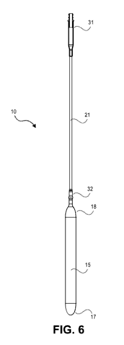
[0017] Figure 6 schematically shows an assembly including blood pump 10
coupled with
inflation tube 21;
2
CA 03034270 2019-02-15
WO 2018/039461 PCT/US2017/048429
[0018] Figure 7 is a cross-sectional view along the longitudinal axis of
the proximal end
31 of the inflation tube 21 depicted in Figure 6;
[0019] Figure 8 is a cross-sectional view along the longitudinal axis of
the distal end 32
of the inflation tube 21 depicted in Figure 6 as coupled to the proximal end
of the balloon 15
via balloon opening 19;
[0020] Figure 9 is an expanded cross-sectional view of detail E of Figure
8;
[0021] Figure 10 schematically shows an assembly including balloon 15
coupled with
inflation tube 21;
[0022] Figure 11 is a cross-sectional view along the longitudinal axis of
the assembly
depicted in Figure 10;
[0023] Figure 12 schematically shows the inflation tube 21 of Figure 10;
[0024] Figure 13 is a cross-sectional view of section A-A of the inflation
tube depicted in
Figure 12;
[0025] Figure 14 is a cross-sectional view of section B-B of the inflation
tube depicted in
Figure 12;
[0026] Figure 15 is an expanded view of detail C of Figure 12;
[0027] Figure 16 is an expanded view of detail D of Figure 12;
[0028] Figure 17 is a perspective view of the radiopaque marker 35 in one
embodiment of
the invention;
[0029] Figure 18 schematically illustrates the double hose barb 40 for
coupling the
inflation tube to a drive line in one embodiment of the invention;
[0030] Figure 19 is a side view of the double hose barb depicted in Figure
18;
[0031] Figure 20 is a cross-sectional view of section A-A of the double
hose barb
depicted in Figure 18;
[0032] Figure 21 is an expanded view of detail B of Figure 18;
[0033] Figure 22 is a right side view of the pump of Figure 2 in a deflated
state;
[0034] Figure 23 is a top view of the pump of Figure 2 in a deflated state;
[0035] Figure 24 schematically shows an introducer assembly 50 for use with
implanting
a blood pump of a CAD in a patient;
[0036] Figure 25 is a cross-sectional view of the introducer assembly
depicted in Figure
24;
[0037] Figure 26 is an expanded cross-sectional view of locking component
90 and
associated collet mechanism 75 of the introducer assembly 50 depicted in
Figure 24;
3
CA 03034270 2019-02-15
WO 2018/039461 PCT/US2017/048429
[0038] Figure 27 schematically shows the introducer assembly 50 coupled to
balloon
pump 180 during implantation of the pump;
[0039] Figure 28A schematically shows a CAD implanted in a patient using an
AID 150;
[0040] Figure 28B is a cross-sectional view of AID 150;
[0041] Figure 29A illustrates a skin interface device (SID) 400 comprising
an implantable
base 500 and a SID cap 600;
[0042] Figure 29B illustrates a supracutaneous portion 420 and a
subcutaneous portion
430 of the SID 400 when disposed within a patient;
[0043] Figure 30 shows an access port assembly 800 used to occlude an AID
graft 110
during implantation of a balloon pump;
[0044] Figure 31 shows an assembly of introducer assembly 50 in combination
with an
access port 800 and sheath 810 during implantation of a balloon pump;
[0045] Figure 32 is an expanded cross-sectional view of a distal portion of
a balloon
pump 180 in which a blunt distal end 85 of guidewire 80 is advanced to the
distal tip of the
balloon during delivery;
[0046] Figure 33 shows an assembly of introducer assembly 50 in combination
with a
vacuum source (syringe) during implantation of a balloon pump;
[0047] Figure 34 schematically shows a pump positioned in the proximal
descending
aorta, with the pump's inflation catheter entering the vasculature at the
right subclavian artery
through AID 150; and
[0048] Figure 35 schematically shows a cardiac assist device 300 including
an intra-aortic
pump 180, an internal drive line 170, an arterial interface device 150, a skin
interface device
400, an external drive line 310, and an external driver 320.
[0049] Figure 36 shows an access port assembly 1000 used to occlude an AID
graft
during implantation of a balloon pump;
[0050] Figure 37 is a cross-sectional view of the access port assembly of
Figure 36 along
the longitudinal axis;
[0051] Figure 38 is a cross-sectional view of section A-A of the access
port assembly of
Figure 36;
[0052] Figure 39 schematically shows an assembly having a balloon pump 180
fluidly
coupled to drive line 175 which has AID stopper 178 disposed on the drive
line;
[0053] Figure 40 is an expanded view of detail A of Figure 39;
[0054] Figure 41 is a perspective view of the AID stopper 178 of Figure 40;
[0055] Figure 42 is a cross-sectional view of section A-A of detail A of
Figure 40; and
4
CA 03034270 2019-02-15
WO 2018/039461 PCT/US2017/048429
[0056] Figure 43 is a cross-sectional view of the AID stopper 178 of Figure
41.
DETAILED DESCRIPTION OF THE INVENTION
[0057] United States Patent Application Serial Nos. 14/659,375 and
14/476,656, and
United States Patent Nos. 8,323,174 and 7,892,162 are incorporated herein in
their entireties.
The components, devices, modules, source code, and the like, associated with
the CAD and
components thereof as disclosed in United States Patent Application Serial
Nos. 14/659,375
and 14/476,656, and United States Patent Nos. 8,323,174 and 7,892,162 are also
disposed in
the CAD and components thereof as described herein. In addition, the functions
and methods
disclosed in United States Patent Application Serial Nos. 14/659,375 and
14/476,656, and
United States Patent Nos. 8,323,174 and 7,892,162, that utilize those
components, devices,
modules, source code, and the like, are also operative using the CAD described
herein.
[0058] This invention is described in preferred embodiments in the
following description
with reference to the Figures, in which like numbers represent the same or
similar elements.
Reference throughout this specification to "one embodiment," "an embodiment,"
or similar
language means that a particular feature, structure, or characteristic
described in connection
with the embodiment is included in at least one embodiment of the present
invention. Thus,
appearances of the phrases "in one embodiment," "in an embodiment," and
similar language
throughout this specification may, but do not necessarily, all refer to the
same embodiment.
[0059] The described features, structures, or characteristics of the
invention may be
combined in any suitable manner in one or more embodiments. In the following
description,
numerous specific details are recited to provide a thorough understanding of
embodiments of
the invention. One skilled in the relevant art will recognize, however, that
the invention may
be practiced without one or more of the specific details, or with other
methods, components,
materials, and so forth. In other instances, well-known structures, materials,
or operations are
not shown or described in detail to avoid obscuring aspects of the invention.
[0060] While the blood pump assembly of the present invention is generally
disclosed
with use of a CAD of the disclosure, it may be utilized with a variety of
devices and in a
variety of procedures which involve vascular implantation of such devices.
[0061] In a primary embodiment, the CAD of the disclosure, also referred to
herein as an
iVAS, operates on the principle of counterpulsation similar to an intra-aortic
balloon pump
(IABP). Components of the system are shown in Figure 1. During diastole, pump
inflation
augments the native heart's cardiac output by displacing blood in the aorta,
pushing it
downstream. At the start of systole (peak of the R-wave), the pump deflates,
decreasing
aortic pressure and reducing the work required of the left ventricle during
subsequent
CA 03034270 2019-02-15
WO 2018/039461 PCT/US2017/048429
ejection. Counterpulsation has been a standard treatment for cardiogenic shock
for decades,
providing circulatory support for hours to weeks.
[0062] In various embodiments, implantation of an iVAS requires implanting
four
components: an arterial interface device (AID), a blood pump, a skin interface
device (SID),
and internal drive line. To facilitate implantation of the blood pump, custom
tools and
methodology were developed, including an introducer assembly. Upon
implantation, the
blood pump undergoes repeated inflation/deflation cycles to assist in driving
blood through
the arteries. A key factor which is addressed by the present invention is
reducing blockage
within the artery when the blood pump is in a deflated state during systole.
This is
accomplished by an innovative blood pump (interchangeably referred to herein
as a balloon
pump) structure in which the pump is capable of maintaining a substantially
flat planar shape
when deflated thereby promoting and/or maintaining laminar flow within the
blood vessel.
[0063] Accordingly, in one aspect, the invention provides a blood pump
assembly. With
reference to Figures 2-5, the assembly 10 includes: a) a balloon 15 defining
an elongated
inflatable chamber 16, the balloon 15 having a distal end 17 and a proximal
end 18, wherein
the distal end 17 is rounded and the proximal end 18 has an opening 19; and b)
an inflation
tube 21 coupled to the opening 19 of the proximal end 18 of the balloon 15,
the tube 21
defining a fluid channel in fluid communication with the inflatable chamber
16. The balloon
15 has a central region 25 having an elongated cylindrical shape when in an
inflated state and
a substantially planate shape when in an uninflated state thereby promoting
laminar flow of
fluid within a blood vessel in which the pump is implanted.
[0064] Figure 2 illustrates the blood pump 15 when the balloon is in the
fully inflated
state. Notably, when in a deflated state, the balloon maintains a
substantially flat, planate
geometry as shown in Figures 22-23. Figure 22 is a side view of the blood pump
10 showing
the balloon 15 in a deflated state in which the balloon 15 has a substantially
flat, planate
structure along the longitudinal axis from the distal end 17 to the proximal
end 18 of the
balloon 15. The deflated balloon structure is also illustrated in Figure 23
which is a top view
of the balloon having a flattened, and widened profile.
[0065] Laminar flow is the normal condition for blood flow throughout most
of the
circulatory system. It is characterized by concentric layers of blood moving
in parallel down
the length of a blood vessel. The highest velocity (Vmax) is found in the
center of the vessel.
The lowest velocity (V=0) is found along the vessel wall. The flow profile is
parabolic once
laminar flow is fully developed. This occurs in long, straight blood vessels,
under steady
flow conditions.
6
CA 03034270 2019-02-15
WO 2018/039461 PCT/US2017/048429
[0066] The orderly movement of adjacent layers of blood flow through a
vessel helps to
reduce energy losses in the flowing blood by minimizing viscous interactions
between the
adjacent layers of blood and the wall of the blood vessel. Disruption of
laminar flow leads to
turbulence and increased energy losses. During turbulent flow, blood does not
flow linearly
and smoothly in adjacent layers, but instead the flow can be described as
being chaotic.
Turbulence increases the energy required to drive blood flow because
turbulence increases
the loss of energy in the form of friction, which generates heat. As such,
increased
turbulence requires a higher driving pressure for a given rate of flow which
creates unwanted
strain on the heart of a subject with chronic heart failure.
[0067] The balloon profile in the deflated state promotes uniform laminar
flow in the
vessel within which it is implanted. As such turbid flow is reduced thereby
decreasing the
propensity for clotting, creation of stagnant zones and excessive strain on
the heart.
[0068] The balloon portion 15 of the blood pump assembly 10 is coupled to
inflation tube
21. Figures 12-16 show several views of inflation tube 21 having proximal end
31 and distal
end 32. Distal end 32 of inflation tube 21 couples to opening 19 of balloon
15. The coupled
structure is shown in Figure 9. The distal end 32 of inflation tube 21 is
inserted into opening
19 of balloon 15 such that the interior surface of the balloon opening is in
contact with the
outer surface of inflation tube 21. Optionally, radiopaque ring 35 may be
disposed at the
coupling site. The surface of the inflation tube 21 and the balloon opening 19
may be solvent
bonded together. A coating layer 38 is disposed over the coupling site to
further affix balloon
15 to the inflation tube 21. Notably, coating layer 38 also provides a smooth
exterior profile
to the coupling site so that the exterior surface of the entire assembly has
no protrusions that
may increase turbid flow upon implantation.
[0069] Inflation tube 21 typically has a uniform diameter along its length.
In
embodiments, the outer diameter of the tube is no greater than 4, 5, 6 or 7
mm. Ideally, the
outer diameter is about 7, 6.5, 6, 5.5, 5, 4.5, 4.0 mm or less, such as 6.5,
6.4, 6.3, 6.2, 6.1, 6.0,
5.9, 5.8, 5.7, 5.6, 5.7, 5.6, 5.5, 5.4, 5.3, 5.2, 5.1, 5.0, 4.9, 4.8, 4.7,
4.6, 4.5, 4.4, 4.3, 4.2, 4.1,
4.0, 3.9, 3.8, 3.7, 3.6, 3.5, 3.2, 3.1, 3.0, 2.9, 2.8, 2.7, 2.6, 2.5 mm or
less. In embodiments, the
inner diameter of the tube is no greater than 2.5, 3, 3.5, 4, 4.5 or 5 mm.
Ideally, the inner
diameter is about 5, 4.5, 4.0, 3.5, 3.0 mm or less, such as 5.0, 4.9, 4.8,
4.7, 4.6, 4.5, 4.4, 4.3,
4.2, 4.1, 4.0, 3.9, 3.8, 3.7, 3.6, 3.5, 3.2, 3.1, 3.0, 2.9, 2.8, 2.7, 2.6, 2.5
mm or less. Ideally the
inner diameter is about 3.0 to 3.3 mm. In embodiments, the outer diameter is
sized such that
less than 80, 75, 70, 65, 60, 55, 50, 45% of the cross-sectional area of the
blood vessel that it
is implanted in is occluded. Ideally the outer diameter is sized such that
less than 55, 50 or
7
CA 03034270 2019-02-15
WO 2018/039461 PCT/US2017/048429
45% of the cross-sectional area of the blood vessel that it is implanted in is
occluded and the
inner diameter is greater than 3.0 mm and the outer diameter is less than 6.0
mm. Ideally the
outer diameter is sized such that less than 55, 50 or 45% of the cross-
sectional area of the
blood vessel that it is implanted in is occluded and the inner diameter is
greater than 3.5 mm
and the outer diameter is less than 6.0 mm. Ideally the outer diameter is
sized such that less
than 55, 50 or 45% of the cross-sectional area of the blood vessel that it is
implanted in is
occluded and the inner diameter is greater than 4.0 mm and the outer diameter
is less than 6.0
mm. In embodiments, the inner diameter is about 3.0 to 4.0 mm and the outer
diameter is
about 4.1 to 6.5 mm. In one embodiment, the inner diameter is about 3.2 mm and
the outer
diameter is about 4Ø In one embodiment, the inner diameter is about 3.1 mm
and the outer
diameter is about 4Ø In one embodiment, the inner diameter is about 3.0 mm
and the outer
diameter is about 4Ø In embodiments, the inner diameter is 3.0, 3.1, 3.2,
3.3, 3.4, 3.5, 3.6,
3.7, 3.8 or 3.9 mm and the outer diameter is 4.0, 4.1, 4.2, 4.3, 4.4, 4.5,
4.6, 4.7, 4.8, 4.9 or 5.0
mm. As will be appreciated by one in the art, the inner diameter must be sized
to
accommodate the guidewire including any feature which is present at the blunt
distal end of
the guidewire. As such the blunt distal end will be sized such that the outer
diameter of the
distal end is less than the inner diameter of the inflation tube.
[0070] The wall thickness of inflation tube 21 is typically less than 1 mm,
such as 0.9, 0.8,
0.7, 0.6, 0.5 mm or less. To prevent kinking, inflation tube 21 may include a
stiffening
material, such as a mesh component to add wall stiffness. In one embodiment,
the mesh
component is a wire mesh, optionally composed of medical grade steel or alloy
such as
Nitinolg. In such embodiments, the balloon does not require an additional
radiopaque
marker. Alternative stiffening elements and configurations are known in the
art and may be
incorporated into the inflation tube wall. For example, polymer fibers,
textiles and the like
may be utilized. Additionally, the stiffening elements may be incorporated
into the inflation
tube wall in a variety of geometries, for example, as a mesh, braided or woven
textile, helical
spiral and the like.
[0071] Proximal end 31 of inflation tube 21 is coupled to a pneumatic line,
such as an
internal drive line which is in fluid communication with a fluid driver having
a contractible
bellows for inflating and deflating the balloon. As shown in Figures 18-21, a
double hose
barb 40 connects the inflation tube 21 to the pneumatic drive line.
[0072] The balloon 15 may be composed of any biocompatible material that
provides a
smooth exterior profile and is capable of undergoing repeated
inflation/deflation cycles. In
embodiments, a preferred material includes block copolymers, such as segmented
polyether
8
CA 03034270 2019-02-15
WO 2018/039461 PCT/US2017/048429
polyurethane. In one embodiment, the balloon is composed essentially of
BioSpan (ID sold by
DSM Biomedical Inc.
[0073] The dimensioning of the balloon along with the balloon material are
critical in
maintaining proper functioning of the device when implanted along with
maintaining proper
flow parameters as discussed herein. In embodiments, the balloon has a uniform
wall
thickness along its length which is between about 0.2 to 0.4 mm. In one
embodiment, the
balloon wall thickness is about 0.3 mm. Further, the length of the balloon is
between about
195 to 210 mm, for example, about 200 to 205 mm. In embodiments, the balloon
is
dimensioned such that it has a volume of between about 40 to 60 cc when
inflated. In
embodiments, the balloon has an overall deflated thickness of less than about
1.0, 0.9, 0.8,
0.7, 0.6, 0.5 or 0.4 mm. Ideally, the balloon has an overall deflated
thickness of between
about 0.2-0.8 mm, 0.2-0.4 mm, 0.3-0.6 mm, 0.4-0.6 mm or 0.4-0.8 mm so as to
promote
laminar flow within the vasculature upon deflation of the balloon.
[0074] The balloon must be capable of undergoing a high number of
repetitive
inflation/deflation cycles without failure upon implantation. Ideally, the
balloon has a
lifespan of inflation/deflation cycles of greater than about 25, 50, 75, or
100 million cycles.
As such the device may remain implanted for the duration of a patient's life
upon
implantation, for example, 1, 2, 3, 4, 5 years or more.
[0075] In one embodiment, the blood pump assembly is implanted using an
introducer
assembly as shown in Figures 24-26. With reference to Figures 24-26, the
assembly 50
includes: a) a shaft 55 elongated along a longitudinal axis, the shaft having
a distal end 60, a
proximal end 65, a lumen 70 extending along the longitudinal axis from the
distal end 60 to
the proximal end 65, and a collet mechanism 75 disposed at the proximal end 65
for receiving
a guidewire 80; and b) a locking component 90 having a distal end and a
proximal end, the
locking component adapted such that the distal end of the locking component
reversibly
couples to the proximal end of the shaft. The locking component has a locked
configuration
and an unlocked configuration such that when in the locked configuration, a
gripping force is
created between the collet mechanism 75 and a guidewire 80 inserted within
lumen 70.
[0076] Notably, the proximal end 65 of the shaft is adapted to form a fluid
tight seal with
the locking component 90. This can be accomplished by inclusion of o-ring 95.
The fluid
tight seal prevents blood loss during introduction of the balloon pump 180
into the
vasculature. The o-ring 95 also creates an air tight seal between the
introducer and the pump
180 allowing the pump to be deflated during insertion into the vasculature.
9
CA 03034270 2019-02-15
WO 2018/039461 PCT/US2017/048429
[0077] Figures 28A and 28B illustrate an AID 150 of the iVAS. Referring to
Figure 28A,
a vascular interface 100 is formed using a vascular AID graft 110 attached to
an artery 120
with a suture ring 130 at the position of an incision in the artery. The
particular graft shown
flares at its distal end 140. AID 150 sits inside the AID graft 110, filling
the interior of the
AID graft 110.
[0078] Sewing the suture ring 130 to the subclavian artery is the first
task the surgeon
performs when implanting the system. Next, AID graft 110 is sutured to the
suture ring 130.
[0079] With reference to Figures 28A and 28B, AID 150 comprises a body 155.
In certain
embodiments, body 155 comprises a polyurethane. In certain embodiments, body
155
comprises a polysiloxane. In the illustrated embodiment of Figures 28A and
28B, body 155
is formed to include two lumens extending therethrough. Lumen 160 is utilized
to pass
pneumatic drive line 170 through AID 150.
[0080] The second lumen 165 optionally houses a pressure sensor 190 to
measure arterial
pressure, and sensor leads 192, 194, 196, and 198, to interconnect sensor 190
to SID 400
(Figure 29A and 29B). Sensor leads 192, 194, 196, and 198, are used to provide
power to
sensor 190, provide a ground connection, to provide clock signals to sensor
190, and to
communication arterial pressure signals from sensor 190 to SID 400.
[0081] Lumen 160 which extends through the length of the AID 150 is filled
by the
pneumatic drive line 170. Pneumatic drive line 170 in turn is connected at its
distal end to a
pump 180. In certain embodiments, inflation catheter is formed to have an
inner diameter in
the range 3 to 6 mm (often about 5 mm), although other diameters are possible
as well.
[0082] Not shown in Figure 28A is the proximal end of the pneumatic drive
line 170.
Because the pump 180 needs to inflate and deflate in coordination with the
cardiac cycle in
order to function as a ventricular assist device, the pump must be in fluid
communication
with a driver (for example, an air compressor or pump) via the pneumatic drive
line 170.
[0083] In embodiments wherein such a driver is external to the body as
shown in Figure 1,
the SID 400 (Figures 29A and 29B) allows the design of the system to be
composed of parts
both implanted and external to the patient's body. The pneumatic drive line
170 is attached to
SID 400, and SID 400 is attached to the fluid driver. In certain embodiments,
the driver, the
pneumatic drive line 170 and the pump 180 form a closed air system, wherein
that closed
system includes a well-defined and precisely controlled volume of air. Such a
well-defined
and precisely-controlled volume of air facilitates leak detection.
[0084] In certain embodiments, air volume and movement of air is precisely
controlled
using, for example and without limitation, a bellows driven by one or more
linear actuators.
CA 03034270 2019-02-15
WO 2018/039461 PCT/US2017/048429
In descriptions of the skin interface device herein, the pneumatic drive line
170 is
alternatively referred to as an internal drive line.
[0085] In implantation of the balloon pump 180, once the anastomosis of the
suture ring
130 and AID graft 110 is complete as discussed above, an access port
containing an iris valve
(Figure 30) is inserted into the graft's proximal end creating hemostasis. The
surgeon then
attaches a sheath (Figure 31) to the proximal end of the port. Inside the
sheath is the blood
pump 180 in its deflated state. The other end of the sheath is tied off to the
shaft of the
introducer assembly as illustrated in Figure 31. The function of the access
port is to
minimize blood loss during pump insertion. The sheath is used to collect any
blood that
escapes through the access port.
[0086] With reference to Figures 30 and 31, in implantation of the blood
pump 180, once
the anastomosis of the suture ring 130 and AID graft 110 is complete as
discussed above, an
access port assembly 800 optionally containing an iris valve (Figure 30) is
inserted into AID
graft 110 at its proximal end creating hemostasis. The surgeon then optionally
attaches a
sheath 810 (Figure 31) to the proximal end of the access port assembly 800.
Inside the sheath
810 is the blood pump 180 in its deflated state. The other end of the sheath
810 is tied off to
the shaft of the introducer assembly 50 as illustrated in Figure 31. The
sheath 810 is attached
to the access port assembly 800 and shaft of the introducer assembly 50 via
sutures 820. The
function of the access port is to minimize blood loss during pump insertion.
The sheath is
used to collect any blood that escapes through the access port. The blood pump
180 is then
implanted in the patient's vasculature, i.e., the descending thoracic aorta.
To implant the
pump, the surgeon inserts and guides it down the patient's subclavian artery,
traverses the
subclavian aorta bifurcation, and then travels down the aorta to the final
location. The pump
does not have the mechanical rigidity to permit implantation without the
introducer 50.
[0087] In embodiments, the sheath is not required in implantation. In such
embodiments,
in implantation of the blood pump 180, once the anastomosis of the suture ring
130 and AID
graft 110 is complete as discussed above, an access port assembly 800
containing an iris
valve (Figure 30) is inserted into AID graft 110 at its proximal end creating
hemostasis. The
sheath is not required since AID graft 110 may be reversibly clamped to
prevent blood loss.
The blood pump 180 is then implanted in the patient's vasculature, i.e., the
descending
thoracic aorta. To implant the pump, the surgeon inserts and guides it down
the patient's
subclavian artery, traverses the subclavian aorta bifurcation, and then
travels down the aorta
to the final location. The pump does not have the mechanical rigidity to
permit implantation
without the introducer 50.
11
CA 03034270 2019-02-15
WO 2018/039461 PCT/US2017/048429
[0088] In one embodiment, during installation of the balloon pump 180,
guidewire 80 is
inserted into the balloon pump so the wire's blunt distal end 85 contacts the
distal inside tip
of the pump (Figure 32). Thus the guidewire 80 is within the central lumen of
the balloon
pump 180 during insertion as opposed to being in an auxiliary lumen or on the
outside
surface of the balloon. This allows the balloon to have a single lumen, the
balloon being of
uniform thickness along its length. The distal end of the introducer shaft is
then mechanically
attached to the proximal end of the pump as shown in Figure 27. Collet
mechanism 75 and
associated locking component 90 are used to lock the guidewire 80 into place.
A vacuum
device (i.e., a syringe as in Figure 33) is then used to pull a vacuum on the
pump (not shown)
minimizing its size. Once the pump is placed, the vacuum is released, the
guidewire 80 is
extracted and the shaft is removed.
[0089] In embodiments, the access port assembly 800 may be removed during
implantation of the blood pump 180. As such, the inner diameter of the port
may be sized
large enough such that it can accommodate the AID 150 and the introducer
assembly 50. For
example, once the blood pump 180 is placed within the artery, the access port
assembly 800
may be detached and slid away from the patient over the introducer assembly 50
and
guidewire 80. In embodiments, the inner diameter of the access port is greater
than about 3,
4, 5, 6, 7, 8, 9, 10, 11, 12, 13, 14 or 15 mm. In one embodiment, the inner
diameter of the
access port is equal to or greater than about 7 or 8 mm.
[0090] To facilitate placement and detection of the balloon pump 180 during
installation,
the guidewire 80, or portion thereof, may include a radiopaque material. For
example, blunt
end 85 may be composed of or otherwise include a radiopaque material.
Alternatively, the
balloon pump 180, or portion thereof, may include a radiopaque material. In
one
embodiment, the balloon includes a ring of radiopaque material adjacent and
proximal to the
inflation region of the balloon. For example, the balloon pump 180 may include
a ring
composed of Pt-Ir alloy. In another embodiment, both the guidewire 80, or
portion thereof,
and the balloon pump 180, or portion thereof include a radiopaque material.
[0091] Figure 34 shows (schematically) the AID graft 110 in position on the
right
subclavian artery. This position is advantageous because it allows easy
surgical access and a
relatively short distance to the descending aorta. Figure 34 also shows the
graft secured to
AID 150 by a suture 210. Other suitable positions for the interface include
either common
carotid artery, the brachiocephalic artery, the left subclavian artery, the
descending aorta, and
the abdominal aorta. Downstream branches of the aorta may also be used, such
as the
external iliac and femoral arteries.
12
CA 03034270 2019-02-15
WO 2018/039461 PCT/US2017/048429
[0092] In embodiments, implantation of the balloon pump 180 may be achieved
without
the assistance of an introducer assembly. For example, the balloon pump 180
may be
positioned within the vasculature by pulling the pump into and through the
vasculature. Once
blunt distal end 85 of the guidewire 80 is advanced to the distal inside tip
of the pump, a
snare device is used to grasp the blunt distal end 85 and pull the balloon
pump 180 into
position within the vasculature. The procedure is described as follows.
[0093] As discussed above, sewing the suture ring 130 to the subclavian
artery is
performed and AID graft 110 is sutured to the suture ring 130. Once the
anastomosis of the
suture ring 130 and AID graft 110 is complete, an access port is inserted into
the graft's
proximal end and the port is occluded. Figures 36-38 illustrate an access port
in one
embodiment invention. As shown in Figures 36-38, access port 800 is shown
coupled to
syringe 900 via tubing 950.
[0094] The surgeon next advances the guidewire 80 into the vasculature to
visualize future
pump placement in the aorta and determine the appropriate length of pump to
implant (for
example, pump having overall length including integral drive line of 12 inches
or 16 inches).
The guidewire 80 is then removed from the vasculature.
[0095] The snare device is then introduced into the femoral artery and
advanced along the
vasculature until the distal tip of the snare device exits the vasculature via
the access port.
The snare device generally includes an elongated flexible shaft having a
distal tip configured
to reversibly grasp or couple with the blunt distal end 85 of the guidewire
80. Further, the
elongated flexible shaft of the snare device is of sufficient length such that
the proximal end
of the shaft remains outside of the vasculature at the femoral artery
insertion point when the
distal tip of the shaft is advanced through the access port 800 to exit the
vasculature. To
facilitate advancement of the snare device to the access port at the
subclavian artery, the snare
device may be coupled to a wire, for example a J-wire, which is placed in the
vasculature
above stream of the snare device and used to pull the snare device to the
access port. In
embodiments, guidewire 80 is used to pull the snare device to the access port.
[0096] The blunt distal end 85 of the guidewire 80 and the distal tip of
the snare device
may be configured in any number of geometries that allow for reversible
attachment to one
another without damaging the tip of the balloon. In one embodiment, the blunt
distal end 85
has a smooth rounded geometry, such as a sphere or ellipsoid to prevent the
guidewire from
piercing the distal end of the balloon while also providing a structure for
the grasping
structure of the distal tip of the snare device to couple with. One in the art
would understand
that the grasping structure of the snare device may be configured in a variety
of ways to
13
CA 03034270 2019-02-15
WO 2018/039461 PCT/US2017/048429
facilitate coupling with the blunt distal end 85 of the guidewire while
avoiding damage to the
balloon. For example, the grasping portion may be configured as a wire snare,
grasping jaws,
slotted member for receiving the blunt distal end, and the like. In some
embodiments, the
distal end of the guidewire may include a groove, notch or recess to engage
the grasping
mechanism. In some embodiments, the distal end of the guidewire may include a
bump or
protrusion to engage the grasping mechanism.
[0097] Next, the surgeon inserts the guidewire 80 into the balloon pump 180
and advances
blunt distal end 85 to the distal tip of the balloon pump while the balloon
pump 180 remains
outside of the patient. The distal end of the snare device is coupled with the
blunt distal end
85, for example via a wire loop, and the balloon pump 180 is introduced into
the vasculature
through the access port 800 (i.e., access port 800 as in Figures 36-37) by
progressively
withdrawing the shaft of the snare device from its femoral artery point of
entry thereby
pulling the balloon along the vasculature into position within the descending
aorta. The
access port 800 is optionally simultaneously actuated to permit blood pump
insertion while
minimizing blood loss. Before the balloon pump 180 is introduced into the
vasculature, a
vacuum device is optionally used to pull a vacuum on the pump minimizing its
size. This can
be accomplished via the use of a Tuohy Borst valve coupled at the proximal end
179 of the
drive line 175 as shown in Figure 40 along with a vacuum device coupled to the
valve. Upon
placement of the balloon pump as discussed below, the vacuum is released and
the guidewire
80 is extracted. In alternative embodiments, Figure 36 illustrates an access
port 800 coupled
to syringe 900 via tubing 950 which may be used to pull a vacuum on balloon
pump 180.
[0098] To visualize insertion and correct placement of the balloon pump 180
within the
vasculature, fluoroscopy, or any other suitable imaging method known in the
art is used. In
one embodiment, the blunt distal end 85 of the guidewire 80 along with
radiopaque marker
35 located distally on the balloon pump 180 are used as visual markers to
ensure correct
placement.
[0099] Once the balloon pump 180 is at the desired position, a stopper
portion of the AID
is inserted into the graft portion of the AID. Figure 39 shows an assembly
including balloon
pump 180 connected to drive line 175 which has AID stopper 178. The drive line
175
extends through the central lumen of the AID stopper 178. Figures 41 and 43
illustrate an
AID stopper 178 in an embodiment of the invention.
[0100] Sutures are then tied around the AID graft 110, AID stopper 178 and
drive line 175
to secure the balloon pump's location within the vasculature. The surgeon then
uncouples the
distal end of the snare device from the blunt distal end 85 of the guidewire
80 at the distal end
14
CA 03034270 2019-02-15
WO 2018/039461 PCT/US2017/048429
of the pump. The snare is then withdrawn from the vasculature and the
guidewire 80 is also
withdrawn from the balloon pump 180.
[0101] To ensure that the lumen of the drive line 175 is not overly
compressed by suture
tension, a gauge device may be used to measure or monitor the inner diameter
of the drive
line 175 in the region of the drive line 175 that traverses through the lumen
of the AID
stopper 178. In one embodiment, the gauge device is a malleable rod having a
predetermined
outer diameter which is advanced into the lumen of the drive line to monitor
the inner
diameter of the drive line. The sutures may be adjusted if the surgeon
determines that the
drive line is compressed. This ensures that gaseous fluid flow into the
balloon is not
restricted which would inhibit optimal performance of the system.
[0102] In one embodiment, positioning of the pump within the vasculature is
secured
without the use of sutures. In this embodiment, a clamp is utilized which is
placed over the
AID graft 110, AID stopper 178 and drive line 175. The clamp is presized to
engage the AID
graft 110 and AID stopper 178 without overly compressing the drive line 175.
In
embodiments, the clamp may be an elongated clamp, optionally hinged, which is
configured
to encircle the AID graft 110, AID stopper 178 and drive line 175.
[0103] Referring now to Figure 35, in embodiments, a CAD or iVAS comprises a
pump
180, a pneumatic drive line 170, an AID 150, a SID 400, an external drive line
310, and an
external driver 320.
[0104] At its proximal end, the pump 180 is connected to the distal end of
the pneumatic
drive line 170. An AID 150 is sized and shaped to pass the pneumatic drive
line 170 through
an arterial wall.
[0105] SID 400 connects the proximal end of the pneumatic drive line 170 to
the distal
end of the external drive line 310. The proximal end of the external drive
line 310 is
connected to the driver 320.
[0106] The pump 180, the internal drive line 170, the SID 400, the external
drive line 170,
and the driver 320 can be charged with a pumping medium. In certain
embodiments, the
pumping medium comprises a fluid. A preferred pumping medium is air. In
certain
embodiments, pump 180, the pneumatic drive line 170, the SID 400, the external
drive line
310, and the driver 320 define a closed fluid system. In certain embodiments,
pump 180, the
pneumatic drive line 170, the SID 400, the external drive line 310, and the
driver 320
comprise an open system, wherein the bolus of air inside the system can be
exchanged with
the ambient environment.
CA 03034270 2019-02-15
WO 2018/039461 PCT/US2017/048429
[0107] As
those skilled in the art will appreciate, pump 180 may have various sizes
depending on the anatomy of the patient. In certain embodiments, pump 180 will
typically
have an inflated volume of about 40 to 60 cubic centimeters when inflated to
10 to 20 mmHg
above the maximum systolic pressure.
[0108]
Internal drive line 170 typically has a uniform diameter along its length. In
embodiments, the outer diameter of the drive line is no greater than 4, 5, 6
or 7 mm. Ideally,
the outer diameter is about 7, 6.5, 6, 5.5, 5, 4.5, 4.0 mm or less, such as
6.5, 6.4, 6.3, 6.2, 6.1,
6.0, 5.9, 5.8, 5.7, 5.6, 5.7, 5.6, 5.5, 5.4, 5.3, 5.2, 5.1, 5.0, 4.9, 4.8,
4.7, 4.6, 4.5, 4.4, 4.3, 4.2,
4.1, 4.0, 3.9, 3.8, 3.7, 3.6, 3.5, 3.2, 3.1, 3.0, 2.9, 2.8, 2.7, 2.6, 2.5 mm
or less. In
embodiments, the inner diameter of the tube is no greater than 2.5, 3, 3.5, 4,
4.5 or 5 mm.
Ideally, the inner diameter is about 5, 4.5, 4.0, 3.5, 3.0 mm or less, such as
5.0, 4.9, 4.8, 4.7,
4.6, 4.5, 4.4, 4.3, 4.2, 4.1, 4.0, 3.9, 3.8, 3.7, 3.6, 3.5, 3.2, 3.1, 3.0,
2.9, 2.8, 2.7, 2.6, 2.5 mm or
less. Ideally the inner diameter is about 3.0 to 3.3 mm. In embodiments, the
outer diameter
is sized such that less than 80, 75, 70, 65, 60, 55, 50, 45% of the cross-
sectional area of the
blood vessel that it is implanted in is occluded. Ideally the outer diameter
is sized such that
less than 55, 50 or 45% of the cross-sectional area of the blood vessel that
it is implanted in is
occluded and the inner diameter is greater than 3.0 mm and the outer diameter
is less than 6.0
mm. Ideally the outer diameter is sized such that less than 55, 50 or 45% of
the cross-
sectional area of the blood vessel that it is implanted in is occluded and the
inner diameter is
greater than 3.5 mm and the outer diameter is less than 6.0 mm. Ideally the
outer diameter is
sized such that less than 55, 50 or 45% of the cross-sectional area of the
blood vessel that it is
implanted in is occluded and the inner diameter is greater than 4.0 mm and the
outer diameter
is less than 6.0 mm. In embodiments, the inner diameter is about 3.0 to 4.0 mm
and the outer
diameter is about 4.1 to 6.5 mm. In one embodiment, the inner diameter is
about 3.2 mm and
the outer diameter is about 4Ø In one embodiment, the inner diameter is
about 3.1 mm and
the outer diameter is about 4Ø In one embodiment, the inner diameter is
about 3.0 mm and
the outer diameter is about 4Ø In embodiments, the inner diameter is 3.0,
3.1, 3.2, 3.3, 3.4,
3.5, 3.6, 3.7, 3.8 or 3.9 mm and the outer diameter is 4.0, 4.1, 4.2, 4.3,
4.4, 4.5, 4.6, 4.7, 4.8,
4.9 or 5.0 mm. As will be appreciated by one in the art, the inner diameter
must be sized to
accommodate the guidewire including any feature which is present at the blunt
distal end of
the guidewire. As such the blunt distal end will be sized such that the outer
diameter of the
distal end is less than the inner diameter of the inflation tube.
[0109] The
wall thickness of internal drive line 170 is typically less than 1 mm, such as
0.9, 0.8, 0.7, 0.6, 0.5 mm or less. To prevent kinking, the drive line may
include a stiffening
16
CA 03034270 2019-02-15
WO 2018/039461 PCT/US2017/048429
material, such as a mesh component to add wall stiffness. In one embodiment,
the mesh
component is a wire mesh, optionally composed of medical grade steel or alloy
such as
Nitinolg. In such embodiments, the balloon does not require an additional
radiopaque
marker. Alternative stiffening elements and configurations are known in the
art and may be
incorporated into the drive line wall. For example, polymer fibers, textiles
and the like may
be utilized. Additionally, the stiffening elements may be incorporated into
the drive line wall
in a variety of geometries, for example, as a mesh, braided or woven textile,
helical spiral and
the like.
[0110] In certain embodiments, sensors are connected to one or more
communication
interfaces that, like the pneumatic drive line 170, pass through the AID 150
and AID graft
110 and connect to SID 400. In certain embodiments, these one or more
communication
interfaces provide data to a controller.
[0111] In certain embodiments, one or more sensors transmit data, by wire
or wirelessly,
to Applicants' SID 400. Examples of sensors include, without limitation,
electrical leads to
measure an electrocardiogram, sensors to detect body temperature, sensors to
detect blood
analytes (such as blood gases), sensors to detect intra-arterial pressure
directly or indirectly,
and/or sensors to measure humidity within pump 180. Indirect sensors include,
for example
and without limitation, a microphone to monitor heart sounds.
[0112] In certain embodiments, a controller 530 is disposed in SID 400. In
certain
embodiments, a controller 530 is integral with external driver 320.
[0113] In certain embodiments, signals from one or more sensors are used by
controller
530 to monitor the cardiac cycle and, thereby, the counterpulsation cycle. In
certain
embodiments, combinations of signals from one or more sensors are used by
controller 530 to
monitor the cardiac cycle.
[0114] In certain embodiments, sensors are used to determine the state of
the air inside the
system. In certain embodiments, air pressure is measured to determine whether
the pump is
properly inflating, or if there is a leak in the system. In certain
embodiments, data from the
air pressure sensor is communicated to controller 530.
[0115] In certain embodiments, sensors for arterial blood pressure at the
pump 180 and/or
at the AID 150 are in communication with controller 530. In certain
embodiments, these
sensors communicate a detected arterial blood pressure to the controller 530,
either by wire or
wirelessly.
[0116] Referring now to Figure 29A, SID 400 comprises a SID base 500 and a
SID cap
600. SID base 500 and SID cap 600 are coupled so as to create an air-tight
conduit between
17
CA 03034270 2019-02-15
WO 2018/039461 PCT/US2017/048429
the pneumatic drive line 170 and external air line 310. In this way, pneumatic
drive line 170,
SID 400, and external air line 310, can be part of a closed fluid system. In
certain
embodiments, an air-tight seal is formed using gaskets and other sealing
systems.
[0117] Referring now to Figures 29A and 29B, when implanted skin interface
device 400
includes a SID base 500, comprising a subcutaneous portion 430 internal to the
patient, in
combination a supracutaneous portion 420. SID cap 600 is attached to the
supracutaneous
portion 420 of SID base 500. Those skilled in the art will appreciate that it
is possible to
implant SID 400 in a variety of different locations on the patient, for
example abdominally or
thoracically.
[0118] Referring now to Figures 29A, SID 400 wirelessly provides electrical
energy from
SID cap 600 to SID base 500, and also wirelessly and bi-directionally passes
electrical
signals, i.e. data, between SID cap 600 and SID base 500. In order to optimize
the
transmission of power from SID cap 600 to SID base 500, and at the same time
optimize the
transmission of data between SID cap 600 and SID base 500, Applicants have
"decoupled"
the transmission of power from the transmission of data. The transmission of
power from
SID cap 600 to SID base 500 is done by induction.
[0119] Although the invention has been described with reference to the
above examples, it
will be understood that modifications and variations are encompassed within
the spirit and
scope of the invention. Accordingly, the invention is limited only by the
following claims.
18