Note: Descriptions are shown in the official language in which they were submitted.
CA 03034583 2019-02-21
WO 2018/073665
PCT/IB2017/055406
1
SCANNING OPTICAL PROBE WITH FLEXING MECHANISM
FIELD
[0001] The present disclosure relates generally surgical probes and, more
particularly, to a scanning optical probe with flexing mechanism.
BACKGROUND
[0002] VVidespread interest exists in obtaining high resolution imaging
scans of
anatomical targets. Desirable anatomical targets include, for example, the
interior
surfaces of the eye and vasculature.
[0003] One imaging technology capable of obtaining high resolution scans is
optical coherence tomography (OCT). OCT technology obtains good depth
resolution and need not contact the target surface to be interrogated.
[0004] OCT imaging technology has been incorporated into handheld optical
probes. Such optical probes have been utilized to generate different types of
scans
of the target surface. An OCT image corresponding to a single point on the
surface
of the target is called an A-scan. An OCT image corresponding to traversing
the
imaging beam across a set of target points is typically referred to as a B-
scan.
[0005] Despite the above mentioned OCT-based technologies, handheld
scanning probes face a number of challenges. The challenges arise due to the
nature of OCT image scanning which requires movement of the optical fiber with
respect to the lens (or movement of the fiber/lens assembly together) to
achieve the
scan. In the microsurgical environment (e.g., ophthalmic applications)
actuating the
fiber or fiber/lens assembly is prohibited due to the lack of working space in
the
probe housing. The lack of working space arises due the relatively long
length, small
diameter, and rigidity of the probe housing. Consequently, motion of the fiber
assembly is extremely restricted.
[0006] Accordingly, there is a need for scanning optical probes and methods
that
overcome the above mentioned challenges.
SUMMARY
[0007] A scanning optical probe includes a cannula, optical fiber, lens,
and an
actuating mechanism to deflect the distal end of the fiber. In embodiments,
the
CA 03034583 2019-02-21
WO 2018/073665
PCT/IB2017/055406
2
actuating mechanism includes an actuator and an elongate support member
coupled
to the actuator and extending through the cannula to the distal end of the
probe.
When the elongate support member is activated, the distal end of the fiber is
deflected back and forth, causing the light beam from the optical fiber to
move
across the target surface as desired.
[0008] In embodiments, the elongate support member includes a discrete
predefined flexing region. The flexing region includes a plurality of slots in
the side
wall of the flexible member. Pull rods extend proximally from the flexing
region to a
probe handle. When the rods are reciprocally actuated, the end of the scanning
optical probe is tilted back and forth.
[0009] In another embodiment, the actuating mechanism includes at least one
guide which urges or biases the optical fiber to aim in a first direction.
When the
guides are moved, the optical fiber is repositioned to aim in a second
direction.
[0010] An actuator in the probe handle can be linked to the guide.
Reciprocating
motion from the actuator is transmitted through the various components,
causing the
optical fiber at the distal end of the probe to tilt back and forth. The beam
emitted
from the fiber is processed to generate a scan of a target area.
BRIEF DESCRIPTION OF THE DRAWINGS
[0011] For a more complete understanding of the present disclosure and the
advantages thereof, reference is now made to the following description taken
in
conjunction with the accompanying drawings in which like reference numerals
indicate like features and wherein:
[0012] FIG. 1A is a perspective view of a scanning optical probe in
accordance
with an embodiment of the present invention;
[0013] FIG. 1B is a block diagram of an OCT imaging system including a
scanning optical probe such as the probe shown in FIG. 1A;
[0014] FIG. 2 is an illustration of the scanning optical probe depicted in
FIG. 1A in
an ophthalmic application;
[0015] FIG. 3 is a cross sectional view of a distal section of a scanning
optical
probe in accordance with an embodiment of the present invention;
CA 03034583 2019-02-21
WO 2018/073665
PCT/IB2017/055406
3
[0016] FIG. 4 is a perspective view of a distal section of a scanning
optical probe
depicted in a flexed orientation;
[0017] FIG. 5 is a perspective view of a distal section of a component of
the
scanning optical probe shown in FIG. 3;
[0018] FIGS. 6-8 are side views of a distal section of a scanning optical
probe
shown moving in sequence from a first position to a second position;
[0019] FIG. 9 is a perspective view of a component of the scanning optical
probe
shown in FIGS. 6-8;
[0020] FIG. 10 is a partial perspective view of the proximal section of the
scanning optical probe shown in FIGS. 6-8;
[0021] FIGS. 11-13 are cross sectional views of a distal section of another
scanning optical probe wherein the optical fiber is shown moving in sequence
from a
first position to the second position;
[0022] FIGS. 14-15 are cross sectional views of a distal section of another
scanning optical probe including various internal optical guide members;
[0023] FIG. 16 is a cross sectional view of a distal section of another
scanning
optical probe including a control member;
[0024] FIGS. 17-18 are side views of a distal section of another scanning
optical
probe including a pre-shaped sheath; and
[0025] FIG. 19 is a flow chart depicting a method for scanning a target
area in
accordance with the present invention.
DETAILED DESCRIPTION
[0026] The following description is presented to enable one of ordinary
skill in the
art to make and use the invention and is provided in the context of a patent
application and its requirements. Various modifications to the exemplary
embodiments and the generic principles and features described herein will be
readily
apparent. The exemplary embodiments are mainly described in terms of
particular
methods and systems provided in particular implementations. However, the
methods
and systems will operate effectively in other implementations. Phrases such as
"exemplary embodiment", "one embodiment" and "another embodiment" may refer to
the same or different embodiments as well as to multiple embodiments. The
CA 03034583 2019-02-21
WO 2018/073665
PCT/IB2017/055406
4
embodiments will be described with respect to systems and/or devices having
certain components. However, the systems and/or devices may include more or
less
components than those shown, and variations in the arrangement and type of the
components may be made without departing from the scope of the invention. The
exemplary embodiments will also be described in the context of particular
methods
having certain steps. However, the method and system operate effectively for
other
methods having different and/or additional steps and steps in different orders
that
are not inconsistent with the exemplary embodiments. Thus, the present
invention is
not intended to be limited to the embodiments shown, but is to be accorded the
widest scope consistent with the principles and features described herein. All
patents and publications recited herein are incorporated by reference in their
entirety.
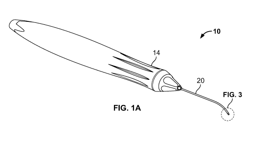
[0027] FIG. 1A depicts a scanning optical probe 10 including a handle 14
and a
cannula 20 extending distally from the handle.
[0028] The cannula 20 or equivalent type of housing structure is shown
having a
tubular shape and includes one bend. However, the cannula can have additional
bends or be straight. Indeed, the shape and size of the cannula or housing may
vary. For example, the inner diameter (ID) may range from 0.25 to 0.6 mm, and
in
embodiments is about 0.4 mm. The outer diameter (OD) may range from 0.4 to 1
mm and in embodiments is about 0.7 mm. The length of the cannula may range
from 20 to 35 mm, and in embodiments, is about 30 mm. In embodiments, the
cannula is a 23 Ga- or smaller cannula.
[0029] The scanning optical probe can be detachably coupled to an OCT
imaging
system to produce OCT scans as described herein. VVith reference to FIG. 1B,
an
exemplary OCT imaging system 150 is shown including an optical source 160, OCT
module 170, a scanning optical probe 180 such as the scanning probe shown in
FIG.
1A, and a controller, computer and display 190. The scanning optical probe 180
may be detachably coupled with the OCT system via one or more interlocking
connectors, cables, and in embodiments, a flexible umbilical cord (not shown).
Additionally, in embodiments, the probe 180 includes an actuator 182 operable
to
move components of the probe as described herein.
CA 03034583 2019-02-21
WO 2018/073665
PCT/IB2017/055406
[0030] Embodiments of the imaging system 150 split a light generated by the
light
source 160 into an imaging beam and a reference beam. The imaging beam can be
guided to a target region.
[0031] The scanning probe 180 collects the imaging light that is reflected
off the
target region. An OCT module 170 then detects the interference between the
reference beam and the returned imaging beam. The OCT module can then create
a depth image of the target region based on the detected interference. This
depth,
or OCT image provides the image of the target region in a range of depth for
every
point the imaging beam is directed. An OCT image corresponding to a single
point
on the surface of the target region, such as the surface of the retina 112, is
called an
A-scan. In imaging systems that scan the imaging beam through a set of target
points, the OCT image is typically referred to as a B-scan.
[0032] In preferred embodiments described herein, the imaging beam is moved
through a set of target points to generate a scan across the target tissue.
The
scanning operations can be performed under the control of a controller and
computer
190 and the results, including the OCT image, can be delivered to a user
through,
e.g., a display 190.
[0033] FIG. 2 illustrates advancing a scanning optical probe into an eye
100
according to some embodiments of the present invention. The eye 100 includes a
sclera 102, cornea 104, anterior chamber 106, a posterior chamber 108, and a
lens
110 between the chambers. The distal inner surface of the eye 100 supports a
retina 30.
[0034] The scanning optical probe can be manipulated by a physician using
handle 14 to advance cannula 20 into a trocar/valve assembly 22/24, through
the
sclera 102, and into posterior chamber 108 until the distal region of the
cannula is in
vicinity of the retina 30. Light 28 is guided to and from the distal end of
the cannula
20 as described herein to obtain an OCT scan of the retina. In accordance with
embodiments of the invention, various actuating mechanisms disposed within the
cannula 20 generate motion for creating the OCT 2-dimensional or B-type scan.
[0035] FIG. 3 depicts an exemplary embodiment of a scanning optical probe
20
having an actuating mechanism for tilting the distal end. The scanning optical
probe
20 includes a cannula 202, lens 204, optical fiber 206, and an actuating
mechanism
in the form of a tubular-shaped flexible member 210. The flexible member 210,
while
CA 03034583 2019-02-21
WO 2018/073665
PCT/IB2017/055406
6
holding optical fiber 206, is adapted to tilt back and forth (e.g., in a
reciprocating
motion) thereby creating the desired light beam motion to support OCT image
scanning as described herein.
[0036] The actuator mechanism 210 can have various constructs. VVith
reference
to FIG. 4, for example, an elongate flexible member includes a tubular body
211, a
first arm 212 extending from the body, a second arm 213 opposite the first
arm, and
a distal face 218 in perpendicular orientation to the axis (Z) of the probe
assembly.
[0037] Optical fiber 206 can be mounted to the distal face 218 with an
adhesive
208 or other suitable bonding agent or process. The face 218 includes an
aperture
220 through which optical fiber 206 extends.
[0038] The second arm 213 is shown extending into the tubular body 211. In
embodiments, the second arm extends through the tubular body and into the
handle
(not shown). The second arm is axially movable and the body 211 is fixed
(e.g.,
bonded) to an outer cannula (such as, e.g., the cannula 202 shown in FIG. 3).
When
the second arm 213 is axially retracted relative to body 211, the assembly is
deflected in the direction Mi. Applying an axially-directed reciprocating
motion to
arm 213 causes the flexible member and optical fiber to rotate back and forth
as
desired.
[0039] Axially directed reciprocating motion can be applied with an
actuator.
Non-limiting exemplary actuators include actuators based on pneumatic,
electrical
solenoid, bimorph piezo strip, voice coil, electrical motor, etc.
[0040] Although the embodiment described above in connection with FIG. 4
describes movement of second arm 213 to cause the deflection of the assembly,
in
other embodiments, second arm 213 is fixed and the body 211 is axially moved
causing the desired deflection.
[0041] FIG. 5 depicts another embodiment of a flexible actuating member
230.
The body 211 is axially movable and a tail end of the second arm 213 is
immobilized
(or otherwise mounted) to the inner wall of a cannula (such as the cannula 202
shown in FIG. 3). Thus, when the body 211 is axially moved (T) relative to the
cannula, the head assembly 230 is rotated (R) because the tail end of the
strip 213 is
immobilized by being bonded to the inner cannula wall. Applying an axially-
directed
reciprocating motion (T) to body 211 causes the flexible member and optical
fiber to
rotate (R) back and forth as desired.
CA 03034583 2019-02-21
WO 2018/073665
PCT/IB2017/055406
7
[0042] The assemblies and components described above can vary widely. In
one
embodiment, the flexible member 210 is integrally formed from one strip of
material.
The strip can be shaped by removing material (e.g., machining, or laser
cutting) from
the tubular shaft 211, thereby defining an elongate free-standing strip and
tail
between slots 214 and 216. The tail of the strip can be folded back on itself,
and into
the body 211, thereby defining the first arm 212, second arm 213, and face
220.
[0043] Exemplary materials of the flexible support member include steel,
alloys,
Nitinol, and other materials capable of carrying out the functions described
herein.
Additionally, the shape of flexible member 210 need not to be tubular. The
flexible
support member can have a wide variety of shapes. The cross sectional shape of
the lumen or passageway extending there through can be, e.g., circular, semi-
circular, arcuate, square, U-shaped, L-shaped, or another open-channel or
closed
shape. Additionally, the lens 204 need not be fixed in the lumen of the
cannula 202.
In embodiments, a lens is mounted to the optical fiber and the fiber and lens
are
moveable as an integrated assembly.
[0044] FIGS. 6-8 depict another embodiment of a distal section of a
scanning
optical probe 300 having an actuating mechanism for tilting the distal end. A
flexible
member 304 is shown extending from cannula 306. The flexible member 304 has a
tubular-shaped body and a predefined flexing region comprised of a plurality
of slots
305. The slots 305, as described further herein, allow for articulation in a
discrete
predefined region of the body 304. An optical fiber 310 extends through the
tubular
body 304, and a lens 312 is fixed or mounted distal to the end of the optical
fiber and
within the tubular body.
[0045] VVith reference to FIG. 9, the tubular member 304 can be formed from
a
tube and material is removed in designated areas to form sets of adjacent
slots 305
(e.g., by laser cutting or machining). Sets of adjacent slots are shown
disposed on
diametrically opposing sides of the tubular body. Three adjacent slots are
shown in
each set, however, the number of adjacent slots per set may vary. In
embodiments,
a set may have 2-20 adjacent slots, and in some embodiments, a set has greater
than 3 adjacent slots.
[0046] Additionally, elongated grooves 322, 324 are formed in the tubular
body
304 thereby defining a first elongated rod 326. A second elongated rod 336 can
be
CA 03034583 2019-02-21
WO 2018/073665
PCT/IB2017/055406
8
formed diametrically opposite the first rod 326 in the same way that the first
elongated rod is formed.
[0047] VVith reference to FIG. 10, elongate rods 326, 336 are shown
extending
proximally through the tubular body 304 and cannula 306, and into a handle
portion
352. The handle portion 352 is shown having an opening 354 to receive a
proximal
section of the tubular element 304 and cannula 306. Tails or ends of the first
and
second rods 326, 336 continue into the handle and are coupled to an actuator.
[0048] In operation, first rod 326 is moved axially in direction (F)
relative to the
second rod 336. The second rod 336 follows the first rod either passively or
actively
in response to the motion. Second rod 336 may be a fixed or moved axially in
direction (R) thereby causing the distal tip of the tubular member 304 to tilt
towards
the second-rod side of the tube 304. Likewise, when the second rod 336 is
actuated,
the first pull-rod 326 can follow in a passive or active manner, thereby
causing the
distal tip to tilt towards the first-rod side of the tube.
[0049] An example of the distal tip of the flexible member 304 being
sequentially
moved from a first position (titled/deflected in the direction A), to a second
position
opposite the first position (titled/deflected in the direction B) by pulling
on the first and
second rods as described above is illustrated in FIGS. 6-8.
[0050] The structures described above (such as, e.g., the rods) can have a
wide
variety of shapes. Exemplary cross sectional shapes include, without
limitation,
rectangular, oval, circle, and square, whether hollow or solid, and or other
cross
sectional shapes. Additionally, the rods and tabs may be integrally formed
with the
flexing region or bonded thereto.
[0051] FIGS. 11-13 depict an embodiment of a distal section of a scanning
optical
probe 400 having an actuating mechanism for tilting an optical fiber 406 and
lens
connected thereto 408 relative to a cannula 404. The distal end of the probe
is
shown having an optional cover 405. Cover 405 can be, e.g., a clear glass
window.
[0052] The actuator mechanism causes the optical fiber 406 to move by means
of
a first support member 420 mounted inside the cannula that extends from inside
the
probe handle (not shown) to the distal end of the cannula 404. The first
support
member 420 could be, but is not limited to, a tube shape. The first support
member
420 has two features (e.g., notches) at the distal end, which form a first
guide in the
CA 03034583 2019-02-21
WO 2018/073665
PCT/IB2017/055406
9
shape of a first tab 422. The first tab 422 is bent inward past the axial
centerline of
the first support member 420.
[0053] The cannula 404 also has notches similar to the notches in first
support
member 420 except that the notches in the cannula are longer to form a second
guide in the form of a second tab 416. The second tab 416 is bent inward past
the
axial centerline of the cannula. The second tab 416 in the cannula is longer
than the
first tab 422 and has less spring force than the first tab 422 in the first
support
member 420. Alternatively, the second tab 416 can be a separate piece and not
part
of cannula 404.
[0054] The first support member 420 is shown disposed inside the cannula
404
and the optical fiber 406 is shown extending through the first support member
420.
Similar to embodiments described herein, the proximal end of the first support
member (not shown) can be connected to a reciprocating actuator in the probe
handle to provide the first support member with axial motion.
[0055] When the first support member 420 is in a retracted position (D1) as
shown in FIG. 11, the first tab 422 on the first support member 420 is located
proximally to the second tab 416 on the cannula 404. The first tab 422 of the
first
support member 420 pushes the optical fiber 406 down, and the second tab 416
on
the cannula (which is located distal to the first tab 422) pushes the fiber
up. The
contour of the fiber 406 is thus angled in the upward direction.
[0056] When the first support member 420 is actuated, the first tab 422 on
the
first support member 420 is axially advanced to the second tab 416 location on
the
cannula 404 as shown in FIG. 12, and then further advanced beyond the second
tab
416 on the cannula 404 as shown in FIG. 13 corresponding to a displacement of
(D2) and (D3) respectively. In embodiments the first tab 422 on the first
support
member 420 has a greater spring force than the second tab 416 on the cannula.
The second tab 416 on the cannula is thus overcome by the force arising from
the
first tab 422, and the fiber 406 is pushed downward and pointed in a downward
direction as shown in FIG. 13.
[0057] Although the lens 408 is shown fixed to the end of the optical fiber
406, the
invention need not be so limited. In other embodiments the lens is fixed in
the
cannula and the fiber moves.
CA 03034583 2019-02-21
WO 2018/073665
PCT/IB2017/055406
[0058] FIG. 14 depicts another embodiment of a distal section of a scanning
optical probe 500 having an actuating mechanism for tilting an optical fiber
506
relative to a cannula 504. A first support member 520 is axially movable in
the
cannula 504. The first support member 520 has a first tab 524 for guiding and
biasing optical fiber 506.
[0059] A second support member 530 is axially movable in the cannula 504.
The
second support member 530 has a second tab 534 complimentary to the first tab
524 to bias and guide optical fiber 506 to tilt back and forth within the
cannula as
described in connection with FIGS. 11-13. Alternately, the second support
member
530 can be stationary.
[0060] In the embodiment shown in FIG. 14, the support members 520 and 530
can be made from two halves of a tube. Each half would have a complimentary
tab
as described above. One or both halves are actuated reciprocally from the
instrument handle to generate the desired motion. Additionally, the cannula
can be a
completely intact tubular cannula, namely, smooth and without tabs and slots
machined therein.
[0061] FIG. 15 depicts another embodiment of a distal section of a scanning
optical probe 500 having an actuating mechanism for tilting an optical fiber
506 back
and forth relative to a cannula 504. The embodiment shown in FIG. 15 is
similar to
the embodiment described in FIG. 14 except that the support members 560, 570
are
two tubes coaxially arranged within the cannula 504. Each support member 560,
570 is axially movable and includes complimentary tabs 562, 572 for guiding
and
biasing optical fiber 506. Alternatively, the support member 570 can be
stationary.
One or both tubes are actuated reciprocally to generate the desired movement
to the
optical fiber 506 as described herein.
[0062] FIG. 16 depicts another embodiment of a distal section of a scanning
optical probe 600 having an actuating mechanism for tilting an optical fiber
606 back
and forth within a cannula 604. An end portion of an elongate control member
612 is
advanced distally to push optical fiber 606 from a first position 615A to a
second
position 615B.
[0063] A first guide 614 is shown biasing optical fiber 606 in a first
position 615A.
[0064] A second guide is shown biasing or guiding the control member 612 in
the
second direction. Consequently, as the control member is advanced distally the
CA 03034583 2019-02-21
WO 2018/073665
PCT/IB2017/055406
11
optical fiber is urged upwards. When the control member is retracted, the
optical
fiber is biased downwards. The control member may be reciprocated at its
proximal
end similar to the actuating techniques described herein.
[0065] FIG. 17 depicts another embodiment of a distal section of an
actuating
mechanism 700 for tilting an optical fiber 704. In the embodiment shown in
FIG. 17,
optical fiber 704 is biased in a first contour. For example, the optical fiber
distal end
is pre-shaped with a curve. An outer sheath 702 is shown coaxially surrounding
optical fiber. Sheath 702 is shown having a pre-set curve in an opposite
direction to
optical fiber. When the sheath and fiber are moved relative to one another,
the distal
end of the optical fiber is deflected back and forth.
[0066] FIG. 18 depicts another embodiment of a distal section of an
actuating
mechanism 700 for tilting an optical fiber 712 similar to that shown in FIG.
17 except
optical fiber 712 is shape-biased in the first position due to biasing guide
member
710. The optical fiber 712 is passive and the guide 710 imparts the pre-shape
onto
the optical fiber. When the sheath 702 and fiber guide assembly are moved
relative
to one another, the optical fiber tilts back and forth.
[0067] The proximal end of the sheath, guide or fiber may be reciprocated
by an
actuator as described herein. Indeed, there are a wide range of constructs
within the
scope of the invention to tilt the optical fiber and or lens assembly back and
forth
within the cannula of a scanning optical probe. Rocking or tilting the optical
fiber
provides the beam motion for scanning and in particular, for performing OCT
scanning of biological tissues.
[0068] FIG. 19 is an embodiment of a method 800 for scanning a target area
of a
patient's eye. For simplicity, some steps may be omitted, interleaved, and/or
combined. The method 800 is described in the context of using the scanning
optical
probe 10 to scan the retina of an eye 100 such as that shown in FIG. 2.
However,
the method 800 can be used in combination with other scanning hand-pieces, and
to
scan other target structures.
[0069] Initially, the scanning optical probe can be removably coupled to an
OCT
imaging system. Next, and with reference to step 810, a cannula is advanced
into
the patient's eye. The cannula is preferably advanced through a fluidly sealed
trocar
cannula assembly such as the trocar cannula assembly 22,24 shown in FIG. 2.
CA 03034583 2019-02-21
WO 2018/073665
PCT/IB2017/055406
12
[0070] The cannula is advanced until the distal section is in the vicinity
of the
target tissue to be scanned. In embodiments, the cannula is curved and
advanced
until the distal section is in the vicinity of the retinal surface.
[0071] Step 820 states tilting an optical fiber back and forth within the
cannula.
Titling the optical fiber may be performed via actuating mechanisms as
described
herein.
[0072] Step 830 states generating a scan of the target area from the beam
emitted from the optical fiber. This step can be performed by sending light
to, and
receiving light reflected from, the target structure. Reflected light is sent
back
through the probe, and to the OCT system module for processing as described
above in connection with FIG. 1B. Additional processing and display of the
scanning
information may be performed with a processor, computer and display.
[0073] In embodiments, the cannula may be repositioned or moved to another
target area. Repositioning the end of the distal section of the cannula serves
to build
a larger scan area of the target structure. Individual scans may be combined
on the
computer and processor to make larger topographic maps of the target surface.
[0074] After the desired steps are completed, the tip of the scanning
optical
probe, and trocar cannula are removed from the patient's eye.
[0075] A scanning optical apparatus, system, and method have been
described.
The apparatus, system and method have been described in accordance with the
exemplary embodiments shown, and one of ordinary skill in the art will readily
recognize that there could be variations to the embodiments, and any
variations
would be within the spirit and scope of invention. Accordingly, many
modifications
may be made by one of ordinary skill in the art without departing from the
spirit and
scope of the appended claims.