Note: Descriptions are shown in the official language in which they were submitted.
CA 03034649 2019-02-20
WO 2018/039463
PCT/US2017/048434
METHODS FOR THE DETECTION OF GENOMIC COPY CHANGES IN DNA
SAMPLES
REFERENCE TO RELATED APPLICATIONS
[0001] This
application claims priority to U.S. Provisional Patent Application
No. 62/379,593, filed August 25, 2016, and U.S. Provisional Patent Application
No.
62/481,538, filed April 4, 2017, each of which are incorporated herein by
reference in their
entireties.
STATEMENT REGARDING SEQUENCE LISTING
[0002] The
sequence listing associated with this application is provided in text
format in lieu of a paper copy, and is hereby incorporated by reference into
the specification.
The name of the text file containing the sequence listing is CLFK 005 02W0
ST25. The
text file is 2,238 KB, was created on August 24, 2017, and is being submitted
electronically
via EFS-Web.
TECHNICAL FIELD
[0003] The
invention relates generally to compositions and methods for the
quantitative genetic analysis of biological samples, e.g., direct tissue
biopsies or peripheral
blood. In particular, the present invention relates to methods for detection
of target-specific
copy number change, as well as genetic characterization and analysis, of
biological samples.
BACKGROUND
[0004] It is
becoming increasing clear that most, if not all, of the most
common human cancers are diseases of the human genome. It is thought that
somatic
mutations accumulate during an individual's lifetime, some of which increase
the probability
that the cell in which they are harbored can develop into a tumor. With just
the wrong
combination of accumulated mutational events, a precancerous growth loses
constraints that
keep uncontrolled proliferation in check and the resulting cell mass becomes a
cancer. The
constellations of mutations that are necessary and sufficient to cause cancer
are often
collectively referred to as "driver mutations." One of the themes that have
emerged from
recent and intensive molecular analysis is that cancer, once thought of as a
single, tissue-
specific disease, is in fact a group of related diseases, each with a unique
molecular
1
CA 03034649 2019-02-20
WO 2018/039463
PCT/US2017/048434
pathology. The human genome project laid the groundwork for genome-wide
analysis of
cancers.
[0005] Changes
in gene copy number are a fundamental driver of biological
diversity. In the context of evolution, duplication of genes and divergence of
function is a
well-recognized driver of species diversity. In the context of human disease,
gene loss and
gene amplification within somatic cells are hallmarks of diseased tissues such
as cancer.
Certain therapeutic agents act specifically on cells with these genomic gain
and/or loss
mutations, however, the identification of these copy number variations is
difficult because
often such mutations are only present within the DNA of diseased or cancerous
cells and are
not found in other cells of the body. While the diseased tissue or cells is
the major source of
the mutated DNA, acquiring DNA through a biopsy is invasive, risky and often
not possible.
The observation that dying tumor or cancer cells release small pieces of their
DNA into the
bloodstream, termed cell free DNA or circulating DNA has allowed for the
development of
genetic tests that can be performed with less invasive techniques, such as a
blood sample.
However, only small amounts of DNA can be obtained from isolating cell free
DNA from a
sample, and only a portion of the total DNA will carry the mutation associated
with the
disease. For example, in the context of cancer genomics, diagnostically
significant tumor
mutations are often only found at minor allele frequencies that are
significantly less than
50%. This is in contrast to conventional SNP genotyping where allele
frequencies are
generally ¨100%, 50% or 0%.
[0006] Thus
there is a need for genomic techniques capable of detecting
genetic copy number changes in specific target loci.
BRIEF SUMMARY
[0007] Methods
of detecting rare mutations in cfDNA have been previously
described in International PCT Publication No. WO 2016/028316. However, these
techniques
still lack the requisite sensitivity to detect the rarest copy number losses
at very minor allele
frequencies. Provided herein are compositions and methods for detection of
target-specific
copy number change that are applicable to several sample types, including
direct tissue
biopsies, peripheral blood, and in particular cfDNA, The compositions and
methods
described herein are sensitive enough to detect changes in copy number that
are present only
a tiny fraction of the total DNA.
2
CA 03034649 2019-02-20
WO 2018/039463
PCT/US2017/048434
[0008] The
present invention includes, inter alia, compositions and methods
that are useful for the detection of a mutational change, SNP, translocation,
inversion,
deletion, change in copy number, or other genetic variation within a sample of
cellular
genomic DNA (e.g., from a tissue biopsy sample) or cfDNA (e.g., from a blood
sample). In
particular, the compositions and methods of the present invention provide an
extremely high
level of resolution that is particularly useful in detecting copy number
variations in a small
fraction of the total cfDNA from a biological sample (e.g., blood).
[0009]
Particular embodiments are drawn to a method for performing a
genetic analysis on a DNA target region from a test sample comprising: (a)
generating a
genomic DNA library comprising a plurality of DNA library fragments, wherein
each of the
DNA library fragments comprises a genomic DNA fragment from the test sample
and an
adaptor; (b) contacting the genomic DNA library with a plurality of capture
probes that
specifically bind to a DNA target region, thereby forming complexes between
the capture
probes and DNA library fragments comprising the DNA target region; and (c)
performing a
quantitative genetic analysis of the genomic DNA fragments comprising the DNA
target
region; wherein the adaptor is a DNA polynucleotide that comprises: an
amplification region,
a sample tag region, and an anchor region; wherein the amplification region
comprises a
polynucleotide sequence capable of serving as a primer recognition site for
PCR
amplification; wherein the sample tag comprises a polynucleotide sequence that
encodes an
identity of the unique library DNA fragment and encodes an identity of the
test sample;
wherein the anchor region comprises a polynucleotide sequence that encodes the
identity of
the test sample and wherein the anchor region is capable of attaching to the
genomic DNA
fragment; and wherein the genetic analysis is performed to detect a genetic
change indicative
of a disease state.
[0010] In some
embodiments, the genetic change indicative of a disease state
is selected from a single nucleotide variant (SNV), an insertion less than 40
nucleotides in
length, a deletion of a DNA region less than 40 nucleotides in length, and/or
a change in copy
number. In particular embodiments, the genetic change indicative of a disease
state is a
change in copy number. In some embodiments, the test sample is a tissue
biopsy. In various
embodiments, the tissue biopsy is taken from a tumor or a tissue suspected of
being a tumor.
In certain embodiments, the genomic DNA is cell free DNA (cfDNA) or cellular
DNA. In
particular embodiments, the genomic DNA is cfDNA is isolated from the test
sample; and
wherein the test sample is a biological sample selected from the group
consisting of: amniotic
3
CA 03034649 2019-02-20
WO 2018/039463
PCT/US2017/048434
fluid, blood, plasma, serum, semen, lymphatic fluid, cerebral spinal fluid,
ocular fluid, urine,
saliva, stool, mucous, and sweat.
[0011] In
certain embodiments, the genomic DNA fragments are obtained the
steps comprising; (i) isolating cellular DNA from the test sample; and (ii)
fragmenting the
cellular DNA to obtain the genomic DNA fragments. In particular embodiments,
step (ii) is
performed by contacting the cellular DNA with at least one digestion enzyme.
In some
embodiments, step (ii) is performed by applying mechanical stress to the
cellular DNA. In
certain embodiments, the mechanical stress is applied by sonicating the
cellular DNA.
[0012] In
particular embodiments, the sample tag further comprises a unique
molecule identifier (UMI) that facilitates the identification of the unique
genomic DNA
fragment.
[0013] In some
embodiments, the amplification region is between 10 and 50
nucleotides in length. In particular embodiments, the amplification region is
between 20 and
30 nucleotides in length. In certain embodiments, the amplification region is
25 nucleotides
in length.
[0014] In some
embodiments, the sample tag is between 5 and 50 nucleotides
in length. In particular embodiments, the sample tag is between 5 and 15
nucleotides in
length. In certain embodiments, the sample tag is 8 nucleotides in length. In
some
embodiments, the UMI multiplier is adjacent to or contained within the sample
tag region.
[0015] In
certain embodiments, the UMI multiplier is between 1 and 5
nucleotides in length. In particular embodiments, the UMI multiplier is 3
nucleotides in
length, and comprises one of 64 possible nucleotide sequences.
[0016] In some
embodiments, the anchor region is between 1 and 50
nucleotides in length. In particular embodiments, the anchor region is between
5 and 25
nucleotides in length. In certain embodiments, the anchor region is 10
nucleotides in length.
[0017]
Particular embodiments of the present invention are drawn to methods
where the step of (a) generating a genomic DNA library comprising a plurality
of DNA
library fragments, comprises attaching the genomic DNA fragments to a
plurality of adaptors.
In certain embodiments, the genomic DNA fragments are end repaired prior to
attaching the
genomic DNA fragments with a plurality of adaptors. In particular embodiments,
the
amplification regions of each adaptor of the plurality of adaptors comprises
an identical
nucleotide sequence.
4
CA 03034649 2019-02-20
WO 2018/039463
PCT/US2017/048434
[0018] In
certain embodiments, the sample tag region of each adaptor of the
plurality of adaptors comprise one of between 2 and 1,000 nucleotide
sequences. In particular
embodiments, the sample tag region of each adaptor of the plurality of
adaptors comprise one
of between 50 and 500 nucleotide sequences. In various embodiments, the sample
tag region
of each adaptor of the plurality of adaptors comprises one of between 100 and
400 nucleotide
sequences. In some embodiments, the sample tag region of each adaptor of the
plurality of
adaptors comprises one of between 200 and 300 nucleotide sequences. In certain
embodiments, the sample tag region of each adaptor of the plurality of
adaptors is 8
nucleotides in length. In some embodiments, each sequence of the nucleotide
sequences are
discrete from any other sequence of the 240 nucleotide sequences by Hamming
distance of at
least two.
[0019] In
particular embodiments, each of the plurality of adaptors comprises
a UMI multiplier that is adjacent to or contained within the sample tag
region. In some
embodiments, each of the plurality of adaptors comprises a UMI multiplier that
is adjacent to
the sample tag region. In certain embodiments, the UMI multiplier of each
adaptor of the
plurality of adaptors is between 1 and 5 nucleotides in length. In some
embodiments, the
UMI multiplier of each adaptor of the plurality of adaptors is three
nucleotides in length.
[0020] In
particular embodiments, the anchor tag region of each adaptor of the
plurality of adaptors comprises one of four nucleotide sequences, and each
sample region of a
given sequence is paired to only one of the four anchor regions of a given
sequence.
[0021] In some
embodiments, the amplification regions of each adaptor of the
plurality of adaptors comprises an identical nucleotide sequence; the sample
tag region of
each adaptor of the plurality of adaptors is 8 nucleotides in length; the
nucleotide sequence of
each sample tag is discrete from any other nucleotide sequence of the sample
tags of the
plurality of adaptors by Hamming distance of at least two; each of the
plurality of adaptors
comprises a UMI multiplier that is adjacent to or contained within the sample
tag region; the
UMI multiplier of each adaptor of the plurality of adaptors is three
nucleotides in length; and
the UMI multiplier of each of the possible nucleotide sequences is paired to
each sample tag
region of the plurality of adaptors; the anchor tag region of each adaptor of
the plurality of
adaptors comprises one of four nucleotide sequences; and each sample region of
a given
sequence is paired to only one of the four anchor regions of a given sequence.
[0022]
Particular embodiments of the present invention are drawn to a method
where the step of attaching the genomic DNA fragments with a plurality of
adaptors
CA 03034649 2019-02-20
WO 2018/039463
PCT/US2017/048434
comprises: (i) attaching an oligonucleotide comprising least a portion of an
anchor region to
each genomic DNA fragment, wherein the oligonucleotide comprising least a
portion of an
anchor region is a DNA duplex comprising a 5' phosphorylated attachment strand
duplexed
with a partner strand, wherein the partner strand is blocked from attachment
by chemical
modification at its 3' end, and wherein the attachment strand is attached to
the genomic DNA
fragment; (ii) contacting the genomic DNA fragments attached to the
oligonucleotides
comprising at least a portion of the anchor region with DNA oligonucleotides
encoding full
length adaptor sequences for each adaptor nucleotide sequence of the plurality
of adaptors;
and (iii) contacting the genomic DNA fragments and the DNA oligonucleotides
encoding the
full length adaptor sequence with T4 polynucleotide kinase, Taq DNA ligase and
full-length
Bst polymerase under conditions suitable for DNA ligation; thereby attaching
the plurality of
adaptors to the genomic DNA fragments. In some embodiments, the genomic DNA
fragments are cfDNA. In certain embodiments, the DNA target region is analyzed
for a
change in copy number.
[0023] In
particular embodiments, step (c) performing a quantitative genetic
analysis of the genomic DNA fragments comprising the DNA target region
comprises
purification of the complexes formed between the capture probes and DNA
library fragments
comprising the DNA target region. In certain embodiments, step (c) comprises
purification of
the complexes formed between the capture probes and DNA library fragments
comprising the
DNA target region, preforming primer extension and/or amplification of the DNA
library
fragments comprising the region of interest from the genomic DNA library. In
some
embodiments, step (c) comprises purification of the complexes formed between
the capture
probes and DNA library fragments comprising the DNA target region, preforming
primer
extension and amplification of the DNA library fragments comprising the region
of interest
from the genomic DNA library. In certain embodiments, step (c) comprises DNA
sequencing
of the DNA library fragments comprising the DNA target region to generate a
plurality of
sequencing reads.
[0024] In some
embodiments, the present invention is drawn to a method
wherein the genomic analysis comprises determining a change of copy number in
a DNA
region of interest, and wherein step (c), performing a quantitative genetic
analysis of the
genomic DNA fragments comprising the DNA target region, comprises determining
a copy
number of the region of interest present in the genomic DNA library derived
from the test
sample, and comparing it to a copy number of the region of interest present in
the genomic
6
CA 03034649 2019-02-20
WO 2018/039463
PCT/US2017/048434
DNA library derived from a reference sample, wherein the reference sample
comprises a
known copy number of the DNA target region.
[0025] In some
embodiments, determining the copy number in the region of
interest comprises DNA sequencing of the DNA library fragments comprising the
DNA
target region to generate a plurality of sequencing reads, wherein each
sequencing read
comprises a unique molecular identification element (UMIE). In some
embodiments, the
UMIE comprises sequencing information from the adaptor and at least a portion
of the
genomic DNA sequence. In some embodiments, sequencing reads comprising
identical
UMIEs are identified as a unique genomic sequence (UGS).
[0026] In some
embodiments, methods of determining the copy number
further comprise determining a raw genomic depth (RGD) for each of the capture
probes
contacted with the genomic DNA library. In some embodiments, determining the
RGD
comprises determining the average number of UGSs associated with each capture
probe
sequence within a group of sample replicates. In some embodiments, capture
probes
associated with a highly variable number of UGSs are identified as noisy
probes and are
removed from further calculations. In some embodiments, determining the RGD
further
comprises calculating an RGD for a sample, comprising calculating a numerical
average of
all RGDs for all capture probes in the sample. In some embodiments, the RGD
values for
noisy probes are not included in calculating an RGD for a sample.
[0027] In some
embodiments, the RGDs for the capture probes are normalized
across all samples in an experimental group by converting the RGD for each
capture probe
into a probe-specific, normalized read count comprising (i) multiplying each
capture probe
RGD in a sample by a normalization constant, wherein the normalization
constant comprises
any real number; and (ii) dividing the product of (i) by the RGD calculated
for the
corresponding sample; or (iii) dividing the product of (i) by an average RGD
calculated from
a subset of probes. In some embodiments, the subset of probes is a set of
control probes.
[0028] In some
embodiments, the probe-specific, normalized read counts are
converted in to a copy number value comprising (i) multiplying the probe-
specific,
normalized read counts of probes directed to autosomal and/or X-linked regions
by 2 in
samples derived from females; (ii) multiplying the probe-specific, normalized
read counts of
probes directed to Y-linked and/or X-linked regions by 1 in samples derived
from males; (iii)
averaging the products of (i) and/or (ii) across all samples in an experiment;
and (iv) dividing
7
CA 03034649 2019-02-20
WO 2018/039463
PCT/US2017/048434
the product of (i) and/or (ii) by the average of (iii). In some embodiments,
the approximate
copy number values for all probes that target a specific gene are averaged.
[0029] In some
embodiments, the present invention is drawn to a method for
highly sensitive detection of copy number gain and copy number loss comprising
(i)
determining an RGD for a capture probe; (ii) normalizing the RGD for the
capture probe
across all samples in an experimental group by converting the RGD for the
capture probe into
a probe-specific, normalized read count; (iii) calculating an approximate copy
number value
for each probe-specific, normalized read count; and (iv) averaging the
approximate copy
number values for all probes that target a specific gene.
[0030] In some
embodiments, the present invention is drawn to a method for
measuring chromosome stability comprising (i) designing and validating a set
of one or more
chromosomal stability probes, wherein the chromosomal stability probes are
uniformly
distributed across human chromosomes; (ii) performing targeted sequencing on
patient
samples using the one or more chromosomal stability probes; (iii) determining
an
approximate copy number value for each chromosomal probe; (iv) determining a
genomic
phenotype of a patient sample, wherein fluctuations in the copy number values
for one or
more chromosomal probes in the patient sample indicate genomic instability.
[0031] In some
embodiments, the present invention is drawn to a method of
treating a cancer in a subject in need thereof, wherein the subject has been
identified as
having a destabilized genome according to the method claim 62, wherein the
method of
treating the cancer comprises administering a pharmaceutically effective
amount of a PARP
inhibitor.
[0032] In some
embodiments, the present invention is drawn to a method
wherein the genomic analysis comprises determining a change of copy number in
a DNA
region of interest, and wherein step (c), performing a quantitative genetic
analysis of the
genomic DNA fragments comprising the DNA target region, comprises determining
a copy
number of the region of interest present in the genomic DNA library derived
from the test
sample, and comparing it to a copy number of the region of interest present in
the genomic
DNA library derived from a reference sample, wherein the reference sample
comprises a
known copy number of the DNA target region. In some embodiments, the region of
interest is
a gene or a portion of the gene. In particular embodiments, the gene is
associated with a
disease. In certain embodiments, the disease is a cancer. In various
embodiments, the gene is
BRCA2, ATM, BRCA1, BRIP1, CHEK2, FANCA, HDAC2, and/or PALB2.
8
CA 03034649 2019-02-20
WO 2018/039463
PCT/US2017/048434
[0033]
Particular embodiments are drawn to a genomic DNA library
comprising a plurality of DNA library fragments, wherein each of the DNA
library fragments
comprises an adaptor and a genomic DNA fragment; wherein the adaptor is a DNA
polynucleotide that comprises: an amplification region, a sample tag region,
and an anchor
region; wherein the amplification region comprises a polynucleotide sequence
capable of
serving as a primer recognition site for PCR amplification; wherein the sample
tag comprises
a polynucleotide sequence that encodes an identity of the unique library DNA
fragment and
encodes an identity of the test sample; and wherein the anchor region
comprises a
polynucleotide sequence that encodes the identity of the test sample, and
wherein the anchor
region is capable of attaching to the genomic DNA fragment. In some
embodiments, the
sample tag further comprises a unique molecule identifier (UMI), wherein the
UMI facilitates
the identification of the unique genomic DNA fragment. In particular
embodiments, the
amplification region is between 10 and 50 nucleotides in length. In particular
embodiments,
the amplification region is 25 nucleotides in length. In particular
embodiments, the sample
tag is between 5 and 50 nucleotides in length. In certain embodiments, the
sample tag is 8
nucleotides in length. In some embodiments, the UMI multiplier is adjacent to
or contained
within the sample tag region. In particular embodiments, the UMI multiplier is
between 1 and
nucleotides in length. In certain embodiments, the anchor region is between 1
and 50
nucleotides in length. In some embodiments, the anchor region is 10
nucleotides in length. In
particular embodiments, the amplification regions of each adaptor of the
plurality of adaptors
comprises an identical nucleotide sequence. In some embodiments, each
nucleotide sequence
of the sample tags are discrete from any other sequence of the nucleotide
sequences of the
sample by Hamming distance of at least two. In certain embodiments, each of
the plurality of
adaptors comprises a UMI multiplier that is adjacent to or contained within
the sample tag
region. In particular embodiments, each of the plurality of adaptors comprises
a UMI
multiplier that is adjacent to the sample tag region. In some embodiments, the
anchor tag
region of each adaptor of the plurality of adaptors comprises one of four
nucleotide
sequences, and wherein each sample region of a given sequence is paired to
only one of the
four anchor regions of a given sequence. In some embodiments, the genomic DNA
fragment
is cfDNA.
[0034] In
certain embodiments, the amplification regions of each adaptor of
the plurality of adaptors comprises an identical nucleotide sequence; the
sample tag region of
each adaptor of the plurality of adaptors is 8 nucleotides in length, the
sample tag region of
9
CA 03034649 2019-02-20
WO 2018/039463
PCT/US2017/048434
each adaptor of the plurality of adaptors comprises a nucleotide sequence that
is discrete from
any other nucleotide sequence of the sample tags of the plurality of adaptors
by Hamming
distance of at least two, the each of the plurality of adaptors comprises a
UMI multiplier that
is adjacent to or contained within the sample tag region, the UMI multiplier
of each adaptor
of the plurality of adaptors is three nucleotides in length, and the UMI
multiplier of each of
the possible nucleotide sequences is paired to each of the sample tag regions
of the plurality
of adaptors, the anchor tag region of each adaptor of the plurality of
adaptors comprises one
of four nucleotide sequences, and each sample region of a given sequence is
paired to only
one of the four anchor regions of a given sequence. In some embodiments, the
genomic DNA
fragment is cfDNA.
[0035] Certain
embodiments are drawn to a plurality of genomic DNA
libraries, comprising more than one genomic library described herein. In some
embodiments,
the nucleic acid sequences of the sample tag regions of a genomic DNA library
belonging to
the plurality of genomic DNA libraries are different from the nucleic acid
sequences of the
sample tag regions of other genomic DNA libraries belonging to the plurality
of genomic
DNA libraries. In particular embodiments, the nucleic acid sequences of the
amplification
regions of a genomic DNA library belonging to the plurality of genomic DNA
libraries are
identical to the nucleic acid sequences of the amplification regions of other
genomic DNA
libraries belonging to the plurality of genomic DNA libraries.
[0036] Certain
embodiments are drawn to a method for genetic analysis of a
DNA target region of cell free DNA (cfDNA) comprising: (a) generating a DNA
library as
described herein; (b) contacting the cfDNA library with a plurality of capture
probes that
specifically bind to a DNA target region, thereby forming complexes between
the capture
probes and DNA library fragments comprising the DNA target region; and (c)
performing a
quantitative genetic analysis of the cfDNA fragments comprising the DNA target
region;
thereby performing genetic analysis of the DNA target region.
[0037] Certain
embodiments are directed to a method of predicting,
diagnosing, or monitoring a genetic disease in a subject comprising: (a)
obtaining a test
sample from the subject; (b) isolating genomic DNA from the test sample; (c)
generating a
DNA library comprising a plurality of DNA library fragments, wherein each of
the DNA
library fragments comprises a genomic DNA fragment from the test sample and an
adaptor;
(d) contacting the cfDNA library with a plurality of capture probes that
specifically bind to a
DNA target region, thereby forming complexes between the capture probes and
DNA library
CA 03034649 2019-02-20
WO 2018/039463
PCT/US2017/048434
fragments comprising the DNA target region; and (e) performing a quantitative
genetic
analysis of one or more target genetic loci associated with the genetic
disease in the cfDNA
clone library, wherein the identification or detection of one or more genetic
lesions in the one
or more target genetic loci is prognostic for, diagnostic of, or monitors the
progression of the
genetic disease. In particular embodiments, the quantitative genetic analysis
comprises DNA
sequencing to generate a plurality of sequencing reads.
[0038]
Particular embodiments are drawn to a set of adaptors that encode an
identify of a unique genomic DNA fragment and an identity of a test sample,
for use in
generating a genomic DNA library, wherein each adaptor in said set of adapters
is a DNA
polynucleotide that comprises: an amplification region, a sample tag region,
and an anchor
region; wherein the amplification region comprises a polynucleotide sequence
capable of
serving as a primer recognition site for PCR amplification; wherein the sample
tag comprises
a polynucleotide sequence that encodes the identity of the unique library DNA
fragment and
encodes the identity of the test sample; and wherein the anchor region
comprises a
polynucleotide sequence that encodes the identity of the test sample, and
wherein the anchor
region is capable of attaching to the genomic DNA fragment. In some
embodiments, the
sample tag further comprises a unique molecule identifier (UMI), wherein the
UMI facilitates
the identification of the unique genomic DNA fragment. In various embodiments,
the
amplification region is between 10 and 50 nucleotides in length. In certain
embodiments, the
amplification region is 25 nucleotides in length. In particular embodiments,
the sample tag is
between 5 and 50 nucleotides in length. In some embodiments, the sample tag is
8
nucleotides in length. In particular embodiments, the UMI multiplier is
adjacent to or
contained within the sample tag region. In some embodiments, the UMI
multiplier is between
1 and 5 nucleotides in length. In particular embodiments, the anchor region is
between 1 and
50 nucleotides in length. In some embodiments, the anchor region is 10
nucleotides in length.
In certain embodiments, the amplification regions of each adaptor of the
plurality of adaptors
comprises an identical nucleotide sequence.
[0039] In some
embodiments, each nucleotide sequence of the sample tags is
discrete from any other nucleotide sequence of the sample tags of the set of
adaptors by
Hamming distance of at least two. In various embodiments, each of the
plurality of adaptors
comprises a UMI multiplier that is adjacent to or contained within the sample
tag region. In
particular embodiments, each of the plurality of adaptors comprises a UMI
multiplier that is
adjacent to the sample tag region.
11
CA 03034649 2019-02-20
WO 2018/039463
PCT/US2017/048434
[0040] In some
embodiments, the anchor tag region of each adaptor of the
plurality of adaptors comprises one of four nucleotide sequences, and wherein
each sample
region of a given sequence is paired to only one of the four anchor regions of
a given
sequence. The set of adaptors claim 75, wherein the amplification regions of
each adaptor of
the plurality of adaptors comprises an identical nucleotide sequence; wherein
the sample tag
region of each adaptor is 8 nucleotides in length, wherein each nucleotide
sequence of the
sample tags is discrete from any other nucleotide sequence of the sample tags
of the set of
adaptors by Hamming distance of at least two, wherein each of the plurality of
adaptors
comprises a UMI multiplier that is adjacent to or contained within the sample
tag region,
wherein the UMI multiplier of each adaptor of the plurality of adaptors is
three nucleotides in
length, wherein the UMI multiplier comprises one of 64 possible nucleotide
sequences, and
wherein the UMI multiplier of each of the 64 possible nucleotide sequences is
paired to each
of the sample tag region of the plurality of adaptors, wherein the anchor tag
region of each
adaptor of the plurality of adaptors comprises one of four nucleotide
sequences, and wherein
each sample region of a given sequence is paired to only one of the four
anchor regions of a
given sequence.
BRIEF DESCRIPTION OF THE SEVERAL VIEWS OF THE DRAWINGS
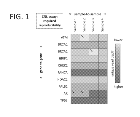
[0041] FIG. 1
shows the framework of the copy number loss (CNL) assay.
Each gene (rows) exhibits a characteristic unique read value that is
represented here by a
shade. Each sample (columns) is interrogated across the same panel of genes.
[0042] FIG. 2
shows a diagram illustrating the drivers of the CNL assay
signal.
[0043] FIG. 3
shows a diagram illustrating steps of an illustrative CNL assay
performed on cell free DNA (cfDNA).
[0044] FIG. 4A
¨ 4E shows diagrams of an illustrative first generation adaptor
(FIG. 4A and 4B) and an adaptor of the present invention (FIGs. 4C-4E). FIG.
4A shows the
first generation adaptor design. FIG. 4B shows that in the first generation
adaptors, there
were a collection of 249 possible sequence tags, each 5 nucleotides (nt) in
length that
attached to a single anchor sequence. FIG. 4C shows a diagram of a second
generation
adaptor. FIG. 4D shows an illustrative set of adaptors that are applied to a
single sample that
consists of four sets of 8mer tag sequences with each set having 60 members.
Each set of 60
12
CA 03034649 2019-02-20
WO 2018/039463
PCT/US2017/048434
tags is specific to one of four anchor sequences. FIG. 4E shows an
illustrative DNA sequence
of a 47 nt adaptor.
[0045] FIG. 5A
¨ FIG. 5B shows a diagram illustrating that shifting the
position of the UMI multiplier within the sample tag can increase the number
of unique
sample tags.
[0046] FIG. 6A
and B shows a diagram illustrating the process of constructing
genomic libraries for a CNL assay. FIG. 6A shows the step where the 10 nt
anchor sequence
is attached to the 3' ends of genomic fragments. FIG. 6B shows the step where
the full length
genomic adaptors are annealed to the initial anchor sequence.
[0047] FIG. 7
shows DNA inputs into CNL libraries. Agarose gel images are
shown with the sizes of markers (bp) indicated at left.
[0048] FIG. 8A
¨ FIG. 8C shows conventional box-and-whiskers plots of
measured gene copies across eight samples as determined by CNL analysis.
[0049] FIG. 9A
¨ FIG. 9B shows Logio P-value plots that quantify significant
deviation-from-normal in CNL measurements for fragmented genomic samples. The
SNP
percentages at the top show the minor allele frequencies of rare, heterozygous
SNPs that are
present in the AATM and AERCA2 samples.
[0050] FIG. 10A
¨ FIG. 10B shows Logio P-value plots that quantify
significant deviation-from-normal in CNL measurements for cfDNA samples spiked
with
fragmented genomic DNA. The SNP percentages at the top show the minor allele
frequencies
of rare, heterozygous SNPs that are present in the AATM and ABRCA2 samples.
[0051] FIG. 11A
¨ 11D illustrate the targeted hybrid capture platform. FIG.
11A shows conversion of cfDNA to a genomic library by the addition of adaptor
sequences
that provide universal, single-primer PCR amplification sequences, sample
multiplexing tags,
and unique molecular identifiers to every genomic clone. FIG. 11B shows
denatured
amplified genomic hybridized with target specific capture probes and primer
extension. FIG.
11C shows a schematic of asymmetric paired-end sequencing. FIG. 11D shows
mapping
statistics for 377,711,020 Illumina NextSeq reads from a typical targeted
capture sequence
run. 98.5% of reads map to their intended targets. Following de-duplication,
20.40% of reads
(77,053,048) are derived from unique genomic clones.
[0052] FIG. 12A
¨ FIG. 12H shows sequences of adaptor oligonucleotides
from Pools 1 ¨ 3.
13
CA 03034649 2019-02-20
WO 2018/039463
PCT/US2017/048434
[0053] FIG. 13A
¨ FIG. 13H shows sequences of adaptor oligonucleotides
from Pools 4 ¨ 6.
[0054] FIG. 14A
¨ FIG. 141 shows sequences of adaptor oligonucleotides
from Pools 7 ¨ 9.
[0055] FIG. 15A
¨ FIG. 15H shows sequences of adaptor oligonucleotides
from Pools 10 ¨ 12.
[0056] FIG. 16A
¨ FIG. 16H shows sequences of adaptor oligonucleotides
from Pools 13¨ 15.
[0057] FIG. 17A
¨ FIG. 18H shows sequences of adaptor oligonucleotides
from Pools 16 ¨ 18.
[0058] FIG. 18A
¨ FIG. 18H shows sequences of adaptor oligonucleotides
from Pools 19 ¨ 21.
[0059] FIG. 19A
¨ FIG. 19H shows sequences of adaptor oligonucleotides
from Pools 22 ¨ 24.
[0060] FIG. 20A
¨ FIG. 20H shows sequences of adaptor oligonucleotides
from Pools 25 ¨ 27.
[0061] FIG. 21A
¨ FIG. 21H shows sequences of adaptor oligonucleotides
from Pools 28 ¨ 30.
[0062] FIG. 22A
¨ FIG. 22H shows sequences of adaptor oligonucleotides
from Pools 31 ¨32.
[0063] FIG. 23A
¨ 23C shows targeted sequencing of the TP53 gene. FIG.
23A illustrates BedFile display of capture probes. FIG. 23B illustrates
coverage depth at each
base position on a scale of 0 to 8000 unique reads. FIG. 23C illustrates a
UCSC gene model
display of known TP53 splice variants. The thicker rectangular regions
represent the amino
acid coding regions for the TP53-encoded protein.
[0064] FIG. 24A
¨ 24C illustrate raw and normalized unique read density for
a single probe, TP53r10 1, across 16 samples. FIG. 24A illustrates the number
of raw unique
reads capture by probe TP53r10 1 for 16 independent sample after removal of
redundant
reads by "de-duplication." FIG. 24B shows global average of unique reads
across 2596
capture probes for all 16 samples. FIG. 24C shows normalized unique read depth
across 16
samples (Calculated as: [sample n unique reads from probe TP53r10 1 x constant
global
average unique reads/probe from sample n]).
14
CA 03034649 2019-02-20
WO 2018/039463
PCT/US2017/048434
[0065] FIG. 25
shows general consistency of the normalized unique read
counts for all 16 samples within any given TP53 probe despite significant
average depth
variation between probes. The normalized unique read counts for all 16 samples
are shown as
"pillars" of tightly spaced bar graphs; the results for all 45 probes that
target TP53 are shown.
Two probes that exhibit "noisy" counting behavior are highlighted with arrows.
Counts from
such probes often appear as outliers in subsequent copy number analysis.
[0066] FIG. 26
illustrates sample-to-sample consistency of normalized probe-
by-probe unique read counts across a broad panel of 2596 probes. The scatter
plots from three
representative samples are shown. Each dot represents a different probe. The x-
axis is the
normalized average unique read depth per probe across 16 samples. The y-axis
is the
normalized unique read depth per probe for three different individual samples.
The consistent
probe-by-probe unique read counts support quantitative analysis of chromosomal
copy
variation.
[0067] FIG. 27A
¨ 27C illustrate copy number analysis of cfDNA from a
healthy female and male donor and from an advanced stage prostate cancer
patient. FIG. 27A
shows analysis of a cfDNA from a healthy female donor. The x-axis is a series
of control
probes that target regions from all 22 autosomal chromosomes, a series of
probes that target
the X-linked AR gene, and a series of probes that target the coding regions of
the TP53 gene.
The Y-axis shows the calculated ploidy for each probe. This approximation is
calculated for
each probe by normalizing the observed unique read counts to a series of
control samples
whose ploidy is known ([unique read count for probe _Y of sample Z] x 2
[average unique
read count for probe _Y for multiple control samples]). FIG. 27B illustrates
that the X-linked
AR gene exhibits a haploid copy number in healthy males. FIG. 27C illustrates
copy number
analysis of cfDNA from an advanced prostate cancer patient and shows evidence
of very
significant aneuploidy across the control probes, amplification of the AR
gene, and loss of the
TP53 gene.
[0068] FIG. 28
shows whole genome aneuploidy analysis of a prostate patient
cfDNA library relative to a control sample. The approximate ploidy for each of
239 control
probes is shown sorted by chromosome. Patient chromosome 2 probes show
consistent copy
loss and the majority of chromosome 5 probes show copy gain. Significant
deviation of
approximate ploidy are seen for many, but not all, of the patient control
probes.
[0069] FIG. 29
shows analytical validation of copy number loss detection.
Genomic DNA from immortalized line NA02718 (monoallelic AATM) and from NA09596
CA 03034649 2019-02-20
WO 2018/039463
PCT/US2017/048434
(monoallelic ABRCA2) were spiked into the "gold standard" genomic DNA from
NA12878
at 16%, resulting in the equivalent of an 8% biallelic deletion minor allele
frequency.
Following targeted sequencing and CNV analysis, the probe-by-probe ploidies
were averaged
for the two target genes. Two unperturbed control genes, BRIP1 and HDAC2, are
shown for
comparison.
DETAILED DESCRIPTION
A. OVERVIEW
[0070] The
present invention includes, inter alia, compositions and methods
that are useful for the detection of a mutational change, SNP, translocation,
inversion,
deletion, change in copy number or other genetic variation within a sample of
cellular
genomic DNA (e.g. from a tissue biopsy sample) or cfDNA (e.g. from a blood
sample). The
compositions and methods of the current invention are particularly useful in
detecting
incredibly hard to detect copy number variations in cfDNA from a biological
sample (e.g.
blood) with exquisite resolution. In particular, some embodiments of the
present invention are
drawn to a method for the detecting copy number of a DNA target region from a
test sample
by generating a genomic DNA library made up of genomic DNA fragments attached
to an
adaptor, capturing DNA target regions with a plurality of capture probes,
isolating the DNA
library fragments comprising the DNA target region, and performing a
quantitative genetic
analysis of the DNA target region to thereby determining the copy number of
the DNA target
region. The adaptors described herein allow for the identification of the
individual DNA
fragment that is being sequenced, as well as the identity of the sample or
source of the
genomic DNA.
[0071] The
present invention contemplates, in part, compositions and methods
for detection of target-specific copy number changes that are applicable to
several sample
types, including but not limited to direct tissue biopsies and peripheral
blood. In the context
of cancer genomics, and in particular cell free DNA (cfDNA) assays for the
analysis of solid
tumors, the amount of tumor DNA is often a very small fraction of the overall
DNA. Further,
copy number loss is difficult to detect in genomic DNA assays, and in
particular, genomic
DNA assays where copy number change may only be present in a portion of the
total
genomic DNA from a sample, e.g., cfDNA assays. For example, most of the cell-
free DNA
extracted from a cancer patient will be derived from normal sources and have a
diploid copy
number (except for X-linked genes in male subjects). In a cancer patient, the
fraction of DNA
16
CA 03034649 2019-02-20
WO 2018/039463
PCT/US2017/048434
derived from tumors often has a low minor allele frequency, such as for
example, a patient in
which 2% of the circulating DNA extracted from plasma is derived from the
tumor. The loss
of one copy of a tumor suppressor gene (for example, BRCA1 in breast cancer)
means that
the minor allele frequency for the absence of detectable genomic fragments is
1%. In this
scenario, a copy number loss assay engineered must be able to discriminate
between 100
copies (normal) and 99 copies (heterozygous gene loss). Thus, particular
embodiments
contemplate that the methods and compositions of the present invention allow
for the
detection of copy number change with sufficient resolution to detect changes
in copy number
at minor allele frequencies even in the context of cfDNA.
[0072] To
achieve this level of discrimination, the present invention provides
novel sample adaptor designs. The adaptors of the present invention are
designed to include
features that are critical for successful copy number loss assay performance
including (i) even
performance across adaptors; (ii) a high number of unique molecule identifiers
(UMIs); (iii)
high efficiency attachment; and (iv) accommodation of sample multiplexing. For
example,
the adaptors of the present invention provide the following:
[0073] Even
performance across adaptors: Bioinformatics analysis often
looks at intra-sample probe performance and inter-sample probe performance.
Thus, it is
contemplated that any performance fluctuation between adaptor pools across
samples will
negatively impact the ability to detect the subtle variations required by CNL
analysis. In the
present invention, this evenness of performance is achieved by having multiple
anchor tags
that are all represented in each sample tag pool, with the fixed sample tag
regions (which
serve to identify both the sample and the genomic fragments) being randomly
selected for
each pool, and a UMI multiplier that increases the unique sample tag sequences
for
identifying the genomic fragments.
[0074] High
number of Unique Molecule Identifiers (UMIs): While adaptors
must be functionally equivalent from a molecular biology perspective, they
must possess a
very large number of unique sequence tags 10,000)
that augment the identification of
unique genomic fragments. In this context, by "augment," it is meant that each
genomic clone
fragment has a particular pair of fragmentation sites corresponding to the
position in the
genomic sequence where the double-strand DNA was cleaved. This cleavage site
is used to
differentiate unique genomic clones since each clone is likely to possess a
different cleavage
site. However, in libraries that possess thousands of independent clones,
uniquely derived
fragments will often possess the exact same cleavage sites. Genomic clones
(i.e. fragments)
17
CA 03034649 2019-02-20
WO 2018/039463
PCT/US2017/048434
sharing the same cleavage site can be classified as either unique or as
redundant with respect
to other clone sequences derived from the same sample. By attaching adaptors
that introduce
a high diversity of sequence tags, different genomic clones sharing the same
cleavage site are
more likely to be identified as unique. In this system, the UMI is created by
a combination of
the sample tag region with the UMI multiplier. The combination of the UMI and
the cleavage
site create a unique molecular identifier element (UMIE), which facilitates
the classification
of sequence reads as redundant reads or unique reads. Particular embodiments
contemplate
that the UMI multiplier could comprise longer or shorter sequences to increase
or lower the
overall UMI complexity.
[0075] High
efficiency attachment: Adaptors must attach to genomic
fragments with high efficiency. In most oncology applications, the quantities
of available
cellular DNA or cfDNA are limited and therefore conversion of these genomic
fragments to
genomic library clones must be highly efficient. In order to achieve this, in
some aspects of
the present invention, the adaptor systems described herein convert about 25%
to about 50%
or greater of the genomic input fragments are converted into genomic library
clones.
[0076]
Accommodation of sample multiplexing: In general, there must be
pools of different sets of adaptors where each unique adaptor of the set is
attached to a
different sample. At the same time, each member of the set of adaptors must
possess
essentially identical behavior (from a sequence counting perspective) to all
other members in
a set. In order to achieve this, in some embodiments, the sample tag regions
have a Hamming
distance of 2 between any other possible sample tag combinations reducing the
chance for a
read to be spuriously assigned to the wrong sample. In some embodiments, each
set of
adaptors is split into pools that are paired with specific anchor regions,
allowing for further
reduction in the possibility of an error in sample de-multiplexing. For
example, in an 8mer
tag with Hamming distance of 2, the total number of possible sequences is
16,384.
[0077] In a
particular embodiment, pre-specified pools of adaptor
oligonucleotides are provided. Such pre-specified pools are used to represent
a single sample.
That is, each adapter sequence in each pool of X adapter oligonucleotides
(16,384 in the
example given above) is distinct from each adapter sequence in every other
pool used to
identify other samples. One of skill in the art will recognize the number of
distinct pre-
specified pools that are possible for the adapter oligonucleotides will depend
on the length of
the sample tag and/or the UMI multiplier.
18
CA 03034649 2019-02-20
WO 2018/039463
PCT/US2017/048434
[0078] Thus, in
certain embodiments the adaptors comprise a sequence, i.e.,
the sample tag and adjacent and/or encompassed UMI multiplier that represents
or identifies
both the sample and uniquely identifies the genetic fragment. This is in stark
contrast to the
current systems that are used in the art that use a randomly generated tag to
identify the
sequence and a separate barcode or sequencer indexing to allow for
multiplexing.
[0079] An
illustrative embodiment for detecting target-specific copy number
changes within DNA obtained from a sample is shown in FIG. 3. While FIG. 3
generates a
DNA library from cfDNA, this illustrative procedure could be used with DNA
from other
sources, e.g., fragmented cellular DNA. As shown in FIG. 3, cfDNA is collected
(top panel).
Next, a genomic library is generated from cfDNA by conjugating genomic library
adaptors
(gray circles) of the present invention to the genomic DNA. Genomic DNA
fragments are
captured with capture probes (black circles) that recognize the genomic region
of interested.
The genomic DNA of interest is sequenced, and data analysis is performed for
copy loss
analysis and/or characterization of the genomic DNA of interest.
[0080] The
practice of particular embodiments of the invention will employ,
unless indicated specifically to the contrary, conventional methods of
chemistry,
biochemistry, organic chemistry, molecular biology, microbiology, recombinant
DNA
techniques, genetics, immunology, and cell biology that are within the skill
of the art, many
of which are described below for the purpose of illustration. Such techniques
are explained
fully in the literature. See, e.g., Sambrook, et al., Molecular Cloning: A
Laboratory Manual
(3rd Edition, 2001); Sambrook, et al., Molecular Cloning: A Laboratory Manual
(2nd
Edition, 1989); Maniatis et al., Molecular Cloning: A Laboratory Manual
(1982); Ausubel et
al., Current Protocols in Molecular Biology (John Wiley and Sons, updated July
2008); Short
Protocols in Molecular Biology: A Compendium of Methods from Current Protocols
in
Molecular Biology, Greene Pub. Associates and Wiley-Interscience; Glover, DNA
Cloning: A
Practical Approach, vol. I & II (IRL Press, Oxford, 1985); Anand, Techniques
for the
Analysis of Complex Genomes, (Academic Press, New York, 1992); Transcription
and
Translation (B. Hames & S. Higgins, Eds., 1984); Perbal, A Practical Guide to
Molecular
Cloning (1984); and Harlow and Lane, Antibodies, (Cold Spring Harbor
Laboratory Press,
Cold Spring Harbor, N.Y., 1998).
19
CA 03034649 2019-02-20
WO 2018/039463
PCT/US2017/048434
B. DEFINITIONS
[0081] Unless
defined otherwise, all technical and scientific terms used herein
have the same meaning as commonly understood by those of ordinary skill in the
art to which
the invention belongs. Although any methods and materials similar or
equivalent to those
described herein can be used in the practice or testing of the present
invention, preferred
embodiments of compositions, methods and materials are described herein. For
the purposes
of the present invention, the following terms are defined below.
[0082] The
articles "a," "an," and "the" are used herein to refer to one or to
more than one (i.e. to at least one) of the grammatical object of the article.
By way of
example, "an element" means one element or more than one element.
[0083] The use
of the alternative (e.g., "or") should be understood to mean
either one, both, or any combination thereof of the alternatives.
[0084] The term
"and/or" should be understood to mean either one, or both of
the alternatives.
[0085] As used
herein, the term "about" or "approximately" refers to a
quantity, level, value, number, frequency, percentage, dimension, size,
amount, weight or
length that varies by as much as 15%, 10%, 9%, 8%, 7%, 6%, 5%, 4%, 3%, 2% or
1% to a
reference quantity, level, value, number, frequency, percentage, dimension,
size, amount,
weight or length. In one embodiment, the term "about" or "approximately"
refers a range of
quantity, level, value, number, frequency, percentage, dimension, size,
amount, weight or
length 15%, 10%, 9%, 8%, 7%, 6%, 5%, 4%, 3%, 2%, or 1%
about a
reference quantity, level, value, number, frequency, percentage, dimension,
size, amount,
weight or length.
[0086]
Throughout this specification, unless the context requires otherwise,
the words "comprise", "comprises," and "comprising" will be understood to
imply the
inclusion of a stated step or element or group of steps or elements but not
the exclusion of
any other step or element or group of steps or elements. In particular
embodiments, the terms
"include," "has," "contains," and "comprise" are used synonymously.
[0087] By
"consisting of' is meant including, and limited to, whatever follows
the phrase "consisting of" Thus, the phrase "consisting of' indicates that the
listed elements
are required or mandatory, and that no other elements may be present.
[0088] By
"consisting essentially of' is meant including any elements listed
after the phrase, and limited to other elements that do not interfere with or
contribute to the
CA 03034649 2019-02-20
WO 2018/039463
PCT/US2017/048434
activity or action specified in the disclosure for the listed elements. Thus,
the phrase
"consisting essentially of' indicates that the listed elements are required or
mandatory, but
that no other elements are optional and may or may not be present depending
upon whether
or not they affect the activity or action of the listed elements.
[0089]
Reference throughout this specification to "one embodiment," "an
embodiment," "a particular embodiment," "a related embodiment," "a certain
embodiment,"
"an additional embodiment," or "a further embodiment" or combinations thereof
means that a
particular feature, structure or characteristic described in connection with
the embodiment is
included in at least one embodiment of the present invention. Thus, the
appearances of the
foregoing phrases in various places throughout this specification are not
necessarily all
referring to the same embodiment. Furthermore, the particular features,
structures, or
characteristics may be combined in any suitable manner in one or more
embodiments.
[0090] As used
herein, the term "isolated" means material that is substantially
or essentially free from components that normally accompany it in its native
state. In
particular embodiments, the term "obtained" or "derived" is used synonymously
with
isolated.
[0091] As used
herein, the term "DNA" refers to deoxyribonucleic acid. In
various embodiments, the term DNA refers to genomic DNA, recombinant DNA,
synthetic
DNA, or cDNA. In one embodiment, DNA refers to genomic DNA or cDNA. In
particular
embodiments, the DNA comprises a "target region." DNA libraries contemplated
herein
include genomic DNA libraries and cDNA libraries constructed from RNA, e.g.,
an RNA
expression library. In various embodiments, the DNA libraries comprise one or
more
additional DNA sequences and/or tags.
[0092] The
terms "target genetic locus" and "DNA target region" are used
interchangeably herein and refer to a region of interest within a DNA
sequence. In various
embodiments, targeted genetic analyses are performed on the target genetic
locus. In
particular embodiments, the DNA target region is a region of a gene that is
associated with a
particular genetic state, genetic condition, genetic diseases; fetal testing;
genetic mosaicism,
paternity testing; predicting response to drug treatment; diagnosing or
monitoring a medical
condition; microbiome profiling; pathogen screening; or organ transplant
monitoring. In
further embodiments, the DNA target region is a DNA sequence that is
associated with a
particular human chromosome, such as a particular autosomal or X-linked
chromosome, or
region thereof (e.g., a unique chromosome region).
21
CA 03034649 2019-02-20
WO 2018/039463
PCT/US2017/048434
[0093] As used
herein, the terms "circulating DNA," "circulating cell-free
DNA," and "cell-free DNA" are often used interchangeably and refer to DNA that
is
extracellular DNA, DNA that has been extruded from cells, or DNA that has been
released
from necrotic or apoptotic cells. This term is often used in contrast to
"cellular genomic
DNA" or "cellular DNA," which are used interchangeably herein and refer to
genomic DNA
that is contained within the cell (i.e. the nuclease) and is only accessible
to molecular
biological techniques such as those described herein, by lysing or otherwise
disrupting the
integrity of the cell.
[0094] A
"subject," "individual," or "patient" as used herein, includes any
animal that exhibits a symptom of a condition that can be detected or
identified with
compositions contemplated herein. Suitable subjects include laboratory animals
(such as
mouse, rat, rabbit, or guinea pig), farm animals (such as horses, cows, sheep,
pigs), and
domestic animals or pets (such as a cat or dog). In particular embodiments,
the subject is a
mammal. In certain embodiments, the subject is a non-human primate and, in
preferred
embodiments, the subject is a human.
[0095] As used
herein, the term "paired" when used with respect to two
different polynucleotide sequences or regions of DNA comprising different
polynucleotide
sequences, means that the two different polynucleotide sequences or regions of
DNA
comprising different polynucleotide sequences are present on the same
polynucleotide. For
example, if a particular sample tag region of DNA is said to be paired to
particular
amplification region of DNA, it is meant that the sample tag region and the
amplification tag
are present on the same DNA polynucleotide molecule.
C. METHODS OF COPY NUMBER ANALYSIS
[0096] In
various embodiments, a method for copy number analysis of a DNA
target region DNA is provided. In certain embodiments, copy number analysis is
performed
by generating a genomic DNA library of DNA library fragments that each contain
genomic
DNA fragment and an adaptor, isolating the DNA library fragments containing
the DNA
target regions, and performing a quantitative genetic analysis of the DNA
target region. By
"quantitative genetic analysis" it is meant an analysis performed by any
molecular biological
technique that is able to quantify changes in a DNA (e.g., a gene, genetic
locus, target region
of interest, etc.) including but not limited to DNA mutations, SNPs,
translocations, deletions,
22
CA 03034649 2019-02-20
WO 2018/039463
PCT/US2017/048434
and copy number variations (CNVs). In certain embodiments, the quantitative
genetic
analysis is performed by sequencing, for example, next generation sequencing.
[0097] Next-
generation DNA sequencing (NGS) is ideally suited for two
diagnostic applications. The first is the determination of DNA sequence on a
vast scale. In the
present context, this capability enables the search for rare, actionable
variants that guide
effective treatment decisions. The second is counting gene copy number. The
output of
millions of independent sequences can enable precise measurement of gene copy
number on
a genome-wide scale. The emergence of non-invasive prenatal testing for fetal
trisomy from
maternal blood samples is a testament to this capability. RNAseq, that is, the
technology of
gene expression profiling using NGS is another example, albeit the input is
RNA (cDNA)
rather than genomic DNA. Comparisons of current capture methods are described
Samorodnitsky etal. J Mol Diagn. 2015 Jan;17(1):64-75.
[0098] The
present invention extends NGS counting capability into the realm
of targeted hybrid capture methods. The methods described here are effective
for the
detection of copy number variation at least in part because they possess the
following four
qualities:
(a) The
present methods differentiate between unique clones and redundant
clones. NGS sequencing of amplified genomic DNA library fragments results in a
plurality of
individual NGS reads, each comprising adaptor-encoded sequence information
linked to a
specific human genomic sequence. These elements define the identity of every
clone.
Because captured genomic regions are amplified by PCR, it is not uncommon for
the same
clone to be encountered several times in a subsequent NGS analysis. Groups of
reads that are
derived from a single cloning and capture process are termed "redundant
reads." Two or
more redundant reads are identified as redundant reads based on the sequencing
information
provided by the unique molecular identification elements (UMIE). The UMIE
refers to the
combination of the sequence information from the adaptor tags and the start of
the genomic
DNA sequence. Two or more reads comprising identical UMIEs are identified as
redundant
reads. Redundant reads are grouped together and a single, representative
consensus sequence
is assembled from families of redundant reads. This consensus sequence is
designated as a
"unique read" or a "unique genomic sequence" (UGS). Each unique read
represents a
separate clone from the original DNA specimen. The process of identifying and
grouping
redundant clone families and of generating a single unique read representative
of this family
is defined as "deduplication." The adaptors used to create genomic libraries
possess a very
23
CA 03034649 2019-02-20
WO 2018/039463
PCT/US2017/048434
deep repertoire of unique sample tag information (15,360 codes per adaptor).
When applied
in conjunction with the exact mapping coordinates of each captured genomic
clone (which
can span >100 different positions relative to a capture probe), each unique
clone that is
generated in a genomic library and subsequently retrieved by a target-specific
capture probe
has an extremely high likelihood of being differentiable from all other unique
clones that
encompass the same capture environment. The ability to differentiate between
unique clones
and redundant clones is central to the methods described herein.
(b) The adaptors used to create genomic libraries permit sample
multiplexing
without creating adaptor-to-adaptor variability in copy number counts. A
central foundation
of copy number determination is the simultaneous analysis of a set of samples
that have all
been processed within a single sequencing run. This allows positive and
negative controls to
be included along with clinical samples. A major issue with previous adaptor
design
iterations induced subtle shifts in gene copy counts among identical control
samples, in effect
setting a signal-to-noise uncertainty threshold that was too high to be
clinically useful in
blood-based, solid tumor genotyping assays. The present invention overcomes
this issue and
substantially lowers the signal-to-noise threshold such that single copy gene
loss is detectable
at < 2% minor allele frequency. This improved signal recognition enables the
methods of the
present invention to have significant clinical utility in circulating tumor
DNA assays.
(c) The proprietary targeted hybrid capture method used herein must produce
highly uniform "on-target" read coverage across all targets. Methods that rely
on counting of
unique genomic fragments to estimate copy number, such as the ones described
herein, must
achieve near-saturation in terms of encountering all possible unique
fragments. Near-
saturation is only achieved by oversampling, that is to say, gathering more
sequencing reads
than the number of unique reads that will ultimately be encountered. To be
practical, scalable,
and economical, the unique reads in a targeted hybrid capture library must
exhibit sufficient
uniformity such that < 10-fold oversampling of on-target reads, and preferably
< 4-fold
oversampling of on-target reads will capture > 90% of unique on-target reads
at all target
loci.
(d) The targeted hybrid capture method (See U.S. Patent Publication No.
2014-
0274731) must have high on-target capture rates. To be practical, scalable and
economical, in
other words to be a distinguishing feature of the present disclosure relative
to other art in the
field, the method must achieve >90%, preferably >95% on-target reads. With on-
target
24
CA 03034649 2019-02-20
WO 2018/039463
PCT/US2017/048434
mapping rates exceeding 95%, the requirement for 4 to 10-fold oversampling of
on-target
reads and the requirement for overall oversampling are one in the same.
[0099] In some
embodiments, the number of copies of the DNA target region
present in the sample is determined by the quantitative genetic analysis. In
some
embodiments, the copy number of the DNA target region is determined by
comparing the
amount of copies of DNA target regions present in the sample and comparing it
to amounts of
DNA target regions present in one or more samples with known copy number.
[00100]
Particular embodiments contemplate that the compositions and
methods described herein are particularly useful for detecting changes in copy
number in a
sample of genomic DNA, where only a portion of the total genomic DNA in the
sample has a
change in copy number. For example, a significant tumor mutation may be
present in a
sample, e.g. a sample of cell free DNA, that is present in a minor allele
frequency that is
significantly less than 50% ( e.g., in the range of 0.1% to >20%), in contrast
to conventional
SNP genotyping where allele frequencies are generally ¨100%, 50% or 0%. One of
skill of
the art will recognize that the compositions and methods of the current
invention are also
useful in detecting other types of mutation including single nucleotide
variants (SNVs), short
(e.g., less than 40 base pairs (bp)) insertions, and deletions (indels), and
genomic
rearrangements including oncogenic gene fusions.
[00101] In
certain embodiments, the compositions and/or methods of the
present invention described herein are useful for, capable of, suited for,
and/or able to detect,
identify, observe, and/or reveal a change in copy number of one or more DNA
target regions
present in less than about 20%, less than about 19%, less than about 18%, less
than about
17%, less than about 16%, less than about 15%, less than about 14%, less than
about 13%,
less than about 12%, less than about 11%, less than about 10%, less than about
9%, less than
about 8%, less than about 7%, less than about 6%, less than about 5%, less
than about 4%,
less than about 3%, less than about 2%, less than about 1%, less than about
0.5%, less than
about 0.2%, or less than about 0.1% of the total genomic DNA from the sample.
In some
embodiments, the methods of the present invention are useful for, capable of,
suited for,
and/or able to detect, identify, observe, and/or reveal a change in copy
number of one or more
DNA target regions present in between about 0.01% to about 100%, about 0.01%
to about
50%, and or about 0.1% to about 20% of the total genomic DNA from the sample.
[00102]
Particular embodiments are represented by the conceptual framework
that is illustrated in FIG. 1. In FIG. 1, each gene is represented by a row
and each patient
CA 03034649 2019-02-20
WO 2018/039463
PCT/US2017/048434
sample is represented as a column. Within any given genomic DNA sample, the
number of
fragments counted for each individual gene will have some variability, and
that for any given
DNA region of interest, e.g. a gene, perturbations in copy number are detected
as significant
fragment count deviations relative to the normalized counts to the DNA target
region in other
samples. Such an assay requires the gene-by-gene fragment counting profile
within a sample
to be reproducible, and also requires the sample-by-sample counting profiles
to be highly
comparable. Both assay requirements demand excellent signal-to-noise counting
discrimination.
[00103] Some
embodiments contemplate that the assay elements that contribute
to increasing the signal to noise ratio are the genomic input, the number of
probes, and the
sequencing depth, as illustrated in FIG. 2.
[00104] In
particular embodiments, a method for genetic analysis of cfDNA
comprises: generating and amplifying a cfDNA library, determining the number
of genome
equivalents in the cfDNA library; and performing a quantitative genetic
analysis of one or
more genomic target loci.
[00105]
Particular embodiments contemplate that the any of the methods and
compositions described herein are effective for use to efficiently analyze,
detect, diagnose,
and/or monitor genetic states, genetic conditions, genetic diseases, genetic
mosaicism, fetal
diagnostics, paternity testing, microbiome profiling, pathogen screening, and
organ transplant
monitoring using genomic DNA, e.g., cellular or cfDNA, where all or where only
a portion of
the total genomic DNA in the sample has a feature of interest, e.g. a genetic
lesion, mutation,
single nucleotide variant (SNV). In some embodiments, a feature of interest is
a genetic
feature associated with a disease or condition. For example, a significant
tumor mutation may
be present in a sample, e.g. a sample of cfDNA, that is present in a minor
allele frequency
that is significantly less than 50% (e.g. in the range of 0.1% to >20%), in
contrast to
conventional SNP genotyping where allele frequencies are generally ¨100%, 50%
or 0%.
[00106] In
certain embodiments, the compositions and/or methods of the
present invention described herein are useful for, capable of, suited for,
and/or able to detect,
identify, observe, and/or reveal a genetic lesion of one or more DNA target
regions present in
less than about 20%, less than about 19%, less than about 18%, less than about
17%, less than
about 16%, less than about 15%, less than about 14%, less than about 13%, less
than about
12%, less than about 11%, less than about 10%, less than about 9%, less than
about 8%, less
than about 7%, less than about 6%, less than about 5%, less than about 4%,
less than about
26
CA 03034649 2019-02-20
WO 2018/039463
PCT/US2017/048434
3%, less than about 2%, less than about 1%, less than about 0.5%, less than
about 0.2%, or
less than about 0.1% of the total genomic DNA from the sample. In some
embodiments, the
methods of the present invention are useful for, capable of, suited for,
and/or able to detect,
identify, observe, and/or reveal a genetic lesion of one or more DNA target
regions present in
between about 0.01% to about 100%, about 0.01% to about 50%, and or about 0.1%
to about
20% of the total genomic DNA from the sample.
1. GENERATING A DNA LIBRARY
[00107] In
particular embodiments, methods of genetic analysis contemplated
herein comprise generating a DNA library comprising treating cfDNA or
fragmented cellular
genomic DNA with one or more end-repair enzymes to generate end-repaired DNA
and
attaching one or more adaptors to each end of the end-repaired DNA to generate
the DNA
library. Genomic DNA
[00108] In
particular embodiments, the methods and compositions
contemplated herein are designed to efficiently analyze, detect, diagnose,
and/or monitor
change in copy number using genomic DNA as an analyte. In certain embodiments,
copy
number analysis is performed by generating a genomic DNA library from genomic
DNA
obtained from a test sample, e.g., a biological sample such as a tissue
biopsy. In certain
embodiments, the genomic DNA is circulating or cell free DNA. In some
embodiments, the
genomic DNA is cellular genomic DNA.
[00109] In
certain embodiments, genomic DNA is obtained from a tissue
sample or biopsy taken from a tissue, including but not limited to, bone
marrow, esophagus,
stomach, duodenum, rectum, colon, ileum, pancreases, lung, liver, prostate,
brain, nerves,
meningeal tissue, renal tissue, endometrial tissue, cervical tissue, breast,
lymph node, muscle,
and skin. In certain embodiments, the tissue sample is a biopsy of a tumor or
a suspected
tumor. In particular embodiments, the tumor is cancerous or suspected of being
cancerous. In
particular embodiments, the tissue sample comprises cancer cells or cells
suspected of being
cancerous.
[00110] Methods
for purifying genomic DNA from cells or from a biologic
tissue comprised of cells are well known in the art, and the skilled artisan
will recognize
optimal procedures or commercial kits depending on the tissue and the
conditions in which
the tissue is obtained. Some embodiments contemplate that purifying cellular
DNA from a
27
CA 03034649 2019-02-20
WO 2018/039463
PCT/US2017/048434
tissue will require cell disruption or cell lysis to expose the cellular DNA
within, for example
by chemical and physical methods such as blending, grinding or sonicating the
tissue sample;
removing membrane lipids by adding a detergent or surfactants which also
serves in cell
lysis, optionally removing proteins, for example by adding a protease;
removing RNA, for
example by adding an RNase; and DNA purification, for example from detergents,
proteins,
salts and reagents used during cell lysis step. DNA purification may be
performed by
precipitation, for example with ethanol or isopropanol; by phenol¨chloroform
extraction.
[00111] In
particular embodiments, cellular DNA obtained from tissues and/or
cells are fragmented prior to and or during obtaining, generating, making,
forming, and/or
producing a genomic DNA library as described herein. One of skill in the art
will understand
that there are several suitable techniques for DNA fragmentation, and is able
to recognize and
identify suitable techniques for fragmenting cellular DNA for the purposes of
generating a
genomic DNA library for DNA sequencing, including but not limited to next-
generation
sequencing. Certain embodiments contemplate that cellular DNA can be
fragmented into
fragments of appropriate and/or sufficient length for generating a library by
methods
including but not limited to physical fragmentation, enzymatic fragmentation,
and chemical
shearing.
[00112] Physical
fragmentation can include, but is not limited to, acoustic
shearing, sonication, and hydrodynamic shear. In some embodiments, cellular
DNA is
fragmented by physical fragmentation. In particular embodiments, cellular DNA
is
fragmented by acoustic shearing or sonication. Particular embodiments
contemplate that
acoustic shearing and sonication are common physical methods used to shear
cellular DNA.
The Covaris0 instrument (Woburn, MA) is an acoustic device for breaking DNA
into 100-
5kb bp. Covaris also manufactures tubes (gTubes) which will process samples in
the 6-20 kb
for Mate-Pair libraries. The Bioruptor0 (Denville, NJ) is a sonication device
utilized for
shearing chromatin, DNA and disrupting tissues. Small volumes of DNA can be
sheared to
150-1kb in length. Hydroshear from Digilab (Marlborough, MA) utilizes
hydrodynamic
forces to shear DNA. Nebulizers (Life Tech, Grand Island, NY) can also be used
to atomize
liquid using compressed air, shearing DNA into 100-3kb fragments in seconds.
Nebulization
is low cost, but the process can cause a loss of about 30% of the cellular DNA
from the
original sample. In certain embodiments, cellular DNA is fragmented by
sonication.
[00113]
Enzymatic fragmentation can include, but is not limited to, treatment
with a restriction endonuclease, e.g. DNase I, or treatment with a nonspecific
nuclease. In
28
CA 03034649 2019-02-20
WO 2018/039463
PCT/US2017/048434
some embodiments, cellular DNA is fragmented by enzymatic fragmentation. In
particular
embodiments, the cellular DNA is fragmented by treatment with a restriction
endonuclease.
In some embodiments, the cellular DNA is fragmented by treatment with a
nonspecific
nuclease. In certain embodiments, the cellular DNA is fragmented by treatment
with a
transposase. Certain embodiments contemplate that enzymatic methods to shear
cellular
DNA into small pieces include DNAse I, a combination of maltose binding
protein (MBP)-
T7 Endo I and a non-specific nuclease Vibrio vulnificus (Vvn) New England
Biolabs's
(Ipswich, MA) Fragmentase and Nextera tagmentation technology (Itlumina, San
Diego,
CA). The combination of non-specific nuclease and T7 Endo synergistically work
to produce
non-specific nicks and counter nicks, generating fragments that disassociate 8
nucleotides or
less from the nick site. Tagmentation uses a transposase to simultaneously
fragment and
insert adapters onto double stranded DNA.
[00114] Chemical
fragmentation can include treatment with heat and divalent
metal cation. In some embodiments, genomic DNA is fragmented by chemical
fragmentation.
Particular embodiments contemplate that chemical shear is more commonly used
for the
breakup of long RNA fragments as opposed to genomic DNA. Chemical
fragmentation is
typically performed through the heat digestion of DNA with a divalent metal
cation
(magnesium or zinc). The length of DNA fragments can be adjusted by increasing
or
decreasing the time of incubation.
[00115] In
particular embodiments, the methods and compositions
contemplated herein are designed to efficiently analyze, detect, diagnose,
and/or monitor
change in copy number using cell-free DNA (cfDNA) as an analyte. The size
distribution of
cfDNA ranges from about 150 bp to about 180 bp fragments. Fragmentation of
cfDNA may
be the result of endonucleolytic and/or exonucleolytic activity and presents a
formidable
challenge to the accurate, reliable, and robust analysis of cfDNA. Another
challenge for
analyzing cfDNA is its short half-life in the blood stream, on the order of
about 15 minutes.
Without wishing to be bound to any particular theory, the present invention
contemplates, in
part, that analysis of cfDNA is like a "liquid biopsy" and is a real-time
snapshot of current
biological processes.
[00116]
Moreover, because cfDNA is not found within cells and may be
obtained from a number of suitable sources including, but not limited to,
biological fluids and
stool samples, it is not subject to the existing limitations that plague next
generation
sequencing analysis, such as direct access to the tissues being analyzed.
29
CA 03034649 2019-02-20
WO 2018/039463
PCT/US2017/048434
[00117]
Illustrative examples of biological fluids that are suitable sources from
which to isolate cfDNA in particular embodiments include, but are not limited
to amniotic
fluid, blood, plasma, serum, semen, lymphatic fluid, cerebral spinal fluid,
ocular fluid, urine,
saliva, mucous, and sweat. In particular embodiments, the biological fluid is
blood or blood
plasma.
[00118] In
certain embodiments, commercially available kits and other
methods known to the skilled artisan can used to isolate cfDNA directly from
the biological
fluids of a subject or from a previously obtained and optionally stabilized
biological sample,
e.g., by freezing and/or addition of enzyme chelating agents including, but
not limited to
EDTA, EGTA, or other chelating agents specific for divalent cations.
(a) Generating End-Repaired cfDNA
[00119] In
particular embodiments, generating a genomic DNA library
comprises the end-repair of isolated cfDNA or fragmented cellular DNA. The
fragmented
cfDNA or cellular DNA is processed by end-repair enzymes to generate end-
repaired cfDNA
with blunt ends, 5'-overhangs, or 3'-overhangs. In some embodiments, the end-
repair
enzymes can yield for example. In some embodiments, the end-repaired cfDNA or
cellular
DNA contains blunt ends. In some embodiments, the end-repaired cellular DNA or
cfDNA is
processed to contain blunt ends. In some embodiments, the blunt ends of the
end-repaired
cfDNA or cellular DNA are further modified to contain a single base pair
overhang. In some
embodiments, end-repaired cfDNA or cellular DNA containing blunt ends can be
further
processed to contain adenine (A)/thymine (T) overhang. In some embodiments,
end-repaired
cfDNA or cellular DNA containing blunt ends can be further processed to
contain adenine
(A)/thymine (T) overhang as the single base pair overhang. In some
embodiments, the end-
repaired cfDNA or cellular DNA has non-templated 3' overhangs. In some
embodiments, the
end-repaired cfDNA or cellular DNA is processed to contain 3' overhangs. In
some
embodiments, the end-repaired cfDNA or cellular DNA is processed with terminal
transferase (TdT) to contain 3' overhangs. In some embodiments, a G-tail can
be added by
TdT. In some embodiments, the end-repaired cfDNA or cellular DNA is processed
to contain
overhang ends using partial digestion with any known restriction enzymes
(e.g., with the
enzyme Sau3A, and the like.
CA 03034649 2019-02-20
WO 2018/039463
PCT/US2017/048434
(b) Attaching Adaptor Molecules to End-Repaired cfDNA
[00120] In
particular embodiments, generating a cfDNA library comprises
attaching one or more adaptors to each end of the end-repaired cfDNA. The
present invention
contemplates, in part, an adaptor module designed to accommodate large numbers
of genome
equivalents in cfDNA libraries. Adaptor modules are configured to measure the
number of
genome equivalents present in cfDNA libraries, and, by extension, the
sensitivity of
sequencing assays used to identify sequence mutations.
[00121] As used
herein, the terms "adaptor" and "adaptor module" are used for
interchangeably, and refer to a polynucleotide comprising that comprises at
least three
elements: an amplification region, a sample tag region, and an anchor region.
In particular
embodiments, the adaptor comprises an amplification region, a sample tag
region, and an
anchor region. In some embodiments, the adaptor also comprises a unique
molecule identifier
(UMI). In particular embodiments, the adaptor comprises one or amplification
regions, one or
more sample tag regions, one or more UMIs, and/or one or more anchor regions.
In some
embodiments, the adaptor comprises, in order from 5' to 3', an amplification
region, a sample
tag region, a UMI, and an anchor region. In particular embodiments, the
adaptor comprises,
in order from 5' to 3', an amplification region, a sample tag region, a UMI,
and an anchor
region. In certain embodiments, the UMI is contained within the sample tag
region, and the
adaptor comprises, in order from 5' to 3', an amplification region, an
integrated sample
tag/UMI region, and an anchor region.
[00122] As used
herein, the term "amplification region" refers to an element of
the adaptor molecule that comprises a polynucleotide sequence capable of
serving as a primer
recognition site for PCR amplification. In particular embodiments, an adaptor
comprises an
amplification region that comprises one or more primer recognition sequences
for single-
primer amplification of a genomic DNA library. In some embodiments, the
amplification
region comprises one, two, three, four, five, six, seven, eight, nine, ten, or
more primer
recognition sequences for single-primer amplification of a genomic DNA
library.
[00123] In some
embodiments, the amplification region is about is between 5
and 50 nucleotides, between 10 and 45 nucleotides, between 15 and 40
nucleotides, or
between 20 and 30 nucleotides in length. In some embodiments, the
amplification region is
nucleotides, 11 nucleotides, 12 nucleotides, 13 nucleotides, 14 nucleotides,
15 nucleotides,
16 nucleotides, 17 nucleotides, about 18 nucleotides, 19 nucleotides, 20
nucleotides, 21
nucleotides, 22 nucleotides, 23 nucleotides, 24 nucleotides, 25 nucleotides,
26 nucleotides, 27
31
CA 03034649 2019-02-20
WO 2018/039463
PCT/US2017/048434
nucleotides, 28 nucleotides, 29 nucleotides, 30 nucleotides, 31 nucleotides,
32 nucleotides, 33
nucleotides, 34 nucleotides, 35 nucleotides, 36 nucleotides, 37 nucleotides,
38 nucleotides, 39
nucleotides, or 40 nucleotides or more. In particular embodiments, the
amplification region is
25 nucleotides in length.
[00124] As used
herein, the term "sample tag" or sample tag region" are used
interchangeably and refer to an element of the adaptor that comprises a
polynucleotide
sequence that uniquely identifies the particular DNA fragment as well as the
sample from
which it was derived.
[00125] In
certain embodiments, the sample tag region is about is between 3
and 50 nucleotides, between 3 and 25 nucleotides, or between 5 and 15
nucleotides in length.
In some embodiments, the sample tag region is 3 nucleotides, 4 nucleotides, 5
nucleotides, 6
nucleotides, 7 nucleotides, 8 nucleotides, 9 nucleotides, 10 nucleotides,
about 11 nucleotides,
12 nucleotides, 13 nucleotides, 14 nucleotides, 15 nucleotides, 16
nucleotides, 17 nucleotides,
18 nucleotides, 19 nucleotides, or 20 nucleotides or more in length.
[00126] In
certain embodiments, the adaptor comprises a UMI multiplier,
wherein the UMI multiplier is at least 1, at least 2, at least 3, at least 4,
at least 5, at least 6, at
least 7, at least 8, at least 9, or at least 10 nucleotides in length.
[00127] In
certain embodiments, each nucleotide position of the UMI multiplier
can comprise any of adenine, guanine, cytosine, or thymine. Thus, in some
embodiments, a
UMI multiplier comprising n number of nucleotides can comprise any of n4
possible
nucleotide sequences. In some embodiments, the UMI multiplier is one
nucleotide in length
and comprises one of four possible sequences. In some embodiments, the UMI
multiplier is
two nucleotides in length and comprises one of sixteen possible sequences. In
some
embodiments, the UMI multiplier is three nucleotides in length and comprises
one of 64
possible sequences. In some embodiments, the UMI multiplier is four
nucleotides in length
and comprises one of 256 possible sequences. In some embodiments, the UMI
multiplier is
five nucleotides in length and comprises one of 1,024 possible sequences. In
some
embodiments, the UMI multiplier is six nucleotides in length and comprises one
of 4,096
possible sequences. In some embodiments, the UMI multiplier is seven
nucleotides in length
and comprises one of 16,384 possible sequences. In some embodiments, the UMI
multiplier
is eight nucleotides in length and comprises one of 65,5336 possible
sequences. In some
embodiments, the UMI multiplier is nine nucleotides in length and comprises
one of 262,144
32
CA 03034649 2019-02-20
WO 2018/039463
PCT/US2017/048434
possible sequences. In some embodiments, the UMI multiplier is ten or more
nucleotides in
length and comprises one of 1,048,576 or more possible sequences.
[00128] In
particular embodiments, the adaptor comprises a UMI multiplier,
wherein the UMI multiplier is adjacent to or contained within the sample tag
region (FIG.
5A). Illustrative examples of UMI multipliers adjacent or contained within the
sample tag are
shown in FIG. 5B. In FIG. 5B, an 8-mer sample tag region is shown with an
adjacent UMI
multiplier (top and bottom rows) or a UMI multiplier incorporated within the
sample tag
(middle 7 rows). In some embodiments, that adaptor comprises a sample tag that
is eight
nucleotides in length and a UMI multiplier that is three nucleotides in length
and comprises
one of 64 possible sequences, and wherein the UMI multiplier is adjacent to or
contained
within the sample tag region. In some embodiments, identical processes attach
full length
adaptor to the other end of the genomic fragments.
[00129] In
particular embodiments, an adaptor module comprises one or more
anchor sequences. As used herein, an "anchor region" and "anchor sequence" are
used
interchangeably and refer to a nucleotide sequence that hybridizes to a
partner
oligonucleotide. In some embodiments, the anchor region comprises the
following three
properties: (1) each anchor sequence is part of a family of two or more anchor
sequences that
collectively represent each of the four possible DNA bases at each site within
extension; this
feature, balanced base representation, is useful to calibrate proper base
calling in sequencing
reads in particular embodiments; (2) each anchor sequence is composed of only
two of four
possible bases, and these are specifically chosen to be either and equal
number of A + C or an
equal number of G + T; an anchor sequence formed from only two bases reduces
the
possibility that the anchor sequence will participate in secondary structure
formation that
would preclude proper adaptor function; and (3) because each anchor sequence
is composed
of equal numbers of A + C or G + T, each anchor sequence shares roughly the
same melting
temperature and duplex stability as every other anchor sequence in a set of
four.
[00130] In some
embodiments, the anchor sequences is between 1 and 50
nucleotides in length. In some embodiments, the anchor sequences is between 4
and 40
nucleotides in length. In certain embodiments, the anchor region is between 5
and 25
nucleotides in length. In particular embodiments, the anchor region is at
least 4 nucleotides,
at least six nucleotides, at least 8 nucleotides, at least 10 nucleotides, at
least 12 nucleotides,
at least 14 nucleotides, or at least 16 nucleotides in length. In particular
embodiments, the
anchor region is 10 nucleotides in length.
33
CA 03034649 2019-02-20
WO 2018/039463
PCT/US2017/048434
[00131] In
particular embodiments, an attachment step comprises
attaching/ligating an adaptor module to the end-repaired cfDNA or cellular DNA
to generate
a "tagged" genomic DNA library. In some embodiments, a single adaptor module
is
employed. In some embodiments, two, three, four or five adaptor modules are
employed. In
some embodiments, an adaptor module of identical sequence is attached to each
end of the
fragmented end-repaired DNA.
[00132] In some
embodiments, a plurality of adaptor species is attached to an
end-repaired cellular or cell free genomic DNA fragments. Each of the
plurality of adaptors
may comprise one or more amplification regions for the amplification of the
cfDNA or
cellular DNA library, one or more sample tag regions for the identification of
the cfDNA or
cellular genomic DNA fragment and identification of the individual sample; and
one or more
sequences for DNA sequencing.
[00133] In some
embodiments, a plurality of adaptor species is attached to an
end-repaired cellular or cell free genomic DNA fragments of a sample, and the
plurality of
adaptors all comprise amplification regions of an identical nucleotide
sequence.
[00134] In
certain embodiments, the genomic DNA from a sample is attached
with a plurality of adaptors that comprise sample tag sequences that all are
different from
other sequences of sample tag regions in adaptors that are attached to genomic
DNA
fragments from other samples.
[00135] In
particular embodiments, a plurality of adaptor species is attached to
an end-repaired cellular or cell free genomic DNA fragments from a sample, and
the plurality
of adaptors all comprise one or more sample tag regions comprising one of
between 2 and
10,000 nucleotide sequences, one of between 5 and 5,000 nucleotide sequences,
one of
between 25 and 1,000 nucleotide sequences, one of between 50 and 500
nucleotide
sequences.one of between 100 and 400 nucleotide sequences, or one of between
200 and 300
nucleotide sequences. In some embodiments, the sample tag region of each
adaptor is 8
nucleotides in length, and each sample tag region of the plurality of adaptors
comprises one
of 240 nucleotide sequences.
[00136] In
certain embodiments, a plurality of adaptor species is attached to an
end-repaired cellular or cell free genomic DNA fragments from a sample, and
the sample tag
regions of the plurality of adaptors comprises nucleotide sequences that are
different from
each other by a Hamming distance of 1, 2, 3, 4 or greater than 4. In
particular embodiments,
the Hamming distance is 2.
34
CA 03034649 2019-02-20
WO 2018/039463
PCT/US2017/048434
[00137] In
particular embodiments, the sample tag regions of the plurality of
adaptors that are attached to genomic DNA fragments of a sample are 8
nucleotides in length,
and comprise one of 240 nucleotide sequences that are different from each
other by a
Hamming distance of 2.
[00138] In
certain embodiments, the sample tag region serves to identify
individual genomic DNA fragments and to identify the individual sample, i.e.,
the genomic
library source. For example, when the sample tags of a plurality of adaptors
attached to a
sample have one of 240 possible sequences, each sample is identified as having
one of 240
possible tags, and each sample receives a set of 240 tags that are discrete
from any other
sample by Hamming distance of two (meaning two base changes are required to
change one
tag into another). These same tags are used to enumerate clone diversity and
thus they also
serve as sequence tags, i.e., to identify genomic DNA fragments. To further
augment the
diversity of possible sequence tags, UMI multipliers may be added. For
example, a UMI
multiplier can be added to the adaptor region comprising 3 nucleotides
consisting of the 64
possible combinations of 3 bases. In addition, the plurality of adaptors can
comprise more
than one anchor sequence. For example, a plurality of adaptors may contain 4
different
anchor sequences are used simultaneously. These anchor sequences may also be
used during
sample de-multiplexing to lower errors.
[00139] FIG. 4
shows an illustrative comparison between a first generation
adaptor (FIG. 4A and 4B) and an adaptor of the present invention (FIG. 4C ¨
FIG. 4E). FIG.
4A and FIG. 4B show an example of first generation adaptor that is 40 nt in
length and
consisted of a discrete PCR amplification sequence, sequence tag, and sample
tag. Here, the
sample is identified by a fixed sequence (sequence tag) that is present on all
adaptors that are
used to generate a DNA library from the sample. Individual genomic fragments
are identified
by a separate and distinct sequences (sequence tag). FIG. 4C ¨ FIG. 4E show an
illustrative
example of an adaptor from the present invention. The illustrative adaptor
shown is 47
nucleotides in length, and the sequence tag is combined with the sample tag.
There is an
additional 3 nt sequence, the UMI multiplier, consisting of the 64 possible
combinations of 3
bases. The 10 nt anchor sequence is one of four different distinct sequences.
[00140] Thus, in
the illustrative example (See FIG. 4C ¨ FIG. 4E), a set of
adaptors that are used in connection with a single sample comprise 240 sample
tag sequences
that can be split into four sets of sample tag sequences with each set
comprising 60 tags (one
for each nucleotide, A, C, T and G). Thus, each set of 60 tags is specific to
one of four anchor
CA 03034649 2019-02-20
WO 2018/039463
PCT/US2017/048434
sequences. In total, a pool of 240 possible sample tag configurations are
possible per sample.
Specifically, in this scenario, the 240 sample tag sequences are divided into
four sets of 60
sequences, with each set directed to a specific anchor region. Therefore, the
sample ID
involves not only the sequence information from the eight nucleotide sample
tag, but also the
associated anchor sequence information. In addition, the position of sequences
within the
read is fixed, and therefore the sample tags and anchor sequences must have a
fixed position
within a sequencing read in order to pass inclusion filters for downstream
consideration.
Further, the inclusion of the UMI multiplier increases the sequence tag
diversity from 240 to
240 x 64 = 15,360 possible sequence tags.
[00141]
Attachment of one or more adaptors contemplated herein may be
carried out by methods known to those of ordinary skill in the art. In
particular embodiments,
one or more adaptors contemplated herein are attached to end-repaired cfDNA
that comprises
blunt ends. In certain embodiments, one or more adaptors contemplated herein
are attached to
end-repaired cfDNA that comprises complementary ends appropriate for the
attachment
method employed. In certain embodiments, one or more adaptors contemplated
herein are
attached to end-repaired cfDNA that comprises a 3' overhang.
[00142] In some
embodiments, attaching the genomic DNA fragments to a
plurality of adaptors includes the steps of attaching the end repaired cfDNA
or cellular DNA
fragments to an oligonucleotide containing at least a portion of an anchor
region. In some
embodiments, the oligonucleotide contains the whole anchor region. In
particular
embodiments, the oligonucleotide is a DNA duplex comprising a 5'
phosphorylated
attachment strand duplexed with a partner strand, wherein the partner strand
is blocked from
attachment by chemical modification at its 3' end, and wherein the attachment
strand is
attached to the genomic DNA fragment. In certain embodiments, the DNA
fragments
attached with at least a portion of the anchor region are then annealed with
DNA
oligonucleotides encoding the full length adaptor sequences. In particular
embodiments, one
or more polynucleotide kinases, one or more DNA ligases, and/or one or more
DNA
polymerases are added to the genomic DNA fragments and the DNA
oligonucleotides
encoding the full length adaptor sequence. In some embodiments, the
polynucleotide kinase
is T4 polynucleotide kinase. In some embodiments, the DNA ligase is Taq DNA
ligase. In
certain embodiments, the DNA polymerase is Taq polymerase. In particular
embodiments,
the DNA polymerase is full length Bst polymerase.
36
CA 03034649 2019-02-20
WO 2018/039463
PCT/US2017/048434
[00143] FIG. 6
shows an illustrative method for attaching a plurality of
adaptors to the 3' end of repaired DNA fragments. In the first step, the
anchor sequence is
attached to the 3' ends of genomic fragments. In this step, the anchor portion
is a DNA
duplex in which the ten nucleotide 5' phosphorylated "attachment strand" is
duplexed with an
eight nucleotide "partner strand" that is blocked from attachment by chemical
modification at
its 3' end. The anchor duplex is blunt-ended on the phosphorylated/blocked end
and can
therefore attach to blunt-ended genomic fragments. In the next step, pools of
oligonucleotides
encoding the full adaptor sequences are annealed to the initial anchor
sequence. The
combined action of T4 polynucleotide kinase, Taq DNA ligase, and full-length
Bst
polymerase attach this oligonucleotide via ligation as illustrated for the top
strand and extend
the initial anchor sequence by DNA polymerization on the bottom strand to
complete the full-
length adaptor sequence. Identical processes may be used to attach full length
adaptors to the
5' end of the genomic fragments.
2. DNA LIBRARY AMPLIFICATION
[00144] In
particular embodiments, methods of genetic analysis contemplated
herein comprise amplification of a genomic DNA library, e.g. a cellular DNA
library or a
cfDNA library, to generate a DNA clone library or a library of DNA clones,
e.g., a cfDNA
clone library or a library of cfDNA clones, or a cellular DNA clone library or
a library of
cellular DNA clones. Each molecule of the DNA library comprises an adaptor
attached to
each end of an end-repaired DNA fragments, and each adaptor comprises one or
more
amplification regions. In some embodiments, different adaptors are attached to
different ends
of the end-repaired cfDNA. In particular embodiments, different adaptors are
attached to
different ends of the end-repaired cellular DNA.
[00145] In some
embodiments, the same adaptor is attached to both ends of the
DNA fragment. Attachment of the same adaptor to both ends of end-repaired DNA
allows for
PCR amplification with a single primer sequence. In particular embodiments, a
portion of the
adaptor attached-cfDNA library will be amplified using standard PCR techniques
with a
single primer sequence driving amplification. In one embodiment, the single
primer sequence
is about 25 nucleotides, optionally with a projected Tm of? 55 C under
standard ionic
strength conditions.
37
CA 03034649 2019-02-20
WO 2018/039463
PCT/US2017/048434
[00146] In
particular embodiments, picograms of the initial genomic DNA
library, e.g. a cellular DNA library or cfDNA library, are amplified into
micrograms of DNA
clones, implying a 10,000-fold amplification. The amount of amplified product
can be
measured using methods known in the art, e.g., quantification on a Qubit 2.0
or Nanodrop
instrument.
3. DETERMINING THE NUMBER OF GENOME EQUIVALENTS
[00147] In
various embodiments, a method for genetic analysis of genomic
DNA comprises determining the number of genome equivalents in the DNA clone
library. As
used herein, the term "genome equivalent" refers to the number of genome
copies in each
library. An important challenge met by the compositions and methods
contemplated herein is
achieving sufficient assay sensitivity to detect and analysis rare genetic
mutations or
differences in genetic sequence. To determine assay sensitivity value on a
sample-by-sample
basis, the numbers of different and distinct sequences that are present in
each sample are
measured by measuring the number of genome equivalents that are present in a
sequencing
library. To establish sensitivity, the number of genome equivalents must be
measured for
each sample library.
[00148] The
number of genome equivalents can be determined by qPCR assay
or by using bioinformatics-based counting after sequencing is performed. In
the process flow
of clinical samples, qPCR measurement of genome equivalents is used as a QC
step for DNA
libraries, e.g., cfDNA libraries or genomic DNA libraries. It establishes an
expectation for
assay sensitivity prior to sequence analysis and allows a sample to be
excluded from analysis
if its corresponding DNA clone library lacks the required depth of genome
equivalents.
Ultimately, the bioinformatics-based counting of genome equivalents is also
used to identify
the genome equivalents ¨ and hence the assay sensitivity and false negative
estimates ¨ for
each given DNA clone library.
[00149] The
empirical qPCR assay and statistical counting assays should be
well correlated. In cases where sequencing fails to reveal the sequence depth
in a DNA clone
library, reprocessing of the DNA clone library and/or additional sequencing
may be required.
[00150] In one
embodiment, the genome equivalents in a cellular DNA or
cfDNA clone library are determined using a quantitative PCR (qPCR) assay. In a
particular
embodiment, a standard library of known concentration is used to construct a
standard curve
38
CA 03034649 2019-02-20
WO 2018/039463
PCT/US2017/048434
and the measurements from the qPCR assay are fit to the resulting standard
curve and a value
for genome equivalents is derived from the fit. The present inventors have
discovered that a
qPCR "repeat-based" assay comprising one primer that specifically hybridizes
to a common
sequence in the genome, e.g., a repeat sequence, and another primer that binds
to the primer
binding site in the adaptor, measured an 8-fold increase in genome equivalents
compared to
methods using just the adaptor specific primer (present on both ends of the
cfDNA clone).
The number of genome equivalents measured by the repeat-based assays provides
a more
consistent library-to-library performance and a better alignment between qPCR
estimates of
genome equivalents and bioinformatically counted tag equivalents in sequencing
runs.
[00151] Illustrative examples of repeats suitable for use in the
repeat-based
genome equivalent assays contemplated herein include, but not limited to:
short interspersed
nuclear elements (SINEs), e.g., Alu repeats; long interspersed nuclear
elements (LINEs), e.g.,
LINE1, LINE2, LINE3; microsatellite repeat elements, e.g., short tandem
repeats (STRs),
simple sequence repeats (SSRs); and mammalian-wide interspersed repeats
(MIRs).
[00152] In one embodiment, the repeat is an Alu repeat.
4. QUANTITATIVE GENETIC ANALYSIS
[00153] In various embodiments, a method for genetic analysis of
genomic
DNA, e.g., genomic cellular or cfDNA, comprises quantitative genetic analysis
of one or
more target genetic loci of the DNA library clones. Quantitative genetic
analysis comprises
one or more of, or all of, the following steps: capturing DNA clones
comprising a target
genetic locus; amplification of the captured targeted genetic locus;
sequencing of the
amplified captured targeted genetic locus; and bioinformatic analysis of the
resulting
sequence reads. As used herein, the terms "DNA library clone" refer to a DNA
library
fragment wherein the combination of the adaptor and the genomic DNA fragment
result in a
unique DNA sequence (e.g., a DNA sequence that can be distinguished from that
of another
DNA library clone).
(a) Capture of Target Genetic Locus
[00154] The present invention contemplates, in part, a capture probe
module
designed to retain the efficiency and reliability of larger probes but that
minimizes
39
CA 03034649 2019-02-20
WO 2018/039463
PCT/US2017/048434
uninformative sequence generation in a genomic DNA library that comprises
smaller DNA
fragments, e.g., a cfDNA clone library. A "capture probe" or "capture probe
module" as used
herein, are used interchangeably and refer to a polynucleotide that comprises
a capture probe
sequence and a tail sequence. In particular embodiments, the capture probe
module sequence
or a portion thereof serves as a primer binding site for one or more
sequencing primers.
[00155] In
particular embodiments, a capture probe module comprises a
capture probe. As used herein a "capture probe" refers to a region capable of
hybridizing to a
specific DNA target region. In some embodiments, the capture probes are used
with genomic
DNA library constructed from cellular DNA. In particular embodiments, the
capture probes
are used with genomic DNA library constructed from cfDNA. Because the average
size of
cfDNA is about 150 to about 170 bp and is highly fragmented, certain
embodiments are
directed compositions and methods contemplated herein comprise the use of high
density and
relatively short capture probes to interrogate DNA target regions of interest.
In some
embodiments, the capture probes are capable of hybridizing to DNA target
regions that are
distributed across all chromosomal segments at a uniform density. A set of
such capture
probes is referred to herein as "chromosomal stability probes." Chromosomal
stability probes
are used to interrogate copy number variations on a genome-wide scale in order
to provide a
genome-wise measurement of chromosomal copy number (e.g., chromosomal ploidy).
[00156] One
particular concern with using high density capture probes is that
generally capture probes are designed using specific "sequence rules." For
example, regions
of redundant sequence or that exhibit extreme base composition biases are
generally excluded
in designing capture probes. However, the present inventors have discovered
that the lack of
flexibility in capture probe design rules does not substantially impact probe
performance. In
contrast, capture probes chosen strictly by positional constraint provided on-
target sequence
information; exhibit very little off-target and unmappable read capture; and
yield uniform,
useful, on-target reads with only few exceptions. Moreover, the high
redundancy at close
probe spacing more than compensates for occasional poor-performing capture
probes.
[00157] In
particular embodiments, a target region is targeted by a plurality of
capture probes, wherein any two or more capture probes are designed to bind to
the target
region within 10 nucleotides of each other, within 15 nucleotides of each
other, within 20
nucleotides of each other, within 25 nucleotides of each other, within 30
nucleotides of each
other, within 35 nucleotides of each other, within 40 nucleotides of each
other, within 45
CA 03034649 2019-02-20
WO 2018/039463
PCT/US2017/048434
nucleotides of each other, or within 50 nucleotides or more of each other, as
well as all
intervening nucleotide lengths.
[00158] In one
embodiment, the capture probe is about 25 nucleotides, about
26 nucleotides, about 27 nucleotides, about 28 nucleotides, about 29
nucleotides, about 30
nucleotides, about 31 nucleotides, about 32 nucleotides, about 33 nucleotides,
about 34
nucleotides, about 35 nucleotides, about 36 nucleotides, about 37 nucleotides,
about 38
nucleotides, about 39 nucleotides, about 40 nucleotides, about 41 nucleotides,
about 42
nucleotides, about 43 nucleotides, about 44 nucleotides, or about 45
nucleotides.
[00159] In one
embodiment, the capture probe is about 100 nucleotides, about
200 nucleotides, about 300 nucleotides, about 400 nucleotides, or about 100
nucleotides. In
another embodiment, the capture probe is from about 100 nucleotides to about
500
nucleotides, about 200 nucleotides to about 500 nucleotides, about 300
nucleotides to about
500 nucleotides, or about 400 nucleotides to about 500 nucleotides, or any
intervening range
thereof
[00160] In a
particular embodiment, the capture probe is 60 nucleotides. In
another embodiment, the capture probe is substantially smaller than 60
nucleotides but
hybridizes comparably, as well as, or better than a 60 nucleotide capture
probe targeting the
same DNA target region. In a certain embodiment, the capture probe is 40
nucleotides.
[00161] In
certain embodiments, a capture probe module comprises a tail
sequence. As used herein, the term "tail sequence" refers to a polynucleotide
at the 5' end of
the capture probe module, which in particular embodiments can serve as a
primer binding
site. In particular embodiments, a sequencing primer binds to the primer
binding site in the
tail region.
[00162] In
particular embodiments, the tail sequence is about 5 to about 100
nucleotides, about 10 to about 100 nucleotides, about 5 to about 75
nucleotides, about 5 to
about 50 nucleotides, about 5 to about 25 nucleotides, or about 5 to about 20
nucleotides. In
certain embodiments, the third region is from about 10 to about 50
nucleotides, about 15 to
about 40 nucleotides, about 20 to about 30 nucleotides or about 20
nucleotides, or any
intervening number of nucleotides.
[00163] In
particular embodiments, the tail sequence is about 30 nucleotides,
about 31 nucleotides, about 32 nucleotides, about 33 nucleotides, about 34
nucleotides, about
35 nucleotides, about 36 nucleotides, about 37 nucleotides, about 38
nucleotides, about 39
nucleotides, or about 40 nucleotides.
41
CA 03034649 2019-02-20
WO 2018/039463
PCT/US2017/048434
[00164] In
various embodiments, the capture probe module comprises a
specific member of a binding pair to enable isolation and/or purification of
one or more
captured fragments of a tagged and or amplified genomic DNA library (e.g., a
cellular or
cfDNA library) that hybridizes to the capture probe. In particular
embodiments, the capture
probe module is conjugate to biotin or another suitable hapten, e.g.,
dinitrophenol,
digoxigenin.
[00165] In
various embodiments, the capture probe module is hybridized to a
tagged and optionally amplified DNA library to form a complex. In some
embodiments, the
multifunctional capture probe module substantially hybridizes to a specific
genomic target
region in the DNA library.
[00166]
Hybridization or hybridizing conditions can include any reaction
conditions where two nucleotide sequences form a stable complex; for example,
the tagged
DNA library and capture probe module forming a stable tagged DNA library
¨capture probe
module complex. Such reaction conditions are well known in the art and those
of skill in the
art will appreciated that such conditions can be modified as appropriate,
e.g., decreased
annealing temperatures with shorter length capture probes, and within the
scope of the
present invention. Substantial hybridization can occur when the second region
of the capture
probe complex exhibits 100%, 99%, 98%, 97%, 96%, 95%, 94%, 93%, 92% 91%, 90%,
89%, 88%, 85%, 80%, 75%, or 70% sequence identity, homology or complementarity
to a
region of the tagged DNA library.
[00167] In
particular embodiments, the capture probe is about 40 nucleotides
and has an optimal annealing temperature of about 44 C to about 47 C.
[00168] In
certain embodiments, the methods contemplated herein comprise
isolating a tagged cfDNA library¨capture probe module complex. In particular
embodiments, methods for isolating DNA complexes are well known to those
skilled in the
art and any methods deemed appropriate by one of skill in the art can be
employed with the
methods of the present invention (Ausubel et al., Current Protocols in
Molecular Biology,
2007-2012). In particular embodiments, the complexes are isolated using
biotin¨streptavidin
isolation techniques.
[00169] In
particular embodiments, removal of the single stranded 3'-ends from
the isolated tagged DNA library fragments-capture probe module complex is
contemplated.
In certain embodiments, the methods comprise 3'-5' exonuclease enzymatic
processing of the
42
CA 03034649 2019-02-20
WO 2018/039463
PCT/US2017/048434
isolated tagged DNA library-multifunctional capture probe module complex to
remove the
single stranded 3' ends.
[00170] In
certain other embodiments, the methods comprise performing 5'-3'
DNA polymerase extension of multifunctional capture probe utilizing the
isolated tagged
DNA library fragments as template.
[00171] In
certain other embodiments, the methods comprise creating a hybrid
capture probe-isolated tagged DNA target molecule, e.g., a tagged cfDNA target
molecule or
a tagged cellular DNA target molecule, through the concerted action of a 5'
FLAP
endonuclease, DNA polymerization and nick closure by a DNA ligase.
[00172] A
variety of enzymes can be employed for the 3'-5' exonuclease
enzymatic processing of the isolated tagged DNA library-multifunctional
capture probe
module complex. Illustrative examples of suitable enzymes, which exhibit 3'-5'
exonuclease
enzymatic activity, that can be employed in particular embodiments include,
but are not
limited to: T4 or Exonucleases I, III, V (See also, Shevelev IV, Hubscher U.,
Nat Rev Mol
Cell Biol. 3(5):364-76 (2002)). In particular embodiments, the enzyme
comprising 3'-5'
exonuclease activity is T4 polymerase. In particular embodiments, an enzyme
which exhibits
3'-5' exonuclease enzymatic activity and is capable of primer template
extension can be
employed, including for example T4 or Exonucleases I, III, V. Id.
[00173] In some
embodiments, the methods contemplated herein comprise
performing sequencing and/or PCR on the 3'-5' exonuclease enzymatically
processed
complex discussed supra and elsewhere herein. In particular embodiments, a
tail portion of a
capture probe molecule is copied in order to generate a hybrid nucleic acid
molecule. In one
embodiment, the hybrid nucleic acid molecule generated comprises the target
region capable
of hybridizing to the capture probe module and the complement of the capture
probe module
tail sequence.
[00174] In a
particular embodiment, genetic analysis comprises a) hybridizing
one or more capture probe modules to one or more target genetic loci in a
plurality of
genomic DNA library clones to form one or more capture probe module-DNA
library clone
complexes; b) isolating the one or more capture probe module-DNA library clone
complexes
from a); c) enzymatically processing the one or more isolated capture probe
module-DNA
library clone complexes from step b); d) performing PCR on the enzymatically
processed
complex from c) wherein the tail portion of the capture probe molecule is
copied in order to
generate amplified hybrid nucleic acid molecules, wherein the amplified hybrid
nucleic acid
43
CA 03034649 2019-02-20
WO 2018/039463
PCT/US2017/048434
molecules comprise a target sequence in the target genomic locus capable of
hybridizing to
the capture probe and the complement of the capture probe module tail
sequence; and e)
performing quantitative genetic analysis on the amplified hybrid nucleic acid
molecules from
d).
[00175] In a
particular embodiment, methods for determining copy number of a
specific target genetic locus are contemplated comprising: a) hybridizing one
or more capture
probe modules to one or more target genetic loci in a plurality of DNA library
clones to form
one or more capture probe module-DNA library clone complexes; b) isolating the
one or
more capture probe module-DNA library clone complexes from a); c)
enzymatically
processing the one or more isolated capture probe module-DNA library clone
complexes
from step b); d) performing PCR on the enzymatically processed complex from c)
wherein
the tail portion of the capture probe molecule is copied in order to generate
amplified hybrid
nucleic acid molecules, wherein the amplified hybrid nucleic acid molecules
comprise a
target sequence in the target genetic locus capable of hybridizing to the
capture probe and the
complement of the capture probe module tail sequence; e) performing PCR
amplification of
the amplified hybrid nucleic acid molecules in d); and 0 quantitating the PCR
reaction in e),
wherein the quantitation allows for a determination of copy number of the
specific target
region.
[00176] In one
embodiment, the enzymatic processing of step c) comprises
performing 3'-5' exonuclease enzymatic processing on the one or more capture
probe
module-DNA library clone complexes from b) using an enzyme with 3'-5'
exonuclease
activity to remove the single stranded 3' ends; creating one or more hybrid
capture probe
module-cfDNA library clone molecules through the concerted action of a 5' FLAP
endonuclease, DNA polymerization and nick closure by a DNA ligase; or
performing 5'-3'
DNA polymerase extension of the capture probe using the isolated DNA clone in
the
complex as a template.
[00177] In one
embodiment, the enzymatic processing of step c) comprises
performing 5'-3' DNA polymerase extension of the capture probe using the
isolated DNA
clone in the complex as a template.
[00178] In
particular embodiments, PCR can be performed using any standard
PCR reaction conditions well known to those of skill in the art. In certain
embodiments, the
PCR reaction in e) employs two PCR primers. In one embodiment, the PCR
reaction in e)
employs a first PCR primer that hybridizes to a repeat within the target
genetic locus. In a
44
CA 03034649 2019-02-20
WO 2018/039463
PCT/US2017/048434
particular embodiment, the PCR reaction in e) employs a second PCR primer that
hybridizes
to the hybrid nucleic acid molecules at the target genetic locus/tail
junction. In certain
embodiments, the PCR reaction in e) employs a first PCR primer that hybridizes
to the target
genetic locus and a second PCR primer hybridizes to the amplified hybrid
nucleic acid
molecules at the target genetic locus/tail junction. In particular
embodiments, the second
primer hybridizes to the target genetic locus/tail junction such that at least
one or more
nucleotides of the primer hybridize to the target genetic locus and at least
one or more
nucleotides of the primer hybridize to the tail sequence.
[00179] In
certain embodiments, the amplified hybrid nucleic acid molecules
obtained from step e) are sequenced and the sequences aligned horizontally,
i.e., aligned to
one another but not aligned to a reference sequence. In particular
embodiments, steps a)
through e) are repeated one or more times with one or more capture probe
modules. The
capture probe modules can be the same or different and designed to target
either cfDNA
strand of a target genetic locus. In some embodiments, when the capture probes
are different,
they hybridize at overlapping or adjacent target sequences within a target
genetic locus in the
tagged cfDNA clone library. In one embodiment, a high density capture probe
strategy is
used wherein a plurality of capture probes hybridize to a target genetic
locus, and wherein
each of the plurality of capture probes hybridizes to the target genetic locus
within about 5,
10, 15, 20, 25, 30, 35, 40, 45, 50, 100, 200 bp or more of any other capture
probe that
hybridizes to the target genetic locus in a tagged DNA clone library,
including all intervening
distances.
[00180] In some
embodiments, the method can be performed using two capture
probe modules per target genetic locus, wherein one hybridizes to the "Watson"
strand (non-
coding or template strand) upstream of the target region and one hybridizes to
the "Crick"
strand (coding or non-template strand) downstream of the target region.
[00181] In
particular embodiments, the methods contemplated herein can
further be performed multiple times with any number of capture probe modules,
for example
2, 3, 4, 5, 6, 7, 8, 9, or 10 or more capture probe modules per target genetic
locus any number
of which hybridize to the Watson or Crick strand in any combination. In some
embodiments,
the sequences obtained can be aligned to one another in order to identify any
of a number of
differences.
[00182] In
certain embodiments, a plurality of target genetic loci are
interrogated, e.g., 100, 200, 300, 400, 500, 600, 700, 800, 900, 1000, 1500,
2000, 2500, 3000,
CA 03034649 2019-02-20
WO 2018/039463
PCT/US2017/048434
3500, 4000, 4500, 5000, 10000, 50000, 100000, 500000 or more in a single
reaction, using
one or more capture probe modules.
(b) Sequencing
[00183] In
particular embodiments, the quantitative genetic analysis comprises
sequencing a plurality of hybrid nucleic acid molecules, as discussed
elsewhere herein, supra,
to generate sufficient sequencing depths to obtain a plurality of unique
sequencing reads. The
terms "unique reads" or "unique genomic sequences" (UGS) are used
interchangeably herein
and are identified by grouping individual redundant reads together into a
"family."
Redundant reads are sequence reads that share an identical UMIE (e.g., share
the same read
code and the same DNA sequence start position within genomic sequence) and are
derived
from a single attachment event and are therefore amplification-derived
"siblings" of one
another. A single consensus representative of a family of redundant reads is
carried forward
as a unique read or UGS. Each unique read or UGS is considered a unique
attachment event.
The sum of unique reads corresponding to a particular capture probe is
referred to as the "raw
genomic depth" (RGD) for that particular capture probe. Each capture probe
yields a set of
unique reads that are computationally distilled from total reads by grouping
into families. The
unique reads for a given sample (e.g., raw genomic depth for a sample) are
then computed as
the average of all the unique reads observed on a probe-by-probe basis. Unique
reads are
important because each unique read must be derived from a unique genomic DNA
clone.
Each unique read represents the input and analysis of a haploid equivalent of
genomic DNA.
The sum of unique reads is the sum of haploid genomes analyzed. The number of
genomes
analyzed, in turn, defines the sensitivity of the sequencing assay. By way of
a non-limiting
example, if the average unique read count is 100 genome equivalents, then that
particular
assay has a sensitivity of being able to detect one mutant read in 100, or 1%.
Any observation
less than this is not defensible.
[00184] Cases
where there is an obvious copy number change (e.g., instances
of noisy probes) are excluded from the data set used to compute the sample
average. Herein,
a "noisy probe" refers to a probe that captures a highly variable number of
unique reads
among a large set identical samples (e.g., a highly variable number of unique
reads among 12
¨ 16 sample replicates). In some embodiments, the number of unique reads
associated with a
noisy probe is increased compared to the average number of unique reads for
the sample by
46
CA 03034649 2019-02-20
WO 2018/039463
PCT/US2017/048434
50% or more. In some embodiments, the number of unique reads associated with a
noisy
probe is decreased compared to the average number of unique reads for the
sample by 50% or
more. In some embodiments, about 2% to about 4% of probes used in a particular
analysis are
identified as noisy probes and are excluded from calculations to determine the
average
number of unique reads for a given sample.
[00185] In some
embodiments, sequencing reads are identified as either "on-
target reads" or "off-target reads." On-target reads possess a genomic DNA
sequence that
maps within the vicinity of a capture probe used to create the genomic
library. In some
embodiments, where each genomic sequence is physically linked to a specific
capture probe
and where the sequence of the genomic segment and capture probe are both
determined as a
unified piece of information, an on-target read is defined as any genomic
sequence whose
starting coordinate maps within 400 bp, and more generally within 200 bp of
the 3' end of the
corresponding capture probe. Off-target reads are defined as having genomic
sequence that
aligns to the reference genome at a location? 500 base pairs (and more often
mapping to
entirely different chromosomes) relative to the capture probe.
[00186] In
particular embodiments, the quantitative genetic analysis comprises
multiplex sequencing of hybrid nucleic acid molecules derived from a plurality
of samples.
[00187] In
various embodiments, the quantitative genetic analysis comprises
obtaining one or more or a plurality of tagged DNA library clones, each clone
comprising a
first DNA sequence and a second DNA sequence, wherein the first DNA sequence
comprises
a sequence in a targeted genetic locus and the second DNA sequence comprises a
capture
probe sequence; performing a paired end sequencing reaction on the one or more
clones and
obtaining one or more sequencing reads or performing a sequencing reaction on
the one or
more clones in which a single long sequencing read of greater than about 100,
200, 300, 400,
500 or more nucleotides is obtained, wherein the read is sufficient to
identify both the first
DNA sequence and the second DNA sequence; and ordering or clustering the
sequencing
reads of the one or more clones according to the probe sequences of the
sequencing reads.
(c) Bioinformatics Analysis
[00188] In
various embodiments, the quantitative genetic analysis further
comprises bioinformatic analysis of the sequencing reads. Bioinformatic
analysis excludes
any purely mental analysis performed in the absence of a composition or method
for
sequencing. In certain embodiments, bioinformatics analysis includes, but is
not limited to:
47
CA 03034649 2019-02-20
WO 2018/039463
PCT/US2017/048434
sequence alignments; genome equivalents analysis; single nucleotide variant
(SNV) analysis;
gene copy number variation (CNV) analysis; measurement of chromosomal copy
number;
and detection of genetic lesions. In particular embodiments, bioinformatics
analysis is useful
to quantify the number of genome equivalents analyzed in the cfDNA clone
library; to detect
the genetic state of a target genetic locus; to detect genetic lesions in a
target genetic locus;
and to measure copy number fluctuations within a target genetic locus.
[00189] Sequence
alignments may be performed between the sequence reads
and one or more human reference DNA sequences. In particular embodiments,
sequencing
alignments can be used to detect genetic lesions in a target genetic locus
including, but not
limited to detection of a nucleotide transition or transversion, a nucleotide
insertion or
deletion, a genomic rearrangement, a change in copy number, or a gene fusion.
Detection of
genetic lesions that are causal or prognostic indicators may be useful in the
diagnosis,
prognosis, treatment, and/or monitoring of a particular genetic condition or
disease.
[00190] Also
contemplated herein, are methods for sequence alignment
analysis that can be performed without the need for alignment to a reference
sequence,
referred to herein as horizontal sequence analysis. Such analysis can be
performed on any
sequences generated by the methods contemplated herein or any other methods.
In particular
embodiments, the sequence analysis comprises performing sequence alignments on
the reads
obtained by the methods contemplated herein.
[00191] In one
embodiment, the genome equivalents in a cfDNA clone library
are determined using bioinformatics-based counting after sequencing is
performed. Each
sequencing read is associated with a particular capture probe, and the
collection of reads
assigned to each capture probe is parsed into groups. Within a group, sets of
individual reads
share the same read code and the same DNA sequence start position within
genomic
sequence. These individual reads are grouped into a "family" and a single
consensus
representative of this family is carried forward as a "unique read." All of
the individual reads
that constituted a family are derived from a single attachment event and thus,
they are
amplification-derived "siblings" of one another. Each unique read is
considered a unique
attachment event and the sum of unique reads is considered equivalent to the
number of
genome equivalents analyzed.
[00192] As the
number of unique clones approaches the total number of
possible sequence combinations, probability dictates that the same code and
start site
combinations will be created by independent events and that these independent
events will be
48
CA 03034649 2019-02-20
WO 2018/039463
PCT/US2017/048434
inappropriately grouped within single families. The net result will be an
underestimate of
genome equivalents analyzed, and rare mutant reads may be discarded as
sequencing errors
because they overlap with wild-type reads bearing the same identifiers.
[00193] In
particular embodiments, to provide an accurate analysis for cfDNA
clone libraries, the number of genome equivalents analyzed is about 1/10,
about 1/12, about
1/14, about 1/16, about 1/18, about 1/20, about 1/25 or less the number of
possible unique
clones. It should be understood that the procedure outlined above is merely
illustrative and
not limiting.
[00194] In some
embodiments, the number of genome equivalents to be
analyzed may need to be increased. To expand the depth of genome equivalents,
at least two
solutions are contemplated. The first solution is to use more than one adaptor
set per sample.
By combining adaptors, it is possible to multiplicatively expand the total
number of possible
clones and therefore, expand the comfortable limits of genomic input. The
second solution is
to expand the read code by 1, 2, 3, 4, or 5, or more bases. The number of
possible read codes
that differ by at least 2 bases from every other read code scales as 4(11)
where n is the number
of bases within a read code. Thus, in a non-limiting example, if a read code
is 5 nucleotides
and 4(5-1) = 256; therefore, the inclusion of additional bases expands the
available repertoire
by a factor of four for each additional base.
[00195] In one
embodiment, quantitative genetic analysis comprises
bioinformatic analysis of sequencing reads to identify rare single nucleotide
variants (SNV).
[00196] Next-
generation sequencing has an inherent error rate of roughly 0.02-
0.02%, meaning that anywhere from 1/200 to 1/500 base calls are incorrect. To
detect
variants and other mutations that occur at frequencies lower than this, for
example at
frequencies of 1 per 1000 sequences, it is necessary to invoke molecular
annotation
strategies. By way of a non-limiting example, analysis of 5000 unique
molecules using
targeted sequence capture technology would generate ¨ at sufficient sequencing
depths of
>50,000 reads ¨ a collection of 5000 unique reads, with each unique read
belonging to a
"family" of reads that all possess the same read code. A SNV that occurs
within a family is a
candidate for being a rare variant. When this same variant is observed in more
than one
family, it becomes a very strong candidate for being a rare variant that
exists within the
starting sample. In contrast, variants that occur sporadically within families
are likely to be
sequencing errors and variants that occur within one and only one family are
either rare or the
49
CA 03034649 2019-02-20
WO 2018/039463
PCT/US2017/048434
result of a base alteration that occurred ex vivo (e.g., oxidation of a DNA
base or PCR-
introduced errors).
[00197] In one
embodiment, the methods of detecting SNVs comprise
introducing 10-fold more genomic input (genomes or genome equivalents) as the
desired
target sensitivity of the assay. In one non-limiting example, if the desired
sensitivity is 2% (2
in 100), then the experimental target is an input of 2000 genomes.
[00198] In
particular embodiments, bioinformatics analysis of sequencing data
is used to detect or identify SNV associated with a genetic state, condition
or disease, genetic
mosaicism, fetal testing, paternity testing, predicting response to drug
treatment, diagnosing
or monitoring a medical condition, microbiome profiling, pathogen screening,
and
monitoring organ transplants.
[00199] In
various embodiments, a method for copy number determination
analysis is provided comprising obtaining one or more or a plurality of
clones, each clone
comprising a first DNA sequence and a second DNA sequence, wherein the first
DNA
sequence comprises a sequence in a targeted genetic locus and the second DNA
sequence
comprises a capture probe sequence. In related embodiments, a paired end
sequencing
reaction on the one or more clones is performed and one or more sequencing
reads are
obtained. In another embodiment, a sequencing reaction on the one or more
clones is
performed in which a single long sequencing read of greater than about 100
nucleotides is
obtained, wherein the read is sufficient to identify both the first DNA
sequence and the
second DNA sequence. The sequencing reads of the one or more clones can be
ordered or
clustered according to the probe sequence of the sequencing reads.
[00200] Copy
number analyses include, but are not limited to, analyses that
examine the number of copies of a particular gene or mutation that occurs in a
given genomic
DNA sample and can further include quantitative determination of the number of
copies of a
given gene or sequence differences in a given sample. In particular
embodiments, copy
number analysis is used to detect or identify gene amplification associated
with genetic states,
conditions, or diseases, fetal testing, genetic mosaicism, paternity testing,
predicting response
to drug treatment, diagnosing or monitoring a medical condition, microbiome
profiling,
pathogen screening, and monitoring organ transplants.
[00201] In some
embodiments, copy number analysis is used to measure
chromosomal instability. In such embodiments, sets of capture probes that
comprise
chromosomal stability probes are used to determine copy number variations at a
uniform
CA 03034649 2019-02-20
WO 2018/039463
PCT/US2017/048434
density across all sets of chromosomes. Copy number analyses are performed for
each
chromosomal stability probe and the chromosomal stability probes are then
ordered
according to their chromosomal target. This allows for visualization of copy
number losses or
gains across the genome and can serve as a measure of chromosomal stability.
[00202] In
particular embodiments, bioinformatics analysis of sequencing data
is used to detect or identify one or more sequences or genetic lesions in a
target locus
including, but not limited to detection of a nucleotide transition or
transversion, a nucleotide
insertion or deletion, a genomic rearrangement, a change in copy number, or a
gene fusion.
Detection of genetic lesions that are causal or prognostic indicators may be
useful in the
diagnosis, prognosis, treatment, and/or monitoring of a particular genetic
condition or
disease. In one embodiment, genetic lesions are associated with genetic
states, conditions, or
diseases, fetal testing, genetic mosaicism, paternity testing, predicting
response to drug
treatment, diagnosing or monitoring a medical condition, microbiome profiling,
pathogen
screening, and monitoring organ transplants.
D. CLINICAL APPLICATIONS OF QUANTITATIVE CNL ASSAYS
[00203] In
various embodiments, the present invention contemplates a method
of detecting, identifying, predicting, diagnosing, or monitoring a condition
or disease in a
subject by detecting a mutational change, SNP, translocation, inversion,
deletion, change in
copy number or other genetic variation in a region of interest.
E. CLINICAL APPLICATIONS OF QUANTITATIVE GENETIC ANALYSIS
[00204] In
various embodiments, the present invention contemplates a method
of detecting, identifying, predicting, diagnosing, or monitoring a condition
or disease in a
subject.
[00205] In
particular embodiments, a method of detecting, identifying,
predicting, diagnosing, or monitoring a genetic state, condition or disease in
a subject
comprises performing a quantitative genetic analysis of one or more target
genetic loci in a
DNA clone library to detect or identify a change in the sequence at the one or
more target
genetic loci. In some embodiments, the change is a change in copy number.
51
CA 03034649 2019-02-20
WO 2018/039463
PCT/US2017/048434
[00206] In one
embodiment, a method of detecting, identifying, predicting,
diagnosing, or monitoring a genetic state, condition or disease comprises
isolating or
obtaining cellular DNA or cfDNA from a biological sample of a subject;
treating the cellular
DNA or cfDNA with one or more end-repair enzymes to generate end-repaired DNA;
attaching one or more adaptors to each end of the end-repaired DNA to generate
a genomic
DNA library; amplifying the DNA library to generate a DNA clone library;
determining the
number of genome equivalents in the DNA clone library; and performing a
quantitative
genetic analysis of one or more target genetic loci in a DNA clone library to
detect or identify
a change in the sequence, e.g., an SNP, a translocation, an inversion, a
deletion, or a change
in copy number at of the one or more target genetic loci.
[00207] In
particular embodiments, a method of detecting, identifying,
predicting, diagnosing, or monitoring a genetic state, or genetic condition or
disease selected
from the group consisting of: genetic diseases; genetic mosaicism; fetal
testing; paternity
testing; paternity testing; predicting response to drug treatment; diagnosing
or monitoring a
medical condition; microbiome profiling; pathogen screening; and organ
transplant
monitoring comprising isolating or obtaining genomic DNA from a biological
sample of a
subject; treating the DNA with one or more end-repair enzymes to generate end-
repaired
DNA; attaching one or more adaptors to each end of the end-repaired DNA to
generate a
genomic DNA library; amplifying the genomic DNA library to generate a DNA
clone library;
determining the number of genome equivalents in the DNA clone library; and
performing a
quantitative genetic analysis of one or more target genetic loci in a DNA
clone library to
detect or identify a nucleotide transition or transversion, a nucleotide
insertion or deletion, a
genomic rearrangement, a change in copy number, or a gene fusion in the
sequence at the one
or more target genetic loci.
[00208]
Illustrative examples of genetic diseases that can be detected,
identified, predicted, diagnosed, or monitored with the compositions and
methods
contemplated herein include, but are not limited to cancer, Alzheimer's
disease (APOE1),
Charcot-Marie-Tooth disease, Leber hereditary optic neuropathy (LHON),
Angelman
syndrome (UBE3A, ubiquitin-protein ligase E3A), Prader-Willi syndrome (region
in
chromosome 15), 0-Thalassaemia (HBB, 0-Globin), Gaucher disease (type I) (GBA,
Glucocerebrosidase), Cystic fibrosis (CFTR Epithelial chloride channel),
Sickle cell disease
(HBB, 0-Globin), Tay¨Sachs disease (HEXA, Hexosaminidase A), Phenylketonuria
(PAH,
Phenylalanine hydrolyase), Familial hypercholesterolaemia (LDLR, Low density
lipoprotein
52
CA 03034649 2019-02-20
WO 2018/039463
PCT/US2017/048434
receptor), Adult polycystic kidney disease (PKD1, Polycystin), Huntington
disease (HDD,
Huntingtin), Neurofibromatosis type I (NF1, NF1 tumour suppressor gene),
Myotonic
dystrophy (DM, Myotonin), Tuberous sclerosis (TSC1, Tuberin), Achondroplasia
(FGFR3,
Fibroblast growth factor receptor), Fragile X syndrome (FMR1, RNA-binding
protein),
Duchenne muscular dystrophy (DMD, Dystrophin), Haemophilia A (F8C, Blood
coagulation
factor VIII), Lesch¨Nyhan syndrome (HPRT1, Hypoxanthine guanine
ribosyltransferase 1),
and Adrenoleukodystrophy (ABCD1).
[00209]
Illustrative examples of cancers that can be detected, identified,
predicted, diagnosed, or monitored with the compositions and methods
contemplated herein
include, but are not limited to: B cell cancer, e.g., multiple myeloma,
melanomas, breast
cancer, lung cancer (such as non-small cell lung carcinoma or NSCLC), bronchus
cancer,
colorectal cancer, prostate cancer, pancreatic cancer, stomach cancer, ovarian
cancer, urinary
bladder cancer, brain or central nervous system cancer, peripheral nervous
system cancer,
esophageal cancer, cervical cancer, uterine or endometrial cancer, cancer of
the oral cavity or
pharynx, liver cancer, kidney cancer, testicular cancer, biliary tract cancer,
small bowel or
appendix cancer, salivary gland cancer, thyroid gland cancer, adrenal gland
cancer,
osteosarcoma, chondrosarcoma, cancer of hematological tissues,
adenocarcinomas,
inflammatory myofibroblastic tumors, gastrointestinal stromal tumor (GIST),
colon cancer,
multiple myeloma (MM), myelodysplastic syndrome (MDS), myeloproliferative
disorder
(MPD), acute lymphocytic leukemia (ALL), acute myelocytic leukemia (AML),
chronic
myelocytic leukemia (CML), chronic lymphocytic leukemia (CLL), polycythemia
Vera,
Hodgkin lymphoma, non-Hodgkin lymphoma (NHL), soft-tissue sarcoma,
fibrosarcoma,
myxosarcoma, liposarcoma, osteogenic sarcoma, chordoma, angiosarcoma,
endotheliosarcoma, lymphangiosarcoma, lymphangioendotheliosarcoma, synovioma,
mesothelioma, Ewing's tumor, leiomyosarcoma, rhabdomyosarcoma, squamous cell
carcinoma, basal cell carcinoma, adenocarcinoma, sweat gland carcinoma,
sebaceous gland
carcinoma, papillary carcinoma, papillary adenocarcinomas, medullary
carcinoma,
bronchogenic carcinoma, renal cell carcinoma, hepatoma, bile duct carcinoma,
choriocarcinoma, seminoma, embryonal carcinoma, Wilms' tumor, bladder
carcinoma,
epithelial carcinoma, glioma, astrocytoma, medulloblastoma, craniopharyngioma,
ependymoma, pinealoma, hemangioblastoma, acoustic neuroma, oligodendroglioma,
meningioma, neuroblastoma, retinoblastoma, follicular lymphoma, diffuse large
B-cell
lymphoma, mantle cell lymphoma, hepatocellular carcinoma, thyroid cancer,
gastric cancer,
53
CA 03034649 2019-02-20
WO 2018/039463
PCT/US2017/048434
head and neck cancer, small cell cancers, essential thrombocythemia, agnogenic
myeloid
metaplasia, hypereosinophilic syndrome, systemic mastocytosis, familiar
hypereosinophilia,
chronic eosinophilic leukemia, neuroendocrine cancers, carcinoid tumors, and
the like.
[00210] In one
embodiment, the genetic lesion is a lesion annotated in the
Cosmic database (the lesions and sequence data are available online and can be
downloaded
from the Cancer Gene Census section of the Cosmic website) or a lesion
annotated in the
Cancer Genome Atlas (the lesions and sequence data are available online and
can be
downloaded from The Cancer Genome Atlas website).
[00211]
Illustrative examples of genes that harbor one or more genetic lesions
associated with cancer that can be detected, identified, predicted, diagnosed,
or monitored
with the compositions and methods contemplated herein include, but are not
limited to
ABCB1, ABCC2, ABCC4, ABCG2, ABL1, ABL2, AKT1, AKT2, AKT3, ALDH4A1, ALK,
APC, AR, ARAF, ARFRP1, ARID1A, ATM, ATR, AURKA, AURKB, BCL2, BCL2A1,
BCL2L1, BCL2L2, BCL6, BRAF, BRCA1, BRCA2, Clorf144, CARD11, CBL, CCND1,
CCND2, CCND3, CCNE1, CDH1, CDH2, CDH20, CDH5, CDK4, CDK6, CDK8,
CDKN2A, CDKN2B, CDKN2C, CEBPA, CHEK1, CHEK2, CRKL, CRLF2, CTNNB1,
CYP1B1, CYP2C19, CYP2C8, CYP2D6, CYP3A4, CYP3A5, DNMT3A, DOT1L, DPYD,
EGFR, EPHA3, EPHA5, EPHA6, EPHA7, EPHB1, EPHB4, EPHB6, EPHX1, ERBB2,
ERBB3, ERBB4, ERCC2, ERG, ESR1, ESR2, ETV1, ETV4, ETV5, ETV6, EWSR1, EZH2,
FANCA, FBXW7, FCGR3A, FGFR1, FGFR2, FGFR3, FGFR4, FLT1, FLT3, FLT4,
FOXP4, GATA1, GNAll, GNAQ, GNAS, GPR124, GSTP1, GUCY1A2, HOXA3, HRAS,
HSP9OAA1, IDH1, IDH2, IGF1R, IGF2R, IKBKE, IKZFl, INHBA, IRS2, ITPA, JAK1,
JAK2, JAK3, JUN, KDR, KIT, KRAS, LRP1B, LRP2, LTK, MAN1B1, MAP2K1,
MAP2K2, MAP2K4, MCL1, MDM2, MDM4, MEN1, MET, MITF, MLH1, MLL, MPL,
MRE11A, MSH2, MSH6, MTHFR, MTOR, MUTYH, MYC, MYCL1, MYCN, NF1, NF2,
NKX2-1, NOTCH1, NPM1, NQ01, NRAS, NRP2, NTRK1, NTRK3, PAK3, PAX5,
PDGFRA, PDGFRB, PIK3CA, PIK3R1, PKHD1, PLCG1, PRKDC, PTCH1, PTEN,
PTPN11, PTPRD, RAF1, RARA, RB1, RET, RICTOR, RPTOR, RUNX1, SLC19A1,
SLC22A2, SLC01B3, SMAD2, SMAD3, SMAD4, SMARCA4, SMARCB1, SMO, SOD2,
SOX10, SOX2, SRC, STK11, SULT1A1, TBX22, TET2, TGFBR2, TMPRSS2, TNFRSF14,
TOP1, TP53, TPMT, TSC1, TSC2, TYMS, UGT1A1, UMPS, USP9X, VHL, and WT1.
54
CA 03034649 2019-02-20
WO 2018/039463
PCT/US2017/048434
[00212] In
particular embodiments, the genetic lesion comprises a nucleotide
transition or transversion, a nucleotide insertion or deletion, a genomic
rearrangement, a
change in copy number, or a gene fusion.
[00213] In one
embodiment, the genetic lesion is a gene fusion that fuses the 3'
coding region of the ALK gene to another gene.
[00214] In one
embodiment, the genetic lesion is a gene fusion that fuses the 3'
coding region of the ALK gene to the EML4 gene.
[00215]
Illustrative examples of conditions suitable for fetal testing that can be
detected, identified, predicted, diagnosed, or monitored with the compositions
and methods
contemplated herein include but are not limited to: Down Syndrome (Trisomy
21), Edwards
Syndrome (Trisomy 18), Patau Syndrome (Trisomy 13), Klinefelter's Syndrome
(XXY),
Triple X syndrome, XYY syndrome, Trisomy 8, Trisomy 16, Turner Syndrome (XO),
Robertsonian translocation, DiGeorge Syndrome and Wolf-Hirschhorn Syndrome.
[00216]
Illustrative examples of alleles suitable for paternity testing that can be
detected, identified, predicted, diagnosed, or monitored with the compositions
and methods
contemplated herein include but are not limited to 16 or more of: D2051082,
D65474,
D12ATA63, D2251045, D1051248, D151677, D1154463, D452364, D951122, D251776,
D1051425, D353053, D552500, D151627, D354529, D25441, D175974, D651017,
D452408, D952157, Amelogenin, D1751301, D1GATA113, D185853, D205482, and
D1451434.
[00217]
Illustrative examples of genes suitable for predicting the response to
drug treatment that can be detected, identified, predicted, diagnosed, or
monitored with the
compositions and methods contemplated herein include, but are not limited to,
one or more of
the following genes: ABCB1 (ATP-binding cassette, sub-family B (MDR/TAP),
member 1),
ACE (angiotensin I converting enzyme), ADH1A (alcohol dehydrogenase lA (class
I), alpha
polypeptide), ADH1B (alcohol dehydrogenase IB (class I), beta polypeptide),
ADH1C
(alcohol dehydrogenase 1C (class I), gamma polypeptide), ADRB1 (adrenergic,
beta-1-,
receptor), ADRB2 (adrenergic, beta-2-, receptor, surface), AHR (aryl
hydrocarbon receptor),
ALDH1A1 (aldehyde dehydrogenase 1 family, member Al), ALOX5 (arachidonate 5-
lipoxygenase), BRCA1 (breast cancer 1, early onset), COMT (catechol-O-
methyltransferase),
CYP2A6 (cytochrome P450, family 2, subfamily A, polypeptide 6), CYP2B6
(cytochrome
P450, family 2, subfamily B, polypeptide 6), CYP2C9 (cytochrome P450, family
2,
subfamily C, polypeptide 9), CYP2C19 (cytochrome P450, family 2, subfamily C,
CA 03034649 2019-02-20
WO 2018/039463
PCT/US2017/048434
polypeptide 19), CYP2D6 (cytochrome P450, family 2, subfamily D, polypeptide
6), CYP2J2
(cytochrome P450, family 2, subfamily J, polypeptide 2), CYP3A4 (cytochrome
P450, family
3, subfamily A, polypeptide 4), CYP3A5 (cytochrome P450, family 3, subfamily
A,
polypeptide 5), DPYD (dihydropyrimidine dehydrogenase), DRD2 (dopamine
receptor D2),
F5 (coagulation factor V), GSTP1 (glutathione S-transferase pi), HMGCR (3-
hydroxy-3-
methylglutaryl-Coenzyme A reductase), KCNH2 (potassium voltage-gated channel,
subfamily H (eag-related), member 2), KCNJ11 (potassium inwardly-rectifying
channel,
subfamily J, member 11), MTHFR (5,10-methylenetetrahydrofolate reductase
(NADPH)),
NQ01 (NAD(P)H dehydrogenase, quinone 1), P2RY1 (purinergic receptor P2Y, G-
protein
coupled, 1), P2RY12 (purinergic receptor P2Y, G-protein coupled, 12), PTGIS
(prostaglandin 12 (prostacyclin) synthase), SCN5A (sodium channel, voltage-
gated, type V,
alpha (long QT syndrome 3)), SLC19A1 (solute carrier family 19 (folate
transporter),
member 1), SLCO1B1 (solute carrier organic anion transporter family, member
1B1),
SULT1A1 (sulfotransferase family, cytosolic, 1A, phenol-preferring, member 1),
TPMT
(thiopurine S-methyltransferase), TYMS (thymidylate synthetase), UGT1A1 (UDP
glucuronosyltransferase 1 family, polypeptide Al), VDR (vitamin D (1,25-
dihydroxyvitamin
D3) receptor), VKORC1 (vitamin K epoxide reductase complex, subunit 1).
[00218]
Illustrative examples of medical conditions that can be detected,
identified, predicted, diagnosed, or monitored with the compositions and
methods
contemplated herein include, but are not limited to: stroke, transient
ischemic attack,
traumatic brain injury, heart disease, heart attack, angina, atherosclerosis,
and high blood
pressure.
[00219]
Illustrative examples of pathogens that can be screened for with the
compositions and methods contemplated herein include, but are not limited to:
bacteria fungi,
and viruses.
[00220]
Illustrative examples of bacterial species that can be screened for with
the compositions and methods contemplated herein include, but are not limited
to: a
Mycobacterium spp., a Pneumococcus spp., an Escherichia spp., a Campylobacter
spp., a
Corynebacterium spp., a Clostridium spp., a Streptococcus spp., a
Staphylococcus spp., a
Pseudomonas spp., a Shigella spp., a Treponema spp., or a Salmonella spp.
[00221]
Illustrative examples of fungal species that can be screened for with
the compositions and methods contemplated herein include, but are not limited
to: an
Aspergillis spp., a Blastomyces spp., a Candida spp., a Coccicioides spp., a
Cryptococcus
56
CA 03034649 2019-02-20
WO 2018/039463
PCT/US2017/048434
spp., dermatophytes, a Tinea spp., a Trichophyton spp., a Microsporum spp., a
Fusarium spp.,
a Histoplasma spp., a Mucoromycotina spp., a Pneumocystis spp., a Sporothrix
spp., an
Exserophilum spp., or a Cladosporium spp.
[00222]
Illustrative examples of viruses that can be screened for with the
compositions and methods contemplated herein include, but are not limited to:
Influenza A
such as H1N1, H1N2, H3N2 and H5N1 (bird flu), Influenza B, Influenza C virus,
Hepatitis A
virus, Hepatitis B virus, Hepatitis C virus, Hepatitis D virus, Hepatitis E
virus, Rotavirus, any
virus of the Norwalk virus group, enteric adenoviruses, parvovirus, Dengue
fever virus,
Monkey pox, Mononegavirales, Lyssavirus such as rabies virus, Lagos bat virus,
Mokola
virus, Duvenhage virus, European bat virus 1 & 2 and Australian bat virus,
Ephemerovirus,
Vesiculovirus, Vesicular Stomatitis Virus (VSV), Herpesviruses such as Herpes
simplex
virus types 1 and 2, varicella zoster, cytomegalovirus, Epstein-Bar virus
(EBV), human
herpesviruses (HHV), human herpesvirus type 6 and 8, Moloney murine leukemia
virus (M-
MuLV), Moloney murine sarcoma virus (MoMSV), Harvey murine sarcoma virus
(HaMuSV), murine mammary tumor virus (MuMTV), gibbon ape leukemia virus
(GaLV),
feline leukemia virus (FLV), spumavirus, Friend murine leukemia virus, Murine
Stem Cell
Virus (MSCV) and Rous Sarcoma Virus (RSV), HIV (human immunodeficiency virus;
including HIV type 1, and HIV type 2), visna-maedi virus (VMV) virus, the
caprine arthritis-
encephalitis virus (CAEV), equine infectious anemia virus (EIAV), feline
immunodeficiency
virus (FIV), bovine immune deficiency virus (BIV), and simian immunodeficiency
virus
(SIV), papilloma virus, murine gammaherpesvirus, Arenaviruses such as
Argentine
hemorrhagic fever virus, Bolivian hemorrhagic fever virus, Sabia-associated
hemorrhagic
fever virus, Venezuelan hemorrhagic fever virus, Lassa fever virus, Machupo
virus,
Lymphocytic choriomeningitis virus (LCMV), Bunyaviridiae such as Crimean-Congo
hemorrhagic fever virus, Hantavirus, hemorrhagic fever with renal syndrome
causing virus,
Rift Valley fever virus, Filoviridae (filovirus) including Ebola hemorrhagic
fever and
Marburg hemorrhagic fever, Flaviviridae including Kaysanur Forest disease
virus, Omsk
hemorrhagic fever virus, Tick-borne encephalitis causing virus and
Paramyxoviridae such as
Hendra virus and Nipah virus, variola major and variola minor (smallpox),
alphaviruses such
as Venezuelan equine encephalitis virus, eastern equine encephalitis virus,
western equine
encephalitis virus, SARS-associated coronavirus (SARS-CoV), West Nile virus,
and any
encephaliltis causing virus.
57
CA 03034649 2019-02-20
WO 2018/039463
PCT/US2017/048434
[00223]
Illustrative examples of genes suitable for monitoring an organ
transplant in a transplant recipient that can be detected, identified,
predicted, diagnosed, or
monitored with the compositions and methods contemplated herein include, but
are not
limited to, one or more of the following genes: HLA-A, HLA-B, HLA-C, HLA-DR,
HLA-
DP, and HLA-DQ.
[00224] In
particular embodiments, a bioinformatic analysis is used to quantify
the number of genome equivalents analyzed in the cfDNA clone library; detect
genetic
variants in a target genetic locus; detect mutations within a target genetic
locus; detect genetic
fusions within a target genetic locus; or measure copy number fluctuations
within a target
genetic locus.
F. COMPANION DIAGNOSTICS
[00225] In
various embodiments, a companion diagnostic for a genetic disease
is provided, comprising: isolating or obtaining genomic DNA from a biological
sample of a
subject; treating the DNA with one or more end-repair enzymes to generate end-
repaired
DNA; attaching one or more adaptors to each end of the end-repaired DNA to
generate a
DNA library; amplifying the DNA library to generate a DNA clone library;
determining the
number of genome equivalents in the DNA clone library; and performing a
quantitative
genetic analysis of one or more biomarkers associated with the genetic disease
in the DNA
clone library, wherein detection of, or failure to detect, at least one of the
one or more
biomarkers indicates whether the subject should be treated for the genetic
disease. In some
embodiments, the DNA is cfDNA. In particular embodiments, the DNA is cellular
DNA.
[00226] As used
herein, the term "companion diagnostic" refers to a diagnostic
test that is linked to a particular anti-cancer therapy. In a particular
embodiment, the
diagnostic methods comprise detection of genetic lesion in a biomarker
associated with in a
biological sample, thereby allowing for prompt identification of patients
should or should not
be treated with the anti-cancer therapy.
[00227] Anti-
cancer therapy includes, but is not limited to surgery, radiation,
chemotherapeutics, anti-cancer drugs, and immunomodulators.
[00228]
Illustrative examples of anti-cancer drugs include, but are not limited
to: alkylating agents such as thiotepa and cyclophosphamide (CYTOXANTm); alkyl
sulfonates such as busulfan, improsulfan and piposulfan; aziridines such as
benzodopa,
58
CA 03034649 2019-02-20
WO 2018/039463
PCT/US2017/048434
carboquone, meturedopa, and uredopa; ethylenimines and methylamelamines
including
altretamine, triethylenemelamine, trietylenephosphoramide,
triethylenethiophosphaoramide
and trimethylolomelamine resume; nitrogen mustards such as chlorambucil,
chlornaphazine,
cholophosphamide, estramustine, ifosfamide, mechlorethamine, mechlorethamine
oxide
hydrochloride, melphalan, novembichin, phenesterine, prednimustine,
trofosfamide, uracil
mustard; nitrosureas such as carmustine, chlorozotocin, fotemustine,
lomustine, nimustine,
ranimustine; antibiotics such as aclacinomysins, actinomycin, authramycin,
azaserine,
bleomy cins, cactinomy cin, calicheamicin, carabicin, carminomy cin,
carzinophil in,
chromomycins, dactinomycin, daunorubicin, detorubicin, 6-diazo-5-oxo-L-
norleucine,
doxorubicin and its pegylated formulations, epirubicin, esorubicin,
idarubicin,
marcellomycin, mitomycins, mycophenolic acid, nogalamycin, olivomycins,
peplomycin,
potfiromycin, puromycin, quelamycin, rodorubicin, streptonigrin, streptozocin,
tubercidin,
ubenimex, zinostatin, zorubicin; anti-metabolites such as methotrexate and 5-
fluorouracil (5-
FU); folic acid analogues such as denopterin, methotrexate, pteropterin,
trimetrexate; purine
analogs such as fludarabine, 6-mercaptopurine, thiamiprine, thioguanine;
pyrimidine analogs
such as ancitabine, azacitidine, 6-azauridine, carmofur, cytarabine,
dideoxyuridine,
doxifluridine, enocitabine, floxuridine, 5-FU; androgens such as calusterone,
dromostanolone
propionate, epitiostanol, mepitiostane, testolactone; anti-adrenals such as
aminoglutethimide,
mitotane, trilostane; folic acid replenisher such as frolinic acid;
aceglatone; aldophosphamide
glycoside; aminolevulinic acid; amsacrine; bestrabucil; bisantrene;
edatraxate; defofamine;
demecolcine; diaziquone; elformithine; elliptinium acetate; etoglucid; gallium
nitrate;
hydroxyurea; lentinan; lonidamine; mitoguazone; mitoxantrone; mopidamol;
nitracrine;
pentostatin; phenamet; pirarubicin; podophyllinic acid; 2-ethylhydrazide;
procarbazine;
PSKO; razoxane; sizofiran; spirogermanium; tenuazonic acid; triaziquone; 2,
2',2"-
trichlorotriethylamine; urethan; vindesine; dacarbazine; mannomustine;
mitobronitol;
mitolactol; pipobroman; gacytosine; arabinoside ("Ara-C"); cyclophosphamide;
thiotepa;
taxoids, e.g., paclitaxel (TAXOLO, Bristol-Myers Squibb Oncology, Princeton,
N.J.) and
doxetaxel (TAXOTEREO., Rhne-Poulenc Rorer, Antony, France); chlorambucil;
gemcitabine; 6-thioguanine; mercaptopurine; methotrexate; platinum analogs
such as
cisplatin and carboplatin; vinblastine; platinum; etoposide (VP-16);
ifosfamide; mitomycin C;
mitoxantrone; vincristine; vinorelbine; navelbine; novantrone; teniposide;
aminopterin;
xeloda; ibandronate; CPT-11; topoisomerase inhibitor RFS 2000;
difluoromethylomithine
(DMF0); retinoic acid derivatives such as TargretinTm (bexarotene), PanretinTM
(alitretinoin);
59
CA 03034649 2019-02-20
WO 2018/039463
PCT/US2017/048434
ONTAKTm (denileukin diftitox) ; esperamicins; capecitabine; and
pharmaceutically
acceptable salts, acids or derivatives of any of the above. Also included in
this definition are
anti-hormonal agents that act to regulate or inhibit hormone action on cancers
such as anti-
estrogens including for example tamoxifen, raloxifene, aromatase inhibiting
4(5)-imidazoles,
4-hydroxytamoxifen, trioxifene, keoxifene, LY117018, onapristone, and
toremifene
(Fareston); and anti-androgens such as flutamide, nilutamide, bicalutamide,
leuprolide, and
goserelin; and pharmaceutically acceptable salts, acids or derivatives of any
of the above.
[00229]
Illustrative examples of immunomodulators include, but are not limited
to: cyclosporine, tacrolimus, tresperimus, pimecrolimus, sirolimus, verolimus,
laflunimus,
laquinimod and imiquimod, as well as analogs, derivatives, salts, ions and
complexes thereof
[00230] In some
embodiments, an anti-cancer drug may include a poly-ADP
ribose polymerase (PARP) inhibitor. Illustrative examples of PARP inhibitors
include, but
are not limited to, olaparib (AZD-2281), rucaparib (AG014699 or PF-01367338,
niraparib
(MK-4827), talazoparib (BMN-673) veliparib (ABT-888), CEP 9722, E7016, BGB-
290, 3-
aminobenzamide.
[00231] All
publications, patent applications, and issued patents cited in this
specification are herein incorporated by reference as if each individual
publication, patent
application, or issued patent were specifically and individually indicated to
be incorporated
by reference. In particular, the entire contents of International PCT
Publication No. WO
2016/028316 are specifically incorporated by reference.
[00232] Although
the foregoing invention has been described in some detail by
way of illustration and example for purposes of clarity of understanding, it
will be readily
apparent to one of ordinary skill in the art in light of the teachings of this
invention that
certain changes and modifications may be made thereto without departing from
the spirit or
scope of the appended claims. The following examples are provided by way of
illustration
only and not by way of limitation. Those of skill in the art will readily
recognize a variety of
noncritical parameters that could be changed or modified to yield essentially
similar results.
EXAMPLES
Example 1: Copy Number Analysis of Samples Containin2 Blends Of Fra2mented
Genomic DNA
[00233]
Meticulous blends of fragmented genomic DNA were generated that
contained DNA derived from AATM or ABRCA2 immortalized human samples spiked
into a
CA 03034649 2019-02-20
WO 2018/039463
PCT/US2017/048434
fragmented wild-type human gDNA sample. The advantage of this sample type is
that the
composition can be carefully controlled and sample availability is essentially
unlimited.
[00234] Wild-
type, human female genomic DNA was purified from whole
blood samples donated by a healthy volunteer. Genomic DNA isolated from an
immortalized
cell harboring a heterozygous deletion covering the entire ATM gene (NA09596,
AATM) and
a separate sample bearing a heterozygous deletion of BRCA2 (NA02718, AERCA2)
were
obtained from the Coriell repository. Importantly, these samples appeared to
have an
otherwise normal ploidy across the remainder of the genomes. The AATM sample
was
derived from a male donor and was therefore also hemizygous in copy number for
the X-
linked AR gene. Cell free DNA (cfDNA) was obtained from healthy donor plasma
samples
of female or male origin. For library construction, genomic DNA was sonicated
on a setting
of 200 bp with a Covaris instrument, then further size selected using a "two-
sided" DNA
bead purification. Library input DNA samples are shown in FIG. 7.
[00235]
Appropriate combinations of fragmented and cfDNA samples were
blended to defined percentages, end-repaired, and converted to genomic
libraries.
Approximately 500 ng of each library was combined in sets of eight samples and
hybridized
to the copy number loss (CNL) prostate probe pool that contained 2304 DNA
probes.
Following sample processing, each set of eight samples was sequenced on an
Illumina
NextSeq NGS instrument to a depth of ¨480 million pass-filter reads; this
corresponds to 60
million reads/sample. Roughly 95% of reads possessed legitimate sample ID tags
and aligned
to the human reference genome and of these, ¨98% mapped to the intended target
loci. The
overall sequencing depth, measured as the number of reads per input genome per
probe
(calculated as on-target reads (60 million) divided by average genome depth
(2500) and
divided by probe count (2400)) was approximately 10 reads per genome per
probe. A graphic
representation of the copy number loss analysis is shown in FIG. 1. Copy
number
perturbations are highlighted by arrows. (Sample 1, 5% male DNA into female
DNA; sample
2, 5% AATM DNA (male) into female DNA; sample 3, 5% ABRCA2 DNA (female) into
female DNA; sample 4, pure female DNA).
[00236] The CNL
caller identifies redundant reads and condenses these into a
single consensus reads that are then quantified at each probe location. This
information was
further condensed into gene-by-gene copy number averages. Finally, a
statistical significance
61
CA 03034649 2019-02-20
WO 2018/039463
PCT/US2017/048434
was assigned to deviations detected in each CNL measurement; this is shown
graphically as
the logioP-value of statistical significance.
[00237] FIG. 8
shows box-and-whisker plots of copy number determinations
for the AR (FIG. 8B) and ATM (FIG. 8C) genes in fragmented and blended genomic
libraries. Because the AATM sample is male, the AR gene (X-linked, hemizygous)
and the
ATM gene both exhibited CNL behavior. As anticipated, the magnitude of
measured copy
variation was modest. The statistical analysis shown in FIG. 9B demonstrates
that the
observed copy fluctuation was statistically significant. Moreover, very little
significant
fluctuation was observed in the remaining genes that were predicted to exhibit
uniform copy
characteristics. These values correlated well with frequencies predicted for
the various
genomic blends. FIG. 10 shows that statistically significant copy fluctuation
was also readily
observed in samples that were primarily cfDNA with minor spike-ins of either
cfDNA from
the opposite sex or minor additions of fragmented gDNA. These values
correlated well with
frequencies predicted for the various genomic blends. The results seen with
both fragmented
gDNA and with cfDNA were comparable, thereby demonstrating the integrity of
the assay
and suggesting that the integrity will translate to clinical samples.
[00238] These
data demonstrate the ability of the assay system to detect subtle
changes in gene copy number down to minor allele frequencies of 2%. While the
focus of
demonstrated examples presented is on copy number loss, the technology is
equally well
suited to the detection of copy number gains, including increases in gene copy
that occur
through chromosomal arm duplications and focal amplifications. This assay
further retains
the ability to detect other types of genomic variants, including SNVs, indels
and gene fusions
(chromosomal rearrangements). Importantly, these data demonstrate that the
method can be
applied to genomic DNA derived from plasma, but also to genomic DNA derived
from other
sources such as tissue and other bodily sources.
Example 2: Copy number analysis of cfDNA from healthy donors and a cancer
patient
[00239] The
following example illustrate the manner in which the molecular
features added during genomic library construction and post-hybridization
processing are
used to generate copy number analysis. DNA was extracted from the plasma of
sixteen
healthy donors and one castration-resistant prostate cancer patient using the
Qiagen
Circulating Nucleic Acids Extraction kit (Qiagen, Hilden, Germany). The yield
of double-
strand DNA was quantified using a Qubit fluorometer (Thermo Fisher, Waltham,
MA) and
62
CA 03034649 2019-02-20
WO 2018/039463
PCT/US2017/048434
the corresponding hsDNA quantitation kit. Size analysis was performed using
gel
electrophoresis on 2% agarose gels with PCR markers as size standards (New
England
Biolabs, Ipswich, MA). Approximately 40 ¨ 100 ng of cfDNA, depending on the
yield of
cfDNA from the sample, was used for library construction.
[00240] The
basic features of library construction are illustrated in FIG. 11A ¨
11C. The cfDNA was first dephosphorylated and then repaired to blunt ends in a
two-step
process. Short, 10 nt anchor sequences consisting of a phosphorylated ligation
strand and an
inert partner strand were then ligated to the cfDNA. The eight
oligonucleotides used to create
the set of four anchor sequences are shown in Table 1.
Table 1: Li2ation anchor oli2onucleotides
Oligo ID Nucleic Acid Sequence SEQ ID NO:
Partner strand oLigation strand oligoo_16-1 GTATGCC[3-dA-Q]*
1
Partner strand oLigation strand oligoo_16-2 AGCGTTA[3-dC-Q]* 2
Partner strand ()Ligation strand oligoo_16-3 TCGACAT[3-dG-Q]* 3
Partner strand oLigation strand oligoo_16-4 CATCAGG[3-dT-Q]* 4
Ligation strand oligo_16-1 /5Phos/TGG CAT ACG T** 5
Ligation strand oligo_16-2 /5Phos/GTA ACG CTA G** 6
Ligation strand oligo_16-3 /5Phos/CAT GTC GAT C** 7
Ligation strand oligo_16-4 /5Phos/ACC TGA TGC A** 8
*[3-d(A, C, G, or T)-Q] denotes a modified base in which the hydroxyl group
resides on the 2' position of the ribose ring
** /5Phos/ denotes the chemical addition of a 5' phosphate group to the 5'
base position
[00241] The
adaptor structures were completed by the addition of full-length
adaptor sequences that annealed to the anchor sequence. Thirty-two sets of
adaptor
sequences, each composed of 240 members, are shown in FIG. 12 ¨ FIG. 22. These
adaptors
were attached to the cfDNA and extended through the concerted actions of
polynucleotide
kinase, DNA polymerase and DNA ligase to generate genomic libraries. As a pre-
sequencing
quality control step, the resulting genomic libraries were quantified by qPCR
for depth of
coverage. The genomic libraries were then amplified and hybridized to probe
sets targeting
specific genes (FIG. 11B). Following hybridization, primer extension of the
probe was used
to copy the captured genomic sequences and the information encoded in the
attached adaptor
(FIG. 11C). An example of post sequencing analysis using standard next-
generation analysis
software is shown in FIG. 11D. This analysis was performed on a sequencing run
that
63
CA 03034649 2019-02-20
WO 2018/039463
PCT/US2017/048434
contained 32 samples (28 cancer patient samples and 4 wild-type controls) and
it displays the
overall distribution of sequencing reads.
[00242] A
central feature of the targeted hybrid capture platform described
herein is that it provides multiple types of genomic information. One
essential function of
capture probes is to provide mutation detection across target regions at a
high depth of
coverage. This function is governed by the sequence context, density, and
placement of the
capture probes and is illustrated in FIG. 23 with the TP53 gene (TP53 probe
sequences are
shown in Table 2 below). Of equal significance, the targeted hybrid capture
platform assay
generated a readout of equal depth of coverage in regions where no significant
mutations
were detected. These data are critical to physicians and patients as they add
statistical
significance in cases where no deleterious mutations were detected.
Table 2: TP53 Probes
Name Sequence SEQ ID NO:
TP53 1 GGCACAGACCCTCTCACTCATGTGATGTCATCTCTCCTCC 7689
TP53 2 ATGGGGGTGGGAGGCTGTCAGTGGGGAACAAGAAGTGGAG 7690
TP53 3 GTCAGTCTGAGTCAGGCCCTTCTGTCTTGAACATGAGTTT 7691
TP53 4 CCTGAAGTCCAAAAAGGGTCAGTCTACCTCCCGCCATAAA 7692
TP53 5 TCATGCTGGATCCCCACTTTTCCTCTTGCAGCAGCCAGAC 7693
TP53 6 GTTGGGGTGGGGGTGGTGGGCCTGCCCTTCCAATGGATCC 7694
TP53 7 CAGTTTCCATAGGTCTGAAAATGTTTCCTGACTCAGAGGG 7695
TP53 8 CTGCCATGGAGGAGCCGCAGTCAGATCCTAGCGTCGAGCC 7696
TP53 9 GCAGAGACCTGTGGGAAGCGAAAATTCCATGGGACTGACT 7697
TP53 10 CTGGGGGGCTGGGGGGCTGAGGACCTGGTCCTCTGACTGC 7698
TP53 11 GCAGGGGGATACGGCCAGGCATTGAAGTCTCATGGAAGCC 7699
TP53 12 GTGGCCCCTGCACCAGCAGCTCCTACACCGGCGGCCCCTG 7700
TP53 13 GGGGGGAGCAGCCTCTGGCATTCTGGGAGCTTCATCTGGA 7701
TP53 14 CCGTGCAAGTCACAGACTTGGCTGTCCCAGAATGCAAGAA 7702
TP53 15 CCCCGGACGATATTGAACAATGGTTCACTGAAGACCCAGG 7703
TP53 16 CCAGAAAACCTACCAGGGCAGCTACGGTTTCCGTCTGGGC 7704
TP53 17 TAGGTTTTCTGGGAAGGGACAGAAGATGACAGGGGCCAGG 7705
TP53 18 TGCTTTATCTGTTCACTTGTGCCCTGACTTTCAACTCTGT 7706
TP53 19 CCTGGGCAACCAGCCCTGTCGTCTCTCCAGCCCCAGCTGC 7707
TP53 20 TTTGCCAACTGGCCAAGACCTGCCCTGTGCAGCTGTGGGT 7708
TP53 21 CCATCGCTATCTGAGCAGCGCTCATGGTGGGGGCAGCGCC 7709
TP53 22 GCCATCTACAAGCAGTCACAGCACATGACGGAGGTTGTGA 7710
64
CA 03034649 2019-02-20
WO 2018/039463
PCT/US2017/048434
TP53 23 CATGGCGCGGACGCGGGTGCCGGGCGGGGGTGTGGAATCA 7711
TP53 24 CCAGGGTCCCCAGGCCTCTGATTCCTCACTGATTGCTCTT 7712
TP53 25 GAGGGCCACTGACAACCACCCTTAACCCCTCCTCCCAGAG 7713
TP53 26 CCTCAGGCGGCTCATAGGGCACCACCACACTATGTCGAAA 7714
TP53 27 AGGAAATTTGCGTGTGGAGTATTTGGATGACAGAAACACT 7715
TP53 28 CTTGCCACAGGTCTCCCCAAGGCGCACTGGCCTCATCTTG 7716
TP53 29 GAGGCAAGCAGAGGCTGGGGCACAGCAGGCCAGTGTGCAG 7717
TP53 30 CCTGGAGTCTTCCAGTGTGATGATGGTGAGGATGGGCCTC 7718
TP53 31 ACTACATGTGTAACAGTTCCTGCATGGGCGGCATGAACCG 7719
TP53 32 GGACAGGTAGGACCTGATTTCCTTACTGCCTCTTGCTTCT 7720
TP53 33 CTGCACCCTTGGTCTCCTCCACCGCTTCTTGTCCTGCTTG 7721
TP53 34 TCTCTTTTCCTATCCTGAGTAGTGGTAATCTACTGGGACG 7722
TP53 35 CCTCGCTTAGTGCTCCCTGGGGGCAGCTCGTGGTGAGGCT 7723
TP53 36 GACCGGCGCACAGAGGAAGAGAATCTCCGCAAGAAAGGGG 7724
TP53 37 TCTCCCAGGACAGGCACAAACACGCACCTCAAAGCTGTTC 7725
TP53 38 TGCCTCAGATTCACTTTTATCACCTTTCCTTGCCTCTTTC 7726
TP53 39 GGCATTTTGAGTGTTAGACTGGAAACTTTCCACTTGATAA 7727
TP53 40 CCTGAAGGGTGAAATATTCTCCATCCAGTGGTTTCTTCTT 7728
TP53 41 CCTAGCACTGCCCAACAACACCAGCTCCTCTCCCCAGCCA 7729
TP53 42 CATCTTTTAACTCAGGTACTGTGTATATACTTACTTCTCC 7730
TP53 43 ATGGCTTTCCAACCTAGGAAGGCAGGGGAGTAGGGCCAGG 7731
TP53 44 CCTGGAGTGAGCCCTGCTCCCCCCTGGCTCCTTCCCAGCC 7732
TP53 45 TCCGAGAGCTGAATGAGGCCTTGGAACTCAAGGATGCCCA 7733
[00243] The
linkage of the capture probe with captured genomic sequence
(FIG. 11C) also facilitated measurement of genomic depth at each probe
location. The
number of unique reads associated with every capture probe used in the
experiment was
measured (FIG. 24). The data shown in FIG. 24 was derived from a sequencing
run in which
16 healthy donor cfDNA samples were analyzed. The depth of unique reads
encountered in
each sample at one probe location in the TP53 gene were calculated (Raw unique
read counts
shown in FIG. 24A). Each sample comprised a unique library depth, as reflected
in the broad
sample-to-sample distribution of unique reads. The global average of unique
read depth
across all 2596 capture probes in the experiment was also calculated (FIG.
24B).
Significantly, normalization of the observed read depth at the single probe
site displayed in
FIG. 24C by the global unique read depth measured for all probes revealed a
uniform density
of normalized unique reads. These data indicate that the capture performance
of a particular
CA 03034649 2019-02-20
WO 2018/039463
PCT/US2017/048434
probe chosen for analysis was uniform from sample-to-sample and proportional
to the
genomic depth of each individual library.
[00244] This
same normalization function was applied to the 45 TP53-specific
probes shown in FIG. 23 (normalization data shown in FIG. 25). Whereas FIG. 23
shows the
aggregate contribution of all probes to the sequencing depth of TP53 coding
regions, FIG. 25
shows the normalized depth retrieved by each individual probe. The normalized
depth
retrieved by each individual probe was generally consistent from sample-to-
sample for any
given probe but somewhat variable when one probe was compared to another.
Several factors
governed the differences in the post-normalization capture depths observed
between probes,
the most significant being the placement of probes relative to one another and
the proximity
of probes to genomic repeat regions. Not all probes exhibited uniform capture
behavior; two
probes whose capture performance were not consistent are highlighted by arrows
in FIG. 25.
However, these data indicate that such probes are rare and easily identified.
As such, and they
can be excluded from downstream copy number analysis.
[00245] The
uniform capture performance exhibited by the 45 TP53 targeting
probes in FIG. 25 is a general feature of the targeted hybrid capture platform
described
herein. In FIG. 26, the average capture depth for each probe in a panel of
2596 capture probes
was calculated for all 16 normal cfDNA libraries that were profiled in this
experiment. The
average was then compared individually with three representative samples using
scatter plot
analysis. Each dot represents a different probe and its position on the graph
is a comparison
of the average on the x-axis and the individual sample on the y-axis. The
tight diagonal
distribution of the majority of probes reflected the highly-correlated unique
read capture
performance of most probes (R2 correlation > 0.95 for all three graphs).
Importantly, the
consistency of probe-by-probe sequencing depth supports the use of the
targeted hybrid
capture platform in copy number measurement.
[00246] With
respect to copy number, the most straightforward treatment of
probe data is to further normalize the adjusted genomic depth values that
occur in autosomal
chromosomes to a diploid-averaged value of "2". The same is true for probe
values that occur
in females for X-linked loci. For X-linked and Y-linked regions in normal
males, averaged
copy values are appropriately set to "1". This numerical transformation was
applied to a set
of chromosomal control probes (239 probes that target select loci on all 22
autosomal
chromosomes, Table 3), a set of 199 probes that target the X-linked AR gene,
and the 45
TP53-specific probes considered in detail above (FIG. 27A and 27B). Each dot
represents the
66
CA 03034649 2019-02-20
WO 2018/039463 PCT/US2017/048434
value for an individual probe. With the exception of infrequent "noisy"
probes, the vast
majority of individual probe counts in regions anticipated to be diploid
possessed values that
were approximately "2". Probes for the AR gene in a healthy male fluctuated
with an average
value close to the anticipated "1."
Table 3: Chromosomal Control Probes
Name Sequence SEQ ID NO:
Chill GTGTCTCGGCAACCACTCTTCACCAATATCACAGTGGACA 7734
Chr 1 2 ATCCAAGGGGAGGAGATCAGTGCCCCTATTTGTATCGCAC 7735
Chr 1 3 ACTTACTGAAGCAAGAACCTCATCAAGCTGCCTCCCACCA 7736
Chr 1 4 AGTTTGTGATCCTCCTGTGGGCAACCTCAGCAGTCTGGTT 7737
Chr 1 5 GGAGAGCGGAGCTGCTCAGAGCTTGGCCAGGTTCTAAGTG 7738
Chr 1 6 GACTGTGGCAATGAGGCAGCTAAGTGGTTCACCAACTTCT 7739
Chr 1 7 GGTGTATTTTGACAACGGTGGACCCAGACACTGGAGTCAT 7740
Chr 1 8 GTTGGTCTATTCTTGCGGTTGTAAAAGTGGCCCAGAGTGA 7741
Chr 1 9 GTGAGCCTTCTCTCACCATTCTGTCCAAAATAGCAGCCCT 7742
Chr 1 10 CAGCCTAGATATGATTCCTCACTACCCTGTTCCATGGTTC 7743
Chill 1 AAAGAATGTGTTGGCTCATGATCAGACTTGAGCACTTGGG 7744
Chi 112 CCTAGGCTGTTGCTGCTGGACCTGTTTGTGCTTCATCACA 7745
Chr 2 1 CAGTTGACCCTTCAGCCACAGGGGTTTGAACTTTGAAGGA 7746
Chr 2 2 AGGACCTGAGTATGCACGTTTTGGTATACTGGGTAGGGGT 7747
Chr 2 3 TATCAGCTGGGATGGTCCGGTCAGCAGCATTACCCTGTTT 7748
Chr 2 4 TGCCTGCTCAGCCCAGATTTCAGTCATGCTGGCCATAAAC 7749
Chr 2 5 CTGGGGGGTGAGGTTTGAGGTTTGAGTGTGGGATGTGAGG 7750
Chr 2 6 CCAGCTTTTTCAGAAGCTGGGAAAGTAATAACCCGTGTTG 7751
Chr 2 7 CCCAGCGCCCGTGGCTTTGGCTCCTCAGTCCCATTTAAAT 7752
Chr 2 8 TATACCACCAAGTCTACCTACTGCCTGCACATGCTATGGC 7753
Chr 2 9 GGTCAATCCGGCACTACTGGTTGTCCAAAGGGAGGTTACT 7754
Chr 2 10 AATCAAACATCAGGACCGCCCACAGCACAGGTCAATGAAC 7755
Chr 2 11 GTGTCTCCTGGAGGTGCATGGGTGGTTTTGAACTTCATTG 7756
Chr 2 12 GACCCATGTAAGGGGTTGGGTTATGTTCTCCTTTTGCCCA 7757
Chr 2 13 TCACTGACATGCGAAGCTGGGAACGAGAAAATGCACATCC 7758
Chr 2 14 TCCTACAGTGCTTAGGGATGAATCTGGCAAAGAAGGATGC 7759
Chr 2 15 GAAAGCAGTCCTTACCACAAGAAGACCCCGATGTGGTGGT 7760
Chr 2 16 ATTGCTCACTGGCTGGCTTGCATTTGGTATGCGATTGGGA 7761
Chr 2 17 GTCCCTGGGACCATCTGTGCATTGTTCTTGTAACTGGAAA 7762
Chr 2 18 GACCGAATGGCGAACGCAGTGAATAGATCAGGAGGGAAAA 7763
67
CA 03034649 2019-02-20
WO 2018/039463
PCT/US2017/048434
Chr 3 1 GAAGGAATGGAGTGGAACAGATAGGGGTGAGGGAATAACG 7764
Chr 3 2 CCACTGCCATCCTCAGAGGGAGATTCACAAGTCTCACAAT 7765
Chr 3 3 ATCCAGGCTTCATGTTCAAATGCAATGGCCCTTGCCCCAT 7766
Chr 3 4 AAATTTCCCCTGGCTCCCTACTGCTTTGCAGGCCAAGTAA 7767
Chr 3 5 ACCTTAAAGACGGGCCCACATCTCTTTGGATGGGATTAGG 7768
Chr 3 6 GGGCTTCGGTTTTGGCGAAGGTGCTCACAATCTTGATATC 7769
Chr 3 7 TGAGCTGTCCTTCATGCCTGCATTTCCCATGTCTGTCTTC 7770
Chr 3 8 ATCTTTATCCAGGGCTACCAGTGGTGGGTCCAAAATGACT 7771
Chr 3 9 TACAGGTGAAGGATGTCAACGAGTTTGCTCCCACCTTCAA 7772
Chr 3 10 GCTGTTGTGACGGAGGGCAAGATCTATGACAGCATTCTGC 7773
Chr 3 11 AATGAAGGGGATTCAAGCCTTGCCACCGACTTACAGGAAG 7774
Chr 3 12 TGTGAGCGTACTTTCTCCCCCAGGTTGAAGAGGAATGAGT 7775
Chr 4 1 ATTCCAAGTCCAGGTCCCAAATCTATCAGTACCGGCTGGC 7776
Chr 4 2 GACACAGAGTGCATGAAGACCGTTCAAATATGTCAGGGAC 7777
Chr 4 3 CATGAGTCCTTCTATGACTCCCTCTCAGACATGCAGGAAG 7778
Chr 4 4 TTTTTAGGAGACAGGTACCCACTGTCTGGTGACGAGGACT 7779
Chr 4 5 CCTTCTGTTGAGTCGCTAGGAGATGCCTCAGTTCAACAAT 7780
Chr 4 6 GACAGAAACTTCATACCCAAGAGCTGCTTTCTCAGCTGGA 7781
Chr 4 7 CAGGCAACTTTGGCAAGACCAAGTCAGCCTTCTCATCTCT 7782
Chr 4 8 CCCTTGCTACCATCACTGTTGTCATCTGTGCTTGCATTCC 7783
Chr 5 1 AGGTCTCACTCCAACTGCCCCTGTATTAGAGCTAGGCTGC 7784
Chr 5 2 GAAACCATGCGGGATTCATCTTTGTCAGAGTGGAGCGGCA 7785
Chr 5 3 TATGAAATTAGGCGGTGGTTGGACGTGACTGTGTGTTGAC 7786
Chr 5 4 TGAAACTTGCATGACATACTGCGGCTGCCCATTCACTAGG 7787
Chr 5 5 TGCTTCTTGTTTATAACTCCCCTGGCCACCATCTCGGGCT 7788
Chr 5 6 ATTCCCTCTCATTTGTGGTTGGTGGCTGGATATCTGTTCC 7789
Chr 5 7 AGCATCAGCATTTCCCTGTGGACTTACCTCTCTCAGTAGT 7790
Chr 5 8 AAAATTTAAAGGTCGGCGGTAAGGCTGAAAGCCAACAGGC 7791
Chr 5 9 GAGTGTGTCGGTCAGAAGGAACACCTGAGAAACCGCTTTA 7792
Chr 5 10 CATAGCAAATACCTGTCGCTGAGCCAGGAGTAAAGTCTGG 7793
Chr 5 11 AAGAGGCTCTGAGCTCTTGATAGAGGTTACATGGGGAGCA 7794
Chr 5 12 GGAGACAACTTAGGAGGTTATCTAGACCATTCCCGCCTTC 7795
Chr 5 13 GTGTTICCTCCCAGCATGCACTTIGTGGCTGCCTTTCTTT 7796
Chr 5 14 TGGCTTGTGTAGCGTGTTTCATTTTGGAACCTTGGAGCCG 7797
Chr 5 15 GACACCTCTGGTGCAGTTTTGAGGCTGGCCGGGAAGGGAT 7798
Chr 5 16 GTTTCAGATCTTGCAATGGGAGGGATCGACTCGGCCCTTT 7799
Chr 5 17 TGCCTAAATCAGAAATGGGCTACTTCCCTTGGCCACATCC 7800
Chr 5 18 CAATCTACCACCTCAAGGTTCACGCGTGGATTCTACACCT 7801
68
CA 03034649 2019-02-20
WO 2018/039463
PCT/US2017/048434
Chr 6 1 GAGTTTTTCTTTCAGGTAGTCTGAGATGGCCCGCACCAAG 7802
Chr 6 2 TACTATAAAGAAGGCACCTCTAGGCTTGGCAAGCACACGT 7803
Chr 6 3 GGCAGATTCGATGGGACTTTAGACACTTGCTTTGCTCCCT 7804
Chr 6 4 CAAATGTCCCCATGCAAACATGTCCCGCACTGTGTGGTAA 7805
Chr 6 5 ACATGTGTAATCTTCTTCTCCTAGGGCGGCAGAACTCATG 7806
Chr 6 6 CCCGAGGAAAGCTCCTCTTTGCTGACTGTAATGTACTGCA 7807
Chr 6 7 GAGGACAGCATTCGCATATCAGGTCGAAATTTCTCCGCGA 7808
Chr 6 8 GTCCAGCTTTCATCCTTGATCCTGCTACTCTAGGCTCTCC 7809
Chr 6 9 ACTGATGGTGTTCACTTGCACCATCAGGTCTGATGGAGGA 7810
Chr 6 10 AATTGGTTCACAAAGCGTCGGGTGATCCAGTAACAGTCGA 7811
Chr 6 11 CAGAACTCTGCTCTAACGCCAAGCCTTCAATATGTCTTCG 7812
Chr 7 1 CAATTCTTACCATCCACAAAATGGATCCAGACAACTGTTC 7813
Chr 7 2 ACTACACCTCAGATATATTTCTTCATGAAGACCTCACAGT 7814
Chr 7 3 TGCTATAGACGCACAAACGACCGCGAGCCACAAATCAAGC 7815
Chr 7 4 CCATGACTTATGTGCAGCTTGCGCATCCAGGGGTAGATCT 7816
Chr 7 5 AGGAGTTGGTGGCTAAACCGCTGACTTTTCTATTGCAGAC 7817
Chr 7 6 GAAATATAACAGGACCAGAAGTGGCTCGCAGGAGACTCAT 7818
Chr 7 7 TAGCCAGACAGAAGGCGGACACTGATGATACCTCAAGACT 7819
Chr 7 8 GTTTGCCACCAGCGAAGAGAGCCATCCTGGTAGAATTGGA 7820
Chr 7 9 GGAGATATGCACTTGCCCTTTGGTAATCCTGCTCCTTCTG 7821
Chr 7 10 AAAACTAACCAGTAAGTACAGGGAGGGACCGAGAGGCATC 7822
Chr 7 11 AAGAACACCAGTCCATAAAGACGCATGTCCGGTGATGCCT 7823
Chr 7 12 AATCTGTTTAGACTGAGCAACTGTGCCAGCAGAGGGACCT 7824
Chr 8 1 AAGATGGCGAAGGTCTCAGAGCTTTACGATGTCACTTGGG 7825
Chr 8 2 CCATGCCTGCCAGCTGATAAGATTTGGTTACCTTTCCATG 7826
Chr 8 3 GCTGCAAGAAAGCGTAAGATTGCCATTCGAAAAGCCCAGG 7827
Chr 8 4 ATGCAGGAGTACAATGTGGGCATGTCCACCCTCTACGACA 7828
Chr 8 5 AGAACGGCTTTGCTGTCTTCCGGCAAACCTATGGTTCTGA 7829
Chr 8 6 TGGCTTTGGCGCTTTAAGGCCAGACACGGCATTAAAAAGC 7830
Chr 8 7 GCAGGCAGAGAAAGATGGCTTTAGAAACCTCTTCCCCACC 7831
Chr 8 8 TCAGCTGTGGCCATTGGTGGATCTCATCCTTAGTACTAGT 7832
Chr 8 9 CCATGGTTCTGTGAGACTGGTAGAAAGCACAGACCCCTTA 7833
Chr 9 1 AATGTGCTTATCACTCGTGATGGGGTCCTGAAGCTGGCAG 7834
Chr 9 2 AGGGTCTCATTTTAAGACAGCTTGATTTGAGGGTGAGGGG 7835
Chr 9 3 CAGTTGCAAACCATACTTCCTTCAGCCCAGTCCTGTCTAT 7836
Chr 9 4 GTCTAAGGGCATCTTACCTCCAAGAACTGCTTGAGGCGTA 7837
Chr 9 5 TACCTAGGGAATGACCACTAAGCACCATCTCCGTCACTCT 7838
Chr 9 6 GGAAGAGAGGAGGGTCATCCAGTCAGTTTTGCAGGAATCT 7839
69
CA 03034649 2019-02-20
WO 2018/039463
PCT/US2017/048434
Chr 9 7 TGCTGCAGTGTCGGAAGAAACCTACCTGCGTTTCTTAGAA 7840
Chr 9 8 CATCATACCTATGGCATAGCCATCAGGGCACTGCAGTTTG 7841
Chr 9 9 TATATCTCACGTGACCGAGGATGGGTCGTGGGCATTCACA 7842
Chr 9 10 GAAATGGCCATCTATAGGTGGGAACCACTCCAGTGTCACA 7843
Chr 10 1 GGAAACCTTTCAGTCTCTACTAGAAGCGCGGAGAGAACTC 7844
Chr 102 TCTGGCCGGCATTCATTTAAGGCCTAAGGATGAAGGCGGT 7845
Chr 10 3 AGATACCCTATCGTTCCTTATCTCAGCGAAACAACTCCCC 7846
Chr 10 4 CGCAACTCCTCCAGATCGCAGTGGTGCTTCTTCACTTTCA 7847
Chr 10 5 TGATTCCATGGTTGCCCGTATACTCCATAAGGCGGTACTT 7848
Chr 106 ATACCATATCCGGCTTGGTTAGGAGGAGGTATTACAGGGG 7849
Chr 10 7 GTACCTGTTAACCCAGACGCAATTCCTCCACAGTACACAG 7850
Chr 11 1 ATGTGACACTTGCATCCAGGGAGGTCACCATCTGTGTATG 7851
Chr 11 2 CTAGGTCCTGAAGAGGTGGCAAGGAACCAGGACAGAACAT 7852
Chr 11 3 TCTGTCATTGGTGACGCCATCTAGACTCTTGGCTTTGGGA 7853
Chr 11 4 AAGGTATAGAGCTGGGCGGCTTTCCTCGTTATAGGTGGAG 7854
Chr 11 5 CTCCTACGTAGCCGGGTAGAAACTTATGGCAGAAGTCAGG 7855
Chr 11 6 TGGATTCCCAGGGTTAATTGTGACCCATTGCAGGAAGGTG 7856
Chr 11 7 AATGCTGTCCTACTATGGTCTGTACCTGTCCCAGAGGTGG 7857
Chr 11 8 GTGCACCTGGAGAGCATACAGGGCACTGACTTGTAGATCA 7858
Chr 11 9 TTCCATCTCGCATAACCTGCCCCTAAACTCTTCTCGGTTC 7859
Chr 11 10 ATGAAGGCCTGCTTTGAGTTATCAGATAGGAAGGGGCCAG 7860
Chr 11 11 AGGTCATGTCCCGCTTTTGGCTGAACCTAGTTTTGCCCAA 7861
Chr 12 1 CTGCATTCTCCATGAGTAGAGTACGAGCCTCATGTTGGTA 7862
Chr 122 AAGGCTGTCTTCACCAACTGGGTAGGTGTGGATCAAGACC 7863
Chr 12 3 CTGACTTTGGTGTTGGGGAGTCGGTGGTCCTTCTTCCATT 7864
Chr 124 ACTGCAGAGGACCAGACTGGGAAAACAACGATATGGCAGG 7865
Chr 12 5 CCTGGCTTAGAAGTCTGGCCGGTCCTTCTTCAGCTTCTTA 7866
Chr 126 AATCTCAGAAAGAGTTCCTGGGACCATGGCAAATGGTGGC 7867
Chr 127 ACATTATATCCGGTCCAGGAATATCTGGCTCAGGCTGGGT 7868
Chr 12 8 AAGCACAGGAAATGTGCCTCACACGACTTCACATGCCCTT 7869
Chr 12 9 GGGGGCTTTGCGGGAAGAGGGGACTAAACAACCCTTCTGT 7870
Chr 12 10 AAAAGAAATGCGATCAGCGCAACCCATCCGGTGTGGCGCT 7871
Chr 12 11 GGCAGTGGTACCATGACATACTTAGCAGAGATGGACTACA 7872
Chr 13 1 ATTTCCCATGCGAGAGGTAGCTTGCCCAGGCTGTTGGATA 7873
Chr 13 2 TTCCATGCCGAGTCCTGATGGAAACTAGCACTGAAAGACC 7874
Chr 13 3 TCACGGGAGCTTCCTTCACTGAGTTCTGCGAATCTGAAGC 7875
Chr 13 4 TTTCCAGAGATGAAGCACTACCCAGTCTTACCCAAGTTCG 7876
Chr 13 5 CCACCGAGAACAGTGATGAAGGACTTAAAGTGAGAGATGG 7877
CA 03034649 2019-02-20
WO 2018/039463
PCT/US2017/048434
Chr 13 6 GTTCACTCGTCGGTTTTTCACCAACCACAGACTAGCCTCA 7878
Chr 13 7 ACGCAGCTGTGTTGAGTGCACAGGAAGCTCTTAGGGTTAA 7879
Chr 13 8 TCTCAGTGAACAGAGGGCTCACTGAGAGGACTTTGAATAC 7880
Chr 13 9 ATGGCACAGGCCACATACTGGAATGAATGACGGGCTTCAT 7881
Chr 13 10 TGCTGCTTGATGGTGGCATCACTGTCCCCTCATTCCATGA 7882
Chr 14 1 GGACACATGTGGACAGTGTGAAACCTCAGAACACTAACCC 7883
Chr 142 AAGTTCTTATCCTTAGGGACCCAGCGGAGACCTTGGTTCT 7884
Chr 14 3 CGACGATGCCTGGGAATAGGATCCATGGGATTGATGAGAA 7885
Chr 144 GGGAGCCATGAAGATTTCTCCCAGCTCCTGAGGAACTTTG 7886
Chr 14 5 TCTGGTCCTCAAGTCCTCAGCTGTAGAAGTTCTCATTGCG 7887
Chr 146 TGCCAACCCTGGAAACTGGCTTGTGTGTCCACAACAGAAA 7888
Chr 15 1 TAGGTGACAGCACTGTCCTTTCCCTGCCATTTGCAGGGAA 7889
Chr 15 2 TTCTTCTAGATGGCAGACATTGTTGAGGCCTCCCGTACCT 7890
Chr 15 3 AGAGAGCTGCGAGACAAGACTTGGAGTGCGACAAGATTTC 7891
Chr 15 4 TTCAATCAGGTACTCCGAGTTCCCTTGGAGGCCAAAAGGA 7892
Chr 15 5 AGGAATATGGGGTCCATCTGAGACTCGCAAGTGATGATAC 7893
Chr 15 6 GATCTCCAGGACCAGCTCTCAGAAATGCACGATGAACTGG 7894
Chr 15 7 ACAGTGTGATGGAGCAGCAGTCCAAGTTCATCCTCCAAGA 7895
Chr 15 8 AAGATGACAGGATCCAGGAAACAAGACGCATGGGCCAGAA 7896
Chr 15 9 AAAGAGTGGGTCTGTTAATAATCAGGCCGAGACCACCAGC 7897
Chr 15 10 CACCCTTGTTCGTGGCCCTTGCTTGGTAAACTGGTATCCA 7898
Chr 15 11 CCCAAGTATGGGTGAGGATGCTAGAAATGCCCACATAATG 7899
Chr 15 12 AAGACTGTCATTGGTAGGTCATGATCCTTGGCAGCATGAC 7900
Chr 16 1 GTGGGGACGGTCATTATCAGCTTTCTGGACACACAGACAG 7901
Chr 162 TGAGAGGCCAAAGAATATCAGTTGACTCTGGATCAGGGGC 7902
Chr 163 GAGGCTTTTTAGGGCAGCGAGAAAACGGGAACTTCATTCC 7903
Chr 164 AGGACTTCTCTGGACCTGTGCCTCAACTACTCACCTGGAT 7904
Chr 16 5 TGGCCACAAATGTTGCCTCCAGCTGCTCAATGTTCTCCAA 7905
Chr 166 CTGGCATTGGTGAGTAATAGGAGCCAGACGGGTCTGTGTT 7906
Chr 167 ATACTTACCTGCACGAGAATGAGTTTGGAGCGCAAGGGGG 7907
Chr 16 8 TTCCCCCAGAGACTCTGTCCACTATGGACATTAAAATGTG 7908
Chr 16 9 GTGCTACCCTCCTCCCTTCAGGTTATGTGGTCCAGGCTTT 7909
Chr 16 10 TAAGTGGAACAACATTCCCTTCATTATAGCCCTTCGTGGG 7910
Chr 16 11 GCAACGTCAACAACTACTACGTGCACAAGCGCCTCTACTG 7911
Chr 17 1 GCGGATGTCGTTATGGGACAGGTACAAGTAGATAAGTTGC 7912
Chr 172 GTGGTCACCATCTCTTCAAACCATTTGGACTGGGCCTGGT 7913
Chr 17 3 AAGCCAAGGAGTTCTGAGAGAGCTTAGCTAAGTTCTTCGC 7914
Chr 174 TTTTTTAGTACCCCAGTGTGTAAGACCAACTGAGGGTGGC 7915
71
CA 03034649 2019-02-20
WO 2018/039463
PCT/US2017/048434
Chr 17 5 GTTGTCATTGGGGCTATAGACATAAGCACCTTCCGGAATC 7916
Chr 176 CTGAGTGTGCGAGGGGAAGATATTGGTGAAGACCTGTTCT 7917
Chr 177 GTCAGACCCTGTCCTCGTCTCCTTTACCTTGTCTCGATTT 7918
Chr 17 8 TAAACTATGCTCGCCACCACTCAGCACTCACCTCTTGGGC 7919
Chr 17 9 GGCAACTTCCTGAGACAGATCGGTAAAAACAACCCCTTCT 7920
Chr 17 10 TCAACTGTATTTCATCAGAGAGATGTGGCTTTCCCAGACA 7921
Chr 17 11 GTTTCCCTCATGTTCCCCCAGGTTCTGTCAGGTGAAGCTG 7922
Chr 18 1 TTAACCCATCTCTACCCGTCCTGTGTCAAGAACGGAGGCT 7923
Chr 182 CTGCCCAAAATAGAAACCGAGGTTCTCCGTGACCTACATC 7924
Chr 18 3 TTCCTTTGCAGTAACAGCGGGAACATGAAGCCGCCACTCT 7925
Chr 184 TGGTTTGCCAGTTCAGACACCCAGCCAAATTGCCCTCTCA 7926
Chr 18 5 TAGTGCAGCTGGCTTTGAGCCTGTTCCCGAATGTTCAGAT 7927
Chr 186 AGGGTAATAGCACCAAGCTCTAGTCTACCCACCTCTCTGA 7928
Chr 187 CCGCATCTCTGGAGTAGGAATTGATCAGCCACCATATGGG 7929
Chr 18 8 CTATGAGCATACTGGGGAGGGAAACCTCTAAGCGGAACTT 7930
Chr 18 9 AAAAACCTGCAGGAAGGAGACCTGAATGCAACTGTGGGTC 7931
Chr 18 10 CAGGTGCTCCAAACCTTCCAGTCTATGTTGTAGATTGCAG 7932
Chr 18 11 GCCATACTAACCTACTTCTCCTTGAAGCTCTTGGCCCATC 7933
Chr 19 1 ACTGTGAGATAGCCCTCATCATCTTCAACAGCGCCAACCG 7934
Chr 192 AGATACACGGTCACAGACGCCATGTGTTGTGGCTTCTGCA 7935
Chr 193 CACATCCTCTCACCTTTTCCGAAGGTTGCAGCTCCTTCTC 7936
Chr 194 TCTGTCTCACCGGTCCCTTCATTCCTAGGCAACTGTAGAT 7937
Chr 19 5 ATATCATGGTCTGTATCCCCCAGGTACCTTGACACAGGCC 7938
Chr 196 CTCTCCGCCTTTCTTTAGACCTGAGCATGCAGAATTCCGA 7939
Chr 197 AAGGCATTTAAATGGGACAGCGTCCCATGCGTGACTTCTC 7940
Chr 19 8 TCTTTCTAACAGACGAACAGCCTACACCTACAACCCCGAG 7941
Chr 199 GTCCCAGCCCAAAAGCATCTTGGGTAAGGATTTGGGATCA 7942
Chr 19 10 GTTGTTCTGGGCCAGTGTTAGTTGCTCACATGTCCTGTCT 7943
Chr 19 11 AACATGCCTCTTAGTCCTGGGCCATACCTTAGCCTTGTGC 7944
Chr 20 1 TAACCTCCAAAAGAGGTACCCATTGGCGCTCAACCGAATT 7945
Chr 20 2 CTATATCTCCGACTATGCCTTCTTGGGCACTGCACTGCTG 7946
Chr 20 3 TCTAGATGGAAGCTGTATCCAAGGATGCTCCGGAATGTTG 7947
Chr 20 4 ATCTTCTCTGCCTGCCGCACTAGCTTCTTGGTGACTTCTC 7948
Chr 20 5 ATCGAGTTGTCGAGCCCCATGATTCGACACCAAGATCCCA 7949
Chr 20 6 AGGTGCTTGTTTTACTCTCTCCAGGTGATGATGCCAGGGA 7950
Chr 20 7 GTGCACTGTCAGATCTTGGAAACGGCCAAAGGATTTTTCC 7951
Chr 20 8 CATTTTGCAGGAGGCTGCTAATTAAGGCTGAGGGCCATCA 7952
Chr 20 9 TCAATGGTAGACTGGAGTACCTTGCCAGGGCAGAGAAAAA 7953
72
CA 03034649 2019-02-20
WO 2018/039463
PCT/US2017/048434
Chi 20 10 CTCCTCCAGGAGCTGGCAGCATCAAGACCCCACTTCGCTT 7954
Chr 21 1 AAATAATAGCAGGCGTTGAGATGTCCCTTCCCCAGCACTC 7955
Chr 21 2 AAGTCTGACAGCATCTGCTTGAACTGAGGCACAGTGATGG 7956
Chr 21 3 ATTCGTGATGGCGCTCATTTCCATAAAGGACGACAGGTCA 7957
Chr 21 4 GAAGAGTGAATTCCCGCTTCTGCGCCAACATTCTGTTTCC 7958
Chr 21 5 ACAGGTGAAGTCTTTGCGTGCCTCCCTGTTGGACTCAAAT 7959
Chr 21 6 TAATGATATTCTGGCACAAGGAGCAGAGCCCCTCTTCTTC 7960
Chr 21 7 AGACCCAGCCTACCTGCATGATCTCTTGTACAGCTTTGCA 7961
Chr 21 8 TCATGGAACATGGGCCTTGCAAAGGGGTCAAGATCACAAC 7962
Chr 21 9 GTCAAAAAGGTCCAATCAGCTAGAGACTAGGCCAGACCCA 7963
Chr 22 1 TGTGACCACCCTAAAGGGAGGGCAGAAGCCGAGTCACCCT 7964
Chr 22 2 ACGCCTCCACCTGCTGCTAGGACTCCCCTCCCAAACAAAG 7965
Chr 22 3 CACAGTCTAGACCCTGATGGGCGATCTCAGTAGTGCTGTT 7966
Chr 22 4 CCTATCAACGTGCAAGTGGGATTTGTCTCCACTGGCTTTC 7967
Chr 22 5 GAAAATCATTCCCCATTCTGCAGGATCCGTTCCCCTGGCA 7968
Chr 22 6 AGTGGGACATACCAACTTGATGAGGCAGTTGTGCGAGTTC 7969
Chr 22 7 GTAAACAGCTGTCTTCTTACCCTACAGATCATTGGGCAGG 7970
Chr 22 8 CAGAAGGATACTAGAATGGAATGTCCTGCGTGACGAAAGC 7971
Chr 22 9 AGTTCACATCTGATTCTCCTATGGCTGCTAGGCTCCAGGA 7972
[00247]
Significantly, when the same analysis was applied to cfDNA collected
from the blood plasma fraction of a castration-resistant prostate cancer
patient using healthy
samples as normalization controls, three prominent features emerged (FIG.
27C). First, all of
the control probes exhibited noisy counting behavior. Second, the counts
across all AR
probes were significantly elevated from a normal value of "1" to an amplified
value of
approximately "5". Amplification of the AR gene is consistently observed in
advanced
prostate cancer patients. Third, the TP53 probe counts, while more tightly
clustered,
possessed an average value far closer to "1" than the expected value of "2."
This likely
reflected inactivation of one or both alleles of TP53 by copy number loss in
the fraction of
circulating DNA derived from tumor tissue.
[00248] These
data indicated that the methods of the present invention
comprise three important karyotyping aspects. Namely, the methods described
herein detect
generalized chromosomal aneuploidy, copy increases of specific, targeted
genes, and copy
losses in the same specific, targeted genes. These result further indicate
that the methods and
73
CA 03034649 2019-02-20
WO 2018/039463
PCT/US2017/048434
platforms described herein can guide the use of precision therapies, as all
three of these
genomic abnormalities occur frequently in cancer.
[00249]
Generalized chromosomal aneuploidy for castration-resistant prostate
cancer patient samples (blue dots) relative to a healthy control (brown dots)
was measured
(FIG. 28). In this analysis, the approximate ploidy for all 239 control probes
used in the
experiment were ordered according to their chromosomal targets. For some
chromosomes
(e.g., chromosome 1 and chromosome 22) a similar ploidy value of "2" was
observed
between patient and control samples. In other cases, deviation between the two
samples was
observed. The degree of information regarding overall genomic ploidy provided
by these
experiments was constrained by the number and density of control probes used.
However,
these data indicate that a denser probe panel covering all chromosomal
segments at uniform
density can be used ¨ in conjunction with the additional unique features of
the present
invention. Such analyses will provide a higher resolution, genome-wide
measurement of
chromosomal copy number.
[00250] These
data further highlight the capabilities of the present invention as
a guide for precision therapy. For example, tumors that possess genomic
deficiencies in
homologous recombination repair often exhibit highly destabilized chromosomal
ploidies,
and patients with such tumors are good candidates for inhibitors of the PARP
enzyme
complex (See Popova et al., Genome Biol. 2009;10(11):R128). Unlike most
sequencing
assays that seek to genotype a tumor, the assays described herein use
sequencing to detect
destabilized chromosomal ploidy as a tumor phenotype, even if the causal
mutations driving
this phenotype remain hidden from targeted analysis.
[00251] The
ability to detect gene loss in DNA shed from solid tumors is
especially significant. Mutation and deletion of tumor suppressor genes is a
frequent event in
cancer genomes; moreover, individuals with germline loss of tumor suppressor
genes are
uniquely vulnerable to developing cancer later in life. The diagnostic value
of a liquid biopsy
copy number loss (CNL) assay is directly proportional to its sensitivity. To
determine the
lower limit of detection for the invention described here, the immortalized
lines described in
Example 1 were systematically diluted into the "genome-in-a-bottle" reference
cell line,
NA12878. One line had a single copy deletion (monoallelic loss) of ATM, the
other a single
copy deletion of BRCA2. The experiment included four control samples of pure
NA12878
and eight spike-in samples containing 16% of each monoallelic deletion line
(FIG. 29). For
reporting purposes, this corresponds to an 8% minor allele frequency of
biallelic loss.
74
CA 03034649 2019-02-20
WO 2018/039463
PCT/US2017/048434
Averaged values for all probes targeting specific genes and two additional,
undeleted control
genes are shown in FIG. 29. Copy loss of ATM and BRCA2 was confined to spike-
in
samples only. Additional computational treatment of the data revealed
confident copy loss
calling of biallelic deletions down to 2% minor allele frequencies. This
sensitivity indicated
that the present invention required no specialized considerations in order to
routinely include
copy loss calls in standard blood-based genotyping assays.
[00252] These
data demonstrate the use of probe-specific genomic capture data
for the analysis of copy number, including both copy number gain and copy
number loss of
target genomic loci. Additionally, the invention described herein has been
shown to possess
the sensitive ability to detect single nucleotide variants, insertions and
deletions ranging from
single nucleotides to many thousands of base pairs, and gene fusions resulting
from
chromosomal rearrangement by aberrant mutational processes (See PCT
Publication No. WO
2016/028316; and U.S. Patent Publication No. 2014-0274731). All of these
mutational
processes can contribute to the transformation of normal tissue to neoplastic
cancers, and as
precision therapies continue to emerge, accurate diagnosis of these diseased
genomic
signatures will become an increasingly indispensable feature of precision
medicine.