Note: Descriptions are shown in the official language in which they were submitted.
CA 03034706 2019-02-21
WO 2018/039180
PCT/US2017/047928
TRANSGENIC NON-HUMAN ANIMALS PRODUCING MODIFIED HEAVY
CHAIN-ONLY ANTIBODIES
SEQUENCE LISTING
The instant application contains a Sequence Listing which has been submitted
electronically in ASCII
format and is hereby incorporated by reference in its entirety. Said ASCII
copy, created on August 11, 2017, is
named TNO-0001-WO_SL.txt and is 26,401 bytes in size.
FIELD OF THE INVENTION
The present invention concerns tmnsgenic non-human animals producing modified
heavy chain-only
antibodies (HCAbs). In particular, the invention concerns transgenic non-human
animals, such as transgenic rats or
mice, producing modified human or chimeric HCAbs with reduced propensity to
aggregate, antibodies so prepared
and methods of making and using the same.
BACKGROUND OF THE INVENTION
Heavy Chain-Only Antibodies
The basic four-chain antibody unit is a heterotetrameric glycoprotein composed
of two identical light (L)
chains and two identical heavy (H) chains. In the case of IgGs, the 4-chain
unit is generally about 150,000 daltons.
Each L chain is linked to a H chain by one covalent disulfide bond, while the
two H chains are linked to each other
by one or more disulfide bonds depending on the H chain isotype. Each H and L
chain also has regularly spaced
intrachain disulfide bridges. Each H chain has at the N-terminus, a variable
domain (VH) followed by three constant
domains (CH) for each of the a and y chains and four CH domains for [I, and c
isotypes. Each L chain has at the N-
terminus, a variable domain (VL) followed by a constant domain (CL) at its
other end. The VL is aligned with the VH
and the CL is aligned with the first constant domain of the heavy chain (CH1).
Particular amino acid residues are
believed to form an interface between the light chain and heavy chain variable
domains. The pairing of a VH and VL
together forms a single antigen-binding site. An IgM antibody consists of 5 of
the basic heterotetramer unit along
with an additional polypeptide called J chain, and therefore contain 10
antigen binding sites, while secreted IgA
antibodies can polymerize to form polyvalent assemblages comprising 2-5 of the
basic 4-chain units along with J
chain. For the structure and properties of the different classes of
antibodies, see, e.g., Basic and Clinical
Immunology, 8th edition, Daniel P. Stites, Abba I. Ten and Tristram G. Parslow
(eds.), Appleton & Lange, Norwalk,
CT, 1994, page 71 and Chapter 6. In such antibodies, interaction of the VH and
VL domains forms an antigen
binding region, although binding is facilitated by the CHi domain and parts of
the CL domain.
The L chain from any vertebrate species can be assigned to one of two distinct
types, called kappa (K) and
lambda (2.), based on the amino acid sequences of their constant domains.
Depending on the amino acid sequence of
the constant domain of their heavy chains (CH), immunoglobulins can be
assigned to different classes or isotypes.
There are five classes of immunoglobulins: IgA, IgD, IgE, IgG, and IgM, having
heavy chains designated a, 6, c, y,
1
CA 03034706 2019-02-21
WO 2018/039180
PCT/US2017/047928
and la, respectively. The y and a classes are further divided into subclasses
on the basis of relatively minor
differences in CH sequence and function, e.g., humans express the following
subclasses: IgGl, IgG2, IgG3, IgG4,
IgAl, and IgA2.
In a conventional IgG antibody, the association of the heavy chain and light
chain is due in part to a
hydrophobic interaction between the light chain constant region and the CH1
constant domain of the heavy chain.
There are additional residues in the heavy chain framework 2 (FR2) and
framework 4 (FR4) regions that also
contribute to this hydrophobic interaction between the heavy and light chains.
It is known, however, that sera of camelids (sub-order Tylopoda which includes
camels, dromedaries and
llamas) contain a major type of antibodies composed solely of paired H-chains
(heavy-chain only antibodies or
HCAbs). The HCAbs of Camelidae (Came/us dromedarius, Came/us bactrianus, Lama
glama, Lama guanaco,
Lama alpaca and Lama vicugna) have a unique structure consisting of a single
variable domain (VHH), a hinge
region and two constant domains (CH2 and CH3), which are highly homologous to
the CH2 and CH3 domains of
classical antibodies. These HCAbs lack the first domain of the constant region
(CH1) which is present in the
genome, but is spliced out during mRNA processing. The absence of the CH1
domain explains the absence of the
light chain in the HCAbs, since this domain is the anchoring place for the
constant domain of the light chain. Such
HCAbs naturally evolved to confer antigen-binding specificity and high
affinity by three CDRs from conventional
antibodies or fragments thereof (Muyldermans, 2001; J Biotechnol 74:277-302;
Revets et al., 2005; Expert Opin
Biol Ther 5:111-124).
Cartilaginous fish have also evolved a distinctive type of immunoglobulin,
designated as IgNAR, which
lacks the light polypeptide chains and is composed entirely by heavy chains.
The ability of heavy chain-only antibodies devoid of light chain to bind
antigen was established in the
1960s (Jaton et al. (1968) Biochemistry, 7, 4185-4195). Heavy chain
immunoglobulin physically separated from
light chain retained 80% of antigen-binding activity relative to the
tetrameric antibody.
Sitia et al. (1990) Cell, 60, 781-790 demonstrated that removal of the CH1
domain from a rearranged
mouse [I, gene results in the production of a heavy chain-only antibody,
devoid of light chain, in mammalian cell
culture. The antibodies produced retained VH binding specificity and effector
functions.
The discovery of camelid heavy chain antibodies stimulated interest in
developing human single domain
antibodies in artificial systems such as phage display. Early human domain
antibodies identified this way were
prone to aggregation and had solubility problems likely due to the exposed
hydrophobic patches in the framework
regions that are normally buried in the interface with the light chain
constant region. Subsequent studies that
elucidated the crystal structure of human VH domain antibodies identified
surface exposed residues of human
domain antibodies. Barthelemy et al. (2008)J. Biol. Chem., 283, 3639-3654
report a comprehensive analysis of the
factors contributing to the stability and solubility of autonomous human VH
domains.
The cloned and isolated VHH domain is a stable polypeptide having the full
antigen-binding capacity of the
original HCAb. Nanobodies are the smallest available intact antigen binding
fragments (about 12-15 kDa)
possessing the full antigen-binding capacity of the original heavy chain of
the heavy-chain antibodies that have
evolved, which are fully functional in the absence of light chains. These VHH
domains form the basis of a new
2
CA 03034706 2019-02-21
WO 2018/039180
PCT/US2017/047928
generation of therapeutic antibodies, named nanobodies, which are suitable for
intravenous oral or topical
administration, can be readily manufactured in mono- or multi-valent forms
exhibiting high potency and binding
affinity to one or more targets.
Single domain VHH antibodies, including methods for their preparation, are
described, for example, in
W02004062551.
Mice in which the 2 (lambda) light (L) chain locus and/or the 2 and lc (kappa)
L chain loci have been
functionally silenced and antibodies produced by such mice are described in
U.S. Patent Nos. 7,541,513 and
8,367,888. Recombinant production of heavy-chain-only antibodies in mice and
rats has been reported, for
example, in W02006008548; U.S. Application Publication No. 20100122358; Nguyen
et al., 2003, Immunology;
109(1), 93-101; Braggemann et al., Crit Rev. Immunol.; 2006, 26(5):377-90; and
Zou et al., 2007, J Exp
204(13): 3271-3283. The production of knockout rats via embryo microinjections
of zinc-finger nucleases is
described in Geurts et al., 2009, Science, 325(5939):433. The characterization
of immunoglobulin heavy chain
knockout rats is reported by Menoret et al., 2010, European Journal of
Immunology, 40:2932-2941. Soluble heavy
chain-only antibodies and transgenic rodents comprising a heterologous heavy
chain locus producing such
antibodies are described in U. S. Patent Nos. 8,883,150. CAR-T structures
comprising single-domain antibodies as
binding (targeting) domain are described, for example, in In-Sofia et al.,
2011, Experimental Cell Research
317:2630-2641 and Jamnani et al., 2014, Biochim Biophys Acta, 1840:378-386.
Despite recent advances, there is a need for improved methods for the
production of heavy chain-only
antibodies, which have less propensity for aggregation and retain high
affinity for their intended target.
SUMMARY OF THE INVENTION
The present invention is based, at least in part, on the finding that heavy
chain-only antibodies (HCAbs)
with less propensity for aggregation can be prepared by replacement of the
native amino acid residue at the first
position of the fourth framework region (FR4) of a HCAb by another amino acid
residue that is capable of
disrupting a surface-exposed hydrophobic patch comprising or associated with
the native amino acid residue at that
position. Such hydrophobic patches are normally buried in the interface with
the antibody light chain constant
region but become surface exposed in HCAbs and are, at least partially, for
the unwanted aggregation and light
chain association of HCAbs.
In one aspect, the invention concerns an isolated human or chimeric heavy
chain-only antibody (HCAb)
comprising a heavy chain variable (VH) domain, comprising complementarity
determining regions (CDRs) and
framework regions (FRs), having binding affinity to a target antigen in the
absence of an antibody light chain,
wherein in said VH domain the native amino acid residue at the first position
of the fourth framework region (FR4)
of said HCAb is substituted by a different amino acid residue that is capable
of disrupting a surface-exposed
hydrophobic patch comprising or associated with the native amino acid residue
at that position.
In one embodiment, the HCAb is a human antibody.
In another embodiment, in the HCAb the native amino acid residue at the first
position of FR4 is
substituted by a polar amino acid residue.
3
CA 03034706 2019-02-21
WO 2018/039180
PCT/US2017/047928
In yet another embodiment, in the HCAb the native amino acid residue at the
first position of FR4 is
substituted by a positively charged amino acid residue, such as, for example,
lysine (K), arginine (R) or histidine (H),
preferably arginine (R).
In a particular embodiment, the HCAb comprises a tryptophan (W) to arginine
(R) substitution at the first
amino acid residue in the fourth framework (FR4) region.
In all embodiments, the HCAbs may comprise one or more further mutations in
one or more framework
regions.
In all embodiments, the HCAbs may have reduced propensity for aggregation
relative to a corresponding
antibody comprising the native amino acid residue at the first amino acid
residue in FR4.
In all embodiments, the HCAbs may have a binding affinity of about 1pM to
about liaM to its target
antigen.
In another aspect, the invention concerns an isolated human or chimeric heavy
chain-only antibody (HCAb)
having binding affinity to a target antigen in the absence of an antibody
light chain, comprising a heavy chain
variable (VH) domain comprising complementarity determining regions (CDRs) and
framework regions (FRs),
wherein said HCAb comprises a tryptophane (T) to arginine (R) substitution at
the first amino acid position in the
fourth FR region (FR4) of the native humanVH amino acid sequence.
In one embodiment, the HCAb further comprises a heavy chain constant (CH)
domain, lacking a CH1
region, and can be an IgG antibody, such as an IgG1 antibody.
In another embodiment, the HCAb comprises one or more further mutations in one
or more FR regions.
In yet another embodiment, the HCAb has reduced propensity for aggregation
relative to a corresponding
antibody comprising the native amino acid residue at the first amino acid
residue in FR4.
In another aspect, the invention concerns a chimeric antigen receptor (CAR)
comprising a heavy chain-only
antibody as herein described. In one embodiment, the CAR comprises a single
human VH domain.
In a further aspect, the invention concerns an isolated autonomous human
antibody heavy chain variable
(VH) domain comprising complementary determining regions (CDRs) and framework
regions (FR), having binding
affinity to a target antigen comprising a substitution of a different amino
acid residue, that is capable of disrupting a
surface-exposed hydrophobic patch comprising or associated with the native
amino acid residue at that position, for
the native amino acid residue at the first amino acid residue in the fourth
framework (FR4) region.
In one embodiment, in the isolated autonomous human VH domain the native amino
acid residue at the
first position of FR4 is substituted by a polar amino acid residue.
In another embodiment, the native amino acid residue at the first position of
FR4 is substituted by a
positively charged amino acid residue, such as a lysine (K), arginine (R) or
histidine (H), residue, preferably an
arginine (R) residue.
In a further embodiment, the isolated autonomous human VH domain comprises a
tryptophan (W) to
arginine (R) substitution at the first amino acid residue in the fourth
framework (FR4) region.
In a still further embodiment, the isolated autonomous human VH domain
comprises one or more further
mutations in one or more framework regions.
4
CA 03034706 2019-02-21
WO 2018/039180
PCT/US2017/047928
In a further aspect, the invention concerns a multi-valent binding protein
containing multiple antigen
binding domains that include at least one human VH domain comprising
complementarity determining regions
(CDRs) and framework regions (FRs), having binding affinity to a target
antigen, wherein in said VH domain the
native amino acid residue at the first position of the fourth framework region
(FR4) of said multi-valent binding
protein is substituted by a different amino acid residue that is capable of
disrupting a surface-exposed hydrophobic
patch comprising or associated with the native amino acid residue at that
position.
In one embodiment, in the multi-valent binding protein the native amino acid
residue at the first position of
FR4 is substituted by a polar amino acid residue.
In another embodiment, in the multi-valent binding protein the native amino
acid residue at the first
.. position of FR4 is substituted by a positively charged amino acid residue,
such as a lysine (K), arginine (R) or
histidine (H) residue, preferably an arginine (R) residue.
In a further embodiment, thr multi-valent binding protein comprises a
tryptophan (W) to arginine (R)
substitution at the first amino acid residue in the fourth framework (FR4)
region.
In all embodiments, the multi-valent binding protein may comprise one or more
further mutations in one or
more framework regions.
In a further embodiment, the invention concerns a recombinant heavy chain-only
immunoglobulin (Ig)
locus comprising one or more human V gene segments, one or more human D gene
segments, and one or more
human J gene segments, which when recombined with each other in the genome of
a non-human animal, and
following affinitymaturation, encode a heavy chain variable (VH) region
comprising complementarity determining
regions (CDRs) and framework (FR) regions, wherein at least one of said human
J segments comprises a codon
encoding a non-native amino acid residue at the first position of the fourth
framework region (FR4) that is capable
of disrupting a surface-exposed hydrophobic patch comprising or associated
with the native amino acid residue at
that position.
In one embodiment, the recombinant heavy chain-only Ig locus further comprises
a constant (C) region
gene segment, encoding an immunoglobulin constant effector region lacking CH1
functionality.
In various embodiments, the recombinant heavy chain-only Ig locuscomprises two
to 40 D gene segments,
and/or two to 20 J gene segments.
In another embodiment, more than one of the human J segments comprise a codon
encoding a non-native
amino acid residue at the first position of the fourth framework region (FR4)
that is capable of disrupting a surface-
exposed hydrophobic patch comprising or associated with the native amino acid
residue at that position.
In yet another embodiment, in the recombinant heavy chain-only Ig locus of all
of the human J segments
comprise a codon encoding a non-native amino acid residue at the first
position of the fourth framework region
(FR4) that is capable of disrupting a surface-exposed hydrophobic patch
comprising or associated with the native
amino acid residue at that position.
In a further embodiment, in the encoded heavy VH region the native amino acid
residue at the first position
of FR4 is substituted by a polar amino acid residue, such as a lysine (K),
arginine (R) or histidine (H), preferably an
arginine (R) residue.
5
CA 03034706 2019-02-21
WO 2018/039180
PCT/US2017/047928
In a still further embodiment, the encoded VH region comprises a tryptophan
(W) to arginine (R)
substitution at the first amino acid residue in the fourth framework (FR4)
region.
In yet another embodiment, the recombinant heavy chain-only Ig locus comprises
a J4 gene segment in
which the codon for W is replaced by R.
In all embodiments, the recombinant heavy chain-only Ig locus encodes a VH
region which comprises one
or more further mutations in one or more framework regions.
In a further embodiment, the recombinant heavy chain-only Ig locus encodes a
human or humanized heavy
chain-only antibody comprising a VH region as hereinabove described.
In another aspect, the invention concerns a transgenic non-human animal
comprising a recombinant heavy
chain-only Ig locus as hereinabove described.
In various embodiment, the transgenic non-human animal is a non-human mammal,
as non-human
vertebrate, a rodent, a mouse, or a rat, such as a UniRatTM.
In a further aspect, the invention concerns a transgenic non-human animal that
does not express any
functional immunoglobulin light chain genes and comprises a heterologous heavy
chain-only Ig locus comprising
one or more V gene segments, one or more D gene segments, and one or more J
gene segments, which when
recombined with each other and following affinity maturation encode a VH
domain comprising complementarity
determining regions (CDRs) and framework regions (FRs), in which the native
amino acid residue at the first
position of the fourth framework region (FR4) of said VH domain is substituted
by a different amino acid residue
that is capable of disrupting a surface-exposed hydrophobic patch comprising
or associated with the native amino
acid residue at that position, and one or more constant effector region gene
segments, each of which encodes an
antibody constant effector region including CH1 functionality, wherein the
gene segments are arranged such that a V,
a D and a J gene segment and a constant region gene segment recombine to
produce a rearranged affinity matured
heavy chain-only gene locus encoding a heavy chain-only antibody (HCAb).
In one embodiment, in the VH domain of said transgenic non-human animal the
native amino acid residue
at the first position of FR4 is substituted by a polar amino acid residue, or
a positively charged amino acid residues,
such as lysine (K), arginine (R) or histidine (H) residue, preferably an
arginine (R) residue.
In another embodiment, in the transgenic non-human animal the VH domain
comprises a tryptophan (W) to
arginine (R) substitution at the first amino acid residue in the fourth
framework (FR4) region.
In yet another embodiment, in the transgenic non-human animal the heterologous
heavy chain-only locus
comprises a J4 segment in which a codon for W is replaced by a codon for R.
In all embodiments, the encoded heavy chain-only antibody comprises one or
more further mutations in
one or more framework regions.
In a further embodiment, the transgenic non-human animal is a mammal, a
vertebrate, a rodent, a mouse or
a rat, such as a UniRatTM.
In all aspects, in certain embodiments, the heavy chain-only antibodies herein
do not contain mutations in
other framework regions, including the FR1, FR2, and FR3 regions.
6
CA 03034706 2019-02-21
WO 2018/039180
PCT/US2017/047928
In all aspect, in certain embodiments, the heavy chain-only antibodies herein
do not contain additional
framework mutations typically present in camelides, such as camel, llama,
dromedary, alpaca or guanaco.
In all aspects, in certain embodiments, the heavy chain-only antibodies herein
may comprise one or more
further mutations in one or more framework regions, including the FR1, FR2,
FR3 and/or FR4 regions, such as, for
example, in the FR2 region or in the FR2 and FR4 regions.
In all aspects and embodiments, the target antigens to which the HCAbs of the
present invention has
binding affinity include, without limitation, cell surface receptors and tumor
antigens, such as, for example, EGFR,
ErbB2 (HER2), ErbB3 (HER3), ErbB4 (HER4), CTLA-4/CD152, RANKL, TNF-a, CD20, IL-
12/IL-23, IL1-0, IL-
17A, IL-17F, CD38, NGF, IGF-1, IL-12, CD20, CD30, CD39, CD73, CD40, PD-1, PDL-
1, PD-L2, BCMA, BTLA,
thymic stromal lymphopoietin (TSP), Follicle Stimulating Hormone Receptor
(FSHR), Prostate Specific Membrane
Antigen (PSMA), Prostate Stem Cell Antigen (PSCA), CD i37, OX-40, and IL-33.
In all aspects and embodiments, HCAb binding domains may be part of a multi-
specific binding protein
that bind to multiple different epitopes of the same target antigen or
multiple different epitopes on more than one
target antigen. Multi-specific, such as bispecific HCAbs are specifically
included, including, for example, bispecific
HCAb structures having the following binding affinities: epithelial cell
adhesion molecule (EpCam) x CD3; CD i9
CD3; EpCam x CD3; TNF-a x IL-17; IL-la x IL-10; CD30 x CD16A; human epidermal
growth factor receptor 2
(HER2) x HER3; IL-4 x IL-13; angiopoietin 2 (Ang-2) x vascular endothelial
growth factor a (VEGF-A); Factor
IXa x Factor X; epidermal growth factor receptor (EGFR) x HER3; IL-17A x IL-
17F; HER2 x HER3;
carcinoembryonic antigen x CD3; CD20 x CD3; CD123 x CD3; BCMA xCD3, PSMA x
PSCA x CD3, PSMA x
CD3, PSCA x CD3, CD19 x CD22 x CD3, CD22 x CD3, CD38 x PD1, CD38 x PD-L1, CD38
x CD73, CD38 x
CD39, PD1 x CD39 x CD73, and PD1 x CD73.
BRIEF DESCRIPTION OF THE DRAWINGS
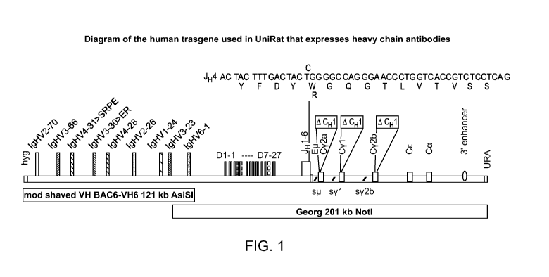
FIG. 1 is a diagram of the human transgene with a J4 gene segment (SEQ ID NO:
40) expressing a R at
position 101 in UniRatTM (SEQ ID NO: 41) that express heavy chain-only
antibodies, as described in the examples.
FIG. 2 is a diagram of the human transgene with all J segments (SEQ ID NOS 42-
47, respectively, in order
of appearance) expressing Rs at position 101 in UmRatTM that express heavy
chain-only antibodies, as described in
the examples.
FIG. 3 shows the J gene usage in UmRatTM and OmniFlicTM, where the latter is a
transgenic rat with the
same human V gene cluster as UniRatTM but expresses a fixed kappa light
chains.
FIG. 4 illustrates that a W->R mutation at the first amino acid residue of the
fourth framework region
(FR4) in heavy chain-only human antibodies inhibits association with lambda
light chain.
FIG. 5 Free lambda protein association with heavy chain antibodies in the same
CDR3 family. The figure
shows a multiple sequence alignment of 11 VH sequences from heavy chain
antibodies in the same CDR3 family
(SEQ ID NOS 48-58, respectively, in order of appearance). All of these
sequences contain a W at position 101.
7
CA 03034706 2019-02-21
WO 2018/039180
PCT/US2017/047928
FIG. 6 illustrates two structures of a chimeric antigen receptor using a human
VH extracellular binding
domain, comparing an scFv CAR-T structure (panel A) and a CAR-T structure
(panel B) using a human heavy
chain-only antibody of the present invention.
FIG. 7 shows various multi-specific HCAb constructs comprising human VH
binding domains.
DETAILED DESCRIPTION OF THE PREFERRED EMBODIMENTS
The practice of the present invention will employ, unless otherwise indicated,
conventional techniques of
molecular biology (including recombinant techniques), microbiology, cell
biology, biochemistry, and immunology,
which are within the skill of the art. Such techniques are explained fully in
the literature, such as, "Molecular
Cloning: A Laboratory Manual", second edition (Sambrook et al., 1989);
"Oligonucleotide Synthesis" (M. J. Gait,
ed., 1984); "Animal Cell Culture" (R. I. Freshney, ed., 1987); "Methods in
Enzymology" (Academic Press, Inc.);
"Current Protocols in Molecular Biology" (F. M. Ausubel et al., eds., 1987,
and periodic updates); "PCR: The
Polymerase Chain Reaction", (Mullis et al., ed., 1994); "A Practical Guide to
Molecular Cloning" (Perbal Bernard
V., 1988); "Phage Display: A Laboratory Manual" (Barbas et al., 2001).
All references cited herein, including patent applications and publications,
are incorporated by reference in
their entirety.
I. Definitions
As used herein, a "transgenic non-human animal" as defined herein is a non-
human animal capable of
producing a human or humanized heavy chain-only antibody in which the amino
acid residue at the first position of
the fourth framework region (FR4) is replaced by another residue that is
capable of disrupting a surface-exposed
hydrophobic patch comprising or associated with such residue. In one
embodiment, the native amino acid residue is
replaced by a charged amino acid residue, such as a positively charged amino
acid residue. The transgenic non-
human animal preferably is a mammal, including, without limitation, rats,
mice, bovines, monkeys, pigs, sheep, goat,
rabbits, dogs, cats, guinea pigs, hamsters and the like. Preferably, the
transgenic non-human animal is a rodent,
preferably a rat or a mouse, most preferably a UniRatTM. The choice of
transgenic animal is only limited by the
ability to produce a human or chimeric heavy chain-only human or chimeric
antibody with the FR4 mutation
described herein.
As used herein, a "genetic modification" is one or more alterations in the non-
human animal's gene
sequences. A non-limiting example is insertion of a transgene into the genome
of the transgenic animal.
As used herein, the term "transgene" refers to exogenous DNA containing a
promoter, reporter gene, poly
adenylation signal and other elements to enhance expression (insulators,
introns). This exogenous DNA integrates
into the genome of a one-cell embryo from which a transgenic animal develops
and the transgene remains in the
genome of the mature animal. The integrated transgene DNA can occur at single
or multiple places in the genome of
the egg or mouse and also single to multiple (several hundred) tandem copies
of the transgene can integrate at each
genomic location.
"Conventional antibodies" are usually heterotetrameric glycoproteins of about
150,000 daltons, composed
of two identical light (L) chains and two identical heavy (H) chains. Each
light chain is linked to a heavy chain by
8
CA 03034706 2019-02-21
WO 2018/039180
PCT/US2017/047928
one covalent disulfide bond, while the number of disulfide linkages varies
among the heavy chains of different
immunoglobulin isotypes. Each heavy and light chain also has regularly spaced
intrachain disulfide bridges. Each
heavy chain has at one end a variable domain (VH) followed by a number of
constant domains. Each light chain has
a variable domain at one end (VI) and a constant domain at its other end; the
constant domain of the light chain is
aligned with the first constant domain of the heavy chain, and the light-chain
variable domain is aligned with the
variable domain of the heavy chain. Particular amino acid residues are
believed to form an interface between the
light- and heavy-chain variable domains.
Antibody residues herein are numbered according to the Kabat numbering system
(e.g., Kabat et al.,
Sequences of Immunological Interest. 5th Ed. Public Health Service, National
Institutes of Health, Bethesda, Md.
(1991)). According to this numbering, the first amino acid residue of the FR4
region is at amino acid position 101.
The term "variable" refers to the fact that certain portions of the variable
domains differ extensively in
sequence among antibodies and are used in the binding and specificity of each
particular antibody for its particular
antigen. However, the variability is not evenly distributed throughout the
variable domains of antibodies. It is
concentrated in three segments called complementarity-determining regions
(CDRs) or hypervariable regions both
in the light-chain and the heavy-chain variable domains. The more highly
conserved portions of variable domains
are called the framework (FR). The variable domains of native heavy and light
chains each comprise four FR
regions, largely adopting a 0-sheet configuration, connected by three CDRs,
which form loops connecting, and in
some cases forming part of, the 0-sheet structure. The CDRs in each chain are
held together in close proximity by
the FR regions and, with the CDRs from the other chain, contribute to the
formation of the antigen-binding site of
antibodies (see Kabat et al., NIH Publ. No. 91-3242, Vol. 1, pages 647-669
(1991)). The constant domains are not
involved directly in binding an antibody to an antigen, but exhibit various
effector functions, such as participation of
the antibody in antibody-dependent cellular toxicity.
The term "monoclonal antibody" as used herein refers to an antibody obtained
from a population of
substantially homogeneous antibodies, i.e., the individual antibodies
comprising the population are identical except
for possible naturally occurring mutations that may be present in minor
amounts. Monoclonal antibodies are highly
specific, being directed against a single antigenic site. Furthermore, in
contrast to conventional (polyclonal) antibody
preparations which typically include different antibodies directed against
different determinants (epitopes), each
monoclonal antibody is directed against a single determinant on the antigen.
The terms "heavy chain-only antibody," "heavy chain antibody" and "HCAb" are
used interchangeably,
and refer, in the broadest sense, to antibodies lacking the light chain of a
conventional antibody. Since the
homodimeric HCAbs lack a light chain and thus a VL domain, the antigen is
recognized by one single domain, i.e.,
the variable domain of the heavy chain of a heavy chain antibody (VH or VHH
when referring to the heavy chain
variable domain of camelids). The term specifically includes, without
limitation, homodimeric antibodies
comprising the VHH antigen-binding domain and the CH2 and CH3 constant domains
or a camelid antibody, in the
absence of the CH1 domain; functional (antigen-binding) variants of such
antibodies, soluble VH variants, Ig-NAR
comprising a homodimer of one variable domain (V-NAR) and five C-like constant
domains (C-NAR) and
functional fragments thereof; and soluble single domain antibodies (sdAbs) or
nanobodies. The heavy chain-only
9
CA 03034706 2019-02-21
WO 2018/039180
PCT/US2017/047928
antibodies of the present invention comprise at least one heavy chain variable
(VH) domain in which the amino acid
residue at the first position of FR4 (amino acid residue 101 according to
Kabat numbering) is replaced by another
residue that is capable of disrupting a surface-exposed hydrophobic patch
comprising or associated with such
residue. In one embodiment, the native amino acid residue is replaced by a
charged amino acid residue, such as a
positively charged amino acid residue. The heavy chain-only antibodies of the
present invention preferably are
human or chimeric antibodies, preferably comprising a Trp (W) to Arg (R)
mutation at amino acid position 101
(W101R mutation). In one embodiment, the heavy chain-only antibodies herein
are used as a binding (targeting)
domain of a chimeric antigen receptor (CAR).
The term "soluble single domain antibody (sdAb)" is used to refer, in the
broadest sense, to polypeptides
comprising the heavy chain variable domain of a heavy-chain antibody or of a
conventional IgG, in the absence of
constant domains. The basic sdAb structure is usually comprised of four
framework regions (FR1-FR4) interrupted
by three complementary determining regions (CDR1-CDR3). Thus, a sdAb may be
represented by the following
structure: FR1-CDR1-FR2-CDR2-FR3-CDR3-FR4. For further review, see, e.g. Holt
et al, "Domain
antibodies:proteins for therapy" Trends in Biotechnology (2003):Vol. 21, No.
11:484-490.
Antibodies of the invention include multi-specific antibodies. Multi-specific
antibodies have more than one
binding specificity. The term "multi-specific" specifically includes
"bispecific" and "trispecific, " as well as
higher-order independent specific binding affinities, such as higher-order
polyepitopic specificity, as well as
tetravalent antibodies and antibody fragments. The terms "multi-specific
antibody," multi-specific single chain-only
antibody" and "multi-specific HCAb" are used herein in the broadest sense and
cover all antibodies with more than
one binding specificity.
The term "valent" as used herein refers to a specified number of binding sites
in an antibody molecule.
A "multi-valent" antibody has two or more binding sites. Thus, the terms
"bivalent", "trivalent", and
"tetravalent" refers to the presence of two binding sites, three binding
sites, and four binding sites, respectively. A
bispecific antibody according to the invention is at least bivalent and may be
trivalent, tetravalent, or otherwise
multi-valent. Multi-specific single chain-only antibodies of the present
invention, e.g., bispecific antibodies, include
multi-valent single chain-only antibodies.
The term "chimeric antigen receptor" or "CAR" is used herein in the broadest
sense to refer to an
engineered receptor, which grafts a desired binding specificity (e.g. the
antigen-binding region of a monoclonal
antibody or other ligand) to membrane-spanning and intracellular-signaling
domains. Typically, the receptor is used
to graft the specificity of a monoclonal antibody onto a T cell to create J
Nall Cancer Inst, 2015; 108(7):dvj439; and
Jackson et al., Nature Reviews Clinical Oncology, 2016; 13:370-383. A
representative CAR-T construct
comprising a human VH extracellular binding domain is shown in FIG. 6.
By "recombinant immunoglobulin (Ig) locus" is meant an Ig locus that lacks a
portion of the endogenous Ig
locus and/or comprises at least one fragment that is not endogenous to the Ig
locus in the subject mammal. Such a
fragment may be human or non-human, and may include any Ig gene segment or
portion thereof, or may constitute
the entire Ig locus. A recombinant Ig locus is preferably a functional locus
capable of undergoing gene
rearrangement and producing a repertoire of immunoglobulins in the transgenic
animal. Recombinant Ig loci include
CA 03034706 2019-02-21
WO 2018/039180
PCT/US2017/047928
recombinant Ig light chain loci and recombinant Ig heavy chain loci. Once
incorporated into the genome of a host,
an artificial Ig locus may be referred to as a recombinant Ig locus.
By "transgenic antibody" is meant an antibody encoded by a recombinant Ig
locus and produced by or
otherwise derived from a transgenic non-human mammal comprising the
recombinant Ig locus in accordance with
the invention. A transgenic antibody derived from a subject transgenic mammal
includes a transgenic antibody
produced using an isolated cell or nucleic acid obtained from the subject
transgenic animal, or using a cell or nucleic
acid derived from an isolated cell or nucleic acid obtained from the subject
transgenic animal. In a preferred
embodiment, a transgenic antibody comprises an amino acid sequence encoded by
an integrated donor
polynucleotide or portion thereof
The term "Ig gene segment" as used herein refers to segments of DNA encoding
various portions of an Ig
molecule, which are present in the germline of non-human animals and humans,
and which are brought together in B
cells to form rearranged Ig genes. Thus, "Ig gene segments" as used herein can
refer to V gene segments, D gene
segments. J gene segments and C region genes, as well as portions thereof
The term "human Ig gene segment as used herein includes both naturally
occurring sequences of a human
Ig gene segment, degenerate forms of naturally occurring sequences of a human
Ig gene segment, as well as
synthetic sequences that encode a polypeptide sequence substantially identical
to the polypeptide encoded by a
naturally occurring sequence of a human Ig gene segment. By "substantially" is
meant that the degree of amino acid
sequence identity is at least about 85%-95%. Preferably, the degree of amino
acid sequence identity is greater than
90%, more preferably greater than 95%.
The term "heavy chain-only locus" as defined herein refers to a locus encoding
a VH domain in which the
first amino acid residue of the antibody FR4 region is positively charged,
comprising one or more V gene segments,
one or more D gene segment and one or more J gene segments, optionally linked
to one or more heavy chain
effector region gene segments, each of which encodes an antibody constant
effector region lacking CH1 domain
functionality. Preferably, the heavy chain-only locus comprises from about
five to about twenty V gene fragments,
about two to about 40 D gene fragments, and about two to about twenty J gene
fragments, where the V/D/J
fragments are preferably of human origin. The terms "D gene segment" and "J
gene segment" also include within
their scope derivatives, homologues and fragments thereof as long as the
resultant segment can recombine with the
remaining components of a heavy chain antibody locus as herein described to
generate a heavy chain-only antibody.
D and J gene segments may be derived from naturally-occurring sources or they
may be synthesized using methods
familiar to those skilled in the art and described herein. In one embodiment,
in the J4 gene segment a codon for W
(TGG) is replaced by a codon for R (CGG) to encode an R instead of W at the
first amino acid position of FR4
(position 101 of the heavy chain-only antibody following Kabat numbering). D
and J gene segments may
incorporate codons for defined additional amino acid residues or defined amino
acid substitutions or deletions to
increase CDR3 diversity. The term "V gene segment" encompasses naturally
occurring V gene segments derived
from a non-human animal, such as a non-human mammal, e.g. rodent engineered to
introduce a positively charged
amino acid residue at the first residue of the FR4 region. The "V gene
segment" must be capable of recombining
11
CA 03034706 2019-02-21
WO 2018/039180
PCT/US2017/047928
with a D gene segment, a J gene segment and a heavy chain constant region,
which excludes a CH1 exon, to
generate a heavy chain-only antibody herein when the nucleic acid is
expressed.
By "human idiotype" is meant a polypeptide sequence epitope present on a human
antibody in the
immunoglobulin heavy and/or light chain variable region. The term "human
idiotype" as used herein includes both
naturally occurring sequences of a human antibody, as well as synthetic
sequences substantially identical to the
polypeptide found in naturally occurring human antibodies. By "substantially"
is meant that the degree of amino
acid sequence identity is at least about 85%-95%. Preferably, the degree of
amino acid sequence identity is greater
than 90%, more preferably greater than 95%.
By a "chimeric antibody" or a "chimeric immunoglobulin" is meant an
immunoglobulin molecule
comprising amino acid sequences from at least two different Ig loci, e.g., a
transgenic antibody comprising a portion
encoded by a human Ig locus and a portion encoded by a rat Ig locus. Chimeric
antibodies include tmnsgenic
antibodies with non-human Fc-regions or artificial Fc-regions, and human
idiotypes. Such immunoglobulins can be
isolated from animals of the invention that have been engineered to produce
such chimeric antibodies.
"Binding affinity" refers to the strength of the sum total of noncovalent
interactions between a single
binding site of a molecule (e.g., an antibody) and its binding partner (e.g.,
an antigen). Unless indicated otherwise,
as used herein, "binding affinity" refers to intrinsic binding affinity which
reflects a 1:1 interaction between
members of a binding pair (e.g., antibody and antigen). The affinity of a
molecule X for its partner Y can generally
be represented by the dissociation constant (Kd). The Kd of the HCAbs of the
present invention is typically
between about 1 pm and about 1 lam For example, the Kd can be about 200 nM,
150 nM, 100 nM, 60 nM, 50 nM, 40
nM, 30 nM, 20 nM, 10 nM, 8 nM, 6 nM, 4 nM, 2 nM, 1 nM, or stronger. Affinity
can be measured by common
methods known in the art. Low-affinity antibodies generally bind antigen
slowly and tend to dissociate readily,
whereas high-affinity antibodies generally bind antigen faster and tend to
remain.
As used herein, the "Kd" or "Kd value" refers to a dissociation constant
measured by using surface
plasmon resonance assays, for example, using a BIAcoreTm-2000 or a BIAcoreTm-
3000 (BIAcore, Inc., Piscataway,
N.J.) at 25 C. with immobilized antigen CMS chips at .about.10 response units
(RU). For further details see, e.g.,
Chen et al., J. 114bl. Biol. 293:865-881 (1999).
An "epitope " is the site on the surface of an antigen molecule to which a
single antibody molecule binds.
Generally an antigen has several or many different epitopes and reacts with
many different antibodies. The term
specifically includes linear epitopes and conformational epitopes.
"Polyepitopic specificity" refers to the ability to specifically bind to two
or more different epitopes on the
same or different target(s).
An antibody binds "essentially the same epitope " as a reference antibody,
when the two antibodies
recognize identical or sterically overlapping epitopes. The most widely used
and rapid methods for determining
whether two epitopes bind to identical or sterically overlapping epitopes are
competition assays, which can be
configured in all number of different formats, using either labeled antigen or
labeled antibody. Usually, the antigen
is immobilized on a 96-well plate, and the ability of unlabeled antibodies to
block the binding of labeled antibodies
is measured using radioactive or enzyme labels.
12
CA 03034706 2019-02-21
WO 2018/039180
PCT/US2017/047928
"Epitope mapping" is the process of identifying the binding sites, or
epitopes, of antibodies on their target
antigens. Antibody epitopes may be linear epitopes or conformational epitopes.
Linear epitopes are formed by a
continuous sequence of amino acids in a protein. Conformational epitopes are
formed of amino acids that are
discontinuous in the protein sequence, but which are brought together upon
folding of the protein into its three-
.. dimensional structure.
"Tumor", as used herein, refers to all neoplastic cell growth and
proliferation, whether malignant or benign,
and all pre-cancerous and cancerous cells and tissues. The term "tumor"
includes both solid tumors and
hematologic cancers.
The terms "cancer" and "cancerous" refer to or describe the physiological
condition in mammals that is
typically characterized by unregulated cell growth. Examples of cancer include
but are not limited to, carcinoma,
lymphoma, blastoma, sarcoma, and leukemia. More particular examples of cancers
include breast cancer, gastric
cancer, squamous cell cancer, glioblastoma, cervical cancer, ovarian cancer,
liver cancer, bladder cancer, hepatoma,
colon cancer, colorectal cancer, endometrial carcinoma, salivary gland
carcinoma, kidney cancer, renal cancer,
prostate cancer, vulval cancer, thyroid cancer, hepatic carcinoma, head and
neck cancer, rectal cancer, colorectal
cancer, lung cancer including small-cell lung cancer, non-small cell lung
cancer, adenocarcinoma of the lung and
squamous carcinoma of the lung, squamous cell cancer (e.g. epithelial squamous
cell cancer), prostate cancer,
cancer of the peritoneum, hepatocellular cancer, gastric or stomach cancer
including gastrointestinal cancer,
pancreatic cancer, glioblastoma, retinoblastoma, astrocytoma, thecomas,
arrhenoblastomas, hepatoma, hematologic
malignancies including non-Hodgkins lymphoma (NHL), multiple myeloma and acute
hematologic malignancies,
.. endometrial or uterine carcinoma, endometriosis, fibrosarcomas,
choriocarcinoma, salivary gland carcinoma, vulval
cancer, thyroid cancer, esophageal carcinomas, hepatic carcinoma, anal
carcinoma, penile carcinoma,
nasopharyngeal carcinoma, laryngeal carcinomas, Kaposi's sarcoma, melanoma,
skin carcinomas, Schwannoma,
oligodendroglioma, neuroblastomas, rhabdomyosarcoma, osteogenic sarcoma,
leiomyosarcomas, urinary tract
carcinomas, thyroid carcinomas, Wilm's tumor, as well as B-cell lymphoma
(including low grade/follicular non-
Hodgkin's lymphoma (NHL); small lymphocytic (SL) NHL; intermediate
grade/follicular NHL; intermediate grade
diffuse NHL; high grade immunoblastic NHL; high grade lymphoblastic NHL; high
grade small non-cleaved cell
NHL; bulky disease NHL; mantle cell lymphoma; AIDS-related lymphoma; and
Waldenstrom's
Macroglobulinemia); chronic lymphocytic leukemia (CLL); acute lymphoblastic
leukemia (ALL); Hairy cell
leukemia; chronic myeloblastic leukemia; and post-transplant
lymphoproliferative disorder (PTLD), as well as
abnormal vascular proliferation associated with phakomatoses, and Meigs'
syndrome.
Detailed Description
The HCAbs of the invention are human or chimeric having the native amino acid
residue at the first
position of the FR4 region (amino acid position 101 according to the Kabat
numbering system), substituted by
another amino acid residue, which is capable of disrupting a surface-exposed
hydrophobic patch comprising or
associated with the native amino acid residue at that position. Such
hydrophobic patches are normally buried in the
interface with the antibody light chain constant region but become surface
exposed in HCAbs and are, at least
13
CA 03034706 2019-02-21
WO 2018/039180
PCT/US2017/047928
partially, for the unwanted aggregation and light chain association of HCAbs.
The substituted amino acid residue
preferably is charged, and more preferably is positively charged. The
resultant HCAbs preferably have high
antigen-binding affinity and solubility under physiological conditions in the
absence of aggregation.
Specifically included are heavy-chain only antibodies lacking the camelid VHH
framework and mutations,
and their functional VH regions. Such heavy-chain only antibodies can, for
example, be produced in transgenic rats
or mice which comprise fully human heavy chain-only gene loci as described,
e.g. in W02006/008548, but other
transgenic mammals, such as rabbit, guinea pig, rat can also be used, rats and
mice being preferred.. Heavy chain
only antibodies, including their VHH or VH functional fragments, can also be
produced by recombinant DNA
technology, by expression of the encoding nucleic acid in a suitable
eukaryotic or prokaryotic host, including E. coli
or yeast.
Domains of heavy-chain only antibodies combine advantages of antibodies and
small molecule drugs: can
be mono- or multi-valent; have low toxicity; and are cost-effective to
manufacture. Due to their small size, these
domains are easy to administer, including oral or topical administration, are
characterized by high stability,
including gastrointestinal stability; and their half-life can be tailored to
the desired use or indication. In addition,
VH and VHH domains of HCAbs can be manufactured in a cost effective manner.
In one embodiment, domains of HCAbs are nanobodies, as hereinabove defined.
In one embodiment, the HCAb binding domains are part of a multi-specific
binding protein that bind to
multiple different epitopes of the same target antigen or multiple different
epitopes on more than one target antigen.
In one embodiment, the antibody is a bispecific antibody. Various multi-
specific structures comprising human VH
binding domains are illustrated in FIG. 7. The multi-specific, or bi-specific,
HCAbs of the present invention may,
for example, bind to two or more sites on the same soluble target, or two or
more sites on the same cell surface
(receptor) target, such as tumor antigen, or one or more soluble targets and
one or more cell surface receptor targets.
In certain embodiments, the bispecific HCAb structures herein have the
following binding affinities: epithelial cell
adhesion molecule (EpCam) x CD3; CD19 x CD3; EpCam x CD3; TNF-a x IL-17; IL-la
x IL-10; CD30 x CD16A;
human epidermal growth factor receptor 2 (HER2) x HER3; IL-4 x IL-13;
angiopoietin 2 (Ang-2) x vascular
endothelial growth factor a (VEGF-A); Factor IXa x Factor X; epidermal growth
factor receptor (EGFR) x HER3;
IL-17A x IL-17F; HER2 x HER3; carcinoembryonic antigen x CD3; CD20 x CD3;
CD123 x CD3; BCMA xCD3,
PSMA x PSCA x CD3, PSMA x CD3, PSCA x CD3, CD19 x CD22 x CD3, CD22 x CD3, CD38
x PD1, CD38 x
PD-L1, CD38 x CD73, CD38 x CD39, PD1 x CD39 x CD73, and PD1 x CD73.
In a preferred embodiment, the HCAbs herein are produced by transgenic
animals, including transgenic
mice and rats, preferably rats, in which the endogenous immunoglobulin genes
are knocked out or disabled. In a
preferred embodiment, the HCAbs herein are produced in UniRatTM. UniRatTM have
their endogenous
immunoglobulin genes silenced and use a human immunoglobulin heavy-chain
translocus to express a diverse,
naturally optimized repertoire of fully human HCAbs. While endogenous
immunoglobulin loci in rats can be
knocked out or silenced using a variety technologies, in UniRatTM the zinc-
finger (endo)nuclease (ZNF) technology
was used to inactivate the endogenous rat heavy chain J-locus, light chain CK
locus and light chain a locus. ZNF
constructs for microinjection into oocytes can produce IgH and IgL knock out
(KO) lines. For details see, e.g.
14
CA 03034706 2019-02-21
WO 2018/039180
PCT/US2017/047928
Geurts et al., 2009, Science 325:433 Characterization of Ig heavy chain
knockout rats has been reported by Menoret
et al., 2010, Eur. J. Immunol. 40:2932-2941. Advantages of the ZNF technology
are that non-homologous end
joining to silence a gene or locus via deletions up to several kb can also
provide a target site for homologous
integration (Cui et al., 2011, Nat Biotechnol 29:64-67. UniRatTM HCAbs bind
epitopes that cannot be attacked with
conventional antibodies. Their high specificity, affinity, and small size make
them ideal for mono- and poly-specific
applications.
In the heavy chain-only antibodies of the present invention the native amino
acid residue at the first
position of the fourth framework region (FR4) is replaced by a different amino
acid residue, that is capable of
disrupting a surface-exposed hydrophobic patch comprising or associated with
the native amino acid residue at that
position. In one embodiment, the substituted amino acid residue is charged. In
another embodiment, the substituted
amino acid residue is positively charged, such as lysine (Lys, K), arginine
(Arg, R) or histidine (His, H), preferably
arginine (R). As shown in the alignment of FIG. 5 VH sequences from heavy
chain antibodies in the same CDR3
family all contain a Trp (W) at position 101, thus in a preferred embodiment
the heavy chain-only antibodies derived
from the transgenic animals of the present invention contain a Trp to Arg
mutation at position 101.
The human or chimeric heavy chain-only antibodies of the present invention can
be generated against any
desired target antigen and have great potential for a variety of clinical
applications. Target antigens for therapeutic
applications include, without limitation, EGFR, ErbB2 (HER2), ErbB3 (HER3),
ErbB4 (HER4), CTLA-4/CD152,
RANKL, TNF-a, CD20, IL-12/IL-23, IL1-0, IL-17A, IL-17F, CD38, NGF, IGF-1, IL-
12, CD20, CD30, CD39,
CD73, CD40, PD-1, PDL-1, PD-L2, BCMA, BTLA, thymic stromal lymphopoietin
(TSP), Follicle Stimulating
Hormone Receptor (FSHR), Prostate Specific Membrane Antigen (PSMA), Prostate
Stem Cell Antigen (PSCA),
CD137, OX-40, and IL-33. Therapeutic indications include, without limitation,
treatment of solid tumors,
hematologic tumors, inflammatory diseases, such as rheumatoid arthritis,
psoriasis, Crohn's disease, ulcerative
colitis, metabolic disorders, cardiovascular diseases, respiratory,
dermatologic, central nervous system, hematologic,
eye/ear, liver diseases.
Target tumors include, for example, breast cancer, gastric cancer, squamous
cell cancer, glioblastoma,
cervical cancer, ovarian cancer, liver cancer, bladder cancer, hepatoma, colon
cancer, colorectal cancer, endometrial
carcinoma, salivary gland carcinoma, kidney cancer, renal cancer, prostate
cancer, vulval cancer, thyroid cancer,
hepatic carcinoma, head and neck cancer, rectal cancer, colorectal cancer,
lung cancer including small-cell lung
cancer, non-small cell lung cancer, adenocarcinoma of the lung and squamous
carcinoma of the lung, squamous cell
cancer (e.g. epithelial squamous cell cancer), prostate cancer, cancer of the
peritoneum, hepatocellular cancer,
gastric or stomach cancer including gastrointestinal cancer, pancreatic
cancer, glioblastoma, retinoblastoma,
astrocytoma, thecomas, arrhenoblastomas, hepatoma, hematologic malignancies
including non-Hodgkins lymphoma
(NHL), multiple myeloma (MM) and acute hematologic malignancies, endometrial
or uterine carcinoma,
endometriosis, fibrosarcomas, choriocarcinoma, salivary gland carcinoma,
vulval cancer, thyroid cancer, esophageal
carcinomas, hepatic carcinoma, anal carcinoma, penile carcinoma,
nasopharyngeal carcinoma, laryngeal carcinomas,
Kaposi's sarcoma, melanoma, skin carcinomas, Schwannoma, oligodendroglioma,
neuroblastomas,
rhabdomyosarcoma, osteogenic sarcoma, leiomyosarcomas, urinary tract
carcinomas, thyroid carcinomas, Wilm's
CA 03034706 2019-02-21
WO 2018/039180
PCT/US2017/047928
tumor, as well as B-cell lymphoma (including low grade/follicular non-
Hodgkin's lymphoma (NHL); small
lymphocytic (SL) NHL; intermediate grade/follicular NHL; intermediate grade
diffuse NHL; high grade
immunoblastic NHL; high grade lymphoblastic NHL; high grade small non-cleaved
cell NHL; bulky disease NHL;
mantle cell lymphoma; AIDS-related lymphoma; and Waldenstrom's
Macroglobulinemia); chronic lymphocytic
leukemia (CLL); acute lymphoblastic leukemia (ALL); Hairy cell leukemia;
chronic myeloblastic leukemia; and
post-transplant lymphoproliferative disorder (PTLD), as well as abnormal
vascular proliferation associated with
phakomatoses, and Meigs' syndrome.
Further details of the invention are illustrated by the following non-limiting
Examples, which use the
following abbreviations:
BAC Bacterial artificial chromosome
YAC Yeast artificial chromosome
ZFN Zinc-finder nuclease
Heavy chain
Constant region
V Variable region
Diversity segment
Joining segment
EXAMPLES
Example 1: Generation of genetically engineered rats expressing heavy chain-
only antibodies
(HCAbs)
Previously identified, characterized and, in part, modified BACs and YACs
accommodate human heavy
chain variable region genes and rat constant region genes (Osborn et al.,
2013, J. Immunol. 190:1481-1490; Ma et al.,
2013, J. Immunol. Methods 400-401:78-86). To enable heavy-chain antibody
expression, a rat constant region BAC
was modified by removal of Cjt and deletion of CH1 exons in all Cys. Heavy-
chain-only expression was then
enforced by silencing of the endogenous heavy and light chain (kappa and
lambda) loci.
Construction of modified human IgH loci on YACs and BACs.
A 'human ¨ rat' IgH locus was constructed and assembled in several parts. This
involved the modification
and joining of rat C region genes downstream of human JHs and subsequently,
the upstream addition of the human
VH6 ¨D-segment region. Two BACs with separate clusters of human VH genes [BAC6
and BAC3] were then co-
injected with the BAC termed Georg, encoding the assembled and modified region
comprising human VH6 ¨ all Ds
¨ all JHs - rat Cy2a/1/2b (ACH1).
For the introduction of modifications at precise locations in the DNA sequence
and for simultaneously
joining multiple large DNA regions, technologies were developed to assemble
sequences with overlapping ends in S.
cerevisiae as circular YAC (cYAC) and, subsequently, to convert such cYACs
into BACs. Advantages of YACs
16
CA 03034706 2019-02-21
WO 2018/039180
PCT/US2017/047928
include their capacity to retain large DNA inserts, the ease of homologous
alterations in the yeast host and the
maintenance of sequence stability especially in the highly repetitive regions
(e.g. switch regions, enhancers). On the
other hand BACs, propagated in E. coli, offer the advantages of easy
preparation and large yield. In addition,
detailed restriction mapping and sequence analysis can be better achieved from
BACs than YACs. Two self-
replicating S. cerevisiae/E. Coli shuttle vectors, pBelo-CEN-URA and pCAU were
constructed. Briefly, S.
cerevisiae CEN4 was cut out as an AvrII fragment from pYAC-RC (Marchuk and
Collins, 1988; Nucleic Acids Res.
16(15):7743) and ligated to SpeI¨linearised pAP599 (Ma et al. MolMicrobiol.
2007; 66(1):14-25). The resulting
plasmid contains CEN4 cloned downstream of URA3. From this, an ApaLI¨BamHI
URA3 - CEN4 fragment was
cut out, and ligated to ApaLI and BamHI digested pBACBeloll (New England
Biolabs) to yield pBelo-CEN-URA.
The S. cerevisiae autonomously replicating sequence ARS209 was synthesized and
cloned into a unique SexAI site
in pBelo-CEN-URA to yield pCAU.
To facilitate the modifications of human JH4, rat Cp. and Cyl regions, a ¨ 37
kb SacII-fragment spanning
from ¨ 2.2 kb upstream of the human JHs to ¨ 5.5 kb downstream of the rat Cyl
coding region was cut out from the
BAC construct Annabel (Osborn et al., 2013; J. Immunol. 190:1481-1490) and
cloned into a unique SacII site in
pBelo-CEN-URA [pBelo + SacII, 37 kb]. In addition, to modify the rat Cy2b
region, a ¨ 19 kb SacII ¨ SwaI
fragment from Annabel spanning from ¨ 6.9 kb upstream of the y2b switch region
to ¨ 2.0 kb downstream of the
Cy2b coding region was cloned into SacII and HpaI ¨ double digested pBelo-CEN-
URA [pBelo + SacII ¨ SwaI, 19
kb]. Both plasmids were used as templates for amplifying various human and rat
genomic regions and to establish
the required restriction fragments.
The DNA region spanning from ¨ 3.1 kb upstream of the human JHs and including
rat Cji with some 3'
region was modified and assembled in pCAU as a 16.7 kb SnaBI ¨ FspI fragment.
The modified region includes all
authentic human JHs except a T ¨> C point mutation being introduced into JH4
(resulting into a W ¨> R amino acid
change) followed by the rat intergenic region from the JHs until [ECM, which
was deleted along with the rest of Cji
coding region and replaced precisely by rat Cy2a sequence lacking CH1
(starting from the intron immediately
upstream of Hinge to the 3' end of the membrane exons). This construct was
derived by the assembly of the
following 5 overlapping fragments in yeast as cYAC and then converted into a
BAC: an amplified ¨ 4.3 kb fragment
using primers HC27 ¨ 1 and ¨2 covering the region upstream of human JH to
mutated JH4 (the point mutation
introduced via the latter primer indicated by
an amplified ¨3.4 kb fragment using primers HC27 ¨ 3 and ¨4
spanning from mutated JH4 (indicated by to upstream of the [t switch
region, a ¨5.2 kb AflII ¨ fragment
encompassing the [t switch region and the flanking sequences cut out from
pBelo + SacII 37 kb, an amplified rat
Cy2a lacking CH1 fused to sequences flanking rat Cji using long primers HC27 ¨
5 and ¨6, and amplification of the
pCAU vector using primers HC27 ¨7 and ¨ 8. This resulted in pCAU + HuJ - Rat
Cy2a(-CH1). All modified
regions were checked by sequencing to confirm the accuracy.
Rat Cyl lacking CH1 and Cy2b lacking CH1 were individually generated via PCR.
A ¨1.7 kb fragment
located immediately upstream of the Cyl coding region with a 3' tail matching
the 5' end of the intron between CH1
and hinge was amplified using primers HC27 ¨ 9 and ¨ 10. Cyl was amplified as
a ¨ 3.9 kb fragment from the
17
CA 03034706 2019-02-21
WO 2018/039180
PCT/US2017/047928
intron between CH1 and hinge to the 3' end of the coding region using primers
HC27 ¨ 11 and ¨ 12. Subsequently,
the ¨ 1.7 kb and ¨ 3.9 kb fragments were both gel purified and joined via
overlapping PCR using primers HC27 ¨ 9
and ¨ 12 to yield a ¨ 5.6 kb fragment. Similarly, for Cy2b without CH1, a ¨
0.3 kb fragment upstream of the Cy2b
coding region was amplified using primers HC27 ¨ 13 and ¨ 14, and a ¨ 5.4 kb
fragment - spanning the area from
the intron between CH1 and hinge to the 3' end of the coding region - was
amplified using primers HC27 ¨ 15 and ¨
16, and subsequently, these two fragments were joined via overlapping PCR
using primers HC27 ¨ 13 and ¨ 16 to
yield a ¨ 5.7 kb fragment. pCAU + Rat Cyl, 2b(-CH1s) was constructed to
contain the following: 100 bp homology
region matching the 3' end of rat Cp., followed by Cyl and Cy2b in the genomic
configuration except the CHls of
both were deleted. Six overlapping fragments were used to construct pCAU + Rat
Cyl, 2b(-CH1s): a ¨ 10.2 kb SpeI
¨ Nan I fragment spanning from the 3' Cia homology region followed by the yl
switch region cut out from pBelo +
SacII, 37 kb, the ¨5.6 kb PCR fragment containing Cyl without CH1 as described
above, an amplified ¨ 7.4 kb
fragment covering the intergenic region between Cyl and Cy2b using primers
HC27 ¨ 17 and ¨ 18, a ¨ 11.3 kb XhoI
fragment encompassing the rat Cy2b switch region cut out from pBelo + SacII ¨
SwaI 19 kb, the ¨ 5.7 kb PCR
fragment containing Cy2b without CH1, and the amplified pCAU vector using
primers HC27 ¨ 19 and ¨ 20. The rat
genomic region in pCAU + Rat Cyl, 2b(-CH1s) can be cut out as a single ¨ 40 kb
FspI fragment.
Finally, the BAC (Georg) encoding the human VH6 -Ds - JHs-rat C regions with
all the modifications was
assembled using the following four overlapping fragments: a purified ¨ 78.2 kb
FspAI ¨ MluI fragment
encompassing the human VH6 ¨Ds region cut out from BAC10 (CTD-3216M13,
Invitrogen), the 16.7 kb SnaBI ¨
FspI fragment cut out from pCAU + HuJ - Rat Cy2a(-CH1) as described above, the
¨ 40 kb FspI fragment cut out
from pCAU + Rat Cyl, 2b(-CH1s), and a purified ¨ 77.2 kb SwaI ¨ SacII fragment
cut out from construct Annabel
which includes the intergenic region between Cy2b and CE followed by CE, C,
the 3' enhancer region, the pBelo-
CEN-URA vector, and the 5' region upstream of human VH6. This final construct
was checked extensively via
restriction mapping and partial sequencing. The (human VH6 -Ds - JHs-rat C)
region can be cut out and purified as a
¨ 201 kb NotI fragment.
BAC6 contains the human genomic region from VH4-39 to VH3-23, while BAC3
contains a downstream
region from VH3-11 to VH6-1 (the most D proximal VH gene). To provide an
overlap between BAC6 and BAC3, a
10.6 kb fragment located at the 5' end of the human VH loci in BAC3 was
integrated downstream of VH3-23 in
BAC6 as described previously (Osborn et al. 2013, supra). The human VH genes
in BAC6 were cut out as a ¨182-kb
AsiSI -AscI fragment. BAC3 was unmodified and the human VH genes in this BAC
were cut out as a ¨ 173 kb NotI
¨ fragment.
Oligonucleotides:
HC27 ¨1: GTATTACACACAAAATGGGAAAAGCTG (SEQ ID NO: 1)
HC27 ¨2: CCK1GTAGTCAAAGTAGTCACATTGTGGGAGGC (SEQ ID NO: 2)
HC27 ¨ 3:
CCTTAATGGGGCCTCCCACAATGTGACTACTTTGACTACGGGGCCAGGGAACCCTGGTCACCG (SEQ
ID NO: 3)
18
CA 03034706 2019-02-21
WO 2018/039180
PCT/US2017/047928
HC27 ¨4: GAATCCTAGGATTGCCTTCTTAGCCTG (SEQ ID NO: 4)
HC27 ¨5:
CCATAGACCAAACTTACCTACTATCTAGTCCTGCCAACCTTAAGAGCAGCAACATGGAGACAGCAGAG
TGTAGAGAGATCTCCTGACTGGCAGGAGGCAAGAAGATGGATTCTTACTCGTCCATTTCTCTTTTATCC
CTCTCTGGTCCTAGAGAACAACCAGGGGATGAGGGGCTC (SEQ ID NO: 5)
HC27 ¨6:
GCACAAGTGGACAAAGTCTTTGGCCAGTCTAGAAAGAAGCCCGTCTCAGAGATCAAAGCTGGAGGGC
AACACAGGAAAGATGTGGGAATAAGTTTACTAGTCATACAGGCAGGAACCCCAGGCCCAGAGGTAGT
GTCCCTGTGGGAGGGTCTCTTGCTCTCTGATGTCCTTCCATGCTGAGAGTTAGGGCCCTTGTCCAATCA
TGTTC (SEQ ID NO: 6)
HC27 ¨7:
GAATTTTGCCCAAGTTTTTTCAGCTTTTCCCATTTTGTGTGTAATACGTACACACCGCAGGGTAATAAC
TG (SEQ ID NO:?)
HC27 ¨ 8:
GACGGGCTTCTTTCTAGACTGGCCAAAGACTTTGTCCACTTGTGCGCAGTTATCTATGCTGTCTCACCA
TAGAG (SEQ ID NO: 8)
HC27 ¨9: GGAGGTCTAGGCTGGAGCTGATCCAG (SEQ ID NO: 9)
HC27 ¨ 10: CCTCGTCCCCTGGTTGTTCTCTCAAGAAAAAGTATGCGTGATCATTTTGTC (SEQ ID
NO: 10)
HC27 ¨ 11: AGAGAACAACCAGGGGACGAGG (SEQ ID NO: 11)
HC27 ¨ 12: GTCCACATAGTCCTCCAGAGAGAGAAG (SEQ ID NO: 12)
HC27 ¨ 13: GACCCAAGTCCAGTTCCCAACAACCAC (SEQ ID NO: 13)
HC27 ¨ 14: CCTCGTCCCCTGGTTGTCCTCTCAAGAGAGGAGGGAGTGTGAGCTTTTCC (SEQ ID
NO: 14)
HC27 ¨ 15: AGAGGACAACCAGGGGACGAGGGGCTC (SEQ ID NO: 16)
HC27 ¨ 16: GCATGGGGAAGGGGCATTGTATGTAGG (SEQ ID NO: 17)
HC27 ¨ 17: CAGATCACACTGTCTGCTCACTTCAC (SEQ ID NO: 18)
HC27 ¨ 18: AAGGCAGCAGGATGGAAGCTGATGTCG (SEQ ID NO: 19)
HC27 ¨ 19:
GCTGGAGGGCAACACAGGAAAGATGTGGGAATAAGTTTACTAGTCATACAGGCAGGAACCCCAGGCC
CAGAGGTAGTGTCCCTGTGGGAGGGTCTCTTGCGCACACACCGCAGGGTAATAACTG (SEQ ID NO: 20)
HC27 ¨ 20:
GATTTAAATGTCAATTGGTGAGTCTTCTGGGGCTTCCTACATACAATGCCCCTTCCCCATGCGCAGTTA
TCTATGCTGTCTCACCATAGAG (SEQ ID NO: 21)
Construction of Georg II
19
CA 03034706 2019-02-21
WO 2018/039180
PCT/US2017/047928
A 'human ¨ rat' IgH locus was constructed and assembled in several parts. This
involved the modification
and joining of rat C region genes downstream of mutated human Jill ¨ JH6 each
containing a point mutation encoding
W ¨> R and subsequently, the upstream addition of the human VH6-1 ¨D-segment
region. This BAC is named as
Georg II.
Firstly, the mutated human JH1 JI-16 is synthesized in a 2.3 kb ScaI fragment
(Thermo Fisher Scientific, see
below).
Secondly, the mutated human Jill ¨ JH6is joined with ¨ 3.1 kb DNA region
upstream of human JHs and ¨
11.4 kb region spanning from rat sequence immediately downstream of JHs to rat
Cjt whose coding region has been
replaced by rat Cy2a coding region but lacking CH1 in a BAC termed pCAU +
HuJ(W-R)-Rat Cy2a(-CH1). The
entire modified region in this BAC can be cut out as a 16.7 kb SnaBI ¨ FspI
fragment. This BAC was derived by the
assembly of the following 4 overlapping fragments in yeast as cYAC and then
converted into a BAC: an amplified
2.7 kb fragment using primers HC32 ¨ 1 and ¨2 covering the region upstream of
human JHõ the 2.3 kb mutated
human Jill ¨ hi6 _ ScaI fragment, an amplified ¨2.1 kb fragment using primers
HC32 ¨ 3 and ¨ 4 covering rat region
immediately downstream of his, and a ¨ 18.4 kb MluI ¨ HpaI fragment cut out
from pCAU + HuJ - Rat Cy2a(-CH1)
(containing rat Cjt locus with coding region replaced by rat Cy2a coding
region lacking CH1 and pCAU vector,
described in 11C27 construction method'). All modified regions were checked by
sequencing to confirm the
accuracy.
Finally, Georg II encoding the human VH6-1 -Ds ¨ mutated JHs ¨ modified rat C
regions was assembled
using the following 4 overlapping fragments: a purified ¨ 78.2 kb FspAI ¨ MluI
fragment encompassing the human
VH6-1 ¨Ds region cut out from BAC10 (CTD-3216M13, Invitrogen), the 16.7 kb
SnaBI ¨ FspI fragment cut out
from pCAU + HuJ(W-R)-Rat Cy2a(-CH1) as described above, the ¨ 40 kb FspI
fragment cut out from pCAU + Rat
Cyl, 2b(-CH1s) (described in 11C27 construction method'), and a purified ¨
77.2 kb SwaI ¨ SacII fragment cut out
from construct Annabel which includes the intergenic region between Cy2b and
CE followed by CE, Ca, the 3'
enhancer region, the pBelo-CEN-URA vector, and the 5' region upstream of human
VH6-1. This final construct was
checked extensively via restriction mapping and partial sequencing. The (human
VH6-1 -Ds ¨ mutated JHs- modified
rat C) region can be cut out and purified as a ¨ 201 kb NotI fragment.
Microinjection to generate HC32 and HC33 transgenic rats
BAC6 contains the human genomic region from VH4-39 to VH3-23, while BAC3
contains a downstream
region from VH3-11 to VH6-1 (the most D proximal VH gene). To provide an
overlap between BAC6 and BAC3, a
10.6 kb fragment located at the 5' end of the human VH loci in BAC3 was
integrated downstream of VH3-23 in
BAC6 as described previously (Osborn et al. 2013, J. Immunol. 190:1481-1490).
The human VH genes in BAC6
were cut out as a ¨182-kb AsiSI -AscI fragment. BAC3 was unmodified and the
human VH genes in this BAC were
cut out as a ¨ 173 kb NotI ¨ fragment. Both fragments were purified and co-
injected with the ¨ 201 kb NotI
fragment from Georg II into rat embryos to construct HC32.
BAC9 contains the human genomic region from VH3-74 to VH3-53. BAC(14+5)
contains a downstream
region from VH3-53 to VH3-13 and a 6.1 kb region immediately upstream of VH6-1
was added to its 3' to provide
CA 03034706 2019-02-21
WO 2018/039180
PCT/US2017/047928
an overlap to Georg II. The human VH region in BAC9 was cut out as a ¨185 kb
NotI fragment, and that from
BAC(14+5) was cut out as a ¨ 209 kb BsiwI fragment. Both fragments were
purified and co-injected with the ¨ 201
kb NotI fragment from Georg II into rat embryos to construct HC33.
DNA purification
Linear YACs, circular YACs and BAC fragments after digests, were purified by
electro-elution using
ElutrapTm (Schleicher and Schuell) (Gu et al., J. Biochem. Biophys
Methods.,1992, 24:45-50) from strips cut from
0.8% agarose gels run conventionally or from pulsed-field-gel electrophoresis
(PFGE). The DNA concentration was
usually several ng/Ial in a volume of ¨100 1. For fragments up to ¨200 kb the
DNA was precipitated and re-
dissolved in micro-injection buffer (10 mM Tris-HC1 pH 7.5, 100 mM EDTA pH 8
and 100 mM NaCl but without
Spermine/Spermidine) to the desired concentration.
The purification of circular YACs from yeast was carried out using Nucleobond
AX silica-based anion-
exchange resin (Macherey-Nagel, Germany). Briefly, spheroplasts were made
using zymolyase or lyticase and
pelleted (Davies et al., 1996, Human antibody repertoires in transgenic mice:
manipulation of transfer of YACs. In
Antibody Engineering: A Practical Approach. J. McCafferty, H.R. Hoogenboom,
and D. J. Chiswell eds. IRL,
Oxford, U.K., p. 59-76). The cells then underwent alkaline lysis, binding to
AX100 column and elution as described
in the Nucleobond method for a low-copy plasmid. Contaminating yeast
chromosomal DNA was hydrolyzed using
Plasmid _SafeTM ATP-Dependent DNase (Epicentre Biotechnologies) followed by a
final cleanup step using
SureClean (Bioline). An aliquot of DH10 electrocompetent cells (Invitrogen)
was then transformed with the circular
YAC to obtain BAC colonies. For the separation of the insert DNA for
microinjection, 150-200 kb, from BAC
vector DNA, ¨10 kb, a filtration step with Sepharose 4B-CL was used (Yang et
al., 1997, Nat. Biotechnol. 1997,
15;859-865).
Gel analyses
Purified YAC and BAC DNA was analyzed by restriction digest and separation on
conventional 0.7%
agarose gels (Sambrook and Russell, 2001). Larger fragments, 50-200 kb, were
separated by PFGE (Biorad Chef
MapperTm) at 8 C, using 0.8% PFC Agarose in 0.5% TBE, at 2-20 sec switch time
for 16 h, 6V/cm, 10mA.
Purification allowed a direct comparison of the resulting fragments with the
predicted size obtained from the
sequence analysis. Alterations were analyzed by PCR and sequencing.
Microinjection
Outbred SD/Hsd strain animals were housed in standard microisolator cages
under approved animal care
protocols in animal facility that is accredited by the Association for the
Assessment and Accreditation for
Laboratory Animal Care (AAALAC). The rats were maintained on a 14-10 h
light/dark cycle with ad libitum access
to food and water. Four to five week old SD/Hsd female rats were injected with
20-25 IU PMSG (Sigma-Aldrich)
followed 48 hours later with 20-25 IU hCG (Sigma-Aldrich) before breeding to
outbred SD/Hsd males. Fertilized 1-
cell stage embryos were collected for subsequent microinjection. Manipulated
embryos were transferred to
pseudopregnant SD/Hsd female rats to be carried to parturition.
21
CA 03034706 2019-02-21
WO 2018/039180
PCT/US2017/047928
Purified DNA encoding recombinant immunoglobulin loci was resuspended in
microinjection buffer with
mM Spermine and 10 mM Spemidine. The DNA was injected into fertilized oocytes
at various concentrations
from 0.5 to 3 ng/itl.
Plasmid DNA or mRNA encoding ZFNs specific for rat immunoglobulin genes were
injected into fertilized
5 oocytes at various concentrations from 0.5 to 10 ng/ul.
Zinc-finger Nucleases (ZFNs)
ZFNs specific for rat immunoglobulin genes were generated.
The ZFN specific for rat Ckappa had the following binding site:
ATGAGCAGCACCCTCtcgttgACCAAGGCTGACTATGAA (SEQ ID NO: 22)
10 ZFNs specific for rat J-locus sequences had the following binding sites:
CAGGTGTGCCCATCCagctgaGTTAAGGTGGAG (SEQ ID NO: 23)
and
CAGGACCAGGACACCTGCAgcagcTGGCAGGAAGCAGGT (SEQ ID NO: 24)
ZFNs specific for rat Cy-locus sequences had the following binding sites:
AACAGCCATTTGcagaccAAAGGGAAGGAAAGA (SEQ ID NO: 25)
and
TTCTACCCTGGTGTTATGacagtgGTCTGGAAGGCAGATGGT (SEQ ID NO: 26)
Rats with transloci.
Transgenic rats carrying artificial heavy chain immunoglobulin loci in
unrearranged configuration were
generated. RT-PCR and serum analysis (ELISA) of transgenic rats revealed
productive rearrangement of transgenic
immunoglobulin loci and expression of heavy chain only antibodies of various
isotypes in serum. Immunization of
transgenic rats resulted in production of high affinity antigen-specific heavy
chain only antibodies.
Novel Zinc-finger-nuclease knock-out technology.
For further optimization of heavy chain-only antibody generation in transgenic
rats, knockout rats with
inactivated endogenous rat immunoglobulin loci were generated.
For the inactivation of rat heavy immunoglobulin heavy chain expression and
rat 2 light chain expression,
ZFNs were microinjected into single cell rat embryos. Subsequently, embryos
were transferred to pseudopregnant
female rats and carried to parturition. Animals with mutated heavy chain and
light chain loci were identified by
PCR. Analysis of such animals demonstrated inactivation of rat immunoglobulin
heavy and light chain expression in
mutant animals.
22
CA 03034706 2019-02-21
WO 2018/039180
PCT/US2017/047928
Example 2: Generation of antigen-specific heavy chain-only antibodies (HCAbs)
in rats
For the generation of antigen-specific heavy chain-only antibodies in rats,
genetically engineered rats
expressing heavy chain only antibodies, such as those described in Example 1,
are immunized in various ways.
Immunization with inactivated virus
Influenza viruses with various different hemagglutinin and neuraminidase genes
is provided by the
Immunology and Pathogenesis Branch, Influenza Division, CDC, Atlanta, GA.
Virus stock is propagated in the
allantois cavities of 10-day-old embryonated chicken eggs and purified through
a 10%-50% sucrose gradient by
means of ultracentrifugation. Viruses are resuspended in phosphate-buffered
saline and inactivated by treatment
with 0.05% formalin at 4 C for 2 weeks. Inactivated virus and alumn solution
(Pierce) are mixed in a 3:1 ratio and
incubated at room temperature for 1 h before immunization. Genetically
engineered rats expressing heavy chain-
only antibodies are immunized with whole inactive.
Immunization with proteins or peptides
Typically immunogens (proteins or peptides like human immunoglobulin kappa
light chain, human IgM or
IgG heavy chain, human serum albumin, protein-peptide conjugates) are diluted
to 0.05-0.15 ml with sterile saline
and combined with adjuvant to a final volume of 0.1-0.3ml. Many appropriate
adjuvants are available (i.e. heat
inactivated Bordetella pertussis, aluminium hydroxide gel, Quil A or saponin,
bacterial lipopolysaccharide or anti-
CD40) but none have the activity of Complete Freund's Adjuvant (CFA) and
Incomplete Freund's Adjuvant (IFA).
The concentration of soluble immunogens such as proteins and peptides may vary
between 5 lag and 5mg in the final
preparation. The first immunization (priming) with immunogen in CFA is
administered intraperitoneally and/or
subcutaneously and/or intramuscularly. If intact cells are used as immunogens
they are best injected
intraperitoneally and/or intravenously. Cells are diluted in saline and 1 to
20 million cells are administered per
injection. Cells that survive in the rat will yield best immunization results.
After the first immunization with
immunogens in CFA a second immunization in IFA (booster) is usually delivered
4 weeks later. This sequence
leads to the development of B cells producing high affinity antibodies. If the
immunogen is weak booster
immunizations are administered every 2 weeks until a strong humoral response
is achieved. The immunogen
concentrations can be lower in booster immunizations and intravenous routes
can be used. Serum is collected from
rats every 2 weeks to determine the humoral response.
Immunization with the Lipid II pentapeptides, Penicillin Binding Proteins,
beta-lactamases, Sortases, and
other membrane proteins of prokaryotes is conducted as outlined above.
Immunization with Lipids, Glycolipids, and Carbohydrate Polymers
Immunization with poly-N-acetyl-0-(1-6)-glucosamine (PNAG) and other
Carbohydrate polymers
Purified dPNAG, PNAG or other carbohydrate polymers are conjugated to
Diptheria Toxin, albumin or
other carrier protein by reductive amination. Aldehyde groups are first
introduced onto the surface of the carrier
protein by treatment with glutaraldehyde. Activated carrier proteins are
subsequently reacted with dPNAG (or other
carbohydrate polymers) through its free amino groups in the presence of the
reducing agent sodium NaCNBH3.
Animals are immunized with PNAG and dPNAG-DT conjugates subcutaneously or
footpads at day 0 and boosted at
subsequent weeks with 0.15-100-pg doses of conjugated PNAG or dPNAG in
Complete Freunds Adjuvant or other
23
CA 03034706 2019-02-21
WO 2018/039180
PCT/US2017/047928
Adjuvants. Boost immunizations are preferably in Incomplete Freunds. Blood is
withdrawn weekly specific
antibody titers are determined by ELISA. B cells are recovered from draining
lymph nodes or spleen and
hybridomas generated using standard methods. Alternatively, yeast cells are
used to select for antigen-binding
antibodies or fragments.
Immunization with Lipid II
Purified Lipid II with a C55 or shorter lipid tail are mixed with Titermax,
liposomes, Ribi adjuvant or other
adjuvants. In addition, proteins, such as KLH or ovalbumin, and
immunostimulatory compounds, such as lipid A or
Complete Freunds Adjuvant, are added to the mix. The primary immunization is
performed with 0.1-1mg of Lipid
II in the footpad or subcutaneously at day 0 and boosted at subsequent weeks
with 0.15-100- g doses of conjugated
PNAG or dPNAG in Complete Freunds Adjuvant or other Adjuvants. Boost
immunizations are preferably in
Incomplete Freunds. Blood is withdrawn weekly specific antibody titers are
determined by ELISA. B cells are
recovered from draining lymph nodes or spleen and hybridomas generated using
standard methods. Alternatively,
yeast cells are used to select for antigen-binding antibodies or fragments.
Immunization with Lipid A
Core-lipid A is prepared according to the method of Bogard et al., 1987,
Infection and Immunity, 55:4, 899-
908. Acinetobacter baumannii ATCC 19606 or other Gram-negative bacteria are
cultured in Luria broth, collected
by centrifugation, suspended in freeze-drying medium to a cell density of 109
CFU/mL and placed into pre-weight
ampoule or glass vial to fill no more than 1/3 of the volume. Sterile cotton
or glass wool is inserted into the neck of
the ampoule or a bung is inserted into the vial. Sample is frozen slowly in an
ultra low freezer and then the vacuum
is applied overnight for primary drying. Secondary drying is performed by
increasing the temperature to 20 C for
several hours. Lyophilized samples is stored at 4 C. 10 mg of the lyophilized
cells is suspended in 400 1_, of
isobutyric acid and 1M ammonium hydroxide (5:3, v/v) and incubated in a screw-
cap test tube with stir bar at 100 C
for 2 hours. Samples is cooled in ice water and centrifuged at 2000 x g for 15
minutes. The supernatant is transferred
to a new tube, diluted with equal part water and lyophilized. The sample is
washed twice with 400 1_, of methanol
and centrifuged at 2000 x g for 15 minutes and the bottom organic layers plus
their interfaces are saved and dried
under a stream of nitrogen.. Five milligrams of Lipid A is then suspended in 5
ml of 0.5% (wt/vol) triethylamide.
(Sigma, St. Louis, Mo.) after which 5 mg of acid-treated bacteria is added.
The mixture was stirred slowly for 30
min at room temperature and dried in vacuo with a Speed Vac centrifuge.
Lipid A-coated cells are used to immunize animals intraperitoneally or
footpad. A dose of 50 p1 of core
LPS-coated cell suspension per injection was used in a 1:1 mixture with
Freund's incomplete adjuvant (Difco) (De
Kievit & Lam, Journal of Bacteriology, Dec. 1994, p. 7129-7139). Animals are
immunized on days 0, 4, 9, 14, and
28. Hybridoma cell lines are screened for the production of anti-LPS
antibodies by enzyme-linked immunosorbent
assay (ELISA) and LPS purified from different sources (Avanti Polar Lipids,
Alabaster, Ala). Supernatants are also
tested on heat-killed or life gram-negative bacteria such as Klebsiella ., E.
coli, Pseudomonas aeruginosa,
Acitenobacter Baumanii, and Salmonella. Broadly reactive antibodies are
selected and competition assays with
Polymyxin B are performed.
Minimum Inhibitory Concentration(MIC) of HCAbs
24
CA 03034706 2019-02-21
WO 2018/039180
PCT/US2017/047928
A variety of laboratory methods can be used to measure the in vitro
susceptibility of bacteria to
antimicrobial agents. Either broth or agar dilution methods may be used to
measure quantitatively the in vitro
activity of an antimicrobial agent against a given bacterial isolate. To
perform the tests, a series of tubes or plates is
prepared with a broth or agar medium to which various concentrations of the
antimicrobial agents are added. The
tubes or plates are then inoculated with a standardized suspension of the test
organism. After overnight incubation at
35 2 C, the tests are examined and the minimal inhibitory concentration
(MIC) is determined. The final result is
significantly influenced by methodology, which must be carefully controlled if
reproducible results (intralaboratory
and interlaboratory) are to be achieved. The MIC obtained using a dilution
test tells the concentration of
antimicrobial agent required to inhibit the infecting organism. The MIC,
however, does not represent an absolute
value. The "true" MIC is somewhere between the lowest test concentration that
inhibits the organism's growth (that
is, the MIC reading) and the next lower test concentration. If, for example,
twofold dilutions were used and the MIC
is 16 mg/mL, the "true" MIC would be between 16 and 8 lag/mL. Even under the
best of controlled conditions, a
dilution test may not yield the same end point each time it is performed.
Generally, the acceptable reproducibility of
the test is within one twofold dilution of the actual end point.
In vitro activity of the antibodies or fragments thereof and comparator
antibiotics (Vancomycin,
Polymyxins, Cephalosporins, or other beta-lactams ) will be tested by Broth
Microdilution MIC assay according to
the standard CLSI methodology (Clinical and Laboratory Standards Institute.
2003. Methods for dilution
antimicrobial susceptibility tests for bacteria that grow aerobically.
Approved standard M7-A7).
Activity of antibodies or fragments thereof are tested against Staphylococcus
aureus ATCC 29213, Gram-
positive and Gram-negative bacteria. MIC is determined as follows:
1. Bacterial inoculum preparation. Pick single colony of each strain from
the fresh plate (less than 1
week old) and transfer to 5 ml of cation adjusted Muller ¨ Hinton Broth (CAM
HB). Incubate in a 37 C shaker
overnight.
2. The next morning, do a 1:100 dilution of the overnight cultures (50 ul
into 5 m1). Incubate 2-4
hours in a 37 C shaker.
3. After 2-4 hrs (absorbance aprox. 0.3 ¨0.5 at 600 nm) centrifuge bacteria
(5000 rpm, 5 min),
resuspend in PBS, adjust cultures to MacFarland 0.5 and transfer 50 ul of
adjusted culture into 9950 ul CAM HB,
mix by vortexing.
4. After 2-4 hrs (absorbance aprox. 0.3 ¨ 0.5 at 600 nm) centrifuge
bacteria (5000 rpm, 5 min),
resuspend in PBS, adjust cultures to MacFarland 0.5 and transfer 50 ul of
adjusted culture into 9950 ul of 1.1x
CAMHB (cation adjusted Muller ¨ Hinton Broth), mix by vortexing.
5. Mother plate preparation [one mother plate for 10 daughter plates, one
daughter plate per strain]:
(prepare while bacteria are growing, step 5): Add 200 ul of compound stocks to
first wells of the deep well plate and
100 ul DMSO to all other wells. Do 2-fold serial dilutions of compounds in
wells 2-11 (transfer 100 ul from 1 to 2,
mix 4 times by pipetting, transfer 100 ul from 2 to 3, mix etc., discard 100
ul from well 11). Well 12 is control with
DMSO only.
Add 900 ul of sterilized distilled water to each well, mix by pipetting.
CA 03034706 2019-02-21
WO 2018/039180
PCT/US2017/047928
6. MIC plate preparation: Transfer lOul from each well of mother plate to
corresponding wells of
daughter plates. Add 90 ul of bacterial inoculum to each well, mix by
pipetting.
7. Incubate 24 hours at 37 C and score for growth. Growth is considered
either a turbid well, an
obvious pellet, or >3 pinpoints of bacteria present in the well. The lowest
concentration of compound to show no
.. growth is scored as the MIC.
Vancomycin MICs for Staphylococcus aureus vary between 0.1ug/m1 and 10 ug/ml
using the standard
broth microdilution MIC assay. MICs of intact HCAbs for Staph. aureus vary
between 0.025ug/m1 and 25ug/ml. If
variable region fragments or nanobodies are used the MIC for Staph. aureus
varies between 0.008ug/m1 and 8ug/ml.
(Vancomycin MIC of Staph. Aureus: 0.25-4 ug/ml, Vancomycin MIC of MRSA: 1-138
ug/ml, Mol.
Weight Vancomycin is 1450 dalton, Mol. Weight of HCAb is 80,000 daltons, HCAb
is ¨50X heavier than
Vancomycin, Affinity of Vancomycin for Lipid II is 50nM, Affinity of HCAb will
be between 0.5 to 10 nM. HCAbs
will have MIC between 0.025 and 25 ug/ml).
Cl/n. Alicrobiol. 2006, 44(11):3883. DOI: A. Bruckner Guiqing Wang, Janet F.
Hindler, Kevin W. Ward
and David. Increased Vancomycin MICs for Staphylococcus aureus Clinical
Isolates from a University Hospital
during a 5-Year Period)
/1//C of HCAbs in the Presence ofImmunoglobulins, Complement and Phagocytic
Cells
Opsonization phagocytic assays (OPA) are performed as described by U.S. Patent
No. 8,410,249. OPAs
measure the kill of bacteria in the presence of antibody, complement, and
phagocytic cells. Complement and cells
of humans are commonly used. HCAbs or fragments are tested at concentrations
between 0.0 lug/ml and 10p.g/ml.
Killing of bacteria is measured by determining colony forming units before and
after incubation with all the
components. Opsonization is also visualized under the microscope. Either human
polymorphonuclear leukocytes
(PMNs) or differentiated HL60 promyelocytic leukemia (HL60) cells are used as
effector cells. Complement in
human serum or baby rabbit serum is used as a source for complement. In brief,
HCAbs or other specimens are
serially diluted in twofold steps in a 96-well microtiter plate with Hanks
balanced salt solution and are then
incubated with bacteria (-2,000 CFU per well) and complement for 30 min at 37
C on an orbital shaker. The
optimal shaking speed is determined for each bacterial strain to minimize
nonspecific killing or overgrowth. Freshly
isolated human PMNs or differentiated HL60 cells (effector cells) are added at
a 400:1 ratio to the bacterium
(target)-complement-serum mixture and the mixture is incubated at 37 C for 45
min.. OPA titer is the serum dilution
that causes a 50% reduction of the CFU (killing) compared to the CFU from the
control wells containing all reagents
except HCAbs or fragments thereof HCAbs kill bacteria between lOng/m1 and
10pg/ml.
Immunization with cells
Method of immunization with cells are well known in the art. For example, for
the expression of anti-
CD3e antibodies, human Jurkat cells are grown in tissue culture. Expression of
the desired human antibody is
analyzed by incubation of Jurkat cells with monoclonal antibody OKT3.
Subsequently, unbound OKT3 is removed
by washing of the cells, and bound OKT3 is detected with anti-mouse IgG
conjugated with fluoresceine and flow
cytometric analysis.
26
CA 03034706 2019-02-21
WO 2018/039180
PCT/US2017/047928
Rat T cell hybridoma cells are transfected by electroporation with an
eukaryotic expression plasmid
encoding human CD3 as described (Transy et al., 1989, Eur J Immunol 19(5):947-
50). Transfectants expressing
human CD3 are enriched by FACS and propagated in tissue culture.
Genetically engineered rats expressing heavy chain only (HCO) antibodies, are
immunized by injection of
30x10(6) Jurkat cells intraperitoneally. Four and eight weeks after the
primary immunization rats are immunized
with rat T cells expressing human CD3. Animals expressing anti-human CD3e
heavy chain only antibodies are used
for the isolation of monoclonal heavy chain only anti-CD3e antibodies
DNA-based immunization protocols
Gene vaccines, or the use of antigen-encoding DNAs to immunize, represent an
alternative approach to the
development of strong antibody responses in rats.
The route of DNA inoculation is in general the skin, muscle and any other
route that supports transfection
and expression of the antigen. Purified plasmid DNAs that have been designed
to express antigens such an
influenza virus hemagglutinin glycoprotein or other human or viral antigens
are used. Routes of DNA inoculation
include the following: intravenous (tail vein), intraperitoneal; intramuscular
(both quadriceps), intranasal,
intradermal (such as foot pad), and subcutaneous (such as scruff of the neck).
In general, 10-100 jag of DNA is
administered in 100 p1 of saline per inoculation site or DNA is administered
with appropriate vehicles such as gold
particles or certain formulations (http://ww w. incellart. corn/index.
php?page -gerietic-immunization&menu- 3.3) that
facilitate uptake and transfection of cells. The immunization scheme is
similar to the protocol described above;
primary immunization followed by booster immunizations.
Purification of heavy chain only antibodies
For the purification of antibodies, blood is collected from immunized rats and
serum or plasma is obtained
by centrifugation, which separates the coagulated cell pellet from the liquid
top phase containing serum antibodies.
Antibodies from serum of plasma are purified by standard procedures. Such
procedures include precipitation, ion
exchange chromatography, and/or affinity chromatography. For the purification
of IgG protein A or poteen G can be
used (Braggemann et al., JI, 142, 3145, 1989).
Example 3: Isolation of antibody expressing B cells from rats.
Isolation of B cells from spleen, lymph nodes or peripheral blood
A single-cell suspension is prepared from the spleen or lymph nodes of an
immunized rat. Cells can be
used without further enrichment, after removal of erythrocytes or after the
isolation of B cells, memory B cells,
antigen-specific B cells or plasma cells. Enrichment can lead to better
results and as a minimum removal of
erythrocytes is recommended. Memory B cells are isolated by depletion of
unwanted cells and subsequent positive
selection. Unwanted cells, for example, T cells, NK cells, monocytes,
dendritic cells, granulocytes, platelets, and
erythroid cells are depleted using a cocktail of antibodies against CD2, CD14,
CD16, CD23, CD36, CD43, and
CD235a (Glycophorin A). Positive selection with antibodies specific for IgG or
CD19 results in highly enriched B
.. cells (between 50%-95%). Antigen-specific B cells are obtained by exposing
cells to antigen(s) tagged with
fluorescent markers and/or magnetic beads. Subsequently, cells tagged with
fluorochrome and/or magnetic beads
27
CA 03034706 2019-02-21
WO 2018/039180
PCT/US2017/047928
are separated using (flow cytometry or a fluorescence activated cell sorter
[FACS]) a FACS sorter and/or magnets.
As plasma cells may express little surface Ig, intracellular staining may be
applied.
Isolation of B cells by fluorescence activated cell sorting
FACS-based methods are used to separate cells by their individual properties.
It is important that cells are
in a single¨cell suspension. Single cell suspensions prepared from peripheral
blood, spleen or other immune organs
of immunized rats are mixed with fluorochrome-tagged antibodies specific for B
cell markers such as CD19, CD138,
CD27 or IgG. Alternatively, cells are incubated with fluorochrome-tagged
antigens. The cell concentration is
between 1-20 million cells/ml in an appropriate buffer such as PBS. For
example, memory B cells can be isolated
by selecting cells positive for CD27 and negative for CD45R. Plasma cells can
be isolated by selecting for cells
.. positive for CD138 and negative for CD45R. Cells are loaded onto the FACS
machine and gated cells are deposited
into 96 well plates or tubes containing media. If necessary positive controls
for each fluorochrome are used in the
experiment, which allows background subtraction to calculate the compensation.
Isolation of B cells from bone marrow
Bone marrow plasma cells (BMPCs) are isolated from immunized animals as
described (Reddy et al., 2010,
Nature Biotechnology 28, 965-969). Muscle and fat tissue are removed from the
harvested tibias and femurs. The
ends of both tibias and femurs are clipped with surgical scissors and bone
marrow is flushed out with a 26-gauge
insulin syringe (Becton Dickinson, BD). Bone marrow is collected in sterile-
filtered buffer no. 1 (PBS, 0.1% BSA, 2
mM EDTA). Bone marrow cells are collected by filtration through a cell
strainer (BD) with mechanical disruption
and washed with 20 ml PBS and collected in a 50m1 tube (Falcon, BD). Bone
marrow cells are centrifuged at 335g
for 10 min at 4 C. Supernatant is decanted and the cell pellet is resuspended
in 3 ml of red cell lysis buffer
(eBioscience) and shaken gently at 25 C for 5 min. Cell suspension is diluted
with 20 ml of PBS and centrifuged at
335g for 10 min at 4 C. Supernatant is decanted and cell pellet resuspended in
1 ml of buffer no.1
Bone marrow cell suspensions are incubated with biotinylated anti-CD45R and
anti-CD49b antibodies. The
cell suspension is then rotated at 4 C for 20 min. This is followed by
centrifugation at 930g for 6 min at 4 C,
removal of supernatant and re-suspension of the cell pellet in 1.5 ml of
buffer no. 1. Streptavidin conjugated M28
magnetic beads (Invitrogen) are washed and resuspended according to the
manufacturer's protocol. Magnetic beads
(50 ul) are added to each cell suspension and the mixture is rotated at 4 C
for 20 min. The cell suspensions are then
placed on Dynabead magnets (Invitrogen) and supernatant (negative fraction,
cells unconjugated to beads) are
collected and cells bound to beads are discarded.
Prewashed streptavidin M280 magnetic beads are incubated for 30 min at 4 C
with biotinylated anti-
CD138 with 0.75 ug antibody per 25u1 of magnetic beads mixture. Beads are then
washed according to the
manufacturer's protocol and resuspended in buffer no. 1. The negative cell
fraction (depleted of CD45R+ and
CD49b+ cells) collected as above is incubated with 50 ul of CD138-conjugated
magnetic beads and the suspension
is rotated at 4 C for 30 min. Beads with CD138+ bound cells are isolated by
the magnet, washed 3 times with buffer
no.1, and the negative (CD138-) cells unbound to beads are discarded. The
positive CD138+ bead-bound cells are
collected and stored at 4 C until further processed.
Alternatively, the method described in Ouisse et al., BMC Biotechnology, 2017,
17:3, 1-17 can be used.
28
CA 03034706 2019-02-21
WO 2018/039180
PCT/US2017/047928
Example 4: Generation of hybridomas
Isolated B cells are immortalized by fusion with myeloma cells such as X63 or
YB2/0 cells as described
(Kohler and Milstein, Nature, 256, 495, 1975) . Hybridoma cells are cultured
in selective media and antibody
producing hybridoma cells are generated by limiting dilution or single cell
sorting.
Example 5: Isolation of cDNAs encoding heavy chain only antibodies
Generation of cDNA sequences from isolated cells
Isolated cells are centrifuged at 930g at 4 C for 5 min. Cells are lysed with
TRI reagent and total RNA is
isolated according to the manufacturer's protocol in the Ribopure RNA
isolation kit (Ambion). mRNA is isolated
from total RNA with oligo dT resin and the Poly(A) purist kit (Ambion)
according to the manufacturer's protocol.
mRNA concentration is measured with an ND-1000 spectrophotometer (Nanodrop).
The isolated mRNA is used for first-strand cDNA synthesis by reverse
transcription with the Maloney
murine leukemia virus reverse transcriptase (MMLV-RT, Ambion). cDNA synthesis
is performed by RT-PCR
priming using 5Ong of mRNA template and oligo dT- primers according to the
manufacturer's protocol of
Retroscript (Ambion). After cDNA construction, PCR amplification is performed
to amplify heavy chain only
antibodies. A list of primers is shown in Table 1:
Table 1: Discloses SEQ ID NOS 27-39, respectively, in order of appearance
Human VH Gene Oligo Sequence VH Gene Matches
VH1 ATGGACTGGACCTGGAGGATCC 1-02, 1-03, 1-08,
7-04.1
VH1-24 ATGGACTGCACCTGGAGGATCC 1-24
VH2 TCCACGCTCCTGCTGCTGAC 2-05
VH2-26 GCTACACACTCCTGCTGCTGACC 2-26
VH3 ATGGAGTTTGGGCTGAGCTGG 3-11, 3-23, 3-30,
3-33
VH3-07 ATGGAATTGGGGCTGAGCTG 3-07
VH3-09 ATGGAGTIGGGACTGAGCTGGA 3-09
VH3-35 ATGGAATTTGGCCTGAGCTGG 3-35
VH3-38 ATGCAGTTTGTGCTGAGCTGG 3-38
VH4 TGAAACACCTGTGGITCTICC 4-04, 4-28, 4-31,
4-34
VH4-39 TGAAGCACCTGTGGITCTICC 4-39
VH6 TCATCTTCCTGCCCGTGCTGG 6-01
Rat IgM CH2 GCTTTCAGTGATGGTCAGTGTGCTTATGAC
A 50 1 PCR reaction consists of 0.2 mM forward and reverse primer mixes, 5 ul
of Thermopol buffer
(NEB), 2 ul of unpurified cDNA, 1 ul of Taq DNA polymerase (NEB) and 39 ul of
double-distilled H20. The PCR
thermocycle program is 92 C for 3 min; 4 cycles (92 C for 1 min, 50 C for 1
min, 72oC for 1 min); 4 cycles (92 C
29
CA 03034706 2019-02-21
WO 2018/039180
PCT/US2017/047928
for 1 min, 55 C for 1 min, 72 C for 1 min), 20 cycles (92 C for 1 min, 63 C
for 1 min, 72 C for 1 min); 72 C for 7
min, 4 C storage. PCR gene products are gel purified and DNA sequenced.
Example 6: Cloning and expression of recombinant heavy chain-only antibodies
PCR products are subcloned into a plasmid vector. For expression in eukaryotic
cells cDNA encoding
heavy chain only antibody are cloned into an expression vector as described
(Tiller et al., 2008; 329(1-2):112-124).
Alternatively, the genes encoding heavy chain only antibodies are cloned into
a minicircle producing
plasmid as described (Kay et al., 2010; Nature Biotechnology 28.12 (2010):
1287-1289).
Alternatively, genes encoding heavy chain only antibodies are synthesized from
overlapping
oligonucleotides using a modified thermodynamically balanced inside-out
nucleation PCR (Gao at al., 2003; Nucleic
Acids Research.; 31(22):e143) and cloned into an eukaryotic expression vector.
Alternatively, genes encoding heavy chain-only antibodies are synthesized and
cloned into a plasmid.
For the assembly of multiple expression cassettes encoding various heavy chain
only antibodies in an
artificial chromosome, multiple expression cassettes are ligated with each
other and subsequently cloned into a BAC
vector, which is propagated in bacteria. For transfection ElectroMAXTm DH10BTm
cells from Invitrogen are used
(http://tools.irivitrogemcorniconteritisfslinarivals/18290015.pdf) .
Alternatively, ligated expression cassettes are
further ligated with yeast artificial chromosome arms, which are propagated in
yeast cells (Davies et al., 1996,
supra).
Plasmid purification
GenEluteTm plasmid miniprep kits from Sigma-Aldrich are used for plasmid
isolation from ¨5m1 (or larger)
overnight bacterial culture (gtp://www.sigynaaldrich.comilife-
science/rnolectilar-biologyldna-and-ma-
pu 611m:don/plasm id-rniniffep-kit.httn1). This involves harvesting bacterial
cells by centrifugation followed by
alkaline lysis. DNA is then column-bound, washed and eluted and ready for
digests or sequencing.
BAC purification
NucleoBondR BAC100 from Clontech is a kit designed for BAC purification
(littp://www. clontech. corn/products/detail asp?tabno=2&product id=186802).
For this bacteria are harvested from
200 ml culture and lysed by using a modified alkaline/SDS procedure. The
bacterial lysate is cleared by filtration
and loaded onto the equilibrated column, where plasmid DNA binds to the anion
exchange resin. After subsequent
washing steps, the purified plasmid DNA is eluted in a high-salt buffer and
precipitated with isopropanol. The
plasmid DNA is reconstituted in 1E buffer for further use.
YAC purification
Linear YACs, circular YACs and BAC fragments after digests, are purified by
electro-elution using
ElutrapTm (Schleicher and Schuell)(Gu et al., 1992, supra) from strips cut
from 0.8% agarose gels run
conventionally or from pulsed-field-gel electrophoresis (PFGE). The purified
DNA is precipitated and re-dissolved
in buffer to the desired concentration.
The purification of circular YACs from yeast is carried out using Nucleobond
AX silica-based anion-
exchange resin (Macherey-Nagel, Germany). Briefly, spheroplasts are made using
zymolyase or lyticase and
CA 03034706 2019-02-21
WO 2018/039180
PCT/US2017/047928
pelleted (Davies et al., 1996, supra). The cells then undergo alkaline lysis,
binding to AX100 column and elution as
described in the Nucleobond method for a low-copy plasmid. Contaminating yeast
chromosomal DNA is hydolyzed
using Plasmid _SafeTM ATP-Dependent DNase (Epicentre Biotechnologies) followed
by a final cleanup step using
SureClean (Bioline). An aliquot of DH10 electrocompetent cells (Invitrogen) is
then transformed with the circular
YAC to obtain BAC colonies (see above). For the separation of the insert DNA,
150-200 kb, from BAC vector DNA,
¨10 kb, a filtration step with Sepharose 4B-CL is used (Yang et al., supra).
Transfection of cells with plasmid or BAC DNA
For the expression of recombinant heavy chain only antibodies, eukaryotic
cells are transfected as
described (Andreason and Evans, 1989, Anal. Biochem. 180(2):269-75; Baker and
Cotten, 1997, Nucleic Acid Res.,
25(10):1950-6; http://www.millipore.com/cellbiology/cb3/mammaliancell). Cells
expressing heavy chain only
antibodies are isolated using various selection methods. Limiting dilution or
cell sorting is used for the isolation of
single cells. Clones are analyzed for heavy chain only antibody expression.
Example 7: J gene usage in UniRatTM and OmniFlicTM
FIGs. 1 shows the human transgene used in UniRatTM to express heavy chain-only
antibodies, as described
in the previous Examples. OmniFlicTM uses the same human V gene cluster as
UniRat but expresses a fixed lc light
chain.
FIG. 2 shows the human transgene used in UniRatTM to express heavy chain-only
antibodies with all J
genes expressing an Arginine at position 101, as described in the previous
Examples.
The expressed antibody repertoire of UniRatTM and OnmiFlicTM was determined by
next-generation
sequencing of the full VH region from mRNA isolated from lymph-node derived B
cells from immunized animals.
Full VH sequences from expressed antibodies were aligned to the human IGHV and
IGHJ germline sequences. The
frequency of J gene usage was calculated from at least 6 independent UniRatTM
and OmniFlicTM animals. FIG. 3
shows that UniRatTM uses IGHJ4 containing the W101R mutation at a higher
frequency than OmniFlicTM that has
the wild type IGHJ4 sequence. OmniFlicTM also uses IGHJ6 at a much higher
frequency than UniRat.TM
Example 8: W101 mutation inhibits k association
Lambda association to a large collection of 1058 heavy chain antibodies was
measured by standard ELISA.
Of the 1058 total heavy chain antibodies, 859 contained an Rat position 101,
and 199 contained a W at position 101.
FIG. 4 shows that only 2.7% of the R101 heavy chain antibodies showed a
significant association with free lambda
protein, determined as an ELISA signal of 10-fold or greater over background
signal. In contrast, 31.2% of the
W101 heavy chain antibodies showed a significant association with free lambda
protein using the same criteria.
These results indicate that the W101R mutation is highly protective against
lambda association, and that W101
heavy chain antibodies have a much higher likelihood of associating with
lambda than R101.
Example 9: Free k protein association with heavy chain-only antibodies in the
same CDR3 family
31
CA 03034706 2019-02-21
WO 2018/039180
PCT/US2017/047928
FIG. 5 shows a multiple sequence alignment of 11 VH sequences from heavy chain
antibodies in the same
CDR3 family. All of these sequences contain a W at position 101. The top 7
sequences in the alignment were all
positive for lambda association measured by ELISA. The bottom 4 sequences in
the alignment were all negative for
lambda association also measured by ELISA. This family of VH sequences shows
additional mutations at positions
a and b that discriminate between the lambda positive and negative sequences.
A Ser or Glu at either position a or b
eliminates lambda association. However, Ser or Glu at these positions in other
CDR3 families did not have the same
association with lambda binding. Results from this family suggest that there
are compensatory mutations in W101
VH sequences that prevent lambda association, but that these compensatory
mutations are specific to a CDR3 family.
Example 10: D-J junction diversity is different between UniRatTM and
OmniFlicTM when IGHJ6 is
used.
As shown in Example 6, IGHJ6 is used 3 times more frequently in OmniFlicTM
compared to UniRatTM.
Furthermore, as shown in FIG. 3, when IGHJ6 is used in UniRatTM, the stretch
of 5 Tyr residues seen in the
germline IGHJ6 sequence is shortened to 1 Tyr most frequently. In contrast, a
stretch of 4 Tyr residues is the most
common length in OmniFlicTM when IGHJ6 is used. This suggests that there is
selective pressure to shorten the
stretch of 5 Tyr residues present in the germline IGHJ6 sequence in heavy
chain antibodies containing W101 when
IGHJ6 is used.
Example 11: Chimeric antigen receptor using a human VH extracellular binding
domain
Expressing a chimeric antigen receptor on a primary T cell requires expressing
a single chimeric protein
that contains an extracellular antigen binding domain and an intracellular
signaling domain. Single chain Fv
fragments are typically used as the antigen binding domain. An example of an
scFv chimeric antigen receptor is
shown in panel A of FIG. 6. Alternatively, a single human VH binding domain is
used as the extracellular binding
domain (panel B or FIG. 6). Using a single human VH has the advantage of being
a smaller and less complex
protein to express and is less immunogenic.
While preferred embodiments of the present invention have been shown and
described herein, it will be
obvious to those skilled in the art that such embodiments are provided by way
of example only. Numerous
variations, changes, and substitutions will now occur to those skilled in the
art without departing from the invention.
It should be understood that various alternatives to the embodiments of the
invention described herein may be
employed in practicing the invention. It is intended that the following claims
define the scope of the invention and
that methods and structures within the scope of these claims and their
equivalents be covered thereby.
32