Note: Descriptions are shown in the official language in which they were submitted.
CA 03034778 2019-02-21
WO 2018/039250
PCT/US2017/048043
UTERINE HEMORRHAGE CONTROLLING SYSTEM AND METHOD
CROSS REFERENCE TO RELATED APPLICATIONS
[0001] This application claims the benefit of U.S. Provisional Application
No.
62/378,889, filed August 24, 2016, which is incorporated by reference in its
entirety.
BACKGROUND
[0002] Postpartum hemorrhage, defined as excessive blood loss after birth,
is the leading
cause of maternal death in the world, claiming the lives of over 125,000
mothers every year.
Inability to control postpartum bleeding can require a woman to receive
multiple blood
transfusions, and in severe cases, a full hysterectomy or death. Therefore, it
is desirable to
control such postpartum bleeding, if possible, at its onset. The cause of
postpartum
hemorrhage, in approximately 80% of cases, is uterine atony, which is the
inability of the
woman's uterus to contract after delivering the child. Risk factors for
uterine atony include
prolonged stage of labor, preeclampsia, and multiparity. Accordingly, a system
that is able to
rapidly induce uterine contraction, which may reduce or entirely stop uterine
hemorrhaging,
is needed.
SUMMARY
[0003] An insertable device is insertable into a uterus. The insertable
device comprises a
connecting portion and a suction portion of a tube and a seal. The connecting
portion of the
tube couples to a pump that, when actuated, generates a change in pressure.
The suction
portion of the tube inserts into a uterus, and the suction portion comprises a
first loop
comprising an opening defined along a medial surface of the first loop. The
opening is
oriented away from an interior wall of the uterus upon insertion of the
suction portion into the
1
CA 03034778 2019-02-21
WO 2018/039250
PCT/US2017/048043
uterus. The seal is positioned along a length of the connecting portion
proximal to the
suction portion. The seal abuts a vaginal canal upon insertion of the
insertable device and
forms a seal between the vaginal canal and the uterus.
BRIEF DESCRIPTION OF THE DRAWINGS
[0004] Figures (FIG.) 1A, 1B, and 1C illustrate a system for controlling
uterine
hemorrhaging, according to an embodiment.
[0005] FIG. 2 illustrates an insertable device, according to an embodiment.
[0006] FIG. 3 illustrates a tube of the insertable device of FIG. 2,
according to an
embodiment.
[0007] FIGS. 4A and 4B illustrate cross-sectional views of an extrusion for
creating the
tube of FIG. 3, according to an embodiment.
[0008] FIGS. 5A and 5B illustrate cross-sectional views of an additional
embodiment of
an extrusion for creating the tube of FIG. 3.
[0009] FIGS. 6A and 6B illustrate a method for attaching a bridge to a tube
of FIG. 5,
according to an embodiment.
[0010] FIGS. 7A-7E illustrate additional embodiments of an insertable
device.
[0011] FIG. 8 illustrates a cross-sectional view of an additional
embodiment of an
extrusion for creating a tube of FIG. 3.
[0012] The figures depict various embodiments of the present invention for
purposes of
illustration only. One skilled in the art will readily recognize from the
following discussion
that alternative embodiments of the structures and methods illustrated herein
may be
employed without departing from the principles of the invention described
herein.
2
CA 03034778 2019-02-21
WO 2018/039250
PCT/US2017/048043
DETAILED DESCRIPTION
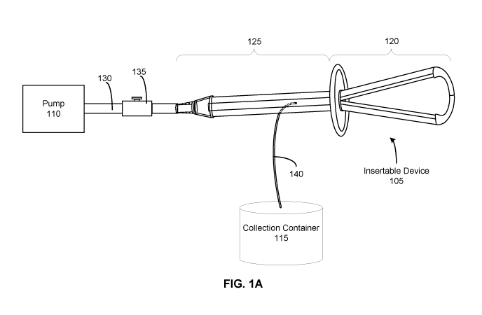
[0013] FIG. lA illustrates a system 100 for controlling uterine
hemorrhaging, according
to an embodiment. The system 100 functions to reduce or entirely stop uterine
hemorrhaging, which may occur after childbirth when a woman experiences
uterine atony,
wherein the uterus fails to contract. Controlling uterine hemorrhaging
substantially reduces
the total blood lost from the uterus and may reduce a woman's need for a blood
transfusion or
a hysterectomy. In the embodiment of FIG. 1A, the system 100 facilitates
contraction of the
uterus by sealing an opening to the uterus and providing a pressure change
within the uterus.
Changing the pressure generates a vacuum within the uterus that results in a
uniform
mechanical stimulus to the uterine wall in order to facilitate tamponade and
contractile
movement of the tissue. In the embodiment of FIG. 1A, the system 100 includes
an
insertable device 105, a pump 110, and a collection container 115.
[0014] The insertable device 105 is configured to be inserted into the
uterus to transmit
the pressure change provided by the pump 110. In the embodiment of FIG. 1A,
the insertable
device 105 is delivered transvaginally (through the vagina) such that a distal
portion 120 of
the insertable device 105 is positioned within the uterus while a proximal
portion 125 of the
insertable device 105 remains external to the uterus. The distal portion 120
may have a
flexible structure such that it conforms to the anatomy of the uterus and
allows the distal
portion 120 to create a seal at the opening of the uterus. The proximal
portion 125 of the
insertable device 105 couples to the pump 110. The distal and proximal
portions 120, 125
may have one or more channels and/or openings that allow fluid communication
(e.g., of air,
biological materials, etc.) between the uterus, the pump 110, and the
collection container 115.
The one or more channels and/or openings transfer the vacuum between the
uterus and the
pump 110. In some embodiments, the insertable device 105 may have a sheath
that facilitates
insertion of the insertable device 105 into the uterus and may additionally
prevent a
3
CA 03034778 2019-02-21
WO 2018/039250
PCT/US2017/048043
premature connection of the airflow from the pump 110 to the uterus. The
insertable device
105 will be discussed in further detail with regards to FIG. 2.
[0015] The pump 110 creates a pressure change that generates a vacuum
within the
uterus. In the embodiment of FIG. 1A, the pump 110 is coupled to the proximal
portion 125
of the insertable device 105. In some embodiments, a connection tubing 130
attaches to the
proximal portion 125 at a first end and attaches to the pump 110 at a second
end, thereby
coupling the pump 110 and the insertable device 105. In some embodiments, the
connection
tubing 130 includes a directional control valve 135 that allows fluid to flow
in one direction
and prevents fluid from flowing in the opposite direction.
[0016] When actuated, the pump 110 creates an airflow that is transmitted
through the
channels and/or openings of the insertable device 105 to the uterus. In
general, vacuum
pumps are configured to remove molecules from a sealed volume in order to
leave behind a
partial vacuum. Since the uterus is sealed by the distal portion 120 of the
insertable device
105, the airflow by the pump 110 decreases the pressure inside the uterus,
causing the uterine
pressure to drop lower than the atmospheric pressure outside of the uterus and
resulting in a
negative pressure. The negative pressure ensures that the airflow travels in a
single direction
from the uterus and through the insertable device 105 towards the pump 110.
The negative
pressure generates a vacuum inside the uterus, which facilitates tamponade,
arterial vessel
constriction, and contractile movement of the uterine wall by providing a
uniform mechanical
stimulus. In addition, generating a vacuum allows biological materials within
the uterus to be
removed. Biological materials may include blood, tissue, etc. The pump 110 may
be
power/automatically operated or manually operated. The embodiments in which
the pump
110 is manually operated, the pump 110 may create a negative pressure within
the uterus
when in a first state, and in a second state, the pump 110 may draw biological
materials into
the collection container 115 while maintain the negative pressure within the
uterus. While a
4
CA 03034778 2019-02-21
WO 2018/039250
PCT/US2017/048043
"negative pressure" is referred to throughout, some embodiments may generate a
positive
pressure inside the uterus to facilitate contractions of the uterus.
[0017] The collection container 115 collects the biological materials
removed from the
uterus. In the embodiment of FIG. 1A, the collection container 115 is coupled
to the
proximal portion 125 of the insertable device 105. The collection container
115 may be
coupled via a connection tubing 140 that is integrated into the proximal
portion 125. When
the pump 110 is actuated, the biological materials travel from the uterus,
into the openings
and/or channels of the insertable device 105, and through the connecting
tubing 140 into the
collection container 115. Collecting biological materials from the uterus may
allow a user to
monitor and measure the amount of blood loss due to uterine hemorrhaging.
Monitoring the
blood loss additionally allows the user to determine whether, when, and/or to
what extent
uterine contraction has occurred. In some embodiments, the collection
container 115 may be
configured as an inline filter that is positioned before the connection tubing
130 to the pump
110, allowing biological materials from the uterus to flow through the
insertable device 105
and be filtered out before the pump 110.
[0018] In some embodiments, the system 100 may be used to prevent
postpartum
hemorrhage in addition to monitoring and/or treating it. For example, the
system 100 may be
used in any woman after birth to aid uterine contraction. The flexibility of
the insertable
device 105 allows a healthcare provider (e.g., nurse, physician, surgeon,
etc.) to palpate a
woman's uterine tissue abdominally in order to detect if and/or when the
uterus has
contracted. In addition, the flexibility of the insertable device 105 allows
the insertable
device 105 to be flexed and positioned while other vaginal wall or tissue
repair surgical
procedures are being conducted.
[0019] In some embodiments, the insertable device 105 may be configured for
insertion
into a vaginal canal or a cervical canal such that the insertable device 105
remains external to
CA 03034778 2019-02-21
WO 2018/039250
PCT/US2017/048043
the uterus. In a similar manner as described above, the distal portion 120
creates a seal
between a vaginal opening or a cervical opening and the uterus. Creating the
seal allows the
airflow by the pump 110 to decrease the pressure inside the uterus, causing
the uterine
pressure to drop lower than the atmospheric pressure outside of the uterus and
generating a
vacuum inside the uterus. As previously described, this facilitates tamponade,
arterial vessel
constriction, and contractile movement of the uterine wall by providing a
uniform mechanical
stimulus. This configuration of the insertable device 105 may provide a less
invasive method
for controlling uterine hemorrhaging.
[0020] FIG. 1B illustrates a system 100 for controlling uterine
hemorrhaging, according
to an additional embodiment. In the embodiment of FIG. 1B, the system 100
includes an
insertable device 105, a pump 110, and a collection container 115. As
illustrated in FIG. 1B,
the collection container 115 is connected in-line with the pump 110. The
proximal portion
125 of the insertable device 105 couples to the collection container 115 and
the pump 110 via
the connection tubing 130. In this embodiment, the fluid (e.g., air,
biological materials, etc.)
flows through the connection tubing 130 towards the pump 110 when the pump 110
is
activated. The biological material is removed from the connection tubing 130
prior to
reaching the pump 110 and collects in the collection container 115.
[0021] FIG. 1C illustrates a system 100 for controlling uterine
hemorrhaging, according
to an additional embodiment. In the embodiment of FIG. 1C, the system 100
includes an
insertable device 105 and a pump 110. The proximal portion 125 of the
insertable device 105
couples to the pump 110 via the connection tubing 130. In this embodiment, the
collection
container 115 is integrated with the pump 110 such that the fluid (e.g., air,
biological
materials, etc.) flows through the connection tubing 130 into the pump 110, in
which the
biological materials are collected into a separate compartment of the pump
110. The
compartment that collects the biological material may be removable from the
pump 110.
6
CA 03034778 2019-02-21
WO 2018/039250
PCT/US2017/048043
This may assist a healthcare provider in monitoring the amount of biological
material
collected.
[0022] FIG. 2 illustrates the insertable device 105, according to an
embodiment. As
described with regards to FIG. 1A, the insertable device 105 is configured to
be inserted into
the uterus to transmit the pressure change provided by the pump 110. The
distal portion 120
is inserted into the uterus and seal an opening to the uterus, and the
proximal portion couples
to the pump 110. In the embodiment of FIG. 2, the insertable device 105
includes a tube
connector 205, a tube 210, a bridge 215, and a seal 220. The tube 210 includes
a connecting
portion 225 and a suction portion 230. Each component of the insertable device
105 may
have a variety of designs and may be interchangeable to create alternate
configurations of the
insertable device 105.
[0023] The tube connector 205 couples the insertable device 105 to the pump
110. In
some embodiments, the tube connector 205 couples to the pump 110 via
connection tubing
(e.g., connecting tubing 130). In the embodiment of FIG. 2, the tube connector
205 is tapered
such that it may be inserted into the connection tubing and secured to the
connection tubing
via an interference it (e.g., press fit or friction fit). In the embodiment of
FIG. 2, the tube
connector 205 attaches to a proximal end of the connecting portion 225 of the
tube 210. The
tube connector 205 may be attached to the connecting portion 225 via an
interference fit (e.g.,
press fit or friction fit), an adhesive, a threaded interface, or any other
suitable securing
mechanism. The tube connector 205 may be composed of rigid or semi-rigid
plastic (e.g.,
polyethylene, polypropylene) or any other suitable material.
[0024] The tube 210 acts as a conduit for air and biological materials. The
tube 210 may
comprise one or more channels that couple airflow from the pump 110 to the
uterus to
transmit a change in pressure inside the uterus. In the embodiment of FIG. 2,
the tube 210
includes a single channel having surface features on an internal surface of
the channel.
7
CA 03034778 2019-02-21
WO 2018/039250
PCT/US2017/048043
Alternate embodiments of the tube 210 will be discussed in further detail with
regards to
FIGS. 7B-7E. The internal surface features protrude from the internal surface
of the channel
and may aid the flow of air and biological materials through the tube 210. The
internal
surface features may have a variety of configurations, which will be discussed
in further
detail with regards to FIGS. 4-5. In the embodiment of FIG. 2, the tube 210
includes the
connecting portion 225 and the suction portion 230.
[0025] The connecting portion 225 transmits airflow from the pump 110 to
the suction
portion 230 of the tube 210. The tube connector 205 is attached at a proximal
end of the
connecting portion 225 and removably secures the connecting portion 225 to the
pump 110.
The channel of the tube 210 extends down the length of the connecting portion
225. At a
distal end of the connecting portion 225, the tube 210 branches out to form
the suction
portion 230.
[0026] In the embodiment of FIG. 2, the connecting portion 225 and the
suction portion
230 are integrally formed of the same piece of material (i.e., have a unitary
construction). To
create the suction portion 230 of the tube 210, the tube's 210 single piece of
material is at
least partially physically split in half, thereby creating two branches 235a,
235b of the suction
portion 230 and exposing the channel of the tube 210 on a medial side of each
branch 235.
Exposing the channel of the tube 210 connects the airflow between the pump 110
and the
uterus, allowing the tube 210 to transmit the change in pressure provided by
the pump 110 to
the uterus. In addition, the exposed channels are beneficially located along a
medial side of
each branch 235 of the suction portion 230 such that the exposed channels are
oriented away
from an interior wall of the uterus when the insertable device 105 is
inserted. This
configuration prevents uterine tissue or other tissue from obstructing the
exposed channels
and preventing airflow when the pump 110 is actuated. In some embodiments, the
orientation of the exposed channels on the suction portion 230 may vary. For
example, the
8
CA 03034778 2019-02-21
WO 2018/039250
PCT/US2017/048043
exposed channels may be oriented at an off-axis angle from the medial surface
of the suction
portion 230. In other words, the exposed channels may be oriented at an angle
relative to an
axis (e.g., a bisecting axis) of the medial surface. In some embodiments, the
exposed
channels may be located on a surface other than the medial surface of the
suction portion 230
(e.g., a lateral surface of the suction portion 230). In these embodiments,
channels may not
be exposed after the tube 210 is split to create the two branches 235a, 235b.
In other
embodiments, the exposed channels on the suction portion 230 may be some
combination
thereof In addition, some exposed channels may not be configured to pass
biological
material from the uterus. In some embodiments, the connecting portion 225 and
the suction
portion 230 may be separate components/pieces of material that are coupled to
each other.
[0027] The bridge 215 spans between the branches 235 of the suction portion
230. Each
end of the bridge 215 is attached to a distal end of a branch 235 of the
suction portion 230.
The bridge 215 may be attached via an interference fit (e.g., friction fit or
press fit), an
adhesive, a threaded interface, or some combination thereof In the embodiment
of FIG. 2,
the length of the bridge 215 is sufficient enough to maintain a separation
between the
branches 235 of the suction portion 230. In some implementations, the manner
in which the
branches 235 are constructed, for example as described with respect to FIG. 3
below, the
branches 235 have a natural tendency to come together in a resting state
(i.e., when no
external force is exerted on the branches 235). However, this tendency may
cause the
exposed channels on each branch 235 to become obstructed in the resting state.
Thus, the
bridge 215 exerts a force on each branch 235 to separate the branches 235 into
a split state.
This configuration prevents the branches 235 of the suction portion 230 from
collapsing into
each other when the pump 110 is actuated and thereby obstructing the airflow
between the
pump 110 and the uterus. In addition, the bridge 215 maintains the alignment
of the
branches 235 of the suction portion 230 such that the exposed channels along
the medial side
9
CA 03034778 2019-02-21
WO 2018/039250
PCT/US2017/048043
of the suction portion 230 remain facing inward towards each other. In one
embodiment this
is accomplished by forming the bridge 215, which has a substantially curved
body with
rounded edges, directionally-limiting so that the insertable device 105 is
positioned
comfortably within the uterus when inserted and to prevent damage to the
uterine wall, while
also maintaining its orientation when inserted so as to keep the exposed
channel oriented
inward as discussed. In some embodiments, the shape of the bridge 215 may vary
in terms of
the length, width, curvature, thickness, etc. As a result, the configuration
of the distal portion
120 may vary, for example, to form a circular, elliptical, triangular, or horn-
shaped loop, or
any other suitable geometries for placement within the uterus. Alternate
embodiments of the
bridge 215 will be discussed with regards to FIG. 7E. In some embodiments, the
insertable
device 105 may not include a bridge 215.
[0028] The seal 220 creates a seal at the opening of the uterus. In the
embodiment of
FIG. 2, the seal 220 is a disk positioned at a distal end of the connecting
portion 225, adjacent
to the suction portion 230, however in alternate embodiments it may be placed
at varying
locations along the connecting portion 225 depending on the size/length of the
other
elements. The disk may be circular, elliptical, or any other suitable geometry
for sealing an
opening to the uterus. The disk may or may not include a lip around its
perimeter, wherein
the lip may help position the seal against the uterine wall. In addition, the
disk may have a
convex or a concave profile. The seal 220 may be composed of semi-flexible
plastics, such
as silicone, polyethylene, polypropylene, or any other suitable medical-grade
material. The
flexible material of the seal 220 allows the seal 220 to conform to the
anatomy of the uterus
such that the seal 220 may be positioned against an opening of the uterus to
form a seal
between the uterus and an environment external to the uterus. Sealing the
uterus allows the
insertable device 105 to create a vacuum and maintain a negative pressure
within the uterus
to facilitate contraction of the uterus. In some embodiments, the seal 220 may
be configured
CA 03034778 2019-02-21
WO 2018/039250
PCT/US2017/048043
to form a seal at any point from the vulva, the cervix, the vaginal canal, or
within the uterus.
Additional embodiments of the seal 220 include a plurality of disks, a cup, a
balloon, and a
sleeve, which may be interchangeable with other components of the insertable
device 105, for
example as described with respect to any of the exemplary embodiments herein.
These
additional embodiments will each be discussed in further detail with regards
to FIGS. 7A-7E.
[0029] In some embodiments, the insertable device 105 may include a sheath
(not shown)
that facilitates insertion of the insertable device 105 into the uterus and
may additionally
prevent a premature connection of the airflow from the pump 110 to the uterus.
The sheath
may cover a portion of the tube 210, the bridge 215, and/or the seal 220, or
some combination
thereof As an example, the sheath may be in the form of a translatable outer
tube that
encloses a portion of the tube 210, a removable membrane that encloses distal
portions of the
insertable device 105, or other structures having a similar configuration. The
sheath may be
removed once the suction portion 230 is positioned within the uterus. Removal
of the sheath
may simultaneously release or position the seal 220 in the uterus to create
the seal. When use
of the system 100 is complete, the sheath may be re-installed onto the
insertable device 105.
The re-installation process may simultaneously break the seal from the seal
200 and cut off
the connection of the airflow between the pump 110 and the uterus.
[0030] FIG. 3 illustrates the tube 210 of the insertable device of FIG. 2,
according to an
embodiment. In the embodiment of FIGS. 2-3, the tube 210 is manufactured
through an
extrusion process, though in other embodiments the tube 210 may be
manufactured in other
ways. An extrusion process is used to create objects having a desired cross-
sectional profile
by pushing a material through a die of the desired cross-section. Materials
such as silicone,
polyethylene, polypropylene, or any other suitable medical-grade material may
be used for
the extrusion process of the tube 210. Once the extrusion is created, the
extrusion may be cut
to a desired length. In the embodiment of FIG. 3, the tube 210 is extruded
using a semi-
11
CA 03034778 2019-02-21
WO 2018/039250
PCT/US2017/048043
flexible material, which allows the insertable device to conform to the
anatomy of a uterus
when inserted.
[0031] In the embodiment of FIGS. 2-3, the extrusion undergoes post-
processing to form
the final product, the tube 210. As described with regards to FIG. 2, the tube
210 includes the
connecting portion 225 and the suction portion 230. To create the suction
portion 230 of the
tube 210, a portion of the extrusion is at least partially split in half along
its length. As
illustrated in FIG. 3, the extrusion is cut from a distal end of the tube 210
down to the length
of the extrusion by a distance d. The distance d is a suitable length that
allows the suction
portion 230 to be inserted comfortably into a uterus. In some embodiments, the
die may be
designed such that the extrusion includes a groove on an internal or external
surface that
extends down a certain length of the extrusion. The groove may facilitate the
cutting process
by indicating the location of the cut and acting as a guide for the tool
performing the cut. In
some embodiments, the groove indicates a location at which the wall thickness
of the
extrusion is thinner than the remainder of the extrusion. A thinner wall
thickness may ease
the cutting process or allow two halves of the extrusion to be separated
manually to form the
suction portion 230.
[0032] In some embodiments, the tube 210 is manufactured through a
GeoTrans0
extrusion process. In this process, the die may be designed such that the
desired cross-section
of the extrusion changes along the length of the extrusion. Specifically, a
desired cross-
section for the connecting portion 225 may differ from a desired cross-section
for the suction
portion 230. For example, the cross-section of the connecting portion 225 may
be
substantially cylindrical or elliptical while the cross-section of the suction
portion 230 may
include one or more channels. In some embodiments, the cross-section of the
extrusion may
change along its length in an alternating pattern. The change in cross-section
between the
12
CA 03034778 2019-02-21
WO 2018/039250
PCT/US2017/048043
connecting portion 225 and the suction portion 230 may be designed to occur
transitionally or
abruptly.
[0033] In addition, the die may be designed such that the cross-section of
the tube 210
includes surface features on an internal surface of the tube 210. Example
surface features are
illustrated in FIG. 3, wherein each branch 235 of the suction portion 230
includes a middle
protrusion that extends down the length of the internal surface of the tube
210. The surface
features may aid the flow of air and biological materials within the tube 210,
which will be
discussed in further detail with regards to FIGS. 4-5. By manufacturing the
tube 210 through
an extrusion process, minimal post-processing is required to form the final
product of the
insertable device 105. Moreover, once a die is manufactured for a desired
extrusion, large
quantities of the extrusion may be produced at a fast rate (e.g., thousands
per day), especially
if the die is configured to create multiple extrusions simultaneously. This
production may
significantly reduce overall manufacturing costs, as well as reduce overall
device complexity
by reducing the number of components in the finished device. In addition, the
design of the
insertable device 105, specifically the split state of the branches 235 and
the bridge 215 that
maintains separation of the branches 235, provides greater design flexibility
for the surface
features on the channel as the branches 235 are not required to bend or curve
substantially.
[0034] FIG. 4A illustrates a cross-sectional view of an extrusion 400 for
creating the tube
210, according to an embodiment. Specifically, a cross-section of the suction
portion 230 of
the tube 210 is shown before two halves 405a, 405b of the extrusion 400 are
split to create
the separate branches of the suction portion 230. In the embodiment of FIG.
4A, the
extrusion 400 includes an outer wall 410 and a channel 415. The outer wall 410
forms the
external boundary of the tube 210. The outer wall 410 surrounds the channel
415, through
which air and biological materials can pass. As illustrated in FIG. 4A, the
outer wall 410 is
substantially of uniform thickness. The thickness of the outer wall 410 may be
between
13
CA 03034778 2019-02-21
WO 2018/039250
PCT/US2017/048043
approximately 1 to 2.5 millimeters (mm). The outer wall 410 includes two
grooves 420a,
420b that are located on opposite edges of the outer wall. In the embodiment
of FIG. 4A, the
grooves 420a, 420b are located on an internal and external surface of the
outer wall 410, such
that the thickness of the outer wall 410 narrows at the grooves 420a, 420b. As
described with
regards to FIG. 3, the grooves 420a, 420b facilitate the separation of the two
halves 405a,
405b to form the separate branches of the suction portion 230 of the tube 210.
In the
embodiment of FIG. 4A, the grooves 420a, 420b extend down the length of the
tube 210 for
the suction portion 230. In some embodiments, the connecting portion 225 may
not include
the grooves 420a, 420b and only the suction portion 230 includes the grooves
420a, 420b.
This configuration may prevent propagating the separation of the two halves
405a, 405b into
the connecting portion 225. For embodiments in which the grooves 420a, 420b
extend down
the length of the tube 210 for the connecting portion 225, the cross-sectional
view shown in
FIG. 4A also illustrates the cross-section of the connecting portion 225.
100351 In the embodiment of FIG. 4A, the surface of the channel 415
includes the
following surface features: protrusions 425a, 425b and protrusions 430a, 430b,
430c, 430d.
As illustrated in FIG. 4A, the surface features are arranged such that the
cross-section of the
extrusion 400 is substantially symmetrical. This configuration ensures that
each branch of
the suction portion 230 includes the same surface features once the extrusion
400 is split
along the grooves 420a, 420b. The protrusions 425a, 425b are positioned at a
middle portion
of the channel 415 and protrude into the center of the channel 415 towards
each other. The
protrusions 430a, 430b, are positioned to the right of grooves 420a, 420b,
respectively, and
protrude towards each other, while protrusions 430c, 430d, are positioned to
the left of
grooves 420b, 420a, respectively, and protrude towards each other. This
configuration of
protrusions 425, 430 divides the channel 415 into smaller channels, which
provides each
14
CA 03034778 2019-02-21
WO 2018/039250
PCT/US2017/048043
branch of the suction portion 230 with certain properties, discussed in
further detail with
regards to FIG. 4B.
100361 FIG. 4B illustrates a cross-sectional view of a branch 435a of the
suction portion
230, according to an embodiment. Specifically, the cross-section shown is of
the half 405a
after the two halves 405a, 405b of the extrusion 400 are split to create
separate branches of
the suction portion 230. The half 405a forms the branch 435a. While branch
435b is not
shown, it is formed by the half 405b. As illustrated in FIG. 4B, the
protrusions 425a, 430a,
430b divide the channel 415 into two smaller channels 440a, 440b, wherein each
channel has
a respective opening 445a, 445b. As described with regards to FIG. 2, openings
in the
suction portion 230 allow fluid communication between the uterus and the pump
110.
Dividing the channel 415 into smaller channels 440a, 440b distributes the
airflow from the
pump 110 and increases the number of pathways by which air and biological
materials can
travel. Thus, if a single pathway becomes obstructed by biological materials,
other pathways
remain accessible, and the system 100 can effectively maintain a negative
pressure inside the
uterus. In addition, to further prevent the channels 445a, 445b from becoming
obstructed, the
openings 445a, 445b are located on a medial side of the respective branches
435 such that
when the suction portion 230 is inserted into the uterus, the outer wall 410
faces the uterine
wall and the openings 445a, 445b. This configuration prevents uterine tissue
or other tissue
from obstructing the openings 445a, 445b and preventing airflow when the pump
110 is
actuated.
[0037] In the embodiment of FIG. 4B, the openings 445a, 445b are configured
to allow
biological materials of a certain size through the openings 445a, 445b and
into the channels
440a, 440b such that these biological materials may be removed from the uterus
and collected
in the collection container 115. In the embodiment of FIG. 4B, the size of
each opening
445a, 445b is between approximately 1 to 4 millimeters (mm). In some
embodiments, the
CA 03034778 2019-02-21
WO 2018/039250
PCT/US2017/048043
size of each opening 445a, 445b is between 2 to 3.5 millimeters (mm). Openings
of this size
may additionally be configured to break up masses of biological material that
have formed a
clot. By breaking up the masses, the openings 445a, 445b are able to prevent
obstruction of
the airflow from the pump 110 and allow the biological material to be
collected in the
collection container 115.
100381 FIG. 5A illustrates a cross-sectional view of an additional
embodiment of an
extrusion 500 for creating the tube 210. Specifically, a cross-section of the
suction portion
230 of the tube 210 is shown before two halves 505a, 505b of the extrusion 500
are split to
create the separate branches of the suction portion 230. Similar to extrusion
400, the
extrusion 500 includes an outer wall 510 and a channel 515. The outer wall 510
forms the
external boundary of the tube 210. The outer wall 510 surrounds the channel
515, through
which air and biological materials can pass. As illustrated in FIG. 5A, the
outer wall 510 is
substantially of uniform thickness. The thickness of the outer wall 510 may be
between
approximately 1 to 2.5 millimeters (mm). The outer wall 510 includes two
grooves 520a,
520b that are located on opposite edges of the outer wall. Similar to grooves
420a, 420b, the
grooves 520a, 520b facilitate the separation of the two halves 505a, 505b to
form the separate
branches of the suction portion 230 of the tube 210. In the embodiment of FIG.
5A, the
grooves 520a, 520b are located only on an internal surface of the outer wall
510, but, in some
embodiments, the grooves 520a, 520b may be located on both an internal and
external surface
of the outer wall 510.
[0039] In the embodiment of FIG. 5A, the surface of the channel 515
includes the
following surface features: protrusions 525a, 525b. As illustrated in FIG. 5A,
the protrusions
525a, 525b are arranged such that the cross-section of the extrusion 500 is
substantially
symmetrical. This configuration ensures that each branch of the suction
portion 230 includes
the same surface features once the extrusion 500 is split along the grooves
520a, 520b. The
16
CA 03034778 2019-02-21
WO 2018/039250
PCT/US2017/048043
protrusions 525a, 525b are positioned at a middle portion of the channel 515
and protrude
into the center of the channel 515 towards each other. In the embodiment of
FIG. 5A, each
protrusion 525a, 525b has a distal end that extends in a different direction
to the portion of
the protrusion that extends from the outer wall. In the illustrated
embodiment, this different
direction of extension forms a shape similar to an umbrella, wherein each
protrusion 525
includes a middle support structure with a curved arm extending from each side
of the
support structure. In other embodiments, other shapes may be formed, such as T-
shaped
protrusions, and so on. Regardless of the exact shape, this general
configuration of
protrusions 525a, 525b divides the channel 515 into smaller channels, which
provides each
branch of the suction portion 230 with certain properties, discussed in
further detail with
regards to FIG. 5B
[0040] FIG. 5B illustrates a cross-sectional view of a branch 535a of the
suction portion
230. Specifically, the cross-section shown is of the half 505a after the two
halves 505a, 505b
of the extrusion 500 are split to create separate branches of the suction
portion 230. The half
505a forms the branch 535a. While branch 535b is not shown, it is formed by
the half 505b.
As illustrated in FIG. 5B, the protrusion 525a divides the channel 515 into
two smaller
channels 540a, 540b wherein each channel has a respective opening 545a, 545b.
Similar to
the embodiment of FIG. 4, dividing the channel 515 into smaller channels 540a,
540b
distributes the airflow from the pump 110 and increases the number of pathways
by which air
and biological materials can travel. In the embodiment of FIG. 5B, the
openings 545a, 545b
are distanced apart from each other due to the configuration of the protrusion
525a.
Separating the openings 545a, 545b by a distance may decrease the likelihood
of the
openings 545a, 545b becoming obstructed by biological materials at the same
time,
potentially by the same mass of biological material. Thus, if a first opening
becomes
obstructed by biological materials, at least a second opening remains
accessible, and the
17
CA 03034778 2019-02-21
WO 2018/039250
PCT/US2017/048043
system 100 can effectively maintain a negative pressure inside the uterus. In
addition, to
further prevent the channels openings 545a, 545b from becoming obstructed, the
openings
545a, 545b are located on a medial side of the respective branches 535 such
that when the
suction portion 230 is inserted into the uterus, the outer wall 510 faces the
uterine wall and
the openings 545a, 545b . This configuration prevents uterine tissue or other
tissue from
obstructing the openings 545a, 545b and preventing airflow when the pump 110
is actuated.
[0041] Similar to the embodiment of FIG. 4B, in the embodiment of FIG. 5B,
the
openings 545a, 545b are configured to allow biological materials of a certain
size through the
openings 545a, 545b and into the channels 540a, 540b such that these
biological materials
may be removed from the uterus and, in some embodiments, collected in the
collection
container 115. In the embodiment of FIG. 4B, the size of each opening 545a,
545b is
between approximately 1 to 4 millimeters (mm). Openings of this size may
additionally be
configured to break up a mass of biological material (e.g., a clot or clump of
tissue). By
breaking up the mass, the openings 545a, 545b are able to prevent obstruction
of the airflow
from the pump 110 and allow the biological material to be collected in the
collection
container 115.
[0042] FIG. 6A illustrates a method for attaching the bridge 215 to the
tube 210,
according to an embodiment. As illustrated in FIG. 6A, a first end of the
bridge 215 is
attached to a distal end of branch 235a of the suction portion 230, and a
second end of the
bridge 215 is attached to a distal end of branch 235b of the suction portion
230. This
configuration of the suction portion of the tube 210 ensures that the branches
235 are
separated and remain in a split state when inserted into the uterus.
[0043] In the embodiment of FIG. 6A, each end of the bridge 215 includes a
mating
protrusion 605a (605b not shown) that extends from an end of the bridge 215.
To secure the
bridge 215 to the suction portion 230, the mating protrusions 605a, 605b are
inserted into a
18
CA 03034778 2019-02-21
WO 2018/039250
PCT/US2017/048043
channel of the branches 235b, 235a, respectively. The mating protrusions 605a,
605b may be
inserted with an interference fit (e.g., friction fit or press fit), an
adhesive, a threaded
interface, or some combination thereof. In the embodiment of FIG. 6A, the
mating
protrusions 605a, 605b are silicone bonded to the respective branches 235b,
235a.
[0044] FIG. 6B illustrates mating protrusion 605a that secures a first end
of the bridge
215 to the branch 235a, according to an embodiment. The mating protrusion 605a
may
include a cavity 610 that complements surface features located on a channel of
the branch
235a. In the embodiment of FIG. 6B, the shape of the cavity 610 is designed to
complement
the shape of the surface features described with regards to FIG. 5B. In this
configuration, the
surface feature 525a may be inserted into the cavity 610, which may improve
the stability and
security of the attachment between the bridge 215 and the branch 235a. In
addition, the
cavity 610 provides a greater surface area for silicone bonding the mating
protrusions 605a,
605b to the respective branches 235b, 235a. Other embodiments may have a
cavity 610
designed to complement the shape of the surface features illustrated in FIGS.
4A and 4B or
any other channel surface feature configuration.
[0045] In some embodiments, the bridge 215 may be configured to transmit
the airflow
from the tube 210. The bridge 215 may include channels or holes along a medial
side of the
bridge 215, and the mounting protrusions 605a, 605b may include channels that
couple the
airflow of the suction portion 230 to the bridge 215. This configuration
provides additional
pathways by which the airflow and/or biological materials can travel.
[0046] FIGS. 7A-7E illustrate additional embodiments of an insertable
device. As
described with regards to FIG. 2, an insertable device includes several
components, such as a
tube connector, a tube having a connecting portion and a suction portion, a
bridge, and a seal.
Each component may have a variety of designs, which will be discussed in
further detail. In
19
CA 03034778 2019-02-21
WO 2018/039250
PCT/US2017/048043
addition, different designs of the components may be interchangeable and
combined in
several ways to create different configurations of an insertable device.
[0047] FIG. 7A illustrates an insertable device 700, according to an
embodiment. In the
embodiment of FIG. 7A, the insertable device 700 includes the tube connector
205, the tube
210 having the connecting portion 225 and the suction portion 230, the bridge
215, and a seal
705. Similar to the embodiment of the seal discussed with regards to FIG. 2,
the seal 705
creates a seal at the opening of the uterus. The seal 705 is composed of three
disks 710a,
710b, 710c positioned at a distal end of the connecting portion 225, adjacent
to the suction
portion 230. Each disk 710 may be composed of a semi-flexible material, such
as silicone,
polyethylene, polypropylene, or any other suitable medical-grade material,
allowing each
disk 710 to conform to the anatomy of the uterus. The diameter of each disk
710
incrementally decreases such that the largest disk 710c is closest to the
suction portion 230
and the smallest disk 710a is farthest away from the suction portion 230. In
this
configuration, the decreasing diameters of the disks 710 may improve the
ability of the seal
705 to be positioned against an opening of the uterus such that each disk 710
may abut the
uterine wall to form a seal between the uterus and an environment external to
the uterus.
Having a seal including three disks 710 may improve the quality of the seal
formed and
provide redundancy in maintaining the seal. Other embodiments may include a
varying
number of disks that may be positioned at different distances relative to each
other (e.g., two
disks that are spaced farther apart or ten disks that are spaced closer
together).
[0048] FIG. 7B illustrates an insertable device 715, according to an
embodiment. In the
embodiment of FIG. 7B, the insertable device 700 includes the tube connector
205, a tube
720 having a connecting portion 725 and a suction portion 730, and a seal 735.
Similar to the
embodiment of the tube discussed with regards to FIG. 2, the tube 720 acts as
a conduit for
air and biological materials. The tube 720 may comprise one or more channels
that couple
CA 03034778 2019-02-21
WO 2018/039250
PCT/US2017/048043
airflow from the pump 110 to the uterus to transmit a change in pressure
inside the uterus. In
the embodiment of FIG. 7B, the tube 720 is a flexible tube that is folded to
form the
connecting portion 725 and the suction portion 730. The sections of the tube
720 forming the
connecting portion 725 may be adjoined via an adhesive, one or more over-
molded
components secured around the tube 720, or a heat shrink wrap placed over the
tube 720.
Meanwhile, the suction portion 730 remains a loop. In this configuration, the
construction of
the tube 720 ensures that the alignment of the openings and/or channels on the
medial side of
the loop of the suction portion 730 remain facing away from the uterine wall
upon insertion.
In addition, this configuration eliminates the need for a connecting element
(e.g., bridge 215)
between branches of the tube, which may decrease the cost of manufacturing the
tube 720.
However, the rigidity of the tube 720 needs to be appropriately determined
such that the tube
720 may be flexible enough to form the loop of the suction portion 730 yet
rigid enough to
prevent the suction portion 730 from potentially deforming and obstructing the
openings
and/or channels on the medial side of the loop. In addition, the design of the
surface features
on a surface of the channel of the tube 720 may be limited due to the desired
curvature of the
tube 720.
[0049] In the embodiment of FIG. 7B, the suction portion 730 includes one
or more
openings 738 located along a medial side of the loop such that the openings
738 are oriented
away from an interior wall of the uterus when the insertable device 105 is
inserted. This
configuration prevents uterine tissue or other tissue from obstructing the
openings 738 and
preventing airflow when the pump 110 is actuated. The openings 738 may be
circular,
elliptical, polygonal, or any other suitable shape that allows air and
biological materials to
travel through. In some embodiments, the openings 738 may be channels that
extend along
the medial side of the loop.
21
CA 03034778 2019-02-21
WO 2018/039250
PCT/US2017/048043
[0050] The seal 735 creates a seal at an opening of the uterus. The seal
735 is positioned
at a distal end of the connecting portion 225, adjacent to the suction portion
730. In the
embodiment of FIG. 7B, the seal 735 is shaped similar to a cup, wherein a
bottom portion of
the cup shape is configured to abut the uterine wall at an opening of the
uterus upon insert of
the insertable device 730 into the uterus. The seal 735 is composed of a semi-
flexible
material, such as silicone, polyethylene, polypropylene, or any other suitable
medical-grade
material, that allow the seal 735 to conform to the anatomy of the uterine
wall.
[0051] FIG. 7C illustrates an insertable device 740, according to an
embodiment. In the
embodiment of FIG. 7C, the insertable device 740 includes the tube connector
205, the tube
720 having the connecting portion 725 and the suction portion 730, and the
seal 220. The
insertable device 740 combines the tube configuration described with regards
to FIG. 7B with
the seal configuration described with regards to FIG. 2.
[0052] FIG. 7D illustrates an insertable device 745, according to an
embodiment. The
insertable device 745 is an embodiment of insertable device 740. In the
embodiment of FIG.
7D, the insertable device 745 includes the tube connector 205, the tube 720
having the
connecting portion 725 and the suction portion 730, the seal 220, and a shield
750. The
shield 750 is a porous mesh that allows particles of a certain size to pass
through the mesh.
In the embodiment of FIG. 7D, the shield 750 encloses the suction portion 730
to prevent
uterine tissue from obstructing the openings 738 while allowing other
biological materials
(e.g., blood) to pass through. The shield 750 may be composed of gauze, nylon,
or other
suitable materials that may be placed within the uterus.
[0053] FIG. 7E illustrates an insertable device 755, according to an
embodiment. In the
embodiment of FIG. 7E, the insertable device 755 includes the tube connector
205, a tube
760 having a connecting portion 765 and a suction portion 770, and a seal 775.
22
CA 03034778 2019-02-21
WO 2018/039250
PCT/US2017/048043
[0054] The tube 760 acts as a conduit for air and biological materials.
While the tube 760
is similar in geometry to tube 720, the tube 760 is made of two individual
tubes 780a, 780b
rather than a single folded tube. Each tube 780a, 780b includes an internal
channel extending
down the length of the tube. In the embodiment of FIG. 7E, tubes 780a, 780b
are positioned
adjacent to each other such that a portion of the tubes 780a, 780b can be
adjoined to form the
connecting portion 765. The tubes may be adjoined via an adhesive, one or more
over-
molded components secured around the connecting portion 765, or a heat shrink
wrap placed
over the tube connecting portion 765. The remaining portions of the tubes
780a, 780b are
curved to mate an end of tube 780a to an end of tube 780b, forming a loop to
create the
suction portion 770. The ends of the tubes 780a, 780b may be coupled together
using a plug
785. In the embodiment of FIG. 7E, a first end of the plug 785 is inserted
into a channel of
tube 780a, and a second end of the plug 785 is inserted into a channel of tube
780b. The plug
785 may be secured within the tubes 780a, 780b using an adhesive. The plug 785
may
include openings or channels to form a continuous pathway for airflow and/or
biological
materials through the tube 760. In some embodiments, the plug 785 may be
designed to fit
over the ends of the tubes 780a, 780b as a cross tube connector. For
embodiments in which
the suction portion of the tube comprises two separate branches that are to be
connected, the
connecting element (such as bridge 215 or plug 785) can have a variety of
configurations and
be of any size, depending upon the rigidity of the tube and given that the
connecting element
appropriately mates the branches such that the openings or channels on each
branch remain
unobstructed and allow a vacuum to be created within the uterus upon insertion
of the
insertable device.
[0055] In the embodiment of FIG. 7E, the suction portion 770 includes one
or more
openings 790. The openings 790 are created by skiving the external surface of
the suction
portion 770. A skiving process carves out notches in the surface of the
suction portion 770.
23
CA 03034778 2019-02-21
WO 2018/039250
PCT/US2017/048043
The openings 790 are located along a medial side of the loop such that the
openings 790 are
oriented away from an interior wall of the uterus when the insertable device
105 is inserted.
This configuration prevents uterine tissue or other tissue from obstructing
the openings 790
and preventing airflow when the pump 110 is actuated.
[0056] The seal 775 creates a seal at an opening of the uterus. In the
embodiment of FIG.
7E, the seal 775 is a sleeve or a balloon that can be inflated once the
suction portion 770 is
positioned within the uterus. The seal 775 may be inserted while flattened,
allowing the seal
775 to be properly positioned before the seal 775 is inflated. The seal 775
may be positioned
within the vaginal canal, cervix, or uterus. In the embodiment of FIG. 7E, the
seal 775
includes a tube 795 that may connect to the pump 110 to inflate the seal 775.
The seal 775
may be composed of silicone, polyethylene, polypropylene, or any other
suitable medical-
grade material.
[0057] FIG. 8 illustrates a cross-sectional view of an additional
embodiment of an
extrusion 800 for creating the tube 210. Specifically, a cross-section of the
suction portion
230 of the tube 210 is shown before two halves 805a, 805b of the extrusion 800
are split to
create the separate branches of the suction portion 230. In the embodiment of
FIG. 8, the
extrusion 800 includes an outer wall 810, channel 815, channels 820a, 820b,
and rings 825a,
825b. The outer wall 810 forms the external boundary of the tube 210. The
outer wall 810
encloses the channels 815, 820a, 820b. As illustrated in FIG. 8, the outer
wall 810 is
substantially of uniform thickness. The thickness of the outer wall 810 may be
between
approximately 1 to 2.5 millimeters (mm). The outer wall 810 includes two
grooves 830a,
830b that are located on opposite edges of the outer wall 810. In the
embodiment of FIG. 8,
the grooves 830a, 830b are located on an internal and external surface of the
outer wall 810,
such that the thickness of the outer wall 810 narrows at the grooves 830a,
830b. As described
with regards to FIG. 3, the grooves 830a, 830b facilitate the separation of
the two halves
24
CA 03034778 2019-02-21
WO 2018/039250
PCT/US2017/048043
805a, 805b to form the separate branches of the suction portion 770 of the
tube 760. In the
embodiment of FIG. 8, once the two halves 805a, 805b are separate to form the
branches of
the suction portion 770, the branches are curved towards each other to form a
loop, wherein a
first end of a first branch mates with a first end of a second branch. To
secure the ends of the
branches together, a pin may be inserted into the rings 825a, 825b. The pin
may be adhered
within the rings 825a, 825b.
[0058] Once the two halves 805a, 805b are separated, the channel 815 is
exposed to allow
fluid communication between the pump 110 to the uterus. The wall between the
channel 815
and the respective channels 820a, 820b may include one or more openings (e.g.,
holes or
channels) such that biological material may enter the channel 815 and flow
through to the
channels 820a, 820b. In this configuration, the channels 820a, 820b act as
drain channels.
This configuration may help the channel 815 remain unobstructed.
[0059] The foregoing description of the embodiments of the invention has
been presented
for the purpose of illustration; it is not intended to be exhaustive or to
limit the invention to
the precise forms disclosed. Persons skilled in the relevant art can
appreciate that many
modifications and variations are possible in light of the above disclosure.
[0060] The language used in the specification has been principally selected
for readability
and instructional purposes, and it may not have been selected to delineate or
circumscribe the
inventive subject matter. It is therefore intended that the scope of the
invention be limited not
by this detailed description, but rather by any claims that issue on an
application based
hereon. Accordingly, the disclosure of the embodiments of the invention is
intended to be
illustrative, but not limiting, of the scope of the invention.