Note: Descriptions are shown in the official language in which they were submitted.
SYSTEM AND METHOD FOR USING IMAGING QUALITY METRIC RANKING
FIELD
[0001] The present disclosure relates to medical imaging and more
specifically to medical
imaging in patient treatment
BACKGROUND
[0002] Medical records of a patient often include imaging data, such as
Magnetic
Resonance Imaging (MRI), X-ray, ultrasound (US), and Computed Tomography (CT)
images. The
medical images are typically obtained in connection with a trauma or disease
diagnosis, and may
include multiple related medical images taken at different stages of the
patient's treatment. The
multiple images can also include multiple imaging modalities, i.e. the images
are obtained using
different imaging technologies.
[0003] It is advantageous to have as much of a patient's image data as
possible available
to a practitioner, for assessing the patient. Institutes such as hospitals
incorporate computer-
implemented systems to store and provide image data to practitioners. An
example of a commonly
used system is PACS (picture archiving and communication system). PACS is a
healthcare
technology that captures and stores medical images from multiple modalities,
and allows retrieval
and viewing of the images. One problem with PACS is the lack of
interoperability of different
PACS systems, for example a PACS deployed by an emergency unit of a hospital
may not
communicate with PACS deployed by the radiology or pathology unit of the same
hospital, and
different health institutes may deploy PACS that are non-interoperable. Thus,
the image data
collected by one health unit is not easily accessed by another health unit. A
PACS also requires a
medical professional, such as a radiologist, to review and interpret the image
data and create a
structured report. Further, there is no quality control on the images stored
in a PACS.
[0004] In assessing outcomes, such as a patient status, medical treatment
effectiveness,
health institute success and medical practitioner performance, it would be
ideal to have all medical
images related to a patient accessible from a single access point, and to be
able to ascertain the
reliability of the medical image data.
1
Date Recite/Date Received 2023-10-16
SUMMARY
[0005] An object of the present disclosure is to provide a system and
method for integrating
patient image data and calculating quality metrics for the integrated image
data.
[0006] Thus by one broad aspect of the present invention, a computer-
implemented
method to store and analyze clinical imaging data for a treatment program is
provided, comprising
obtaining two or more images of a patient from two or more modalities,
registering the images to
provide a multimodal image set, determining a position information of a voxel
in the multimodal
image set, calculating a quality metric for the voxel of the multimodal image
set, assigning the
quality metric to the voxel, storing the voxel, the voxel position
information, the quality metric
and at least one searchable header as imaging data in a memory of a computing
device, and
modifying or maintaining the treatment program based on the imaging data.
[0007] By another broad aspect of the present invention, a system for
storing and analyzing
clinical imaging data for a treatment program is provided, comprising a memory
and a processor
interconnected with the memory, the processor configured to obtain two or more
images from two
or more modalities, store the images in the memory, register the images to
provide a multimodal
image set, store the multimodal image set in the memory, calculate a quality
metric for a voxel in
the multimodal image set, store the voxel quality metric with the multimodal
image set in the
memory, and modify or maintain the treatment program based on the calculated
qualitative metric.
BRIEF DESCRIPTION OF THE DRAWINGS
[0008] The foregoing and additional aspects and embodiments of the
present disclosure
will be apparent to those of ordinary skill in the art in view of the detailed
description with
reference to the following figures, in which:
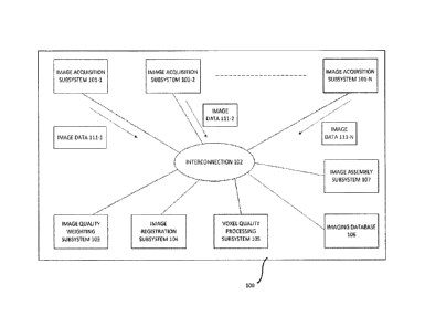
[0009] FIG. 1 illustrates an embodiment of a system to calculate a voxel
and image quality
metric as disclosed herein.
[0010] FIG. 2 illustrates an embodiment of a process flow to calculate a
voxel and image
quality metric as disclosed herein.
DETAILED DESCRIPTION
[0011] Various embodiments and aspects of the disclosure will be
described with
reference to details discussed below. The following description and drawings
are illustrative of
2
Date Recue/Date Received 2023-10-16
the disclosure and are not to be construed as limiting the disclosure.
Numerous specific details
are described to provide a thorough understanding of various embodiments of
the present
disclosure. However, in certain instances, well-known or conventional details
are not described
in order to provide a concise discussion of embodiments of the present
disclosure.
[0012] As used herein, the terms, "comprises" and "comprising" are to be
construed as
being inclusive and open ended, and not exclusive. Specifically, when used in
the specification
and claims, the terms, "comprises" and "comprising" and variations thereof
mean the specified
features, steps or components are included. These telins are not to be
interpreted to exclude the
presence of other features, steps or components.
[0013] As used herein, the term "exemplary" means "serving as an example,
instance, or
illustration," and should not be construed as preferred or advantageous over
other configurations
disclosed herein.
[0014] As used herein, the terms "about" and "approximately" are meant to
cover
variations that may exist in the upper and lower limits of the ranges of
values, such as variations
in properties, parameters, and dimensions. In one non-limiting example, the
terms "about" and
"approximately" mean plus or minus 25 percent or less.
[0015] Unless defined otherwise, all technical and scientific terms used
herein are
intended to have the same meaning as commonly understood to one of ordinary
skill in the art.
Unless otherwise indicated, such as through context, as used herein, the
following terms are
intended to have the following meanings:
[0016] As used herein, the tetin "quantitative state" means a
quantitative measurement of
a patient status.
[0017] As used herein, the term "quantitative registration" means
registration of images
using quantitative data derived from the imaging modality. These quantitative
metrics may
include Ti, T2, cell density, tissue density, tissue anisotropy, tissue
stiffness, fluid flow per
volume or area, electrical conductivity, pH and pressure.
[0018] As used herein, the term "quality metric" means the quality value
assigned to an
image or a voxel of an image.
[0019] Medical practitioners today are increasingly focused on precision
medicine and
targeted therapy. To meet these needs, it is important for medical
practitioners to obtain high
quality patient images at the appropriate times. Furthermore, in order for a
practitioner to achieve
3
Date Recue/Date Received 2023-10-16
precision and be able to get a holistic view of the progress of a treatment
program for a patient, it
is necessary to obtain quantitative states which correlate to the progress.
[0020] As an example, when a cancer patient is treated, an oncologist may
require images
from the radiological unit of a medical facility such as a hospital.
Furthermore, the oncologist may
require these images at different stages of treatment of the patient, such as
when:
- chemotherapy is performed,
- surgery is performed,
- radiotherapy is performed, and
- laser treatment is performed.
[0021] A database of high quality image data is also useful in, for example,
determining the
order of procedures within a treatment program. In the case of cancer
treatment, having a database
of high quality image data may be of assistance to an oncologist seeking to
determine whether it
would be best to begin with chemotherapy or surgery.
[0022] A system used in many institutes for image data storage and access is
PACS (picture
archiving and communication system). PACS is a healthcare technology that
captures and stores
medical images from multiple modalities, and allows retrieval and viewing of
the images. One
problem with PACS is the lack of interoperability of different PACS systems,
for example a PACS
deployed by an emergency unit of a hospital may not communicate with PACS
deployed by the
radiology or pathology unit of the same hospital, and different health
institutes may deploy PACS
that are non-interoperable. Thus, the image data collected by one health unit
is not easily accessed
by another health unit. A PACS also requires a medical professional, such as a
radiologist, to
review and interpret the image data and create a structured report. Further,
there is no quality
control on the images stored in a PACS.
[0023] As would be known to one having skill in the art, image registration
refers to the
process of placing two images in a common coordinate system, such that any
given set of
coordinates in the common system identifies portions of both images depicting
the same area, for
example, of a patient. An example of an image registration process is provided
in
PCT/CA2014/000849 "METHOD, SYSTEM AND APPARATUS FOR QUANTITATIVE
SURGICAL IMAGE REGISTRATION", filed Nov 27 2014, and published on Jun 2, 2016.
Multiple medical images may be registered, i.e. overlaid and aligned, using
any of a number of
methods. Typically, image registration depends on abstracting the image into
features that are
4
Date Recue/Date Received 2023-10-16
common between two (or many) different imaging sets. This includes the
detection of edges in the
image, by way of edge detection algorithms, or features. Another method is the
use of mutual
information metrics between at least two different data sets. Often images are
made of differing
contrasts, however the absolute value of the voxels of the images have no
meaning, the information
lies in the relative value of the grey-scale or color value of pixels.
100241 Image registration is achieved by first using a baseline set of images
(for example,
MRI images), then a second set of images is registered using another modality
(for example CT,
ultrasound, video, optical navigation, etc.). Points on the first set and
second set are co-registered
based on common elements for example via calibration to a common (X, Y, Z)
coordinate or via
setting to some baseline position. In one embodiment, registration and co-
registration of images is
assisted by high-speed computers, processors and artificial intelligence (Al).
100251 US Patent Application Serial No. 15/311,833 (Pub. No. US2017/0103173
Al)
describes a system for acquiring and using medical image data before and after
a medical
procedure. The system also performs image registration. Image data and non-
image data is
collected from a patient pre- and post-operatively, including the patient
status and medical
condition. The pre-operative data is used to plan a medical procedure and the
post-operative data
is used to determine a quality of the outcome of the medical procedure. The
outcome quality
measure is used to further refine medical treatment plans, determine the most
cost-efficient
treatment, score practitioner performance and score institute performance.
Multiple images and
multi-modal images from multiple patients are registered to provide an atlas.
The atlas can be used
for reference when a patient image is obtained. What is lacking in the
described system is a
measurement of the quality of the images and a quality metric for the
registered images, as well as
image registration for a given patient to determine a longitudinal assessment
of the patient's health.
100261 Another approach for image registration is the integration of actual
quantitative
information that can be measured in a 3-dimensional space of the object of
interest, such as
methods described in PCT/CA2014/000849 "METHOD, SYSTEM AND APPARATUS FOR
QUANTITATIVE SURGICAL IMAGE REGISTRATION", filed Nov 27 2014 and published on
Jun 2, 2016. For instance, flow of fluid through vessels can be imaged in a
quantitative manner by
Doppler-flow using ultrasound and optical coherence tomography (OCT), but this
is done at
different imaging scales. The actual measure of the flow however is identical,
and if calculated
through the two modalities, provides a 3D map of an absolute value that is
independent of many
Date Recue/Date Received 2023-10-16
other factors that tend to introduce artifacts or uncertainty into the
registration process.
Additionally, MRI can measure flow by way of a different phenomenon, phase-
contrast imaging,
however the absolute value of flow would be the same as what is measured using
ultrasound or
optical coherence tomography. This allows for two independent measurements of
the same
information at differing scales. Quantitative measurements offer a highly
accurate means to
register, and correlate data, in a reliable means across multiple imaging
modalities that can be
acquired at multiple scales with greater certainty.
[0027] What is important for image registration using quantitative information
is that the data
is acquired in a manner that retains a quantitative physical metric, that the
physical scale of the
volume of data acquired is maintained and known (warps or scaling issues are
corrected), and the
location of where that data was acquired is known. When two or more modalities
can measure the
same imaging metric in a quantitative means, it provides a common point of
information transfer
(tagging, registration, segmentation). This concept extends to MRI, US, OCT,
photo-acoustic
imaging, CT, X-ray, current density imaging methods, Raman spectroscopy, mass
spectroscopy to
name a non-limiting list. In many cases, there is no currently determined
imaging correlate
between the modalities.
[0028] An improvement of the systems and method that is the subject of the
present
disclosure over the works of prior art described above is that multimodal
images from the same
patient are registered, and quality metrics are determined for the registered
images and stored with
the images. More specifically, a quality metric is associated with a voxel of
registered images.
Image quality metrics can provide more accurate conclusions on treatment
effectiveness, patient
outcome and practitioner performance.
[0029] FIGS. 1 and 2 show an example embodiment of the system and method that
is the
subject of the present disclosure. In FIG. 1, system 100 comprises one or more
image acquisition
subsystems 101-1, 101-2 to 101-N. These image acquisition subsystems are used
to capture or
acquire medical images and comprise, for example, an optical coherence
tomography (OCT) unit,
a magnetic resonance imaging (MRI) unit, a Raman spectroscopy unit, a
biochemistry imaging
unit, a biopsy imaging unit, an X-ray unit, an ultrasound unit and a
computerized tomography (CT)
scanning unit. Medical images acquired comprise biopsy images, X-rays,
ultrasound images, MRI
images and CT scans.
6
Date Recue/Date Received 2023-10-16
[0030] In step 201 of FIG. 2, one or more of the image acquisition subsystems
101-1, 101-2
to 101-N captures images and other data related to the captured images. Each
of the image
acquisition subsystems represent a corresponding modality. The data related to
the captured
images comprises quantitative metrics and metadata such as:
- Date of image capture,
- Location of image capture, such as a facility name,
- Name of person capturing the image, such as a physician,
- Level of training of the person collecting the data, which in some
embodiments may
include a score associated with the person's competency,
- Calibration status of the subsystem used to capture the image and the
other data,
- Last date of calibration of the subsystem used to capture the image and
the other data,
- Time of image capture,
- Treatment corresponding to the captured image, and
- Size of captured image file.
[0031] Image acquisition subsystems 101-1, 101-2 to 101-N are coupled to the
other
components of system 100 via interconnection 102 as shown in FIG. 1.
Interconnection 102 is
constructed using one or more communication technologies known to those of
skill in the art.
These communication technologies include, for example, a local area network
(LAN), a campus
area network (CAN), a metropolitan area network (MAN), a fiber optic network,
a wireless
network, a satellite communication link, a terrestrial communication link, a
Bluetooth
communication link or a near field communication (NFC) link. In some
embodiments,
interconnection 102 is comprised of one or more networks. In some embodiments,
interconnection
102 comprises private networks. In other embodiments, interconnection 102
comprises public
networks. In some of these embodiments, interconnection 102 comprises a
mixture of public and
private networks.
[0032] In addition, imaging database 106 is coupled to the other components of
system 100
via interconnection 102 as shown in FIG. 1. Imaging database 106 comprises
records
corresponding to patients which include, for example, image data and related
data, medical records
and other records. In one embodiment, imaging database 106 is coupled to
external databases so
as to retrieve information from the external databases as necessary. In one
embodiment, imaging
database 106 further comprises a database server. The database server receives
one or more
7
Date Recue/Date Received 2023-10-16
commands from, for example, the other components of system 100 and translates
these commands
into appropriate database language commands to retrieve and store data into
database 106. In one
embodiment, imaging database 106 is implemented using one or more database
languages known
to those of skill in the art, including, for example, Structured Query
Language (SQL). In a further
embodiment, since imaging database 106 stores data for a plurality of
patients, there may be a need
to keep the set of data related to each patient separate from the data
relating to the other patients.
In some embodiments, imaging database 106 is partitioned so that data related
to each patient is
separate from the other patients. Then, within each partition, different
groups of people may have
access to different subsets of the data set within the partition. In a further
embodiment, when data
is entered into imaging database 106, associated metadata is added so as to
make it more easily
searchable. In a further embodiment, the associated metadata comprises one or
more tags. In yet
another embodiment, imaging database 106 presents an interface to enable the
entering of search
queries.
[0033] The sizes of captured image files may be in the order of gigabytes (GB)
or even
terabytes (TB). In one embodiment, as part of step 201 the image acquisition
subsystems 101-1 to
101-N perform compression of the captured images so as to reduce storage space
requirements
and transmission bandwidth requirements.
[0034] The one or more image acquisition subsystems 101-1 to 101-N may then
transmit
captured image data 111-1 to 111-N over interconnection 102 to image quality
weighting
subsystem 103. Image data 111-1 to 111-N comprises image files which may or
may not be
compressed, as described above. Additionally, in some embodiments, image data
111-1 to 111-N
comprise the metadata related to said images as described above. Each of image
data 111-1 to 111-
N represents a different modality corresponding to an image acquisition
subsystem 101-1 to 101-
N.
[0035] Image quality weighting subsystem 103 is coupled to the other
components of system
100 via interconnection 102 as shown in FIG. 1 In step 202, image quality
weighting subsystem
103 receives the image data 111-1 to 111-N and performs one or more image
quality weighting
operations. Image quality weighting subsystem 103 can be implemented using
various approaches.
For example, in one embodiment, image quality weighting subsystem 103 is
implemented using a
cloud-based approach. In another embodiment, image quality weighting subsystem
103 is
implemented across one or more facilities, where each of the components of
image quality
8
Date Recue/Date Received 2023-10-16
weighting subsystem 103 are located in different facilities and coupled
together using, for example,
a network-based connection. In a further embodiment, image quality weighting
subsystem 103 is
implemented within a single server or computer. In yet another embodiment,
image quality
weighting subsystem 103 is implemented in software. In another embodiment,
image quality
weighting subsystem 103 is implemented using a combination of software and
hardware.
100361 In one embodiment, in step 202 image quality weighting subsystem 103
first performs
decompression of the received image data if necessary. As part of step 202,
image quality
weighting subsystem 103 either
- creates a new multimodal image set comprising images from one or more
modalities for a
patient, and adds the images corresponding to the patient to the multimodal
image set; or
- augments an existing multimodal image set for the patient by adding the
images from one
or more modalities corresponding to the patient to the multimodal image set.
This multimodal image set is stored in, for example, imaging database 106.
100371 As explained previously, as part of step 202, image quality weighting
subsystem
103 may perform one or more image quality weighting operations using the
received image data,
to obtain corresponding quality metrics such as image quality scores or image
quality measures.
Parameters used within the one or more image quality weighting operations are
combined to
determine the quality metrics. Those parameters are, for example:
- Spatial resolution of the images (slice thickness and in-plane): The
higher the resolution,
the higher the image quality score is likely to be. Image resolution is
relative to the modality
and slice thickness, therefore resolution may be normalized between modalities
by dividing
the resolution by the measured volume.
- Signal to noise ratio (SNR): Higher resolution images may have a lower
signal to noise
ratio, resulting in less grainy images.
- Image contrast: greater contrast may provide improved anatomic detail.
- Image information provided: may include signal averages, flip angles,
bandwidths, field of
view (FOV), voxel size, good enhancement on a T2-weighted spin echo.
- Absence of artifacts, such as movement artifacts.
- Number of slices (partitions).
- Coverage of the image(s).
- Nearness to edge of structures.
9
Date Recue/Date Received 2023-10-16
- The number of overlapping images: The higher the number of overlaid or
overlapping
images, the higher the image quality score is likely to be.
- The degree of match between images: The more overlapping or similar the
two images are
and the less one image must be distorted to register with the other image, the
higher the
image quality score is likely to be.
- The number of modalities of images: The higher the number of modalities
in the
multimodal image set for the patient to which the image corresponds, the
higher the image
quality score is likely to be.
- The level of training and/or competency of the person who captured the
images, which in
some embodiments includes one or more scores associated with their competency:
The
higher the level of training and/or competency, the higher the image quality
score is likely
to be.
- The institution at which the image capture took place.
- Calibration status and last known calibration date of the subsystem
used to acquire the data:
Data from calibrated subsystems will receive a higher quality score than data
from
uricalibrated subsystems. The more recently the calibration of the subsystem,
the higher
the quality score is likely to be.
- Correlation to supporting additional data: The higher the correlation
between image data
and supporting additional data, the higher the image quality score is likely
to be.
100381 These quality metrics and image data are then stored in imaging
database 106,
together with the new or updated multimodal image data set. In one embodiment,
a new record is
either created or an existing record is updated for the patient. In some
embodiments, prior to
creating a new record or updating an existing record, the quality metrics are
compared to one or
more thresholds. If the quality metrics do not exceed the one or more
thresholds, then in some
embodiments, the image data is discarded and not stored in imaging database
106. In other
embodiments, the image data for which the quality metrics do not exceed the
one or more
thresholds is stored in imaging database 106 and designated as lower quality
image data.
100391 Once step 202 is completed, in step 203 the image data and multimodal
image data
set are retrieved by image registration subsystem 104 from imaging database
106 via
interconnection 102, so as to perform image registration using quantitative
metrics obtained in step
Date Recue/Date Received 2023-10-16
201. Image registration subsystem 104 is coupled to the other components of
system 100 via
interconnection 102 as shown in FIG. 1.
[0040] Image registration subsystem 104 can be implemented using various
approaches. As
explained previously, the sizes of the image files are in the order of GB or
'113. Furthermore, it is
likely that the operations necessary to implement image registration are
complex. Therefore, as
would be appreciated by one of skill in the art, due to the combination of
these large image files
and the necessity to perform complex operations, image registration is
implemented using
computer-based solutions. In some embodiments, as explained previously, image
registration is
facilitated using high-speed computers or processors. For example, in one
embodiment, image
registration subsystem 104 is implemented using a cloud-based approach. In
some embodiments,
as explained previously, AI-based techniques are used to perform image
registration. In other
embodiments, image registration subsystem 104 is implemented across one or
more facilities,
where each of the components of image registration subsystem 104 are located
in different
facilities and coupled together using, for example, a network-based
connection. In further
embodiments, image registration subsystem 104 is implemented within a single
server or
computer. In yet another embodiment, image registration subsystem 104 is
implemented in
software. In another embodiment, image registration subsystem 104 is
implemented using a
combination of software and hardware.
[0041] In step 203, as part of the image registration process, image
registration subsystem
104 uses the quantitative metrics obtained in step 201 to perform image
registration. In one
embodiment, multiple images from the same patient, where the images are
collected at any point
before, during or after treatment, are registered. In one embodiment, the
image registration process
involves one or more transformation operations. The record corresponding to
the patient in
imaging database 106 is then updated. In some embodiments, image data which
was designated as
lower quality image data in step 202 are not utilized in the image
registration process.
[0042] Once this is complete, in step 204 the voxel quality processing
subsystem 105
retrieves the records updated in step 203 from imaging database 106 via
interconnection 102 and
either creates or updates one or more voxels. Voxel quality processing
subsystem 105 is coupled
to the other components of system 100 via interconnection 102 as shown in FIG.
1 Voxel quality
processing subsystem 105 can be implemented using various approaches. For
example, in one
embodiment, voxel quality processing subsystem 105 is implemented using a
cloud-based
11
Date Recue/Date Received 2023-10-16
approach. In another embodiment, voxel quality processing subsystem 105 is
implemented across
one or more facilities, where each of the components of voxel quality
processing subsystem 105
are located in different facilities and coupled together using, for example, a
network-based
connection. In a further embodiment, voxel quality processing subsystem 105 is
implemented
within a single server or computer. In yet another embodiment, voxel quality
processing subsystem
105 is implemented in software. In another embodiment, voxel quality
processing subsystem 105
is implemented using a combination of software and hardware. Then, in step
204, for the
multimodal image set, the voxel quality processing subsystem 105 creates one
or more voxels if
these voxels have not already been created, or updates a set of voxels
corresponding to the
multimodal image set. As part of the creation or updating process, the voxel
quality processing
subsystem determines position information corresponding to the voxel.
100431 In step 205, the voxel quality processing subsystem 105 calculates
a voxel quality
metric for each voxel using a plurality of parameters. These parameters
comprise, for example:
- Resolutions of images,
- Quantity of images, and
- Quantity of imaging modalities.
The calculation of the voxel quality metric for each voxel enables improved
diagnosis and medical
decision making. It also enables better prediction of the outcomes of
treatment programs and
comparison of actual and predicted outcomes.
100441 In step 206, the voxel quality processing subsystem 105 assigns the
calculated voxel
quality metric to the corresponding voxel. Furthermore, as part of step 206,
voxel quality
processing subsystem 105 uses the patient record to create or update a
searchable header. In one
embodiment, the searchable header comprises at least one of: a physician, a
diagnosis, a treatment,
a patient outcome and a medical facility. The searchable header, set of
voxels, the position
information corresponding to each member of the set of voxels, and the
assigned voxel quality
metrics are stored within imaging database 106. The creation and updating of a
searchable header
serves to improve electronic database search and retrieval.
100451 In step 207, the image assembly subsystem 107 retrieves the stored
voxels from
imaging database 106 via interconnection 102 and assembles a plurality of the
stored voxels, each
having an associated voxel quality metric, to provide an image of a patient
tissue or region with
an associated image quality metric. Image assembly subsystem 107 is coupled to
the other
12
Date Recue/Date Received 2023-10-16
components of system 100 via interconnection 102 as shown in FIG. 1.Image
assembly subsystem
107 can be implemented using various approaches. For example, in one
embodiment, image
assembly subsystem 107 is implemented using a cloud-based approach. In another
embodiment,
image assembly subsystem 107 is implemented across one or more facilities,
where each of the
components of image assembly subsystem 107 are located in different facilities
and coupled
together using, for example, a network-based connection. In a further
embodiment, image
assembly subsystem 107 is implemented within a single server or computer. In
yet another
embodiment, image assembly subsystem 107 is implemented in software. In
another embodiment,
image assembly subsystem 107 is implemented using a combination of software
and hardware.
The voxels are assembled into an image using the voxel position information.
Thus, at any time a
practitioner or institute may acquire an image of a patient corresponding to a
point in time or
treatment program and be informed of the quality of the image. Once assembly
is completed, the
image is stored in imaging database 106 by image assembly subsystem 107 using
interconnection
102.
100461 Voxel and image quality metrics can provide more accurate conclusions
on treatment
effectiveness, patient outcome and practitioner performance. For example, a
patient undergoes a
program comprising multiple treatments performed at different times. At a time
corresponding to
one of the multiple treatments, the processes outlined above are repeated. In
this way, images
corresponding to the different times are captured and processed as explained
above.
100471 For example, images are captured and processed as described above
before, during
and after a treatment of a patient. Once this is completed, the images are
analyzed to determine the
outcome of the treatment and any other diagnoses which may be relevant. The
outcome and records
corresponding to the treatment are stored in the record corresponding to the
patient in imaging
database 106. In an additional embodiment, the searchable header as explained
above is either
created or updated to reflect the outcomes, diagnoses and treatments. In a
further embodiment, in
the case of multiple treatment steps, the order of treatment steps is also
stored and indexed in
imaging database 106 as part of the patient record.
100481
The progress of a disease or a condition may be better predicted with these
higher
quality images. Then, based on these improved predictions, better and more
timely treatment
decisions may be made. Predicted outcomes may then be compared with actual
outcomes to gauge
13
Date Recue/Date Received 2023-10-16
the success or failure of treatment decisions made. Validations of the type
and amount of delivered
medication are also facilitated using these higher quality images.
[0049] Image data sets with corresponding quality metrics can be weighed
to formulate
better decisions on predictive outcomes and to make a diagnosis of tissue
type, such as a tumor
type.
100501 These higher quality images may increase the spatial accuracy of
an MR image or
of an imaging tool, such as a Raman probe. A spectroscopic measurement from a
Raman tracked
probe or a view from a registered fluorescence microscope may be more
accurately positioned
within a tissue or in relation to a tumor.
[0051] These higher quality images and corresponding information on the
quality metric
may also be used to better evaluate the effectiveness of addition of
treatments, such as alternating
electric field therapy, leading edge radiation therapy, focused ultrasound and
immunotherapy.
[0052] In some embodiments, based on these improved predictions and
higher quality
images, modifications are made to treatments. In some example embodiments, one
or more of the
steps in the treatment is changed based on these improved predictions and
higher quality images.
In other example embodiments, the order of the steps in the treatment is
changed based on these
improved predictions and higher quality images. In other embodiments, one or
more steps are
eliminated based on these improved predictions and higher quality images. In
some embodiments,
treatments are left unchanged.
[0053] Although the algorithms described above including those with reference
to the
foregoing flow charts have been described separately, it should be understood
that any two or more
of the algorithms disclosed herein can be combined in any combination. Any of
the methods,
algorithms, implementations, or procedures described herein can include
machine-readable
instructions for execution by: (a) a processor, (b) a controller, and/or (c)
any other suitable
processing device. Any algorithm, software, or method disclosed herein can be
embodied in
software stored on a non-transitory tangible medium such as, for example, a
flash memory, a CD-
ROM, a floppy disk, a hard drive, a digital versatile disk (DVD), or other
memory devices, but
persons of ordinary skill in the art will readily appreciate that the entire
algorithm and/or parts
thereof could alternatively be executed by a device other than a controller
and/or embodied in
firmware or dedicated hardware in a well-known manner (e.g., it may be
implemented by an
application specific integrated circuit (ASIC), a programmable logic device
(PLD), a field
14
Date Recue/Date Received 2023-10-16
programmable logic device (FPLD), discrete logic, etc.). Also, some or all of
the machine-readable
instructions represented in any flowchart depicted herein can be implemented
manually as opposed
to automatically by a controller, processor, or similar computing device or
machine. Further,
although specific algorithms are described with reference to flowcharts
depicted herein, persons
of ordinary skill in the art will readily appreciate that many other methods
of implementing the
example machine readable instructions may alternatively be used. For example,
the order of
execution of the blocks may be changed, and/or some of the blocks described
may be changed,
eliminated, or combined.
[0054] It should be noted that the algorithms are illustrated and discussed
herein as having
various modules which perform particular functions and interact with one
another. It should be
understood that these modules are merely segregated based on their function
for the sake of
description and represent computer hardware and/or executable software code
which is stored on
a computer-readable medium for execution on appropriate computing hardware.
The various
functions of the different modules and units can be combined or segregated as
hardware and/or
software stored on a non-transitory computer-readable medium as above as
modules in any
manner, and can be used separately or in combination.
[0055] While particular implementations and applications of the present
disclosure have been
illustrated and described, it is to be understood that the present disclosure
is not limited to the
precise construction and compositions disclosed herein and that various
modifications, changes,
and variations can be apparent from the foregoing descriptions without
departing from the spirit
and scope of an invention as defined in the appended claims.
Date Recue/Date Received 2023-10-16