Note: Descriptions are shown in the official language in which they were submitted.
CA 03035256 2019-02-26
WO 2018/045118 PCT/US2017/049516
BREAST TREATMENT DEVICE
[0001] This application claims priority under 35 USC 119 to US
Provisional Application Number 62/381,865, which was filed on August 31, 2016
and
is hereby incorporated by reference in its entirety.
[0002] The present disclosure relates generally to devices for
improving
breast surgeries, including tissue matrices specially shaped and sized for
breast
reconstruction or augmentation.
[0003] The use of acellular tissue matrices such as ALLODERM , a
dermal acellular matrix produced by LIFECELL CORPORATION (Branchburg, NJ),
for use in breast procedures has become increasingly popular with plastic
surgeons.
Such materials provide a number of advantages and can be used to replace or
augment supportive structures after, for example, mastectomy. Such materials
can
also be useful in reconstructive or aesthetic procedures (e.g., breast
augmentation)
by providing additional support for breast implants, allowing improved control
of
breast shape, preventing skin rippling, and/or preventing or treating other
problems
that may occur with breast augmentation (e.g., symmastia and bottoming out.)
[0004] For many surgical procedures, in order to achieve desired
results,
surgeons must alter the shape of sheets of tissue matrices. However, correctly
forming the necessary shapes and implanting the materials properly can be time
consuming, especially for less experienced surgeons. Furthermore, tissue
matrices
such as acellular dermal matrices can be expensive. Accordingly, requiring
surgeons
to reshape or resize relatively large pieces of such materials is not cost
effective. To
improve both surgical results and efficiency (in terms of both operative time
and
cost), pre-sized or pre-shaped tissue matrices can be beneficial. In addition,
to
provide coverage to select portions of implants or tissue matrices (e.g., the
anterior
1
CA 03035256 2019-02-26
WO 2018/045118 PCT/US2017/049516
surface or skin-contacting surface) improved, pre-formed shapes may be useful.
Furthermore, matrices that are sized and shaped to facilitate complete
coverage of
the implant, complete coverage of selected parts (the anterior portion and/or
parts of
the superior/inferior/lateral/posterior implant), and/or attachment to
surrounding
structures can be useful. In addition, matrices sized and shaped to provide
support
to the breast and/or an implant, or to reinforce, augment, or otherwise
protect or
improve the quality of the overlying dermal tissue in prepectoral or other
breast
reconstructive procedures is desired for some patients.
[0005] The present application provides improved breast treatment
devices including tissue matrix materials specially shaped and/or sized to
improve
surgical breast procedures.
[0006] Accordingly, in some embodiments, a breast treatment device is
provided. The device can include a sheet of acellular tissue matrix, wherein
the
sheet of acellular tissue matrix comprises a flexible sheet with a top surface
and a
bottom surface, wherein the sheet has a first section and a second section,
and the
first and second sections have different shapes and are attached to one
another, and
wherein the first section includes curved first and second edges, and the
second
section includes curved first and second edges.
[0007] In some embodiments, a breast treatment device is provided. The
device can include a sheet of acellular tissue matrix, wherein the sheet of
acellular
tissue matrix comprises a flexible sheet with a top surface and a bottom
surface,
wherein the sheet has an upper curved border having a first degree of
curvature and
a lower curved border having a second degree of curvature, wherein the lower
curved border is shaped and sized to conform to a desired shape of a lower
margin
of a breast, and wherein the upper curved border is sized and shaped such that
the
2
CA 03035256 2019-02-26
WO 2018/045118 PCT/US2017/049516
flexible sheet of acellular tissue matrix can cover substantially all of the
anterior
surface of a breast implant or tissue expander when implanted in a breast.
[0008] In some embodiments, a breast treatment device is provided. The
device can include a sheet of acellular tissue matrix, wherein the sheet of
acellular
tissue matrix comprises a flexible sheet with a top surface and a bottom
surface,
wherein the sheet comprises a lower curved border and an upper curved border,
wherein the upper curved border and lower curved border are joined at apices
at
lateral ends of the device, and the sheet is symmetrically shaped about an
axis
midway between the apices and parallel to the top and bottom surfaces when
lying
on a flat surface, wherein the lower border forms a single outward arc shape,
and
wherein the upper border has three arc sections including first and second
sections
each extending from one of the apices, and a third section joining the first
and
second sections, the third section having a degree of curvature that is
different than
the degree of curvature of the first and section sections.
[0009] In some embodiments, a breast treatment device is provided. The
device can include a sheet of acellular tissue matrix, wherein the sheet of
acellular
tissue matrix comprises a flexible sheet with a top surface and a bottom
surface,
wherein the sheet has an upper curved border having a first degree of
curvature and
a lower curved border having a second degree of curvature, wherein the lower
curved border is shaped and sized to conform to a desired shape of a lower
margin
of a breast, and wherein the sheet is sized and shaped to provide an interface
between subcutaneous tissue and the entire anterior surface of a breast
implant or
tissue expander.
[0010] Also provided are methods of treatment that include implanting
the
disclosed devices within a breast along with a breast implant or tissue
expander.
3
CA 03035256 2019-02-26
WO 2018/045118 PCT/US2017/049516
BRIEF DESCRIPTION OF THE DRAWINGS
[0011] Reference will now be made to exemplary embodiments, examples
of which are illustrated in the accompanying drawings. Wherever possible, the
same
reference numbers will be used throughout the drawings to refer to the same or
like
parts. The drawings are not necessarily to scale.
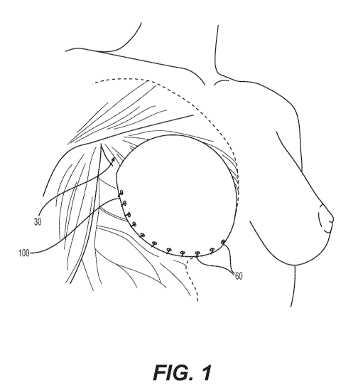
[0012] Fig. 1 illustrates a breast treatment device for more complete
coverage of a breast implant or tissue expander in a pre-pectoral position,
according
to certain embodiments.
[0013] Fig. 2 illustrates a breast treatment device for more complete
coverage of and/or support of a breast implant or tissue expander, according
to
certain embodiments.
[0014] Fig. 3 illustrates another breast treatment device for more
complete
coverage of and/or support of a breast implant or tissue expander, according
to
certain embodiments.
[0015] Fig. 4 illustrates another breast treatment device for more
complete
coverage of and/or support of a breast implant or tissue expander, according
to
certain embodiments.
[0016] Fig. 5A is a frontal view of the breast treatment device
including an
acellular tissue matrix positioned over a breast implant to illustrate how the
device
provides coverage to the implant or tissue expander or supports the implant or
reinforces surrounding tissue.
[0017] Fig. 5B is a side view of the breast treatment device of Fig.
5A
including an acellular tissue matrix positioned over a breast implant to
illustrate how
4
CA 03035256 2019-02-26
WO 2018/045118 PCT/US2017/049516
the device provides coverage to the implant or tissue expander or supports the
implant or reinforces surrounding tissue.
[0018] Fig. 5C is a top view of the breast treatment device of Fig. 5A
including an acellular tissue matrix positioned over a breast implant to
illustrate how
the device provides coverage to the implant or tissue expander or supports the
implant or reinforces surrounding tissue.
[0019] Fig. 6A is a frontal view of a breast treatment device
including an
acellular tissue matrix positioned over a breast implant to illustrate how the
device
provides coverage to the implant or tissue expander.
[0020] Fig. 6B is a side view of the breast treatment device of Fig.
6A
including an acellular tissue matrix positioned over a breast implant to
illustrate how
the device provides coverage to the implant or tissue expander.
[0021] Fig. 6C is a top view of the breast treatment device of Fig. 6A
including an acellular tissue matrix positioned over a breast implant to
illustrate how
the device provides coverage to the implant or tissue expander.
[0022] Fig. 7A is a frontal view of a breast treatment device
including an
acellular tissue matrix positioned over a breast implant to illustrate how the
device
provides coverage to the implant or tissue expander.
[0023] Fig. 7B is a side view of the breast treatment device of Fig.
7A
including an acellular tissue matrix positioned over a breast implant to
illustrate how
the device provides coverage to the implant or tissue expander.
[0024] Fig. 7C is a top view of the breast treatment device of Fig. 7A
including an acellular tissue matrix positioned over a breast implant to
illustrate how
the device provides coverage to the implant or tissue expander.
CA 03035256 2019-02-26
WO 2018/045118 PCT/US2017/049516
[0025] Fig. 8A illustrates implantation of the breast treatment device
of
Fig. 2 in a prepectoral position along with a breast implant.
[0026] Fig. 8B illustrates implantation of the breast treatment device
of
Fig. 3 in a prepectoral position along with a breast implant.
[0027] Fig. 9 illustrates a breast treatment device for more complete
coverage of a breast implant or tissue expander and/or support or
reinforcement of
surrounding tissues, wherein the device further includes preformed tabs or
extensions for attachment to tissue, according to certain embodiments.
[0028] Fig. 10 illustrates a breast treatment device for more complete
coverage of a breast implant or tissue expander and/or support or
reinforcement of
surrounding tissues, wherein the device further includes preformed slits or
openings,
according to certain embodiments.
[0029] Fig. 11 illustrates a breast treatment device for more complete
coverage of a breast implant or tissue expander and/or support or
reinforcement of
surrounding tissues, wherein the device further includes preformed holes or
openings, according to certain embodiments.
[0030] Fig. 12 illustrates a breast treatment device for more complete
coverage of a breast implant or tissue expander and/or support or
reinforcement of
surrounding tissues, wherein the device further includes preformed holes or
pilot
holes, according to certain embodiments.
[0031] Fig. 13 illustrates a breast treatment device in accordance
with the
embodiments of Fig. 3 for more complete coverage of a breast implant or tissue
expander and/or support or reinforcement of surrounding tissues, wherein the
device
further includes preformed holes or pilot holes, according to certain
embodiments.
6
CA 03035256 2019-02-26
WO 2018/045118 PCT/US2017/049516
[0032] Fig. 14 illustrates another breast treatment device in
accordance
with the embodiments of Fig. 3 for more complete coverage of a breast implant
or
tissue expander and/or support or reinforcement of surrounding tissues,
wherein the
device further includes preformed holes or pilot holes, according to certain
embodiments.
Description of Exemplary Embodiments
[0033] Reference will now be made in detail to various embodiments of
the disclosed devices and methods, examples of which are illustrated in the
accompanying drawings. Wherever possible, the same reference numbers will be
used throughout the drawings to refer to the same or like parts.
[0034] In this application, the use of the singular includes the
plural unless
specifically stated otherwise. In this application, the use of "or" means
"and/or"
unless stated otherwise. Furthermore, the use of the term "including", as well
as
other forms, such as "includes" and "included", is not limiting. Any range
described
herein will be understood to include the endpoints and all values between the
endpoints.
[0035] The section headings used herein are for organizational
purposes
only and are not to be construed as limiting the subject matter described. All
documents, or portions of documents, cited in this application, including but
not
limited to patents, patent applications, articles, books, and treatises, are
hereby
expressly incorporated by reference in their entirety for any purpose.
[0036] The present disclosure relates generally to devices for
surgical
breast procedures and systems and methods relating to such devices. The
devices
can be used for tissue augmentation, repair or regeneration of damaged tissue,
7
CA 03035256 2019-02-26
WO 2018/045118 PCT/US2017/049516
and/or correction of tissue defects. As such, the devices, systems, and
methods
discussed herein can be suitable for a wide range of surgical applications,
such as,
for example, aesthetic surgery, breast reconstruction, breast augmentation,
breast
enhancement, breast reduction, and revisionary breast surgeries.
[0037] The tissue matrices used to produce the devices described
herein
can include a variety of different materials. For example, an acellular tissue
matrix or
other tissue product can be selected to allow tissue ingrowth and remodeling
to
assist in regeneration of tissue normally found at the site where the matrix
is
implanted. For example, an acellular tissue matrix, when implanted on or into
subdermal tissue, fascia, mammary tissue, muscle, bone, adipose or other
tissue,
may be selected to allow regeneration of the tissue without excessive fibrosis
or scar
formation. In certain embodiments, the devices can be formed from ALLODERM or
STRATTICETm (LIFECELLO CORPORATION, BRANCHBURG, NJ) which are human
and porcine acellular dermal matrices, respectively. Alternatively, other
suitable
acellular tissue matrices can be used. For example, a number of suitable
biological
scaffold materials are described by Badylak et al. "Extracellular Matrix as a
Biological
Scaffold Material: Structure and Function," Acta Biomaterialia (2008),
doi:10.1016/j.actbio.2008.09.013. The devices described herein can be produced
from a variety of different human or animal tissues including human, porcine,
ovine,
bovine, or other animals tissues.
[0038] Tissue matrix products, such as acellular dermal tissue
matrices,
are widely used in surgical breast procedures. For example, sheets of
acellular
dermal matrix can be provided as a square or rectangular sample, which can be
cut
to a desired shape if needed. In addition, certain preformed tissue matrix
shapes are
available. For example, crescent or other curved shapes are available to
reduce the
8
CA 03035256 2019-02-26
WO 2018/045118 PCT/US2017/049516
amount of tissue matrix needed while providing an appropriate shape for an
aesthetically desirable surgical result.
[0039] For some surgical applications, however, different shapes and
sizes for tissue matrices would be beneficial. For example, when implanting a
breast
implant or tissue expander in a pre-pectoral position, i.e., anterior to the
pectoral
muscles, it would be beneficial in some cases to provide a tissue matrix shape
and
size that allows one or more of (1) complete or near complete anterior
coverage of
an implant or tissue expander, (2) minimized need for resizing or shaping the
tissue
matrix, or (3) preshaped borders that facilitate attachment to anatomical
structures to
produce desired surgical results (e.g., aesthetic or reconstructive result
with low
likelihood of complications).
[0040] Fig. 1 illustrates a breast treatment device 100 for more
complete
coverage of a breast implant or tissue expander in a pre-pectoral position
and/or to
support a breast implant or tissue expander, or help regenerate, reinforce,
augment,
or support surrounding tissue such as overlying dermis and subdermal tissue,
according to certain embodiments. Although the devices and methods discussed
herein are made with respect to, primarily, prepectoral procedures, the
devices can
be used by surgeons for other procedures. The device 100 can include a
flexible
sheet of acellular tissue matrix, as discussed above. As discussed in more
detail
below, the device 100 can be affixed to a chest wall 30 or other appropriate
tissue to
cover an implant or tissue expander (not shown in Fig. 1). The device can be
secured in place using sutures 60 or other surgical fixation devices (e.g.,
staples,
clips, surgical adhesives).
[0041] Figs. 2-4 are top views of various embodiments of devices,
according to the present disclosure. The devices illustrated in Figs. 2-4 can
each
9
CA 03035256 2019-02-26
WO 2018/045118 PCT/US2017/049516
include flexible sheets of acellular tissue matrix, which can have one of the
illustrated
shapes when laid flat. Each of the devices 100, 200, 300, can allow complete
or
substantially complete coverage of the anterior portion of a breast implant or
tissue
expander, including an implant or tissue expander positioned anterior to the
pectoralis muscles. In addition, or alternatively, the devices can help
support a
breast implant or tissue expander, or help regenerate, reinforce, augment, or
support
surrounding tissue such as overlying dermis and subdermal tissue. When placed
in
contact with overlying tissue, the tissue matrix will support tissue
regeneration,
ultimately becoming infiltrated by cells and becoming vascularized, thereby
providing
enhanced tissue coverage to improve surgical outcomes, e.g., by preventing
various
possible adverse events such as rippling, loss of tissue integrity.
[0042] Fig. 2 illustrates a breast treatment device 100 for more
complete
coverage of a breast implant or tissue expander and/or to support a breast
implant or
tissue expander, or help regenerate, reinforce, augment, or support
surrounding
tissue such as overlying dermis and subdermal tissue, according to certain
embodiments. As shown, the device 100 includes a sheet of acellular tissue
matrix.
The sheet can include a top surface and a bottom surface (the surfaces
correspond
to the front and back of the two-dimensional image of Fig. 2).
[0043] The sheet forming the device 100 has a first section 104 and a
second section 108, and the first 104 and second 108 sections have different
shapes
and are attached to one another at a joining section 110. The first section
includes
curved first 106 and second edges 111, and the second section includes curved
first
114 and second edges 112.
[0044] The curvature of the edges 106, 111, 114, 112 of the first 104
and
second 108 sections can be varied to produce a desired shape. For example, in
one
CA 03035256 2019-02-26
WO 2018/045118 PCT/US2017/049516
embodiment the first edge 106 of the first section 104 has a degree of
curvature that
is greater than a degree of curvature of the second edge 111 of the first
section 104.
In addition, the first edge 114 of the second section 108 can have a degree of
curvature that is greater than a degree of curvature of the second edge 112 of
the
second section 108. As shown, the first edges 106, 114 of the sections 104,
108 are
the edges at opposite ends of the device 100.
[0045] The second edge 111 and second edge 112 will be understood to
refer to a curved edge extending from opposite apices 117/117', 119/119' of
the
sections 104, 108 (i.e., edges along dashed lines 113, 115). But, as shown in
Fig. 2,
the sections 104 and 108 are joined at a joining section 110, such that the
first
section 104 and second section 108 are attached to one another along the
second
edges 111,112 of each of the first section 104 and second section 108. The
joining
section 110 may simply be a continuation of a single sheet of acellular tissue
matrix
forming the device 100. As shown, the apices 117/117', 119/119' are pointed to
form
an acute angle, but the apices may alternatively be curved or rounded.
[0046] The device 100 is illustrated as having two-dimensional
symmetry
about a line or axis 120 passing midway through the tissue matrix 100 when the
device lies flat. Variations in the shape may be made, or the device may be
made
nearly or perfectly symmetric. Furthermore, the device 100, having first and
second
sections 104, 108 can more readily conform to an implant or expander shape,
provide improved support to an implant or expander, or provide complete
overlying
tissue contact by virtue of spaces on the lateral sides of the joining section
110, i.e.,
between the second edges 111, 112, where a gap is formed.
[0047] Fig. 3 illustrates another breast treatment device 200 for more
complete coverage of a breast implant or tissue expander, according to certain
11
CA 03035256 2019-02-26
WO 2018/045118 PCT/US2017/049516
embodiments. As shown, the device 200 includes a sheet of acellular tissue
matrix,
wherein the sheet of acellular tissue matrix comprises a flexible sheet 200
with a top
surface and a bottom surface (the surfaces correspond to the front and back of
the
two-dimensional image of Fig. 3).
[0048] The sheet 200 can be sized and shaped to allow coverage of a
breast implant or tissue expander, provide improved support to an implant or
expander, or provide complete overlying tissue contact. As shown, the sheet
200 has
an upper curved border 210 having a first degree of curvature and a lower
curved
border 220 having a second degree of curvature. The upper border 210 and lower
border 220 can be joined at lateral apices 224, 228, which can include a sharp
angle
or rounder edges.
[0049] As with the device 100 of Fig. 2, the device 200 can be sized
and
shaped to allow coverage of a breast implant or tissue expander, particularly
for
coverage of an anterior portion of the implant or expander when implanted in a
prepectoral position. In addition or alternatively, the device can provide
improved
support to an implant or expander, or provide complete overlying tissue
contact. In
one embodiment, the lower curved border 220 is shaped and sized to conform to
a
desired shape of a lower margin of a breast, and the upper curved border 210
is
sized and shape such that the flexible sheet of acellular tissue matrix can
cover
substantially all of the anterior surface of a breast implant to tissue
expander
[0050] Many implants or expanders will have a shape and size such that
the implant volume at the lower pole is greater than that at the upper pole.
The
device 200 (as well as other devices described herein), allows coverage and
support
of such implants with little or no additional manipulation by surgeons (e.g.,
no cutting
to size and shape). As such, the devices 200 (and 100, 300) prevent waste of
12
CA 03035256 2019-02-26
WO 2018/045118 PCT/US2017/049516
valuable tissue matrix material, save substantial operating room time, and
have
preformed margins that produce a desired configuration when implanted.
[0051] The size and shape of the devices 100, 200, 300 can be selected
based on typical implant or tissue expander (when fully expanded) shapes and
volumes. For example, for the device 200 of Fig. 3, the device can include a
lower
section 230 and upper section 240 and the height or length of the lower and
upper
sections 230, 240 can be selected based on the desired implant or expander
size
and shape as well as a the need for additional material to cover tissue or
affix the
device to surrounding structures. Exemplary, sizes can include, for example a
height
(from the bottom or lower border 220 to top of upper border 210) from 15-25
cm, and
a width from apices 224, 228 of 15-30cm. For example, a small device may have
a
height of 15cm and width of 17-18cm; a medium device a height of 18-19cm and
width of 21-22cm; and a large device a height of 20-21cm and width of 23-24
cm;
and an extra-large device a height of 22-23cm and width of 26-27cm.
[0052] Fig. 4 illustrates another breast treatment device 300 for more
complete coverage of a breast implant or tissue expander. The device 300 can
include a sheet 300 of acellular tissue matrix, wherein the sheet of acellular
tissue
matrix comprises a flexible sheet with a top surface and a bottom surface (the
surfaces correspond to the front and back of the two-dimensional image of Fig.
4).
[0053] The sheet 300 can include a lower curved border 314 and an
upper curved border 316, wherein the upper border 316 and lower border 314 are
joined at apices 308, 309 at lateral ends of the device 300. As with the other
devices
100, 200, the apices 308, 309 can be sharp angles or can be rounded.
13
CA 03035256 2019-02-26
WO 2018/045118 PCT/US2017/049516
[0054] The device 300 can have a configuration such that when lying on
a
flat surface, the sheet 300 is symmetrically shaped about an axis 320 midway
between the apices 308, 309 and parallel to the top and bottom surfaces.
[0055] The device 300 can also be shaped such that the lower border
314
forms a single outward arc shape (lower section 302), and the upper border 316
has
three arc sections 306, 306', 307, including first and second sections 306,
306' each
extending from one of the apices 308, 309, and a third section 307 joining the
first
and second sections 306, 306', the third section having a degree of curvature
that is
different than the degree of curvature of the first and section 306, 306'
sections. The
arc sections 306, 306', 307 can form the upper portion or section 304, while
the
lower border 314 defines the lower section 302.
[0056] As discussed previously, the devices described herein can be
used
to allow coverage of a tissue expander or implant, including implants or
expanders
positioned in a prepectoral position. In addition or alternatively, the device
can
provide improved support to an implant or expander, or provide complete
overlying
tissue contact. As such, Fig. 5A illustrates a frontal view of the breast
treatment
device 100 including an acellular tissue matrix positioned over a breast
implant to
illustrate how the device provides coverage to the implant or tissue expander.
It will
be understood that when implanted, and as discussed further below, the tissue
matrix will contact overlying dermal or subcutaneous tissue, as well as
possible
contact and connection with muscle or other tissues, and the matrix can
support the
implant, allow ingrowth of tissue, and provide tissue regeneration, support,
and
vascularization, in some cases for patients for whom insufficient tissue or
insufficient
tissue strength or vascularity would have been present in the absence of the
tissue
14
CA 03035256 2019-02-26
WO 2018/045118 PCT/US2017/049516
matrix. As such, the tissue matrix allows prepectoral positioning while
avoiding other,
often difficult procedures.
[0057] Fig. 5B and 5C are side and top views, respectively, of the
device
of Fig. 5A. Figs. 6A-6C provide comparable views of the device 200 of Fig. 3
over an
implant; and Fig. 7A. Figs. 7A-7C provide comparable views of the device 300
of
Fig. 4 over an implant. It should be noted that the frontal view is described
in
reference to how the device and implant should be viewed with respect to a
patient if
the implant covered by the devices 100, 200, 300 were located on the anterior
chest
wall. So, for example, Fig. 5A is referred to as a frontal view as it is a
view showing
the front of the device 100 when covering an implant (behind the device) as it
would
be viewed from the front of a patient in whom the device is implanted.
[0058] Fig. 8A illustrates implantation of the breast treatment device
100
of Fig. 2 in a prepectoral position along with a breast implant. And Fig. 8B
illustrates
implantation of the breast treatment device 200 of Fig. 3 in a prepectoral
position
along with a breast implant. A similar implantation process and configuration
would
be applicable to the other devices 300 or variations thereof described
throughout. As
shown, the devices 100, 200 are implanted to cover an implant 20 or expander
on an
anterior portion of the chest wall 30. One section 108 (or upper and lower
section of
device 200) is positioned to cover a lower portion of the implant, while the
other
section 104 covers an upper portion of the implant 20.
[0059] To secure the devices 100, 200 (or any other device described
herein) in place, parts of the device 100, 200, such as the lower border 114,
220
and/or upper border 106, 210 can be affixed to tissue using sutures, clips,
staples,
adhesives, or other suitable surgical fixation systems. In some cases, the
device
100, 200 (or device 300) can be sized to provide an amount of tissue matrix
that
CA 03035256 2019-02-26
WO 2018/045118 PCT/US2017/049516
wraps around the posterior portion of the implant or expander, e.g., at the
lower
margin/inframammary fold and/or at the superior surface of the implant or
expander.
The devices may be sized to wrap between, for example, 1-3 cm, 1-2 cm around
the posterior portion of the implant or expander at either or both of the
inferior or
superior portions of the implant or expander.
[0060] In some cases, the implant or tissue expander includes suture
tabs
or other fixation components to allow the device 100, 200, or 300 to be
secured to
the implant or expander. In such cases, the device can be joined to the
expander or
implant prior to or during implantation before final positioning within an
implant site.
In cases where the device 100, 200, 300 is sized to wrap partially around a
posterior
portion of the implant or expander, the tabs or fixation devices can be
posteriorly
located so that the device can be secured to the posterior aspect of the
implant or
expander, while the device is in contact with overlying subdermal tissues when
implanted.
[0061] The devices described herein can further be modified to
facilitate
fixation to tissue for proper implantation. For example, the devices can
include
features that provide additional material for attachment to anchors such as
sutures
and/or can include features that guide proper or easier placement of sutures
or other
anchors. In addition, or alternatively, the devices can include openings,
slits, or holes
that provide for one or more of improved drainage or fluid flow, better
coverage of
the implant or expander, or changes in mechanical properties (e.g., more
flexibility
due to presence of slits, holes, or other mechanical modifications). Figs. 9-
14
illustrate various modified devices, and although shown with respect to the
device
shape of Figs. 2 and 3 (Figs. 13 and 14 illustrate embodiments of the device
of Fig.
16
CA 03035256 2019-02-26
WO 2018/045118 PCT/US2017/049516
3, it will be understood that similar modifications can be used with the other
described devices of Fig. 4.
[0062] Fig. 9 illustrates a breast treatment device 900, wherein the
device
further includes preformed tabs 910 or extensions for attachment to tissue,
according
to certain embodiments. The tabs or extensions 910 can provide additional area
for
passing sutures or other anchors, or can be specifically shaped to engage with
fixations devices located on the surface of an implant or expander. Although a
finite
and specific number of tabs 910 is illustrated, additional or fewer tabs 910
may be
used.
[0063] Fig. 10 illustrates a breast treatment device 1000, wherein the
device further includes preformed slits 1010 or openings, according to certain
embodiments. The slits 1010 or openings can allow flow of fluid through the
tissue
matrix, thereby preventing certain complications (e.g., seroma or inability to
drain
infectious fluids). In addition, the slits 1010 can be shaped and sized to
allow
expansion or more flexible coverage of an implant or expander.
[0064] Fig. 11 illustrates a breast treatment device 1100, wherein the
device further includes preformed holes 1110 or openings, according to certain
embodiments. Similar to the openings 1010 of Fig. 10, the holes 1110 can allow
fluid
to flow through the material. The openings 1110 and slits 1010 can be arranged
in
number, size, and location based on a variety of factors.
[0065] Fig. 12 illustrates a breast treatment device 1200, wherein the
device further includes preformed holes or pilot holes (holes and pilot holes
represented by any of 1210, 1220, or 1230), according to certain embodiments.
The
holes or pilot holes 1210 can be provided to allow easier, more rapid, or
better
17
CA 03035256 2019-02-26
WO 2018/045118 PCT/US2017/049516
fixation. For example, the holes or pilot holes can be positioned in a row or
locations
that correspond to a desired spacing or positioning to provide secure
fixation, e.g.,
along the lower border corresponding to the inframammary fold when implanted.
The
holes or pilot holes can pass completely through the device to allow passage
of
sutures or other anchors, or can include a countersink or divot formation to
provide
an area of less density or strength to allow easy anchor passage.
[0066] The holes or pilot holes can be positioned on the lower section
of
the device (holes 1210, 1220) and/or upper section 1230 near edges. In
addition,
holes or pilot holes may be formed at other regions if desired. Further, the
holes or
pilot holes can be in two or more rows, as illustrated, to allow multiple
points of
fixation and/or to give the surgeon some choice in selecting holes location.
[0067] Similar to Fig. 12, Figs. 13 and 14 illustrate embodiments of
the
device 200 of Fig. 3, but further including holes, openings, or pilot holes.
As shown
in Fig. 13, the holes, openings, or pilot holes 2210 may be localizes to a
portion of
the device, e.g., the lower section, thereby providing openings only around
the lower
pole of the implant or tissue expander. Alternatively, the holes, openings or
pilot
holes can be arranged in other patterns or throughout the surface of the
device, as
shown in Fig. 14.
[0068] The holes, openings, and pilot holes will generally be
positioned
and of a number such that they do not cause an undesirable loss of strength or
area
for cellular ingrowth. In addition, the holes, openings, or pilot holes may be
a
distance from the edges of the devices such that they do not overlap with
areas
where sutures may be placed, or alternatively, can be placed to provide
preformed
opening/pilot openings to guide where sutures may be placed.
18
CA 03035256 2019-02-26
WO 2018/045118 PCT/US2017/049516
[0069] In some cases, the tissue matrices can be produced from
materials
that include a basement membrane on at least one surface. For example, the
devices can be produced from an acellular dermal matrix, and either the top
surface
or bottom surface can include an epithelial basement membrane across the
surface.
When implanted next to a breast implant or tissue expander, the basement
membrane covered surface may face towards the implant or tissue expander such
that the surface not including a basement membrane faces overlying
vascularized
tissue.
[0070] Methods of treatment using the devices discussed herein as well
as devices produced for use in such methods are further contemplated as within
the
scope of the present inventions. The methods are illustrated and discussed
above
with respect to Figs. 8A and 8B, and aspects of the methods are elaborated
upon
herein. The devices can be used for improving various procedures, such as
prepectoral implantation of an implant. In many cases, the method will first
include
performing a procedure to remove tissue, e.g., for surgical oncology, and can
therefore, include mastectomy, lumpectomy, or variations on those procedures.
The
methods and device may also be used for augmentation procedures without, or in
a
separate procedure from mastectomy or other procedures (e.g., for staged
reconstruction). When used for implantation for augmentation, or in a
subsequent
procedure, a surgeon may first form a pocket or space in the subcutaneous
region.
[0071] After performing a mastectomy or other procedure and ensuring a
proper space for the implant or expander, a surgeon may then place the tissue
matrix materials described herein within the space, and affix portions of the
tissue
matrices to tissues such as the chest wall or muscle, as illustrated in Figs.
8A and
8B. As an example, the tissue can be affixed to the superior medial and
lateral edges
19
CA 03035256 2019-02-26
WO 2018/045118 PCT/US2017/049516
of the pectoralis major and to fascial at the level of the inframammary fold.
As such,
the tissue matrix comes in contact with overlying tissue (e.g., dermis) and is
prepared to provide support to the implant or expander and subsequently allow
tissue ingrowth and vascularization of overlying tissue.
[0072] Next, and implant or expander can be placed within the pocket,
and remaining edges of the tissue matrix are sutured or otherwise attached to
tissue
to close the implant pocket, followed by closure of the surgical site.
[0073] It will be appreciated that the tissue matrix may alternatively
be
wrapped around the implant or expander outside the body, and the entire device
(e.g., implant/expander and tissue matrix) can then be placed in the surgical
site.
Further, implants or expanders may include a structure for securing to the
tissue
matrix and/or chest wall or other tissue.
[0074] Other embodiments will be apparent to those skilled in the art
from
consideration of the specification and practice of this disclosure. It is
intended that
the specification and examples be considered as exemplary only, with the true
scope
and spirit of the disclosed devices and methods being indicated by the
following
claims.