Note: Descriptions are shown in the official language in which they were submitted.
CA 03035723 2019-03-04
[DESCRIPTION]
[Invention Title]
DEGLYCOSYLATED ANTIBODY SPECIFICALLY BINDING TO
CLEC14A AND USES THEREOF
[Technical Field]
The present disclosure relates to a deglycosylated
antibody binding specifically to C-type lectin domain
family 14, member A (c1ec14a). More particularly, the
present disclosure relates to a deglycosylated antibody
that comprises a light chain variable region comprising
CDR1 having a certain sequence and binds specifically to
clec14, and use thereof, for example, a pharmaceutical
composition containing the antibody for preventing or
treating an angiogenesis-related disease.
[Background Art]
Tumor angiogenesis plays a key role in the process of
tumors. Vascular endothelial growth factors (VEGFs) and
epidermal growth factor receptors (EGFRs) are key factors in
angiogenesis which is a very promising target in the
treatment of cancers. Bevacizumab (Avastie), which is an
anti-VEGF antibody, is used to treat patients who suffer
from a disease such as metastatic colorectal cancer, renal
cell carcinoma, non-small cell lung cancer, or malignant
1
CA 03035723 2019-03-04
brain glioma. Cetuximab, which is an anti-EGFR antibody,
can inhibit cell-contacts between endothelial cells and
suppress expression of angiogenesis factors such as VFGFs,
interleukin-8 and basic fibroblast growth factors.
However, Avastin , which is a single agent, has no
efficacy in clinic trials and is used for combination
therapy with a variety of chemical drugs. Combination
therapy involves use of various chemical drugs in
conjunction with the treatment, thus having a risk of
various side effects on patients. In addition, Avastin is
known to inhibit VEGF signaling actions of both tumor
vessels and normal blood vessels, and thus result in side
effects such as proteinuria, hypertension, bleeding and
gastrointestinal perforation through induction of defects to
the normal blood vessels.
Furthermore, Avastin may cause tolerance when used
for a long time. Since high levels of VEGF-A, -B and -C,
PIGFs (placental growth factors) and VEGF receptor-1 are
expressed in tolerant colorectal cancer cells, expression of
various pro-angiogenic soluble factors and receptors may be
increased in spite of treatment with VEGF-neutralizing
antibodies.
There are unmet-needs for developing novel antibody
medications to solve these side-effects and tolerance of
Avastin. In this regard, the present inventors found that
2
CA 03035723 2019-03-04
CTLD of Clec14a, so called C-type lectin-like domain (CTLD),
a series of epidermal growth factor-like domain and with
sushi-like domain, all of which are named as an
extracellular domain included in a type I transmembrane
protein, plays a key role in actin cytoskeletal
rearrangement that is important for cell migration. In
addition, based on this fact, the present inventors
screened human antibodies specific to c1ec14a-CTLD and
found that the screened antibodies can inhibit tumor
angiogenesis by exhibiting high cross-reactivity to human
and mouse c1ec14a-CTLDs, and suppressing migration of
vascular endothelial cells, tube formation and endothelial
cell-cell contacts. As a result, the present inventors
filed a PCT patent application (WO 2013-187556).
In order to evaluate efficacy and toxicity of
developed antibodies in an animal model, large scale
production of antibodies is required and stability of
antibodies is important. However, in the process of
purifying human antibodies specific to clecl4a-CTLD, protein
aggregation occurs. This indicates that the process of
improving stability is needed to increase an antibody yield.
The present disclosure was completed based on the
finding that not only desired efficacies, but also antibody
stability can be accomplished by glycosylation changes in
antibodies based on prediction of glycosylation, since the
3
CA 03035723 2019-03-04
glycosylation of antibodies may affect stability as well as
functions of the antibodies during large scale production.
[Disclosure]
[Technical Problem]
Therefore, the present disclosure has been made in
view of the above problems, and it is one object of the
present disclosure to provide a deglycosylated antibody
binding specifically to c1ec14a, wherein CDR of c1ec14a-
CTLD IgG of clone 1, which is a c1ec14a-CTLD-specific
antibody disclosed in PCT Patent Application Laid-open No.
W02013/187556, is grafted to commercially available
therapeutic antibodies, and is replaced with the framework
of the therapeutic antibody, and a part of glycosylation
sites in light chain CDR1 amino acid sequences is
substituted with other amino acid sequences.
It is another object of the present disclosure to
provide a pharmaceutical composition containing the
antibody for preventing or treating an angiogenesis-
related disease.
[Technical Solution]
In accordance with the present disclosure, the above
and other objects can be accomplished by the provision of
an antibody binding specifically to clecl4a, wherein the
4
CA 03035723 2019-03-04
antibody comprises a light chain variable region comprising
CDR1 of TGSSSNIGXXXVT (SEQ ID NO: 1), wherein each of amino
acids X at positions 9, 10 and 11 in the SEQ ID NO: 1 is any
one selected from the group consisting of R, C, G, A, T, W,
S, N, and V.
In accordance with another aspect of the present
disclosure, provided is a pharmaceutical composition
containing the antibody for preventing or treating an
angiogenesis-related disease.
Other technical features and examples of the present
disclosure will be more clearly described in the following
Detailed Description of the Invention and Claims mentioned
later.
[BRIEF DESCRIPTION OF THE DRAWINGS]
The above and other objects, features and other
advantages of the present disclosure will be more clearly
understood from the following detailed description taken
in conjunction with the accompanying drawings, in which:
FIG. lA is a schematic diagram illustrating CDR
grafting of parent antibody IgG from four types of
therapeutic antibodies, i.e., adalimumab
(Humirae),
omalizumab (Xolair ), trastuzumab (Herceptin ) and
bevacizumab (Avastie);
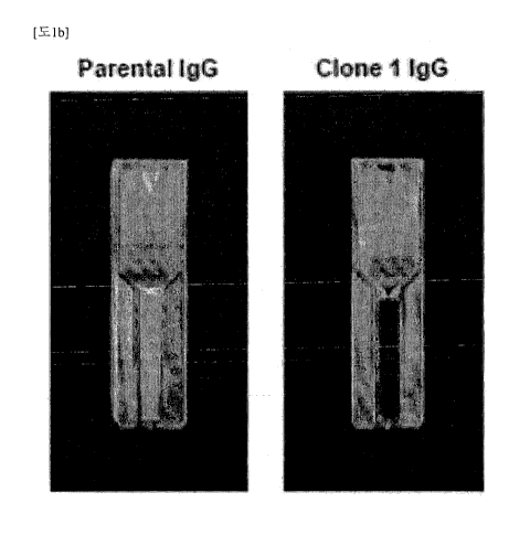
FIG. 1B shows results of visual observation regarding
5
CA 03035723 2019-03-04
aggregation of antibodies in parent antibody IgG and clone
1 IgG preparations;
FIG. 1C shows results of measurement using
spectrophotometry regarding aggregation indices of parent
antibody IgG (green) and clone 1 IgG (orange), wherein the
aggregation index is calculated in accordance with 100 X
[Abs340/(Abs280 - Abs340)];
FIG. 1D shows results of analysis regarding parent
antibody IgG (green) and clone 1 IgG (orange) before and
after precipitation of antibodies;
FIG. 2A is a schematic diagram illustrating a
deglycosylation process to screen four types of
glycosylated IgG clones (deglyco C1-C4) by phage display
technology;
FIG. 2B shows results of measurement using ELISA
regarding the binding specificity of four types of
deglycosylated IgG to hclecl4a-CTLD-Fc, mclecl4a-CTLD-Fc
and Fc alone;
FIG. 2C shows results of screening using HUVEC tube
formation assay regarding optimized candidate antibodies
for suppressing clecl4a-mediated angiogenesis, wherein
clone 1 IgG is used as a positive control group;
FIG. 2D shows results of screening using HUVEC tube
formation assay regarding optimized candidate antibodies
for suppressing clecl4a-mediated angiogenesis, wherein the
6
CA 03035723 2019-03-04
total number of branches is represented as percentage (%)
of tube formation of control group (MOCK);
FIG. 2E shows results of investigation using wound
healing assay regarding effects of deglyco Cl IgG on
migration of endothelial cells;
FIG. 2F is a graph showing results of investigation
using wound healing assay regarding effects of deglyco Cl
IgG on migration of endothelial cells, wherein wound
difference is represented as a percentage (%) of cell
migration of control group (MOCK);
FIG. 2G shows results of investigation regarding
mobility of deglyco Cl IgG and clone 1 IgG under reduction
conditions using one-dimensional electrophoresis;
FIG. 2H shows results of investigation regarding
homogeneity of deglyco Cl IgG and clone 1 IgG using two-
dimensional electrophoresis;
FIG. 3A shows results of competitive ELISA through
addition of parent antibody IgG binding to deglyco Cl IgG-
HRP and hclecl4a-CTLD-Fc;
FIG. 3B shows results of flow cytometry in the
presence of parent antibody IgG (green) or deglyco Cl IgG
(red), or the absence thereof (MOCK, black);
FIG. 3C shows results of measurement regarding
affinity of parent antibody IgG (green) or deglyco Cl IgG
(red) to hclecl4a-ECD-myc using biolayer interferometry
7
CA 03035723 2019-03-04
assay through Octet RED96 system (*Kp=eguilibrium
dissociation constant; Kõ=association rate constant;
Koff=dissociation rate constant);
FIG. 3D shows results of HUVEC tube formation assay
in the presence of parent antibody IgG (green), deglyco Cl
IgG (red) or bevacizumab (blue) or in the absence thereof
(MOCK, black);
FIG. 3E shows results of HUVEC tube formation assay,
wherein the total number of branches is represented as a
percentage of tube formation of control group (MOCK);
FIG. 3F shows results of wound healing assay in the
presence of parent antibody IgG (green), deglyco Cl IgG
(red) or bevacizumab (blue) or in the absence thereof
(MOCK, black). An image was captured at Oh (upper part)
and 8h (lower part) using an optical microscope;
FIG. 3G shows results of wound healing assay, wherein
wound difference is represented as a percentage (%) of
cell migration of control group (MOCK);
FIG. 4A shows results of counting using an optical
microscope regarding HEK293F cells transfected with wild-
type c1ec14a and cultured for 6h in the presence of parent
antibody IgG (green) or deglyco Cl IgG (red) or the absence
thereof (MOCK, black);
FIG. 4E shows the number of aggregates per field
calculated as a percentage (%) of clecl4a-mediated cell-
8
CA 03035723 2019-03-04
cell contacts of the control group (MOCK);
FIG. 4C shows results of measurement using cell ELISA
regarding hc1ec14a-CTLD-Fc-HRP bound to HUVECs coated on a
microtiter plate in the presence or absence of parent
antibody IgG (green) or deglyco Cl IgG (red) with an
increasing concentration;
FIG. 4D shows results of measurement using cell ELISA
regarding c1ec14a-ECD-myc bound to c1ec14a-CTLD-Fc coated
on a microtiter plate in the absence or presence of parent
antibody IgG (green) or deglyco Cl IgG (red) with an
increasing concentration;
FIG. 5A shows results of investigation on cell
viability based on an absorbance at 450 nm regarding HUVECs
incubated in the absence of deglyco Cl IgG (black) or
presence (red) or in the presence of 5-FU (yellow) for 2
days;
FIG. 5E shows results of culture of HUVECs in the
presence of hTNFa (pink), the absence of deglyco Cl IgG
(black), or the presence of deglyco Cl IgG (red), wherein
analysis is conducted by flow cytometry after staining
with anti-ICAM-1 (upper part) or VCAM-1 (lower part)
polyclonal antibody. hTNFa is a positive control group for
endothelial cell activity;
FIG. 5C shows results of Rhodamine-Phalloidin and
DAPI staining regarding HUVECs cultured in the absence or
9
CA 03035723 2019-03-04
presence of deglyco Cl IgG, wherein the morphology of
HUVECs is observed with a confocal microscope;
FIG. 5D shows results of immunoblot analysis on
phosphorylation of VEGF-dependent VEGFR (VEGF receptor),
Akt and ERK (extracellular signal-regulated kinase) in the
presence of VEGF or VEGF and deglyco Cl IgG, or the
absence thereof (MOCK);
FIG. 5E shows evaluation results of in vitro and in
vivo toxicity of optimized candidate antibodies,
specifically, measurement results of serum concentrations
of GOT, GPT, BUN, CRE, and TBIL 30 days after antibody
injection;
FIG. 5F shows evaluation results of in vitro and in
vivo toxicity of optimized candidate antibodies,
specifically, measurement results of mouse body weight one
day and 28 days after antibody injection;
FIG. 5G shows evaluation results of in vitro and in
vivo toxicity of optimized candidate antibodies,
specifically, results of TUNEL assay analysis regarding
apoptosis conditions of renal and hepatic tissues 30 days
after antibody injection;
FIG. 6A shows results of VEGF-dependent tube
formation assay in the presence of deglyco Cl IgG (red) or
the absence thereof (MOCK);
FIG. 6B shows results of VEGF-dependent tube
CA 03035723 2019-03-04
formation assay, wherein the total number of branches is
represented as a percentage (%) of tube formation of
control group (MOCK);
FIG. 6C is an image showing VEGF-dependent blood
vessel growth in a cut aortic ring cultured in the presence
of parent antibody IgG (green), deglyco Cl IgG (red) and
bevacizumab (blue), or the absence thereof (MOCK);
FIG. 6D shows results of counting regarding the
number of grown blood vessels;
FIG. 6E is an image showing VEGF-dependent
microvessel formation in a Matrigel plug of a nude mouse
in the presence of parent antibody IgG, deglyco Cl IgG and
bevacizumab, or the absence thereof (MOCK);
FIG. 6F shows results of investigation on microvessel
formation, based on measured hemoglobin content, wherein
the hemoglobin content is represented as a percentage (%)
of hemoglobin content of control group (MOCK);
FIG. 7A shows results of immunohistochemistry using
antibodies to c1ec14a and CD31 to detect specific
expression of c1ec14a in blood vessels of SNU182 cancer
cell xenograft mouse tissues;
FIG. 7B shows results of immunohistochemistry using
antibodies to clecl4a and CD31 to detect specific
expression of c1ec14a in blood vessels of CFPAC-1 cancer
cell xenograft mouse tissues;
11
CA 03035723 2019-03-04
FIG. 7C shows results of immunohistochemistry
staining using antibodies to c1ec14a and CD31 to detect
specific expression of c1ec14a in tumor vessels of liver
cancer tissues;
FIG. 7D shows results of immunohistochemistry
staining using antibodies to c1ec14a and CD31 to detect
specific expression of c1ec14a in tumor vessels of
pancreatic cancer tissues;
FIG. 7E shows measurement results of SNU182-, CFPAC-1
or U87 cell-derived microvessel formation in the absence
(MOCK), the presence of deglyco Cl IgG (red) or the
presence of bevacizutab (blue);
FIG. 7F shows measurement results of SNU182-, CFPAC-1
or U87 cell-derived microvessel formation, wherein the
hemoglobin content is represented as a percentage of
hemoglobin content of control group (MOCK);
FIG. 7G shows measurement results of
immunohistochemistry using C031 regarding microvessel
formation by HCT116 and HCT116/Beva cell-derived tumors in
the presence of deglyco Cl IgG and bevacizumab or the
absence thereof (MOCK);
FIG. 7H shows a percentage of CD31 positive per field
represented with respect to the microvessel density of
control group (MOCK);
FIG. 71 shows results of evaluation regarding effects
12
CA 03035723 2019-03-04
of deglyco Cl IgG on tumor growth, wherein deglyco Cl IgG
can reduce tumor size while not affecting body weight, and
tumor volume and body weight are measured on a weekly
basis for one month;
FIG. 8A shows final antibody production of produced
optimized candidate antibodies;
FIG. 8B shows results of investigation using SDS-PAGE
regarding 90% purification of produced optimized candidate
antibodies and molecular weights of light chains and heavy
chains;
FIG. 9A shows results of investigation regarding
changes in tube length after treatment of HUVECs with VEGF
and the optimized antibodies;
FIG. 9B shows results of investigation regarding the
number of branches after treatment of HUVEC with VEGF and
the optimized antibodies;
FIG. 10 is a representative image showing changes in
tube length (progression of angiogenesis) after treatment
with VEGF and optimized antibodies;
FIGS. 11A and 11B show results of tube formation
analysis after treatment with clone 1, 13 and 16
antibodies exhibiting anti-angiogenesis activity against
HUVEC cells in the presence of EGM;
FIGS. 12A and 12B show results of analysis regarding
whether or not the optimized antibodies including clone 1,
13
CA 03035723 2019-03-04
13 and 16 antibodies suppress clecl4a-mediated cell-cell
contacts in c1ec14a-expressed HEK293 cells;
FIGS. 13A and 13B show results of analysis regarding
whether or not clone 1, 13 and 16 antibodies exhibit
inhibitory activity against endothelial migration;
FIG. 14A is a schematic diagram illustrating
competitive ELISA to identify antigen-binding sites of
screened antibodies;
FIG. 14B shows results of analysis regarding whether
or not the optimized candidate antibodies bind to antigen,
comparatively with deglyco Cl;
FIG. 15 shows results of investigation using flow
cytometry regarding whether or not clone 1, 13 and 16
antibodies have the ability to bind to human umbilical
vein endothelial cells (HUVECs) and mouse aortic
endothelial cells (MAECs);
FIG. 16A is a schematic diagram illustrating the role
of CLEC14a-CTLD on cell-cell contacts in vascular
endothelial cells and the mechanism by which the CLEC14-
CTLD-conjugated antibody acts as an inhibitor of cell-cell
contacts;
FIG. 16B shows investigation results of the role of
CTLD on CLEC14a-CLEC14a bonding;
FIG. 16C shows results of analysis using competitive
ELISA regarding whether or not clone 1, 13 and 16
14
CA 03035723 2019-03-04
antibodies suppress CTLD domain-mediated CLEC14a molecular
bonding;
FIG. 17A illustrates CLEC14a down-regulation on the
surface of vascular endothelial cells by a CLEC14a-CTLD-
conjugated antibody; and
FIG. 17B illustrates results of investigation using
cell ELISA regarding CLEC14a down-regulation of the
optimized antibody.
[Best Model???
In one aspect, the present disclosure describes an
antibody or antigen binding fragment thereof binding
specifically to c1ec14a (C-type lectin domain family 14,
member A), wherein the antibody comprises a light chain
variable region comprising CDR1 of TGSSSNIGXXXVT (SEQ ID NO:
1), wherein each of amino acids X at positions 9, 10 and 11
of SEQ ID NO: 1 is any one selected from the group
consisting of R, C, G, A, T, W, S, N, and V.
The term "antibody" as used herein refers to a protein
molecule including an immunoglobulin molecule that
specifically recognizes an antigen and thus immunologically
reacts with the specific antigen, and includes a polyclonal
antibody, a monoclonal antibody, whole antibody and an
antibody fragment. In addition, chimeric antibodies (e.g.,
humanized mouse antibodies), bivalent or bispecific
CA 03035723 2019-03-04
molecules (e.g., bispecific antibodies),
diabodies,
triabodies and tetrabodies fall within the scope of the
antibody used in the present disclosure.
The whole antibody is composed of two overall length
light chains and two overall length heavy chains, wherein
each light chain is linked to a heavy chain by a disulfide
bond. There are five antibody isotypes known as IgA, IgD,
IgE, IgM, and IgG existing in mammals, and IgG is further
classified into four antibody subtypes of IgGl, IgG2, IgG3
and IgG4.
The term "antibody fragment" as used herein refers to
a fragment that at least maintains an antigen-binding
ability and includes Fab, F(ab'), F(ab')2, and Fv. Fab
includes a variable region of each of the heavy chain and
the light chain, the constant domain of the light chain, and
the first constant domain (CH1) of the heavy chain, each
having an antigen-binding site. Fab' is different from Fab
in that it further includes at least one cysteine residue at
a C-terminus of the CH1 domain of the heavy chain. F(ab')2
includes two Fab' molecules having a disulfide bond between
cysteine residues in a hinge region. An Fv (variable
fragment) including a variable region of each of the heavy
chain and the light chain is the minimal antibody fragment
having original specificity of parent immunoglobuiin.
Disulfide-stabilized Fv (dsFv) is formed by binding the
16
CA 03035723 2019-03-04
variable region of the light chain to the variable region of
the heavy chain via a disulfide bond. Single chain Fv
(scFV) is an Fv where the respective variable regions of the
heavy chain and the light chain are covalently linked via a
peptide linker. These antibody fragments can be obtained by
treating the whole antibody with a protease (for example,
papain or pepsin providing Fab or F(ab')2), and are
preferably constructed by genetic recombination technology.
The term "monoclonal antibody" as used herein refers
to an antibody molecule having a uniform molecule
composition which is obtained from a substantially identical
population of antibodies and exhibits a binding specificity
and affinity to a single epitope.
In general, immunoglobulin has a basic structural unit
including one heavy chain and two light chains. Each heavy
chain includes one variable region and three constant
domains, whereas each light chain includes one variable
region and one constant domain. The variable region of each
of the heavy chain and the light chain includes three
complementarity-determining regions (referred to as "CDRs")
and four framework regions. CDRs
function to bind to
epitopes of antibodies. CDRs on each chain start from the N-
terminus and are arranged in an order of CDR1, CDR2, and
CDR3. These CDRs are distinguished from one another by the
chain on which they are positioned.
17
CA 03035723 2019-03-04
Regarding the antibodies according to the present
disclosure, firstly, CDR grafting was conducted to conjugate
six sequences of CDR1 to CDR3 (SEQ ID NOS: 11 to 16) of
heavy chain and light chain variable regions of clecl4a-CTLD
IgG clone 1, the c1ec14a-CTLD human antibody (hereinafter,
parent antibody) disclosed in WO 2013/187556, filed by the
present inventors, with framework regions of four types of
therapeutic antibodies approved by the FDA, i.e., adalimumab
(Humirae), omalizumab (Xolaire), trastuzumab (Herceptine),
and bevacizumab (Avastin )
According to one embodiment of the present disclosure,
aggregation degree and developability index (DI) of
sequences of the four types of CDR-grafted antibodies and
the parent antibody were observed. An aggregation score is a
parameter for predicting an aggregation degree on sequences.
As aggregation score decreases, aggregation decreases. The
DI index is a parameter for predicting protein stability in
a solution. As DI index decreases, solubility and stability
of antibody increase. As a result, the parent antibody is
predicted to have a relatively high aggregation degree and
DI index, whereas, among four types of antibodies, the
antibody substituted with the framework of omalizumab
(Xolair ) exhibits superior aggregation degree and DI index.
In addition, the antibody substituted with the
framework of omalizumab (Xolair ) secures excellent
18
CA 03035723 2019-03-04
productivity, as compared to three other types of CDR-
grafted antibodies, and shows no aggregation. In addition,
only the antibody substituted with the framework of
omalizumab (Xolair ) exhibits cross-reactivity to human and
mouse CTLDs, similar antigen reactivity to the parent
antibody and inhibitory activity against tube formation,
comparable to the parent antibody.
Accordingly, the antibody according to the present
disclosure may include at least one heavy chain variable
region framework selected from the group consisting of SEQ
ID NOS: 17 to 20. In
addition, the antibody according to
the present disclosure may include at least one light chain
variable region framework selected from the group consisting
of SEQ ID NOS: 21 to 24.
The antibody according to the present disclosure may
be a deglycosylated antibody. Regarding the term
"glycosylation" as used herein, in the case of a
glycoprotein, for example, an antibody, whether or not
glycosylation occurs, and structures or morphologies of
glycoforms may be changed depending on the type of host
cells, methods for manipulating recombinants and culture
conditions. That is, in the process of producing
glycoproteins, various types of glycoforms are produced
depending on differences in glycoform structures or amounts
of constituent saccharides of glycoforms, or the like, so
19
CA 03035723 2019-03-04
heterogeneity may be present due to differences in
production conditions. Glycoproteins having different
glycoform structures are different from natural type in
terms of in vivo kinetics and tissue distribution, or are
antagonistic to the natural type, which may result in
adverse reactions, and act as an antigen when administered
continuously for a long time, which may cause immunological
problems. As such, glycoforms may be key factors that can
affect pharmacological effects and in vivo kinetics.
In an attempt to regulate glycoforms, the antibody
according to the present disclosure may be an antibody
obtained by deglycosylating an antibody substituted with an
omalizumab (Xolair ) framework. The deglycosylated antibody
is intended to include a non-deglycosylated immunoglobulin
Fc fragment and, for example, an N-linked glycosylation site
or an 0-linked glycosylation site may be modified or
removed. N-linked glycosylation may mean the attachment of
the sugar (glycan) chain to a side chain of an asparagine
(Asn) residue, and 0-linked glycosylation may mean the
attachment of one of N-acetyl-galactosamine, galactose or
xylose to hydroxyamino acid, more commonly, serine or
threonine.
The modification or removal of glycosylation sites may
be carried out by an ordinary method such as a chemical,
enzymatic Or genetically engineered method using
CA 03035723 2019-03-04
microorganisms, but is not limited thereto. In a specific
embodiment of the present disclosure, the presence of one N-
glycosylation site in L-CDR1 of an antibody is predicted, a
random scFv library at a probable N-glycosylation site is
produced using (NNK)3, and a deglycosylated antibody is then
screened by phage display technology.
Based on this trial, the present disclosure provides
an antibody comprising a light chain variable region
comprising CDR1 of TGSSSNIGXXXVT (SEQ ID NO: 1), wherein
each of the amino acids at positions 9, 10 and 11 of the SEQ
ID NO: 1 is any one selected from the group consisting of R,
C, G, A, T, W, S, N, and V.
In one embodiment, each of the amino acids at
positions 9, 10 and 11 of the SEQ ID NO: 1 may be RCG, ATA,
WSN or AVV. When the amino acid is RCG, the antibody may
include a light chain variable region including CDR1 of
TGSSSNIGRCGVT (SEQ ID NO: 2), when the amino acid is ATA,
the antibody may include a light chain variable region
including CDR1 of TGSSSNIGATAVT (SEQ ID NO: 3), when the
amino acid is WSN, the antibody may include a light chain
variable region including CDR1 of TGSSSNIGWSNVT (SEQ ID NO:
4), and when the amino acid is AVV, the antibody may include
a light chain variable region including CDR1 of
TGSSSNIGAVVVT (SEQ ID NO: 5).
According to one embodiment of the present disclosure,
21
CA 03035723 2019-03-04
it can be seen that the four types of screened
deglycosylated antibodies, i.e., deglyco-Cl to deglyco-C4,
do not show aggregation and have high final purification
yields. In addition, results of investigation as to whether
or not characteristics of the four types of screened
deglycosylated antibodies are maintained well, as compared
with an antibody substituted with a Xolair framework, show
that all of the four types of deglycosylated antibodies,
i.e., deglyco-Cl to deglyco-C4 maintain cross-reactivity to
human and mouse CTLDs.
The amino acids at positions 9, 10 and 11 of the SEQ
ID NO: 1 are preferably RCG. When amino acids at positions
9, 10 and 11 of the SEQ ID NO: 1 are RCG, the antibody
according to the present disclosure may include a light
chain variable region including CDR1 of TGSSSNIGRCGVT (SEQ
ID NO: 2).
According to one embodiment of the present disclosure,
the antibody comprising a light chain variable region
comprising CDR1 of SEQ ID NO: 2 is represented by "deglyco-
Cl". In order to identify whether or not efficacies of the
deglycosylated antibodies are maintained, tube formation
ability is observed. As a result, it can be seen that
deglyco-C1 exhibits similar inhibitory activity against tube
formation to the antibody (clone 1 IgG) CDR-grafted to the
framework region of the omalizumab antibody. Furthermore,
22
CA 03035723 2019-03-04
deglycosylation degree of deglyco-Cl is identified. As a
result, it can be seen that glycosylation patterns are
removed and disappeared.
The term "human antibody" as used herein refers to a
molecule that consists entirely of amino acid sequences of
all components of human immunoglobulin including CDRs,
framework regions and the like. Human antibodies have at
least three potential benefits in the treatment of human
diseases. First, human antibodies further preferably
interact with the human immune system to destroy target
cells more effectively by, for example, complement-dependent
cytotoxicity (CDC) or antibody dependent cell-mediated
cytotoxicity (ADCC). Another benefit is that the human
immune system does not recognize the human antibody as being
a foreign molecule. Furthermore, the half-lives of human
antibodies are similar to those of antibodies endogenously
derived from the human circulatory system, even when
administered in smaller amounts or with less frequency. The
antibody according to the present disclosure is preferably a
human monoclonal antibody and is useful for the treatment of
angiogenesis-related diseases or cancer, because it has
strong affinity to clecl4a, preferably, c1ec14a-CTLD
expressed on human endothelial cells which effectively
inhibits c1ec14a-mediated angiogenesis, and because it has
low immunogenicity since both heavy chains and light chains
23
CA 03035723 2019-03-04
are derived from humans.
As used herein, the term "clecl4a (C-type lectin
domain family 14, member A)" means a member of C-type
lectin/C-type lectin-like domain (CTL/CTLD) superfamily.
Clec14a is a type I transmembrane protein, the extracellular
domain of which consists of a C-type lectin-like domain
(CTLD), a series of epidermal growth factor-like domains,
and a sushi-like domain. Information associated with
c1ec14a can be obtained from certified database such as NCBI
GenBank. For example, human clecl4a may have Gene ID No
161198, but is not limited thereto. "C-type lectin-like
domain (CTLD) of c1ec14a (C-type lectin domain family 14,
member A)" may be also referred to as "clec14a-CTLD" or
"clecl4a-CTLD", wherein all thereof may be interchangeably
used with one another.
The term "epitope" as used herein refers to a site
that determines antigen-specificity, which may be
interchangeably used with an antigenic determinant or an
antigen determining site.
In addition, according to the present disclosure, in
order to improve affinity of the deglycosylated antibody,
major amino acid residues of HCDR3 relating to antigen-
antibody reactivity can be modified. For example,
determination is conducted by alanine scanning, randomized
antibody (ab) libraries with induced random mutation are
24
CA 03035723 2019-03-04
constructed, and antibodies having higher reactivity than
the parent antibody are screened. Respective antibodies
capable of maintaining characteristics and functions better
than the parent antibody can be screened by yeast surface
display and phage display technologies.
In one embodiment, the antibody according to the
present disclosure may include a light chain variable region
including CDR1 selected from the group consisting of SEQ ID
NOS: 2 to 5. The antibody according to the present
disclosure may include a light chain variable region further
including CDR2 of SEQ ID NO: 15 and CDR3 of SEQ ID NO: 16,
in addition to CDR1 described above. The antibody according
to the present disclosure may include: light chain CDR1 of
SEQ ID NO: 2, light chain CDR2 of SEQ ID NO: 15 and light
chain CDR3 of SEQ ID NO: 16; light chain CDR1 of SEQ ID NO:
3, light chain CDR2 of SEQ ID NO: 15 and light chain CDR3 of
SEQ ID NO: 16; light chain CDR1 of SEQ ID NO: 4, light chain
CDR2 of SEQ ID NO: 15 and light chain CDR3 of SEQ ID NO: 16;
or light chain CDR1 of SEQ ID NO: 5, light chain CDR2 of SEQ
ID NO: 15 and light chain CDR3 of SEQ ID NO: 16.
The antibody according to the present disclosure may
include a light chain variable region selected from the
group consisting of SEQ ID NOS: 7 to 10. The antibody
according to the present disclosure may include a light
chain variable region including CDR1 OF SEQ ID NO: 2. The
CA 03035723 2019-03-04
antibody may include a light chain variable region including
SEQ ID NO: 7.
In one embodiment, the antibody according to the
present disclosure may include a heavy chain variable region
including CDR3 selected from the group consisting of SEQ ID
NO: 13, and SEQ ID NOS: 25 to 60. The antibody may include
a heavy chain variable region further including CDR1 OF SEQ
ID NO: 11 and CDR2 OF SEQ ID NO: 13, in addition to the
CDR3. The antibody according to the present disclosure may
include: heavy chain CDR1 of SEQ ID NO: 11, heavy chain CDR2
of SEQ ID NO: 12 and heavy chain CDR3 of SEQ ID NO: 13;
heavy chain CDR1 of SEQ ID NO: 11, heavy chain CDR2 of SEQ
ID NO: 12 and heavy chain CDR3 of SEQ ID NO: 25; heavy chain
CDR1 of SEQ ID NO: 11, heavy chain CDR2 of SEQ ID NO: 12 and
heavy chain CDR3 of SEQ ID NO: 37; or heavy chain CDR1 of
SEQ ID NO: 11, heavy chain CDR2 of SEQ ID NO: 12 and heavy
chain CDR3 of SEQ ID NO: 40.
The antibody may include a heavy chain variable region
selected from the group consisting of SEQ ID NOS: 6, and 61
to 96. The antibody according to the present disclosure may
include a heavy chain variable region including CDR3 of SEQ
ID NO: 13 and/or a heavy chain variable region of SEQ ID NO:
6.
The antibody according to the present disclosure may
comprise:
26
CA 03035723 2019-03-04
a light chain CDR1 of SEQ ID NO: 2, a light chain CDR2
of SEQ ID NO: 15 and a light chain CDR3 of SEQ ID NO: 16, a
heavy chain CDR1 of SEQ ID NO: 11, a heavy chain CDR2 of SEQ
ID NO: 12 and a heavy chain CDR3 of SEQ ID NO: 13;
a light chain CDR1 of SEQ ID NO: 3, a light chain CDR2
of SEQ ID NO: 15 and a light chain CDR3 of SEQ ID NO: 16, a
heavy chain CDR1 of SEQ ID NO: 11, a heavy chain CDR2 of SEQ
ID NO: 12 and a heavy chain CDR3 of SEQ ID NO: 13;
a light chain CDR1 of SEQ ID NO: 4, a light chain CDR2
of SEQ ID NO: 15 and a light chain CDR3 of SEQ ID NO: 16, a
heavy chain CDR1 of SEQ ID NO: 11, a heavy chain CDR2 of SEQ
ID NO: 12 and a heavy chain CDR3 of SEQ ID NO: 13;
a light chain CDR1 of SEQ ID NO: 5, a light chain CDR2
of SEQ ID NO: 15 and a light chain CDR3 of SEQ ID NO: 16, a
heavy chain CDR1 of SEQ ID NO: 11, a heavy chain CDR2 of SEQ
ID NO: 12 and a heavy chain CDR3 of SEQ ID NO: 13;
a light chain CDR1 of SEQ ID NO: 2, a light chain CDR2
of SEQ ID NO: 15 and a light chain CDR3 of SEQ ID NO: 16, a
heavy chain CDR1 of SEQ ID NO: 11, a heavy chain CDR2 of SEQ
ID NO: 12 and a heavy chain CDR3 of SEQ ID NO: 25;
a light chain CDR1 of SEQ ID NO: 2, a light chain CDR2
of SEQ ID NO: 15 and a light chain CDR3 of SEQ ID NO: 16, a
heavy chain CDR1 of SEQ ID NO: 11, a heavy chain CDR2 of SEQ
ID NO: 12 and a heavy chain CDR3 of SEQ ID NO: 37; or
a light chain CDR1 of SEQ ID NO: 2, a light chain CDR2
27
CA 03035723 2019-03-04
of SEQ ID NO: 15 and a light chain CDR3 of SEQ ID NO: 16, a
heavy chain CDR1 of SEQ ID NO: 11, a heavy chain CDR2 of SEQ
ID NO: 12 and a heavy chain CDR3 of SEQ ID NO: 40.
The antibody according to the present disclosure may
include:
a light chain variable region of SEQ ID NO: 7 and a
heavy chain variable region of SEQ ID NO: 6;
a light chain variable region of SEQ ID NO: 8 and a
heavy chain variable region of SEQ ID NO: 6;
a light chain variable region of SEQ ID NO: 9 and a
heavy chain variable region of SEQ ID NO: 6;
a light chain variable region of SEQ ID NO: 10 and a
heavy chain variable region of SEQ ID NO: 6;
a light chain variable region of SEQ ID NO: 7 and a
heavy chain variable region of SEQ ID NO: 61;
a light chain variable region of SEQ ID NO: 7 and a
heavy chain variable region of SEQ ID NO: 73; or
a light chain variable region of SEQ ID NO: 7 and a
heavy chain variable region of SEQ ID NO: 76.
When the antibody of the present disclosure includes a
constant domain, it can be derived from IgG, IgA, IgD, IgE
or IgM, or a combination or hybrid thereof.
The term "combination" as used herein means that a
polypeptide encoding a single chain immunoglobulin Fc
fragment having the identical origin is linked to a single
28
CA 03035723 2019-03-04
chain polypeptide having a different origin to produce a
dimer or multimer. This dimer or multimer may be produced
from two or more constant domains selected from the group
consisting of constant domains of IgG, IgA, IgD, IgE and
IgM.
The term "hybrid" as used herein means that sequences
encoding two or more heavy chain constant domains having
different origins are present in a single chain
immunoglobulin heavy chain constant domain. For example, a
domain hybrid may be composed of one to four domains
selected from the group consisting of CH1, CH2, CH3 and CH4
of IgG, IgA, IgD, IgE and IgM. In addition, a combination
of hybrids may be formed from heavy chain constant domains
of IgG subtypes, i.e., IgGl, IgG2, IgG3 and IgG4. The
combination of hybrids is as defined above.
In addition, the antibody of the present disclosure
may further include a light chain constant region, which may
be derived from a lambda (A) or kappa (K) light chain.
Preferably, the antibody may be a human monoclonal
antibody that binds specifically to not only human c1ec14a-
CTLD, but also mouse clecl4a-CTLD to inhibit angiogenesis.
The ability of human antibodies to function in both humans
and mice, i.e., cross-reactivity, provides an advantage of
enabling the human antibodies to be applicable to a
preclinical study in mice.
29
CA 03035723 2019-03-04
In another aspect, the present disclosure is directed
to a composition containing the antibody for suppressing
angiogenesis, or a pharmaceutical composition containing the
antibody for preventing or treating an angiogenesis-related
disease.
Since the antibody regarding the present disclosure is
capable of effectively suppressing angiogenesis, a
composition containing the antibody as an effective
ingredient is also useful for suppressing angiogenesis, and
is also useful for preventing or treating an angiogenesis-
related disease.
The term "suppression of angiogenesis" as used herein
means suppression of formation or growth of new blood
vessels from previously existing vessels. For the purposes
of the present disclosure, suppression of angiogenesis can
be accomplished by suppressing cell migration or
intercellular contacts, more preferably, by suppressing
clecl4a-mediated cell migration,
clecl4a-mediated
intercellular contacts, HUVEC migration, tube formation, or
clecl4a CLTD-CLTD complex formation.
The term "angiogenesis-related disease" as used herein
means a disease that is related to incidence or development
of angiogenesis. Any disease may fall within the scope of
the angiogenesis-related disease without limitation so long
as it can be treated with the antibody. Examples of the
CA 03035723 2019-03-04
angiogenesis-related disease include, but are not limited
to, cancer, metastasis, diabetic retinopathy, retinopathy of
prematurity, corneal graft rejection, macular degeneration,
neovascular glaucoma, erythrosis, proliferative retinopathy,
psoriasis, hemophilic arthritis, microvessel formation of
atherosclerotic plagues, keloid, wound granulation, vascular
adhesion, rheumatoid arthritis, osteoarthritis, autoimmune
diseases, Crohn's disease, restenosis, atherosclerosis,
intestinal adhesions, cat scratch disease, ulcer, liver
cirrhosis, nephritis, diabetic nephropathy, diabetes
mellitus, inflammatory diseases and neurodegenerative
diseases. In addition, the cancer is selected from the
group consisting of esophageal cancer, stomach cancer, large
intestine cancer, rectal cancer, oral cancer, pharynx
cancer, larynx cancer, lung cancer, colon cancer, breast
cancer, uterine cervical cancer, endometrial cancer, ovarian
cancer, prostate cancer, testis cancer, bladder cancer,
renal cancer, liver cancer, pancreatic cancer, bone cancer,
connective tissue cancer, skin cancer, brain cancer, thyroid
cancer, leukemia, Hodgkin's lymphoma, lymphoma and multiple
myeloid blood cancer, but is not limited thereto.
The term "prevention" or "prophylaxis" as used herein
refers to any action causing the suppression or delay of the
onset of a disease of interest by administering the antibody
or composition according to the present disclosure. The
31
CA 03035723 2019-03-04
term "treatment" or "therapy" as used herein refers to any
action causing an improvement in symptoms of a disease of
interest or the beneficial alteration of the symptoms by
administering the antibody or composition according to the
present disclosure.
The composition including the antibody of the present
disclosure is preferably a pharmaceutical composition and
may further contain an appropriate vehicle, excipient or
diluent typically used in the art.
The pharmaceutical composition containing a
pharmaceutically acceptable vehicle may be in a variety of
oral or parenteral dosage forms such as tablets, pills,
powders, granules, capsules, suspensions, internal use
solutions, emulsions, syrups, sterile aqueous solutions,
non-aqueous solutions, lyophilizates and suppositories. In
this regard, the pharmaceutical composition of the present
disclosure may be formulated in combination with a diluent
or excipient such as a filler, a thickener, a binder, a
wetting agent, a disintegrant, a surfactant or the like.
Solid formulations for oral administration may take the form
of tablets, pills, powders, granules, capsules and the like.
Regarding these solid suspensions, the compound of the
present disclosure may be formulated in combination with one
or more excipients such as starch, calcium carbonate,
sucrose, lactose or gelatin. In addition to a simple
32
CA 03035723 2019-03-04
excipient, a lubricant such as magnesium stearate or talc
may be further used. Liquid formulations for oral
administration may include suspensions,
solutions,
emulsions, syrups and the like. A simple diluent such as
water or liquid paraffin, various excipients such as wetting
agents, sweeteners, aromatics, preservatives and the like
may be incorporated in the liquid formulations. In
addition, the pharmaceutical composition of the present
disclosure may be in a parenteral dosage form such as a
sterile aqueous solution, a non-aqueous solvent, a
suspension, an emulsion, a lyophilizate, a suppository or
the like. Injectable propylene glycol, polyethylene glycol,
vegetable oils such as olive oil and esters such as ethyl
oleate may be suitable for non-aqueous solvents and
suspensions. Basic ingredients of the suppositories include
Witepsol, macrogol, Tween 61, cacao butter, laurin butter
and glycerogelatin.
The composition of the present disclosure is
administered in a pharmaceutically effective amount. The
term "pharmaceutically effective amount" as used herein
refers to an amount of a pharmaceutical composition for
treating a disease which is sufficient at a reasonable
benefit/risk ratio applicable to all medical treatments.
The effective amount may be changed depending on a variety
of factors including severity of the disease to be treated,
33
CA 03035723 2019-03-04
age and gender of the patient, type of disease, drug
activity, sensitivity to the drug, administration time,
administration route, excretion rate, treatment period, co-
administration of drugs and other parameters known in the
art. The composition of the present disclosure may be
administered alone or in combination with other
therapeutics. In this case, the composition of the present
disclosure may be administered sequentially or
simultaneously with conventional therapeutics. In addition,
the composition may be administered in a single dose or may
be divided into multiple doses. When thoroughly taking into
consideration these factors, it is important to administer a
minimal amount sufficient to achieve maximum efficacy
without side effects, and this dosage can be readily
determined by those skilled in the art. The dosage of the
pharmaceutical composition of the present disclosure is not
particularly limited, but depends on a variety of factors
including health conditions and body weight of patient,
severity of disease, type of drug, administration route and
administration period. The composition may be administered
in a single dose or multiple doses daily to mammals
including rats, mice, domestic animals, humans and the like,
via a typically acceptable route, for example, orally,
rectally, intravenously, subcutaneously, intrauterinely, or
intracerebrovascularly.
34
CA 03035723 2019-03-04
In another aspect, the present disclosure is directed
to a method for suppressing angiogenesis or a method for
preventing or treating an angiogenesis-related disease, each
method including administering the antibody or the
composition to a subject in need thereof.
The antibody, the composition and suppression of
angiogenesis are as described above.
More specifically, the suppression method according to
the present disclosure includes administering a
pharmaceutical composition at a pharmaceutically effective
amount to a subject in need of suppression of angiogenesis.
The subject may be a mammal such as a dog, cow, horse,
rabbit, mouse, rat, chicken or human, but is not limited
thereto. The pharmaceutical composition may be administered
parenterally, subcutaneously,
intraperitoneally,
intrapulmonarily or intranasally, or by an appropriate
method including intralesional administration for local
treatment, if needed. A preferred dosage of the
pharmaceutical composition of the present disclosure may
depend on a variety of factors including health conditions
and body weight of the subject, severity of the disease,
type of drug, administration route and administration period
and can be readily determined by those skilled in the art.
In another aspect, the present disclosure is directed
to a method for preventing or treating cancer including
CA 03035723 2019-03-04
administering a pharmaceutical composition including the
antibody for preventing or treating cancer, or the antibody,
to a subject in need thereof. The terms "antibody",
"prevention" and "treatment" are as mentioned above.
Any cancer can be applied without limitation so long
as it can be treated with the antibody of the present
disclosure. Cancer in which progression of c1ec14a-mediated
tumor occurs is preferred. Specifically, the antibody of
the present disclosure can prevent the onset or progression
of cancer by suppressing angiogenesis. Examples of the
cancer include, but are not limited to, esophageal cancer,
stomach cancer, large intestine cancer, rectal cancer, oral
cancer, pharynx cancer, larynx cancer, lung cancer, colon
cancer, breast cancer, uterine cervical cancer, endometrial
cancer, ovarian cancer, prostate cancer, testis cancer,
bladder cancer, renal cancer, liver cancer, pancreatic
cancer, bone cancer, connective tissue cancer, skin cancer,
brain cancer, thyroid cancer, leukemia, Hodgkin's lymphoma,
lymphoma and multiple myeloid blood cancer.
In addition, the antibody of the present disclosure
may be used in combination with other antibodies or
biologically active agents or substances for various
purposes.
Hereinafter, the present disclosure will be described
more in detail with reference to examples. However, it is
36
CA 03035723 2019-03-04
obvious to those skilled in the art that these examples are
provided only for illustration of the present disclosure and
should not be construed as limiting the scope of the present
disclosure.
Example 1: Formation of antibodies with improved
stability by in siiico-based CDR grafting
It was reported in WO 2013/187556 that the c1ec14a-
CTLD parent antibody can specifically regulate angiogenesis
characteristics in vitro. In this regard, it was found that
the parent antibody (clone 1 of WO 2013/187556) shows
significant aggregation during purification and an
additional optimal process was needed to improve an antibody
yield. Accordingly, in silico-based CDR grafting was
conducted. Six CDRs in heavy and light chain variable
regions of the parent antibody (Table 1) were grafted to
respective framework regions of four types of therapeutic
antibodies approved by FDA, i.e., adalimumab (Humira!'),
omalizumab (Xolair ), trastuzumab (Herceptin ), bevacizumab
(Avastin ) (FIG. 1A).
[Table 1] Six CDRs in heavy and light chain variable regions
of parent antibody
Types CDR Sequence SEQ ID NO:
VH CDR1 GFTFSGYDMS SEQ
ID NO: 11
37
CA 03035723 2019-03-04
CDR2 GIYPDGGNTYYADSVKG SEQ
ID NO: 12
CDR3 GATWWVLGPFDY SEQ
ID NO: 13
CDR1 TGSSSNIGNNSVT SEQ
ID NO: 14
VL CDR2 ADSHRPS SEQ
ID NO: 15
CDR3 GAWDDSLSGYV SEQ
ID NO: 16
Developability indices (DIs) of the four CDR-grafted
antibodies (clone 1 to 4 IgGs) and the parent antibody were
compared using in silico-based analysis and Discovery Studio
3.1 Software available from Accelrys Inc. DI is a rapid in
silico prediction parameter used to evaluate a monoclonal
antibody according to aggregation characteristics. As DI
increases, aggregation degree increases.
As can be seen from Table 2, among the parent antibody
and the four CDR-grafted antibodies, clone 1 IgG having the
framework region (specific sequence of the framework region
is shown in Table 3) of omalizumab (Xolair ) exhibits lower
DI than the parent antibody and other CDR-grafted antibodies
(clone 2-4 IgGs), which means that clone 1 IgG has excellent
stability.
[Table 2] Summary of theoretical solubility of parent
antibody IgG and CDR-grafted IgG antibodies
38
CA 03035723 2019-03-04
=
Developability
Antibodies Rank Index (DI)
Clone 1 IgG 1 1613
Clone 2 IgG 2 170.17
Clone 3 IgG 3 IS I .75
Clone 4 IgG 4 182.46
Parental IgG 5 244.20
In order to identify improved aggregation degree of
clone 1 IgG, aggregation of purified parent antibody IgG and
clone I IgG was compared by visual observation and then was
confirmed by spectrophotometry. Aggregation was
observed
only in the parent antibody IgG, and was not visually
observed in clone 1 IgG (FIG. 1B). Aggregation indices of
the parent antibody IgG and clone 1 IgG measured by
spectrophotometry were about 23.4 and 1.5, respectively,
which means that clone 1 IgG has higher solubility than
parent antibody IgG (FIG. 1C).
For further comparison in stability between parent
antibody IgG and clone 1 IgG, a water-soluble antibody was
weighed after aggregates were precipitated and removed by
centrifugation. The antibody of about 95% of the parent
antibody IgG preparation was aggregated, whereas no antibody
aggregation was observed from clone 1 IgG (FIG. 1D). This
result suggests that in silico-based CDR grafting is a
39
CA 03035723 2019-03-04
method which is effective for producing an antibody platform
with improved stability.
Example 2: Selection (screening) of optimal antibodies
by continuous deglycosylation process and functional
separation
Heterogeneity in glycosylation of therapeutic proteins
can have significant impacts on characteristics and
qualities in the development and production of
biopharmaceuticals. Predicted N-glycosylation sites in
light chain CDR1 of clone 1 IgG were investigated through
glycosylation evaluation of in silico-based clone 1 IgG. In
order to remove such predicted N-glycosylation site, a semi-
synthetic antibody library having random mutation at the N-
glycosylation site was established and deglycosylation
process was then conducted. Specifically, semi-synthetic
scFvs (single chain variable fragments) randomized with NNK
trinucleotide oligonucleotides were established and were
subjected to continuous bio-panning by phage display
technology including human and mouse clecl4a-CILDs coated
with magnetic beads. Finally, DNAs of the selected scFv
clones were sequenced and different amino acid sequences
were randomly selected from four types of clones and
predicted N-glycosylation sites strongly reactive to both
CA 03035723 2019-03-04
human and mouse c1ec14a-CTLDs (hc1ec14a-CTLD and mc1ec14a-
CTLD) (FIG. 2A).
[Table 3]
Ab Types FRI CDR1 FR2 CDR2 FR3 CDR3 FR4
RFTIS
RDDS
EVQLV
WIRQ GIYPD KNTF
ESGGG GATW WGQG
GFTFS APGK GGNTY YLQM
VH LVQPG WVLGP TLVT
GYDMS GLEW YADSV NSLR
GSLRL FDY VSS
VA KG AEDT
SCAVS
AVYY
Deg C 1 CAR
GVPS
DIQLT RFSGS
WYQQ
QSPSS TGSSS GSGT GAWD FGQG
KPGK ADSHR
VL LSASV NIGRC DFTLT DSLSG TKVEI
APKL PS
GDRVT GVT ISSLQ YV KR
LIY
ITC PEDFA
TYYC
RFTIS
RDDS
EVQLV
WIRQ GIYPD KNTF
ESGGG GATW WGQG
GFTFS APGK GGNTY YLQM
VH LVQPG WVLGP TLVT
GYDMS GLEW YADSV NSLR
GSLRL FDY VSS
Deg C2 VA KG AEDT
SCAVS
AVYY
CAR
DIQLT TGSSS WYQQ GVPS GAWD FGQG
ADSHR
VL QSPSS NIGAT KPGK RFSGS DSLSG TKVEI
PS
LSASV AVT APKL GSGT YV KR
41
CA 03035723 2019-03-04
GDRVT LW DFTLT
ITC 1SSLQ
PEDFA
TYYC
RFTIS
RDDS
EVQLV
WIRQ GIYPD KNTF
ESGGG GATW WGQG
GFTFS APGK GGNTY YLQM
V H LVQPG WVLGP
TLVT
GYDNIS GLEW YADSV NSLR
GSLRL FDY VSS
VA KG AEDT
SCAVS
AVYY
Deg C3 CAR
GVPS
RFSGS DIQLT
WYQQ
QSPSS TGSSS GSGT GAWD FGQG
KPGK ADSHR
VL LSASV NIGWS DFTLT
DSLSG TKVEI
APKL PS
GDRVT NVT ISSLQ YV KR
LIY
PEDFA ITC
TYYC
RFT] S
RDDS
EVQLV
WIRQ GIYPD KNTF
ESGGG GATW WGQG
GFTFS APGK GGNTY YLQM
VH LVQPG WVLGP TLVT
GYDMS GLEW YADSV NSLR
GSLRL FDY VSS
VA KG AEDT
Deg C4 SCAVS
AVYY
CAR
DIQLT WYQQ GVPS
TGSSS GAWD FGQG
QSPSS KPGK ADSHR RFSGS
VL NIGAV DSLSG
TKVE1
LSASV APKL PS GSGT
VVT YV KR
GDRVT LW Y DFTLT
42
CA 03035723 2019-03-04
ITC ISSLQ
PEDFA
TYYC
Then, the scFv clones were converted into IgG,
expressed in human embryonic kidney (HEK) 293 cells and then
purified. Results of ELISA analysis showed that all
purified deglycosylated IgGs (deglyco Cl-C4 IgGs)
specifically bound to both hc1ec14a-CTLD-Fc and mc1ec14a-
CTLD-Fc, but did not bind to Fc alone, which means that the
deglycosylated IgGs are antibodies that bind specifically to
c1ec14a-CTLD (FIG. 2B).
In addition, as can be seen from Table 4, four types
of deglycosylated IgGs (deglyco Cl-C4 IgGs) were not
aggregated at all, and in particular, deglyco Cl and deglyco
C2 had high final purification yields.
[Table 4] Aggregation and purification amounts of four types
of deglycosylated antibodies
43
CA 03035723 2019-03-04
Scale: based on 100 ml culture
Deglyco-Ci, mg Deglyco.C2, mg Deg1yco-
C3, mg Deglyco-C4, mg
Before 9 NWç Before After, Before After
Before After f
Fraction', 0.011 0.009 0.019 0.018 0.014 0.012
0.019 0.015
Fraction2 3.792 3.763 3.718 3.702 2 209 2.109
1135 1.126
Frac11on3 0.938 0.921 1 113 1170 0.580 0.520
0.255 0.251
--r
Fraction4 0.129 0.128 0.201 01 : 0.129 0.127
0.057 0.050
Final um 4.859 4.821 5.032 5 018 2.918 2 768
1.390 1.442
Aggregation X X X X
Genetic mutation introduced during
antibody
engineering induces unpredicted defects, resulting in
deterioration in antibody functions. In order to isolate
antibodies for suppressing c1ec14a-mediated angiogenesis,
tube formation and endothelial cell migration assay using
human umbilical vein endothelial cells (HUVECs) were
conducted in the presence or absence of clone 1 IgG and
deglyco Cl-C4 IgGs. Among the four types of deglyco IgGs,
deglyco Cl IgG was selected as a candidate antibody. The
reason for this is that deglyco Cl IgG exhibited the most
excellent inhibitory activity against HUVEC tube formation
(FIGS. 2C and 2D) and migration (FIGS. 2E and 2F). In
addition, it can be seen that deglyco Cl IgG includes
desired alterations (N32R/N33C/S34G) in the amino acid
sequence in light chain CDR1.
In order to analyze biochemical properties of deglyco
Cl IgG, mobility of deglyco Cl IgG and clone 1 IgG was
44
CA 03035723 2019-03-04
evaluated under reduction conditions using one-dimensional
electrophoresis. The molecular weight of deglyco Cl VL was
25 kDa, which is similar to an estimated molecular weight,
whereas the molecular weight of clone 1 VL taking the
glycosylation form before deglycosylation was higher (FIG.
2G).
Results of two-dimensional electrophoresis showed that
the heterogeneous pattern of clone 1 scFv present within a
range lower than an isoelectric point (pI) was not present
in deglyco Cl scFv, which means improved homogeneity of
deglyco Cl IgG (FIG. 2H).
In conclusion, these results suggest that the selected
candidate antibodies are anti-angiogenesis antibodies that
effectively suppress pathological angiogenesis and have
improved homogeneity.
Example 3: Biochemical and functional properties of
optimized candidate antibodies
In order to identify the position of clecl4a-CTLD to
which deglyco Cl IgG binds, HRP (horseradish peroxidase)-
conjugated deglyco Cl IgG (deglyco Cl IgG-HRP) was produced
and competitive ELISA was then conducted.
The deglyco Cl IgG-HRP binding to hclecl4a-CTLD-Fc was
potently competitive by addition of parent antibody IgG,
CA 03035723 2019-03-04
which may mean that the parent antibody and deglyco Cl have
a similar clecl4a-CTLD binding position (FIG. 3A).
In order to identify whether or not deglyco Cl IgG
binds to human endothelial cells, flow cytometry was
conducted with HUVECs. Deglyco Cl IgG specifically bound to
the surfaces of HUVECs in a similar manner to the parent
antibody IgG. In addition, in order to measure binding
affinity of Cl IgG to hc1ec14a-ECD (extracellular domain of
human c1ec14a), binding affinity of antibody-antigen binding
was measured in real time. It can be seen that deglyco Cl
IgG has a KD constant (equilibrium dissociation constant) of
6 nM, which is similar to that of parent antibody IgG (FIG.
3C).
Overall, these results indicate that the binding
properties of the optimized candidate antibody were
maintained to a similar extent to those of the parent
antibody IgG, in spite of the two-step optimization process.
In order to compare in vitro angiogenesis suppression
effect between deglyco Cl IgG and bevacizumab, tube
formation and migration assay were conducted in the presence
or absence of parent antibody IgG, deglyco Cl IgG or
bevacizumab as a positive control group. Deglyco Cl IgG
significantly suppressed HUVEC tube formation (FIGS. 3D and
3E) and inhibited migration to a similar extent to
bevacizumab in vitro (FIGS. 3F and 3G). This means that the
46
CA 03035723 2019-03-04
candidate antibody has anti-angiogenesis activity and
efficacy, comparable to bevacizumab.
Example 4: New action mechanisms of optimized
candidate antibodies
In order to identify the mechanism by which deglyco Cl
IgG acts on angiogenesis, the number of cell aggregates
formed from HEK293F cells transfected with wild-type c1ec14a
and cultured in the presence or absence of parent antibody
IgG or deglyco Cl IgG was monitored to conduct functional
assay on c1ec14a-mediated cell-cell contacts.
The parent antibody IgG was used as a positive control
group to specifically block c1ec14a-mediated cell-cell
contacts. Deglyco Cl IgG had inhibitory efficacy against
aggregation of wild-type c1ec14a-transfected cells in the
similar manner to the parent antibody IgG, which suggests
that deglyco Cl IgG has inhibitory efficacy on c1ec14a-
mediated cell-cell contacts in angiogenesis (FIGS. 4A and
4B).
In order to analyze action mechanisms of deglyco Cl
IgG at a molecule-scale, specifically, in angiogenesis,
HUVECs were treated with hclecl4a-CTLD-Fc-HRP (HRP-labeled
hc1ec14a-CTLD-Fc) in the absence or presence of an
increasing concentration of parent antibody IgG or deglyco
Cl IgG. Deglyco Cl IgG significantly suppressed binding of
47
CA 03035723 2019-03-04
clecl4a-CTLD to HUVECs, in a concentration-dependent manner,
like parent antibody IgG (FIG. 4C).
In addition, hclecl4a-CTLD-Fc-HRP was incubated
together with purified hc1ec14a-ECD in the presence of an
increasing concentration of parent antibody IgG or deglyco
Cl IgG, or in the absence thereof. Deglyco Cl IgG directly
suppressed molecular interaction between hc1ec14-CTLD and
hc1ec14a-ECD, in a concentration-dependent manner, like
parent antibody IgG (FIG. 4D).
Overall, these results suggest that the produced
antibody serves as an interaction blocker specifically
suppressing endothelial cell-cell contacts
during
angiogenesis by directly blocking molecular interaction
between c1ec14a-CTLD-mediated c1ec14a molecules.
Example 5: Effects of optimized candidate antibodies
on toxicity of endothelial cells and VEGF signaling in
endothelial cells
In order to evaluate effects of deglyco Cl IgG on
toxicity of endothelial cells, after treatment with deglyco
Cl IgG, viability of HUVECs was measured. Viability was
measured in accordance with the manufacturer's manual
through cell counting Kit-8 (#CK04-13, Dojindo Laboratories,
Kumamoto, Japan).
48
CA 03035723 2019-03-04
HUVECs did not exhibit cytotoxic effects on these
cells, whereas 5-FU (5-fluorouracil) significantly reduced
viability of HUVECs (FIG. 5A).
In addition, the morphology of HUVEC was evaluated in
the presence or absence of deglyco Cl IgG using
immunocytochemistry. Deglyco Cl IgG did not change the
morphology of HUVECs (FIG. 5B). In order to evaluate
effects of deglyco Cl IgG on the activity of endothelial
cells, which is an initial inflammatory response to a
harmful stimulus, HUVECs were treated with deglyco Cl IgG,
and expression of an endothelial cell activity marker
including VCAM-1 (vascular cell adhesion molecule-1) and
ICAM-1 (intercellular cell adhesion molecule-1) was measured
to monitor HUVEC activity. Human tumor necrosis factor-
alpha (hTNFa) was used as a positive control group for
endothelial cell activity. Deglyco Cl IgG had almost no
impact on HUVEC activity, whereas hTNFa induced HUVEC
activity, as expected (FIG. 5C).
In order to investigate effects of deglyco Cl IgG on
VEGF-dependent signaling actions in endothelial cells, VEGF-
treated HUVECs were subjected to immunoblot analysis in the
presence or absence of deglyco Cl IgG to monitor changes in
phosphorylation of VEGFR (VEGF receptor), Akt and ERR
(extracellular signal-regulated kinase). Deglyco Cl IgG had
49
CA 03035723 2019-03-04
almost no effect on VEGF-dependent phosphorylation of VEGFR,
Akt and ERK in HUVECs (FIG. 5D).
In order to investigate in vivo toxicity of deglyco Cl
IgG, deglyco Cl IgG was intravenously injected into a normal
mouse twice a week, and hepatic and renal functions, body
weight, and apoptosis conditions of hepatic and renal
tissues between control groups and experimental groups were
then compared. The hepatic function was investigated by
measuring serum concentrations of glutamic-oxaloacetic
transaminase (GOT), glutamic pyruvic transaminase (GPT), and
total bilirubin (TBIL), and the renal function was
investigated by measuring the concentrations of blood urea
nitrogen (BUN) and creatinine (CRE). Apoptosis was measured
by TUNEL staining.
As a result, there was no significant change in
hepatic and renal functions (FIG. 5D). There was no
significant change in body weight as well (FIG. 5E). In
addition, apoptosis conditions were observed (FIG. 5F).
These results suggest that deglyco Cl IgG does not induce
serious in vivo toxicity.
Overall, these results suggest that deglyco Cl IgG
neither induces serious endothelial cytotoxicity nor has a
negative impact on VEGF-mediated signaling in normal
endothelial cells in vivo.
50
CA 03035723 2019-03-04
Example 6: Effects of optimized candidate antibodies
on VEGF-dependent angiogenesis
In order to investigate in vitro effects of deglyco Cl
IgG on VEGF-dependent angiogenesis, HUVECs were treated with
VEGF in the presence or absence of deglyco Cl IgG, and then
HUVEC tube formation assay was conducted. 150 pl of
Matrigel was added to a 48-well plate and incubated at 37 C
for 30 minutes. HUVEC (105) cultured in EGM-2 were
collected, seeded on the Matrigel-coated plate and incubated
at 37 C for 18 hours in the presence or absence of clone 1
IgG, parent antibody IgG, deglycosylated c1ec14a-CTLD IgG or
bevacizumab. HUVECs cultured in VEGF-containing EBM
(endothelial cell basal medium) were seeded to the Matrigel-
coated plate and incubated at 37 C for 18 hours in the
presence or absence of deglyco Cl IgG (20 pg m1-1).
Deglyco Cl IgG removed VEGF-dependent tube formation
significantly and almost perfectly (FIG. 6A).
In order to further investigate effects of deglyco Cl
IgG on VEGF-dependent angiogenesis, ex vivo rat aortic ring
assay was conducted in the presence or absence of deglyco Cl
IgG and bevacizumab. Blood vessels were almost not grown in
the rat aorta in endothelial basal medium (EBM), whereas
most blood vessels were grown in the rat aorta under VEGF
expression conditions. Furthermore, it was observed that
deglyco Cl IgG reduced the number of blood vessels grown in
51
CA 03035723 2019-03-04
the rat aorta to a similar extent to bevacizumab, when
cultured in combination with VEGF (FIGS. 6B and 6C).
In order to observe in vivo efficacies of deglyco Cl
IgG on VEGF-dependent angiogenesis, a mouse Matrigel model
was established, hemoglobin level was measured as a marker
of microvessel formation, and inhibitory activities against
microvessel formation of deglyco Cl IgG and bevacizumab were
compared by a mouse Matrigel plug assay. Deglyco Cl IgG
significantly reduced a hemoglobin content in a similar
manner to bevacizumab (FIGS. 6D and 6E).
Overall, these results suggest that the produced
antibodies have efficacy of in vivo inhibiting VEGF-
dependent abnormal angiogenesis.
Example 7: Effects of optimized candidate antibodies
on tumor angiogenesis
In order to investigate correlations between tumor
angiogenesis and c1ec14a, first, xenograft tumor models
including SNU182 human liver cell or CFPAC-1 human
pancreatic cancer cell carcinomas were established, and
immunohistochemistry was conducted on commercially available
antibodies of c1ec14a and CD31, which is a well-known
endothelial marker protein. Immunohistochemistry was
conducted as follows. HUVECs (5X10⁴) grown on a 0.1% (w/v)
gelatin-coated glass cover slip were incubated in the
52
CA 03035723 2019-03-04
presence or absence of deglyco Cl IgG for 24 hours at 37 C.
The cells were immobilized with 4% (w/v) PFA, blocked with
PBS supplemented with 5% (w/v) BSA and 0.1% (v/v) Triton X-
100 at 37 C for one hour and then incubated in Rhodamine-
Phailoidin (1 unit/well) and Hoechst 33258 stains for one
hour.
Clecl4a was expressed in SNU182- and CFPAC-1 tumor-
xenograft blood vessels, similar to CD31, which means that
clecl4a is expressed specifically in tumor blood vessels
(FIGS. 7A and 7B).
In order to evaluate specific expression of clecl4a in
tumor blood vessels among clinical samples using
immunohistochemistry, expression of clecl4a was compared
between blood vessels of normal tissues and blood vessels of
tissues derived from liver cancer and pancreatic cancer
patients. As a result, blood vessels of cancer patient
tissues showed a remarkable specific increase in clecl4a,
whereas normal blood vessels did not show the same (FIGS. 7C
and 7D).
In order to investigate in vivo effects of deglyco Cl
IgG on tumor angiogenesis, tumor cell-derived Matrigel plug
angiogenesis assay was conducted on thymus-removed nude
mice. SNU182- and CFPAC-1 cells and U87 human glioma cells
containing Matrigel were transplanted into thymus-removed
nude mice in the presence of deglyco Cl IgG and bevacizumab
53
CA 03035723 2019-03-04
with a single dose of 10 mg/kg or in the absence thereof. 2
weeks later, the Matrigel plug was removed and the total
hemoglobulin content of each group was measured with an
ELISA reader. Deglyco Cl IgG significantly reduced
hemoglobin derived from SNU182-, CFPAC-1 and U87 human
glioma cells to a similar extent to bevacizumab (FIGS. 7E
and 7F).
With respect to U87 human glioma, deglyco Cl IgG has
no impact on body weight and significantly reduces the tumor
sizes of U87 cells, like bevacizumab (FIG. 71). It can be
seen that deglyco Cl IgG has no significant impact on
survival, shape or activation of HUVECs in vitro. This
means that the optimized antibodies do not induce
significant in vivo endothelial toxicity.
Matrigel plug angiogenesis assay was conducted in the
presence of deglyco Cl IgG or bevacizumab with a single dose
of 5 mg/kg or the absence thereof using HC1116 human
colorectal cancer cells and bevacizumab-applied HCT116 human
colorectal cancer cells (HCT116 and HCT116/Beva). 14 days
later, the Matrigel plug was removed, and
immunohistochemistry was conducted on CD31 and microvascular
markers. A microvessel density was measured by quantifying
CD31 positive per field in an image each obtained by a
confocal microscope. It can be seen that deglyco Cl IgG and
bevacizumab similarly and significantly reduced microvessel
54
CA 03035723 2019-03-04
formation ability of HCT116 cells. Similarly, microvessel
formation by HCT116/Beva cells was significantly suppressed
by deglyco Cl IgG, but was not suppressed by bevacizumab
(FIGS. 7G and 7H). This shows that deglyco Cl IgG is
potently capable of suppressing angiogenesis of tumors in
bevacizumab-tolerant colorectal cancer cells.
Overall, these results suggest that the produced
antibody has efficacy of in vivo suppressing tumor
angiogenesis.
Example 8: Selection and efficacy analysis of
optimized candidate antibodies
Example 8-1. Selection of optimized candidate
antibodies
In order to select (screening) additional optimized
antibodies, 36 types of antibodies were further selected and
antibodies having improved affinity, as compared with
deglycoC1, were selected. Mammal expression vectors
including 36 types of antibodies (IgG antibodies) shown in
Table 5 were transfected into HEK293 cells using
polyethylenimine (PEI) and then each cultured in an amount
of 40 ml for seven days. The culture solution was obtained
by centrifugation and then purified by protein A affinity
chromatography (affinity column chromatography with protein
A sepharose column). A purification degree of 90% was
CA 03035723 2019-03-04
identified by SDS-PAGE and molecular weights of light chains
and heavy chains were simultaneously identified (FIG. 8A).
Opti 1, Opti 3 and Opti 16 (clones 1, 3 and 16) were
primarily selected as clones having excellent production
efficiency (about 50 mg/L or more) when calculated as liter-
based culture.
[Table 5]
VH FR1 CDR1 FR2 CDR2 FR3 CDR3 FR4
Opti EVQLVESGG GFTFS WIRQAP GIYPDGG RFTISRDDSKN GAATK SEQ ID WGQGT
1 GLVQPGGSL GYDMS GKGLEW NTYYADS TFYLQMNSLRA VIGKF No: 25 LVTVS
RLSCAVS VA VKG EDTAVYYCAR DC
Opti EVQLVESGG GFTFS WIRQAP GIYPDGG RFTISRDDSKN GAASP SEQ ID WGQGT
2 GLVQPGGSL GYDMS GKGLEW NTYYADS TFYLQMNSLRA VLGPF No: 26 LVTVS
RLSCAVS VA VKG EDTAVYYCAR DE
Opti EVQLVESGG GFTFS WIRQAP GIYPDGG RFTISRDDSKN GATWW SEQ ID WGQGT
3 GLVQPGGSL GYDMS GKGLEW NTYYADS TFYLQMNSLRA VLGAF No: 27 LVTVS
RLSCAVS VA VKG EDTAVYYCAR DY
Opti EVQLVESGG GFTFS WIRQAP GIYPDGG RFTISRDDSKN GAEFV SEQ ID WGQGT
4 GLVQPGGSL GYDMS GKGLEW NTYYADS TFYLQMNSLRA VLGSF No: 28 LVTVS
RLSCAVS VA VKG EDTAVYYCAR DM
Opti EVQLVESGG GFTFS WIRQAP GIYPDGG RFTISRDDSKN GAARC SEQ ID WGQGT
5 GLVQPGGSL GYDMS GKGLEW NTYYADS TFYLQMNSLRA VLGNF No: 29 LVTVS
RLSCAVS VA VKG EDTAVYYCAR DH
Opti EVQLVESGG GFTFS WIRQAP GIYPDGG RFTISRDDSKN GASRT SEQ ID WGQGT
6 GLVQPGGSL GYDMS GKGLEW NTYYADS TFYLQMNSLRA VLGNF No: 30 LVTVS
RLSCAVS VA VKG EDTAVYYCAR DD
Opti EVQLVESGG GFTFS WIRQAP GIYPDGG RFTISRDDSKN GAHLT SEQ ID WGQGT
7 GLVQPGGSL GYDMS GKGLEW NTYYADS TFYLQMNSLRA VLGIF No: 31 LVTVS
RLSCAVS VA VKG EDTAVYYCAR DT
Opti EVQLVESGG GFTFS WIRQAP GIYPDGG RFTISRDDSKN GAHIY SEQ ID WGQGT
8 GLVQPGGSL GYDMS GKGLEW NTYYADS TFYLQMNSLRA VLGQF No: 32 LVTVS
RLSCAVS VA VKG EDTAVYYCAR DG
Opti EVQLVESGG GFTFS WIRQAP GIYPDGG RFTISRDDSKN GANLG SEQ ID WGQGT
9 GLVQPGGSL GYDMS GKGLEW NTYYADS TFYLQMNSLRA VLGEF No: 33 LVTVS
RLSCAVS VA VKG EDTAVYYCAR DK
56
CA 03035723 2019-03-04
Opti EVQLVESGG GFTFS WIRQAP GIYPDGG RFTISRDDSKN GAVPP SEQ ID WGQGT
GLVQPGGSL GYDMS GKGLEW NTYYADS TFYLQMNSLRA VLGAF No: 34 LVTVS
RLSCAVS VA VKG EDTAVYYCAR DT
Opti EVQLVESGG GFTFS WIRQAP GIYPDGG RFTISRDDSKN GARID SEQ ID WGQGT
11 GLVQPGGSL GYDMS GKGLEW NTYYADS TFYLQMNSLRA VLGPF No: 35 LVTVS
RLSCAVS VA VKG EDTAVYYCAR DA
Opti EVQLVESGG GFTFS WIRQAP GIYPDGG RFTISRDDSKN GAKPP SEQ ID WGQGT
12 GLVQPGGSL GYDMS GKGLEW NTYYADS TFYLQMNSLRA VLGQF No: 36 LVTVS
RLSCAVS VA VKG EDTAVYYCAR DR
Opti EVQLVESGG GFTFS WIRQAP GIYPDGG RFTISRDDSKN GAMEQ SEQ ID WGQGT
13 GLVQPGGSL GYDMS GKGLEW NTYYADS TFYLQMNSLRA VLGPF No: 37 LVTVS
RLSCAVS VA VKG EDTAVYYCAR DL
Opti EVQLVESGG GFTFS WIRQAP GIYPDGG RFTISRDDSKN GARSW SEQ ID WGQGT
14 GLVQPGGSL GYDMS GKGLEW NTYYADS TFYLQMNSLRA VLGEF No: 38 LVTVS
RLSCAVS VA VKG EDTAVYYCAR DK
Opti EVQLVESGG GFTFS WIRQAP GIYPDGG RFTISRDDSKN GAECA SEQ ID WGQGT
GLVQPGGSL GYDMS GKGLEW NTYYADS TFYLQMNSLRA VLGNF No: 39 LVTVS
RLSCAVS VA VKG EDTAVYYCAR DY
Opti EVQLVESGG GFTFS WIRQAP GIYPDGG RFTISRDDSKN GARRQ SEQ ID WGQGT
16 GLVQPGGSL GYDMS GKGLEW NTYYADS TFYLQMNSLRA VLGCF No: 40 LVTVS
RLSCAVS VA VKG EDTAVYYCAR DF
Opti EVQLVESGG GFTFS WIRQAP GIYPDGG RFTISRDDSKN GASAP SEQ ID WGQGT
17 GLVQPGGSL GYDMS GKGLEW NTYYADS TFYLQMNSLRA VLGDF No: 41 LVTVS
RLSCAVS VA VKG EDTAVYYCAR DY
Opti EVQLVESGG GFTFS WIRQAP GIYPDGG RFTISRDDSKN GASTP SEQ ID WGQGT
18 GLVQPGGSL GYDMS GKGLEW NTYYADS TFYLQMNSLRA VLGSF No: 42 LVTVS
RLSCAVS VA VKG EDTAVYYCAR DN
Opti EVQLVESGG GFTFS WIRQAP GIYPDGG RFTISRDDSKN GAHYN SEQ ID WGQGT
19 GLVQPGGSL GYDMS GKGLEW NTYYADS TFYLQMNSLRA VLGPF No: 43 LVTVS
RLSCAVS VA VKG EDTAVYYCAR DS
Opti EVQLVESGG GFTFS WIRQAP GIYPDGG RFTISRDDSKN GARTC SEQ ID WGQGT
GLVQPGGSL GYDMS GKGLEW NTYYADS TFYLQMNSLRA VLGPF No: 44 LVTVS
RLSCAVS VA VKG EDTAVYYCAR DH
Opti EVQLVESGG GFTFS WIRQAP GIYPDGG RFTISRDDSKN GAIAP SEQ ID WGQGT
21 GLVQPGGSL GYDMS GKGLEW NTYYADS TFYLQMNSLRA VLGGF No: 45 LVTVS
RLSCAVS VA VKG EDTAVYYCAR DR
Opti EVQLVESGG GFTFS WIRQAP GIYPDGG RFTISRDDSKN GARQS SEQ ID WGQGT
22 GLVQPGGSL GYDMS GKGLEW NTYYADS TFYLQMNSLRA VLGPF No: 46 LVTVS
RLSCAVS VA VKG EDTAVYYCAR DA
Opti EVQLVESGG GFTFS WIRQAP GIYPDGG RFTISRDDSKN GAHYN SEQ ID WGQGT
23 GLVQPGGSL GYDMS GKGLEW NTYYADS TFYLQMNSLRA VLGPF No: 47 LVTVS
RLSCAVS VA VKG EDTAVYYCAR DS
57
CA 03035723 2019-03-04
Opti EVQLVESGG GFTFS WIRQAP GIYPDGG RFTISRDDSKN GAKYY SEQ ID WGQGT
24 GLVQPGGSL GYDMS GKGLEW NTYYADS TFYLQMNSLRA VLGHF No: 48 LVTVS
RLSCAVS VA VKG EDTAVYYCAR DE
Opti EVQLVESGG GFTFS WIRQAP GIYPDGG RFTISRDDSKN GAALR SEQ ID WGQGT
25 GLVQPGGSL GYDMS GKGLEW NTYYADS TFYLQMNSLRA VLGMF No: 49 LVTVS
RLSCAVS VA VKG EDTAVYYCAR DL
Opti EVQLVESGG GFTFS WIRQAP GIYPDGG RFTISRDDSKN GAHRR SEQ ID WGQGT
26 GLVQPGGSL GYDMS GKGLEW NTYYADS TFYLQMNSLRA VLGWF No: 50 LVTVS
RLSCAVS VA VKG EDTAVYYCAR DD
Opti EVQLVESGG GFTFS WIRQAP GIYPDGG RFTISRDDSKN GAYHT SEQ ID WGQGT
27 GLVQPGGSL GYDMS GKGLEW NTYYADS TFYLQMNSLRA VLGQF No: 51 LVTVS
RLSCAVS VA VKG EDTAVYYCAR DQ
Opti EVQLVESGG GFTFS WIRQAP GIYPDGG RFTISRDDSKN GAQST SEQ ID WGQGT
28 GLVQPGGSL GYDMS GKGLEW NTYYADS TFYLQMNSLRA VLGNF No: 52 LVTVS
RLSCAVS VA VKG EDTAVYYCAR DT
Opti EVQLVESGG GFTFS WIRQAP GIYPDGG RFTISRDDSKN GAHMR SEQ ID WGQGT
29 GLVQPGGSL GYDMS GKGLEW NTYYADS TFYLQMNSLRA VLGPF No: 53 LVTVS
RLSCAVS VA VKG EDTAVYYCAR DG
Opti EVQLVESGG GFTFS WIRQAP GIYPDGG RFTISRDDSKN GAALH SEQ ID WGQGT
30 GLVQPGGSL GYDMS GKGLEW NTYYADS TFYLQMNSLRA VLGPF No: 54 LVTVS
RLSCAVS VA VKG EDTAVYYCAR DP
Opti EVQLVESGG GFTFS WIRQAP GIYPDGG RFTISRDDSKN GAHGQ SEQ ID WGQGT
31 GLVQPGGSL GYDMS GKGLEW NTYYADS TFYLQMNSLRA VLGDF No: 55 LVTVS
RLSCAVS VA VKG EDTAVYYCAR DQ
Opti EVQLVESGG GFTFS WIRQAP GIYPDGG RFTISRDDSKN GASQE SEQ ID WGQGT
32 GLVQPGGSL GYDMS GKGLEW NTYYADS TFYLQMNSLRA VLGDF No: 56 LVTVS
RLSCAVS VA VKG EDTAVYYCAR DV
Opti EVQLVESGG GFTFS WIRQAP GIYPDGG RFTISRDDSKN SATWW SEQ ID WGQGT
33 GLVQPGGSL GYDMS GKGLEW NTYYADS TFYLQMNSLRA MSEPR No: 57 LVTVS
RLSCAVS VA VKG EDTAVYYCAR SY
Opti EVQLVESGG GFTFS WIRQAP GIYPDGG RFTISRDDSKN GADNH SEQ ID WGQGT
34 GLVQPGGSL GYDMS GKGLEW NTYYADS TFYLQMNSLRA VLGDF No: 58 LVTVS
RLSCAVS VA VKG EDTAVYYCAR DV
Opti EVQLVESGG GFTFS WIRQAP GIYPDGG RFTISRDDSKN GAHKP SEQ ID WGQGT
35 GLVQPGGSL GYDMS GKGLEW NTYYADS TFYLQMNSLRA VLGDF No: 59 LVTVS
RLSCAVS VA VKG EDTAVYYCAR DA
Opti EVQLVESGG GFTFS WIRQAP GIYPDGG RFTISRDDSKN GAHHA SEQ ID WGQGT
36 GLVQPGGSL GYDMS GKGLEW NTYYADS TFYLQMNSLRA VLGPF No: 60 LVTVS
RLSCAVS VA VKG EDTAVYYCAR DR
VL FR1 CDR1 FR2 CDR2 FR3 CDR3 FR4
Deg DIQLTQSPS TGSSS WYQQKP ADSHRPS GVPSRFSGSGS GAWDDSLSGTV FGQGT
Cl SLSASVGDR NIGRC GKAPKL GTDFTLTISSL KVEIK
VTITC GVT LIY QPEDFATYYC
58
CA 03035723 2019-03-04
Example 8-2. Analysis of in vitro efficacy of selected
antibodies
In order to identify inhibitory activity of optimized
candidate antibodies against angiogenesis dependent upon
VEGF, which is a key factor of angiogenesis, anti-
angiogenesis activity was comparatively analyzed using an
IncuCyte FLR live content imaging system (Essen Bioscience
Inc.) providing real-time analysis of angiogenesis. First,
GFP-transfected HUVECs were seeded on a 94-well plate, were
treated with 36 types of antibodies at a concentration of 20
ug/ml and in vitro efficacy was then compared. At this
time, optimized antibodies were comparatively analyzed in
terms of efficacy with parental IgG (parent antibody: clone
1 of WO 2013/187556), deglyco Cl and bevacizumab (Avastin)
as positive control groups.
Results are shown in FIGS. 9A and 9B. The results
showed that, in consideration of both tube length (FIG. 9A)
and branch point (FIG. 9B), the treated antibodies have
anti-angiogenesis activity and, in particular, clones 1, 3
and 16 have potent efficacy.
As can be seen from FIG. 10 showing representative
results obtained by investigating changes in tube length
(progression of angiogenesis) after treatment with VEGF and
optimized antibodies, a single group (treated with only
59
CA 03035723 2019-03-04
VEGF) showed an increase in tube length (progression of
angiogenesis), and hIgG (human IgG, control group antibody),
which is a negative control group of the administered
antibody, had no response. In addition, Suramin
(angiogenesis inhibitor, small chemical) and Avastin (IgG)
were used as positive control groups. In particular,
optimized antibody clones 1, 3 and 16 have the same efficacy
as Suramin and Avastin as the positive control groups.
EGM is a type of complex including a variety of
angiogenesis factors, which has been used to induce strong
angiogenesis using vascular endothelial cells. Accordingly,
in order to investigate whether or not anti-angiogenesis
activity exists in the presence of various angiogenesis
factors such as EGM as well as in the presence of VEGF-
dependent angiogenesis of optimized antibodies, HUVEC cells
were seeded on a 48-well microtiter plate, the parent
antibody (original Cl) and deglyco Cl were used as types of
positive control groups, the cells were treated with 20
ug/ml of clones 1, 13 and 16 exhibiting excellent anti-
angiogenesis activity, and tube formation degree was then
comparatively analyzed.
Results are shown in FIGS. 11A and 11B. The results
showed that the selected antibodies significantly suppressed
EGM-dependant angiogenesis. Accordingly, it can be seen
that the optimized antibodies including clone 1, 13, 16
CA 03035723 2019-03-04
antibodies are capable of efficiently suppressing
angiogenesis that can be caused by various angiogenesis
factors.
In addition to tube length, the numbers of branches
and tube formation as described above, the major mechanisms
of angiogenesis are considered to be endothelial cell-cell
contacts. Accordingly, in order to conduct Clecl4a-mediated
cell-cell contacts, c1ec14a-containing mammal expression
vectors were transfected into HEK293 cells. It could be seen
that cell-cell contacts (aggregates, used as a marker of
c1ec14a-mediated cell conjugation) of HEK293 cells were
increased. Aggregates were directly counted with a manual
counter under a microscope and efficacies of the treated
antibodies were comparatively analyzed. In order to
investigate anti-angiogenesis activity of the treated
antibodies under this condition, 20 ugiml of each antibody
was treated, and parent antibody (original Cl) and deglyco
Cl were used as positive control groups. Results are shown
in FIGS. 12A and 12B. The results showed that, in
conclusion, all the antibodies potently inhibited Clecl4a-
mediated cell-cell contacts.
In order to analyze in vitro efficacies of the
optimized candidate antibodies on endothelial migration,
which is another function of angiogenesis, HUVEC migration
assay was conducted using an IncuCyte FLR live content
61
CA 03035723 2019-03-04
imaging system (Essen Bioscience Inc). At this time,
regarding a wound, a uniform wound was created using a wound
maker commercially available from Incucyte and was treated
with 20 ug/ml of respective antibodies, and inhibitory
activity against migration was comparatively evaluated.
Results are shown in FIGS. 13A and 13B. In conclusion, the
results showed tha the optimized antibodies including clones
1, 13 and 16 have the same endothelial migration inhibitory
activity as parent antibody (original Cl), deglyco Cl IgG,
and Avastin.
Example 8-3. Identification of antigen-binding sites
of selected antibodies
In order to identify antigen-binding sites of selected
antibodies, competitive ELISA was conducted. Specifically,
first, a CTLD antigen was coated on a 96-well microtiter
plate and binding of the CTLD antigen to HRP-conjugated
deglyco Cl IgG was induced (FIG. 14A). At the same time,
the resulting structure was treated with 36 types of
antibodies and whether or not they bound to the antigen
comparatively with deglyco Cl was investigated to identify
antigen binding sites of deglyco Cl IgG and 36 types of
antibodies. Results are shown in FIG. 14B, which showed
that all the 36 types of antibodies have similar antigen-
binding sites to deglyco Cl IgG.
62
CA 03035723 2019-03-04
Example 8-4. Identification of cross-species
reactivity of optimized antibodies
In order to identify cross-species reactivity of the
optimized antibodies, HUVECs (human umbilical vein
endothelial cells) and MAECs (mouse aortic endothelial
cells) were each cultured, the two cell groups were treated
with 20 ug/ml of the parent antibody (original Cl), deglyco
Cl, and clones 1, 13 and 16, and whether or not binding
ability exists on the surface of two types of cells was
identified by flow cytometry. Results are shown in FIG. 15.
In conclusion, three types of clones 1, 13 and 16 of the
optimized antibodies had cross-species reactivity meaning
the ability to bind to human and mouse CLEC14a.
Example 8-5. Analysis of action mechanism of optimized
antibodies
In order to analyze action mechanisms of optimized
antibodies, first, whether or not the optimized antibodies
have inhibitory activity against cell-cell contacts in
vascular endothelial cells was investigated. The present
inventors found that CLEC14a, in particular, the CTLD
domain, plays a key role for cell-cell contacts in vascular
endothelial cells and the CLEC14-CTLD-conjugated antibody
63
CA 03035723 2019-03-04
(original Cl) serves as an inhibitor of interaction
therebetween (FIG. 16A).
In order to demonstrate these features, wild-type
CLEC14a and CTLD domain-deleted CLEC14a (DCTLD) were each
transfected into COS-7 cells and crushed cells thereof were
coated on a 96-well microtiter plate. In addition, purified
HRP-conjugated CTLD-Fc was incubated and CTLD binding was
observed. Results are shown in FIG. 16B. In conclusion, it
can be seen that the CTLD domain, which is important for
CLEC14a-CLEC14a binding. FIG. 16C shows results of
comparative analysis using competitive ELISA regarding the
binding ability of HRP-conjugated CTLD-Fc to CLEC14a, while
increasing the molar ratio of antigen to antibody in an
order of 0:1, 1:1 and 1:2. In this regard, for the
antibodies, the parent antibody (original Cl) and deglyco Cl
were used as positive control groups, and three types of
optimized antibody representative clones 1, 13 and 16 were
treated as control groups. Results showed that all the
treated antibodies concentration-dependently potently
inhibited CTLD-mediated interaction between CLEC14a
molecules.
In addition, in order to analyze action mechanisms of
the optimized antibodies, CLEC14a down-regulation on
vascular endothelial cell surface was investigated. FIG.
17A illustrates CLEC14a down-regulation of the vascular
64
CA 03035723 2019-03-04
endothelial cell surface by a conventional CLEC14a-CTLD-
conjugated antibody (parent antibody: original C1). In
order to identify whether or not the optimized antibodies
have CLEC14a down-regulation, HUVECs were coated on a 96-
well microtiter plate and treated with 20 ug/ml of the
parent antibody (original Cl), deglyco Cl, and three types
of clone 1, 13 and 16 optimized antibodies, and then
quantity levels of CLEC14a present on HUVEC surfaces were
finally measured over time by cell ELISA. The quantity
levels of CLEC14a was measured by using a commercially
available sheep anti-CLEC14a antibody (sheep anti-CLEC14a
Ab), using an HRP-conjugated anti-sheep antibody as a
secondary antibody, conducting staining using TMB and then
measuring an absorbance at 450 nm with an ELISA reader.
Results are shown in FIG. 17B, which indicated that the
treated antibodies exhibit down-regulation of CLEC14a on
vascular endothelial cell surfaces.
[ Application in Industrial Field ]
The deglycosylated antibody specifically binding to
clecl4a according to the present disclosure has high
production amounts, maintains cross-reactivity to humans and
mice, exhibits desired antigen reactivity, and has excellent
solubility and stability due to less or almost no
aggregation, as well as improved inhibitory activity against
CA 03035723 2019-03-04
tube formation, thereby providing an improved antibody with
enhanced characteristics and efficacies.
Although the present disclosure has been described in
detail with reference to specific configurations, those
skilled in the art will appreciate that this description
is provided as preferred embodiments for illustrative
purposes and should not be construed as limiting the scope
of the present disclosure. Therefore, the substantial
scope of the present disclosure is defined by the
accompanying claims filed and equivalents thereto.
66