Note: Descriptions are shown in the official language in which they were submitted.
CA 03036278 2019-03-06
WO 2018/048936
PCT/US2017/050322
METHODS OF DETECTING PER CELL PD-Li EXPRESSION AND
USES THEREOF
CROSS-REFERENCE TO RELATED APPLICATIONS
Pursuant to 35 U.S.C. 119 (e), this application claims priority to the
filing date of the
United States Provisional Patent Application Serial No. 62/384,037, filed
September 6, 2016,
the disclosure of which is herein incorporated by reference.
INTRODUCTION
Cancer remains one of the leading causes of death globally, with an estimated
12.7
million annual cases around the world affecting both sexes equally. This
number is expected
to increase to 21 million by 2030.
The immune system is intimately involved with tumor development, playing a
particularly decisive role during disease progression to metastasis. The
impact of the immune
system on a cancer is not strictly inhibitory as the complex cross talk
between immunity and
cancer cells also enhances tumor growth. The involvement of the immune system
in cancer
progression is now generally regarded as a hallmark of cancer. Thus, how the
immune system
responds to a cancer determines the eventual outcome. Even in cases where a
subject's
immune system does mount a significant initial response to a cancer, the
cancer may still
evade the destructive elements of the immune response through various
mechanisms
including the expression of immune check-point proteins to trigger immune
suppression.
Further mechanisms resulting in evasion of immune attack include the selection
of tumor
variants resistant to immune effectors (i.e., "immuno-editing") and
progressive formation of an
immune suppressive environment within the tumor.
Immunotherapies seek to rationally redirect a subject's immune system to
effectively
target the cancer and/or prevent immune evasion.
SUMMARY
Methods are provided for detecting the per cell programmed-death ligand 1 (PD-
L1)
expression of neoplasia cells. Aspects of the methods include cytometrically
assaying a
labeled cell suspension to quantify per cell PD-L1 expression to detect
whether a neoplastic
cell that expresses PD-L1 above a predetermined threshold is present in the
neoplasia
sample. In addition, kits that find use in practicing the subject methods are
also provided.
1
CA 03036278 2019-03-06
WO 2018/048936
PCT/US2017/050322
BRIEF DESCRIPTION OF THE DRAWINGS
The invention is best understood from the following detailed description when
read in
conjunction with the accompanying drawings. It is emphasized that, according
to common
practice, the various features of the drawings are not to-scale. On the
contrary, the dimensions
.. of the various features are arbitrarily expanded or reduced for clarity.
Included in the drawings
are the following figures.
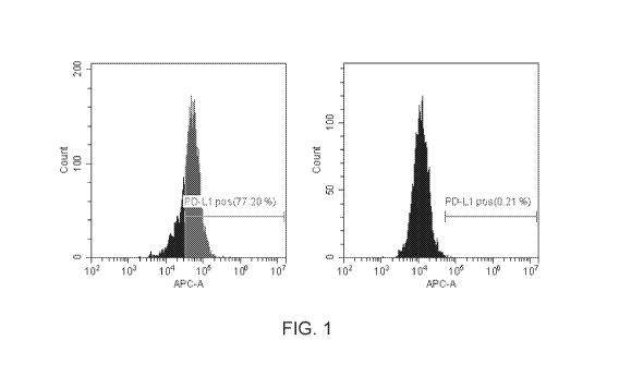
FIG. 1 depicts clear cytometric separation of PD-L1 positive control and
negative
control cells using PD-L1 labeling and flow cytometric analysis as described
herein.
FIG. 2 depicts the linearity of PD-L1 positive cell detection in mixed samples
prepared
with various percentages of PD-L1 positive cells spiked into negative control
samples.
FIG. 3 depicts MESF bead based standardization of PD-L1 fluorescence for per
cell
PD-L1 quantification as used in an embodiment as described herein.
FIG. 4 depicts quantitative PD-L1 expression analysis of tumor and immune cell
subsets and validation of the specific detection of PD-L1 expressing cells in
patient derived
samples.
FIG. 5 provides Table 2.
FIG. 6 depicts the proliferation of lung tumor tissue immune cell infiltrates
assayed
according to an embodiment described herein.
FIG. 7 depicts a loss of macrophages and an increase of non-T cells in tumor
tissue as
assayed according to an embodiment described herein.
FIG. 8 depicts an increase in aneuploidy, as compared to diploidy, of
lymphocytes
present in tumor tissue as assayed according to an embodiment described
herein.
DETAILED DESCRIPTION
Methods are provided for detecting the per cell programmed-death ligand 1 (PD-
L1)
expression of neoplasia cells. Aspects of the methods include cytometrically
assaying a
labeled cell suspension to quantify per cell PD-L1 expression to detect
whether a neoplastic
cell that expresses PD-L1 above a predetermined threshold is present in the
neoplasia
sample. In addition, kits that find use in practicing the subject methods are
also provided.
Before the present methods and kits are described, it is to be understood that
this
invention is not limited to particular methods or kits described, as such may,
of course, vary. It
is also to be understood that the terminology used herein is for the purpose
of describing
particular embodiments only, and is not intended to be limiting, since the
scope of the present
invention will be limited only by the appended claims.
2
CA 03036278 2019-03-06
WO 2018/048936
PCT/US2017/050322
Where a range of values is provided, it is understood that each intervening
value, to
the tenth of the unit of the lower limit unless the context clearly dictates
otherwise, between the
upper and lower limits of that range is also specifically disclosed. Each
smaller range between
any stated value or intervening value in a stated range and any other stated
or intervening
value in that stated range is encompassed within the invention. The upper and
lower limits of
these smaller ranges may independently be included or excluded in the range,
and each
range where either, neither or both limits are included in the smaller ranges
is also
encompassed within the invention, subject to any specifically excluded limit
in the stated
range. Where the stated range includes one or both of the limits, ranges
excluding either or
both of those included limits are also included in the invention.
Unless defined otherwise, all technical and scientific terms used herein have
the same
meaning as commonly understood by one of ordinary skill in the art to which
this invention
belongs. Although any methods and materials similar or equivalent to those
described herein
can be used in the practice or testing of the present invention, some
potential and preferred
methods and materials are now described. All publications mentioned herein are
incorporated
herein by reference to disclose and describe the methods and/or materials in
connection with
which the publications are cited. It is understood that the present disclosure
supersedes any
disclosure of an incorporated publication to the extent there is a
contradiction.
As will be apparent to those of skill in the art upon reading this disclosure,
each of the
individual embodiments described and illustrated herein has discrete
components and
features which may be readily separated from or combined with the features of
any of the
other several embodiments without departing from the scope or spirit of the
present invention.
Any recited method can be carried out in the order of events recited or in any
other order which
is logically possible.
It must be noted that as used herein and in the appended claims, the singular
forms
"a", "an", and "the" include plural referents unless the context clearly
dictates otherwise. Thus,
for example, reference to "a cell" includes a plurality of such cells and
reference to "the labeled
binding member" includes reference to one or more labeled binding members and
equivalents
thereof, e.g. antibodies, known to those skilled in the art, and so forth.
The publications discussed herein are provided solely for their disclosure
prior to the
filing date of the present disclosure. Nothing herein is to be construed as an
admission that
the present invention is not entitled to antedate such publication by virtue
of prior invention.
Further, the dates of publication provided may be different from the actual
publication dates
which may need to be independently confirmed.
3
CA 03036278 2019-03-06
WO 2018/048936
PCT/US2017/050322
METHODS
As summarized above, embodiments of the invention are directed to methods of
detecting a cell that expresses programmed-death ligand 1 (PD-L1) above a
predetermined
threshold. Cells in which PD-L1 expression may be detected in the subject
methods include, in
various instances, neoplastic cells and immune cells. Accordingly, in some
instances a cell
e.g., a neoplastic cell, detected in the subject methods will have a per cell
expression level of
PD-L1 protein that exceeds a predetermined threshold.
By "per cell expression level of PD-L1" or "per cell expression of PD-L1", as
used
herein, is meant the quantity of PD-L1 molecules present on the surface of a
cell. Methods of
the present disclosure include cytometrically assaying a cellular sample to
quantify the per cell
expression of PD-L1 and subsequently detecting one or more cells in the sample
that have a
per cell expression level of PD-L1 protein that exceeds the predetermined
threshold. Various
means of cytometrically assaying a cellular sample, described in more detail
below, may be
employed in the subject methods.
PD-L1 Expressinq Cells
As summarized above, the present disclosure provides methods of detecting
cells
expressing PD-L1 above a predetermined threshold. PD-L1 expression may be
quantified on
a per cell basis based on the level of PD-L1 protein expression or the level
of PD-L1 encoding
transcript (i.e., m RNA) expression. In some instances, the subject method
quantifies only per
cell PD-L1 protein expression and does not quantify per cell PD-L1 transcript
expression. In
some instances, a combined method of quantifying both PD-L1 protein levels and
PD-L1
transcription levels may be employed.
As described in more detail below, embodiments of the instant methods may
include
cytometrically assaying a cell suspension to detect a cell expressing PD-L1
above a
predetermined threshold. As used herein, the term "cytometrically assaying"
describes the
measuring of cellular parameters on a cell-by-cell basis where such measuring
allows for the
detection of individual cells that have, or the counting of a cell population
that shares, a certain
cellular parameter or set of parameters. One such parameter that is
cytologically assayed in
the subject methods is per cell expression of PD-L1.
PD-L1 (also known as CD274) binds programmed cell death protein 1 (PD-1), a
protein encoded by the PDCD1 gene that is a cell surface receptor expressed on
1-cells. PD-1
functions as an immune checkpoint by preventing the activation of 1-cells,
which reduces
autoimmunity and promotes self-tolerance. PD-L1 has been found to be expressed
on a
number of different cancer cell types. The presence of PD-L1 on a cancer cell
inhibits T cell
4
CA 03036278 2019-03-06
WO 2018/048936
PCT/US2017/050322
activation, contributing to cancer cell immune evasion. A number of cancer
therapies are
directed to preventing cancer cell immune evasion by inhibiting the PD-1/PD-L1
interaction.
The present disclosure includes detecting neoplastic cells expressing PD-L1
above a
predetermined threshold. Neoplastic cells having a per cell PD-L1 expression
level above a
predetermined threshold may be more likely to effectively evade the host
immune system.
Neoplastic cells having a per cell PD-L1 expression level above a
predetermined threshold
may be more likely to be affected by therapies directed at disrupting the PD-
1/PD-L1
interaction. In some instances, by effectively quantifying PD-L1 expression on
the surface of a
neoplastic cell and detecting neoplastic cells that express PD-L1 above a
threshold level the
effectiveness of therapies targeting the PD-1/PD-L1 interaction may be
predicted.
The present disclosure includes methods of identifying whether a neoplasia in
a
subject is anti-PD-1/PD-L1 immunotherapy responsive. As used herein, anti-PD-
1/PD-L1
immunotherapy responsive generally refers to the responsiveness of a neoplasia
to a
treatment targeting the interaction between PD-1 and PD-L1, including e.g., by
using an
antagonist to PD-1 and/or PD-L1. As such, a cell having a PD-L1 expression
level above a
predetermined threshold may, in some instances, be referred to as an anti-PD-
1/PD-L1
immunotherapy responsive cell. A neoplasia having one or more anti-PD-1/PD-L1
immunotherapy responsive cells may, in some instances, be referred to as an
anti-PD-1/PD-L1 immunotherapy responsive neoplasia. The responsiveness of a
cell or a
neoplasia to an anti-PD-1/PD-L1 immunotherapy may be predicted or determined.
For
example, in some instances, a method that detects the presence of a cell
expressing PD-L1
above a predetermined threshold may be predictive that the neoplasia from
which the cell is
derived is anti-PD-1/PD-L1 immunotherapy responsive. In some instances, e.g.,
the presence
of a cell expressing PD-L1 above a predetermined threshold positively
identifies that the
.. neoplasia from which the cell is derived is anti-PD-1/PD-L1 immunotherapy
responsive. The
subject methods find use in detecting cells having a level of PD-L1 expression
above a
predetermined threshold derived from various different neoplasms, described in
more detail
below.
Cells detected in the methods of the present disclosure will have a level of
PD-L1
expression above a predetermined threshold. As such, the methods of the
instant disclosure
include cytometrically quantifying per cell expression levels of PD-L1 to
identify cells
expressing PD-L1 protein and/or PD-L1 transcript above a predetermined
threshold.
Predetermined thresholds for PD-L1 expression useful in the instant disclosure
will vary
depending on various factors include e.g., the cell type assayed (e.g., the
type of neoplasm
from which the cell is derived), other measured cellular parameters (e.g.,
cell cycle
5
CA 03036278 2019-03-06
WO 2018/048936
PCT/US2017/050322
parameters, aneuploidy parameters, etc.), and whether PD-L1 protein or
transcript are
detected. As described in more detail below, PD-L1 expression is determined
cytometrically
where PD-L1 protein expression may be determined by a variety of protocols
including, but not
limited to, contacting the cell with a labeled specific binding member that
binds PD-L1 protein
on the surface of the cell. In some instances, PD-L1 expression is determined
cytometrically
where PD-L1 transcript expression may be determined by a variety of protocols
including, but
not limited to, contacting the cell with a labeled specific binding member
that binds PD-L1
transcripts within the cell.
In some instances, quantifying per cell PD-L1 expression may include
calibrating
PD-L1 fluorescence of PD-L1 specific binding partner labeled cells to a
reference standard.
Depending on the context, a reference standard may be cytometrically assayed
in parallel, in
series or simultaneously with the assayed cells. For example, in some
instances, a reference
standard may be cytometrically assayed to calibrate the assay for
quantification and then the
label cell suspension sample may be assayed using the calibrated cytometric
assay. In some
instances, a reference standard may be added to (i.e., spiked into) the label
cell suspension
sample and the calibration based on the reference standard for quantifying the
per cell
expression of the labeled cells may be performed during cytometric analysis of
cells. In some
instances, calibration with a reference standard may be performed between or
during each run
of a labeled cell sample. In some instances, calibration with a reference
standard may be
performed between or during each batch of runs.
Any convenient reference standard for calibrating labeled cell fluorescence to
per cell
marker expression may be employed in the herein described assays including but
not limited
to e.g., standardized microspheres (i.e., beads), standardized control cells,
standardized
fluorescent particles, and the like. In some instances, spectrally equivalent
microsphere
standards, such as e.g., Molecules of Equivalent Soluble Fluorochrome (MESF)
beads or
Mean Equivalent Fluorochrome (MEFL) beads, may be used. Microsphere standards
useful in
quantitative cytometry will vary any will generally include microspheres
labeled with a known
amount of fluorophore bound per microsphere or microspheres will a known
valency for
binding fluorophore labeled molecules. Microsphere standards for quantitative
cytometry
simulate fluorescent dye attachment to the cell membrane of target cells and
allow for
calibration of cytometric assays, including e.g., flow cytometric assays or
cell cytometric
assays, for quantification.
For example, in some instances, microsphere standards for quantitative
cytometry will
include two or more populations, including e.g., 2 populations, 3 populations,
4 populations, 5
populations, 6 populations, etc., of microspheres labeled with different
amounts of a
6
CA 03036278 2019-03-06
WO 2018/048936
PCT/US2017/050322
fluorophore. The fluorophore chosen will generally be the same as or
equivalent to or
comparable with the fluorophore used in one or more of the labeled specific
binding members
of the described methods. Useful fluorophores in microsphere standards include
but are not
limited to e.g., Alexa Fluor 488, Alexa Fluor 647, FIT, PE, Cy5, APC, etc. In
some instances,
two or more microsphere standards having different fluorophores may be mixed,
e.g., where
quantification of two or more differently labeled specific binding members are
used in a subject
method. In some instances, microsphere standards having different fluorophores
are not
mixed and different populations of microspheres having different amounts of a
single type of
fluorophore bound may be employed.
Microsphere standards may be directly conjugated to the fluorescent label or,
in some
instances, fluorescently labeled antibody may be bound to the microsphere
standard. In some
instances, a microsphere standard may be non-fluorescent but "label-able".
Label-able
microsphere standards will generally have a known antibody binding capacity
allowing for
staining of the microsphere with a known amount of a user's antibody,
including e.g., the same
antibody used as a specific binding member in a herein described method. Label-
able
microsphere standards may, in some instances, be employed in conjunction with
a pre-labeled
microsphere standard allowing for determination of the fluorophore to protein
(FTP) ratio of the
particular labeled specific binding member employed in the method and/or
further calibration.
Various different microsphere standards, including e.g., fluorescently labeled
microsphere
standards and label-able microsphere standards, for quantitative cytometry
that may find use
in the herein described methods include but are not limited to e.g., those
commercially
available from Bangs Laboratories, Inc. (Fishers, IN), BD Biosciences (San
Jose, CA), and the
like.
In some embodiments, the fluorescence of a labeled specific binding member
and/or
cells labeled with such may be calibrated to microsphere standards (e.g., by
assessing the
fluorescence of two or more populations of microspheres labeled with different
amounts of a
fluorophore) to establish a standard curve. Following or during the
establishment of a standard
curve a labeled cell suspension sample may be assayed and per cell PD-L1
expression may
be determined. Quantified per cell PD-L1 expression levels may be compared to
a
predetermined threshold, including e.g., a threshold established based on the
number of
molecules of PD-L1 protein expressed per cell, a threshold established based
on background
fluorescence, a threshold established based on background expression
(including e.g., per
cell expression) of PD-L1, and the like.
In some instances, a predetermined threshold for per cell PD-L1 expression may
be
expressed as a number of molecules of the PD-L1 protein per cell, including
but not limited to
7
CA 03036278 2019-03-06
WO 2018/048936
PCT/US2017/050322
e.g., a threshold of 10 molecules per cell, a threshold of 20 molecules per
cell, a threshold of
30 molecules per cell, a threshold of 40 molecules per cell, a threshold of 50
molecules per
cell, a threshold of 60 molecules per cell, a threshold of 70 molecules per
cell, a threshold of
80 molecules per cell, a threshold of 90 molecules per cell, a threshold of
100 molecules per
cell, a threshold of 200 molecules per cell, a threshold of 300 molecules per
cell, a threshold of
400 molecules per cell, a threshold of 500 molecules per cell, a threshold of
600 molecules per
cell, a threshold of 700 molecules per cell, a threshold of 800 molecules per
cell, a threshold of
900 molecules per cell, a threshold of 1000 molecules per cell, a threshold of
1100 molecules
per cell, a threshold of 1200 molecules per cell, a threshold of 1300
molecules per cell, a
threshold of 1400 molecules per cell, a threshold of 1500 molecules per cell,
a threshold of
1600 molecules per cell, a threshold of 1700 molecules per cell, a threshold
of 1800 molecules
per cell, a threshold of 1900 molecules per cell, a threshold of 2000
molecules per cell, etc.
Accordingly, in some instances, a cell is detected as expressing PD-L1 above a
predetermined threshold if the cell is identified as having a per cell number
of PD-L1 protein
molecules expressed on the surface of the cell that is above one or more of
the predetermined
thresholds listed above.
In some instances, the methods described herein detect a single cell having a
level of
PD-L1 expression above a predetermined threshold. In some instances, the
presence of a
single detected cell having a level of PD-L1 expression above a predetermined
threshold is
considered significant. In some instances, the methods described herein may
include a
threshold of cells having a level of PD-L1 expression above a predetermined
threshold for the
detected cells to be considered significant (i.e., a minimum size for the
population of cells
having a level of PD-L1 expression above a predetermined threshold to be
considered
significant). Depending on the context, the size of the detected population of
cells expressing
PD-L1 above the threshold will vary and may range from one cell to millions of
cells, including
but not limited to e.g., one cell, one cell or more, 10 cells or more, 100
cells or more, 1,000
cells or more, 10,000 cells or more, 100,000 cells or more.
In some instances, the size of the detected population of cells expressing PD-
L1 above
the predetermined threshold may be expressed in relative terms. For example,
the size of the
population may be expressed as a percentage of all the cells in the sample, a
percentage of all
the cells analyzed, a percentage of all of the cells of a particular type
within the sample, a
percentage of all of the cells of a particular type that were analyzed, etc.
In some instances,
the size of the detected population may exceed 0.01% or more of the neoplastic
cells in the
cell suspension sample, including but not limited to e.g., 0.1% or more, 1% or
more, 10% or
more, etc.
8
CA 03036278 2019-03-06
WO 2018/048936
PCT/US2017/050322
In some instances, in order to classify a cell, e.g., a neoplasia cell, as PD-
L1
expressing or likely to be anti-PD-1/PD-L1 immunotherapy responsive or
detected as
anti-PD-1/PD-L1 immunotherapy responsive the size of the population of cells
detected as
expressing PD-L1 above a predetermined threshold must exceed a predetermined
threshold.
.. As described above, the threshold for the size of the detected population,
e.g., for a neoplasia
to be considered PD-L1 expressing, will vary based on a number of factors and
in some
instances may be one cell. In some instances, the threshold for the size of
the detected
population, e.g., for a neoplasia to be considered PD-L1 expressing, the
population must
exceed more than one cell including two cells or more including but not
limited to e.g., 0.01%
.. or more of the neoplastic cells in the sample, 0.1% or more of the
neoplastic cells in the
sample, 1% or more of the neoplastic cells of the sample, and the like.
Cytometric Assays
As summarized above, methods of the present disclosure include cytometrically
.. assaying a labeled cell suspension. Various methods of cytometrically
assaying a labeled cell
suspension may find use in the herein described methods including but not
limited to e.g., flow
cytometrically assaying using a flow cytometer, cell cytometrically assaying a
labeled cell
suspension, e.g., by using a cell cytometer, and the like. Labeled cell
suspension samples
may be assayed for per cell PD-L1 expression. In some cases, additional
cellular parameters,
assayed cytometrically, may also find use in detecting neoplastic cells of the
instant
disclosure. Accordingly, various methods of cytometrically assaying a labeled
cell suspension
to measure various cellular parameters may be employed.
In some embodiments, cytometrically assaying a cellular sample may be
performed
using flow cytometry. Flow cytometry is a methodology using multi-parameter
data for
identifying and distinguishing between different particle (e.g., cell) types
i.e., particles that vary
from one another in terms of label (wavelength, intensity), size, etc., in a
fluid medium. In flow
cytometrically analyzing a sample, an aliquot of the sample is first
introduced into the flow path
of the flow cytometer. When in the flow path, the cells in the sample are
passed substantially
one at a time through one or more sensing regions, where each of the cells is
exposed
separately and individually to a source of light at a single wavelength (or in
some instances
two or more distinct sources of light) and measurements of cellular
parameters, e.g., light
scatter parameters, and/or marker parameters, e.g., fluorescent emissions, as
desired, are
separately recorded for each cell. The data recorded for each cell is analyzed
in real time or
stored in a data storage and analysis means, such as a computer, for later
analysis, as
desired.
9
CA 03036278 2019-03-06
WO 2018/048936
PCT/US2017/050322
In flow cytometry-based methods, the cells are passed, in suspension,
substantially
one at a time in a flow path through one or more sensing regions where in each
region each
cell is illuminated by an energy source. The energy source may include an
illuminator that
emits light of a single wavelength, such as that provided by a laser (e.g.,
He/Ne or argon) or a
mercury arc lamp or an LED with appropriate filters. For example, light at 488
nm may be used
as a wavelength of emission in a flow cytometer having a single sensing
region. For flow
cytometers that emit light at two distinct wavelengths, additional wavelengths
of emission light
may be employed, where specific wavelengths of interest include, but are not
limited to:
405nm, 535 nm, 561 nm, 635 nm, 642 nm, and the like. Following excitation of a
labeled
specific binding member bound to a polypeptide by an energy source, the
excited label emits
fluorescence and the quantitative level of the polypeptide on each cell may be
detected, by
one or more fluorescence detectors, as it passes through the one or more
sensing regions.
In flow cytometry, in addition to detecting fluorescent light emitted from
cells labeled
with fluorescent markers, detectors, e.g., light collectors, such as
photomultiplier tubes (or
"PMT"), an avalanche photodiode (APD), etc., are also used to record light
that passes
through each cell (generally referred to as forward light scatter), light that
is reflected
orthogonal to the direction of the flow of the cells through the sensing
region (generally
referred to as orthogonal or side light scatter) as the cells pass through the
sensing region and
is illuminated by the energy source. Each type of data that is obtained, e.g.,
forward light
scatter (or FSC), orthogonal light scatter (SSC), and fluorescence emissions
(FL1, FL2, etc.),
comprise a separate parameter for each cell (or each "event").
Flow cytometers may further include one or more electrical detectors. In
certain
embodiments, an electrical detector may be employed for detecting a
disturbance caused by a
particle or cell passing through an electrical field propagated across an
aperture in the path of
the particles/cells. Such flow cytometers having electrical detectors will
contain a
corresponding electrical energy emitting source that propagates an electrical
field across the
flow path or an aperture through which cells are directed. Any convenient
electrical field and/or
combination of fields with appropriate detector(s) may be used for the
detection and/or
measurement of particles (or cells) passing through the field including but
not limited to, e.g., a
direct current electrical field, alternating current electrical field, a radio-
frequency field, and the
like.
Flow cytometers further include data acquisition, analysis and recording
means, such
as a computer, wherein multiple data channels record data from each detector
for each cell as
it passes through the sensing region. The purpose of the analysis system is to
classify and
count cells wherein each cell presents itself as a set of digitized parameter
values and to
CA 03036278 2019-03-06
WO 2018/048936
PCT/US2017/050322
accumulate data for the sample as a whole.
A particular cell subpopulation of interest may be analyzed by "gating" based
on the
data collected for the entire population. To select an appropriate gate, the
data is plotted so as
to obtain appropriate separation of subpopulations, e.g., by adjusting the
configuration of the
instrument, including e.g., excitation parameters, collection parameters,
compensation
parameters, etc. In some instances, this procedure is done by plotting forward
light scatter
(FSC) vs. side (i.e., orthogonal) light scatter (SSC) on a two dimensional dot
plot. The flow
cytometer operator then selects the desired subpopulation of cells (i.e.,
those cells within the
gate) and excludes cells which are not within the gate. Where desired, the
operator may select
the gate by drawing a line around the desired subpopulation using a cursor on
a computer
screen. Only those cells within the gate are then further analyzed by plotting
the other
parameters for these cells, such as fluorescence.
Any flow cytometer that is capable of obtaining fluorescence data, e.g., as
described
above, may be employed. Useful flow cytometers include those utilizing various
different
means of flowing a cell through the sensing region substantially one at a time
including, e.g., a
flow cell, a microfluidics chip, etc. Non-limiting examples of flow cytometer
systems of interest
are those available from commercial suppliers including but not limited to,
e.g.,
Becton-Dickenson (Franklin Lakes, NJ), Life Technologies (Grand Island, NY),
Acea
Biosciences (San Diego, CA), Beckman-Coulter, Inc. (Indianapolis, IN), Bio-Rad
Laboratories,
Inc. (Hercules, CA), Cytonome, Inc. (Boston, MA), Amnis Corporation (Seattle,
WA), EMD
Millipore (Billerica, MA), Sony Biotechnology, Inc. (San Jose, CA), Stratedigm
Corporation
(San Jose, CA), Union Biometrica, Inc. (Holliston, MA), Cytek Development
(Fremont, CA),
Propel Labs, Inc. (Fort Collins, CO), Orf low Technologies (Ketchum, ID),
handyem inc.
(Quebec, Canada), Sysmex Corporation (Kobe, Japan), Partec Japan, Inc.
(Tsuchiura,
Japan), Bay bioscience (Kobe, Japan), Furukawa Electric Co. Ltd. (Tokyo,
Japan), On-chip
Biotechnologies Co., Ltd (Tokyo, Japan), Apogee Flow Systems Ltd.
(Hertfordshire, United
Kingdom), and the like.
In some embodiments, cytometrically assaying a cellular sample may be
performed
using a cell cytometer. As used herein, the term "cell cytometer" (also
referred to as an
"imaging cytometer" or "automated imaging cytometer") generally refers to an
automated or
semi-automated cell imaging device capable of imaging cells deposited on or in
an imaging
vessel to collect data on all or most of the cells of a sample. In cell
cytometry, imaging may be
performed according to a variety of different methods. In some instances, a
cell cytometer may
collect a widefield image at low magnification (e.g., 5X, 10X, etc.) of the
cells present on or in
an imaging vessel to identify the location of the cells and/or screen the
cells for a particular
11
CA 03036278 2019-03-06
WO 2018/048936
PCT/US2017/050322
parameter (e.g., size, shape, color, fluorescence, etc.). After identifying
the location of the
cells a cell cytometer may proceed to collect higher magnification (e.g., 20X,
40X, 60X, 100X,
etc.) images of all or a portion of the identified cells, e.g., in a targeted
manner.
In other instances, a cell cytometer may image cells present on or in an
imaging vessel
by scanning the imaging vessel. Scanning may be performed at low or high
magnification. In
some instances, scanning is performed at high magnification to capture images
of all or most
of the cells. In some instances, scanning is performed at low magnification to
identify the
location of the cells on or in the imaging vessel. After identifying the
location of the cells a cell
cytometer may proceed to collect higher magnification images of all or a
portion of the
identified cells, e.g., in a targeted manner, or may rescan the located cells
at high
magnification.
The imaging vessels used in cell cytometer systems will vary. In some
instances,
commonly used laboratory imaging devices such as e.g., microscope slides, may
serve as an
imaging vessel in a cell cytometer system. In some instances, a cell cytometer
imaging vessel
may be specifically designed for use with a particular cell cytometer. Useful
imaging vessels
include but are not limited to e.g., slides (e.g., microscope slides), dishes
(e.g., glass bottom
imaging dishes), plates (e.g., multi-well imaging plates), etc. Imaging
vessels will generally
have optical properties amendable to microscopy, e.g., optical clarity, in at
least a portion of
the vessel. Imaging vessels may or may not have individual compartments. For
example, a
microscope slide utilized as an imaging vessel does not generally have
individual
compartments and cells deposited on a slide may be spread about the surface of
the slide.
Alternatively, a multi-well imaging plate utilized as an imaging vessel does
have individual
compartments (i.e., wells) into which one or more cells may be deposited.
Cell cytometers include an imaging component such as, e.g., an automated
microscope. The imaging component of a cell cytometer may include one or more
objectives
of various magnification power (e.g., 5X, 10X, 20X, 40X 60X, 100X, etc.) for
collecting light
transmitted, reflected or emitted from the object (e.g., cell) being imaged.
Light collected by
the objective will generally be processed through one or more dichroic
mirrors, filters or lenses
before being directed to an image capture device.
Suitable image capturing devices may include one or more digital cameras
(including
color and monochrome cameras) capable of capturing a digital image and a means
of storing
the digital image and/or transferring the image to attached image processing
circuitry or to an
attached storage device for later transfer to image processing circuitry.
Suitable digital color
cameras will vary and will generally include any digital camera (e.g., with
one or more CCD or
CMOS sensors). Suitable digital cameras include but are not limited to e.g.,
custom built digital
12
CA 03036278 2019-03-06
WO 2018/048936
PCT/US2017/050322
cameras, consumer grade digital color cameras (e.g., consumer grade digital
color cameras
converted for microscopic use) and those digital microscopy color cameras
commercially
available from various manufactures including but not limited to e.g., Dino-
Eye, Dino-Lite,
Jenoptik ProgRes, KoPa, Leica, Motic, Olympus, Omano, OptixCam, PixelLINK,
Zeiss, etc.
Cell cytometers further include data acquisition, analysis and recording
means, such
as a computer, wherein one or more data channels record data from one or more
image
capture devices for each cell or most of the cells of the imaging vessel. The
purpose of the
analysis system is to classify and count cells wherein each cell presents
itself as a set of
digitized parameter values and to accumulate data for the sample as a whole.
In some cases,
cell cytometers record images of each cell and may be connected to a user
interface where
such images may be reviewed by a user of the device.
Cell cytometer based methods for detecting cells expressing a particular
polypeptide
may include contacting the cells of a sample with a fluorescent labeled
specific binding
member and detecting fluorescently labeled cells by imaging using the cell
cytometer. As
described in more detail elsewhere herein, the fluorescence of each labeled
cell may be
cytometrically quantified to identify the per cell expression of a particular
polypeptide, e.g., to
detect whether a cell expresses the polypeptide above a predetermined
threshold.
Any cell cytometer that is capable of obtaining fluorescence data, e.g., as
described
above, may be employed. Useful cell cytometers include those utilizing various
different
means of automated cell cytometric imaging to analyze all or most of the cells
of a sample.
Non-limiting examples of cell cytometer systems of interest are those
available from
commercial suppliers including but not limited to, e.g., Nexcelom Bioscience
LLC (Lawrence,
MA), Molecular Devices, LLC (Sunnyvale, CA), Thorlabs Inc. (Newton, New
Jersey), TTP
Labtech Ltd. (United Kingdom), and the like.
Methods of the instant disclosure include cytometrically quantifying per cell
expression
levels of particular polypeptides to identify cells expressing the polypeptide
above a
predetermined threshold. Methods of the instant disclosure may include
cytometrically
quantifying per cell PD-L1 expression to identify one or more cells expressing
PD-L1 above a
predetermined threshold. However, the levels of other markers besides PD-L1
may also be
assessed in the herein described methods including e.g., cell cycle associated
expression
products (e.g., cell cycle associated RNAs, cell cycle associated
polypeptides, etc.),
immune-related expression products (e.g., immune-related RNAs, immune-related
polypeptides, etc.), DNA content, etc. Detection of cells having a level of a
biomarker, e.g.,
above or below a predetermined threshold, or not having such other markers may
serve to
identify further cell parameters useful in the herein described methods.
13
CA 03036278 2019-03-06
WO 2018/048936
PCT/US2017/050322
Predetermined thresholds may find use in identifying cells based on their
expression of
a particular polypeptide, as described above, or other cellular parameters
including but not
limited to e.g., cell cycle markers, aneuploidy markers, and the like.
Predetermined thresholds for polypeptide expression useful in the instant
disclosure
will vary depending on the polypeptide detected and the particular context. In
some instances,
a predetermined threshold for per cell polypeptide expression may be expressed
as a number
of molecules of the polypeptide per cell, including but not limited to e.g., a
threshold of 10
molecules per cell, a threshold of 20 molecules per cell, a threshold of 30
molecules per cell, a
threshold of 40 molecules per cell, a threshold of 50 molecules per cell, a
threshold of 60
molecules per cell, a threshold of 70 molecules per cell, a threshold of 80
molecules per cell, a
threshold of 90 molecules per cell, a threshold of 100 molecules per cell, a
threshold of 200
molecules per cell, a threshold of 300 molecules per cell, a threshold of 400
molecules per
cell, a threshold of 500 molecules per cell, a threshold of 600 molecules per
cell, a threshold of
700 molecules per cell, a threshold of 800 molecules per cell, a threshold of
900 molecules per
cell, a threshold of 1000 molecules per cell, a threshold of 1100 molecules
per cell, a threshold
of 1200 molecules per cell, a threshold of 1300 molecules per cell, a
threshold of 1400
molecules per cell, a threshold of 1500 molecules per cell, a threshold of
1600 molecules per
cell, a threshold of 1700 molecules per cell, a threshold of 1800 molecules
per cell, a threshold
of 1900 molecules per cell, a threshold of 2000 molecules per cell, etc.
In other instances, a predetermined threshold may be a relative level of a
marker.
Relative levels of a marker may be determined by a variety of means including
e.g.,
determined by making a comparison of the levels of expression of a marker in
two separate
populations of cells known to differ in their level of the subject marker. For
example, a first cell
population known to have a high level of Marker X is measured, e.g., on a
cytometer, and
compared to a second cell population, known to have a low level of Marker X
and the
comparison is used to determine a threshold level that may be used to
categorize cells as
either having a low or a high level of Marker X.
Relative levels of a marker may be determined by making a comparison of the
levels of
marker within a population of cells, e.g., a population of cells of unknown
levels of Marker X or
a population of cells suspected of containing subpopulations of cells having
different levels of
Marker X. For example, the level of Marker X is measured on a cytometer of at
least a
sufficient number of cells such that the measurements may be plotted, e.g., on
a histogram,
and separation between two or more subpopulations of cells is revealed based
on individual
cell levels of Marker X. Accordingly, the cytometer operator may then
determine a threshold
level between the subpopulations that may be used to categorize cells as
belonging to a
14
CA 03036278 2019-03-06
WO 2018/048936
PCT/US2017/050322
particular subpopulation, e.g., a subpopulation having a low level of Marker X
or a
subpopulation having high level of Marker X.
In some instances, a threshold may be based on the limit of detection of the
cytometer.
For example, cells of a population of cells may be identified as having a
particular marker (i.e.,
being positive for a particular marker) if the cells have any detectable level
of a particular
marker. Likewise, cells of a population of cells may be identified as not
having a particular
marker (i.e., being negative for a particular marker) if the cells do not have
a detectable level of
a particular marker. Accordingly, the detection level of the cytometer may be
used to
determine the marker threshold, as desired.
In some instances, a threshold may be based on previously determined marker
levels,
e.g., from previously performed control experiments or previously acquired
reference
expression levels. For example, marker levels determined in previously
analyzed samples
may be used to determine marker threshold levels. In some instances, marker
levels expected
of cells obtained from healthy subjects may be used to determine normal marker
levels such
that a marker threshold that is representative of the normal marker range may
be determined.
In such instances, marker expression outside, i.e., above or below, the normal
marker range is
considered to be either above or below the particular marker threshold. In
some instances,
use of such previously determined marker levels or previously determined
threshold levels
allows analysis of cells and the identification of cellular subpopulations in
the absence of a
control or reference cellular sample.
As noted above, methods of the instant disclosure may include assaying cell
cycle
parameters. Useful cell cycle parameters include but are not limited to e.g.,
proliferation, cell
cycle phase (G1, G2, M,G2-M, S, Go, post Gl, and the like), etc. Cell cycle
parameters may be
assessed on a per cell basis, including e.g., identifying whether a cell is
proliferative,
identifying the cell cycle phase of a cell, etc. Any convenient means of
determining a cell cycle
parameter of a cell may be employed in the subject methods. In some instances,
a method
may not only quantify a particular cell type but also determine whether the
quantified cell type
is proliferative including e.g., the number or percent of proliferative cells
within the quantified
cell type. In some instances, a method may not only quantify an immune cell
type but also
determine whether the quantified immune cell type is proliferative including
e.g., the number or
percent of proliferative immune cells within the quantified immune cell type.
In some
instances, a method may determine whether proliferative tumor infiltrating
lymphocytes are
present and/or the quantity thereof. In some instances, a method may determine
whether
proliferative CD4+ cells are present and/or the quantity thereof. In some
instances, a method
may determine whether proliferative CD8+ cells are present and/or the quantity
thereof. In
CA 03036278 2019-03-06
WO 2018/048936
PCT/US2017/050322
some instances, a method may determine the ratio of CD4/CD8 cells and whether
the
proliferative CD4 and/or CD8 cells are proliferative and/or the quantity
thereof.
In some instances, assaying the cell cycle of a cell may include determining
the DNA
content of the cell (i.e., the per cell DNA content). Various methods may be
employed for
assaying the cell cycle of a cell by determining the per cell DNA content. In
some instances, a
DNA labeling reagent (e.g., a nucleic acid dye or stain that contains
intrinsic fluorescence)
may be employed to label the DNA of the cell and the amount of DNA may be
quantified based
on the measuring the intensity of the label. Depending on the method of
cytometry employed
in the method, DNA content may be used to assess cell cycle in various ways.
In one
.. embodiment, e.g., regardless of the type of cytometry employed (e.g., flow
cytometry, cell
cytometry, etc.), the fluorescent intensity of cells labeled with a DNA
labeling reagent may
analyzed on the cytometer and plotted on a histogram. From the histogram the
relative
amount of DNA content may be determined for each cell allowing for the
identification of the
cell cycle phase of each cell. In some instances, such a histogram may
represent a cytometric
cell cycle profile, also referred to as a cytometric DNA profile.
In some instances, assaying the cell cycle of a cell may include assaying an
expressed
cell cycle marker (also referred to as a cell cycle biomarker). Expressed cell
cycle markers, as
used herein, refer to those cellular markers (e.g., cell surface markers and
intracellular
markers) that are specifically expressed or absent during one or more
particular phases of the
cell cycle. Accordingly a labeled binding member specific for an expressed
cell cycle marker
include to those specific binding members that bind components of the cell
cycle machinery of
the cell. Cell cycle biomarkers may be useful, in some instances, in
determining the cell cycle
phase of or determining whether or not a cell is proliferative. Cell cycle
biomarkers useful in
the methods described herein will vary depending on the particular assay
and/or the particular
cell type and/or cell population to be detected. In some instances, cell cycle
biomarkers that
may find use in the methods described herein include but are not limited to,
e.g., Ki67, cyclin
D1, cyclin E, phosphorylated histone H3, and the like.
Expressed cell cycle markers may be detected in various ways. For example, an
expressed cell cycle biomarker may be detected at the protein level, e.g.,
through the use of a
labeled specific binding member specific for the cell cycle biomarker protein.
In some
instances, an expressed cell cycle biomarker may be detected at the RNA level,
e.g., through
the use of a labeled specific binding member specific for the cell cycle
biomarker RNA.
As noted above, methods of the instant disclosure may include assaying
aneuploidy.
Any convenient method of measuring aneuploidy cytometrically may be employed
in the
subject methods. In some instances, a cell may be identified as aneuploid
based on the
16
CA 03036278 2019-03-06
WO 2018/048936
PCT/US2017/050322
measured DNA content of the cell where an aneuploid cell will generally have
an abnormally
high level of DNA content representing duplication of all or a portion of the
cell's genome.
Similar methods to those described above for assessing DNA content in regards
to cell cycle
assessments may be employed for detecting aneuploidy. In some instances,
relative DNA
content greater than or equal to a threshold DNA content value for a normal
cell may indicate
that the cell is aneuploid where the threshold may be greater than or equal to
() 1.05 times
the DNA content of a normal cell including but not limited to, e.g., 1.06
times, 1.07 times,
1.08 times, 1.09 times, 1.10 times, 1.11 times, 1.12 times and 1.13
times the DNA
content of a normal cell.
In some instances, chromosome specific probes or gene specific probes may be
employed to assess aneuploidy. For example, fluorescent in situ hybridization
(FISH) using
gene specific or chromosome specific probes may be employed to determine the
overall
ploidy of a cell or to detect the duplication of a particular gene or
chromosome. For example, in
a diploid organism, the presence of more than two probes for a specific gene
or a specific
chromosome may indicate that the subject cell is aneuploid.
Ploidy assessments (e.g., assessing the ploidy of a cell, including e.g.,
whether a cell
is aneuploid, diploid, etc.) may be employed in the subject methods for
various purposes. For
example, in some instances, a ploidy assessment may be employed to determine
whether
cells of a population are aneuploid or diploid, including e.g., to determine
whether a neoplastic
cell is aneuploid or diploid, whether an immune cell is aneuploid or diploid,
or the like. In some
instances, a ploidy assessment may inform other characteristics of the sample
and/or the
subject, e.g., by a relationship between the ploidy status of a detected cell
and other cell types
that may be present in the subject. For example, in some instances, the
identification of certain
aneuploid cells may be indicative and/or predictive of the presence of a
neoplastic cell type in
a subject other than the detected aneuploid cell type, e.g., the presence of
aneuploid immune
cells in a tumor tissue of the subject may be indicative of the presence of
circulating tumor
cells in the subject. In some instances, the presence of aneuploid immune
cells in a lung tumor
tissue may be indicative of the presence of circulating tumor cells in the
subject.
Assessments of cellular parameters may be used in the subject methods to
detect
cells that have one or more characteristics detected by measuring the
described parameters.
For example, in some instances, a detected cell may be determined to be an
aneuploid cell,
e.g., based on one or more assessed aneuploidy parameters of the cell. In some
instances, a
detected cell may be determined to be a proliferative cell, e.g., based on one
or more
assessed cell cycle parameters of the cell. Cells may be detected as having a
combination of
characteristics detected by measuring the described parameters. For example, a
cell may be
17
CA 03036278 2019-03-06
WO 2018/048936
PCT/US2017/050322
determined to be both proliferative and aneuploid. In addition, the absence of
a characteristic
may also be used when detecting a particular cell including e.g., where the
cell is not
proliferative, where the cell is not aneuploid, etc. In some instances, a
detected cell may have
one characteristic and lack another, e.g., where the cell is proliferative but
not aneuploid,
where the cell is aneuploid but no proliferative, etc. Any combination of the
herein described
parameters may find use in the methods of the present disclosure.
In some instances, the methods of the instant disclosure may further include
determining whether a subject cell is or is not an immune cell. Various
methods may be
employed for determining whether a subject cell is or is not an immune cell
including e.g.,
through detecting the presence or absence of one or more immune cell markers,
e.g., through
contacting the cell with a labeled specific binding member specific for an
immune cell marker.
For example, in some instances, a non-immune neoplasia cell may be detected
based
on expressing PD-L1 above a predetermined threshold and not labeling with an
immune cell
specific binding member added to the cell suspension. Accordingly, in some
instances, the
method may further include determining that the identified cell is not an
immune cell, e.g., by
contacting the cell suspension with one or more labeled specific binding
members for immune
cells.
Accordingly, in some instances, a cell and/or a population of cells may be
identified as
being negative for a particular immune cell marker or having a level of
expression of an
immune cell marker that is below a predetermined threshold indicative that the
cell is, in fact,
not an immune cell or a particular type of immune cell. Useful immune cell
markers, e.g., for
identifying a PD-L1 expressing cell as not an immune cell include but are not
limited to e.g.,
CD114, CD117, CD11a, CD11b, CD14, CD15, CD16, CD182, CD19, CD20, 0D22, 0D24,
0D25, CD3, CD30, CD31, 0D34, 0D38, CD4, 0D45, 0D56, CD61, CD8, CD91, Foxp3,
and
the like. Accordingly, in some instances, a detected neoplasia cell may be
further
characterized as lacking expression of or having expression of below a
predetermined
threshold of one or more immune cell markers, e.g., as detected using an
antibody to an
immune cell marker including e.g., those listed above.
In some instances, a cell may be assayed in the herein described methods for
expression of a combination of immune cell markers including but not limited
to e.g., any
combination of the here described markers. For example, in some instances, a
PD-L1
expressing cell may be assayed for expression of CD8 and 0D45 and may be
identified as not
being an immune cell when the detected cell is negative for CD8 and 0D45 or
expresses CD8
and 0D45 below a predetermined threshold indicative of the cell not being an
immune cell.
In some instances, a cell may be detected based on expressing PD-L1 above a
18
CA 03036278 2019-03-06
WO 2018/048936
PCT/US2017/050322
predetermined threshold and labeling with an immune cell specific binding
member added to
the cell suspension. Accordingly, in some instances, the method may further
include
determining that the identified cell is an immune cell, e.g., by contacting
the cell suspension
with one or more labeled specific binding members for immune cells.
Accordingly, in some instances, a cell and/or a population of cells may be
identified as
being positive for a particular immune cell marker or having a level of
expression of an immune
cell marker that is above a predetermined threshold indicative that the cell
is, in fact, an
immune cell or a particular type of immune cell. Useful immune cell markers,
e.g., for
identifying a PD-L1 expressing cell as an immune cell include but are not
limited to e.g.,
CD114, CD117, CD11a, CD11b, CD14, CD15, CD16, CD182, CD19, 0D20, 0D22, 0D24,
0D25, CD3, 0D30, CD31, 0D34, 0D38, CD4, 0D45, 0D56, CD61, CD8, CD91, Foxp3,
and
the like. Accordingly, in some instances, a detected cell may be further
characterized as
having expression of or having expression of above a predetermined threshold
of one or more
immune cell markers, e.g., as detected using an antibody to an immune cell
marker including
e.g., those listed above.
In some instances, a cell may be assayed in the herein described methods for
expression of a combination of immune cell markers including but not limited
to e.g., any
combination of the here described markers. For example, in some instances, a
PD-L1
expressing cell may be assayed for expression of CD8 and 0D45 and may be
identified as
being an immune cell when the detected cell is positive for CD8 and 0D45 or
expresses CD8
and 0D45 above one or more predetermined thresholds indicative of the cell
being an immune
cell.
In some instances, the herein described methods may further include assaying
one or
more markers for circulating tumor cells (CTC), e.g., in order to determine if
a detected PD-L1
expressing cell is a CTC. As used herein, the term "CTC" generally refers to
those neoplastic
cells that have sloughed off of a tumor (e.g., the edge of a tumor) and have
been swept away
by the bloodstream or lymphatic system thus causing the CTC to circulate in
the body. CTC
makers include e.g., those markers used in identifying CTCs in the blood
stream including but
not limited to e.g., Epithelial cell adhesion molecule (EpCAM), cytokeratin 8,
cytokeratin 18
and cytokeratin 19. In some instances, CTCs may be further characterized as
being negative
for one or more immune cell markers, including but not limited to e.g., one or
more of those
immune cell markers described herein. For example, in some instances, a
detected CTC will
be negative for 0D45.
In some instances, CTCs may be further identified and/or characterized based
on the
expression of one or more cancer antigens and/or one or more cancer associated
antigens.
19
CA 03036278 2019-03-06
WO 2018/048936
PCT/US2017/050322
Non-limiting examples of cancer antigens include but are not limited to e.g.,
0D19, 0D20,
0D38, 0D30, Her2/neu, ERBB2, CA125, MUC-1, prostate-specific membrane antigen
(PSMA), 0D44 surface adhesion molecule, mesothelin, carcinoembryonic antigen
(CEA),
epidermal growth factor receptor (EGFR), EGFRvIll, vascular endothelial growth
factor
.. receptor-2 (VEGFR2), high molecular weight-melanoma associated antigen (HMW-
MAA),
MAGE-Al , IL-13R-a2, GD2, and the like. Cancer-associated antigens also
include, e.g.,
4-i BB, 514, adenocarcinoma antigen, alpha-fetoprotein, BAFF, B-lymphoma cell,
C242
antigen, CA-125, carbonic anhydrase 9 (CA-IX), C-MET, CCR4, CD152, CD19, CD20,
CD200, CD22, CD221, CD23 (IgE receptor), CD28, CD30 (TNFRSF8), CD33, CD4,
CD40,
CD44 v6, CD51, CD52, CD56, CD74, CD80, CEA, CN10888, CTLA-4, DRS, EGFR, EpCAM,
CD3, FAP, fibronectin extra domain-B, folate receptor 1, GD2, GD3 ganglioside,
glycoprotein
75, GPNMB, HER2/neu, HGF, human scatter factor receptor kinase, IGF-1
receptor, IGF-I,
IgG1, L1-CAM, IL-13, IL-6, insulin-like growth factor I receptor, integrin
a51, integrin avp3,
MORAb-009, MS4A1, MUC1, mucin CanAg, N-glycolylneuraminic acid, NPC-1C, PDGF-R
a,
PDL192, phosphatidylserine, prostatic carcinoma cells, RANKL, RON, ROR1, SCH
900105,
SDC1, SLAMF7, TAG-72, tenascin C, TGF beta 2, TGF-p, TRAIL-R1, TRAIL-R2, tumor
antigen CTAA16.88, VEGF-A, VEGFR-1, VEGFR2, and vimentin.
In some instances, methods of the instant disclosure include detecting a cell
expressing PD-L1 above a predetermined threshold and further analyzing the
cell, e.g., based
on detection of one or more of the CTC markers described above, to determine
if the cell is a
CTC. In some instances, a CTC or a population of CTCs are collected from a
sample (e.g., a
blood sample of a subject) and the CTC or population of CTCs are assayed
according to the
methods described herein, e.g., to determine if the CTC or CTCs of the
population express
PD-L1 above a predetermined threshold.
Methods of the instant disclosure include the detection of a cell expressing
PD-L1
above a predetermined threshold. In some instances, the instant methods may
encompass
the detection of a plurality of cells expressing PD-L1 above the predetermined
threshold. For
example, in some instances, the size of a population of cells expressing PD-L1
above the
predetermined threshold may be determined. Quantification of the size of a
population of cells
expressing PD-L1 above the predetermined threshold may be measured
cytometrically. For
example, in some instances, a flow cytometer may be used to count the number
of cells that
express PD-L1 above a predetermined threshold. In some instances, a cell
cytometer may be
used to count the number of cells that express PD-L1 above a predetermined
threshold. By
counting the number of cells the size of the PD-L1 expressing population may
be determined.
20
CA 03036278 2019-03-06
WO 2018/048936
PCT/US2017/050322
Samp/es
As summarized above, methods of the instant disclosure include detecting
whether a
neoplastic cell that expresses PD-L1 above a predetermined threshold is
present in a
neoplasia sample. The herein described methods are applicable to various
neoplasia samples
where a neoplasia sample may include a sample of any neoplastic (i.e.,
abnormally growing)
tissue or cell population or cell. Abnormal tissue growth may be determined by
a variety of
means including e.g., by comparing the growth of the subject tissue to the
growth of an
appropriate normal or healthy tissue. Neoplasms include benign neoplasms, in
situ
neoplasms, malignant neoplasms, and neoplasms of uncertain or unknown
behavior.
Malignant neoplasms include cancer and accordingly the subject methods may
include
detecting whether a cancer cell that expresses PD-L1 above a predetermined
threshold is
present in a cancer sample.
The methods described herein find use in detecting whether a neoplastic cell
that
expresses PD-L1 above a predetermined threshold is present in a variety of
different
neoplasia samples including e.g., samples obtained from various cancers,
including but not
limited to e.g., Acute Lymphoblastic Leukemia (ALL), Acute Myeloid Leukemia
(AML),
Adrenocortical Carcinoma, AIDS-Related Cancers (e.g., Kaposi Sarcoma,
Lymphoma, etc.),
Anal Cancer, Appendix Cancer, Astrocytomas, Atypical Teratoid/Rhabdoid Tumor,
Basal Cell
Carcinoma, Bile Duct Cancer (Extrahepatic), Bladder Cancer, Bone Cancer (e.g.,
Ewing
Sarcoma, Osteosarcoma and Malignant Fibrous Histiocytoma, etc.), Brain Stem
Glioma, Brain
Tumors (e.g., Astrocytomas, Central Nervous System Embryonal Tumors, Central
Nervous
System Germ Cell Tumors, Craniopharyngioma, Ependymoma, etc.), Breast Cancer
(e.g.,
female breast cancer, male breast cancer, childhood breast cancer, etc.),
Bronchial Tumors,
Burkitt Lymphoma, Carcinoid Tumor (e.g., Childhood, Gastrointestinal, etc.),
Carcinoma of
Unknown Primary, Cardiac (Heart) Tumors, Central Nervous System (e.g.,
Atypical
Teratoid/Rhabdoid Tumor, Embryonal Tumors, Germ Cell Tumor, Lymphoma, etc.),
Cervical
Cancer, Childhood Cancers, Chordoma, Chronic Lymphocytic Leukemia (CLL),
Chronic
Myelogenous Leukemia (CML), Chronic Myeloproliferative Neoplasms, Colon
Cancer,
Colorectal Cancer, Craniopharyngioma, Cutaneous T-Cell Lymphoma, Duct (e.g.,
Bile Duct,
Extrahepatic, etc.), Ductal Carcinoma In Situ (DCIS), Embryonal Tumors,
Endometrial Cancer,
Ependymoma, Esophageal Cancer, Esthesioneuroblastoma, Ewing Sarcoma,
Extracranial
Germ Cell Tumor, Extragonadal Germ Cell Tumor, Extrahepatic Bile Duct Cancer,
Eye Cancer
(e.g., Intraocular Melanoma, Retinoblastoma, etc.), Fibrous Histiocytoma of
Bone (e.g.,
Malignant, Osteosarcoma, ect.), Gallbladder Cancer, Gastric (Stomach) Cancer,
Gastrointestinal Carcinoid Tumor, Gastrointestinal Stromal Tumors (GIST), Germ
Cell Tumor
21
CA 03036278 2019-03-06
WO 2018/048936
PCT/US2017/050322
(e.g., Extracranial, Extragonadal, Ovarian, Testicular, etc.), Gestational
Trophoblastic
Disease, Glioma, Hairy Cell Leukemia, Head and Neck Cancer, Heart Cancer,
Hepatocellular
(Liver) Cancer, Histiocytosis (e.g., Langerhans Cell, etc.), Hodgkin Lymphoma,
Hypopharyngeal Cancer, Intraocular Melanoma, Islet Cell Tumors (e.g.,
Pancreatic
Neuroendocrine Tumors, etc.), Kaposi Sarcoma, Kidney Cancer (e.g., Renal Cell,
Wilms
Tumor, Childhood Kidney Tumors, etc.), Langerhans Cell Histiocytosis,
Laryngeal Cancer,
Leukemia (e.g., Acute Lymphoblastic (ALL), Acute Myeloid (AML), Chronic
Lymphocytic
(CLL), Chronic Myelogenous (CML), Hairy Cell, etc.), Lip and Oral Cavity
Cancer, Liver
Cancer (Primary), Lobular Carcinoma In Situ (LCIS), Lung Cancer (e.g., Non-
Small Cell,
Small Cell, etc.), Lymphoma (e.g., AIDS-Related, Burkitt, Cutaneous T-Cell,
Hodgkin,
Non-Hodgkin, Primary Central Nervous System (CNS), etc.), Macroglobulinemia
(e.g.,
Waldenstrom, etc.), Male Breast Cancer, Malignant Fibrous Histiocytoma of Bone
and
Osteosarcoma, Melanoma, Merkel Cell Carcinoma, Mesothelioma, Metastatic
Squamous
Neck Cancer with Occult Primary, Midline Tract Carcinoma Involving NUT Gene,
Mouth
Cancer, Multiple Endocrine Neoplasia Syndromes, Multiple Myeloma/Plasma Cell
Neoplasm,
Mycosis Fungoides, Myelodysplastic Syndromes,
Myelodysplastic/Myeloproliferative
Neoplasms, Myelogenous Leukemia (e.g., Chronic (CML), etc.), Myeloid Leukemia
(e.g.,
Acute (AML), etc.), Myeloproliferative Neoplasms (e.g., Chronic, etc.), Nasal
Cavity and
Paranasal Sinus Cancer, Nasopharyngeal Cancer, Neuroblastoma, Non-Hodgkin
Lymphoma,
Non-Small Cell Lung Cancer, Oral Cancer, Oral Cavity Cancer (e.g., Lip, etc.),
Oropharyngeal
Cancer, Osteosarcoma and Malignant Fibrous Histiocytoma of Bone, Ovarian
Cancer (e.g.,
Epithelial, Germ Cell Tumor, Low Malignant Potential Tumor, etc.), Pancreatic
Cancer,
Pancreatic Neuroendocrine Tumors (Islet Cell Tumors), Papillomatosis,
Paraganglioma,
Paranasal Sinus and Nasal Cavity Cancer, Parathyroid Cancer, Penile Cancer,
Pharyngeal
Cancer, Pheochromocytoma, Pituitary Tumor, Pleuropulmonary Blastoma, Primary
Central
Nervous System (CNS) Lymphoma, Prostate Cancer, Rectal Cancer, Renal Cell
(Kidney)
Cancer, Renal Pelvis and Ureter, Transitional Cell Cancer, Retinoblastoma,
Rhabdomyosarcoma, Salivary Gland Cancer, Sarcoma (e.g., Ewing, Kaposi,
Osteosarcoma,
Rhabdomyosarcoma, Soft Tissue, Uterine, etc.), Sezary Syndrome, Skin Cancer
(e.g.,
Childhood, Melanoma, Merkel Cell Carcinoma, Nonmelanoma, etc.), Small Cell
Lung Cancer,
Small Intestine Cancer, Soft Tissue Sarcoma, Squamous Cell Carcinoma, Squamous
Neck
Cancer (e.g., with Occult Primary, Metastatic, etc.), Stomach (Gastric)
Cancer, T-Cell
Lymphoma, Testicular Cancer, Throat Cancer, Thymoma and Thymic Carcinoma,
Thyroid
Cancer, Transitional Cell Cancer of the Renal Pelvis and Ureter, Ureter and
Renal Pelvis
Cancer, Urethral Cancer, Uterine Cancer (e.g., Endometrial, etc.), Uterine
Sarcoma, Vaginal
22
CA 03036278 2019-03-06
WO 2018/048936
PCT/US2017/050322
Cancer, Vulvar Cancer, Waldenstrom Macroglobulinemia, Wilms Tumor, and the
like.
Samples useful in the herein described methods may be samples obtained from a
primary tumor, e.g., from a biopsy or surgical resection, or a non-tumor
tissue. Non-tumor
tissues may be assessed to for various reasons including but not limited to
for cancer
surveillance. For example, in some instances a non-tumor tissue may be assayed
to detect a
cell of a neoplasia expressing PD-L1 above a predetermined threshold therefore
identifying
the presence of the neoplasia. Both solid and fluid non-tumor samples may be
assessed.
Useful solid tissues that may be assessed include but are not limited to e.g.,
tissue adjacent to
an existing cancer (e.g., skin tissue, lung tissue, breast tissue, etc.),
lymph node tissue, etc.
Useful fluid samples that may be assessed include essentially any bodily fluid
sample
including but are not limited to e.g., blood samples, lymph fluid samples,
etc.
Cancer and tumor tissues that may be assessed likewise include solid and
liquid
samples. For example, in the case of a hematopoietic cancer a blood sample or
a bone
marrow sample may be assessed. In some instances, the sample assessed
according to the
herein described methods is a solid tumor sample. Solid tumor samples may be
obtained from
a variety of different cancers, including e.g., any of those cancers listed
above. In some
instances, a solid tumor sample may be a cancer of an epithelial tissue or a
an epithelial
cancer.
Epithelial cancers include carcinomas. Non-limiting examples of carcinomas
include
acinar carcinoma, acinic cell carcinoma, acinous carcinoma, adenocystic
carcinoma,
adenoid cystic carcinoma, adenosquamous carcinoma, adnexal carcinoma,
adrenocortical
carcinoma, alveolar carcinoma, ameloblastic carcinoma, apocrine carcinoma,
basal cell
carcinoma, bronchioloalveolar carcinoma, bronchogenic carcinoma,
cholangiocellular
carcinoma, chorionic carcinoma, clear cell carcinoma, colloid carcinoma,
cribriform
carcinoma, ductal carcinoma in situ, embryonal carcinoma, carcinoma en
cuirasse,
endometrioid carcinoma, epidermoid carcinoma, carcinoma ex mixed tumor,
carcinoma ex
pleomorphic adenoma, follicular carcinoma of thyroid gland, hepatocellular
carcinoma,
carcinoma in si"tu, intraductal carcinoma, I-10rthle cell carcinoma,
inflammatory carcinoma of
the breast, large cell carcinoma, invasive lobular carcinoma, lobular
carcinoma, lobular
.. carcinoma in situ (LCIS), medullary carcinoma, meningeal carcinoma, Merkel
cell carcinoma,
mucinous carcinoma, mucoepidermoid carcinoma, nasopharyngeal carcinoma,
non¨small
cell carcinoma, non¨small cell lung carcinoma (NSCLC), oat cell carcinoma,
papillary
carcinoma, renal cell carcinoma, scirrhous carcinoma, sebaceous carcinoma,
carcinoma
simplex, signet-ring cell carcinoma, small cell carcinoma , small cell lung
carcinoma, spindle
cell carcinoma, squamous cell carcinoma, terminal duct carcinoma, transitional
cell
23
CA 03036278 2019-03-06
WO 2018/048936
PCT/US2017/050322
carcinoma, tubular carcinoma, verrucous carcinoma, and the like.
In some instances, the methods described herein find use in detecting whether
a
neoplastic cell that expresses PD-L1 above a predetermined threshold is
present in an
epithelial tumor sample, including e.g., an epithelial lung cancer tumor, an
epithelial breast
cancer tumor, etc. In some instances, the methods described herein find use in
detecting
whether a neoplastic cell that expresses PD-L1 above a predetermined threshold
is present in
a non-small cell lung cancer (NSCLC) tumor. In some instances, the epithelial
tumor is a
squamous cell carcinoma, an adenocarcinoma or an adenosquamous and the
detected cell(s)
include squamous cell carcinoma cells, adenocarcinoma cells or adenosquamous
carcinoma
cells.
In some instances, the herein described methods may be performed on a
neoplasia
sample that has been previously identified as PD-L1 positive. In some
instances, the herein
described methods may be performed on a neoplasia sample that are generally
considered to
or expected to express PD-L1. Tumors that have been previously shown to
express PD-L1
include but are not limited to e.g., renal cell carcinoma (RCC), melanoma,
ovarian cancer,
NSCLC, etc. In some instances, a neoplasia sample may have been previously
identified as
PD-L1 positive by immunohistochemistry (INC). PD-L1 IHC as a companion
diagnostic to
therapy has been problematic in determining which patients will be responsive
to therapy.
Issues with PD-L1 IHC include subjectivity of the reviewer, processing
variability, differences in
semi-quantitative cut offs, variability in staining of tumor cells, staining
of immune cells,
staining of stromal cells, etc. In some instances, the methods described
herein may be utilized
to validate the results of a previous PD-L1 IHC assay. In some instances, the
herein described
methods may be used in place of a PD-L1 IHC assay as the method of the instant
disclosure
do not suffer from the issues associated with PD-L1 IHC.
Neoplasia samples containing neoplasia cells may be obtained using any
convenient
sample collection method, including but not limited to those biopsy methods
for obtaining solid
tissue biopsies and biopsy aspirates. In some instances, a sample containing
neoplastic cells
may be obtained as part of a separate medical procedure performed for a
purpose other than
obtaining the sample, including but not limited to a surgical procedure. In
other instances, a
sample containing neoplastic cells may be obtained independently, e.g., not as
part of a
separate medical procedure. Sample collection methods will vary and will
depend upon, e.g.,
whether the collection is or is not performed as part of an additional medical
procedure, the
particular type of sample to be obtained, the primary purpose for obtaining
the sample and/or
the method by which the sample is to be processed and/or analyzed.
Samples used in the methods of the present disclosure may be collected by any
24
CA 03036278 2019-03-06
WO 2018/048936
PCT/US2017/050322
convenient means. In some instances, a neoplasia sample is prepared from a
biopsy.
Depending on the type of cancer and/or the type of biopsy performed the sample
may be
prepared from a solid tissue biopsy or a liquid biopsy.
In some instances, a sample may be prepared from a surgical biopsy. Any
convenient
and appropriate technique for surgical biopsy may be utilized for collection
of a sample to be
assessed according to the methods described herein including but not limited
to, e.g.,
excisional biopsy, incisional biopsy, wire localization biopsy, and the like.
In some instances, a
surgical biopsy may be obtained as a part of a surgical procedure which has a
primary
purpose other than obtaining the sample, e.g., including but not limited to
tumor resection,
mastectomy, lymph node surgery, axillary lymph node dissection, sentinel lymph
node
surgery, and the like.
In some instances, a sample may be obtained by a needle biopsy. Any convenient
and
appropriate technique for needle biopsy may be utilized for collection of a
sample to be
analyzed according to the methods described herein including but not limited
to, e.g., fine
needle aspiration (FNA), core needle biopsy, stereotactic core biopsy, vacuum
assisted
biopsy, and the like.
FNA biopsy may be performed on both palpable and non-palpable lesions and
involves
the introduction of a small-gauge needle, e.g., ranging from 18 to 25 gauge,
into the mass or
suspected area and the extraction of cellular material. Whether FNA is
performed with or
without co-imaging may vary and will depend on various factors including
whether the lesion is
palpable. In instances where FNA is performed with co-imaging the technique
may be referred
to as image-guided FNA and may include but is not limited to radiological
imaging techniques
such as ultrasound, computed tomography (CT), fluoroscopy, mammography, MRI,
and the
like. FNA techniques, and variations thereof, useful in collecting samples as
described herein
will vary and selection of specific techniques will depend on various factors
including but not
limited to, e.g., the characteristics of the subject, the characteristics of
the particular detected
lesion, the analysis procedure, etc. Variations of such FNA techniques include
but are not
limited to, e.g., the open-ended needle (i.e., the "French technique"), the
negative pressure
technique, imaging-guided FNA, and the like. As such, particular FNA
techniques may or may
not include negative suction. For example, in the French technique FNA short,
rapid strokes
within the lesion cause dislodgement of cells and allow effective collection
within the needle
via capillary action without the need for negative suction. In some instances,
e.g., when
excess fluid (e.g., of a cystic lesion), a syringe with plunger removed may be
employed in
collecting a sample by FNA. In some instances, negative pressure may be
utilized to draw the
sample into a syringe. In some instances, a syringe holder or aspiration gun
or aspiration
CA 03036278 2019-03-06
WO 2018/048936
PCT/US2017/050322
handle may be used.
Core needle biopsy may be performed on both palpable and non-palpable lesions
and
involves the introduction of a hollow core needle into the mass or suspected
area and the
extraction of cellular material. Whether core needle biopsy is performed with
or without
co-imaging may vary and will depend on various factors including whether the
lesion is
palpable. In instances where core needle biopsy is performed with co-imaging
the technique
may be referred to as image-guided core needle biopsy or stereotactic core
needle biopsy and
may include but is not limited to radiological imaging techniques such as
ultrasound,
computed tomography (CT), fluoroscopy, mammography, MRI, and the like.
Variations of
such core needle biopsy techniques include but are not limited to, e.g.,
vacuum-assisted core
biopsy, imaging-guided core biopsy, and the like. As such, particular core
needle biopsy
techniques may or may not include an incision made in the skin prior to
insertion of the core
biopsy needle. For example, in the vacuum-assisted core biopsy a small cut is
made and a
hollow probe is put through the cut and guided to the lesion site and then a
cylinder of tissue is
then pulled into the probe by vacuum pressure. In general, a core needle
biopsy obtains more
tissue than the described FNA technique.
In some instances, the term "needle biopsy" may generally refer any biopsy
which can
be performed without anesthesia or may require only local anesthesia and which
are not
considered surgical procedures. In some instances, such biopsies may utilize
devices other
than "needles" such as, but not limited to, those devices that may be utilized
to obtain a punch
biopsy, e.g., a skin punch biopsy. Such devices include but are not limited
to, e.g., those
devices used in the collection of skin punch biopsies.
According to the particular biopsy method employed and depending on the
specifics of
a particular subject and/or a subject's particular lesion one biopsy or
multiple biopsies may be
performed. For example, in some instances, a single biopsy, e.g., a single FNA
biopsy or a
single core needle biopsy, may be performed to sufficiently sample a
particular subject or a
particular subject's lesion. In other instances, multiple biopsies, e.g.,
multiple FNA biopsies or
multiple core needle biopsies, may be performed for the collection of a single
sample or
multiple samples from a subject or a subject's lesion. In instances where
multiple biopsies are
collected the actual number of biopsies will vary depending on the particular
subject and/or the
particular lesion or lesions of the subject and, as such, may range from 2 to
10 or more
biopsies, including but not limited to, e.g., 2 biopsies, 3 biopsies, 4
biopsies, 5 biopsies, 6
biopsies, 7 biopsies, 8 biopsies, 9 biopsies, 10 biopsies, etc. Multiple
biopsies may be
collected in a co-timely manner or may be collected over a pre-determined
period of time, e.g.,
as part of a surveillance protocol.
26
CA 03036278 2019-03-06
WO 2018/048936
PCT/US2017/050322
Neoplasia samples collected according to the methods described herein may be
solid,
semi-solid, or liquid samples. For example, in some instances, by nature of
the collection
technique utilized, e.g., techniques that cause the dissociation or aspiration
of cells, the
collected sample may be a liquid sample upon collection. In other instances,
by nature of the
collection technique utilized, e.g., surgical collection or core sample
collection, the collected
sample may be a solid or semi-solid sample upon collection. In embodiments
where the
collected sample is a solid or semi-solid sample the cells of the sample may
be dissociated to
form a liquid sample following collection. Methods of dissociating solid and
semi-solid tissue
samples include but are not limited to mechanical dissociation, chemical
dissociation,
enzymatic dissociation, and combinations thereof.
In some instances, solid tumor samples may be subjected to mechanical
homogenization. Any convenient method of mechanical homogenization may find
use
preparing a solid tissue sample for downstream steps including but not limited
homogenization
performed using a commercially available homogenization device including e.g.,
those
available from IncellDx (Menlo Park, CA), such as e.g., those provided with
the incelPREP
(IncellDx, Inc) kit, Claremont BioSolutions (Upland, CA) including e.g., the
microHomogenizer
(Claremont BioSolutions), the microDisruptor (Claremont BioSolutions), and the
like.
Mechanical homogenization may be performed in any suitable solution, including
e.g., a
buffer. In some instances, mechanical homogenization may be combined with
chemical or
enzymatic homogenization. In some instances, a fixation reagent is added
during
homogenization. In some instances, a fixation reagent is added following,
including
immediately following, homogenization. Fixation reagents, described in more
detail below, that
may be added following homogenization include but are not limited to e.g., the
incelPREP
(IncellDx, Inc). In some instances, a fixation solution may be a combination
fixation/permeabilization reagent.
In some instances, the fixative used in preparing the labeled cell suspension
sample
provides for the ability to cytometrically separate PD-L1 expressing cell from
PD-L1
non-expressing cells, including but not limited to e.g., to effectively
cytometrically separate
cells having a per cell PD-L1 expression level above a predetermined threshold
from those
having a per cell PD-L1 expression level below the predetermined threshold.
Upon collection or preparation of the sample, e.g., dissociation or
homogenization, the
cells of the resultant liquid cell suspension of may be fixed and/or
permeabilized as desired.
As such, aspects of the methods may include fixing the cells of the suspension
by contacting
the sample with a suitable fixation reagent. Fixation reagents of interest are
those that fix the
cells at a desired time-point. Any convenient fixation reagent may be
employed, where
27
CA 03036278 2019-03-06
WO 2018/048936
PCT/US2017/050322
suitable fixation reagents include, but are not limited to mildly cross-
linking agents. In some
instances, a mildly cross-linking agent may be a formaldehyde-based fixative
including but not
limited to e.g., formaldehyde, paraformaldehyde, formaldehyde/acetone,
IncelIFP (IncellDx,
Inc), etc. In some instances, an alcohol-based fixative may be employed
including but not
.. limited to e.g., methanol/acetone, ethanol, etc. In some instances,
formaldehyde-based
fixatives may be used at a final concentration of about 1 to 2%.
In some instances, the cells in the sample are permeabilized by contacting the
cells
with a permeabilizing reagent. Permeabilizing reagents of interest are
reagents that allow the
labeled biomarker probes, e.g., as described in greater detail below, to
access to the
intracellular environment. Any convenient permeabilizing reagent may be
employed, where
suitable reagents include, but are not limited to: mild detergents, such as
Triton X-100, NP-40,
saponin, etc.; methanol, and the like.
Samples used in the methods of the present disclosure are assayed
cytometrically.
Accordingly, in some instances, a neoplasia sample may be processed to
generate a cell
suspension suitable for cytometric assays. Processing to generate sample
suitable for
cytometric assays may include e.g., any individual step or combination of the
steps described
above including e.g., homogenization, dissociation, fixation,
permeabilization, etc. The
amount of processing required will depend on various factors including the
source of the
sample where solid tissue samples will generally require more processing that
a liquid sample.
For example, processing of a liquid sample, e.g., hematopoietic sample, may
not require
homogenization or dissociation and thus may only require fixation and/or
permeabilization as
desired.
The cells of a cell suspension sample will generally be labeled with one or
more
labeled specific binding members. For example, methods of the present
disclosure will
generally include contacting a cell suspension sample with a labeled specific
binding member
specific for PD-Li. Other specific binding members and other labeling reagents
may find use
in the subject methods for labeling various aspects of a cell or cells of a
population as
described herein including but not limited to e.g., a maker (e.g., an immune
cell marker), the
nucleus of the cell, etc. Such regents are described in more detail below.
Contacting, e.g., contacting a cell of a cell suspension with a specific
binding member
may be carried out by any convenient and appropriate means. In some instances,
a cell of a
cell suspension may be contacted with a specific binding member by adding an
aliquot of the
specific binding member to the cell suspension. A contacted cell suspension
may be incubated
and/or post-fixed as desired.
28
CA 03036278 2019-03-06
WO 2018/048936
PCT/US2017/050322
Reaqents
A summarized above, the instant methods include the detection of a cell
expressing a
per cell level of PD-L1 above a predetermined threshold and thus include
various reagents
useful in practicing the methods. For example, the instant methods generally
include detecting
a cell expressing PD-L1 above a predetermined threshold by contacting the cell
with a labeled
specific binding member reagent in order to allow cytometric assays to be
performed.
In order to effectively cytometrically quantify the per cell level of a
particular
polypeptide a direct correlation between the amount of fluorescence measured
from the
labeled specific binding member and the number of polypeptides bound by the
specific binding
member may be desired. In some instances, the amount of fluorescence emitted
by the
labeled specific binding member is linearly correlated to the number of
polypeptides bound by
the labeled specific binding member. Generally, but not exclusively, a labeled
specific binding
member will bind one molecule of the target polypeptide. As such, in some
instances, there
may be a one-to-one correlation between the amount of fluorescence detected
from a plurality
of labeled specific binding members bound to polypeptides on the surface of a
cell and the
number of the polypeptides expressed by the cell. Accordingly, in instances
where per cell
expression of a polypeptide is quantified cytometrically, the labeled specific
binding members
used may all uniformly have the same amount of attached label such that each
specific
binding member emits essentially the same amount of fluorescence. For example,
a labeled
specific binding member may have a single attached label or a single
fluorescent moiety.
Alternatively, a labeled specific binding member may have a plurality of
attached label (e.g., 2
attached labels, 3 attached labels, 4 attached labels, etc.) or a plurality of
fluorescent moieties
(e.g., 2 moieties, 3 moieties, 4 moieties, etc.) provided the plurality is the
same for each
molecule of labeled specific binding member.
Specific binding agents of interest include antibody binding agents, proteins,
peptides,
haptens, nucleic acids, etc. The term "antibody binding agent" as used herein
includes
polyclonal or monoclonal antibodies or fragments that are sufficient to bind
to an analyte of
interest. The antibody fragments can be, for example, monomeric Fab fragments,
monomeric
Fab fragments, or dimeric F(ab)2 fragments. Also within the scope of the term
"antibody
binding agent" are molecules produced by antibody engineering, such as single-
chain
antibody molecules (scFv) or humanized or chimeric antibodies produced from
monoclonal
antibodies by replacement of the constant regions of the heavy and light
chains to produce
chimeric antibodies or replacement of both the constant regions and the
framework portions of
the variable regions to produce humanized antibodies. Nucleic acid binding
agents of interest
are nucleic acids that specifically bind or specifically hybridize to
biomarker nucleic acids in a
29
CA 03036278 2019-03-06
WO 2018/048936
PCT/US2017/050322
cell. The length of these nucleic acids may vary, so long as it is sufficient
for the
oligonucleotide to serve as a specific binding agent, and in some instances
ranges from 13 to
100 nt, such as 14 to 50 nt, e.g., 15 to 25 nt, including but not limited to,
e.g., 15 nt, 16 nt, 17 nt,
18 nt, 19 nt, 20 nt, 21 nt, 22 nt, 23 nt, 24 nt, and 25 nt. The
oligonucleotides that make up these
nucleic acid binding agents may be DNA or RNA, or a synthetic analogue
thereof, as desired.
As described above, the specific binding members described herein will
generally be
detectably labeled (i.e., have an attached detectable label, be bound by a
detectable label,
etc.). Therefore, in addition to a specific binding domain that specifically
binds or specifically
hybridizes to the biomarker of interest, the specific binding agent may
further include or may
be bound by or attached to a detectable label. Of interest as detectable
labels are fluorescent
dyes. Fluorescent dyes (fluorophores) can be selected from any of the many
dyes suitable for
use in imaging applications (e.g., fluorescent microscopy) and cytometry
applications. A large
number of dyes are commercially available from a variety of sources, such as,
for example,
Molecular Probes (Eugene, OR) and Exciton (Dayton, OH). Examples of
fluorophores of
interest include, but are not limited to, 4-acetamido-4'-
isothiocyanatostilbene -2,2'disulfonic
acid; acridine and derivatives such as acridine, acridine orange, acridine
yellow, acridine red,
and acridine isothiocyanate; 5-(2'-aminoethyl)aminonaphthalene-1-sulfonic acid
(EDANS);
4-amino-N[3-vinylsulfonyl)phenyl]naphthalimide-3,5 disulfonate (Lucifer Yellow
VS);
N-(4-anilino-1-naphthyl)maleimide; anthranilamide; Brilliant Yellow; coumarin
and derivatives
such as coumarin, 7-amino-4-methylcoumarin (AMC, Coumarin 120),
7-amino-4-trifluoromethylcouluarin (Coumarin 151); cyanine and derivatives
such as
cyanosine, Cy3, Cy5, Cy5.5, and 0y7; 4',6-diaminidino-2-phenylindole (DAPI);
5,
5"-dibromopyrogallol-sulfonephthalein (Bromopyrogallol Red);
7-diethylamino-34-isothiocyanatopheny1)-4-methylcoumarin;
diethylaminocoumarin;
diethylenetriamine pentaacetate; 4,4'-diisothiocyanatodihydro-stilbene-2,2'-
disulfonic acid;
4,4'-diisothiocyanatostilbene-2,2'-disulfonic acid; 5-
[dimethylamino]naphthalene-1-sulfonyl
chloride (DNS, dansyl chloride); 4-(4'-dimethylaminophenylazo)benzoic acid
(DABCYL);
4-dimethylaminophenylazopheny1-4'-isothiocyanate (DAB ITC); eosin and
derivatives such as
eosin and eosin isothiocyanate; erythrosin and derivatives such as erythrosin
B and erythrosin
isothiocyanate; ethidium; fluorescein and derivatives such as 5-
carboxyfluorescein (FAM),
5-(4,6-dichlorotriazin-2-yl)aminofluorescein (DTAF),
2'7'-dimethoxy-4'5'-dichloro-6-carboxyfluorescein (JOE), fluorescein
isothiocyanate (FITC),
fluorescein chlorotriazinyl, naphthofluorescein, and QFITC (XRITC);
fluorescamine; IR144;
IR1446; Green Fluorescent Protein (GFP); Reef Coral Fluorescent Protein
(RCFP);
LissamineTm; Lissamine rhodamine, Lucifer yellow; Malachite Green
isothiocyanate;
CA 03036278 2019-03-06
WO 2018/048936
PCT/US2017/050322
4-methylumbelliferone; ortho cresolphthalein; nitrotyrosine; pararosaniline;
Nile Red; Oregon
Green; Phenol Red; B-phycoerythrin; o-phthaldialdehyde; pyrene and derivatives
such as
pyrene, pyrene butyrate and succinimidyl 1-pyrene butyrate; Reactive Red 4
(Cibacron TM
Brilliant Red 3B-A); rhodamine and derivatives such as 6-carboxy-X-rhodamine
(ROX),
6-carboxyrhodamine (R6G), 4,7-dichlororhodamine lissamine, rhodamine B
sulfonyl chloride,
rhodamine (Rhod), rhodamine B, rhodamine 123, rhodamine X isothiocyanate,
sulforhodamine B, sulforhodamine 101, sulfonyl chloride derivative of
sulforhodamine 101
(Texas Red), N,N,N',N'-tetramethy1-6-carboxyrhodamine (TAMRA), tetramethyl
rhodamine,
and tetramethyl rhodamine isothiocyanate (TRITC); riboflavin; rosolic acid and
terbium
chelate derivatives; xanthene; or combinations thereof. Other fluorophores or
combinations
thereof known to those skilled in the art may also be used, for example those
available from
Molecular Probes (Eugene, OR) and Exciton (Dayton, OH).
The methods of the present disclosure generally include the use of a labeled
binding
member specific of PD-L1 (i.e., a labeled PD-L1 specific binding member) to
label cells of the
cell suspension thus generating a labeled cell suspension that may be
cytometrically assayed.
As described above, depending on the context, a labeled binding member
specific for PD-L1
may specifically bind PD-L1 protein or may specifically bind PD-L1 transcript.
In some instances, a labeled binding member specific for PD-L1 protein may
specifically bind PD-L1 protein expressed on the surface of a cell. Human PD-
L1 protein is a
290 amino acid polypeptide having a signal peptide domain from residue 1 to
about residue
18, an extracellular topological domain from about residue 19 to about residue
238, a
transmembrane domain from about residue 239 to about residue 259, and a
cytoplasmic
topological domain from about residue 260 to 290. The primary isoform of human
PD-L1
(programmed cell death 1 ligand 1 isoform a precursor NP_054862.1) has the
following amino
acid sequence:
MRIFAVFIFMTYWHLLNAFTVTVPKDLYVVEYGSNMTIECKFPVEKQLDLAALIVYWEMEDK
NIIQFVHGEEDLKVQHSSYRQRARLLKDQLSLGNAALQITDVKLQDAGVYRCMISYGGADY
KRITVKVNAPYNKINQRILVVDPVTSEHELTCQAEGYPKAEVIWTSSDHQVLSGKTTTTNSK
REEKLFNVTSTLRINTTTNEIFYCTFRRLDPEENHTAELVIPELPLAHPPNERTHLVILGAILLCL
.. GVALTFIFRLRKGRMMDVKKCGIQDTNSKKQSDTHLEET (SEQ ID NO:1).
Human PD-L1 also has a minor alternatively spliced isoform (programmed cell
death 1 ligand
1 isoform b precursor NP_001254635.1) having the following amino acid
sequence:
MRIFAVFIFMTYWHLLNAPYNKINQRILVVDPVTSEHELTCQAEGYPKAEVIWTSSDHQVLSG
KTTTTNSKREEKLFNVTSTLRINTTTNEIFYCTFRRLDPEENHTAELVIPELPLAHPPNERTHL
VILGAILLCLGVALTFIFRLRKGRMMDVKKCGIQDTNSKKQSDTHLEET (SEQ ID NO:2).
31
CA 03036278 2019-03-06
WO 2018/048936
PCT/US2017/050322
Useful specific binding members specific for PD-L1 protein include but are not
limited
to antibodies, including but not limited to antibodies that bind one or both
of the above human
PD-L1 isoforms, the sequences of which are provided. In some instances, anti-
PD-L1 (i.e.,
anti-0D274) antibodies that are directly conjugated to a fluorophore may find
use in the
described methods. Useful commercially available directly conjugated anti-PD-
L1 antibodies
include but are not limited to e.g., those listed in Table 1 below.
Table 1.
Commercial Supplier Antibody Fluor. Conjugation
Acris Antibodies Anti-human 0D274/PDL1 FITC, PE
GmbH
Acris Antibodies Anti-mouse 0D274/PDL1 FITC
GmbH
Novus Biologicals Goat Polyclonal Anti-Mouse Allophycocyanin,
Fluorescein
B7-H1/PD-L1/0D274 Antibody
Novus Biologicals Mouse Monoclonal Anti-Human Phycoerythrin, Alexa
Fluor
B7-Hi/PD-Li /0D274 Antibody 700, Alexa Fluor 647,
Alexa
Fluor 594, Alexa Fluor 405,
Allophycocyanin, PerCP,
Phycoerythrin, Alexa Fluor
488, Alexa Fluor 350, Alexa
Fluor 750
Novus Biologicals Rat Monoclonal Anti-Human/Mouse PerCP, Alexa Fluor 647,
B7-H1/PD-L1/0D274 Antibody Alexa Fluor 594, DyLight
488,
Alexa Fluor 405, Alexa Fluor
488, Alexa Fluor 647, Alexa
Fluor 700, DyLight 755,
DyLight 350, PE, DyLight
680, Allophycocyanin,
DyLight 405, DyLight 405LS
R&D Systems Anti-Mouse B7-H1/PD-L1 Antibody Allophycocyanin,
Fluorescein, Alexa Fluor 594,
Alexa Fluor 647
R&D Systems Anti-Human B7-H1/PD-L1 Antibody Allophycocyanin, Alexa
Fluor
405, Alexa Fluor 488, Alexa
Fluor 594, Alexa Fluor 647,
Alexa Fluor 700,
Phycoerythrin, PerCP
Bio-Rad (Formerly Rat anti-mouse 0D274 Antibody FITC, Alexa Fluor 488,
Alexa
AbD Serotec) Fluor 647
Bio-Rad (Formerly Mouse anti-human 0D274 Antibody Alexa Fluor 488, Alexa
Fluor
AbD Serotec) 647, FITC, RPE
GeneTex Anti-human PD-L1 antibody FITC, Phycoerythrin (PE)
GeneTex Anti-mouse PD-L1 antibody FITC
Tonbo Biotechnologies Anti-Mouse 0D274 (PD-L1, B7-H1) PE
(10F.9G2)
LifeSpan BioSciences Anti-human 0D274 / B7-H1 / PD-L1 FITC, PE
Antibody
LifeSpan BioSciences Anti-human 0D274 / B7-H1 / PD-L1 FITC, RPE
32
CA 03036278 2019-03-06
WO 2018/048936
PCT/US2017/050322
Antibody (clone MIH2)
LifeSpan BioSciences Anti-human 0D274 / B7-H1 / PD-L1 FITC
Antibody (clone MIH6)
LifeSpan BioSciences Anti-human 0D274 / B7-H1 / PD-L1 APC
Antibody (aal 9-238, clone 12K56)
LifeSpan BioSciences Anti-human 0D274 / B7-H1 / PD-L1 PE
Antibody (clone 27A2)
LifeSpan BioSciences Anti-human 0D274 / B7-H1 / PD-L1 FITC, RPE
Antibody (clone ANC6H1)
LifeSpan BioSciences Anti-mouse 0D274 / B7-H1 / PD-L1 PE
Antibody (clone MIH5)
LifeSpan BioSciences Anti-mouse 0D274 / B7-H1 / PD-L1 APC, FITC
Antibody (clone 10F.9G2)
LifeSpan BioSciences Anti-mouse 0D274 / B7-H1 / PD-L1 APC, PE
Antibody (clone 29E.2A3)
BioLegend Anti-mouse 0D274 (B7-H1, PD-L1) APC, Brilliant Violet
421,
Antibody PE/DZL594, Brilliant
Violet
711, PE, PE/Cy7, Brilliant
Violet 605
BioLegend Anti-human 0D274 (B7-H1, PD-L1) APC, Brilliant Violet
421,
Antibody Brilliant Violet 711,
Brilliant
Violet 605, APC, PE, PE/Cy7,
PE/DZL594, PerCP/Cy5.5
GenWay Biotech, Inc. Anti-human 0D274 Antibody FITC
GenWay Biotech, Inc. Anti-mouse 0D274 Antibody FITC
Abcam Anti-human PD-L1 antibody (MIH2) Phycoerythrin
Abcam Anti-human PD-L1 antibody (28-8) Alexa Fluor 647
Abcam Anti-mouse PD-L1 antibody Phycoerythrin
(10F.9G2)
BD Biosciences Rat anti-mouse 0D274 Antibody PE, APC, BV711
BD Biosciences Mouse anti-human 0D274 Antibody APC, BB515, BV421,
BV650,
BV786, FITC, PE, PE-0F594,
PE-Cy7
Cell Signaling Anti-human PD-L1 (El L3N) XP Alexa Fluor 488, Alexa
Fluor
Technology Rabbit mAb 647, PE
In some instances, an unconjugated anti-PD-L1 antibody may find use in the
herein
described methods, including but not limited to e.g., those unconjugated anti-
PD-L1
antibodies available from commercial suppliers including e.g., those
commercial suppliers
listed above in Table 1. In some instances, an unconjugated anti-PD-L1
antibody may be
conjugated prior to use including but not limited to e.g., where the
unconjugated antibody is
conjugated to a fluorophore.
Anti-PD-L1 protein specific binding members are not limited to antibodies and
may
also, in some instances, include e.g., anti-PD-L1 aptamers, anti-PD-L1
haptens, etc.
Additionally, synthetic specific binding members specific for PD-L1 protein
may also be
derived from the PD-L1 binding portion of PD-1. For example, in some
instances, a PD-L1
33
CA 03036278 2019-03-06
WO 2018/048936
PCT/US2017/050322
specific binding member may be rationally designed to include a PD-1-derived
PD-L1 binding
domain e.g., based on the binding interaction of PD-1 and PD-L1, e.g., as
shown in RCSB
Protein Data Bank (PDB) structure 4ZQK and described in Zak et al., (2015)
Structure
23:2341-2348; the disclosure of which is incorporated herein by reference in
its entirety.
In some instances, the methods described herein may make use of a labeled
binding
member specific for PD-L1 transcript. A labeled binding member specific for PD-
L1 transcript
may specifically bind PD-L1 mRNA expressed in a cell. Useful specific binding
members
specific for PD-L1 transcript include but are not limited to oligonucleotides
having
complementary sequence to all or a portion of the PD-L1 mRNA sequence. In some
instances,
a useful oligonucleotide probe may include a sequence complementary to a human
PD-L1
mRNA transcript including but not limited to e.g.:
Homo sapiens 0D274 molecule (0D274), transcript variant 1, mRNA (NM_014143.3):
GGCGCAACGCTGAGCAGCTGGCGCGTCCCGCGCGGCCCCAGTTCTGCGCAGCTTCCC
GAGGCTCCGCACCAGCCGCGCTTCTGTCCGCCTGCAGGGCATTCCAGAAAGATGAGGA
TATTTGCTGTCTTTATATTCATGACCTACTGGCATTTGCTGAACGCATTTACTGTCACGGTT
CCCAAGGACCTATATGTGGTAGAGTATGGTAGCAATATGACAATTGAATGCAAATTCCCAG
TAGAAAAACAATTAGACCTGGCTGCACTAATTGTCTATTGGGAAATGGAGGATAAGAACA
TTATTCAATTTGTGCATGGAGAGGAAGACCTGAAGGTTCAGCATAGTAGCTACAGACAGA
GGGCCCGGCTGTTGAAGGACCAGCTCTCCCTGGGAAATGCTGCACTTCAGATCACAGA
TGTGAAATTGCAGGATGCAGGGGTGTACCGCTGCATGATCAGCTATGGTGGTGCCGACT
ACAAGCGAATTACTGTGAAAGTCAATGCCCCATACAACAAAATCAACCAAAGAATTTTGG
TTGTGGATCCAGTCACCTCTGAACATGAACTGACATGTCAGGCTGAGGGCTACCCCAAG
GCCGAAGTCATCTGGACAAGCAGTGACCATCAAGTCCTGAGTGGTAAGACCACCACCA
CCAATTCCAAGAGAGAGGAGAAGCTTTTCAATGTGACCAGCACACTGAGAATCAACACA
ACAACTAATGAGATTTTCTACTGCACTTTTAGGAGATTAGATCCTGAGGAAAACCATACAG
CTGAATTGGTCATCCCAGAACTACCTCTGGCACATCCTCCAAATGAAAGGACTCACTTG
GTAATTCTGGGAGCCATCTTATTATGCCTTGGTGTAGCACTGACATTCATCTTCCGTTTAA
GAAAAGGGAGAATGATGGATGTGAAAAAATGTGGCATCCAAGATACAAACTCAAAGAAG
CAAAGTGATACACATTTGGAGGAGACGTAATCCAGCATTGGAACTTCTGATCTTCAAGCA
GGGATTCTCAACCTGTGGTTTAGGGGTTCATCGGGGCTGAGCGTGACAAGAGGAAGGA
ATGGGCCCGTGGGATGCAGGCAATGTGGGACTTAAAAGGCCCAAGCACTGAAAATGGA
ACCTGGCGAAAGCAGAGGAGGAGAATGAAGAAAGATGGAGTCAAACAGGGAGCCTGG
AGGGAGACCTTGATACTTTCAAATGCCTGAGGGGCTCATCGACGCCTGTGACAGGGAG
AAAGGATACTTCTGAACAAGGAGCCTCCAAGCAAATCATCCATTGCTCATCCTAGGAAGA
CGGGTTGAGAATCCCTAATTTGAGGGTCAGTTCCTGCAGAAGTGCCCTTTGCCTCCACT
34
CA 03036278 2019-03-06
WO 2018/048936
PCT/US2017/050322
CAATGCCTCAATTTGTTTTCTGCATGACTGAGAGTCTCAGTGTTGGAACGGGACAGTATT
TATGTATGAGTTTTTCCTATTTATTTTGAGTCTGTGAGGTCTTCTTGTCATGTGAGTGTGG
TTGTGAATGATTTCTTTTGAAGATATATTGTAGTAGATGTTACAATTTTGTCGCCAAACTAA
ACTTGCTGCTTAATGATTTGCTCACATCTAGTAAAACATGGAGTATTTGTAAGGTGCTTGG
TCTCCTCTATAACTACAAGTATACATTGGAAGCATAAAGATCAAACCGTTGGTTGCATAGG
ATGTCACCTTTATTTAACCCATTAATACTCTGGTTGACCTAATCTTATTCTCAGACCTCAAG
TGTCTGTGCAGTATCTGTTCCATTTAAATATCAGCTTTACAATTATGTGGTAGCCTACACAC
ATAATCTCATTTCATCGCTGTAACCACCCTGTTGTGATAACCACTATTATTTTACCCATCGT
ACAGCTGAGGAAGCAAACAGATTAAGTAACTTGCCCAAACCAGTAAATAGCAGACCTCA
GACTGCCACCCACTGTCCTTTTATAATACAATTTACAGCTATATTTTACTTTAAGCAATTCTT
TTATTCAAAAACCATTTATTAAGTGCCCTTGCAATATCAATCGCTGTGCCAGGCATTGAAT
CTACAGATGTGAGCAAGACAAAGTACCTGTCCTCAAGGAGCTCATAGTATAATGAGGAGA
TTAACAAGAAAATGTATTATTACAATTTAGTCCAGTGTCATAGCATAAGGATGATGCGAGG
GGAAAACCCGAGCAGTGTTGCCAAGAGGAGGAAATAGGCCAATGTGGTCTGGGACGGT
TGGATATACTTAAACATCTTAATAATCAGAGTAATTTTCATTTACAAAGAGAGGTCGGTACT
TAAAATAACCCTGAAAAATAACACTGGAATTCCTTTTCTAGCATTATATTTATTCCTGATTTG
CCITTGCCATATAATCTAATGCTIGITTATATAGTGICTGGTATTGITTAACAGTTCTGICTT
TICTATTTAAATGCCACTAAATITTAAATTCATACCITTCCATGATTCAAAATTCAAAAGATC
CCATGGGAGATGGTTGGAAAATCTCCACTTCATCCTCCAAGCCATTCAAGTTTCCTTTCC
AGAAGCAACTGCTACTGCCITTCATTCATATGTICTICTAAAGATAGICTACATTIGGAAAT
GTATGTTAAAAGCACGTATTTTTAAAATTTTTTTCCTAAATAGTAACACATTGTATGTCTG CT
GIGTACTITGCTATTTTTATTTATTITAGTGITTCTTATATAGCAGATGGAATGAATTTGAAG
TTCCCAGGGCTGAGGATCCATGCCTTCTTTGTTTCTAAGTTATCTTTCCCATAGCTTTTCA
TTATCTTICATATGATCCAGTATATGTTAAATATGICCTACATATACATTTAGACAACCACCAT
TTGTTAAGTATTTGCTCTAGGACAGAGTTTGGATTTGTTTATGTTTGCTCAAAAGGAGACC
CATGGGCTCTCCAGGGTGCACTGAGTCAATCTAGTCCTAAAAAGCAATCTTATTATTAACT
CIGTATGACAGAATCATGICTGGAACTITTGITTICTGCTITCTGICAAGTATAAACTICA
CTTTGATGCTGTACTTGCAAAATCACATTTTCTTTCTGGAAATTCCGGCAGTGTACCTTGA
CTGCTAGCTACCCTGTGCCAGAAAAGCCTCATTCGTTGTGCTTGAACCCTTGAATGCCA
CCAGCTGTCATCACTACACAGCCCTCCTAAGAGGCTTCCTGGAGGTTTCGAGATTCAGA
TGCCCTGGGAGATCCCAGAGTTTCCTTTCCCTCTTGGCCATATTCTGGTGTCAATGACAA
GGAGTACCTTGGCTTTGCCACATGTCAAGGCTGAAGAAACAGTGTCTCCAACAGAGCTC
CTIGIGTTATCTGITTGTACATGTGCATTTGTACAGTAATTGGIGTGACAGTGITCTTTGT
GTGAATTACAGGCAAGAATTGTGGCTGAGCAAGGCACATAGTCTACTCAGTCTATTCCTA
AGTCCTAACTCCTCCTTGTGGTGTTGGATTTGTAAGGCACTTTATCCCTTTTGTCTCATGT
CA 03036278 2019-03-06
WO 2018/048936
PCT/US2017/050322
TTCATCGTAAATGGCATAGGCAGAGATGATACCTAATTCTGCATTTGATTGTCACTTTTTGT
ACCTGCATTAATTTAATAAAATATTCTTATTTATTTTGTTACTTGGTACACCAGCATGTCCAT
TTTCTTGTTTATTTTGTGTTTAATAAAATGTTCAGTTTAACATCCCAGTGGAGAAAGTTAAA
AAA (SEQ ID NO:3) or
Homo sapiens 0D274 molecule (0D274), transcript variant 2, mRNA
(NM_001267706.1):
GGCGCAACGCTGAGCAGCTGGCGCGTCCCGCGCGGCCCCAGTTCTGCGCAGCTTCCC
GAGGCTCCGCACCAGCCGCGCTTCTGTCCGCCTGCAGGGCATTCCAGAAAGATGAGGA
TATTTGCTGTCTTTATATTCATGACCTACTGGCATTTGCTGAACGCCCCATACAACAAAAT
CAACCAAAGAATTTTGGTTGTGGATCCAGTCACCTCTGAACATGAACTGACATGTCAGG
CTGAGGGCTACCCCAAGGCCGAAGTCATCTGGACAAGCAGTGACCATCAAGTCCTGAG
TGGTAAGACCACCACCACCAATTCCAAGAGAGAGGAGAAGCTTTTCAATGTGACCAGCA
CACTGAGAATCAACACAACAACTAATGAGATTTTCTACTGCACTTTTAGGAGATTAGATCC
TGAGGAAAACCATACAGCTGAATTGGTCATCCCAGAACTACCTCTGGCACATCCTCCAAA
TGAAAGGACTCACTTGGTAATTCTGGGAGCCATCTTATTATGCCTTGGTGTAGCACTGAC
ATTCATCTTCCGTTTAAGAAAAGGGAGAATGATGGATGTGAAAAAATGTGGCATCCAAGA
TACAAACTCAAAGAAGCAAAGTGATACACATTIGGAGGAGACGTAATCCAGCATTGGAAC
TTCTGATCTTCAAGCAGGGATTCTCAACCTGTGGTTTAGGGGTTCATCGGGGCTGAGCG
TGACAAGAGGAAGGAATGGGCCCGTGGGATGCAGGCAATGTGGGACTTAAAAGGCCCA
AGCACTGAAAATGGAACCTGGCGAAAGCAGAGGAGGAGAATGAAGAAAGATGGAGTCA
AACAGGGAGCCTGGAGGGAGACCTTGATACTTTCAAATGCCTGAGGGGCTCATCGACG
CCTGTGACAGGGAGAAAGGATACTTCTGAACAAGGAGCCTCCAAGCAAATCATCCATTG
CTCATCCTAGGAAGACGGGTTGAGAATCCCTAATTTGAGGGTCAGTTCCTGCAGAAGTG
CCCTTTGCCTCCACTCAATGCCTCAATTTGTTTTCTGCATGACTGAGAGTCTCAGTGTTG
GAACGGGACAGTATTTATGTATGAGTTTTTCCTATTTATTTTGAGTCTGTGAGGTCTTCTT
GTCATGTGAGTGTGGTTGTGAATGATTTCTTTTGAAGATATATTGTAGTAGATGTTACAATT
TTGTCGCCAAACTAAACTTGCTGCTTAATGATTTGCTCACATCTAGTAAAACATGGAGTAT
TTGTAAGGTGCTTGGTCTCCTCTATAACTACAAGTATACATTGGAAGCATAAAGATCAAAC
CGTTGGTTGCATAGGATGTCACCTTTATTTAACCCATTAATACTCTGGTTGACCTAATCTTA
TTCTCAGACCTCAAGTGTCTGTGCAGTATCTGTTCCATTTAAATATCAGCTTTACAATTATG
TGGTAGCCTACACACATAATCTCATTTCATCGCTGTAACCACCCTGTTGTGATAACCACTA
TTATTITACCCATCGTACAGCTGAGGAAGCAAACAGATTAAGTAACTTGCCCAAACCAGT
AAATAGCAGACCTCAGACTGCCACCCACTGTCCTTTTATAATACAATTTACAGCTATATTTT
ACTTTAAGCAATTCTTTTATTCAAAAACCATTTATTAAGTGCCCTTGCAATATCAATCGCTG
TGCCAGGCATTGAATCTACAGATGTGAGCAAGACAAAGTACCTGTCCTCAAGGAGCTCA
TAGTATAATGAGGAGATTAACAAGAAAATGTATTATTACAATTTAGTCCAGTGTCATAGCAT
36
CA 03036278 2019-03-06
WO 2018/048936
PCT/US2017/050322
AAGGATGATGCGAGGGGAAAACCCGAGCAGTGTTGCCAAGAGGAGGAAATAGGCCAAT
GTGGTCTGGGACGGTTGGATATACTTAAACATCTTAATAATCAGAGTAATTTTCATTTACAA
AGAGAGGTCGGTACTTAAAATAACCCTGAAAAATAACACTGGAATTCCTTTTCTAGCATTA
TATTTATTCCTGATTTGCCITTGCCATATAATCTAATGCTIGTTTATATAGTGICTGGTATTG
TTTAACAGTTCTGTCTTTTCTATTTAAATGCCACTAAATTTTAAATTCATACCTTTCCATGAT
TCAAAATTCAAAAGATCCCATGGGAGATGGTTGGAAAATCTCCACTTCATCCTCCAAGCC
ATTCAAGTTTCCTTTCCAGAAGCAACTGCTACTGCCTTTCATTCATATGTTCTTCTAAAGAT
AGTCTACATTTGGAAATGTATGTTAAAAGCACGTATTTTTAAAATTTTTTTCCTAAATAGTAA
CACATTGTATGICTGCTGIGTACTITGCTATTITTATTTATITTAGTGITTCTTATATAGCAG
ATGGAATGAATTTGAAGTTCCCAGGGCTGAGGATCCATGCCTTCTTTGTTTCTAAGTTATC
TTTCCCATAGCTTTTCATTATCTTTCATATGATCCAGTATATGTTAAATATGTCCTACATATAC
ATTTAGACAACCACCATTIGTTAAGTATTTGCTCTAGGACAGAGITTGGATTTGITTATGIT
TGCTCAAAAGGAGACCCATGGGCTCTCCAGGGTGCACTGAGTCAATCTAGTCCTAAAAA
GCAATCTTATTATTAACTCTGTATGACAGAATCATGICTGGAACTITTGITTICTGCTTICT
GICAAGTATAAACTICACTITGATGCTGTACTTGCAAAATCACATTTICTITCTGGAAATTC
CGGCAGTGTACCTTGACTGCTAGCTACCCTGTGCCAGAAAAGCCTCATTCGTTGTGCTT
GAACCCTTGAATGCCACCAGCTGTCATCACTACACAGCCCTCCTAAGAGGCTTCCTGGA
GGITTCGAGATTCAGATGCCCIGGGAGATCCCAGAGTTICCITTCCCTCTIGGCCATATT
CTGGTGTCAATGACAAGGAGTACCTTGGCTTTGCCACATGTCAAGGCTGAAGAAACAGT
GTCTCCAACAGAGCTCCTTGTGTTATCTGTTTGTACATGTGCATTTGTACAGTAATTGGTG
TGACAGTGTTCTTTGTGTGAATTACAGGCAAGAATTGTGGCTGAGCAAGGCACATAGTCT
ACTCAGTCTATTCCTAAGTCCTAACTCCTCCTTGTGGTGTTGGATTTGTAAGGCACTTTAT
CCCTTTTGTCTCATGTTTCATCGTAAATGGCATAGGCAGAGATGATACCTAATTCTGCATT
TGATTGICACTITTIGTACCTGCATTAATTTAATAAAATATTCTTATTTATITTGTTACTIGGT
ACACCAGCATGICCATTITCTIGITTATTITGTGITTAATAAAATGITCAGTTTAACATCCC
AGTGGAGAAAGTTAAAAAA (SEQ ID NO :4).
Useful oligonucleotide probes may be DNA or RNA based and may include but are
not
limited to those probes used in in situ hybridization, including e.g., DNA in
situ hybridization
probes, RNA in situ hybridization probes (e.g., riboprobes), as well as anti-
sense probes
having one or more synthetic components including e.g., one or more synthetic
nucleoside
bases (such as e.g., a locked nucleic acid (LNA) and the like). Such probes
may vary in length,
ranging in some instances from 13 to 100 nt, such as 14 to 50 nt, e.g., 15 to
25 nt, etc.
Oligonucleotide probes may be directly conjugated with a fluorophore,
including e.g., those
fluorophores described herein. In some instances, oligonucleotide probes may
be conjugated
37
CA 03036278 2019-03-06
WO 2018/048936
PCT/US2017/050322
with a moiety that allows for binding of a label once the oligonucleotide is
hybridized. For
example, an oligonucleotide may be conjugated to one or more biotin molecules
allowing the
oligonucleotide to be labeled after hybridization e.g., by introducing
fluorescently labeled
streptavidin.
Reagents useful in the herein described methods may also include labeled
specific
binding members specific for immune cells. Such specific binding members may
allow for the
identification of immune cells within a cell population of the instant
disclosure and/or
identification of a cell as not being an immune cell as described above.
Any convenient labeled specific binding member for an immune cell may find use
in the
herein described methods including but not limited to e.g., antibodies
specific for individual
immune cell markers including but not limited to e.g., an anti-CD114 antibody,
an anti-CD117
antibody, an anti-CD11 a antibody, an anti-CD11 b antibody, an anti-CD14
antibody, an
anti-CD15 antibody, an anti-CD16 antibody, an anti-CD182 antibody, an anti-
CD19 antibody,
an anti-CD20 antibody, an anti-CD22 antibody, an anti-CD24 antibody, an anti-
CD25 antibody,
an anti-CD3 antibody, an anti-CD30 antibody, an anti-CD31 antibody, an anti-
CD34 antibody,
an anti-CD38 antibody, an anti-CD4 antibody, an anti-CD45 antibody, an anti-
CD56 antibody,
an anti-CD61 antibody, an anti-CD8 antibody, an anti-CD91 antibody, an anti-
Foxp3 antibody,
and the like. Accordingly, in some instances, a detected neoplasia cell may be
further
characterized as lacking expression of or having expression of below a
predetermined
threshold of one or more immune cell markers, e.g., as detected using an
antibody to an
immune cell marker including e.g., those listed above.
As described above, e.g., regarding the detection of DNA content, the herein
described
methods may include detection of DNA using one or more DNA labeling reagents.
Various
DNA labeling reagents may find use in the herein described methods including
but not limited
to: Hoechst 33342
(2'-(4-Ethoxypheny1)-5-(4-methyl-1-piperaziny1)-1H,1'H-2,5'-bibenzimidazole
trihydrochloride)
and Hoechst 33258
(4-[6-(4-Methyl-1-piperaziny1)-1',3'-dihydro-1H,2'H-2,5'-bibenzimidazol-2'-
ylidene]-2,5-cycloh
exadien-l-one trihydrochloride) and others of the Hoechst series; SYTO 40,
SYTO 11, 12, 13,
14, 15, 16, 20, 21, 22, 23, 24, 25 (green); SYTO 17,59 (red), DAPI, DRAQSTM
(an
anthraquinone dye with high affinity for double stranded DNA), YOYO-1,
propidium iodide,
YO-PRO-3, TO-PRO-3, YOYO-3 and 1010-3, SYTOX Green, SYTOX, methyl green,
acridine homodimer, 7-aminoactinomycin D, 9-amino-6-chloro-2-methoxyactridine.
The above-described markers include intracellular markers. As used herein, the
term
"intracellular markers" refers to components of the cell that are within the
cell beyond the outer
38
CA 03036278 2019-03-06
WO 2018/048936
PCT/US2017/050322
surface of the plasma membrane. Such components may be or may be within any
interior
component of the cell including but not limited to the inner surface of the
plasma membrane,
the cytoplasm, the nucleus, mitochondria, endoplasmic reticulum, etc. As such,
labeling or
detection of intracellular markers requires transport of a specific label or
specific binding agent
of the intracellular marker across at least the outer surface of the plasma
membrane. In some
instances, a label or specific binding agent for an intracellular marker may
be membrane
permeable thus not requiring modulation of membrane permeability for labeling
of the
intracellular marker. In some embodiments, a label or specific binding agent
for an intracellular
marker may be membrane impermeable thus requiring modulation of membrane
permeability
for labeling of the intracellular marker, including, e.g., preparation and or
treatment of the cells
with one or more permeabilizing reagents as described herein.
The above-described markers include cell surface markers. As used herein, the
term
"cell surface markers" refers to components of the cell that are at least
exposed, partially or
completely, on the outer surface of the plasma membrane of cell and thus may
be accessed
without modulating cell permeability, e.g., without the use of one or more
permeabilizing
reagents as described herein. In some instances, cell surface markers include
components of
the cell that have a portion exposed on the outer surface of the cell membrane
but also contain
an intracellular portion and/or a transmembrane portion.
As described herein and as will be readily apparent to one or ordinary skill
in the art,
any combination of the agents and labels described herein may be employed in
the methods
described provided the combination is appropriate and the components do not
physically or
optically interfere. For example, where alterations or substitutions of
particular labels can
and/or should be employed in order to allow for the combination of two or more
desired
components is within the skill of the ordinary artisan. As a non-limiting
example, where a
particular fluorescent label of a biomarker interferes optically (e.g., has an
overlapping
emission spectra) with a desired DNA labeling agent of a particular emission
wavelength, the
fluorescent label of the biomarker may be substituted with a different
fluorescent label having
no or less emission spectra overlap with the desired DNA labeling agent.
Methods of Treating
As summarized above, the present disclosure includes methods of treating a
subject
for a neoplasia. The terms "subject," "individual," "host," and "patient," are
used
interchangeably herein and refer to any mammalian subject for whom diagnosis,
treatment, or
therapy is desired, particularly humans.
Aspects of the subject methods generally include identifying an anti-PD-1/PD-
L1
39
CA 03036278 2019-03-06
WO 2018/048936
PCT/US2017/050322
immunotherapy responsive neoplasia in a subject and treating the subject by
administering to
the subject an anti-PD-1/PD-L1 immunotherapy. Identifying a neoplasia in a
subject as an
anti-PD-1/PD-L1 immunotherapy responsive may be performed according to any of
the
methods described herein and will generally include cytometrically detecting a
cell of the
neoplasia that expresses PD-L1 above a predetermined threshold.
Subjects treated according to the herein described methods include subjects
having a
neoplasia suspected of expressing PD-L1. In some instances, subjects treated
according to
the herein described methods may be subjects having a neoplasia suspected of
or showing
symptoms of immune evasion. Various indicators may suggest immune evasion by a
neoplasia including but not limited to e.g., tumor growth or progression,
immunosuppression,
unresponsiveness to immunotherapy, and the like. Various factors and events
present in the
tumor microenvironment may be directly indicative of immune evasion or tumor
progression,
which indirectly indicates immune evasion, including but not limited to e.g.,
presence of
activated T cells in the absence of appropriate costimulation, tumor cell
expression of T cell-
inhibitory molecules (e.g., HLA-G, HLA-E, etc.), tumor antigen loss,
downregulation of MHC
molecules, regulatory T cells (Tregs) (e.g., CD4+CD25+ Tregs, CD4+CD25+
FoxP3+, etc.),
presence of CD1d-restricted T cells, immunosuppressive factors and tumor-
derived cytokines
(e.g., transforming growth factor (TGF)-8, tumor necrosis factor (TNF)-a,
VEGF, IL-1, IL-1 p,
IL-6, IL-8, IL-10, GM-CSF, type I IFNs, gangliosides, receptor-binding cancer-
associated
surface antigen (RCAS1), etc.), presence of immunosuppressive myeloid cell
populations
(immature myeloid cell populations e.g., those expressing iNOS (also known as
N052) or
arginase 1 (ARG1), myeloid-derived suppressor cells (MDSCs), modulated
dendritic cells
(DCs), alternatively-activated M1 and M2 macrophages, CD11b+Gr1+ MDSCs, etc.),
TCR
-chain downregulation, upregulation of immunosuppressive enzymes (e.g.,
indoleamine
2,3-dioxygenase (ID0), arginase, inhibitor of nuclear factor kappa-B kinase
(IKK)2, etc.), and
the like. In some instances, particular tumor characteristics may also be
indicative of immune
evasion, including but not limited to e.g., tumor resistance to cytotoxic
pathways (e.g., as seen
in tumors with FAS mutations), mutations in the gene encoding the TRAIL
receptor death
receptor 5 (DRS), overexpression of the anti-apoptotic molecules (e.g., FLIP,
BCL-XL, etc.),
and the like.
In some instances, the herein described methods of detecting anti-PD-1/PD-L1
immunotherapy responsive cells in a subject may serve to limit the
administration of an
anti-PD-1/PD-L1 immunotherapy to a subject having an immune-related disorder
or a subject
that is at increased risk of developing an immune-related disorder. By virtue
of being naturally
expressed on immune cells, PD-L1 targeted therapies can negatively impact
immune cells of
CA 03036278 2019-03-06
WO 2018/048936
PCT/US2017/050322
a subject. In some instances, the methods of the present disclosure may be
used to screen
subjects prior to anti-PD-1/PD-L1 immunotherapy, including e.g., those
subjects most likely to
be negatively impacted by anti-PD-1/PD-L1 immunotherapy or have adverse events
due to
anti-PD-1/PD-L1 immunotherapy, including e.g., those subjects with immune-
related
.. disorders.
Non-limiting examples of immune-related disorders include but are not limited
to e.g.,
autoimmune disorders such as e.g., Acute Disseminated Encephalomyelitis
(ADEM), Acute
necrotizing hemorrhagic leukoencephalitis, Addison's disease,
Agammaglobulinemia,
Alopecia areata, Amyloidosis, Ankylosing spondylitis, Anti-GBM/Anti-TBM
nephritis,
.. Antiphospholipid syndrome (APS), Autoimmune angioedema, Autoimmune aplastic
anemia,
Autoimmune dysautonomia, Autoimmune hepatitis, Autoimmune hyperlipidemia,
Autoimmune
immunodeficiency, Autoimmune inner ear disease (AIED), Autoimmune myocarditis,
Autoimmune oophoritis, Autoimmune pancreatitis, Autoimmune retinopathy,
Autoimmune
thrombocytopenic purpura (ATP), Autoimmune thyroid disease, Autoimmune
urticaria, Axonal
& neuronal neuropathies, Balo disease, Behcet's disease, Bullous pemphigoid,
Cardiomyopathy, Castleman disease, Celiac disease, Chagas disease, Chronic
fatigue
syndrome, Chronic inflammatory demyelinating polyneuropathy (CIDP), Chronic
recurrent
multifocal ostomyelitis (CRMO), Churg-Strauss syndrome, Cicatricial
pemphigoid/benign
mucosa! pemphigoid, Crohn's disease, Cogans syndrome, Cold agglutinin disease,
.. Congenital heart block, Coxsackie myocarditis, CREST disease, Essential
mixed
cryoglobulinemia, Demyelinating neuropathies, Dermatitis herpetiformis,
Dermatomyositis,
Devic's disease (neuromyelitis optica), Discoid lupus, Dressler's syndrome,
Endometriosis,
Eosinophilic esophagitis, Eosinophilic fasciitis, Erythema nodosum,
Experimental allergic
encephalomyelitis, Evans syndrome, Fibromyalgia, Fibrosing alveolitis, Giant
cell arteritis
(temporal arteritis), Giant cell myocarditis, Glomerulonephritis,
Goodpasture's syndrome,
Granulomatosis with Polyangiitis (GPA) (formerly called Wegener's
Granulomatosis), Graves'
disease, Guillain-Barre syndrome, Hashimoto's encephalitis, Hashimoto's
thyroiditis,
Hemolytic anemia, Henoch-Schonlein purpura, Herpes gestationis,
Hypogammaglobulinemia,
Idiopathic thrombocytopenic purpura (ITP), IgA nephropathy, IgG4-related
sclerosing disease,
Immunoregulatory lipoproteins, Inclusion body myositis, Interstitial cystitis,
Juvenile arthritis,
Juvenile diabetes (Type 1 diabetes), Juvenile myositis, Kawasaki syndrome,
Lambert-Eaton
syndrome, Leukocytoclastic vasculitis, Lichen planus, Lichen sclerosus,
Ligneous
conjunctivitis, Linear IgA disease (LAD), Lupus (SLE), Lyme disease, chronic,
Meniere's
disease, Microscopic polyangiitis, Mixed connective tissue disease (MCTD),
Mooren's ulcer,
Mucha-Habermann disease, Multiple sclerosis, Myasthenia gravis, Myositis,
Narcolepsy,
41
CA 03036278 2019-03-06
WO 2018/048936
PCT/US2017/050322
Neuromyelitis optica (Devic's), Neutropenia, Ocular cicatricial pemphigoid,
Optic neuritis,
Palindromic rheumatism, PANDAS (Pediatric Autoimmune Neuropsychiatric
Disorders
Associated with Streptococcus), Paraneoplastic cerebellar degeneration,
Paroxysmal
nocturnal hemoglobinuria (PNH), Parry Romberg syndrome, Parsonnage-Turner
syndrome,
Pars planitis (peripheral uveitis), Pemphigus, Peripheral neuropathy,
Perivenous
encephalomyelitis, Pernicious anemia, POEMS syndrome, Polyarteritis nodosa,
Type I, II, & Ill
autoimmune polyglandular syndromes, Polymyalgia rheumatica, Polymyositis,
Postmyocardial infarction syndrome, Postpericardiotomy syndrome, Progesterone
dermatitis,
Primary biliary cirrhosis, Primary sclerosing cholangitis, Psoriasis,
Psoriatic arthritis, Idiopathic
pulmonary fibrosis, Pyoderma gangrenosum, Pure red cell aplasia, Raynauds
phenomenon,
Reactive Arthritis, Reflex sympathetic dystrophy, Reiter's syndrome, Relapsing
polychondritis,
Restless legs syndrome, Retroperitoneal fibrosis, Rheumatic fever, Rheumatoid
arthritis,
Sarcoidosis, Schmidt syndrome, Scleritis, Scleroderma, Sjogren's syndrome,
Sperm &
testicular autoimm unity, Stiff person syndrome, Subacute bacterial
endocarditis (SBE),
Susac's syndrome, Sympathetic ophthalmia, Takayasu's arteritis, Temporal
arteritis/Giant cell
arteritis, Thrombocytopenic purpura (TTP), Tolosa-Hunt syndrome, Transverse
myelitis, Type
1 diabetes, Ulcerative colitis, Undifferentiated connective tissue disease
(UCTD), Uveitis,
Vasculitis, Vesiculobullous dermatosis, Vitiligo, Wegener's granulomatosis
(now termed
Granulomatosis with Polyangiitis (GPA), and the like.
In some instances, immune-related disorders may also include an activated
immune
system, e.g., as present in a subject fighting an infection or other immune
stimulating
condition.
In some instances, a subject with a neoplasia may be tested to determine
whether one
or more cells of the subject's neoplasia expresses PD-L1 above a predetermined
threshold
and, if the one or more cells is detected, the subject may be subsequently
treated with an
anti-PD-1/PD-L1 immunotherapy. Any neoplasia may be assayed to assess the
likelihood that
the subject's neoplasia is anti-PD-1/PD-L1 immunotherapy responsiveness,
including whether
or not the subject's neoplasia has previously shown indicators of immune
evasion.
In some instances, a subject diagnosed with a neoplasia is assessed to
determine the
likelihood of responsiveness to anti-PD-1/PD-L1 immunotherapy, as described
herein, prior to
receiving any other treatment for the neoplasia. In some instances, a subject
diagnosed with a
neoplasia is assessed to determine the likelihood of responsiveness to anti-PD-
1/PD-L1
immunotherapy, as described herein, while receiving a course of therapy for
the neoplasia. In
some instances, a subject diagnosed with a neoplasia is assessed to determine
the likelihood
of responsiveness to anti-PD-1/PD-L1 immunotherapy, as described herein, after
receiving a
42
CA 03036278 2019-03-06
WO 2018/048936
PCT/US2017/050322
course of therapy for the neoplasia.
Neoplasia therapies that may be administered to a subject before, during or
after a
subject is assessed for anti-PD-1/PD-L1 immunotherapy responsiveness will vary
depending
on numerous factors including e.g., the type of neoplasia, the subject's
medical history,
general state of health and/or any co-morbidities, and the like. Useful
neoplasia therapies
include but are not limited to e.g., radiation therapy, chemotherapy,
immunotherapy, and the
like.
In some instances, a subject may be assessed for anti-PD-1/PD-L1 immunotherapy
responsiveness before a course of therapy is begun including but not limited
to e.g.,
immunotherapy. For example, in some instances, a medical professional may
assay a subject
to determine the anti-PD-1/PD-L1 immunotherapy responsiveness of the subject's
cancer
prior to administering an anti-PD-1/PD-L1 immunotherapy and the medical
professional may
administer the therapy only if the subject's neoplasia is identified as likely
to be
anti-PD-1/PD-L1 immunotherapy responsive, e.g., through the detection of one
or more cells
expressing PD-L1 above a predetermined threshold.
The amount of time before starting a course of treatment that a subject may be
assessed to determine whether the neoplasia of the subject is anti-PD-1/PD-L1
immunotherapy responsive may vary and may range from 1 day or less to a month
or more
including but not limited to e.g., 1 day, 2 days, 3 days, 4 days, 5 days, 6
days, 1 week, 2 weeks,
3 weeks, 1 month, etc. In some instances, a course of treatment may be begun
the same day
that an assessment for anti-PD-1/PD-L1 immunotherapy responsiveness of a
subject's
neoplasia is performed.
In some instances, a subject may be assessed for anti-PD-1/PD-L1 immunotherapy
responsiveness after a course of treatment has already been administered. For
example, in
some instances, a subject's neoplasia may be assayed for anti-PD-1/PD-L1
immunotherapy
responsiveness after a failed course of immunotherapy, including but not
limited to e.g.,
anti-PD-1/PD-L1 immunotherapy. In some instances, if the assessment identifies
the
neoplasia as anti-PD-1/PD-L1 immunotherapy responsive then the anti-PD-1/PD-L1
immunotherapy may be attempted a second time. In some instances, if the
assessment
identifies the neoplasia as not anti-PD-1/PD-L1 immunotherapy responsive, then
the medical
professional may not attempt anti-PD-1/PD-L1 immunotherapy a second time. In
some
instances, an assessment indicating that a neoplasia is not anti-PD-1/PD-L1
immunotherapy
responsive may indicate that another course of therapy (e.g., non-PD-L1
immunotherapy,
chemotherapy, radiation therapy, etc.) is warranted.
The amount of time after a course of treatment has ended that a subject may be
43
CA 03036278 2019-03-06
WO 2018/048936
PCT/US2017/050322
assessed to determine whether the neoplasia of the subject is anti-PD-1/PD-L1
immunotherapy responsive may vary and may range from 1 day or less to a month
or more
including but not limited to e.g., 1 day, 2 days, 3 days, 4 days, 5 days, 6
days, 1 week, 2 weeks,
3 weeks, 1 month, etc. In some instances, an assessment for anti-PD-1/PD-L1
immunotherapy responsiveness of a subject's neoplasia is performed the same
day on which
the course of therapy is ended. In some instances, an assessment for anti-PD-
1/PD-L1
immunotherapy responsiveness may be performed during a long-term follow-up
assessment
of a subject. The length of time after a course of treatment at which point
long-term follow-up is
performed will vary and may range from 3 months or less to 10 years or more
including but not
limited to e.g., 3 months, 4 months, 5 months, 6 months, 7 months, 8 months, 9
months, 10
months, 11 months, one year, 1.5 years, 2 years, 3 years, 4 years, 5 years, 6
years, 7 years, 8
years, 9 years, 10 years, etc.
In some instances, an assessment of whether a subject has an anti-PD-1/PD-L1
immunotherapy responsive neoplasia may be performed during a course of therapy
to treat
the subject for the neoplasia. For example, a course of therapy to treat a
subject for a
neoplasia may be begun and during the course of therapy one or more
assessments of
anti-PD-1/PD-L1 immunotherapy responsiveness may be performed, e.g., to
monitor the
therapy. In some instances, a subject may be receiving a course of
immunotherapy, including
e.g., a course of anti-PD-1/PD-L1 immunotherapy, and one or more assessments
of the
anti-PD-1/PD-L1 immunotherapy responsiveness of the subject's neoplasia may be
performed during the immunotherapy. Such assessments may be performed for a
variety of
reasons including but not limited to e.g., the assess whether to continue the
immunotherapy,
to assess whether to alter the course of immunotherapy (e.g., change the
immunotherapy
drug being administered, change the dose of immunotherapy drug being
administered,
change the frequency of administration, etc.). For example, if an assessment
during a course
of therapy indicates that the subject's neoplasia is anti-PD-1/PD-L1
immunotherapy
responsive then the subject may be switched to an anti-PD-1/PD-L1
immunotherapy or the
subject's dose of an anti-PD-1/PD-L1 immunotherapy may be increased or the
frequency of
administering the subject an anti-PD-1/PD-L1 immunotherapy may be increased.
Conversely,
if an assessment during a course of therapy indicates that the subject's
neoplasia is not
anti-PD-1/PD-L1 immunotherapy responsive then the subject may be switched to a
non-anti-PD-1/PD-L1 immunotherapy or the subject's dose of an anti-PD-1/PD-L1
immunotherapy may be decreased or terminated or the frequency of administering
the subject
an anti-PD-1/PD-L1 immunotherapy may be decreased or terminated.
Assessments made during a course of treatment may also be referred to herein
as
44
CA 03036278 2019-03-06
WO 2018/048936
PCT/US2017/050322
monitoring, including monitoring anti-PD-1/PD-L1 immunotherapy responsiveness.
Besides
monitoring subjects receiving anti-PD-1/PD-L1 immunotherapy for anti-PD-1/PD-
L1
immunotherapy responsiveness, the subject methods also include monitoring a
neoplasia of a
subject for anti-PD-1/PD-L1 immunotherapy responsiveness during the course of
a treatment
that is not an anti-PD-1/PD-L1 immunotherapy treatment. For example, in some
instances, a
subject undergoing radiation therapy may be monitored for anti-PD-1/PD-L1
immunotherapy
responsiveness and, if the subject's neoplasia is identified as anti-PD-1/PD-
L1
immunotherapy responsive then a course of anti-PD-1/PD-L1 immunotherapy may be
initiated, in conjunction with or instead of the radiation therapy. In some
instances, a subject
undergoing chemotherapy may be monitored for anti-PD-1/PD-L1 immunotherapy
responsiveness and, if the subject's neoplasia is identified as anti-PD-1/PD-
L1
immunotherapy responsive then a course of anti-PD-1/PD-L1 immunotherapy may be
initiated, in conjunction with or instead of the chemotherapy. In some
instances, a subject
undergoing non-anti-PD-1/PD-L1 immunotherapy (i.e., immunotherapy having a
target other
than PD-L1 or PD-1) may be monitored for anti-PD-1/PD-L1 immunotherapy
responsiveness
and, if the subject's neoplasia is identified as anti-PD-1/PD-L1 immunotherapy
responsive
then a course of anti-PD-1/PD-L1 immunotherapy may be initiated, in
conjunction with or
instead of the non-anti-PD-1/PD-L1 immunotherapy.
Methods of treating a subject having a neoplasia that is or is predicted to be
anti-PD-1/PD-L1 immunotherapy responsive will generally include administering
the subject
an anti-PD-1/PD-L1 immunotherapy. Any anti-PD-1/PD-L1 immunotherapy may find
use in the
subject methods including but not limited to e.g., those therapies that
include administering to
a subject an effective amount of one or more anti-PD-1/PD-L1 therapeutic
antagonists where
such antagonists include but are not limited to e.g., OPDIVO (nivolumab),
KEYTRUDA
(pembrolizumab), Tecentriq TM (atezolizumab), durvalumab (MEDI4736), avelumab
(MSB0010718C), BMS-936559 (MDX-1105), CA-170, BMS-202, BMS-8, BMS-37, BMS-242
and the like.
Nivolumab (OPDIVOC) is a humanized IgG4 anti-PD-1 monoclonal antibody used to
treat cancer. Pembrolizumab (KEYTRUDAC,), formerly known as MK-3475,
lambrolizumab,
etc., is a humanized antibody used in cancer immunotherapy targeting the PD-1
receptor.
Atezolizumab (TecentriqTm) is a fully humanized, engineered monoclonal
antibody of IgG1
isotype against the PD-L1 protein. Durvalumab (Medlmmune) is a therapeutic
monoclonal
antibody that targets PD-L1. Avelumab (also known as MSB00107180; Merck KGaA,
Darmstadt, Germany & Pfizer) is a fully human monoclonal PD-L1 antibody of
isotype IgG1.
BMS-936559 (also known as MDX-1105; Bristol-Myers Squibb) is a blocking
antibody that has
CA 03036278 2019-03-06
WO 2018/048936
PCT/US2017/050322
been shown to bind to PD-L1 and prevent its binding to PD-1 (see e.g., U.S.
NIH Clinical Trial
No. NCT00729664). CA-170 (Curis, Inc.) is a small molecule PD-L1 antagonist.
BMS-202,
BMS-8, BMS-37, BMS-242 are small molecule PD-1/PD-L1 complex antagonists that
bind
PD-1 (see e.g., Kaz et al., (2016) Oncotarget 7(21); the disclosure of which
is incorporated
herein by reference in its entirety).
Anti-PD-L1 antagonists, including e.g., antibodies, useful in the methods
described
herein include but are not limited to e.g., those described in U.S. Patent
Nos. 7,722,868;
7,794,710; 7,892,540; 7,943,743; 8,168,179; 8,217,149; 8,354,509; 8,383,796;
8,460,927;
8,552,154; 8,741,295; 8,747,833; 8,779,108; 8,952,136; 8,981,063; 9,045,545;
9,102,725;
9,109,034; 9,175,082; 9,212,224; 9,273,135 and 9,402,888; the disclosures of
which are
incorporated herein by reference in their entirety.
Anti-PD-1 antagonists, including e.g., antibodies, useful in the methods
described
herein include but are not limited to e.g., those described in 6,808,710;
7,029,674; 7,101,550;
7,488,802; 7,521,051; 8,008,449; 8,088,905; 8,168,757; 8,460,886; 8,709,416;
8,951,518;
8,952,136; 8,993,731; 9,067,998; 9,084,776; 9,102,725; 9,102,727; 9,102,728;
9,109,034;
9,181,342; 9,205,148; 9,217,034; 9,220,776; 9,308,253; 9,358,289; 9,387,247
and 9,402,899;
the disclosures of which are incorporated herein by reference in their
entirety..
Compositions that include one or more of the subject anti-PD-1/PD-L1
antagonists
may be administered once per day, a few or several times per day, or even
multiple times per
day, depending upon, among other things, the indication being treated and the
judgment of the
prescribing physician.
Methods of administration may be chosen depending on the condition being
treated
and the anti-PD-1/PD-L1 pharmaceutical composition being administered.
Administration of
the subject agent(s) can be done in a variety of ways, including, but not
limited to,
subcutaneously, intravenously, intraperitoneally, intramuscularly, and
possibly direct injection
to specified organs or tumors, although systemic administration may also be
used.
Administration of the pharmaceutical compositions may be through a single
route or
concurrently by several routes.
By "effective amount" is meant an amount sufficient to have a therapeutic
effect. A
effective amount that will treat a neoplasia will modulate the symptoms and/or
the size of the
neoplasia typically by at least about 1%, including but not limited to e.g.,
at least about 10%; at
least about 20%; at least about 30%; at least about 50%. Such will result in,
e.g., statistically
significant and quantifiable changes in the numbers of cells being affected.
This may be a
decrease in the size of the primary tumor, a decrease in the numbers of
micrometastases in
distant organs, a decrease in recurrent metastatic disease, etc.
46
CA 03036278 2019-03-06
WO 2018/048936
PCT/US2017/050322
KITS
Also provided are kits for practicing one or more of the above-described
methods. The
subject kits may vary greatly. Reagents and devices included in the subject
kits include those
mentioned above with respect to the methods of detecting a neoplastic cell
that expresses
PD-L1 above a predetermined threshold.
These would include, for example, specific binding members for PD-L1,
including, for
example, an antibody specific for PD-L1. Subject kits may further include one
or more sample
preparation reagents including but not limited to, e.g., cell fixatives, cell
permeabilizing
reagents, cell labeling reagents, buffers, diluents, etc. The above components
may be present
in separate containers or one or more components may be combined into a single
container,
e.g., a glass or plastic vial. In some instances, kits of the instant
disclosure may further include
a sample preparation device such as e.g., a homogenizer.
Kits may further include sample obtainment devices, e.g., blood collection
devices or
biopsy collection devices. Non-limiting examples of biopsy collection devices
include but are
not limited to e.g., needle biopsy devices, core biopsy devices, punch biopsy
devices, surgical
biopsy devices, vacuum assisted biopsy devices, etc. In some instances, kits
may further
include one or more reagents and/or devices for cell dissociation including
but not limited to
e.g., enzymes, enzyme inhibitors, detergents, cell dissociation media or
buffer, vortex devices,
nutating devices, rocking devices, etc. Subject kits may further include
control reagents and
samples including but not limited to, e.g., control cell samples (e.g.,
positive control cellular
samples, negative control cellular samples, etc.) calibration reagents (e.g.,
fluorescent beads,
pre-labeled cells, etc.).
In addition to the above components, the subject kits may further include
instructions
for practicing the subject methods. These instructions may be present in the
subject kits in a
variety of forms, one or more of which may be present in the kit. One form in
which these
instructions may be present is as printed information on a suitable medium or
substrate, e.g., a
piece or pieces of paper on which the information is printed, in the packaging
of the kit, in a
package insert, etc. Yet another means would be a computer readable medium,
e.g., diskette,
CD, etc., on which the information has been recorded. Yet another means that
may be present
is a website address which may be used via the internet to access the
information at a
removed site. Any convenient means may be present in the kits.
47
CA 03036278 2019-03-06
WO 2018/048936
PCT/US2017/050322
Notwithstanding the appended claims, the disclosure is also defined by the
following
clauses:
1. A method of detecting whether a neoplastic cell that expresses
programmed-death
ligand 1 (PD-L1) above a predetermined threshold is present in a neoplasia
sample, the
method comprising:
contacting the neoplasia sample with a labeled binding member specific for PD-
L1 to
generate a labeled cell suspension;
cytometrically assaying the labeled cell suspension to quantify per cell PD-L1
expression to detect whether a neoplastic cell that expresses PD-L1 above a
predetermined
threshold is present in the neoplasia sample.
2. The method according to Clause 1, wherein the cytometrically assaying
further
comprises assaying cell cycle.
3. The method according to Clause 2, wherein assaying cell cycle comprises
quantifying
per cell DNA content.
4. The method according to Clause 2 or 3, wherein assaying cell cycle
comprises
detecting an expressed cell cycle marker.
5. The method according to any of the preceding clauses, wherein the method
further
comprises contacting the cell suspension sample with a DNA labeling reagent.
6. The method according to any of the preceding clauses, wherein the method
further
comprises contacting the neoplasia sample with a labeled binding member
specific for an
expressed cell cycle marker.
7. The method according to any of the preceding clauses, wherein the
cytometrically
assaying further comprises assaying aneuploidy.
8. The method according to any of the preceding clauses, wherein the
detected cell is
proliferative.
9. The method according to any of the preceding clauses, wherein the
detected cell is
aneuploid.
10. The method according to any of the preceding clauses, wherein the
labeling further
comprises contacting the neoplasia sample with at least one labeled binding
member specific
for immune cells.
11. The method according to Clause 10, wherein the at least one labeled
binding member
specific for immune cells comprises a labeled binding member specific for
lymphocyte marker
CD45.
12. The method according to Clause 10 or 11, wherein the at least one
labeled binding
member specific for immune cells comprises a labeled binding member specific
for
48
CA 03036278 2019-03-06
WO 2018/048936
PCT/US2017/050322
lymphocyte marker CD8.
13. The method according to any of Clauses 10-12, wherein the method
comprises
detecting cells that are negative for at least one marker of immune cells.
14. The method according to any of the preceding clauses, wherein the
neoplasia sample
is a body fluid sample.
15. The method according to Clause 14, wherein the body fluid sample is a
blood sample.
16. The method according to Clause 14 or 15, wherein the detected cell is
circulating
tumor cell.
17. The method according to any of Clauses 14-16, wherein the detected cell
is a
hem atopoietic cancer cell.
18. The method according to any of Clauses 1 to 13, wherein the neoplasia
sample is a
solid tumor sample.
19. The method according to Clause 18, wherein the solid tumor is an
epithelial tumor.
20. The method according to Clause 18 or 19, wherein the solid tumor is a
lung cancer
tumor.
21. The method according to Clause 20, wherein the lung cancer tumor is a
non-small cell
lung cancer (NSCLC) tumor.
22. The method according to Clause 18 or 19, wherein the solid tumor is a
breast cancer
tumor.
23. The method according to any of Clauses 18 to 22, wherein the detected
cells comprise
squamous cell carcinoma cells.
24. The method according to any of Clauses 18 to 22, wherein the detected
cells comprise
adenocarcinoma cells.
25. The method according to any of Clauses 18 to 22, wherein the detected
cells comprise
adenosquamous carcinoma cells.
26. The method according to any of the preceding clauses, wherein the
neoplasia sample
is prepared from a biopsy.
27. The method according to Clause 26, wherein the biopsy is a solid tissue
biopsy.
28. The method according to Clause 27, wherein the method further comprises
preparing
the neoplasia sample from the solid tissue biopsy.
29. The method of Clause 28, wherein preparing the neoplasia sample from
the solid
tissue biopsy comprises homogenizing tissue of the solid tissue biopsy.
30. The method according to Clause 26, wherein the biopsy is a liquid
biopsy.
31. The method according to Clause 30, wherein the method further comprises
preparing
the neoplasia sample from the liquid biopsy.
49
CA 03036278 2019-03-06
WO 2018/048936
PCT/US2017/050322
32. The method according to Clause 26, wherein the biopsy is a fine needle
aspiration
(FNA) biopsy.
33. The method according to Clause 32, wherein the method further comprises
preparing
the neoplasia sample from the FNA biopsy.
34. The method according to any of the preceding clauses, wherein the cells
of the labeled
cell suspension sample are fixed.
35. The method according to any of the preceding clauses, wherein the
method further
comprises fixing the cells of the neoplasia sample.
36. The method according to Clause 35, wherein fixing the cells of the
neoplasia sample is
performed prior to the contacting.
37. The method according to Clause 35, wherein fixing the cells of the
neoplasia sample is
performed during the contacting.
38. The method according to Clause 35, wherein fixing the cells of the
neoplasia sample is
performed after the contacting.
39. The method according to any of Clauses 35 to 38, wherein fixing the
cells comprises
contacting the cells of the neoplasia sample with a mildly crosslinking agent.
40. The method according to Clause 39, wherein the mildly crosslinking
agent comprises a
formaldehyde-based fixative.
41. The method according to any of the preceding clauses, wherein the
predetermined
threshold is 100 or more PD-L1 molecules per cell.
42. The method according to clause 41, wherein the predetermined threshold
is 500 or
more PD-L1 molecules per cell.
43. The method according to clause 42, wherein the predetermined threshold
is 1000 or
more PD-L1 molecules.
44. A method of identifying whether a neoplasia in a subject is anti-
programmed-death
ligand 1 (PD-L1) immunotherapy responsive, the method comprising:
contacting a cell suspension sample prepared from the neoplasia with a labeled
binding member specific for PD-L1 to generate a labeled cell suspension;
cytometrically assaying the labeled cell suspension to detect whether a
population of
cells that each express a level of PD-L1 that exceeds a predetermined
threshold is present to
identify whether the neoplasia is anti-PD-1/PD-L1 immunotherapy responsive.
45. The method according to Clause 44, wherein the cytometrically assaying
further
comprises assaying cell cycle.
46. The method according to Clause 45, wherein assaying cell cycle
comprises
quantifying per cell DNA content.
CA 03036278 2019-03-06
WO 2018/048936
PCT/US2017/050322
47. The method according to Clause 45 or 46, wherein assaying cell cycle
comprises
detecting an expressed cell cycle marker.
48. The method according to any of Clauses 44 to 47, wherein the method
further
comprises contacting the cell suspension sample with a DNA labeling reagent.
49. The method according to any of Clauses 44 to 48, wherein the method
further
comprises contacting the cell suspension sample with a labeled binding member
specific for
an expressed cell cycle marker.
50. The method according to any of Clauses 44 to 49, wherein the
cytometrically assaying
further comprises assaying aneuploidy.
51. The method according to any of Clauses 44 to 50, wherein the population
of cells is
proliferative.
52. The method according to any of Clauses 44 to 51, wherein the population
of cells is
aneuploid.
53. The method according to Clause 52, wherein the aneuploid cells indicate
the presence
.. of circulating tumor cells in the subject.
54. The method according to any of Clauses 44 to 53, wherein the labeling
further
comprises contacting the cell suspension sample with at least one labeled
binding member
specific for immune cells.
55. The method according to Clause 54, wherein the at least one labeled
binding member
specific for immune cells comprises a labeled binding member specific for
lymphocyte marker
CD45.
56. The method according to Clause 54 or 55, wherein the at least one
labeled binding
member specific for immune cells comprises a labeled binding member specific
for
lymphocyte marker CD8.
57. The method according to any of Clauses 54 to 56, wherein the method
further
comprises cytometrically assaying the labeled cell suspension to detect
whether proliferative
immune cells are present.
58. The method according to Clause 57, wherein the method further
comprises quantifying
the amount of proliferative immune cells.
59. The method according to any of Clauses 54 to 58, wherein method
comprises
identifying whether the population of cells is negative for a marker of immune
cells.
60. The method according to any of Clauses 44 to 59, wherein the population
of cells
comprises circulating tumor cells.
61. The method according to any of Clauses 44 to 60, wherein the neoplasia
is a
hematopoietic cancer.
51
CA 03036278 2019-03-06
WO 2018/048936
PCT/US2017/050322
62. The method according to any of Clauses 44 to 60, wherein the neoplasia
is a solid
tumor.
63. The method according to Clause 62, wherein the solid tumor is an
epithelial tumor.
64. The method according to Clause 62 or 61, wherein the solid tumor is a
lung cancer
tumor.
65. The method according to Clause 64, wherein the lung cancer tumor is a
non-small cell
lung cancer (NSCLC) tumor.
66. The method according to Clause 62 or 63, wherein the solid tumor is a
breast cancer
tumor.
67. The method according to any of Clauses 63 to 66, wherein the population
of cells
comprises squamous cell carcinoma cells.
68. The method according to any of Clauses 63 to 66, wherein the population
of cells
comprises adenocarcinoma cells.
69. The method according to any of Clauses 63 to 66, wherein the population
of cells
comprises adenosquamous carcinoma cells.
70. The method according to any of Clauses 44 to 69, wherein the cell
suspension sample
is prepared from a biopsy.
71. The method according to Clause 70, wherein the biopsy is a solid tissue
biopsy.
72. The method according to Clause 71, wherein the method further comprises
preparing
the cell suspension sample from the solid tissue biopsy.
73. The method according to Clause 72, wherein preparing the cell
suspension sample
from the solid tissue biopsy comprises homogenizing tissue of the solid tissue
biopsy.
74. The method according to Clause 70, wherein the biopsy is a liquid
biopsy.
75. The method according to Clause 74, wherein the method further comprises
preparing
the cell suspension sample from the liquid biopsy.
76. The method according to Clause 70, wherein the biopsy is a fine needle
aspiration
(FNA) biopsy.
77. The method according to Clause 76, wherein the method further comprises
preparing
the cell suspension sample from the FNA biopsy.
78. The method according to any of Clauses 44 to 77, wherein the cells of
the labeled cell
suspension sample are fixed.
79. The method according to any of Clauses 44 to 78, wherein the method
further
comprises fixing the cells of the cell suspension sample.
80. The method according to Clause 79, wherein fixing the cells of the cell
suspension is
performed prior to the contacting.
52
CA 03036278 2019-03-06
WO 2018/048936
PCT/US2017/050322
81. The method according to Clause 79, wherein fixing the cells of the cell
suspension is
performed during the contacting.
82. The method according to Clause 79, wherein fixing the cells of the cell
suspension is
performed after the contacting.
83. The method according to any of Clauses 79 to 82, wherein fixing the
cells comprises
contacting the cells of the cell suspension sample with a mildly crosslinking
agent.
84. The method according to Clause 83, wherein the mildly crosslinking
agent comprises a
formaldehyde-based fixative.
85. The method according to any of Clauses 44 to 84, wherein the
predetermined
threshold is 100 or more PD-L1 molecules per cell.
86. The method according to clause 85, wherein the predetermined threshold
is 500 or
more PD-L1 molecules per cell.
87. The method according to clause 86, wherein the predetermined threshold
is 1000 or
more PD-L1 molecules.
88. The method according to any of Clauses 44 to 87, wherein the
cytometrically assaying
further comprises quantifying the size of the population of cells.
89. The method according to Clause 88, wherein if the size of the
population of cells
exceeds 1% of the neoplastic cells in the cell suspension sample the neoplasia
is identified as
anti-PD-1/PD-L1 immunotherapy responsive.
90. The method according to any of Clauses 44 to 89, wherein the neoplasia
has been
previously identified as PD-L1 positive by immunohistochemistry.
91. A method of treating a subject for a neoplasia, the method
comprising:
administering an anti-PD-1/PD-L1 immunotherapy to a subject comprising an
anti-PD-1/PD-L1 immunotherapy responsive neoplasia, wherein the neoplasia is
identified as
.. anti-PD-1/PD-L1 immunotherapy responsive according to the method of any of
Clauses 44 to
90.
92. The method according to Clause 91, wherein the subject has been
previously treated
with chemotherapy.
93. The method according to Clause 91 or 92, wherein the subject has been
previously
treated with radiation therapy.
94. The method according to any of Clauses 91 to 93, wherein the subject
has been
previously treated with immunotherapy.
95. The method according to any of Clauses 91 to 94, wherein the subject
has an
immune-related disorder or is at increased risk of developing an immune-
related disorder.
96. The method according to any of Clauses 91 to 95, wherein the anti-PD-
1/PD-L1
53
CA 03036278 2019-03-06
WO 2018/048936
PCT/US2017/050322
immunotherapy comprises administering to the subject one or more anti-PD-1/PD-
L1
therapeutic antagonists selected from the group consisting of: OPDIVO
(nivolumab),
KEYTRUDA (pembrolizumab), Tecentriq TM (atezolizumab), durvalumab
(MEDI4736), avelumab (MSB0010718C), BMS-936559 (MDX-1105), CA-170, BMS-202,
BMS-8, BMS-37 and BMS-242.
97. The method according to any of Clauses 91 to 96, wherein the method
further
comprises monitoring the anti-PD-1/PD-L1 immunotherapy responsiveness of the
neoplasia
during the therapy and continuing the therapy only when the neoplasia is
identified as
anti-PD-1/PD-L1 immunotherapy responsive.
98. The method according to Clause 97, wherein the monitoring comprises:
contacting a cell suspension sample from the neoplasia with a labeled binding
member
specific for PD-L1 to generate a labeled cell suspension;
cytometrically assaying the labeled cell suspension to detect whether a
population of
cells that each express a level of PD-L1 that exceeds a predetermined
threshold is present to
identify whether the tumor is anti-PD-1/PD-L1 immunotherapy responsive.
99. A kit comprising:
a labeled binding member specific for PD-L1; and
a cell suspension fixation solution comprising a fixation reagent.
100. The kit according to Clause 99, wherein the fixation reagent is a
mildly crosslinking
agent.
101. The kit according to Clause 100, wherein the mildly crosslinking agent
comprises a
formaldehyde-based fixative.
102. The kit according to any of Clauses 99 to 101, further comprising a
permeabilization
reagent.
103. The kit according to Clause 102, wherein the cell suspension fixation
solution
comprises the permeabilization reagent.
104. The kit according to any of Clauses 99 to 103, further comprising a
homogenization
device.
105. The kit according to any of Clauses 99 to 104, further comprising a DNA
labeling
reagent.
106. The kit according to any of Clauses 99 to 105, further comprising a
labeled binding
member specific for an expressed cell cycle marker.
107. The kit according to any of Clauses 99 to 106, further comprising at
least one labeled
binding member specific for immune cells.
108. The kit according to Clause 107, wherein the at least one labeled binding
member
54
CA 03036278 2019-03-06
WO 2018/048936
PCT/US2017/050322
specific for immune cells comprises a labeled binding member specific for
lymphocyte marker
CD45.
109. The kit according to Clause 107 or 108, wherein the at least one labeled
binding
member specific for immune cells comprises a labeled binding member specific
for
lymphocyte marker CD8.
110. The kit according to any of Clauses 99 to 109, wherein the kit further
comprises a
biopsy collection device.
EXPERIMENTAL
The following examples are put forth so as to provide those of ordinary skill
in the art
with a complete disclosure and description of how to make and use the present
invention, and
are not intended to limit the scope of what the inventors regard as their
invention nor are they
intended to represent that the experiments below are all or the only
experiments performed.
Efforts have been made to ensure accuracy with respect to numbers used (e.g.
amounts,
temperature, etc.) but some experimental errors and deviations should be
accounted for.
Unless indicated otherwise, parts are parts by weight, molecular weight is
weight average
molecular weight, temperature is in degrees Centigrade, and pressure is at or
near
atmospheric.
General methods in molecular and cellular biochemistry can be found in such
standard
textbooks as Molecular Cloning: A Laboratory Manual, 3rd Ed. (Sambrook et al.,
HaRBor
Laboratory Press 2001); Short Protocols in Molecular Biology, 4th Ed. (Ausubel
et al. eds.,
John Wiley & Sons 1999); Protein Methods (BoIlag et al., John Wiley & Sons
1996); Nonviral
Vectors for Gene Therapy (Wagner et al. eds., Academic Press 1999); Viral
Vectors (Kaplift &
Loewy eds., Academic Press 1995); Immunology Methods Manual (I. Lefkovits ed.,
Academic
Press 1997); and Cell and Tissue Culture: Laboratory Procedures in
Biotechnology (Doyle &
Griffiths, John Wiley & Sons 1998), the disclosures of which are incorporated
herein by
reference. Reagents, cloning vectors, and kits for genetic manipulation
referred to in this
disclosure are available from commercial vendors such as BioRad, Stratagene,
Invitrogen,
Sigma-Aldrich, and ClonTech.
EXAMPLE 1
Cell suspensions were prepared from freshly collected lung tissue or samples
collected by Fine Needle Aspiration (FNA) according to the Fine Needle non-
aspiration
cytology (FNNAC) method (a.k.a. "Cytopuncture" method) as described in
Brifford et al. (Acta
CA 03036278 2019-03-06
WO 2018/048936
PCT/US2017/050322
Cytol. 1982 Mar-Apr;26 (2):195-200). Samples were collected from subjects with
healthy
lungs as well as from patients with non-small cell lung cancer (NSCLC).
The freshly collected samples were prepared using the IncellDx, Inc. incelPREP
Sample Preparation protocol (IncellDx, Inc. Menlo Park, CA). Briefly, the
incelPrep protocol is
designed to prepare single cell suspensions from fresh human sourced tissue
samples. A
homogenizer gently disrupts the tissue and the incelPrep reagent fixes and
permeabilizes the
individual cells in suspension. The incelPrep protocol is non-enzymatic. A
small sample of the
obtained tissue was placed in the provided 2 mL microcentrifuge tube with
integrated cap.
Dulbecco's Phosphate-Buffered Saline, pH 7.4 (D-PBS) (about 800 pL) and the
homogenizer
were added to the tube. The homogenizer was connected to the Power Supply and
set to spin
the blades at low speed (voltage adjusted to about 1V). Gentle tissue
disruption occurred,
dislodging the intact cells without shearing the cell membranes. After tissue
disruption was
complete, the homogenizer was discarded. The produced cell suspension was
centrifuged (at
about 600g for 5 min.) to facilitate removal of the D-PBS by aspiration. The
centrifuged cells
were resuspended in the ince//Prep reagent (about 2 mL) for fixation and
permeabilization at
ambient temperature for at least 1 hour.
The cell suspensions, either FNNAC or incelPrep prepared, were centrifuged
(600g
for 5 min.), aspirated and a PBS/2% bovine serum albumin (BSA) solution was
added. The
PBS/2% BSA solution was removed by centrifugation and aspiration. The cell
suspensions
were labeled with an antibody panel containing antibodies for the detection of
immune cells
(CD3/CD8/CD45) and anti-PD-L1 antibody (Clone 28-8 or El L3N; fluorescent
conjugated) for
min. at ambient temperature. The labeling mix was removed by centrifugation
and
aspiration and the cells were washed with 1 mL PBS/2% BSA solution with gentle
vortexing.
Following removal of the wash solution, 200 pL of DAPI labeling solution at
working
25 concentration was added and the labeled cell suspensions were incubated
in the dark, at
ambient temperature, for 30 min.
A Beckman Coulter CytoFLEX platform having a 488 nm and 405 nm laser
configuration was used for cytometric analysis. Control samples (SUPM2, PD-L1
positive
control cells; PC3 PD-L1 negative control cells) were analyzed for PD-L1
expression. The
30 results of control sample cytometric analysis, provided in FIG. 1 (left
panel¨positive control;
right panel¨negative control), showed clear separation between PD-L1
expressing and
PD-L1 non-expressing cells.
Linearity of flow cytometric quantification of percent PD-L1 positive cells
was
evaluated. Briefly, cell mixing experiments were performed using samples of
negative cells
spiked with a known amount of positive control cells and the samples were
assayed for flow
56
CA 03036278 2019-03-06
WO 2018/048936
PCT/US2017/050322
cytometric detection of the positive cells using various anti-PD-L1
antibodies. Data from
linearity testing is provided in FIG. 2, showing strong linearity for the
calculated PD-L1 positive
percentage (y-axis) across mixed cell samples having various percentages of PD-
L1 positive
control cells (x-axis).
Linearity of flow cytometric quantification of per cell PD-L1 expression was
also
assayed. Calculated per cell PD-L1 receptor expression was based on mean
fluorescence
intensity of the cell staining using an anti-PD-L1 antibody clone with a known
fluorescence to
protein (FTP) ratio. Corresponding standard Molecules of Equivalent Soluble
Fluorochrome
(MESF) beads were run with each sample to facilitate quantification. The use
of MESF beads,
which have known copies of a fluorochrome, allows for the derivation of a
quantitative
standard curve based on total fluorescence of a bead or cell versus the number
of
fluorescence copies in the known standard. Receptor copy numbers were
determined on a per
cell basis by running the MESF bead standards with every run and calculating,
based on the
known number of fluorescence molecules bound to the antibody (FTP), the
receptor copies
per cell/cell type. An example of MESF-based standardized quantification is
presented in FIG.
3.
Using a semi-automated, 96-well panel containing the 28-8 anti-PD-L1 clone
labeled
as a 1:1 FTP with Alexa Fluor 647, PD-L1 flow cytometric quantitative assays
were validated
using patient derived NSCLC samples. Assays included the quantification of the
percentage of
cell expressing PD-L1 over background as well as the number of PD-L1 receptors
on a per cell
basis. Such quantification was performed for both tumor cells and immune cell
subsets from
healthy ("normal lung") and NSCLC ("Tumor") patient samples. As shown in FIG.
4, detection
of PD-L1 tumor cells was limited to tumor samples, validating use of this
approach to detect
PD-L1 positive cells cytometrically.
Testing has shown that PD-L1 expression can be similarly quantified in diploid
cells as
well as aneuploid cells. Using this approach various quantitative measures may
be assessed
including e.g., the percentage of tumor cells expressing PD-L1, the average
number PD-L1
receptors expressed per tumor cell, the percentage of immune cells expressing
PD-L1, the
average number PD-L1 receptors expressed per immune cell, the percent
aneuploid tumor
cell population expressing PD-L1, the percent diploid tumor cell population
expressing PD-L1,
the percent tumor infiltrating lymphocytes (TIL), etc.
EXAMPLE 2
Further clinical lung samples were prepared and flow cytometrically analyzed
to obtain
57
CA 03036278 2019-03-06
WO 2018/048936
PCT/US2017/050322
PD-L1 and cell cycle dye data for tumor and immune cells. Examples of the data
obtained are
shown in Table 2, provided in FIG. 5. Also provided for comparison in Table 2
is the percent
PD-L1+ tumor data as assessed by immunohistochemistry (INC).
Data obtained from 22 clinical lung samples, along with control data used as
baseline,
was further analyzed. As the herein described approach quantifies tumor
infiltrating
lymphocytes (TIL) and cell cycle dye data, the proliferation state of the TILs
can be assessed.
FIG. 6 shows the post-G1 amounts of TILs and helper T cells as determined from
normal
tissue samples, tumor tissue samples (from two independent assays), and
control cell
samples. This data shows increased proliferation of the TILs present in tumor
tissue as
compared to those present in normal tissue. In addition, when control PBMCs
are used as a
marker of PBMCs, the tumor tissues were seen to have an influx of CD8+ TILs
(35.9% vs.
22.3%). Taken together, this data demonstrates an increase in TILs within the
tumor tissue
space and that those TILs have increased proliferation as compared to normal
tissue.
The relative presence of macrophages as compared to non-T cells was also
compared
in the obtained data. As shown in FIG. 7, reduced numbers of macrophages were
observed in
lung tumor tissue samples (two independent assays) as compared to normal lung
tissue
samples. Thus, the analysis method described herein also demonstrates that
tumor tissues
lose antigen presenting cells, as compared to normal tissue. Accordingly,
altogether the data
presented in this example shows that the described method can detect a loss of
macrophages, a gain of non-T cells and an increase in proliferation of CD8+
TILs and both
CD3+ subsets in tumor tissue. In addition, as shown in FIG. 8, the described
method can also
detect an increase in aneuploidy among cytotoxic T lymphocytes (CTL), as
compared to the
amount of diploid CTLs, in the lung tissue samples (normalized for CTLs in
normal tissue).
These data further demonstrate the ability of the herein described methods to
detect
and quantify clinically useful characteristics of tumor tissue samples,
including characteristics
of immune cells present in tumor tissues, such as e.g., the size of certain
immune cell
populations within the tumor tissue, the amount of aneuploidy within the
populations, and the
like. Not only can the assay quantify these cell populations in the tumor
tissue, and ratios
thereof, but the use of cell cycle dye analysis further allows one to
determine the functional
state of the cells detected, including e.g., the proliferation of immune
infiltrates.
EXAMPLE 3
As summarized above, the methods described herein can be further employed to,
directly or indirectly, analyze tissue samples for circulating tumor cells
(CTC). As an example,
58
CA 03036278 2019-03-06
WO 2018/048936
PCT/US2017/050322
lung tissue samples were flow cytometrically analyzed for immune cell markers,
PD-L1 and
cell cycle parameters using a cell cycle dye as well as CTC detection. Table
3, as follows,
provides the data from such assessment.
Table 3
CTC
Sample CD3 CD8 PD-L1(T)% PD-L1(A)%
Aneuploid # PD-L1
/ 84.7 35.2 22.5 28.0 Yes 243 Yes
2 85.7 58.4 0.6 0.6 Yes 9 Yes
3 80.6 28.85 14.5 No 0
4 79.5 43.5 40.1 50.1 Yes 12 Yes
As can be seen in the above data, the detection of aneuploidy in the primary
lung
tumor is indicative of the presence of OTCs. Accordingly, the results
presented in this example
further support the use of aneuploidy data, obtained according to the herein
described
methods from a tumor tissue sample, for predicting whether OTCs are likely to
be present
The preceding merely illustrates the principles of the invention. It will be
appreciated
that those skilled in the art will be able to devise various arrangements
which, although not
explicitly described or shown herein, embody the principles of the invention
and are included
within its spirit and scope. Furthermore, all examples and conditional
language recited herein
are principally intended to aid the reader in understanding the principles of
the invention and
the concepts contributed by the inventors to furthering the art, and are to be
construed as
being without limitation to such specifically recited examples and conditions.
Moreover, all
statements herein reciting principles, aspects, and embodiments of the
invention as well as
specific examples thereof, are intended to encompass both structural and
functional
equivalents thereof. Additionally, it is intended that such equivalents
include both currently
known equivalents and equivalents developed in the future, i.e., any elements
developed that
perform the same function, regardless of structure. The scope of the present
invention,
therefore, is not intended to be limited to the exemplary embodiments shown
and described
herein. Rather, the scope and spirit of the present invention is embodied by
the appended
claims.
59