Note: Descriptions are shown in the official language in which they were submitted.
CA 03036364 2019-03-08
WO 2018/049133
PCT/US2017/050642
ELECTROMECHANICAL INGESTIBLE DEVICE FOR DELIVERY OF
A DISPENSABLE SUBSTANCE
Cross-Reference to Related Applications
The present application claims priority under 35 U.S.C. 119 to: U.S.
Provisional
Patent Application No. 62/385,553, filed on September 9, 2016, entitled
"Electromechanical
Ingestible Device for Delivery of a Dispensable Substance;" U.S. Provisional
Patent
Application No. 62/478,955, filed on March 30, 2017, and entitled
"Electromechanical
Ingestible Device for Delivery of a Dispensable Substance;" U.S. Provisional
Patent
Application No. 62/478,753, filed on March 30, 2017, and entitled "Treatment
of a Disease
1() of the Gastrointestinal Trace with an IL-6R Inhibitor;" U.S.
Provisional Patent Application
No. 62/480,187 filed on March 31, 2017, entitled "Localization Systems and
Method for an
Ingestible Device;" U.S. Provisional Patent Application No. 62/540,873 filed
on August 3,
2017, entitled "Localization Systems and Method for an Ingestible Device;" and
U.S.
Provisional Patent Application No. 62/545,129 filed on August 14, 2017,
entitled "Treatment
of a Disease of the Gastrointestinal Tract with a CD40/CD4OL Inhibitor."
Incorporation by Reference
This application incorporates by reference the following patent applications:
USSN
14/460,893; 15/514,413; 15/680,400; 15/680,430; 15/694,458; 62/376,688;
62/385,553;
62/478,753; 62/478,955; 62/434,188; 62/434,320; 62/431,297; 62/434,797;
62/480,187;
62/502,383; 62/540,873; and 62/545,129.
Field
The disclosure generally to ingestible devices capable of delivering a
dispensable
substance, such as, for example, a therapeutic agent, as well as related
components, systems
and methods. The disclosure also generally relates to an attachable storage
reservoir
configured to be used with an ingestible device and capable of storing
dispensable substance,
such as, for example, a therapeutic agent, as well as related components,
systems and
methods.
Background
The gastrointestinal (GI) tract generally provides a therapeutic medium for an
individual's body. At times, therapeutic agents may need to be dispensed to
specified
-1-
CA 03036364 2019-03-08
WO 2018/049133
PCT/US2017/050642
locations within the small intestine, which is more effective than oral
administration of the
therapeutic agents to cure some medical conditions. For example, therapeutic
agents applied
directly within the small intestine would not be contaminated in the stomach,
and thus allow a
higher dose to be delivered at a specific location within the small intestine.
However,
dispensing therapeutic agents directly within the small intestine inside a
human body can be
difficult, because a device or mechanism is needed to carry the therapeutic
agents to a desired
location within the small intestine and then automatically deliver the
therapeutic agent at the
desired location. Such a device or mechanism also needs to be operated in a
safe manner as
the device or mechanism needs to enter the human body.
Summary
The disclosure provides ingestible devices that can deliver a dispensable
substance,
such as, for example, a therapeutic agent within the GI tract of a subject.
The delivery can be
highly controlled. The delivery can be performed at a desired location with
the GI tract of the
subject with relatively high accuracy. Optionally, the ingestible device can
include a
mechanism that can be used to control/manipulate the position of the
ingestible device within
the GI tract of the subject. The amount of dispensable substance delivered, as
well as its
release profile, can be controlled to a relatively high accuracy.
The disclosure also provides attachable storage reservoirs that can be, for
example,
configured for use with an ingestible device. The storage reservoirs can be
developed so that,
for example, a dispensable substance (e.g., a therapeutic agent) can be
disposed in the storage
reservoir before, during or after the storage reservoir is packaged, shipped
and/or housed.
With such an approach, it is possible to provide a storage reservoir
containing a desired
dispensable substance a relatively short time period before the dispensable
substance is to be
delivered. For example, the dispensable substance can be disposed with the
storage reservoir
at a given point in time, and soon thereafter the storage reservoir containing
the dispensable
substance can be attached to/within the ingestible device shortly before
ingestion of the
ingestible device.
In one aspect, the disclosure provides an ingestible device includes a storage
reservoir
configured to store a dispensable substance, and a force generator component
configured so
that, when the force generator generates a force, the dispensable substance
exits the ingestible
device via an opening in the ingestible device.
The ingestible device can include a housing.
The force generator can be at least partially disposed within the housing.
-2-
CA 03036364 2019-03-08
WO 2018/049133
PCT/US2017/050642
The force generator can be completely disposed within the housing.
The storage reservoir can be at least partially disposed within the housing.
The storage reservoir can be completely disposed within the housing.
The housing can include a first end, a second end a wall extending between the
first
.. and second ends. The storage reservoir can be adjacent the first end.
The storage reservoir can be attachable to the ingestible device.
The storage can be an integral component of the ingestible device.
The ingestible device can further include an injection device configured so
that, when
the force generator generates the force, the force moves the injection device
to force the
dispensable substance out of the ingestible device via the opening.
The injection device can include a syringe.
The ingestible device can further include a component configured to position
the
injection device at an epithelial layer and spread the epithelial layer prior
to a delivery of the
dispensable substance.
The injection device can be configured so that the force it generates is
sufficient to
penetrate a mucosa membrane.
The injection device can include: a piston; a needle guide disposed within the
storage
reservoir and having an end attached to the piston; a spring connected to the
needle guide;
and an injection needle through a portion of the needle guide and the spring.
The spring can be configured to be compressed, and the injection needle can be
configured to extend out of the ingestible device as the piston moves.
The injection device can include: a truss mechanism supporting an injection
needle;
and a balloon configured to expand to force the truss mechanism with the
injection needle to
extend out of the storage reservoir.
The injection device can include a membrane configured so that, when the force
generator generates the force, the force moves the membrane to force the
dispensable
substance out of the ingestible device via the opening.
The membrane can include a piston configured so that, when the force generator
generates the force, the force moves the membrane to force the dispensable
substance out of
.. the ingestible device via the opening.
The ingestible device can further include an optical sensing unit configured
to detect a
reflectance from an environment external to the ingestible device.
The ingestible device can include a housing, and the optical sensing unit
configured to
detect a reflectance from an environment external to the housing.
-3-
CA 03036364 2019-03-08
WO 2018/049133
PCT/US2017/050642
The ingestible device can be configured to determine a location of the
ingestible
device based on the reflectance detected by the optical sensing unit.
The force generator can generate the force based on the reflectance detected
by the
optical sensing unit.
The ingestible device can further include an electronic component within the
housing,
wherein the electronic component is configured to activate the force
generator.
The force generator can be adjacent the electronic component.
The ingestible device can further include a safety device configured to
relieve an
internal pressure within the housing.
The storage reservoir can store the dispensable substance.
The ingestible device can further include an occluder. The occlude can have a
first
state in which it is configured to prevent the dispensable substance from
exiting the ingestible
device via the opening in the ingestible device. The occluder can have a
second state in
which it is configured to allow the dispensable substance to exit the
ingestible device via the
opening in the ingestible device.
The occluder can include magnets.
The occluder can include a sliding pin.
The occluder can include a burst disc.
The occluder can include an enteric coating.
The occluder can include an enteric coating and a sliding pin.
The occluder can include an enteric coating and magnets.
The occluder can include a dissolvable pin.
The occluder can include a dissolvable pin and an enteric coating.
The occluder can include wax.
The wax can be in the form of a plug.
The occluder can include can further include wire leads configured to melt the
wax.
The wire leads can be configured to be activated by a power source in response
to a
command from at least one computer processor.
The occluder can be disposed within a housing of the ingestible device.
The ingestible device can further include a bellow between the force generator
and the
storage reservoir.
The force generator can be configured to apply the force to the bellow to
cause the
dispensable substance to exit the opening in the ingestible device.
The ingestible device can further include a member in the opening of the
ingestible
-4-
CA 03036364 2019-03-08
WO 2018/049133
PCT/US2017/050642
device.
The member can be in the shape of a plug.
The member can include a bioabsorable material.
The force generator can include a gas generating cell configured to generate a
gas to
provide the force.
The force generator can include a pressurized gas chamber.
The force generator can include a vacuumed chamber.
The force generator can include a spring.
The force generator can include a compressed spring and a tensioned spring.
The force generator can include a gear motor.
The ingestible device can include an inlet configured to draw fluid into the
storage
reservoir.
The ingestible device can further include an auger device disposed around a
portion of
the gearmotor that is within the storage reservoir. The auger device can be
configured to be
driven by the gearmotor to rotate to mix the dispensable substance and fluid
drawn into the
storage reservoir.
The ingestible device can further include a wiper device longitudinally
connected to a
portion of the gearmotor that is within the storage reservoir. The wiper
device can be
configured to be driven by the gearmotor to rotate to mix the dispensable
substance and fluid
drawn into the storage reservoir.
The ingestible device can further include: a helix component disposed around a
portion of the gearmotor that is within the storage reservoir; and a piston
disposed at one end
of the helix component. The helix component can be configured to be driven by
the
gearmotor to rotate such that the piston is configured to move longitudinally
along pitches of
the helix component and towards the exit valve.
The ingestible device can include a housing configured to maintain its
mechanical
integrity during use of the ingestible device.
The ingestible device can include a housing configured to maintain its
mechanical
integrity when a pressure within the housing increases during use of the
ingestible device.
The ingestible device can further include a mechanism configured to reduce a
gas
pressure within ingestible device.
The mechanism can include a gas absorbing material.
The mechanism can include an oxygen absorbing material.
The mechanism can include a relief valve configured to open when a pressure
within
-5-
CA 03036364 2019-03-08
WO 2018/049133
PCT/US2017/050642
at least a region of the ingestible device reaches a threshold level.
The storage reservoir can include a plurality of chambers.
Each of the plurality of the chambers can be configured to store a different
dispensable substance.
The ingestible device can be configured to release the different dispensable
substances from the ingestible device at the same time.
The ingestible device can be configured to release the different dispensable
substances from the ingestible device in a sequential manner.
The ingestible device can include an electronic component configured to
control
to generation of the force by the force generator to provide a metered dose
of the dispensable
substance to exit the opening in the ingestible device.
The dispensable substance can include a therapeutic agent.
The therapeutic agent can be in at least one form selected from the group
consisting of
a powder, a granule, a liquid, and a semi-liquid gel.
The ingestible device can further include a mechanism to attach the ingestible
device
to a wall of the GI tract of a subject.
The mechanism can include a hook which is extendable.
The hook can be retractable.
The hook can include a needle configured to pierce the wall of the GI tract.
The hook can be hollow and configured to provide the dispensable substance to
the
wall of the GI tract.
The hook can include a bioresorable material.
The ingestible device can further include an enteric coating supported by at
least a
portion of a housing of the ingestible device.
The ingestible device can further include an actuator.
The actuator can include a pump.
The actuator can include an osmotic pump.
The ingestible device can further include at least two different enteric
coatings.
In one aspect the disclosure provides an ingestible device that includes: a
housing; an
enteric coating supported by at least a portion the housing; and a storage
reservoir in the
housing. The storage reservoir can be configured to store a dispensable
substance.
The housing can have an opening. In a first state of the ingestible device,
the enteric
coating can cover the opening so that the enteric coating completely prevents
the dispensable
substance from exiting the ingestible device via the opening. In a second
state of the
-6-
CA 03036364 2019-03-08
WO 2018/049133
PCT/US2017/050642
ingestible device, the enteric coating can be at least partially dissolved so
that enteric coating
at least partially allows the dispensable substance to exit the ingestible
device via the
opening.
The housing can include first and second portions. In a first state of the
ingestible
device, the enteric coating can hold the first and second portions together.
In a second state
of the ingestible device, the enteric coating can be at least partially
dissolved so that enteric
coating at least partially releases the first and second portions from each
other.
In one aspect, the disclosure provides an ingestible device that include: a
housing; an
actuator located within the housing; and a storage reservoir located within
the housing. The
to storage reservoir can be configured to store a dispensable substance.
The actuator can include a pump.
The pump can include an osmotic pump and/or a peristalsis-driven pump.
The ingestible device can further include: a semipermeable membrane; and a
pressure
chamber disposed adjacent the semipermeable membrane.
The ingestible device can include: a first reagent chamber configured to store
a first
reagent; a second reagent chamber configured to store a second reagent; and a
diaphragm
sealing both the first reagent chamber from the second reagent chamber.
The diaphragm can be configured to break by a first pressure generated by the
pump
so that: the first reagent enters the pressure chamber via the semipermeable
membrane; the
second reagent enters the pressure chamber via the semipermeable membrane; and
the first
reagent interacts with the second reagent to generate a second pressure.
The ingestible device can further include a piston adjacent to the pressure
chamber,
wherein the piston is configured to move under the influence of the second
pressure.
The storage reservoir can include a bellow configured to be compressed under
the
influence of the second pressure.
The actuator can include: a detachable section; an osmotic pump disposed
adjacent to
the detachable section; and a dissolvable material attaching the detachable
section to the
osmotic pump. The detachable section can be configured to detach from the
osmotic pump
when the dissolvable material dissolves.
The ingestible device can further include a suction device configured to suck
a portion
of an intestinal wall into the housing through the dispensing outlet when a
portion of the
ingestible device contacts a wall of the GI tract of a subject.
The suction device can include a barb disc disposed inwardly within the
storage
device.
-7-
CA 03036364 2019-03-08
WO 2018/049133
PCT/US2017/050642
The ingestible device can further include first and second enteric coatings.
The actuator can include: a first semipermeable membrane adjacent the first
enteric
coating; a first chamber adjacent the first semipermeable membrane and storing
soluble
particles; and a mesh at an outlet of the first chamber and configured to
prevent the soluble
particles from exiting the first chamber.
The actuator can be configured to generate a suction force into the ingestible
device
when the first enteric coating dissolves.
The actuator can further include: a second semipermeable membrane adjacent the
second enteric coating; a second chamber adjacent the second semipermeable
membrane and
storing soluble particles; and a piston between the second chamber and the
storage reservoir.
The actuator can be configured so that, when the second enteric coating
dissolves, the piston
move.
In one aspect, the disclosure provides an ingestible device that includes: a
housing;
a first actuation component in the housing; a second actuation component, both
located
within the housing; a first enteric coating attached to the first actuation
component; a second
enteric coating attached to the second actuation component; and a storage
reservoir located
within the housing. The storage reservoir can be configured to store a
dispensable substance,
and the housing can have an opening in fluid communication with the storage
reservoir.
The first enteric coating can be configured to dissolve when exposed to
luminal fluid within a
first period of time, and the second enteric coating can be configured to
dissolve when
exposed to luminal fluid within a second period of time that is longer than
the first period of
time.
The actuator can include: a first semipermeable membrane adjacent the first
enteric
coating; a first chamber adjacent the first semipermeable membrane and storing
soluble
particles; and a mesh at an outlet of the first chamber and configured to
prevent the soluble
particles from exiting the first chamber.
The ingestible device can further include a suction device proximate to the
opening.
The actuator can be configured to generate a suction force into the ingestible
device when the
first enteric coating dissolves.
The actuator can further include: a second semipermeable membrane adjacent the
second enteric coating; a second chamber adjacent the second semipermeable
membrane and
storing soluble particles; and a piston between the second chamber and the
storage reservoir.
The actuator can be configured so that, when the second enteric coating
dissolves, the piston
moves toward the opening.
-8-
CA 03036364 2019-03-08
WO 2018/049133
PCT/US2017/050642
The ingestible device can further include an injection device having a first
end
proximate to the dispensing outlet and a second end connected to the storage
reservoir. The
injection device can be configured to deliver the dispensable substance when
the piston is
propelled towards the dispensing outlet.
The ingestible device can include a plurality of exit valves.
The exit valve can have a tapered sidewall.
The exit valve can have a straight sidewall.
The ingestible device can include a plurality of dispensing outlets configured
to
deliver the dispensable substance out of the housing from the storage
reservoir.
The dispensing outlet can have a tapered sidewall.
The dispensing outlet can have a straight sidewall.
A method can include moving, by the second pressure, the traveling member to
deliver the pre-loaded dispensable substance out of the ingestible device via
a plurality of
dispensing outlets.
The ingestible device can include a plurality of openings configured so that,
when the
force generator generates a force, the dispensable substance exits the
ingestible device via the
plurality of openings.
The housing can have a plurality of openings configured to allow the
dispensable
substance to exit the ingestible device via the plurality of openings.
The ingestible device can further include: one or more processing devices; and
one
more machine readable hardware storage devices storing instructions that are
executable by
the one or more processing devices to determine a location of the ingestible
device in a
portion of a GI tract of a subject to an accuracy of at least 85%.
The ingestible device can further include one or more processing devices; and
one
more machine readable hardware storage devices storing instructions that are
executable by
the one or more processing devices to determine that the ingestible device is
in the cecum of
a subject to an accuracy of at least 70%.
The ingestible device can further include one or more processing devices; and
one
more machine readable hardware storage devices storing instructions that are
executable by
the one or more processing devices to transmit data to a device capable of
implementing the
data to determine a location of the medical device in a portion of a GI tract
of a subject to an
accuracy of at least 85%.
The ingestible device can further include: one or more processing devices; and
one
more machine readable hardware storage devices storing instructions that are
executable by
-9-
CA 03036364 2019-03-08
WO 2018/049133
PCT/US2017/050642
the one or more processing devices to transmit data to an external device
capable of
implementing the data to determine that the ingestible device is in the cecum
of subject to an
accuracy of at least 70%.
The ingestible device can further include first and second light sources,
wherein the
first light source is configured to emit light at a first wavelength, and the
second light source
is configured to emit light at a second wavelength different from the first
wavelength.
The ingestible device can further include first and second detectors, wherein
the first
detector is configured to detect light at the first wavelength, and the second
detector is
configured to detect light at the second wavelength.
The reservoir can include a dispensable substance.
The reservoir can be configured to partially fit within the housing of the
ingestible
device.
The reservoir can be configured to entirely fit within the housing of the
ingestible
device.
The reservoir can include a housing, and the housing comprises a plastic.
The plastic can include at least one material selected from the group
consisting of
PVC, silicone and polycarbonate.
The reservoir can include a housing, and the housing comprises a metal-based
material.
The metal-based material can include an alloy.
The metal-based material can include stainless steel.
The reservoir can be configured to attach to the housing of the ingestible
device.
The reservoir can be configured to friction fit with the ingestible device.
The reservoir can be configured to be held to the ingestible device via a
biasing
mechanism.
The biasing mechanism can include at least one member selected from the group
consisting of a spring, a latch, a hook, a magnet, and electromagnetic
radiation.
The reservoir can be configured to fit into a groove or a track in the housing
of the
ingestible device.
The reservoir can be configured to snap fit to the ingestible device.
The reservoir can be configured to be pierced.
The reservoir can be configured to carry electronic components.
The ingestible device can satisfy FDA requirements.
The reservoir can be configured to be used with an ingestible device disclosed
herein.
-10-
CA 03036364 2019-03-08
WO 2018/049133
PCT/US2017/050642
In one aspect, the disclosure provides a kit that includes: an ingestible
device; and a
reservoir configured for use in an ingestible device. The reservoir can be
configured to hold
a dispensable substance.
The kit can further include the dispensable substance in the reservoir.
The reservoir can be a reservoir as disclosed herein.
In one aspect, the disclosure provides a method that includes delivering a
therapeutic
agent to a subject using an ingestible device as disclosed herein.
The therapeutic agent can be delivered to a location in the GI tract of a
subject.
In one aspect, the disclosure provides a method that includes attaching a
reservoir as
disclosed herein to an ingestible device.
The method can further include disposing a therapeutic agent in the reservoir
before
attaching the reservoir to the ingestible device.
The method can further include, after attaching the reservoir to the
ingestible device,
using the ingestible device to deliver the therapeutic agent to a subject.
The therapeutic agent can be delivered to a location in the GI tract of a
subject.
The method can further include determining a location of the ingestible
medical
device in a portion of a GI tract of a subject to an accuracy of at least 85%.
Determining the location of the ingestible device within the GI tract of a
subject can
include determining reflected light signals within the GI tract, wherein the
reflected signals
comprise light of at least two different wavelengths.
The reflected signals can include light of at least three different
wavelengths.
The electromechanical ingestible device for delivery of a dispensable
substance
provides an ingestible device that has a housing, an electric component, a gas-
generating cell,
a storage reservoir, an exit valve, and a safety device, according to some
embodiments
described herein. The housing is defined by a first end, a second end
substantially opposite
from the first end, and a wall extending longitudinally from the first end to
the second end.
The electronic component is located within the housing. The gas-generating
cell is located
within the housing and adjacent to the electronic component, and the
electronic component is
configured to activate the gas-generating cell to generate gas. The storage
reservoir located
within the housing, and the storage reservoir stores a dispensable substance
and a first end of
the storage reservoir is connected to the first end of the housing. The exit
valve is located at
the first end of the housing, and the exit valve is configured to allow the
dispensable
substance to be released out of the first end of the housing from the storage
reservoir. The
safety device is placed within or attached to the housing, and the safety
device is configured
-11-
CA 03036364 2019-03-08
WO 2018/049133
PCT/US2017/050642
to relieve an internal pressure within the housing when the internal pressure
exceeds a
threshold level.
In some embodiments, the housing has a polycarbonate wall of a thickness
substantially sufficient to withstand an internal explosion without a
fracture.
In some embodiments, the safe device includes oxygen absorbing material that
absorbs oxygen within the housing to avoid an internal explosion.
In some embodiments, the safety device includes an inert non-conductive
dielectric
that isolates the gas-generating cell from other components within the
housing.
In some embodiments, the safety device includes a relief valve placed at the
first end
of the housing, and the relief valve is configured to open when the internal
pressure inside the
housing reaches the threshold level.
In some embodiments, the safety device includes a rupture disc placed at the
first end
of the housing, and the rupture disc is configured to breach when the internal
pressure inside
the housing reaches the threshold level.
In some embodiments, the housing is configured to breach in a controlled
manner
when the internal pressure inside the housing reaches the threshold level.
In some embodiments, the gas-generating cell is a hydrogen-generating cell
that is
mounted above and sealed from the electronic component.
In some embodiments, the ingestible device has a piston adjacent to the gas-
generating cell, wherein the piston is propelled to move towards the first end
of the housing
via a pressure from the gas-generating cell.
In some embodiments, the piston is integrated with the gas-generating cell in
a form
of a silicone seal wrapping around the gas-generating cell, and the gas-
generating cell is
movable with the piston.
In some embodiments, the storage reservoir is in a form of a bellow that is
configured
to be compressed via a pressure from the gas-generating cell.
In some embodiments, the storage reservoir includes a plurality of chambers,
and each
of the plurality of the chambers stores a different dispensable substance.
In some embodiments, the different dispensable substances are released at a
same
time via the exit valve.
In some embodiments, the different dispensable substance from each of the
plurality
of the chambers is delivered via the exit valve in a sequential manner.
In some embodiments, the different dispensable substances from each of the
plurality
of the chambers is controlled by a different membrane, and the electronic
component controls
-12-
CA 03036364 2019-03-08
WO 2018/049133
PCT/US2017/050642
the gas-generating cell to release gas to propel a membrane to deliver a
respective
dispensable substance.
In some embodiments, the ingestible device includes a flexible diaphragm
adjacent to
the gas-generating cell, wherein the flexible diaphragm is configured to
deform towards the
first end of the housing via a pressure from the gas-generating cell.
In some embodiments, the ingestible device includes a capillary plate placed
between
the gas-generating cell and the first end of the housing, and a wax seal
between the gas-
generating cell and the storage reservoir, wherein the wax seal is configured
to melt and the
dispensable substance is pushed through the capillary plate by a pressure from
the gas-
generating cell.
In some embodiments, the capillary plate is made up of concentric rings of
micro
channels.
In some embodiments, the gas-generating cell is wrapped within a bent foil
that is
configured to deform via the pressure from the gas-generating cell.
In some embodiments, the wall is configured to split into two clamshell halves
along
a longitudinal axis, and the ingestible device further includes a diaphragm
placed along the
longitudinal axis in one clamshell half and wrapping around the electronic
component. The
diaphragm is configured to deflect into the other clamshell half via a
pressure from the gas-
generating cell.
In some embodiments, the exit valve has an umbrella shape and the first end of
the
housing has a plurality of ports under the exit valve to direct the
dispensable substance out of
the housing radially.
In some embodiments, the exit valve has a ring around the first end of the
housing and
has a plurality of evenly distributed ports on the ring to direct the
dispensable substance out
of the housing.
In some embodiments, the exit valve includes a dome slit extending out of the
first
end of the housing, and the dispensable substance is delivered through the
dome slit.
In some embodiments, the exit valve includes a hole at the first end of the
first end of
the housing, and the hole is sealed by a wax or silicone material configured
to break by the
internal pressure from within the housing.
In some embodiments, the exit valve is placed at a center of gravity at the
first end of
the housing to reduce unbalanced force and rotation of capsule when the
dispensable
substance is delivered through the exit valve.
-13-
CA 03036364 2019-03-08
WO 2018/049133
PCT/US2017/050642
In some embodiments, the ingestible device further includes an optical sensing
unit
located proximal to the first end or the second end of the housing. The
optical sensing unit is
configured to transmit an illumination towards an environment external to the
housing and to
detect a reflectance from the environment resulting from the illumination. The
electronic
component is further configured to: identify a location of the ingestible
device based on the
reflectance; and activate the gas-generating cell to generate gas when the
identified location
matches with a predefined location.
In some embodiments, the electronic component is further configured to control
the
gas-generating cell to cause an internal pressure for a metered dose of the
dispensable
.. substance to be delivered out of the housing based on a characteristic of
the reflectance.
In some embodiments, the electronic component includes a variable resistor to
control
an amount of gas generated by the gas-generating cell to meter the dose of the
dispensable
substance.
In some embodiments, the metered dose of the dispensable substance is a one-
time
dose or a systematic delivery of multiple doses.
In some embodiments, the storage reservoir stores 10 to 1500 L of the
dispensable substance.
In some embodiments, the housing includes a loading port to load the
dispensable
substance into the storage reservoir.
In some embodiments, the dispensable substance includes a therapeutic agent in
a
form of powder, granule, liquid, or semi-liquid gel.
Some embodiments described herein provide an ingestible device that includes a
housing, an electronic component, a gas-generating cell, a storage reservoir,
an injection
device and a safety device. The housing is defined by a first end, a second
end substantially
opposite from the first end, and a wall extending longitudinally from the
first end to the
second end. The electronic component is located within the housing. The gas-
generating cell
located within the housing and adjacent to the electronic component, and the
electronic
component is configured to activate the gas-generating cell to generate gas.
The storage
reservoir is located within the housing, and the storage reservoir stores a
dispensable
.. substance and a first end of the storage reservoir is connected to the
first end of the housing.
The injection device is located at the first end of the housing, and the jet
injection device is
configured to inject the dispensable substance out of the housing from the
storage reservoir.
The safety device placed within or attached to the housing, and the safety
device is
configured to relieve an internal pressure within the housing.
-14-
CA 03036364 2019-03-08
WO 2018/049133
PCT/US2017/050642
In some embodiments, the dispensable substance is released through the
injection
device with a force substantially strong to penetrate a mucosa membrane.
In some embodiments, the ingestible device further includes a component
attached to
an exterior of the housing, wherein the component is configured to position
the injection
device at an epithelial layer and spread the epithelial layer prior to a
delivery of the
dispensable substance.
In some embodiments, the injection device is a syringe connected to or located
within
the housing and having an injecting part extending out of the housing.
In some embodiments, the injection device includes an injecting outlet that is
.. configured to penetrate an epithelial layer to inject the dispensable
substance.
Some embodiments described herein provide an ingestible device that includes a
housing, an optical sensing unit, an electronic component, a gas-generating
cell, a storage
reservoir, a membrane, and a dispensing outlet. The housing is defined by a
first end, a
second end substantially opposite from the first end, and a wall extending
longitudinally from
the first end to the second end. The optical sensing unit is located on a side
of the housing,
and the optical sensing unit is configured to detect a reflectance from an
environment external
to the housing. The electronic component is located within the housing. The
gas-generating
cell is located within the housing and adjacent to the electronic component.
The electronic
component is configured to activate the gas-generating cell to generate gas in
response to
identifying a location of the ingestible device based on the reflectance. The
storage reservoir
is located within the housing, and the storage reservoir stores a dispensable
substance and a
first end of the storage reservoir is connected to the first end of the
housing. The membrane
is in contact with the gas-generating cell and configured to move or deform
into the storage
reservoir by a pressure generated by the gas-generating cell. The dispensing
outlet is placed
at the first end of the housing, and the dispensing outlet is configured to
deliver the
dispensable substance out of the housing from the storage reservoir.
In some embodiments, the dispensing outlet includes an exit valve located at
the
second end of the storage reservoir, and the exist valve is configured to
allow the dispensable
substance to be released out of the first end of the housing from the storage
reservoir.
In some embodiments, the dispensing outlet has an umbrella shape and the
second end
of the housing has a plurality of ports under the exit valve to direct the
dispensable substance
out of the housing radially.
-15-
CA 03036364 2019-03-08
WO 2018/049133
PCT/US2017/050642
In some embodiments, the dispensing outlet has a ring around the second end of
the
housing and has a plurality of evenly distributed ports on the ring to direct
the dispensable
substance out of the housing.
In some embodiments, the dispensing outlet includes a dome slit extending out
of the
second end of the housing, and the dispensable substance is delivered through
the dome slit.
In some embodiments, the dispensing outlet includes a hole at the second end
of the
second end of the housing, and the hole is sealed by a wax or silicone
material configured to
break by a burst of internal pressure from within the housing.
In some embodiments, the dispensing outlet is placed at a center of gravity at
the
.. second end of the housing to reduce unbalanced force and rotation of
capsule when the
dispensable substance is delivered through the dispensing outlet.
In some embodiments, the dispensing outlet includes an injection nozzle
located at the
first end of the storage reservoir and an injection outlet configured to
inject the dispensable
substance out of the housing from the storage reservoir
In some embodiments, the dispensable substance is released through the
dispensing
outlet with a force substantially strong to penetrate a mucosa membrane.
In some embodiments, the ingestible device further includes a component
attached to
an exterior of the housing, and the component is configured to position the
dispensing outlet
at an epithelial layer and spread the epithelial layer prior to a delivery of
the dispensable
.. substance.
In some embodiments, the dispensing outlet is connected to a syringe connected
to or
located within the housing and having an injecting part extending out of the
housing, and a
gas actuator is located within the housing. The gas actuator electronically
controls the
syringe to inject the dispensable substance to a location that the injecting
part is in contact
with.
In some embodiments, the injecting part is configured to penetrate an
epithelial layer
to inject the dispensable substance.
In some embodiments, the electronic component is configured to automatically
activate the gas-generating cell in response to an identification of the
location of the
ingestible device without any triggering mechanism external to the ingestible
device, or any
pre-programmed activation condition.
In some embodiments, the location of the ingestible device is identified based
on the
reflectance indicative of optical characteristics of the location without
assessing a pH level of
the external environment.
-16-
CA 03036364 2019-03-08
WO 2018/049133
PCT/US2017/050642
In some embodiments, the location includes any of a first section immediately
after a
pyloric sphincter, or a second section immediately prior to an ileocecal
valve.
It should be appreciated that all combinations of the foregoing concepts and
additional
concepts discussed in greater detail below (provided such concepts are not
mutually
inconsistent) are contemplated as being part of the inventive subject matter
disclosed herein.
In particular, all combinations of claimed subject matter appearing at the end
of this
disclosure are contemplated as being part of the inventive subject matter
disclosed herein. It
should also be appreciated that terminology explicitly employed herein that
also may appear
in any disclosure incorporated by reference should be accorded a meaning most
consistent
with the particular concepts disclosed herein.
Brief Description of the Drawings
The drawings primarily are for illustrative purposes and are not intended to
limit the
scope of the inventive subject matter described herein. The drawings are not
necessarily to
scale; in some instances, various aspects of the inventive subject matter
disclosed herein may
be shown exaggerated or enlarged in the drawings to facilitate an
understanding of different
features. In the drawings, like reference characters generally refer to like
features (e.g.,
functionally similar and/or structurally similar elements).
FIG. 1 provides an example mock-up diagram illustrating aspects of a structure
of an
ingestible device 100 for delivering a dispensable substance, according to
some embodiments
described herein.
FIG. 2 provides an example diagram illustrating aspects of a mechanism for a
gas-
generating cell configured to generate a gas to dispense a substance,
according to some
embodiments described herein.
FIG. 3 provide example structural diagrams illustrating aspects of an
ingestible device
100 having a piston to push for dispensable substance delivery, according to
some
embodiments described herein.
FIG. 4 provides an example structural diagram illustrating aspects of an
umbrella-
shaped exit valve structure as a dispensing outlet of the ingestible device,
according to some
embodiments described herein.
FIG. 5 provides an example structural diagram illustrating aspects of a ring-
shaped
exit valve structure as a dispensing outlet of the ingestible device,
according to some
embodiments described herein.
-17-
CA 03036364 2019-03-08
WO 2018/049133
PCT/US2017/050642
FIG. 6 provides an example structural diagram illustrating aspects of a dome
slit as a
dispensing outlet of the ingestible device, according to some embodiments
described herein.
FIG. 7 provides an example structural diagram illustrating aspects of a hole
placed at
one end of the housing as a dispensing outlet of the ingestible device,
according to some
embodiments described herein.
FIG. 8 provides an example structural diagram illustrating aspects of an
ingestible
device 100 having a bellow structure for a storage reservoir of dispensable
substances,
according to some embodiments described herein.
FIG. 9 provides an example structural diagram illustrating aspects of an
ingestible
to device having a flexible diaphragm to deform for dispensable substance
delivery, according
to some embodiments described herein.
FIG. 10 provides an example structural diagram illustrating aspects of an
ingestible
device having an integrated piston and gas-generating cell such that the gas-
generating cell is
movable with the piston to push for dispensable substance delivery, according
to some
embodiments described herein.
FIG. 11 provides an example structural diagram illustrating aspects of an
ingestible
device having a capillary to direct dispensable substances out of the storage
reservoir,
according to some embodiments described herein.
FIG. 12 provides an example structural diagram illustrating aspects of an
ingestible
device having a clamshell-shaped housing and a sideways split diaphragm to
deform for
dispensable substance delivery, according to some embodiments described
herein.
FIG. 13 provides an example structural diagram illustrating aspects of an
ingestible
device having an elastomer bladder, according to some embodiments described
herein.
FIG. 14 provides example structural diagrams illustrating aspects of an
ingestible
device using an elastomer bladder to provide the pressure to deliver the
dispensable
substance, according to some embodiments described herein.
FIG. 15 provides example structural diagrams illustrating aspects of an
ingestible
device having a gear motor to dispense the dispensable substance out of the
storage reservoir,
according to some embodiments described herein.
FIG. 16 provides example structural diagrams illustrating aspects of an
ingestible
device using an auger to dispense the dispensable substance out of the storage
reservoir,
according to some embodiments described herein.
-18-
CA 03036364 2019-03-08
WO 2018/049133
PCT/US2017/050642
FIG. 17 provides example structural diagrams illustrating aspects of an
ingestible
device using a wiper to dispense the dispensable substance out of the storage
reservoir,
according to some embodiments described herein.
FIG. 18 provides example structural diagrams illustrating aspects of an
ingestible
device using a piston to drive the wiper described in FIG. 17, according to
some
embodiments described herein.
FIG. 19 provides an example structural diagram illustrating aspects of an
ingestible
device using osmotic pressure to dispense a dispensable substance, according
to some
embodiments described herein.
FIG. 20 provides an example structural diagram illustrating aspects of an
ingestible
device using diffusion of the dispensable substance by luminal fluid,
according to some
embodiments described herein.
FIG. 21 provides an example structural diagram illustrating aspects of an
ingestible
device having a splittable housing, according to some embodiments described
herein.
FIGS. 22-24 provide example structural diagrams illustrating aspects of
anchoring
mechanisms of an ingestible device to anchor the ingestible device to the
intestine for
dispensable substance delivery, according to some embodiments described
herein.
FIGS. 25-26 provide example structural diagrams illustrating aspects of an
intestinal
gripper of the ingestible device to grip a portion of the intestinal wall for
delivering the
dispensable substance, according to some embodiments described herein.
FIGS. 27-30 provide example structural diagrams illustrating aspects of an
expanding
stent of the ingestible device to lodge the ingestible device at a particular
location in the GI
tract for dispensing, according to some embodiments described herein.
FIG. 31 provides an example structural diagram illustrating aspects of an
ingestible
device having a jet delivery mechanism, according to some embodiments
described herein.
FIG. 32 provides alternative example structural diagram for an ingestible
device
having a jet delivery mechanism with enhanced usable volume of dispensable
substance,
according to some embodiments described herein.
FIG. 33 provides an example structural diagram for a jet delivery mechanism
with
multiple nozzles, according to some embodiments described herein.
FIG. 34 provides an alternative example structural diagram for a jet delivery
mechanism with chemical actuation, according to some embodiments described
herein.
-19-
CA 03036364 2019-03-08
WO 2018/049133
PCT/US2017/050642
FIG. 35 provides example structural diagrams illustrating direct injection of
dispensable substance with a needle by an ingestible device, according to some
embodiments
described herein.
FIG. 36 provides alternative example structural diagrams illustrating a non-
axial
configuration of the injection needle for delivery, according to some
embodiments described
herein.
FIG. 37 provides an example structural diagram illustrating a non-axial
configuration
of the injection needle driven by an osmotic cell, according to some
embodiments described
herein.
FIG. 38 provides example structural diagrams illustrating using osmotic
pressure to
adhere a suction device of the ingestible device to the intestinal wall,
according to some
embodiments described herein.
FIG. 39 provides an example structural diagram illustrating an ingestible
device
employing an osmotic mechanism and a suction device as illustrated in FIG. 38,
according to
some embodiments described herein.
FIG. 40 provide example structural diagrams illustrating aspects of tumbling
suction
by an ingestible device as described in FIG. 39, according to some embodiments
described
herein.
FIG. 41 provides an example structural diagram illustrating an ingestible
device
employing a combination of a tumbling suction and needle injection, according
to some
embodiments described herein.
FIGS. 42-43 provide example structural diagrams illustrating an ingestible
device
employing a combination of a tumbling suction and needle injection, according
to some
embodiments described herein.
FIG. 44 provides an example structural diagram illustrating aspects of an
electronic
component including a printed circuit board (PCB) within the housing of the
ingestible
device, according to some embodiments described herein.
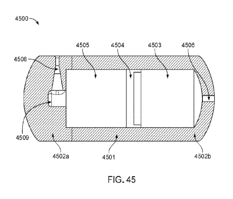
FIG. 45 illustrates an ingestible device including a pre-pressurized actuator
chamber
and a sliding piston, according to some embodiments described herein.
FIG. 46A illustrates a portion of an ingestible device including burst disc in
line with
a nozzle portion, according to some embodiments described herein.
FIG. 46B illustrates a partial sectional view of a burst disc holder,
according to some
embodiments described herein.
-20-
CA 03036364 2019-03-08
WO 2018/049133
PCT/US2017/050642
FIG. 47 illustrates a portion of an ingestible device including enteric
coating
occlusion component, according to some embodiments described herein.
FIG. 48 shows stacked layers of an enteric coating for an ingestible device,
according
to some embodiments described herein.
FIG. 49 illustrates an ingestible device including a magnetic occlusion
component, a
burst disc, and a pre-pressurized actuator chamber, according to some
embodiments
described herein.
FIG. 50 illustrates an ingestible device including a magnetic occlusion
component and
pre-pressurized actuator chamber, according to some embodiments described
herein.
FIG. 51 illustrates an ingestible device including enteric sliding occlusion
component
and pre-pressurized actuator chamber and a sliding piston, according to some
embodiments
described herein.
FIG. 52 illustrates an ingestible device including dissolvable pin occlusion
component
and a pre-pressurized chamber and a sliding piston, according to some
embodiments
described herein.
FIG. 53 illustrates an ingestible device including wax plug with wire lead
activators,
according to some embodiments described herein.
FIG. 54 illustrates an ingestible device including a pre-pressurized chamber
and a
bellows, according to some embodiments described herein.
FIG. 55 illustrates an ingestible device including a spring actuator and a
sliding
piston, according to some embodiments described herein.
FIG. 56 illustrates an ingestible device including a spring actuated slidable
housing
portion, according to some embodiments described herein.
FIG. 57 illustrates an ingestible device with another spring actuated slidable
housing
portion, according to some embodiments described herein.
FIG. 58 illustrates an ingestible device including a melt away occlusion
component
and a pressurized chamber, according to some embodiments described herein.
FIG. 59 illustrates an ingestible device including a dissolvable pin occlusion
component and a spring actuated sliding piston, according to some embodiments
described
herein.
FIG. 60 illustrates an ingestible device including shuttle slider occlusion
component
and a pressurized chamber, according to some embodiments described herein.
FIG. 61 illustrates an ingestible device including hydrogen cell actuator and
burst disc
occlusion component, according to some embodiments described herein.
-21-
CA 03036364 2019-03-08
WO 2018/049133
PCT/US2017/050642
FIG. 62 illustrates another ingestible including hydrogen cell actuator and
burst disc
occlusion component, according to some embodiments described herein.
FIG. 63 illustrates an ingestible device including a vacuum actuator chamber
and
enteric coating occlusion components, according to some embodiments described
herein.
FIG. 64 illustrates an ingestible device including an attachable reservoir,
according to
some embodiments described herein.
FIG. 65 is a view of an example embodiment of an ingestible device, in
accordance
with some embodiments of the disclosure.
FIG. 66 is an exploded view of the ingestible device of FIG. 65, in accordance
with
some embodiments of the disclosure.
FIG. 67 is a diagram of an ingestible device during an example transit through
a GI
tract, in accordance with some embodiments of the disclosure.
FIG. 68 is a diagram of an ingestible device during an example transit through
a
jejunum, in accordance with some embodiments of the disclosure.
FIG. 69 is a flowchart of illustrative steps for determining a location of an
ingestible
device as it transits through a GI tract, in accordance with some embodiments
of the
disclosure.
FIG. 70 is a flowchart of illustrative steps for detecting transitions from a
stomach to a
duodenum and from a duodenum back to a stomach, which may be used when
determining a
location of an ingestible device as it transits through a GI tract, in
accordance with some
embodiments of the disclosure.
FIG. 71 is a plot illustrating data collected during an example operation of
an
ingestible device, which may be used when determining a location of an
ingestible device as
it transits through a GI tract, in accordance with some embodiments of the
disclosure.
FIG. 72 is another plot illustrating data collected during an example
operation of an
ingestible device, which may be used when determining a location of an
ingestible device as
it transits through a GI tract, in accordance with some embodiments of the
disclosure.
FIG. 73 is a flowchart of illustrative steps for detecting a transition from a
duodenum
to a jejunum, which may be used when determining a location of an ingestible
device as it
transits through a GI tract, in accordance with some embodiments of the
disclosure.
FIG. 74 is a plot illustrating data collected during an example operation of
an
ingestible device, which may be used when detecting a transition from a
duodenum to a
jejunum, in accordance with some embodiments of the disclosure.
-22-
CA 03036364 2019-03-08
WO 2018/049133
PCT/US2017/050642
FIG. 75 is a plot illustrating muscle contractions detected by an ingestible
device over
time, which may be used when determining a location of an ingestible device as
it transits
through a GI tract, in accordance with some embodiments of the disclosure.
FIG. 76 is a flowchart of illustrative steps for detecting a transition from a
jejenum to
an ileum, which may be used when determining a location of an ingestible
device as it
transits through a GI tract, in accordance with some embodiments of the
disclosure.
FIG. 77 is a flowchart of illustrative steps for detecting a transition from a
jejenum to
an ileum, which may be used when determining a location of an ingestible
device as it
transits through a GI tract, in accordance with some embodiments of the
disclosure.
FIG. 78 is a flowchart of illustrative steps for detecting a transition from
an ileum to a
cecum, which may be used when determining a location of an ingestible device
as it transits
through a GI tract, in accordance with some embodiments of the disclosure.
FIG. 79 is a flowchart of illustrative steps for detecting a transition from a
cecum to a
colon, which may be used when determining a location of an ingestible device
as it transits
.. through a GI tract, in accordance with some embodiments of the disclosure.
FIG. 80 illustrates a tapered silicon bellows.
FIG. 81 illustrates a tapered silicone bellows in the simulated device jig.
FIG. 82 illustrates a smooth PVC bellows.
FIG. 83 illustrates a smooth PVC bellows in the simulated device jig.
FIG. 84 demonstrates a principle of a competition assay performed in an
experiment.
FIG. 85 shows AlphaLISA data.
FIG. 86 shows AlphaLISA data.
FIG. 87 shows AlphaLISA data.
FIG. 88 illustrates a test method.
FIG. 89 illustrates an assay principle.
FIG. 90 illustrates an ingestible device.
FIG. 91 illustrates a wax valve system.
FIG. 92A illustrates a wax valve system in a closed position.
FIG. 92B illustrates a wax valve system in an open position.
FIG. 93 illustrates an ingestible device with two outlets for dispensing.
-23-
CA 03036364 2019-03-08
WO 2018/049133
PCT/US2017/050642
Detailed Description
Following below are more detailed descriptions of various concepts related to,
and
exemplary embodiments of, ingestible devices capable of delivering a
dispensable substance,
such as, for example, a therapeutic agent, as well as related components,
systems and
methods. Also following below are more detailed descriptions of various
concepts related to,
and exemplary embodiments of, attachable storage reservoir configured to be
used with an
ingestible device and capable of storing dispensable substance, such as, for
example, a
therapeutic agent, as well as related components, systems and methods.
Various systems, devices, and methods are described herein to provide an
example of
to at least one embodiment for the subject matter described herein. No
embodiment limits any
subject matter described herein and any claimed subject matter may cover
systems, devices,
and methods that differ from those described herein. It is possible that the
claimed subject
matter are not limited to systems, devices, and methods having all of the
features of any one
systems, devices, and methods described herein or to features common to
multiple or all of
the systems, devices, and methods described herein. It may be possible that a
system, device,
or method described herein is not an embodiment of any claimed subject matter.
Any subject
matter disclosed in systems, devices, and methods described herein that is not
claimed in this
document may be the subject matter of another protective instrument, for
example, a
continuing patent application, and the applicants, inventors or owners do not
intend to
abandon, disclaim or dedicate to the public any such subject matter by its
disclosure in this
document.
It will be appreciated that, for simplicity and clarity of illustration, where
considered
appropriate, reference numerals may be repeated among the figures to indicate
corresponding
or analogous elements. In addition, numerous specific details are set forth in
order to provide
a thorough understanding of the embodiments described herein. However, it will
be
understood by those of ordinary skill in the art that the embodiments
described herein may be
practiced without these specific details. In other instances, well-known
methods, procedures
and components have not been described in detail so as not to obscure the
embodiments
described herein. In addition, the description is not to be considered as
limiting the scope of
the embodiments described herein.
It should be noted that terms of degree such as "substantially", "about" and
"approximately" when used herein mean a reasonable amount of deviation of the
modified
term such that the result is not significantly changed. These terms of degree
should be
-24-
CA 03036364 2019-03-08
WO 2018/049133
PCT/US2017/050642
construed as including a deviation of the modified term if this deviation
would not negate the
meaning of the term it modifies.
In addition, as used herein, the wording "and/or" is intended to represent an
inclusive-
or. That is, "X and/or Y" is intended to mean X or Y or both, for example. As
a further
example, "X, Y, and/or Z" is intended to mean X or Y or Z or any combination
thereof.
As used herein, the term "coupled" indicates that two elements can be directly
coupled to one another or coupled to one another through one or more
intermediate elements.
As used herein, the term "body" refers to the body of a patient, a subject or
an
individual who receives the ingestible device. The patient or subject is
generally a human or
other animal.
As used herein, the term "gastrointestinal tract" or "GI tract" refers to all
portions of
an organ system responsible for consuming and digesting foodstuffs, absorbing
nutrients, and
expelling waste. This includes orifices and organs such as the mouth, throat,
esophagus,
stomach, small intestine, large intestine, rectum, anus, and the like, as well
as the various
passageways and sphincters connecting the aforementioned parts.
As used herein, the term "reflectance" refers to a value derived from light
emitted by
the device, reflected back to the device, and received by a detector in or on
the device. For
example, in some embodiments this refers to light emitted by the device,
wherein a portion of
the light is reflected by a surface external to the device, and the light is
received by a detector
located in or on the device.
As used herein, the term "illumination" refers to any electromagnetic
emission. In
some embodiments, an illumination may be within the range of Infrared Light
(IR), the
visible spectrum and ultraviolet light (UV), and an illumination may have a
majority of its
power centered at a particular wavelength in the range of 100nm to 1000nm. In
some
embodiments, it may be advantageous to use an illumination with a majority of
its power
limited to one of the infrared (750nm-1000nm), red (620nm-750nm), green (495nm-
570nm),
blue (450nm-495nm), or ultraviolet (100nm-400nm) spectrums. In some
embodiments, a
plurality of illuminations with different wavelengths may be used.
The various embodiments described herein generally relate to an ingestible
device
that is configured to arrive at a specific location within the
gastrointestinal (GI) tract via oral
consumption and, in some embodiments, for releasing substances including
medicaments and
therapeutics at the specific location. In another embodiment, the ingestible
device may be
used for releasing substances including medicaments and therapeutics in other
parts of the
body, such as but not limited to the female reproductive tract, and/or the
like. In some
-25-
CA 03036364 2019-03-08
WO 2018/049133
PCT/US2017/050642
embodiments, the release of the dispensable substances may take a form similar
to a bolus or
a bust of dispensing. In some embodiments, the release of the substances may
take a form
similar to systemic therapeutic agent delivery. The ingestible device may
include a release
structure that helps the substance to be delivered on the inner surface, e.g.,
the mucosa layer,
of the GI tract, or through a penetration of the mucosa layer.
FIG. 1 provides an example mock-up diagram illustrating aspects of a structure
of an
ingestible device 100 for delivering a dispensable substance, according to
some embodiments
described herein. In some embodiments, the ingestible device 100 may generally
be in the
shape of a capsule, a pill or any swallowable form that may be orally consumed
by an
individual. In this way, the ingestible device 100 may be ingested by a
patient and may be
prescribed by healthcare practitioners and patients.
The ingestible device 100 includes a housing 101 that may take a shape similar
to a
capsule, a pill, and/or the like, which may include two ends 102a-b. The
housing 101 may be
designed to withstand the chemical and mechanical environment of the GI tract
(e.g., effects
of muscle contractile forces and concentrated hydrochloric acid in the
stomach). A broad
range of materials that may be used for the housing 101. Examples of these
materials
include, but are not limited to, thermoplastics, fluoropolymers, elastomers,
stainless steel and
glass complying with ISO 10993 and USP Class VI specifications for
biocompatibility; and
any other suitable materials and combinations thereof. In certain embodiments,
these
materials may further include liquid silicone rubber material with a hardness
level of 10 to 90
as determined using a durometer (e.g., MED4942TM manufactured by NuSilTm), a
soft
biocompatible polymer material such as, but not limited to, polyvinyl chloride
(PVC),
polyethersulfone (PES), polyethylene (PE), polyurethane (PU) or
polytetrafluoroethylene
(PTFE), and a rigid polymer material coated with a biocompatible material that
is soft or
pliable (e.g., a poly(methyl methacrylate) (PMMA) material coated with
silicone polymer).
Use of different materials for different components may enable
functionalization of certain
surfaces for interaction with proteins, antibodies, and other biomarkers. For
example,
Teflon may be used as a material in the ingestible device 10 for movable
components in
order to reduce friction between these components. Other example materials may
include
other materials commonly used in micro-fabrication, such as
polydimethylsiloxane (PDMS),
borosilicate glass, and/or silicon. Although specific materials may be
referred to herein as
being used to construct the device for illustrative purposes, the materials
recited are not
intended to be limiting, and one skilled in the art may easily adapt the
device to use any
-26-
CA 03036364 2019-03-08
WO 2018/049133
PCT/US2017/050642
number of different materials without affecting the overall operation or
functionality of the
device.
In some embodiments, the housing 101 of the ingestible device 100 may be
manufactured from a type of plastic, such as a photosensitive acrylic polymer
material or an
inert polycarbonate material. The housing 101 may also be formed using
material that can be
sterilized by chemicals. In some implementation, the wall of the housing 101
may have a
thickness of 0.5mm-lmm, which is sufficient to sustain an internal explosion
(e.g., caused by
hydrogen ignition or over pressure inside the housing).
The housing 101 may or may not have a pH-sensitive enteric coating to detect
or
otherwise be sensitive to a pH level of the environment external to the
ingestible device. In
some specific parts of the GI tract, such as but not limited to sections
immediately after
passing through the pyloric sphincter, or sections immediately prior to the
ileocecal valve, it
may be difficult to target a specific location solely based on the pH level.
Instead of relying
on the pH level, the ingestible device 100 includes an optical sensing unit
that transmits an
illumination to the environment and collects a reflectance, based on which,
the region-
specific location of the ingestible device may be identified based on optical
characteristics of
the reflectance. For example, the ingestible device may deliver a therapeutic
agent to a
specific location within the GI tract that harbors an injury such as a lesion.
The specific
location may be pre-determined through a previously conducted endoscopy.
Further
discussion on determining a location of the ingestible device may be found in
connection
with FIG. 44.
The housing 101 may be formed by coupling two enclosure portions together. For
example, the two enclosure portions can be mated and fused together with an
adhesive
material, such as a cyanoacrylate variant. The housing 101, in effect,
protects the interior of
the ingestible device 100 from its external environment and protects the
external environment
(e.g., the gastrointestinal tract) from components inside the ingestible
device 100.
The ingestible device 100 may include an electronic component within the
housing
100. The electronic component may be placed proximally to an end 102b of the
housing, and
includes a printed circuit board (PCB), a battery, an optical sensing unit,
and/or the like.
Further example structures of the electronic component may be illustrated in
FIG. 44.
The ingestible device 100 further includes a gas-generating cell 103 that is
configured
to generate gas and thus cause an internal pressure within the housing 101. In
one
implementation, the gas-generating cell 103 may be a hydrogen-generating cell,
such as but
not limited to a Vartag Hydrogen Gas-generating Cell. In another
implementation, one or
-27-
CA 03036364 2019-03-08
WO 2018/049133
PCT/US2017/050642
more other gas-generating cells that generate an inert gas that is harmless to
the human body
may be used.
In some implementations, the gas-generating cell may include or be connected
to a
separate channel or valve of the ingestible device such that gas may be
release through the
channel or valve to create a motion to alter the position of the ingestible
device within the GI
tract. Such gas release can also be used to position the ingestible device
relative to the
intestinal lining. In another implementation, gas may be released through the
separate
channel or valve to alter the surface orientation of the intestinal tissue
prior to delivery of the
dispensable substance.
A traveling plunger 104 may be placed on top of the gas-generating cell 103
within
the housing 101. The traveling plunger 104 is a membrane that separates the
gas-generating
cell 103 and a storage reservoir that stores the dispensable substance 105. In
some
implementations, the traveling plunger 104 may be a movable piston, as is
further illustrated
in FIGS. 3-4. In some implementations, the traveling plunger 104 may instead
be a flexible
membrane such as but not limited to a diaphragm, as further illustrated in
FIG. 10. In some
implementations, the traveling plunger 104, which may have the form of a
flexible
diaphragm, may be placed along an axial direction of the housing 101, instead
of being
placed on top of the gas-generating cell 103, as is further illustrated in
FIG. 44. The traveling
plunger or the membrane 104 may move (when the membrane 104 is a piston) or
deform
(when the membrane 104 is a diaphragm) towards a direction of the end 102a of
the housing,
when the gas-generating cell 103 generates gas to create an internal pressure
that pushes the
membrane 104. In this way, the membrane or traveling plunger 104 may push the
dispensable substance 105 out of the housing via a dispensing outlet 107.
The housing 101 may include a storage reservoir storing one or more
dispensable
substances 105 adjacent to the traveling plunger 104. The dispensable
substance 105 may be
a therapeutic or medical agent that may take a form of a powder, a compressed
powder, a
fluid, a semi-liquid gel, or any other dispensable or deliverable form. The
delivery of the
dispensable substance 105 may take a form such as but not limited to bolus,
semi-bolus,
continuous, systemic, burst therapeutic agent delivery, and/or the like.
In some implementations, the storage reservoir may include multiple chambers,
and
each chamber stores a different dispensable substance. For example, the
different
dispensable substances can be released at the same time via the dispensing
outlet 107.
Alternatively, the multiple chambers may take a form of different layers
within the storage
reservoir such that the different dispensable substance from each chamber is
delivered
-28-
CA 03036364 2019-03-08
WO 2018/049133
PCT/US2017/050642
sequentially in an order. In one example, each of the multiple chambers is
controlled by a
separate traveling plunger, which may be propelled by gas generation. The
electronic
component may control the gas-generating cell 103 to generate gas to propel a
specific
traveling plunger, e.g., via a separate gas generation chamber, etc., to
deliver the respective
substance. In some embodiments, the content of the multiple chambers may be
mixed or
combined prior to release, for example, to activate the therapeutic agent.
The ingestible device 100 may include a dispensing outlet 107 at one end 102a
of the
housing 101 to direct the dispensable substance 105 out of the housing. The
dispensing outlet
107 may include an exit valve (as further illustrated in FIGS. 5-6), a slit or
a hole (as further
illustrated in FIGS. 7-8), a jet injection nozzle with a syringe, and/or the
like. When the
traveling plunger 104 moves towards the end 102a of the housing 101, an
internal pressure
within the storage reservoir may increase and push the dispensing outlet to be
open to let the
dispensable substance 105 be released out of the housing 101.
The use of a hydrogen-generating cell within an ingestible device 100 may
incur a
variety of safety risks, which can be mitigated by proper device design. For
example,
hydrogen ignition/heating within or external to the ingestible device 100,
over pressure
within the housing 101, hydrogen toxicity may damage the use of the ingestible
device. A
safety device such as a pressure relief device 106 may be placed within or
attached to the
housing 101 to mitigate the safety risk.
In some embodiments, hydrogen ignition inside the ingestible device may occur
if air
(containing oxygen) is present inside the ingestible device 100. For example,
hydrogen
requires very little energy to initiate combustion, e.g., with a minimum
energy for ignition in
air of just 0.017mJ. When silver oxide batteries (e.g., see 131 in FIG. 3) are
used in the
ingestible device 100, the likelihood of a spark (though remote) cannot be
eliminated and
may result from inductive charges and introduced air gap, or other scenarios.
For example, the pressure Pi within the ingestible device 100 may be estimated
based
on a worst-scenario analysis of Hydrogen-Air mixture as approximately Pi =
1479 kPa or 215
psi. As the hydrogen concentration within the ingestible device 100 may likely
increase with
respect to time, a sufficient concentration of hydrogen may be assumed to lie
between the
Lower Explosive Limit (LEL) and Upper Explosive Limit (UEL) of 4% and 75%
respectively. Thus the final pressure may be higher, given the initial
pressure within the
ingestible device may exceed 101.3kPa due to the addition of hydrogen.
Assuming the
housing 101 fractures at this maximum pressure, the amount of energy E
released may be
given by:
-29-
CA 03036364 2019-03-08
WO 2018/049133
PCT/US2017/050642
E
_
¨ X V1
(1479000Pa ¨ 101300Pa\ = E (
4
X 0,03000I.¨ j uies
t-1
where Vi denotes an approximate volume of the ingestible device. As a
reference, this
energy translates to a lg mass (approximately 1/4 of an assembled capsule)
travelling at 82 m/s
(-180 mph), or about the same amount of energy contained in a propelled BB gun
pellet,
which may cause harm to the patient.
To mitigate the hydrogen ignition, in one implementation, the housing 101 may
be
made of polycarbonate with a wall thickness of 1.6mm, which may sustain a
pressure of
410psi and thus withstand the effects of an internal explosion without
fracture.
1() In an implementation, the interior wall of the housing 101 may be made
of (or include
a layer made of) oxygen absorbing material to reduce the concentration of
oxygen (e.g.,
below 2%) to avoid an internal explosion.
In an implementation, an inert non-conductive liquid dielectric (such as
silicone oil,
food grade castor oil, wax, and/or the like) may be applied to contain
electrical connections
within the ingestible device 100 to isolate any possible ignition from
hydrogen within the
ingestible device 100. For example, the isolation may also reduce the risk of
hydrogen
ignition external to the ingestible device, e.g., when the generated hydrogen
from the gas-
generating cell 103 mixed with an appropriate amount of air (oxygen) contained
in bubbles
travels along the GI tract, any ignition may result in an injury to the small
intestine.
In one scenario, over pressurization may happen within the ingestible device
100, e.g.,
when the dispensing outlet 107 becomes blocked or otherwise sealed from the
release of
hydrogen. In this case, the housing 101 may have a fracture when the hydrogen
gas reaches a
threshold level of pressure. For example, when the ingestible device has a
volume of 5004,
(the volume hydrogen may communicate with), and the hydrogen released can be
40mL, the
internal pressure generated by the hydrogen can be as high as:
Vc,
p, =
= 14;7psi ¨ = 1I76psi = 3.108MPa
0.5
-30-
CA 03036364 2019-03-08
WO 2018/049133
PCT/US2017/050642
The energy released via a fracture or explosion of the over pressurization may
be calculated
as:
e' 1081v1Pa ,10 1 31,4Pa`\
E
1 369 ¨ 1 ) 10-7 = 4.6 joules
The amount of energy may inflict harm to the patient.
In one implementation, the housing 101 may be designed to breach or fracture
in a
controlled manner and thus can relieve the internal pressure. For example,
when the internal
pressure within a chamber filled with the gas from the gas-generating cell 103
exceeds a
1() threshold level, the wall around the chamber may be designed to
fracture or break to relieve
the pressure.
In an implementation, a pressure relief device 106 may be placed within the
housing
101, e.g., at the end 102a of the housing 101. For example, the pressure
relief device 106
may be a pressure relief rupture disc that is configured to release content
within housing 101.
Thus if the dispensing outlet 107 is clogged or blocked and the pressure
within the storage
reservoir exceeds a threshold level, the pressure relief device 106 may be
torn to release the
dispensable substance out. The advantage of the pressure relief device 106 is
that it may also,
in part, serve to prevent upstream contamination of the dispensable substance,
because the
internal pressure is relieved by directing the dispensable substance out of
the housing such
that the gas would not leak into the storage reservoir.
In some implementations, the housing 101 may include small holes (e.g., with a
diameter smaller than 2 mm), e.g., on the side of the housing 101, or at the
end 102a to
facilitate loading the dispensable substance into the storage reservoir. The
holes, when more
than two, may be positioned axially or radially on the housing 101. The holes
may
subsequently be sealed with a UV curable cyanoacrylate.
In some implementations, a feedback control circuit (e.g., a feedback
resistor, etc.)
may be added to send feedback from the gas-generating cell 103 to the
electronic component
such that when the internal pressure reaches a threshold level, the electronic
component may
control the gas-generating cell 103 to turn off gas generation, or to activate
other safety
mechanism (e.g., feedback-controlled release valve, etc.). For example, an
internal pressure
sensor may be used to measure the internal pressure within the ingestible
device and generate
feedback to the feedback control circuit.
-31-
CA 03036364 2019-03-08
WO 2018/049133
PCT/US2017/050642
FIG. 2 provides an example diagram illustrating aspects of a mechanism for a
gas-
generating cell 103 configured to generate a gas to dispense a substance,
according to some
embodiments described herein. As shown in FIG. 2, the gas-generating cell 103
generates a
gas 111, which can propel the dispensable substance 105 out of the dispensing
outlet 107. A
variable resistor 108 may be connected to a circuit with the gas-generating
cell 103 such that
the variable resistor 108 may be used to control an intensity and/or an amount
of gas 111
(e.g., hydrogen) generated by the cell 103. Specifically, the gas-generating
cell 103 may be a
battery form factor cell that is capable of generating hydrogen when a
resistor is applied. In
this way, as the gas-generating cell 103 only needs the use of a resistor only
without any
active power requirements, the gas-generating cell 103 may be integrated into
an ingestible
device such as a capsule with limited energy/power available. For example, the
gas-
generating cell 103 may be compatible with a capsule at a size of 26mm x 13mm
or smaller.
In some implementations, based on the elution rate of gas from the cell, and
an
internal volume of the ingestible device, it may take time to generate
sufficient gas 111 to
deliver the substance 105, and the time required may be 30 seconds or longer.
For example,
the time to generate a volume of hydrogen equivalent to 5004, of fluid would
be
approximately 5 minutes. A longer period of time may be needed based upon non-
ideal
conditions within the ingestible device, such as friction, etc. Thus, given
that the production
of gas (e.g., hydrogen) may take time, gas generation may need to start prior
to the ingestible
device arriving at the site of delivery to build pressure up within the
device. The ingestible
device may then need to know when it is approaching the site of delivery. For
example, the
device may start producing gas on an "entry transition," which is determined
by temperature,
so as to produce enough gas to be close to the pressure high enough to deliver
the dispensable
substance. The ingestible device may then only start producing gas again when
it arrives at
the site of delivery, which will cause the internal pressure within the
ingestible device to
reach a level required by the dispensing outlet to release the dispensable
substance. In
addition, for region-specific delivery, the ingestible device may estimate the
time it takes to
build up enough pressure to deliver the dispensable substance before the
ingestible device
arrives at a specific location, to activate gas generation.
For example, for systemic delivery, when an internal volume of the ingestible
device
is around 500 L, a gas generation time of 2 hours, an initial pressure of
approximately 300
pound per square inch absolute (psia) may be generated, with higher and lower
pressures
possible. The generated pressure may drop when air enters the storage
reservoir, which was
previously occupied by the dispensable substance during the dispensing
process. For
-32-
CA 03036364 2019-03-08
WO 2018/049133
PCT/US2017/050642
systemic dispensable substance delivery, a force with a generated pressure of
approximately
100 to 360 pound per square inch (psi) may be required for dermal penetration,
e.g., to
penetrate the mucosa or epithelial layer. The pressure may also vary depending
on the nozzle
design at the dispensing outlet, fluid viscosity, and surrounding tissue
proximity and
properties.
The gas 111 that may be generated for a continuous delivery of dispensable
substance
(e.g., lcc H2 in 4 hours, 16 breaths per minute at 0.5L tidal volume) may
equate to 1 cc
hydrogen in approximately 2000L of exhaled air, or approximately 0.5 ppm H2,
which is
below physiologic values of exhaled hydrogen. Reducing this time to 10 minutes
equates to
approximately 13ppm hydrogen. Thus, due to the length of intestine that may be
covered
during this time period, the ingestible device may possess a higher localized
value than
physiologic.
FIG. 3 provides example structural diagrams illustrating aspects of an
ingestible
device 100 having a piston to push for dispensable substance delivery,
according to some
embodiments described herein. The ingestible device 100 may have one or more
batteries
131 placed at one end 102a of the housing 101 to provide power for the
ingestible device 100.
The PCB 132 may be placed adjacent to the battery 131, and the gas-generating
cell 103 may
be mounted above the PCB 132. The gas-generating cell 103 may be sealed from
the bottom
chamber (e.g., space including 131 and 132) of the ingestible device 100. A
movable piston
134 may be placed adjacent to the gas-generating cell 103. In this way, gas
generation from
the gas-generating cell 103 may propel the piston 134 to move towards the
other end 102b of
the housing 101 such that the dispensable substance in the storage reservoir
135 can be
pushed out of the housing through the dispensing outlet 107, e.g., the
movement is shown at
136, with the piston 134 at a position after dispensing the substance. The
storage reservoir
135 may have a volume of approximately 6004, or even more dispensable
substance, which
may be dispensed at one shot, or gradually over a period of time.
The battery cells 131 may have a height of 1.65 mm each, and one to three
batteries
may be used. The height of the piston may be reduced with custom molded part
for around
1.5mm to save space. If the gas-generating cell 103 is integrated with the
piston 134 (e.g., as
further illustrated in FIG. 10), the overall height of the PCB, batteries and
gas-generating cell
in total can be reduced to around 5 mm, thus providing more space for
dispensable substance
storage. For example, for an ingestible device of 7.8 mm in length (e.g., from
end 102a to the
other end 102b), a storage reservoir 134 of approximately 6004, may be used
for dispensable
substance delivery. For another example, for an ingestible device of 17.5 mm
in length, a
-33-
CA 03036364 2019-03-08
WO 2018/049133
PCT/US2017/050642
storage reservoir 134 of approximately 13004, may be used for dispensable
substance
delivery.
FIG. 4 provides an example structural diagram illustrating aspects of an
umbrella-
shaped exit valve structure as a dispensing outlet of the ingestible device,
according to some
.. embodiments described herein. The exit valve may be placed at one end 102b
of the housing
of an ingestible device (e.g., 100 in FIG. 3). The exit valve may include an
umbrella-shaped
lid 141 placed on top of one end 102b of the housing, under which the end 102b
of the
housing may include radially and evenly distributed holes 142 around the
center of the round-
shaped end 102b. When the dispensable substance is pushed out from inside the
housing
1() .. through the holes 142, the umbrella-shaped valve 141 may be lifted due
to the pressure to let
the dispensable substance out of the holes 142 be radially directed through
the radially
distributed notches 143 on the edge of the round-shaped end 102b of the
housing. The
umbrella-shaped valve 141, which covers the delivery holes 142, may also serve
to keep the
dispensing outlet holes 142 free of contaminants outside the ingestible device
100. The
notches 143 and the holes 142 may be radially and evenly distributed around a
central axis of
gravity based on the geometry of the ingestible device (e.g., a cylinder
shape, etc.) to reduce
or avoid rotation or tilting of the ingestible device under a movement force
of the dispensable
substance.
FIG. 5 provides an example structural diagram illustrating aspects of a ring-
shaped
exit valve structure as a dispensing outlet of the ingestible device,
according to some
embodiments described herein. The housing 101 of the ingestible device may
include a ring-
shaped valve 151 at a place around the housing 101 and proximal to the end
102b of the
housing 101. The ring-shaped valve 151 may have evenly and radially
distributed ports 157
around the ring to direct the dispensable substance out of the housing 101.
The ring-shaped
.. valve 151 may also designed to prevent blockage from contaminants outside
the ingestible
device 100. The ring-shaped valve 151 may optionally have a number of evenly
distributed
slits 153 for even distribution of the dispensable substance to maintain
balance of the
ingestible device.
FIG. 6 provides an example structural diagram illustrating aspects of a dome
slit as a
dispensing outlet of the ingestible device, according to some embodiments
described herein.
The storage reservoir of the ingestible device may include a bellow 161
(further illustrated in
FIG. 8), and one end of the bellow 161 may include a dome slit 162 extending
out of the end
102b of the housing 101. Thus, when the bellow is being compressed, the
dispensable
substance may be propelled out of the bellow through the dome slit 162.
-34-
CA 03036364 2019-03-08
WO 2018/049133
PCT/US2017/050642
In one implementation, unlike the radial distribution through the valve in
FIGS. 4-5,
the bellow 161 with the dome slit 162 may be able to eject the dispensable
substance at a high
velocity and thus a jet delivery may be implemented. The internal pressure
generated to
compress the bellow 161 may be sufficient to generate a delivery at a velocity
to penetrate a
mucosa layer within the small intestine.
FIG. 7 provides an example structural diagram illustrating aspects of a hole
placed at
one end of the housing as a dispensing outlet of the ingestible device,
according to some
embodiments described herein. A delivery hole 171 may be placed at the end
102b of the
housing 101 for burst delivery. The hole 171 may be sealed with thin wax or
silicone, which
may be broken by a force from inside of the housing 101 such that the
dispensable substance
can be released.
In one implementation, an injection nozzle (not shown in FIGS. 6-7) or a
syringe may
be placed at one end 102b of the housing 101 for a jet delivery. For example,
the nozzle or
syringe may have an injecting needle extending out of the dome slit 162 or the
delivery hole
171. The injection nozzle may use osmotic pressure from luminal fluid inside
the small
intestine to drive the dispensing mechanism. The injecting needle may be used
to penetrate
into a mucosa layer within the small intestine. The various delivery
mechanisms described in
FIGS. 4-7 may be configured to deliver the dispensable substance out of the
ingestible device
either radially or longitudinally.
FIG. 8 provides an example structural diagram illustrating aspects of an
ingestible
device 100 having a bellow structure for a storage reservoir of dispensable
substances,
according to some embodiments described herein. A gas-generating cell 103
(similar to that
described in FIGS. 1-3) may be mounted to the PCB 132 and batteries 131. A
collapsible
silicone bellow 161 is placed on top of the gas-generating cell 103, with one
end of the
bellow 161 in contact with the gas-generating cell 103 and the other end in
contact with one
end 102b of the housing 101. Dispensable substance may be loaded into the
bellow 161,
which may be compressed by gas generation from the gas-generating cell 103 to
dispense the
dispensable substance out of the housing 101. The shape of the bellow 161 may
aid in
controlled delivery. The dispensing outlet 107 may use any of the exit valves
described in
FIGS. 4-5, or the dome slit 162 as described in FIG. 6.
FIG. 9 provides an example structural diagram illustrating aspects of an
ingestible
device having a flexible diaphragm to deform for dispensable substance
delivery, according
to some embodiments described herein. The ingestible device 100 may have a
flexible
diaphragm 165 that may deform towards the dispensing outlet 107 when the gas-
generating
-35-
CA 03036364 2019-03-08
WO 2018/049133
PCT/US2017/050642
cell 103 generates gas. The dispensable substance may then be propelled by the
deformed
diaphragm out of the housing through the dispensing outlet 107. The dispensing
outlet 107
shown at FIG. 9 is in the form of a ring valve as discussed in FIG. 5,
however, any outlet
design described in FIGS. 4-7 can be applied.
For the respective example ingestible device with the deformable diaphragm,
with a
total length of 7.8 mm, the ingestible device may store and deliver
approximately 6004, of
dispensable substance. For another example, for an ingestible device of 17.5
mm in length,
the ingestible device may store and deliver approximately 12504, of
dispensable substance.
FIG. 10 provides an example structural diagram illustrating aspects of an
ingestible
device having an integrated piston and gas generating cell such that the gas-
generating cell is
movable with the piston to push for dispensable substance delivery, according
to some
embodiments described herein. The ingestible device 100 may include a piston
166 that is
made up of, or is integrated with, the gas generation cell 103. The gas-
generating cell 103
may be sealed with custom silicone. In this way, the gas-generating cell 103
may move with
the piston 166 when generating gas. Contacts may be embedded in a seal 166
wrapping
around the gas-generating cell 103 with leads connected to PCB 132. The moving
gas-
generating cell structure may allow approximately 400 L capacity for
dispensable substance
storage and delivery.
FIG. 11 provides an example structural diagram illustrating aspects of an
ingestible
device having a capillary to direct dispensable substances out of the storage
reservoir,
according to some embodiments described herein. A capillary structure 169 may
be placed
between the gas-generating cell 103 and an end of the housing 101. The
dispensable
substance may be stored in the empty chamber 135 around the gas-generating
cell 103 and
within the capillary structure 169. A wax seal may be applied between the gas-
generating
cell 103 and the capillary structure 169. At an appropriate time when it is
identified that the
ingestible device is at a specific location within the intestine, the wax seal
may melt, e.g., via
a generated heat by the PCB 132 or the gas-generating cell 103, and then the
gas generation
pressure may push the dispensable substance in the chamber 135 through the
capillary plates
169 for dispensing through a dispensing outlet 107 out of the housing 101.
Optionally, an
exterior wax seal may be positioned between the capillary structure 169 and
the dispensing
outlet 107 to avoid leakage or early delivery.
In one example, the capillary structure 169 may include concentric rings of
capillary
plates. In another example, the capillary structure 170 between the storage
reservoir 135 and
an end of the housing 101 may be made of a bent foil. The ingestible device
100 with the
-36-
CA 03036364 2019-03-08
WO 2018/049133
PCT/US2017/050642
capillary structure may have approximately 450 uL capacity of dispensable
substance. In
some implementations, additional volume may be obtained by reducing the size
of PCB 132,
removing one or two batteries, integrating the gas-generating cell 103 into
the PCB 132,
and/or the like. The dispensable substance volume may increase to 900 L.
FIG. 12 provides an example structural diagram illustrating aspects of an
ingestible
device having a clamshell-shaped housing and a sideways split diaphragm to
deform for
dispensable substance delivery, according to some embodiments described
herein. The
housing 101 of ingestible device may include two clamshell halves 101a-b along
the long
axis of the housing. The clamshell structure may be used to open and load
dispensable
substance into the ingestible device, or to insert a separate storage
reservoir into the ingestible
device. In the ingestible device 100a, a diaphragm 123 may be placed along one
clamshell
half 101b and extend to wrap around the gas-generating cell 103. In this
manner, the
diaphragm 123 may act as a dam between the storage reservoir 135 storing the
dispensable
substance and electronics 132 to protect the electronics 132. When the gas-
generating cell
.. 103 generates a gas, an internal pressure may deflect the diaphragm 123
into other half of
clamshell 101a, and thus propel the dispensable substance within the storage
reservoir 135 to
be released out of the housing 101. The ingestible devices 100b-c provide
different views of
the sideway split diaphragm 123 from two different angles.
FIG. 13 provides an example structural diagram illustrating aspects of an
ingestible
device having an elastomer bladder to provide the pressure to deliver the
dispensable
substance, according to some embodiments described herein. An elastomeric
component 152
(e.g., a bladder, a balloon, etc.) can be placed in the storage reservoir 135
storing the
dispensable substance such that when the elastomeric component 152 expands to
fill the
volume of the storage reservoir 135, the dispensable substance can be pushed
out of the
.. ingestible device via an outlet, e.g., one or more holes 156 on the wall of
the ingestible
device.
The elastomeric component 152 may be made of flexible material that takes up a
small volume in its free state but able to expand to fill the volume of the
storage reservoir
135. A residual volume may exist, as the elastomeric component 152 at its free
state still
takes up space within the storage reservoir and thus reduces the volume of
dispensable
substance that can be filled into the storage reservoir 135. For example, as
shown in FIG. 13,
the elastomeric component 152, e.g., a balloon, even at a free state, may be
slightly inflated,
and thus takes up a volume of 57 L.
-37-
CA 03036364 2019-03-08
WO 2018/049133
PCT/US2017/050642
One or more holes 156 on the outer shell of the ingestible device 100 can
allow
luminal fluid to be drawn into the void behind the elastomeric component 152.
The ports of
the holes 156 on the outer wall may be shaped to prevent irritation during the
use the
ingestible device 100, e.g., with no edges to catch any tissue within the GI
tract during travel,
and also to reduce fouling from the back filling of luminal fluid.
A wax plug can be used to seal the one or more holes 156, which can be pushed
out
by pressure when the dispensable substance is released. Additional coating,
removable
casing or decal can be added, e.g., on top of the one or more holes 156, to
ensure the
dispensable substance is not dispensed during handling.
FIG. 14 provides example structural diagrams illustrating aspects of pre-
loading an
ingestible device having an elastomer bladder to reduce the residual volume,
according to
some embodiments described herein. To reduce the residual volume and to load
the
ingestible device with the maximum possible dispensable substance, the
elastomeric
component 152 can be stretched and filled by a rod 155 prior to filling the
storage reservoir
154 with the dispensable substance. In this way, the free state of the
elastomeric component
152 is preloaded under tension, which can reduce the residual volume. One or
more holes
156 on the outer shell of the ingestible device 100 can allow luminal fluid to
be drawn into
the void behind the elastomeric component 152. The ports of the holes 156 on
the outer wall
may be shaped to prevent irritation during the use the ingestible device 100,
e.g., with no
edges to catch any tissue within the GI tract during travel, and also to
reduce fouling from the
back filling of luminal fluid.
In this way, when the residual volume is reduced, the payload capacity of the
ingestible device can be increased, e.g., a dose of up to 8000_, can be
carried by the ingestible
device. Because of the increased payload capacity, the overall size of the
ingestible device
can thus be reduced, which may be easier for a patient to administer and may
also increase
packing density to improve distribution logistics.
FIG. 15 provides example structural diagrams illustrating aspects of an
ingestible
device having a gear motor to dispense the dispensable substance out of the
storage reservoir,
according to some embodiments described herein. A gear motor mechanism 160 may
be
placed within the ingestible device, with one end connected to the battery
cells 131, and the
other end placed at the opposite end of the ingestible device 100. The gear
motor 160 can be
driven to a specific motion, e.g., rotating, etc., such that the dispensable
substance in the
storage reservoir can be dispensed out of the ingestible device 100 by the
motion. The
dispensable substance can be dispensed as a bolus or a gradual dose over a
given time period
-38-
CA 03036364 2019-03-08
WO 2018/049133
PCT/US2017/050642
with a variable dose rate, e.g., by adjusting the velocity, pattern, and power
of the gear motor
motion. The storage reservoir 135 may carry a volume of up to 720uL, as the
gear motor
mechanism may need an enhanced battery system that can limit the overall
payload volume.
FIG. 16 provides example structural diagrams illustrating aspects of an
ingestible
device using an auger to dispense the dispensable substance out of the storage
reservoir,
according to some embodiments described herein. The gear motor 160 can be
equipped with
an auger device around the outer wall of the gear motor 160 such that the
auger device can be
driven to stir or wipe the dispensable substance to be dispensed out of the
storage reservoir.
The diameter of the auger device 161 is smaller than the diameter of the
storage
reservoir such that the auger device 161 does not touch the inner wall of the
ingestible device
100, to avoid any scratch or damage to the wall of the ingestible device 100.
The sealing of
the ingestible device 100 can be static between the housing of the gear motor
160 and the
body of the ingestible device 100. This can reduce the amount of torque
required to drive the
auger 161, and may allow the user of a smaller gear motor 160, which can
increase the
available payload volume within the ingestible device 100.
The auger mechanism can allow the dispensable substance to be flushed out of
the
ingestible device 100 and to be progressively diluted as the luminal fluid is
drawn into the
storage reservoir. The dispensable substance is then delivered out of the
storage reservoir at a
declining concentration. As a result, a low residual volume of dispensable
substance may
remain within the storage reservoir, depending on the number of times the
auger mechanism
is instantiated to dispense the dispensable substance.
FIG. 17 provides example structural diagrams illustrating aspects of an
ingestible
device using a wiper to dispense the dispensable substance out of the storage
reservoir,
according to some embodiments described herein. A wiper device 163 can be
connected to
the gear motor 160 such that the gear motor 160 rotationally drives the wiper
device 163 to
dispense the dispensable substance. Two ports 164a-b on the outer casing of
the ingestible
device 100 on either side of the wiper would act as inlets or outlets. The
inlet (e.g., 164a or
164b) is configured to allow luminal fluid to fill the void created by the
moving wiper 163
and regulate the resulting negative pressure. The wiper 163 is configured in a
size that the
edge of the wiper 163 substantially touches the inner wall of the storage
reservoir to maintain
a seal and properly deliver the dispensable substance. As a result, the batter
may need to
provide a higher power to maintain the rotation of the gear motor 160 against
friction. A wax
seal may be used to retain the dispensable substance while not in use. The
wiper mechanism
-39-
CA 03036364 2019-03-08
WO 2018/049133
PCT/US2017/050642
in FIG. 17 is effective at delivering the entire payload (low/zero residual
volume) to
maximize the delivery of the dispensable substance and to minimize waste of
the substance.
FIG. 18 provides example structural diagrams illustrating aspects of an
ingestible
device using a piston to drive the wiper described in FIG. 17, according to
some
embodiments described herein. A fine pitch helix 165 is disposed over the gear
motor 160,
and a piston 166 disposed around a portion of the gear motor 160 and at one
end of the helix
165. Thus, when the gear motor 160 rotates, the fine pitched helix 165 is
driven to rotate to
cause the piston to move towards the opposite end of the helix 165. As a
result, the
movement of the piston pushes the dispensable substance out of the storage
reservoir.
A passive umbrella valve 167, e.g., see also 141 in FIG. 4, can be used at the
outlet of
the ingestible device 100. The pitch of the helix 165 can be specified to
improve control of
metering the dispensing dose over time. A low residual volume can be achieved,
as the piston
166 can empty the payload volume of the storage reservoir.
FIG. 19 provides an example structural diagram illustrating aspects of an
ingestible
device using osmotic pressure to dispense a dispensable substance, according
to some
embodiments described herein. In some implementations, osmosis may be used to
increase
internal pressure of the ingestible device in order to dispense the substance.
Osmosis or
luminal fluid can enter the ingestible device through a flow path 173 via an
inlet on the outer
wall of the ingestible device. A wax plug 172 may be used to keep the luminal
fluid
separated from the semipermeable membrane 179 until the ingestible device is
in the desired
location, e.g., the wax plug can be flushed away to allow luminal fluid in to
the chamber
adjacent to the membrane 179. Thus, the fluid can permeate through the
semipermeable
membrane 179, which is placed at one end of the storage reservoir 15, to enter
the storage
reservoir 135.
A volume of dry salt 180 (solute) can be positioned on one side (e.g., the
side of the
storage reservoir) of a semipermeable membrane 179 that allow water from the
GI fluid
(solvent) to be drawn through to combine into a solution. The solution is not
able to move
through the reverse direction of the semipermeable membrane 179. Thus, as more
and more
GI fluid is drawn into the storage reservoir 135 to form the solution,
pressure can be built on
the one side of the membrane 179, which can be harnessed to evacuate the
dispensable
substance.
When mucus and mineral "residue" (e.g., from the GI fluid) may impede water
from
passing smoothly through the membrane 179, a variety of different options can
be adopted.
For example, a piston 171 can be used to drive the dispensable substance out
of the ingestible
-40-
CA 03036364 2019-03-08
WO 2018/049133
PCT/US2017/050642
device. Or for another example, as part of the dispensable substance is pushed
out of the
ingestible device due to osmotic pressure, luminal fluid can slowly fills in
the cavity within
the storage reservoir 135 (as further discussed in connection with FIG. 20) to
diffuse the
remaining dispensable substance. The mixture of the diffused dispensable
substance can thus
be forced out of a one-way valve.
The osmotic dispensing mechanism can be built upon a housing 176 placed inside
the
ingestible device. The PCB 177 can be mounted on top of the housing 176. An
insulator 175
can be placed between the PCB 177 and the flow path channel 173 to insulate
the PCB 177
from any direct contact with GI fluid. The resistor 174 may be configured to
provide a low-
cost heating element. When the PCB 177 generates a current to pass through the
resistor 174,
the resistor 174 may generate heat such that the wax plug 172 that is placed
adjacent to or
proximate to the resistor may transition from solid to liquid upon heating.
Any external
gastric pressure or internally generated pressure may then cause the melted
wax plug 172 to
be pushed out of the original position, thus allowing gastric fluid to enter
the device to drive
the osmotic mechanism.
The ingestible device using osmotic pressure can provide more safety advantage
over
a gas-generating cell, as the gas, usually hydrogen, can be a combustible gas
and may cause
safety hazard when administered into a human body.
FIG. 20 provides an example structural diagram illustrating aspects of an
ingestible
device using diffusion of the dispensable substance by luminal fluid,
according to some
embodiments described herein. Various ports 181a-b and 182 may be added to the
outer wall
of the ingestible device. These ports may be temporarily sealed, e.g., using a
wax seal or
enteric coating. Once activated, the ports may be opened and the dispensable
substance may
slowly mix into the GI tract with the luminal fluid. An elastomeric membrane
183b filled
with the dispensable substance may be placed within storage reservoir 135 to
utilize
peristaltic pressure within the GI tract to encourage mixing of the luminal
fluid with the
dispensable substance. For example, the port 182 may be sealed with an enteric
coating or a
wax plug. When the enteric coating melts or the wax plug is removed from the
original
position by peristaltic pressure, the luminal fluid can enter the storage
reservoir 135 to mix
with the dispensable substance. When the ports 181a-b are open, luminal fluid
may also
enter the storage reservoir via the ports 181a-b to generate a pressure to
force the elastomeric
membrane 183b to collapse to a position 183a, and thus dispensing the
dispensable substance
mixed with the luminal fluid out of the storage reservoir 135 via the port
182.
-41-
CA 03036364 2019-03-08
WO 2018/049133
PCT/US2017/050642
As the dispensable substance is gradually dispensed out of the storage
reservoir 135,
the cavity in the storage reservoir can be flushed multiple times, e.g., by
drawing in luminal
fluid into the storage reservoir 135. As a result, a higher concentration of
the dispensable
substance is delivered to the GI tract at the beginning, and the concentration
may decline over
time.
FIG. 21 provides an example structural diagram illustrating aspects of an
ingestible
device having a splittable housing, according to some embodiments described
herein. Instead
of using ports on the outer wall of the ingestible device to allow luminal
fluid to draw into the
storage reservoir and mix with the dispensable substance, as shown in FIG. 20,
a splittable
housing 183 can be adopted with a combination of a mechanical latch 186 and an
enteric
coating 187. At 191, the latch 186 is held closed with an enteric coating 187.
At 192, the
enteric coating may dissolve when the ingestible device enters into the GI
tract, and the latch
186 releases. At 193, when the latch releases, the housing 185 may split into
two halves, and
thus release the dispensable substance. In this way, the entire payload can be
dispensed.
The release of the latch can be activated at a predetermined location, e.g.,
by using a
melting wax plug to release the latching mechanism and/or by using the
localization system
to determine the location for activation. Further discussion of localization
of the ingestible
device may be found in PCT International Application No. PCT/US2015/052500,
filed on
September 25, 2015, which is herein expressly incorporated by reference. The
splittable
housing can also be actuated by osmotic pressure.
FIGS. 22-24 provide example structural diagrams illustrating aspects of
anchoring
mechanisms of an ingestible device to anchor the ingestible device to the
intestine for
dispensable substance delivery, according to some embodiments described
herein. As shown
in FIG. 22, the ingestible device 100 can be anchored within the intestine by
extending hooks
203a-d from the ingestible device 100 after it has entered the region of
interest. At 201, as
the ingestible device 100 travels along the GI tract, the hooks 203a-d are
contained within the
ingestible device. At 202, when the ingestible device 100 determines it has
arrived at a
location within the GI tract, the hooks 203a-d can be actuated to extend
outside of the
ingestible device 100 to catch in the intestinal wall and hold the ingestible
device 100 in the
respective location. The hooks 203a-d can be oriented to catch the intestinal
wall regardless
of the instant orientation of the ingestible device 100. The hooks 203a-d can
also retract,
dissolve, or detach from the intestinal wall after the dispensable substance
has been delivered
at the anchored location.
-42-
CA 03036364 2019-03-08
WO 2018/049133
PCT/US2017/050642
As shown in FIG. 23, the hooks 203a-d could also extend radially from the
ingestible
device, and pierce into the intestinal wall to hold the ingestible device 100
in place. As
shown in FIG. 24, if the extending hooks (e.g., 203a-b) are hollow, the hooks
can be used to
both anchor the ingestible device and inject the dispensable substance into
the intestinal wall.
FIGS. 25-26 provide example structural diagrams illustrating aspects of an
intestinal
gripper of the ingestible device to grip a portion of the intestinal wall for
delivering the
dispensable substance, according to some embodiments described herein. As
shown in FIG.
25, a piston 205 is connected to two anchoring arms 204a-b within the
ingestible device, e.g.,
at 206. When internally generated pressure (e.g., by a gas-generating cell or
osmotic pressure
as discussed throughout this disclosure) moves the piston 205 forward, the two
anchoring
arms 204a-b can be consequently pushed to extend out of the ingestible device
and close to
grip a portion of the intestinal wall, e.g., at 207. The anchoring arms 204a-b
(two arms are
shown in FIG. 25 for illustrative purpose), which can be two or more arms, can
be arranged
in a circular pattern to form a suction-like form at state 207. Alternatively,
the anchoring
arms 204a-b can be arranged in a rectangular pattern for a simple construction
(e.g., fewer
anchoring arms may be used). The anchoring arms 204a-b can be made of a rigid
material
but with a pivot allowing the arms to close at state 207, or the anchoring
arms 204a-b can be
made of flexible metal elements that can be bent as the piston 205 moves
towards the outlet
of the ingestible device.
As shown in FIG. 26, an injecting needle 206 can be used with the anchoring
arms
204a-b to inject dispensable substance into the intestinal wall after a
portion of the intestinal
wall is gripped. For example, as pressure from a actuation mechanism (e.g.,
gas or osmotic
pressure, etc.) 211 can propel the piston 205 to move towards an outlet of the
ingestible
device, the storage reservoir 135 storing the dispensable substance 105, e.g.,
therapeutic
agent, and a plunger 212 housing the anchoring arms 204a-b and an extendable
needle 206
can all be moved towards the outlet of the ingestible device. At state 213,
the plunger 212
can move to a position that the anchoring arms 204a-b and the needle 206 are
extended out of
the ingestible device, and consequently, the anchoring arms 204a-b grip a
portion of the
intestinal wall and the needle 206 is inserted into the gripped portion. At
state 214, a fluid
path can be opened at one end of the plunger 212 such that the dispensable
substance 105 is
injected via the needle 206 into the intestinal wall.
FIGS. 27-30 provide example structural diagrams illustrating aspects of an
expanding
stent to lodge the ingestible device at a particular location in the GI tract
for dispensing,
according to some embodiments described herein. Examples of cylindrical stents
are shown
-43-
CA 03036364 2019-03-08
WO 2018/049133
PCT/US2017/050642
in FIGS. 27-28, e.g., a solid cylinder 220 or a hollow frame stent 21, etc.
The outer surface
of the stent 220-221 may be coated in patches that may diffuse the dispensable
substance into
the small intestine while the stent itself protects the dispensable substance
from enzymes in
the luminal fluid.
In some implementations, the stent may be formed by a shape memory alloy that
returns to an expanded state upon transition to body temperature. The stent
may be formed
by a pre-tensioned structure that expand upon dissolving of an enteric
coating. The stent
material may also dissolve or is comprised of material that releases
therapeutic agent.
Expanding stent geometries, e.g., unravelling coils, or other geometries that
allow expansion
of the device along the short or long axis of the cylinder 220 may be
utilized. The patch may
consist of an embedded therapeutic agent in a dissolvable matrix. The
dissolvable matrix
allows for the re-collapsing of the therapeutic agent, or the stent material
itself dissolves over
time.
As shown in FIG. 29, the stent is placed pressing or adhering a patch or other
dispensable substance-dispensing object to the surface of the mucosal layer.
The intent is to
allow dispensable substance to diffuse through the front of the patch towards
the epithelial
cells while protecting the body of dispensable substance from degrading agents
in the luminal
fluid. The outer surface of the stent may also contain dissolving micro-
needles to encourage
delivery of the dispensable substance. The stent may be deployed by splitting
open the
ingestible device to allow a mechanically driven stent to expand by spring
force. The stent
may also inflate like a balloon when deployed. An example of an inflatable
stent inflated
using osmotic pressure is shown in FIG. 30.
If the stent is in the form of a hollow cylinder, intestinal fluid can still
pass through
the center of the stent and thus does not cause a blockage while the
dispensable substance is
being dispensed. The surface of the stent may include needles, hooks, or
mucosal adhesives
to provide grip to the intestinal wall to lodge the ingestible device to a
certain location within
the GI tract.
FIG. 31 provides an example structural diagram illustrating aspects of an
ingestible
device having a jet delivery mechanism, according to some embodiments
described herein.
The ingestible device 100 may be a variant of the ingestible device 100
described in FIG. 1,
with the outlet 107 being an injection nozzle 301. In some implementations,
when the gas-
generating cell 103 generates gas 302 to propel the piston 104 to move towards
the nozzle
301 such that the dispensable substance 105 can be pushed under the pressure
to break the
burst disc 106 to be injected via the nozzle 301. To generate sufficient
pressure within the
-44-
CA 03036364 2019-03-08
WO 2018/049133
PCT/US2017/050642
ingestible device for injection, an amount of 325pL gas may be required to
expel the
dispensable substance 105. Thus the payload of the dispensable substance 105
may be
limited, e.g., 300p1_, maximum.
The ingestible device may be pushed away from the intestinal wall before
injection so
that the dispensable substance 105 can penetrate the tissues.
FIG. 32 provides alternative example structural diagram for an ingestible
device
having a jet delivery mechanism with enhanced usable volume of dispensable
substance,
according to some embodiments described herein. The nozzle 301 may be placed
at the
center of the ingestible device. Gas channels 303 may be placed longitudinally
along the wall
of the ingestible device to transport gas from the gas-generating cell 103 to
propel the piston
104, which is placed at an end of the ingestible device. The direction of
piston 104
movement may be reversed, e.g., from one end of the ingestible device towards
the center of
the ingestible device. In this way, approximately 6900_, of total gas space
can be used within
the ingestible device, and as a result, greater force can be generated to
inject the dispensable
substance to break the burst disc 106. The mechanism described in FIG. 32,
with a larger
pressure chamber for gas, may be used for multiple jets, as shown in FIG. 32.
FIG. 33 provides an example structural diagram for a jet delivery mechanism
with
multiple nozzles, according to some embodiments described herein. A three-
position nozzle
304 is configured with three evenly spaced jets 305a-c, which can balance the
reaction forces
of the jet delivery and deliver the dispensable substance at three sites 305a-
c. In some
implementations, a different number of nozzles, e.g., two, four, five, six,
etc., may be used in
a similar way as illustrated by the three-position nozzle 304. In some
implementations, a
two-position nozzle is configured with the nozzles 180 degrees apart.
In some implementations, a multi-nozzle outlet similar to 304, can be used to
deliver
the dispensable substance with controlled pressured based on factors such as
but not limited
to the location of the GI tract that the ingestible device, the thickness of
the mucosal wall, the
nature of the dispensable substance (how deep the dispensable substance is to
be delivered
into the wall of the GI tract), and/or the like. For example, when the
dispensable substance is
to be delivered locally, e.g., past the mucus and into the first layer of
cells, the ingestible
device may adjust the amount of pressure to be generated for the local
delivery. As another
example, when the dispensable substance is to be delivered systemically, e.g.,
to be delivered
in to the submucosa, the ingestible device may configure a relatively higher
pressure amount
for the high velocity jet to initiate a jet delivery.
-45-
CA 03036364 2019-03-08
WO 2018/049133
PCT/US2017/050642
In some implementations, the ingestible device may delivery the dispensable
substance in a series of release events by controlling the timing and amount
of pressure
generated. For example, a pre-defined delivery pattern may be stored at the
PCT 132 (as
further illustrated in FIG. 44) to deliver the dispensable substance in a
series of delivery
events, e.g., intermittently (e.g., every few seconds, etc.), constantly or
continuously (e.g.,
delivering the full payload within a few seconds, etc.). The amount of
pressure to be
generated by the ingestible device is thus pre-programmed into the PCT 132
based on the
delivery pattern.
In some implementations, a microcontroller in the PCB 132 (as further
illustrated in
.. FIG. 44) may control the amount of pressure generated for delivery based on
a matrix of
delivery variables. The delivery variables may include, but not limited to the
number of
radial nozzles (e.g., 1, 2, 3, 4, 5, etc.), the design of the nozzle (e.g.,
shape and geometry
parameters of the nozzle, etc.), the number of release events (e.g., 1, 2, 3,
4, 5, continuous,
etc.), time duration between the release events (e.g., 5 seconds, 10 seconds,
30 seconds, 2
minutes, etc.), the range of pressure that the ingestible device is configured
to generate (e.g.,
50 psi, 150 psi, 300 psi, etc.), the amount of dispensable substance (e.g.,
payload 101,t1 to
1500 1, etc.), the distance of the ingestible device from tissue or the inner
wall of the GI tract
(referred to as "offset distance"), and/or the like.
FIG. 34 provides an alternative example structural diagram for a jet delivery
mechanism with chemical actuation, according to some embodiments described
herein. The
ingestible device may use chemical reaction of mixing one or more reagents 311-
312 to
generate a sufficient volume of gas into the pressure chamber 315 to propel
the piston 104 for
dispensable substance delivery. The chemical reaction may be initiated using a
combination
of an enteric coating 307 on the outer wall of the ingestible device, which
may dissolve to
expose the pump 310 in the GI tract. Once exposed to the GI fluid, the pump
310 may be
driven by osmosis (osmotic-driven pump) or by peristalsis (peristalsis-driven
pump, such as a
physiologic peristalsis-driven pump) and may apply enough pressure to break
the diaphragm
seal/spring 309. When the diaphragm seal 309 is broken, a first reagent 311
that is pre-stored
in a separate chamber from a second reagent 312 can get in contact with the
second reagent
312 to initiate the chemical reaction. The reaction can create gas that may
pass through the
semipermeable membrane 179 into the pressure chamber 315. An integrated shear
ring 308
is disposed on one end of the piston 104 to stabilize the piston 104, and may
release the
stored energy from the pressure chamber 315 to deliver the payload substance
105 through
the jet nozzles 301 (two nozzles are shown for illustrative purpose).
-46-
CA 03036364 2019-03-08
WO 2018/049133
PCT/US2017/050642
The release of gases during chemical reactions can increase the pressure
within the
ingestible device, providing the desired drive mechanism. The chemical
reaction between
acids and bases is considered as a fast reaction which can produce large
amounts of gas as a
product. The accumulation of product gas within the small capsule may provide
the required
pressure for a drug delivery jet. The amount of gas and pressure can be
controlled by careful
selection of the reaction and the stoichiometry of the process. An ideal
chemical reaction has
to be fast and should not release toxic or unsafe products for in-vivo use.
The reaction between acetic acid and sodium bicarbonate may be implemented.
The
products include carbonic acid and sodium acetate. Preliminary analysis of
acid and base
dissociation constants (pKa, pKb) of the chemical reaction indicates that the
equilibrium
tends to favor the right side of reaction, producing large quotients of the
products relative to
the reactants.
CH COOH isiraHCO , ______________________ ¨> CH ,COONa H , 09
3
Completion of the reaction involves dissociation of the reactants in water and
release of their
ions.
(CH3 C )017 ,C00 +H
( )
ISTaHCO
3 fl .17 HCO ______________________________________ Na
3
In a low pressure aquatic environment, carbonic acid decomposes into water and
carbon dioxide in gaseous form. With the release of carbon dioxide in a small
container, the
pressure within the container will rise providing the pressure drive needed
for pushing a jet of
drug toward the target tissue. Initial analysis of the equilibrium constant
(KC and KP) and
Henry's law for gases, suggest that, with sufficient carbonic acid production,
carbon dioxide
is released into the closed chamber adequate to meet the pressure
requirements.
CO _____________________________________ -->- 0 + CO 2( >gr
3( Aq )
In an aquatic environment carbonic acid will also dissociate and release
protons. The
rate of dissociation of carbonic acid, is a function of alkalinity of the
solution and the partial
pressure of the released gas.
-47-
CA 03036364 2019-03-08
WO 2018/049133
PCT/US2017/050642
2_
H , CO H HCO 2 + CO
3.44 ¨ 3 3
The above described reactions are reversible reactions suggesting that with an
increase in the ratio of the products to the reactants, the rate of reaction
will decrease. In
addition to the stoichiometric ratios, the partial pressures of the products
can also affect the
rate and direction of reaction. The release of multiple ions, limitations on
ionization and
solubility of the reactants and products, and impacts of the changes in
pressure make the
chemical drive system a complicated environment to model. As a result, careful
selection and
analysis of the stoichiometric ratios, molarities of acid and size of the
chamber is needed
before an efficient and safe chemical drive system is implemented.
As an example, the ingestible device may have a 400 1 of payload 105, 50% of
which
by volume is acetic acid in water combining with dry carbonate. The ingestible
device may
need a 16511.1 pressure chamber 315 and a 30 1 of reagents 312. Moving the
30111 reagent
with an osmotic pump at 100 pound-force per square inch (PSI) may require less
than 111.1 of
salt for the osmotic pump 310 having a 4mm-diameter osmotic membrane.
FIG. 35 provides example structural diagrams illustrating direct injection of
dispensable substance with a needle by an ingestible device, according to some
embodiments
described herein. The ingestible device may include a single needle 317 that
is housed within
a needle guide 318, or an array of needles (now show in FIG. 35), which may be
extended out
of and retracted back inside the ingestible device. One end of the needle
guide 318 is placed
within a piston 316.
At state 321, the needle 317 is inside the ingestible device and the piston
316 is in a
home position at one end of the storage reservoir. At state 322, when the
ingestible device is
actuated, e.g., by a gas-generating cell generating gas, the piston 316 moves
towards an outlet
of the ingestible device. The friction between the piston 316 and the needle
guide 318 is
higher than the impedance force at the inner wall of the ingestible device, so
that the needle
317 advances out of the ingestible device. A spring 319 can be compressed at
the axial end
of the ingestible device to allow the needle 317 to extend out of the
ingestible device. At
state 323, once the spring 319 is fully collapsed, the pressure from the gas-
generating cell
may keep building up to a level sufficient to overcome the static friction
between the piston
318 and the needle guide 318 so that the piston 316 keeps moving towards the
outlet of the
ingestible device and thus expose dispensable substance to the end of the
needle 317 that is
still inside the ingestible device. In this way, the substance can be
dispensed through the
-48-
CA 03036364 2019-03-08
WO 2018/049133
PCT/US2017/050642
needle 317 (with a hollow center) when the piston keeps moving. At state 324,
after the
dispensing, the gas-generating cell is turned off Spikes may be placed at the
inner wall at
one end 325 of the ingestible device to puncture holes in the piston 316 when
the piston 316
reaches the end 325. After the holes are punched, the pressure on the two
sides of the piston
316 may be balanced and the spring 316 may drive the piston 316 and needle 317
back into a
retracting position, e.g., the needle 317 within the ingestible device.
FIG. 36 provides alternative example structural diagrams illustrating a non-
axial
configuration of the injection needle for delivery, according to some
embodiments described
herein. A radial version of the direct injection as described in FIG. 35 is
illustrated in FIG.
36, by using a balloon 336 (similar to a catheter balloon) filled with the
dispensable substance
and placed inside the ingestible device. A flexible truss or linkage mechanism
334a-334b and
332a-b can be used to support a needle 333 to be moved out of the ingestible
device. At state
331, when the balloon 336 is deflated, the needle and the linkage mechanism
332a-b and
334a-b are within the ingestible device. At state 332, when the balloon 336 is
activated, e.g.,
by gas generated by a gas-generating cell, the two nodes of the linkage
mechanism 332a-b
may be propelled to move towards each other because of the deformation of the
balloon 336.
As a result, the truss 334a-b with the needle 333 may be forced out of the
ingestible device.
At state 333, when the balloon keeps deforming 336 and thus further pushes the
truss 334a-b,
the needle 333 may be pushed out to inject the dispensable substance from the
ingestible
device to the GI tract.
A gear motor or optionally a piston driven by either osmosis or a gas cell can
be used
to inflate the balloon 336. FIG. 37 provides an example structural diagram
illustrating a non-
axial configuration of the injection needle driven by an osmotic cell,
according to some
embodiments described herein. An osmotic cell 337 can be used to generate
pressure to
inflate the balloon 336. Once the balloon 336 is inflated, the needle may
deliver the
substance out of the ingestible device in a similar manner as discussed in
FIG. 36.
FIG. 38 provides example structural diagrams illustrating using osmotic
pressure to
adhere a suction device of the ingestible device to the intestinal wall,
according to some
embodiments described herein. The ingestible device 100 may have an osmotic
mechanism
that has a chamber 342 storing salt crystals. The chamber 342 includes a mesh
341 placed in
proximate to a burst valve 340 at one end of the chamber 342, and a reverse
osmosis (RO)
membrane 343 placed in proximate to a valve 345 on the other end of the
chamber 342. A
suction device, e.g., two or more suction fingers 347a-b (two fingers are
shown in FIG. 38 for
-49-
CA 03036364 2019-03-08
WO 2018/049133
PCT/US2017/050642
illustrative purpose), is placed outside of the chamber 342, with an open
outlet exposed to
luminal fluid in the GI tract.
At state 351, the osmotic mechanism is inactivated, e.g., the valve 345 is
closed so
that no luminal fluid is drawn into the osmotic chamber 342. At state 352,
when the osmotic
mechanism is activated by opening the valve 345, luminal fluid enters the
ingestible device
100 through an outlet of the suction device 347a-b and enters the osmotic
chamber 342
through the valve 345. The salt in the chamber 342 is then dissolved into the
fluid. The RO
membrane 343 prevent any fluid to flow in the reverse direction, e.g., from
inside the
chamber 342 to the valve 345. The fluid continues to flow, e.g., through the
flow path 346,
until all the salt contained in the chamber 342 is dissolved or until
intestinal tissue is drawn
into the suction device 347a-b. As luminal fluid keeps flowing into the
chamber 342, the
solution of the luminal fluid with dissolved salt in the chamber 342 may
reduce osmotic
pressure such that the suction force at 347a-b may also be reduced. In this
way, suction of
the intestinal tissue may stall before the tissue is in contact with the valve
345 to avoid
damage to the intestinal tissue. If the valve 340 is a burst valve, when more
and more
luminal fluid enters into the chamber 342, the luminal fluid may eventually
break the burst
valve 340 and osmotic flow may reverse, actively pushing the intestinal tissue
out of the
suction device 347a-b. The mesh 341 placed in proximate to the burst valve 340
may prevent
the salt crystals from exiting the chamber 342.
FIG. 39 provides an example structural diagram illustrating an ingestible
device
employing an osmotic mechanism and a suction device as illustrated in FIG. 38,
according to
some embodiments described herein. The ingestible device 100, as shown in FIG.
39, an
outlet 107 is placed at one end of the ingestible device 100. A suction
device, e.g., two or
more suction fingers 347a-b (similar to those depicted in FIG. 38), is
disposed in proximate
to the outlet 107. The outlet 107 is in connection with a storage reservoir
135 storing the
dispensable substance (e.g., therapeutic agent) 105. The storage reservoir 135
is in contact
with a piston 363 (similar to 104 in FIG. 1), which can be propelled by
pressure generated
from the osmotic pump 362 to move towards the outlet 107. The osmotic pump 362
is
similar to the osmotic mechanism described in FIG. 38. A breakaway section 361
is placed
in proximate to the other end (opposite to the end where the outlet 107 is
disposed) of the
ingestible device 100, as further described in FIG. 40.
FIG. 40 provide example structural diagrams illustrating aspects of tumbling
suction
by an ingestible device as described in FIG. 39, according to some embodiments
described
herein. As shown in FIG. 39, the ingestible device 100 does not require any
electronics or
-50-
CA 03036364 2019-03-08
WO 2018/049133
PCT/US2017/050642
other actuation elements. As shown in FIG. 40, the ingestible device 100 may
constantly,
intermittently, or periodically tumble when travelling through the intestine
370. When the
ingestible device tumbles to a position that the outlet 107 is in direct
contact with the
intestinal wall 371, a suction process similar to that described in FIG. 38
may occur.
.. Additional structural elements such as fins, flutes or the like may be
added to the outer wall
of the ingestible device 100 to promote the tumbling motion.
As shown in FIG. 40, when the ingestible device 100 tumbles from position 372a
or
372b to position 372c, the axial end, e.g., the outlet 107, contacts the
intestinal wall 371 with
some pressure. The amount of pressure may push a small amount of tissue of the
intestinal
1() wall 371 to enter the outlet 107, such that the inward-pointing suction
fingers 347a-b may
latch onto the intestinal wall 371. At position 372d, the breakaway section
361, which is pre-
fixed at one end of the ingestible device using an enteric coating or glucose
based retaining
feature, may be removed from the ingestible device 100 to expose the osmotic
pump 362, as
the enteric coating or glucose can dissolve in the GI tract.
FIG. 41 provides an example structural diagram illustrating an ingestible
device
employing a combination of a tumbling suction and needle injection, according
to some
embodiments described herein. As shown in FIG. 41, the ingestible device
includes an
inward barb disc 347 hosting a number of suction fingers/barbs (e.g., similar
to 347a-b as
shown in FIG. 39). A diaphragm spring 319 is connected to one end of a storage
reservoir of
the ingestible device, and one end of a needle 317 is connected to the spring
319. The other
end of the needle 317 is housed within a piston 316 at the other end of the
storage reservoir.
The piston is located proximate to an osmotic cell 363, which is similar to
the osmotic pump
362 in FIG. 39. The dissolving material 361 is similar to the breakaway device
361 in FIG.
39.
Therefore, as the ingestible device enters and travels along the intestine,
which the
dissolving material 361 may dissolve and thus expose the osmotic cell 362 to
the luminal
fluid. The osmotic cell 362 may then generate osmotic pressure to propel the
piston 316 to
move towards the inward barb disc 347. In the meantime, the ingestible device
may suck a
portion of the intestinal tissue by the inward barb disc 347, e.g., in a
similar manner as
described in FIG. 40. The needle 317 may then, when the piston 316 keeps
moving due to
the osmotic pressure, extend into the intestinal tissue grabbed by the inward
barb disc 327,
and inject the dispensable substance into the intestinal tissue. The needle
movement and
injection process may be similar to that described in FIG. 35.
-51-
CA 03036364 2019-03-08
WO 2018/049133
PCT/US2017/050642
FIGS. 42-43 provide example structural diagrams illustrating an ingestible
device
employing a combination of a tumbling suction and needle injection, according
to some
embodiments described herein. The osmotic mechanism described in FIG. 38 may
be
combined with an osmotically driven piston 363, a statically positioned needle
317 that has a
hollow center and is exposed to the dispensable substance stored in the
storage reservoir 135,
and two enteric coatings 371a-b to inject a dispensable substance into the
intestinal wall.
The ingestible device has an empty chamber 375 connected to a suction device
347
for the luminal fluid to draw into. The inner wall of the empty chamber 375
may be covered
by short-delay enteric coating 371b. As shown in FIG. 43, at state 401, the
ingestible device,
.. similar to that described in FIG. 42, may enter into the intestine. At
state 402, the short-delay
enteric coating 371b may break down and thus allow luminal fluid to contact
the
semipermeable membrane 343b. Water is then drawn into the salt chamber 342b
from the
luminal fluid in the chamber 375, and then released out of the slat chamber
342b by breaking
the burst valve 340. A mesh screen 341 prevents whole salt crystals from being
released
.. outside the ingestible device. In this way, as water keeps being drawn from
the luminal fluid
in chamber 375, the osmotic actuation draws more luminal fluid into the
chamber 375, and
tissue of the intestinal wall may be sucked into the chamber 375 by the
suction device 347.
The suction device 347, when drawing the intestinal tissue, can also anchor
the ingestible
device to the intestinal wall. The needle 317 may pierce into the grabbed
tissue of intestinal
wall.
At state 403 in FIG. 43, after tissue of the intestinal wall has been grabbed
by the
suction device 347, the long-delay enteric coating 371a, which is placed at an
end of the
ingestible device, may break down and allow luminal fluid to contact the
semipermeable
membrane 343a placed in proximate to the end of the ingestible device. Water
is then drawn
into the salt chamber 342a, and actuates a piston 363 by creating osmotic
pressure. The
piston 363 then pushes the dispensable substance out of the storage reservoir
135 via the
needle 317 into the intestinal tissue grabbed by the suction device 347.
After the dispensable substance payload is fully released from the storage
reservoir
135, osmotic pressure will build behind the piston 363 within the storage
reservoir. This
pressure can be used to retract the needle 317 or expand the suction device
347 opening to
release the tissue of the intestinal wall. Alternatively, after a period,
after the dispensable
substance has been delivered and the piston 363 is pushed to the end of the
storage reservoir
135, the ingestible device may naturally detach from the intestinal wall and
can then be safely
-52-
CA 03036364 2019-03-08
WO 2018/049133
PCT/US2017/050642
passed. The needle 317 and barbs of suction device 347 are located within the
ingestible
device and cannot contact the intestinal wall without vacuum.
The example ingestible device shown in FIG. 42 may deliver 400 1 of payload
dispensable substance, with a reduced total size as compared to that having a
gas-generating
cell and a gas chamber.
The hooks, stents, needles, barbs, suction devices or the like, shown
throughout FIGS.
1-43, may be made of bioresorbable or biodegradable materials, such as, but
not limited to
polyglycolide, poly-L-lactic acid (PLLA), poly-L-D-lactic acid (PLDA), Poly c-
caprolactone-
Poly Lactic Acid (PCL-PLA) blends and alloys, polyorthoester (POE), poly(DL-
lactide)
(PDLLA), poly(lactide-co-glycolide)(PLGA), polydioxanone (PD 5),
polycaprolactone
(PCL), Poly (alkyl cyanoacrylates) (PCA), Polyanhydrides, Poly(ortho esters),
or any
bioresorbable polymer with suitable material properties such as the
degradation rates and
rigidity to hook, grab, pierce into, or grip the intestinal wall and may
dissolve or be absorbed
in the human body after an amount of time.
FIG. 44 provides an example structural diagram illustrating aspects of an
electronic
component including a PCB within the housing of the ingestible device,
according to some
embodiments described herein. The PCB 132 may take a form that fit into the
ingestible
device and wrap around the gas-generating cell. In one example, when the PCB
132 has a
separate battery cell 131, the overall height of the PCB 132 may be
substantially 19 mm. In
another example, the PCB 132 may achieve a reduced height of 14mm with an
integrated
gas-generating cell 131, which may further reduce the height of the PCB 132 to
be under 10
mm. In this way, the PCB design may save space for the storage reservoir.
In some embodiments, the PCB 132 may include (but is not limited to) any
combination of a microcontroller, an optical sensing unit, a power supply
(such as a battery
131), a communication unit, communication peripherals to connect the different
components,
and/or the like.
In some embodiments, the microcontroller includes programming, control and
memory circuits for holding and executing firmware or software, and
coordinating all
functions of the ingestible device (e.g., see 100 in FIG. 1) and the other
peripherals embedded
on the PCB 132. For example, the microcontroller may be implemented using a 32-
bit
microcontroller, such as the 5TM32 family of microcontrollers from
STMicroelectronicsTM,
although any suitable microcontroller may be used.
In some embodiments, the communication unit can receive operating instructions
from an external device, such as a base station (e.g., an infrared transmitter
and/or receiver on
-53-
CA 03036364 2019-03-08
WO 2018/049133
PCT/US2017/050642
a dock). The base station may be used for initially programming the ingestible
device (e.g.,
100 in FIG. 1) with operating instructions and/or communicating with the
ingestible device
during operation in real-time or after the ingestible device is retrieved from
the body. In
some embodiments, the communication unit does not receive any operating
instructions from
5 an external device, and instead the ingestible device operates
autonomously in vivo.
In another embodiment, endoscopic tattooing may be used to mark or identify a
location of disease (e.g., luminal digestive tract lesions) or an upstream of
a disease. The
identification of the specific location of disease may in turn trigger an
operation of the
ingestible device. For example, the optical sensing unit may detect the
presence of an
10 endoscopic tattoo (e.g., a green dye) administered during an earlier
procedure, which may
trigger the release of a therapeutic at or near the disease site by activating
gas generation of
the gas-generating cell.
In other embodiments, a triggering mechanism or marker such as a stainless
steel clip
or a magnet may be used to mark or identify sites of disease. For example, the
mechanism
may trigger an operation of the ingestible device, e.g., the magnet may open a
valve on the
ingestible device such that the dispensable substance may be delivered. The
local triggering
mechanism (e.g., endoscopic tattooing) may not necessarily be for region-
specific delivery of
dispensable substances, such as, for example, therapeutic agents.
In some embodiments, the communication sub-unit can include an optical
encoder,
such as an infrared emitter and receiver. The IR emitter and receiver can be
configured to
operate using modulated infrared light, i.e. light within a wavelength range
of step 850 nm to
930 nm. Furthermore, the IR receiver may be included in the ingestible device
for receiving
programming instructions from the IR transmitter at the base station and the
IR transmitter
may be included in the ingestible device for transmitting data to the IR
receiver at the base
station. Bidirectional IR communication between the ingestible device and the
base station
can therefore be provided. It will be understood that other types of optical
encoders or
communication sub-units can be used in some embodiments; for example, some
communication sub-units may utilize Bluetooth, radio frequency (RF)
communications, near
field communications, and the like, rather than (or in addition to) optical
signals.
The optical sensing unit can be placed on the PCB at a position that is on the
side of
the housing of the ingestible device. The optical sensing unit may include
various sensors to
obtain in vivo information while the ingestible device is in transit inside
the body. Various
sensors, such as radial sensors around the housing of the ingestible device
and axial sensors
along the axis of the ingestible device, can be provided at different
locations of the ingestible
-54-
CA 03036364 2019-03-08
WO 2018/049133
PCT/US2017/050642
device to help identify where the ingestible device may be within the body. In
some
embodiments, the data provided by the sensors can be used for triggering an
operation of the
ingestible device, e.g., to trigger the gas-generating cell (e.g., 103 in FIG.
3) to start
generating gas. Each sensor can include an illuminator that can generate an
illumination to
an external environment of the ingestible device, and a detector that can
detect a reflectance
from the environment in response to the generated illumination. The
reflectance may be
transmitted to the microcontroller via the communication peripherals on the
PCB 132, to
identify a specific location of the ingestible device.
For example, the optical sensing unit may include sensors with an infrared
Light-
Emitting Diode (IR-LED) as an illuminator, and a detector sensitive to
illumination in the
infrared spectrum. The sensors may, in some embodiments, include a yellow-
green LED
emitting light having a wavelength of approximately 571m as an illuminator. In
some
embodiments, the sensors may comprise a green LED emitting light having a
wavelength of
approximately 517nm and a red LED emitting light having a wavelength of
approximately
632 nm. In some embodiments, the sensors may include an RGB LED package
capable of
emitting illumination at a plurality of different wavelengths.
In some embodiments, the sensors may include collimated light sources. The
collimated light sources can orient reflective light in order to maximize
reflectance from
certain external environments, such as anatomies that are circular in shape.
For example, the
illumination may be provided by collimated light sources, which may be
provided using LED
binning or supplemental lenses, or by a combination of collimated and non-
collimated light
sources.
The detected reflectance in response to the illumination may be used to
determine by
the microcontroller a location of the ingestible device in the GI tract. In
this way, the
ingestible device may keep track of a current region of the gastrointestinal
tract surrounding
the device, and monitor the environment around the device to determine changes
from one
region to another. In some embodiments, the ingestible device may autonomously
identify a
location of the device within the gastrointestinal tract of a body by
monitoring the changes
from one region to another. In some embodiments, the ingestible device may
function as a
state machine, wherein the state tracks the current portion of the
gastrointestinal tract where
the ingestible device is located. The ingestible device may distinguish
between various
locations including a starting point outside the body, a stomach, a duodenum,
a jejunum, a
caecum, a large intestine, and an exit point outside the body. In some
embodiments the
ingestible device may distinguish only between a stomach, a small intestine,
(e.g., a small
-55-
CA 03036364 2019-03-08
WO 2018/049133
PCT/US2017/050642
intestine which may include the duodenum and the jejunum), and a large
intestine (e.g., a
large intestine which may include the caecum, and the large intestine). In
some
embodiments, the ingestible device may distinguish between a subset of the
above mentioned
locations, and/or a combination of the above locations and other locations,
such as a mouth,
an ileum, or a rectum.
In some embodiments, the ingestible device may transmit illumination at a
first
wavelength towards an environment external to a housing of the ingestible
device, detect the
resulting reflectance, and store a reflectance value in a data set based on
the first reflectance.
For example, the ingestible device may transmit illumination at a red
wavelength, detect a red
reflectance, and store a reflectance value in a red data set that indicates
how much light was
measured in the red reflectance. The ingestible device may repeat this process
for a number
of other types of illumination at other wavelengths, such as blue, green, or
infrared
wavelengths. The ingestible device may keep track of reflectance data gathered
from
reflectance sensors (i.e., radial detectors) in each of the red, green, blue
and IR spectra.
This data may then be used by an onboard microprocessor to perform a
localization
algorithm that identifies a pyloric transition from stomach to the duodenum
portion of the
small intestine; a treitz transition from the duodenum to the jejunum; an
ileocaecal transition
from the ileum (i.e., the area located at the end of the jejunum) to the
caecum; and a caecal
transition from the caecum to the rest of the large intestine. This can be
accomplished by
using a plurality of different wavelengths of light, measuring the different
amounts of light
reflected by the environment around the device, and determining the location
of the device in
view of the different optical absorption properties of the different regions
of the
gastrointestinal tract. The ingestible device may gather this data at periodic
intervals, and in
some embodiments, these may be spaced one second to 10 minutes apart. For
example, the
ingestible device may intermittently, constantly or periodically detect a
location of the
ingestible device until it is determined that the ingestible device is within
the small intestine,
and then the ingestible device may activate the gas-generating cell to
generate gas and thus
propel a dispensable substance out of the housing. It is to be noted that in
at least some
implementations, the electronic component is configured to automatically
activate the gas-
generating cell in response to an identification of the location of the
ingestible device, e.g.,
based on analyzing the obtained reflectance as discussed above. No external
triggering
outside the ingestible device is needed. Alternatively, the electronic
component is not pre-
programmed with any activation condition, such as but not limited to
activation after a pre-
determined period, and/or the like. In at least another implementation, the
ingestible device
-56-
CA 03036364 2019-03-08
WO 2018/049133
PCT/US2017/050642
does not rely on a pH-sensitive enteric coating to determine the location,
e.g., especially in
specific parts of the GI tract where it is difficult to target based solely on
pH-sensitive enteric
coating, such as but not limited to sections immediately after passing through
the pyloric
sphincter, or sections immediately prior to the ileocecal valve. Further
discussion of
localization of the ingestible device may be found in PCT International
Application No.
PCT/US2015/052500, filed on September 25, 2015, which is herein expressly
incorporated
by reference.
The memory unit can be provided with a memory storage component, such as a
flash
storage, EEPROM, and the like. The memory unit can be used to store the
instructions
received from the base station and to store various other operational data,
such as transit data
and sensor data collected by the optical sensing unit. For example, the memory
unit can store
pre-defined reflectance parameters that indicates a specific location and
instructions to
identify an instant location of the ingestible device based on a measured
reflectance. For
another example, the memory unit can store pre-defined parameters and
instructions for an
amount of the dispensable substance to be delivered, and when there are
multiple chambers
for different dispensable substances, instructions to determine and to
dispense a dispensable
substance based on the obtained reflectance. In some embodiments, the
microcontroller can
operate to execute the instructions stored at the memory unit, which may
involve operating
other components of the ingestible device, such as the optical sensing unit,
the
communication unit and the power supply.
In some embodiments, the power supply can include one or more batteries 131
formed from different chemical compositions, such as lithium polymer, lithium
carbon, silver
oxide, alkaline, and the like. This can be helpful in accommodating the
different power
requirements of the various components in the ingestible device. In some
embodiments, the
power supply may include a silver oxide battery cell for supplying power to
the various
components in the ingestible device. The battery cells 131 that supply power
to the power
supply may operate at 1.55V. For example, a silver oxide coin cell type
battery, such as
those manufactured by RenataTM, may be used since the silver oxide coin cell
battery has
discharge characteristics that suit the operation of the ingestible device. In
some
embodiments, other types of battery cells may be used.
In some embodiments, it is possible to include one or more battery cells 131.
For
example, multiple coin cells may be used to provide higher voltage for the
operation of the
ingestible device. It may also be possible for the power supply 131 to include
one or more
different types of battery cells.
-57-
CA 03036364 2019-03-08
WO 2018/049133
PCT/US2017/050642
Also, the power supply may be split into one or more cell groups to prevent a
temporary interruption or change at the power supply from affecting the
overall operation of
the ingestible device. For example, an example power supply can include three
cells and
each cell is operable to provide 1.55 volts. In one example embodiment, the
three cells can
be provided as one cell group operable to provide 4.65 volts as the full
voltage. A voltage
regulator may control the voltage that is provided by the cell group. The
voltage regulator
may operate to provide a regulated voltage, such as 3.3 volts, to the
microcontroller, while
operating to provide the full voltage to the optical sensing unit. In another
example
embodiment, the three cells can be provided as two different cell groups, with
a first cell
1() group including two cells and a second cell group including one cell.
The first cell group,
therefore, can provide 3.1 volts while the second cell group can be provide
1.55 volts. The
first cell group may be operable to provide 3.1 volts to the microcontroller
to prevent voltage
variations. The first cell group and the second cell group can then be
combined to provide
4.65 volts to the optical sensing unit.
In some embodiments, the power supply 131 may be removed from the ingestible
device to be recharged by recharging circuitry that is external to the
ingestible device. In
some embodiments, the power supply 131 may be recharged while in the
ingestible device
when recharging circuitry is included on the PCB 132; for example, by
providing circuitry
that allows the ingestible device to be inductively coupled to a base station
and charged
wirelessly.
In some embodiments, an ingestible device has a drive mechanism that provides
positive pressure to provide sufficient energy to deliver a bolus of
dispensable substance
(e.g., therapeutic agent) to a desired location, such as the side wall of the
small intestine, by
way of a high velocity jet through a nozzle. Exemplary alternative
applications for the drive
mechanisms include providing energy to release mechanisms, and providing
suction to attach
devices to the intestinal wall.
FIG. 45 illustrates an ingestible device 4500 including a pre-pressurized
actuator
chamber 4503 and a sliding piston 4504, according to some embodiments
described herein.
Ingestible device 4500 includes a device housing 4501. The device housing 4501
is
composed of a cap portion 4502a and a base portion 4502b in the illustrated
embodiments.
Ingestible device 4500 also includes a pre-pressurized actuator chamber 4503
that is
pressurized to a target pressure, for example during manufacture or via air
fill port 4506 prior
to ingestion. The capsule incorporates an active release mechanism that
activates as the
capsule reaches the target location. As the release mechanism activates,
sliding piston 4504
-58-
CA 03036364 2019-03-08
WO 2018/049133
PCT/US2017/050642
will rapidly move to the left, pushing a high pressure jet of dispensable
substance (e.g.,
therapeutic agent) through the nozzle.
Depending on the material used to form the walls of the device housing 4501,
the
material could diffuse the compressed gas in the pre-pressurized actuator
chamber 4503 over
time, decreasing the internal pressure. To ensure that pressure is maintained
in the ingestible
device 4500 over a period between fabrication and patient use, packaging could
be
pressurized to equal the internal pressure of the pill in certain embodiments;
therefore,
preventing the permeation of compressed gas from the ingestible device 4500.
Assuming the
gas expansion within the capsule occurs very fast and an adiabatic polytropic
process takes
place, gas laws are used to correlate the initial and final pressure of the
gas with its volume
change ratio.
For a polytropic process
pv = CTE
p1 vI
k-
P
where p is the pressure, v is gas volume and k is the specific heat ratio of
the gas (1.4 for air).
The delivery pressure profile for a range of initial pressure and volume
change ratios
are presented in Table 1. It can be observed that with increasing the volume
ratio of the
compressed gas in the pre-pressurized actuator chamber 4503, the variations in
the delivery
pressure becomes smaller. However, increasing the volume ratio will be at the
cost of
reduced dispensable substance (e.g., therapeutic agent) volume. A compromise
between the
two parameters can be made to arrive at a desirable pressure profile with
adequate
dispensable substance (e.g., therapeutic agent) volume. Table 1 below provides
sample
results of implementation of pressurized gas for dispensable substance (e.g.,
therapeutic
agent) delivery embodiments disclosed herein.
Table 1
Gas initial Gas volume Estimated volume of the Delivery
pressure start-
pressure (psi) change ratio* therapeutic agent end (psi)
250 2/3 333 250-141
1/2 500 250-94
1/3 666 250-53
1/4 750 250-35
-59-
CA 03036364 2019-03-08
WO 2018/049133
PCT/US2017/050642
200 2/3 333 200-113
1/2 500 200-75
1/3 666 200-42
1/4 750 200-28
150 2/3 333 150-85
1/2 500 150-56
1/3 666 150-32
1/4 750 150-21
100 2/3 333 100-56
1/2 500 100-37
1/3 666 100-21
1/4 750 100-14
* Represents the ratio of the initial to final volume of the gas before and
after drug delivery.
** Assumes the total available volume within the capsule for both gas and the
drug is 1000u1.
In certain embodiments, the ingestible device 4500 is filled with a liquid
dispensable
substance (e.g., liquid therapeutic agent) in reservoir 4505. The liquid
dispensable substance
is ejected from reservoir 4505 via piston 4504 sliding in reservoir 4505 in
response to
actuation by pressurized gas in the pre-pressurized actuator chamber 4503
equilibrating. The
pressurized gas in the pre-pressurized gas chamber 4503 is initially
maintained in a
pressurized state via an occlusion component, such as plug 4508, preventing
the ejection of
the dispensable substance from reservoir 4505. For example, the device 4500 is
placed in an
external pressure chamber and chamber 4503 within the ingestible device 4500
is elevated to
a target pressure. Air fill port 4506 is sealed (with adhesive or similar) in
pressure chamber
4503 and plug 4508 is installed in conduit 4509. When plug 4508 is removed,
for example
by being dissolved based on a reaction occurring at or near the target site
the pressure in
chamber 4503 is lowered as the chamber 4503 volume increases as the piston
4504 moves
further into reservoir 4505 as the dispensable substance is evacuated from
reservoir 4505 via
exit conduit 4509.
In certain embodiments, the pressure in chamber 4503 is generated within the
chamber itself via the release of gases during chemical reactions within the
chamber. The
chemical reaction between acids and bases is considered as a fast reaction,
which can produce
large amounts of gas as a product. The accumulation of product gas within the
small capsule
may provide the required pressure for a dispensable substance (e.g.,
therapeutic agent)
delivery jet. The amount of gas and pressure can be controlled by careful
selection of the
reaction and the stoichiometry of the process. An ideal chemical reaction has
to be fast and
should not release toxic or unsafe products for in-vivo use. In particular
embodiments, acetic
acid and sodium bicarbonate are employed for the chemical reaction. All the
reactants and
products are considered to be ingestible in small quantities. The products
include carbonic
acid and sodium acetate. In a low-pressure aquatic environment, Carbonic acid
will
decompose into water and carbon dioxide in gaseous form. With the release of
carbon
-60-
CA 03036364 2019-03-08
WO 2018/049133
PCT/US2017/050642
dioxide in a small container, the pressure within the chamber 4503 will rise
providing the
force needed for pushing piston 4504 through reservoir 4505 towards the exit
conduit 4509 to
eject the dispensable substance therefrom.
In some embodiments, a burst disc may enable the release of drug by
purposefully
fracturing at a targeted pressure. This approach is commonly utilized in
industrial
applications as a safety mechanism in pressurized systems.
FIG. 46A illustrates a burst disc 4608 with an in line nozzle 4509. FIG. 46B
illustrates a partial sectional view of a burst disc holder 4610, according to
some
embodiments described herein. A burst disc 4608 may enable the release of a
dispensable
1() substance, such as, for example, a therapeutic agent (for example from
reservoir 4505) by
purposefully fracturing at a targeted pressure allowing the dispensable
substance to exit a
nozzle 4509 to a target location within the GI tract. A burst disc 4608 can be
used as the sole
occlusion component in certain embodiments and can be used to provide
isolation between
upstream contamination and the dispensable substance payload in embodiments
including
another occlusion component. The burst disc 4608 can be held in place via
clamped outer
rings 4611 of disc holder 4610 as demonstrated in FIG. 46B.
In certain embodiments, a custom built burst disc may be used. Advantages to
this
approach can include one or more of the following: reduced cost; increased
control over the
design and sizing; customization of design for operational and burst pressure
properties;
increased options for material type; and quality and tolerance control.
Assuming for the sake
of discussion that the burst disc is designed in the form of a portion of a
thin walled spherical
pressure vessel (concave inside-convex outside), as shown in FIG. 46B, an
analytical
approach can be taken as follows. In this approach the shear and tensile
stresses on the thin
walled pressure vessel are estimated. Mohr circle theorem is used to estimate
the principal
stresses including tensile and shear stresses. For a thin walled spherical
sphere the principal
stresses on the outer wall are given by:
pr
(.7 = a - _____________________________________
1
2
pr
11"ax
t
where p is the pressure on the inner surface, r is the radius of the sphere
that the burst disc is
cut from and t is the thickness of the vessel.
-61-
CA 03036364 2019-03-08
WO 2018/049133
PCT/US2017/050642
The maximum shear stress on the inner wall of the vessel is given by:
I pr t
=(cri p) ¨
max
As described by the equations, the maximum stresses on the burst disc are a
function
of pressure, diameter, and thickness of the wall. The impacts of stress
concentrations on the
perimeter of the disc is not considered in this approach. In order for a burst
disc to operate
properly through the envisioned operating pressures, the principal stresses
(normal and shear
stresses) on the inner and outer surface of the vessel are desirably smaller
than the maximum
allowable stress. The maximum allowable stresses are often the ultimate
tensile and shear
stresses of the material.
1() Another factor that can affect the performance of a burst disc is the
elongation of
material just before rupture. Plastic deformation and elongation of a material
before rupture,
results in changes in the equivalent diameter of the sphere under stress. This
can result in a
delay in rupture. It can be desirable to select material with minimal plastic
elongation before
the rupture. The design limitations, material availability from the supplier,
and the product
thickness and tolerances make the process of burst disc design a complicated
process. A
dynamic model was developed. The model receives the design properties as the
inputs,
subsequently calculating the minimum thickness of the material required.
Beyond this, it also
investigates the impacts of thickness tolerance and estimates a pressure range
for the rupture
of the material. This model compares the maximum principal stress of the
system with the
maximum tensile and shear stress of each material. Table 2 lists results for
11 different shim
materials from a supplier. The definition for operating pressure in Table 2 is
a nominal
pressure for design and does not impact conveyed results, whereas minimum and
maximum
pressures are anticipated pressures for rupture. This analysis does not
consider variations to
the tensile strength of the base material, which could add further spread for
minimum and
maximum pressures.
-62-
CA 03036364 2019-03-08
WO 2018/049133
PCT/US2017/050642
Table 2
,
At1.100-4114 Staintes6 Steel - Steet-AM11910
Steel-AtStill9S
AlS1,3=Q2
Materkft No. 1 2 3 4
11.k.icknegqine11) a 01.100 .o.on2oo 010250 0_00100
Fnii teerante Ana) ,100005 0..00005 0.001)10 0.00010
.C*erating zaressoreigssig 200 200 200 200
.Rwta.t.e.p-Esw.rE-Mia 42.50 300 .410 210 210
,Rwtore pieogoe-itgax.6o10 300 410 220 270
Elam:a/It-a: at ttptztre=Pf-po., 2 N,eN 9 12 2.9 9
=
A}1145,kii . .43=1235-foil Brass119-a)11-
Ni21)0-.artted
attaealed
Matedas# No.. 5 6 7 a
Foil thkkomsne.h) =_-$ _02,703 .0 .0160ri, 0.00700
!, .#30100
Faci &lea:neer/mkt 0.00005 0.00005. 0.00010 0.00005
wessurt*sii :200 200 NO 200
Roptur-epresw.re.-Mist W., 300 300 300 230
Root ore preN;sui e.-Maxtosij :300 MO 300 1412,
Vnfa- Jot? ,lw, at twnerel%pet 21.t)t-N 5 ? 29 . 17
Iltasnitml . 6-tm,s199-liwti .. . Bronze 510
Mativiat No. 9
FET.Ii titicittlesslogit) OiN500 0.00250 0_00550
.F411 f of man c e &chi 0 .moso ammo 0.00055
tot.Iiig..p.rma.we tiosil :200 200 200
Rogtzweiores-Nlin kaft) 230 300 750
:Rupture p; e5sole-IVE2x6adj. .290 500 900
Etatnall*/t at twfure (Ws' 2 h165.1 54 14 7
From the analysis, a number of off the shelf available materials could be
considered
suitable for use. Selection may depend upon relevant pressures, material
properties, and
.. material compatibility to dispensable substance (e.g., therapeutic agent).
In some
embodiments, it may be desirable to use materials with similar properties as
Material #3 and
#4 with which chemical resistance and biocompatibility can be simulated and
used. In
certain embodiments, it may be desirable to use stainless steel 316L.
In some embodiments, one or more enteric coatings may be used as a trigger
1() mechanism. FIG. 47 illustrates a portion of an ingestible device 4700
including an enteric
coating occlusion component 4708, according to some embodiments described
herein. As a
cost-effective method to detect general entry into the small intestine,
enteric coatings are used
in particular embodiments as the occlusion component or as at least a portion
of the occlusion
component that is reconfigured based on changes in the regional pH levels.
Accordingly, the
pH level provides an effective location detection signal for reconfiguring the
occlusion
component and activating or releasing the dispensable substance actuator. In
certain
embodiments, pressure acts on the underside of the enteric coating 4708, and
as the coating
-63-
CA 03036364 2019-03-08
WO 2018/049133
PCT/US2017/050642
4708 is weakened due to exposure to the small intestinal luminal fluid, it
fails, allowing the
release of a dispensable substance from the reservoir 4505. Complimentary
coatings for
sealing or strength (e.g. wax) may be incorporated on the interior or exterior
surface of the
enteric coating to provide isolation of the dispensable substance (e.g.,
therapeutic agent) from
the enteric coating (which may dissolve the coating), or to add structural
support. In some
embodiments, an advantage of incorporating the enteric coating on a tapering
geometry of
conduit 4509 is that any pressure provided on the interior surface further
compresses the
coating.
FIG. 48 shows stacked layers of an enteric coating 4708 for an ingestible
device,
1() according to some embodiments described herein. As an alternative to
direct actuation with
enteric coatings, concepts are disclosed that utilize a coating to expose a
secondary feature
that cause jet release. Some of these mechanisms rely on the exposure of an
osmogen layer
4708d to drive fluid. Figure 48, shows an implementation of an enteric coating
4708a
exposing a membrane 4708c and osmogen 4708d via mesh layer 4708b, which would
drive
the flow of water into the capsule.
FIG. 49 illustrates an ingestible device 4900 including a magnetic occlusion
component 4908b, a burst disc 4608, and a pre-pressurized actuator chamber
4903, according
to some embodiments described herein. FIG. 50 illustrates an ingestible device
5000
including a magnetic occlusion component, a pre-pressurized actuator chamber
4903 and a
bioabsorbable plug 5008, according to some embodiments described herein. A
magnetic
stack (as shown Figure 49 and Figure 50), which upon peristaltic or osmotic
pressure
application releases pneumatic pressure, allowing for the delivery of a jet of
dispensable
substance through a conduit 4509. As shown by FIG. 49 and FIG. 50, osmotic
pressure may
be used to reconfigure the occlusion component that includes magnets 4908a and
4908b in
FIGS 49 and 50. The enteric coating 4908c dissolves when exposed to luminal
fluid,
exposing the membrane 4908d and osmogen 4908e. The membrane 4908d and osmogen
4908e facilitate the movement of liquid to create osmotic pressure on the
magnet 4908a. As
the osmotic pressure builds up, magnet 4908a will be pushed up in proximity to
magnet
4908b. Magnet 4908b will be pulled down providing a flow through path for a
gas from
pressurized chamber 4905 to interact with the reservoir 4905 via connecting
conduit 4911.
The advantage of this system is that the mechanism may be completely sealed
from the
exterior of the capsule, allowing for pressure to only project into the
chamber 4905. Note that
an enteric coating/membrane stack 4908c, 4908d could be replaced by a method
of
leveraging peristalsis for pushing magnet 4908a. Figure 49 is implemented with
a burst disc
-64-
CA 03036364 2019-03-08
WO 2018/049133
PCT/US2017/050642
4608 as the sealing/release mechanism once the chamber 4905 is exposed to the
pressurized
chamber 4903. Figure 50 is implemented with a bioabsorbable plug 5008 (e.g.
enteric
coating) that is dissolved and expelled once the reservoir 4905 is exposed to
the pressurized
actuator chamber 4903.
FIG. 51 illustrates an ingestible device 5100 including enteric sliding
occlusion
component 5102, a pre-pressurized actuator chamber 4903 and a sliding
component 5108,
according to some embodiments described herein. An osmotic drive 4908,
including an
enteric coating 5102 and semipermeable membrane 5104, is configured to move a
sliding
component 5108. The sliding component 5108, once pushed by the osmotic drive
4908, will
1() allow a flow-through port 4911 to connect the pressurized actuator
chamber 4903 to the
reservoir 4905, providing dispensable substance delivery through the nozzle
5108.
FIG. 52 illustrates an ingestible device 5200 including dissolvable pin
occlusion
component, a drug chamber 5202, a pre-pressurized chamber 5204 and a sliding
piston 5206,
according to some embodiments described herein. In another embodiment, an
enteric coating
5208b is dissolved, exposing a structural pin 5208a (such as a glucose spike
or hydrogel) that
dissolves in the presence of intestinal luminal fluid. With this design, as
long as the pin 5208a
is in place, the force exerted on the piston 5206 and the drug chamber 5202 is
not large
enough for the burst disk 4608 to rupture. The enteric coating 5208b and pin
5208a will
dissolve as the capsule 5200 is ingested and as a result, the pressure force
on the piston 5206
will increase. The full force of the pre-pressurized chamber 5204 translated
onto the drug
chamber 5202 via the piston 5206 is large enough to rupture the burst disk
4608. The rupture
of the burst disk 4608 results in a pressurized jet of liquid being delivered
from the drug
chamber 5202 through the nozzle 4509.
FIG. 53 illustrates an ingestible device 5300 including wax plug 5308a with
wire lead
activators 5308b, according to some embodiments described herein. In this
method, the
dispensing site is identified based on collected reflected light. The
reflectance of light in
green and red spectrums (with iterations to this methodology and algorithm
actively being
pursued) are measured and an algorithm is used to correlate the measured
reflectance with the
location in the Gastrointestinal (GI) tract. This method provides a non-pH
based system to
determine the anatomical locations of the capsule during fasted transit. As
the capsule 5300
reaches the target location, a signal is generated which will be used to
activate an alternative
release mechanism.
With the inclusion of a printed circuit board assembly (herein referred to as
PCBA)
with light-based localization technologies, a melting wax-based approach is
presented. This
-65-
CA 03036364 2019-03-08
WO 2018/049133
PCT/US2017/050642
functions by receiving an electronic signal from an algorithmically defined
detection point
and providing energy to a resistive heating element. This heating element
causes a phase
transition from solid to liquid, releasing the pressure and driving the jet of
dispensable
substance into the intestinal wall. One of the limiting factors to this
approach is the additional
cost associated with the PCBA.
Various embodiments disclosed herein use a nozzle, such as nozzle 4509, to
create a
high-pressure jet of fluid able to penetrate the intestinal wall. The nozzle
can be directly
connected to the dispensable substance reservoir (except, for example, when
burst disc is
used) and as a result the dispensable substance may dispense inadvertently if
the opening of
the nozzle is not sealed off One approach to mitigate this includes using a
bioabsorbable
material to close off the opening of the nozzle. A bioabsorbable plug refers
to a plug made
out of material that can be absorbed by the body if injected into the
intestinal wall or luminal
region. If the plug is in direct contact with the dispensable substance, a
small sealant layer
can be used to separate the plug from the dispensable substance to avoid
unwanted
dissolution. Particular embodiments use a passive bioabsorbable plug that only
operates as a
sealing mechanism to close of the nozzle. A passive bioabsorbable plug may be
used to seal
off the dispensable substance chamber and avoid any unwanted spill of the
dispensable
substance. In this case, the internal pressure of the dispensable substance
chamber is low and
another mechanism is used to activate the release of the dispensable
substance. As the
capsule reaches the target location, the pressure within the dispensable
substance chamber
rises up to a predefined value. This can be done through use of any of the
above-discussed
release mechanisms. With the activation of the release mechanism, high-
pressure fluid will
overcome the adhesion of the bioabsorbable plug to the nozzle wall and will
push the plug
out with a jet of fluid. In this case, the plug does not play a significant
role in activation and
release of the dispensable substance. After being shut out of the nozzle, the
plug might fall
within the GI tract or be injected into the intestinal wall. In both cases
after a certain period, it
will be absorbed into the body.
Certain embodiments implement an active plug that acts both as the sealing
mechanism and reconfigurable occlusion component for releasing the actuator
mechanism.
An active bioabsorbable plug acts as both the release mechanism and the
sealing mechanism
on the dispensable substance chamber. In this case, the plug is used to close
off the opening
of the nozzle. The dispensable substance chamber is already pressurized and,
as a result, the
plug is under pressure. The external body of the plug might be in contact with
the GI fluid or
be covered with an enteric coating. As the capsule transits through the GI
tract, the plug will
-66-
CA 03036364 2019-03-08
WO 2018/049133
PCT/US2017/050642
start dissolving (in case of enteric covered plug, the cover will dissolve
before the plug starts
dissolving). With time, the structural integrity of the plug will weaken as
parts of the plug
dissolves. After a predefined amount of time, the structure of the plug will
weaken and will
not be able to hold of the high-pressure liquid any longer. At this point, the
plug will shear off
from the opening of the nozzle and will be pushed out with the flow of high-
pressure
dispensable substance. In this case, the plug acts both as the sealing and
release mechanism
and as a result the term "active" is used.
FIG. 54 illustrates an ingestible device 5400 including a pre-pressurized
chamber
5403 and a bellows 5404, according to some embodiments described herein.
Ingestible
1() device 5400 includes two chambers: pressure actuator chamber 5403 and
reservoir 5405
(Figure 11). These two chambers are separated by the bellows 5404. The
pressure chamber
5403 is filled with high-pressure gas and provides the drive mechanism needed
to push the
dispensable substance out of the nozzle 5411. An adhesive layer 5412 is
located on the
housing opposite the nozzle 5411. The occlusion component or release mechanism
for this
concept consists of a bioabsorbable plug 5408a (enteric coatings, glucose
based with other
matrices and combinations possible, see section 5.5) separated from the liquid
dispensable
substance by a protectant layer 5408b. The plug 5408a is configured to
withstand the pressure
force exerted by the gas in pressurized actuator chamber 5403. The force
needed to keep the
plug 5408a in place is a function of cross section area where the plug 5408a
is installed.
Because in this embodiment, plug is 5408a placed at the small cross section
area of the
nozzle outlet 5411, the force exerted on the plug 5408a is relatively small.
As the capsule
5400 is digested and moves through GI tract, the bioabsorbable plug 5408a will
start
dissolving (see section 5.5). After certain amount of time (which can be
controlled by the
properties of the bioabsorbable plug), the plug 5408a will weaken or fully
dissolve in GI
fluid. After the plug 5408a dissolves, the protectant layer 5408b will be
ejected and the
dispensable substance (e.g., in the form of a jet) will hit desired tissue,
such as the internal
wall of the target location (e.g., the internal wall of the small intestine).
In some embodiments, an alternative to a pre-pressurized gas chamber is to use
a
spring mechanism to provide the required pressure for the jet delivery
mechanism. In certain
embodiments, it may desirable to satisfy one or more of the following:
= Outer diameter of the spring smaller than inner diameter of the capsule.
= Compressed length of the spring minimized to leave more space for drug.
-67-
CA 03036364 2019-03-08
WO 2018/049133
PCT/US2017/050642
= Free length of the spring maximized and larger than free length of inner
cavity of the capsule to ensure an acceptable driving pressure is provided
throughout the entire time step of jet delivery.
= Spring rate should be large enough to provide acceptable pressure from
the
beginning until the end of drug delivery.
= Initial pressure provided by the spring should be in the range of 100 to
250
PSI and the final pressure should not fall below 50 psi.
Based on the above factors, different springs from various suppliers may be
considered. Sample results of spring analysis/selection are presented in Table
3. Optionally,
a custom spring may be implemented. The use of conical springs could also be
implemented,
potentially with a reduction in the solid length of the spring. In some
embodiments, a piston
may be implemented with a spring such that piston could drive the fluid from
the chamber. In
certain embodiments, the piston could have one or more sealed interfaces.
Table 3
OD Er,es,3%ss-gtirt S=okiri 4r*p:tz ti5s1 R.X6
Defivery p;ess-ss5; ssumb.EKr - s.uppii=z,r -
if s=ncli:ps4
;L201 1. t5 a ______________ et-3 1-50-
0_42 C.^ .554 72 2.53)- CEC -I EK20-02.f
stoc&.5.0ms AterexpEensz.n.FLOXil
0.234 D. K. 236-513 F0040234-7.5a0-6M-
0..8.8`0-C--#4-E
'MAP. tnesprovszore.oczn
.111.3.3 n
FIG. 55 illustrates an ingestible 5500 device including a spring actuator 5503
and a
sliding piston 5504, according to some embodiments described herein.
Ingestible device
5500 uses the potential energy stored in a spring 5503 when compressed as the
driving or
actuating mechanism for jet delivery of the dispensable substance. The
occlusion component
or release mechanism consists of bioabsorbable plug 5508a separated from the
reservoir 5505
by a protectant layer 5508b. In this embodiment, the inner volume of the
capsule 5500 is
divided into two sections separated by a sliding piston 5504. The left section
(e.g., reservoir
5505) is filled with dispensable substance and a spring 5503 is mounted in the
right section.
The piston 5504 can freely move to the right or left depending on the net
force exerted on the
piston 5504 (Figure 55). An 0-ring 5510 is used to provide the sealing
required between the
two sections, with alternative sealing means possible. Compressed spring 5503
applies a
force on the piston 5504 and the piston 5504 transfers this force to the
liquid dispensable
substance in form of pressure. The same pressure will be transferred to the
plug 5508a sealing
the nozzle 5512. However, this pressure acts on a small area (area of the plug
5508a).
Therefore, the large force exerted by the spring 5503 translates into a small
force on the
-68-
CA 03036364 2019-03-08
WO 2018/049133
PCT/US2017/050642
sealing plug 5508a. As the capsule 5500 is digested, it moves through GI tract
and the
bioabsorbable sealing plug 5508a will start dissolving. After certain amount
of time, the plug
will weaken or fully dissolve in GI fluid. As soon as the plug 5508a weakens
to the design
threshold, the pressure inside the reservoir 5503 drops, the spring 5503 will
expand
delivering dispensable substance (e.g., in the form of a high-pressure jet of
fluid) through the
opening.
FIG. 56 illustrates an ingestible device 5600 including a spring actuated
slidable
housing portion 5602b, according to some embodiments described herein.
Ingestible device
5600 consists of a pressurized actuator 5603 chamber, a reservoir 5605
separated from the
pressure actuator chamber 5603 by a deformable body 5604 such as bellows and a
spring/enteric coating release mechanism The spring 5608a is mounted on the
polycarbonate
cap 5602a from one end and to a sliding cap 5602b on the other end (FIG. 56).
The stainless
steel top slider 5602b can slide to the left and right opening and closing the
nozzle 5611. An
enteric ring 5608b is used to keep the top slider closed. An 0-ring and a
bioabsorbable plug
5609 are used to provide the required sealing. An adhesive seal 5612 is
located on the
housing, on the opposite end of the capsule 5600 from the spring 5608a.
Compressed gas
applies a force on the bellows 5604 and the bellows 5604 transfer this force
to the liquid
dispensable substance in form of pressure. The same pressure will be
transferred to the slider
5602b in form of a radial force. However, this pressure acts on a small area
(area of the exit
orifice 5607). Therefore, the transverse load on the slider 5602b is
relatively small. When the
capsule 5600 is assembled, the spring 5608a is compressed (slider 5602b in
closed mode),
and the enteric coating 5608b keeps the slider 5602b in position. As the
capsule 5600 is
digested, it moves through GI tract. The enteric coating 5608b will dissolve
when the capsule
5600 passes through the intestinal fluid. With the dissolution of the enteric
coating 5608b,
the spring 5608a will push the slider 5602b back away from the capsule 5600
(open mode).
As a result, the exit orifice 5607 becomes concentric with the nozzle 5611 and
the jet of fluid
will be released.
FIG. 57 illustrates an ingestible device 5700 with another spring actuated
slidable
housing portion 5712, according to some embodiments described herein.
Ingestible device
5700 uses a compressed spring (spring 5703) as the drive mechanism and a
compressed
spring 5708a (spring with sliding top cap 5712 as the release mechanism. A
piston 5704
separates the reservoir 5705 from the spring chamber and an enteric coating
5708b is used to
initiate the release mechanism. An 0-ring 5710 is used to provide sealing
between the piston
5704 and cylinder. Compressed spring 5703 applies a force on the piston 5704
and the piston
-69-
CA 03036364 2019-03-08
WO 2018/049133
PCT/US2017/050642
5704 transfers this force to the liquid dispensable substance in the form of
pressure. The same
pressure will be transferred to the top cap slider 5712 in form of a radial
force. However, this
pressure acts on a small area (area of the exit orifice 5714) resulting in a
small transverse
force on the top slider 5712. When the capsule 5700 is assembled, spring 5703
is left in
compressed mode (slider 5712 in closed position). As the capsule 5700 is
digested, it moves
through GI tract. The enteric coating 5708b will dissolve when the capsule
5700 passes
through the intestinal fluid. With the dissolution of the enteric coating
5708b, the spring
5708a will push the slider 5712 back away from the capsule 5700 (open mode).
As a result,
the exit orifice 5714 becomes concentric with the nozzle 5716 and the jet of
fluid will be
1() released.
FIG. 58 illustrates an ingestible device 5800 including a melt away occlusion
component 5808a and a pressurized chamber 5803, according to some embodiments
described herein. Ingestible device 5800 consists of two chambers, one chamber
is filled
with dispensable substance and the other chamber is filled with pressurized
gas. A wax valve
5808a actuated by localization board 5822 is used as the occlusion component.
A large
section of the pressure chamber 5803 is occupied by the release mechanism and
the required
batteries 5821. Wax valve wires 5808b are connected to the wax valve 5808a and
will melt
the wax using an electric current. The timing of this operation is controlled
by the localization
board 5822. In this embodiment, a fully controlled release mechanism is used.
As the
capsule 5800 reaches target area, the localization kit will activate and
direct a predetermined
electric current toward the wax valve 5808a. A heating element will receive
this current and
will melt or weaken the wax valve 5808a. With weakening or removal of the wax
from the
nozzle 5810, gas pressure from the pressurized chamber 5803 will push the
bellows 5804
resulting in a pressurized jet of liquid dispensable substance exiting the
nozzle 5810, thus
delivering the dispensable substance.
FIG. 59 illustrates an ingestible device 5900 including a dissolvable pin
occlusion
component 5908 and a spring actuated sliding piston 5914, according to some
embodiments
described herein. One of the main challenges of designing an effective capsule
is the sealing
between the two chambers inside the capsule since there is a significant
pressure difference
between the two chambers, the dispensable substance tends to move from the
dispensable
substance chamber into the pressure or spring chamber. Certain embodiments
address this by
reducing the pressure difference between the two chambers during the shelf
life and before jet
delivery. For example, ingestible device 5900 includes a compressed spring
5903 is retained
using a dissolvable pin 5908. Additionally, an 0-ring 5912 is used to provide
sealing between
-70-
CA 03036364 2019-03-08
WO 2018/049133
PCT/US2017/050642
the piston 5914 and housing. With this design, as long as the pin 5908 is in
place, there is no
force exerted on the piston 5904 and the liquid in chamber 5906. The force
exerted by the
spring 5903 will result in shear stress on the pin 5908. The pin 5908 will
dissolve as the
capsule 5900 is ingested and as a result, the spring force will translate into
a pressurized jet of
liquid. An enteric coating on the ends of the pin 5908 could further enhance
the specificity of
the triggering location. During the shelf life and before ingestion of the
capsule 5900, there is
not a significant amount of pressure acting on the dispensable substance and
consequently,
sealing challenges are easier to address. With a 200-psi design pressure, the
pin would be
expected to hold approximately 20 lbf, and would involve design consideration
to the shear
strength of the dissolvable pin. As the capsule 5900 passes through the GI
tract, the pin 5908
will start dissolving. As the pin 5908 dissolves, there is no support for the
piston 5904 to keep
the piston 5904 in place. The force of the spring 5903 will result in a
significant pressure in
the fluid. At a certain point the pin 5908 will fail and the piston 5904 will
move to the left
releasing a high-pressure jet of fluid through the nozzle 5910.
FIG. 60 illustrates an ingestible device 6000 including shuttle slider
occlusion
component 6012 and a pressurized chamber 6010, according to some embodiments
described
herein. Ingestible device 6000 includes two chambers separated by a wall 6002
made of
polycarbonate. The right chamber is an adhesive seal 6028 and a pressurized
chamber 6010,
pressurized with gas, and a bellows 6006 is installed in the left chamber.
There are no
openings connecting the two chambers 6006, 6010. An osmotic release mechanism
is used to
connect the two chambers 6006, 6010 through a sliding valve 6012. As shown in
FIG. 60,
osmogen 6014 is contained within a small container below the sliding valve
6012. Osmogen
6014 is separated from the GI fluid by a water permeable membrane 6016 covered
with
enteric coating 6018. On the top of the osmogen 6014, a shuttle slider 6012 is
mounted. The
slider 6012 has an opening 6020 in the middle. The slider shuttle 6012 is
sandwiched
between two slabs of polycarbonate with a pressure through port 6022. When the
slider
shuttle 6012 is in closed form, the holes on the polycarbonate slabs are not
concentric with
the hole on the slider shuttle 6012. When the slider shuttle 6012 is in open
mode, the holes of
the slider and polycarbonate slabs surrounding it all will be concentric
letting gas and
pressure exchange between the two chambers 6006, 6010.
As the ingestible device 6000 reaches the target location in the small
intestine, the
enteric coating 6018 separating the membrane 6016 from the GI fluid will
dissolve. Water
will start moving through the membrane 6016 into the osmogene 6014. With time,
the
volume of water within the osmogene 6014 will increase building up the
pressure on the
-71-
CA 03036364 2019-03-08
WO 2018/049133
PCT/US2017/050642
sliding shuttle 6012. As the pressure reaches certain value, the shuttle 6012
will slide up and
its port will become concentric with the ports on the two-polycarbonate slab
next to the
slider. At this point, high-pressure gas will move to left chamber. This
results in an increase
in the pressure on the bellows 6006. As the pressure on the bellows 6006
reaches certain
value, the bioabsorbable plug 6024 will be ejected from the nozzle 6026 and a
jet of
dispensable substance will be delivered to the target tissue.
FIG. 61 illustrates an ingestible device 6100 including a hydrogen cell
actuator 6112
and a burst disc occlusion component 6106, according to some embodiments
described
herein. Ingestible device 6100 employs hydrogen cells 6103 as the dispensable
substance
actuator. The selection of the dispensing site is determined algorithmically.
The localization
algorithm is used to control the time of activation of hydrogen cell 6103.
Bellows 6104 are
used to separate the dispensable substance from the localization device and
hydrogen cell
6103. A burst disc 6106 is used to ensure that the dispensable substance does
not eject the
nozzle 6108 before its pressure reaches the design pressure. The capsule 6100
may also
include a retention disk 6114 proximate to the burst disk 6106. As the capsule
6100 reaches
the target location in GI tract, localization kit will activate the hydrogen
cell 6103. With
activation, the cell will start releasing hydrogen into the small closed
volume 6105 inside the
capsule 6100 and the pressure will increase as more and more hydrogen
releases. The
hydrogen cell 6103 is powered using a battery 6110 and is controlled and/or
actuated using a
printed circuit board assembly 6112. As the hydrogen pressure increases in the
capsule 6100,
the pressure on the bellows 6104 will rise as well, pushing the dispensable
substance on the
burst disc 6106. When the pressure of the dispensable substance reaches the
rupture pressure
of the burst disc 6106, the disc 6106 will burst directing the high-pressure
dispensable
substance through the nozzle 6108 to the target tissue.
FIG. 62 illustrates another ingestible device 6200 including a hydrogen cell
actuator
6014 and a burst disc occlusion component 6206, according to some embodiments
described
herein. In these embodiments, the internal chamber of the ingestible device
6200 is divided
into two sections. The left section 6202, which is enclosed by bellows 6204,
contains liquid
dispensable substance. A burst disc 6206 is used to stop the flow of the
dispensable substance
though the nozzle 6218 until the dispensable substance pressure reaches the
design criteria.
The left section 6202 further includes a retention ring 6222. The drive
mechanism is installed
in the right chamber 6208. The drive mechanism is activated through a
bioabsorbable coating
mechanism, which is mounted on the outer surface 6210 of the capsule 6200. The
main
difference between FIGs. 61 and 62is the replacement of localization kit with
bioabsorbable
-72-
CA 03036364 2019-03-08
WO 2018/049133
PCT/US2017/050642
coating mechanism. These results in reduced costs and increase in the volume
available for
the dispensable substance bellows.
As the capsule 6200 is ingested, it will pass through the digestion system.
When the
capsule 6200 enters small intestine, the bioabsorbable coating 6212 will
dissolve in the
intestinal fluid. The segmented conductors 6214 are exposed to the intestinal
fluid, which acts
as liquid conductor to close the hydrogen release circuit 6216. The capsule
6200 is activated
and starts releasing hydrogen inside the right chamber 6208 of the capsule
6200, which
includes a pressurizable chamber 6220. With the capsule 6200 fully sealed,
release of
hydrogen results in pressure rise inside the capsule 6200. As the gas pressure
increases, the
pressure on the bellows 6204 will rise and consequently, the dispensable
substance pressure
inside the bellows 6204 will increase. When the dispensable substance pressure
reaches the
design threshold of the burst disc 6206, the disc 6206 will rupture and high-
pressure
dispensable substance will flow through the nozzle 6218 toward the target
area.
As an alternative to incorporating a pressurized air chamber, a vacuum may be
substituted for the purposes of attachment to the intestinal wall. Similar to
positive pressure
concept, the suction concept incorporates an active release/localization
mechanism which
would activate as the capsule reaches the target location. As the release
mechanism activates,
the suction mechanism will provide the required drive to suck the tissue into
the capsule (or
attach the capsule to the tissue). Upon attachment, another drive mechanism
(such as needle
and osmotic pressure) may be used to inject the drug into the tissue. One
potential advantage
of this concept may be to deliver relatively large payloads of dispensable
substance directly
to the desired location, e.g., tissue of the GI tract of a subject.
FIG. 63 illustrates an ingestible device 6300 including a vacuum actuator
chamber
6308 and enteric coating occlusion components, according to some embodiments
described
.. herein. Certain embodiments include a vacuum actuator. The vacuum actuator
6308 can be
used to attach the ingestible device to the intestinal wall and/or draw the
dispensable
substance from the dispensable substance reservoir, for example during
attachment to the
intestinal wall. In certain embodiments, the ingestible device provides
suction of
approximately 7.5psi vacuum (7.2 psi absolute pressure)
Similar to positive pressure embodiments, the suction actuator includes an
active
release/localization mechanism activated as the capsule reaches the target
location in
response to a detection signal, e.g., a coating configured to dissolve in
response to chemical
interactions with chemicals generally found in close proximity to the target
location. As the
release mechanism activates, the suction mechanism will provide the required
drive to suck
-73-
CA 03036364 2019-03-08
WO 2018/049133
PCT/US2017/050642
the tissue into the capsule 6300 (or attach the capsule to the tissue). Upon
attachment, another
drive mechanism (such as needle and osmotic pressure) can be used to inject
the dispensable
substance into the tissue.
Ingestible device 6300 describes a dispensable substance delivery mechanism
based
on such suction and direct needle injection. Unlike some of the previous
concepts where a
high-pressure jet of fluid is used to inject the dispensable substance into
the tissue, in this
concept, the direct penetration of needle 6306 into the tissue is the
dispensable substance
delivery mechanism. The capsule 6300 includes several chambers inside a steel
body. These
chambers are needle chamber 6310, pre-vacuumed chamber 6312 (on the left) and,
dispensable substance bellows 6314, salt chamber 6316 (on the right). The
needle chamber
has an opening on the top with grip spears pointing toward the inner volume of
the chamber.
The sharp end of the needle 6306 is mounted in the middle of sucker opening
6304. The other
end of the needle 3606 sits in the bellows 6314. The needle chamber 6310 has a
port 6318 on
the bottom connecting it to the pre-vacuumed chamber 6312. This port 6318 is
sealed with
short delay enteric coating. The pre-vacuumed chamber 6312 sits at the bottom
of the needle
chamber 6310 and has two ports 6320, 6318. One 6320 on the left of the chamber
is used to
create the vacuum pressure and the other connects it to the needle chamber.
The vacuum port
6320 is sealed with adhesive seal 6322 after the chamber 6312 is vacuumed to
the required
pressure. The right hand section of the capsule consists of two chambers:
dispensable
substance chamber 6314 and salt chamber 6316. One chamber 6314 is enclosed by
the
bellows 6324 and will hold the liquid dispensable substance. One end of the
needle 3606 sits
inside the chamber 6314 providing a low resistance path for the dispensable
substance to flow
through the needle 6306 toward the target. The bellows 6324 is surrounded by
the salt
chamber 6316. This chamber 6316 has an opening 6326 to the surroundings
through a semi-
permeable membrane 6328. This membrane is covered with long delay enteric
coating.
As the capsule 6300 is ingested, it will move through the GI tract. The GI
tract fluid
will enter the needle chamber 6310 and will dissolve the short delay enteric
coating. With
proper design of the capsule 6300, it can be ensured that the coating will
fully dissolve as the
capsule 6300 reaches the target area. With the dissolution of the short delay
coating, the port
6322 is exposed and the pre-vacuumed chamber 6312 will suck the target tissue
into the
needle chamber 6310. The needle 6306 will penetrate the tissue and the spears
6330 will keep
the capsule 6300 attached to the tissue. With time, the long delay enteric
coating will also
dissolve exposing the port 6326 and the semi-permeable membrane 6328 to the GI
fluid. Due
to the osmotic effect, fluid will transport into the salt chamber 6316 through
the membrane
-74-
CA 03036364 2019-03-08
WO 2018/049133
PCT/US2017/050642
6328 increasing the pressure of the salt chamber 6316 and the bellows 6324. As
the pressure
of the salt chamber 6316 increases, the dispensable substance will be pushed
out of the
bellows 6324 through the needle 6306 into the target tissue. With time, due to
natural defense
mechanism of the body, the spears' 6330 grip to the tissue will weaken and the
tissue will be
released.
FIG. 64 illustrates a system 6400 that includes an ingestible device 6410 and
an
attachable storage reservoir 6420. Ingestible device 6410 can be designed as
described
elsewhere herein, except that it does not include a storage reservoir as an
integral component.
Attachable reservoir 6420 can be designed as described elsewhere herein,
except that it is not
ix) .. an integral component of ingestible device 6410.
In some embodiments, storage reservoir 6420 is loaded with a dispensable
substance
(e.g., therapeutic agent) prior to being positioned in and/or coupled to
ingestible device 6410.
As shown in FIG. 64, ingestible device housing 6410 includes one or more
openings 6430
configured to house storage reservoir 6420. However, other embodiments can be
implemented for attaching an attachable storage reservoir to an ingestible
device, some of
which are discussed below. Optionally ingestible device housing 6410 includes
one or more
openings configured as a vent.
Typically, a dispensable substance is disposed in storage reservoir 6410, and
storage
reservoir 6420 is subsequently attached to ingestible device 6410. For
example, reservoir
.. 6420 can be manufactured, packaged and/or shipped separately from device
6410.
Optionally, reservoir 6420 and device 6410 are combined relatively soon before
a subject is
to ingest the device. Given that the safe life time for the ingestible device
devoid of
dispensable substance (e.g., therapeutic agent) is likely to be substantially
longer than the safe
life time of the dispensable substance (e.g., therapeutic agent), in some
cases, using an
attachable device can be desirable, particularly if it is considered
undesirable or inconvenient
to load dispensable substance into an ingestible device in which a storage
reservoir is an
integral component.
In general, an attachable storage reservoir and ingestible device can be
designed in
any appropriate fashion so that the storage reservoir can attach to the
ingestible device when
.. desired. Examples of designs include a storage reservoir that fits entirely
within the
ingestible device (e.g., in the ingestible device so that the storage
reservoir is sealed within
the device at the time the device is ingested by a subject), a storage
reservoir that fits partially
within the ingestible device, and a storage reservoir that is carried by the
housing of the
device. In some embodiments, the storage reservoir snap fits with the
ingestible device. In
-75-
CA 03036364 2019-03-08
WO 2018/049133
PCT/US2017/050642
certain embodiments, the storage reservoir is friction fit with the ingestible
device.
Optionally, the storage reservoir is connected with the ingestible device via
a threaded
connection. Such a threaded connection could include a seal, such as an 0-ring
seal. In some
embodiments, the storage reservoir is held together with the ingestible device
via a biasing
mechanism, such as one or more springs, one or more latches, one or more
hooks, one or
more magnets, and/or electromagnetic radiation. In certain embodiments, the
storage
reservoir can be a pierceable member. In some embodiments, the ingestible
device has a
sleeve into which the storage reservoir securely fits. In some embodiments,
the storage
reservoir is disposed in/on a slidable track/groove so that it can move onto a
piercing needle
1() when delivery of the dispensable substance is desired. In certain
embodiments a seal can be
used in addition to the attachment mechanism to reduce or even prevent ingress
of fluid
and/or to capture internal pressure within the capsule (e.g., for gas-
generating cell
embodiments and/or pre-pressurized embodiments). In certain embodiments, the
storage
reservoir is made of a soft plastic coating, which is contacted with a needle
at any orientation
to deliver the dispensable substance when desired. Generally, the storage
reservoir can be
made of one or more appropriate materials, such as, for example, one or more
plastics and/or
one or more metals or alloys. Exemplary materials include silicone, polyvinyl
chloride,
polycarbonate and stainless steel. Optionally, the design may be such that the
storage
reservoir carries some or all of the electrical componentry to be used by the
ingestible device.
Although the foregoing discussion relates to one storage reservoir, it is to
be understood that
an ingestible device can be designed to carry any desired number (e.g., two,
three, four, five)
storage reservoirs. Different storage reservoirs can have the same or
different designs. In
some embodiments, the ingestible device (when fully assembled and packaged)
satisfies the
regulatory requirements for marketing a medical device in one or more
jurisdictions selected
from the United States of America, the European Union or any member state
thereof, Japan,
China, Brazil, Canada, Mexico, Colombia, Argentina, Chile, Peru, Russia, the
UK,
Switzerland, Norway, Turkey, Israel, any member state of the Gulf Cooperative
Council,
South Africa, India, Australia, New Zealand, South Korea, Singapore, Thailand,
the
Philippines, Malaysia, Viet Nam, and Indonesia.
Although the foregoing description is with respect to system 6400 including
ingestible
device 6410 and attachable reservoir 6420, the disclosure is not limited in
this sense. For
example, in some embodiments, an ingestible device can contain one or more
storage
reservoirs as an integral component and also be designed for use with one or
more attachable
reservoirs. Optionally, attachable reservoir 6420 can also include features to
allow
-76-
CA 03036364 2019-03-08
WO 2018/049133
PCT/US2017/050642
recognition of the reservoir for the purposes of adjusting the dispensing
parameters of the
capsule and/or prevent the re-use of the device. Typically, the ingestible
devices disclosed
herein include one or more processing devices, and one more machine readable
hardware
storage devices. In some embodiments, the one or more machine readable
hardware storage
.. devices store instructions that are executable by the one or more
processing devices to
determine the location of the ingestible device in a portion of a GI tract of
the subject. In
certain embodiments, the one or more machine readable hardware storage devices
store
instructions that are executable by the one or more processing devices to
transmit data to an
external device (e.g., a base station external to the subject, such as a base
station carried on an
to article worn by the subject) capable of implementing the data to
determine the location of the
device within the GI tract of the subject.
In some embodiments, the location of the ingestible device within the GI tract
of the
subject can be determined to an accuracy of at least 85%, e.g., at least 90%,
at least 95%, at
least 97%, at least 98%, at least 99%, 100%. In such embodiments, the portion
of the portion
of the GI tract of the subject can include, for example, the esophagus, the
stomach,
duodenum, the jejunum, and/or the terminal ileum, cecum and colon.
In certain embodiments, the location of the ingestible device within the
esophagus of
the subject can be determined to an accuracy of at least 85%, e.g., at least
90%, at least 95%,
at least 97%, at least 98%, at least 99%, 100%.
In some embodiments, the location of the ingestible device within the stomach
of the
subject can be determined to an accuracy of at least 85%, e.g., at least 90%,
at least 95%, at
least 97%, at least 98%, at least 99%, 100%.
In certain embodiments, the location of the ingestible device within the
duodenum of
the subject can be determined to an accuracy of at least 85%, e.g., at least
90%, at least 95%,
at least 97%, at least 98%, at least 99%, 100%.
In some embodiments, the location of the ingestible device within the jejunum
of the
subject can be determined to an accuracy of at least 85%, e.g., at least 90%,
at least 95%, at
least 97%, at least 98%, at least 99%, 100%.
In certain embodiments, the location of the ingestible device within the
terminal
ileum, cecum and colon of the subject can be determined to an accuracy of at
least 85%, e.g.,
at least 90%, at least 95%, at least 97%, at least 98%, at least 99%, 100%.
In some embodiments, the location of the ingestible device within the cecum of
the
subject can be determined to an accuracy of at least 85%, e.g., at least 90%,
at least 95%, at
least 97%, at least 98%, at least 99%, 100%.
-77-
CA 03036364 2019-03-08
WO 2018/049133
PCT/US2017/050642
As used herein, the term "reflectance" refers to a value derived from light
emitted by
the device, reflected back to the device, and received by a detector in or on
the device. For
example, in some embodiments this refers to light emitted by the device,
wherein a portion of
the light is reflected by a surface external to the device, and the light is
received by a detector
located in or on the device.
As used herein, the term "illumination" refers to any electromagnetic
emission. In
some embodiments, an illumination may be within the range of Infrared Light
(IR), the
visible spectrum and ultraviolet light (UV), and an illumination may have a
majority of its
power centered at a particular wavelength in the range of 100nm to 1000nm. In
some
.. embodiments, it may be advantageous to use an illumination with a majority
of its power
limited to one of the infrared (750nm-1000nm), red (600nm-750nm), green (495nm-
600nm),
blue (400nm-495nm), or ultraviolet (100nm-400nm) spectrums. In some
embodiments a
plurality of illuminations with different wavelengths may be used. For
illustrative purposes,
the embodiments described herein may refer to the use of green or blue
spectrums of light.
.. However, it is understood that these embodiments may use any suitable light
having a
wavelength that is substantially or approximately within the green or blue
spectra defined
above, and the localization systems and methods described herein may use any
suitable
spectra of light.
Referring now to FIG. 65, shown therein is a view of an example embodiment of
an
.. ingestible device 65100, which may be used to identify a location within a
gastrointestinal
(GI) tract. It is to be understood that certain details regarding the design
of ingestible device
65100 are not shown in FIG. 65 and the following figures, and that, in
general, various aspect
of ingestible devices described elsewhere herein can be implemented in
ingestible device
65100 and the ingestible devices shown in the following figures.
In some embodiments, ingestible device 65100 may be configured to autonomously
determine whether it is located in the stomach, a particular portion of the
small intestine such
as a duodenum, jejunum, or ileum, or the large intestine by utilizing sensors
operating with
different wavelengths of light. Additionally, ingestible device 65100 may be
configured to
autonomously determine whether it is located within certain portions of the
small intestine or
.. large intestine, such as the duodenum, the jejunum, the cecum, or the
colon.
Ingestible device 65100 may have a housing 65102 shaped similar to a pill or
capsule.
The housing 65102 of ingestible device 65100 may have a first end portion
65104, and a
second end portion 65106. The first end portion 65104 may include a first wall
portion
65108, and second end portion 65106 may include a second wall portion 65110.
In some
-78-
CA 03036364 2019-03-08
WO 2018/049133
PCT/US2017/050642
embodiments, first end portion 65104 and second end portion 65106 of
ingestible device
65100 may be manufactured separately, and may be affixed together by a
connecting portion
65112.
In some embodiments, ingestible device 65100 may include an optically
transparent
window 65114. Optically transparent window 65114 may be transparent to various
types of
illumination in the visible spectrum, infrared spectrum, or ultraviolet light
spectrum, and
ingestible device 65100 may have various sensors and illuminators located
within the housing
65102, and behind the transparent window 65114. This may allow ingestible
device 65100 to
be configured to transmit illumination at different wavelengths through
transparent window
1() 65114 to an environment external to housing 65102 of ingestible device
65100, and to detect
a reflectance from a portion of the illumination that is reflected back
through transparent
window 65114 from the environment external to housing 65102. Ingestible device
65100
may then use the detected level of reflectance in order to determine a
location of ingestible
device 65100 within a GI tract. In some embodiments, optically transparent
window 65114
may be of any shape and size, and may wrap around the circumference of
ingestible device
65100. In this case, ingestible device 65100 may have multiple sets of sensors
and
illuminators positioned at different locations azimuthally behind window
65114.
In some embodiments, ingestible device 65100 may optionally include an opening
65116 in the second wall portion 65110. In some embodiments, the second wall
portion
65110 may be configured to rotate around the longitudinal axis of ingestible
device 65100
(e.g., via a suitable motor or other actuator housed within ingestible device
65100). This may
allow ingestible device 65100 to obtain a fluid sample from the GI tract, or
release a
substance into the GI tract, through opening 65116.
FIG. 66 shows an exploded view of ingestible device 65100. In some
embodiments,
ingestible device 65100 may optionally include a rotation assemb1y65 118.
Optional rotation
assembly 65118 may include a motor 65118-1 driven by a microcontroller (e.g.,
a
microcontroller coupled to printed circuit board 65120), a rotation position
sensing ring
65118-2, and a storage sub-unit 65118-3 configured to fit snugly within the
second end
portion 65104. In some embodiments, rotation assembly 65118 may cause second
end
portion 65104, and opening 65116, to rotate relative to the storage sub-unit
65118-3. In some
embodiments, there may be cavities on the side of storage sub-unit 65118-3
that function as
storage chambers. When the opening 65116 is aligned with a cavity on the side
of the storage
sub-unit 65118-3, the cavity on the side of the storage sub-unit 65118-3 may
be exposed to
the environment external to the housing 65102 of ingestible device 65100. In
some
-79-
CA 03036364 2019-03-08
WO 2018/049133
PCT/US2017/050642
embodiments, the storage sub-unit 65118-3 may be loaded with a medicament or
other
substance prior to the ingestible device 65100 being administered to a
subject. In this case,
the medicament or other substance may be released from the ingestible device
65100 by
aligning opening 65116 with the cavity within storage sub-unit 65118-3. In
some
embodiments, the storage sub-unit 65118-3 may be configured to hold a fluid
sample
obtained from the GI tract. For example, ingestible device 65100 may be
configured to align
opening 65116 with the cavity within storage sub-unit 65118-3, thus allowing a
fluid sample
from the GI tract to enter the cavity within storage sub-unit 65118-3.
Afterwards, ingestible
device 65100 may be configured to seal the fluid sample within storage sub-
unit 65118-3 by
further rotating the second end portion 65106 relative to storage sub-unit
65118-3. In some
embodiments, storage sub-unit 118-3 may also contain a hydrophilic sponge,
which may
enable ingestible device 65100 to better draw certain types of fluid samples
into ingestible
device 65100. In some embodiments, ingestible device 65100 may be configured
to either
obtain a sample from within the GI tract, or to release a substance into the
GI tract, in
response to determining that ingestible device 65100 has reached a
predetermined location
within the GI tract. For example, ingestible device 65100 may be configured to
obtain a fluid
sample from the GI tract in response to determining that the ingestible device
has entered the
jejunum portion of the small intestine (e.g., as determined by process 65900
discussed in
relation to FIG. 73). Other ingestible devices capable of obtaining samples or
releasing
substances are discussed in commonly-assigned PCT Application No.
PCT/CA2013/000133
filed February 15, 2013, commonly-assigned U.S. Provisional Application No.
62/385,553,
and commonly-assigned U.S. Provisional Application No. 62/376,688, which each
are hereby
incorporated by reference herein in their entirety. It is understood that any
suitable method of
obtaining samples or releasing substances may be incorporated into some of the
embodiments
of the ingestible devices disclosed herein, and that the systems and methods
for determining a
location of an ingestible device may be incorporated into any suitable type of
ingestible
device.
Ingestible device 65100 may include a printed circuit board (PCB) 65120, and a
battery 65128 configured to power PCB 65120. PCB 65120 may include a
programmable
microcontroller, and control and memory circuitry for holding and executing
firmware or
software for coordinating the operation of ingestible device 65100, and the
various
components of ingestible device 65100. For example, PCB 65120 may include
memory
circuitry for storing data, such as data sets of measurements collected by
sensing sub-unit
65126, or instructions to be executed by control circuitry to implement a
localization process,
-80-
CA 03036364 2019-03-08
WO 2018/049133
PCT/US2017/050642
such as, for example, one or more of the processes, discussed herein,
including those
discussed below in connection with one or more of the associated flow charts.
PCB 65120
may include a detector 65122 and an illuminator 65124, which together form
sensing sub-unit
65126. In some embodiments, control circuitry within PCB 65120 may include
processing
units, communication circuitry, or any other suitable type of circuitry for
operating ingestible
device 65100. For illustrative purposes, only a single detector 65122 and a
single illuminator
65124 forming a single sensing sub-unit65 126 are shown. However, it is
understood that in
some embodiments there may be multiple sensing sub-units, each with a separate
illuminator
and detector, within ingestible device 65100. For example, there may be
several sensing sub-
units spaced azimuthally around the circumference of the PCB 65120, which may
enable
ingestible device 65100 to transmit illumination and detect reflectances or
ambient light in all
directions around the circumference of the device. In some embodiments,
sensing sub-unit
65126 may be configured to generate an illumination using illuminator 65124,
which is
directed through the window 65114 in a radial direction away from ingestible
device 65100.
This illumination may reflect off of the environment external to ingestible
device 65100, and
the reflected light coming back into ingestible device 65100 through window
65114 may be
detected as a reflectance by detector 65122.
In some embodiments, window 65114 may be of any suitable shape and size. For
example, window 65114 may extend around a full circumference of ingestible
device 65100.
In some embodiments there may be a plurality of sensing sub-units (e.g.,
similar to sensing
sub-unit 65126) located at different positions behind the window. For example,
three sensing
sub-units may be positioned behind the window at the same longitudinal
location, but spaced
120 degrees apart azimuthally. This may enable ingestible device 65100 to
transmit
illuminations in all directions radially around ingestible device 65100, and
to measure each of
the corresponding reflectances.
In some embodiments, illuminator 65124 may be capable of producing
illumination at
a variety of different wavelengths in the ultraviolet, infrared, or visible
spectrum. For
example, illuminator 65124 may be implemented by using Red-Green-Blue Light-
Emitting
diode packages (RGB-LED). These types of RGB-LED packages are able to transmit
red,
blue, or green illumination, or combinations of red, blue, or green
illumination. Similarly,
detector 65122 may be configured to sense reflected light of the same
wavelengths as the
illumination produced by illuminator 65124. For example, if illuminator 65124
is configured
to produce red, blue, or green illumination, detector 65122 may be configured
to detect
different reflectances produced by red, blue, or green illumination (e.g.,
through the use of an
-81-
CA 03036364 2019-03-08
WO 2018/049133
PCT/US2017/050642
appropriately configured photodiode). These detected reflectances may be
stored by
ingestible device 65100 (e.g., within memory circuitry of PCB 65120), and may
then be used
by ingestible device 65100 in determining a location of ingestible device
65100 within the GI
tract (e.g., through the use of process 65500 (FIG. 69), process 65600 (FIG.
70), or process
65900 (FIG. 73)).
It is understood that ingestible device 65100 is intended to be illustrative,
and not
limiting. It will be understood that modifications to the general shape and
structure of the
various devices and mechanisms described in relation to FIG. 65 and FIG. 66
may be made
without significantly changing the functions and operations of the devices and
mechanisms.
For example, ingestible device 65100 may have a housing formed from a single
piece of
molded plastic, rather than being divided into a first end portion 65104 and a
second end
portion 65106. As an alternate example, the location of window 65114 within
ingestible
device 65100 may be moved to some other location, such as the center of
ingestible device
65100, or to one of the ends of ingestible device 65100. Moreover, the systems
and methods
discussed in relation to FIGS. 65-74 may be implemented on any suitable type
of ingestible
device, provided that the ingestible device is capable of detecting
reflectances or levels of
illumination in some capacity. For example, in some embodiments ingestible
device 65100
may be modified to replace detector 65122 with an image sensor, and the
ingestible device
may be configured to measure relative levels of red, blue, or green light by
decomposing a
recorded image into its individual spectral components. Other examples of
ingestible devices
with localization capabilities, which may be utilized in order to implement
the systems and
methods discussed in relation to FIG. 65-75, are discussed in co-owned PCT
Application No.
PCT/U52015/052500 filed on September 25, 2015, which is hereby incorporated by
reference herein in its entirety. Furthermore, it should be noted that the
features and
limitations described in any one embodiment may be applied to any other
embodiment
herein, and the descriptions and examples relating to one embodiment may be
combined with
any other embodiment in a suitable manner.
FIG. 67 is a diagram of an ingestible device during an example transit through
a
gastrointestinal (GI) tract, in accordance with some embodiments of the
disclosure.
Ingestible device 65300 may include any portion of any other ingestible device
discussed in
this disclosure (e.g., ingestible device 65100 (FIG. 65)), and may be any
suitable type of
ingestible device with localization capabilities. For example, ingestible
device 65300 may be
one embodiment of ingestible device 65100 without the optional opening 65116
(FIG. 65) or
optional rotation assembly 65118 (FIG. 66)). In some embodiments, ingestible
device 65300
-82-
CA 03036364 2019-03-08
WO 2018/049133
PCT/US2017/050642
may be ingested by a subject, and as ingestible device 65300 traverses the GI
tract, ingestible
device 65300 may be configured to determine its location within the GI tract.
For example,
the movement of ingestible device 65300 and the amount of light detected by
ingestible
device 65300 (e.g., via detector 65122 (FIG. 66)) may vary substantially
depending on the
location of ingestible device 65300 within the GI tract, and ingestible device
65300 may be
configured to use this information to determine a location of ingestible
device 65300 within
the GI tract. For instance, ingestible device 65300 may detect ambient light
from the
surrounding environment, or reflectances based on illumination generated by
ingestible
device 65300 (e.g., generated by illuminator 65124 (FIG. 65)), and use this
information to
determine a location of ingestible device 65300 through processes, such as
described herein.
The current location of ingestible device 65300, and the time that ingestible
device 65300
detected each transition between the various portions of the GI tract, may
then be stored by
ingestible device 65300 (e.g., in memory circuitry of PCB 65120 (FIG. 66)),
and may be used
for any suitable purpose.
Shortly after ingestible device 65300 is ingested, ingestible device will
traverse the
esophagus 65302, which may connect the subject's mouth to a stomach 65306. In
some
embodiments, ingestible device 65300 may be configured to determine that it
has entered the
esophagus portion GI tract by measuring the amount and type of light (e.g.,
via detector
65122 (FIG. 66)) in the environment surrounding the ingestible device 65300.
For instance,
ingestible device 65300 may detect higher levels of light in the visible
spectrum (e.g., via
detector 65122 (FIG. 66)) while outside the subject's body, as compared to the
levels of light
detected while within the GI tract. In some embodiments, ingestible device
65300 may have
previously stored data (e.g., on memory circuitry of PCB 65120 (FIG. 66))
indicating a
typical level of light detected when outside of the body, and the ingestible
device 65300 may
be configured to determine that entry to the body has occurred when a detected
level of light
(e.g., detected via detector 65122 (FIG. 66)) has been reduced beyond a
threshold level (e.g.,
at least a 20-30% reduction) for a sufficient period of time (e.g., 5.0
seconds).
In some embodiments, ingestible device 65300 may be configured to detect a
transition from esophagus 65302 to stomach 65306 by passing through sphincter
65304. In
some embodiments, ingestible device 65300 may be configured to determine
whether it has
entered stomach 65306 based at least in part on a plurality of parameters,
such as but not
limited to the use of light or temperature measurements (e.g., via detector
65122 (FIG. 66) or
via a thermometer within ingestible device 65300), pH measurements (e.g., via
a pH meter
within ingestible device 65300), time measurements (e.g., as detected through
the use of
-83-
CA 03036364 2019-03-08
WO 2018/049133
PCT/US2017/050642
clock circuitry included within PCB 65120 (FIG. 66)), or any other suitable
information. For
instance, ingestible device 65300 may be configured to determine that
ingestible device
65300 has entered stomach 65306 after detecting that a measured temperature of
ingestible
device 65300 exceeds 31 degrees Celsius. Additionally or alternately,
ingestible device
65300 may be configured to automatically determine it has entered stomach
65306 after one
minute (or another pre-set time duration parameter, 80 seconds, 90 seconds,
etc.) has elapsed
from the time that ingestible device 65300 was ingested, or one minute (or
another pre-set
time duration parameter, 80 seconds, 90 seconds, etc.) from the time that
ingestible device
65300 detected that it has entered the GI tract.
Stomach 65306 is a relatively large, open, and cavernous organ, and therefore
ingestible device 65300 may have a relatively large range of motion. By
comparison, the
motion of ingestible device 65300 is relatively restricted within the tube-
like structure of the
duodenum 65310, the jejunum 65314, and the ileum (not shown), all of which
collectively
form the small intestine. Additionally, the interior of stomach 65306 has
distinct optical
properties from duodenum 65310 and jejunum 65314, which may enable ingestible
device
65300 to detect a transition from stomach 65306 to duodenum 65310 through the
appropriate
use of measured reflectances (e.g., through the use of reflectances measured
by detector
65122 (FIG. 66)), as used in conjunction with process 65600 (FIG. 70)).
In some embodiments, ingestible device 65300 may be configured to detect a
pyloric
transition from stomach 65306 to duodenum 65310 through the pylorus 65308. For
instance,
in some embodiments, ingestible device 65300 may be configured to periodically
generate
illumination in the green and blue wavelengths (e.g., via illuminator 65124
(FIG. 66)), and
measure the resulting reflectances (e.g., via detector 65122 (FIG. 66)).
Ingestible device
65300 may be configured to then use a ratio of the detected green reflectance
to the detected
blue reflectance to determine whether ingestible device 65300 is located
within the stomach
65306, or duodenum 65310 (e.g., via process 65600 (FIG. 70)). In turn, this
may enable
ingestible device 65300 to detect a pyloric transition from stomach 65306 to
duodenum
65310, an example of which is discussed in relation to FIG. 70.
Similarly, in some embodiments, ingestible device 65300 may be configured to
detect
a reverse pyloric transition from duodenum 65310 to stomach 65306. Ingestible
device
65300 will typically transition naturally from stomach 65306 to duodenum
65310, and
onward to jejunum 65314 and the remainder of the GI tract. However, similar to
other
ingested substances, ingestible device 65300 may occasionally transition from
duodenum
65310 back to stomach 65306 as a result of motion of the subject, or due to
the natural
-84-
CA 03036364 2019-03-08
WO 2018/049133
PCT/US2017/050642
behavior of the organs with the GI tract. To accommodate this possibility,
ingestible device
65300 may be configured to continue to periodically generate illumination in
the green and
blue wavelengths (e.g., via illuminator 65124 (FIG. 66)), and measure the
resulting
reflectances (e.g., via detector 65122 (FIG. 66)) to detect whether or not
ingestible device
65300 has returned to stomach 65306. An exemplary detection process is
described in
additional detail in relation to FIG. 70.
After entering duodenum 65310, ingestible device 65300 may be configured to
detect
a transition to the jejunum 65314 through the duodenojejunal flexure 65312.
For example,
ingestible device 65300 may be configured to use reflectances to detect
peristaltic waves
within the jejunum 65314, caused by the contraction of the smooth muscle
tissue lining the
walls of the jejunum 65314. In particular, ingestible device 65300 may be
configured to
begin periodically transmitting illumination (and measuring the resulting
reflectances (e.g.,
via detector 65122 and illuminator 65124 of sensing sub-unit 65126 (FIG. 66))
at a
sufficiently high frequency in order to detect muscle contractions within the
jejunum 65314.
Ingestible device 65300 may then determine that it has entered the jejunum
65314 in response
to having detected either a first muscle contraction, or a predetermined
number of muscle
contractions (e.g., after having detected three muscle contractions in
sequence). The
interaction of ingestible device 65300 with the walls of j ejunum 65314 is
also discussed in
relation to FIG. 68, and an example of this detection process is described in
additional detail
in relation to FIG. 73.
FIG. 68 is a diagram of an ingestible device during an example transit through
a
jejunum, in accordance with some embodiments of the disclosure. Diagrams
65410, 65420,
65430, and 65440 depict ingestible device 65400 as it traverses through a
jejunum (e.g.,
jejunum 65314), and how ingestible device 65400 interacts with peristaltic
waves formed by
walls 65406A and 65406B (collectively, walls 65406) of the jejunum. In some
implementations, ingestible device 65400 may include any portion of any other
ingestible
device discussed in this disclosure (e.g., ingestible device 65100 (FIG. 65)
or ingestible
device 65300 (FIG. 67)), and may be any suitable type of ingestible device
with localization
capabilities. For example, ingestible device 65400 may be substantially
similar to the
ingestible device 65300 (FIG. 67) or ingestible device 65100 (FIG. 66), with
window 65404
being the same as window 65114 (FIG. 65), and sensing sub-unit 65402 being the
same as
sensing sub-unit 65126 (FIG. 66).
Diagram 65410 depicts ingestible device 400 within the jejunum, when the walls
65406 of the jejunum are relaxed. In some embodiments, the confined tube-like
structure of
-85-
CA 03036364 2019-03-08
WO 2018/049133
PCT/US2017/050642
the jejunum naturally causes ingestible device 65400 to be oriented
longitudinally along the
length of the jejunum, with window 65404 facing walls 65406. In this
orientation, ingestible
device 65400 may use sensing sub-unit 65402 to generate illumination (e.g.,
via illuminator
65124 (FIG. 66)) oriented towards walls 65406, and to detect the resulting
reflectances (e.g.,
via detector 65122 (FIG. 66)) from the portion of the illumination reflected
off of walls
65406 and back through window 65404. In some embodiments, ingestible device
65400 may
be configured to use sensing sub-unit 65402 to generate illumination and
measure the
resulting reflectance with sufficient frequency to detect peristaltic waves
within the jejunum.
For instance, in a healthy human subject, peristaltic waves may occur at a
rate of
approximately 0.05 Hz to 0.33 Hz. Therefore, the ingestible device 65400 may
be configured
to generate illumination and measure the resulting reflectance at least once
every 2.5 seconds
(i.e., potentially minimum rate to detect a 0.2 Hz signal), and preferably at
a higher rate, such
as once every 0.5 seconds, which may improve the overall reliability of the
detection process
due to more data points being available. It is understood that the ingestible
device 65400
need not gather measurements at precise intervals, and in some embodiments the
ingestible
device 65400 may be adapted to analyze data gathered at more irregular
intervals, provided
that there are still a sufficient number of appropriately spaced data points
to detect 0.05 Hz to
0.33 Hz signals.
Diagram 65420 depicts ingestible device 65400 within the jejunum, when the
walls
65406 of the jejunum begin to contract and form a peristaltic wave. Diagram
65420 depicts
contracting portion 65408A of wall 65406A and contracting portion 65408B of
wall 65406B
(collectively, contracting portion 65408 of wall 65406) that form a
peristaltic wave within the
jejunum. The peristaltic wave proceeds along the length of the jejunum as
different portions
of wall 65406 contract and relax, causing it to appear as if contracting
portions 65408 of wall
65406 proceed along the length of the jejunum (i.e., as depicted by
contracting portions
65408 proceeding from left to right in diagrams 65410-65430). While in this
position,
ingestible device 65400 may detect a similar level of reflectance (e.g.,
through the use of
illuminator 65124 and detector 65122 of sensing sub-unit 65126 (FIG. 66)) as
detected when
there is no peristaltic wave occurring (e.g., as detected when ingestible
device 65400 is in the
position indicated in diagram 65410).
Diagram 65430 depicts ingestible device 65400 within the jejunum, when the
walls
65406 of the jejunum continue to contract, squeezing around ingestible device
65400. As the
peristaltic wave proceeds along the length of the jejunum, contracting
portions 65408 of wall
65406 may squeeze tightly around ingestible device 65400, bringing the inner
surface of wall
-86-
CA 03036364 2019-03-08
WO 2018/049133
PCT/US2017/050642
65406 into contact with window 65404. While in this position, ingestible
device 65400 may
detect a change in a reflectance detected as a result of illumination produced
by sensing sub-
unit 65402. The absolute value of the change in the measured reflectance may
depend on
several factors, such as the optical properties of the window 65404, the
spectral components
of the illumination, and the optical properties of the walls 65406. However,
ingestible device
65400 may be configured to store a data set with the reflectance values over
time, and search
for periodic changes in the data set consistent with the frequency of the
peristaltic waves
(e.g., by analyzing the data set in the frequency domain, and searching for
peaks between
0.05 Hz to 0.33 Hz). This may enable ingestible device 65400 to detect muscle
contractions
due to peristaltic waves without foreknowledge of the exact changes in
reflectance signal
amplitude that may occur as a result of detecting the muscle contractions of
the peristaltic
wave. An example procedure for detecting muscle contractions is discussed
further in
relation to FIG. 73, and an example of a reflectance data set gathered while
ingestible device
65400 is located within the jejunum is discussed in relation to FIG. 74.
Diagram 440 depicts ingestible device 65400 within the jejunum, when the
peristaltic
wave has moved past ingestible device 65400. Diagram 65440 depicts contracting
portions
65408 that form the peristaltic wave within the jejunum having moved past the
end of
ingestible device 65400. The peristaltic wave proceeds along the length of the
jejunum as
different portions of wall 65406 contract and relax, causing it to appear as
if contracting
portions 65408 of wall 65406 proceed along the length of the jejunum (i.e., as
depicted by
contracting portions 65408 proceeding from left to right in diagrams 65410-
65430). While in
this position, ingestible device 65400 may detect a similar level of
reflectance (e.g., through
the use of illuminator 65124 and detector 65122 of sensing sub-unit 65126
(FIG. 66)) as
detected when there is no peristaltic wave occurring (e.g., as detected when
ingestible device
65400 is in the position indicated in diagram 65410, or diagram 65420).
Depending on the species of the subject, peristaltic waves may occur with
relatively
predictable regularity. After the peristaltic wave has passed over ingestible
device 65400
(e.g., as depicted in diagram 65440), the walls 65406 of the jejunum may relax
again (e.g., as
depicted in diagram 65410), until the next peristaltic wave begins to form. In
some
embodiments, ingestible device 65400 may be configured to continue to gather
reflectance
value data while it is within the GI tract, and may store a data set with the
reflectance values
over time. This may allow ingestible device 65400 to detect each of the muscle
contractions
as the peristaltic wave passes over ingestible device 65400 (e.g., as depicted
in diagram
65430), and may enable ingestible device 65400 to both count the number of
muscle
-87-
CA 03036364 2019-03-08
WO 2018/049133
PCT/US2017/050642
contractions that occur, and to determine that a current location of the
ingestible device
65400 is within the jejunum. For example, ingestible device 65400 may be
configured to
monitor for possible muscle contractions while is inside either the stomach or
the duodenum,
and may determine that ingestible device 65400 has moved to the jejunum in
response to
detecting a muscle contraction consistent with a peristaltic wave.
FIG. 69 is a flowchart illustrating some aspects of a localization process
used by the
ingestible device. Although FIG. 69 may be described in connection with the
ingestible
device 65100 for illustrative purposes, this is not intended to be limiting,
and either portions
or the entirety of the localization procedure 65500 described in FIG. 69 may
be applied to
any device discussed in this application (e.g., the ingestible devices 65100,
65300, and
65400), and any of the ingestible devices may be used to perform one or more
parts of the
process described in FIG. 69. Furthermore, the features of FIG. 69 may be
combined with
any other systems, methods or processes described in this application. For
example, portions
of the process in FIG. 69 may be integrated into or combined with the pyloric
transition
detection procedure described by FIG. 70, or the jejunum detection process
described by FIG.
73.
At 65502, the ingestible device (e.g., ingestible device 65100, 65300, or
65400)
gathers measurements (e.g., through detector 65122 (FIG. 66)) of ambient
light. For
example, ingestible device 65100 may be configured to periodically measure
(e.g., through
detector 65122 (FIG. 66)) the level of ambient light in the environment
surrounding
ingestible device 65100. In some embodiments, the type of ambient light being
measured
may depend on the configuration of detector 65122 within ingestible device
65100. For
example, if detector 65122 is configured to measure red, green, and blue
wavelengths of
light, ingestible device 65100 may be configured to measure the ambient amount
of red,
green, and blue light from the surrounding environment. In some embodiments,
the amount
of ambient light measured by ingestible device 65100 will be larger in the
area external to the
body (e.g., a well-lit room where ingestible device 65100 is being
administered to a subject)
and in the oral cavity of the subject, as compared to the ambient level of
light measured by
ingestible device 65100 when inside of an esophagus, stomach, or other portion
of the GI
tract (e.g., esophagus 65302, stomach 65306, duodenum 65310, or jejunum 65314
(FIG. 67)).
At 65504, the ingestible device (e.g., ingestible device 65100, 65300, or
65400)
determines (e.g., via control circuitry within PCB 65120 (FIG. 66)) whether
the ingestible
device has detected entry into the GI tract. For example, ingestible device
65100 may be
configured to determine when the most recent measurement of ambient light
(e.g., the
-88-
CA 03036364 2019-03-08
WO 2018/049133
PCT/US2017/050642
measurement gathered at 65502) indicates that the ingestible device has
entered the GI tract.
For instance, the first time that ingestible device 65100 gatherers a
measurement of ambient
light at 65502, ingestible device 65100 may store that measurement (e.g., via
storage circuitry
within PCB 65120 (FIG. 66)) as a typical level of ambient light external to
the body.
Ingestible device 65100 may be configured to then compare the most recent
measurement of
ambient light to the typical level of ambient light external to the body
(e.g., via control
circuitry within PCB 65120 (FIG. 66)), and determine that ingestible device
65100 has
entered the GI tract when the most recent measurement of ambient light is
substantially
smaller than the typical level of ambient light external to the body. For
example, ingestible
device 65100 may be configured to detect that it has entered the GI tract in
response to
determining that the most recent measurement of ambient light is less than or
equal to 20% of
the typical level of ambient light external to the body. If ingestible device
65100 determines
that it has detected entry into the GI tract (e.g., that ingestible device
65100 has entered at
least the esophagus 65302 (FIG. 67)), process 65500 proceeds to 65506.
Alternately, if
ingestible device 65100 determines that it has not detected entry into the GI
tract (e.g., as a
result of the most recent measurement being similar to the typical level of
ambient light
external to the body), process 65500 proceeds back to 65502 where the
ingestible device
65100 gathers further measurements. For instance, ingestible device 65100 may
be
configured to wait a predetermined amount of time (e.g., five seconds, ten
seconds, etc.), and
then gather another measurement of the level of ambient light from the
environment
surrounding ingestible device 65100.
At 65506, the ingestible device (e.g., ingestible device 65100, 65300, or
65400) waits
for a transition from the esophagus to the stomach (e.g., from esophagus 65302
to stomach
65306 (FIG. 67)). For example, ingestible device 65100 may be configured to
determine that
it has entered the stomach (e.g., stomach 65306 (FIG. 67)) after waiting a
predetermined
period of time after having entered the GI tract. For instance, a typical
esophageal transit
time in a human patient may be on the order of 15-30 seconds. In this case,
after having
detected that ingestible device 65100 has entered the GI tract at 65504 (i.e.,
after detecting
that ingestible device 65100 has reached at least esophagus 65302 (FIG. 67)),
ingestible
device 65100 may be configured to wait one minute, or a similar amount of time
longer than
the typical esophageal transmit time (e.g., ninety-seconds), before
automatically determining
that ingestible device 65100 has entered at least the stomach (e.g., stomach
65306 (FIG. 67)).
In some embodiments, the ingestible device (e.g., ingestible device 65100,
65300, or
65400) may also determine it has entered the stomach based on measurements of
pH or
-89-
CA 03036364 2019-03-08
WO 2018/049133
PCT/US2017/050642
temperature. For example, ingestible device 65100 may be configured to
determine that it
has entered the stomach if a temperature of ingestible device has increased to
at least 31
degrees Celsius (i.e., consistent with the temperature inside the stomach), or
if a measured pH
of the environment surrounding ingestible device 65100 is sufficiently acidic
(i.e., consistent
with the acidic nature of gastric juices that may be found inside the
stomach).
At 65508, the ingestible device (e.g., ingestible device 65100, 65300, or
65400) stores
data indicating the ingestible device has entered the stomach (e.g., stomach
306 (FIG. 67)).
For example, after having waited a sufficient amount of time at 65506,
ingestible device
65100 may store data (e.g., within storage circuitry of PCB 65120 (FIG. 66))
indicative of
ingestible device 65100 having entered at least the stomach. Once ingestible
device 65100
reaches at least the stomach, process 65500 proceeds to 65510 where ingestible
device 65100
may be configured to gather data to detect entry into the duodenum (e.g.,
duodenum 65310
(FIG. 67)).
In some embodiments, process 65500 may also simultaneously proceed from 65508
to
65520, where ingestible device 65100 may be configured to gather data in order
to detect
muscle contractions and detect entry into the jejunum (e.g., jejunum 65314
(FIG. 67)). In
some embodiments, ingestible device 65100 may be configured to simultaneously
monitor
for entry into the duodenum at 65516-65518, as well as detect for entry into
the jejunum at
65520-65524. This may allow ingestible device 65100 to determine when it has
entered the
jejunum (e.g., as a result of detecting muscle contractions), even when it
fails to first detect
entry into the duodenum (e.g., as a result of very quick transit times of the
ingestible device
through the duodenum).
At 65510, the ingestible device (e.g., ingestible device 65100, 65300, or
65400)
gathers measurements of green and blue reflectance levels (e.g., through the
use of
illuminator 65124 and detector 65122 of sensing sub-unit 65126 (FIG. 66))
while in the
stomach (e.g., stomach 65306 (FIG. 67)). For example, ingestible device 100
may be
configured to periodically gather measurements of green and blue reflectance
levels while in
the stomach. For instance, ingestible device 65100 may be configured to
transmit a green
illumination and a blue illumination (e.g., via illuminator 65124 (FIG. 66))
every five to
fifteen seconds, and measure the resulting reflectance (e.g., via detector
65122 (FIG. 66)).
Every time that ingestible device 65100 gathers a new set of measurements, the
measurements may be added to a stored data set (e.g., stored within memory
circuitry of PCB
65120 (FIG. 66)). The ingestible device 65100 may then use this data set to
determine
-90-
CA 03036364 2019-03-08
WO 2018/049133
PCT/US2017/050642
whether or not ingestible device 65100 is still within a stomach (e.g.,
stomach 65306 (FIG.
67)), or a duodenum (e.g., duodenum 65310 (FIG. 67)).
In some embodiments, the ingestible device (e.g., ingestible device 65100,
65300, or
65400) may be configured to detect a first reflectance based on generating an
illumination of
a first wavelength in approximately the green spectrum of light (between 495-
600 nm), and
detecting a second reflectance based on generating an illumination of the
second wavelength
in approximately the blue spectrum of light (between 400-495 nm). In some
embodiments,
the ingestible device may ensure that the illumination in the green spectrum
and the
illumination in the blue spectrum have wavelengths separated by at least 50
nm. This may
enable ingestible device 65100 to sufficiently distinguish between the two
wavelengths when
detecting the reflectances (e.g., via detector 65122 (FIG. 66)). It is
understood that the
separation of 50 nm is intended to be illustrative, and not limiting, and
depending on the
accuracy of the detectors within ingestible device 65100, smaller separations
may be possible
to be used.
At 65512, the ingestible device (e.g., ingestible device 65100, 65300, or
65400)
determines (e.g., using control circuitry within PCB 65120 (FIG. 66)) whether
the ingestible
device has detected a transition from the stomach (e.g., stomach 65306 (FIG.
67)) to a
duodenum (e.g., duodenum 65310 (FIG. 67)) based on a ratio of green and blue
(G/B)
reflectance levels. For example, ingestible device 65100 may obtain (e.g.,
from memory
circuitry of PCB 65120 (FIG. 66)) a data set containing historical data for
the respective ratio
of the green reflectance to the blue reflectance as measured at a respective
time. Generally
speaking, a duodenum (e.g., duodenum 65310 (FIG. 67)) of a human subject
reflects a higher
ratio of green light to blue light, as compared to the ratio of green light to
blue light that is
reflected by a stomach (e.g., stomach 65306 (FIG. 67)). Based on this,
ingestible device
65100 may be configured to take a first set of ratios from the data set,
representing the result
of recent measurements, and compare them to a second set of ratios from the
data set,
representing the results of past measurements. When the ingestible device
65100 determines
that the mean value of the first set of ratios is substantially larger than
the mean value of the
second set of ratios (i.e., that the ratio of reflected green light to
reflected blue light has
increased), the ingestible device 65100 may determine that it has entered the
duodenum (e.g.,
duodenum 65310 (FIG. 67)) from the stomach (e.g., stomach 65306 (FIG. 66)). If
the
ingestible device 65100 detects a transition from the stomach (e.g., stomach
65306 (FIG. 67))
to a duodenum (e.g., duodenum 65310 (FIG. 67)), process 65500 proceeds to
65514, where
ingestible device 65100 stores data indicating that the ingestible device
65100 has entered the
-91-
CA 03036364 2019-03-08
WO 2018/049133
PCT/US2017/050642
duodenum (e.g., duodenum 65310 (FIG. 67)). Alternatively, if the ingestible
device
determines that the ingestible device has not transitioned from the stomach
(e.g., stomach
65306 (FIG. 67)) to the duodenum (e.g., duodenum 65310 (FIG. 67)), process
65500
proceeds back to 65510 to gather more measurements of green and blue
reflectance levels
while still in the stomach (e.g., stomach 65306 (FIG. 67)). An example
procedure for using
measurements of green and blue reflectances to monitor for transitions between
the stomach
and the duodenum is discussed in greater detail in relation to FIG. 70.
In some embodiments, the first time that ingestible device 65100 detects a
transition
from the stomach (e.g., stomach 65306 (FIG. 67)) to the duodenum (e.g.,
duodenum 65310
(FIG. 67)), ingestible device 65100 may be configured to take a mean of the
second set of
data, (e.g., the set of data previously recorded while in stomach 65306 (FIG.
67)) and store
this as a typical ratio of green light to blue light detected within the
stomach (e.g., stomach
65306 (FIG. 67)) (e.g., within memory circuitry of PCB 65120 (FIG. 67)). This
stored
information may later be used by ingestible device 65100 to determine when
ingestible
device 65100 re-enters the stomach (e.g., stomach 65306 (FIG. 67)) from the
duodenum (e.g.,
duodenum 65310 (FIG. 67)) as a result of a reverse pyloric transition.
At 65514, the ingestible device (e.g., ingestible device 65100, 65300, or
65400) stores
data indicating that the ingestible device has entered the duodenum (e.g.,
duodenum 65310
(FIG. 67)). For example, ingestible device 65100 may store a flag within local
memory (e.g.,
memory circuitry of PCB 65120) indicating that the ingestible device 65100 is
currently in
the duodenum. In some embodiments, the ingestible device 65100 may also store
a
timestamp indicating the time when ingestible device 65100 entered the
duodenum. Once
ingestible device 65100 reaches the duodenum, process 65500 proceeds to 65520
where
ingestible device 65100 may be configured to gather data in order to detect
muscle
contractions and detect entry into the jejunum (e.g., jejunum 65314 (FIG.
67)). Process
65500 also proceeds from 65514 to 65516, where ingestible device 65100 may be
configured
to gather data additional data in order to detect re-entry into the stomach
(e.g., stomach 65306
(FIG. 67)) from the duodenum (e.g., duodenum 65310 (FIG. 67)).
At 65516, the ingestible device (e.g., ingestible device 65100, 65300, or
65400)
gathers measurements (e.g., via sensing sub-unit 65126 (FIG. 66)) of green and
blue
reflectance levels while in the duodenum (e.g., duodenum 65310 (FIG. 67)). For
example,
ingestible device 65100 may be configured to periodically gather measurements
(e.g., via
sensing sub-unit 65126 (FIG. 66)) of green and blue reflectance levels while
in the
duodenum, similar to the measurements made at 65510 while in the stomach. For
instance,
-92-
CA 03036364 2019-03-08
WO 2018/049133
PCT/US2017/050642
ingestible device 65100 may be configured to transmit a green illumination and
a blue
illumination (e.g., via illuminator 65124 (FIG. 66)) every five to fifteen
seconds, and measure
the resulting reflectance (e.g., via detector 65122 (FIG. 66)). Every time
that ingestible
device 65100 gathers a new set of measurements, the measurements may be added
to a stored
data set (e.g., stored within memory circuitry of PCB 65120 (FIG. 66)). The
ingestible
device 65100 may then use this data set to determine whether or not ingestible
device 65100
is still within the duodenum (e.g., duodenum 65310 (FIG. 67)), or if the
ingestible device
65100 has transitioned back into the stomach (e.g., stomach 65306 (FIG. 67)).
At 65518, the ingestible device (e.g., ingestible device 65100, 65300, or
65400)
determines a transition from the duodenum (e.g., duodenum 65310 (FIG. 67)) to
the stomach
(e.g., stomach 65306 (FIG. 67)) based on a ratio of the measured green
reflectance levels to
the measured blue reflectance levels. In some embodiments, ingestible device
65100 may
compare the ratio of the measured green reflectance levels to the measured
blue reflectance
levels recently gathered by ingestible device 65100 (e.g., measurements
gathered at 65516),
.. and determine whether or not the ratio of the measured green reflectance
levels to the
measured blue reflectance levels is similar to the average ratio of the
measured green
reflectance levels to the measured blue reflectance levels seen in the stomach
(e.g., stomach
65306 (FIG. 67)). For instance, ingestible device 65100 may retrieve data
(e.g., from
memory circuitry of PCB 65120 (FIG. 66)) indicative of the average ratio of
the measured
green reflectance levels to the measured blue reflectance levels seen in the
stomach, and
determine that ingestible device 65100 has transitioned back to the stomach if
the recently
measured ratio of the measured green reflectance levels to the measured blue
reflectance
levels is sufficiently similar to the average level in the stomach (e.g.,
within 20% of the
average ratio of the measured green reflectance levels to the measured blue
reflectance levels
seen in the stomach, or within any other suitable threshold level). If the
ingestible device
detects a transition from the duodenum (e.g., duodenum 65310 (FIG. 67)) to the
stomach
(e.g., stomach65 306 (FIG. 67)), process 65500 proceeds to 65508 to store data
indicating the
ingestible device has entered the stomach (e.g., stomach 65306 (FIG. 67)), and
continues to
monitor for further transitions. Alternatively, if the ingestible device does
not detect a
transition from the duodenum (e.g., duodenum 65310 (FIG. 67)) to the stomach
(e.g.,
stomach 65306 (FIG. 67)), process 65500 proceeds to 65516 to gather additional
measurements of green and blue reflectance levels while in the duodenum (e.g.,
duodenum
65310 (FIG. 67)), which may be used to continuously monitor for possible
transitions back
into the stomach. An example procedure for using measurements of green and
blue
-93-
CA 03036364 2019-03-08
WO 2018/049133
PCT/US2017/050642
reflectances to monitor for transitions between the stomach and the duodenum
is discussed in
greater detail in relation to FIG. 70.
At 65520, the ingestible device (e.g., ingestible device 65100, 65300, or
65400)
gathers periodic measurements of the reflectance levels (e.g., via sensing sub-
unit 65126
(FIG. 66)) while in the duodenum (e.g., duodenum 65310 (FIG. 67)). In some
embodiments,
the ingestible device (e.g., ingestible device 65100, 65300, or 65400) may
gather similar
periodic measurements while in the stomach as well. In some embodiments, these
periodic
measurements may enable ingestible device 65100 to detect muscle contractions
(e.g., muscle
contractions due to a peristaltic wave as discussed in relation to FIG. 68),
which may be
indicative of entry into a jejunum (e.g., jejunum 65314 (FIG. 67)). Ingestible
device 65100
may be configured to gather periodic measurements using any suitable
wavelength of
illumination (e.g., by generating illumination using illuminator 65124, and
detecting the
resulting reflectance using detector 65122 (FIG. 66)), or combinations of
wavelengths of
illumination. For example, in some embodiments, ingestible device 65100 may be
configured to generate red, green, and blue illumination, store separate data
sets indicative of
red, green, and blue illumination, and analyze each of the data sets
separately to search for
frequency components in the recorded data indicative of detected muscle
contractions. In
some embodiments, the measurements gathered by ingestible device 65100 at
65520 may be
sufficiently fast as to detect peristaltic waves in a subject. For instance,
in a healthy human
subject, peristaltic waves may occur at a rate of approximately 0.05 Hz to
0.33 Hz.
Therefore, the ingestible device 65400 may be configured to generate
illumination and
measure the resulting reflectance at least once every 2.5 seconds (i.e.,
potentially minimum
rate to detect a 0.2 Hz signal), and preferably at a higher rate, such as once
every 0.5 seconds
or faster, and store values indicative of the resulting reflectances in a data
set (e.g., within
memory circuitry of PCB 65120 (FIG. 66)). After gathering additional data
(e.g., after
gathering one new data point, or a predetermined number of new data points),
process 65500
proceeds to 65522, where ingestible device 65100 determines whether or not a
muscle
contraction has been detected.
At 65522, the ingestible device (e.g., ingestible device 65100, 65300, or
65400)
determines (e.g., via control circuitry within PCB 65120 (FIG. 66)) whether
the ingestible
device detects a muscle contraction based on the measurements of reflectance
levels (e.g., as
gathered by sensing sub-unit 65126 (FIG. 66)). For example, ingestible device
65100 may
obtain a fixed amount of data stored as a result of measurements made at 65520
(e.g., retrieve
the past minute of data from memory circuitry within PCB 65120 (FIG. 66)).
Ingestible
-94-
CA 03036364 2019-03-08
WO 2018/049133
PCT/US2017/050642
device 65100 may then convert the obtained data into the frequency domain, and
search for
peaks in a frequency range that would be consistent with peristaltic waves.
For example, in a
healthy human subject, peristaltic waves may occur at a rate of approximately
0.05 Hz to 0.33
Hz, and an ingestible device 65100 may be configured to search for peaks in
the frequency
domain representation of the data between 0.05 Hz to 0.33 Hz above a threshold
value. If the
ingestible device 65100 detects a contraction based on the reflectance levels
(e.g., based on
detecting peaks in the frequency domain representation of the data between
0.05 Hz to 0.33
Hz), process 65500 proceeds to 65524 to store data indicating that the device
has entered the
jejunum. Alternatively, if the ingestible device 65100 does not detect a
muscle contraction,
process 65500 proceeds to 65520 to gather periodic measurements of the
reflectance levels
while in the duodenum (e.g., duodenum 65310 (FIG. 67)). In some embodiments,
the
ingestible device (e.g., ingestible device 65100, 65300, or 65400) may store
data (e.g., within
memory circuitry of PCB 65120 (FIG. 66)) indicating that a muscle contraction
was detected,
and process 65500 will not proceed from 65522 to 65524 until a sufficient
number of muscle
contractions have been detected.
At 65524, the ingestible device (e.g., ingestible device 65100, 65300, or
65400) stores
data (e.g., within memory circuitry of PCB 65120 (FIG. 66)) indicating that
the device has
entered the jejunum (e.g., jejunum 65314 (FIG. 67)). For example, in response
to detecting
that muscle contraction has occurred at 65522, ingestible device 65100 may
determine that it
has entered the jejunum 65314, and is no longer inside of the duodenum (e.g.,
duodenum
65310 (FIG. 67)) or the stomach (e.g., stomach 65306 (FIG. 67)). In some
embodiments, the
ingestible device 65100 may continue to measure muscle contractions while in
the jejunum,
and may store data indicative of the frequency, number, or strength of the
muscle
contractions over time (e.g., within memory circuitry of PCB 65120 (FIG. 66)).
In some
embodiments, the ingestible device 65100 may also be configured to monitor for
one or more
transitions. Such transitions can include a transition from the jejunum to the
ileum, an
ileoceacal transition from the ileum to the cecum, a transition from the cecum
to the colon, or
detect exit from the body (e.g., by measuring reflectances, temperature, or
levels of ambient
light).
In some embodiments, the ingestible device (e.g., ingestible device 65100,
65300, or
65400) may also determine that it has entered the jejunum (e.g., jejunum 65314
(FIG. 67))
after a pre-determined amount of time has passed after having detected entry
into the
duodenum (e.g., duodenum 65310 (FIG. 67)). For example, barring a reverse
pyloric
transition from the duodenum (e.g., duodenum 65310 (FIG. 67)) back to the
stomach (e.g.,
-95-
CA 03036364 2019-03-08
WO 2018/049133
PCT/US2017/050642
stomach 65306 (FIG. 67)), the typical transit time for an ingestible device to
reach the
jejunum from the duodenum in a healthy human subject is less than three
minutes. In some
embodiments, the ingestible device (e.g., ingestible device 65100, 65300, or
65400) may
therefore be configured to automatically determine that it has entered the
jejunum after
spending at least three minutes within the duodenum. This determination may be
made
separately from the determination made based on measured muscle contractions
(e.g., the
determination made at 65522), and in some embodiments, ingestible device 65100
may
determine that it has entered the jejunum in response to either detecting
muscle contractions,
or after three minutes has elapsed from having entered the duodenum (e.g., as
determined by
storing data at 65514 indicative of the time that ingestible device entered
the duodenum).
For illustrative purposes, 65512-65518 of process 65500 describe the
ingestible
device (e.g., ingestible device 65100, 65300, or 65400) measuring green
reflectances and
blue reflectances, calculating a ratio of the two reflectances, and using this
information to
determine when the ingestible device has transitioned between the duodenum and
stomach.
However, in some embodiments, other wavelengths of light may be used other
than green and
blue, provided that the wavelengths of light chosen have different reflective
properties within
the stomach and the duodenum (e.g., as a result of different reflection
coefficients of the
stomach tissue and the tissue of the duodenum).
It will be understood that the steps and descriptions of the flowcharts of
this
disclosure, including FIG. 69, are merely illustrative. Any of the steps and
descriptions of the
flowcharts, including FIG. 69, may be modified, omitted, rearranged, and
performed in
alternate orders or in parallel, two or more of the steps may be combined, or
any additional
steps may be added, without departing from the scope of the present
disclosure. For example,
the ingestible device 65100 may calculate the mean and the standard deviation
of multiple
data sets in parallel in order to speed up the overall computation time. As
another example,
ingestible device 65100 may gather data periodic measurements and detect
possible muscle
contractions (e.g., at 65520-65522) while simultaneously gathering green and
blue
reflectance levels to determine transitions to and from the stomach and
duodenum (e.g., at
65510-65518). Furthermore, it should be noted that the steps and descriptions
of FIG. 69
may be combined with any other system, device, or method described in this
application,
including processes 65600 (FIG. 70) and 65900 (FIG. 73), and any of the
ingestible devices
or systems discussed in this application (e.g., ingestible devices 65100,
65300, or 65400)
could be used to perform one or more of the steps in FIG. 69.
-96-
CA 03036364 2019-03-08
WO 2018/049133
PCT/US2017/050642
FIG. 70 is a flowchart illustrating some aspects of a process for detecting
transitions
from a stomach to a duodenum and from a duodenum back to a stomach, which may
be used
when determining a location of an ingestible device as it transits through a
gastrointestinal
(GI) tract, in accordance with some embodiments of the disclosure. In some
embodiments,
.. process 65600 may begin when an ingestible device first detects that it has
entered the
stomach, and will continue as long as the ingestible device determines that it
is within the
stomach or the duodenum. In some embodiments, process 65600 may only be
terminated
when an ingestible device determines that it has entered the jejunum, or
otherwise progressed
past the duodenum and the stomach. Although FIG. 70 may be described in
connection with
the ingestible device 65100 for illustrative purposes, this is not intended to
be limiting, and
either portions or the entirety of the duodenum detection process 65600
described in FIG. 70
may be applied to any device discussed in this application (e.g., the
ingestible devices 65100,
65300, or 65400), and any of the ingestible devices may be used to perform one
or more parts
of the process described in FIG. 70. Furthermore, the features of FIG. 70 may
be combined
with any other systems, methods or processes described in this application.
For example,
portions of the process described by the process in FIG. 70 may be integrated
into process
65500 discussed in relation to FIG. 69.
At 65602, the ingestible device (e.g., ingestible device 65100, 65300, or
65400)
retrieves a data set (e.g., from memory circuitry within PCB 65120 (FIG. 66))
with ratios of
the measured green reflectance levels to the measured blue reflectance levels
over time. For
example, ingestible device 65100 may retrieve a data set from PCB 65120
containing
recently recorded ratios of the measured green reflectance levels to the
measured blue
reflectance levels (e.g., as recorded at 65510 or 65516 of process 65500 (FIG.
69)). In some
embodiments, the retrieved data set may include the ratios of the measured
green reflectance
levels to the measured blue reflectance levels over time. Example plots of
data sets of ratios
of the measured green reflectance levels to the measured blue reflectance
levels are discussed
further in relation to FIG. 71 and FIG. 72.
At 65604, the ingestible device (e.g., ingestible device 65100, 65300, or
65400)
includes a new measurement (e.g., as made with sensing sub-unit 65126 (FIG.
66)) of a ratio
.. of the measured green reflectance level to the measured blue reflectance
level in the data set.
For example, ingestible device 65100 may be configured to occasionally record
new data by
transmitting green and blue illumination (e.g., via illuminator 65124 (FIG.
66)), detecting the
amount of reflectance received due to the green and blue illumination (e.g.,
via detector
65122 (FIG. 66)), and storing data indicative of the amount of the received
reflectance (e.g.,
-97-
CA 03036364 2019-03-08
WO 2018/049133
PCT/US2017/050642
in memory circuitry of PCB 65120 (FIG. 66)). The ingestible device 65100 may
be
configured to record new data every five to fifteen seconds, or at any other
convenient
interval of time. For illustrative purposes, ingestible device 65100 is
described as storing and
retrieving the ratio of the measured green reflectance levels to the measured
blue reflectance
levels (e.g., if the amount of detected green reflectance was identical to the
amount of
detected blue reflectance at a given time, the ratio of the green and blue
reflectances would be
"1.0" at that given time); however, it is understood that the green
reflectance data and the
blue reflectance data may be stored separately within the memory of ingestible
device 65100
(e.g., stored as two separate data sets within memory circuitry of PCB 65120
(FIG. 66)).
At 65606, the ingestible device (e.g., ingestible device 65100, 65300, or
65400)
retrieves a first subset of recent data by applying a first sliding window
filter to the data set.
For example, ingestible device 65100 may use a sliding window filter to obtain
a
predetermined amount of the most recent data within the data set, which may
include any
new values of the ratio of the measured green reflectance level to the
measured blue
reflectance level obtained at 65604. For instance, the ingestible device may
be configured to
select between ten and forty data points from the data set, or ingestible
device 65100 may be
configured to select a predetermined range of data values between fifteen
seconds of data and
five minutes of data. In some embodiments, other ranges of data may be
selected, depending
on how frequently measurements are recorded, and the particular application at
hand. For
.. instance, any suitable amount of data may be selected in the sliding
window, provided that it
is sufficient to detect statistically significant differences between the data
selected in a second
sliding window (e.g., the second subset of data selected at 65614).
In some embodiments, the ingestible device (e.g., ingestible device 65100,
65300, or
65400) may also be configured to remove outliers from the data set, or to
smooth out
.. unwanted noise in the data set. For example, ingestible device 65100 may
select the first
subset of data, or any other subset of data, by first obtaining a raw set of
values by applying a
window filter to the data set (e.g., selecting a particular range of data to
be included).
Ingestible device 65100 may then be configured to identify outliers in the raw
set of values;
for instance, by identifying data points that are over three standard
deviations away from the
mean value of the raw set of values, or any other suitable threshold.
Ingestible device 65100
may then determine the subset of data by removing outliers from the raw set of
values. This
may enable ingestible device 65100 to avoid spurious information when
determining whether
or not it is located within the stomach or the duodenum.
-98-
CA 03036364 2019-03-08
WO 2018/049133
PCT/US2017/050642
At 65608, the ingestible device (e.g., ingestible device 65100, 65300, or
65400)
determines whether the most recently detected location was the duodenum (e.g.,
duodenum
65310 (FIG. 67)). In some embodiments, ingestible device 65100 may store a
data flag (e.g.,
within memory circuitry of PCB 65120 (FIG. 66)) indicating the most recent
portion of the
GI tract that the ingestible device 65100 detected itself to be within. For
instance, every time
ingestible device 65100 detects entry to the stomach (e.g., detects entry into
stomach 65306
(FIG. 67) as a result of the decision made at 65610), a flag is stored in
memory indicating the
ingestible device 65100 is in the stomach (e.g., as part of storing data at
65612). If ingestible
device 65100 subsequently detects entry into the duodenum (e.g., detects entry
into
duodenum 65310 (FIG. 67) as a result of a decision made at 65624), another
different flag is
stored in memory indicating that the ingestible device 65100 is in the
duodenum (e.g., as part
of storing data at 65624). In this case, ingestible device 65100 may retrieve
the most recently
stored flag at 65608, and determine whether or not the flag indicates that the
ingestible device
65100 was most recently within the duodenum. If ingestible device 65100
detects that it was
most recently in the duodenum, process 65600 proceeds to 65610 where the
ingestible device
compares the recent measurements of the ratios of the measured green
reflectance levels to
the measured blue reflectance levels (e.g., measurements that include the
recent measurement
made at 65606) to the typical ratios measured within the stomach, and uses
this information
to determine whether a reverse pyloric transition from the duodenum back to
the stomach has
.. occurred. Alternately, if ingestible device 65100 detects that it was not
most recently in the
duodenum (e.g., because it was in the stomach instead), process 65600 proceeds
to 65614
where the ingestible device compares the recent measurements of the ratios of
the measured
green reflectance levels to the measured blue reflectance levels (e.g.,
measurements that
include the recent measurement made at 65606) to past measurements, and uses
this
information to determine whether a pyloric transition from the stomach to the
duodenum has
occurred.
Process65 600 proceeds from 65608 to 65610 when the ingestible device
determined
that it was most recently in the duodenum. At 65610, the ingestible device
(e.g., ingestible
device 65100, 65300, or 65400) determines (e.g., via control circuitry within
PCB 65120
(FIG. 66)) whether the current G/B signal is similar to a recorded average G/B
signal in the
stomach. For example, ingestible device 65100 may be configured to have
previously stored
data (e.g., within memory circuitry of PCB 65120 (FIG. 66)) indicative of the
average ratio of
the measured green reflectance levels to the measured blue reflectance levels
measured in the
stomach. Ingestible device 65100 may then retrieve this stored data indicative
of the average
-99-
CA 03036364 2019-03-08
WO 2018/049133
PCT/US2017/050642
ratio of the measured green reflectance levels to the measured blue
reflectance levels in the
stomach, and compare this against the recent measurements in order to
determine whether or
not ingestible device 65100 has returned back to the stomach from the
duodenum. For
instance, ingestible device 65100 may determine if the mean value of the first
subset of recent
data (i.e., the average value of the recently measured ratios of the measured
green reflectance
levels to the measured blue reflectance levels) is less than the average ratio
of the measured
green reflectance levels to the measured blue reflectance levels within the
stomach, or less
that the average ratio measured within the stomach plus a predetermined number
times the
standard deviation of the ratios measured within the stomach. For instance, if
the average
ratio of the measured green reflectance levels to the measured blue
reflectance levels in the
stomach was "1," with a standard deviation of "0.2," ingestible device 100 may
determine
whether or not the mean value of the first subset of data is less than "1.0 +
k*0.2," where "k"
is a number between zero and five. It is understood that, in some embodiments,
the ingestible
device 65100 may be configured to use a different threshold level to determine
whether or
not the mean value of the first subset of recent data is sufficiently similar
to the average ratio
of the measured green reflectance levels to the measured blue reflectance
levels within the
stomach. In response to determining that the recent ratio of the measured
green reflectance
levels to the measured blue reflectance levels is similar to the average ratio
of measured
green and blue reflectance levels seen in the stomach, process 65600 proceeds
to 65612
where ingestible device 65100 stores data indicating that it has re-entered
the stomach from
the duodenum. Alternately, in response to determining that the recent ratio of
measured
green and blue reflectance levels is sufficiently different from the average
ratio of measured
green and blue reflectance levels seen in the stomach, ingestible device 65100
proceeds
directly to 65604, and continues to obtain new data on an ongoing basis.
At 65612, the ingestible device (e.g., ingestible device 65100, 65300, or
65400) stores
data indicating a reverse pyloric transition from the duodenum to the stomach
was detected.
For example, ingestible device 65100 may store a data flag (e.g., within
memory circuitry of
PCB 65120 (FIG. 66)) indicating that the ingestible device 65100 most recently
detected
itself to be within the stomach portion of the GI tract (e.g., stomach 65306
(FIG. 67)). In
some embodiments, ingestible device 65100 may also store data (e.g., within
memory
circuitry of PCB 65120 (FIG. 66)) indicating a time that ingestible device
65100 detected the
reverse pyloric transition from the duodenum to the stomach. This information
may be used
by ingestible device 65100 at 65608, and as a result process 65600 may proceed
from 65608
to 65614, rather than proceeding from 65618 to 65610. After ingestible device
65100 stores
-100-
CA 03036364 2019-03-08
WO 2018/049133
PCT/US2017/050642
the data indicating a reverse pyloric transition from the duodenum to the
stomach was
detected, process 65600 proceeds to 65604 where ingestible device 65100
continues to gather
additional measurements, and continues to monitor for further transitions
between the
stomach and the duodenum.
Process 65600 proceeds from 65608 to 65614 when the ingestible device
determined
that it was not most recently in the duodenum (e.g., as a result of having
most recently been
in the stomach instead). At 65614, the ingestible device (e.g., ingestible
device 65100,
65300, or 65400) retrieves a second subset of previous data by applying a
second sliding
window filter to the data set. For example, ingestible device 65100 may use a
sliding
window filter to obtain a predetermined amount of older data from a past time
range, which
may be separated from recent time range used to select the first subset of
data gathered at
65606 by a predetermined period of time. In some embodiments, any suitable
amount of data
may be selected by the first and second window filters, and the first and
second window
filters may be separated by any appropriate predetermined amount of time. For
example, in
some embodiments, the first window filter and the second window filter may
each be
configured to select a predetermined range of data values from the data set,
the predetermined
range being between fifteen seconds of data and five minutes of data. In some
embodiments,
the recent measurements and the past measurements may then be separated by a
predetermined period of time that is between one to five times the
predetermined range of
data values. For instance, ingestible device 65100 may select the first subset
of data and the
second subset of data to each be one minute of data selected from the dataset
(i.e., selected to
have a predetermined range of one minute), and the first subset of data and
the second subset
of data are selected from recorded measurements that are at least two minutes
apart (i.e., the
predetermined period of time is two minutes, which is twice the range used to
select the
subsets of data using the window filters). As another example, ingestible
device 100 may
select the first subset of data and the second subset of data to each be five
minutes of data
selected from the dataset (i.e., selected to have a predetermined range of
five minutes), and
the first subset of data and the second subset of data are selected from
recorded
measurements that are at least 10 minutes apart (i.e., the predetermined
period of time is two
minutes, which is twice the range used to select the subsets of data using the
window filters).
In some embodiments, if ingestible device 65100 recently transitioned to the
stomach
from the duodenum (e.g., as determined by checking for recent data stored
within ingestible
device 65100 at 65612), ingestible device 65100 may select the second subset
of data at
65614 from a time frame when ingestible device 65100 is known to be within the
stomach.
-101-
CA 03036364 2019-03-08
WO 2018/049133
PCT/US2017/050642
In some embodiments, ingestible device 65100 may alternately select a
previously recorded
average and standard deviation for ratios of green reflectances and blue
reflectances within
the stomach (e.g., an average and standard deviation typical of data recorded
within the
stomach, as previously recorded within memory circuitry of PCB 65120 at 65620)
in place of
the second subset of data. In this case, ingestible device 65100 may simply
use the
previously recorded average and previously recorded standard deviation when
making a
determination at 65616, rather than expending resources to calculate the mean
and standard
deviation of the second subset.
At 65616, the ingestible device (e.g., ingestible device 65100, 65300, or
65400)
determines whether the difference between the mean of the second subset and
the mean of the
first subset is greater than a predetermined multiple of the standard
deviation of the first
subset. For example, ingestible device 65100 may compute a difference between
a mean of
the first subset of recent data and a mean of a second subset of past data,
and determine
whether this difference is greater than three times the standard deviation of
the second subset
of past data. In some embodiments, it is understood that any convenient
threshold level may
be used other than three times the standard deviation, such as any value
between one and five
times the standard deviation. Also, in some embodiments, the ingestible device
may instead
set the threshold level based on the standard deviation of the second subset
instead of the first
subset. In response to determining that the difference between the mean of the
first subset
and the mean of the second subset is greater than a predetermined multiple of
the standard
deviation of the second subset, process 65600 proceeds to 65618. Otherwise,
process 65600
proceeds back to 65604, where the ingestible device 65604 continues to gather
new data to be
used in monitoring for transitions between the stomach (e.g., stomach 65306
(FIG. 67)) and
the duodenum (e.g., duodenum 65310 (FIG. 67)).
At 65618, the ingestible device (e.g., ingestible device 65100, 65300, or
65400)
determines (e.g., via control circuitry within PCB 65120 (FIG. 66)) whether
the
determination made at 65616 is the first time that the difference between the
mean of the first
subset of recent data and the mean of the second subset of past data is
calculated to be greater
than the standard deviation of the second subset. If the ingestible device
determines that this
is the first time that the difference between the mean of the first subset and
the mean of the
second subset is calculated to be greater than the standard deviation of the
second subset,
process 65600 proceeds to 65620 to store the mean of the second subset of past
data as an
average G/B signal in the stomach. Alternatively, if the ingestible device
determines that the
immediately preceding determination made at 65616 is not the first time that
the difference
-102-
CA 03036364 2019-03-08
WO 2018/049133
PCT/US2017/050642
between the mean of the first subset of recent data and the mean of the second
subset of past
data is calculated to be greater than the standard deviation of the second
subset, process
65600 proceeds directly to 65622.
At 65620, the ingestible device (e.g., ingestible device 65100, 65300, or
65400) stores
the mean of the second subset as an average G/B signal in the stomach. For
example,
ingestible device 65100 may be configured to store the mean of the second
subset of past data
(e.g., store within memory circuitry of PCB 65120 (FIG. 66)) as the average
ratio of the
measured green reflectance levels to the measured blue reflectance levels
measured in the
stomach. In some embodiments, ingestible device 65100 may also store the
standard
deviation of the second subset of past data as a typical standard deviation of
the ratios of the
measured green reflectance levels to the measured blue reflectance levels
detected within the
stomach. This stored information may be used by the ingestible device later on
(e.g., at
65610) to compare against future data, which may enable the ingestible device
to detect
reverse pyloric transitions from the duodenum (e.g., duodenum 65310 (FIG. 67))
back to the
stomach (e.g., stomach 65306 (FIG. 67)), and may generally be used in place of
other
experimental data gathered from the stomach (e.g., in place of the second
subset of data at
65616). After storing the mean of the second subset as an average G/B signal
in the stomach,
process 65600 proceeds to 65622.
At 65622, the ingestible device (e.g., ingestible device 65100, 65300, or
65400)
.. determines whether a difference of the mean of the first subset of recent
data to the mean of
the second subset of past data is greater than a predetermined threshold, "M".
In some
embodiments, the predetermined threshold, "M," will be sufficiently large to
ensure that the
mean of the first subset is substantially larger than the mean of the second
subset, and may
enable ingestible device 65100 to ensure that it detected an actual transition
to the duodenum.
This may be particularly advantageous when the determination made at 65616 is
potentially
unreliable due to the standard deviation of the second subset of past data
being abnormally
small. For example, a typical value of the predetermined threshold "M," may be
on the order
of 0.1 to 0.5. If ingestible device 65100 determines that the difference of
the mean of the first
subset of recent data to the second subset of past data is greater than a
predetermined
threshold, process 65600 proceeds to 65624 to store data indicating that a
pyloric transition
from the stomach to the duodenum (e.g., from stomach 65306 to duodenum 65310
(FIG. 67))
was detected. Alternatively, if the ingestible device determines that the
ratio of the mean of
the first subset to the second subset is less than or equal to the
predetermined threshold, "M"
(i.e., determines that a transition to the duodenum has not occurred), process
65600 proceeds
-103-
CA 03036364 2019-03-08
WO 2018/049133
PCT/US2017/050642
directly to 65604 where ingestible device 65100 continues to make new
measurements and
monitor for possible transitions between the stomach and the duodenum.
In some embodiments, instead of using a difference of the mean of the first
subset of
recent data to the mean of the second subset of past data, the ingestible
device (e.g., ingestible
device 65100, 65300, or 65400) determines whether the ratio of the mean of the
first subset
of recent data to the mean of the second subset of past data is greater than a
predetermined
threshold, "M". In some embodiments, the predetermined threshold, "M," will be
sufficiently large to ensure that the mean of the first subset is
substantially larger than the
mean of the second subset, and may enable ingestible device 65100 to ensure
that it detected
an actual transition to the duodenum. This may be particularly advantageous
when the
determination made at 65616 is potentially unreliable due to the standard
deviation of the
second subset of past data being abnormally small. For example, a typical
value of the
predetermined threshold "M," may be on the order of 1.2 to 2Ø It is
understood any
convenient type of threshold or calculation may be used to determine whether
or not the first
subset of data and the second subset of data are both statistically distinct
from one another,
and also substantially different from one another in terms of overall average
value.
At 65624, the ingestible device (e.g., ingestible device 65100, 65300, or
65400) stores
data indicating a pyloric transition from the stomach to the duodenum was
detected. For
example, ingestible device 65100 may store a data flag (e.g., within memory
circuitry of PCB
65120 (FIG. 66)) indicating that the ingestible device 65100 most recently
detected itself to
be within the duodenum portion of the GI tract (e.g., duodenum 65310 (FIG.
67)). In some
embodiments, ingestible device 65100 may also store data (e.g., within memory
circuitry of
PCB 65120 (FIG. 66)) indicating a time that ingestible device 65100 detected
the pyloric
transition from the stomach to the duodenum. This information may be used by
ingestible
device 65100 at 65608, and as a result process 65600 may proceed from 65608 to
65610,
rather than proceeding from 65618 to 65614. After ingestible device 65100
stores the data
indicating a pyloric transition from the stomach to the duodenum was detected,
process
65600 proceeds to 65604 where ingestible device 65100 continues to gather
additional
measurements, and continues to monitor for further transitions between the
stomach and the
duodenum.
It will be understood that the steps and descriptions of the flowcharts of
this
disclosure, including FIG. 70, are merely illustrative. Any of the steps and
descriptions of the
flowcharts, including FIG. 70, may be modified, omitted, rearranged, and
performed in
alternate orders or in parallel, two or more of the steps may be combined, or
any additional
-104-
CA 03036364 2019-03-08
WO 2018/049133
PCT/US2017/050642
steps may be added, without departing from the scope of the present
disclosure. For example,
the ingestible device 65100 may calculate the mean and the standard deviation
of multiple
data sets in parallel in order to speed up the overall computation time.
Furthermore, it should
be noted that the steps and descriptions of FIG. 70 may be combined with any
other system,
.. device, or method described in this application, and any of the ingestible
devices or systems
discussed in this application could be used to perform one or more of the
steps in FIG. 70.
For example, portions of process 65600 may be incorporated into 65508-65516 of
process
65500 (FIG. 69), and may be part of a more general process for determining a
location of the
ingestible device. As another example, the ratio of detected blue and green
light (e.g., as
.. measured and added to the data set at 65604) may continue even outside of
the stomach or
duodenum, and similar information may be recorded by the ingestible device
throughout its
transit in the GI tract. Example plots of data sets of ratios of measured
green and blue
reflectance levels, which may be gathered throughout the GI tract, are
discussed further in
relation to FIG. 71 and FIG. 72 below.
FIG. 71 is a plot illustrating data collected during an example operation of
an
ingestible device (e.g., ingestible device 65100, 65300, or 65400), which may
be used when
determining a location of an ingestible device as it transits through a
gastrointestinal (GI)
tract, in accordance with some embodiments of the disclosure.
Although FIG. 71 may be described in connection with ingestible device 65100
for
.. illustrative purposes, this is not intended to be limiting, and plot 65700
and data set 65702
may be typical of data gathered by any device discussed in this application.
Plot 65700
depicts the ratios of the measured green reflectance levels to the measured
blue reflectance
levels over time. For example, ingestible device 65100 may have computed the
value for
each point in the data set 65702 by transmitting green and blue illumination
at a given time
.. (e.g., via illuminator 65124 (FIG. 66)), measuring the resulting green and
blue reflectances
(e.g., via detector 65122 (FIG. 66)), calculating the ratio of the resulting
reflectances, and
storing the ratio in the data set along with a timestamp indicating the time
that the
reflectances were gathered.
At 65704, shortly after ingestible device 65100 begins operation, ingestible
device
.. 65100 determines that it has reached at least the stomach (e.g., as a
result of making a
determination similar to the determination discussed in relation to 65506 in
process 65500
(FIG. 69)). Ingestible device 65100 continues to gather additional
measurements of green
and blue reflectance levels, and at 65706 ingestible device 65100 determines
that a pyloric
transition has occurred from the stomach to the duodenum (e.g., as a result of
making a
-105-
CA 03036364 2019-03-08
WO 2018/049133
PCT/US2017/050642
determination similar to the determinations discussed in relation to 65616-
65624 of process
65600 (FIG. 70)). Notably, the values in data set 65702 around 65706 jump up
precipitously,
which is indicative of the higher ratios of measured green reflectance levels
to measured blue
reflectance levels typical of the duodenum.
The remainder of the data set 65702 depicts the ratios of the measured green
reflectance levels to the measured blue reflectance levels throughout the
remainder of the GI
tract. At 65708, ingestible device 65100 has reached the jejunum (e.g., as
determined
through measurements of muscle contractions, as discussed in relation to FIG.
73), and by
65710, ingestible device 65100 has reached the cecum. It is understood that,
in some
embodiments, the overall character and appearance of data set 65702 changes
within the
small intestine (i.e., the duodenum, jejunum, and ileum) versus the cecum.
Within the
jejunum and ileum, there may typically be a wide variation in the ratios of
the measured
green reflectance levels to the measured blue reflectance levels, resulting in
relatively noisy
data with a high standard deviation. By comparison, within the cecum
ingestible device
65100 may measure a relatively stable ratio of the measured green reflectance
levels to the
measured blue reflectance levels. In some embodiments, ingestible device 65100
may be
configured to determine transitions from the small intestine to the cecum
based on these
differences. For example, ingestible device 65100 may compare recent windows
of data to
past windows of data, and detect a transition to the cecum in response to
determining that the
standard deviation of the ratios in the recent window of data is substantially
less than the
standard deviation of the ratios in the past window of data.
FIG. 72 is another plot illustrating data collected during an example
operation of an
ingestible device, which may be used when determining a location of an
ingestible device as
it transits through a gastrointestinal (GI) tract, in accordance with some
embodiments of the
disclosure. Similar to FIG. 71, FIG. 72 may be described in connection with
the ingestible
device 65100 for illustrative purposes. However, this is not intended to be
limiting, and plot
65800 and data set 65802 may be typical of data gathered by any device
discussed in this
application.
At 65804, shortly after ingestible device 65100 begins operation, ingestible
device
65100 determines that it has reached at least the stomach (e.g., as a result
of making a
determination similar to the determination discussed in relation to 65506 in
process 500 (FIG.
69)). Ingestible device 65100 continues to gather additional measurements of
green and blue
reflectance levels (e.g., via sensing sub-unit 65126 (FIG. 66)), and at 65806
ingestible device
65100 determines that a pyloric transition has occurred from the stomach to
the duodenum
-106-
CA 03036364 2019-03-08
WO 2018/049133
PCT/US2017/050642
(e.g., as a result of making a determination similar to the determinations
discussed in relation
to 65616-65624 of process 65600 (FIG. 70)). Notably, the values in data set
65802 around
65806 jump up precipitously, which is indicative of the higher ratios of
measured green
reflectance levels to measured blue reflectance levels typical of the
duodenum, before falling
shortly thereafter. As a result of the reduced values in data set 65802,
ingestible device
65100 determines that a reverse pyloric transition has occurred from the
duodenum back to
the stomach at 65808 (e.g., as a result of making a determination similar to
the determinations
discussed in relation to 65610-65612 of process 65600 (FIG. 70)). At 65810, as
a result of
the values in data set 65802 increasing again, ingestible device 65100
determines that another
.. pyloric transition has occurred from the stomach to the duodenum, and
shortly thereafter
ingestible device 65100 proceeds onwards to the jejunum, ileum, and cecum.
The remainder of the data set 65802 depicts the ratios of the measured green
reflectance levels to the measured blue reflectance levels throughout the
remainder of the GI
tract. Notably, at 65812, ingestible device reaches the transition point
between the ileum and
the cecum. As discussed above in relation to FIG. 71, the transition to the
cecum is marked
by a reduced standard deviation in the ratios of measured green reflectances
and measured
blue reflectances over time, and ingestible device 65100 may be configured to
detect a
transition to the cecum based on determining that the standard deviation of a
recent set of
measurements is substantially smaller than the standard deviation of past
measurements taken
from the jejunum or ileum.
FIG. 73 is a flowchart of illustrative steps for detecting a transition from a
duodenum
to a jejunum, which may be used when determining a location of an ingestible
device as it
transits through a gastrointestinal (GI) tract, in accordance with some
embodiments of the
disclosure. Although FIG. 73 may be described in connection with the
ingestible device
.. 65100 for illustrative purposes, this is not intended to be limiting, and
either portions or the
entirety of process 65900 described in FIG. 73 may be applied to any device
discussed in this
application (e.g., the ingestible devices 65100, 65300, and 65400), and any of
these ingestible
devices may be used to perform one or more parts of the process described in
FIG. 73.
Furthermore, the features of FIG. 73 may be combined with any other systems,
methods or
processes described in this application. For example, portions of the process
described by the
process in FIG. 73 may be integrated into the localization process described
by FIG. 69 (e.g.,
as part of 65520-65524 of process 65500 (FIG. 69)). In some embodiments, an
ingestible
device 65100 may perform process 65900 while in the duodenum, or in response
to detecting
entry to the duodenum. In other embodiments, an ingestible device 65100 may
perform
-107-
CA 03036364 2019-03-08
WO 2018/049133
PCT/US2017/050642
process 65900 while in the stomach, or in response to detecting entry into the
GI tract. It is
also understood that process 65900 may be performed in parallel with any other
process
described in this disclosure (e.g., process 65600 (FIG. 70)), which may enable
ingestible
device 65100 to detect entry into various portions of the GI tract, without
necessarily
detecting entry into a preceding portion of the GI tract.
For illustrative purposes, FIG. 73 may be discussed in terms of ingestible
device
65100 generating and making determinations based on a single set of
reflectance levels
generated at a single wavelength by a single sensing sub-unit (e.g., sensing
sub-unit 65126
(FIG. 66)). However, it is understood that ingestible device 65100 may
generate multiple
wavelengths of illumination from multiple different sensing sub-units
positioned around the
circumference of ingestible device (e.g., multiple sensing sub-units
positioned at different
locations behind window 65114 of ingestible device 65100 (FIG. 65), and each
of the
resulting reflectances may be stored as a separate data set. Moreover, each of
these sets of
reflectance levels may be used to detect muscle contractions by running
multiple versions of
process 65900, each one of which processes data for a different set of
reflectances
corresponding to data sets obtained from measurements of different wavelengths
or
measurements made by different sensing sub-units.
At 65902, the ingestible device (e.g., ingestible device 65100, 65300, or
65400)
retrieves a set of reflectance levels. For example, ingestible device 65100
may retrieve a data
set of previously recorded reflectance levels from memory (e.g., from memory
circuitry of
PCB 65120 (FIG. 66)). Each of the reflectance levels may correspond to
reflectances
previously detected by ingestible device 65100 (e.g., via detector 65122 (FIG.
66)) from
illumination generated by ingestible device 65100 (e.g., via illuminator 65124
(FIG. 66)), and
may represent a value indicative of an amount of light detected in a given
reflectance.
However, it is understood that any suitable frequency of light may be used,
such as light in
the infrared, visible, or ultraviolet spectrums. In some embodiments, the
reflectance levels
may correspond to reflectances previously detected by ingestible device 65100
at periodic
intervals.
At 904, the ingestible device (e.g., ingestible device 65100, 65300, or 65400)
includes
new measurements of reflectance levels in the data set. For example,
ingestible device 65100
may be configured to detect a new reflectance (e.g., transmit illumination and
detect the
resulting reflectance using sensing sub-unit 65126 (FIG. 66)) at regular
intervals, or with
sufficient speed as to detect peristaltic waves. For example, ingestible
device 65100 may be
configured to generate illumination and measure the resulting reflectance once
every three
-108-
CA 03036364 2019-03-08
WO 2018/049133
PCT/US2017/050642
seconds (i.e., potentially minimum rate to detect a 0.17 Hz signal), and
preferably at a higher
rate, as fast at 0.1 second or even faster. It is understood that the periodic
interval between
measurements may be adapted as needed based on the species of the subject, and
the
expected frequency of the peristaltic waves to be measured. Every time
ingestible device
65100 makes a new reflectance level measurement at 65904, the new data is
included to the
data set (e.g., a data set stored within memory circuitry of PCB 65120 (FIG.
66)).
At 65906, the ingestible device (e.g., ingestible device 65100, 65 300, or
65400)
obtains a first subset of recent data by applying a sliding window filter to
the data set. For
example, ingestible device 65100 may retrieve a one-minute worth of data from
the data set.
If the data set includes values for reflectances measured every second, this
would be
approximately 60 data points worth of data. Any suitable type of window size
may be used,
provided that the size of the window is sufficiently large to detect
peristaltic waves (e.g.,
fluctuations on the order of 0.05 Hz to 0.33 Hz for healthy human subjects).
In some
embodiments, ingestible device 65100 may also clean the data, for example, by
removing
outliers from the first subset of data obtained through the use of the sliding
window filter.
At 65908, the ingestible device (e.g., ingestible device 65100, 65300, or
65400)
obtains a second subset of recent data by interpolating the first subset of
recent data. For
example, ingestible device 65100 may interpolate the first subset of data in
order to generate
a second subset of data with a sufficient number of data points (e.g., data
points spaced every
0.5 seconds or greater). In some embodiments, this may enable ingestible
device 65100 to
also replace any outlier data points that may have been removed as part of
applying the
window filter at 65906.
At 65910, the ingestible device (e.g., ingestible device 65100, 65300, or
65400)
calculates a normalized frequency spectrum from the second subset of data. For
example,
ingestible device 65100 may be configured to perform a fast Fourier transform
to convert the
second subset of data from a time domain representation into a frequency
domain
representation. It is understood that depending on the application being used,
and the nature
of the subset of data, any number of suitable procedures (e.g., Fourier
transform procedures)
may be used to determine a frequency spectrum for the second subset of data.
For example,
the sampling frequency and size of the second subset of data may be known in
advance, and
ingestible device 65100 may be configured to have pre-stored values of a
normalized discreet
Fourier transform (DFT) matrix, or the rows of the DFT matrix corresponding to
the 0.05 Hz
to 0.33 Hz frequency components of interest, within memory (e.g., memory
circuitry of PCB
65120 (FIG. 66)). In this case, the ingestible device may use matrix
multiplication between
-109-
CA 03036364 2019-03-08
WO 2018/049133
PCT/US2017/050642
the DFT matrix and the data set to generate an appropriate frequency spectrum.
An example
data set and corresponding frequency spectrum that may be obtained by the
ingestible device
is discussed in greater detail in relation to FIG. 74.
At 65912, the ingestible device (e.g., ingestible device 65100, 65300, or
65400)
determines whether at least a portion of the normalized frequency spectrum is
between 00.05
Hz to 0.33 Hz above a threshold value of 0.5 Hz. Peristaltic waves in a
healthy human
subject occur at a rate between 0.05 Hz to 0.33 Hz, and an ingestible device
experiencing
peristaltic waves (e.g., ingestible device 65400 detecting contractions in
walls 65406 of the
jejunum (FIG. 68)) may detect sinusoidal variations in the amplitude of
detected reflectances
levels that follow a similar 0.05 Hz to 0.33 Hz frequency. If the ingestible
device determines
that a portion of the normalized frequency spectrum between 0.05 Hz to 0.33 Hz
is above a
threshold value of 0.5 Hz, this measurement may be consistent with peristaltic
waves in a
healthy human subject, and process 65900 proceeds to 65914 where ingestible
device 65100
stores data indicating a muscle contraction was detected. Alternatively, if
the ingestible
device determines that no portion of the normalized frequency spectrum between
0.05 Hz to
0.33 Hz above a threshold value of 0.5, process 65900 proceeds directly to
65904 to make
new measurements and to continue to monitor for new muscle contractions. It is
understood
that a threshold value other than 0.5 may be used, and that the exact
threshold may depend on
the sampling frequency and type of frequency spectrum used by ingestible
device 65100.
At 65914, the ingestible device (e.g., ingestible device 65100, 65300, or
65400) stores
data indicating a muscle contraction was detected. For example, ingestible
device 65100 may
store data in memory (e.g., memory circuitry of PCB 65120 (FIG. 66))
indicating that a
muscle contraction was detected, and indicating the time that the muscle
contraction was
detected. In some embodiments, ingestible device 65100 may also monitor the
total number
of muscle contractions detected, or the number of muscle contractions detected
in a given
time frame. In some embodiments, detecting a particular number of muscle
contractions may
be consistent with ingestible device 65100 being within the jejunum (e.g.,
jejunum 65314
(FIG. 67)) of a healthy human subject. After detecting a muscle contraction,
process 65900
proceeds to 65916.
At 65916, the ingestible device (e.g., ingestible device 65100, 65300, or
65400)
determines whether a total number of muscle contractions exceeds a
predetermined threshold
number. For example, ingestible device 65100 may retrieve the total number of
muscle
contractions detected from memory (e.g., from memory circuitry of PCB65 120
(FIG. 66)),
and compare the total number to a threshold value. In some embodiments, the
threshold
-110-
CA 03036364 2019-03-08
WO 2018/049133
PCT/US2017/050642
value may be one, or any number larger than one. The larger the threshold
value, the more
muscle contractions need to be detected before ingestible device 65100 stores
data indicating
that it has entered the jejunum. In practice, setting the threshold value as
three or higher may
prevent the ingestible device from detecting false positives (e.g., due to
natural movement of
the GI tract organs, or due to movement of the subject). If the total number
of contractions
exceeds the predetermined threshold number, process 65900 proceeds to 65918 to
store data
indicating detection of a transition from the duodenum to the jejunum.
Alternatively, if the
total number of contractions does not exceed a predetermined threshold number,
process
65900 proceeds to 65904 to include new measurements of reflectance levels in
the data set.
An example plot of the muscle contractions detected over time is discussed in
greater detail
in relation to FIG. 75.
At 65918, the ingestible device (e.g., ingestible device 65100, 65300, or
65400) stores
data indicating detection of a transition from the duodenum to the jejunum.
For example,
ingestible device 65100 may store data in memory (e.g., from memory circuitry
of PCB
65120 (FIG. 66)) indicating that the jejunum has been reached. In some
embodiments, if
ingestible device 65100 is configured to perform all or part of process 65900
while in the
stomach, ingestible device65 100 may store data at 65918 indicating detection
of a transition
from the stomach directly to the jejunum (e.g., as a result of transitioning
too quickly through
the duodenum for the pyloric transition to be detected using process 65600
(FIG. 70)).
In some embodiments, the ingestible device (e.g., ingestible device 65100,
65300, or
65400) may be configured to obtain a fluid sample from the environment
external to a
housing of the ingestible device in response to identifying a change in the
location of the
ingestible device. For example, ingestible device 65100 may be configured to
obtain a fluid
sample from the environment external to the housing of ingestible device 65100
(e.g.,
through the use of optional opening 65116 and optional rotating assembly 65118
(FIG. 66))
in response to determining that the ingestible device is located within the
jejunum (e.g.,
jejunum 65314 (FIG. 67)). In some embodiments, ingestible device 65100 may
also be
equipped with appropriate diagnostics to detect certain medical conditions
based on the
retrieved fluid sample, such as small intestinal bacterial overgrowth (SIBO).
In some embodiments, the ingestible device (e.g., ingestible device 65100,
65300, or
65400) may be configured to deliver a dispensable substance that is pre-stored
within the
ingestible device from the ingestible device into the gastrointestinal tract
in response to
identifying the change in the location of the ingestible device. For example,
ingestible device
65100 may have a dispensable substance pre-stored within the ingestible device
65100 (e.g.,
-111-
CA 03036364 2019-03-08
WO 2018/049133
PCT/US2017/050642
within a storage chamber or cavity on optional storage sub-unit 65118-3 (FIG.
66)), and
ingestible device 65100 may be configured to dispense the substance into the
gastrointestinal
tract (e.g., through the use of optional opening 65116 and optional rotating
assembly 65118
(FIG. 66)) when the ingestible device 65100 detects that the ingestible device
65100 is
located within the jejunum (e.g., jejunum 65314 (FIG. 67)). In some
embodiments, this may
enable ingestible device 65100 to deliver substances (e.g., therapeutics and
medicaments) at
targeted locations within the GI tract.
In some embodiments, the ingestible device (e.g., ingestible device 65100,
65300, or
65400) may be configured to perform an action based on the total number of
detected muscle
contractions. For example, ingestible device 65100 may be configured to
retrieve data
indicative of the total number of muscle contractions (e.g., from memory
circuitry of PCB
65120 (FIG. 66)), and compare that to an expected number of muscle
contractions in a
healthy individual. In response, the ingestible device may either dispense a
substance into the
gastrointestinal tract (e.g., through the use of optional opening 65116 and
optional rotating
assembly 65118 (FIG. 66)), or may obtain a fluid sample from the environment
external to
the housing of ingestible device 65100 (e.g., through the use of optional
opening 65116 and
optional rotating assembly 65118 (FIG. 66)). For instance, ingestible device
65100 may be
configured to obtain a sample in response to determining that a number of
detected muscle
contractions is abnormal, and differs greatly from the expected number. As
another example,
ingestible device 65100 may be configured to deliver a substance into the GI
tract (such as a
medicament), in response to determining that the detected muscle contractions
are consistent
with a functioning GI tract in a healthy individual.
It will be understood that the steps and descriptions of the flowcharts of
this
disclosure, including FIG. 73, are merely illustrative. Any of the steps and
descriptions of the
flowcharts, including FIG. 73, may be modified, omitted, rearranged, performed
in alternate
orders or in parallel, two or more of the steps may be combined, or any
additional steps may
be added, without departing from the scope of the present disclosure. For
example, the
ingestible device 65100 may calculate the mean and the standard deviation of
multiple data
sets in parallel (e.g., multiple data sets, each one corresponding to a
different wavelength of
reflectance or different sensing sub-unit used to detect the reflectance) in
order to speed up
the overall computation time. Furthermore, it should be noted that the steps
and descriptions
of FIG. 73 may be combined with any other system, device, or method described
in this
application, and any of the ingestible devices or systems discussed in this
application could
be used to perform one or more of the steps in FIG. 73.
-112-
CA 03036364 2019-03-08
WO 2018/049133
PCT/US2017/050642
FIG. 6510 is a plot illustrating data collected during an example operation of
an
ingestible device, which may be used when detecting a transition from a
duodenum to a
jejunum, in accordance with some embodiments of the disclosure. Diagram 651000
depicts a
time domain plot 651002 of a data set of reflectance levels measured by an
ingestible device
(e.g., the second subset of data discussed in relation to 65908 of FIG. 73).
In some
embodiments, ingestible device 65100 may be configured to gather data points
at semi-
regular intervals approximately 0.5 seconds apart. By comparison, diagram
651050 depicts a
frequency domain plot 651004 of the same data set of reflectance levels
measured by an
ingestible device (e.g., as a result of ingestible device 65100 calculating a
frequency spectrum
at 65910 of FIG. 73). In some embodiments, ingestible device 65100 may be
configured to
calculate the frequency spectrum through any convenient means.
In diagram 651050, the range of frequencies 651006 between 0.05 Hz to 0.33 Hz
may
be the range of frequencies that ingestible device 65100 searches in order to
detect muscle
contractions. As shown in diagram 651050, there is a strong peak in the
frequency domain
plot 651004 around 0.14 Hz, which is consistent with the frequency of
peristaltic motion in a
healthy human individual. In this case, an ingestible device 65100 analyzing
frequency
domain plot 651004 may be configured to determine that the data is consistent
with a
detected muscle contraction (e.g., using a process similar to 65912 of process
65900 (FIG.
73)), and may store data (e.g., in memory circuitry of PCB 65120 (FIG. 66))
indicating that a
muscle contraction has been detected. Because the muscle contraction was
detected from the
one-minute window of data ending at 118 minutes, ingestible device 65100 may
also store
data indicating that the muscle contraction was detected at the 118-minute
mark (i.e., which
may indicate that the ingestible device 65100 was turned on and ingested by
the subject 118
minutes ago).
FIG. 75 is a plot illustrating muscle contractions detected by an ingestible
device over
time, which may be used when determining a location of an ingestible device as
it transits
through a gastrointestinal (GI) tract, in accordance with some embodiments of
the disclosure.
In some embodiments, ingestible device 65100 may be configured to detect
muscle
contractions, and store data indicative of when each muscle contraction is
detected (e.g., as
part of 65914 of process 65900 (FIG. 73)). Plot 651100 depicts the detected
muscle
contractions 651106 over time, with each muscle contraction being represented
by a vertical
line reaching from "0" to "1" on the y-axis.
At 651102, around the 10-minute mark, ingestible device 65100 first enters the
duodenum (e.g., as determined by ingestible device 65100 performing process
65600 (FIG.
-113-
CA 03036364 2019-03-08
WO 2018/049133
PCT/US2017/050642
70)). Shortly thereafter, at 651108, ingestible device 65100 begins to detect
several muscle
contractions 1106 in quick succession, which may be indicative of the strong
peristaltic
waves that form in the jejunum (e.g., jejunum 65314 (FIG. 67)). Later, around
651110,
ingestible device 65100 continues to detect intermittent muscle contractions,
which may be
consistent with an ingestible device 65100 within the ileum. Finally at
651104, ingestible
device 65100 transitions out of the small intestine, and into the cecum.
Notably, ingestible
device 65100 detects more frequent muscle contractions in the jejunum portion
of the small
intestine as compared to the ileum portion of the small intestine, and
ingestible device 65100
does not measure any muscle contractions after having exited the small
intestine. In some
embodiments, ingestible device 65100 may incorporate this information into a
localization
process. For example, ingestible device 65100 may be configured to detect a
transition from
a jejunum to an ileum in response to determining that a frequency of detected
muscle
contractions (e.g., the number of muscle contractions measured in a given 10-
minute
window) has fallen below a threshold number. As another example, ingestible
device 65100
may be configured to detect a transition from an ileum to a cecum in response
to determining
that no muscle contractions have been detected for a threshold period of time.
It is
understood that these examples are intended to be illustrative, and not
limiting, and that
measurements of muscle contractions may be combined with any of the other
processes,
systems, or methods discussed in this disclosure.
FIG. 75 is a flowchart 651200 for certain embodiments for determining a
transition of
the device from the jejunum to the ileum. It is to be noted that, in general,
the jejunum is
redder and more vascular than the ileum. Moreover, generally, in comparison to
the ileum,
the jejunum has a thicker intestine wall with more messentary fat. These
differences between
the jejunum and the ileum are expected to result in differences in optical
responses in the
jejunum relative to the ileum. Optionally, one or more optical signals may be
used to
investigate the differences in optical responses. For example, the process can
include
monitoring a change in optical response in reflected red light, blue light,
green light, ratio of
red light to green light, ratio of red light to blue light, and/or ratio of
green light to blue light.
In some embodiments, reflected red light is detected in the process.
Flowchart 651200 represents a single sliding window process. In step 651210,
the
jejenum reference signal is determined based on optical reflection. Typically,
this signal is as
the average signal (e.g., reflected red light) over a period of time since the
device was
determined to enter the jejenum. The period of time can be, for example, from
five minutes
to 40 minutes (e.g., from 10 minutes to 30 minutes, from 15 minutes to 25
minutes). In step
-114-
CA 03036364 2019-03-08
WO 2018/049133
PCT/US2017/050642
651220, the detected signal (e.g., reflected red light) just after the period
of time used in step
651210 is normalized to the reference signal determined in step 651210. In
step 651230, the
signal (e.g., reflected red light) is detected. In step 651240, the mean
signal detected based
on the single sliding window is compared to a signal threshold. The signal
threshold in step
651240 is generally a fraction of the reference signal of the jejenum
reference signal
determined in step 651210. For example, the signal threshold can be from 60%
to 90% (e.g.,
from 70% to 80%) of the jejenum reference signal. If the mean signal exceeds
the signal
threshold, then the process determines that the device has entered the ileum
at step 651250.
If the mean signal does not exceed the signal threshold, then the process
returns to step
1() 651230.
FIG. 77 is a flowchart 651200 for certain embodiments for determining a
transition of
the device from the jejunum to the ileum using a two sliding window process.
In step
651310, the jejenum reference signal is determined based on optical
reflection. Typically,
this signal is as the average signal (e.g., reflected red light) over a period
of time since the
device was determined to enter the jejenum. The period of time can be, for
example, from
five minutes to 40 minutes (e.g., from 10 minutes to 30 minutes, from 15
minutes to 25
minutes). In step 651320, the detected signal (e.g., reflected red light) just
after the period of
time used in step 651310 is normalized to the reference signal determined in
step 651310. In
step 651330, the signal (e.g., reflected red light) is detected. In step
651340, the mean
difference in the signal detected based on the two sliding windows is compared
to a signal
threshold. The signal threshold in step 651340 is based on whether the mean
difference in
the detected signal exceeds a multiple (e.g., from 1.5 times to five times,
from two times to
four times) of the detected signal of the first window. If signal threshold is
exceeded, then
the process determines that the device has entered the ileum at step 651350.
If the signal
threshold is not exceeded, then the process returns to step 651330.
FIG. 78 is a flowchart 651400 for a process for certain embodiments for
determining
a transition of the device from the ileum to the cecum. In general, the
process involves
detecting changes in the reflected optical signal (e.g., red light, blue
light, green light, ratio of
red light to green light, ratio of red light to blue light, and/or ratio of
green light to blue light).
In some embodiments, the process includes detecting changes in the ratio of
reflected red
light to reflected green light, and also detecting changes in the ratio of
reflected green light to
reflected blue light. Generally, in the process 651400, the sliding window
analysis (first and
second windows) discussed with respect to process 65600 is continued.
-115-
CA 03036364 2019-03-08
WO 2018/049133
PCT/US2017/050642
Step 651410 includes setting a first threshold in a detected signal, e.g.,
ratio of
detected red light to detected green light, and setting a second threshold for
the coefficient of
variation for a detected signal, e.g., the coefficient of variation for the
ratio of detected green
light to detected blue light. The first threshold can be set to a fraction
(e.g., from 0.5 to 0.9,
from 0.6 to 0.8) of the average signal (e.g., ratio of detected red light to
detected green light)
in the first window, or a fraction (e.g., from 0.4 to 0.8, from 0.5 to 0.7) of
the mean difference
between the detected signal (e.g., ratio of detected red light to detected
green light) in the two
windows. The second threshold can be set to 0.1 (e.g., 0.05, 0.02).
Step 651420 includes detecting the signals in the first and second windows
that are to
be used for comparing to the first and second thresholds.
Step 651430 includes comparing the detected signals to the first and second
thresholds. If the corresponding value is not below the first threshold or the
corresponding
value is not below the second threshold, then it is determined that the device
has not left the
ileum and entered the cecum, and the process returns to step 651420. If the
corresponding
value is below the first threshold and the corresponding value is below the
second threshold,
then it is determined that the device has left the ileum and entered the
cecum, and the
proceeds to step 651440.
Step 651450 includes determining whether it is the first time that that the
device was
determined to leave the ileum and enter the cecum. If it is the first time
that the device was
determined to leave the ileum and enter the cecum, then the process proceeds
to step 651460.
If it is not the first time that the device has left the ileum and entered the
cecum, then the
process proceeds to step 651470.
Step 651460 includes setting a reference signal. In this step the optical
signal (e.g.,
ratio of detected red light to detected green light) as a reference signal.
Step 651470 includes determining whether the device may have left the cecum
and
returned to the ileum. The device is determined to have left the cecum and
returned to the
ileum if the corresponding detected signal (e.g., ratio of detected red light
to detected green
light) is statistically comparable to the reference signal (determined in step
651460) and the
coefficient of variation for the corresponding detected signal (e.g., ratio of
detected green
light to detected blue light) exceeds the second threshold. If it is
determined that the device
may have left the cecum and returned to the ileum, the process proceeds to
step 651480.
Step 651480 includes continuing to detect the relevant optical signals for a
period of
time (e.g., at least one minute, from five minutes to 15 minutes).
-116-
CA 03036364 2019-03-08
WO 2018/049133
PCT/US2017/050642
Step 651490 includes determining whether the signals determined in step 651480
indicate (using the methodology discussed in step 651470) that the device re-
entered the
ileum. If the signals indicate that the device re-entered the ileum, the
process proceeds to
step 651420. If the signals indicate that the device is in the cecum, the
process proceeds to
step 651492.
Step 651492 includes continuing to monitor the relevant optical signals for a
period of
time (e.g., at least 30 minutes, at least one hour, at least two hours).
Step 651494 includes determining whether the signals determined in step 651492
indicate (using the methodology discussed in step 651470) that the device re-
entered the
ileum. If the signals indicate that the device re-entered the ileum, the
process proceeds to
step 651420. If the signals indicate that the device is in the cecum, the
process proceeds to
step 651496.
At step 651496, the process determines that the device is in the cecum.
FIG. 79 is a flowchart 651500 for a process for certain embodiments for
determining
a transition of the device from the cecum to the colon. In general, the
process involves
detecting changes in the reflected optical signal (e.g., red light, blue
light, green light, ratio of
red light to green light, ratio of red light to blue light, and/or ratio of
green light to blue light).
In some embodiments, the process includes detecting changes in the ratio of
reflected red
light to reflected green light, and also detecting changes in the ratio of
reflected blue light.
Generally, in the process 651500, the sliding window analysis (first and
second windows)
discussed with respect to process 651400 is continued.
In step 651510, optical signals (e.g., the ratio of reflected red signal to
reflected green
signal, and reflected blue signal) are collected for a period of time (e.g.,
at least one minute,
at least five minutes, at least 10 minutes) while the device is in the cecum
(e.g., during step
651480). The average values for the recorded optical signals (e.g., the ratio
of reflected red
signal to reflected green signal, and reflected blue signal) establish the
cecum reference
signals.
In step 651520, the optical signals are detected after it has been determined
that the
device entered the cecum (e.g., at step 651440). The optical signals are
normalized to the
cecum reference signals.
Step 651530 involves determining whether the device has entered the colon.
This
includes determining whether any of three different criteria are satisfied.
The first criterion is
satisfied if the mean difference in the ratio of a detected optical signal
(e.g., ratio of detected
red signal to the detected green) is a multiple greater than one (e.g., 2X,
3X, 4X) the standard
-117-
CA 03036364 2019-03-08
WO 2018/049133
PCT/US2017/050642
deviation of the corresponding signal (e.g., ratio of detected red signal to
the detected green)
in the second window. The second criterion is satisfied if the mean of a
detected optical
signal (e.g., a ratio of detected red light to detected green light) exceeds a
given value (e.g.,
exceeds one). The third criterion is satisfied if the coefficient of variation
of an optical signal
(e.g., detected blue light) in the first window exceeds a given value (e.g.,
exceeds 0.2). If any
of the three criteria are satisfied, then the process proceeds to step 651540.
Otherwise, none
of the three criteria are satisfied, the process returns to step 651520.
For illustrative purposes the disclosure focuses primarily on a number of
different
example embodiments of an ingestible device, and example embodiments of
methods for
.. determining a location of an ingestible device within a GI tract. However,
the possible
ingestible devices that may be constructed are not limited to these
embodiments, and
variations in the shape and design may be made without significantly changing
the functions
and operations of the device. Similarly, the possible procedures for
determining a location of
the ingestible device within the GI tract are not limited to the specific
procedures and
embodiments discussed (e.g., process 65500 (FIG. 69), process 65600 (FIG. 70),
process
65900 (FIG. 73), process 651200 (FIG. 76), process 651300 (FIG. 77), process
651400 (FIG.
78) and process 651500 (FIG. 79)). Also, the applications of the ingestible
devices described
herein are not limited merely to gathering data, sampling and testing portions
of the
gastrointestinal tract, or delivering medicament. For example, in some
embodiments the
ingestible device may be adapted to include a number of chemical, electrical,
or optical
diagnostics for diagnosing a number of diseases. Similarly, a number of
different sensors for
measuring bodily phenomenon or other physiological qualities may be included
on the
ingestible device. For example, the ingestible device may be adapted to
measure elevated
levels of certain chemical compounds or impurities in the gastrointestinal
tract, or the
combination of localization, sampling, and appropriate diagnostic and assay
techniques
incorporated into a sampling chamber may be particularly well suited to
determine the
presence of small intestinal bacterial overgrowth (SIBO).
At least some of the elements of the various embodiments of the ingestible
device
described herein that are implemented via software (e.g., software executed by
control
circuitry within PCB 65120 (FIG. 66)) may be written in a high-level
procedural language
such as object oriented programming, a scripting language or both.
Accordingly, the program
code may be written in C, C++ or any other suitable programming language and
may comprise
modules or classes, as is known to those skilled in object oriented
programming.
Alternatively, or in addition, at least some of the elements of the
embodiments of the
-118-
CA 03036364 2019-03-08
WO 2018/049133
PCT/US2017/050642
ingestible device described herein that are implemented via software may be
written in
assembly language, machine language or firmware as needed. In either case, the
language
may be a compiled or an interpreted language.
At least some of the program code used to implement the ingestible device can
be
stored on a storage media or on a computer readable medium that is readable by
a general or
special purpose programmable computing device having a processor, an operating
system and
the associated hardware and software to implement the functionality of at
least one of the
embodiments described herein. The program code, when read by the computing
device,
configures the computing device to operate in a new, specific and predefined
manner in order
1() to perform at least one of the methods described herein.
Furthermore, at least some of the programs associated with the systems,
devices, and
methods of the example embodiments described herein are capable of being
distributed in a
computer program product comprising a computer readable medium that bears
computer
usable instructions for one or more processors. The medium may be provided in
various
forms, including non-transitory forms such as, but not limited to, one or more
discettes,
compact discs, tapes, chips, and magnetic and electronic storage. In some
embodiments, the
medium may be transitory in nature such as, but not limited to, wire-line
transmissions,
satellite transmissions, internet transmissions (e.g. downloads), media,
digital and analog
signals, and the like. The computer useable instructions may also be in
various formats,
including compiled and non-compiled code.
The techniques described above can be implemented using software for execution
on
a computer. For instance, the software forms procedures in one or more
computer programs
that execute on one or more programmed or programmable computer systems (which
may be
of various architectures such as distributed, client/server, or grid) each
including at least one
processor, at least one data storage system (including volatile and non-
volatile memory
and/or storage elements), at least one input device or port, and at least one
output device or
port.
The software may be provided on a storage medium, such as a CD-ROM, readable
by
a general or special purpose programmable computer or delivered (encoded in a
propagated
signal) over a communication medium of a network to the computer where it is
executed. All
of the functions may be performed on a special purpose computer, or using
special-purpose
hardware, such as coprocessors. The software may be implemented in a
distributed manner
in which different parts of the computation specified by the software are
performed by
different computers. Each such computer program is preferably stored on or
downloaded to a
-119-
CA 03036364 2019-03-08
WO 2018/049133
PCT/US2017/050642
storage media or device (e.g., solid state memory or media, or magnetic or
optical media)
readable by a general or special purpose programmable computer, for
configuring and
operating the computer when the storage media or device is read by the
computer system to
perform the procedures described herein. The inventive system may also be
considered to be
implemented as a computer-readable storage medium, configured with a computer
program,
where the storage medium so configured causes a computer system to operate in
a specific
and predefined manner to perform the functions described herein.
For illustrative purposes the examples given herein focus primarily on a
number of
different example embodiments of an ingestible device. However, the possible
ingestible
devices that may be constructed are not limited to these embodiments, and
variations in the
general shape and design may be made without significantly changing the
functions and
operations of the device. For example, some embodiments of the ingestible
device may
feature a sampling chamber substantially towards the middle of the device,
along with two
sets of axial sensing sub-units, each located on substantially opposite ends
of the device. In
addition, the applications of the ingestible device are not limited merely to
gathering data,
sampling and testing portions of the gastrointestinal tract, or delivering
medicament. For
example, in some embodiments the ingestible device may be adapted to include a
number of
chemical, electrical, or optical diagnostics for diagnosing a number of
diseases. Similarly, a
number of different sensors for measuring bodily phenomenon or other
physiological
qualities may be included on the ingestible device. For example, the
ingestible device may
be adapted to measure elevated levels of certain analytes, chemical compounds
or impurities
in the gastrointestinal tract, or the combination of localization, sampling,
and appropriate
diagnostic and assay techniques incorporated into a sampling chamber may be
particularly
well suited to determine the presence of small intestinal bacterial overgrowth
(MO). It is
also noted that although embodiments described herein focus on an ingestible
device in the
GI tract, such ingestible device described in FIGS. 1-64 may be used for
delivering
substances including medicaments and therapeutics in other parts of the body,
such as but not
limited to the female reproductive tract, and/or the like.
The various embodiments of systems, processes and apparatuses have been
described
herein by way of example only. It is contemplated that the features and
limitations described
in any one embodiment may be applied to any other embodiment herein, and
flowcharts or
examples relating to one embodiment may be combined with any other embodiment
in a
suitable manner, done in different orders, or done in parallel. It should be
noted, the systems
and/or methods described above may be applied to, or used in accordance with,
other systems
-120-
CA 03036364 2019-03-08
WO 2018/049133
PCT/US2017/050642
and/or methods. Various modifications and variations may be made to these
example
embodiments without departing from the spirit and scope of the embodiments,
which is
limited only by the appended embodiments. The appended embodiments should be
given the
broadest interpretation consistent with the description as a whole.
Implementations of the subject matter and the operations described in this
specification can be implemented by digital electronic circuitry, or via
computer software,
firmware, or hardware, including the structures disclosed in this
specification and their
structural equivalents, or in combinations of one or more of them.
Implementations of the
subject matter described in this specification can be implemented as one or
more computer
1() programs, i.e., one or more modules of computer program instructions,
encoded on computer
storage medium for execution by, or to control the operation of, data
processing apparatus.
A computer storage medium can be, or be included in, a computer-readable
storage
device, a computer-readable storage substrate, a random or serial access
memory array or
device, or a combination of one or more of them. Moreover, while a computer
storage
medium is not a propagated signal, a computer storage medium can be a source
or destination
of computer program instructions encoded in an artificially generated
propagated signal. The
computer storage medium can also be, or be included in, one or more separate
physical
components or media (e.g., multiple CDs, discs, or other storage devices).
The operations described in this specification can be implemented as
operations
performed by a data processing apparatus on data stored on one or more
computer-readable
storage devices or received from other sources.
The term "data processing apparatus" encompasses all kinds of apparatus,
devices,
and machines for processing data, including by way of example a programmable
processor, a
computer, a system on a chip, or multiple ones, or combinations, of the
foregoing. The
apparatus can include special purpose logic circuitry, e.g., an FPGA (field
programmable gate
array) or an ASIC (application specific integrated circuit). The apparatus can
also include, in
addition to hardware, code that creates an execution environment for the
computer program
in question, e.g., code that constitutes processor firmware, a protocol stack,
a database
management system, an operating system, a cross-platform runtime environment,
a virtual
machine, or a combination of one or more of them. The apparatus and execution
environment can realize various different computing model infrastructures,
such as web
services, distributed computing and grid computing infrastructures.
A computer program (also known as a program, software, software application,
script,
or code) can be written in any form of programming language, including
compiled or
-121-
CA 03036364 2019-03-08
WO 2018/049133
PCT/US2017/050642
interpreted languages, declarative or procedural languages, and it can be
deployed in any
form, including as a stand-alone program or as a module, component,
subroutine, object, or
other unit suitable for use in a computing environment. A computer program
may, but need
not, correspond to a file in a file system. A program can be stored in a
portion of a file that
holds other programs or data (e.g., one or more scripts stored in a markup
language
document), in a single file dedicated to the program in question, or in
multiple coordinated
files (e.g., files that store one or more modules, sub programs, or portions
of code). A
computer program can be deployed to be executed on one computer or on multiple
computers
that are located at one site or distributed across multiple sites and
interconnected by a
.. communication network.
The processes and logic flows described in this specification can be performed
by one
or more programmable processors executing one or more computer programs to
perform
actions by operating on input data and generating output. The processes and
logic flows can
also be performed by, and apparatus can also be implemented as, special
purpose logic
circuitry, e.g., a FPGA (field programmable gate array) or an ASIC
(application specific
integrated circuit).
Processors suitable for the execution of a computer program include, by way of
example, both general and special purpose microprocessors, and any one or more
processors
of any kind of digital computer. Generally, a processor will receive
instructions and data
from a read only memory or a random access memory or both. The essential
elements of a
computer are a processor for performing actions in accordance with
instructions and one or
more memory devices for storing instructions and data. Generally, a computer
will also
include, or be operatively coupled to receive data from or transfer data to,
or both, one or
more mass storage devices for storing data, e.g., magnetic, magneto optical
discs, or optical
discs. However, a computer need not have such devices. Moreover, a computer
can be
embedded in another device, e.g., a mobile telephone, a personal digital
assistant (PDA), a
mobile audio or video player, a game console, a Global Positioning System
(GPS) receiver,
or a portable storage device (e.g., a universal serial bus (USB) flash drive),
to name just a
few. Devices suitable for storing computer program instructions and data
include all forms of
non-volatile memory, media and memory devices, including by way of example
semiconductor memory devices, e.g., EPROM, EEPROM, and flash memory devices;
magnetic discs, e.g., internal hard discs or removable discs; magneto optical
discs; and CD
ROM and DVD-ROM discs. The processor and the memory can be supplemented by, or
incorporated in, special purpose logic circuitry.
-122-
CA 03036364 2019-03-08
WO 2018/049133
PCT/US2017/050642
To provide for interaction with a user, implementations of the subject matter
described in this specification can be implemented on a computer having a
display device,
e.g., a CRT (cathode ray tube) or LCD (liquid crystal display) monitor, for
displaying
information to the user and a keyboard and a pointing device, e.g., a mouse or
a trackball, by
.. which the user can provide input to the computer. Other kinds of devices
can be used to
provide for interaction with a user as well; for example, feedback provided to
the user can be
any form of sensory feedback, e.g., visual feedback, auditory feedback, or
tactile feedback;
and input from the user can be received in any form, including acoustic,
speech, or tactile
input. In addition, a computer can interact with a user by sending documents
to and receiving
.. documents from a device that is used by the user; for example, by sending
web pages to a
web browser on a user's user device in response to requests received from the
web browser.
Implementations of the subject matter described in this specification can be
implemented in a computing system that includes a back end component, e.g., as
a data
server, or that includes a middleware component, e.g., an application server,
or that includes a
front end component, e.g., a user computer having a graphical display or a Web
browser
through which a user can interact with an implementation of the subject matter
described in
this specification, or any combination of one or more such back end,
middleware, or front end
components. The components of the system can be interconnected by any form or
medium of
digital data communication, e.g., a communication network. Examples of
communication
networks include a local area network ("LAN") and a wide area network ("WAN"),
an inter-
network (e.g., the Internet), and peer-to-peer networks (e.g., ad hoc peer-to-
peer networks).
The computing system can include users and servers. A user and server are
generally
remote from each other and typically interact through a communication network.
The
relationship of user and server arises by virtue of computer programs running
on the
respective computers and having a user-server relationship to each other. In
some
implementations, a server transmits data (e.g., an HTML page) to a user device
(e.g., for
purposes of displaying data to and receiving user input from a user
interacting with the user
device). Data generated at the user device (e.g., a result of the user
interaction) can be
received from the user device at the server.
While this specification contains many specific implementation details, these
should
not be construed as limitations on the scope of any inventions or of what may
be claimed, but
rather as descriptions of features specific to particular implementations of
particular
inventions. Certain features that are described in this specification in the
context of separate
implementations can also be implemented in combination in a single
implementation.
-123-
CA 03036364 2019-03-08
WO 2018/049133
PCT/US2017/050642
Conversely, various features that are described in the context of a single
implementation can
also be implemented in multiple implementations separately or in any suitable
sub
combination. Moreover, although features may be described above as acting in
certain
combinations and even initially claimed as such, one or more features from a
claimed
combination can in some cases be excised from the combination, and the claimed
combination may be directed to a sub combination or variation of a sub
combination.
For the purpose of this disclosure, the term "coupled" means the joining of
two
members directly or indirectly to one another. Such joining may be stationary
or moveable in
nature. Such joining may be achieved with the two members or the two members
and any
ix) additional intermediate members being integrally formed as a single
unitary body with one
another or with the two members or the two members and any additional
intermediate
members being attached to one another. Such joining may be permanent in nature
or may be
removable or releasable in nature.
It should be noted that the orientation of various elements may differ
according to
other exemplary implementations, and that such variations are intended to be
encompassed
by the present disclosure. It is recognized that features of the disclosed
implementations can
be incorporated into other disclosed implementations.
Examples
Experiment 1
An ingestible medical device according to the disclosure ("TLC1") was tested
on 20
subjects to investigate its localization ability. TLC1 was a biocompatible
polycarbonate
capsule that contained a power supply, electronics and software. An onboard
software
algorithm used time, temperature and reflected light spectral data to
determine the location of
the capsule as it traveled the GI tract. The capsule is 0.51 x 1.22 inches
which is larger than a
vitamin pill which is 0.4 x 0.85 inches. The subjects fasted overnight before
participating in
the study. Computerized tomography ("CT") were used as a basis for determining
the
accuracy of the localization data collected with TLC1. One of the 20 subjects
did not follow
the fasting rule. CT data was lacking for another one of the 20 subjects.
Thus, these two
subjects were excluded from further analysis. TLC1 sampled RGB data (radially
transmitted)
every 15 seconds for the first 14 hours after it entered the subject's
stomach, and then
samples every five minutes after that until battery dies. TLC1 did not start
to record optical
data until it reached the subject's stomach. Thus, there was no RGB-based data
for the
mouth-esophagus transition for any of the subjects.
-124-
CA 03036364 2019-03-08
WO 2018/049133
PCT/US2017/050642
In addition, a PillCam SB (Given Imaging) device was tested on 57 subjects.
The
subjects fasted overnight before joining the study. PillCam videos were
recorded within each
subject. The sampling frequency of PillCam is velocity dependent. The faster
PillCam
travels, the faster it would sample data. Each video is about seven to eight
hours long, starting
from when the capsule was administrated into the subject's mouth. RGB optical
data were
recorded in a table. A physician provided notes on where stomach-duodenum
transition and
ileum-cecum transition occurred in each video. Computerized tomography ("CT")
was used
as a basis for determining the accuracy of the localization data collected
with PillCam.
Esophagus-Stomach Transition
For TLC1, it was assumed that this transition occurred one minute after the
patient
ingested the device. For PillCam, the algorithm was as follows:
1. Start mouth-esophagus transition detection after capsule is
activated/administrated
2. Check whether Green < 102.3 and Blue < 94.6
a. If yes, mark as mouth-esophagus transition
b. If no, continue to scan the data
3. After detecting mouth-esophagus transition, continue to monitor Green and
Blue
signals for another 30 seconds, in case of location reversal
a. If either Green > 110.1 or Blue > 105.5, mark it as mouth-esophagus
location
reversal
b. Reset the mouth-esophagus flag and loop through step 2 and 3 until the
confirmed mouth-esophagus transition detected
4. Add one minute to the confirmed mouth-esophagus transition and mark it as
esophagus-stomach transition
For one of the PillCam subjects, there was not a clear cut difference between
the
esophagus and stomach, so this subject was excluded from future analysis of
stomach
localization. Among the 56 valid subjects, 54 of them have correct esophagus-
stomach
transition localization. The total agreement is 54/56=96%. Each of the two
failed cases had
prolonged esophageal of greater than one minute. Thus, adding one minute to
mouth-
esophagus transition was not enough to cover the transition in esophagus for
these two
subjects.
Stomach-Duodenum
For both TLC1 and PillCam, a sliding window analysis was used. The algorithm
used
a dumbbell shape two-sliding-window approach with a two minute gap between the
front
-125-
CA 03036364 2019-03-08
WO 2018/049133
PCT/US2017/050642
(first) and back (second) windows. The two minute gap was designed, at least
in part, to skip
the rapid transition from stomach to small intestine and capture the small
intestine signal after
capsule settles down in small intestine. The algorithm was as follows:
1. Start to check for stomach-duodenum transition after capsule enters stomach
2. Setup the two windows (front and back)
a. Time length of each window: 3 minutes for TLC1; 30 seconds for PillCam
b. Time gap between two windows: 2 minutes for both devices
c. Window sliding step size: 0.5 minute for both devices
3. Compare signals in the two sliding windows
a. If difference in mean is higher than 3 times the standard
deviation of
Green/Blue signal in the back window
i. If this is the first time ever, record the mean and
standard deviation of
signals in the back window as stomach reference
ii. If mean signal in the front window is higher than stomach reference
signal by a certain threshold (0.3 for TLC1 and 0.18 for PillCam),
mark this as a possible stomach-duodenum transition
b. If a possible pyloric transition is detected, continue to scan
for another 10
minutes in case of false positive flag
i. If within this 10 minutes, location reversal is detected, the previous
pyloric transition flag is a false positive flag. Clear the flag and
continue to check
ii. If no location reversal has been identified within 10
minutes following
the possible pyloric transition flag, mark it as a confirmed pyloric
transition
c. Continue monitoring Green/Blue data for another 2 hours after the confirmed
pyloric transition, in case of location reversal
i. If a location reversal is identified, flag the timestamp when reversal
happened and then repeat steps a-c to look for the next pyloric
transition
ii. If the capsule has not gone back to stomach 2 hours after previously
confirmed pyloric transition, stops location reversal monitoring and
assume the capsule would stay in intestinal area
For TLC1, one of the 18 subjects had too few samples (<3 minutes) taken in the
stomach due to the delayed esophagus-stomach transition identification by
previously
developed localization algorithm. Thus, this subject was excluded from the
stomach-
duodenum transition algorithm test. For the rest of the TLC1 subjects, CT
images confirmed
that the detected pyloric transitions for all the subjects were located
somewhere between
stomach and jejunum. Two out of the 17 subjects showed that the capsule went
back to
stomach after first the first stomach-duodenum transition. The total agreement
between the
-126-
CA 03036364 2019-03-08
WO 2018/049133
PCT/US2017/050642
TLC1 algorithm detection and CT scans was 17/17 = 100%.
For one of the PillCam subjects, the capsule stayed in the subject's stomach
all the
time before the video ended. For another two of the PillCam subjects, too few
samples were
taken in the stomach to run the localization algorithm. These three PillCam
subjects were
excluded from the stomach-duodenum transition localization algorithm
performance test.
The performance summary of pyloric transition localization algorithm for
PillCam was as
follows:
1. Good cases (48 subjects):
a. For 25 subjects, our detection matches exactly with the physician's notes
b. For 19 subjects, the difference between the two detections is less than
five
minutes
c. For four subjects, the difference between the two detections is less than
10
minutes (The full transition could take up to 10 minutes before the G/B signal
settled)
2. Failed cases (6 subjects):
a. Four subjects had high standard deviation of Green/Blue signal in the
stomach
b. One subject had bile in the stomach, which greatly affected Green/Blue in
stomach
c. One subject had no Green/Blue change at pyloric transition
The total agreement for the PillCam stomach-duodenum transition localization
algorithm detection and physician's notes was 48/54 = 89%.
Duodenum-Jei enum Transition
For TLC1, it was assumed that the device left the duodenum and entered the
jejenum
three minutes after it was determined that the device entered the duodenum. Of
the 17
subjects noted above with respect to the TLC1 investigation of the stomach-
duodenum
transition, 16 of the subjects mentioned had CT images that confirmed that the
duodenum-
jejenum transition was located somewhere between stomach and jejunum. One of
the 17
subjects had a prolonged transit time in duodenum. The total agreement between
algorithm
detection and CT scans was 16/17 = 94%.
For PillCam, the duodenum-jejenum transition was not determined.
Jei enum-Ileum Transition
It is to be noted that the jejunum is redder and more vascular than ileum, and
that the
jejenum has a thicker intestine wall with more mesentery fat. These
differences can cause
various optical responses between jejunum and ileum, particularly for the
reflected red light
-127-
CA 03036364 2019-03-08
WO 2018/049133
PCT/US2017/050642
signal. For both TLC1 and PillCam, two different approaches were explored to
track the
change of red signal at the jejunum-ileum transition. The first approach was a
single-sliding-
window analysis, where the window is 10 minutes long, and the mean signal was
compared
with a threshold value while the window was moving along. The second approach
was a
two-sliding-window analysis, where each window was 10 minutes long with a 20
minute
spacing between the two windows. The algorithm for the jejunum-ileum
transition
localization was as follows:
1. Obtain 20 minutes of Red signal after the duodenum-jejenum transition,
average the
data and record it as the jejunum reference signal
2. Start to check the jejunum-ileum transition 20 minutes after the device
enters the
jejunum
a. Normalize the newly received data by the jejunum reference signal
b. Two approaches:
i. Single-sliding-window analysis
= Set the transition flag if the mean of reflected red signal is less
than 0.8
ii. Two-sliding-window analysis:
= Set the transition flag if the mean difference in reflected red is
higher than 2X the standard deviation of the reflected red signal
in the front window
For TLC1, 16 of the 18 subjects had CT images that confirmed that the detected
jejunum-ileum transition fell between jejunum and cecum. The total agreement
between
algorithm and CT scans was 16/18 = 89%. This was true for both the single-
sliding-window
and double-sliding-window approaches, and the same two subjects failed in both
approaches.
The performance summary of the jejunum-ileum transition detection for PillCam
is
listed below:
1. Single-sliding-window analysis:
a. 11 cases having jejunum-ileum transition detected somewhere between
jejunum and cecum
b. 24 cases having jejunum-ileum transition detected after cecum
c. 19 cases having no jejunum-ileum transition detected
d. Total agreement: 11/54 = 20%
2. Two-sliding-window analysis:
a. 30 cases having jejunum-ileum transition detected somewhere between
jejunum and cecum
b. 24 cases having jejunum-ileum transition detected after cecum
c. Total agreement: 30/54 = 56%
-128-
CA 03036364 2019-03-08
WO 2018/049133
PCT/US2017/050642
Ileum-Cecum Transition
Data demonstrated that, for TLC1, mean signal of reflected red/green provided
the
most statistical difference before and after the ileum-cecum transition. Data
also
demonstrated that, for TLC1, the coefficient of variation of reflected
green/blue provided the
most statistical contrast at ileum-cecum transition. The analysis based on
PillCam videos
showed very similar statistical trends to those results obtained with TLC1
device. Thus, the
algortithm utilized changes in mean value of reflected red/green and the
coefficient of
variation of reflected green/blue. The algorithm was as follows:
1. Start to monitor ileum-cecum transition after the capsule enters the
stomach
2. Setup the two windows (front (first) and back (second))
a. Use a five minute time length for each window
b. Use a 10 minute gap between the two windows
c. Use a one minute window sliding step size
3. Compare signals in the two sliding windows
a. Set ileum-cecum transition flag if
i. Reflected red/green has a significant change or is lower than a
threshold
ii. Coefficient of variation of reflected green/blue is lower than a
threshold
b. If this is the first ileum-cecum transition detected, record
average reflected
red/green signal in small intestine as small intestine reference signal
c. Mark location reversal (i.e. capsule returns to terminal ileum) if
i. Reflected red/green is statistically comparable with small intestine
reference signal
ii. Coefficient of variation of reflected green/blue is higher than a
threshold
d. If a possible ileum-cecum transition is detected, continue to
scan for another
10 minutes for TLC1 (15 minutes for PillCam) in case of false positive flag
i. If within this time frame (10 minutes for TLC1, 15 minutes for
PillCam), location reversal is detected, the previous ileum-cecum
transition flag is a false positive flag. Clear the flag and continue to
check
ii. If no location reversal has been identified within this
time frame (10
minutes for TLC1, 15 minutes for PillCam) following the possible
ileum-cecum transition flag, mark it as a confirmed ileum-cecum
transition
e. Continue monitoring data for another 2 hours after the confirmed ileum-
cecum
transition, in case of location reversal
-129-
CA 03036364 2019-03-08
WO 2018/049133
PCT/US2017/050642
i. If a location reversal is identified, flag the timestamp when reversal
happened and then repeat steps a-d to look for the next ileum-cecum
transition
ii. If the capsule has not gone back to small intestine 2 hours after
previously confirmed ileum-cecum transition, stop location reversal
monitoring and assume the capsule would stay in large intestinal area
The flag setting and location reversal criteria particularly designed for TLC1
device
were as follows:
1. Set ileum-cecum transition flag if
a. The average reflected red/Green in the front window is less than 0.7 or
mean
difference between the two windows is higher than 0.6
b. And the coefficient of variation of reflected green/blue is
less than 0.02
2. Define as location reversal if
a. The average reflected red/green in the front window is higher than small
intestine reference signal
b. And the coefficient of variation of reflected green/blue is
higher than 0.086
For TLC1, 16 of the 18 subjects had CT images that confirmed that the detected
ileum-cecum transition fell between terminal ileum and colon. The total
agreement between
algorithm and CT scans was 16/18 = 89%. Regarding those two subject where the
ileum-
cecum transition localization algorithm failed, for one subject the ileum-
cecum transition was
detected while TLC1 was still in the subject's terminal ileum, and for the
other subject the
ileum-cecum transition was detected when the device was in the colon.
Among the 57 available PillCam endoscopy videos, for three subjects the
endoscopy
video ended before PillCam reached cecum, and another two subjects had only
very limited
video data (less than five minutes) in the large intestine. These five
subjects were excluded
from ileum-cecum transition localization algorithm performance test. The
performance
summary of ileum-cecum transition detection for PillCam is listed below:
1. Good cases (39 subjects):
a. For 31 subjects, the difference between the PillCam detection and the
physician's notes was less than five minutes
b. For 3 subjects, the difference between the PillCam detection and the
physician's notes was less than 10 minutes
c. For 5 subjects, the difference between the PillCam detection and the
physician's notes was less than 20 minutes (the full transition can take up
to 20 minutes before the signal settles)
2. Marginal/bad cases (13 subjects):
a. Marginal cases (9 subjects)
-130-
CA 03036364 2019-03-08
WO 2018/049133
PCT/US2017/050642
i. The PillCam ileum-cecum transition detection appeared in the
terminal ileum or colon, but the difference between the two
detections was within one hour
b. Failed cases (4 subjects)
i. Reasons of failure:
1. The signal already stabilized in the terminal ileum
2. The signal was highly variable from the entrance to exit
3. There was no statistically significant change in reflected
red/green at ileum-cecum transition
The total agreement between ileocecal transition localization algorithm
detection and
the physician's notes is 39/52 = 75% if considering good cases only. Total
agreement
including possibly acceptable cases is 48/52 = 92.3%
Cecum-Colon Transition
Data demonstrated that, for TLC1, mean signal of reflected red/green provided
the
most statistical difference before and after the cecum-colon transition. Data
also
demonstrated that, for TLC1, the coefficient of variation of reflected bluee
provided the most
statistical contrast at cecum-colon transition. The same signals were used for
PillCam. The
cecum-colon transition localization algorithm was as follows:
1. Obtain 10 minutes of reflected red/green and reflected blue signals
after ileum-cecum
transition, average the data and record it as the cecum reference signals
2. Start to check cecum-colon transition after capsule enters cecum (The cecum-
colon
transition algorithm is dependent on the ileum-cecum transition flag)
a. Normalize the newly received data by the cecum reference signals
b. Two-sliding-window analysis:
i. Use two adjacent 10 minute windows
ii. Set the transition flag if any of the following criteria were met
= The mean difference in reflected red/green was more than 4X
the standard deviation of reflected red/green in the back
(second) window
= The mean of reflected red/green in the front (first) window was
higher than 1.03
= The coefficient of variation of reflected blue signal in the front
(first) window was greater than 0.23
The threshold values above were chosen based on a statistical analysis of data
taken
by TLC1.
For TLC1, 15 of the 18 subjects had the cecum-colon transition detected
somewhere
-131-
CA 03036364 2019-03-08
WO 2018/049133
PCT/US2017/050642
between cecum and colon. One of the subjects had the cecum-colon transition
detected while
TLC1 was still in cecum. The other two subjects had both wrong ileum-cecum
transition
detection and wrong cecum-colon transition detection. The total agreement
between
algorithm and CT scans was 15/18 = 83%.
For PillCam, for three subjects the endoscopy video ended before PillCam
reached
cecum, and for another two subjects there was very limited video data (less
than five minutes)
in the large intestine. These five subjects were excluded from cecum-colon
transition
localization algorithm performance test. The performance summary of cecum-
colon transition
detection for PillCam is listed below:
1. 27 cases had the cecum-colon transition detected somewhere between the
cecum and
the colon
2. one case had the cecum-colon transition detected in the ileum
3. 24 cases had no cecum-colon transition localized
The total agreement: 27/52 = 52%.
The following table summarizes the localization accuracy results.
Transition TLC1 PillCam
Stomach-Duodenum 100% (17/17) 89% (48/54)
Duodenum-Jejenum 94% (16/17) N/A
Ileum-Cecum 89% (16/18) 75% (39/52)
Ileum-terminal 100 % (18/18) 92% (48/52)
ileum/cecum/colon
Experiment 2
Experiments were run to evaluate the effects that bellows material would have
on the
function of a drug used as the dispensable substance. The experiments also
evaluated the
effects on drug function due to shelf life in the bellows.
The drug Exemptia (adalimumab) was loaded into simulated device jigs
containing
either tapered silicone bellows or smooth PVC bellows and allowed to incubate
for 4, 24, or
336 hours at room temperature while protected from light. FIG. 80 illustrates
the tapered
silicone bellows, and FIG. 81 illustrates the tapered silicone bellows in the
simulated device
jig. FIG. 82 illustrates the smooth PVC bellows, and FIG. 83 illustrates the
smooth PVC in
the simulated device jig.
The drug was subsequently extracted using the respective dispensing systems
and
tested by a competitive inhibition assay. The test method has been developed
from the
-132-
CA 03036364 2019-03-08
WO 2018/049133
PCT/US2017/050642
literature (Velayudhan et at., "Demonstration of functional similarity of
proposed biosimilar
ABP501 to adalimumab" BioDrugs 30:339-351 (2016) and Barbeauet et at.,
"Application
Note: Screening for inhibitors of TNFais TNFR1 Binding using AlphaScreenTM
Technology". PerkinElmer Technical Note ASC-016. (2002)), as well as pre-
testing
development work using control drug and experiments using the provided
AlphaLISA test
kits. FIG. 84 demonstrates the principle of the competition assay performed in
the
experiment.
The bellows were loaded as follows: aseptically wiped the dispensing port of
the
simulated capsule jig with 70% ethanol; allowed to air dry for one minute;
used an Exemptia
delivery syringe to load each set of bellows with 200 tL of drug; took a photo
of the loaded
device; gently rotated the device such that the drug is allowed to come in
contact with all
bellows surfaces; protected the bellows from light; and incubate at room
temperature for the
predetermined time period to allow full contact of the drug with all bellows
surfaces.
The drug was extracted as follows: after completion of the incubation period;
the
device jig was inverted such that the dispensing port was positioned over a
sterile collection
microfuge tube and petri dish below; five cubic centimeters of air was drawn
into an
appropriate syringe; the lure lock was attached to the device jig; the syringe
was used to
gently apply positive pressure to the bellow with air such that the drug was
recovered in the
collection microfuge tube; where possible, a video of drug dispensing was
taken; samples
were collected from each bellows type; a control drug sample was collected by
directly
dispensing 200 tL of drug from the commercial dispensing syringe into a
sterile microfuge
tube; the control drug-free sample was collected by directly dispensing 200 tL
of PBS using
a sterile pipette into a sterile microfuge tube; the collected drug was
protected from light; and
the drug was diluted over the following dilution range (250, 125, 25, 2.5,
0.25, 0.025, 0.0125,
0.0025 g) in sterile PBS to determine the ICSO range of the drug.
To determine any effects storage conditions may have on drug efficacy in the
device,
the drug (stored either in the syringe, silicon bellows, PVC bellows) was
stored at room
temperature while protected from light for 24 hours and 72 hours. Samples were
then
extracted and the steps in the preceding paragraph were repeated.
The AlphaLISA (LOCITM) test method was used. Human TNFa standard dilution
ranges were prepared as described in Table 4.
-133-
CA 03036364 2019-03-08
WO 2018/049133
PCT/US2017/050642
Table 4
io.g.IL teconstuted
A 1E-07 100 :WO
human TNEa
60 pt_c.f the A 14.0 3E-06 30 000
C. 60 pL of tube B 120 1E-oa 10 000
.60 pi. of tut* C 1.40 3E-00 3000
.60 gt_. of tuW. D 1.20 1E-00 1 000
F 60 pt_ of tube E 140 3E-10 300
60 pi.. a uboF 120 1E-10 100
of tube -140 0E-11 30
.60 iL o ube. H 1.20 1E-11 10
60. pL a tube 140 3E-12 3
60 IA_ of tube, 120 1E-12 1
60 pL of tube. K 140 3E-13 0.3
tvl " (beaground): 0 100 0 0
N .** (background) 0 100 0 0
0 too 0 0
P " (backgrOund)0 100 0 0
The test was performed as follows: the above standard dilution ranges were in
a
separate 96 well plate; to ensure consistent mixing, samples were mixed up and
down gently
with a pipette five times; a 384 well test plate was prepared according to the
test layout
diagram depicted Table 5; five microliters of 10,000 pg/mL TNFa standard from
the
previously made dilution plate was added to each corresponding concentration
as shown in
Table 5; five microliters of recovered drug (directly from the commercial
syringe (A), from
the silicone bellows (B Si), from the PVC bellows (B PVC), or from the PBS
control (C))
was added into the corresponding wells described in Table 5; the test plate
was incubated for
one hour at room temperature while protected from light; 10 microliters of
acceptor beads
were added to each previously accessed well; the wells were incubated for 30
minutes at
room temperature while protected form light; 10 uL of biotinylated antibody
was added to
each previously accessed well; the wells were incubated for 15 minutes at room
temperature,
while protected from light; the room lights were darkened and 25 microliters
of SA donor
beads were added to each previously accessed well; the wells were incubated
for 30 minutes
at room temperature while protected form light; the plate was read in Alpha
Mode; and the
results were recorded. Upon addition of reagent(s) in the various steps, each
well was
pipetted up and down three times to achieve good mixing.
-134-
CA 03036364 2019-03-08
WO 2018/049133
PCT/US2017/050642
Table 5
2 3 4 5 el 7 I 9 10 11 12 13 14
15 16 17 18 19 20 21 22 23
STD2
STD10 250 250 250 250 250 250 250 250 250 250 250 250 250 250 250 250 250 250
250 250
A 1.00E+05 10 A A A A
AB Si B Si B Si B Si B Si B PVC B PVC B PVC B PVC B PVC CC C C C
51D3
S1D11 125 125 125 125 125 125 125 125 125 125 125 125 125 125 125 125 125 125
125 125
= 30000 3 A A A A
AB Si B Si B Si B Si B Si B PVC B PVC B PVC B PVC B PVC CC C C C
51D4
51D12 25 25 25 25 25 25 25 25 25 25 25 25 25 25 25 25 25 25 25 25
= 10000 1A A A A
AB Si B Si B Si B Si B Si B PVC B PVC B PVC B PVC B PVC CC C C C
51D5
51D13 2.5 2.5 2.5 2.5 2.5 2.5 2.5 2.5 2.5 2.5 2.5 2.5 2.5 2.5 2.5 2.5 2.5 2.5
2.5 2.5
= 3000 0.333 A A A A
AB Si B Si B Si B Si B Si B PVC B PVC B PVC B PVC B PVC CC C C C
51D6 Blank 0.25 0.25 0.25 0.25 0.25 0.25 0.25 0.25
0.25 0.25 0.25 0.25 0.25 0.25 a 25 0.25 0.25
0.25 0.25 a 25
1000 0 A A A A AB Si B Si
B Si B Si B Si B PVC B PVC B PVC B PVC B PVC CC C C C
51D7 Blank 0.025 0.025 0.025 0.025 0.025 0.025 0.025 0.025 0.025 0.025
0.025 0.025 0.025 0.025 0.025 0.025 0.025 0.025 0.025 0.025
K 300 0 A A A A
AB Si B Si B Si B Si B Si B PVC B PVC B PVC B PVC B PVC CC C C C
STD8 Blank 0.013 0.013 0.013 0.013 0.013 0.013 0.013 0.013 0.013 0.013
0.013 0.013 0.013 0.013 0.013 0.013 0.013 0.013 0.013 0.013
5/1 100 0 A A A A
AB Si B Si B Si B Si B Si B PVC B PVC B PVC B PVC B PVC CC C C C
51D9 Blank 0.003 0.003 0.003 0.003 0.003 0.003 0.003 0.003 0.003 0.003
0.003 0.003 0.003 0.003 0.003 0.003 0.003 0.003 0.003 0.003
O 30 0 A A A A
AB Si B Si B Si B Si B Si B PVC B PVC B PVC B PVC B PVC CC C C C
The data are shown in FIGs. 85-87. The data demonstrate that the bellows do
not
negatively impact the drug function after shelf lives of 4, 24 hours, or 336
hours. IC50 values
of the drug dispensed from the bellows were comparable to the IC50 values of
the standard
dispensation method (Table 6). A slight right shift was noted in the bellows
curves after 24
hours (FIG. 86), but this shift was well within the error bars of the curves.
Tables 7-9
represent data of FIGs. 85-87, respectively. Of note, when comparing mean
(n=5) RFU data
between test articles over the concentration ranges significant differences
(p<0.05) were
discerned. However, these significant differences did not favor either test
article over time,
suggesting that they were not related to the performance of the material in
response to the
drug (FIGs. 85-87).
Table 6
Needle control (A) Silicone Bellows (B) PVC Bellows (C)
4 Hours 0.0174 0.0169 0.0172
24 Hours 0.0180 0.0180 0.0180
336 Hours 0.0144 0.0159 0.0163
-135-
CA 03036364 2019-03-08
WO 2018/049133 PCT/US2017/050642
Table 7
Statistics (Student's T-test, 2 tailed, non-pair-wise, for signficance p<0.05)
Drug (micrograms) Needle control (A) Needle control (A) Silicone vs.
PVC
vs. Silicone (B) vs. PVC
0.0001 0.911 0.008* 0.268
0.0025 0.138 0.390 0.822
0.0125 0.122 0.118 0.771
0.025 0.143 0.465 0.020*
0.25 0.591 0.984 0.350
2.5 0.243 0.124 0.169
125 0.867 0.688 0.182
250 0.681 0.184 0.108
*p<0.5 data set
Table 8
Statistics (Student's T-test, 2 tailed, non-pair-wise, for signficance p<0.05)
Drug (micrograms) Needle control (A) Needle control (A) Silicone vs.
PVC
vs. Silicone (B) vs. PVC
0.0001 0.132 0.038* 0.292
0.0025 0.003* 0.076 0.575
0.0125 0.161 0.022* 0.783
0.025 0.058 0.078 0.538
0.25 0.974 0.384 0.198
2.5 0.714 0.080 0.017*
125 0.873 0.731 0.269
250 0.798 0.956 0.903
*p<0.5 data set
Table 9
Statistics (Student's T-test, 2 tailed, non-pair-wise, for signficance p<0.05)
Drug (micrograms) Needle control (A) Needle control (A) Silicone vs.
PVC
vs. Silicone (B) vs. PVC
0.0001 0.858449 0.036847* 0.026444*
0.0025 0.087379 0.280302 0.046767*
-136-
CA 03036364 2019-03-08
WO 2018/049133
PCT/US2017/050642
0.0125 0.469282 0.057232 0.117194
0.025 0.02758* 0.078234 0.373419
0.25 0.411548 0.258928 0.400498
2.5 0.368959 0.156574 0.006719*
125 0.948649 0.246702 0.463735
250 0.485046 0.128993 0.705543
*p<0.5 data set
Experiment 3
Tests were conducted to determine whether certain pressures used to deliver a
drug
with an ingestible device disclosed herein would result in physical damage to
the drug.
A target analyte (Novolog) was loaded into a jet device including a piston
with a
release mechanism. On the back side of the piston, pressure was provided by a
hand pump,
and the release mechanism was released to release Novolog. The end fastener
was screwed
on to secure the nozzle insert and seal the chamber. The jet device was
operated at a target
1() pressure to dispense target analyte into a polypropylene tube for
collection and analysis. For
the minimum pressure test, the jet was operated manually by slowly pushing the
piston
forward to dispense the target analyte. For the maximum pressure test, 150
pounds per square
inch (psi) were applied to the jet device, and the target analyte was
carefully dispensed into a
collection tube. For a positive control, the analyte was dispensed using a
standard syringe,
and a negative control was prepared by running the analyte through a series of
boiling and
freeze thaw cycles to cause intentional physical damage. Drug efficacy was
detected by
running the collected samples in a commercial ELISA kit and comparing detected
concentrations of drug against a standard curve. A reduction in drug recovery
was correlated
to an impact on physical drug conformation resulting in reduced detection by
the ELISA kit.
If the jet dispensing had no impact on drug conformation, the concentration of
the drug
recovered from the device would have been the same as the positive control. If
damage
occurred, the concentration detected would have been similar to the negative
control. The
test matrix is shown in Table 10.
-137-
CA 03036364 2019-03-08
WO 2018/049133
PCT/US2017/050642
Table 10
Sample Number of times Description
replicated
Negative control 3 Novolog (undiluted; inactivated with
repeated boiling and freeze/thaw cycles)
Positive control 3 Novolog hand dispensed with standard
syringe
Jet test (150 psi) 5* Maximum psi jet delivery of
undiluted
Novolog into collection tube
Jet test (hand 3 Minimum PSI jet delivery of
undiluted
dispense) Novolog into collection tube
* one outlier data point removed
The test method is illustrated in FIG. 88. The assay principle is illustrated
in FIG. 89.
The results are shown in Table 11. The statistics are shown in Table 12.
Table 11
Sample Description Mean Recovery Standard %CV
(mU/mL) Deviation
Negative control Novolog (undiluted; 7.99E+05 2.408E+05 30.14%
inactivated with repeated
boiling and freeze/thaw
cycles)
Positive control Novolog hand dispensed 1.47E+07 3.765E+06
25.58%
with standard syringe
Jet test (150 psi) Maximum psi jet delivery 2.11E+07 1.490E+06
7.06%
of undiluted Novolog into
collection tube
Jet test (hand Minimum PSI jet delivery 2.13E+07 1.909E+06 8.98%
dispense) of undiluted Novolog into
collection tube
Table 12
Statistics (Student's T-test, 2 tailed, non-pair-wise, for significance
p<0.05)
Test P Significance
NC v. PC 0.021296
-138-
CA 03036364 2019-03-08
WO 2018/049133
PCT/US2017/050642
JTmax V. JTmin 0.710398 N/S
PC v. JTmax 0.184357 N/S
PC v. JTmin 0.070188 N/S
The assay was able to demonstrate detection of a physically inactivated drug
by this
method and that this is significantly different than a positive control.
Within the context of
the test parameters of this experiment, there was no negative impact on drug
conformation
when dispensed through the jet delivery system at 150 psi. Within the context
of the test
parameters of this experiment, there was no negative impact on drug
conformation when
dispensed through the jet delivery system at <30 psi. There was no significant
(p >0.7)
difference between dispensing at 150 psi or <30 psi through the jet system.
There was no
significant difference compared to the positive control at either pressure.
Other Embodiments
While certain embodiments have been described, the disclosure is not limited
to such
embodiments.
As an example, in some embodiments, an elastomeric bladder can be used to
contain a
dispensable substance (e.g., a therapeutic agent) such that the dispensable
substance is
dispensed form the ingestible device via the compressive forces of the
elastomeric bladder,
when stretched, and a valve system.
FIG. 90 illustrates an ingestible device 9000 having a housing 9010.
Ingestible device
also has an elastomeric bladder 9002 and a valve system 9004. The ingestible
device 9000
further includes a tube 9006, connecting a dispensing port 9008 to bladder
9002. In addition,
ingestible device 9000 has a PCBA 9012. Initially, the elastomeric bladder
9002 contains a
volume of a dispensable substance and stretches to accommodate the volume of
dispensable
substance. The stretched bladder 9002 exerts a compressive force on the
dispensable
substance, so that, ultimately, the dispensable substance exits the bladder
9002 through tube
9006, and finally through the dispensing port 9008 in the housing 9010. Valve
system 9004 is
disposed on or in the tube 9006 and has two positions, closed and open. When
closed, valve
system 9004 prevents fluid communication between bladder 9002 and dispensing
port 9008.
In the closed position, valve system 9004 fully obstructs the flow in tube
9006. In the open
position, valve system 9004 facilitates fluid communication between bladder
9002 and
dispensing port 9008. In the open position, valve system 9004 removes at least
part of the
obstruction from tube 9006. As a result, the compressive force of bladder 9002
presses the
-139-
CA 03036364 2019-03-08
WO 2018/049133
PCT/US2017/050642
dispensable substance from bladder 9002, through tube 9006 and opens valve
system 9004.
The dispensable substance then dispenses onto a section of the GI tract from
dispensing port
9008, as desired. Valve system 9004 is controlled by PCBA 9012. In general,
PCBA 9012 is
configured to receive a signal and actuate valve system 9004 based on the
information
.. contained in the received signal. PCBA 9012 may additionally power the
valve system.
Elastomeric bladder 9002 may be made of any appropriate material. Generally,
bladder 9002 is made of an elastomeric material. Exemplary materials include
latex, silicone,
PVC, rubber, low density polyethylene, polypropylene. Typically, bladder 9002
has a
thickness that provides the desired flexibility. In some embodiments, bladder
9002 has a
1() thickness of from 0.001 inch to 0.0020 inch (e.g., 0.002 inch to .0015
inch).
FIGs. 91and 92A illustrate a wax valve system 9100 as an embodiment of valve
system 9004. The wax valve system 9100 includes a diaphragm 9102, wax 9104,
and a
heating element 9106. Typically, heating element 9106 is in electronic
communication with
PCBA 9012, also shown in FIG. 90. Wax 9102 is solid and is completely covered
by
diaphragm 9102. The configuration of wax 9104 protrudes fully into tube 9006
so that no
liquid or gas may pass through tube 9006. Diaphragm 9102 is stretched due to
the
configuration of wax 9104, however the natural state of diaphragm 9102
obstructs part or
none of tube 9006. Wax valve system 9100 is considered closed when wax 9104
and
stretched diaphragm 9102 fully obstructs tube 9006. Heating element 9106 is
thermally
coupled to wax 9104 so that when heating element 9106 produces heat, wax 9104
melts. As
wax 9104 melts, the pressure from wax 9104 to stretch diaphragm 9102 decreases
and
diaphragm 9102 relaxes to a more natural state. Diaphragm 9102 and wax 9104 no
longer
fully obstruct tube 9006, and wax valve system 9100 is considered open.
FIGs. 92A and 92B show wax valve system 9100 in ingestible device 9000. In
FIG.
92A wax valve system 9100 is closed, and in FIG. 92B wax valve system 9100 is
open.
In FIG. 92A, bladder 9002 is stretched and contains dispensable substance.
Bladder
9002 applies a compressive force on the dispensable substance so that is exits
bladder 9002
through tube 9006. Wax valve system 9100 is in the closed position and bladder
9002 cannot
compress. Wax 9104 supports diaphragm 9102 so that diaphragm 9102 stretches to
obstruct
tube 9006, preventing fluid communication between bladder 9002 and dispensing
port 9008.
Wax valve system 9100 is closed and the dispensable substance cannot be
dispensed through
dispensing port 9008.
PCBA 9012 then receives a signal to dispense the dispensable substance and
sends an
electrical current to heating element 9106. Heating element 9106 melts wax
9104. As a result,
-140-
CA 03036364 2019-03-08
WO 2018/049133
PCT/US2017/050642
diaphragm 9102 is no longer supported by wax 9104, and relaxes from the
obstructive,
stretched position, to a neutral position, shown in FIG. 92B. In the neutral
position,
diaphragm 9102 may partially obstruct tube 9006 or it may obstruct none of
tube 9006. The
neutral position shown in FIG. 92B shows diaphragm 9102 partially obstructing
tube 9006.
When wax valve system 9100 opens, due to the relaxing of diaphragm 9102,
bladder 9002 is
in fluid communication with dispensing port 9008, also shown in FIG. 92B. The
compressive
force of bladder 9002 on the dispensable substance, presses the dispensable
substance
through tube 9006, open wax valve system 9100, and dispensing port 9008, into
the GI tract,
as desired. Bladder 9002 is fully compressed and may contain a residual volume
of
1() dispensable substance.
While certain embodiments have been described in which an ingestible device
includes a single outlet, e.g. nozzle, the disclosure is not limited in this
regard. In general,
any of the embodiments of an ingestible device disclosed herein can be
implemented with
multiple outlets, e.g., nozzles. For example, an ingestible device may include
two outlets,
three outlets, four outlets, five outlets or more than five outlets. In such
embodiments, the
same type of delivery mechanism (e.g., gas generating cell) can be used for
each outlet (e.g.,
nozzle), different types of delivery mechanisms can be used for different
outlets, or a
combination may be used. In some embodiments, the ingestible device may
include a
separate storage reservoir for each dispensable substance, the ingestible
device may include a
separate delivery system for each storage reservoir, or a combination of such
approaches can
be used. In certain embodiments, an ingestible device with multiple outlets is
designed so
that each outlet opens at the same time. Such an approach can enhance
relatively high
pressure delivery of the dispensable substance(s), which such relatively high
pressure is
desirable. In certain embodiments, using multiple outlets (e.g., two outlets)
the amount of
dispensable substance delivered to tissue of the GI tract (e.g., between the
muscularis externa
and the submucosal layer) can be increased. In some embodiments, using
multiple outlets
(e.g., two outlets) can allow for enhancement of force balancing (e.g., the
jet of dispensable
substance has a reduced tendency to case the ingestible device to move away
from a desired
location, such as the intestinal wall). In some embodiments, using multiple
outlets (e.g., two
outlets) can lead to an enhanced probability of delivering some dose of
dispensable substance
in the case where the ingestible device is located at a location relatively
remote from the
target location.
FIG. 93 illustrates an exemplary embodiment of an ingestible device 93000
including
a first outlet 93100A and a second outlet 93100B. Device 93000 also includes a
piston 93200
-141-
CA 03036364 2019-03-08
WO 2018/049133
PCT/US2017/050642
sealed with an 0-ring 93300 and an energy source (e.g., a gas generating cell)
93400 to move
piston 93200. In addition, device 93000 includes a storage reservoir 93500 and
a cap 93600.
During use, energy source 93400 causes piston 93200 to move, forcing the
dispensable
substance in storage reservoir 93500 to exit device 93100 via outlets 93100A
and 93100B.
A nozzle can be designed as appropriate. Examples of nozzle designs include
nozzles
that have straight sidewalls and nozzles that have tapered sidewalls. Examples
of nozzles
with tapered sidewalls are illustrated, for example, in FIGs. 31, 32, 45, 46A,
47, and 49-63.
While certain volumes for a storage reservoir have been disclosed, the
disclosure is
not limited to such volumes. Generally, a storage reservoir can have a volume
as desired.
For example, in any of the embodiments disclosed herein, a storage reservoir
may have a
volume of from 10 1..t.L to 1500 1..t.L (e.g., from 50 1..t.L to 1000 [tL,
from 100 1..t.L to 750 [tL,
from 200 1..t.L to 600 [tL, from 300 L to 500 [tL, from 350 1..t.L to 450 [tL,
400 [tL).
In any embodiment, an ingestible device may include one or more storage
reservoirs
for containing a sample. For example, an ingestible device can be configured
to obtain a
sample while in the GI tract of a subject.
The various embodiments of systems, processes and apparatuses have been
described
herein by way of example only. It is contemplated that the features and
limitations described
in any one embodiment may be applied to any other embodiment herein, and
flowcharts or
examples relating to one embodiment may be combined with any other embodiment
in a
suitable manner, done in different orders, or done in parallel. It should be
noted, the systems
and/or methods described above may be applied to, or used in accordance with,
other systems
and/or methods. Various modifications and variations may be made to these
example
embodiments without departing from the spirit and scope of the embodiments,
and the
appended listing of embodiments should be given the broadest interpretation
consistent with
the description as a whole.
-142-