Note: Descriptions are shown in the official language in which they were submitted.
CA 03036710 2019-03-12
WO 2018/053414 PCT/US2017/052035
CELL-SPECIF1C EXPRESSION OF modRNA
Cross-Reference to Related Applications
[0001]This application claims priority to U.S. provisional application number
62/395,701
filed September 16, 2016, the contents of which are hereby incorporated by
reference
into the present application.
Sequence Listing
[0002]The instant application contains a Sequence Listing, created on
September 7,
2016; the file, in ASCII format, is designated
3710029P_SequenceListing_5T25.txt and
is 11,278 bytes in size. The file is hereby incorporated by reference in its
entirety into
the instant application.
Technical Field
[0003] The present disclosure relates generally to a platform for the cell-
specific
expression of therapeutic proteins in vitro, ex vivo and in vivo, using a cell-
specific
transcriptional regulatory system based on cell-specific miR override of gene
expression suppression.
Background of the Disclosure
[0004] Chemically modified messenger RNA (modRNA) is a therapeutic strategy
that
enables the cellular machinery to produce genes of interest without modifying
the
genome. Thus, modRNA avoids several of the problems that have arisen with
conventional gene therapy, including lack of genomic integration, persistence
of
expression, immunogenicity, difficulty in scalability and production, need for
life-long
monitoring for tumorigenesis and other adverse clinical outcomes, and the
potential for
vector escape into the systemic circulation and long-term expression elsewhere
in the
body.
[0005] modRNA has considerable potential as a therapy for disease. Delivery of
a cell
cycle inducer via modRNA, for example, would trigger growth of beta cells in
individuals
with diabetes or restore proliferation of cardiomyocytes following myocardial
infarction
or heart failure. Diabetic neuropathy may be lessened by the ability to
deliver genes
encoding nerve growth factor. Additionally, with the advent of genome editing
technology, CRISPR/Cas9 or transcription activator-like effector nuclease
(TALEN)
transfection will be safer if delivered in a transient and cell-specific
manner.
1
CA 03036710 2019-03-12
WO 2018/053414 PCT/US2017/052035
[0006] However, none of the available transfection reagents for modRNA offers
both a
high level of gene expression and the ability to target any cell of interest.
For example,
a common in vivo transfection reagent is in vivo-jetPEI (Polyplus-
transfection SA,
Illkirch, France), which is a polymer based reagent that complexes with modRNA
to
form nanoparticles. However, in vivo-jetPEI primarily targets lung tissue in
vivo and
significantly lowers transfection efficacy compared to naked modRNA.
[0007]Therefore, what is needed is a modRNA-based gene delivery system that
achieves a high level of gene expression exclusivity in a cell of interest.
Summary of the Disclosure
[0008] The present disclosure provides an expression regulatory platform for
cell-
specific transcription based on the exploitation of a repressor RNA-binding
protein/k-
motif interaction coupled with cell-specific miR override of the repressor
function to
control expression of a delivered modRNA in a cell-specific fashion. RNA-
binding
proteins such as the archaeal protein L7Ae and eukaryotic homologs thereof
such as
L30e recognize a distinctive RNA motif, the kink-turn (k-turn or k-motif as
referred to
herein). By incorporating the k-motif into a first construct that encodes a
gene of
interest (G01) and including a recognition element for a cell-specific miR in
a second
construct that encodes the RNA-binding protein, suppression of expression of
the GOI
is overridden when the two constructs are co-transfected into the appropriate
cell type.
The platform incorporates modified mRNA
[0009] The present disclosure, therefore, relates to a method for achieving
cell-specific
expression of a modRNA of a gene of interest (G01) the expression of which is
desired
only in the cell of interest. In one aspect, the disclosure describes an
expression
regulatory system for cell-specific transcription, the system comprising a
first nucleic
acid that encodes (1) a cell-specific microRNA (miR) recognition element, and
(2) a
translation suppressor protein; and a second nucleic acid that encodes (1) a
suppressor
protein interaction motif, for example a K-motif, downstream of its 5'UTR that
binds the
translation suppressor protein, and (2) a gene that encodes a protein of
interest. The
nucleic acids are modRNA.
2
CA 03036710 2019-03-12
WO 2018/053414 PCT/US2017/052035
[0010] By swapping out the miR recognition element, cell specificity can be
modulated,
making the system adaptable to other cell types.
[0011] In another aspect, the present disclosure relates to short-term
expression of
cardiomyocyte (CM)-specific modRNA of candidate genes, such as cell cycle
inducer
genes, the expression of which reactivates CM regeneration, which is important
following post-myocardial infarction or in heart failure settings. The method
is based on
the observation that cell cycle inducer genes, for example, Lin28 and Pkm2,
delivered
as modRNA using the cell-specific delivery system of the disclosure following
MI
significantly induces CM and non-CM proliferation. Since increased non-CM
proliferation can lead to enhanced cardiac scarring, it was necessary to
develop a CM-
specific modRNA that allows expression of genes only in cardiomyocytes.
[0012] The present disclosure describes CM-specific modRNA that allows modRNA
translation exclusively in CMs. In one embodiment, CM-specific Lin28 or Pkm2
modRNA expression results in significant CM proliferation without
significantly changing
non-CM proliferation. In another embodiment, based on CM-specific modRNA, a
novel
lineage tracing adult mouse model that is based on co-expression destabilized
Cre
recombinase and candidate genes in Rosa26tdT0mat0 using CM-specific modRNA was
developed.
[0013] In one aspect, the disclosure relates to an expression regulatory
system for
cardiomyocyte-specific expression comprising a first nucleic acid that encodes
a
recognition element for microRNA (miR recognition elements serve as an anti-
miR
approach) that binds specifically to a target cardiomyocyte miR, and prevents
the
translation of a suppressor protein (L7Ae); and a second nucleic acid that
comprises a
gene of interest and a kink-turns motif (K-motif) that are bound by the
suppressor
protein (L7Ae). Binding of L7Ae to the K motif inhibits the expression of the
genes that
had the K motif.
[0014] In one embodiment of the translational regulatory system, the target
cardiomyocyte miR is selected from the group consisting of miR1, miR29,
miR126,
miR133a, miR199, miR208a and miR378. In another embodiment, the target
cardiomyocyte miR is selected from the group consisting of miR1, miR 208a and
miR1
in combination with miR208a.
3
CA 03036710 2019-03-12
WO 2018/053414 PCT/US2017/052035
[0015] In one embodiment of the expression regulatory system, the suppressor
protein
is L7Ae and the protein interaction motif is K-motif. L7Ae is an RNA binding
protein that
represses translation of the targeted transcript. L7Ae targets a specific
sequence
called the k-motif or k-turn. Accordingly, the k-motif is built into the
nucleic acid of the
pair that encodes the GOI. Ordinarily, when the other nucleic acid of the pair
that
encodes L7Ae is expressed normally, L7Ae is able to bind to the k-motif,
thereby
repressing expression of the GOI encoded by that nucleic acid.
[0016] In an embodiment of the present system, the nucleic acid encoding L7Ae
also
contains a cell-specific miR recognition element. When expressed in the
appropriate
cell, cell-specific miR binds the miR recognition element to halt expression
of L7Ae,
eliminating suppression of the GOI on the other nucleic acid.
[0017] In one embodiment of the translational regulatory system, the protein
of interest
is a reporter protein or other gene of interest. In one embodiment of the
translational
regulatory system, the reporter protein or selection marker is a fluorescent
protein, an
antibiotic resistance marker or other gene of interest. In one embodiment of
the
translational regulatory system, the reporter protein or selection marker is
selected from
the group consisting of green fluorescence protein (GFP), inactive human CD25
(ihCD25). In one embodiment of the transcriptional/translational regulatory
system of
the disclosure, the protein of interest is a cell cycle inducer protein. In
one embodiment
of the translational regulatory system, the cell cycle inducer protein is
selected from the
group consisting of Lin28, Pkm2, and Cyclin D2. In one embodiment of the
transcriptional regulatory system, said first nucleic acid comprises the
nucleotide
sequence of SEQ ID NO: 2, SEQ ID NO: 3, or SEQ ID NO: 4. In one embodiment of
the
transcriptional regulatory system, said second nucleic acid comprises the
nucleotide
sequence of SEQ ID NO: 5, SEQ ID NO: 6, SEQ ID NO: 7 or SEQ ID NO: 8.
[0018] In one aspect, the disclosure relates to a composition comprising first
and
second modified RNAs (modRNAs), wherein said first modRNA is an expression
product of the first nucleic acid of claim 1, 2 or 3 and the second modRNA is
an
expression product of the second nucleic acid.
[0019] In one aspect, the disclosure relates to a method for expressing a
protein in
cardiomyocytes (CMs), the method comprising contacting said CMs with a modRNA
4
CA 03036710 2019-03-12
WO 2018/053414 PCT/US2017/052035
encoding an miR recognition element specific for a cardiomyocyte miR target,
wherein
the modRNA comprises the nucleotide sequence of SEQ ID NO: 2, SEQ ID NO: 3, or
SEQ ID NO: 3.
[0020] In one aspect, the disclosure relates to a vector comprising first and
second
nucleic acids as described herein.
[0021] In one aspect, the disclosure relates to a
transcriptional/translational regulatory
kit comprising the first and second nucleic acids as described herein or a
vector
comprising first and second nucleic acids as described herein.
[0022] In one aspect, the disclosure relates to a method for
inducing/reactivating
proliferation of cardiomyocytes following myocardial infarction (MI), the
method
comprising contacting said card iomyocytes or a portion of said myocytes with
a first
modRNA that encodes a cardiomyocyte-specific miR and a second modRNA that
encodes a cell cycle inducer gene.
[0023] In one aspect, the disclosure relates to the disclosed method, wherein
the cell
cycle inducer gene is selected from the group consisting of Lin28, Pkm2 and
Cyclin D2.
Brief Description of the Drawings
[0024] Figure 1 shows a plasmid map of pTEMPLZ used in generating the modRNA
of
the disclosure.
[0025] Figure 2 shows in graphic (top panel) and table form (bottom panel) the
PCR
settings for synthesizing DNA tailed template. Shown in box is the elongation
step that
must be set based on the size of the sequence insert. Elongation step requires
30 sec
per KB of ORF insert. PCR setting is based of manufacturer instructions from
2X KAPA
HiFi HotStart ReadyMix kit.
[0026] Figures 3A ¨ 3C show the results of the quality control analysis for
modRNA
synthesis. A 1% agarose gel determining correct size of the plasmid pTEMPLZ
with
ORF insert and tailed DNA template for IVT. B Ideal Nanodrop result of final
modRNA
CA 03036710 2019-03-12
WO 2018/053414 PCT/US2017/052035
product. Ideal concentration is between 15- 20 ug/ul. 260/280 values closer to
2
indicate purity. C Bioanalyzer result for quality control of synthesized
modRNA.
[0027] Figures 4A- 4C A Whole heart view of mouse heart injected in vivo with
modRNA encoded with LacZ gene. 24 hours after injection, mouse was sacrificed,
the
heart was fixed with 4% PFA, and stained with x-gal. B immunostaining of mouse
heart
injected in vivo with modRNA encoded with nuclear GFP. (left) Cardiomyocytes
(TropT: Red), Endothelial cells (Pecam1: Red) and smooth muscle cells (smMHC:
Red)
positive for nuclear GFP (Green). (DAPI: Blue). C cross section of Rosa26 LacZ
mouse heart injected with modRNA encoded with Cre Recombinase. Transfected
cells
with Cre Recombinase can be stained with x ¨gal resulting in dark blue color.
[0028] Figure 5A and 5B shows adult mouse myocardial infarction and heart
failure
models. Adult mouse myocardial infarction model (MI model) is performed using
a
permanent ligation of left anterior descending coronary artery (LAD) following
direct
intramuscular injection of modRNA. One or more days post MI, hearts are
collected
and used for immunostaining. B Adult mouse heart after MI is highly
transfected with
Luc; LacZ and nGFP modRNAs. A Several cell types are transfected with modRNA,
including cardiomyocytes (CM), cardiac fibroblasts (CF) and endothelial cells
(EC).
[0029] Figures 6A-6H show that Pkm2 expression in adult CMs induces
proliferation
after MI. A. Relative expression of Pkm2 measured by qRT- PCR in mice' hearts
1 or
days after birth. B. Experimental plan for immunostaining of Pkm2 or a-Actinin
(CMs
marker) at different stages of mouse heart development. C. Representative
images of
Pkm2 expression at different stages of mouse heart developmental. D.
pharmacokinetics of Pkm2 expression post modRNA delivery in vivo. E.
Experimental
timeline for measuring the effect of Pkm2 on CMs proliferation F. A
Representative
image of DNA synthesis (Brd1.1 ) in CMs (a-Actinin ) and non-CMs cells (a-
Actinin-) 7
days post-MI. G. & H. Quantification of hallmark proliferation markers in CMs
(F) or
non-CMs (G) in adult mice 7 days post-MI. Results represent 2 independent
experiments (n= 4); white arrow heads point to CMs; yellow arrow heads point
to non-
CMs, ***, P<0.001, **, P<0.01, two-tailed student t-test, Scale bar lOpm.
[0030] Figures 7A-7K show the design and function of crnsmodRNA in vivo. A.
Construct design and experimental timeline used to identify cmsmiRs. B.
6
CA 03036710 2019-03-12
WO 2018/053414 PCT/US2017/052035
Immunostaining images of ihCD25 modRNA expression (red) with or without
recognition elements for different miRs post transfection. C. Quantification
of the
experiment in c. D. modRNAs constructs design used for cmsCre or crnsnGFP
modRNAs
delivery in vivo. E-F. nGFP-K modRNA (green) transfected alone or co-
transfected
miR1-208, 4 days post-MI. (E) Representative images of hearts 7 days post-MI.
(F)
Transfection efficiency with different ratios of nGFP-K and miR1-208. g.
Rosa26mTmG
mice co-transfected with Cre-K+miR1-208. G. Co-transfection of Cre-K+miR1-208.
Red: Troponin I. H. Quantification of the experiment in g. I. Experimental
timeline for
evaluation of cnisPkm2 modRNA effect on proliferation. Quantification of
hallmark
proliferation markers in CMs (J) or non-CMs (K) 7 days post-MI. Results
represent 2
independent experiments (n= 3 mice); ****, P<0.0001, ***, P<0.001, **, P<0.01,
N.S,
Not Significant, two-tailed student t-test (f) or One-way ANOVA, Bonferroni
post-hoc
test (j,c,k). Scale bar lOpm or 50pm in c and f or h, respectively.
[0031] Figures 8A ¨ 8N show that cnisPkm2 modRNA improves cardiac function and
outcome post MI. A. Experimental timeline to evaluate cardiac function and
outcome.
B. MRI assessments of left ventricular systolic function 1 month post-MI.
Images depict
left ventricular chamber (outlined in red) in diastole and systole. C.
Percentage of
ejection fraction for the experiments in b. D. Echo evaluation of delta in
percentage of
fractioning shorting differences between day 2 (baseline) and day 28 post-MI.
E.
Representative pictures of masson trichrome staining to evaluate scar size 28
days
post-MI. F-I. Quantification of scar size (F), heart weight to body weight
ratio (G), CMs
size (H), and capillary density (I) measured 28 days post-MI. J-M. Number of
CMs with
different treatments 28 days post-MI. J. Representative image of the number of
CMs in
each group. D. Quantification of the experiment in j. L. Representative images
of nuclei
of isolated CMs (mono, bi or multi). M. Quantification of the experiment in I
N. Long-
term post-MI survival curve for mice injected with Pkm2-K or luc-K modRNAs and
co-
transfected with miR1-208. Results represent 2 independent experiments (n=5
mice);
***, P<0.001, **, P<0.01, *, P<0.05, One-way AN OVA, Bonferroni post hoc test.
P-
values for long term survival were calculated using the Mantel-Cox log-rank
test. Scale
bar 10pm.
[0032]Figures 9A- 9M show a lineage tracing of CMs expressing cms Pkm2 post-MI
and shows increased number of transfected CMs and induction of key downstream
mediators of Pkm2's functions. A. Experimental timeline used for cardiac
lineage
7
CA 03036710 2019-03-12
WO 2018/053414 PCT/US2017/052035
tracing in R26mTmG mice B. Transfection efficiency (VoGFP ) of CMs or non-CMs
28
days post-MI. C. Representative images of CMs and their progeny (GFP ) 28 days
post-MI. D. Quantification of GFP CMs 3 or 28 days post-MI. Ratio of heart to
body
weight (E), relative size of GFP CMs (F), and number of nuclei in GFP CMs
(G) in
hearts, 28 days post-MI. H. Representative image of GFP CMs, pH31- or Ki67
28 days
post-MI. Quantification of GFP pH3 CMs (I) or GFP Ki67 CMs (J) 28 days
post
transfection with cmsLuc or cnisPkm2 with cmsCre modRNA in MI model. k-m. 2
days
post-MI and administration of crnsihCD25 with cmsLuc or crnsPkm2 modRNAs,
adult CMs
were isolated using magnetic beads. K. qRT-PCR analysis to validate purity of
isolated
CMs. L. Gene expression comparisons of key genes for both PPP (G6PD) and key
downstream indirect transcriptional targets of Pkm2 in adult CMs. M.
Expression of cell-
cycle promoting genes or cell-cycle inhibitors. Results represent 2
independent
experiments (n=3 mice); ***, P<0.001, **, P<0.01, *, P<0.05, N.S, Not
Significant, two-
tailed student t-test (b-j) or ANOVA with Bonferroni post hoc test (k-m).
Scale bar 50 or
lOpm inc or h, respectively.
[0033] Figure 10 (S1) shows that adult CMs are successfully transfected with
modRNA
in vitro. Isolated adult CMs were transfected with nuclear GFP (nGFP) and
imaged 20
hours post transfection (bar=10pm.)
***
[0034] Figure 11 (S2) is a bar graph showing that activation of proliferation
of adult
CMs in vivo using cell cycle inducer modRNAs do not compromise CM integrity.
CM
size was measured using wheat germ agglutinin (WGA) staining for CMs cross-
section
area evaluation in hearts 7 days after MI with different modRNA treatments.
Results
indicate no significant differences in CM integrity and size when cell cycle
inducer Lin28
or Pkm2 modRNAs were delivered in adult mouse MI model. Results represent two
independent experiments with n=3 mice, N.S, not significant, two-tailed
student t-test.
[0035] Figures 12A-12C (S3) show that activation of proliferation of adult CMs
in vivo
using cell cycle inducer modRNAs reduces CM apoptosis and increases capillary
density. CM proliferation (Ki67+) apoptosis (TUNEL+) and capillary density
(Cd31+
luminal structures) were measured in the left ventricle of hearts 7 days post
MI and
different modRNA treatments. A representative data showing Lin28, cell cycle
inducer
modRNA treatment 7 days post MI induce CM proliferation, reduce apoptosis and
increase capillary density. Quantifiable results for apoptosis (B) or
capillary density (C)
8
CA 03036710 2019-03-12
WO 2018/053414 PCT/US2017/052035
level with the different treatments. Results indicate significant reduction in
CM
apoptosis and elevation in capillary density using cell cycle inducer genes
such as
Lin28 (red), and PKM2. Results represent two independent experiments with n=3
mice,
***, P<0.001, two-tailed student t-test.
[0036]Figures 13A-13C (S4) show that miR-1 and miR-208 are expressed
exclusively
in rat neonatal CMs in vitro. A Inactivate human CD25 (ihCD25) modRNA with or
without miR recognition element for miR-208, miR1, miR133a, miR126, miR199,
miR378 and miR29a were transfected into neonatal CM in vitro. 20 hours post
transfection cells were fixed and stained with anti CD25 (red) and Troponoin I
(CM
marker, green). B Images taken for different treatments showing that when
ihCD25
modRNA had recognition elements of miR-1 or miR-208, CMs (Troponoin I+ cells)
were
unable to translate ihCD25 modRNA (Troponoin I+ and ihCD25+ CMs), other
treatments resulted in ihCD25 translated in CMs. This indicate that only miR-1
or miR-
208 are CMs specific. C quantification of the experiments. Results represent
two
independent experiments with n=3 wells, bar=10mm.
[0037]Figures 14A ¨ 14C (S5) show that miR-1 and miR-208 are expressed
exclusively in rat neonatal CMs in vitro. A ihCD25 modRNA with or without miR
recognition element for miR-208 or miR1 were co-transfected with nGFP into
neonatal
CM in vitro. 20 hours post transfection cells were fixed and stained with anti
CD25 (red)
and Troponin I (CMs marker, green nuclear). nGFP was used as transfection
control. B
images taken for different treatments showing that when ihCD25 modRNA had
recogni-ition elements of miR-1 or miR-208 , CMs (Troponoin I+ cells) were
unable to
translate ihCD25 modRNA (Troponoin I+ and ihCD25+ cells), however ihCD25
modRNA without miR recognition elements was able to translate in CMs. all
cells were
transfected with nGFP indicating that modRNA was delivered successfully. C
quantification of the experiments. Results represent two independent
experiments with
n=3 wells, bar=10mm.
[0038] Figures 15A ¨ 15C (S6) show that miR-1 and miR-208 express exclusively
in
adult mouse heart in vivo. A ihCD25 modRNA with or without miR recognition
element
for miR-208 or miR1 were co-transfected with nGFP into adult mouse heart in MI
model
. 20 hours post MI and delivery of modRNA hearts were collected, fixed and
stained
with anti CD25 (red), Troponoin I (CMs marker, white) and nGFP (green). nGFP
was
9
CA 03036710 2019-03-12
WO 2018/053414 PCT/US2017/052035
used as transfection control. B images taken for different treatments showing
that when
ihCD25 modRNA had recognition elements of miR-1 or miR-208, CMs (Troponoin 1+
cells) were unable to translate ihCD25 modRNA (Troponoin 1+ and ihCD25+
cells),
transfecting ihCD25 without miR recognition site resulted in ihCD25 translated
in CMs.
all cells were transfected with nGFP indicating that modRNA was delivered
successfully. C quantification of the experiments. Results represent two
independent
experiments with total n=5 mice, bar=10mm.
[0039] Figures 16A-16C (S7) show that nGFP CMs specific modRNA carrying
recognition element for miR-208, miR-1, or both, in 1:1 ratio, show nGFP
translation
mostly in CMs in vitro. A CMs specific modRNA design. B nGFP CMs specific
modRNA
caring recognition element for miR-208, miR-1, or both, in 1:1 ratio, were
transfected
into neonatal rat CMs in vitro. 20 hours post transfection cells were fixed
and stain for
nGFP (nuclear green) and Troponin I (CM marker, red). C quantification of the
experiment discribed in B. Results represent two independent experiments with
n=3
wells, bar=10mm.
[0040]Figures 17A-17C (S8) show nGFP CMs-specific modRNA carrying recognition
element for miR-208, miR-1, or both, in 1:2.5 ratio or higher, show nGFP
translation
exclusively in CMs in vitro. A CMs specific modRNA design. B nGFP CMs specific
modRNA caring recognition element for miR-208, miR-1, or both, in different
ratios,
were transfected into neonatal rat CMs in vitro. 20 hours post transfection
cells were
fixed and stain for nGFP (green nuclear) and Troponin I (CM marker, red). C
quantification of the experiment described in B. Results represent two
independent
experiments with n=3 wells, bar=10mm.
[0041]Figure 18 (S9) nGFP CM-specific modRNA carrying recognition element for
both miR-1 and miR-208, in 1:0.5 ratio or higher, show nGFP translation
exclusively in
CMs in vivo. Results represent two independent experiments with n=3 mice,
bar=10mm.
[0042]Figures 19A-190 (S10) Pkm2 CM-specific modRNA promotes proliferation
exclusively in CMs. A. Experimental time line. B. modRNA design used in the
experiments. C. Lin28/PKM2 CMs-specific modRNA promotes proliferation
exclusively
in CMs. D PKM2 modRNA carrying a k motif co-transfected with L7AE-miR1 and
miR208 were tested for reactivation of adult mouse cardiomyocytes
proliferation 7 days
CA 03036710 2019-03-12
WO 2018/053414 PCT/US2017/052035
post-delivery in a mouse myocardial infarction model. Red dashed line
represents
control proliferation rate. Results represent 2 independent experiments with
n=2 mice
(total n=4 mice), ***P<0.001, N.S., Not significant; two-tailed student t-
test.
[0043] Figure 20 (S11) L7AE modRNA did not elevate immune response in adult
mouse myocardial infraction model. Quality images showing that no elevation in
immune response (CD45+ cells, red) can be seen after L7AE is been delivered
with or
without recognition elements of different miRs. Scale bar = lOpm.
[0044] Figures 21A-21C (S12) Evaluation of L7AE modRNA with or without miR
recognition element for miR-208 or miR1 or both on CMs proliferation and size,
in vivo.
A Mouse adult heart after MI was injected with Luc, L7AE without miR
recognition
element or with recognition element for miR-1 or miR-208 or both. 7 days post
MI
hearts were collected, fixed and stain for different proliferation markers
such as Ki67,
BrdU, H3P and Aurora B (B) and WGA for measuring CMs size in the treated
hearts
(C). Results indicate that L7AE with miR recognition element for miR-208 or
miR-1 or
both induce CM proliferation without compromising CMs size. Results represent
two
independent experiments with n=3 mice, N.S, not significant., ***, P<0.001,
two-tailed
student t-test.
[0045] Figures 22A-22C (S13) show the transcriptional/translational regulatory
system
used to express nGFP.
***
[0046] Figure 23 shows the results of isolation of transfected adult CMs from
heart post
MI using a CMs specific modRNA approach and magnetic bead sorting. A Isolation
of
adult CMs was performed 2 days post MI and modRNA administration. Anti-hCD25
magnetic beads were used to isolate CD25-positive cells. B CFW mice were
transfected with ihCD25 and nGFP carrying the k motif. All positive cells
isolated with
this approach are GFP+ ihCD25+. C When transfected together with L7AE carrying
recognition elements of miR1 and miR208 (CM specific modRNA) results in
mixture of
transfected CMs (nGFP+ and ihCD25+) and non-transfected CMs and no-cms. D
Using hCD25 magnetic beads allows one to isolate only transfected CMs. Mice=3.
[0047] Figure 24 shows that Lin28 or Pkm2 CMs specific modRNA improve cardiac
function 28 days post MI and injection in mouse myocardial infraction model.
Heart
function was measured for different treated groups at day 2 and day 28 post MI
using
11
CA 03036710 2019-03-12
WO 2018/053414 PCT/US2017/052035
echocardiography. Results show improvement of cardiac function in Lin28 or
Pkm2
treated groups with synergistic effect when Lin28 or Pkm2 were delivered in
CMs
specific manner (+L7Ae miR1 + miR208). n=3 mice, ***, P<0.001, two-tailed
student t-
test.
Detailed Description of the Disclosure
[0048] All patents, published applications and other references cited herein
are hereby
incorporated by reference into the present application. Methodologies used in
developing the present invention are well known to those of skill in the art
unless
otherwise indicated.
[0049] In the description that follows, certain conventions will be followed
as regards the
usage of terminology. In general, terms used herein are intended to be
interpreted
consistently with the meaning of those terms as they are known to those of
skill in the
art. Some definitions are provide purely for the convenience of the reader.
[0050] The term "recognition element for miRNA" or "miRNA recognition element
refers
to single-stranded RNA-based oligonucleotides that are designed to bind
endogenous
miRNA and inhibit the expression of a construct containing the recognition
element
when it is introduced into cells.
[0051] The term "miRNA" refers to sequences that are complementary to mRNA
that
are involved in the cleavage of RNA or the suppression of the translation.
Endogenous
mature miRNAs function as part of the RNA-induced complex, which has the
capacity
to post-transcriptionally regulate mRNAs that have sequences with partial
complementarity to the bound miRNA. Through the hybridization of the anti-
miRNA
sequence to the miRNA sequence, the function of the miRNA sequence is
neutralized
by preventing its selective binding to the target.
[0052] The term "modRNA" refers to a synthetic modified RNA that can be used
for
expression of a gene of interest. Chemical modifications made in the modRNA,
for
example substitution of pseudouridine for uridine, stabilize the molecule and
enhance
transcription. Additionally, unlike delivery of protein agents directly to a
cell, which can
activate the immune system, the delivery of modRNA can be achieved without
immune
12
CA 03036710 2019-03-12
WO 2018/053414 PCT/US2017/052035
impact. The use of modRNA for in vivo and in vitro expression is described in
more
detail in for example, WO 2012/138453.
[0053] The term "inactive human CD25" (ihCD25) refers to a truncated
interleukin-2
receptor that has only the extracellular domain and is unable to signal into
the cell.
Other species, for example, inactive mouse CD25 may also be used in the
disclosed
method.
[0054] The present disclosure relates to methodology for achieving cell-
specific
expression of a modRNA encoding a gene of interest (GO I) the expression of
which is
desired in a cell of interest. In one aspect, the disclosure describes an
expression
regulatory system for cell-specific transcription, the system comprising a
first nucleic
acid having a 5' untranslated region (UTR) and a 3' UTR, where the nucleic
acid
encodes (1) a cell-specific microRNA (miR) recognition element upstream of its
3'UTR,
and (2) a translation suppressor protein; and a second nucleic acid having a
5' UTR
and a 3' UTR that encodes (1) a suppressor protein interaction motif, for
example a K-
motif, downstream of its 5'UTR that binds the translation suppressor protein,
and (2) a
gene that encodes a protein of interest.
[0055] Current treatments for MI address the consequences of myocyte loss, but
are
not effective in enhancing myocardial repair of lost heart muscle (3, 5).
Recently, it was
demonstrated that one day adult mammalian heart cells (mice) can regenerate
heart
themselves via CMs proliferation (7). Examining the genetic differences
between the
regenerative and the non-regenerative stages it was found that the most
differentially
expressed gene between these stages belong to mitosis and cell cycle
categories (7).
[0056] Modified mRNA (modRNA) has emerged as an effective and safe tool for
somatic gene transfer, and has been successfully used by us and others for
gene
delivery to the heart.10,12-15 Here we show that Pyruvate Kinase Muscle
Isozyme M2
(Pkm2), a pro-proliferative factor, frequently dysregulated in cancer,16,17 is
highly
expressed in regenerative fetal and early neonatal CMs, but not in adult CMs.
Restoration of Pkm2 levels using the modRNA delivery of the disclosure
exclusively
into adult CMs (cmsPkm2) post-MI significantly and exclusively induced CMs
proliferation, and was associated with improved cardiac function, reduced scar
size,
increased heart to body weight ratio, reduced CMs size, reduced apoptosis and
increased capillary density. Those regenerative processes translated into
increased
13
CA 03036710 2019-03-12
WO 2018/053414 PCT/US2017/052035
long-term survival post-MI. Using lineage tracing and isolation of Pkm2-
transfected
CMs followed by gene expression analysis post-MI we show an increase in number
of
Pkm2-transfected CMs colonies and the potential involvement of key downstream
effectors of the pro-proliferative cytoplasmic (via the pentose phosphate
pathway (PPP)
18,19) and nuclear (via trans-activation of p-catenin and H if 1 a2 ,21)
functions of Pkm2.
Our results show that a short pulse of a pro-proliferative gene, using a
highly
translatable, clinically adaptable platform is sufficient to induce CM
proliferation and
cardiac regeneration. Those findings underline the therapeutic potential of
crnsPkm2
modRNA in cardiac disease.
[0057] Reactivation of CMs proliferation has been a key element in cardiac
regeneration
strategies. Zebrafish and newt cardiac regeneration is mostly mediated by CMs
proliferation 3'5'7'8. In mammals fetal development, CMs proliferation is a
distinct
pathway for heart growth and regeneration 9'22. It has been shown that after
injury adult
CMs upregulate a subset of fetal genes suggesting that adult CM are not
terminally
differentiated and possess some degree of cell plasticity 4'9. Adult mammalian
CMs can
divide in vitro and in vivo and this ability can be stimulated by upregulating
pro-
proliferative genes 9'22-33. Over the years, several publications have shown
that
reactivation of adult CMs cell cycle re-entry is possible via proteins
23,24,26,30,34, viruses
26'30'31'35 or transgenic mouse models of pro-proliferation genes 25'28'33.
Protein
administration for the purpose of cell cycle induction is challenging due to
the very short
half-life, the difficulty of local administration, lack of CMs specificity and
the inability to
deliver intracellular genes, such as transcription factors. The cardiac
specific adeno-
associated virus (cmsAAV) vector is not immunogenic and used in many heart
studies
but has a very long and sustained expression time that may lead to increased
uncontrolled CMs size and cardiac hypertrophy and arrhythmia. Although
transgenic
mice can be used in CM-specific and transient way, they are not clinically-
relevant for
gene delivery. Challenges with current approaches highlight the need for an
efficient
gene delivery approach that can safely, and locally deliver cell cycle inducer
genes to
the CMs, with a transient, efficient, and controlled manner. Pyruvate Kinase
Muscle
Isozyme M2 (Pkm2) is a cell cycle inducer. During development, Pkm2 is
expressed in
many adult tissues including the spleen and lung, however during adulthood
Pkm2 is
strictly expressed in proliferating cells with high anabolic activity 16,17.
Pkm2 was found
to increase adult cell and cancer cells proliferation, angiogenesis and
prevent apoptosis
caused by oxidative stress 18,20,3642. Pkm2 exerts its functions by its two
distinct
14
CA 03036710 2019-03-12
WO 2018/053414 PCT/US2017/052035
functions: In the cytoplasm, Pkm2 shifts the metabolic fate from glycolysis to
pentose
phosphate pathway (PPP) by reducing the conversion of phosphoenolpyruvate to
pyruvate18,19. This leads to the accumulation of galactose, a glycolysis
intermediate,
and activation of PPP via Glucose-6-phosphate dehydrogenase (G6pd) 43-45. The
PPP
pathway activation leads to the synthesis of nucleotides, amino acids, and
lipids and
the production of reduced NADPH, increase nitric oxide synthase and DNA repair
38,39,41,4648. In addition, Pkm2 has a role also in the nucleus. Pkm2 directly
interacting
with the transcription factors p-catenin and Hifi a. This interaction promotes
the
expression of genes such as in Ccdn1, c-Myc and Vegfa, and BcI2 20,21 See
summary
of Pkm2 role in proliferative or cancer cells in Figure 5 (51).
[0058] Several studies indicate that cell cycle inducer genes can induce CMs
to
proliferate (8-22). However, activation of these genes for long periods in CMs
may lead
to CMs hypertrophy and in some cases to hypertrophic cardiomyopathy and
HF(14). In
addition, systemic delivery of cell cycle inducer genes can lead to
uncontrolled cell
growth of non-CM cells in the heart and throughout the body, and can raise
safety
issues.
[0059]The differential expression of different cell cycle inducer genes in the
heart
changes during heart development. Others and we focused on two different time
points
after birth (Day 1 and Day 10) as they represent developmental stages that the
heart
has regenerative ability via CMs proliferation (day 1) and lacking this
ability (day 10). As
can be seen in Fig la several cell cycle inducer changes significantly between
the two
stages of development. However Pkm2 levels in mice hearts are high during
fetal
development49 and are very significantly decreased by day 10 after birth. As
Pkm2
most highly significant is upstream to several cell cycle inducer genes and
his
changes is the to Co-immunostaining of Pkm2 and the CMs marker a-Actinin
revealed
that Pkm2 was highly expressed in CMs during development and at one-day post
birth,
however, its expression was undetectable 8 weeks after birth (Fig lb&c). Pkm2
expression in the heart post- MI was restricted to immune cells (CD45+) and
non-CMs
but not upregulated in CMs (Supplemental Fig 2a&b). We have restored Pkm2
levels by
direct injection of Pkm2 modRNA into the myocardium (Fig 1c). Pharmacokinetics
study
of Pkm2 levels after myocardial injection indicated that Pkm2 protein
expression
occurred a few hours post injection, and lasted for at least 8 days, but no
longer than 12
days (Fig 1d). To test the effect Pkm2 expression on CMs proliferation we
isolated 4-
CA 03036710 2019-03-12
WO 2018/053414 PCT/US2017/052035
day old neonatal rat CMs and transfected them with Luc control or Pkm2 modRNAs
(Supplemental Fig 3a&b). Pkm2 modRNA was translated 12 hours post transfection
and levels remained up to 10 days post transfection (Supplemental Figure 3c).
Three
days post transfection with Pkm2 or Luc modRNAs there was a significant
increase in
proliferation of Pkm2-transfected CMs (Supplemental Fig 3d&e). To test Pkm2
effects
in MI setting, we directly injected Pkm2 or Luc modRNAs into the myocardium
immediately after LAD ligation.13-15 One week post-MI and injection, Pkm2
significantly induced proliferation of CMs and non-CMs (Fig le-h). We
hypothesized
that the observed improvement in proliferative capacity may translate into
better
regeneration, and result in improved outcome post-MI. However, inducing non-
CMs
proliferation in the heart frequently results in undesired effects, mainly by
promoting
fibrosis and immune response. Hence, we developed a unique CM-specific modRNA
(cmsmodRNA) system that is based on two distinct modRNAs (Fig 2 and
Supplemental
Figures 4&5). The first construct contains L7AE, an archaeal ribosomal protein
that
regulates the translation of genes containing a kink-turn motif (K-motif), a
specific
binding site for L7AE.50,51 Translation of L7AE modRNA suppresses the
translation of
the designed gene of interest modRNA when the two are co-transfected into the
cell.
By adding a CM-specific microRNA (cmsmiR) recognition element to the L7AE gene
3'UTR, we were able to prevent L7AE translation in CMs that abundantly and
mostly
exclusively express those miRs, allowing the translation of the gene of
interest
modRNA strictly in CMs (Supplemental Fig 4). miR1-1 (miR-1), miR-208a (miR-
208)
and miR-199a (miR-199) are reported to be expressed mostly in CMs.52-54 We
tested
the expression of those miRs in our model by generating an inactive human CD25
(ihCD25) - a truncated gene containing only the extracellular domain of hCD25-
as a
reporter gene that can be immunostained when expressed on the surface of
cells/tissues. We have designed two versions of the ihCD25 construct, with or
without
recognition elements for miR-1, miR-208 or miR-199. modRNAs were transfected
into
neonatal CMs (Supplemental figure 5a-f), or injected using the MI model
(Figure 2ga-c
and Supplemental figure 5g&h). miR-1 and miR-208 were found to be CM-specific,
as
indicated by positive ihCD25 staining in non-CM but not in CMs. We designed a
L7AE
modRNA that contains both miR-1 and miR-208 recognition elements (miR-1-208)
(Fig
2d-k), and used a nuclear GFP modRNA (nGFP-K) and a Cre recombinase (Cre-K)
modRNAs that contains a K-motif. In our MI model, transfection of nGFP-K
resulted in
the translation of nGFP in both CMs and non-CMs. However, when nGFP-K was co-
transfected with miR-1-208, nGFP was exclusively translated in CMs (Fig 2e&f).
Co-
16
CA 03036710 2019-03-12
WO 2018/053414 PCT/US2017/052035
transfection Cre-K with miR-1-208 in our MI model using Rosa26 reporter mice
(Rosa26mTmG) resulted in GFP expression strictly in CMs (Fig 2g). Injection of
Cre-K
alone resulted in transfection efficiency of -24.8% of heart section /-2600
cells in left
ventricle (both CMs and non-CMs). However, Cre-K+miR1-208 combination resulted
in
transfection efficiency of 7.7%/-800 cells of exclusively CMs (Fig 2h). We
also show
that the non-mammalian protein L7AE does not exacerbate immune response post
MI
(Supplemental Fig 6). We hypothesize that this is due to the already active
immune
response in the heart immediately post MI. We concluded that the use of L7AE
in mice
model is immunologically safe. To test the functionality of our cmsmodRNA
delivery
platform in our MI model, we directly injected Luc-K, miR1-208, Luc K+miR1-
208,
Pkm2-K, Pkm2+miR1-208 or Pkm2-K+miR1-208 (cmsPkm2). Seven days post
transfection we measured the proliferation rate in the heart (Fig 2i). Pkm2-K
modRNA
alone or Pkm2+miR1-208 significantly increased proliferation of both CMs and
non-
CMs (P<0.001) compared to Luc modRNA (Fig 2 j&k). However, cmsPkm2 modRNA
significantly reactivated the proliferation of only CMs (P<0.001), with no
significant
influence on the proliferation of non-CMs. Using live imaging of neonatal rat
CMs for 24
hours, we found that co-transfection of cmsPkm2 modRNA with cmsnGFP modRNA
increased CMs proliferation in comparison to transfection with cmsnGFP modRNA
alone (Supplemental Movie 1). Additionally, cmsPkm2 modRNA significantly
reduced
apoptosis and increased capillary density in the myocardium 2 or 7 days post-
MI
(Supplemental Fig 7a-e). MRI or echo showed that cmsPkm2 significantly
increased the
percentage of ejection fraction (Figure 3a-d and Supplemental Movies 2&3) and
delta of
percentage fractioning shortening from day 2 (baseline) to day 28 post-MI
(Figure 3d).
Left ventricular internal diameter end systole was increased, while left
ventricular
internal diameter end diastole was significantly increased in cmsPkm2 mice
compared
to control 28 days post-MI (Supplemental Fig 7f-h). 28 days post-MI, Pkm2 or
cmsPkm2 expression significantly reduced cardiac scar formation Additionally,
no
abnormality in the cardiac tissue (e.g. angioma, edema) was observed post
injection of
cmsPkm2 (Fig 3e&f), heart weight to body weight ratio was significantly
increased (Fig
3g) while CMs size was significantly decreased, indicating the CMs
proliferation (Fig 3h
and Supplemental Fig 7i), and capillary density was significantly increased
(Fig 3i) in
Pkm2 or cmsPkm2 modRNA transfections compared to controls. Lastly, cmsPkm2
significantly increased CMs number in the heart without elevating the number
of nuclei
per CM, while increasing the mononuclear fraction compared to control (Fig 3j-
m).
Importantly, long-term survival curve for mice treated immediately after MI
with cmsLuc
17
CA 03036710 2019-03-12
WO 2018/053414 PCT/US2017/052035
or cmsPkm2 modRNAs showed significant improvement in mice survival post-MI and
cmsPkm2 transfection (Fig 3n). To understand the mechanism by which cmsPkm2
improves cardiac function post-MI, we used a lineage-tracing model that
combines
cmsmodRNAs and R26mTmG (Fig 4a-j) to exclusively express Pkm2/Luc in CMs (by
mixing cmsPkm2/cmsLuc + cmsCre modRNA; GFP-labeled CMs, Fig 4a&b), and trace
the fate and properties of transfected CMs over time, after the cmsmodRNA was
no
longer expressed. The number of CMs transfected with cmsPkm2+Cre modRNAs was
higher 3 days post-MI and significantly higher 28 days post-MI compared to
control (Fig
4c&d). Heart weight to body weight ratio was significantly increased (Fig 4e),
while
GFP+ CMs size (Fig 4f) and nuclei number/cell (Fig 4g) was significantly
decreased in
mice treated with cmsPkm2+Cre modRNA compared to control. Importantly, 28 days
post treatment with cmsPkm2+Cre modRNA, GFP+ CMs showed elevated expression
of proliferative markers such as pH3 and Ki67 (Figure 4h-j), long after Pkm2
was not
expressed. Changes in gene expression post cmsPkm2 or cmsLuc together with
cmsihCD25 modRNAs delivery in MI setting were measured 2 days post injection.
Isolated cells were enriched for CMs markers with significantly lower
expression of
Troponin T (Fig 4k). Pkm2 expressing cells significantly upregulated effectors
downstream of its cytoplasmic (G6pd) and nuclear (c-Myc, Cyclin D1, Bc12, VEGF-
A
and Pdk1) functions (Fig 41). In accordance with the increased proliferation,
we
observed an upregulation of cell cycle-promoting genes (Cdc20, Cdk1 and Ccnd2,
Ccnb1), and downregulation of the cell cycle inhibitors (p21 and p27) in Pkm2+
ihCD25+ CMs compared Luc+ ihCD25+ CMs (Figure 4m).
[0060]The rapid downregulation of Pkm2 after birth, which coincides with the
loss of
cardiac regeneration ability,55 points to its involvement in fetal and
neonatal cardiac
regeneration. Additionally, its previously-described pro-proliferative and pro-
survival
roles in cancer, make it an ideal candidate to promote cardiac
function/regeneration.
Our finding that cmsPkm2 improves outcome after MI, most likely by improving
cardiac
function, has physiological and clinical implications, as they underline the
potential
therapeutic value of cmsPkm2 expression immediately post-MI. Our results are
in
agreement with a recent publication showing that a short expression of
synthetic miRs
is sufficient for the induction of CMs proliferation and cardiac
regeneration56. In
addition, the cardiac specificity of our modRNA along with its short
expression time
make it a safe and translatable strategy for cardiac regeneration. Our data
point to the
high potency of Pkm2 and its ability to induce metabolic reprogramming that
better
18
CA 03036710 2019-03-12
WO 2018/053414 PCT/US2017/052035
supports CMs homeostasis with long-term beneficial effects, lasting weeks
after the
protein was no longer expressed. Our experimental approach and tools will
allow us to
further investigate other relevant pro-proliferative and metabolic
reprogramming genes
and their therapeutic potential in different disease models, and to
efficiently and
precisely study CMs cell fate. Notably, our isolation approach using
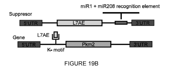
crnsihCD25
(Supplemental Fig 8) overcomes the challenge of FACS sorting of adult
transfected
CMs.31 This study pioneers the use of cmsmodRNAs to manipulate cellular
behavior
and holds a great therapeutic potential for cardiac disease, as modRNA is a
safe,
transient, local, and non-immunogenic platform for gene transfer.
[0061]The field of cardiac gene therapy is expanding, yet its use in the
clinical setting is
limited. Currently the most widely used method for targeting gene expression
to the
heart is through viral vectors, particularly the adeno-associated virus (AAV)
vector (1-3).
During the past few decades several attempts were made to insert genes of
interest
into CMs using adenovirus, associate adeno virus (AAV), lentivirus and DNA
plasmid.
While both AAV and adenovirus possess high CM transfection levels, lentivirus
and
DNA plasmid CMs transfection efficiency is low. Adenoviruses can elicit a
robust
immune response, leaving only AAV as a suitable option for gene delivery
system to
the heart. Using CMs-specific promoters in AAV may allow for cell-cycle
inducers gene
expression strictly in CMs, however its pharmacokinetics in the heart
(expression starts
at day 4 and remains for at least 11 months) may lead to uncontrolled growth
and
hypertrophic cardiomyopathy and HF (3, 5). Additionally, over 60% of healthy
human
individuals possess neutralizing antibodies directed against the AAV capsid
that can
efficiently neutralize gene expression delivered by this method (21). Viral
gene therapy
shows promise yet its applications are limited due to its length of expression
and
inability to regulate gene expression in a quantifiable dose manner (1-3).
[0062]While the use of unmodified exogenous RNA as a gene delivery method is
appealing because it may be safer than plasmid DNA owing to a reduced risk of
genomic integration, it is ineffective due to its instability outside the cell
and the strong
innate immune response it elicits when transfected into cells (10,11).
[0063]Kariko et al. discovered that the substitution of Uridine and Cytidine
with
Pseudouridine and 5-methylcytidine, respectively, drastically reduced the
immune
response elicited from exogenous RNA (11,12). In order to increase stability
and
19
CA 03036710 2019-03-12
WO 2018/053414 PCT/US2017/052035
translational efficiency, a 3"-O-Me-m7G(5')ppp(5')G Anti Reverse Cap Analog
(ARCA)
cap is substituted at the 5' end of the RNA molecule (4,5,10). Modified mRNA
(modRNA) therefore provides a novel and effective gene delivery method that
provides
short-term (1-2 weeks), titratable gene expression for use both in vitro or in
vivo (4-9).
[0064]Modified mRNA (modRNA) has emerged as an effective and safe tool for
somatic gene transfer, and has been successfully used by us and others for
gene
delivery to the heart.10,12-15 Here we show that Pyruvate Kinase Muscle
lsozyme M2
(Pkm2), a pro-proliferative factor, frequently dysregulated in cancer,16,17 is
highly
expressed in regenerative fetal and early neonatal CMs, but not in adult CMs.
Restoration of Pkm2 levels using modRNA delivery exclusively into adult CMs
(cmsPkm2) post-MI significantly and exclusively induced CMs proliferation, and
was
associated with improved cardiac function, reduced scar size, increased heart
to body
weight ratio, reduced CMs size, reduced apoptosis and increased capillary
density.
Those regenerative processes translated into increased long-term survival post-
Ml.
Using lineage tracing and isolation of Pkm2-transfected CMs followed by gene
expression analysis post-MI we show an increase in number of Pkm2-transfected
CMs
colonies and the potential involvement of key downstream effectors of the pro-
proliferative cytoplasmic (via the pentose phosphate pathway (PPP) 18,19,
) and nuclear
(via trans-activation of 8-catenin and Hif1a20,21) functions of Pkm2. Our
results show
that a short pulse of a pro-proliferative gene, using a highly translatable,
clinically
adaptable platform is sufficient to induce CM proliferation and cardiac
regeneration.
Those findings underline the therapeutic potential of cnisPkm2 modRNA in
cardiac
disease.
[0065]It has recently been shown (1) that by using modified mRNA (modRNA)
technology, modRNA can drive a transient, safe gene expression in the heart
with high
transfection levels without eliciting immune response or compromising the
genome(5,
22). Exogenous unmodified mRNA that enters the cell via the cell membrane is
recognized by endosomal Toll-like receptors 7/8 and 3(23, 24). This process
inhibits
protein translation and activates the innate immune response, ultimately
leading to
apoptosis of the hosting cell. ModRNA is synthesized by substituting
ribonucleotides
with naturally modified ribonucleotides. The use of these modified
ribonucleotides
results in changing the secondary structure of the synthesized mRNA, which
prevents
the Toll-like receptors from recognizing the modRNA and therefore permitting
its
CA 03036710 2019-03-12
WO 2018/053414 PCT/US2017/052035
translation to a functional protein by the ribosomal machinery without
eliciting immune
response or compromising the genome (5, 22).
[0066]Applicants previously showed that modRNA transfects different cell types
in the
heart including CMs with high efficiency, leading to immediate and high levels
of protein
expression in a transient, pulse like kinetic (duration of 3-5 days in vitro
and 7-10 days
in vivo). Co-transfection of two individual modRNAs resulted in co-translation
of both.
Using the MI model (5) and Luc, LacZ and nGFP modRNAs delivery in myocardium,
Applicants show that the cardiac tissue after MI is well transfected with
modRNA and
several cell types such as CMs and non-CMs are highly transfected in the left
ventricle.
Applicants then selected several candidate cell cycle inducer genes that had
previously
been shown to have the ability to induce neonatal CMs during cardiac
development
(CDK2, 13 catenin) (16) or reactivation of adult CMs proliferation in
transgenic mouse
models (CyclinD2, cMYC)(12, 14) and others that had shown robust proliferative
potential in different organs and cell types but had never been tested in
cardiomyocytes
and heart (Lin28, PKM2)(24, 25).
[0067] Generally, a platform for making cell specific modified mRNA (modRNA)
is as
follows.
[0068] First, choose a cell type of interest for making cell specific modRNA.
Identify
candidate microRNA (miR) that have been reported to express in the cell of
interest and
preferably only in the cell of interest (e.g., in the case of cardiomyocytes,
miR1, miR29,
miR126, miR133, miR199, miR208, miR378). Identify reverse complement sequences
for each miR sequence that allows recognition of the specific miR to this
sequence.
Add to 3'UTR each of the previous calculated miR reverse complement sequence
to
ihCD25 k motif, a truncated receptor for hCD25 carrying a k motif. This allows
ihCD25
to express only in those cells that are lacking the specific miR that the
reverse
complement sequence is targeting.
[0069] Co-transfect a mixture of cells that contains the cell of interest and
other cell type
(e.g fibroblasts) as well with nGFP modRNA and with different miR-ihCD25
modRNAs.
After about 18 hours, fix the cells and stain the cells for GFP (show
transfected cells
with modRNA) and for reporter gene (with anti hCD25, show cells that are
lacking the
miR that was target) and cell specific markers (e.g., for cardiomyocytes
Troponin I, for
endothelial cells, Pecam1, etc.).
21
CA 03036710 2019-03-12
WO 2018/053414 PCT/US2017/052035
[0070] Identify GFP-positive cells that are also positive for cell specific
marker (e.g
Troponin I for cardiomyocytes) but negative for reporter gene (hCD25). This
means that
this specific miR-ihCD25 was not translated although the modRNA was delivered
to this
cell type. This will indicate that this miR is specifically expressed in the
cell type of
interest and can be used to create cell specific modRNA. Create cell specific
modRNA
by adding to the 3'UTR of L7AE the sequence that inhibits ihCD25 in the cell
of interest.
Co-transfect with mir-L7AE and gene of interest that carrying in his 5' UTR k-
motif.
These two modRNAs will allow you to specifically deliver a gene of interest to
a specific
cell type.
[0071] In one embodiment, Applicant designed and generated modRNAs for each of
the
above genes. Using rat neonatal CMs, Applicant tested the translation of each
modRNA. In addition, the functionality of the protein was tested by measuring
the
proliferation rate of rat neonatal CMs with control and the candidate cell
inducer
modRNAs. All candidate cell cycle inducer modRNAs increase the proliferation
of
neonatal rat CMs and adult CMs proliferation after MI to various extents. Both
Lin28
and PKM2 significantly increased CMs proliferative capacity. Therefore, those
genes
were selected for further investigation.
[0072] Lin28 is a known suppressor of Let7 that tightly controls cell cycle
regulators (25-
29). To test whether Lin28 induces cell cycle regulators, nGFP (control
modRNA) or
Lin28 modRNA was injected immediately after LAD ligation and found a
significant
increase in the expression of Ccnb1, Ccnb2, Cdc20, Cdk1 and Aurka cell cycle
genes
after 3 days using RT-PCR. The use of cell cycle inducer modRNAs such as Lin28
modRNA in a non-specific manner increases proliferation not only in CMs, but
also non-
CMs representing an experiential challenge since the model and hypothesis were
aimed to test aimed to test CMs proliferation as a mean to achieve increased
cardiac
regeneration.
[0073] To address this challenge Applicants designed a CM-specific modRNA
system
that is based on two distinct modRNAs (Fig 5). The first construct is a
suppressor
modRNA the carries L7AE, an archaeal ribosomal protein that regulates the
translation
of a designed gene of interest modRNA with kink-turn motif- a specific binding
site for
L7AE(30, 31). Translation of L7AE modRNA will suppress the translation of the
22
CA 03036710 2019-03-12
WO 2018/053414 PCT/US2017/052035
designed gene of interest modRNA when the two are co-transfected into the
cell. By
adding a CMs-specific microRNA (miR) recognition element to the L7AE gene, we
are
able to prevent L7AE translation in CMs that abundantly and mostly exclusively
express
the miR ("suppress the suppressor" approach) allowing the translation of the
gene of
interest modRNA strictly in CMs. It was shown previously, using miR
recognition
element, results in a reduction of the number of copies of the targeted miR
(32,33).
Reduction in number of miRs in the heart can be detrimental or beneficial to
the heart
(33-42). In our approach we need to be sure we don't reduce miR expression
that is
beneficial to cardiac regeneration but rather reducing miR expression that is
detrimental
to cardiac regeneration. miR1-2 (miR1), miR208a (miR208) and miR199a (miR199)
are
expressed mostly in CMs (33, 39, 41). miR1 and miR208 were found to be
upregulate
after MI in adult animal study and humans (33, 38, 41, 43). miR1 and miR208 up
regulation has detrimental effects, while its down regulation has beneficial
effects after
MI and heart diseases (32-42)
[0074] To test the expression of these miRs in CMs, we have made an inactive
human
CD25 (ihCD25) gene, a truncated gene containing only the extracellular domain
(ECD)
of hCD25- as a reporter gene. We have designed two versions of the ihCD25
construct,
with or without the miR recognition elements for miR-1, miR-208 or miR-199. We
then
transfected the modRNAs into neonatal CMs in vitro and in vivo using the MI
model (Fig
2). As can be seen in Fig 6 both miR-1 and miR-208 were found to be CM-
specific, as
translation of ihCD25 was observed in non-CM but not in CMs. In contrast,
modRNAs
with or without miR-199 recognition element was found not to be CMs specific,
in vitro
and in vivo. Next, we designed a L7AE modRNA that carries both miR-1 and miR-
208
recognition elements (L7AE miR-1 + miR-208). We have also generated a nuclear
GFP
modRNA (nGFP ¨ k-motif) and a destabilized Cre recombinase (DD-Cre ¨ k motif)
modRNAs that includes the k-motif (L7AE recognition site). Using our adult
mouse MI
model, we show that transfection of nGFP ¨ k motif resulted in the translation
of nGFP
in both CMs and non-CMs (Fig 7). However, when nGFP ¨ k motif was co-
transfected
with L7AE miR-1 + miR-208 only CMs translated the nGFP. In addition, co-
transfecting
L7AE miR-1 + miR-208 with a DD-Cre ¨ k motif in a MI model using
Rosa26Tdt0mat0
resulted in gene activation (Tomato fluorescence) strictly in CMs. The
combination of
these two methods allows us to elegantly express our gene/genes of interest
exclusively in CMs, and to allow for linage tracing over longer time periods
after the
gene of interest modRNA is no longer expressed.
23
CA 03036710 2019-03-12
WO 2018/053414 PCT/US2017/052035
[0075]To test the functionality of our CMs-specific modRNA, we directly inject
Luc
control modRNA or Lin28 ¨ K and PKM2-k motif modRNA alone or together with
L7AE
miR-1 + miR-208 (Lin28 /PKM2 CMs specific modRNA) using our MI model. Seven
days post transfection we measured the proliferation (using hallmark
proliferation
markers, BrdU, Ki67, H3P and Aurora B) of both CMs and non-CMs. As depicted in
Fig
8 Lin28-k or PKM2-k motif modRNA alone significantly increased proliferation
of both
CMs and non-CMs (P<0.001) in comparison to Luc modRNA. However, Lin28 and
PKM2 CMs-specific modRNA significantly reactivated the proliferation of only
CMs
(P<0.001), with no significant influence on the proliferation of non-CMs.
Importantly,
since L7AE is not a mammalian protein, to test the immunogenicity of L7AE
after MI we
have injected Luc control modRNA or L7AE modRNA with or without miR-1, miR-208
or
both in adult mouse MI model. As can be seen in Fig 8 we did not witness
significant
elevations in immune response and increased apoptosis with all L7AE modRNAs
after
7 day post MI. We concluded that the use of L7AE in mice is immunologically
safe.
Plasm ids
[0076]pTEMPLZ is a cloning vector into which an ORF of interest can be
inserted
between the UTRs. In one embodiment, plasmids for use in the disclosed method
include those shown in Table 1.
Table 1
1 No miR ¨ L7AE
TTGGACCCTCGTACAGAAGCTAATACGACTCACTATAGGGAAATAAGAGAGAAAAG
AAGAGTAAGAAGAAATATAAGAGCCACCatgtacgtgag atttgaggttcctgagg acatgcagaacg
aagctctgagtctgctggagaaggttaggg ag agcggtaaggtaaag aaaggtaccaacg agacg
acaaaggctgtg
gag ag ggg actggcaaagctcgtttacatcgcag
aggatgttgacccgcctgagatcgttgctcatctgcccctcctctgc
g agg ag aag aatgtgccgtacatttacgttaaaagcaagaacg accttgg aagggctgtgg
gcattgaggtgccatgcg
cttcggcagcg ataatcaacg aggg agagctg ag aaag gagcttgg aagccttgtggag aag
attaaaggccttcag a
agtaaGCTGCCTTCTGCGGGGCTTGCCTTCTGGCCATGCCCTTCTTCTCTCCCTTGC
ACCTGTACCTCTTGGTCTTTGAATAAAGCCTGAGTAGGAA (SEQ ID NO: 1)
2 miR 1 - L7AE
TTGGACCCTCGTACAGAAGCTAATACGACTCACTATAGGGAAATAAGAGAGAAAAG
AAGAGTAAGAAGAAATATAAGAGCCACCatgtacgtgag atttgaggttcctgagg acatgcagaacg
aagctctgagtctgctggagaaggttaggg ag agcggtaaggtaaag aaaggtaccaacg agacg
acaaaggctgtg
gag ag ggg actggcaaagctcgtttacatcgcag ag g atg ttg acccg cctg ag a tcgttg
ctcatctg cccctcctctgc
g agg ag aag aatgtgccgtacatttacgttaaaagcaagaacg accttgg
aagggctgtgggcattgaggtgccatgcg
cttcggcagcg ataatcaacg aggg agagctg ag aaag gagcttgg aagccttgtggag aag
attaaaggccttcag a
agtaaTACATACTTCTTTACATTCCATACATACTTCTTTACATTCCATACATACTTCTTT
24
SZ
mepe66666ee336e3eee6e6643464364eole3643e6e36e664e6643346e364ee3366464e646e36
6ee643646333e333363e336ee beee 64e36e 6e 664364e 6e3e3e33646434e3464336ee
666436e 6
mee364e 6366634e 64e 64e bee6e34366 433443466ee 6e
6e3643344e6e6pe3666433e646646343664
664eo3e 6664e 646e336 6e 664434e 6e 64e644466e363346366ee64eome 6e
634eeee36eoleole bee
oleoee 6ee3666ee 6e 6e 666436466ee 66e346ee
64e36463e6336e366ee36334eomole364446466
woe 66464e 66e36e 66466664446ee 6433e 66e3oleoe 66eeee
66346464363333434e6646436436366
6336433ee6463666ee6ee36e3666443343664664ee 6e 66466e 663e6466433443e6436366eee
6e 6
bee 6466e3643e3434e34366 64e 63e 66463ee 6ee36e366 6466e 6646 6466ee36434e3ee
beeoep
e66436646pole3ee 6e 63e 64646ee be 664e3e44363eeoe664333eolebee64343e336e
666ee bee 6
p6e 66466e 643633e36636e3666eeoleope 6643e 66334e be 6433e
666eee3me6643436646636
64334633eppilemow6434e3644436eee 6e3e336ee64633464ee beeoleooe be
6e364e33e46e64
eoome 664e3ppmee6436634366464ee 64ee664346eepe 64e be
66ee64364e6e6646434e63334436
4336
66ile33e464pe34e36643e3ee36333663e34e3333364343e6pe3e664336336464e3e3ee66433
4433e3e643664e336e364e33436e36e333e 6e3pe34433643e 66 6e36ee
646e3e3e336ee63364e 0
OV009VVV99000V9199VVV9001V91909999101V99000V9199VVV900
1V9190999199VVV999V1V10V010V90V1VV109VV9V0V1901000V9911
Plow >1 ¨ ZLID1d 9
(;:ON CI 1 02s) VVO9V19V91009VVVIVV01110199110100V19100V
09110001010110110 0091V00 9 0101100 01109 9009101100 910 De 644e
e 6e3336ee6e3334364333643336e3e334e 6e bee 66e bee 66e66633443e43364336eee
666e3pp
6e3333666e36e33366ee643e3346463433664664e4e33ee34e36eee33644443e33646ee6ee3336e
3333e336436ee364ee 66ee3364e34e33e6e436664663643ee3e436466e3e be 66eee334e
bee636
ee 6e364e3ee 6 ee 666eeee336636636e 646e
66644e46pll64646646643336643e34646334e3olee 6
6434666ee3364346ee 6eep433e3p6e 66466366e 64666e
66ee34336ee63344666ee664e3e36436
ee36e 6eme36464443463e 66466333333e 634363634666643636333633e
64e4346433443663446666
4e3636463ee3446646ee46434e3663366663e36436436e36336e 63e
6e36636e633366363e6433
6336366e 66e 6e336366ee 6e 6
636e366ee336364366466e364446e36e33ee33464663436664e 0
OV009VVV99000V9199VVV9001V91909999101V99000V9199VVV900
1V9190999199VVV999V1V10V010V90V1VV109VV9V0V1901000V9911
Plow >1 ¨ 8Zun
(17 :ON CI 1 02s) VV99V19V91009VVVIVV91110199110100V191
00V091100010101101100091V00 99101100 9110999909101100 910 91
V1101901091111109VVOVIV1101901091111109VVOVIv11oie01091111
100VVOVIV1101901091111109VV3VV0011V0V1110110VIVOVIV0011V0V
1110110VIVOVIV0011V0V1110110VIVOVIV0011V0V1110110VIVOVIee4be
e6e3443366eee44e bee 6e664644336ee 664436e 6 beee be 6436e 6e 666e 63ee34ee4e
636e3663443
6364e336466e6pe36664643666ee664433e63ee6ee36eeee4463e444e3e46336464ee bee be
66e 6
364343343333 6434eop 644634e 6e 64336333e 6116 le 6 6e 6 eo 634e3e4p63436eee
36 643e 6666e be 6
6464366eee3e 63e 6e 63ee33e466eee beee466ee46636e be
666e4466ee6e664364346e643436ee
60ee 6e064e0e 66e64004466e6Me be 6460e464e 00V009V9VVIVIVVV9VV9VV19V9VV
9VVVV9V9V9VVIVVV999V1V10V010V9OVIVV109VV9V0V1901000V9911
EVL1 ¨ e8OZ ¨ w
( :ON CI 1 02s) VVO9V19V91009
VVVIVV01110199110100 V10100V0 011000101011011000 91V00 991011
00911099990910110091091V1101901091111109VV0V1V1101901091
11110 evvo VIV110100100111110 9VVOVIV1101901091111109VVO Veelbe
e6e3443366eee44e bee 6e664644336ee 664436e 6 beee be 6436e be 666e 63eeoleele
636e3663443
6364e336466e6pe36664643666ee664433e63ee6ee36eeee4463e444e3e46336464ee bee be
66e 6
364343343333 6434e343 611634e 6e 643o 6333e 64464e 6 6e 6e3 634eoem6343 beee 6
643e 6666e be 6
6464366eee3e 63e 6e 63ee33e466eee beee466ee46636e be
666e4466ee6e664364346e643436ee
60ee 6e064e0e 66e64004466e6Me be 6460e464e 00V009V9VVIVIVVV9VV9VV19V9VV
9WVV9V9V9VVIVVV999V1V10V010V90V1VV109VV9V0V1901000V9911
EVL1 ¨ e8OZ w
(Z :ON CI 1 02s) VV99V10V9
1009VVVIVV91110109110100V19100V001100010101101100091V009
910110001100990001011000100V0011V0V1110110VIVOVIV0011V0V
SEOZSO/LIOZSIVIDd titS0/8I0Z OM
ZT-0-6TOZ OTL9E0E0 VD
9Z
Lao& Aow->i d jou peloefu! enn welsAs (speeq oneu6e w) 6unJos peseq -(gNI 04)
gNI 0
uewnq (pe woP Jelnileoave Apo) emoeu! annenouu! gwvµ sV\10 peloelsuail Apo
eposi
o pesn eq ueo siAjo Li! Alen!snioxe IseJew! 40 seue6
uo!ssathe eue6 Tue!suag
e SMOII eLfl u6!sep peseq-VNHPow o!4!Oeds-s1A10 lenou Jno JeLgeqm Tsel
01ELLOO]
BUIVOS flea aypeds-no paseq-wvelpow gzaw
8int
(d10) awaid 6u!peej ued0
aLn rs
:ON 01 OES N
(8 :ON CII OES) vv9
OVIDV01.000VVV1VVOLL10.1.0911.0100V10.1.00V0011.00010101.1011.000
elV009010 33e11099990910110091095ePlee3ee6ee6e46e6ee66e6e0e6
e 6636e366433e3lo 66646e64334334334636eole64364334m64643 66336646e
36e466eoolole 6e 3e
e3eme4e33463e 6e 664emee3643 6 64eee 6e3e 6eoeleeeoppe 6e3ee3ee3e34634336pop3e
be 6
46e6e6433463366ee633336ee366eolo36ee6e6ee6466eoom6e346eme6e664eee6466e3e36
4e4eol36e33336eoo3e66466ee3ebee6663eoo3e64eeee36434636e6e6436433466e6e3e3eplo
6 66e3me 666e33463646eolellem664e 6e36666466463pleo3emee6e be 6e3eoo6ee64eeee
666
wooeoolooee666e3643e3466eoopoo6e636eeooe6646e3o6e364eeoo46eee364eee6e3emee
ee66eeebeee6eoeebee6looeeolooeoe646eeoeee6oeeoeoee66343e33643436eeoeo6leeol
64eeome3e666433463peoo6elopeee66e3e46434364e4elo43eo466636eeeemee6e363344466e
6e6ee364ee6464oee61164emee66ee66ee3e400664e3o6eeeope3eoo63e3e000le6e6e33633
3e 64e 63e64643436e 6e 366e336436643364664eoleop6oeolo6loe 6
666464e643643oeleope 65jeo
OVOODVVV99000VD1DOVVV9001V91909999101V99000VD1DOVVV900
1V9190999199VVVODOV1V_LOVOlOVDOV1VV_LOOVVOVOV1901000V9911
pewpow uou ¨ plow >1 gzaou! 8
(L :ON CII OES) VVO
DV1DVD_LOODVVV1VV911101991101.00V19.100V091.1000101.011Ø11.000
eiv000eionooatioeeeeoeionooDIODeele466eeebebeebeeeeeeoole6466
e466eee6e6e e beeeeeeoole 6366e466eee be bee beeeeee3ole6e6
66ee3e46436e63e664e36
6olopeole66633633633e6463446e664364334664eoeole63636ee be 63eemooe beee36e6poo
63346e000eo6e6loomoeomeoe63336436p6463333663e636634e00000eoee6e36eoomoeoo
e63363436e364636e3663e6 6e6ole3ee3eoo63olebee3pee6466eeo3e3663ee bee 6e36ee3e
633664e34e4e43463ee3e336e3ee3e43ee3e46e66436ee3e366664334e3ee3663e66e66ee3p3e
634e3666ee6436e634e3633ee 64664333e3e63666e63446ee6466e633636333e6ee3e43ee366
3e63e66ee3pp34e33e3636e66e33463e4366ee633364e3363346ee3443443e63e36e36ee64e3
eme60000mo6336e3443646e3646366oelooe6l000eme646opooe0006643336463336436eeo6
600emeo6434e3446ee6l000e6436ee366oelooe3364e63666e63666e636633464636e3446eeoe
33663eee463e 63663e 66436e63466p34e33364664666633e3446436e 66e 63666ee36e
64664e0
OVOODVVV99000VD1DOVVV9001V91909999101V99000VD1DOVVV900
1V9190999199VVVODOV1V_LOVOlOVDOV1VV_LOOVVOVOV1901000V9911
Plcul >1 ¨ ddeonu z
(9 :ON CI 1 02s) VVODVIDV01.009VVV1VVOL1101.90.11.0100V1.9
1.00V091.1000101.01.1.01.100091V0090101100011.00999001.011000100
e6llooe46433646e4646364eooeoeemeope6643436643336366466633e643646ileo46 6464e
be 666
ee6ee34op366e633366ee3664464e664e3366p3eee46463p33e63464e66e64366643364ee6436
463364e 66ee46464364643334434e3664633e46434e33366e336343643e
6e33334eee6343e64633644e
34e4334366634333633e466e336646ee33e343646e66e3664346ee33e34364634eile33666646e3
6
43646ee3443343366e6646336466646336336436ee6e3e3333e636e33epe333636643363363343
ee66e63pe43
6e36443e33m34e3364366e6e366e6e6333644e6433e36e364e3634464366e664343
SEOZSO/LIOZSIVIDd titS0/8I0Z OM
ZT-0-6TOZ OTL9E0E0 VD
CA 03036710 2019-03-12
WO 2018/053414 PCT/US2017/052035
ihCD25 k-motif modRNA in heart after MI, cells were Isolated and sorted out
with
CD25-specific magnetic beads. Both nGFP-positive CM's and non-CM's were
observed. When nGFP k-motif, ihCD25 k-motif with L7AE miR1 -miR208 were co-
transfected and without magnetic separation, CM specific nGFP expression was
seen.
The culture also contains nGFP-negative CMs and non-CMs. When magnetic
separation was applied, only pure nGFP-positive CMs were observed (Figure 9).
[0078] In one embodiment, production of exogenous genes is driven by
expression of
anti-miRs from a first replicon that also encodes a repressor protein.
Expressed anti-
miRs bind miRs that occur naturally in human and primate cardiomyocytes and
transcription of the repressor protein is prevented. In the absence of
repressor protein,
expression of a gene of interest from a second replicon encoding the gene and
containing the repressor protein recognition site can proceed.
Cell Cycle Inducer Genes
[0079] Expression of a gene of interest, for example, a proliferation-inducing
gene can
be made cardiomyocyte-specific by placing transcription/translation of the
gene under
the control of a transcription/translational regulatory system in which one of
a pair of
nucleic acids encodes an anti-microRNA (anti-miR) that binds specifically to a
target
cardiomyocyte-specific miR. A second nucleic acid translation suppressor
protein and
a second nucleic acid that comprises a suppressor protein interaction motif
that binds
the translation suppressor protein and a gene that encodes a protein of
interest.
[0080] Using the method described herein, the expression is transient,
avoiding the
problems associated with unlimited expression, such as hypertrophy.
[0081] Repressor/suppressor protein that binds a specific RNA motif inserted
in the 5'-
untranslated region of an mRNA modulates the translation of that message in
mammalian cells. The expression specificity to human and primate
cardiomyocytes is
achieved by the inclusion in the repressor/suppressor oligonucleotide of a
sequence
that encodes a recognition element specific to endogenous miRNAs found in
mouse,
pig, human and non-human primate cardiomyocytes.
27
CA 03036710 2019-03-12
WO 2018/053414 PCT/US2017/052035
[0082] The synthesis of modRNA for in vivo use involves four stages: DNA
template
creation containing the desired transcript, in vitro transcription (IVT), 5'
phosphate
removal with Antarctic phosphatase, and precipitation with 5M ammonium acetate
salt.
Investigation into the use of modRNA for experimental and clinical purposes is
growing
rapidly. Daily transfection with modRNA encoding reprograming factors OCT4,
SOX2,
MYC, and KLF4 were successful at reprogramming human fibroblasts back to
pluripotency (5,8). Additionally, modRNA has been shown to be capable of
directing cell
fate in vitro by using MyoD modRNA that resulted in the conversion of
fibroblasts to
skeletal muscle cells (2). ModRNA has also shown promise in directing cell
fate in vivo.
The expanding use of modRNA technology in vivo and its potential use in the
field of
cardiac gene therapy motivated us to generate a step-wise, streamlined
protocol for the
effective synthesis of modRNA for in vivo use.
Cardiomyocytes
[0083] In one embodiment, the present disclosure relates to a method of
treating a
subject following myocardial infarction (MI) or heart failure (HF) in a
subject comprising
administering an effective amount of a composition comprising at least two
synthetic
modRNAs to a subject in need thereof.
[0084] Protein expression lasts for from 5 to 20 days, in some embodiments
from 7 to
14 days, and results in a low immunological response as compared to non-
modified
RNA.
[0085] Inter alia, the present disclosure describes a new set of candidate
cell cycle
inducer genes: Lin28, Pkm2, and Cyclin D2, which when delivered as modRNA, can
reactivate mammalian cardiomyocyte (CM) proliferation in vivo (without
increasing CM
size or nuclei number), reduce CM apoptosis and increase overall left
ventricle
vascularization post myocardial infarction (MI). When expression of the cell
cycle
inducer genes is placed under the control of a transcriptional/translational
regulatory
(an expression regulatory) system for cardiomyocyte-specific transcription
(expression),
the result is a tool for cardiomyocyte specific expression of the cell cycle
inducer gene-
driven proliferation following injury, for example, as the result of
myocardial infarction
(MI) or heart failure (HF).
28
CA 03036710 2019-03-12
WO 2018/053414 PCT/US2017/052035
[0086]Modified mRNA (modRNA) is a safe, efficient, transient, and non-
immunogenic
gene delivery system that allows one to investigate the effect of cell cycle
inducer
genes on CMs following MI or HF. Kariko et al. discovered that the
substitution of
uridine and cytidine with pseudouridine and 5-methylcytidine, respectively,
drastically
reduced the immune response elicited from exogenous RNA (11,12). Investigation
into
the mechanism revealed that the nucleoside substitutions resulted in a
conformational
change in the RNA that caused reduced response by toll-like receptors 3, 7,
and 8
(TLR3, TLR7, TLR 8), and retinoic acid-inducible gene 1 (RIG-1) (13). A
further
decrease in RIG-1 response from modRNA was seen upon removal of the 5'
triphosphates (4,10). In order to increase stability and translational
efficiency, a 3"-O-
Me-m7G(5')ppp(5')G Anti Reverse Cap Analog (ARCA) cap is substituted at the 5'
end
of the RNA molecule (4,5,10).
Cell selection by anti-hCD25 affinity
[0087]Cell selection of cardiomyocytes by traditional FACS cell sorting can be
problematic due to the size of the cells. An alternative approach to cell
selection was
devised. In one embodiment, using the transcription regulatory system of the
disclosure, a nucleic acid that encodes the hCD25 extracellular domain (ECD)
is
included in the construct that contains the nucleic acid that encodes the gene
of
interest. To isolate CMs that transiently express either control or candidate
gene
modRNA, cells that co-express the hCD25 ECD plus the gene of interest are
selected
using an anti-CD25 ECD antibody in an affinity chromatography column or using
a
panning method. These cells are used to generate gene expression profiles
using
RNA-seq technique, and identify differentially expressed genes
EXAMPLES
Example 1: Materials
[0088]The following materials are used in conjunction with the disclosed
method.
[0089]All solutions should be made in Nuclease Free water unless otherwise
specified.
All materials used in this protocol should be nuclease free.
[0090] Equipment used includes the following:
1. PCR therm ocycler
2. Microfuge
29
CA 03036710 2019-03-12
WO 2018/053414 PCT/US2017/052035
3. Vortex mixer
4. Thermomixer(EPPENDORFTm)
5. Nano-Drop
6. Nuclease-free water
7. 15 ml Nuclease Free conical tubes
8. Nuclease free strip PCR tubes
9. Ethanol (100% and 70%)
10.2 ml Ambion Elution Tubes
[0091] Primers used for tail PCR are as follows:
Forward Primer: 5'-TTG GAC CCT CGT ACA GAA GCT AAT ACG-3' (SEQ ID NO: 9)
Reverse Primer: 5'-TTT TTT TTT TTT TTT TTT TTT TTT TTT TTT TTT TTT TTT TTT
TTT TTT TTT TTT TTT TTT TTT TTT TTT TTT TTT TTT TTT TTT TTT TTT TTT TTT
TTT TTT TTT TTT TTT TTT TTT TTT TCT TCC TAC TCA GGC TTT ATT CAA AGA
CCA-3' (SEQ ID NO: 10)
[0092] Construction of DNA template for in vitro transcription using pTEMPLZ
plasmid
is as follows:
1. T4 Polynucleotide kinase enzyme
2. 100mM ATP
3. 2X KAPA HiFi HotStart ReadyMix PCR master mix
4. Ale/enzyme
5. Afel enzyme
6. Antarctic phosphatase enzyme
7. T4 DNA ligase enzyme
8. One Shot ccdB Survival TM 2 Ti Phage-Resistant (T1R) cells
9. QIAquick gel extraction kit
10. QIAquick PCR purification kit
11. One shot chemically competent E. coli
12. QIAprep spin Miniprep kit
13. 10 x Phosphorylation Buffer
CA 03036710 2019-03-12
WO 2018/053414 PCT/US2017/052035
[0093] Synthesis of linear DNA template with a poly T tail for IVT reaction
1. 2X KAPA HiFi HotStart ReadyMix PCR master mix
2. Primer Solution: 1 pM each of forward and reverse primer
3. Dpnl enzyme
4. QIAquick PCR purification kit
[0094] In vitro transcription reaction
1. Ambion T7 Megascript Kit (life technologies Cat#: am1334-5)
2. GTP 75 mM solution (provided in Megascript kit)
3. ATP 75mM solution (provided in Megascript kit)
4. CTP 75mM solution (provided in Megascript kit)
5. 5-Methylpseudouridine-5'-Triphosphate (Trilink)
6. Trilink Biotechnologies Anti Reverse Cap Analog, 3'-0-Me-m7G(5')ppp(5')G 10
pmoles (Cat #: N-7003)
7. T7 TURBO DNase enzyme (provided in Megascript kit)
8. Ambion MEGAclearTM Transcription Clean-Up kit. (life technologies; cat#:
AM1908).
[0095] RNA phosphatase treatment
1. Antarctic phosphatase enzyme
[0096] RNA precipitation
1. 5M Ammonium Acetate Salt solution (Provided in AmbionMEGAclearTm Kit)
2. Elution Buffer (Provided in AmbionMEGAclearTm Kit)
[0097] Preparation for modRNA injection
1. Lipofectamine RNAiMAXtransfection reagent (Thermofisher Cat#: 13778150)
2. OptiMEM Reduced Serum Medium, no phenol red
3. Ultra-Fine insulin syringe needle 31g 8mm
Methods
31
CA 03036710 2019-03-12
WO 2018/053414 PCT/US2017/052035
[0098] The following methods are used in conjunction with the disclosed
method. All
procedures were carried out at room temperature, in a non-sterile environment
unless
otherwise specified. All materials used should be nuclease free.
Synthesis of modRNA
[0099] ModRNAs were transcribed in vitro from a plasmid templates using a
custom
ribonucleotide blend of anti-reverse cap analog, 3"-O-Me-m7G(5')ppp(5')G (6
mM,
TriLink Biotechnologies), guanosine triphosphate (1.5 mM, Life Technology),
adenosine
triphosphate (7.5 mM, Life Technology), cytidine triphosphate (7.5 mM, Life
Technology) and N1-Methylpseudouridine-5'-Triphosphate (7.5 mM, TriLink
Biotechnologies) as described previously.ALu mRNA was purified using megaclear
kit
(Life Technology) and was treated with antarctic phosphatase (New England
Biolabs),
followed by re-purification using Megaclear kit. mRNA was quantitated by
Nanodrop
(Thermo Scientific), precipitated with ethanol and ammonium acetate, and
resuspended
in 10 mM TrisHCI, 1 mM EDTA. For a detailed protocol please see our recent
publication.
[00100] modRNA transfection. In vivo transfection of modRNA was done using
sucrose citrate buffer containing 20p1 of sucrose in nuclease-free water
(0.3g/m1), 20p1
of citrate (0.1M pH=7; Sigma) mixed with 20p1 of different concentrations of
modRNA in
saline to a total volume of 60p1. The transfection mixture was directly
injected (3
individual injections, 20p1 each) into the myocardium. For in vitro
transfection, we used
RNAiMAX transfection reagent (Life Technologies) that was used according to
manufacturer's recommendation.
Construction of DNA template for in vitro transcription using pTEMPLZ plasmid
carrying
k motif.
[00101] pTEMPLZ is a cloning vector into which an ORF of interest can be
inserted between the UTR's (Figure 1). The 5'- and 3'-UTRs are synthesized de
novo
by synthetic oligos. The synthesized UTR's are annealed together and amplified
using
forward and reverse primers. To provide an entry site for the ORF, Ale! and
Afel
restriction sites are introduced in between 5' and 3' UTRs. (The adenine
nucleotide (A)
of the 1st codon (ATG) can be omitted from forward primer sequence as it is
provided
by the Ale! site.) The PCR-amplified fragment and pZEr0-2 vector with
ampicillin
resistance are digested with Hind!!! and Not!, and ligated together to create
pTEMPLZ.
(pTEMPLZ plasmid and derivatives should be propagated in bacterial strain
resistant to
32
CA 03036710 2019-03-12
WO 2018/053414 PCT/US2017/052035
the ccdB gene product such as One Shot ccdB SurvivalTM 2 Ti Phage-Resistant
(Ti R) cells.)
[00102] Before insertion into pTEMPLZ, the ORF is amplified by using
phosphorylated forward and reverse primer pair for the gene of interest.
Phosphorylation of the primers is done using the T4 polynucleotide kinase
enzyme
according to reaction below:
x Phosphorylation Buffer 5 pl
Forward primer (100 M) 3 pl
Reverse Primer (100 M) 3 pl
100 mM ATP 0.5 pl
T4 polynucleotide kinase 10 U
Nuclease free Water 50 pl
Incubate reaction at 37 C for 1 hour.
To inactivate enzyme, the reaction is incubated at 65 C for 20 min. The
reaction is
diluted to 300 pl by adding 250 pl of water giving final 1 pM primer mixture.
[00103] Amplification of the ORF of interest is done using the PCR
reaction
below:
Primer mix (1 pM) from above i0 p1
Template DNA 1-100 ng
Water 50 pl
HiFi HotStart ready mix (2 x) 25 pl
The mixture is run in Thermocycler with settings according to Figure 2. The
amplified
target is isolated using Q1Aquick gel extraction kit. Before insertion of ORF,
pTEMPlz is
linearized and dephosphorylated. To linearize plasmid, pTEMPlz is digested
with Alel
and Afel according to the reaction below:
pTEMPLZ Plasmid DNA 2 pg
Nuclease Free Water 30 pl
10 x Buffer 4 3p1
Alel 5U
33
CA 03036710 2019-03-12
WO 2018/053414 PCT/US2017/052035
Afel 5U
Incubate in thermomixer for 1 hour at 37 C. Digest was purified using QIAquick
PCR
purification kit and eluted in 30 pl of elution buffer.
[00104] Linearized pTEMPLZ is dephosphorylated according to reaction
below:
Linearized plasmid from step 1 30 pl
x antarctic phosphatase buffer 5 pl
Antarctic phosphatase 5 U
Nuclease Free Water 50 pl
The reaction is incubated at 37 C for 1 hour. Enzyme is inactivated by
incubating at 65
C for 15 min.
[00105] Linearized and dephosphorylated plasmid is isolated using QIAquick
gel
extraction kit and the quantity of pTEMPLZ product is determined using
nanodrop.
Plasmid can be stored in -20 C for future use.
[00106] Blunt end ligation of ORF of interest is performed into pTEMPLZ
according to the reaction below:
Linearized dephosphorylated TEMPlz plasm id 50 ng
Amplified ORF 3-fold molar excess
10 x T4 DNA ligase buffer 2 pl
T4 DNA ligase 4 U
Nuclease Free Water 20 pl
Mix reagents and incubate overnight on melting ice at room temp or at 16 C.
Negative
control ligation reaction might be necessary to monitor self-ligation of
plasmid.
Transformation of plasmid is performed with competent cells and grow on an
ampicillin
agar plate.
[00107] To isolate positive clones with the correct orientation colony PCR
is
performed. Between 8-10 colonies are extracted from ampicillin agar plates
with a
pipette tip. Individual tips are stabbed in 200 pl of Luria Broth (LB) and
rinsed several
times in 75 pl of TE buffer under pH 8.0, tips are incubated in 37 C in a
shaker. Tubes
are then boiled for 5 min to lyse bacteria and spun to pellet debris. Colony
PCR is
performed with 2 pl of supernatant using forward primer and gene specific
reverse
primer. PCR sample is run on 1% agarose gel to identify clones with positive
orientation. 200 pl of LB is cultured with correct orientation clones in
larger volume of
34
CA 03036710 2019-03-12
WO 2018/053414 PCT/US2017/052035
LB overnight in a 37 C shaker and extracted using QIAprep spin Miniprep kit.
The
quantity of plasmid product is determined using NANODROPTM (Thermo Fisher
Scientific) and diluted to a concentration between 1-5 ng/ul.
Synthesis of tailed DNA template
[00108] A 1600 pl PCR master solution was prepared according to the
reaction
below:
Plasmid solution (1-5 ng/pl) (see Note 3) 400 pl
Primer solution (1pM primers) 400 pl
2X KAPA HiFi HotStart ReadyMix. 800 pl
50 pl of PCR master solution was aliquoted into 32 separate PCR tubes. PCR is
run
using the thermo cycler (setting listed in Figure 2). The length of elongation
step may
vary depending on DNA polymerase used and ORF length (If using 2X KAPA HiFi
HotStart ReadyMix, for example, elongation step of Thermocycler should be set
at a
ratio of 30 sec per Kb of ORF length.).
[00109] To digest methylated plasmid DNA, product is combined into one
EPPENDORFTM tube and digested with 30 pl of Dpnl. The PCR product is purified
using QIAquick PCR Purification Kit (Qiagen cat #: 28106) and the final
product eluted
in nuclease free water. The concentration of tailed product is measured using
nanodrop machine and concentration is adjusted using nuclease free water to
100-200
ng/ pl.
For quality control analysis, purity of Tailed DNA template product is checked
on a 1%
agarose gel together with the original DNA plasmid (Figure 3a).
In vitro transcription (IVT) reaction (1m1 reaction volume)
[00110] A custom NTP mixture is prepared in one EPPENDORFTM tube according
to Table 2. Reagents for IVT reaction are mixed in the following order into
one
EPPENDORFTM tube:
a. 400 pl of custom NTP's from table 2.
b. 400plof the DNA tailed template (200 ng/pl).
c. Vortex 10x Reaction Buffer from the T7 megascript kit to dissolve any
precipitate and add 100 pl.
CA 03036710 2019-03-12
WO 2018/053414 PCT/US2017/052035
d. Add 100 pl of T7 Enzyme. This will give you a lml 1VT reaction
e. Mix thoroughly and Incubate in thermomixer at 37 C for 4-6 hours.
30 pl of T7 Turbo DNase was added and mixed gently and then ncubated at 37 C
in
Thermomixer for 15-20 min to halt the reaction (see Note 5).
Purify reaction using Ambion MEGAclearTM Transcription Clean-Up kit and elute
each
tube three times with 50 pl of 95 C elution buffer to obtain 150 pl of RNA
product in
each tube. Combine the RNA mixture from each tube into one EPPENDORFTM tube.
RNA phosphatase treatment
[00111] Nuclease-free water is added to the RNA to obtain a 1.5m1
solution. 150
pl of Antarctic Phosphatase Buffer (10x) and 150 pl of Antartic Phosphatase
enzyme is
added, mixed thoroughly and incubated in thermomixer at 37 C for 1 hour.
RNA precipitation using ammonium acetate
[00112] The 1800 pl RNA solution is transferred to a 15m1 conical tube.
180 pl of
5M ammonium acetate is added and mixed thoroughly. 5200 pl of cold (-20 C)
100%
ethanol is added to solution and aliquoted into 3-4 2m1EPPENDORFTm tubes. Let
tubes stand in -20 C overnight. The tubes are centrifuged at 10,000 rpm for
30min at
4 C. The supernatant is then carefully discarded. Each pellet is dissolved in
500p1 of
70% ethanol. modRNA ethanol solutions from each tube are consolidated into 1
EPPENDORFTM tube. The tube is centrifuged at 10,000 rpm for 30 min at 4 C. The
supernatant is gently poured out and discarded, and using a kimwipe, the
inside of the
tube is gently cleaned. Care is taken not to disturb the pellet. The tube is
inverted and
let stand for no more than 2 min to air-dry pellet. Using a pipette, any small
drops of
ethanol left around the pellet are gently removed. The pellet is resuspended
using 45-
50 pl of elution buffer. modRNA is left in elution buffer for 5 min then
gently pipetted
until the pellet is dissolved. RNA solution can now be used in vivo, stored in
-20 C for
up to 6 months, or -80 C for 5 years.
ModRNA yield
[00113] Concentration is measured using nanodrop machine (Figure 3b). The
ratio of A260/A280 should be greater than 1.8 with values closer to 2.0
indicating higher
purity. Depending on yield, concentration should be close to 20 pg/ul. For
better quality
36
CA 03036710 2019-03-12
WO 2018/053414 PCT/US2017/052035
control analysis a 1p1 sample from the final modRNA solution is diluted in 100
pl of
nuclease free water. The sample is analyzed using a bioanalyzer machine
(Figure 3c).
Preparation of modRNA for myocardial injection in mice
[00114] 40 pl of RNAiMax is combined with 5p1 of OptiMEM in an EPPENDORFTM
tube and vortexed. The mixture is allowed to sit for 10 min at room
temperature. In
another EPPENDORFTM tube 150-200ug of modRNA is combined with 5p1 of OptiMEM.
The tube is spun down to eliminate liquid on the sides of the tube. After
letting the
RNAiMAX and OptiMEM mixture sit for 10 min at room temperature, the liquid
from the
tube with the modRNA mixture is added to the tube with the RNAiMAX mixture.
(In
some embodiments, it is important to add the modRNA mixture to the RNAiMAX
mixture and not the other way around.) The combined mixture is allowed to
stand for
15 min at room temperature. The mixture is extracted into a 31 gauge insulin
syringe
and injected into mouse myocardium. (Example of result shown in Figure 4).
Mice
[00115] All animal procedures were performed under protocols approved by
the
Icahn School of Medicine at Mount Sinai Institutional Care and Use Committee.
CFW
(Swiss Webster) mice or Rosa26mTmG mice, male and female, were used. ModRNAs
are synthesized by in vitro transcription as described above. Modified
nucleotides
(Trident) are pseudouridine, 5-methyl-cytidine, and cap analog. A total of 100-
200 pg
modified RNA complexed with RNAiMax transfection reagent is injected into the
pen-
infarct region of the myocardium in an open chest surgery post induction of
MI. MRI is
performed under light anesthesia (titrated to heart rate and sedation level).
LAD
ligation and histological analysis is performed as described previously (46.).
Three to
eight animals used for each experiment. For long-term survival, CFW (8-10-week-
old)
treated with CM-specific Luc or Pkm2 modRNAs (n=10) post induction of MI, and
were
left to recover for 6 months in the animal facility. Deaths were monitored and
documented over time.
Isolation of cells from adult mice heart
[00116] Hearts are excised and perfused using the Langendorff technique,
the
cells are processed further by using CD25 specific magnetic beads (dynabeads
CD25,
37
CA 03036710 2019-03-12
WO 2018/053414 PCT/US2017/052035
Thermo Fisher Scientific) and RNA is isolated from cells using a RNeasy mini
kit
(Qiagen). The RNA is further used for RNA-seq and RT-PCR analysis.
Adult mouse myocardial infarction and heart failure models
[00117] The MI model described in Figure 5 was used to test the
therapeutic
effect after treatments with CMs-specific Lin28, Pkm2 in CFW or Rosa26c1325
mice. The
experimental design includes 4 control groups treated with a) vehicle only, b)
100pg/heart Luc carrying a K-motif modRNA, c) 100pg/heart L7AE modRNA that
carry
both miR-1 and miR-208 recognition elements (L7AE miR1 + miR208) and d) Luc
CMs
specific modRNA contain a mixture of Luc carrying a K-motif modRNA and L7AE
miR1
+ miR208, 100pg/heart from each modRNA (total 200pg/heart). Controls groups
will
serve to assess any unspecific effect of reduction of miR-1 and miR-208 in the
heart
that is not directly related to cell cycle inducer CMs-specific modRNAs.
Applicants
compared the control groups with 4 experimental groups using 100pg/heart of a)
Lin28
and b) Pkm2, carrying a K-motif modRNA and mixture of d) Lin28, and e) Pkm2,
carrying a K-motif modRNA with L7AE miR1 + miR208 (cell cycle inducer CMs
specific
modRNA, 100pg/heart from each modRNA with (total modRNA 200pg/heart). We will
analyze improved cardiac function after 28 days post MI using MRI and reduced
scar
formation, and increased capillary density into higher rates of long term
survival in
comparison to control modRNA. We will also use the Rosa26Tdtomato mice for
lineage
tracing model of the transfected CMs in MI model. Our experimental design
includes 1
control group treated with CMs-specific Luc (50pg/heart) and DD-Cre
(50pg/heart)
mixed together with 100pg/heart L7AE miR1 + miR208. We will compare the
control
groups with 3 experimental groups treated with CMs-specific cell cycle inducer
modRNAs. We will use a mixture containing DD-Cre (50pg/heart) and cell cycle
inducer
gene (Lin28 and Pkm2 50pg/heart) carrying a K-motif modRNA mixed together with
100pg/heart of L7AE miR-1 + miR-208 (total modRNA 200pg/heart). The heart of
Rosa26Tdt0mat0 mice, will be directed intramuscular injected with total of 100
or 200pg
modRNA. Using CMs lineage tracing model we will test 28 days post injection
transfect
CMs size using CMs cross-section area evaluation with anti wheat germ
agglutinin
(WGA) antibody in immunofluorescence analysis. We will count the number of
transfected CMs per left ventricle and evaluate the number of nuclei per CMs
in each of
the treatments. These testing using Rosa26Tdt0mat0 mice will allow us to
evaluate the
changes in CMs function after different treatments with CMs specific modRNAs
over
time.
38
CA 03036710 2019-03-12
WO 2018/053414 PCT/US2017/052035
[00118] The MI model described in Fig 5 was also used for testing gene
expression changes in transfected CMs and non-transfected cells of the left
ventricle.
Our experimental design includes 3 control groups treated with different CMs-
specific
Luc (50pg/heart) and inactivate (only extracellular domain) human CD25
(ihCD25, see
Fig 1 and 8, 50pg/heart) mixed together with 100pg/heart L7AE miR1 + miR208
(total
modRNA 200pg/heart). Control groups are compared with 3 experimental groups
treated with CMs-specific cell cycle inducer modRNAs. Applicants used a
mixture
containing ihCD25, (50pg/heart) and cell cycle inducer gene (Lin28 and Pkm2,
50pg/heart) carrying a K-motif modRNA mixed together with 100pg/heart of L7AE
miR-
1 + miR-208 (total modRNA 200pg/heart). Three days' post treatment with 200pg
CMs-
specific of different Luc controls or cell cycle inducer, Lin28 or Pkm2 in CFW
mice
(n=10), mice will be sacrificed and hearts will be dissociated with
collagenase.
Transfected CMs will be isolated from cardiac cell suspension using our CMs-
specific
modRNAsorting approach. This sorting approach is based on the use of CMs-
specific
modRNA ihCD25 and commercially available anti hCD25 magnetic beads (Thermo
Fisher). Magnetic beads isolation is been used for variety of application,
including cell
sorting, for over 30 years. The magnetic beads that been used for cell sorting
are pre-
coupled with antibody that can recognize cell surface gene. As transfected CMs
usually
don't carry a specific cell surface genes, we use the truncated hCD25 to mark
the
transfected CMs and allow anti hCD25 magnetic beads to recognize and to attach
exclusively to transfected CMs. Using a magnet and elution of residual beads
and un-
transfected cells will result in pure isolated transfected CMs cell
population.
Immediately after isolation RNA is extracted from the sorted transfected CMs
and from
the eluted fraction of cells (non-transfected cells) and sent for RNA-seq
using
H I s eq2500 system in the Mount Sinai Genomics Core Facility. Some RNA is
used for
validation of RNA-seq measurements using Quantitative reverse transcription
polymerase chain reaction (qRT-PCR). Downstream targets selection is based on:
a)
differentially expressed genes in control Luc vs. modRNA specifically in CMs;
b)
overlapping candidates between the different cell cycle inducer modRNAs
treatments;
c) Differentially expressed genes in CMs that may influence the gene
expression
observed in non-transfected cells. For example, up regulation of a relevant
receptor in
the non-transfected population indicates the secretion of its ligand from CMs.
d) data
mining of the literature. 3-5 targets are selected for validation using above
approach.
39
CA 03036710 2019-03-12
WO 2018/053414 PCT/US2017/052035
To test the hypothesis, we injected with CMs-specific nGFP k motif, inactivate
(only
extracellular domain) human CD25 (ihCD25) mixed together with L7AE miR1-miR208
after MI and we successfully sorted out nGFP and ihCD25 expressing CMs after
24 hrs
post injection Fig 19.
Magnetic Resonance Imaging (MRI) and Echocardiography (Echo).
[00119] CFW mice (8-weeks old) treated with Luck motif, Luck motif + miR1-
208,
miR1-208, Pkm2 k motif and Pkm2 k motif + miR1-208 modRNA were subjected to
MRI
assessment on day 28 post LAD ligation.11 We obtained delayed-enhancement CINE
images on a 7-T Bruker Pharmascan with cardiac and respiratory gating (SA
Instruments, Inc, Stony Brook, New York). Mice were anesthetized with 1-2%
isoflurane/air mixture. ECG, respiratory, and temperature probes were placed
on the
mouse, which was kept warm during scans. Imaging was performed 10 to 20 min
after
IV injection of 0.3 mmol/kg gadolinium-diethylene triamine pentaacetic acid. A
stack of
eight to ten short-axis slices of the heart spanning the apex to the base were
acquired
with an ECG-triggered and respiratory-gated FLASH sequence with the following
parameters: echo time (TE) 2.7 msec with resolution of 200 pm x 200 pm; slice
thickness of 1 mm; 16 frames per R-R interval; 4 excitations with flip angle
at 60 .
Ejection fraction was calculated as the difference in end-diastolic and end-
systolic
volumes, divided by the end-diastolic volume. MRI acquisition and analyses
were
performed blinded to treatment groups. For Echo evaluation of left ventricular
systolic
function a GE cares in site (V7R5049) equipped with a 40 MHz mouse ultrasound
probe were used. Fractional shortening was calculated based on end diastolic
and end
systolic dimensions obtained from M-mode ultrasound. Echocardiograms were
performed on 6-8 hearts/ treatment groups.
RNA isolation and gene expression profiling using Real-Time PCR
[00120] Total RNA was isolated using the RNeasy mini kit (Qiagen) and
reverse
transcribed using Superscript III reverse transcriptase (Invitrogen),
according to the
manufacturer's instructions. Real-time qPCR analyses were performed on a
Mastercycler realplex 4 Sequence Detector (Eppendorf) using SYBR Green
(QuantitectTM SYBR Green PCR Kit, Qiagen). Data were normalized to 18s
expression,
where appropriate (endogenous controls). Fold-changes in gene expression were
determined by the aacT method and were presented relative to an internal
control.
PCR primer sequences are shown in Supplemental Table 3.
CA 03036710 2019-03-12
WO 2018/053414 PCT/US2017/052035
Table 3
Gene Forward Reverse
Pkm2 gtctggagaaacagccaagg cggagttcctcgaatagctg
Tnnt2 ctgagacagaggaggccaac ttccgctctgtcttctggat
Mhy6 cagaacaccagcctcatcaa cccagtacctccgaaagtca
Pecaml ctgccagtccgaaaatggaac cttcatccaccggggctatc
Cdh5 attgagacagaccccaaacg ttctggttttctggcagctt
aSMA aagctgcggctagaggtca ccctccctttgatggctgag
WT1 agacacacaggtgtgaaacca atgagtcctggtgtgggtct
Myc aggcagctctggagtgagag cctggctcgcagattgtaag
Hifla gggtacaagaaaccacccat gaggctgtgtcgactgagaa
Pdk1 accaggacagccaatacaag cctcggtcactcatcttcac
Cdc20 ttcgtgttcgagagcgatttg accttggaactagatttgccag
Cdk1 tttcggccttgccagagcgtt gtggagtagcgagccgagcc
Ccnd2 gtcacccctcacgacttcat ttccagttgcaatcatcgac
Ccnb1 aaggtgcctgtgtgtgaacc gtcagccccatcatctgcg
18s agtccctgccctttgtacaca cgatccgagggcctcacta
HDac4 aaccttagtggggtgctgtg aaggcacaaactcgcatctt
Hand2 ccagctacatcgcctacctc tggttttcttgtcgttgctg
M e0x2 cacagtgcctgaaatcacca ctggctgtgtttgtcaatgg
Gata4 tccagcctgaacatctaccc ccatagtcaccaaggctgct
Mstn tggctcctactggacctctc tgccttttaagatgcagcag
MYHC cagaacaccagcctcatcaa gctccttcttcagctcctca
Lineage tracing in R26mTmG mice
[00121] Rosa26mTmG mice were obtained from the Jackson Laboratory. All
experiments were performed on age- and sex-matched mice with equal ratio of
male
and female mice. Healthy mice were chosen randomly from the expansion colony
for
each experiment. In this mice line, membrane-targeted tdTomato is expressed
under
the control of ubiquitous promoter on Rosa26 locus, whereas membrane-targeted
eGFP becomes active after Cre-mediated excision of floxed tdTomato. CM-
specific Cre
modRNA (Cre K-motif + miR1-miR208) was used to exclusively express Cre in
transfected CMs. This allowed for lineage tracing of the transfected CMs and
their
progeny long after the modRNA expression was diminished (>10 days). Rosa26mTmG
mice were genotyped by PCR with tail DNA as described in the Jackson
Laboratory
Genotyping Protocols. Primer sequences are as follows: Rosa26mT/mG, wild type
forward, 5' CTCTGCTGCCTCCTGGCTTCT-3', wild type reverse, 5'-
CGAGGCGGATCACAAGCAATA-3', and mutant reverse, 5'-
TCAATGGGCGGGGGTCGTT-3'. In this model, we measured the transfection level of
CM-specific Cre modRNA, CMs size and number, and the number of nuclei in CMs
post transfection with CM-specific Luc or Pkm2 modRNAs.
41
CA 03036710 2019-03-12
WO 2018/053414
PCT/US2017/052035
Neonatal rat and adult mouse CMs isolation
[00122] CMs
from 3-4 day old neonatal rat's heart were isolated as previously
described. L Neonatal rats' ventricular CMs were isolated from 4 day-old
Sprague
Dawley rats (Jackson). We used multiple rounds of digestion with 0.14-mg/mL
collagenase II (Invitrogen). After each digestion, the supernatant was
collected in Horse
serum (Invitrogen). Total cell suspension was centrifuged at 1500 rpm for 5
min.
Supernatants were discarded and cells were resuspended in DMEM (GIBCO) medium
with 0.1 mM ascorbic acid (Sigma), 0.5% Insulin-Transferrin-Selenium (100x),
penicillin
(100 U/mL) and streptomycin (100 pg/mL). Cells were plated in plastic culture
dishes
for 90 min until most of the non-myocytes attached to the dish and myocytes
remained
in suspension. Myocytes were then seeded at 1 x 10⁵ cells/well in a 24we11
plate.
Neonatal rat CMs were incubated for 48 hours in DMEM medium containing 5%
horse
serum plus Ara c. After incubation, cells were transfected with different
doses of
different modRNAs as described in the text. Adult CMs were isolated from CFW
mice
after 28 days post MI and modRNA injection using Langendorff's method as
previously
described.a For CMs count, we averaged 3 different counts/sample and 3
hearts/group
using a hemocytometer. The total number of CMs counted was approximately 150-
200
CMs/aliquot (10 ul aliquots samples using a wide-bore pipette from the total
volume of
CMs obtained following digestion). The cultured CMs were stained with a-
actinin (CMs,
Red) antibody (abcam) and Hoechst 33342 for nuclei counts. For nuclei count,
approximately 1x10³CMs were counted per sample, using 3-4 independent samples
per group. nuclei count was plotted as percentage of counted CMs. For
isolation of
transfected adult CMs and RNA isolation please see supplemental figure 8.
Mouse MI model and histology
[00123] All
surgical and experimental procedures with mice were performed in
accordance with protocols approved by Institutional Animal Care and Use
Committees
at Icahn School of Medicine at Mount Sinai Institutional Animal Care and Use
Committee (IACUC) and the MSSM Center for Comparative Medicine and Surgery
(CCMS). CFW, R26mTmG mice (6-8 weeks old) were anesthetized with isoflurane.
MI
was induced by permanent ligation of the LAD, as previously described.
Briefly, the left
thoracic region was shaved and sterilized. After intubation, the heart was
exposed
through a left thoracotomy. A suture was placed to ligate the LAD. The
thoracotomy
42
CA 03036710 2019-03-12
WO 2018/053414 PCT/US2017/052035
and skin were sutured closed in layers. Excess air was removed from the
thoracic
cavity, and the mouse was removed from ventilation when normal breathing was
established. In order to determine the effect of modRNA on cardiovascular
outcome
after MI, modRNAs (100-150 pg/heart) were injected into the infarct zone
immediately
after LAD ligation. The pen-infarct zone near the apex was either snap-frozen
for RNA
isolation and subsequent real-time qPCR studies, or fixed in 4% PFA for cryo-
sectioning and immunostaining. In all experiments, the surgeon was blinded to
the
treatment group. For assessment of heart histology, hearts were collected at
the end of
each study. The hearts were excised, briefly washed in PBS, perfused with
perfusion
buffer, weighted and fixed in 4% PFA at 4 C overnight. On the next day hearts
were
washed with PBS and incubated overnight in 30% sucrose. Next, hearts were put
in
OCT, were frozen and stored at -80 C. The heart blocks were transverse
sectioned at
8-9pm using cryostat. The slides were further processed for evaluation using
immunostaining (see below) or histological scar staining using Masson's
trichrome
staining kit (Sigma) and were performed according to standard procedures.
Measuring
ratio of heart-weight to body-weight was done using a scale. The ratio was
measured at
the end point of each experiment. This ratio was calculated as the heart
tissue weight
relative to the mouse total body-weight in grams (g).
lmmunostaining of heart sections following modRNA treatment
[00124] The mouse hearts were harvested and perfused using perfusion
buffer
and 4% paraformaldehyde (PFA). Hearts were fixed in 4% PFA/PBS overnight on
shaker and then washed with PBS for lhr and incubated in 30% sucrose/PBS at 4
C
overnight. The next day, hearts were fixed in OCT and frozen at -80 C.
Tansverse
heart sections (8-10 pM) were made by cryostat. Frozen sections were
rehydrated in
PBS for 5 min followed by permeabilization with PBS with 0.1% triton X100
(PBST) for
7 min. Slides were then treated with 3% H202 for 5 min. After 3 washes with
PBST for 5
minutes each, the samples were blocked with PBS + 5% Donkey normal serum +
0.1%
Triton X100 (PBSST) for 2 hours at room temperature and primary antibodies
diluted in
PBSST were added. Slides were then incubated overnight at 4 C. Slides were
washed
with PBST (5 times for 4 minutes each) followed by incubation with a secondary
antibody (Invitrogen, 1:200) diluted in PBST for 2 hours at room temperature.
The
samples were further washed with PBST (3 times for 5 min each) and stained
with
Hoechst 33342 (1pg/m1) diluted in PBST for 7 min. After 5 washes with PBST for
4
43
CA 03036710 2019-03-12
WO 2018/053414 PCT/US2017/052035
minutes each, and one time with tap water (for 4 minutes), slides were mounted
with
mounting medium (VECTASHIELD) for imaging. Stained slides were stored at 4 C.
All
staining were performed on 3-8 hearts/group, with 2-3 sections/heart. In the
case of
immunostaining with wheat germ agglutinin (WGA) for CMs size quantification,
images
at 20X magnification were captured and ImageJ was used to determine the area
of
each cell. Quantitative analyses involved counting of multiple fields from 3-6
independent hearts per group, and 3 sections/heart (-50 cells per field
assessed, to a
total -250 cells per sample). For BrdU immunostaining, BrdU (1mg/ml, Sigma)
was
added to the drinking water of adult mice (2-3-month-old) for 7-10 days before
harvesting the hearts. Quantitative analyses involved counting BrdU positive
CMs in
multiple fields from three independent samples per group, and 3
sections/heart. The
total number of CMs counted was -1-2 x10³ CMs per section. TUNEL
immunostaining,
was performed according to manufacturer's recommendations (In-Situ Cell Death
Detection Kit, Fluorescein, Cat# 11684795910, Roche). For Immunostaining of
neonatal CMs following modRNA treatment, modRNA-transfected neonatal CMs were
fixed on coverslips with 3.7% PFA for 15 min at room temperature. Following
permeabilization with 0.5% Triton X in PBS for 10 min at room temperature,
cells were
blocked with 5% normal goat/ Donkey serum + 0.5% Tween 20 for 30 minutes.
Coverslips were incubated with primary antibodies (see supplemental Table 1)
in humid
chamber for 1 hour at room temperature followed by incubation with
corresponding
secondary antibodies conjugated to Alexa Fluor 488, Alexa Fluor 647 and Alexa
Fluor
555, and Hoechst 33342 staining for nuclei visualization (all from
Invitrogene). The
fluorescent images were taken on Zeiss fluorescent microscopy at 10X, 20X and
40X
magnification.
Live cell imaging of isolated rat neonatal cardiomyocytes
[00125] The time-lapse images of isolated rat neonatal cardiomyocytes post
transfection with nGFP CMs specific modRNA or co-transfected with nGFP and CM-
specific Pkm2 modRNAs were acquired with a 10x objective lens every 10 sec
with a
confocal spinning disk microscope (Zeiss) following 24 hours of time-lapse
acquisition.
Statistical analysis
[00126] Statistical significance was determined by paired t-test for the
MRI results,
Log-rank (Mantel- Cox) test for survival curves or Student's t-test or One-way
AN OVA,
44
CA 03036710 2019-03-12
WO 2018/053414 PCT/US2017/052035
Bonferroni post hoc test for other experiments as detailed in the respective
figure
legends, with *p < 0.05 or lower considered significant. All graphs represent
average
values, and values were reported as mean standard error of the mean. Two-
sided
Student's t-test was based on assumed normal distributions. For the
quantification of
the number of CD31 luminal structure, WGA, CD45, CD3, TUNEL, BrdU , ki67 ,
pH31-
or Aurora B CMs, the results acquired from at least 3 heart sections.
Table 2
vial
GTP
¶ o;
------===================----------=======================------
¨======================================------------=========--
Nik 36p1= from A m bipq
ATP
75mM71.111:1:8301.111.1ydoill:1:Atobji.dhliii
kit
...............................................................................
................ ...............................
...............................................................................
................................
GTP
:175mi.yk 183p1 from Ambi.*
....... = = = = = = ..... . . . . . ... . . . . ... . . .. 4 .. . ...
... . õ .. .... õ:õ:õ = . , ..
m=ii5 frOmaTnitn
pud Ourttfinem Egmtall
N:ociease Fre# N/A: 2Q5 j.4.1 from Am
bio0
yVat.pr,
CA 03036710 2019-03-12
WO 2018/053414 PCT/US2017/052035
References
1 Dargie, H. Heart failure post-myocardial infarction: a review of the
issues. Heart
91 Suppl 2, ii3-6; discussion ii31, ii43-38, doi:10.1136/hrt.2005.062018
(2005).
2 Lin, Z. & Pu, W. T. Strategies for cardiac regeneration and repair.
Science
translational medicine 6, 239rv231, doi:10.1126/scitranslmed.3006681 (2014).
3 Bader, D. & Oberpriller, J. 0. Repair and reorganization of minced
cardiac
muscle in the adult newt (Notophthalmus viridescens). Journal of morphology
155, 349-357, doi:10.1002/jmor.1051550307 (1978).
4 Engel, F. B. Cardiomyocyte proliferation: a platform for mammalian
cardiac
repair. Cell cycle 4, 1360-1363, doi:10.4161/cc.4.10.2081 (2005).
Major, R. J. & Poss, K. D. Zebrafish Heart Regeneration as a Model for Cardiac
Tissue Repair. Drug discovery today. Disease models 4, 219-225,
doi:10.1016/j.ddmod.2007.09.002 (2007).
6 Poss, K. D. Getting to the heart of regeneration in zebrafish. Seminars
in cell &
developmental biology 18, 36-45, doi:10.1016/j.semcdb.2006.11.009 (2007).
7 Poss, K. D., Wilson, L. G. & Keating, M. T. Heart regeneration in
zebrafish.
Science 298, 2188-2190, doi:10.1126/science.1077857 (2002).
8 Singh, B. N., Koyano-Nakagawa, N., Garry, J. P. & Weaver, C. V. Heart of
newt:
a recipe for regeneration. Journal of cardiovascular translational research 3,
397-
409, doi:10.1007/s12265-010-9191-9 (2010).
9 Leone, M., Magadum, A. & Engel, F. B. Cardiomyocyte proliferation in
cardiac
development and regeneration: a guide to methodologies and interpretations.
American journal of physiology. Heart and circulatory physiology 309, H1237-
1250, doi:10.1152/ajpheart.00559.2015 (2015).
Turnbull, I. C. et al. Myocardial Delivery of Lipidoid Nanoparticle Carrying
modRNA Induces Rapid and Transient Expression. Molecular therapy : the
journal of the American Society of Gene Therapy 24, 66-75,
doi:10.1038/mt.2015.193 (2016).
11 Chien, K. R., Zangi, L. & Lui, K. 0. Synthetic chemically modified mRNA
(modRNA): toward a new technology platform for cardiovascular biology and
medicine. Cold Spring Harbor perspectives in medicine 5, a014035,
doi:10.1101/cshperspect.a014035 (2015).
12 Huang, C. L. et al. Synthetic chemically modified mrna-based delivery of
cytoprotective factor promotes early cardiomyocyte survival post-acute
myocardial infarction. Molecular pharmaceutics 12,
991-996,
doi:10.1021/mp5006239 (2015).
13 Kondrat, J., Sultana, N. & Zangi, L. Synthesis of Modified mRNA for
Myocardial
Delivery. Methods in molecular biology 1521, 127-138, doi:10.1007/978-1-4939-
6588-5 8 (2017).
14 Zangi, L. et al. Modified mRNA directs the fate of heart progenitor
cells and
induces vascular regeneration after myocardial infarction. Nature
biotechnology
31, 898-907, doi:10.1038/nbt.2682 (2013).
Zangi, L. et al. Insulin-Like Growth Factor 1 Receptor-Dependent Pathway
Drives Epicardial Adipose Tissue Formation After Myocardial Injury.
Circulation
135, 59-72, doi:10.1161/CIRCULATIONAHA.116.022064 (2017).
16 Israelsen, W. J. et al. PKM2 isoform-specific deletion reveals a
differential
requirement for pyruvate kinase in tumor cells. Cell 155, 397-409,
doi:10.1016/j.ce11.2013.09.025 (2013).
46
CA 03036710 2019-03-12
WO 2018/053414 PCT/US2017/052035
17 Zheng, X. et al. Metabolic reprogramming during neuronal differentiation
from
aerobic glycolysis to neuronal oxidative phosphorylation. eLife 5,
doi:10.7554/eLife.13374 (2016).
18 Dong, G. et al. PKM2 and cancer: The function of PKM2 beyond glycolysis.
Oncology letters 11, 1980-1986, doi:10.3892/o1.2016.4168 (2016).
19 Riganti, C., Gazzano, E., Polimeni, M., Aldieri, E. & Ghigo, D. The
pentose
phosphate pathway: an antioxidant defense and a crossroad in tumor cell fate.
Free radical biology & medicine 53,
421-436,
doi:10.1016/j.freeradbiomed.2012.05.006 (2012).
20 Luo, W. et al. Pyruvate kinase M2 is a PHD3-stimulated coactivator for
hypoxia-
inducible factor 1. Cell 145, 732-744, doi:10.1016/j.ce11.2011.03.054 (2011).
21 Azoitei, N. et al. PKM2 promotes tumor angiogenesis by regulating HIF-
1alpha
through NF-kappaB activation. Molecular cancer 15, 3, doi:10.1186/s12943-015-
0490-2 (2016).
22 Heo, J. S. & Lee, J. C. beta-Catenin mediates cyclic strain-stimulated
cardiomyogenesis in mouse embryonic stem cells through ROS-dependent and
integrin-mediated PI3K/Akt pathways. Journal of cellular biochemistry 112,
1880-
1889, doi:10.1002/jcb.23108 (2011).
23 Beigi, F. et al. C3orf58, a novel paracrine protein, stimulates
cardiomyocyte cell-
cycle progression through the PI3K-AKT-CDK7 pathway. Circulation research
113, 372-380, doi:10.1161/CIRCRESAHA.113.301075 (2013).
24 Bersell, K., Arab, S., Haring, B. & Kuhn, B. Neuregulin1/ErbB4 signaling
induces
cardiomyocyte proliferation and repair of heart injury. Cell 138, 257-270,
doi:10.1016/j.ce11.2009.04.060 (2009).
25 D'Uva, G. et al. ERBB2 triggers mammalian heart regeneration by
promoting
cardiomyocyte dedifferentiation and proliferation. Nature cell biology 17, 627-
638, doi:10.1038/ncb3149 (2015).
26 Engel, F. B. et al. p38 MAP kinase inhibition enables proliferation of
adult
mammalian cardiomyocytes. Genes & development 19, 1175-1187,
doi:10.1101/gad.1306705 (2005).
27 Lee, H. G. et al. Cell cycle re-entry and mitochondrial defects in myc-
mediated
hypertrophic cardiomyopathy and heart failure. PloS one 4, e7172,
doi: 10.1371/journal. pone.0007172 (2009).
28 Liao, H. S. et al. Cardiac-specific overexpression of cyclin-dependent
kinase 2
increases smaller mononuclear cardiomyocytes. Circulation research 88, 443-
450 (2001).
29 Ozhan, G. & Weidinger, G. Wnt/beta-catenin signaling in heart
regeneration. Cell
regeneration 4, 3, doi:10.1186/s13619-015-0017-8 (2015).
30 Kuhn, B. et al. Periostin induces proliferation of differentiated
cardiomyocytes
and promotes cardiac repair. Nature medicine 13, 962-969, doi:10.1038/nm1619
(2007).
31 Lin, Z. et al. Cardiac-specific YAP activation improves cardiac function
and
survival in an experimental murine MI model. Circulation research 115, 354-
363,
doi:10.1161/CIRCRESAHA.115.303632 (2014).
32 Heallen, T. et al. Hippo signaling impedes adult heart regeneration.
Development
140, 4683-4690, doi:10.1242/dev.102798 (2013).
33 Heallen, T. et al. Hippo pathway inhibits Wnt signaling to restrain
cardiomyocyte
proliferation and heart size. Science 332, 458-461,
doi:10.1126/science.1199010
(2011).
34 Wei, K. et al. Epicardial FSTL1 reconstitution regenerates the adult
mammalian
heart. Nature 525, 479-485, doi:10.1038/nature15372 (2015).
47
CA 03036710 2019-03-12
WO 2018/053414 PCT/US2017/052035
35 Ebelt, H. et al. Directed expression of dominant-negative p73 enables
proliferation of cardiomyocytes in mice. Journal of molecular and cellular
cardiology 45, 411-419, doi:10.1016/j.yjmcc.2008.06.006 (2008).
36 Gao, X., Wang, H., Yang, J. J., Liu, X. & Liu, Z. R. Pyruvate kinase M2
regulates
gene transcription by acting as a protein kinase. Molecular cell 45, 598-609,
doi:10.1016/j.molce1.2012.01.001 (2012).
37 Gupta, V. & Bamezai, R. N. Human pyruvate kinase M2: a multifunctional
protein. Protein science : a publication of the Protein Society 19, 2031-2044,
doi:10.1002/pro.505 (2010).
38 Luo, W. & Semenza, G. L. Pyruvate kinase M2 regulates glucose metabolism
by
functioning as a coactivator for hypoxia-inducible factor 1 in cancer cells.
Oncotarget 2, 551-556, doi:10.18632/oncotarget.299 (2011).
39 Mazurek, S. Pyruvate kinase type M2: a key regulator of the metabolic
budget
system in tumor cells. The international journal of biochemistry & cell
biology 43,
969-980, doi:10.1016/j.bioce1.2010.02.005 (2011).
40 Spoden, G. A. et al. Pyruvate kinase isoenzyme M2 is a glycolytic sensor
differentially regulating cell proliferation, cell size and apoptotic cell
death
dependent on glucose supply. Experimental cell research 315, 2765-2774,
doi:10.1016/j.yexcr.2009.06.024 (2009).
41 Vander Heiden, M. G., Cantley, L. C. & Thompson, C. B. Understanding the
Warburg effect: the metabolic requirements of cell proliferation. Science 324,
1029-1033, doi:10.1126/science.1160809 (2009).
42 Wu, S. & Le, H. Dual roles of PKM2 in cancer metabolism. Acta biochimica
et
biophysica Sinica 45, 27-35, doi:10.1093/abbs/gms106 (2013).
43 Kumar, B. & Bamezai, R. N. Moderate DNA damage promotes metabolic flux
into PPP via PKM2 Y-105 phosphorylation: a feature that favours cancer cells.
Molecular biology reports 42, 1317-1321, doi:10.1007/s11033-015-3876-8
(2015).
44 Salani, B. et al. IGF1 regulates PKM2 function through Akt
phosphorylation. Cell
cycle 14, 1559-1567, doi:10.1080/15384101.2015.1026490 (2015).
45 Wong, N., De Melo, J. & Tang, D. PKM2, a Central Point of Regulation in
Cancer
Metabolism. International journal of cell biology 2013, 242513,
doi:10.1155/2013/242513 (2013).
46 Luo, N. et al. Induction of Apoptosis in Human Leukemic Cell Lines by
Diallyl
Disulfide via Modulation of EGFR/ERK/PKM2 Signaling Pathways. Asian Pacific
journal of cancer prevention: APJCP 16, 3509-3515 (2015).
47 Zhang, J. et al. Nuclear translocation of PKM2 modulates astrocyte
proliferation
via p27 and -catenin pathway after spinal cord injury. Cell cycle 14, 2609-
2618,
doi:10.1080/15384101.2015.1064203 (2015).
48 David, C. J., Chen, M., Assanah, M., Canoll, P. & Manley, J. L. HnRNP
proteins
controlled by c-Myc deregulate pyruvate kinase mRNA splicing in cancer. Nature
463, 364-368, doi:10.1038/nature08697 (2010).
49 Haubner, B. J. et al. Complete cardiac regeneration in a mouse model of
myocardial infarction. Aging 4, 966-977, doi:10.18632/aging.100526 (2012).
50 Hamma, T. & Ferre-D'Amare, A. R. Structure of protein L7Ae bound to a K-
turn
derived from an archaeal box H/ACA sRNA at 1.8 A resolution. Structure 12,
893-903, doi:10.1016/j.str.2004.03.015 (2004).
51 Wroblewska, L. et al. Mammalian synthetic circuits with RNA binding
proteins for
RNA-only delivery. Nature biotechnology 33, 839-841, doi:10.1038/nbt.3301
(2015).
48
CA 03036710 2019-03-12
WO 2018/053414 PCT/US2017/052035
52 van Rooij, E. & Olson, E. N. MicroRNA therapeutics for cardiovascular
disease:
opportunities and obstacles. Nature reviews. Drug discovery 11, 860-872,
doi:10.1038/nrd3864 (2012).
53 Williams, A. H., Liu, N., van Rooij, E. & Olson, E. N. MicroRNA control
of muscle
development and disease. Current opinion in cell biology 21, 461-469,
doi:10.1016/j.ceb.2009.01.029 (2009).
54 Ye, Y., Perez-Polo, J. R., Qian, J. & Birnbaum, Y. The role of microRNA
in
modulating myocardial ischemia-reperfusion injury. Physiological genomics 43,
534-542, doi:10.1152/physiolgenomics.00130.2010 (2011).
55 Porrello, E. R. et al. Transient regenerative potential of the neonatal
mouse
heart. Science 331, 1078-1080, doi:10.1126/science.1200708 (2011).
56 Lesizza, P. et al. Single-Dose Intracardiac Injection of Pro-
Regenerative
MicroRNAs Improves Cardiac Function After Myocardial Infarction. Circulation
research, doi:10.1161/CIRCRESAHA.116.309589 (2017).
49