Note: Descriptions are shown in the official language in which they were submitted.
ANTIBODY SPECIFICALLY BINDING TO PD-1 AND FUNCTIONAL FRAGMENT
THEREOF
[1] The present application claims priority to Chinese Patent Application No.
CN201610827099.1, filed on September 14, 2016 with State Intellectual Property
Office and
entitled "Antibody Specifically Binding to PD-1 and Functional Fragment
Thereof".
FIELD
[2] The present disclosure relates to the field of medical biotechnology
and humanized
antibody engineering research, and in particular to an antibody specifically
binding to PD-1 and
functional fragments thereof.
BACKGROUND
[3] Programmed death-1 (PD-1) is a recently-advanced immune checkpoint
involved in the
regulation of T cell activation, which can regulate the strength and duration
of immune responses.
Under normal conditions, PD-1 can mediate and maintain the autoimmune
tolerance of the body
and prevent the excessive activation of the immune system during the
inflammatory reaction
which causes damages to tissues, having a positive effect on avoiding the
occurrence of
autoimmune diseases. Under pathological conditions, PD-1 involves in tumor
immunity as well
as the occurrence and development of various autoimmune diseases (Anticancer
Agents Med
Chem. 2015; 15(3):307-13. Hematol Oncol Stem Cell Ther. 2014 Mar; 7(1):1-17.
Trends Mol
Med. 2015 Jan; 21(1):24-33. Immunity. 2013 Jul 25; 39(1):61-73. J Clin Oncol.
2015 Jun 10;
33(17): 1974-82).
[4] PD-1 belongs to the CD28 family. But unlike other members of the CD28
family, such as
CTLA4, which can form a covalent dimer linked by a disulfide bond, PD-1 exists
as a monomer.
The structure of PD-1 mainly includes the extracellular immunoglobulin
variable region-like
domain, the hydrophobic transmembrane region and the intracellular region, and
the intracellular
region has two independent phosphorylation sites, that is, the immunoreceptor
tyrosine-based
inhibitory motif and the immunoreceptor tyrosine-based switch motif,
respectively. The
1
Date Recue/Date Received 2020-04-22
CA 03036912 2019-03-14
expression of PD-1 is inducible, and mainly on the surface of activated T
cells and also B cells,
NK cells, monocytes, and DC cells. The ligand of PD-1 includes PD-Li
(programmed death
ligand 1), PD-L2 (programmed death ligand 2), and its ligands belong to the B7
family. PD-L1
may be induced and expressed on various immune cell surfaces including T
cells, B cells,
monocytes, macrophages, DC cells, and endothelial cells, epidermal cells,
etc., while PD-L2 may
be induced and expressed on some immune cells including macrophages, DC cells,
B cells
(Autoimmun Rev, 2013, 12(11): 1091-1100; Front Immunol, 2013, 4: 481. Nat Rev
Cancer, 2012,
12(4): 252-264; Trends Mol Med. 2015 Jan; 21(1):24-33).
[5] It has been found in tumor studies that PD-L1 is highly expressed on
cell surfaces of a
variety of tumors, including melanoma, lung cancer, kidney cancer, breast
cancer, ovarian cancer,
cervical cancer, bladder cancer, esophageal cancer, gastric cancer, pancreatic
cancer, and
intestinal cancer, while PD-L2 is highly expressed on cell surfaces of B cell
lymphoma. Through
highly expressed PD-L1 or PD-L2, tumor cells bind to PD-1 on T cells, and
transmit
immunosuppressive signals, resulting in body immune tolerance to tumor cells,
which is
beneficial to the growth and metastasis of tumor cells. The high expression of
PD-1 ligand is
closely related to poor prognosis and drug resistance in tumor patients
(Hematol Oncol Stem
Cell Then, 2014 Mar; 7(1):1-17). Moreover, studies have also found that up-
regulated expression
of PD-1 on the surface of T cells, especially on the surface of T cells
infiltrated within tumor
cells, is also closely related to poor prognosis (Trends Mol Med., 2015 Jan;
21(1):24-33).
[61 It is a recent hot spot to develop antibodies that block the PD-1/PD-Ls
signaling pathway
to fight tumors. Clinically, PD-1/PD-Ls blocking antibodies have two distinct
features: first. the
efficacy is not limited to a certain tumor type, the strong and long-lasting
anti-tumor efficacy is
in a broad spectrum of tumors, as clinical evaluation involves more and more
tumor types, this
feature will be further verified. Second, the safety of these antibodies is
pretty good, and only has
some immune-related side effects, instead of those common side effects of some
chemotherapeutic drugs and targeted drugs, such as fatigue, white blood cell
reduction, baldness,
diarrhea and rash. The PD-1 antibody Nivolumab has been marketed for the
treatment of
advanced melanoma, non-small cell lung cancer and renal cell carcinoma, and
Pembrolizuamb
has been marketed for the treatment of advanced melanoma and non-small cell
lung cancer. A
problem worthy to be pointed out is that current good anti-tumor efficacy of
PD-1/PD-Ls
2
12694005.2
34273/40
CA 03036912 2019-03-14
blocking antibodies can only benefit a small number of patients, most patients
have innate drug
resistance, or will develop secondary drug resistance (Oncology (Williston
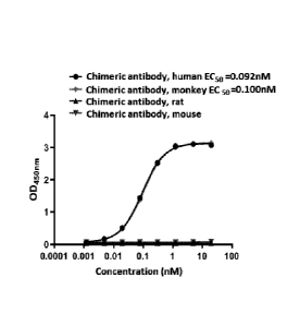
Park), 2014 Nov;28
Suppl 3:15-28).
[71 In view of this, the present disclosure has been specifically
proposed.
SUMMARY
[8] The present disclosure is based on an obtained parental anti-human PD-1
murine
monoclonal antibody having the ability to specifically bind to human PD-1
protein, by cloning,
identification and gene structure analysis to determine its CDR region,
construct corresponding
chimeric antibody and humanized antibody, establish corresponding eukaryotic
cell expression
system and produce and purify the chimeric antibody and the humanized
antibody.
[9] In order to achieve the above goal of the present disclosure, the
following technical
solutions are specially adopted:
[10] An antibody capable of specifically binding to PD-1 and functional
fragment thereof,
wherein the antibody or the functional fragment comprises a light chain and a
heavy chain;
[11] the light chain comprises a light chain CDR consisting of CDR-L1, CDR-L2
and CDR-
L3; the heavy chain comprises a heavy chain CDR consisting of CDR-Hl, CDR-H2
and CDR-
H3;
[12] the amino acid sequences of the CDR-L1, CDR-L2. and CDR-L3 are
respectively set
forth in SEQ ID NO: 1, 5 and 6, or respectively set forth in SEQ ID NO: 2, 5
and 6, or
respectively set forth in SEQ ID NO: 3, 5 and 6, or respectively set forth in
SEQ ID NO: 4, 5 and
6; the amino acid sequences of the CDR-H1, CDR-H2, and CDR-H3 are respectively
set forth in
SEQ ID NO: 7, 8 and 9.
[131 Preferably, the antibody or the functional fragment thereof includes a PD-
1 chimeric
antibody and a functional fragment thereof, and a PD-1 humanized antibody and
a functional
fragment thereof. That is, it may also be interpreted as that the antibody or
the functional
fragment thereof includes a PD-1 chimeric antibody and a functional fragment
thereof, or the
antibody or the functional fragment thereof includes a PD-1 humanized antibody
and a
functional fragment thereof.
[14] It is well known in the art that both the binding specificity and
affinity of an antibody are
3
12694005.2
34273/40
CA 03036912 2019-03-14
mainly determined by the CDR, and the amino acid sequence of the non-CDR
region can be
easily changed according to the well-known existing techniques to obtain a
variant having
similar biological activities. In the present disclosure, the monoclonal
antibody variants have
CDR sequences identical to the CDR sequences of above-mentioned humanized
antibodies, thus,
they have similar biological activities.
[15] Preferably, the antibody and the functional fragment thereof as described
above, wherein
the antibody comprises a constant region sequence of any one selected from the
group consisting
of human IgGI, IgG2, IgG3, IgG4, IgA, IgM, IgE and IgD.
[16] Preferably, antibody and the functional fragment thereof as described
above, wherein the
functional fragment comprises one or more selected from the group consisting
of F(a13)2, Fab',
Fab, Fv, scFv, bispecific antibody and antibody minimal recognition unit.
[17] The "functional fragment' of the present disclosure specifically refers
to an antibody
fragment having the same specificity to PD-1 as that of the parent antibody.
In addition to the
above mentioned functional fragments, any fragment of which half-life has been
increased may
be also included.
[18] scFy (Sc = single strand), bispecific antibody (diabodies).
[19] These functional fragments typically have the same binding specificity as
the antibody
from which they are derived. One ordinary skill in the art can learn from what
is described in the
specification of the present disclosure that the antibody fragment of the
present disclosure and
obtain the above mentioned function fragment by a method such as enzymatic
digestion
(including pepsin or papain) and/or a method of chemically reducing split
disulfide bonds.
[20] The antibody fragments can also be obtained by peptide synthesis by
recombinant genetic
techniques, which are also known to those having ordinary skill in the art, or
by automated
peptide synthesizers such as an automated peptide synthesizer sold by such as
Applied
BioSystems.
[21] Preferably, the antibody and the functional fragment thereof as described
above, wherein
the amino acid sequences of light chain variable region and heavy chain
variable region of the
PD-1 chimeric antibody and the functional fragment thereof are respectively
set forth in SEQ ID
NO: 10 and SEQ ID NO: 14, or respectively set forth in SEQ ID NO: Ii and SEQ
ID NO:14, or
respectively set forth in SEQ ID NO: 12 and SEQ ID NO: 14, or respectively set
forth in SEQ ID
4
12694005.2
34273/40
CA 03036912 2019-03-14
NO: 13 and SEQ ID NO: 14.
[22] Further preferably, the antibody and the functional fragment thereof as
described above,
wherein the amino acid sequences of the light chain constant region and the
heavy chain constant
region of the PD-1 chimeric antibody and the functional fragment thereof are
respectively set
forth in SEQ ID NO: 15 and SEQ ID NO: 16.
[23] Preferably, the antibody and the functional fragment thereof as described
above, wherein
light chain framework region of the PD-1 humanized antibody and the functional
fragment
thereof comprises FR-L1, FR-L2, FR-L3 and FR-L4, and heavy chain framework
region of the
PD-1 humanized antibody and the functional fragment thereof comprises FR-HI,
FR-H2, FR-H3
and FR-114;
[24] the FR-L1 is selected from the amino acid sequence set forth in SEQ ID
NO: 17 and the
amino sequence having the following substitution or a combination thereof:
[25] the amino acid D is replaced by E;
[26] the 2nd amino acid V is replaced by I;
[27] the 13th amino acid Lis replaced by V;
[28] the 19th amino acid A is replaced by V;
[29] the FR-L2 is selected from the amino acid sequence set forth in SEQ ID
NO: 18 and the
amino sequence having the following substitution or a combination thereof:
[30] the 6th amino acid P is replaced by S;
[31] the 7th amino acid G is replaced by H;
[32] the 9th amino acid A is replaced by S;
[33] the FR-L3 is selected from the amino acid sequence set forth in SEQ ID
NO: 19 and the
amino sequence having the following substitution or a combination thereof:
[34] the 22th amino acid L is replaced by V;
[351 the 24th amino acid P is replaced by T;
[36] the 28th amino acid A is replaced by G;
[37] the 31th amino acid F is replaced by Y;
[38] the FR-L4 is selected from the amino acid sequence set forth in SEQ ID
NO: 20 and the
amino sequence having the following substitution or a combination thereof:
[391 the 7t amino acid V is replaced by L;
5
12694005.2
34273/40
CA 03036912 2019-03-14
[40] the FR-H1 is selected from the amino acid sequence set forth in SEQ ID
NO: 21;
[41] the FR-H2 is selected from the amino acid sequence set forth in SEQ ID
NO: 22 and the
amino sequence having the following substitution or a combination thereof:
[42] the 5th amino acid A is replaced by T;
[43] the 14th amino acid A is replaced by S;
[44] the FR-H3 is selected from the amino acid sequence set forth in SEQ ID
NO: 23 and the
amino sequence having the following substitution or a combination thereof:
[45] the 12th amino acid N is replaced by T;
[46] the 14th amino acid Y is replaced by H;
[47] the 18th amino acid N is replaced by S;
[48] the FR-H4 is selected from the amino acid sequence set forth in SEQ ID
NO: 24.
[49] Usually, when transplanting CDRs of a murine antibody to a human
framework, selection
of a human framework with high sequence homology will have a certain success
rate. However,
studies have shown that many CDR grafts require a back mutation to restore
certain antibody
activity. How to choose the right human source framework is the major
bottleneck.
[50] The CDR is the major relevant site for antigen binding, but in most
cases, the FR
(framework region) has a significant influence on the conformation of the
binding site. In order
to obtain a high affinity humanized antibody, in the present disclosure, a
suitable FR region is
selected and the relevant FR residue is reversed back to the original murine
amino acid or a
amino acid presented in human and having the same function.
[51] Preferably, light chain variable region sequence of the PD-1 humanized
antibody and the
functional fragment thereof is one selected from SEQ ID NO: 25 to 36;
[52] preferably, heavy chain variable region sequence of the PD-1 humanized
antibody and the
functional fragment thereof is one selected from SEQ ID NO: 37 to 42;
[53] more preferably, the light chain variable region sequence of the PD-1
humanized
antibody and the functional fragment thereof is set forth in SEQ ID NO: 25:
the corresponding
heavy chain variable region sequence is set forth in SEQ ID NO: 37;
[54] alternatively, the light chain variable region sequence of the PD-1
humanized antibody
and functional fragment thereof is set forth in SEQ ID NO: 25; the
corresponding heavy chain
variable region sequence is set forth in SEQ ID NO: 38;
6
12694005.2
34273/40
CA 03036912 2019-03-14
[55] alternatively, the light chain variable region sequence of the PD-1
humanized antibody
and functional fragment thereof is set forth in SEQ ID NO: 29; the
corresponding heavy chain
variable region sequence is set forth in SEQ ID NO: 38;
[56] alternatively, the light chain variable region sequence of the PD-1
humanized antibody
and functional fragment thereof is set forth in SEQ ID NO: 30; the
corresponding heavy chain
variable region sequence is set forth in SEQ ID NO: 38;
[57] alternatively, the light chain variable region sequence of the PD-1
humanized antibody
and functional fragment thereof is set forth in SEQ ID NO: 31; the
corresponding heavy chain
variable region sequence is set forth in SEQ ID NO: 38;
[58] alternatively, the light chain variable region sequence of the PD-1
humanized antibody
and functional fragment thereof is set forth in SEQ ID NO: 26; the
corresponding heavy chain
variable region sequence is set forth in SEQ ID NO: 38;
[59] alternatively, the light chain variable region sequence of the PD-1
humanized antibody
and functional fragment thereof is set forth in SEQ ID NO: 28; the
corresponding heavy chain
variable region sequence is set forth in SEQ ID NO: 40:
[60] alternatively, the light chain variable region sequence of the PD-1
humanized antibody
and functional fragment thereof is set forth in SEQ ID NO: 25; the
corresponding heavy chain
variable region sequence is set forth in SEQ ID NO: 40;
[61] alternatively, the light chain variable region sequence of the PD-1
humanized antibody
and functional fragment thereof is set forth in SEQ ID NO: 29; the
corresponding heavy chain
variable region sequence is set forth in SEQ ID NO: 40;
[62] alternatively, the light chain variable region sequence of the PD-1
humanized antibody
and functional fragment thereof is set forth in SEQ ID NO: 30; the
corresponding heavy chain
variable region sequence is set forth in SEQ ID NO: 40;
[63] alternatively, the light chain variable region sequence of the PD-1
humanized antibody
and functional fragment thereof is set forth in SEQ ID NO: 31; the
corresponding heavy chain
variable region sequence is set forth in SEQ ID NO: 40;
[64] alternatively, the light chain variable region sequence of the PD-1
humanized antibody
and functional fragment thereof is set forth in SEQ ID NO: 28; the
corresponding heavy chain
variable region sequence is set forth in SEQ ID NO: 38;
7
12694005 2
34273/40
CA 03036912 2019-03-14
[65] alternatively, the light chain variable region sequence of the PD-1
humanized antibody
and functional fragment thereof is set forth in SEQ ID NO: 27; the
corresponding heavy chain
variable region sequence is set forth in SEQ ID NO: 39;
[66] alternatively, the light chain variable region sequence of the PD-1
humanized antibody
and functional fragment thereof is set forth in SEQ ID NO: 32; the
corresponding heavy chain
variable region sequence is set forth in SEQ ID NO: 39:
[67] alternatively, the light chain variable region sequence of the PD-1
humanized antibody
and functional fragment thereof is set forth in SEQ ID NO: 33; the
corresponding heavy chain
variable region sequence is set forth in SEQ ID NO: 39;
[68] alternatively, the light chain variable region sequence of the PD-1
humanized antibody
and functional fragment thereof is set forth in SEQ ID NO: 34; the
corresponding heavy chain
variable region sequence is set forth in SEQ ID NO: 39;
[69] alternatively, the light chain variable region sequence of the PD-1
humanized antibody
and functional fragment thereof is set forth in SEQ ID NO: 35; the
corresponding heavy chain
variable region sequence is set forth in SEQ ID NO: 41;
[70] alternatively, the light chain variable region sequence of the PD-1
humanized antibody
and functional fragment thereof is set forth in SEQ ID NO: 36; the
corresponding heavy chain
variable region sequence is set forth in SEQ ID NO: 42;
[71] more preferably, the amino acid sequences of the light chain constant
region sequence
and the heavy chain constant region sequence of the PD-1 humanized antibody
and the
functional fragment thereof are set forth in SEQ ID NO: 15 and SEQ ID NO: 16,
respectively.
[72] It should be noted that, in addition to the above-mentioned amino acid
sequences in the
present application, the production of chimeric antibodies and humanized
antibodies can be
achieved by any method known by those having ordinary skill in the art, such
as by designing
recombinant humanized antibody based on sequenced CDRs of murine antibodies,
the murine
antibody is secreted by myeloma cells from immunized mice or by myeloma cells
fused to
splenocytes of other species which fused to myeloma cells. The immunized
animal may include
a transgenic mouse having a human immunoglobulin locus which can directly
produce a human
antibody. Another possible embodiment may include screening the library using
phage display
technology.
8
12694005.2
34273/40
CA 03036912 2019-03-14
[73] An isolated nucleic acid molecule, which is selected from:
[74] A) DNA or RNA encoding the antibody and the functional fragment thereof
as described
above; and
[75] B) a nucleic acid complementary to the nucleic acid defined in A).
[76] A vector, which contains a nucleic acid molecule as described above.
[77] The present disclosure further provides at least one nuclear construct
encoding a nucleic
acid molecule as described above, preferably a vector, more preferably an
expression vector,
such as a plasmid, which is described in one embodiment of the present
application.
[78] A host cell, which is transformed with a vector as described above.
[79] The host cell is a eukaryotic cell, such as a mammalian cell.
[80] A method of producing an antibody capable of specifically binding to PD-1
and a
functional fragment thereof includes the following steps:
1811
culturing host cells as described above in a medium and under suitable culture
conditions; and
[82] recovering produced antibody and its functional fragments from the
culture medium or
from the cultured host cells.
[83] A composition, which comprises the antibody and/or the functional
fragment thereof, or a
compound of the antibody and other components, or a compound of the antibody
functional
fragment and other components, as an active ingredient.
[84] Preferably, the composition as described above, the antibody and the
functional fragment
thereof are coupled to at least one diagnostic agent and/or therapeutic agent
to form an
immunoconjugate.
[85] Preferably, the diagnostic agent is one or more selected from the group
consisting of a
radionuclide, a radioactive contrast agent, a paramagnetic ion, a metal, a
fluorescent label, a
chemiluminescent label, an ultrasound contrast agent, and a photosensitizer.
[861 Preferably, the radionuclide is one or more selected from the group
consisting of 'win,
177La, 18F, 52Fe, 62ca, 64ctl, 67ca, 67-a,
"Ga, 86y 90Y "Zr, "MTC, 94TC, "MTh, 1201, 1231, 1241,
1251, 131/, 1541
58r;d 32p5 ll 13m 150 K 186-e,
p .5.-"
'"Re, 51Mn, 52mMn, 55Co, 72As, 75Br, 76Br, 82mRb
and 83Sr.
1871 Preferably, the paramagnetic ion is one or more selected from the group
consisting of
9
12694005.2
34273/40
CA 03036912 2019-03-14
chromium (III), manganese (II), iron (III), iron (II), cobalt (II), nickel
(II), copper (II),
neodymium (III), samarium (III), ytterbium (III), gadolinium (III), vanadium
(II), terbium (111),
dysprosium (III), holmium (III) and erbium (HI).
[88] Preferably, the fluorescent label is one or more selected from the group
consisting of
Alexa 350, Alexa 405, Alexa 430, Alexa 488, Alexa 555, Alexa 647, AMCA,
aminoacridine,
BODIPY 630/650, BODIPY 650/665, BODIPY-FL, BODIPY-R6G, BODIPY-TMR, BOD1PY-
TRX, 5-carboxy-4', 5'-dichloro-2', T-
dimethoxyfluorescence, 5-carboxy-2'.4',5',7'-
tetrachlorofluorescein, 5-carboxyfluorescein, 5-carboxyrhodamine, 6-
carboxyrhodamine, 6-
carboxytetramethylrhodamine, Cascade Blue, Cy2, Cy3, Cy5, Cy7. 6-FAM, dansyl
chloride,
fluorescein, HEX, 6-JOE, NBD (7-nitrobenzo-2-oxa-1,3-diazole), Oregon Green
488, Oregon
Green 500, Oregon Green 514, Pacific Blue, phthalic acid, terephthalic acid,
isophthalic acid,
cresol fast violet, cresyl violet, brilliant cresyl blue, 4-Aminobenzoic acid,
erythrosine,
phthalocyanine, azomethine, cyanine, xanthine, succinyl fluorescein, rare
earth metal cryptate,
tri-bipyridyldiamine oxime, europium cryptate compound or chelate, diamine,
dicyanine, La
Jolla blue dye, allophycocyanin, allococyanin B, phycocyanin C, phycocyanin R,
thiamine, R-
phycoerythrin, C-Phycocyanin, phycoerythrin R, REG, rhodamine green, rhodamine
isothiocyanate, rhodamine red, ROX, TAMRA, TET, TRIT (tetramethylrhodamine
isothiol),
tetramethylrhodamine and Texas Red.
[89] Preferably, the therapeutic agent is one or more selected from the group
consisting of a
naked antibody, a cytotoxic agent, a drug, a radionuclide, a boron atom, an
immunomodulator, an
anti-apoptotic agent, a photosensitizing therapeutic, an immunoconjugates and
a oligonucleotide.
[90] Preferably, the drug is one or more selected from the group consisting of
methotrexate,
fluorouracil, mercaptopurine, hydroxyurea, cytarabine, nitrogen mustard,
cyclophosphamide,
thiotepa, cisplatin, mitomycin, bleomycin, camptothecin, podophyllotoxin,
actinomycin D,
doxorubicin, daunorubicin, vinblastine, paclitaxel, cephalotaxus alkaloids and
L-asparaginase.
[91] Preferably, the oligonucleotide is one or more selected from the group
consisting of
shRNA, miRNA and siRNA.
[921 Preferably, the immunomodulator is one or more selected from the group
consisting of a
cytokine, a chemokine, a stem cell growth factor, a lymphotoxin, a
hematopoietic factor, a
colony stimulating factor (CSF), an interferon, an erythropoietin, a
thrombopoietin, a tumor
12694005.2
34273/40
CA 03036912 2019-03-14
necrosis factor (TNF), an interleukin (IL), granulocyte-colony stimulating
factor (G-CSF),
granulocyte macrophage-colony stimulating factor (GM-CSF) and stem cell growth
factor.
[93] Wherein, the cytokine is preferably one or more selected from the group
consisting of
human growth hormone, N-methionyl human growth hormone, bovine growth hormone,
parathyroid hormone, thyroxine, insulin, proinsulin, relaxin, prorelaxin,
follicle-stimulating
hormone (FSH), thyroid stimulating hormone (TSH), luteinizing hormone (LH),
liver growth
factor, prostaglandin, fibroblast growth factor, prolactin, placental
lactogen, OB protein, tumor
necrosis factor-a, tumor necrosis factor43, Mullerian inhibitor, mouse
gonadotropin-related
peptide, inhibin, activin, vascular endothelial growth factor, integrin,
thrombopoietin (TP0),
.. NGF-p, platelet-growth factor, TGF-a, TGF-P, insulin-like growth factor-I,
insulin-like growth
factor-11, erythropoietin (EPO), osteoinductive factor, interferon-a,
interferon-0, interferon-y,
macrophage-CSF (M-CSF), IL-1, IL-la, IL-2, IL-3, IL-4, 1L-5, IL-6, IL-7, IL-8,
IL-9, IL-10, IL-
11, IL-12, IL-13, IL-14, IL-15, IL-16, IL-17, 1L-18, IL-21, IL-25, LIF, FLT-3,
angiostatin,
thrombospondin, endostatin, tumor necrosis factor and LT.
[94] The chemokine is preferably one or more selected from the group
consisting of RANTES,
MCAF, MIP1-a, MIP1-13, and IP-10.
[95] Preferably, the radionuclide is one or more selected from the group
consisting
'''At, 1"Lu, 211Bt, 212B/, 213B/, 211At,62Cu, 67Cu,
90y 125 131 133/, 32p 33p 47-c,
S 1"Ag, 67Ga,
11 2 223 225 7 21
7 89
I53Sm, 161 152 166 161 166 186 188 189 2 Tb, Dy,
Dy, Ho, Ho, Re, Re, Re, Pb, Pb, Ra, Ac, As, Sr,
99mo, 105Rh, 149pm, 169Er, 194.-ir , 5g
-Co, 80mBr, 99mTc, w3mRh, 1"9Pt, 119Sb, 189m0s, 192Ir, 219Rn,
21.5p0, 221Fr, 255Fm, 'C,
13N, 150, 75Br, 'Au, 199Au, 224A c, 77Br, 1131nIn, 95Ru, 97Ru, 1 3Ru, 165Ru,
203Hg, i21tnTe7 122mrre, 125inTe, 165T,m, 1677,m, 168Tm, 197pt, 109pd, 142pr,
143pr, 1Tb61-- ,
57Co,
58Co, 'Cr, 59Fe, 75Se, 201-- ,
T1 76Br and 169Yb.
[96] Use of the composition as described above for the manufacture of a
medicament in
prevention and/or treatment of an autoimmune disease, an immune response
against transplant,
an allergy, an infection, a neurodegenerative disease, or a tumor.
[97] Preferably, the autoimmune disease is one or more selected from the group
consisting of
arthritis, rheumatoid arthritis, psoriasis, multiple sclerosis, ulcerative
colitis. Crohn's disease,
systemic lupus erythematosus, glomerulonephritis, dilatation cardiomyopathy-
like disease,
Sjogren's syndrome, allergic contact dermatitis, polymyositis, scleroderma,
periarterial
11
12694005.2
34273/40
CA 03036912 2019-03-14
polyarteritis, rheumatic fever, vitiligo, insulin-dependent diabetes mellitus,
Behcet's syndrome
and chronic thyroiditis.
[98] Preferably, the neurodegenerative disease is one or more selected from
the group
consisting of Parkinson's disease, Huntington's disease, Machado-Joseph
disease, amyotrophic
lateral sclerosis and Creutzfeldt-Jakob disease.
[99] Preferably, the tumor is one or more selected from the group consisting
of leukemia,
lymphoma, myeloma, brain tumor, head and neck squamous cell carcinoma, non-
small cell lung
cancer, nasopharyngeal carcinoma, esophageal cancer, gastric cancer,
pancreatic cancer,
gallbladder cancer, liver cancer, colorectal cancer, breast cancer, ovarian
cancer, cervical cancer,
endometrial cancer, uterine sarcoma, prostate cancer, bladder cancer, renal
cell carcinoma, and
melanoma.
[100] Use of the antibody and the functional the fragment thereof as described
above for the
manufacture of a medicament for preventing and/or treating of an autoimmune
disease, an
immune response against a transplant, an allergy, an infection, a
neurodegenerative disease and a
tumor.
11011 A drug for preventing and/or treating of an autoimmune disease, an
immune response
against a transplant, an allergy, an infection, a neurodegenerative disease
and a tumor, the drug
comprises the antibody capable of specifically binding to PD-1 and the
functional fragment
capable of specifically binding to PD-1 thereof as described above, and
pharmaceutically
acceptable carrier;
[102] Alternatively, the drug comprises the composition as described above and
pharmaceutically acceptable carrier.
[103] Herein, the term "pharmaceutically acceptable" means that the compound
is
physiologically acceptable when the compound is administered to a human, and
does not cause
an allergic reaction such as a gastrointestinal disorder, dizziness or other
allergic reaction, or a
systemic allergic reaction similar to these allergic reactions.
[104] In the present disclosure, "pharmaceutically acceptable carrier"
includes, but is not
limited to, binders (such as microcrystalline cellulose, alginates, gelatin
and
polyvinylpyrrolidone), fillers (such as starch, sucrose, glucose and anhydrous
lactic acid),
disintegrants (such as cross-linked PVP, cross-linked carboxymethyl sodium
starch,
12
12694005.2
34273/40
CA 03036912 2019-03-14
croscarmellose sodium and low-substituted hydroxypropyl cellulose), lubricants
(magnesium
stearate, aluminum stearate, talc, polyethylene glycol, sodium benzoate),
wetting agent (such as
glycerin), surfactants (such as cetyl alcohol), and absorption enhancers,
flavoring agents,
sweeteners, diluents, coating agents, etc.
[105] Use of the antibody and the functional fragment thereof as described
above in prevention
and/or treatment of an autoimrnune disease, an immune response against a
transplant, an allergy,
an infection, a neurodegenerative disease, or a tumor.
[106] Preferably, the autoimmune disease is one or more selected from the
group consisting of
arthritis, rheumatoid arthritis, psoriasis, multiple sclerosis, ulcerative
colitis, Crohn's disease,
systemic lupus erythematosus, glomerulonephritis, dilatation cardiomyopathy-
like disease,
Sjogren's syndrome, allergic contact dermatitis, polymyositis, scleroderma,
periarterial
polyarteritis, rheumatic fever, vitiligo, insulin-dependent diabetes mellitus,
Behcet's syndrome
and chronic thyroiditis.
[107] Preferably, the neurodegenerative disease is one or more selected from
the group
consisting of Parkinson's disease, Huntington's disease, Machado-Joseph
disease, amyotrophic
lateral sclerosis and Creutzfeldt-Jakob disease.
[108] Preferably, the tumor is one or more selected from the group consisting
of leukemia,
lymphoma, myeloma, brain tumor, head and neck squamous cell carcinoma, non-
small cell lung
cancer, nasopharyngeal carcinoma, esophageal cancer, gastric cancer,
pancreatic cancer,
gallbladder cancer, liver cancer, colorectal cancer, breast cancer, ovarian
cancer, cervical cancer,
endometrial cancer, uterine sarcoma, prostate cancer, bladder cancer, renal
cell carcinoma, and
melanoma.
[109] A method of preventing and/or treating an autoimmune disease, an immune
response
against a transplant, an allergy, an infection, a neurodegenerative disease or
a tumor, comprises
administering the drug to a subject in need thereof.
[110] Preferably, the above-mentioned individual is a human being.
BRIEF DESCRIPTION OF DRAWINGS
[111] In order to more clearly illustrate the specific embodiments of the
present disclosure or
.. the technical solutions in the conventional art, the drawings used in the
specific embodiments or
13
12694005.2
34273/40
CA 03036912 2019-03-14
the description of the conventional art will be briefly described below, and
it is obvious that the
drawings in the following description are some embodiments of the present
disclosure and a
person having ordinary skill in the art can obtain other drawings based on
these drawings without
any creative work.
[112] Figure 1 shows the human PD-1 binding activity of the monoclonal
antibody secreted by
Clone No. 2 described in Example 1;
[113] Figure 2 shows the PD-1/PD-L1 blocking activity of the monoclonal
antibody secreted by
Clone No. 2 in Example 1;
[114] Figure 3 shows the human PD-1 binding activity of the anti-human PD-1
chimeric
monoclonal antibody in Example 3;
[115] Figure 4 shows the species specificity of the anti-human PD-1 chimeric
monoclonal
antibody in Example 4;
[116] Figure 5 shows the binding specificity of the anti-human PD-1 chimeric
monoclonal
antibody in Example 4;
[1171 Figure 6 shows the PD-1/PD-L1, PD-1/PD-L2 blocking activity of the anti-
human PD-1
chimeric monoclonal antibody in Example 5;
1118] Figure 7 shows the T cell function regulatory activity of the anti-human
PD-1 chimeric
monoclonal antibody in Example 6;
[119] Figure 8 shows the concentration-time curve of the anti-human PD-1
chimeric
monoclonal antibody after a single intravenous injection in rat in Example 7.
[120] Figure 9 shows an in vivo antitumor efficacy of the anti-human PD-1
chimeric
monoclonal antibody in Example 8.
DETAILED DESCRIPTION
[121] The embodiments of the present disclosure will be described in detail
below with
reference to the embodiments. However, a person having ordinary skill in the
art will understand
that the following embodiments are merely to illustrate present disclosure and
are not intended to
limit the scope of the disclosure. For those embodiments in which specific
conditions are not
specified, they were carried out according to the conventional conditions or
the conditions
recommended by the manufacturer. For those used reagents or instruments of
which the
14
12694005.2
34273/40
CA 03036912 2019-03-14
manufacturers are not indicated, they were all commercially available
conventional products.
Example 1. Preparation of Murine Anti-human PD-1 Monoclonal Antibody
1.1. Immunization of Animal
11221 Female BALB/c mice, 6 to 8 weeks old, purchased from Beijing Huafukang
Biotechnology Co., Ltd., were used as experimental animals. One week after the
mice were
acclimated to the environment, immunization began. For the initial
immunization, 100 jig of
recombinant human PD-1-Fe protein was thoroughly mixed with Freund's complete
adjuvant
(Sigma-Aldrich, Catalog Number F5881) to form an emulsion, which was
intraperitoneally
injected into the mice. Two weeks later, booster immunizations were performed.
For the booster
immunization, 50 p.g of recombinant human PD-1-Fc protein was thoroughly mixed
with
Freund's incomplete adjuvant (Sigma-Aldrich, Catalog Number F5806) to form an
emulsion,
which was intraperitoneally injected into the mice. The immunization was
boosted in the same
way every 2 weeks, for a total 3 times. On the seventh day after the last
immunization, blood was
collected from retro orbital venous plexus of the mice and centrifuged to
separate serum, and the
antibody titer was determined by ELISA. Mice with high titers were selected
for hybridization to
make hybridomas. Three days before the hybridization, 50 jig of recombinant
human PD-1-Fc
protein was intraperitoneally injected into mice without adjuvant. On the day
of hybridization,
the spleen was aseptically removed to prepare a single spleen cell suspension
for use.
1.2. Preparation of Hybridomas
[123] Myeloma cells SP2/0 in logarithmic growth phase were centrifuged at 1000
rpm for 5
minutes, the supernatant was discarded, and the cells were suspended in
incomplete DMEM
medium (Gibco, cat No. 11965) and counted. The cells needed were taken, washed
twice with an
incomplete culture medium. At the same time, a spleen cell suspension prepared
from a mouse
after immunization was washed twice with an incomplete culture medium. The
myeloma cells
and the spleen cells were mixed at a ratio of 1 : 10 or 1 : 5, and washed once
with an incomplete
culture medium in a 50 mL plastic centrifuge tube, and then centrifuged at
1200 rpm for 8
minutes. The supernatant was discarded and a Pasteur pipette was used to
remove residual liquid.
The centrifuge tube was gently tapped on palm to make the precipitated cells
loose and even, and
12694005.2
34273/40
CA 03036912 2019-03-14
then the tube was placed in 40 C water bath to preheat. 1 mL of 45% PEG-4000
(pH 8.0, Sigma,
cat No. P7181) preheated to 40 C was added with 1 mL pipette at about 1
minute (with an
optimum time of 45 seconds), stirred gently with a pipette when adding
(stirred with a pipette),
visible particles should be seen with the naked eyes. 20 to 30 mL of
incomplete medium
preheated to 37 C was added to the tube with 10 mL pipette within 90 seconds
to terminate PEG
action, and allowed to stand at 20 to 37 C for 10 minutes. The tube was
centrifuged at 1000 rpm
for 5 minutes, and the supernatant was discarded. 5 mL of HAT medium (DMEM +
HAT, Sigma,
cat No.1 H0262-10VL) was added, and the precipitated cells were mixed gently
(remember not
to blow vigorously so as not to separate the fused cells) to make a well mixed
suspension.
Additional HAT medium was added until 80 to 100 mL (the spleen cell
concentration was made
to be 1 to 2 x 10⁶/mL). The suspension was dispensed into a 96-well cell
culture plate, 0.1 mL
per well; and a 24-well plate, 1.0 to 1.5 mL per well. The plates were
incubated at 37 C
incubator with 6% CO2. Generally, six 96-well plates were used. After 5 days,
1/2 medium was
replaced with fresh HAT medium. After 7 to 10 days, the HAT medium was
replaced with HT
medium (DMEM + HT, Sigma cat No. H0137-10VL). The growth of hybridoma cells
was
observed routinely, and the supernatant was collected for antibody detection
after the confluence
of the cells reached 1/10 or more. The positive colonies were expanded and
frozen.
1.3. Clone Screening and Identification
[1241 ELISA was used to screen anti-human PD-1 antibody from hybridoma culture
supernatants. Recombinant human PD-1 (purchased from Sino Biological Inc.,
Catalog Number
10377-H08H) was coated on a 96-well high-absorbing ELISA plate with a
carbonate buffer
solution with pH 9.6, the coating concentration was 1 mg/mL, the coating
amount was 100 gL per
well, and the coating was carried out at 4 C overnight. The plate was washed
five times with
PBST, blocked with 300 pt/well of PBST containing 1% BSA, and then incubated
at 25 C for 1
hour. The plate was washed five times with PBST. 100 pt culture supernatant
samples and the
positive serum control were added to each well respectively, and then the
plated was incubated at
25 C for 1 hour. The plate was washed five times with PBST. Then, 100 I,
horseradish
peroxidase-labeled anti-mouse IgG antibody (Abeam, Catalog Number Ab7068)
1:10000 diluted
16
12694005.2
34273/40
CA 03036912 2019-03-14
in PBST containing 1% BSA was added to each well, and then the plated was
incubated at 25 C
for 1 hour. The plate was washed five times with PBST. 100 L/well of
colorimetric substrate
TMB was added and incubated at room temperature for 10 minutes. Color
development was
terminated by adding 100 L/well of 1 M H2SO4. The absorbance at 450 nm was
read on a
microplate reader. Positive clones capable of producing anti-human PD- I
antibody were selected
based on the reading value at OD 450 nm.
[125] Whether the anti-human PD-1 antibodies produced by positive clones could
block the
binding of PD-1/PD-L1 was determined by ELISA. Recombinant human PD-I -Fe was
coated on
a 96-well high-absorbing ELISA plate with a carbonate buffer solution with pH
9.6, the coating
concentration was 1 pg/mL, the coating amount was 100 L per well, and the
coating was carried
out at 4 C overnight. The plate was washed five times with PBST, blocked with
300 uL/well of
PBST containing 1% BSA, and then incubated at 25 C for 1 hour. The plate was
washed five
times with PBST. 50 1.11. anti-human PD-1 antibody sample and positive control
were added to
each well respectively, and then biotin-labeled PD-L1 was added at a
concentration of 20 nM
(final concentration 10 nM), 50 idL/well, and then incubated at 25 C for 90
minutes. The plate
was washed five times with PBST. Then, Streptavidin-HRP (BD Pharmingen,
Catalog Number
554066) 1:1000 diluted in PBST containing 1% BSA was added, 100 I. /well, and
then
incubated at 25 C for 1 hour. The plate was washed five times with PBST. 100
L/well of
colorimetric substrate TMB was added and incubated at room temperature for 10
minutes. Color
development was terminated by adding 100 pt/well of 1 M H2SO4. The absorbance
at 450 nm
was read on a microplate reader. The anti-human PD-I antibody capable of
inhibiting the biding
of human PD-1-Fc/biotin-labeled PD-Ll was determined as having neutralization
activity.
Positive clones capable of producing anti-human PD-1 neutralization antibody
were selected
based on the blocking ability.
[126] As shown in Figure 1, Clone No. 2 had strong human PD-1 binding
activity; as shown in
Figure 2, Clone No. 2 also had pretty strong blocking activity against the
binding of human PD-
1/PD-Ll.
1.4. Sequencing of Monoclonal Antibody
[1271 The clones having both antigen-binding activity and antigen-
neutralization activity
17
12694005.2
34273/40
CA 03036912 2019-03-14
obtained by screening were subjected to sequencing of antibody DNA sequence.
Cellular mRNA
was first extracted using RNAprep Pure Kit (Tiangen, DP430). The steps were as
follows: lx107
cells were centrifuged at 300x g for 5 minutes and collected into a centrifuge
tube, and all
supernatant was carefully aspirated. The lysis step was carried out
immediately. The bottom of
the centrifuge tube was flicked to loose the cell pellet, 600 jiL of lysis
buffer RL was added and
vortexed. All solution was transferred to a filtration column CS (the
filtration column CS was
placed in a collection tube), centrifuged at 12,000 rpm (-13,400x g) for 2
minutes, and the
filtrate was collected. One fold volume of 70% ethanol (usually 350 pi, or 600
pL) was added to
the filtrate, well mixed, the obtained solution and precipitate were
transferred into an adsorption
column CR3 (the adsorption column CR3 was put into a collection tube),
centrifuged at 12,000
rpm (-13,400x g) for 30 to 60 seconds, the liquid waste in the collection tube
was removed, the
adsorption column CR3 was put back into the collection tube. 350 jiL of
deproteinized solution
RW1 was added to the adsorption column CR3, centrifuged at 12,000 rpm (-
13,400xg) for 30 to
60 seconds, the liquid waste in the collection tube was removed, the
adsorption column CR3 was
put back into the collection tube. 80 pt of DNase I working solution was added
to the center of
the adsorption column CR3 and the column CR3 was allowed to stand at room
temperature for
15 minutes. 350 piL of deproteinized solution RW1 was added to the adsorption
column CR3,
centrifuged at 12,000 rpm (-13,400xg) for 30 to 60 seconds, the liquid waste
in the collection
tube was removed, the adsorption column CR3 was put back into the collection
tube. 500 pl. of
rinsing solution RW was added to the adsorption column CR3 (checked whether
ethanol had
been added before use), the column CR3 was allowed to stand at room
temperature for 2 minutes,
centrifuged at 12,000 rpm (-13,400x g) for 30 to 60 seconds, the liquid waste
in the collection
tube was removed, the adsorption column CR3 was put back into the collection
tube. The
column CR3 was centrifuged at 12,000 rpm (- 13,400x g) for 2 minutes, and the
waste was
removed. The adsorption column CR3 was left at room temperature for a few
minutes to let the
residual rinsing solution in the adsorbent material thoroughly dry. The
adsorption column CR3
was transferred into a new RNase-Free centrifuge tube, 30 to 100 pt of RNase-
Free ddH20 was
added, the tube was allowed to stand at room temperature for 2 minutes, and
then centrifuged at
12,000 rpm (-13,400x g) for 2 minutes to obtain a RNA solution.
[128] The first strand of cDNA was synthesized using the QuantScript RT kit
(Tiangen, KR103).
18
12694005.2
34273/40
CA 03036912 2019-03-14
The steps are as follows: the template RNA was thawed on ice; the primer,
10xRT mix
(containing RNasin and DTT), Super pure dNTP mixture, RNase-Free ddH20 were
thawed at
room temperature (15 to 25 C), and placed on ice immediately after thawing.
Each solution was
well mixed by vortexer before use, the tube was centrifuged briefly to collect
residual liquid on
the side of the tube. Reverse transcription system mixture (Tiangen Bio Quant
cDNA First-
Strand Synthesis Kit, Catalog Number KR103-04; 10x Reverse Transcription
Buffer 2 1.tL, Ultra-
Pure dNTP 2 4, Random Primer 2 pt, Reverse Transcription Enzyme I L) was
prepared
according to Table I. The mixture was mixed thoroughly, the duration of vortex
was no more
than 5 minutes; and then centrifuged briefly and placed on ice. Finally, the
template RNA (50 ng
to 2 pig) was added to the mixture, mixed thoroughly, the duration of vortex
was no more than 5
seconds, centrifuged briefly to collect residual liquid on the sides of the
tube, incubated at 37 C
for 60 minutes. The first strand of cDNA produced by reverse transcription was
used for
subsequent PCR reaction.
[129] The primers used in the PCR reaction are as shown in Table 1.
VHprimer
Fl:GAGGTGAAGCTGCAGGAGTCAGGACCTAGCCTGGTG
R1:AGGT(C/G)(A/C)AACTGCAG(C/G)AGTC(AJT)GG
R2:AGGT(C/G)(A/C)AGCTGCAG(C/G)AGTC(A/T)GG
R3:AGGT(C/G)CAGCTGCAG(C/G)AGTC(A/T)GG
R4: C CAGGGGC C AGTGGATAG AC A A GCTTGGGTGTC GTTTT
F2:ATAGACAGATGGGGGTGTCGTTTTGGC
F3:CTTGACCAGGCATCCTAGAGTCA
F4:AGGGGCCAGTGGATAGACTGATGG
F5:AGGGACCAAGGGATAGACAGATGG
R5:(G/C)A(A/G)GT(A/T/C/G)(A/C)AGCTG(G/C)AG(G/C)AGTC
R6: (G/C )A(A/G)GT(A/T/C/G)(AJC)AGCTG(G/C )AG (G/C )AGTC(A/T)GG
VLprimer
R1 : GGTGATATCGTGAT(A/G)A C (C/A)CA(G/A)GATGAACTCTC
R2: GGTGATATC (A/T)TG (A/C)TGAC C CAA (A/T)CTCC ACTCTC
19
12694005.2
34273/40
CA 03036912 2019-03-14
R3:GGTGATATCGT(G/T)CTCAC(C/T)CA(A/G)TCTCCAGCAAT
Fl : GGGAAGATGGATC CAGTTGGTGC AGC ATCAGC
F2:GGATACAGTTGGTGCAGCATC
R4:GA(C/T)ATTGTG(A/C)T(G/C)AC(A/C)CA(A/G)(A/T)CT(A/C)CA
-
[130] When primers were used, any upstream primer of the VH primers could be
used with any
downstream primer; in the same way, any upstream primer of the VL primers
could also be used
with any downstream primer. The target band obtained by PCR amplification was
cloned into the
pGEM-T vector. A single clone was picked for DNA sequencing.
Example 2. Preparation of Chimeric Anti-human PD-1 Monoclonal Antibody
[131] The amino acid sequence of the light chain variable region of the
antibody obtained by
PCR amplification is set forth in SEQ ID NO: 10, and the amino acid sequence
of the heavy
chain variable region of antibody is set forth in SEQ ID NO: 14. The sequence
of the
complementarity-determining region can be obtained by excluding the sequence
of the
framework region from the mouse variable region sequence; wherein the amino
acid sequences
of the three complementarity-determining regions CDR-LI, CDR-L2, CDR-L3 of the
light chain
are set forth in SEQ ID NO: 1, 5 and 6, respectively; the amino acid sequences
of the three
complementarity-determining regions CDR-I-11, CDR-H2, CDR-H3 of the heavy
chain are set
forth in SEQ ID NO: 7, 8 and 9, respectively. The above-mentioned variable
region sequences
were cloned into a eukaryotic expression vector XOGC, the amino acid sequence
of the light
chain constant region of the antibody is set forth in SEQ ID NO: 15, and the
amino acid
sequence of the heavy chain constant region of the antibody is set forth in
SEQ ID NO: 16. The
vectors expressing the antibody light chain (the full-length of the light
chain was the light chain
variable region of the antibody linked to SEQ ID NO: 15) and the heavy chain
(the full-length of
the heavy chain was the heavy chain variable region of antibody linked to SEQ
ID NO: 16) were
transfected into 293F cell line (FreeStyleTm 293-F Cells, Catalog Number
R79007, Invitrogen).
Cells were subcultured one day prior to transfection. Cells On the day of
transfection, cells were
harvested by centrifugation and then resuspended in fresh FreeStyle" 293
Expression Medium
(FreeStyleTM 293 Expression Medium, Catalog Number 12338001, Gibco) at a
density of
200x105 cells/mL. Plasmids were added based on the transfection volume to a
final
12694005.2
34273/40
CA 03036912 2019-03-14
concentration of 36.67 jig /mL, mixed gently; then linear PEI
(polyethyleneimine, linear, M.W.
25000, Catalog Number 43896, Alfa Aesar) was added to a final concentration of
55 gg/mL,
mixed gently. Thereafter, the cells were placed in a shaker at 120 rpm and
incubated at 37 C for
1 hour. 19-fold transfection volume of fresh medium was then added and the
cells were
.. continually cultured at 37 C in a shaker at 120 rpm. The culture
supernatant 5 to 6 days after
transfection was collected by centrifugation.
Example 3. Binding Activity and Kinetics of Chimeric Anti-human PD-1
Monoclonal
Antibody
[132] The binding activity of anti-human PD-1 chimeric monoclonal antibody to
its antigen
human PD-1 was determined by ELISA. Recombinant human PD-1 (purchased from
Sino
Biological Inc.) was coated on a 96-well high-absorbing ELISA plate with a
carbonate buffer
solution with pH 9.6õ the coating concentration was 1 gg/mL, the coating
amount was 100 !IL
per well, and the coating was carried out at 4 C overnight. The plate was
washed five times with
PBST and blocked with 300 gL/well of PBST containing 1% BSA, and then
incubated at 25 C
for 1 hour. The plate was washed five times with PBST. The monoclonal antibody
control,
Pembrolizumab, and the anti-human PD-1 chimeric monoclonal antibody samples
serially
diluted in PBST containing 1% BSA were added, 100 gL per well, incubated at 25
'V for 1 hour.
The plate was washed five times with PBST. Then, horseradish peroxidase-
labeled anti-human
IgG antibody (Chemicon, Catalog Number AP309P) 1:2000 diluted in PBST
containing 1% BSA
was added, 100 pt per well, incubated at 25 C for 1 hour. The plate was
washed five times with
PBST. 100 gL/well of colorimetric substrate TMB was added and incubated at
room temperature
for 10 minutes. Color development was terminated by adding 100 1AL/well of 1 M
H2SO4. The
absorbance at 450 nm was read on a microplate reader.
[133] The result is as shown in Figure 3, the anti-human PD-1 chimeric
monoclonal antibody
has good binding affinity to human PD-1, which is similar to the binding
activity of
Pembrolizumab.
[134] The kinetics of anti-human PD-1 chimeric monoclonal antibody binding to
its antigen
human PD-1 was detected using Biacore X100. The instrument utilizes an optical
surface
plasmon resonance technique to detect association and dissociation between a
molecule coupled
21
12694005.2
34273/40
CA 03036912 2019-03-14
on a sensor chip and an analyte. CM5 chips (GE Healthcare, BR-1000-12) were
used. Brief
experiment procedure was as follow: anti-human PD-1 chimeric antibody was
diluted to 2 jig/mL
with a running buffer (1xHBS-EP + 10 mM HEPES, 150 mM NaC1, 3 mM EDTA, 0.05%
surfactant P20, pH 7.4), then injected at a rate of 10 utimin onto a CMS chip
coupled with anti-
human IgG, lasted for 60 seconds. In the association phase, the antigen PD-1
was diluted to
multiple concentrations with a running buffer, and injected at a rate of 30
pt/min for 180
seconds. In the dissociation phase, the duration of the dissociation was 1200
seconds. Glycine
solution (GE Healthcare, BR-1003-54) was used to regenerate for 30 seconds at
a speed of 10
jiL/min. The experiment method for the control antibody was similar, except
the duration of
dissociation was adjusted to 600 seconds. Association rate constant and
dissociation rate constant
were analyzed and calculated by Biacore X100 evaluation software. See Table 2
for the
association rate constant, dissociation rate constant and dissociation
equilibrium constant of the
anti-human PD-1 chimeric antibodies. The data demonstrates that, compared to
Pembrolizumab,
after binding to antigen PD-1, anti-human PD-1 chimeric monoclonal antibody
could maintain
the binding state for a longer time and is not easy to be dissociated, which
contributes greatly to
its biological functions.
Table 2. Binding Kinetics of Anti-Human PD-1 Chimeric Antibody to Human PD-1
Sample Kon(l/Ms) Koff (1/s) KD (nM)
Pembrolizumab 3.731E + 5 2.708E - 3 7.257
Anti-human PD-1 Chimeric Antibody 2.150E + 5 2.950E - 4 1.372
Example 4. Species Specificity and Binding Specificity of Chimeric Anti-human
PD-1
Monoclonal Antibody
[135] The species specificity of the anti-human PD-1 chimeric monoclonal
antibody was
determined by ELISA. Recombinant human PD-1, monkey PD-1, rat PD-1 and mouse
PD-1 (all
purchased from Sino Biological Inc.), were coated on a 96-well high-absorbing
ELISA plate with
a carbonate buffer solution with pH 9.6, the coating concentration was 1
p.g/mL, the coating
amount was 100 jiL per well, and the coating was carried out at 4 C
overnight. The plate was
washed five times with PBST and blocked with 300 iL/well of PBST containing 1%
BSA, and
then incubated at 25 C for 1 hour. The plate was washed five times with PBST.
The control and
22
12694005.2
34273/40
CA 03036912 2019-03-14
the anti-human PD-1 chimeric monoclonal antibody sample serially diluted in
PBST containing
1% BSA were added, 100 1.(1.. per well, incubated at 25 C for 1 hour. The
plate was washed five
times with PBST. Then, horseradish peroxidase-labeled anti-human IgG antibody
(Chemicon,
Catalog Number AP309P) 1:2000 diluted in PBST containing 1% BSA was added, 100
1., per
well, incubated at 25 C for 1 hour. The plate was washed five times with
PBST. 100 uL/well of
colorimetric substrate TMB was added and incubated at room temperature for 10
minutes. Color
development was terminated by adding 100 pt/well of 1 M H2SO4. The absorbance
at 450 nm
was read on a microplate reader.
[136] The binding specificity of the anti-human PD-1 chimeric monoclonal
antibody was
determined by ELISA. Recombinant human PD-1, CD28, CTLA4, ICOS, BTLA, PD-L1,
PD-L2,
CD80, CD86, B7-H2 (all purchased from Sino Biological Inc.), were coated on a
96-well high-
absorbing ELISA plate with a carbonate buffer solution with pH 9.6, the
coating concentration
was 1 g/mL, the coating amount was 100 !IL per well, and the coating was
carried at 4 C out
overnight. The plate was washed five times with PBST and blocked with 300
pL/well of PBST
containing 1% BSA and incubated at 25 C for 1 hour. The plate was washed five
times with
PBST. The control and the anti-human PD-1 chimeric monoclonal antibody sample
diluted in
PBST containing 1% BSA were added, 100 pit per well, incubated at 25 'C for 1
hour. The plate
was washed five times with PBST. Then, horseradish peroxidase-labeled anti-
human IgG
antibody (Chemicon, Catalog Number AP309P) 1:2000 diluted in PBST containing
1% BSA was
added, 100 pt was added to each well, incubated at 25 C for 1 hour. The plate
was washed five
times with PBST. 100 pt/well of colorimetric substrate TMB was added and
incubated at room
temperature for 10 minutes. Color development was terminated by adding 100
uL/well of I M
H2SO4. The absorbance at 450 nm was read on a microplate reader.
[137] The result is as shown in figure 4, the anti-human PD-1 chimeric
monoclonal antibody
could bind to human PD-1 and monkey PD-1 with similar affinity, but did not
bind to rat and
mouse PD-1, indicating that it is species-specific. In addition, as shown in
Figure 5, the anti-
human PD-1 chimeric monoclonal antibody also has excellent binding
specificity, which only
binds to PD-1 but not other members of CD28 family or B7 family.
Example 5. PD-1 and Ligands Blocking Activity of Chimeric Anti-human PD-1
Monoclonal
23
12694005.2
14271/40
CA 03036912 2019-03-14
Antibody
[138] Recombinant human PD-1-Pc was coated on a 96-well high-absorbing ELISA
plate with
a carbonate buffer solution with pH 9.6, the coating concentration was 1
ftg/mL, the coating
amount was 100 iL per well, and the coating was carried at 4 'V out overnight.
The plate was
washed five times with PBST and blocked with 300 p.L/well of PBST containing
1% BSA and
incubated at 25 C for 1 hour. The plate was washed five times with PBST. The
positive control
and the anti-human PD-1 antibody sample were added, 50 uL per well. And then
biotin-labeled
PD-Li was added at a concentration of 20 nM (final concentration 10 nM), or
biotin-labeled PD-
L2 at a concentration of 320 nM (final concentration 160 nM), 50 FL per well,
incubated at
25 C for 90 minutes. The plate was washed five times with PBST. Then.
Streptavidin-HRP (BD
Pharmingen, Catalog Number 554066) 1:1000 diluted in PBST containing 1% BSA
was added,
100 pL per well, incubated at 25 C for 1 hour. The plate was washed five
times with PBST. 100
pt/well of colorimetric substrate TMB was added and incubated at room
temperature for 10
minutes. Color development was terminated by adding 100 ftL/well of 1 M H2SO4.
The
absorbance at 450 nm was read on a microplate reader.
[139] The result is as shown in Figure 6, the anti-human PD-1 chimeric
monoclonal antibody
has similar PD-1/PD-L1 and PD-1/PD-L2 blocking activity compared to that of
Pembrolizumab.
Example 6 T Cell Function Regulatory Activity by Chimeric Anti-Human PD-1
Monoclonal Antibody
[1401 The PBMC used in the experiment was purchased from Lonza, Catalog Number
CC-2702.
[141] Induction of DC cells with PBMC: PBMCs were resuscitated with complete
medium
(RPMI 1640 + 10% FBS), then washed once with serum-free medium; the cells were
resuspended in serum-free medium, and seeded into a cell culture flask, and
then incubated at
37 C in an incubator with 5% CO2. After 90 minutes, the non-adherent cells
and medium were
removed; the adherent monocytes were cultured in complete medium containing
100 ng/mL
GM-CSF and 100 ng/mL IL-4, and the medium was changed after 3 days. After the
cells were
cultured for another 3 days, the medium was changed to complete medium
containing 100
ng/mL GM-CSF, 100 ng/mL IL-4 and 20 ng/mL TNF-alpha and cultured for one more
day to
complete the induction of DC cells. T cells were isolated from another
individual-derived PBMC:
24
12694005.2
34273/40
CA 03036912 2019-03-14
T cells were isolated using a Pan T Cell Isolation Kit from Miltenyi Biotech
(Catalog Number
5150414820) followed the instructions for the specific experiment procedure.
The induced
mature DC cells were seeded into a 96-well plate. 10,000 cells per well, and
isolated T cells were
added, 100,000 cells per well; and then the sample to be tested was added and
incubated for 120
hours together. At the end of the incubation, the supernatant was collected,
and the levels of IL-2
and IFN-gamma (IFN-y) were detected using an ELISA kit purchased from
RayBiotech.
[142] The result is as shown in Figure 7, in the MLR system, the anti-human PD-
l chimeric
monoclonal antibody enhanced the secretion of 1L-2 and IFN-gamma (IFN-y) and
showed a
similar effect on regulation of T cell functional activity compared to that of
Pembrolizumab.
Example 7. Pharmacokinetics Study of Chimeric Anti-Human PD-1 Monoclonal
Antibody
in Rats
[143] Female SD rats, 6 to 8 weeks old, purchased from Beijing Huafukang
Biotechnology Co.,
Ltd., were used as experimental animals. One week after the rats were
acclimated to the
environment, the rats were randomly divided into groups, 3 rats per group.
Anti-human PD-1
chimeric monoclonal antibody and control monoclonal antibody Pembrolizumab
were
administered respectively at a dose of 20 nmol/kg by intravenous injection,
single dose. At 0, 5
minutes, 30 minutes, 1 hour, 4 hours, 8 hours, 24 hours, 48 hours, 72 hours,
96 hours, 120 hours,
168 hours, 216 hours, 264 hours, 312 hours after administration, the retro-
orbital blood sample
was collected without anticoagulation, and the blood sample was allowed to
stand at room
temperature for 30 minutes to 1 hour; after coagulation, the blood sample was
centrifuged at
3,000 rpm for 10 minutes, the obtained serum sample was frozen at -80 'V and
stored for testing.
[144] The concentrations of anti-human PD-1 chimeric monoclonal antibody and
control
monoclonal antibody Pembrolizumab in the serum were determined by ELISA.
Briefly, human
recombinant PD-1 protein was coated on a high-absorbing ELISA plate with a
carbonate buffer
solution with pH 9.6 at 4 C overnight. The plate was washed with PBST. To
prevent non-
specific binding, the plate was blocked with PBST containing 5% nonfat milk
powder, and then
washed with PBST. Then, the serum sample to be tested diluted with PBST
containing 10%
mixed rat serum and 1% BSA was added and incubated at 25 C for 1 hour, and
the plate was
washed with PBST. Horseradish peroxidase-labeled anti-human IgG antibody
(Chemicon,
12694005.2
34273/40
CA 03036912 2019-03-14
Catalog Number AP309P) diluted in PBST containing 5% skimmed milk powder was
added,
incubated at 25 C for 1 hour, the then plate was washed with PBST. Finally,
color development
was carried out using the colorimetric substrate TMB at room temperature for
10 minutes. Color
development was terminated by adding 100 ttL/well of 1 M H2SO4. The absorbance
at 450 nm
was read on a microplate reader.
[145] The result is as shown in Figure 8, a single intravenous injection dose
of 20 nmol/kg of
anti-human PD-1 chimeric monoclonal antibody or control monoclonal antibody
Pembrolizumab
showed similar concentration-time curves and pharmacokinetic features in rats.
The
pharmacological parameters of the anti-human PD-1 chimeric monoclonal antibody
are as
.. follows: half-life t112 was 212 hours; the area under the concentration-
time curve AUC0_312hr was
33967 nM.hr; the estimated initial concentration Co was 464 nM; the apparent
volume of
distribution Vd was 118 mL/kg; the clearance CL was 0.39 mL/hr/kg; the mean
residence time
MRTIast was 119 hours.
Example 8. Antitumor Efficacy of Chimeric Anti-Human PD-1 Monoclonal Antibody
In
Vivo
[146] The growth inhibitory effect of Chimeric Anti-human PD-1 monoclonal
antibody on
HCC827 tumor xenografts inoculated in PBMC humanized mice was detected in the
present
example.
[147] NCG immunodeficient mice, female, 6-8 weeks old, purchased from Nanjing
Galaxy
BioPharma Co., Ltd., were used as experimental materials. One week after the
mice were
acclimated to the environment, each mouse was inoculated with lx 107 HCC827
human non-
small cell lung cancer cells (purchased from the Basic Medical Cell Center of
the Institute of
Basic Medical Sciences, Chinese Academy of Medical Sciences). When the tumor
size reached
about 100 mm3, the mice were divided into groups according to the tumor size,
6 mice per group,
including a solvent control group, an anti-human PD-1 chimeric monoclonal
antibody
administration group and a Pembrolizumab administration group. Each mouse was
intravenously
injected 5x 10⁶ human PBMC cells to humanize the immune system, and then the
solvent or
antibody was administered according to the group design, the dose was 70
nmol/kg, i.p.. The
mice were administered twice a week for 3 weeks. From the day of
administration, the tumor
26
12694005.2
34273/40
CA 03036912 2019-03-14
size was measured 3 times a week, longest diameter "a" and width "b" were
measured, the tumor
seize was calculated as: (mm3) = (a x b2)/2.
[148] The result is as shown in Figure 9, the anti-human PD-1 chimeric
monoclonal antibody
has antitumor activity and inhibited the growth of HCC827 non-small cell lung
cancer graft in
PBMC humanized mice, showing that it has comparable or slightly stronger anti-
tumor efficacy
compared to that of Pembrolizumab.
Example 9. Preparation of Humanized Anti-Human P1)-1 Monoclonal Antibody
[149] The humanized anti-human PD-1 monoclonal antibody was obtained according
to the
method of Leung et al. (Molecule Immunol, 1995, 32: 1413-27).
[1501 The humanized template that best matchs murine antibody variable region
sequence was
selected from the Germline database. The template for the light chain variable
region is IGKV3-
11*01, the sequence is set forth in SEQ ID NO: 43; the template for the heavy
chain variable
region is IGHV3-23*04, and the sequence is set forth in SEQ ID NO: 44. The CDR
regions of
the selected human template were replaced by the murine antibody CDR regions.
The obtained
grafted humanized antibody light chain variable region has a sequence set
forth in SEQ ID NO:
45, and the grafted humanized antibody heavy chain variable region has a
sequence set forth in
SEQ ID NO: 46. Sites on SEQ ID NO: 45 and SEQ ID NO: 46 were selected for back
mutation
and NQS site on the CDR1 region of SEQ ID NO: 45 was selected for mutation to
remove
possible glycosylation site. The obtained CDR-Ll sequence is set forth in SEQ
ID NO: 2, or
SEQ ID NO: 3, or ID NO: 4; the obtained light chain variable region sequence
is set forth in
SEQ ID NO: 25 to 36; the obtained heavy chain variable region sequence is set
forth in SEQ ID
NO: 37 to 42. The light chain variable region was linked to the light chain
constant region (SEQ
ID NO: 15) to obtain the corresponding full-length sequence of the light
chain; the heavy chain
variable region was linked to the heavy chain constant region (SEQ ID NO: 16)
to obtain the
corresponding full-length sequence of the heavy chain. The usable humanized
sequence was
obtained by affinity and stability screening. After affinity and stability
screening, the obtained
light chain and heavy chain variable region sequence information of humanized
sequences are
shown in Table 3.
27
2694005.2
34273/40
CA 03036912 2019-03-14
Table 3
VL SEQ ID NO:
Chimeric Monoclonal 10
Antibody
AH00290 / 25
AH00291/
AF100296
AH00293 26
AH00294 27
AH00295 28
AH00298
AH00291-N26Q/ 29
AH00296-N26Q
AH00291-N26S/ 30
AH00296-N26S
AH00291-S28A/ 31
AH00296-S28A
AII00294-N26Q 32
AH00294-N26S 33
AH00294-S28A 34
BMIII 35
BMIV 36
VH SEQ ID NO:
Chimeric Monoclonal 14
Antibody
AH00290 37
AH00291 / 38
AH00293/
28
12694005.2
34273/40
CA 03036912 2019-03-14
AH00298/
A1100291-N26Q/
AH00291-N26S/
AH00291-S28A
AH00294 / 39
AH00294-N26Q/
AH00294-N26S/
AH00294-S28A
AH00295 / 40
AH00296/
AH00296-N26Q/
AH00296-N26S/
AH00296-S28A
BMIII 41
BMIV 42
Example 10. Biological Activity of Humanized Anti-human PD-1 Monoclonal
Antibody In
Vitro
[151] The in vitro biological activity of humanized anti-human PD-1 monoclonal
antibody was
determined, including binding activity to human PD-1 and the blocking activity
against the
binding of PD-1/PD-Li. The humanized sequences to be tested included: AH00290,
AH00291,
AH00293, AH00294, AH00295, AH00296, AH00298, BM 111, BM IV. AH00290-N26Q,
AH00291-N26S, AH00291-S28A, AH00294-N26Q, AH00294-N26S, AH00294-S28A,
AH00296-N26Q, AH00296-N26S, AH00296-S28A; the method of determination was
ELISA,
and the specific experiment procedure was the same as the method of
determining chimeric anti-
human PD-1 monoclonal antibody.
[152] The experiment result is shown in Table 4. Compared to the above-
mentioned chimeric
anti-human PD-1 monoclonal antibody, all of the tested humanized sequences
maintained pretty
good activity, showing strong PD-1 binding activity and blocking activity
against the binding of
PD-1/PD-L I .
29
12694005.2
34273/40
CA 03036912 2019-03-14
Table 4 PD-1 Binding Activity and the Blocking Activity against the Binding of
PD-1/PD-L1 of
Anti-human PD-1 Humanized Antibody
PD-1 PD-1/PD-L1
Sample Binding Activity Blocking Activity
(EC50, nM) (IC50, nM)
Chimeric monoclonal
antibody 0.031 1.453
AH00290 0.024 1.086
AH00291 0.025 1.105
A1100293 0.026 1.201
AH00294 0.032 1.350
AH00295 0.025 1.188
AH00296 0.027 1.207
AH00298 0.028 1.215
BMIII 0.034 1.197
BMIV 0.028 1.298
A1100291-N26Q 0.046 1.569
AH00291-N26S 0.039 1.431
AH00291-S28A 0.042 1.361
A1100294-N26Q 0.041 1.491
AH00294-N26S 0.043 1.479
AH00294-S28A 0.047 1.464
AH00296-N26Q 0.044 1.274
A1100296-N26S 0.037 1.066
AH00296-S28A 0.048 1.755
Example 11. Detection of Purity and Thermal Stability of Humanized Anti-human
PD-1
Monoclonal Antibody by Size-exclusion High-performance Liquid Chromatography
(SE-
HPLC)
12694005.2
34273/40
CA 03036912 2019-03-14
[1531 TSKgel SuperSW3000 chromatography column (Catalog Number: 0018675) was
used.
The mobile phase was 0.1mo1/1 of phosphate buffer (NaH2PO4-Na2HPO4), 0.1
ino1/1 of sodium
sulfate buffer, pH 6.7; the flow rate was 0.35 mL/min; the column temperature
was 25 C;
sample pool temperature was 4 'V; detection wavelength was 280 nm. The sample
was diluted
with sample buffer to 1 mg/mL, and the injection volume was 5 L. The
experiment result was
processed by Agilent High Performance Liquid Chromatograph 1260 System
Workstation, and
purity was calculated by the percentage of the main peak using area
normalization method. The
humanized anti-human PD-1 monoclonal antibody prepared above was subjected to
SE-HPLC
purity assay. To determine the thermal stability of these monoclonal
antibodies, the samples were
placed under high temperature conditions of 40 C, and the samples were
subjected to SE-HPLC
assay at week 2 and week 4 respectively to observe thermal stability, and the
result is as shown
in Table 5 below. All of the humanized anti-human PD-1 antibodies showed good
and
considerable stability except A1100296-S28A.
Table 5 Thermal Stability of Humanized Anti-human PD-I Monoclonal Antibody at
40 C by
SE-HPLC
SE-HPLC Purity ( % )
Humanized Anti-human PD-1 Monoclonal Antibody ____
1=0 Week 2 Week 4
BMIII 99.23 98.16
95.78
BMIV 98.19 98.42
94.80
AH00290 98.97 98.04
94.73
A1100291 99.30 98.22
95.87
AH00293 97.79 96.55
94.32
AH00294 98.77 97.68
96.52
AH00295 99.24 98.16
96.17
AH00296 99.63 98.55
96.73
AH00298 99.34 98.13
95.87
AH00291-N26Q 98.55 98.56
97.90
AH00291-N26S 99.05 99.08
98.50
A1100291-S28A 98.95 98,89
98.40
31
12694005.2
34273/40
CA 03036912 2019-03-14
AH00294-N26Q 99.14 99.08
98.64
AH00294-N26S 99.23 99.19
98.64
AH00294-S28A 99.30 99.33
98.69
AH00296-N26Q 99.10 99.10
98.27
AH00296-N26S 99.60 99.59
98.96
AH00296-S28A 99.70 84.38
62.42
Example 12. Determination of Tm Value of Humanized Anti-human PD-1 Monoclonal
Antibody
[154] The melting temperature (Tm) of the humanized anti-human PD-1 monoclonal
antibody
was determined by Differential Scanning Fluorimetry (DSF). DSF is a method for
detecting the
thermal denaturation process of proteins in a sample by using the fluorescence
intensity change
of the fluorescent indicator to determine the protein denaturation
temperature. The reagent used
was SYPRO Orange Protein Fluorescent Dye (Sigma-Aldrich, USA, Catalog Number
S5692;
5000x concentration, in DMSO). AB 7500 Real Time PCR machine was purchased
from Applied
Biosystems, Inc., USA. The protein fluorescent dye was diluted 1:50 with
sample buffer, and 1
viL of the diluted dye was mixed with 19 I. of protein solution, so the final
dilution of the
fluorescent dye was 1:1000. The diluted fluorescent dye was added to a 96-well
plate, and three
parallel wells were set for each sample. The plate was sealed with an optical
sealing film,
centrifuged at 1000 rpm for 2 minutes to remove air bubbles. The RT-PCR
program was set as
follows: melting curve was set in continuous mode, scanning temperature range
was 25 to 99 C,
heating rate was 1% (about 1 C/min), and then 25 C for 2 min. Data was
collected during
heating, the reporter group was set as "ROX", the quenching group was set as
"None", and the
reaction volume was 20 L. The sample concentration was 1 mg/mL, and the
reference solution
was sample buffer. Fluorescence curves and the first derivative were plotted
using Protein
Thermal ShiftTM Software v1.3 software. In the DSF test, the midpoint
temperature of the first
transition of the protein is usually considered as the denaturation
temperature of the thermal
stability of the protein. The Tm values of the humanized anti-human PD-1
monoclonal antibody
were measured and the result is as shown in Table 6 below. All of the
humanized anti-human PD-
1 monoclonal antibodies have pretty good Tm value.
32
12694005.2
34273/40
CA 03036912 2019-03-14
Table 6. Tm Value of Humanized Anti-human PD-1 Monoclonal Antibody
Humanized Anti-human PD-1 Monoclonal Antibody Tm Value
BMIII 70.7 C
BMIV 65.2 C
A1400290 66.5 C
AH00291 67.7 C
AH00293 69.1 C
AH00294 67.9 C
AH00295 70.5 C
AH00296 70.0 C
AH00298 67.9 C
AH00291-N26Q 68.5 C
AH00291-N26S 67.8 C
AH00291-S28A 68.8 C
AH00294-N26Q 66.6 C
A1100294-N26S 65.9 C
AH00294-S28A 68.4 C
AH00296-N26Q 67.6 C
AH00296-N26S 70.1 C
AH00296-S28A 69.1 C
Example 13. Detection of Charge Isomers of Humanized Anti-Human PD-1
Monoclonal
Antibody by Cation Exchange Chromatography (CEX)
[155] Cation exchange chromatography column MabPac SCX-10 was used, 4 mm x 250
mm
(Catalog Number: 78655). 20 mmo1/1 of 2-(N-morpholine) ethanesulfonic acid
(MES) (pH 5.6)
and 60 mmo1/1 of sodium chloride were used as mobile phase A; 20 mmo1/1 of MES
(pH 5.6) and
300 mmo1/1 of sodium chloride were used as mobile phase B. The How rate was
0.5 mL/min; the
33
12694005.2
34273/40
CA 03036912 2019-03-14
column temperature was 25 C; sample pool temperature was 4 C; detection
wavelength was
280 nm; the sample loading volume was 50 jiL (1 mg/mL); the elution was
carried out in a linear
gradient from 5 to 50% over 60 minutes. The experiment result was processed by
Agilent High
Performance Liquid Chromatograph 1260 System Workstation, and the percentage
of the peak
area was calculated by the area normalization method. The humanized anti-human
PD-1
monoclonal antibodies were subjected to CEX detection. To determine the
chemical stability of
these monoclonal antibodies, the above samples were put under high temperature
conditions of
40 C, and the samples were taken out at week 2 and week 4 respectively for
CEX detection and
the changes in the proportion of charge variants was observed. The result is
as shown in Table 7.
All of the humanized anti-human PD-1 antibodies have a relatively low
proportion of charge
variants except AH00296-S28A.
Table 7. Changes in Charge Variants of Humanized Anti-human PD-1 Monoclonal
Antibody at
40 C by CEX
_ ______________________________________________________________________
Changes in Charge Variants
T=0 Week 2
___________________________________________________________ --- ________
Sample Main Acidic Main Acidic Basic
Basic Peak
Peak Peak Peak Peak Peak
( % )
( %) ( % ) ( % ) ( %) ( % )
BMIII 68.2 18.2 13.6 64.0 22.8 13.2
BMIV 64.9 21.1 14.0 58.0 27.1 14.9
AH00290 65.6 20.1 14.3 61.5 25.8 12.7 _
AH00291 66.6 20.0 13.4 61.5 24.8 13.7
AH00293 69.2 20.7 10.1 62.6 26.7 10.7
AH00294 68.4 17.8 13.8 615 22.3 14.2
AH00295 65.8 17.3 16.9 59.1 24.8 16.1
AH00296 66.9 19.3 13.8 59.7 25.2 15.1
AH00298 66.1 19.4 14.5 62.7 22.2 15.1
AH00291-N26Q 77.2 8.8 14.0 73.8 10.8 15.4
AH00291-N26S 77.6 8.2 14.2 71.6 11.4 17.0
34
12694005.2
34273/40
CA 03036912 2019-03-14
AH00291-S28A 74.6 8.5 16.9 71.9 10.4 17.9
AH00294-N26Q 74.9 7.7 17.4 71.1 10.8 18.1
AH00294-N26S 78.0 7.7 14.3 73.2 11.2 15.6
AH00294-S28A 77.9 7.2 14.9 73.8 7.2 14.9
AH00296-N26Q 78.8 7.4 13.8 72.6 12.4 14.9
AH00296-N26S 76.9 7.3 15.8 70.6 12.6 16.8
A1100296-S28A 78.1 7.4 14.5 54.2 22.9 23.0
[156] Finally, it should be understood that the above embodiments are only
used to illustrate the
technical solution of the present disclosure instead of limiting it; although
the present disclosure
has been described in detail with reference to the foregoing embodiments, it
should be
understood by those having ordinary skill in the art that the technical
solutions described in the
foregoing embodiments may be modified, or some or all of the technical
features may be
equivalently replaced; and the modifications or replacements, however, would
not make the
substances of the corresponding technical solutions depart from the scope of
the technical
solutions of the embodiments of the present disclosure.
[157] Industrial Applicability: the antibody and functional fragment thereof
provided by the
present disclosure can specifically bind to PD-1, and can be used for
preventing and/or treating
of an autoimmune disease (for example, arthritis, rheumatoid arthritis,
psoriasis, multiple
sclerosis, ulcerative colitis, Crohn's disease, systemic lupus erythematosus,
glomerulonephritis,
dilatation cardiomyopathy-like disease, Sjogren's syndrome, allergic contact
dermatitis,
polymyositis, scleroderma, periarterial polyarteritis, rheumatic fever,
vitiligo, insulin-dependent
diabetes mellitus, Behcet's syndrome and chronic thyroiditis), an immune
response against a
transplant, an allergy, an infection, a neurodegenerative disease (for
example, Parkinson's disease,
Huntington's disease, Machado-Joseph disease, amyotrophie lateral sclerosis,
Creutzfeldt-Jakob
disease) and a tumor (for example, leukemia, lymphoma, myeloma, brain tumor,
head and neck
squamous cell carcinoma, non-small cell lung cancer, nasopharyngeal carcinoma,
esophageal
cancer, gastric cancer, pancreatic cancer, gallbladder cancer, liver cancer,
colorectal cancer,
breast cancer, ovarian cancer, cervical cancer, endometrial cancer, uterine
sarcoma, prostate
cancer, bladder cancer, renal cell carcinoma, and melanoma), etc.
12694005.2
34273/40