Note: Descriptions are shown in the official language in which they were submitted.
CA 03036932 2019-03-13
WO 2018/067736 PCT/US2017/055193
APPARATUSES, METHODS AND SYSTEMS FOR AUTOMATED PROCESSING OF
NUCLEIC ACIDS AND ELECTROPHORETIC SAMPLE PREPARATION
CROSS-REFERENCE TO RELATED APPLICATIONS
[0001] This application is a non-provisional of and claims priority to US
Provisional Application
No. 62/404,112, filed October 4, 2016, and entitled "Apparatuses, Methods and
Systems for
Automated Processing of Nucleic Acids and Electrophoretic Sample Preparation."
All of the
aforementioned applications are herein expressly incorporated by reference in
their entireties.
FIELD OF THE INVENTION
[0002] Some embodiments of the present disclosure present apparatuses, methods
and systems for
automated processing of nucleic acids, as well as electrophoretic sample
preparation.
SUMMARY OF SOME OF THE EMBODIMENTS
[0003] In some embodiments, systems, methods and devices are provided which
include reagents,
a disposable cassette, an instrument, and protocols for purification of DNA
starting with intact
cells. In some embodiments, a disposable cassette is provided which includes a
base, a central
channel, and an elution module. The elution channel is configured to divide
the central channel
into a first chamber and a second chamber. The elution module may comprise a
first and second
membrane. The first membrane may be attached to a proximal side of the elution
module and
traverse the central channel, thereby forming an end of the first chamber. The
second membrane
may be attached to a distal side of the elution module and traverse the
central channel, thereby
forming an end of the second chamber. The elution module may be configured to
receive a sample
between the proximal side and the distal side.
[0004] In some embodiments, a disposable cassette for an automated molecular
process apparatus
includes a base housing, a central channel arranged in the housing, and an
elution module
configured to be received in the central channel and to divide the central
channel into a first
chamber and a second chamber. The elution module may comprise an elution
module housing
1
CA 03036932 2019-03-13
WO 2018/067736 PCT/US2017/055193
having a proximal side, a distal side, and an elution module channel passing
from the proximal
side to the distal side. A first membrane may be attached to a proximal side
of the elution module.
The proximal side of the elution channel traverses the central channel and
forms an end of the first
chamber. A second membrane may be attached to a distal side of the elution
module, with the
distal side of the elution module being parallel to the proximal side of the
channel and forming an
end of the second chamber. The elution module also includes a porthole that is
in fluid
communication with the elution module channel and is configured for receiving
a sample.
[0005] The base may have slots, and the cassette may further comprise at least
two electrode
holders that are configured to fit within the slots. The electrode holders may
be configured to
receive electrodes such that at least one electrode is arranged within the
first chamber and at least
one electrode is arranged within the second chamber. In some embodiments, the
first and second
membranes are configured to pass molecules upon application of current
thereto.
[0006] In some embodiments, the first membrane is more porous than the second
membrane. The
second membrane may be configured to retain nucleic acid molecules. The
nucleic acid molecules
may comprise DNA.
[0007] The elution module may be comprised of plastic. The first and second
membrane may be
heat bonded to the plastic of the proximal and distal sides of the elution
module, respectively. The
first and second membranes may be configured to substantially block fluid
flow.
[0008] In some implementations, the first chamber and the second chamber
contain a buffer
solution.
[0009] The elution module may further comprise openings configured to receive
fasteners to affix
the module to the cassette. The elution module may be configured for clamping
attachment to the
cassette.
[0010] An elution module may be provided for a disposable cassette used in an
automated
molecular processing apparatus, wherein the cassette includes a central
channel arranged therein
for which the elution module is placed to divide the channel into a first
chamber and a second
chamber. The module includes a housing having a proximal side, a distal side,
and an elution
module channel passing from the proximal side to the distal side. The module
also comprises a
first membrane attached to a proximal side of the elution module, where the
proximal side of the
2
CA 03036932 2019-03-13
WO 2018/067736 PCT/US2017/055193
elution module forms an end of the first chamber, and a second membrane
attached to a distal side
of the elution module, where the distal side of the elution module is parallel
to the proximal side
of the channel and forms an end of the second chamber. The elution module also
includes a
porthole that is in fluid communication with the elution module channel and is
configured for
receiving a sample.
[0011] The porosity of the first membrane may be greater than the porosity of
the second
membrane. The second membrane may be configured to retain nucleic acid
molecules. The
nucleic acid molecules may comprise DNA.
[0012] The housing may be comprised of plastic and the first membrane and the
second membrane
may be heat bonded to respective sides of the housing. The first and second
membranes may be
configured to substantially block fluid flow. The first and second membranes
may be configured
to pass molecules upon the application of current thereto.
[0013] The elution module may have openings that are configured to receive
fasteners to affix the
module to the cassette. The housing may be configured for clamping attachment
to the cassette.
[0014] A method for preparing a cassette may include providing a base having a
central channel
having a first end and a second end. An elution module is also provided,
wherein the elution
module has a central plastic piece, a first membrane attached to a first side
of the central plastic
piece, and a second membrane attached to a second side of the central plastic
piece. At least two
electrode holders may be provided, each having a wire connected thereto. A
casting dam that is
configured to block a portion between the firs tend of the central channel and
the first membrane
is also provided, as is a cover that is configured to cover at least a portion
of the central channel.
The elution module may be attached to the base, the elution module spaced
apart from the first end
and the second end. The first end faces the first end of the central channel,
and the second
membrane faces the second end of the central channel. The casting dam may be
placed to abut the
firs tend of the central channel to create gap between a distal end of the
casting dam and the first
membrane. The gap may be casted by filling the gap with agarose and allowing
the agarose to gel.
The casting dam may be removed to reveal a portion of the central channel
between the first end
and the agarose gel. The first electrode holder may be attached between the
first end of the central
channel and the first membrane, and the second electrode holder may be
attached between the
second end of the central channel and the second membrane. The portion of the
channel and an
3
CA 03036932 2019-03-13
WO 2018/067736 PCT/US2017/055193
area between the second membrane and the second end of the central channel may
be filled with
electrophoresis buffer, and the cover may be attached to the base.
[0015] A sample may be inserted into the elution module, wherein the sample
includes target
molecules. A current may be applied via the electrode holders, which causes at
least the target
molecules to move towards the first membrane. The target molecules may be
collected at or near
the first membrane.
BRIEF DESCRIPTION OF THE DRAWINGS
[0016] FIGURES 1A-C show various cut-away views of a device according to some
embodiments.
[0017] FIGURES 2A-F show a device overview according to some embodiments.
[0018] FIGURES 3A-B show an elution module according to some embodiments.
[0019] FIGURES 4A-B show a base and elution module according to some
embodiments.
[0020] FIGURES 5A-C show an electrode holder, base, and elution module
according to some
embodiments.
[0021] FIGURES 6A-B show a device according to some embodiments.
[0022] FIGURES 7A-C show a device with a lid according to some embodiments.
[0023] FIGURES 8A-D show a device and elution module according to some
embodiments.
[0024] FIGURE 9A shows a device according to some embodiments.
[0025] FIGURE 9B shows a device with a pipet used to add agarose according to
some
embodiments
[0026] FIGURE 9C shows a device with added agarose according to some
embodiments.
[0027] FIGURE 10A shows a device with a first buffer added, according to some
embodiments.
[0028] FIGURE 10B shows a device with a second buffer added, according to some
embodiments.
4
CA 03036932 2019-03-13
WO 2018/067736 PCT/US2017/055193
[0029] FIGURE 10C shows a device with a first and second buffer and electrodes
in the buffer
chambers, according to some embodiments.
[0030] FIGURES 11A-C show a device according to some embodiments.
[0031] FIGURE 12 shows a device capable of running four samples
simultaneously, according to
some embodiments.
[0032] FIGURE 13 shows a device capable of running four samples
simultaneously, according to
some embodiments.
[0033] FIGURE 14 shows a table for size fractionation of purified DNA using a
one dimensional
device, according to some embodiments.
[0034] FIGURE 15 shows an example lane sample for size fractionation of
purified DNA using
a one dimensional device, according to some embodiments.
[0035] FIGURE 16 shows a table for size fractionation of purified DNA,
according to some
embodiments.
[0036] FIGURE 17 shows an example size fractionation of purified DNA,
according to some
embodiments.
[0037] FIGURE 18 shows data for an example size fractionation of purified DNA,
according to
some embodiments.
[0038] FIGURE 19 shows a table for isolation of bacterial DNA, according to
some embodiments.
[0039] FIGURE 20 shows a table for isolation of bacterial DNA, according to
some embodiments.
[0040] FIGURE 21 shows a table for isolation of bacterial DNA, according to
some embodiments.
[0041] FIGURES 22A-B show examples of isolation of bacterial DNA, according to
some
embodiments.
[0042] FIGURE 23 shows an example of an isolation of bacterial DNA, according
to some
embodiments.
[0043] FIGURE 24 shows an example isolation of high mol wt DNA from white
blood cells,
according to some embodiments.
CA 03036932 2019-03-13
WO 2018/067736 PCT/US2017/055193
[0044] FIGURE 25 shows an example isolation of high mol wt DNA from white
blood cells,
according to some embodiments.
[0045] FIGURES 26A-B show an example isolation of high molecular weight DNA
from white
blood cells, according to some embodiments.
[0046] FIGURE 27 shows an example isolation of high molecular weight DNA from
white blood
cells, according to some embodiments.
[0047] FIGURE 28 shows a cutaway view of a device, according to some
embodiments.
[0048] FIGURE 29 shows another view of the example size fractionation of
purified DNA of
FIGURE 17, according to some embodiments.
[0049] FIGURES 30A-E show top-view schematics of a HMW DNA extraction
workflow,
according to some embodiments.
[0050] FIGURES 31A-K show top-view schematics of a workflow, according to some
embodiments.
[0051] FIGURES 32A-D show top-view chematics of a size selection workflow,
according to
some embodiments.
DETAILED DESCRIPTION OF SOME OF THE EMBODIMENTS
[0052] Apparatuses, systems, and methods described herein include reagents, a
disposable
cassette, an instrument, and protocols for purification of DNA starting with
intact cells. These
apparatuses, systems, and methods demonstrate purification of high molecular
weight genomic
DNA from either mammalian white blood cells or lysozyme treated E coli cells,
as well as size
fractionation of DNA starting with purified DNA.
[0053] In some embodiments, the apparatuses, systems, and methods described
herein include a
simple, low cost disposable ("cassette"), ability to handle large or small
amounts of sample,
suitability for use as either a manual system with one or a few samples, or as
an automated system
suitable for large numbers of samples.
[0054] Multiple, sequential enzymatic reactions may be performed. During such
reactions, the
DNA remains embedded in an agarose matrix. This may allow for the ability to
use either liquid
6
CA 03036932 2019-03-13
WO 2018/067736 PCT/US2017/055193
handling (pipetting) or electrophoresis to add and remove reagents such as
enzymes, cofactors or
buffers. Further, since the DNA may remain embedded in an agarose matrix,
intermediate
purification steps using particles, such as SPRI beads, or other processes
such as Ethanol
precipitation, may not be needed, thus avoiding the complexity, cost and loss
of sample with such
protocols.
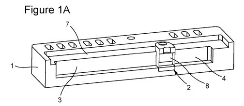
[0055] Figure 1A shows a base 1 fitted with an elution module 2. The elution
module 2 divides
the central channel into two compartments 3, 4. The two membranes bound a
sample compartment
8. In Figure 1B, a block of agarose 5 is cast next to the elution module 2 and
buffer is added to
fill the chambers 3, 4. If the buffer level is below the shelf 7, there is no
bulk flow between the
chambers 3, 4, but if the buffer is higher than shelf 7, liquid can flow
between the buffer chambers
3, 4. Nonetheless, there is a continuous fluid path for electrophoresis, as
the membranes are
permeable to ions and current. Figure 1C shows electrode holders 6 with
platinum wire added to
the configuration(s) shown in Figures 1A-B. The platinum wire is connected to
a power supply.
A sample is added to the sample compartment 8 via porthole 9.
[0056] As shown in Figures 2A-F, the cassette may consist of an elution
module, a base, and
electrode holders. Figure 2A shows an exploded view of the elution module
showing membranes
1A, 1C and acrylic elution module 1B. The assembled elution module is shown in
Figure 2B,
where the membranes (1A, 1C) are sealed the acrylic elution module (1C) by
heat bonding. An
exemplary base is shown in Figure 2C, while Figure 2D shows the base with the
elution module
(of Figure 2B) inserted therein. In some embodiments, the elution module may
be held down by
two screws, as shown in Figure 2D. Figure 2E shows an electrode holder 6. The
electrode holder
of Figure 2E may be inserted into the base, as shown in Figure 2F. As shown,
several electrode
holders 6 may be inserted into the base. In an exemplary embodiment, one or
more electrode
holders may be placed on a first side of the elution module, and one or more
electrode holders may
be placed on a second side of the elution module. Casting dams may also be
provided (Figure 9)
to allow casting of agarose gels in the cassette.
[0057] The elution module, as shown in Figure 3, may consist of two
rectangular pieces of
membrane which are heat bonded (and/or otherwise attached) to a central
plastic piece. The first
membrane 1 may allow passage of DNA and protein, such as Durapor. The central
piece may
comprise the elution module body 2. The elution module body may be machined
acrylic. The
7
CA 03036932 2019-03-13
WO 2018/067736 PCT/US2017/055193
body 2 may have at least one hole 3 configured to pass a screw, such as an M2
or M3 screw. The
screws may be used to hold the elution module in place when the elution module
is screwed into
the body (e.g., Figure 2C). The body may also have a porthole 4 and a central
channel 5. A second
membrane 6 may be configured to retain DNA molecules, such as, for example, a
membrane
having a 10kd cutoff PES. When assembled, as shown in Figure 3B, the membranes
1, 6 enclose
the channel 5 to form a space that is bounded on two sides by membrane. This
is shown in cross
section in Figure 4B (6).
[0058] As discussed above, the two membranes, which are bonded to the elution
module, may
have different properties. One membrane is chosen to retain molecules of
interest. For example,
a first membrane may be a PES (poly ether sulfone) membrane rated to retain
molecules larger
than 10,000 Daltons in mass, and the other membrane may be chosen so that DNA
molecules can
pass through the membrane, e.g., a membrane rate to have pores with a nominal
0.5 micron size.
[0059] The nominal or rated properties of the membranes may vary. Both
membranes may be
permeable to water and ions, so that the electric field can pass through the
elution module. One
membrane may retain molecules of interest, while the other membrane may be
relatively porous,
as explained below.
[0060] In some embodiments, a porous membrane, may be Durapor PVDF HVPP
membrane
(EMD Millipore Corporation, Chicago IL 60673) with 0.45 micron pores, or
Durapor with 5.0
micron pores (Millipore type SVPP, Catalog Number: SVLP09050). The nonporous
membrane
may be Biomax PES, catalog SF1J007A1. Other membranes that may be used,
including
membranes of regenerated cellulose. Some such membranes are described in
Millipore:
Ultrafiltration Membranes: Ultrafiltration membranes for Macromolecule
Processing Product
Selection Guide, April 2008, Millipore Corporation, which is incorporated
herein by reference in
its entirety.
[0061] As shown in Figure 3A-B, the elution module has a porthole 4 which
allows liquid to be
added or removed from the membrane bounded space; the module also has holes 3
which allow
the module to be attached to the base with screws (as shown in Figures 2D, 2F,
7).
[0062] Electrode holders are fitted with platinum wire, and are inserted into
slots in the base
(Figures 2F, 6).
8
CA 03036932 2019-03-13
WO 2018/067736 PCT/US2017/055193
[0063] As shown in Figures 4A, the base 1 may have a central channel 4, an
oval slot 5 for holding
the elution module, and slots 2 to hold the electrode holders. The base may
also have a cutout 3
for a lid.
[0064] Figure 4B shows an elution module 6 inserted in the base 1. The central
channel in the
base is divided into two buffer chambers 4a and 4b by the elution module 6. In
the elution module
6, there is a sample compartment 7 bounded by membranes. When liquid is added,
the buffer
chambers 4a and 4b, and the sample compartment 7 form a linear liquid path.
The membranes
substantially block bulk fluid flow, but allow ions and other molecules to
pass when current is
applied.
[0065] Figure 5A shows an electrode holder 1 according to some embodiments.
The electrode
holder 1 may be configured with a tab 3 for holding electrode wire, which may
also have at least
one hole 2 to receive the electrode wire. Figure 5B shows the tab with wire 5
wrapped around the
tab and through holes 2. In some embodiments, the electrode wire is platinum.
The electrode
holder 1 may also be configured with a tab 4 (Figure 5A, B) that is configured
to fit in one of the
slots 7 the base 8, as shown in Figure 5C.
[0066] Figure 6A shows the base 1 with electrode holders 2 and elution module
3 inserted therein.
Figure 6B shows a membrane 6 affixed to the elution module 3, and the elution
module 3 is held
in place using screws 4. As shown, the elution module 3 has a porthole 5.
[0067] Figure 7A shows a lid 1 (also referred to herein as a "cover") that is
configured to fit into
the base 2 shown in Figure 7B. In some embodiments, the lid may cover
substantially all of the
base. In the shown embodiment, the cover 1 is configured to fit in an inset
portion of the base 1.
The base 100 may have an opening configured to fit around the elution module
and openings
configured to receive the electrode holder tabs 3 (Figure 5A). Figure 7C shows
the base
configured with the elution module, the cover, and the electrode holders.
[0068] Figures 8A-D show an embodiment of an exploded view of an elution
module (Figure
8A), the elution module (Figure 8B), a base (Figure 8C), and the elution
module configured
within the base (Figure 8D).
[0069] Figure 9A shows a device ready for casting of agarose. The elution
module 1 is inserted
into the base 2, and fitted with a casting dam 3. In some embodiments, the
casting dam 3 may be
9
CA 03036932 2019-03-13
WO 2018/067736 PCT/US2017/055193
sized to make a gap 4 of about 1 cm between the casting dam 3 and the elution
module 1. As
shown in Figure 9B, a Pasteur pipet 5 may be used to add agarose to the space
4 between the
casting dam 3 and elution module 1. Figure 9C shows that the casting dam 3 has
been removed,
and a block of agarose 6 remains in the gap 4. Any extra agarose 7 may be
trimmed before use.
[0070] In some embodiments, an elution membrane may be inserted into the base
(Figure 1A) and
a block of agarose may be cast (Figure 9B and Example /) by adding a casting
dam 3 (Figure 9)
and then molten agarose solution; the agarose gels to from a hydrogel that is
continuous with the
adjacent membrane. As noted above, the two membranes may be different ¨ e.g.,
one is relatively
porous (nominal 0.5 micron pores) and one retains DNA molecules (10,000 Dalton
nominal
rating). The block of agarose may be cast next to the porous membrane.
[0071] The buffer chambers 3, 4 (Figure 1C) may be filled with electrophoresis
buffer, and
sample may be added through the porthole 9 into the elution module. Figure 10A
shows the buffer
added after the agarose as gelled. The buffer may be added to the buffer
chamber that is adjacent
to the agarose. Buffer may also be added to the other buffer chamber, as shown
in Figure 10B.
As shown in Figure 10C, electrode holders (with platinum wire as conductor)
may be added, and
the wires are attached to a power supply.
[0072] This is also shown in Figures 11A-C. In Figure 11A, the base 1 is
fitted with elution
module 2. The elution module divides the central channel in two compartments
3, 4 such that each
membrane of the elution module 2 forms an end of each compartment 3, 4. A
block of agarose 5
is cast next to the elution module, as shown in Figure 11B. As shown in Figure
11C, the electrode
holders 6 may be added. In some embodiment, one electrode holder is configured
on each side of
the elution module 3 and a tab of each electrode holder is inserted into its
respective chamber 3, 4.
In other embodiments, more than one electrode holder is configured on one or
both sides. The
liquid can flow between buffer chambers 3, 4 only if the liquid level exceeds
the height of the shelf
7.
[0073] When current is applied, e.g., with a positive electrode in chamber 3;
thus, negatively
charged particles such as cells or DNA migrate toward the agarose/membrane
side of the sample
compartment. Since the pores in agarose gels are smaller than cells, the cells
will become entangled
near or at the surface of the agarose coated membrane. Sodium dodecyl sulfate
("SD S") is then
added into the elution module via the porthole (Figures 1C, 9) and
electrophoresis continues.
CA 03036932 2019-03-13
WO 2018/067736 PCT/US2017/055193
[0074] As the SDS migrates toward the positive electrode, the SDS will
encounter the cells
entangled at the agarose membrane surface. The SDS will cause the cells to
lyse and protein to
become coated with SDS, and the cellular debris and SDS coated protein will
migrate through the
agarose gel and into the buffer chamber 3.
[0075] However, intact chromosomes, either mammalian or bacterial, may not
migrate
appreciably into the gel. As explained in Chapter 7, "preparation,
manipulation, and pulse
strategy for one-dimensional Pulsed-field gel Electrophoresis (OFPFGE)", in
"Pulsed-Field Gel
Electrophoresis", eds M Burmeister and L Ulanovsky, Huuman Press, Totowa New
Jersey, 1992,
which incorporated herein by reference in its entirety, intact mammalian or
bacterial chromosomes
do not migrate appreciably into agarose gels with DC fields; DNA molecules of
up to 6 megabase
pairs will migrate, albeit only under special conditions.
[0076] Thus, intact chromosomes will remain entangled at the agarose/membrane
surface. Since
the chromosomal DNA is near or at the agarose/membrane surface, it is
accessible to enzymes
added to the sample compartment. Such enzyme can diffuse a short distance into
the agarose layer
and act on the entangled DNA molecules. Diffusion of proteins in agarose gels
is discussed in
Pluen, Alain, et al., "Diffusion of Macromolecules in Agarose Gels: Comparison
of Linear and
Globular Configurations," Biophysical Journal, Volume 77, July 1999, pp. 542-
552, incorporated
herein by reference in its entirety (see Figure 2, which shows that proteins
of up to 100,000 Daltons
will diffuse into agarose gels). Thus, addition of an enzyme that makes double
strand breaks in
DNA will convert the immobile, entangled chromosomes into mobile, entangled
shorter
fragments; i.e., as long as the fragments are less then approximately 2
megabase pairs, they will
show some mobility during agarose gel electrophoresis.
[0077] Thus, after adding cells and then SDS, and then enzyme, cut DNA can be
recovered in the
elution module by electrophoresis with the positive electrode in the buffer
chamber 4 (Figure 1A).
The DNA molecules will migrate out of the gel and into the sample compartment
and migrate
toward the non-agarose coated membrane. As noted above, this membrane is
chosen so that DNA
molecules are retained in the sample compartment, and do not pass through the
membrane.
Elution Module Example
11
CA 03036932 2019-03-13
WO 2018/067736 PCT/US2017/055193
[0078] As shown in Figures 3A-B, the elution module may have a plastic body
(2), with a central
channel 5, holes 3, and a porthole 4. The membranes 1 and 6 may be heat staked
to the plastic
body to produce an assembled elution module 7. A cross section of an elution
module inserted in
a base is shown in Figure 4B; the sample compartment 7 may be bounded on both
sides by
membrane and accessible via the porthole.
[0079] The elution module may be affixed to the base, which can be done by,
e.g., gluing,
ultrasonic welding, press fit, etc. For the specific examples below, the
elution module has been
fixed to the base in two different ways.
[0080] In the first method, screws were used. As shown in Figures 6A-B, holes
(Figure 3, 3)
allow two nonconductive screws 4 (nylon M2) to pass through the elution module
into threaded
holes in the base. Alternatively, an elution module without holes can be fixed
to the base with a
clamp. This is shown in Figure 26.
[0081] As noted above, the membranes 1,6 (Figure 3) may be different. One
membrane may be
chosen so that DNA and protein molecules can transit through the membrane
relatively
unhindered. In some embodiments, Durapor PVDF HVPP membrane (EMD Millipore
Corporation, Chicago IL 60673) with 0.45 micron pores may be used; in other
embodiments,
Durapor with 5.0 micron pores (Millipore type SVPP, Catalog Number: SVLP09050
may be used.
The second membrane may be chosen to retain, but not bind, molecules of
interest. In some
embodiments, Biomax PES, catalog SF1J007A10 may be used. The membrane is
bonded so that
the size selective PES surface is on the inside, facing the sample
compartment.
[0082] In some embodiments, prior to use, the PES membrane is made
hydrophilic, so that air
bubbles are not trapped when buffer is added. For example, drop of glycerol
ethanol solution (equal
parts by weight of glycerol and ethanol) may be added to the outer surface of
the PES membrane,
and the elution module is allowed to sit at room temperature for at least
several hours.
[0083] The following Examples correspond to at least some of the embodiments
disclosed above,
the steps/processes also correspond to further embodiments.
Example 1
[0084] Cassette assembly and casting agarose
12
CA 03036932 2019-03-13
WO 2018/067736 PCT/US2017/055193
[0085] An elution module with Durapor and PES membrane was prepared by first
treating the PES
with glycerol/ethanol solution; after a few hours, the elution module was
filled with buffer (0.5X
KBB, sage science; 0.5X KBB contains 51 mM Tris base; 24 mM Taps; 0.08 mM
EDTA),using a
pipet to add liquid through the porthole into the central compartment.
[0086] To demonstrate that the membranes are firmly bonded to the elution
module, slight
pressure was applied by pressing on the liquid at the top of the porthole.
[0087] The buffer is aspirated using a pipettor, and the elution module
carefully dried by blotting
the plastic and Durapor with a paper towel; the PES surface was not touched.
[0088] It is thought that if the Durapor is dry, then when molten agarose
solution is added, the
agarose will be taken up by capillary action into the Durapor, thereby forming
a durable, tight seal
between the agarose and the membrane.
[0089] Figure 9A shows a base 2 with an elution module 1 and a casting dam 3.
The casting dam
is sized so that the gap 4 between the dam and the elution module is 10 mm.
[0090] Figure 9B shows a molten agarose solution (0.75% wt/v seakem gold
agarose (Lonza), in
0.5X KBB buffer (Sage Science; the agarose is dissolved by heating and the
solution stored at 65
degrees centrigrade for up to several days prior to use) being added with a
disposable pasteur pipet
5.
[0091] The agarose is added to be level with the shelf 3 (Figure 4A-B, see
also Figures 1A-C, 7,
Figures 11A-C, 7).
[0092] After the agarose cools to form a gel, the casting dam is removed
(Figure 9C), and extra
agarose 7 which filled the thin space between the casting dam and the base is
removed with a
disposable scalpel.
[0093] In this example, the elution module was simply pressed into the base;
an identical
procedure is used for modules that have screw holes, except that the module is
fixed to the base
with two screws.
Example 2: Size fractionation of purified DNA using a one Dimensional device.
13
CA 03036932 2019-03-13
WO 2018/067736 PCT/US2017/055193
[0094] In this example, we demonstrate that purified DNA can be size
fractionated using a simple,
rapid, high throughput linear device.
Cassette preparation
[0095] An elution module was prepared as described in Example /, except that
it was fixed to the
base with M2 screws.
[0096] Membranes are Durapore PVDF HVPP .45um Roll Stock (EMD Millipore,
Chicago IL)
and PES Biomax 10 kD 27 inches SF1J007A10) Agarose was cast as described in
Example /, and
after the agarose gelled, the casting dam was removed, and 0.5X KBB buffer was
added to the
buffer chambers (Figure 4B, 4a 4b) and buffer was added to the sample
compartment of the elution
module.
[0097] Electrodes were added and connected to a Pippin Pulse power supply
(Sage Science).
[0098] The device was run at 50 V DC, with the positive electrode on the
Durapor side, for a few
minutes to condition the device. The current (measured with a BK precision
Mini-Pro Digital
Multimeter Model 2405A) was 4.5 mA.
Sample Preparation
[0099] Mix 20 microliters of lambda DNA (catalog number N3013, New England
Biolabs,
Ipswich MA, 500 microgram /mL), 20 microliters of 2 log ladder (catalog number
N3200, New
England Biolabs, Ipswich MA, 1,000 microgram /mL), and 410 microliters of TE
buffer (TE buffer
has 10 mM Tris HC1 pH 7.5 and 1 mM EDTA). The DNA concentration was determined
with a
Qubit HS assay (Catalog number: Q32851 ThermoFisher); the result is 52
nanogram/microliter,
which is 78% of the expected value based on the vendors specification.
Sample loading and DNA fractionation
[0100] The elution module sample compartment was emptied using a pipette, and
430 microliters
of sample, 2 microliters of Xylene Cyanol dye solution (10 milligram/mL) and
100 microliters of
TE were added to the sample compartment, and the solution gently mixed.
14
CA 03036932 2019-03-13
WO 2018/067736 PCT/US2017/055193
[0101] Electrophoresis was done for forty minutes at 50 V DC, using a Pippin
Pulse power supply,
with the positive electrode in the buffer chamber next to the agarose coated
Durapor.
[0102] At fifteen minutes, it is observed that the Xylene cyanol dye has
formed a broad band in
the agarose gel next to the Durapor; the band moves to the end of the gel by
forty minutes. The
current drops from 4.5 mA to 2.5 mA during the run.
[0103] The buffer in the elution module was recovered (Fraction 1); the
elution module was rinsed
with 0.5x KBB (Fraction 2); the elution module was filled with .5X KBB and the
porthole was
sealed with a rubber stopper.
[0104] The buffer chambers were rinsed twice with 0.5X KBB and then refilled
with fresh buffer.
[0105] Recovery of DNA still in the agarose gel was by electrophoresis; 50 V
DC for 10 min with
the PES side positive, then one minute using a pulse program of 4 msec fwd/4
msec reverse (In
the Pippin Pulse software, values for the waveform parameters were
4/4/0/0/0/0/1000), then 25 V
DC with the PES side negative, for eight seconds, to back the DNA off the
membrane.
[0106] The DNA in the elution module was recovered as fraction 3.
[0107] The Elution step was repeated 4 more times, and the material recovered
as fractions 4 ¨ 7.
[0108] The concentration of DNA in the different fractions was determined
using the Qubit HS
assay ¨ see Figure 14.
[0109] DNA was examined by agarose gel electrophoresis (0.75% seakem gold
(Lonza), 0.5X
KBB buffer (Sage) using a Sage Pippin Pulse power supply, 100 V DC for 120
minutes; the gel
was stained with Ethidium Bromide and photographed with UV transillumination.
Agarose gel of
size fractionated DNA is shown in Figure 15.
Results
[0110] As shown by agarose gel electrophoresis, the starting material consists
of fragments
ranging in size from 0.1 to 48.5 KBp. If size fractionation has occurred, then
there should be loss
of smaller fragments. As can be seen, fragments smaller than 2 Kbp are not
recovered in the eluted
DNA, thus demonstrating size fractionation with a cutoff between 2 and 3 Kbp.
By Qubit assay,
we recovered 39% of the input sample.
CA 03036932 2019-03-13
WO 2018/067736 PCT/US2017/055193
Example 3: Size fractionation of purified DNA
[0111] A cassette was prepared as described in Example 2.
[0112] The sample in a total volume of 450 uL, contained 7,000 nanograms of E
coli genomic
DNA (Lofstrand Laboratories) and 18,000 nanograms of 2 log ladder (New England
Biolabs,
Ipswich MA, a series of discrete bands from 0.1 to 10 kbp in size); the DNA is
diluted in TE buffer
(10 millimolar Tris HC1, ph 7.5; 1 millimolar EDTA).
[0113] The sample was loaded into an elution module and 100 microliters of TE
buffer was added,
and after mixing 15 microliters was taken and saved as fraction 0 (input).
[0114] The DNAs were size fractionated by electrophoresis, using a pippin
pulse controller, with
the positive electrode on the Durapor side of the EM, using the following
schedule:
[0115] Ten minutes, DC, 50 V
[0116] 110 minutes, pulse field, 40 V. The pulse field was defined by the
following values entered
into the Pippin Pulse software: 150,50,30,10,3,1,81.
[0117] During the DC portion of electrophoresis, the volume in the elution
module decreased, and
at seven minutes, 240 microliters of 0.5X KBB was added.
[0118] At the end of the size fractionation step, the contents of the elution
module were recovered
and saved as Fraction 1, after size step.
[0119] The DNA was then eluted out of the agarose gel into the elution module.
[0120] Using the following electrophoresis schedule, with the positive
electrode on the PES side
Time Voltage Waveform
min
2 50V DC
0.5 50V 4/4/0/0/0/0/1000
80V 300/100/30/10/30/10/45
0.5 50V 4/4/0/0/0/0/1000
8 sec -25 DC (the PES side is negative)
The material in the elution module was recovered and saved as Fraction 3, 1st
elution.
The elution process was repeated three more times.
16
CA 03036932 2019-03-13
WO 2018/067736 PCT/US2017/055193
Results
[0121] Qubit assay ¨ Figure 16.
[0122] Gel electrophoresis ¨ Figures 17 (image without lettering shown in
Figure 29), Figure 18.
[0123] This shows that when the desired high molecular weight E coli genomic
DNA (center of
mass aproximately 30 Kbp) was mixed with undesired low molecular weight DNA (2
log ladder)
and then size fractinated, that fragments smaller then about 12 kbp were
removed
[0124] This demonstrates that the size fractionation cutoff depends on the
electrophoretic
conditions used.
Example 4: Isolation of Bacterial DNA
Device preparation
[0125] Two cassettes were prepared as described in Example /, except the
agarose gel column
next to the Durapor membrane is 0.5cm long.
[0126] Bacterial Growth and Conditions for spheroplast formation
[0127] Strain MG1655 (ATCC 700926), is propagated on M9 minimal plates with 1%
glucose, 1
millimolar Thiamine, 0.2 millimolar magnesium sulfate, 0.1 millimolar calcium
chloride, 0.1% 5-
fluoroorotic acid, and 20 pg/mL uracil.
[0128] Overnight cultures are made by inoculating a single colony into 5-40
mLs of Trypticase
soy broth, and allowing the cells to grow overnight at 37 degrees Centigrade
with shaking.
[0129] Spheroplasts are prepared by incubating E coli cells with lysozyme
[0130] Lysozyme (Epicentre, Ready-LyseTM Lysozyme Solution, catalog number
R1804M,
37,500 units/uL) was diluted 1:40 by mixing 2.5 microliters of lysozyme with
100 microliters of
TES20+BSA buffer. TES20 is 10 millimolar Tris 7.5; 1 millimolar EDTA; 100
millimolar NaCl;
20% w/v sucrose. TES20+BSA is 1 mL of TES20 plus 5 microliters of BSA (New
England
Biolabs, Ipswich MA, 20 milligram/mL)
17
CA 03036932 2019-03-13
WO 2018/067736 PCT/US2017/055193
[0131] The amount of lysozyme needed for lysis was determined as follows: A
series of tubes
were prepared as follows: To a 1.7 mL microfuge tube, 800 microliters of
ACPS20 buffer (10
millimolar Tris HC1 pH 7.5; 5 millimolar EDTA; 20% wt/v sucrose) was added,
followed by 500
microliters of the overnight E coli culture. The tube was mixed (vortex mixer)
and the cells pelleted
by centrifugation (14,000 x g one minute). The supernatant was decanted, and
the cell pellet re-
suspended in 100 microliters ACPS20 by vortexing. As shown in Figure 19,
different amounts of
lysozyme were added, and lysis was checked by taking an aliquot and diluting
1:10 into water;
unlysed cells formed a turbid solution on dilution, while lysed cells form a
clear solution.
[0132] Lysis of cells in mixture of lysozyme and Achromopeptidase (per patent
US 4,900,677,
which is incorporated herein by reference in its entirety). 100 microliters of
a 1 milligram per mL
solution of BSA (New England Biolabs, Ipswich MA) in water was added to one
vial of
Achromopeptidase (Sigma catalog # A3422, 25,000 units, 1 milligram)
[0133] Aliquots were made and stored at -20 degrees Centigrade.
[0134] A series of tubes was prepared as above, and as shown in Figure 20,
lysozme and
achromopeptidase were added. Lysis is checked as above by dilution into water.
[0135] The results show that by itself (tube 12) Achromopeptidase does not
lyse cells, but that
ACP is synergistic with lysozyme, e.g. tubes 7-9 in Figure 20 are more highly
lysed then tubes 1-
3 in Figure 19.
Preparation of spheroplasts and isolation of DNA
[0136] Two tubes of cells were prepared as described above. To tube one, 1.5
microliter of diluted
lysozyme was added; to tube 2 was added 1.5 microliter of lysozyme and 1
microliter of ACP. The
tubes were vortexed and allowed to sit at room temperature for forty minutes.
Sample
[0137] Elution modules were loaded with a mixture of 100 microliters of
spheroplasts and 300
microliters of ACPS20 buffer. After filling the elution modules, 10
microliters was withdrawn and
18
CA 03036932 2019-03-13
WO 2018/067736 PCT/US2017/055193
diluted into 190 microliters of QLB (Qubit lysis buffer, 0.5x KBB, 1% weight
to volume SDS, 5
millimolar EDTA, 50 millimolar NaCl); the tubes were allowed to sit at room
temperature.
[0138] As a tracking dye, 1 microliter of 10 milligram/ml phenol red was added
to each elution
module, and the contents gently mixed with a pipette.
[0139] The Spheroplasts were entangled into the agarose coated membrane with
electrophoresis
(40 V DC, twenty minutes, with the positive electrode on the Durapor side).
During this period,
the phenol red migrated out of the elution module and into the agarose as a
broad band.
[0140] To the elution module, 100 microliter of 10% SDS was added, the
solution was mixed and
electrophoresis continued as above for forty minutes.
[0141] After a few minutes, it was observed that the volume of liquid in the
elution module was
increasing; the porthole in the top of the elution module was sealed with a
rubber stopper.
[0142] It is believed that the increase in volume after addition of SDS is due
to electroendosmosis,
and the net change in liquid reflects the net balance of electroendosmosis.
Electroendosmosis is
due to fixed charges. During the first step, the majority of charges are
negative charges on the PES
membrane. As a result, the net flow of water is through the PES membrane and
out of the elution
module.
[0143] After addition of SDS, positive proteins, which coat DNA in vivo, are
removed. Since the
long DNA molecules are immobile, and have a high net negative charge, they
serve as an
electroendosmotic pump, moving liquid into the chamber.
[0144] At the end of the electrophoresis step, the buffer chambers were washed
by removing and
refilling with 0.5X KBB. The rubber stopper was removed from the elution
module and the
contents aspirated and saved as Fraction 1.
[0145] The elution module was rinsed twice with 0.5x KBB (Fraction 2), and
then with 500
microliters of enzyme reaction buffer (Fraction 3). Enzyme reaction buffer
(ERB) is 0.5x KBB;
32 milligram/mL hydroxy propyl beta cyclodextrin [ACROS Organics, 97%, catalog
#
297560250, CAS 128446-35-5]; 10 millimolar Mg(C1)2; 50 micrograms /mL BSA).
[0146] The elution module was then filled with 500 microliters of ERB to which
had been added
microliters of 20 milligram /mL BSA (New England Biolabs, Ipswich M)); 1.5
microliters of
fragmentase enzyme (New England Biolabs, Ipswich MA); and 1 microliter of T7
Endonuclease
19
CA 03036932 2019-03-13
WO 2018/067736 PCT/US2017/055193
I (New England Biolabs, Ipswich MA). After thirty minutes at room temperature,
15 microliters
of 500 millimolar EDTA was added to the elution module, the contents were
mixed, and the
solution removed (Fraction 4). The elution module was rinsed with 0.5X KBB
(Fraction 5), and
refilled with the same buffer. The digested DNA was recovered by
electrophoresis (50 V DC, two
minutes, PES side positive; thirty seconds with a pulse train of 4 msec
foward/ 4 msec reverse).
The contents of the elution module were removed and saved as Fraction
6/Elution 1
[0147] The elution process was repeated two more times, generate
Fraction7/Elution 2 and
Fraction8/Elution 3.
Qubit analysis of fractions
[0148] The concentration of DNA in each fraction was measured using a Qubit HS
assay.
[0149] From the Qubit HS assay of fraction 0, input, the total amount of DNA
in the spheroplasts
added to the elution module was 40 micrograms.
[0150] As shown in Figure 21, the amount of DNA recovered in the elution
fractions from cells
treated with lysozyme was 5,492 nanograms, 11% of the input.
[0151] The amount of DNA recovered in the other fractions was 7%. Very little
DNA (1% was
recovered in fractions 4 and 5; this shows that after digestion with
fragmentase the DNA is still
entangled in the agarose and not free to diffuse into the sample compartment.
[0152] Similar results were obtained with cells treated with both lysozyme and
achromopeptidase
(Figure 21).
Analysis of DNA by agarose gel electrophoresis
[0153] Figure 22A: analysis of E coli DNA by agarose gel electrophoresis.
[0154] 0.75% seakem gold agarose (Lonza); 0.5X KBB (Sage); a Pippin Pulse
(Sage) was used
with the following waveform parameters: 150; 50; 30; 10; 3; 1; 48.
[0155] The gel was run for 8 hrs at 80 Volts.
Lane Sample
CA 03036932 2019-03-13
WO 2018/067736 PCT/US2017/055193
1 Phage T4 DNA, 166 Kbp (T4 GT7 DNA catalog #318-03971)
2 New England Biolabs, Ipswich MA) 1 kb extend marker (catalog # N3239S)
3 Elution 1 from cells treated with both lysoszyme and achromopeptidase
4 Elution 2 from cells treated with both lysoszyme and achromopeptidase
Elution 3 from cells treated with both lysoszyme and achromopeptidase
6 Elution 1 from cells treated with both lysoszyme
7 Elution 2 from cells treated with both lysoszyme
8 Elution 3 from cells treated with both lysoszyme
9 New England Biolabs, Ipswich MA) 1 kb extend marker (catalog # N3239S)
[0156] The majority of the DNA migrates as a band at the limit mobility,
aproximately 45 Kbp
[0157] Figure 22B: analysis of E coli DNA by agarose gel electrophoresis.
[0158] 0.75% seakem gold agarose (Lonza); 0.5X KBB (Sage); a Pippin Pulse
(Sage) was used
with the following waveform parameters: 300; 100; 30; 10; 30; 10; 45.
[0159] The gel was run for 12 hrs at 80 Volts.
Lane Sample
1 New England Biolabs, Ipswich MA) 1 kb extend marker (catalog # N32395)
2 Elution 1 from cells treated with both lysoszyme
3 Elution 2 from cells treated with both lysoszyme
4 Elution 1 from cells treated with both lysoszyme+achromopeptidase
5 Lambda DNA ladder (Lambda DNA, 48.5 Kb, ligated following the protcol
described in
Nucleic Acids Res. 1990 May 25;18(10):3090.
6 T4 DNA
[0160] See Figure 23.
[0161] E coli DNA from oneD analyzed by agarose gel electrophoresis.
[0162] 1% SGK gel, 0.5X KBB with BioRad CHEF mapper, program Molecular weight:
low 50
K, high 1000 K; Gradient: 6 V/cm; Angle: 120; Run time: 14: 54; Initial switch
time: 6.75 s; Final
switch time: 1 m 33.69 s; Ramping factor: linear
Lane Sample
1 Yeast Chromosome ladder (New England Biolabs, Ipswich MA))
2 Elution 1 from cells treated with lysoszyme
3 Elution 2 from cells treated with lysoszyme
4 Lambda Ladder (New England Biolabs, Ipswich MA))
21
CA 03036932 2019-03-13
WO 2018/067736 PCT/US2017/055193
Example 5: Isolation of high molecular weight DNA from white blood cells.
Device
[0163] As shown in Figure 7A-B, a lid 1 is fitted to a base 2; silicone grease
is applied to shelf 4.
The lid serves to divide the buffer chambers into separate anonic and cathodic
compartments that
can communicate only through the elution module.
[0164] The lid also serves to define the top surface of the agarose gel.
Assembly
[0165] A solution of Glycerol/Et0H is applied to the PES membrane of an
elution module. After
the Et0H evaporates, the elution module is filled with .5X KBB buffer and
examined for leaks.
The elution module is then dried by removing the buffer and carefully blotting
dry with a paper
towel; the elution module is placed in the base.
[0166] A small amount of silicone grease is applied to the shelf in the base,
and the lid is the added;
the assembly is held together with spring clamps. A casting dam is used to
form an agarose block
next to the Durapor membrane; after the agarose gels, it is trimmed to a 5 mm
long block.
[0167] The device is then filled with buffer and run for a few minutes at 50 V
DC, with the positive
electrode on the Durapor side. The current is observed to be 4.3 mA
White Blood Cells
[0168] All steps at 4 degrees centigrade.
[0169] White blood cells (also referred to herein as "WBCs") are prepared from
whole blood from
goats (Lampire, 3599 Farm School Rd, Ottsville, PA 18942) with ACD
anticoagulant. To 37 mL
cold RBC lysis buffer (1X buffer is 155 millimolar Ammonium Chloride; 10
millimolar NaHCO3;
1 millimolar Na2EDTA) was added 10 mL of whole blood; tubes were mixed by
inversion, and
incubated for five minutes at 4 degrees centigrade with occasional mixing. The
WBCs are
recovered by centrifugation (2,400 x g for four minutes.)
22
CA 03036932 2019-03-13
WO 2018/067736 PCT/US2017/055193
[0170] The supernatant was decanted and the reddish pellet of white cells
washed by re-
suspending (vortex) in 20 mL RBC lysis buffer and centrifugation at 2,200 x g
for two minutes.
[0171] The wash step is repeated 2-3X until the cell pellet has only a trace
of red color.
[0172] The cells are re-suspended in 1.5 mL of FSE (50% v/v Sage Ficoll
loading buffer; 80
milligram/mL sucrose; 10 millimolar EDTA) and filtered (40 micron sterile cell
strainer, Fisher
Scientific catalog # 22363547).
[0173] The white blood cells can be stored at 4 degrees centigrade for several
days. If the cell
suspension is not a homogeneous, creamy solution it is vortexed or re-
filtered. If re-filtered, the
concentration of cells or DNA needs to be re-measured.
Quantification of DNA with a Qubit HS assay.
[0174] Gently mix the WBCs by swirling the tube, and transfer 10 microliters
to a microfuge tube;
then add 190 microliters of Qubit lysis buffer and mix by pipetting; the
solution will become
snotty. Incubate at 58 degrees centigrade for ten minutes. Cool to room
temperature, add 600
microliter of TE and vortex full speed for ten seconds.
[0175] Assay 0.5 to 1 microliter of the lysed cell mix with a Qubit HS assay.
The expected
concentration of DNA is 200 ¨ 300 nanogram/uL
Quantification of Cells by Cell counting
[0176] A BioRad TC20 automated cell counter was used to determine total cell
counts and percent
viability with trypan blue, following the vendor's directions.
Load cells and isolate DNA
[0177] To prepare the sample, mix 75 microliters WBCs (69,000 cells/uL,
nominal 416 nanogram
per microliter of DNA, assuming 6 picogram /cell) with 350 microliters of SEK
buffer (0.5x KBB;
millimolar EDTA; 80 milligram/mL sucrose; 10 microgram /mL phenol red) and add
to the
elution module.
23
CA 03036932 2019-03-13
WO 2018/067736 PCT/US2017/055193
[0178] To determine the concentration of DNA in this solution (the input), two
10 microliter
aliquots were withdrawn and added to 190 microliters of qubit lysis buffer;
the solutions were
vortexed and stored at room temperature until assayed with a Qubit HS assay.
[0179] The sample was Electrophoresed for thirteen minutes at 50 V DC, with
the positive
electrode on the Durapor side. After thirteen minutes, 150 microliter of 10%
SDS was added to
the elution module and the solution mixed gently. A 20 microliter aliquot
(Fraction 0) was taken
and added to 190 microliter of qubit lysis buffer to determine the
concentration of DNA.
[0180] The elution module was sealed with a stopper, and electrophoresis was
continued for
another ten minutes at 50 V DC. The buffer in the buffer chambers was
replaced, and
electrophoresis continued for another ten minutes.
[0181] The contents of the elution module were removed, and 12 microliter of
500 millimolar
EDTA was added; this is Fraction 1, post SDS. The elution module was rinsed
with KBB and the
liquid saved as Fraction 2, post SDS rinse.
[0182] The buffer chambers were rinsed three times to remove SDS, and fresh
buffer was placed
in the buffer chambers. The elution module was rinsed with 500 microliter of
ERB (Fraction 3,
ERB rinse). DNA was digested by adding 500 microliter of ERB, to which had
been added 5
microliters of 20 mg/mL BSA (New England Biolabs, Ipswich M)), 1.5 microliters
of Fragmentase
enzyme (New England Biolabs, Ipswich MA), and 0.5 microliter of T7
Endonuclease I (New
England Biolabs, Ipswich MA); incubation was for ten minutes at 37 degrees
Centigrade (the entire
device was placed on a thermostatted aluminum plate (Benchmark "myBlock" dry
block heater
unit). At the end of the incubation, 15 microliter of .5 M EDTA was added to
the elution module,
the contents were gently mixed, and the solution was aspirated from the
ELUTION MODULE and
saved as Fraction 4, ERB.
[0183] DNA was then recovered by electroelution; the elution module was filled
with 500
microliters of 0.5X KBB and 5 microliters of 0.5 M EDTA, and voltage applied
(50 V DC, 90
seconds, with the positive electrode on the Durapor side); the solution was
recovered as Fraction
5, elution 1.
[0184] The elution module was refilled with .5x KBB, and DNA eluted for two
minutes, 50 V DC.
24
CA 03036932 2019-03-13
WO 2018/067736 PCT/US2017/055193
[0185] The solution was saved (Fraction 6, elution 2). The elution module was
refilled, and
electrophoresis applied for four minutes, followed by five seconds of 25 V DC
with the positive
electrode on the Durapor side (reverse current pulse, to back DNA off of the
PES membrane). The
device was allowed to sit at room temperature, covered to avoid evaporation,
overnight; the
material in the ELUTION MODULE was recovered the next day (Fraction 7, elution
3).
Results
[0186] The amount of DNA in each fraction was determined using a Qubit HS
assay.
[0187] See Figure 24.
[0188] As shown in Figure 25, 20,663 ng of DNA were loaded into the elution
module, and that
after thirteen minutes of electrophoresis at 50 V DC, Durapor side positive,
only 33% of the DNA
(6,800 ng) was present in the elution module in a form that could be recovered
after adding SDS.
This suggests that the DNA was bound or entangled in some form on either the
Durapor or agarose,
or both, and that this DNA was not released by washing (Fractions 2, 3) or
treatment with Enzyme
(Fraction 4).
[0189] The results show that 39% (8,100 ng) of the input DNA could be
recovered in the elution.
[0190] The size of the DNA in each fraction was determined by agarose gel
electrophoresis.
[0191] 30 microliters of fractions 4,5,6, and 7 were analyzed on a Pulse field
agarose gel (BioRad
CHEF mapper; program is default for separation of 50 to 1,000 KBp fragments,
with a time factor
of 0.5; the gel is 0.75% seakem gold agarose (Lonza) in 0.5X KBB buffer
(Sage); the running
temperature is 14 degrees Centigrade.
Lane sample
1 1 Kb extend ladder (New England Biolabs, Ipswich MA))
2 T4 phage DNA
3 Ladder of Lambda phage DNA (New England Biolabs, Ipswich MA))
4 Empty
Fraction 4, enzyme
6 Fraction 5, first elution (90 seconds)
7 Fraction 6, 2nd eltution (2 min)
8 Fraction 6 3rd elution (4 min)
CA 03036932 2019-03-13
WO 2018/067736 PCT/US2017/055193
[0192] The results show that high molecular weight DNA, with a center of mass
at approximately
160 (Fraction 5) to 300 kb (fraction 6) was obtained. In fraction 7, some DNA
is visible at limit
mobility (¨ 2 Mbp in this gel system).
Example 6: Isolation of High Molecular Weight DNA from White Blood Cells
[0193] In this example, rapid, high yield recovery of DNA from white blood
cells, using a device
configuration where there is a single buffer chamber, is demonstrated ¨ that
is, there is no barrier
between the anodic and cathodic buffer chambers.
Device assembly
[0194] As shown in Figure 26A, an elution module was inserted in the base, and
a casting dam
was placed manually approximately 5 mm from the elution module; agarose was
added with a
pipet to fill the space up to the lid shelf
[0195] After the agarose gels, the dam was removed and buffer (0.5x KBB) was
added to fill the
buffer chambers almost up to the top of the elution; the buffer flows freely
around the side of the
ELUTION MODULE on the shelf.
[0196] The ELUTION MODULE is filled with buffer prior to use.
[0197] To ensure that the elution module is firmly in place, a spring clamp
was added, as shown
in Figure 26B.
[0198] Device in use; the base rests on metal blocks for cooling; two
electrode holders with
platinum wire are shown; a clamp holds the elution module in place
[0199] Buffer can flow between the two electrodes around the side of the
elution module.
Sample
[0200] WBCs from goat whole blood were prepared as described in Example 3. Two
aliquots of
Cells were diluted into TBS and counted with a BioRad cell counter, following
manufacturer's
instructions. The results were:
26
CA 03036932 2019-03-13
WO 2018/067736 PCT/US2017/055193
SampleCell s/uL %viable DNA, ng microliter(1)
1 79,000 47
2 108,000 47
Avg 93,500 47 561
(1)Assuming 6 pg of DNA per cell
Cells were stored overnight at 4 degrees Centigrade, and then recounted
SampleCell s/uL %viable ng/uL
1 84,000 35
2 80,000 35
Avg 82,000 35 490
[0201] 41 microliters of cells (20 microgram of DNA) were mixed with 290
microliters of SEK
Buffer (0.5x KBB; 5 millimolar EDTA; 80 milligram/mL sucrose; 10 microgram /mL
phenol red)
and added to the elution module. To determine the starting concentration of
DNA ("input"), two
microliter aliquots were taken and each aliquot was diluted into 190
microliters of QLB (Qubit
lysis buffer, 0.5x KBB, 1% SDS, 5 millimolar EDTA, 50 millimolar NaCl);
samples were mixed
and stored at room temperature.
[0202] Cells were electrophoresed into the agarose coated Durapor;
electrophoresis was 50 V DC,
with the positive electrode on the Durapor side of the ELUTION MODULE. The
current was 6.2
mA.
[0203] After eight minutes thirty seconds, electrophoresis was paused; the
phenol red dye in the
SEK had migrated out of the ELUTION MODULE and halfway thru the agarose as a
broad band.
[0204] To the elution module was added 80 microliter of 10% SDS and 160
microliter of .5X
KBB; the elution module contents were mixed gently and the porthole sealed
with a rubber stopper.
27
CA 03036932 2019-03-13
WO 2018/067736 PCT/US2017/055193
[0205] Restart electrophoresis at 50 V; the current was 9.8 mA; switch to 40
V; the current was
7.6 mA.
[0206] It is observed that addition of SDS to the elution module usually
causes the current to rise.
Since the joule heat is proportional to the current, the voltage is decreased
to avoid overheating.
[0207] Continue electrophoresis for a total time of twenty minutes, then
pause.
[0208] Remove the rubber stopper, and take 10 microliter from the ELUTION
MODULE; add to
90 microliter QLB as Fraction 1, "post SDS lysis step".
[0209] Rinse the buffer chambers 3X with fresh buffer to remove SDS from the
device.
[0210] Rinse the ELUTION MODULE 2x with buffer, and save as Fraction 2, "post
SDS rinse".
[0211] Rinse a 3rd time with KBB, and save as Fraction 3, "post SDS rinse".
[0212] Mix 500 microliter of ERB (5x KBB; 32 milligram/mL hydroxy propyl beta
cyclodextrin;
millimolar Mg(C1)2; 50 microgram /mL BSA) with 5 microliter of 20 mg/mL BSA
(New
England Biolabs, Ipswich MA)); 1 microliter of T7 endonuclease I (New England
Biolabs, Ipswich
MA)); and 1 microliter of Fragmentase (New England Biolabs, Ipswich MA)); add
the
ERB/enzyme cocktail to the ELUTION MODULE and let sit room temperature for 36
minutes.
[0213] Recover the solution; add 15 microliter of .5 M EDTA, and save as
Fraction 4 "Enzyme
mix".
[0214] To 500 microliter of .5X KBB add EDTA to 25 millimolar and DTT to 5
millimolar; add
to the ELUTION MODULE and let sit for five minutes; recover and save as
Fraction 5, "Post
enzyme wash".
[0215] Add 400 microliter of KBB and elute DNA with electrophoresis (two
minutes, 40 V DC,
Durapor side negative). Recover the sample and save as Fraction 6, "elution 1"
[0216] Repeat the elution process; observe that the second elution is
"snotty," and indication of
high molecular weight DNA; savve as Fraction 7, "elution 2"
[0217] 400 microliter of KBB was added to the elution module, and DNA eluted
with the
following program, using a Pippin pulse controller.
Step Electrophoresis
28
CA 03036932 2019-03-13
WO 2018/067736 PCT/US2017/055193
1 50 V DC 2 min
2 50 V with a pulse of 4 msec fwd/4 msec rev (forward is with the PES side
Positive)
3 50 V DC 2 min
4 50 V pulse 2 min
50 V DC 2 min
6 50 V pulse, 12 minutes
Recover the material in the elution module as Fraction 8, "elution 3"
Results
[0218] The amount of DNA in each fraction was determined using a Qubit HS
assay.
[0219] See Figure 27.
Conclusion
[0220] The data shows that we can recover 30% of the input DNA, using a format
where there is
a single buffer chamber that is not divided into an anodic and cathodic
compartment.
[0221] Further, the data shows that the volume of liquid in the elution module
sample compartment
changes during electrophoresis. During the first step, when cell are migrating
to, and becoming
entangled in the agarose coated membrane, the volume in the sample compartment
decreases.
[0222] During the second step, after SDS is added, it is observed that after a
few minutes the
volume starts to increase.
[0223] We believe that changes in volume are due to electro endosmosis. In the
first step, we
believe that the majority of fixed charges are negative charges on the PES
membrane; this causes
water to be pumped through the PES and out of the elution module.
[0224] After addition of SDS, chromosomal DNA is freed from positively charged
proteins (e.g.,
histones); as a result, the immobile, entangled chromosomal DNA acts as a
fixed negative charge,
pumping buffer into the elution module.
Example 7, prophetic: A device designed to run multiple samples simultaneously
29
CA 03036932 2019-03-13
WO 2018/067736 PCT/US2017/055193
[0225] One advantage of our system for purifying DNA is that it is adaptable
to handle large
numbers of samples.
[0226] Large numbers of samples are common (Ledford, Heidi, "AstraZeneca
launches project to
sequence 2 million genomes," Nature: International Weekly Journal of Science,
532, 427, April
28, 2016, incorporated herein by reference in its entirety) thus there is a
need for systems that can
handle hundreds or thousands of samples reliably, rapidly, and at low cost.
[0227] The demand for large sample capacity is evident from the large number
of products sold
for laboratory automation (e.g., Tecan Liquid Handling and Robotics product
lines, incorporated
herein by reference in its entirety).
[0228] Such products automate steps such as liquid handling, moving
disposables such as
cassettes, and collection and analysis of data.
[0229] Figures 12-13 show a cassette configured to hold four elution modules,
which allows for
analysis of four samples at a time. The cassette is fitted with four elution
modules, and four agarose
gels are cast, one for each elution module; buffer is added and the cassette
is sealed. Procedures
for automated casting of agarose gels, filling of cassettes with buffer and
cassette sealing are
utilized by Sage Science for production of cassettes for the Pippin and ELF
instruments (e.g.,
SageHLS High Molecular Weight Library System, PippinHT DNA Size Selection
System,
SageELF Sample Fractionation System, BluePippin Size Selection System, Pippin
Prep DNA Size
Selection System, SageHLS, PippinHT, SageELF, Pippin Prep, BluePippin, all of
which are
incorporated herein by reference in its entirety).
[0230] Cassettes, filled with buffer and sealed, are suitable for storage
until needed.
[0231] Automation of liquid handling steps for devices such as the cassette
shown in Figure 13
can be accomplished with liquid handling robots (e.g., Tecan Freedom EVO
Series, incorporated
herein by reference in its entirety). Such robots can be configured to hold
various samples and
reagents, and to deliver reagents to a disposable cassette, such as the one
shown in Figure 13.
[0232] This allows for the automation of all the liquid handling steps
required for isolation of
DNA.
[0233] In addition, a means of providing electrodes may be used. Sage Science
has demonstrated
(HLS) instruments which have electrodes on a movable lid
CA 03036932 2019-03-13
WO 2018/067736 PCT/US2017/055193
Example 8, electrophoresis of SDS in a cassette with three buffer chambers
[0234] See Figure 28.
[0235] A base 4 was fitted with an elution module 13 with a sample compartment
5 and a porthole
6. A block of agarose 7 was cast next to the elution module using a casting
dam as described in
Example 6.
[0236] An M2 screw, nylon, 8, was added to the threaded hole 14, and then two
casting dams were
placed in the base 4 around the screw 8, and molten agarose was added; after
the agarose hardened
to form a block 9, the casting dams were removed, and the buffer chamber 11
was filled with
buffer.
[0237] After twenty minutes, it was observed that very little liquid was
present in buffer chamber
10. We infer that the agarose block 9 forms a seal separating buffer chambers
10 and 11.
[0238] A similar experiment, but without the screw 8 and screw hole 14
resulted in a block of
agarose 8 that did not form a seal.
[0239] The sample compartment 5 and the buffer chambers 10,11,12 were filled
with buffer, and
electrodes 1,2,3 were added.
[0240] A small amount of tracking dye (Xylene Cyanol) was added to the sample
compartment so
that the solution was easily visible to the naked eye.
[0241] A Pippin pulse power supply was used to supply 50 V DC between
electrodes 2 and 3, with
electrode 2 positive. It was observed that the Xylene Cyanol tracking dye
moved from the sample
compartment 2, through the agarose block 7 and into the buffer chamber 11.
[0242] A pippin pulse power supply was then connected to electrodes 1 and 2,
with electrode 1
positive, and 50 V DC was applied. It was observed that the tracking dye moved
out of buffer
chamber 11, through agarose block 7, and into buffer chamber 10; the solution
in buffer chamber
11 changed from dark blue to clear.
[0243] We infer that this sequential electrophoresis allowed us to transport,
by electrophoresis,
negatively charged Xylene Cyanol molecules (xylene cyanol has a net negative
charge, Ter Ming
Tan, Timothy, et al., "Gel Electrophoresis: DNA Science without the DNA!,"
Biochemistry and
31
CA 03036932 2019-03-13
WO 2018/067736 PCT/US2017/055193
Molecular Biology Education, vol. 35, No. 5, pp. 342-349, 2007, incorporated
herein by reference
in its entirety) from the sample compartment and sequester them in the buffer
chamber 10.
Example 9: prophetic, isolation of DNA without washing of the cassette.
[0244] In Example 6, we describe isolation of high molecular weight DNA. In
that example, SDS
is used to deproteinize the cells, and the SDS, and SDS coated protein, is
removed from the cassette
so that enzymatic digestion can occur, and so that the SDS and SDS coated
protein do not
contaminate the purified DNA during the elution step.
[0245] During this process, washing was used to remove SDS and SDS coated
protein from the
buffer chambers, so that the SDS and SDS coated protein would not contaminate
the DNA during
elution.
[0246] In this example we use electrophoretic transport to sequester the SDS
and SDS coated
protein, so that washing is not needed.
[0247] A cassette is prepared as described with respect to Figure 28, and a
sample is prepared and
loaded as described in Example 6.
[0248] Electrophoresis to entangle cells in the agarose coated membrane is as
described in
Example 6, except that electrodes 2, 3 (Figure 28) are used. SDS is added as
described in Example
6, and electrophoresis is used to cause cell lysis and deproteinization, as
described in Example 6,
except that electrodes 2, 3 are used.
[0249] Current (50 V DC) is then applied between electrodes 1, 2, with
electrode 1 positive, for 1
hour to transport SDS and SDC coated protein, from buffer chamber 11, through
agarose gel 9 and
into buffer chamber 10.
[0250] The DNA, which is entangled in the agarose coated membrane, is then
treated with
fragmentase, as described in Example 6, except that the buffer chambers are
not washed to remove
SDS. The DNA is recovered as describe in Example 6, except that electrodes 2,
3 are used, with
electrode 3 positive.
[0251] This example shows purification of high molecular weight DNA from a
cellular sample
without the need to wash the cassette to remove SDS or other contaminants.
32
CA 03036932 2019-03-13
WO 2018/067736 PCT/US2017/055193
Other applications
[0252] This application is also related to:
= US Application No. 15/183,097, filed June 15, 2016
= US Application No. 14/297,001, filed June 5, 2014
= US Application No. 13/751,606, filed January 28, 2013
= US Application No. 12/760,548, filed April 14, 2010
= US Application No. 12/576,148, filed October 8, 2009
= US Provisional Application No. 61/150,243, filed February 5, 2009
= US Provisional Application No. 61/195,566, filed October 8, 2008
= US Application No. 15/464,278, filed March 20, 2017
= US Application No. 14/051,300, filed October 10, 2013
= US Provisional Application No. 61/766,910, filed February 20, 2013
= US Provisional Application No. 61/713,916, filed October 15, 2012
= US Provisional Application No. 61/713,156, filed October 12, 2012
= US Application No. 15/519,516, filed April 14, 2017
= PCT Application No. PCT/U52015/055833, filed October 15, 2015,
= US Provisional Application No. 62/183,514, filed June 23, 2015
= US Provisional Application No. 62/064,454, filed October 15, 2014
[0253] The aforementioned applications are all expressly incorporated by
reference herein in their
entireties.
[0254] Any and all references to publications or other documents, including
but not limited to,
patents, patent applications, articles, webpages, books, etc., presented in
the present application,
are herein incorporated by reference in their entirety.
[0255] Example embodiments of the devices, systems and methods have been
described herein.
As noted elsewhere, these embodiments have been described for illustrative
purposes only and are
not limiting. Other embodiments are possible and are covered by the
disclosure, which will be
apparent from the teachings contained herein. Thus, the breadth and scope of
the disclosure should
not be limited by any of the above-described embodiments but should be defined
only in
accordance with claims supported by the present disclosure and their
equivalents. Moreover,
embodiments of the subject disclosure may include methods, systems and devices
which may
33
CA 03036932 2019-03-13
WO 2018/067736 PCT/US2017/055193
further include any and all elements from any other disclosed methods,
systems, and devices,
including any and all elements corresponding to molecular processing. In other
words, elements
from one or another disclosed embodiments may be interchangeable with elements
from other
disclosed embodiments. In addition, one or more features/elements of disclosed
embodiments may
be removed and still result in patentable subject matter (and thus, resulting
in yet more
embodiments of the subject disclosure). Correspondingly, some embodiments of
the present
disclosure may be patentably distinct from one and/or another reference/prior
art by specifically
lacking one or more elements/features of a system, device and/or method
disclosed in such prior
art. In other words, claims to certain embodiments may contain negative
limitation to specifically
exclude one or more elements/features resulting in embodiments which are
patentably distinct
from the prior art which include such features/elements.
34