Note: Descriptions are shown in the official language in which they were submitted.
1 CA 03036941 2019-03-14
- 1 -
Description
THERAPY FOR DRUG-RESISTANT CANCER BY ADMINISTRATION OF
ANTI-HER2 ANTIBODY/DRUG CONJUGATE
Technical Field
[0001]
The present invention relates to the treatment of
drug-resistant cancer (hereinafter, also simply
referred to as "resistant cancer"), in particular,
cancer that has acquired resistance, with an antibody-
drug conjugate having exatecan conjugated to an anti-
HER2 antibody via a linker structure.
Background Art
[0002]
An antibody-drug conjugate (ADC) having a drug
with cytotoxicity conjugated to an antibody, whose
antigen is expressed on the surface of cancer cells and
which also binds to an antigen capable of cellular
internalization, and therefore can deliver the drug
selectively to cancer cells, is thus expected to cause
accumulation of the drug within cancer cells and to
kill the cancer cells (Non-patent Literatures 1 to 3).
As an ADC, Mylotarg (registered trademark; INN:
gemtuzumab ozogamicin) in which calicheamicin is
conjugated to an anti-CD33 antibody is approved as a
therapeutic agent for acute myeloid leukemia. Further,
% CA 03036941 2019-03-14
- 2 -
Adcetris (registered trademark; INN: brentuximab
vedotin), in which auristatin E is conjugated to an
anti-CD30 antibody, has been approved as a therapeutic
agent for Hodgkin's lymphoma and anaplastic large cell
lymphoma (Non-patent Literature 4). Kadcyla
(registered trademark; T-DM1; INN: trastAzumab
emtansine; Non-patent Literature 34) in which an anti-
HER2 antibody trastuzumab is conjugated to an antitumor
drug maytansinoid (DM1) via a linker structure, has
been further approved. The drugs contained in ADCs
which have been approved until now target DNA or
tubulin.
[0003]
With regard to an antitumor agent, camptothecin
derivatives, as low-molecular-weight compounds that
inhibit topoisomerase I to exhibit an antitumor effect,
are known. Among these, an antitumor compound
represented by the following formula:
[Formula 1]
NH
2
Me
0
/
0
HO i
/ 0
Me
[0004]
=
CA 03036941 2019-03-14
*
- 3 -
(exatecan, IUPAC name: (1S,9S)-1-amino-9-ethy1-5-
fluoro-1,2,3,9,12,15-hexahydro-9-hydroxy-4-methyl-
10H,13H-benzo[de]pyrano[3',4':6,7]indolizino[1,2-
b]quinolin-10,13-dione (which can also be referred to
as chemical name: (1S,9S)-1-amino-9-ethy1-5-fluoro-2,3-
dihydro-9-hydroxy-4-methy1-1H,12H-
benzo[de]pyrano[3',4':6,7]indolizino[1,2-b]quinolin-
10,13(9H,15H)-dione)) is a water soluble derivative of
camptothecin (Patent Literatures 1 and 2). Unlike
Irinotecan currently used in clinical settings, this
compound does not require activation by an enzyme for
exhibiting its antitumor effect. Further, its
inhibitory activity on topoisomerase I was observed to
be higher than SN-38 which is the main pharmaceutically
active substance of irinotecan and topotecan also used
in clinical settings, and higher in vitro cytocidal
activity was confirmed against various cancer cells.
In particular, it was confirmed to have the effect
against cancer cells that have resistance to SN-38 or
the like due to expression of P-glycoprotein. Further,
in a human tumor subcutaneously transplanted mouse
model, it was confirmed to have a potent antitumor
effect, and thus has undergone clinical studies, but
has not been placed on the market yet (Non-patent
Literatures 5 to 10). It remains unclear whether or
not exatecan acts effectively as a drug for an ADC.
[0005]
CA 03036941 2019--14
- 4 -
DE-310 is a complex in which exatecan is
conjugated to a biodegradable carboxymethyldextran
polyalcohol polymer via a GGFG peptide spacer (Patent
Literature 3). By converting exatecan into the form of
a polymer prodrug, a high blood retention property can
be maintained and also a high targeting property to
tumor areas is passively increased by utilizing the
increased permeability of newly formed blood vessels
within tumors and retention property in tumor tissues.
With DE-310, through cleavage of the peptide spacer by
enzyme, exatecan and exatecan with glycine connected to
an amino group are continuously released as main active
substance, and as a result, the pharmacokinetics are
improved. DE-310 was found to have higher
effectiveness than exatecan administered alone even
though the total dosage of exatecan contained in DE-310
is lower than in the case of administration of exatecan
alone according to various tumor evaluation models in
non-clinical studies. A clinical study was conducted
for DE-310, and effective cases were also confirmed,
including a report suggesting that the main active
substance accumulates in tumors more than in normal
tissues. However, there is also a report indicating
that accumulation of DE-310 and the main active
substance in tumors is not much different from
accumulation in normal tissues in humans, and thus no
passive targeting is observed in humans (Non-patent
Literatures 11 to 14). As a result, DE-310 was not
CA 03036941 2019-03-14
- 5 -
also commercialized, and it remains unclear whether or
not exatecan effectively acts as a drug directed to
such targeting.
[0006]
As a compound relating to DE-310, a complex in
which a structure moiety represented by -NH-(CH2)4-
C(=0)- is inserted between the -GGFG- spacer and
exatecan to form -GGFG-NH-(CH2)4-C(-0)- used as a
spacer structure is also known (Patent Literature 4).
However, the antitumor effect of said complex is not
known at all.
[0007]
HER2 is one of the products of a typical growth
factor receptor type oncogene identified as human
epidermal cell growth factor receptor 2-related
oncogene, and is a transmembrane receptor protein
having a molecular weight of 185 kDa and having a
tyrosine kinase domain (Non-patent Literature 15). The
DNA sequence and amino acid sequence of HER2 are
disclosed on a public database, and can be referred to,
for example, under Accession No. 511730 (GenBank),
NP 004439.2 (NCBI), or the like.
HER2 (neu, ErbB-2) is one of the members of the
EGFR (epidermal growth factor receptor) family and is
activated by autophosphorylation at intracellular
tyrosine residues by its homodimer formation or
heterodimer formation with another EGER receptor HER1
(EGFR, ErbB-1), HER3 (ErbB-3), or HER4 (ErbB-4) (Non-
CA 03036941 2019--14
- 6 -
patent Literatures 16 to 18), thereby playing an
important role in cell growth, differentiation, and
survival in normal cells and cancer cells (Non-patent
Literatures 19 and 20). HER2 is overexpressed in
various cancer types such as breast cancer, gastric
cancer, and ovarian cancer (Non-patent Literatures 21
to 26) and has been reported to be a negative prognosis
factor for breast cancer (Non-patent Literatures 27 and
28).
[0008]
Trastuzumab is a humanized antibody of a mouse
anti-HER2 antibody 4D5 (Non-patent Literature 29 and
Patent Literature 5), named as recombinant humanized
anti-HER2 monoclonal antibody (huMAb4D5-8, rhuMAb HER2,
Herceptin (registered trademark)) (Patent Literature 6).
Trastuzumab specifically binds to the extracellular
domain IV of HER2 and induces antibody-dependent
cellular cytotoxicity (ADCC) or exerts an anticancer
effect via the inhibition of signal transduction from
HER2 (Non-patent Literatures 30 and 31). Trastuzumab
is highly effective for tumors overexpressing HER2
(Non-patent Literature 32) and as such, was launched in
1999 in the USA and in 2001 in Japan as a therapeutic
agent for patients with metastatic breast cancer
overexpres sing HER2.
Although the therapeutic effect of trastuzumab on
breast cancer has been adequately proven (Non-patent
Literature 33), allegedly about 15% of patients with
CA 03036941 2019-03-14
- 7 -
breast cancer overexpressing HER2 who have received a
wide range of conventional anticancer therapies are
responders to trastuzumab. About 85% of patients of
this population have no or merely weak response to
trastuzumab treatment.
[0009]
Thus, the need for a therapeutic agent targeting
HER2 expression-related diseases has been recognized
for patients affected by tumors overexpressing HER2
with no or weak response to trastuzumab or HER2-related
disorders. T-DM1 having an antitumor drug conjugated
to trastuzumab via a linker structure, and pertuzumab
(Perjeta (registered trademark); Non-patent Literature
35 and Patent Literature 7) designed to target the
extracellular domain II of HER2 and inhibit heterodimer
formation have been developed. However, their
responsiveness, activity strength, and accepted
indications are still insufficient, and there are
unsatisfied needs for targeting HER2.
[0010]
As an antibody-drug conjugate, an antibody-drug
conjugate having an anti-HER2 antibody and exatecan as
components is known, and it has been revealed that,
particularly, an antibody-drug conjugate having a
structure given below has excellent properties (Patent
Literature 8). Specifically, this is an antibody-drug
conjugate in which a linker and a drug represented by
CA 03036941 2019-03-14
- 8 -
the following formula are conjugated to an anti-HER2
antibody:
-(Succinimid-3-yl-N)-CH2CH2CH2CH2CH2-C(=0)-GGFG-NH-CH2-0-
CH2-C(=0)-(NH-DX)
wherein
-(Succinimid-3-yl-N)- has a structure represented by
the following formula:
[Formula 21
0
\-4
0
which is connected to the anti-HER2 antibody at
position 3 thereof via a thioether bond and is
connected to the methylene group in the linker
structure containing this structure on the nitrogen
atom at position 1, and
-(NH-DX) represents a group represented by the
following formula:
[Formula 3]
NH
2
Me 0
/
0
HO i
/ 0
Me
CA 03036941 2019-03-14
1
- 9 -
wherein the nitrogen atom of the amino group at
position 1 is the connecting position.
[0011]
In the anti-HER2 antibody-drug conjugate described
above, the drug-linker structure represented by the
following formula:
-(Succinimid-3-yl-N)-CH2CH2CH2CH2CH2-C(=0)-GGFG-NH-CH2-0-
CH2-C(=0)-(NH-DX)
are conjugated per molecule of the anti-HER2 antibody.
Eight units at the maximum of this drug-linker
structure can be connected to the interchain disulfide
bond sites (2 sites between the heavy chain and the
heavy chain, and 2 sites between the heavy chain and
the light chain) of the antibody via thioether bonds.
An anti-HER2 antibody-drug conjugate having almost 8
units of the drug-linker structure conjugated, which is
close to this maximum number, has been obtained. It
has been revealed that such an antibody-drug conjugate
having a large number of conjugated drug molecules per
antibody molecule exerts an excellent anticancer effect.
For example, preclinical research using cancer-bearing
mice has confirmed that the antibody-drug conjugate has
cytocidal activity even if the expression of HER2 in
cancer cells is low expression (Patent Literature 8 and
Non-patent Literature 36). Hence, the anti-HER2
antibody-drug conjugate described above is expected as
an excellent anticancer drug and is under clinical
trial.
CA 03036941 2019-03-14
- 10 -
[0012]
[Citation List]
[Patent Literatures]
[Patent Literature 1] Japanese Patent Laid-Open No. 5-
59061
[Patent Literature 2] Japanese Patent Laid-Open No. 8-
337584
[Patent Literature 3] International Publication No. WO
1997/46260
[Patent Literature 4] International Publication No. WO
2000/25825
[Patent Literature 5] U.S. Patent No. 5677171
[Patent Literature 6] U.S. Patent No. 5821337
[Patent Literature 7] International Publication No. WO
01/00244
[Patent Literature 8] International Publication No. WO
2015/115091
[Non-patent Literatures]
[0013]
[Non-patent Literature 1] Ducry, L., et al.,
Bioconjugate Chem. (2010) 21, 5-13.
[Non-patent Literature 2] Alley, S. C., et al., Current
Opinion in Chemical Biology (2010) 14, 529-537.
[Non-patent Literature 3] Damle N.K. Expert Opin. Biol.
Ther. (2004) 4, 1445-1452.
[Non-patent Literature 4] Senter P. D., et al., Nature
Biotechnology (2012) 30, 631-637.
CA 03036941 2019-03-14
- 11 -
[Non-patent Literature 5] Kumazawa, E., Tohgo, A., Exp.
Opin. Invest. Drugs (1998) 7, 625-632.
[Non-patent Literature 6] Mitsui, I., et al., Jpn J.
Cancer Res. (1995) 86, 776-782.
[Non-patent Literature 7] Takiguchi, S., et al., Jpn J.
Cancer Res. (1997) 88, 760-769.
[Non-patent Literature 8] Joto, N. et al. Int J Cancer
(1997) 72, 680-686.
[Non-patent Literature 9] Kumazawa, E. et al., Cancer
Chemother. Pharmacol. (1998) 42, 210-220.
[Non-patent Literature 10] De Jager, R., et al., Ann N
Y Acad Sci (2000) 922, 260-273.
[Non-patent Literature 11] Inoue, K. et al., Polymer
Drugs in the Clinical Stage, Edited by Maeda et al.
(2003) 145-153.
[Non-patent Literature 12] Kumazawa, E. et al., Cancer
Sci (2004) 95, 168-175.
[Non-patent Literature 13] Soepenberg, 0. et al.,
Clinical Cancer Research, (2005) 11, 703-711.
[Non-patent Literature 14] Wente M. N. et al.,
Investigational New Drugs (2005) 23, 339-347.
[Non-patent Literature 15] Coussens L, et al., Science.
1985;230(4730):1132-1139.
[Non-patent Literature 16] Graus-Porta G, et al., EMBO
J. 1997;16;1647-1655.
[Non-patent Literature 17] Karnagaran D, et al., EMBO J.
1996;15:254-264.
CA 03036941 2019-03-14
=
- 12 -
[Non-patent Literature 18] Sliwkowski MX, et al., J.
Biol. Chem. 1994;269:14661-14665.
[Non-patent Literature 19] Di Fore PP, et al., Science.
1987;237:178-182.
[Non-patent Literature 20] Hudziak RM, et al., Proc
Natl Acad Sci U S A. 1987;84:7159-7163.
[Non-patent Literature 21] Hardwick R, et al., Eur. J
Surg Oncol. 1997 (23):30-35.
[Non-patent Literature 22] Korkaya H, et al., Oncogene.
2008;27(47):6120-6130.
[Non-patent Literature 23] Yano T, et al., Oncol Rep.
2006;15(1):65-71.
[Non-patent Literature 24] Slamon DJ, et al., Science.
1987;235:177-182.
[Non-patent Literature 25] Gravalos C, et al., Ann
Oncol 19: 1523-1529, 2008.
[Non-patent Literature 26] Fukushige S et al., Mol Cell
Biol 6: 955-958, 1986.
[Non-patent Literature 27] Slamon DJ, et al. Science.
1989;244:707-712.
[Non-patent Literature 28] KaptaLn S et al., Diagn Mol
Pathol 10:139-152, 2001.
[Non-patent Literature 291 Fendly. et al., Cancer
Research 1990(50):1550-1558.
[Non-patent Literature 30] Sliwkowski MX, et al., Semin
Oncol. 1999;26(4,Suppl 12):60-70.
[Non-patent Literature 31] Hudis CA, et al., N Engl J
Med. 357: 39-51, 2007.
CA 03036941 2019-03-14
- 13 -
[Non-patent Literature 32] Vogel CL, et al., J Clin
Oncol. 2002;20(3):719-726.
[Non-patent Literature 33] Baselga et al., J. Clin.
Oncol. 14:737-744 (1996).
[Non-patent Literature 34] Howard A. et al., J Clin
Oncol 2011;29:398-405.
[Non-patent Literature 35] Adams CW, et al., Cancer
Immunol Immunother. 2006;6:717-727.
[Non-patent Literature 36] Ogitani Y. et al., Clinical
Cancer Research, 2016, Oct 15;22(20):5097-5108, Epub
2016 Mar 29.
[Summary of Invention]
[Technical Problem]
[0014]
It is known that when administration for treatment
is continued, an anticancer drug is confirmed to
temporarily have an effect, but loses its therapeutic
effect due to the acquired resistance (hereinafter,
also referred to as "secondary resistance" in the
present invention) of cancer cells. For example, it is
known that as a result of treating HER2-expressing
cancer with trastuzumab emtansine, cancer that has
acquired resistance or refractoriness to trastuzumab
emtansine occurs newly. Thus, there is a demand for a
medicine that can provide a novel treatment method
effective for cancer that has acquired such resistance
(hereinafter, also referred to as "secondary resistant
cancer" in the present invention). A main object of
CA 03036941 2019-03-14
- 14 -
the present invention is to provide a therapeutic agent
and a treatment method having a sufficient therapeutic
effect even on HER2-expressing cancer that has acquired
resistance or refractoriness by treatment with an
existing anti-HER2 drug.
Furthermore, Cancer originally having resistance
or refractoriness to existing anti-HER2 drug, albeit
expressing HER2 (in other words, HER2-expressing cancer
having resistance or refractoriness intrinsic to the
cancer to an existing anti-HER2 drug independently of
treatment with the existing anti-HER2 drug) is known.
Examples of such HER2-expressing cancer can include
HER2 low-expressing cancer and solid cancer other than
breast cancer and gastric cancer (e.g., colorectal
cancer and non-small cell lung cancer). Another main
object of the present invention is to provide a
therapeutic agent and a treatment method having a
sufficient therapeutic effect even on such HER2-
expressing cancer.
[Solution to Problem]
[0015]
The present inventors found in preclinical and
clinical trials that an antibody-drug conjugate in
which a linker and a drug represented by the following
formula:
- (Succinimid-3-yl-N) -CH2CH2CH2CH2CH2-C (-0) -GGFG-NH-CH2-0-
CH2-C(=0)-(NH-DX)
CA 03036941 2019-03-14
- 15 -
are conjugated to an anti-HER2 antibody exhibits an
excellent antitumor effect on HER2-expressing cancer
having resistance or refractoriness to an existing
anti-HER2 drug and has also favorable safety profile.
This antibody-drug conjugate can be expected to offer
effective treatment even for secondary resistant cancer.
[0016]
Specifically, the present invention provides the
following [1] to [144].
[1] A therapeutic agent for HER2-expressing cancer
having resistance or refractoriness to an existing
anti-HER2 drug, comprising an antibody-drug conjugate
in which a linker and a drug represented by the
following formula are conjugated to an anti-HER2
antibody:
-(Succinimid-3-yl-N)-CH2CH2CH2CH2CH2-C(=0)-GGFG-NH-CH2-0-
CH2-C(=0)-(NH-DX)
wherein
-(Succinimid-3-yl-N)- has a structure represented by
the following formula:
[Formula 4]
0
\-4
N--
0
which is connected to the anti-HER2 antibody at
position 3 thereof via a thioether bond and is
CA 03036941 2019-03-14
- 16 -
connected to the methylene group in the linker
structure containing this structure on the nitrogen
atom at Position 1,
-(NH-DX) represents a group represented by the
following formula:
[Formula 5]
_¨
Me 0
/
0
HO
/ 0
Me
wherein the nitrogen atom of the amino group at
position 1 is the connecting position, and
-GGFG- represents the tetrapeptide residue of -Gly-Gly-
Phe-Gly-.
[2] The therapeutic agent according to [1], wherein
the resistance or refractoriness is resistance or
refractoriness acquired by the cancer due to treatment
with the existing anti-HER2 drug.
[3] The therapeutic agent according to [1], wherein
the resistance or refractoriness is resistance or
refractoriness intrinsic to the cancer independently of
treatment with the existing anti-HER2 drug.
[4] The therapeutic agent according to any of [1] to
[3], wherein the existing anti-HER2 drug is at least
CA 03036941 2019-03-14
- 17 -
one selected from the group consisting of trastuzumab
emtansine, trastuzumab, pertuzumab, and lapatinib.
[5] The therapeutic agent according to any of [1] to
[3], wherein the existing anti-HER2 drug is trastuzumab
emtansine.
[6] The therapeutic agent according to any of [1] to
[3], wherein the existing anti-HER2 drug is trastuzumab.
[7] The therapeutic agent according to any of [1] to
[6], for use in administrating to a patient having a
history of treatment with an existing anticancer drug.
[8] The therapeutic agent according to [7], wherein
the existing anticancer drug comprises at least one
selected from the group consisting of trastuzumab
emtansine, trastuzumab, pertuzumab, lapatinib,
irinotecan, cisplatin, carboplatin, oxaliplatin,
fluorouracil, gemcitabine, capecitabine, paclitaxel,
docetaxel, doxorubicin, epirubicin, cyclophosphamide,
mitomycin C, a tegatur-gimeracil-oteracil combination
drug, cetuximab, panitumumab, bevacizumab, ramncirumab,
regorafenib, a trifluridine-tipiracil combination drug,
gefitinib, erlotinib, afatinib, methotrexate, and
pemetrexed.
[9] The therapeutic agent according to [7], wherein
the existing anticancer drug comprises trastuzumab
emtansine.
[10] The therapeutic agent according to [7], wherein
the existing anticancer drug comprises trastuzumab.
CA 03036941 2019-03-14
- 18 -
[11] The therapeutic agent according to [7], wherein
the existing anticancer drug comprises irinotecan.
[12] The therapeutic agent according to any of [1] to
[11], wherein an average number of units of the drug-
linker structure conjugated per antibody molecule of
the antibody-drug conjugate is in a range of 7 to 8.
[13] The therapeutic agent according to any of [1] to
[11], wherein an average number of units of the drug-
linker structure conjugated per antibody molecule of
the antibody-drug conjugate is in a range of 7.5 to 8.
[14] The therapeutic agent according to any of [1] to
[13], wherein the anti-HER2 antibody in the antibody-
drug conjugate is an antibody comprising a heavy chain
consisting of an amino acid sequence consisting of
amino acid residues 1 to 449 of SEQ ID NO: 1 and a
light chain consisting of an amino acid sequence
consisting of amino acid residues 1 to 214 of SEQ ID
NO: 2.
[15] The therapeutic agent according to any of [1] to
[13], wherein the anti-HER2 antibody in the antibody-
drug conjugate is an antibody comprising a heavy chain
consisting of the amino acid sequence represented by
SEQ ID NO: 1 and a light chain consisting of the amino
acid sequence represented by SEQ ID NO: 2.
[16] The therapeutic agent according to any of [1] to
[15], wherein a dose per administration of the
antibody-drug conjugate is in a range of 5.4 mg/kg to 8
mg/kg.
CA 03036941 2019-03-14
=
- 19 -
[17] The therapeutic agent according to any of [1] to
[15], wherein a dose ner administration of the
antibody-drug conjugate is 5.4 mg/kg.
[18] The therapeutic agent according to any of [1] to
[15], wherein a dose per administration of the
antibody-drug conjugate is 6.4 mg/kg.
[19] The therapeutic agent according to any of [1] to
[15], wherein a dose per administration of the
antibody-drug conjugate is 7.4 mg/kg.
[20] The therapeutic agent according to any of [1] to
[15], wherein a dose per administration of the
antibody-drug conjugate is 8 mg/kg.
[21] The therapeutic agent according to any of [1] to
[20], wherein the antibody-drug conjugate is
administered once every 3 weeks.
[22] The therapeutic agent according to any of [1] to
[21:, for use in treatment of at least one cancer
selected from the group consisting of breast cancer,
gastric cancer, colorectal cancer, non-small cell lung
cancer, esophageal cancer, salivary gland cancer,
esophagogastric junction adenocarcinoma, bile duct
cancer, Paget's disease, pancreatic cancer, ovarian
cancer, uterine cancer and sarcoma.
[23] The therapeutic agent according to any of [1] to
[21], for use in treatment of breast cancer.
[24] The therapeutic agent according to any of [1] to
[21], for use in treatment of gastric cancer.
CA 03036941 2019-03-14
- 20 -
[25] The therapeutic agent according to any of [1] to
[21], for use in treatment of gastric cancer and
esophagogastric junction adenocarcinoma.
[26] The therapeutic agent according to any of [1] to
[21], for use in treatment of colorectal cancer.
[27] The therapeutic agent according to any of [1] to
[21], for use in treatment of non-small cell lung
cancer.
[28] The therapeutic agent according to any of [1] to
[21], for use in treatment of salivary gland cancer.
[29] The therapeutic agent according to any of [1] to
[26], wherein the HER2-expressing cancer is HER2-
overexpressing cancer.
[30] The therapeutic agent according to [29], wherein
the HER2-overexpressing cancer is cancer given a score
of 3+ for the expression of HER2 in an
immunohistochemical method.
[31] The therapeutic agent according to [29], wherein
the HER2-overexpressing cancer is cancer given a score
of 2+ for the expression of HER2 in an
immunohistochemical method and determined as positive
for the expression of HER2 in an in situ hybridization
method.
[32] The therapeutic agent according to any of [1] to
[28], wherein the HER2-expressing cancer is HER2 low-
expressing cancer.
[33] The therapeutic agent according to [32], wherein
the HER2 low-expressing cancer is cancer given a score
CA 03036941 2019-03-14
=
- 21 -
of 2+ for the expression of HER2 in an
immunohistochemical method and determined as negative
for the expression of HER2 in an in situ hybridization
method.
[34] The therapeutic agent according to [32], wherein
the HER2 low-expressing cancer is cancer given a score
of 1+ for the expression of HER2 in an
immunohistochemical method.
[35] The therapeutic agent according to any of [1] to
[34], for use in treatment of inoperable or recurrent
cancer.
[36] The therapeutic agent according to any of [1] to
[35], comprising a pharmaceutically acceptable
formulation component.
[0017]
[37] A method for treating HER2-expressing cancer
having resistance or refractoriness to an existing
anti-HER2 drug, comprising administering an antibody-
drug conjugate in which a linker and a drug represented
by the following formula are conjugated to an anti-HER2
antibody to a patient in need of the treatment of the
HER2-expressing cancer having resistance or
refractoriness to an existing anti-HER2 drug:
-(Succinimid-3-yl-N)-CH2CH2CH2CH2CH2-C(=0)-GGFG-NH-CH2-0-
CH2-C(=0)-(NH-DX)
wherein
-(SuccLnimid-3-yl-N)- has a structure represented by
the following formula:
CA 03036941 2019-03-14
= =
- 22 -
[Formula 6]
0
N--
0
which is connected to the anti-HER2 antibody at
position 3 thereof via a thioether bond and is
connected to the methylene group in the linker
structure containing this structure on the nitrogen
atom at position 1,
-(NH-DX) represents a group represented by the
following formula:
[Formula 7]
N--
Me 0
/
0
HO i
/ 0
Me
wherein the nitrogen atom of the amino group at
position 1 is the connecting position, and
-GGFG- represents the tetrapeptide residue of -Gly-Gly-
Phe-Gly-.
[38] The method according to [37], wherein the
resistance or refractoriness is resistance or
CA 03036941 2019-03-14
- 23 -
refractoriness acquired by the cancer due to treatment
with the existing anti-HER2 drug.
[39] The method according to [37], wherein the
resistance or refractoriness is resistance or
refractoriness intrinsic to the cancer independently of
treatment with the existing anti-HER2 drug.
[401 The method according to any of [37] to [39],
wherein the existing anti-HER2 drug is at least one
selected from the group consisting of trastuzumab
emtansine, trastuzumab, pertuzumab, and lapatinib.
[41] The method according to any of [37] to [39],
wherein the existing anti-HER2 drug is trastuzumab
emtansine.
[42] The method according to any of [37] to [39],
wherein the existing anti-HER2 drug is trastuzumab.
[43] The method according to any of [37] to [42], which
is performed for a patient having a history of
treatment with an existing anticancer drug.
[44] The method according to [43], wherein the existing
anticancer drug comprises at least one selected from
the group consisting of trastuzumab emtansine,
trastuzumab, pertuzumab, lapatinib, irinotecan,
cisplatin, carboplatin, oxaliplatin, fluorouracil,
gemcitabine, capecitabine, paclitaxel, docetaxel,
doxorubicin, epirubicin, cyclophosphamide, mitomycin C,
a tegafur-gimeracil-oteracil combination drug,
cetuximab, panitumumab, bevacizumab, ramucirumab,
regorafenib, a trifluridine-tipiracil combination drug,
CA 03036941 2019-03-14
=
- 24 -
gefitinib, erlotinih, afatinib, methotrexate, and
pemetrexed.
[45] The method according to [43], wherein the existing
anticancer drug comprises trastuzumab emtansine.
[46] The method according to [43], wherein the existing
anticancer drug comprises trastuzumab.
[47] The method according to [43], wherein the existing
anticancer drug comprises irinotecan.
[48] The method according to any of [37] to [47],
wherein an average number of units of the drug-linker
structure conjugated Per antibody molecule of the
antibody-drug conjugate is in a range of 7 to 8.
[49] The method according to any of [37] to [47],
wherein an average number of units of the drug-linker
structure conjugated per antibody molecule of the
antibody-drug conjugate is in a range of 7.5 to 8.
[50] The method according to any of [37] to [49],
wherein the anti-HER2 antibody in the antibody-drug
conjugate is an antibody comprising a heavy chain
consisting of an amino acid sequence consisting of
amino acid residues 1 to 449 of SEQ ID NO: 1 and a
light chain consisting of an amino acid sequence
consisting of amino acid residues 1 to 214 of SEQ ID
NO: 2.
[51] The method according to any of [37] to [49],
wherein the anti-HER2 antibody in the antibody-drug
conjugate is an antibody comprising a heavy chain
consisting of the amino acid sequence represented by
4 CA 03036941 2019-03-14
- 25 -
SEQ ID NO: 1 and a light chain consisting of the amino
acid sequence represented by SEQ ID NO: 2.
[52] The method according to any of [37] to [51],
wherein a dose per administration of the antibody-drug
conjugate is in a range of 5.4 mg/kg to 8 mg/kg.
[53] The method according to any of [37] to [51],
wherein a dose per administration of the antibody-drug
conjugate is 5.4 mg/kg.
[54] The method according to any of [37] to [51],
wherein a dose per administration of the antibody-drug
conjugate is 6.4 mg/kg.
[55] The method according to any of [37] to [51],
wherein a dose per administration of the antibody-drug
conjugate is 7.4 mg/kg.
[56] The method according to any of [37] to [51],
wherein a dose per administration of the antibody-drug
conjugate is 8 mg/kg.
[57] The method according to any of [37] to [56],
wherein the antibody-drug conjugate is administered
once every 3 weeks.
[58] The method according to any of [37] to [57], for
use in treatment of at least one cancer selected from
the group consisting of breast cancer, gastric cancer,
colorectal cancer, non-small cell lung cancer,
esophageal cancer, salivary gland
cancer,
esophagogastric junction adenocarcinoma, bile duct
cancer, Paget's disease, pancreatic cancer, ovarian
cancer, uterine cancer and sarcoma.
CA 03036941 2019-03-14
- 26 -
[59] The method according to any of [37] to [57], for
use in treatment of breast cancer.
[60] The method according to any of [37] to [57], for
use in treatment of gastric cancer.
[61] The method according to any of [37] to [57], for
use in treatment of gastric cancer and esophagogastric
junction adenocarcinoma.
[62] The method according to any of [37] to [57], for
use in treatment of colorectal cancer.
[63] The method according to any of [37] to [57], for
use in treatment of non-small cell lung cancer.
[64] The method according to any of [37] to [57], for
use in treatment of salivary gland cancer.
[65] The method according to any of [37] to [64],
wherein the HER2-expressing cancer is HER2-
overexpressing cancer.
[66] The method according to [65], wherein the HER2-
overexpressing cancer is cancer given a score of 3+ for
the expression of HER2 in an immunohistochemical method.
[67] The method according to [65], wherein the HER2-
overexpressing cancer is cancer given a score of 2+ for
the expression of HER2 in an immunohistochemical method
and determined as positive for the expression of HER2
in an in situ hybridization method.
[68] The method according to any of [37] to [64],
wherein the HER2-expressing cancer is HER2 low-
expressing cancer.
= CA 03036941 2019-03-14
- 27 -
[69] The method according to [68], wherein the HER2
low-expressing cancer is cancer given a score of 2+ for
the expression of HER2 in an immunohistochemical method
and determined as negative for the expression of HER2
in an in situ hybridization method.
[70] The method according to [68], wherein the HER2
low-expressing cancer is cancer given a score of 1+ for
the expression of HER2 in an immunohistochemical method.
[71] The method according to any of [37] to [70], for
use in treatment of inoperable or recurrent cancer.
[72] The method according to any of [37] to [71],
wherein the antibody-drug conjugate is administered
together with a pharmaceutically acceptable formulation
component.
[0018]
[73] An antibody-drug conjugate in which a linker and a
drug represented by the following formula are
conjugated to an anti-HER2 antibody, for use as a
therapeutic agent for HER2-expressing cancer having
resistance or refractoriness to an existing anti-HER2
drug:
-(Succinimid-3-yl-N)-CH2CH2CH2CH2CH2-C(=0)-GGFG-NH-CH2-0-
CH2-C(=0)-(NH-DX)
wherein
-(Succinimid-3-yl-N)- has a structure represented by
the following formula:
[Formula 8]
CA 03036941 2019-03-14
- 28 -
0
N--
0
which is connected to the anti-HER2 antibody at
position 3 thereof via a thioether bond and is
connected to the methylene group in the linker
structure containing this structure on the nitrogen
atom at position 1,
-(NH-DX) represents a group represented by the
following formula:
[Formula 9]
N--
Me 0
/
0
HO
/ 0
Me
wherein the nitrogen atom of the amino group at
position 1 is the connecting position, and
-GGFG- represents the tetrapeptide residue of -Gly-Gly-
Phe-Gly-.
[74] The antibody-drug conjugate according to [73],
wherein the resistance or refractoriness is resistance
or refractoriness acquired by the cancer due to
treatment with the existing anti-HER2 drug.
CA 03036941 2019-03-14
- 29 -
[75] The antibody-drug conjugate according to [73],
wherein the resistance or refractoriness is resistance
or refractoriness intrinsic to the cancer independently
of treatment with the existing anti-HER2 drug.
[76] The antibody-drug conjugate according to any of
[73] to [75], wherein the existing anti-HER2 drug is at
least one selected from the group consisting of
trastuzumab emtansine, trastuzumab, pertuzumab, and
lapatinib.
[77] The antibody-drug conjugate according to any of
[73] to [75], wherein the existing anti-HER2 drug is
trastuzumab emtansine.
[78] The antibody-drug conjugate according to any of
[73] to [75], wherein the existing anti-HER2 drug is
trastuzumab.
[79] The antibody-drug conjugate according to any of
[73] to [78], for use in administrating to a patient
having a history of treatment with an existing
anticancer drug.
[80] The antibody-drug conjugate according to [79],
wherein the existing anticancer drug comprises at least
one selected from the group consisting of trastuzumab
emtansine, trastuzumab, pertuzumab, lapatinib,
irinotecan, cisplatin, carboplatin, oxaliplatin,
fluorouracil, gemcitabine, capecitabine, paclitaxel,
docetaxel, doxorubicin, epirubicin, cyclophosphamide,
mitomycin C, a tegafur-gimeracil-oteracil combination
drug, cetuximab, panitumumab, bevacizumab, ramucirumab,
CA 03036941 2019-03-14
- 30 -
regorafenib, a trifluridine-tipiracil combination drug,
gefitinib, erlotinib, afatinib, methotrexate, and
pemetrexed.
[81] The antibody-drug conjugate according to [79],
wherein the existing anticancer drug comprises
trastuzumab emtansine.
[82] The antibody-drug conjugate according to [79],
wherein the existing anticancer drug comprises
trastuzumab.
[83] The antibody-drug conjugate according to [79],
wherein the existing anticancer drug comprises
irinotecan.
[84] The antibody-drug conjugate according to any of
[73] to [83], wherein an average number of units of the
drug-linker structure conjugated per antibody molecule
of the antibody-drug conjugate is in a range of 7 to 8.
[85] The antibody-drug conjugate according to any of
[73] to [83], wherein an average number of units of the
drug-linker structure conjugated per antibody molecule
of the antibody-drug conjugate is in a range of 7.5 to
8.
[86] The antibody-drug conjugate according to any of
[73] to [85], wherein the anti-HER2 antibody in the
antibody-drug conjugate is an antibody comprising a
heavy chain consisting of an amino acid sequence
consisting of amino acid residues 1 to 449 of SEQ ID
NO: 1 and a light chain consisting of an amino acid
CA 03036941 2019-03-14
- 31 -
sequence consisting of amino acid residues 1 to 214 of
SEQ ID NO: 2.
[87] The antibody-drug conjugate according to any of
[73] to [85], wherein the anti-HER2 antibody in the
antibody-drug conjugate is an antibody comprising a
heavy chain consisting of the amino acid sequence
represented by SEQ ID NO: 1 and a light chain
consisting of the amino acid sequence represented by
SEQ ID NO: 2.
[88] The antibody-drug conjugate according to any of
[73] to [87], wherein a dose per administration of the
antibody-drug conjugate is in a range of 5.4 mg/kg to 8
mg/kg.
[89] The antibody-drug conjugate according to any of
[73] to [87], wherein a dose per administration of the
antibody-drug conjugate is 5.4 mg/kg.
[90] The antibody-drug conjugate according to any of
[73] to [87], wherein a dose per administration of the
antibody-drug conjugate is 6.4 mg/kg.
[91] The antibody-drug conjugate according to any of
[73] to [87], wherein a dose per administration of the
antibody-drug conjugate is 7.4 mg/kg.
[92] The antibody-drug conjugate according to any of
[73] to [87], wherein a dose per administration of the
antibody-drug conjugate is 8 mg/kg.
[93] The antibody-drug conjugate according to any of
[73] to [92], wherein the antibody-drug conjugate is
administered once every 3 weeks.
CA 03036941 2019-03-14
- 32 -
[94] The antibody-drug conjugate according to any of
[73] to [93], for use in treatment of at least one
cancer selected from the group consisting of breast
cancer, gastric cancer, colorectal cancer, non-small
cell lung cancer, esophageal cancer, salivary gland
cancer, esophagogastric junction adenocarcinoma, bile
duct cancer, Paget's disease, pancreatic cancer,
ovarian cancer, uterine cancer and sarcoma.
[95] The antibody-drug conjugate according to any of
[73] to [93], for use in treatment of breast cancer.
[96] The antibody-drug conjugate according to any of
[73] to [93], for use in treatment of gastric cancer.
[97] The antibody-drug conjugate according to any of
[73] to [93], for use in treatment of gastric cancer
and esophagogastric junction adenocarcinoma.
[98] The antibody-drug conjugate according to any of
[73] to [93], for use in treatment of colorectal cancer.
[99] The antibody-drug conjugate according to any of
[73] to [93], for use in treatment of non-small cell
lung cancer.
[100] The antibody-drug conjugate according to any of
[73] to [93], for use in treatment of salivary gland
cancer.
[101] The antibody-drug conjugate according to any of
[73] to [100], wherein the HER2-expressing cancer is
HER2-overexpressing cancer.
[102] The antibody-drug conjugate according to [101],
wherein the HER2-overexpressing cancer is cancer given
CA 03036941 2019--14
- 33 -
a score of 3+ for the expression of HER2 in an
immunohistochemical method.
[103] The antibody-drug conjugate according to [101],
wherein the HER2-overexpressing cancer is cancer given
a score of 2+ for the expression of HER2 in an
immunohistochemical method and determined as positive
for the expression of HER2 in an in situ hybridization
method.
[104] The antibody-drug conjugate according to any of
[73] to [100], wherein the HER2-expressing cancer is
HER2 low-expressing cancer.
[105] The antibody-drug conjugate according to [104],
wherein the HER2 low-expressing cancer is cancer given
a score of 2+ for the expression of HER2 in an
immunohistochemical method and determined as negative
for the expression of HER2 in an in situ hybridization
method.
[106] The antibody-drug conjugate according to [104],
wherein the HER2 low-expressing cancer is cancer given
a score of 1+ for the expression of HER2 in an
immunohistochemical method.
[107] The antibody-drug conjugate according to any of
[73] to [106], for use in treatment of inoperable or
recurrent cancer.
[108] The antibody-drug conjugate according to any of
[73] to [107], which is administered together with a
pharmaceutically acceptable formulation component.
[0019]
CA 03036941 2019-03-14
- 34 -
[109] Use of an antibody-drug conjugate in which a
linker and a drug represented by the following formula
are conjugated to an anti-HER2 antibody, for the
production of a medicine for treating HER2-expressing
cancer having resistance or refractoriness to an
existing anti-HER2 drug:
- (Succinimid-3-yl-N) -CH2CH2CH2CH2CH2-C (-0) -GGFG-NH-0H2-0-
CH2-C(=0)-(NH-DX)
wherein
-(Succinimid-3-yl-N)- has a structure represented by
the following formula:
[Formula 10]
0
Th<
0
which is connected to the anti-HER2 antibody at
position 3 thereof via a thioether bond and is
connected to the methylene group in the linker
structure containing this structure on the nitrogen
atom at position 1,
-(NH-DX) represents a group represented by the
following formula:
[Formula 11]
CA 03036941 2019-03-14
*
- 35 -
H
Me 0
/
0
HO i
/ 0
Me
wherein the nitrogen atom of the amino group at
position 1 is the connecting position, and
-GGFG- represents the tetrapeptide residue of -Gly-Gly-
Phe-Gly-.
[110] The use according to [109], wherein the
resistance or refractoriness is resistance or
refractoriness acquired by the cancer due to treatment
with the existing anti-HER2 drug.
[111] The use according to [109], wherein the
resistance or refractoriness is resistance or
refractoriness intrinsic to the cancer independently of
treatment with the existing anti-HER2 drug.
[112] The use according to any of [109] to [111],
wherein the existing anti-HER2 drug is at least one
selected from the group consisting of trastuzumab
emtansine, trastuzumab, pertuzumab, and lapatinib.
[113] The use according to any of [109] to [111],
wherein the existing anti-HER2 drug is trastuzumab
emtansine.
[114] The use according to any of [109] to [111],
wherein the existing anti-HER2 drug is trastuzumab.
CA 03036941 2019-03-14
- 36 -
[115] The use according to any of [109] to [114],
which is for the production of the medicine for use in
administrating to a patient having a history of
treatment with an existing anticancer drug.
[116] The use according to [115], wherein the existing
anticancer drug comprises at least one selected from
the group consisting of trastuzumab emtansine,
trastuzumab, pertuzumab, lapatinib, irinotecan,
cisplatin, earboplatin, oxaliplatin, fluorouracil,
gemcitabine, capecitabine, paclitaxel, docetaxel,
doxorubicin, epirubicin, cyclophosphamide, mitomycin C,
a tegafur-gimeracil-oteracil combination drug,
cetuximab, panitumumab, bevacizumab, ramucirumab,
regorafenib, a trifluridine-tipiracii combination drug,
gefitinib, erlotinib, afatinib, methotrexate, and
pemetrexed.
[117] The use according to [115], wherein the existing
anticancer drug comprises trastuzumab emtansine.
[118] The use according to [115], wherein the existing
anticancer drug comprises trastuzumab.
[119] The use according to [115], wherein the existing
anticancer drug comprises irinotecan.
[120] The use according to any of [109] to [119],
wherein an average number of units of the drug-linker
structure conjugated per antibody molecule of the
antibody-drug conjugate is in a range of 7 to 8.
[121] The use according to any of [109] to [119],
wherein an average number of units of the drug-linker
= CA 03036941 2019-03-14
- 37 -
structure conjugated per antibody molecule of the
antibody-drug conjugate is in a range of 7.5 to 8.
[122] The use according to any of [109] to [121],
wherein the anti-HER2 antibody in the antibody-drug
conjugate is an antibody comprising a heavy chain
consisting of an amino acid sequence consisting of
amino acid residues 1 to 149 of SEQ ID NO: 1 and a
light chain consisting of an amino acid sequence
consisting of amino acid residues 1 to 214 of SEQ ID
NO: 2.
[123] The use according to any of [109] to [121],
wherein the anti-HER2 antibody in the antibody-drug
conjugate is an antibody comprising a heavy chain
consisting of the amino acid sequence represented by
SEQ ID NO: 1 and a light chain consisting of the amino
acid sequence represented by SEQ ID NO: 2.
[124] The use according to any of [109] to [123],
wherein a dose per administration of the antibody-drug
conjugate is in a range of 5.4 mg/kg to 8 mg/kg.
[125] The use according to any of [109] to [123],
wherein a dose per administration of the antibody-drug
conjugate is 5.4 mg/kg.
[126] The use according to any of [109] to [123],
wherein a dose per administration of the antibody-drug
conjugate is 6.4 mg/kg.
[127] The use according to any of [109] to [123],
wherein a dose per administration of the antibody-drug
conjugate is 7.4 mg/kg.
CA 03036941 2019-03-14
- 38 -
[128] The use according to any of [109] to [123],
wherein a dose per administration of the antibody-drug
conjugate is 8 mg/kg.
[129] The use according to any of [109] to [128],
wherein the antibody-drug conjugate is administered
once every 3 weeks.
[130] The use according to any of [109] to [129],
which is for the Production of the medicine for use in
treatment of at least one cancer selected from the
group consisting of breast cancer, gastric cancer,
colorectal cancer, non-small cell lung cancer,
esophageal cancer, salivary gland cancer,
esophagogastric junction adenocarcinoma, bile duct
cancer, Paget's disease, pancreatic cancer, ovarian
cancer, uterine cancer and sarcoma.
[131] The use according to any of [109] to [129],
which is for the production of the medicine for use in
treatment of breast cancer.
[132] The use according to any of [109] to [129],
which is for the production of the medicine for use in
treatment of gastric cancer.
[133] The use according to any of [109] to [129],
which is for the production of the medicine for use in
treatment of gastric cancer and esophagogastric
junction adenocarcinoma.
[134] The use according to any of [109] to [129],
which is for the production of the medicine for use in
treatment of colorectal cancer.
CA 03036941 2019-03-14
- 39 -
[135] The use according to any of [109] to [129],
which is for the production of the medicine for use in
treatment of non-small cell lung cancer.
[136] The use according to any of [109] to [129],
which is for the production of the medicine for use in
treatment of salivary gland cancer.
[137] The use according to any of [109] to [136],
wherein the HER2-expressing cancer is HER2-
overexpressing cancer.
[138] The use according to [137], wherein the HER2-
overexpressing cancer is cancer given a score of 3+ for
the expression of HER2 in an immunohistochemical method.
[139] The use according to [137], wherein the cancer
overexpressing HER2 is HER2-overexpressing cancer given
a score of 2+ for the expression of HER2 in an
immunohistochemical method and determined as positive
for the expression of HER2 in an in situ hybridization
method.
[140] The use according to any of [109] to [136],
wherein the HER2-expressing cancer is HER2 low-
expressing cancer.
[141] The use according to [140], wherein the HER2
low-expressing cancer is cancer given a score of 2+ for
the expression of HER2 in an immunohistochemical method
and determined as negative for the expression of HER2
in an in situ hybridization method.
CA 03036941 2019-03-14
4
- 40 -
[142] The use according to [140], wherein the HER2
low-expressing cancer is cancer given a score of 1+ for
the expression of HER2 in an immunohistochemical method.
[143] The use according to any of [109] to [142],
which is for the production of the medicine for use in
treatment of inoperable or recurrent cancer.
[144] The use according to any of [109] to [143],
wherein the medicine comprises a pharmaceutically
acceptable formulation component.
[0020]
The present invention can also be defined as
follows.
[1] Use of an antibody-drug conjugate in which a
linker and a drug represented by the following formula
are conjugated to an anti-HER2 antibody, a salt thereof,
or a hydrate thereof for the treatment of resistant
cancer:
-(Succinimid-3-yl-N)-CH2CH2CH2CH2CH2-C(=0)-GGFG-NH-CH2-0-
CH2-C(=0)-(NH-DX)
wherein
-(Succinimid-3-yl-N)- has a structure represented by
the following formula:
[Formula 12]
0
N--
0
CA 03036941 2019-03-14
=
- 41 -
which is connected to the anti-HER2 antibody at
position 3 thereof via a thioether bond and is
connected to the methylene group in the linker
structure containing this structure on the nitrogen
atom at position 1, and
-(NH-DX) represents a group represented by the
following formula:
[Formula 13]
NH
s 2
Me 0
/
0
HO
0
Me
wherein the nitrogen atom of the amino group at
position 1 is the connecting position.
[0021]
[2] The use according to [1], wherein the resistant
cancer is secondary resistant cancer.
[3] The use according to [2], wherein the secondary
resistance is secondary resistance caused by the
administration of an antibody-drug conjugate comprising
an anti-HER2 antibody.
[4] The use according to [2] or [3], wherein the
secondary resistance is secondary resistance acquired
by the administration of T-DM1 which is an anti-HER2
antibody-drug conjugate.
CA 03036941 2019-03-14
4
- 42 -
[5] The use according to [2], wherein the secondary
resistance is secondary resistance caused by the
administration of an anti-HER2 antibody.
[6] The use according to any of [1] to [5], wherein an
average number of units of the drug-linker structure
conjugated per antibody molecule of the antibody-drug
conjugate is in a range of 2 to 8.
[7] The use according to any of [1] to [5], wherein an
average number of units of the drug-linker structure
conjugated per antibody molecule of the antibody-drug
conjugate is in a range of 3 to 8.
[8] The use according to any of [1] to [5], wherein an
average number of units of the drug-linker structure
conjugated per antibody molecule of the antibody-drug
conjugate is in a range of 7 to 8.
[9] The use according to any of [1] to [5], wherein an
average number of units of the drug-linker structure
conjugated per antibody molecule of the antibody-drug
conjugate is in a range of 7.5 to 8.
[10] The use according to any of [1] to [9], wherein
the dose of the antibody-drug conjugate is in a range
of 0.8 mg/kg to 8 mg/kg.
[11] The use according to any of [1] to [10], wherein
the antibody-drug conjugate is administered once every
3 weeks.
[12] The use according to any of [1] to [11], wherein
the resistant cancer is lung cancer, urothelial cancer,
colorectal cancer, prostate cancer, ovarian cancer,
CA 03036941 2019-03-14
4
- 43 -
pancreatic cancer, breast cancer, bladder cancer,
gastric cancer, gastrointestinal stromal tumor, uterine
cervix cancer, esophageal cancer, squamous cell
carcinoma, peritoneal cancer, liver
cancer,
hepatocellular cancer, colon cancer, rectal cancer,
colorectal cancer, endometrial cancer, uterine cancer,
salivary gland cancer, kidney cancer, vulval cancer,
thyroid cancer, penis cancer, leukemia, malignant
lymphoma, plasmacytoma, myeloma, or sarcoma.
[0022]
[13] A pharmaceutical composition for treatment of
resistant cancer, comprising an antibody-drug conjugate
in which a linker and a drug represented by the
following formula are conjugated to an anti-HER2
antibody, a salt thereof, or a hydrate thereof as an
active component, and a pharmaceutically acceptable
formulation component:
-(Succinimid-3-yl-N)-CH2CH2CH2CH2CH2-C(---0)-GGFG-NH-CH2-0-
CH2-C(=0)-(NH-DX)
wherein
-(Succinimid-3-yl-N)- has a structure represented by
the following formula:
[Formula 14]
0
N--
0
CA 03036941 2019-03-14
- 44 -
which is connected to the anti-HER2 antibody at
position 3 thereof via a thioether bond and is
connected to the methylene group in the linker
structure containing this structure on the nitrogen
atom at position 1, and
-(NH-DX) represents a group represented by the
following formula:
[Formula 151
NH
2
Me õo 0
1
/
0
HO i
0
Me
wherein the nitrogen atom of the amino group at
position 1 is the connecting position.
[14] The pharmaceutical composition for treatment
according to [13], wherein the resistant cancer is
secondary resistant cancer.
[15] The pharmaceutical composition for
treatment
according to [13], wherein the secondary resistance is
secondary resistance caused by the administration of an
antibody-drug conjugate comprising an anti-HFR2
antibody.
[16] The pharmaceutical composition for treatment
according to [14] or [15], wherein the secondary
resistance is secondary resistance acquired by the
CA 03036941 2019-03-14
4
- 45 -
administration of T-DM1 which is an anti-HER2 antibody-
drug conjugate.
[17] The pharmaceutical
composition for treatment
according to [14], wherein the secondary resistance is
secondary resistance caused by the administration of an
anti-HER2 antibody.
[18] The pharmaceutical
composition for treatment
according to any of [13] to [17], wherein an average
number of units of the drug-linker structure conjugated
per antibody molecule of the antibody-drug conjugate is
in a range of 2 to 8.
[19] The pharmaceutical
composition for treatment
according to any of [13] to [17], wherein an average
number of units of the drug-linker structure conjugated
per antibody molecule of the antibody-drug conjugate is
in a range of 3 to 8.
[20] The pharmaceutical
composition for treatment
according to any of [13] to [17], wherein an average
number of units of the drug-linker structure conjugated
per antibody molecule of the antibody-drug conjugate is
in a range of 7 to 8.
[21] The pharmaceutical
composition for treatment
according to any of [13] to [17], wherein an average
number of units of the drug-linker structure conjugated
per antibody molecule of the antibody-drug conjugate is
in a range of 7.5 to 8.
[22] The pharmaceutical composition for
treatment
according to any of [13] to [21], wherein a dose of the
CA 03036941 2019-03-14
- 46 -
antibody-drug conjugate is in a range of 0.8 mg/kg to 8
mg/kg.
[23] The pharmaceutical composition for
treatment
according to any of [13] to [21], which is administered
once every 3 weeks.
[24] The pharmaceutical composition for
treatment
according to any of [13] to [23], wherein the resistant
cancer is lung cancer, urothelial cancer, colorectal
cancer, prostate cancer, ovarian cancer, pancreatic
cancer, breast cancer, bladder cancer, gastric cancer,
gastrointestinal stromal tumor, uterine cervix cancer,
esophageal cancer, squamous cell carcinoma, peritoneal
cancer, liver cancer, hepatocellular cancer, colon
cancer, rectal cancer, colorectal cancer, endometrial
cancer, uterine cancer, salivary gland cancer, kidney
cancer, vulval cancer, thyroid cancer, penis cancer,
leukemia, malignant lymphoma, plasmacytoma, myeloma, or
sarcoma.
[0023]
[25] A method for treating resistant cancer, comprising
administering an antibody-drug conjugate in which a
linker and a drug represented by the following formula
are conjugated to an anti-HER2 antibody, a salt thereof,
or a hydrate thereof:
-(Succinimid-3-yl-N)-CH2CH2CH2CH2CH2-C(=0)-GGFG-NH-CH2-0-
CH2-C(=0)-(NH-DX)
wherein
CA 03036941 2019-03-14
- 47 -
-(Succinimid-3-yl-N)- has a structure represented by
the following formula:
[Formula 16]
0
N--
0
which is connected to the anti-HER2 antibody at
position 3 thereof via a thioether bond and is
connected to the methylene group in the linker
structure containing this structure on the nitrogen
atom at position 1, and
-(NH-DX) represents a group represented by the
following formula:
[Formula 17]
NH,
.0 4
Me 0
1
/
0
HO s
0
Me
wherein the nitrogen atom of the amino group at
position 1 is the connecting position.
[26] The treatment method according to [25], wherein
the resistant cancer is secondary resistant cancer.
CA 03036941 2019-03-14
- 48 -
[27] The treatment method according to [26], wherein
the secondary resistance is secondary resistance caused
by the administration of an antibody-drug conjugate
comprising an anti-HER2 antibody.
[28] The treatment method according to [26] or [27],
wherein the secondary resistance is secondary
resistance acquired by the administration of T-DM1
which is an anti-HER2 antibody-drug conjugate.
[29] The treatment method according to [26], wherein
the secondary resistance is secondary resistance caused
by the administration of an anti-HER2 antibody.
[30] The treatment method according to any of [25] to
[29], wherein an average number of units of the drug-
linker structure conjugated per antibody molecule of
the antibody-drug conjugate is in a range of 2 to 8.
[31] The treatment method according to any of [25] to
[29], wherein an average number of units of the drug-
linker structure conjugated per antibody molecule of
the antibody-drug conjugate is in a range of 3 to 8.
[32] The treatment method according to any of [25] to
[291, wherein an average number of units of the drug-
linker structure conjugated per antibody molecule of
the antibody-drug conjugate is in a range of 7 to 8.
[33] The treatment method according to any of [25] to
[29], wherein an average number of units of the drug-
linker structure conjugated per antibody molecule of
the antibody-drug conjugate is in a range of 7.5 to 8.
CA 03036941 2019-03-14
- 49 -
[34] The treatment method according to any of [25] to
[33], wherein a dose of the antibody-drug conjugate is
in a range of 0.8 mg/kg to 8 mg/kg.
[35] The treatment method according to any of [25] to
[34], wherein the antibody-drug conjugate is
administered once every 3 weeks.
[36] The treatment method according to any of [25] to
[35], wherein the resistant cancer is lung cancer,
urothelial cancer, colorectal cancer, prostate cancer,
ovarian cancer, pancreatic cancer, breast cancer,
bladder cancer, gastric cancer, gastrointestinal
stromal tumor, uterine cervix cancer, esophageal cancer,
squamous cell carcinoma, peritoneal cancer, liver
cancer, hepatocellular cancer, colon cancer, rectal
cancer, colorectal cancer, endometrial cancer, uterine
cancer, salivary gland cancer, kidney cancer, vulval
cancer, thyroid cancer, penis cancer, leukemia,
malignant lymphoma, plasmacytoma, myeloma, or sarcoma.
[0024]
[37] A pharmaceutical composition for treatment
comprising an antibody-drug conjugate in which a linker
and a drug represented by the following formula are
conjugated to an anti-HER2 antibody, a salt thereof, or
a hydrate thereof as an active component, and a
pharmaceutically acceptable formulation component, and
being applied to a patient with cancer that exhibits
resistance to an anticancer drug:
CA 03036941 2019-03-14
c
- 50 -
-(Succinimid-3-yl-N)-CH2CH2CH2CH2CH2-C(=0)-GGFG-NH-CH2-0-
CH2-C(=0)-(NH-DX)
wherein
-(Succinimid-3-yl-N)- has a structure represented by
the following formula:
[Formula 18]
0
N--
0
which is connected to the anti-HER2 antibody at
position 3 thereof via a thioether bond and is
connected to the methylene group in the linker
structure containing this structure on the nitrogen
atom at position 1, and
-(NH-DX) represents a group represented by the
following formula:
[Formula 19]
õAIH2
Me 0
/
0
HO
7 0
Me
wherein the nitrogen atom of the amino group at
position 1 is the connecting position.
CA 03036941 2019-03-14
- 51 -
[38] The pharmaceutical composition for treatment
according to [37], which is applied to a cancer patient
having a history of treatment with the anticancer drug.
[39] The pharmaceutical composition for
treatment
according to [37] or [38], which is used instead of or
in combination with an additional anticancer drug.
[40] The pharmaceutical composition for
treatment
according to any of [37] to [39], wherein the
resistance to an anticancer drug is secondary
resistance.
[41] The pharmaceutical composition for treatment
according to any of [37] to [40], wherein the
anticancer drug is an antibody-drug conjugate
comprising an anti-HER2 antibody.
[42] The pharmaceutical composition for
treatment
according to [41], wherein the anticancer drug is
trastuzumab emtansine (T-DM1).
[43] The pharmaceutical composition for treatment
according to any of [37] to [40], wherein the
anticancer drug is an anti-HER2 antibody.
[44] The pharmaceutical composition for
treatment
according to any of [37] to [43], wherein an average
number of units of the drug-linker structure conjugated
per antibody molecule of the antibody-drug conjugate is
in a range of 2 to 8.
[45] The pharmaceutical composition for treatment
according to any of [37] to [43], wherein an average
number of units of the drug-linker structure conjugated
CA 03036941 2019-03-14
- 52 -
per antibody molecule of the antibody-drug conjugate is
in a range of 3 to 8.
[46] The pharmaceutical composition for
treatment
according to any of [37] to [43], wherein an average
number of units of the drug-linker structure conjugated
per antibody molecule of the antibody-drug conjugate is
in a range of 7 to 8.
[47] The pharmaceutical composition for
treatment
according to any of [37] to [43], wherein an average
number of units of the drug-linker structure conjugated
per antibody molecule of the antibody-drug conjugate is
in a range of 7.5 to 8.
[48] The pharmaceutical composition for
treatment
according to any of [37] to [47], wherein a dose of the
antibody-drug conjugate is in a range of 0.8 mg/kg to 8
mg/kg.
[49] The pharmaceutical composition for
treatment
according to any of [37] to [48], which is administered
once every 3 weeks.
[50] The pharmaceutical composition for treatment
according to any of [37] to [49, wherein the resistant
cancer is lung cancer, urothelial cancer, colorectal
cancer, prostate cancer, ovarian cancer, pancreatic
cancer, breast cancer, bladder cancer, gastric cancer,
gastrointestinal stromal tumor, uterine cervix cancer,
esophageal cancer, squamous cell carcinoma, peritoneal
cancer, liver cancer, hepatocellular cancer, colon
cancer, rectal cancer, colorectal cancer, endometrial
84988005
- 53 -
cancer, uterine cancer, salivary gland cancer, kidney cancer,
vulval cancer, thyroid cancer, penis cancer, leukemia,
malignant lymphoma, plasmacytoma, myeloma, or sarcoma.
The invention as claimed relates to a therapeutic agent
for treating HER2-expressing cancer having resistance or
refractoriness to an existing anti-HER2 drug, comprising an
antibody-drug conjugate in which a linker and a drug
represented by the following formula are conjugated to an anti-
HER2 antibody: -(Succinimid-3-yl-N)-CH2CH2CH2CH2CH2-C(=0)-GGFG-
NH-CH2-0-CH2-C(=0)-(NH-DX) wherein -(Succinimid-3-yl-N)- has a
structure represented by the following formula: [Formula 1]
0
which is connected to the anti-HER2 antibody at position 3
thereof via a thioether bond and is connected to the methylene
group in the linker structure containing this structure on the
nitrogen atom at position 1, -(NH-DX) represents a group
represented by the following formula: [Formula 2]
Date Recue/Date Received 2021-05-19
84988005
- 53a -
H
Me
0
N
F N /
0
HO i
0
Ms
wherein the nitrogen atom of the amino group at position 1 is
the connecting position, and -GGFG- represents the tetrapeptide
residue of -Gly-Gly-Phe-Gly-, wherein the antibody-drug
conjugate is for administration once every 3 weeks, wherein a
dose per administration of the antibody-drug conjugate is in a
range of 5.4 mg/kg to 8 mg/kg, wherein the existing anti-HER2
drug is at least one selected from the group consisting of
Trastuzumab emtansine, Trastuzumab, and Pertuzumab, and wherein
the anti-HER2 antibody in the antibody-drug conjugate is: (a)
an antibody comprising a heavy chain consisting of an amino
acid sequence consisting of amino acid residues 1 to 449 of SEQ
ID NO: 1 and a light chain consisting of an amino acid sequence
consisting of amino acid residues 1 to 214 of SEQ ID NO: 2; or
(b) an antibody comprising a heavy chain consisting of the
amino acid sequence represented by SEQ ID NO: 1 and a light
chain consisting of the amino acid sequence represented by SEQ
ID NO: 2; and
The invention as claimed relates to use of an antibody-
drug conjugate for treating HER2-expressing cancer having
resistance or refractoriness to an existing anti-HER2 drug in a
patient in need thereof, wherein the antibody-drug conjugate
Date Recue/Date Received 2021-05-19
84988005
- 53b -
comprises a linker and a drug represented by the following
formula conjugated to an anti-HER2 antibody: -(Succinimid-3-yl-
N) -CH2CH2CH2CH2CH2-C (=0) -GGFG-NH-CH2-0-CH2-C (=0) - (NH-DX)
wherein
-(Succinimid-3-yl-N)- has a structure represented by the
following formula: [Formula 3]
0
which is connected to the anti-HER2 antibody at position 3
thereof via a thioether bond and is connected to the methylene
group in the linker structure containing this structure on the
nitrogen atom at position 1, -(NH-DX) represents a group
represented by the following formula: [Formula 4]
N
Me 0
I N
/
0
HO i
/ 0
Me
wherein the nitrogen atom of the amino group at position 1 is
the connecting position, and -GGFG- represents the tetrapeptide
residue of -Gly-Gly-Phe-Gly-, wherein the antibody-drug
conjugate is for administration once every 3 weeks, wherein a
Date Recue/Date Received 2021-05-19
84988005
- 53c -
dose per administration of the antibody-drug conjugate is in a
range of 5.4 mg/kg to 8 mg/kg, wherein the existing anti-HER2
drug is at least one selected from the group consisting of
Trastuzumab emtansine, Trastuzumab, and Pertuzumab, and wherein
the anti-HER2 antibody in the antibody-drug conjugate is: (a)
an antibody comprising a heavy chain consisting of an amino
acid sequence consisting of amino acid residues 1 to 449 of SEQ
ID NO: 1 and a light chain consisting of an amino acid sequence
consisting of amino acid residues 1 to 214 of SEQ ID NO: 2; or
(b) an antibody comprising a heavy chain consisting of the
amino acid sequence represented by SEQ ID NO: 1 and a light
chain consisting of the amino acid sequence represented by SEQ
ID NO: 2.
Advantageous Effects of Invention
[0025]
The therapeutic agent comprising an antibody-drug
conjugate, used in the present invention exhibits an excellent
antitumor effect on HER2-expressing cancer having resistance or
refractoriness to an existing anti-HER2 drug and exhibits a
high antitumor effect even on secondary resistant cancer. The
therapeutic agent has also favorable safety profile and can
therefor provide an effective treatment method.
Date Recue/Date Received 2021-05-19
84988005
- 53d -
Brief Description of Drawings
[0026]
[Figure 1] Figure 1 shows an amino acid sequence of a heavy
chain of a humanized anti-HER2 monoclonal antibody (SEQ ID NO:
1).
[Figure 2] Figure 2 shows an amino acid sequence of a light
chain of a humanized anti-HER2 monoclonal antibody (SEQ ID NO:
2).
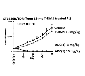
[Figure 3] Figure 3 is a diagram showing the antitumor effect
of an antibody-drug conjugate (1) or T-DM1 on a nude mouse with
subcutaneously transplanted tumor of HER2-positive human breast
cancer 5T1616B/TDR that
Date Recue/Date Received 2021-05-19
CA 03036941 2019-03-14
- 54 -
acquired secondary resistance to T-DM1. In the drawing,
the abscissa depicts days after initial administration,
and the ordinate depicts tumor volume.
[Figure 4] Figure 4 is a diagram showing the antitumor
effect of an antibody-drug conjugate (1) or T-DM1 on a
nude mouse with subcutaneously transplanted tumor of
HER2-positive human breast cancer ST1360B/TDR that
acquired secondary resistance to T-DM1. In the drawing,
the abscissa depicts days after initial administration,
and the ordinate depicts tumor volume.
[Figure 5] Figure 5 is a diagram showing the
pharmacokinetics of the antibody-drug conjugate (1) in
a clinical study.
[Figure 6] Figure 6 is a diagram showing the safety and
tolerability of the antibody-drug conjugate (1) in a
clinical study.
[Figure 7] Figure 7 is a diagram showing ORR (objective
response rate) and DCR (disease control rate) as to the
efficacy of the antibody-drug conjugate (1) in a
clinical study.
[Figure 8] Figure 8 is a diagram showing best % change
from baseline in tumor size as to the efficacy of the
antibody-drug conjugate (1) in a clinical study.
[Figure 9] Figure 9 is a diagram showing a treatment
period and a therapeutic effect as to the efficacy of
the antibody-drug conjugate (1) in a clinical study.
CA 03036941 2019-03-14
- 55 -
[Figure 101 Figure 10 is a diagram showing best %
change from baseline in tumor size as to the efficacy
of the antibody-drug conjugate (1) in a clinical study.
[Figure 11] Figure 11 is a diagram showing time-
dependent change in tumor shrinkage (%) as to the
efficacy of the antibody-drug conjugate (1) on breast
cancer in a clinical study.
[Figure 12] Figure 12 is a diagram showing time-
dependent change in tumor shrinkage (%) as to the
efficacy of the antibody-drug conjugate (1) on gastric
cancer in a clinical study.
[Figure 13] Figure 13 is a diagram showing best %
change from baseline in tumor size as to the efficacy
of the antibody-drug conjugate (1) on HER2-expressing
solid cancer (except for breast cancer and gastric
cancer) in a clinical study. In the drawing, "C"
represents a cohort of colorectal cancer, "L"
represents a cohort of non-small cell lung cancer, "S"
represents a cohort of salivary gland cancer, "P"
represents a cohort of Paget's disease, "Ch" represents
a cohort of bile duct cancer, and "E" represents a
cohort of esophageal cancer. In the drawing, "*" shows
that the treatment is ongoing.
[Figure 14] Figure 14 is a diagram showing time-
dependent change in tumor shrinkage (%) as to the
efficacy of the antibody-drug conjugate (1) on HER2-
exiDressing solid cancer (except for breast cancer and
gastric cancer) in a clinical study. In the drawing,
CA 03036941 2019-03-14
- 56 -
"Colorectal" represents a cohort of colorectal cancer,
"NSCLC" represents a cohort of non-small cell lung
cancer, "Salivary" represents a cohort of salivary
gland cancer, and "Other" represents a cohort of other
cancers.
Description of Embodiments
[0027]
Hereinafter, preferred modes for carrying out the
present invention are described with reference to the
drawings. The embodiments described below are given
merely for illustrating one example of a typical
embodiment of the present invention and are not
intended to limit the scope of the present invention.
[0028]
The antibody-drug conjugate used in the present
invention is an anti-HER2 antibody-drug conjugate in
which a linker and a drug represented by the following
formula are connected to an anti-HER2 antibody.
-(Succinimid-3-yl-N)-CH2CH2CH2CH2CH2-C(=0)-GGFG-NH-CH2-0-
CH2-C(=0)-(NH-DX)
In the formula, -(Succinimid-3-yl-N)- has a
structure represented by the following formula:
[Formula 20]
CA 03036941 2019-03-14
- 57 -
0
N--
0
which is connected to the anti-HER2 antibody at
position 3 thereof via a thioether bond and is
connected to the methylene group in the linker
structure containing this structure on the nitrogen
atom at position 1.
-(NH-DX) is a group represented by the following
formula:
[Formula 21]
Me 0
/
0
HO :
/ 0
Me
wherein the nitrogen atom of the amino group at
position 1 is the connecting position.
-GGFG- represents the tetrapeptide residue of -
Gly-Gly-Phe-Gly-.
In this specification, a partial structure
consisting of the linker and the drug in the antibody-
drug conjugate is referred to as a "drug-linker
structure". This drug-linker structure is connected to
CA 03036941 2019-03-14
4
- 58 -
a thiol group (in other words, the sulfur atom of a
cysteine residue) formed at the interchain disulfide
bond sites (2 sites between the heavy chain and the
heavy chain, and 2 sites between the heavy chain and
the light chain) of the antibody.
The anti-HER2 antibody-drug conjugate used in the
present invention can have a structure represented by
the following formula:
[Formula 22]
o
Anti-HE R2 antibody __________
criv" N 14J1*-N-"-0
Hr H H
0 0 0 .0N H
Me 0
N
0
OH 0
Here, the drug-linker structure is conjugated to
the anti-HER2 antibody via a thioether bond.
Furthermore, n has the same meaning as DAR (drug-to-
antibody ratio) and represents the number of conjugated
drug molecules per antibody molecule. DAR is an
average value, i.e., a numeric value defined and
indicated as the average number of conjugated drug
molecules. In the case of the antibody-drug conjugate
of the present invention, n is 2 to 8, preferably 3 to
8, more preferably 7 to 8, and further preferably 7.5
to 8, and n of about 8 can be preferably used.
CA 03036941 2019-03-14
=
- 59 -
Hereinafter, the antibody-drug conjugate used in
the present invention is explained in detail
hereinbelow.
[0029]
[Antibody]
The anti-HER2 antibody used in the anti-HER2
antibody-drug conjugate used in the present invention
may be derived from any species, and preferred examples
of the species can include humans, rats, mice, and
rabbits. In case when the antibody is derived from
other than human species, it is preferably chimerized
or humanized using a well known technique. The
antibody of the present invention may be a polyclonal
antibody or a monoclonal antibody and is preferably a
monoclonal antibody.
The anti-HER2 antibody is the antibody, which is
capable of targeting tumor cells, that is, possesses a
property of recognizing a tumor cell, a property of
binding to a tumor cell, a property of internalizing in
a tumor cell, cytocidal activity against tumor cells,
or the like, and can be conjugated with a drug having
antitumor activity via a linker to form an antibody-
drug conjugate.
The binding activity of the antibody against tumor
cells can be confirmed using flow cytometry. The
internalization of the antibody into tumor cells can be
confirmed using (1) an assay of visualizing an antibody
incorporated in cells under a fluorescence microscope
CA 03036941 2019--14
A
- 60 -
using a secondary antibody (fluorescently labeled)
binding to the therapeutic antibody (Cell Death and
Differentiation (2008) 15, 751-761), (2) an assay of
measuring a fluorescence intensity incorporated in
cells using a secondary antibody (fluorescently
labeled) binding to the therapeutic antibody (Molecular
Biology of the Cell, Vol. 15, 5268-5282, December 2004),
or (3) a Mab-ZAP assay using an immunotoxin binding to
the therapeutic antibody wherein the toxin is released
upon incorporation into cells to inhibit cell growth
(Bio Techniques 28: 162-165, January 2000). As the
immunotoxin, a recombinant complex protein of a
diphtheria toxin catalytic domain and protein G may be
used.
The antitumor activity of the antibody can be
confirmed in vitro by determining inhibitory activity
against cell growth. For example, a cancer cell line
overexpressing a target protein for the antibody is
cultured, and the antibody is added at varying
concentrations into the culture system to determine an
inhibitory activity against focus formation, colony
formation, and spheroid growth. The antitumor activity
can be confirmed in vivo, for example, by administering
the antibody to a nude mouse with a transplanted tumor
cell line highly expressing the target protein, and
determining change in the cancer cell.
Since the compound conjugated in the antibody-drug
conjugate exerts an antitumor effect, it is preferred
CA 03036941 2019-03-14
= =
- 61 -
but not essential that the antibody itself should have
an antitumor effect. For the purpose of specifically
and selectively exerting the cytotoxic activity of the
antitumor compound against tumor cells, it is important
and also preferred that the antibody should have the
property of internalizing to migrate into tumor cells.
[0030]
The anti-HER2 antibody can be obtained by a
procedure known in the art. For example, the antibody
of the present invention can be obtained using a method
usually carried out in the art, which involves
immunizing animals with an antigenic polypeptide and
collecting and purifying antibodies produced in vivo.
The origin of the antigen is not limited to humans, and
the animals may be immunized with an antigen derived
from a non-human animal such as a mouse, a rat and the
like. In this case, the cross-reactivity of antibodies
binding to the obtained heterologous antigen with human
antigens can he tested to screen for an antibody
applicable to a human disease.
Alternatively, antibody-producing cells which
produce antibodies against the antigen are fused with
myeloma cells according to a method known in the art
(e.g., Kohler and Milstein, Nature (1975) 256, p. 495-
497; and Kennet, R. ed., Monoclonal Antibodies, p. 365-
367, Plenum Press, N.Y. (1980)) to establish hybridomas,
from which monoclonal antibodies can in turn be
obtained.
CA 03036941 2019-03-14
- 62 -
The antigen can be obtained by genetically
engineering host cells to produce a gene encoding the
antigenic protein. Specifically, vectors that permit
expression of the antigen gene are prepared and
transferred to host cells so that the gene is expressed.
The antigen thus expressed can be purified. The
antibody can also be obtained by a method of immunizing
animals with the above-described genetically engineered
antigen-expressing cells or a cell line expressing the
antigen.
[0031]
The anti-HER2 antibodies that can be used in the
present invention are not particularly limited and are
preferably, for example, those having properties as
described below.
(1) An anti-HER2 antibody having the following
properties:
(a) specifically binding to HER2, and
(b) having an activity of internalizing in HER2-
expressing cells by binding to HER2.
(2) The antibody according to (1) above, wherein the
antibody binds to the extraceilular domain of HER2.
(3) The antibody according to (1) or (2) above, wherein
the antibody is a monoclonal antibody.
(4) The antibody according to any of (1) to (3) above,
wherein the antibody has an antibody-dependent cellular
cytotoxicity (ADCC) activity and/or a complement-
dependent cytotoxicity (CDC) activity
CA 03036941 2019-03-14
- 63 -
(5) The antibody according to any of (1) to (4) above,
wherein the antibody is a mouse monoclonal antibody, a
chimeric monoclonal antibody, or a humanized monoclonal
antibody.
(6) The antibody according to any of (1) to (5) above,
wherein the antibody is a humanized monoclonal antibody
comprising a heavy chain consisting of the amino acid
sequence represented by SEQ ID NO: 1 and a light chain
consisting of the amino acid sequence represented by
SEQ ID NO: 2.
(7) The antibody according to any of (1) to (6) above,
wherein the antibody lacks a lysine residue at the
carboxyl terminus of the heavy chain.
(8) The antibody according to (7) above, wherein the
antibody comprises a heavy chain consisting of an amino
acid sequence consisting of amino acid residues 1 to
449 of SEQ ID NO: 1 and a light chain consisting of an
amino acid sequence consisting of amino acid residues 1
to 214 of SEQ ID NO: 2.
(9) An antibody obtained by a method for producing the
antibody according to any of (1) to (8) above, the
method comprising the steps of: culturing a host cell
transformed with an expression vector containing a
polynucleotide encoding the antibody; and collecting
the antibody of interest from the cultures obtained in
the preceding step.
[0032]
CA 03036941 2019-03-14
- 64 -
Hereinafter, the anti-HER2 antibody used in the
invention is described.
The terms "cancer" and "tumor" as used herein are
used with the same meaning.
The term "gene" as used herein includes not only
DNA, but also mRNA thereof, cDNA thereof and cRNA
thereof.
The term "polynucleotide" as used herein is used
with the same meaning as a nucleic acid and also
includes DNA, RNA, probes, oligonucleotides, and
primers.
The terms "polypeptide", "protein" and "protein"
as used herein are used without distinction.
The term "cell" as used herein also includes cells
in an animal individual and cultured cells.
The term "HER2" as used herein is used with the
same meaning as HER2 protein.
Examples of the anti-HER2 antibody as used herein
can include, but not particularly limited to,
pertuzumab (International Patent Publication No. WO
01/00245) and trastuzumab (U.S. Patent No. 5821337).
Trastuzumab is preferred. However, the anti-HER2
antibody of the present invention is not limited
thereto as long as it is an anti-HER2 antibody
specifically binding to HER2, and more preferably
having an activity of internalizing in HER2-expressing
cells by binding to HER2.
CA 03036941 2019-03-14
= =
- 65 -
The term "trastuzumab" as used herein is also
called HERCEPTIN (registered trademark), huMAh4D5-8, or
rhuMAb4D5-8 and is a humanized antibody comprising a
heavy chain consisting of an amino acid sequence
consisting of amino acid residues 1 to 449 of SEQ ID
NO: 1 (Figure 1) and a light chain consisting of an
amino acid sequence consisting of amino acid residues 1
to 214 of SEQ ID NO: 2 (Figure 2).
The term "specifically binding" as used herein
means binding that is not nonspecific adsorption.
Examples of the criterion for determining whether the
binding is specific or not can include dissociation
constant (hereinafter referred to as "Kd"). The Ed
value of the antibody for the HER2 protein is
preferably 1 x 10-h M or smaller, 5 x 10⁻⁶ M or smaller,
2 X 10⁻⁶ M or smaller, or 1 X 10⁻⁶ M or smaller, more
preferably 5 x 10⁻⁷ M or smaller, 2 x 10⁻⁷ M or smaller,
or 1 x 10⁻⁷ M or smaller, further preferably 5 x 10⁻⁸ M
or smaller, 2 X 10⁻⁸ M or smaller, or 1 x 10⁻⁸ M or
smaller, and most preferably 5 x 10⁻⁹ M or smaller, 2 x
10-8 M or smaller, or 1 x 10⁻⁹ M or smaller. The
binding between the HER2 protein and the antibody can
be measured using a method known in the art, such as
surface plasmon resonance, ELISA, or RIA.
The term "CDR" as used herein refers to a
complementarity determining region (CDR). It is known
that each heavy and light chain of an antibody molecule
has three complementarity determining regions (CDRs).
CA 03036941 2019-03-14
L
- 66 -
The CDR is also called the hypervariable domain, and is
present in a variable region of each heavy and light
chain of an antibody. It is a site which has unusually
high variability in its primary structure, and there
are three separate CDRs in the primary structure of
each heavy and light polypeptide chain. In this
specification, as for the CDRs of an antibody, the CDRs
of the heavy chain are represented by CDRH1, CDRH2, and
CDRH3 from the amino-terminal side of the amino acid
sequence of the heavy chain, and the CDRs of the light
chain are represented by CDRL1, CDRL2, and CDRL3 from
the amino-terminal side of the amino acid sequence of
the light chain. These sites are proximate to one
another in the tertiary structure and determine the
specificity for an antigen to which the antibody binds.
The phrase "hybridization is performed under
stringent conditions" as used herein refers to a
process in which hybridization is performed under
conditions under which identification can be achieved
by performing hybridization at 68 C in a commercially
available hybridization solution
ExpressHyb
Hybridization Solution (manufactured by Clontech, Inc.)
or by performing hybridization at 68 C in the presence
of 0.7 to 1.0 M NaC1 using a filter having DNA
immobilized thereon, followed by performing washing at
68 C using 0.1 to 2 x SSC solution (1 x SSC solution is
composed of 150 mM NaCl and 15 mM sodium citrate) or
under conditions equivalent thereto.
CA 03036941 2019-03-14
- 67 -
[0033]
1. HER2
HER2 is one of the oncogene products of a typical
growth factor receptor oncogene identified as human
epidermal cell growth factor receptor 2-related
oncogene, and is a transmembrane receptor protein
having a molecular weight of 185 kDa and having a
tyrosine kinase domain. HER2 is a member of the EGFR
family consisting of HER1 (EGFR, ErbB-1), HER2 (neu,
Erb-2), HER3 (ErbB-3), and HER4 (ErbB-4) and is known
to be autophosphorylated at intracellular tyrosine
residues by its homodimer formation or heterodimer
formation with another EGER receptor HER1, HER3, or
HER4 and is itself activated in that manner, thereby
playing an important role in cell growth,
differentiation, and survival in normal cells and tumor
cells.
As for the HER2 protein to be used in the present
invention, the HER2 protein can be directly purified
from HER2-expressing cells of a human or a non-human
mammal (such as a rat or a mouse) and used, or a cell
membrane fraction of the above-described cells can be
prepared and used. Further, HER2 can be obtained by in
vitro synthesis thereof or production thereof in a host
cell through genetic engineering. In the genetic
engineering, specifically, after HER2 cDNA is
integrated into a vector capable of expressing HER2
cDNA, the HER2 protein can be obtained by synthesizing
CA 03036941 2019-03-14
- 68 -
it in a solution containing an enzyme, a substrate and
an energy substance required for transcription and
translation, or by expressing HER2 in another
prokaryotic or eucaryotic transformed host cell.
Alternatively, the above-described genetically
engineered HER2-expressing cells, or a cell line
expressing HER2 may be used as the HER2 protein.
The DNA sequence and amino acid sequence of HER2
are disclosed on a public database, and can be referred
to, for example, under Accession No. M11730 (GenBank),
NP 004439.2 (NCSI), or the like.
Further, a protein which consists of an amino acid
sequence wherein one or several amino acids are
substituted, deleted and/or added in any of the above-
described amino acid sequences of HER2 and also has a
biological activity equivalent to that of the protein
is also included in HER2.
Human HER2 protein is composed of a signal
sequence consisting of N-terminal 22 amino acid
residues, an extracellular domain consisting of 630
amino acid residues, a transmembrane domain consisting
of 23 amino acid residues, and an intracellular domain
consisting of 580 amino acid residues.
[0034]
2. Production of anti-HER2 antibody
The antibody against HER2 of the present invention
can be obtained according to, for example, a method
usually carried out in the art, which involves
CA 03036941 2019-03-14
A
- 69 -
immunizing animals with HER2 or an arbitrary
polypeptide selected from the amino acid sequence of
HER2 and collecting and purifying antibodies produced
in vivo. The biological species of HER2 to be used as
an antigen is not limited to being human, and an animal
can be immunized with HER2 derived from an animal other
than humans such as a mouse or a rat or with rat
p185neu. In this case, by examining the cross-
reactivity between an antibody binding to the obtained
heterologous HER2 and human HER2, an antibody
applicable to a human disease can be selected.
Further, a monoclonal antibody can be obtained
from a hybridoma established by fusing antibody-
producing cells which produce an antibody against HER2
with myeloma cells according to a known method (for
example, Kohler and Milstein, Nature, (1975) 256, pp-
495-497; Kennet, R. ed., Monoclonal Antibodies, pp.
365-367, Plenum Press, N.Y. (1980)).
HER2 to be used as an antigen can be obtained by
expressing HER2 gene in a host cell using genetic
engineering.
Specifically, a vector capable of expressing HER2
gene is produced, and the resulting vector is
transfected into a host cell to express the gene, and
then, the expressed HER2 is purified.
Alternatively, the above-described genetically
engineered HER2-expressing cells, or a cell line
expressing HER2 may be used as the HER2 protein. The
CA 03036941 2019--14
A
- 70 -
anti-HER2 antibody can be obtained by a preocedure
known in the art. Hereinafter, a method of obtaining
an antibody against HER2 is specifically described.
[0035]
(1) Preparation of antigen
Examples of the antigen to be used for producing
the anti-HER2 antibody include HER2, or a polypeptide
consisting of a partial amino acid sequence comprising
at least 6 consecutive amino acids of HER2, or a
derivative obtained by adding a given amino acid
sequence or carrier thereto.
HER2 can be purified directly from human tumor
tissues or tumor cells and used. Further, HER2 can be
obtained by synthesizing it in vitro or by producing it
in a host cell by genetic engineering.
With respect to the genetic engineering,
specifically, after HER2 cDNA is integrated into a
vector capable of expressing HER2 cDNA, HER2 can be
obtained by synthesizing it in a solution containing an
enzyme, a substrate and an energy substance required
for transcription and translation, or by expressing
HER2 in another prokaryotic or eucaryotic transformed
host cell.
Further, the antigen can also he obtained as a
secretory protein by expressing a fusion protein
obtained by ligating the extracellular domain of HER2,
which is a membrane protein, to the constant region of
an antibody in an appropriate host-vector system.
CA 03036941 2019-03-14
- 71 -
HER2 cDNA can be obtained by, for example, a so-
called PCR method in which a polymerase chain reaction
is performed using a cDNA library expressing HER2 cDNA
as a template and primers which specifically amplify
HER2 cDNA (PCR; Saiki, R. K., et al., Science, (1988)
239, pp. 487-489).
As the in vitro synthesis of the polypeptide, for
example, Rapid Translation System (RTS) manufactured by
Roche Diagnostics, Inc. can be exemplified, but it is
not limited thereto.
Examples of the prokaryotic host cells include
Escherichia coli and Bacillus subtilis. In order to
transform the host cells with a target gene, the host
cells are transformed by a plasmid vector comprising a
replicon, i.e., a replication origin derived from a
species compatible with the host, and a regulatory
sequence. Further, the vector preferably has a
sequence capable of imposing phenotypic selectivity on
the transformed cell.
Examples of the eucaryotic host cells include
vertebrate cells, insect cells, and yeast cells. As
the vertebrate cells, for example, simian COS cells
(Gluzman, Y., Cell, (1981) 23, pp. 175-182, ATCC CRL-
1650; ATCC: American Type Culture Collection), murine
fibroblasts NIH3T3 (ATCC No. CRL-1658), and
dihydrofolate reductase-deficient strains (Urlaub, G.
and Chasin, L. A., Proc. Natl. Acad. Sci. USA (1980) 77,
pp. 4126-4220) of Chinese hamster ovarian cells (CHO
CA 03036941 2019-03-14
A
- 72 -
cells; ATCC: CCL-61); and the like are often used,
however, the cells are not limited thereto.
The thus obtained transformant can be cultured
according to a method usually carried out in the art,
and by the culturing of the transformant, a target
polypeptide is produced intracellularly or
extracellularly.
A suitable medium to be used for the culturing can
be selected from various commonly used culture media
depending on the employed host cells. If Escherichia
colt is employed, for example, an LB medium
supplemented with an antibiotic such as ampicillin or
IPMG as needed can be used.
A recombinant protein produced intracellularly or
extracellularly by the transformant through such
culturing can be separated and purified by any of
various known separation methods utilizing the physical
or chemical property of the protein.
Specific examples of the methods include treatment
with a common protein precipitant, ultrafiltration,
various types of liquid chromatography such as
molecular sieve chromatography (gel filtration),
adsorption chromatography, ion exchange chromatography,
and affinity chromatography, dialysis, and a
combination thereof.
Further, by attaching a tag of six histidine
residues to a recombinant protein to be expressed, the
protein can be efficiently purified with a nickel
CA 03036941 2019-03-14
- 73 -
affinity column. Alternatively, by attaching the IgG
Fc region to a recombinant protein to be expressed, the
protein can be efficiently purified with a protein A
column.
By combining the above-described methods, a large
amount of a target polypeptide can be easily produced
in high yield and high purity.
The above-described transformant itself may be
used as the antigen. A cell line expressing HER2 may
also be used as the antigen. Examples of such a cell
line can include human breast cancer lines SK-BR-3, BT-
474, KPL-4, and JIMT-1, a human gastric cancer line
NCI-N87, and a human ovarian cancer line SK-OV-3. The
cell line of the present invention is not limited to
these cell lines as long as it expresses HER2.
[0036]
(2) Production of anti-HER2 monoclonal antibody
Examples of the antibody specifically bind to HER2
include a monoclonal antibody specifically bind to HER2,
and a method of obtaining such antibody is as described
below.
The production of a monoclonal antibody generally
requires the following operational steps of:
(a) purifying a biopolymer to be used as an
antigen, or Preparing antigen-expressing cells;
(b) preparing antibody-producing cells by
immunizing an animal by injection of the antigen,
CA 03036941 2019-03-14
- 74 -
collecting the blood, assaying its antibody titer to
determine when the spleen is excised;
(c) preparing myeloma cells (hereinafter referred
to as "myeloma");
(d) fusing the antibody-producing cells with the
myeloma;
(e) screening a group of hybridomas producing a
desired antibody;
(f) dividing the hybridomas into single cell
clones (cloning);
(g) optionally, culturing the hybridoma or rearing
an animal implanted with the hybridoma for producing a
large amount of monoclonal antibody;
(h) examining the thus produced monoclonal
antibody for biological activity and binding
specificity, or assaying the same for properties as a
labeled reagent; and the like.
Hereinafter, the method of producing a monoclonal
antibody will be described in detail following the
above steps, however, the method is not limited thereto,
and, for example, antibody-producing cells other than
spleen cells and myeloma can be used.
[0C37]
(a) Purification of antigen
As the antigen, HER2 prepared by the method as
described above or a partial peptide thereof can be
used.
CA 03036941 2019-03-14
A
- 75 -
Further, a membrane fraction prepared from
recombinant cells expressing HER2 or the recombinant
cells expressing HER2 themselves, and also a partial
peptide of the protein of the present invention
chemically synthesized by a method known to those
skilled in the art can also be used as the antigen.
Furthermore, a HER2-expressing cell line can also
be used as the antigen.
[0038]
(b) Preparation of antibody-producing cells
The antigen obtained in the step (a) is mixed with
an adjuvant such as Freund's complete or incomplete
adjuvant or auxiliary agent such as aluminum potassium
sulfate and the resulting mixture is used as an
immunogen to immunize an experimental animal. Another
method involves immunizing an experimental animal with
antigen-expressing cells as an immunogen. As the
experimental animal, any animal used in a known
hybridoma production method can be used without
hindrance. Specifically, for example, a mouse, a rat,
a goat, sheep, cattle, a horse, or the like can be used.
However, from the viewpoint of ease of availability of
myeloma cells to be fused with the extracted antibody-
producing cells, a mouse or a rat is preferably used as
the animal to be immunized.
Further, the strain of a mouse or a rat to be used
is not particularly limited, and in the case of a mouse,
for example, various strains such as A, AKR, BALE/c,
CA 03036941 2019-03-14
A
- 76 -
BDP, BA, CE, C3H, 57BL, C57BL, C57L, DBA, FL, HTH, HT1,
LP, NZB, NZW, RE, R III, SJL, SWR, WB, and 129 and the
like can be used, and in the case of a rat, for example,
Wistar, Low, Lewis, Sprague, Dawley, ACI, EN, Fischer
and the like can be used.
These mice and rats are commercially available
from breeders/distributors of experimental animals, for
example, CLEA Japan, Inc. and Charles River
Laboratories Japan, Inc.
As the animal to be immunized, in consideration of
compatibility of fusing with myelolia cells described
below, in the case of a mouse, BALB/c strain, and in
the case of a rat, Wistar and Low strains are
particularly preferred.
Further, in consideration of antigenic homology
between humans and mice, it is also preferred to use a
mouse having decreased biological function to remove
auto-antibodies, that is, a mouse with an autoimmune
disease.
The age of such mouse or rat at the time of
immunization is preferably 5 to 12 weeks of age, more
preferably 6 to 8 weeks of age.
In order to immunize an animal with HER2 or a
recombinant thereof, for example, a known method
described in detail in, for example, Weir, D. M.,
Handbook of Experimental Immunology Vol. I. II. III.,
Blackwell Scientific Publications, Oxford (1987); Kabat,
E. A. and Mayer, M. M., Experimental Immunochemistry,
CA 03036941 2019-03-14
- 77 -
Charles C Thomas Publisher Springfield, Illinois (1964)
or the like can be used.
Among these immunization methods, a preferred
specific method in the present invention is, for
example, as follows.
That is, first, a membrane protein fraction
serving as the antigen or cells caused to express the
antigen is/are intradermally or intraperitoneally
administrated to an animal. However, the combination
of both routes of administration is preferred for
increasing the immunization efficiency, and when
intradermal administration is performed in the first
half and intraperitoneal administration is performed in
the latter half or only at the last dosing, the
immunization efficiency can be particularly increased.
The administration schedule of the antigen varies
depending on the type of animal to be immunized,
individual difference or the like. However, in general,
an administration schedule in which the frequency of
administration of the antigen is 3 to 6 times and the
dosing interval is 2 to 6 weeks is preferred, and an
administration schedule in which the frequency of
administration of the antigen is 3 to 4 times and the
dosing interval is 2 to 4 weeks is more preferred.
Further, the dose of the antigen varies depending
on the type of animal, individual differences or the
like, however, the dose is generally set to 0.05 to 5
mg, preferably about 0.1 to 0.5 mg.
CA 03036941 2019-03-14
- 78 -
A booster immunization is performed 1 to 6 weeks,
Preferably 1 to 4 weeks, more preferably 1 to 3 weeks
after the administration of the antigen as described
above. When the immunogen is cells, 1 x 10⁶ to 1 x 10⁷
cells are used.
The dose of the antigen at the time of performing
the booster immunization varies depending on the type
or size of animal or the like, however, in the case of,
for example, a mouse, the dose is generally set to 0.05
to 5 mg, preferably 0.1 to 0.5 mg, more preferably
about 0.1 to 0.2 mg. When the immunogen is cells, 1 x
106 to 1 x 10⁷ cells are used.
Spleen cells or lymphocytes including antibody-
producing cells are aseptically removed from the
immunized animal after 1 to 10 days, preferably 2 to 5
days, more preferably 2 to 3 days from the booster
immunization. At this time, the antibody titer is
measured, and if an animal having a sufficiently
increased antibody titer is used as a supply source of
the antibody-producing cells, the subsequent procedure
can be carried out more efficiently.
Examples of the method of measuring the antibody
titer to be used here include an RIA method and an
ELISA method, but the method is not limited thereto.
For example, if an ELISA method is employed, the
measurement of the antibody titer in the invention can
be carried out according to the procedures as described
below.
CA 03036941 2019-03-14
- 79 -
First, a purified or partially purified antigen is
adsorbed to the surface of a solid phase such as a 96-
well plate for ELISA, and the surface of the solid
phase having no antigen adsorbed thereto is covered
with a protein unrelated to the antigen such as bovine
serum albumin (BSA). After washing the surface, the
surface is brought into contact with a serially-diluted
sample (for example, mouse serum) as a primary antibody
to allow the antibody in the sample to bind to the
antigen.
Further, as a secondary antibody, an antibody
labeled with an enzyme against a mouse antibody is
added and is allowed to bind to the mouse antibody.
After washing, a substrate for the enzyme is added and
a change in absorbance which occurs due to color
development induced by degradation of the substrate or
the like is measured and the antibody titer is
calculated based on the measurement.
The separation of the antibody-producing cells
from the spleen cells or lymphocytes of the immunized
animal can be carried out according to a known method
(for example, Kohler et al., Nature (1975), 256, p.
495; Kohler et al., Eur. J. Immunol. (1977), 6, p. 511;
Milstein et al., Nature (1977), 266, p. 550; Walsh,
Nature (1977), 266, p. 495). For example, in the case
of spleen cells, a general method in which the
antibody-producing cells are separated by homogenizing
the spleen to obtain the cells through filtration with
CA 03036941 2019-03-14
- 80 -
a stainless steel mesh and suspending the cells in
Eagle's Minimum Essential Medium (MEM) can be employed.
[0039]
(c) Preparation of myeloma cells (hereinafter referred
to as "myeloma")
The myeloma cells to be used for cell fusion are
not particularly limited and suitable cells can be
selected from known cell lines. However, in
consideration of convenience when a hybridoma is
selected from fused cells, it is preferred to use an
HGPRT (hypoxanthine-guanine phosphoribosyl transferase)
deficient strain whose selection procedure has been
established.
More specifically, examples of the HGPRT-deficient
strain include X63-Ag8(X63), NS1-ANS/1(NS1), P3X63-
Ag8.U1(P3U1), X63-Ag8.653(X63.653), SP2/0-Ag14(SP2/0),
MPC11-45.6TG1.7(45.6TG), FO, S149/5XXO, and BU.1
derived from mice; 210.RSY3.Ag.1.2.3(Y3) derived from
rats; and U266AR(SKO-007), GM1500.GTG-Al2(GM1500),
UC729-6, LICR-LOW-HMy2(HMy2) and 8226AR/NIP4-1(NP41)
derived from humans. These HGPRT-deficient strains are
available from, for example, ATCC or the like.
These cell strains are subcultured in an
appropriate medium such as an 8-azaguanine medium [a
medium obtained by adding 8-azaguanine to an RPMI 1640
medium supplemented with glutamine, 2-mercaptoethanol,
gentamicin, and fetal calf serum (hereinafter referred
to as "FCS")], Iscove's Modified Dulbecco's Medium
CA 03036941 2019-03-14
- 81 -
(hereinafter referred to as "IMDM"), or Dulbecco's
Modified Eagle Medium (hereinafter referred to as
"DMEM"). In this case, 3 to 4 days before performing
cell fusion, the cells are subcultured in a normal
medium (for example, an ASF104 medium (manufactured by
Ajinomoto Co., Ltd.) containing 10% FCS) to ensure not
less than 2 x 10⁷ cells on the day of cell fusion.
[0040]
(d) Cell fusion
Fusion between the antibody-producing cells and
the myeloma cells can be appropriately performed
according to a known method (Weir, D. M. Handbook of
Experimental Immunology Vol. I. II. III., Blackwell
Scientific Publications, Oxford (1987); Kabat, E. A.
and Mayer, M. M., Experimental Immunochemistry, Charles
C Thomas Publisher, Springfield, Illinois (1964), etc.),
under conditions such that the survival rate of cells
is not excessively reduced.
As such a method, for example, a chemical method
in which the antibody-producing cells and the myeloma
cells are mixed in a solution containing a polymer such
as polyethylene glycol at a high concentration, a
physical method using electric stimulation, or the like
can he used. Among these methods, a specific example
of the chemical method is as described below.
That is, in the case where polyethylene glycol is
used in the solution containing a polymer at a high
concentration, the antibody-producing cells and the
CA 03(136941 2019-03-14
- 82 -
myeloma cells are mixed in a solution of polyethylene
glycol having a molecular weight of 1500 to 6000, more
preferably 2000 to 4000 at a temperature of from 30 to
40 C, preferably from 35 to 38 C for 1 to 10 minutes,
preferably 5 to 8 minutes.
[0041]
(e) Selection of a group of hybridomas
The method of selecting hybridomas obtained by the
above-described cell fusion is not particularly limited.
Usually, an HAT (hypoxanthine, aminopterin, thymidine)
selection method (Kohler et al., Nature (1975), 256, p.
495; Milstein et al., Nature (1977), 266, p. 550) is
used.
This method is effective when hybridomas are
obtained using the myeloma cells of an HGPRT-deficient
strain which cannot survive in the presence of
aminopterin. That is, by culturing unfused cells and
hybridomas in an HAT medium, only hybridomas resistant
to aminopterin are selectively allowed to survive and
proliferate.
[0042]
(f) Division into single cell clone (cloning)
As a cloning method for hybridomas, a known method
such as a methylcellulose method, a soft agarose method,
or a limiting dilution method can be used (see, for
example, Barbara, B. M. and Stanley, M. S.: Selected
Methods in Cellular Immunology, W. H. Freeman and
Company, San Francisco (1980)). Among these methods,
CA 03036941 2019-03-14
- 83 -
particularly, a three-dimensional culture method such
as a methylcellulose method is preferred. For example,
the group of hybridomas produced by cell fusion are
suspended in a methylcellulose medium such as
ClonaCell-HY Selection Medium D (manufactured by
StemCell Technologies, Inc., #03804) and cultured.
Then, the formed hybridoma colonies are collected,
whereby monoclonal hybridomas can he obtained. The
collected respective hybridoma colonies are cultured,
and a hybridoma which has been confirmed to have a
stable antibody titer in an obtained hybridoma culture
supernatant is selected as a HER2 monoclonal antibody-
producing hybridoma strain.
[0043]
(g) Preparation of monoclonal antibody by culturing
hybridoma
By culturing the thus selected hybridoma, a
monoclonal antibody can be efficiently obtained.
However, prior to culturing, it is preferred to perform
screening of a hybridoma which produces a target
monoclonal antibody.
In such screening, a known method can be employed.
The measurement of the antibody titer in the
invention can be carried out by, for example, an ELISA
method explained in item (b) described above.
The hybridoma obtained by the method described
above can be stored in a frozen state in liquid
nitrogen or in a freezer at -80 C or below.
CA 03036941 2019-03-14
- 84 -
After completion of cloning, the medium is changed
from an HT medium to a normal medium, and the hybridoma
is cultured.
Large-scale culture is performed by rotation
culture using a large culture bottle or by spinner
culture. From the supernatant obtained by the large-
scale culture, a monoclonal antibody which specifically
binds to the protein of the invention can be obtained
by purification using a method known to those skilled
in the art such as gel filtration.
Further, the hybridoma is injected into the
abdominal cavity of a mouse of the same strain as the
hybridoma (for example, the above-described BALB/c) or
a Nu/Nu mouse to proliferate the hybridoma, whereby the
ascites containing a large amount of the monoclonal
antibody of the invention can be obtained.
In the case where the hybridoma is administrated
in the abdominal cavity, if a mineral oil such as
2,6,10,14-tetramethyl pentadecane (pristane) is
administrated 3 to 7 days prior thereto, a larger
amount of the ascites can be obtained.
For example, an immunosuppressant is previously
injected into the abdominal cavity of a mouse of the
same strain as the hybridoma to inactivate T cells. 20
days thereafter, 106 to 107 hybridoma clone cells are
suspended in a serum-free medium (0.5 ml), and the
suspension is administrated in the abdominal cavity of
the mouse. In general, when the abdomen is expanded
CA 03036941 2019-03-14
- 85 -
and filled with the ascites, the ascites is collected
from the mouse. By this method, the monoclonal
antibody can be obtained at a concentration which is
about 100 times or much higher than that in the culture
solution.
The monoclonal antibody obtained by the above-
described method can be purified by a method described
in, for example, Weir, D. M.: Handbook of Experimental
Immunology Vol. I, II, III, Blackwell Scientific
Publications, Oxford (1978).
The thus obtained monoclonal antibody has high
antigen specificity for HER2. Examples of the
monoclonal antibody of the Present invention can
include, but are not particularly limited to, a mouse
monoclonal antibody 405 (ATCC CRL 10463).
[0044]
(h) Assay of monoclonal antibody
The isotype and subclass of the thus obtained
monoclonal antibody can be determined as follows.
First, examples of the identification method
include an Ouchterlony method, an ELISA method, and an
RIA method.
An Cuchterlony method is simple, but when the
concentration of the monoclonal antibody is low, a
condensation operation is required.
On the other hand, when an ELISA method or an RIA
method is used, by directly reacting the culture
supernatant with an antigen-adsorbed solid phase and
CA 03036941 2019-03-14
- 86 -
using antibodies corresponding to various types of
immunoglobulin isotypes and subclasses as secondary
antibodies, the isotype and subclass of the monoclonal
antibody can be identified.
In addition, as a simpler method, a commercially
available identification kit (for example, Mouse Typer
Kit manufactured by Bio-Rad Laboratories, Inc.) or the
like can also be used.
Further, the quantitative determination of a
protein can be performed by the Folin Lowry method and
a method of calculation based on the absorbance at 280
nm (1.4 (OD 280) = Immunoglobulin 1 mg/ml).
Further, even when the monoclonal antibody is
separately and independently obtained by performing
again the steps of (a) to (h) in (2), it is possible to
obtain an antibody having a cytotoxic activity
equivalent to that of the HER2 antibody obtained in the
the step of (g). As one example of such an antibody,
an antibody which binds to the same epitope as the HER2
antibody obtained in the step of (g) can be exemplified.
If a newly produced monoclonal antibody binds to a
partial peptide or a partial tertiary structure to
which the anti-HER2 antibody binds, it can be
determined that the monoclonal antibody binds to the
same epitooe as the anti-HER2 antibody. Further, by
confirming that the monoclonal antibody competes with
the anti-HER2 antibody for the binding to HER2 (that is,
the monoclonal antibody inhibits the binding between
CA 03036941 2019--14
- 87 -
the anti-HER2 antibody and HER2), it can be determined
that the monoclonal antibody binds to the same epitope
as the anti-HER2 antibody even if the specific epitope
sequence or structure has not been determined. When it
is confirmed that the monoclonal antibody binds to the
same epitope as the anti-HER2 antibody, the monoclonal
antibody is strongly expected to have an antigen-
binding affinity or biological activity equivalent to
that of the anti-HER2 antibody.
[0045]
(3) Other antibodies
The antibody of the invention includes not only
the above-described monoclonal antibody against HER2
but also a recombinant antibody obtained by artificial
modification for the purpose of decreasing heterologous
antigenicity to humans such as a chimeric antibody, a
humanized antibody and a human antibody. These
antibodies can be produced using a known method.
As the chimeric antibody, an antibody in which
antibody variable and constant regions are derived from
different species, for example, a chimeric antibody in
which a mouse- or rat-derived antibody variable region
is connected to a human-derived antibody constant
region can be exemplified (see Proc. Natl. Acad. Sci.
USA, 81, 6851-6855, (1984)). Examples of the chimeric
antibody of the present invention can include, but are
not particularly limited to, a chimeric antibody 4D5
CA 03036941 2019-03-14
- 88 -
comprising a heavy chain constant region of human IgG1
or IgG2.
As the humanized antibody, an antibody obtained by
integrating only a complementarity determining region
(CDR) of a heterologous antibody into a human-derived
antibody (see Nature (1986) 321, pp. 522-525), and an
antibody obtained by grafting a part of the amino acid
residues of the framework of a heterologous antibody as
well as the CDR sequence of the heterologous antibody
to a human antibody by a CDR-grafting method (WO
90/07861), and an antibody humanized using gene
conversion mutagenesis strategy (U.S. Patent No.
5821337) can be exemplified.
[0046]
The term "several" as used herein refers to 1 to
10, 1 to 9, 1 to 8, 1 to 7, 1 to 6, 1 to 5, 1 to 4, 1
to 3, or 1 or 2.
[0047]
As the amino acid substitution in this
specification, a conservative amino acid substitution
is preferred. The conservative amino acid substitution
refers to a substitution occurring within a group of
amino acids related to amino acid side chains.
Preferred amino acid groups are as follows: an acidic
group (aspartic acid and glutamic acid); a basic group
(lysine, arginine, and histidine); a non-polar group
(alanine, valine, leucine, isoleucine, proline,
phenylalanine, methionine, and tryptophan); and an
CA 03036941 2019-03-14
- 89 -
uncharged polar family (glycine, asparagine, glutamine,
cysteine, serine, threonine, and tyrosine). More
preferred amino acid groups are as follows: an
aliphatic hydroxy group (serine and threonine); an
amide-containing group (asparagine and glutamine); an
aliphatic group (alanine, valine, leucine, and
isoleucine); and an aromatic group (phenylalanine,
tryptophan, and tyrosine). Such an amino acid
substitution is preferably performed within a range
which does not impair the properties of a substance
having the original amino acid sequence.
[0048]
By combining a sequence having a high homology
with the above-described heavy chain amino acid
sequence with a sequence having a high homology with
the above-described light chain amino acid sequence, it
is possible to select an antibody having a biological
activity equivalent to that of each of the above-
described antibodies. Such a homology is generally a
homology of 80% or more, preferably a homology of 90%
or more, more preferably a homology of 95% or more,
most preferably a homology of 99% or more. Further, by
combining an amino acid sequence wherein one to several
amino acid residues are substituted, deleted or added
in the heavy chain or light chain amino acid sequence,
it is also possible to select an antibody having a
biological activity equivalent to that of each of the
CA 03036941 2019-03-14
- 90 -
above-described antibodies. The term "homology" as
used herein is used with the same meaning as "identity".
[0049]
The homology between two amino acid sequences can
be determined using default parameters of Blast
algorithm version 2.2.2 (Altschul, Stephen F., Thomas L.
Madden, Alejandro A. Schaeffer, Jinghui Zhang, Zheng
Zhang, Webb Miller, and David J. Lipman (1997), "Gapped
BLAST and PSI-BLAST: a new generation of protein
database search programs", Nucleic Acids Res. 25: 3389-
3402). The Blast algorithm can be used also through
the Internet by accessing the site
www.ncbi.nlm.nih.gov/blast.
[0050]
Further, the antibody of the invention includes a
human antibody which binds to HER2. An anti-HER2 human
antibody refers to an anti-HER2 antibody having only a
human-derived amino acid sequence. The anti-HER2 human
antibody can be obtained by a method using a human
antibody-producing mouse having a human chromosome
fragment comprising heavy and light chain genes of a
human antibody (see Tomizaka, K. et al., Nature
Genetics (1997) 16, pp. 133-143; Kuroiwa, Y. et al.,
Nucl. Acids Res. (1998) 26, pp. 3447-3448; Yoshida, H.
et al., Animal Cell Technology: Basic and Applied
Aspects vol. 10, pp. 69-73 (Kitagawa, Y., Matuda, T.
and Tijima, S. eds.), Kluwer Academic Publishers, 1999;
CA 03036941 2019-03-14
= 0
- 91 -
Tomizuka, K. et al., Proc. Natl. Acad. Sci. USA (2000)
97, pp. 722-727, etc.).
[0051]
Such a human antibody-producing mouse can be
created specifically as follows. A genetically
modified animal in which endogenous immunoglobulin
heavy and light chain gene loci have been disrupted,
and instead, human immunoglobulin heavy and light chain
gene loci have been introduced via a yeast artificial
chromosome (YAC) vector or the like is created by
producing a knockout animal and a transgenic animal and
mating these animals.
Further, according to a recombinant DNA technique,
by using cDNAs encoding each of such a heavy chain and
a light chain of a human antibody, and preferably a
vector comprising such cDNAs, eukaryotic cells are
transformed, and a transformant cell which produces a
recombinant human monoclonal antibody is cultured,
whereby the antibody can also be obtained from the
culture supernatant.
Here, as the host, for example, eukaryotic cells,
preferably mammalian cells such as CHO cells,
lymphocytes, or myeloma cells can be used.
[0052]
Further, a method of obtaining a phage display-
derived human antibody selected from a human antibody
library (see Wormstone, I. M. et al., Investigative
Ophthalmology & Visual Science. (2002) 43 (7), pp.
CA 03036941 2019-03-14
= =
- 92 -
2301-2308; Carmen, S. et al., Briefings in Functional
Genomics and Proteomics (2002), 1 (2), pp. 189-203;
Siriwardena, D. et al., Ophthalmology (2002) 109 (3),
pp. 427-431, etc.) is also known.
For example, a phage display method in which a
variable region of a human antibody is expressed on the
surface of a phage as a single-chain antibody (scFv),
and a phage which binds to an antigen is selected
(Nature Biotechnology (2005), 23, (9), pp. 1105-1116)
can be used.
By analyzing the gene of the phage selected based
on the binding to an antigen, a DNA sequence encoding
the variable region of a human antibody which binds to
an antigen can be determined.
If the DNA sequence of scFv which binds to an
antigen is determined, a human antibody can be obtained
by preparing an expression vector comprising the
sequence and introducing the vector into an appropriate
host to express it (WO 92/01047, WO 92/20791, WO
93/06213, WO 93/11236, WO 93/19172, WO 95/01438, WO
95/15388; Annu. Rev. Immunol. (1994) 12, pp. 433-455,
Nature Biotechnology (2005) 23 (9), pp. 1105-1116).
[0053]
As one example of another index for use in the
comparison of the properties of antibodies, the
stability of antibodies can be exemplified. The
differential scanning calorimetry (DSC) is a device
capable of quickly and accurately measuring a thermal
CA 03036941 2019--14
- 93 -
denaturation midpoint temperature (Tm) to be used as a
favorable index of the relative conformational
stability of proteins. By measuring the Tm values
using DSC and comparing the values, a difference in
thermal stability can be compared. It is known that
the storage stability of antibodies shows some
correlation with the thermal stability of antibodies
(Lori Burton, et. al., Pharmaceutical Development and
Technology (2007) 12, pp. 265-273), and a preferred
antibody can be selected by using thermal stability as
an index. Examples of other indices for selecting
antibodies include the following features: the yield in
an appropriate host cell is high; and the aggregability
in an aqueous solution is low. For example, an
antibody which shows the highest yield does not always
show the highest thermal stability, and therefore, it
is necessary to select an antibody most suitable for
the administration to humans by making comprehensive
evaluation based on the above-described indices.
[0054]
A modified variant of the antibody is also
included in the antibody used in the present invention.
The modified variant refers to a variant obtained by
subjecting the antibody of the present invention to
chemical or biological modification. Examples of the
chemically modified variant include variants chemically
modified by linking a chemical moiety to an amino acid
skeleton, chemically modified variants having a bond of
CA 03036941 2019-03-14
- 94 -
a chemical moiety to an N-linked or 0-linked
carbohydrate chain, etc. Examples of the biologically
modified variant include variants obtained by post-
translational modification (such as N-linked or 0-
linked glycosyiation, N- or C-terminal processing,
deamidation, isomerization of aspartic acid, or
oxidation of methionine), and variants in which a
methionine residue has been added to the N terminus by
being expressed in a prokaryotic host cell. Further,
an antibody labeled so as to enable the detection or
isolation of the antibody or an antigen of the present
invention, for example, an enzyme-labeled antibody, a
fluorescence-labeled antibody, and an affinity-labeled
antibody are also included in the meaning of the
modified variant. Such a modified variant of the
antibody of the present invention is useful for
improving the stability and blood retention of the
antibody, reducing the antigenicity thereof, detecting
or isolating an antibody or an antigen, and so on.
[0055]
Further, by regulating the modification of a
glycan which is linked to the antibody used in the
present invention (glycosylation, defucosylation, etc.),
it is possible to enhance an antibody-dependent
cellular cytotoxic activity. As the technique for
regulating the modification of a glycan of antibodies,
WO 99/54342, WO 00/61739, WO 02/31140, etc. are known.
However, the technique is not limited thereto. In the
CA 03036941 2019-03-14
.41=
- 95 -
antibody of the present invention, an antibody in which
the modification of a glycan is regulated is also
included.
In the case where an antibody is produced by first
isolating an antibody gene and then introducing the
gene into an appropriate host, a combination of an
appropriate host and an appropriate expression vector
can be used. Specific examples of the antibody gene
include a combination of a gene encoding a heavy chain
sequence of an antibody described in this specification
and a gene encoding a light chain sequence thereof.
When a host cell is transformed, it is possible to
insert the heavy chain sequence gene and the light
chain sequence gene into the same expression vector,
and also into different expression vectors separately.
In the case where eukaryotic cells are used as the
host, animal cells, plant cells, and eukaryotic
microorganisms can be used. As the animal cells,
mammalian cells, for example, simian COS cells (Gluzman,
Y., Cell, (1981) 23, pp. 175-182, ATCC CRL-1650),
murine fibroblasts NIH3T3 (ATCC No. CRL-1658), and
dihydrofolate reductase-deficient strains (Urlaub, G.
and Chasin, L. A., Proc. Natl. Acad. Sci. USA (1980) 77,
pp. 4126-4220) of Chinese hamster ovarian cells (CHO
cells; ATCC: CCL-61) can be exemplified.
In the case where prokaryotic cells are used, for
example, Escherichia coli and Bacillus subtilis can be
exemplified.
CA 03036941 2019--14
- 96 -
By introducing a desired antibody gene into these
cells through transformation, and culturing the thus
transformed cells in vitro, the antibody can be
obtained. In the above-described culture method, the
yield may sometimes vary depending on the sequence of
the antibody, and therefore, it is possible to select
an antibody which is easily produced as a
pharmaceutical by using the yield as an index among the
antibodies having an equivalent binding activity.
Therefore, in the antibody of the present invention, an
antibody obtained by a method of producing an antibody,
characterized by including a step of culturing the
transformed host cell and a step of collecting a
desired antibody from a cultured product obtained in
the culturing step is also included.
[0056]
It is known as to an antibody produced in a
cultured mammalian cell that a lysine residue at the
carboxyl terminus of the heavy chain thereof is deleted
(Journal of Chromatography A, 705: 129-134 (1995)), and
it is also known that two amino acid residues (glycine
and lysine) at the carboxyl terminus of the heavy chain
thereof are deleted and a proline residue newly located
at the carboxyl terminus is amidated (Analytical
Biochemistry, 360: 75-83 (2007)). However, such
deletion and modification of the heavy chain sequence
do not affect the antigen-binding affinity and the
effector function (the activation of a complement, the
CA 03036941 2019-03-14
- 97 -
antibody-dependent cellular cytotoxicity, etc.) of the
antibody. Therefore, in the antibody according to the
present invention, an antibody subjected to such
modification and a functional fragment of the antibody
is also included, and a deletion variant in which one
or two amino acids have been deleted at the carboxyl
terminus of the heavy chain, a variant obtained by
amidation of the deletion variant (for example, a heavy
chain in which the carboxyl terminal proline residue
has been amidated), and the like are also included.
The type of deletion variant having a deletion at the
carboxyl terminus of the heavy chain of the antibody
according to the invention is not limited to the above
variants as long as the antigen-binding affinity and
the effector function are conserved. The two heavy
chains constituting the antibody according to the
invention may be of one type selected from the group
consisting of a full-length heavy chain and the above-
described deletion variant, or may be of two types in
combination selected therefrom. The ratio of the
amount of each deletion variant can be affected by the
type of cultured mammalian cells which produce the
antibody according to the present invention and the
culture conditions, however, those in which one amino
acid residue at the carboxyl terminus has been deleted,
preferably, in both of the two heavy chains, in the
antibody according to the present invention can be
exemplified.
CA 03036941 2019-03-14
- 98 -
[0057]
As isotype of the antibody used in the present
invention, for example, IgG (IgGl, IgG2, IgG3, IgG4)
can be exemplified, and IgG1 or IgG2 can be exemplified
preferably.
[0058]
As the biological activity of the antibody,
generally an antigen-binding activity, an activity of
internalizing in cells expressing an antigen by binding
to the antigen, an activity of neutralizing the
activity of an antigen, an activity of enhancing the
activity of an antigen, an antibody-dependent cellular
cytotoxicity (ADCC) activity, a complement-dependent
cytotoxicity (CDC) activity, and an antibody-dependent
cell-mediated phagocytosis (ADCP) can be exemplified.
The biological activity of the antibody of the present
invention is a binding activity to HER2, and preferably
an activity of internalizing in HER2-expressing cells
by binding to HER2. Further, the
antibody of the
present invention may have an ADCC activity, a CDC
activity, and/or an ADCP activity in addition to an
activity of internalizing in cells.
[0059]
The obtained antibody can be purified to
homogeneity. The separation and purification of the
antibody may be performed employing a conventional
protein separation and purification method. For
example, the antibody can be separated and purified by
CA 03036941 2019-03-14
- 99 -
appropriately selecting and combining column
chromatography, filter filtration, ultrafiltration,
salt precipitation, dialysis, preparative
polyacrylamide gel electrophoresis, isoelectric
focusing electrophoresis, and the like (Strategies for
Protein Purification and Characterization: A Laboratory
Course Manual, Daniel R. Marshak et al. eds., Cold
Spring Harbor Laboratory Press (1996); Antibodies: A
Laboratory Manual. Ed Harlow and David Lane, Cold
Spring Harbor Laboratory (1988)), but the method is not
limited thereto.
Examples of such chromatography include affinity
chromatography, Ion exchange chromatography,
hydrophobic chromatography, gel filtration
chromatography, reverse phase chromatography, and
adsorption chromatography.
Such chromatography can be performed employing
liquid chromatography such as HPLC or FPLC.
As a column to be used in affinity chromatography,
a Protean A column and a Protein G column can be
exemplified. For example, as a column using a Protein
A column, Hyper D, POROS, Sepharose FF (Pharmacia
Corporation) and the like can be exemplified.
Further, by using a carrier having an antigen
immobilized thereon, the antibody can also be purified
utilizing the binding property of the antibody to the
antigen.
[0060]
CA 03036941 2019-03-14
- 100 -
[Antitumor co=ound]
The antitumor compound to be conjugated to the
anti-HER2 antibody-drug conjugate used in the present
invention is explained. This antitumor compound is
exatecan (IUPAC name: (1S,9S)-1-amino-9-ethy1-5-fluoro-
1,2,3,9,12,15-hexahydro-9-hydroxy-4-methyl-10H,13H-
benzo[de]pyrano[31,.4':6,7]indolizino[1,2-b]quinolin-
10,13-dione (which can also be referred to as chemical
name: (1S,95)-1-amino-9-
ethyl-5-fluoro-2,3-dihydro-9-
hydroxy-4-methyl-1H,12H-
benzo[de]pyrano[31,4':6,7]indolizino[1,2-b]quinolin-
10,13(9H,15H)-dione), one of the camptothecin
derivatives. exatecan is a compound represented by the
following formula:
[0061]
[Formula 23]
Me 0
/
0
HO i
0
Me
[0062]
Because exatecan has a camptothecin structure, it
is known that the equilibrium shifts to a structure
with a closed lactone ring (closed ring) in an aqueous
acidic medium (for example, pH 3 or so) but it shifts
CA 03036941 2019-03-14
- 101 -
to a structure with an open lactone ring (open ring) in
an aqueous basic medium (for example, pH 10 or so). It
is needless to say that any antibody-drug conjugate
being introduced with an exatecan residue corresponding
to the closed ring structure and the open ring
structure is also within the scope of the antibody-drug
conjugate used in the present invention.
[0063]
[Linker structure]
With regard to the anti-HER2 antibody-drug
conjugate used in the present invention, the linker
structure for conjugating an antitumor compound to the
anti-HER2 antibody is explained. The linker can be
represented by the following formula:
-(Succinimid-3-yl-N)-CH2CH2CH2CH2CH2-C(-0)-GGFG-NH-CH2-0-
CH2-C(=0)-
wherein
-(Succinimid-3-y1-N)- has a structure represented by
the following formula:
[Formula 24]
0
\-4
N--
0
which is connected to the anti-HER2 antibody at
position 3 thereof via a thioether bond and is
connected to the methylene group in the linker
CA 03036941 2019-03-14
7 7
- 102 -
structure containing this structure on the nitrogen
atom at position 1, and
-GGFG- represents the tetrapeptide residue of -Gly-Gly-
Phe-Gly-.
[0064]
[Compound that is released in tumor cell]
With regard to the anti-HER2 antibody-drug
conjugate used in the present invention, when it is
transferred to the inside of tumor cells, the linker
moiety is cleaved and the drug derivative having a
structure represented by the formula:
NH2-CH2-0-CH2-C(=0)-(NH-DX)
may be released.
It has been confirmed that, as the aminal
structure in the molecule of the drug derivative is
unstable, it again undergoes a self-degradation to
release a compound represented by the formula:
HO-CH2-C(=0)-(NH-DX).
The compound can be represented by the following
formula:
[Formula 25]
CA 03036941 2019-03-14
- 103 -
()yr..,OH
NH
Me 0
/
0
_________________ OHO
(hereinafter, also referred to as "Compound 1" in the
present invention).
[0065]
Compound 1 is considered as the main
pharmaceutically active substance of antitumor activity
possessed by the antibody-drug conjugate used in the
present invention and has been confirmed to have a
topoisomerase I inhibitory effect (Ogitani Y. et al.,
Clinical Cancer Research, 2016, Oct 15; 22 (20):5097-
5108, Epub 2016 Mar 29).
It is also known that the antibody-drug conjugate
used in the present invention has a bystander effect
(Ogitani Y. et al., Cancer Science (2016) 107, 1039-
1046). After the antibody-drug conjugate used in the
present invention internalizes in HER2-expressing
cancer cells, this bystander effect is exerted by
released Compound 1 that exerts an antitumor effect
even on adjacent cancer cells expressing no HER2.
[0066]
[Production method]
CA 03036941 2019-03-14
- 104 -
The antibody-drug conjugate used in the present
invention can be produced by reacting an anti-HER2
antibody having a thiol group (also referred to as a
sulfhydryl group) with the following compound
(hereinafter, also referred to as "Compound 2" in the
present invention):
(maleimid-N-y1)-CH2CH2CH2CH2CH2-C(=0)-GGFG-NH-CH2-0-CH2-
C(=0)-(NH-DX)
wherein
(maleimid-N-y1)- is a group represented by the
following formula:
[Formula 26]
0
--N
0
wherein the nitrogen atom is the connecting position,
-(NH-DX) is a group represented by the following
formula:
[Formula 27]
Me 0
/
0
H 0
7 0
Me
CA 03036941 2019-03-14
- 105 -
wherein the nitrogen atom of the amino group at
position 1 is the connecting position, and
-GGFG- represents the tetrapeptide residue of -Gly-Gly-
Phe-Gly-.
[0067]
Compound 2 can be produced with reference to a
production method described in Examples 26, 32, and 33
of WO 2015/115091, etc. Compound 2 can be represented
by the chemical name N-[6-(2,5-dioxo-2,5-dihydro-1H-
pyrrol-1-yl)hexanoyl]glycylglycyl-L-phenylalanyl-N-[(2-
{[(1S,95)-9-ethyl-5-fluoro-9-hydroxy-4-methyl-10,13-
dioxo-2,3,9,10,13,15-hexahydro-1H,12H-
benzo[de]pyrano[31,4':6,7]indolizino[1,2-b]quinolin-1-
yl]aminol-2-oxoethoxy)methyllglycineamide.
[0068]
The anti-HER2 antibody having a sulfhydryl group
can be obtained by a method well known to those skilled
in the art (Hermanson, G.T, Bioconjugate Techniques, pp.
56-136, pp. 456-493, Academic Press (1996)). Examples
include: the anti-HER2 antibody is reacted with a
reducing agent such as tris(2-carboxyethyl)phosphine
hydrochloride (TCEP) to reduce the disulfide bond in
the hinge part in the antibody to form a sulfhydryl
group, but it is not limited thereto.
Specifically, using 0.3 to 3 molar equivalents of
TCEP as a reducing agent per disulfide at the hinge
part in the antibody and reacting with the anti-HER2
antibody in a buffer solution containing a chelating
CA 03036941 2019-03-14
- 106 -
agent, the anti-HER2 antibody with partially or
completely reduced disulfide at the hinge part in the
antibody can be obtained. Examples of the chelating
agent include ethylenediamine tetraacetic acid (EDTA)
and diethylenetriamine pentaacetic acid (DTPA). It can
be used at the concentration of 1 mM to 20 mM.
Examples of the buffer solution which may be used
include a solution of sodium phosphate, sodium borate,
or sodium acetate. Specifically, by reacting the anti-
HER2 antibody with TCEP at 4 C to 37 C for 1 to 4 hours,
an anti-HER2 antibody having a partially or completely
reduced sulfhydryl grout) can be obtained.
Meanwhile, by conducting the reaction for adding a
sulfhydryl group to a drug-linker moiety, the drug-
linker moiety can be conjugated by a thioether bond.
Using 2 to 20 molar equivalents of Compound 2 per
the anti-HER2 antibody having a sulfhydryl group, the
antibody-drug conjugate (1) in which 2 to 8 drug
molecules are conjugated per anti-HER2 antibody
molecule can be produced. Specifically, it is
sufficient that the solution containing Compound 2
dissolved therein is added to a buffer solution
containing the anti-HER2 antibody having a sulfhydryl
group for the reaction. Herein, examples of the buffer
solution which may be used include sodium acetate
solution, sodium phosphate, and sodium borate. The pH
for the reaction is 5 to 9, and more preferably the
reaction is performed near pH 7. Examples of the
CA 03036941 2019-03-14
4
- 107 -
solvent for dissolving the Compound 2 include an
organic solvent such as dimethyl sulfoxide (DMSO),
dimethylformamide (DMF), dimethyl ace tamide (DMA), and
N-methyl-2-pyridone (NMP).
It is sufficient that the organic solvent solution
containing Compound 2 dissolved therein is added at 1
to 20% v/v to a buffer solution containing the anti-
HER2 antibody having a sulfhydryi group for the
reaction. The reaction temperature is 0 to 37 C, more
preferably 10 to 25 C, and the reaction time is 0.5 to
2 hours. The reaction can be terminated by
deactivating the reactivity of unreacted Compound 2
with a thiol-containing reagent. Examples of the
thiol-containing reagent include cysteine and N-acetyl-
L-cysteine (NAC). More specifically, 1 to 2 molar
equivalents of NAC are added to the Compound 2 used and,
by incubating at room temperature for 10 to 30 minutes,
the reaction can be terminated.
The produced antibody-drug conjugate can, after
concentration, buffer exchange, purification, and
measurement of antibody concentration and average
number of conjugated drug molecules per antibody
molecule according to common procedures described below,
be subjected to identification of the antibody-drag
conjugate.
[0069]
Common procedure A: Concentration of aqueous
solution of antibody or antibody-drug conjugate
CA 03036941 2019-03-14
- 108 -
To a Amicon Ultra (50,000 MWCO, Millipore Co.)
container, a solution of antibody or antibody-drug
conjugate was added and the solution of the antibody or
antibody-drug conjugate was concentrated by
centrifugation (centrifuge for 5 to 20 minutes at 2000
G to 3800 G) using a centrifuge (Allegra X-15R, Beckman
Coulter, Inc.).
[0070]
Common procedure B: Measurement of antibody
COncentration
Using a UV detector (Nanodrop 1000, Thermo Fisher
Scientific Inc.), measurement of the antibody
concentration was performed according to the method
defined by the manufacturer. At that time, a 280 nm
absorption coefficient different for each antibody was
used (1.3 mLmg-lcm-1 to 1.8 mLmg-1cm-1).
[0071]
Common procedure C-1: Buffer Exchange for antibody
NAP-25 column (Cat. No. 17-0852-02, GE Healthcare
Japan Corporation) using Sephadex G-25 carrier was
equilibrated with phosphate buffer (10 mM, pH 6.0; it
is referred to as PBS6.0/EDTA in the specification)
containing sodium chloride (137 mM) and ethylene
diamine tetraacetic acid (EDTA, 5 mM) according to the
method defined by the manufacturer. Aqueous solution
of the antibody was applied in an amount of 2.5 mL to
single NAP-25 column, and then the fraction (3.5 mL)
eluted with 3.5 mL of PBS6.0/EDTA was collected. The
CA 03036941 2019-03-14
- 109 -
resulting fraction was concentrated by the Common
procedure A. After measuring the concentration of the
antibody using the Common procedure B, the antibody
concentration Was adjusted to 10 mg/mL using
PBS6.0/EDTA.
Common procedure C-2: Buffer Exchange for antibody
NAP-25 column (Cat. No. 17-0852-02, GE Healthcare
Japan Corporation) using Sephadex G-25 carrier was
equilibrated with phosphate buffer (50 mM, pH 6.5; it
is referred to as PBS6.5/EDTA in the specification)
containing sodium chloride (50 mM) and EDTA (2 mM)
according to the method defined by the manufacturer.
Aqueous solution of the antibody was applied in an
amount of 2.5 mL to single NAP-25 column, and then the
fraction (3.5 mL) eluted with 3.5 mL of PBS6.5/EDTA was
collected. The resulting fraction was concentrated by
the Common procedure A. After measuring the
concentration of the antibody using the Common
procedure B, the antibody concentration was adjusted to
20 mg/mL using PBS6.5/EDTA.
[0072]
Common procedure D: Purification of antibody-drug
conjugate
NAP-25 column was equilibrated with any buffer
selected from commercially available phosphate buffer
(PBS7.4, Cat. No. 10010-023, Invitrogen), sodium
phosphate buffer (10 mM, pH 6.0; it is referred to as
PBS6.0) containing sodium chloride (137 mM), and
CA 03036M12019-03-14
- 110 -
acetate buffer containing sorbitol (5%) (10 mM, pH 5.5;
it is referred to as ABS in the specification).
Aqueous solution of the antibody-drug conjugate
reaction was applied in an amount of about 1.5 mL to
the NAP-25 column, and then eluted with the buffer in
an amount defined by the manufacturer to collect the
antibody fraction. The collected fraction was again
applied to the NAP-25 column and, by repeating 2 to 3
times in total the gel filtration purification process
for eluting with buffer, the antibody-drug conjugate
excludino non-conjugated drug linker and a low-
molecular-weight compound (tris(2-
carboxyethyl)phosphine hydrochloride (TCEP), N-acetyl-
L-cysteine (NAC), and dimethyl sulfoxide) was obtained.
[0073]
Common procedure E: Measurement of antibody
concentration in antibody-drug conjugate and average
number of conjugated drug molecules per antibody
molecule (1).
The conjugated drug concentration in the antibody-
drug conjugate can be calculated by measuring UV
absorbance of an aqueous solution of the antibody-drug
conjugate at two wavelengths of 280 no and 370 nm,
followed by performing the calculation shown below.
Because the total absorbance at any wavelength is
equal to the sum of the absorbance of every light-
absorbing chemical species that are present in the
system (additivity of absorbance), when the molar
CA 03036941 2019-03-14
- ill -
absorption coefficients of the antibody and the drug
remain the same before and after conjugation between
the antibody and the drug, the antibody concentration
and the drug concentration in the antibody-drug
conjugate are expressed by the following equations.
A280 = AD, 280 + AA, 280 = ED,280CD gA, 280CA Equation (I)
A3-70 = AD, 370+ A,,370 = D, 3700D + EA, 370CA Equation (II)
In the above, A280 represents the absorbance of an
aqueous solution of the antibody-drug conjugate at 280
nm, A370 represents the absorbance of an aqueous
solution of the antibody-drug conjugate at 370 nm,
AA,280 represents the absorbance of an antibody at 280
nm, AA, 370 represents the absorbance of an antibody at
370 nm, A0,280 represents the absorbance of a conjugate
precursor at 280 nm, AD,370 represents the absorbance of
a conjugate precursor at 370 nm, gA,280 represents the
molar absorption coefficient of an antibody at 280 nm,
sik,370 represents the molar absorption coefficient of an
antibody at 370 nm, C0,280 represents the molar
absorption coefficient of a conjugate precursor at 280
nm, ED,370 represents the molar absorption coefficient of
a conjugate precursor at 370 nm, CA represents the
antibody concentration in an antibody-drug conjugate,
and CD represent the drug concentration in an antibody-
drug conjugate.
As for 4,28O, EA,370, ED,280, and ED,370 in the above,
previously prepared values (estimated values based on
calculation or measurement values obtained by UV
CA 03036941 2019-03-14
- 112 -
measurement of the compounds) are used. For example,
EA,280 can be estimated from the amino acid sequence of
an antibody using a known calculation method (Protein
Science, 1995, vol. 4, 2411-2423). p.is generally
zero. In Production Examples, as for the molar
absorption coefficient of trastuzumab, 4,280 - 215400
(estimated value based on calculation) and zA,370 = 0
were used. ED,280 and cD,370 can be obtained based on
Lambert-Beer's law (Absorbance = molar concentration x
molar absorption coefficient x cell path length) by
measuring the absorbance of a solution in which the
conjugate precursor to be used is dissolved at a
certain molar concentration. As for the molar
absorption coefficient of a drug linker in the Examples,
&D,280 = 5000 (measured average value) and 0D,370 = 19000
(measured average value) were used, unless otherwise
specified. By measuring A280 and A370 of an aqueous
solution of the antibody-drug conjugate and solving the
simultaneous equations (I) and (II) using the values,
CA and CD can be obtained. Further, by dividing CD by
CA, the average number of conjugated drug molecules per
antibody molecule can be obtained.
[0074]
Common procedure F: Measurement (2) of average
number of conjugated drug molecules per antibody
molecule in antibody-drug conjugate.
The average number of conjugated drug molecules
per antibody molecule in the antibody-drug conjugate
CA 03036941 2019-03-14
=
- 113 -
can also be determined by high-performance liquid
chromatography (HPLC) analysis using the following
method in addition to the aforementioned Common
procedure E.
[F-1. Preparation of sample for HPLC analysis
(reduction of antibody-drug conjugate)]
An antibody-drug conjugate solution (about 1 mg/mL,
60 L) is mixed with an aqueous solution of
dithiothreitol (DTT) (100 mM, 15 L). A sample in
which the disulfide bond between the L chain and the H
chain of the antibody-drug conjugate has been cleaved
by incubating the mixture for 30 minutes at 37 C is
used in HPLC analysis.
[F-2. HPLC analysis]
The HPLC analysis is performed under the following
measurement conditions:
HPLC system: Agilent 1290 HPLC system (Agilent
Technologies, Inc.)
Detector: ultraviolet absorption spectrometer
(measurement wavelength: 280 nm)
Column: PLRP-S (2.1 x 50 mm, 8 m, 1000 angstroms;
Agilent Technologies, Inc., P/N PL1912-1802)
Column temperature: 80 C
Mobile phase A: aqueous solution containing 0.04%
trifluoroacetic acid (TFA)
Mobile phase B: acetonitrile solution containing
0.04% TFA
CA 03036941 2019-03-14
- 114 -
Gradient program: 291-36% (0-12.5 min), 361-42%
(12.5-15 min), 121-29% (15-15.1 min), and 291-29%
(15.1-25 min)
Sample injection volume: 15 L
[F-3. Data analysis]
[F-3-1] Compared with non-conjugated antibody L (L0)
and H (Ho) chains, drug-conjugated L (L chain connected
to one drug molecule: L1) and H (H chain connected to
one drug molecule: HI, H chain connected to two drug
molecule: H2, H chain connected to three drug
molecules: H3) chains exhibit higher hydrophobicity in
proportion to the number of conjugated drug molecules
and thus have a larger retention time. These chains
are therefore eluted in the order of Lo and Ll or Ho, Hi,
H2, and H3. Detection peaks can be assigned to any of
Lo, Ll, Ho, H1, H2, and H3 by the comparison of retention
times with Lo and Ho.
[F-3-2] Since the drug linker has UV absorption, peak
area values are corrected in response to the number of
conjugated drug linker molecules according to the
following expression using the molar absorption
coefficients of the L or H chain and the drug linker.
[0075]
[Expression 1]
Corrected value of the peak area of the L chain (Li)
= Peak area
Molar absorption coefficient of the L chain
Molar absorption coefficient of the L chain + The number of conjugated drug
molecules x Molar absorption coefficient of the drug
CA 03036941 2019-03-14
K
- 115 -
[0076]
[Expression 2]
Corrected value of the peak area of the H chain (Hi)
= Peak area
Molar absorption coefficient of the H chain
X ___________________________________________________________________
Molar absorption coefficient of the H chain* The number of conjugated drug
molecules Molar absorption coefficient of the orug
[0077]
Here, as for the molar absorption coefficient (280
nm) of the L or H chain of each antibody, a value
estimated from the amino acid sequence of the L or H
chain of each antibody by a known calculation method
(Protein Science, 1995, vol. 4, 2411-2423) can be used.
In the case of trastuzumab, a molar absorption
coefficient of 26150 and a molar absorption coefficient
of 81290 were used as estimated values for the L and H
chains, respectively, according to its amino acid
sequence. As for the molar absorption coefficient (280
nm) of the drug linker, the measured molar absorption
coefficient (280 nm) of a compound in which the
maleimide group was converted to succinimide thioether
by the reaction of each drug linker with
mercaptoethanol or N-acetylcysteine was used.
[F-3-3] The peak area ratio (%) of each chain is
calculated for the total of the corrected values of
peak areas according to the following expression.
[0078]
[Expression 3]
CA 03036941 2019-03-14
- 116 -
Peak area ratio of the L chain = ALiX 100
ALO AL1
Ath:
Peak area ratio of the H chain = ________________ X 100
AHO + AH1+ AH2 + AH3
Ai, Am:Corrected values of respective peak areas of Li, H1
[0079]
[F-3-4] The average number of conjugated drug molecules
per antibody molecule in the antibody-drug conjugate is
calculated according to the following expression.
Average number of conjugated drug molecules = (Lo
peak area ratio X 0 + L1 peak area ratio X 1 + Ho peak
area ratio X 0 + H1 peak area ratio X 1 + H2 peak area
ratio X 2 + 113 peak area ratio X 3) / 100 X 2
[008C]
The anti-HER2 antibody-drug conjugate used in the
present invention, when it is left in air or
recrystallized or purified, may absorb moisture or have
adsorption water to turn into a hydrate, and such
compounds or salts containing water are also included
in the anti-HER2 antibody-drug conjugate used in the
present invention.
Compounds labeled with various radioactive or non-
radioactive isotopes are also included in the anti-HER2
antibody-drug conjugate used in the present invention.
One or more atoms constituting the antibody-drug
conjugate of the present invention may contain an
CA 03036941 2019-03-14
- 117 -
atomic isotope at a non-natural ratio. Examples of
atomic isotopes include deuterium (2H), tritium ('H),
iodine-125 (1251), and carbon-14 ('4c).
Further, the
compound of the present invention may be radioactive-
labeled with a radioactive isotope such as tritium ('H),
iodine-125 (1251), carbon-14 (14C), copper-64 (64Cu),
zirconium-89 (89Zr), iodine-124 (1241,
) fluorine-18 ('8F),
indium-ill (1111), carbon-11 (lc) and iodine-131 (131I).
The compound labeled with a radioactive isotope is
useful as a therapeutic or prophylactic agent, a
reagent for research such as an assay reagent and an
agent for diagnosis such as an in vivo diagnostic
imaging agent. Without being related to radioactivity,
any isotope variant type of the antibody-drug conjugate
used in the present invention is within the scope of
the present invention.
[0081]
[Medicine]
The therapeutic agent of the present invention
contains the antibody-drug conjugate used in the
present invention. Also, the treatment method of the
present invention comprises administering the antibody-
drug conjugate used in the present invention to a
patient. These can be used as a therapeutic agent and
a treatment method for HER2-expressing cancer having
resistance or refractoriness to an existing anti-HER2
drug.
[0082]
CA 03036941 2019-03-14
=
- 118 -
In the present invention, the term "resistance" or
"refractoriness" refers to a property of having non-
response to treatment with an anticancer agent and can
also be expressed as "non-responsiveness" or
"unresponsiveness". Furthermore, the term can also be
expressed as "intolerance" because tumor growth cannot
be prevented due to the non-response.
In the present invention, the term "resistance or
refractoriness" may be "resistance or refractoriness
acquired by the cancer due to treatment with an
existing anti-HER2 drug" or may be "resistance or
refractoriness intrinsic to the cancer independently of
treatment with an existing anti-HER2 drug".
In the present invention, the term "HER2-
expressing cancer" refers to cancer and/or tumor
containing cancer cells expressing the HER2 protein on
the cell surface.
In the present invention, the term "existing anti-
HER2 drug" refers to a HER2-targeting drug used in
clinical settings, except for the antibody-drug
conjugate of the present invention, and preferably
refers to an anti-HER2 drug used in standard treatment.
The "existing anti-HER2 drug" is not particularly
limited as long as it satisfies the requirements
described above. The existing anti-HER2 drug is
preferably at least one selected from the group
consisting of trastuzumab emtansine (T-
DM1),
trastuzumab, pertuzumab, and lapatinib, more preferably
CA 03036941 2019-03-14
=
- 119 -
trastuzumab emtansine or trastuzumab, still more
preferably trastuzumab emtansine.
[0083]
The therapeutic agent and the treatment method of
the present invention can be preferably used for
administration to a patient having a history of
treatment with an existing anticancer drug.
In the present invention, the term "existing
anticancer drug" refers to an anticancer drug used in
clinical settings, except for the antibody-drug
conjugate used in the present invention. The "existing
anticancer drug" is not particularly limited as long as
it satisfies the requirements described above.
Preferably, the existing anticancer drug comprises at
least one selected from the group consisting of
trastuzumab emtansine, trastuzumab,
pertuzumab,
lapatinib, irinotecan (CPT-11), cisplatin, carboplatin,
oxaliplatin, fluorouracil (5-FU),
gemcitabine,
capecitabine, paclitaxel, docetaxel,
doxorubicin,
epirubicin, cyclophosphamide, mitomycin C, a tegafur-
gimeracil-oteracil combination drug,
cetuximab,
panitumumab, bevacizumab, ramucirumab, regorafenib, a
trifluridine-tipiracil combination drug, gefitinib,
erlotinib, afatinib, methotrexate, and pemetrexed.
In the case of the treatment of breast cancer, the
"existing anticancer drug" preferably comprises at
least one selected from the group consisting of
trastuzumab emtansine, trastuzumab,
pertuzumab,
CA 03036941 2019-03-14
- 120 -
lapatinib, fluorouracil, paclitaxel, docetaxel,
doxorubicin, epirubicin, cyclophosphamide, and
methotrexate, more preferably comprises trastuzumab
emtansine or trastuzumab, and still more preferably
comprises trastuzumab emtansine.
In the case of the treatment of gastric cancer,
the "existing anticancer drug" preferably comprises at
least one selected from the group consisting of
trastuzumab, irinotecan, cisplatin, fluorouracil,
paclitaxel, docetaxel, doxorubicin, epirubicin, and
mitomycin C, more preferably comprises trastuzumab
and/or irinotecan, and still more preferably comprises
trastuzumab.
In the case of the treatment of colorectal cancer,
the "existing anticancer drug" preferably comprises at
least one selected from the group consisting of
irinotecan, oxaliplatin, fluorouracil, cetuximab,
panitumumab, bevacizumab, ramucirumab, regorafenib, and
a trifluridine-tipiracil combination drug, and more
preferably comprises irinotecan.
In the case of the treatment of non-small cell
lung cancer, the "existing anticancer drug" preferably
comprises at least one selected from the group
consisting of irinotecan, cisplatin, carboplatin,
gemcitabine, gefitThib, erlotinib, afatinib, and
pemetrexed.
[0084]
CA 03036941 2019-03-14
- 121 -
With regard to the therapeutic agent and the
treatment method of the present invention, the dose per
administration of the antibody-drug conjugate used in
the present invention is preferably in a range of 5.4
mg/kg (which represents that the dose per kg of body
weight is 5.4 mg; the same holds true for the
description below) to 8 mg/kg, more preferably 5.4
mg/kg, 6.4 mg/kg, 7.4 mg/kg, or 8 mg/kg, and still more
preferably 5.4 mg/kg or 6.4 mg/kg.
In the therapeutic agent and the treatment method
of the present invention, preferably, the antibody-drug
conjugate used in the present invention is administered
once every 3 weeks.
[0085]
The therapeutic agent and the treatment method of
the present invention can be preferably used for the
treatment of at least one cancer selected from the
group consisting of breast cancer, gastric cancer (also
referred to as gastric adenocarcinoma), colorectal
cancer (also referred to as colon and rectal cancer,
including colon cancer and rectal cancer), non-small
cell lung cancer, esophageal cancer, salivary gland
cancer, esophagogastric junction adenocarcinoma, bile
duct cancer, Paget's disease, pancreatic cancer,
ovarian cancer, uterine cancer and sarcoma, can be more
preferably used for the treatment of at least one
cancer selected from the group consisting of breast
cancer, gastric cancer, colorectal cancer, non-small
CA 03036941 2019-03-14
- 122 -
cell lung cancer, esophageal cancer, salivary gland
cancer, esophagogastric junction adenocarcinoma, bile
duct cancer, and Paget's disease, and can be still more
preferably used for the treatment of breast cancer,
gastric cancer, colorectal cancer, or non-small cell
lung cancer.
For breast cancer, treatment with the existing
anti-HER2 drugs trastuzumab emtansine and trastuzumab
is permitted. For gastric cancer and esophagogastric
junction adenocarcinoma, treatment with the existing
anti-HER2 drug trastuzumab is also permitted. Thus, in
the case of using the therapeutic agent of the present
invention for the treatment of at least one cancer
selected from the group consisting of breast cancer,
gastric cancer, and esophagogastric junction
adenocarcinoma, the "resistance or refractoriness" is
preferably "resistance or refractoriness acquired by
the cancer due to treatment with the existing anti-HER2
drug".
On the other hand, for colorectal cancer, non-
small cell lung cancer, esophageal cancer, salivary
gland cancer, bile duct cancer, Paget's disease,
pancreatic cancer, ovarian cancer, uterine cancer and
sarcoma, any effective treatment method with an
existing anti-HER2 drug has not been established. Thus,
in the case of using the therapeutic agent and the
treatment method of the present invention for the
treatment of at least one cancer selected from the
CA 03036941 2019--14
- 123 -
group consisting of colorectal cancer, non-small cell
lung cancer, esophageal cancer, salivary gland cancer,
bile duct cancer, Paget's disease, pancreatic cancer,
ovarian cancer, uterine cancer and sarcoma, the
"resistance or refractoriness" is preferably
"resistance or refractoriness intrinsic to the cancer
independently of treatment with the existing anti-HER2
drug".
[0086]
The therapeutic agent and the treatment method of
the present invention can be used for HER2-expressing
cancer, which may be HER2-overexpressing cancer or may
be HER2 low-expressing cancer.
In the present invention, the term "HER2-
overexpressing cancer" is not particularly limited as
long as it is recognized as HER2-overexpressing cancer
by those skilled in the art. Preferred examples of the
HER2-overexpressing cancer can include cancer given a
score of 3+ for the expression of HER2 in an
immunohistochemical method (IHC), and cancer given a
score of 2+ for the expression of HER2 in an
immunohistochemical method and determined as positive
for the expression of HER2 in an in situ hybridization
method (ISH). The in situ hybridization method of the
present invention includes a fluorescence in situ
hybridization method (FISH) and a dual color in situ
hybridization method (DISH).
CA 03036941 2019-03-14
- 124 -
In the present invention, the term "HER2 low-
expressing cancer" is not particularly limited as long
as it is recognized as HER2 low-expressing cancer by
those skilled in the art. Preferred examples of the
HER2 low-expressing cancer can include cancer given a
score of 2+ for the expression of HER2 in an
immunohistochemical method and determined as negative
for the expression of HER2 in an in situ hybridization
method, and cancer given a score of 1+ for the
expression of HER2 in an immunohistochemical method.
The method for scoring the degree of HER2
expression by the immunohistochemical method, or the
method for determining positivity or negativity to HER2
expression by the in situ hybridization method is not
particularly limited as long as it is recognized by
those skilled in the art. Examples of the method can
include a method described in the 4th edition of the
guidelines for HER2 testing, breast cancer (developed
by the Japanese Pathology Board for Optimal Use of HER2
for Breast Cancer).
[0087]
The therapeutic agent and the treatment method of
the present invention can be preferably used for the
treatment of inoperable or recurrent cancer.
The therapeutic agent and the treatment method of
the present invention can contain a pharmaceutically
acceptable formulation component for use.
CA 03036941 2019-03-14
- 125 -
In other words, the therapeutic agent of the
present invention can also be used as a pharmaceutical
composition for treatment of resistant cancer
comprising the antibody-drug conjugate used in the
present invention, a salt thereof, or a hydrate thereof
as an active component, and a pharmaceutically
acceptable formulation component.
[0088]
The pharmaceutical composition for treatment of
the present invention exhibits excellent antitumor
activity against cancer that exhibits resistance to an
existing anticancer drug (i.e., resistant cancer),
particularly, cancer that has acquired resistance to an
existing anticancer drug (i.e., secondary resistant
cancer). Thus, the pharmaceutical composition for
treatment of the present invention exerts a remarkable
antitumor effect when applied to a patient group with
cancer having resistance to an existing anticancer drug
(patients having a history of treatment with an
existing anticancer drug) among cancer patients.
The definition of the term "existing anticancer
drug" is as mentioned above, and is preferably an
antibody-drug conjugate comprising an anti-HER2
antibody such as trastuzumab emtansine (T-DM1), or an
anti-HER2 antibody itself such as trastuzumab or
pertuzumab.
The pharmaceutical composition for treatment of
the present invention is administered instead of these
CA 03036941 2019-03-14
- 126 -
existing anticancer drugs or in combination with these
existing anticancer drugs to a cancer patient to
thereby exhibit a high therapeutic effect on cancer
that has acquired resistance to these existing
anticancer drugs.
[0089]
With regard to the pharmaceutical composition for
treatment of the present invention, the dose per
administration of the antibody-drug conjugate used in
the present invention is preferably in a range of 0.8
mg/kg to 8 mg/kg, more preferably 5.4 mg/kg, 6.4 mg/kg,
7.4 mg/kg, or 8 mg/kg, and still more preferably 5.4
mg/kg or 6.4 mg/kg.
The dosing interval of the pharmaceutical
composition for treatment of the present invention can
be once every 1 week (q1w), once every 2 weeks (q2w),
once every 3 weeks (q3w), or once every 4 weeks (q4w),
but is preferably once every 3 weeks.
[0090]
The pharmaceutical composition for treatment of
the present invention can be preferably used when the
resistant cancer is lung cancer, urothelial cancer,
colorectal cancer, prostate cancer, ovarian cancer,
pancreatic cancer, breast cancer, bladder cancer,
gastric cancer, gastrointestinal stromal tumor, uterine
cervix cancer, esophageal cancer, squamous cell
carcinoma, peritoneal cancer, liver cancer,
hepatocellular cancer, colon cancer, rectal cancer,
CA 03036941 2019-03-14
- 127 -
colorectal cancer, endometrial cancer, uterine cancer,
salivary gland cancer, kidney cancer, vulva' cancer,
thyroid cancer, penis cancer, leukemia, malignant
lymphoma, plasmacytoma, myeloma, or sarcoma, can be
more preferably used when the resistant cancer is
breast cancer, gastric cancer, colorectal cancer, or
non-small cell lung cancer, and can be still more
preferably used when the resistant cancer is breast
cancer or gastric cancer.
[0091]
The therapeutic agent and the pharmaceutical
composition for treatment of the present invention can
delay development of cancer cells, inhibit growth
thereof, and further kill cancer cells. These effects
can allow cancer patients to be free from symptoms
caused by cancer or achieve improvement in QOL of
cancer patients and attains a therapeutic effect by
sustaining the lives of the cancer patients. Even if
the anti-HER2 antibody-drug conjugate of the present
invention does not accomplish killing cancer cells, it
can achieve higher QOL of cancer patients while
achieving longer-term survival, by inhibiting or
controlling the growth of cancer cells.
In such drug therapy, it can be used as a drug
alone and in addition, it can be used as a drug in
combination with an additional therapy in adjuvant
therapy and can be combined with surgical operation,
radiotherapy, hormone therapy, or the like.
CA 03036941 2019-03-14
- 128 -
Furthermore, it can also be used as a drug for drug
therapy in neoadjuvant therapy.
In addition to the therapeutic use as described
above, a prophylactic effect of suppressing the growth
of small metastatic cancer cells and further killing
them can also be expected. Particularly, when the
expression of HER2 is confirmed in primary cancer cells,
inhibition of cancer metastasis or a prophylactic
effect can be expected by administering the anti-HER2
antibody-drug conjugate used in the present invention.
For example, an effect of inhibiting and killing cancer
cells in a body fluid in the course of metastasis or an
effect of, for example, inhibiting and killing small
cancer cells immediately after implantation in any
tissue can be expected. Accordingly, inhibition of
cancer metastasis or a prophylactic effect can be
expected, particularly, after surgical removal of
cancer.
The anti-HER2 antibody-drug conjugate used in the
present invention can be expected to exert a
therapeutic effect by application as systemic therapy
to patients, and additionally, by local application to
cancer tissues.
[0092]
The therapeutic agent and the pharmaceutical
composition for treatment of the Present invention can
be preferably administered to a mammal, but it can be
more preferably administered to a human.
CA 03036941 2019-03-14
- 129 -
[0093]
The therapeutic agent and the pharmaceutical
composition for treatment of the present invention may
comprise at least one pharmaceutically acceptable
formulation component for administration. The
pharmaceutically acceptable formulation component can
be suitably selected and applied from formulation
additives or the like that are generally used in the
art, in view of the dosage or administration
concentration of the antibody-drug conjugate used in
the present invention. The pharmaceutically acceptable
formulation component typically includes at least one
pharmaceutical carrier (for example, sterilized liquid).
Herein, the liquid includes, for example, water and oil
(petroleum oil and oil of animal origin, plant origin,
or synthetic origin). The oil may be, for example,
peanut oil, soybean oil, mineral oil, or sesame oil.
Water is a more typical carrier when the therapeutic
agent and the pharmaceutical composition for treatment
of the present invention are intravenously administered.
Saline solution, an aqueous dextrose solution, and an
aqueous glycerol solution can be also used as a liquid
carrier, in particular, for an injection solution. A
suitable pharmaceutical vehicle can be selected from
ones known in the art. If desired, the
pharmaceutically acceptable formulation component may
also include a trace amount of a moisturizing agent, an
emulsifying agent, or a pH buffering agent. Examples
CA 03036941 2019-03-14
=
- 130 -
of suitable pharmaceutically acceptable formulation
component are disclosed in "Remington's Pharmaceutical
Sciences" by E. W. Martin. The formulations correspond
to an administration mode.
[0094]
Various delivery systems are known and they can be
used for administering the therapeutic agent and the
pharmaceutical composition for treatment of the present
invention. Examples of the administration route can
Include intradermal, intramuscular, intraperitoneal,
intravenous, and subcutaneous routes, but not limited
thereto. The administration can be made by injection
or bolus injection, for example. According to a
specific preferred embodiment, the administration of
the therapeutic agent and the pharmaceutical
composition for treatment of the present invention is
performed by injection. Parenteral administration is a
preferred administration route.
[0095]
According to a representative embodiment, the
therapeutic agent and the pharmaceutical composition
for treatment of the present invention are prescribed,
as a composition suitable for
intravenous
administration to human, according to the conventional
procedures. The composition for intravenous
administration is typically a solution in a sterile and
isotonic aqueous buffer solution. If necessary, the
therapeutic agent and the pharmaceutical composition
CA 03036941 2019-03-14
- 131 -
for treatment of the present invention may contain a
solubilizing agent and local anesthetics to alleviate
pain at injection site (for example, lignocaine).
Generally, the ingredient above is provided
individually as any one of lyophilized powder or an
anhydrous concentrate contained in a container which is
obtained by sealing in an ampoule or a sachet having an
amount of the active agent or as a mixture in a unit
dosage form. When the therapeutic agent and the
pharmaceutical composition for treatment of the present
invention are to be administered by injection, they may
be administered from an injection bottle containing
water or saline of sterile pharmaceutical grade. When
the therapeutic agent and the pharmaceutical
composition for treatment of the present invention are
administered by injection, an ampoule of sterile water
or saline for injection may be provided such that the
aforementioned ingredients are admixed with each other
before administration.
[0C96]
The therapeutic agent and the pharmaceutical
composition for treatment of the present invention may
be a pharmaceutical composition comprising only the
anti-HER2 antibody-drug conjugate used in the present
invention or a pharmaceutical composition comprising
the anti-HER2 antibody-drug conjugate used in the
present invention and at least one cancer treating
agent other than the anti-HER2 antibody-drug conjugate
CA 03036941 2019-03-14
- 132 -
used in the present invention. The anti-HER2 antibody-
drug conjugate used in the present invention can be
administered with an existing anticancer drug. The
anticancer effect may be enhanced accordingly. The
existing anticancer drug used for such purpose may be
administered to an individual simultaneously with,
separately from, or subsequently to the antibody-drug
conjugate of the present invention, and may be
administered while varying the administration interval
for each. The definition of the term "existing
anticancer drug" is as mentioned above.
[0097]
The therapeutic agent and the pharmaceutical
composition for treatment of the present invention can
be formulated into a lyophilization formulation or a
liquid formulation as a formulation having the selected
composition and required purity. When formulated as a
lyophilization formulation, it may be a formulation
comprising suitable formulation additives that are used
in the art. Also for a liquid formulation, it can be
formulated as a liquid formulation comprising various
formulation additives that are used in the art.
Examples
[0098]
The present invention is specifically described in
view of the examples shown below. However, the present
CA 03036941 2019-03-14
- 133 -
invention is not limited to these. Further, it is by
no means interpreted in a limited way.
[0099]
[Production Example: Preparation of antibody-drug
conjugate]
An antibody-drug conjugate represented by the
following formula (hereinafter, referred to as an
"antibody-drug conjugate (1)" or "ADC (1)") was
produced according to a production method described in
Patent Literature 8 (WO 2015/115091).
[Formula 28]
o o
Anti-H ER2 antibody
H
0 H
0 0
õNH
Me 0
N
0
OH 0
Here, the drug-linker structure was conjugated to
the antibody via a thioether bond, and n is in a range
of 7 to 8.
[0100]
[Evaluation Example 1: Antitumor test]
Mouse: 6- to 12-week-old female immunodeficient
Crl:Nu(Ncr)-Foxn1Nu mice (Charles River Laboratories
Japan, Inc.) were subjected to the experiment.
Assay and calculation expression: The major axis
and minor axis of tumor were measured twice a week by
CA 03036941 2019-03-14
- 134 -
using an electronic digital caliper, and the tumor
volume (mm) was calculated. The calculation
expression is as shown below.
Tumor volume (mm3) = 0.52 x Major axis (mm) x
[Minor axis (mm)]2
Antibody-drug conjugate (1): An antibody-drug
conjugate of DAR = 7.6 was used. The antibody-drug
conjugate (1) was diluted with a solvent (10 mM
histidine, 10% trehalose, 0.02% polysorbate 20, pH 5.5).
Trastuzumab emtansine (T-DM1) was diluted with saline.
The dilution of the antibody-drug conjugate (1) or the
dilution of T-DM1 was used at a volume of 10 mL/kg for
intravenous administration to the tail vein of each
mouse.
[0101]
Tumor excised from a patient with HER2-positive
breast cancer that acquired resistance after treatment
with T-DM1 was maintained at several passages by
transplantation to female immunodeficient mice. Then,
ST1616B/TDR and ST1360B/TDR were obtained as tumor that
acquired high resistance to T-DM1 by the continuous
administration of T-DM1 to the mice. ST1616B/TDR is
tumor derived from a patient who received T-DM1 by
continuous administration for 13 months, and
ST1360B/TDR is a tumor derived from a patient who
received T-DM1 by continuous administration for 3
months. Both of these tumors overexpressed HER2 (which
CA 03036941 2019-03-14
- 135 -
given a score of 3+ in immunohistochemical staining
(IHC)).
[0102]
A tumor section of the solid tumor was
subcutaneously transplanted to the side of the body of
each female immunodeficient mouse, and the mice were
randomly grouped when the tumor volume reached about
200 mm3. The date of grouping was defined as Day 0.
The antibody-drug conjugate (1) was intravenously
administered at a dose of 3 mg/kg or 10 mg/kg to the
tail vein of each mouse on Day 0. T-DM1 was
intravenously administered at a dose of 10 mg/kg to the
tail vein of each mouse on Days 0, 7, 14, and 21. An
administration group of only the solvent used in the
dilution of the antibody-drug conjugate (1) was
established as a control group.
[0103]
The results are shown in Figure 3 or 4. The
administration of T-DM1 did not inhibit the growth of
the ST1616B/TDR tumor and the 3T1360B/TDR tumor. By
contrast, the administration of the antibody-drug
conjugate (1) at both 3 mg/kg and 10 mg/kg remarkably
inhibited the growth of the tumor. Weight loss of the
mice was not observed in any of the drug administration
groups.
These results demonstrated that the antibody-drug
conjugate (1) has remarkable antitumor activity against
tumor that has acquired resistance to T-DM1 (i.e.,
CA 03036941 2019-03-14
A
- 136 -
secondary resistant cancer). It is also evident that
the antibody-drug conjugate (1) has favorable safety
profile.
[0104]
[Evaluation Example 2: Clinical study]
Antibody-drug conjugates are promising medicines
effective for efficient and specific drug delivery to
oncogene-expressing tumor cells. The antibody-drug
conjugate (1) is a novel HER2-targeting antibody-drug
conjugate having a topoisomerase I inhibitor (Table 1).
DAR of the antibody-drug conjugate (1) used in clinical
studies was in a range of 7 to 8, and the value is
close to 8. Preclinical data demonstrated that the
HER2 targeting thereof is very specific. In
preclinical models, the antibody-drug conjugate (1)
exhibited much wider antitumor spectra and a higher
effect on T-DM1 resistant tumor and HER2 low-expressing
tumor than those of trastuzumab emtansine (T-DM1).
[0105]
Dose escalation part (Part 1) study and dose
expansion part (Part 2) study are ongoing at Phase 1
for HER2-positive breast cancer, gastric cancer, and
HER2-exPressing solid cancer different therefrom as
described below.
[0106]
[Table 1]
ADC (1) T-DM1
Antibody Anti-HER2 monoclonal antibody Trastuzumab (Tmab) ______
Drug Topoisomerase I inhibitor (Dxd) Tubulin inhibitor (DM1)
CA 03036941 2019-03-14
=
- 137 -
DAR* 7-8 3.5
* DAR: Average drug-to-antibody Ratio
[0107]
Protocol:
Open-label, Phase 1 dose escalation study.
The maximum tolerated dose (MTD) is determined by
the mCRM method conforming to the principles of EWOC.
The antibody-drug conjugate (1) is Intravenously
administered once every three weeks until intolerable
toxicity or exacerbation of a pathological condition is
observed.
Dose-limiting toxicity (DLT) is determined at
Cycle 1 (Days 1 to 21).
[0108]
Part 1 study: Dose escalation study (conducted in
Japan)
Breast cancer or gastric
adenocarcinoma/esophagogastric junction adenocarcinoma
The number of test subjects is set to at least 18,
on the assumption that 16% of the test subjects (i.e.,
1/6 of the test subjects) have HER2 expression (IHC 2+,
3+).
[0109]
Part 2 study: Dose expansion study (conducted in Japan
and the USA)
CA 03036941 2019--14
- 138 -
Part 2a; the number of test subjects: 10, HER2
overexpressing breast cancer having a history of
treatment with T-DM1.
Part 2b; the number of test subjects: 40, HER2
overexpres sing gastric adenocarcinoma/esophagogastric
junction adenocarcinoma having a history of treatment
with trastazumab.
Part 2c; the number of test subjects: 20, HER2
low-expressing breast cancer.
Part 2d; the number of test subjects: 20, HER2-
expressing solid cancer except for breast cancer or
gastric adenocarcinoma.
[0110]
Primary objective:
Evaluation of the safety and tolerability of the
antibody-drug conjugate (1).
The maximum tolerated dose and phase 2 study
recommended dose of the antibody-drug conjugate (1) are
determined.
Secondary objective and exploratory objective:
Evaluation of the pharmacokinetics of the
antibody-drug conjugate (1).
Evaluation of the efficacy of the antibody-drug
conjugate (1).
Objective response rate (ORR; complete response
(CR) + partial response (PR)).
Disease control rate (DCR; CR + PR + stable
disease (SD)).
CA 03036941 2019-03-14
- 139 -
Duration of response, duration of SD, response
time, progression-free survival.
Evaluation of a human anti-human antibody for the
antibody-drug conjugate (1).
[0111]
Study results
Part 1 study: Dose escalation study (conducted in
Japan)
(1) Analysis of test subject
The statuses of the test subjects are as shown in
Table 2.
[0112]
[Table 2]
Details of test subjects HER2 status
Enrolled test subjects/ 23/22 IHC
treated test subjects 0 1 (5%)
1+ 3 (14%)
Median age 66(38-79) 2+ 3 (14%)
(range)
3+ 15 (68%)
The number of previous FISH
chemotherapy 5(1-11) Non-amplified 2 (9%)
Amplified 2 (9%)
Not examined 18 (82%)
Tumor type Previous chemotherapy
Breast cancer 16 (73%) Anti-HER2 Therapy 18
(82%)
Trastuzumab 18 (82%)
Gastric cancer 5 (23%) pertuzumab 5 (23%)
Esophagogastnc junction 2. (5%) Lapatinib 4 (18%)
adenocarcinoma T-DM1 13 (59%)
[0113]
(2) Pharmacokinetics
CA 03036941 2019-03-14
- 140 -
The antibody-drug conjugate (1) was administered
once every three weeks (q3w) at any dose of 0.8 mg/kg,
1.6 mg/kg, 3.2 mg/kg, 5.4 mg/kg, 6.4 mg/kg, and 8 mg/kg.
The pharmacokinetics of the antibody-drug conjugate (1)
measured from these administrations is shown in Figure
5.
The exposure of the antibody-drug conjugate (1)
was higher than the dosage ratio at the doses of 3.2
mg/kg or larger, and T112 was prolonged at the doses of
3.2 mg/kg or larger.
The compound described as "Compound 1" in the
drawing is similar in T1/2 to the antibody-drug
conjugate (1) due to the flip-flop phenomenon (data not
shown). Compound 1 has the following structure.
[0114]
[Formula 29]
0
H
Me 0 N,
/
0
¨OHO
The median Cmin (10700 ng/mL) of the antibody-drug
conjugate (1) administered at 6.4 mg/kg at Cycle 1
exceeded the target exposure (4260 ng/ml) based on the
whole concentration of the active component in
CA 03036941 2019-03-14
- 141 -
preclinical settings and was almost the same as the
launched dose 3.6 mg/kg of T-DM1. The target dose of
the antibody-drug conjugate (1) was 5.0 mg/kg.
[0115]
(3) Safety and tolerability
The results about safety and tolerability are
shown in Figure 6.
MTD was not reached in cohorts that received 0.8
mg to 8 mg/kg.
None of the dose levels lead to dose-limiting
toxicity, grade 4, and cardiotoxicity.
The most commonly observed adverse events (AEs)
were mild to moderate gastrointestinal and
hematological events.
Seven adverse events of grade 3 (hypokalemia (1),
anaemia (1), neutrophil count decreased (1), lymphocyte
count decreased (2), alkali phosphatase level increased
(1), cholangitis (1)) occurred in 4 out of 22 test
subjects (18%).
At Cycle 2 or later, adverse events were
responsible for dose reduction in 6 test subjects in
cohorts that received 6.4 mg/kg (n = 4/6) and 8.0 mg/kg
(n = 2/3), but did not lead to the discontinuation of
administration.
[0116]
(4) Efficacy
The efficacy is shown in Figures 7, 8 and 9.
CA 03036941 2019-03-14
=
- 142 -
ORR of 35% (7 PRs) and DCR of 90% were achieved in
20 evaluable test subjects including 12 test subjects
previously treated with T-DM1 and 5 test subjects
having HER2 low-expressing tumor (Figures 8 and 9).
The antibody-drug conjugate (1) achieved ORR of
42% and DCR of 92% for patients with breast cancer non-
responsive or intolerable to standard treatment
involving T-DM1 (Figure 7). The therapeutic effect of
T-DM1 in the prior treatment was ORR of 18% and DCR of
64%. Thus, the antibody-drug conjugate (1) was more
effective than treatment with T-DM1.
One of the cases that achieved PR (partial
response) had a score of IHC1+ at the time of
enrollment (Figure 8).
A great majority of the cases that achieved PR
received the doses of 5.4 mg/kg or lager (Figures 8 and
9).
[0117]
Part 2 study: Dose expansion study (conducted in Japan
and the USA)
(1) Analysis of test subject
The number of test subjects in each cohort, and
the dose of the antibody-drug conjugate (1) in part 2
study are as shown in Table 3. For all the cohorts,
the antibody-drug conjugate (1) was administered once
every 3 weeks.
[0118]
[Table 3]
CA 03036941 2019-03-14
- 143 -
The number
of test Dose
subjects
Part 5.4mg/kg HER2-overexpressing breast cancer having
2a
43/100 6.4mg/kg history of treatment with T-DM1
Part 5.4mg/kg HER2-overexpressing gastric cancer having
41/40
2b 6.4mg/kg history of treatment with trastuzumab
Part HER2 low-expressing breast cancer
2c 10/20 6.4mg/kg
Part HER2-expressing solid cancer except for
2d 25/20 6.4mg/kg breast cancer and gastric cancer
[0119]
(2) Efficacy
(2-1)
Best % change from baseline in tumor size is shown
in Figure 10 as to efficacy in the overall part 2 study.
In the drawing, "Breast cancer HER2 Positive"
represents a cohort of HER2-overexpressing breast
cancer, "Breast cancer HER2 Low" represents a cohort of
HER2 low-expressing breast cancer, "Gastric cancer HER2
Positive" represents a cohort of HER2-overexpressing
gastric cancer, "Gastric cancer HER2 Low" represents a
cohort of HER2 low-expressing gastric cancer, and
"Others" represents HER2-expressing solid cancer except
for breast cancer and gastric cancer. The antibody-
drug conjugate (1) was found to exhibit an excellent
tumor-shrinking effect on any type of cancer regardless
of whether to HER2-overexpressing cancer or HER2 low-
expresing cancer.
CA 03036941 2019--14
- 144 -
[0120]
(2-2)
Time-dependent change in tumor shrinkage (%) is
shown in Figure 11 as to the efficacy of the antibody-
drug conjugate (1) on breast cancer. In the drawing,
"Breast cancer HER2 Positive" represents a cohort of
HER2-overexpressing breast cancer, and "Breast cancer
HER2 Low" represents a cohort of HER2 low-expressing
breast cancer. Also, time-dependent change in tumor
shrinkage (%) is shown in Figure 12 as to the efficacy
of the antibody-drug conjugate (1) on gastric cancer.
In the drawing, "Gastric cancer HER2 Positive"
represents a cohort of HER2-overexpressing gastric
cancer, and "Gastric cancer HER2 Low" represents a
cohort of HER2 low-expressing gastric cancer. The
antibody-drug conjugate (1) was found to exhibit an
excellent tumor shrinkage-maintaining effect on any
type of cancer regardless of whether to HER2-
overexpressing cancer or HER2 low-expressing cancer.
[0121]
(2-3)
ORR (objective response rate) and DCR (disease
control rate) are shown in Table 4 as to efficacy in
the part 2 study. The antibody-drug conjugate (1)
exhibited high ORR and DCR for all the cohorts. The
antibody-drug conjugate (1) exhibited high ORR and DCR,
particularly, for breast cancer patients having a
history of treatment with trastuzumab emtansine (T-DM1),
CA 03036941 2019-03-14
- 145 -
breast cancer patients having a history of treatment
with combined use of trastuzumab emtansine and
pertuzumab, and gastric cancer patients having a
history of treatment with irinotecan (CPT-11).
[0122]
[Table 4]
ORR (the number DCR (the number
of test subjects) of test subjects)
Overall 40.2% (39/97) 91.8% (89/97)
Breast cancer 42.2% (19/45) 97.8% (44/46)
Breast cancer (having history of
treatment with T-DM1) 45.7% (16/35) 100.0% (35/35)
Breast cancer (having history of
treatment with T-DM1 + pertuzumab) 46.7% (14/30) 100.0% (30/30)
Gastric cancer 44.4% (16/36) 88.9% (32/36)
Gastric cancer (having history of
treatment with CPT-11) 44.4% (8/18) 94.4% (17/18)
[C123]
(2-4)
The test subjects of the part 2d study (HER2-
expressing solid cancer except for breast cancer and
gastric cancer) included patients with colorectal
cancer (11 subjects), non-small cell lung cancer (5
subjects), salivary gland cancer (4 subjects), Paget's
disease (2 subjects), esophageal cancer (1 subject),
and bile duct cancer (1 subject). ORR of 33% and DCR
of 91% were achieved in 12 evaluable patients. Two out
of the 5 colorectal cancer patients achieved PR. Two
out of the 4 salivary gland cancer achieved PR.
(2-5)
CA 03036941 2019-03-14
- 146 -
The results of the part 2d study are shown in
Table 5. The antibody-drug conjugate (1) achieved ORR
of 31.8% and DCR of 81.8% for 22 evaluable patients in
the overall part 2d study. Among them, ORR of 20.0%
and DCR of 80.0% were achieved for the cohort of
colorectal cancer. ORR of 20.0% and DCR of 60.0% were
achieved for the cohort of non-small cell lung cancer.
ORR of 75.0% and DCR of 100.0% were achieved for the
cohort of salivary gland cancer. ORR of 33.3% and DCR
of 100.0% were achieved for the cohort of other cancers
(Paget's disease, esophageal cancer, and bile duct
cancer).
[Table 5]
ORR (the number DCR (the number
of test subjects) of test subjects)
Overall part 2d test 31.8% (7/22) 81.8% (18/22)
Colorectal cancer 20.0% (2/10) 80.0% (8/10)
Non-small cell lung cancer 20.0% (1/5) 60.0% (3/5)
Salivary gland cancer 75.0% (3/4) 100.0% (4/4)
Others (Paget's disease, esophageal
33.3%
cancer, and bile duct cancer) (1/3) 100.0% (3/3)
Best % change from baseline in tumor size is shown
in Figure 13 as to the efficacy of the antibody-drug
conjugate (1) in the part 2d study (in the drawing, "C"
represents a cohort of colorectal cancer, "L"
represents a cohort of non-small cell lung cancer, "S"
CA 03036941 2019--14
- 147 -
represents a cohort of salivary gland cancer, "P"
represents a cohort of Paget's disease, "Ch" represents
a cohort of bile duct cancer, and "E" represents a
cohort of esophageal cancer; in the drawing, "*"
represents that the treatment is ongoing).
Time-dependent change in tumor shrinkage (%) is
further shown in Figure 14 (in the drawing,
"Colorectal" represents a cohort of colorectal cancer,
"NSCLC" represents a cohort of non-small cell lung
cancer, "Salivary" represents a cohort of salivary
gland cancer, and "Other" represents a cohort of other
cancers).
The antibody-drug conjugate (1) was found to
exhibit an excellent tumor-shrinking effect on any type
of cancer regardless of whether to HER2-overexpressing
cancer or HER2 low-expressing cancer.
[0124]
(3) Safety and tolerability
The results about safety and tolerability are
shown in Table 6. The most commonly observed adverse
events (AEs) were gastrointestinal toxicity such as
nausea, decreased appetite, and vomiting. Adverse
events of grade 3 or higher were found to be few.
Although myelosuppression such as platelet count
decreased and neutrophil count decreased was also
observed, adverse events of grade 3 or higher were also
found to be few for these.
[0125]
CA 03036941 2019-03-14
- 148 -
[Table 6]
Grade 1 Grade 2 Grade 3 Grade 4 All
(%) (%) (%) (%) (%)
Platelet count decreased 13.5 9.0 8.3 3.8 34.6
Anemia 3.0 12.0 14.3 1.5 30.8
Neutrophil count decreased 0.8 9.8 12.0 3.0 25.6
White blood cell count decreased 0.8 12.8 9.0 1.5 24.1
Nausea 51.9 13.5 1.5 0.0 66.9
Decreased appetite 33.8 20.3 3.8 0.0 57.9
Vomiting 31,6 3.8 1.5 0.0 36.8
Diarrhoea 19.5 5.3 0.8 0.0 25.6
Constipation 18.8 3.0 0.0 0.0 21.8
Alopecia 21.1 6.0 0.0 0.0 27.1
Malaise 18,0 4.5 0.8 0.0 24.1
[0126]
Conclusion
The antibody-drug conjugate (1) exhibited high
tolerability in the part 1 study (dose escalation
study) without reaching MTD.
The antibody-drug conjugate (1) achieved ORR of
35% and DCR of 90% for 20 evaluable test subjects.
The antibody-drug conjugate (1) exhibited a higher
response rate for breast cancer patients previously
treated with T-DM1 than that of T-DM1 in prior
treatment.
In the part 2 study (dose expansion study), the
antibody-drug conjugate (1) was administered once every
3 weeks at doses of 5.4 mg/kg and 6.4 mg/kg. The
antibody-drug conjugate (1) was found to exhibit an
excellent antitumor effect on any type of cancer
regardless of whether to HER2-overexpressing cancer or
CA 03036941 2019-03-14
- 149 -
HER2 low- expressing cancer. Adverse events of grade 3
or higher were confirmed to be few, demonstrating that
the antibody-drug conjugate (1) exhibits favorable
safety profile.
[0127]
These results demonstrated that the antibody-drug
conjugate (1) has an excellent anticancer effect even
on cancer that has acquired resistance by prior
treatment with an anticancer drug. Examples of such
prior treatment can include anti-HER2 therapy
(treatment with an existing anti-HER2 drug, or
treatment with a combination of an existing anti-HER2
drug and any of other anticancer drugs, etc.).
Examples of the anti-HER2 therapy can include
administration of an antibody such as trastuzumab or
pertuzumab, and administration of an anti-HER2
antibody-drug conjugate T-DM1. For the anti-HER2 drug
used in such prior treatment, cancer as a treatment
subject must be confirmed HER2-positive (i.e., HER2
overexpression) by examination before administration.
Thus, the anti-HER2 drug is administered in the
expectation of its effect on the cancer in terms of the
mechanism of action under which the anti-HER2 drug
becomes effective by recognizing HER2. However, the
anti-HER2 drug, when continuously administered, is
confirmed to temporarily have an anticancer effect as
expected, but observed to lead to a pathological
condition in which its anticancer effect is no longer
CA 03036941 2019-03-14
- 150 -
confirmed due to some mechanism. Under such a
situation, the antibody-drug conjugate (1) used in the
present invention was confirmed to have an excellent
anticancer effect even on cancer for which the
administration of an anti-HER2 drug in prior treatment
is no longer effective. Specifically, the antibody-
drug conjugate (1) was confirmed to exhibit an
excellent anticancer effect even on cancer that
acquired resistance (secondary resistant cancer) by the
administration of an existing anti-HER2 drug in prior
treatment.
Moreover, the clinical studies demonstrated that
the antibody-drug conjugate (1) also exhibits an
excellent therapeutic effect on HER2 low-expressing
cancer or solid cancer other than breast cancer and
gastric cancer (e.g., colorectal cancer, non-small cell
lung cancer, salivary gland cancer, Paget's disease,
esophageal cancer, and bile duct cancer). These
cancers are cancers, albeit expressing HER2, on which
the therapeutic effect of an existing anti-HER2 drug is
originally not confirmed (in other words, HER2-
expressing cancer having resistance or refractoriness
intrinsic to the cancer to an existing anti-HER2 drug
independently of treatment with the existing anti-HER2
drug).
These results demonstrated that the therapeutic
agent and the pharmaceutical composition for treatment
comprising the antibody-drug conjugate used in the
CA 03036941 2019-03-14
- 151 -
present invention, and the treatment method comprising
administering the antibody-drug conjugate of the
present invention are excellent in the treatment of
HER2-expressing cancer having resistance or
refractoriness to an existing anti-HER2 drug.
Free Text of Sequence Listing
[0128]
SEQ ID NO: 1 - Amino acid sequence of a heavy chain of
the humanized anti-HER2 monoclonal antibody
SEQ ID NO: 2 - Amino acid sequence of a light chain of
the humanized anti-HER2 monoclonal antibody