Note: Descriptions are shown in the official language in which they were submitted.
1
CATHETER WITH MULTIFUNCTIONAL
MICROINJECTION-MOLDED HOUSING
FIELD OF INVENTION
[0001] The present invention relates to electrophysiologic (EP)
catheters, in particular, EP
catheters for ablating cardiac tissue.
BACKGROUND
[0002] Electrode catheters have been in common use in medical practice
for many years.
Diagnosis and treatment of cardiac arrythmias by means of electrode catheters
include mapping the
electrical properties of heart tissue and selectively ablating cardiac tissue
by application of energy.
Such ablation can cease or modify the propagation of unwanted electrical
signals from one portion
of the heart to another. The ablation process destroys the unwanted electrical
pathways by
formation of non-conducting lesions. Various energy delivery modalities have
been disclosed for
forming lesions, and include use of microwave, laser and more commonly,
radiofrequency energies
to create conduction blocks along the cardiac tissue wall.
[0003] In a two-step procedure--mapping followed by ablation--
electrical activity at locations
within the heart is typically sensed and measured by advancing a catheter
containing one or more
electrical sensors (or electrodes) into the heart, and acquiring data at a
multiplicity of locations.
These data are then utilized to select the tissue target areas at which
ablation is to be performed.
100041 In use, the electrode catheter is inserted into a major vein or
artery, e.g., the femoral
artery, and then guided into the chamber of the heart which is of concern. A
reference electrode is
provided, generally taped to the patient's skin or provided on the ablation
catheter or another
catheter. Radio frequency (RF) current is applied to the ablation electrode of
the catheter, and
flows through the surrounding media, i.e., blood and tissue, toward the
reference electrode. The
-I-
CA 3036944 2019-03-18
1
distribution of current depends on the amount of electrode surface in contact
with the tissue, as
compared to blood which has a higher conductivity than the tissue.
[0005] The distal electrode section of conventional irrigated catheters is
a location of multiple
functions and purposes. The location may include anchors for distal ends of
puller wires or tensile
members. The location may also house an electromagnetic position sensor. A
force sensor may
also be included in that location. One or more ring electrodes may also be
present at that location.
Consequently, the distal electrode section is often cramped with components
criss-crossing and
overlapping each other, making assembly a challenge and the distal electrode
section an area where
damage and defects can occur.
[0006] Accordingly, there is a desire for a catheter whose distal
electrode section has a more
simplified structure and arrangement, with improved integration of multiple
different components.
There is also a desire to use flex circuits for integration of electrical
conductors because flex
circuits are more adaptable and reduce clutter and can be electrically
connected with the use of
electrical traces.
SUMMARY OF THE INVENTION
[0007] An electrophysiology catheter has a distal electrode section
having a micro injection
molded housing component with multiple features to facilitate multiple
functions, including puller
tensile member anchor, integration of electromagnetic position sensor,
connection to force sensor,
ring electrode placement and simplified integration of electrical conductors
and contacts. The
distal electrode section has a more simplified structure and arrangement.
Moreover, the distal
electrode section includes flex circuits for integration of electrical
conductors because flex circuits
are more adaptable to space constraints and can eliminate the use of
traditional welding process for
connecting ring electrodes. Furthermore, flex circuits may be more easily
integrated into the distal
electrode section with the use of electrical traces which can be applied by
deposition methods.
-2-
CA 3036944 2019-03-18
1
[0008] In some embodiments, an electrophysiology catheter has an
elongated catheter body, a
deflection section distal of the catheter body, a distal electrode section and
a control handle
proximal of the catheter body. The distal electrode section includes a housing
with a generally-
cylindrical, hollow housing body with an outer surface, a lumen and an opening
in a sidewall
allowing access into the lumen. The distal electrode section also includes a
flex circuit having a
first portion supported on the outer surface of the housing body and a second
portion extending into
the lumen via the opening in the housing body.
[0009] In some embodiments, the housing body has a micro-injection molded
construction.
[0010] In some embodiments, the flex circuit has a first magnetic
field sensing coil trace, and a
second magnetic field sensing coil trace generally perpendicular to the first
magnetic field sensing
coil.
[0011] In some embodiments, the first and second magnetic field
sensing coil traces are
electrically connected to one or more cables extending through the catheter
body and the deflection
section.
[0012] In some embodiments, the distal electrode section includes a
magnetic field sensing coil
wire wound around the housing body, wherein the third magnetic field sensing
coil wire is
generally perpendicular to the first and second magnetic field sensing coil
traces.
[0013] In some embodiments, the outer surface of the housing body has a
circumferential
recess and the third magnetic field sensing coil wire is situated in the
circumferential recess.
[0014] In some embodiments, the distal electrode assembly includes a
ring electrode and a ring
spacer on the outer surface of the housing body.
[0015] In some embodiments, the housing body has a ridge at its
proximal end, and the ring
electrode distal of the ridge abuts the ridge, and the ring spacer distal of
the ring electrode abuts the
ring electrode.
-3-
CA 3036944 2019-03-18
1
10016] In some embodiments, the housing body has a ridge at its
proximal end, and the ring
spacer distal of the ridge abuts the ridge, and the ring electrode distal of
the ring space abuts the
ring spacer.
[0017] In some embodiments, the distal electrode section further
comprises a force sensor
mounted on a distal end of the housing body.
[0018] In some embodiment, the force sensor has a plurality of strain
gauges electrically
connected to the flex circuit.
[0019] In some embodiments, the force sensor has an on-axis stem and an
annular ring
generally perpendicular to the stem, wherein the strain gauges extend between
the stem and the
annular ring.
[0020] In some embodiment, the distal electrode section includes a tip
electrode distal of the
housing body, wherein the tip electrode has a shell portion, a plug portion
and an internal chamber
configured to receive fluid.
[0021] In some embodiments, the catheter includes a fluid tubing
extending through the
catheter body and the deflection and further into the distal electrode
section, wherein the fluid
tubing has a distal end configured to pass fluid into the internal chamber of
the tip electrode.
[0022] In some embodiment, the catheter includes a puller tensile
member having a U-bend
portion anchored in the housing body.
[0023] In some embodiments, the housing body has a through-opening
through which the
puller tensile member extends.
[0024] In some embodiments, the housing body has two through-openings,
each through which
a respective portion of the puller tensile member extends.
[0025] In some embodiments, the housing body has a recess in which the U-
bend portion of the
puller tensile member lies.
-4-
CA 3036944 2019-03-18
1
[0026] In some embodiments, the recess is arcuate around a distal
opening of the lumen of the
housing body.
[0027] In some embodiments, the housing body has a step between a distal
portion with a
smaller outer diameter and a proximal portion with a larger diameter, wherein
the first portion of
the flex circuit is supported on the distal portion of the housing body.
[0028] In some embodiments, the magnetic sensing coil wire is wound on
the proximal portion
of the house body.
BRIEF DESCRIPTION OF THE DRAWINGS
[0029] These and other features and advantages of the present
invention will be better
understood by reference to the following detailed description when considered
in conjunction with
the accompanying drawings. It is understood that selected structures and
features have not been
shown in certain drawings so as to provide better viewing of the remaining
structures and features.
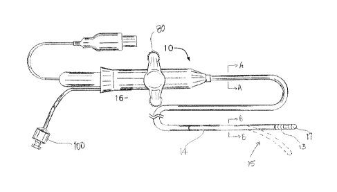
[0030] FIG. 1 is a perspective view of a catheter of the present
invention, in accordance with an
embodiment.
[0031] FIG. 2 is an end cross-sectional view of a catheter body of the
catheter of FIG. 1, taken
along line A¨A.
[0032] FIG. 3 is an end cross-sectional view of an intermediate deflection
section of the
catheter of FIG. 1, taken along line B¨B.
[0033] FIG. 4 is a perspective view of a distal section of the
catheter, with parts broken away,
in accordance with an embodiment.
[0034] FIG. 5A is a perspective view of a multifunctional
microinjection-molded housing of
the distal section of FIG. 4.
[0035] FIG. 5B is an end cross-sectional view of the housing of FIG.
5A, taken along line A¨
A.
-5-
CA 3036944 2019-03-18
1
[0036] FIG. 6 is a perspective view of the housing of FIG. 5A, with a
force sensor, in
accordance with an embodiment.
[0037] FIG. 7 is a perspective view of a flex circuit of FIG. 6.
[0038] FIG. 8 is a top view of the flex circuit of FIG. 7 lying flat.
[0039] FIG. 9 is a top view of a flex circuit lying flat, in
accordance with another embodiment.
DETAILED DESCRIPTION OF THE INVENTION
[0040] FIG. 1 illustrates an embodiment of a catheter 10 having an
elongated catheter body 12
with proximal and distal ends, an intermediate deflectable section 14 at the
distal end of the
catheter body 12, and a distal electrode section 15 with a tip electrode 17
and a micro-injection
molded, multi-functional housing 13. The catheter also includes a control
handle 16 at the
proximal end of the catheter body 12 for controlling bi-directional deflection
of the intermediate
section 14 relative to the catheter body 12.
[0041] With reference to FIG. 2, the catheter body 12 comprises an
elongated tubular
construction having a single, axial or central lumen 18. The catheter body 12
is flexible, i.e.,
bendable, but substantially non-compressible along its length. The catheter
body 12 can be of any
suitable construction and made of any suitable material. In some embodiments,
the catheter body
12 comprises an outer wall 20 made of polyurethane or PEBAX with an imbedded
braided mesh of
stainless steel or the like to increase torsional stiffness of the catheter
body 12 so that, when the
control handle 16 is rotated, the intermediate section 14 of the catheter 10
will rotate in a
corresponding manner.
[0042] The outer diameter of the catheter body 12 is not critical. In
some embodiments, the
outer diameter is about 8 french or 7 french. Likewise the thickness of the
outer wall 20 is not
critical, but is thin enough so that the central lumen 18 can accommodate
components, e.g., puller
tensile members, lead wires, and any other desired wires, cables or tubings.
If desired, the inner
-6-
CA 3036944 2019-03-18
1
surface of the outer wall 20 is lined with a stiffening tube 22 to provide
improved torsional
stability. In some embodiments, the catheter has an outer wall 20 with an
outer diameter of from
about 0.090 inch to about 0.94 inch and an inner diameter of from about 0.061
inch to about 0.065
inch.
[0043] The components extending through the lumen 18 of the catheter
body 12 may include
lead wires 23T and 23R (for the tip electrode 17 and one or more ring
electrodes 21 proximal of the
tip electrode), an irrigation tubing 24 with lumen 25 for delivering fluid to
the tip electrode, one or
more wire(s) and/or cable(s) (collectively "cables") 26 for an EM position
sensor 27 carried in or
near the distal section 15, one wire(s) and/or more cable(s) (collectively
"cables") 58 for a force
sensor 61 housed in the distal section 15, and/or puller tensile members 28A,
28B for deflecting the
intermediate section 14.
[0044] FIG. 3 illustrates an embodiment of the intermediate section 14
which comprises a short
section of tubing 19. The illustrated tubing 19 has multiple lumens, for
example off-axis lumens
31, 32, 33, 34 and on-axis lumen 35. In some embodiments, the lumen 31 carries
the lead wires
23T and 23R, and the position sensor cables 26, the lumen 32 carries the first
puller tensile member
28A, the lumen 33 carries the force sensor cables 58, the lumen 34 carries the
second puller tensile
member 28B, and the lumen 35 carries the irrigation tubing 24. It is
understood that the lumens
may be arranged in different configurations, as needed or appropriate.
[0045] The tubing 19 of the intermediate section 14 is made of a
suitable non-toxic material
that is more flexible than the catheter body 12. A suitable material for the
tubing 19 is braided
polyurethane, i.e., polyurethane with an embedded mesh of braided stainless
steel or the like. The
size of each lumen is not critical, but is sufficient to house the respective
components extending
therethrough.
[0046] Each puller tensile member 28A, 28B has a lubricious coating,
e.g. of Teflon®
The puller tensile members can be made of any suitable metal, such as
stainless steel, Nitinol or
-7-
CA 3036944 2019-03-18
1
Vectran.RTM and the Teflon coating imparts lubricity to the puller tensile
member. In some
embodiments, the puller tensile member has a diameter ranging from about 0.006
to about 0.010
inch.
[0047] As shown in FIG. 2, the portion of each puller tensile member
28A, 28B in the catheter
body 12 passes through a respective compression coil 29 in surrounding
relation. Each compression
coil extends from the proximal end of the catheter body 12 to at or near the
proximal end of the
intermediate section 14. The compression coils are made of any suitable metal,
preferably stainless
steel, and are tightly wound on themselves to provide flexibility, i.e.,
bending, but to resist
compression. The inner diameter of the compression coil is slightly larger
than the diameter of the
puller tensile member. As shown in FIG. 3, each portion of the puller tensile
members 28A, 28B
distal of the compression coil may extend through a respective protective
sheath 36 to prevent the
puller tensile member from cutting into the tubing 19 of the intermediate
section 14 during
deflection.
[0048] Proximal ends of the puller tensile members 28A, 28B are
anchored in the control
handle 16 to deflection actuation mechanisms that are responsive to an
operator's manipulation of a
deflection knob 80 of the control handle 16. Suitable deflection members are
described in U.S.
Patent No. 7377906, titled STEERING MECHANISM FOR BI-DIRECTIONAL CATHETER, the
entire disclosure of which is incorporated herein by reference.
[0049] With reference to FIG. 4, at the distal end of the intermediate
section 14 is the distal
electrode section 15 that includes the tip electrode 17, the micro-injection
molded, multifunctional
housing 13, and a flex circuit 53 supported by the housing 13. In some
embodiments, a relatively
short piece of non-conductive, single-lumened connector tubing 37 extends
between the housing 13
and the distal end of the tubing 19, to provide a lumen 38 which allows
components passing
between the lumen 41 of the housing 13 and the lumens 31-35 of the tubing 19
(see FIG. 3) to
reorient, as needed. These components may include, for example, the electrode
lead wires 23T,
-8-
CA 3036944 2019-03-18
1
23R, the irrigation tubing 24, the force sensor cables 58, the puller tensile
members 28A, 28B, and
the EM position sensor cables 58 (see FIG. 3).
[0050] As shown in FIG. 5A and FIG. 5B, the micro-injection molded,
multifunctional
housing 13 has a generally hollow cylindrical body 39 having a lumen 41, a
distal portion 39D with
an outer diameter DD and a proximal portion 39P with an outer diameter DP,
with DD < DP
creating a first circumferential step S1 at the junction between the portions
39D and 39P. The body
39 also has a radial opening 40 in a sidewall of the distal portion 39D that
provides access into the
lumen 41. The opening 40 has a proximal edge 40P that lies along the step S 1
and a distal edge
40D that has an arcuate configuration. The outer surface of the distal portion
39D is generally
smooth. The outer surface of the proximal portion 39P is generally smooth with
the exception of a
circumferential recess 42 extending around the body 39.
[0051] At the proximal end, the body 39 has an annular ridge 43 whose
outer diameter DR >
DP. The body 39 has a short distal end portion or neck 44 whose outer diameter
DN < DD creates
a second or distal circumferential step S2.
[0052] The lumen 41 extends through the entirety of the body 39. The
lumen 41 at least at the
distal end of the body 39 is partially occluded by a partial peripheral lip 50
that projects inwardly
into the lumen 41 (FIG. 5B). The lip 50 includes two axial through-holes 51A,
51B generally
aligned with lumens 32 and 34, respectively, of the multi-lumened tubing 19 of
the deflection
section 14. Connecting the through-holes 51A, 51B is a curved elongated recess
52 on a distal face
of the lip 50 that follows the peripheral curvature of the lip 50. In that
regard, it is understood that
the puller tensile members 28A and 28B may be portions of a single puller
tensile member that has
a U-bend portion 28U (shown in broken lines) that nests in the elongated
recess 52 with each leg
extending through a respective through-hole 51A, 51B as portions 28A, 28B,
respectively. The
curved elongated recess 52 anchors the U-bend portion 28U so that an operator
manipulating a
deflection knob 11 of the control handle 16 (FIG. 1) acting on proximal ends
of the portions 28A,
-9-
CA 3036944 2019-03-18
1
2813 can deflect the deflection section 14 bi-directionally. The curved
elongated recess 52 anchors
the U-bend portion 28U in a manner that minimizes occlusion or occupation of
the lumen 41.
[0053] The lip 50 may be a formation limited to the distal end of the body
39. In some
embodiments, the lip 50 may be a formation that extends along the inner
surface surrounding the
lumen, as appropriate or desired. In this regard, the through-holes 51A/51B
are elongated passages
that extend the length of the body 39.
[0054] As shown in FIG. 6, a flex circuit 53 is supported by the
housing 13. In some
embodiments, the flex circuit has a T-configuration, with a generally
rectangular distal portion 53D
and an elongated proximal portion or tail 53P extending at about 90 degrees,
as shown in FIG. 7
and FIG. 8. The distal portion 53D has traces X configured as an x-axis coil
and traces Y
configured as a y-axis coil. The distal portion 53D is wrapped around the
outer surface of the distal
portion 39D such that the coil traces X and Y are generally perpendicular to
each other on outer
surface of the distal portion 39D.
[0055] The proximal portion or tail 53P advantageously extends into
the lumen 41 via the
opening 40 in the body 39. The proximal portion 53P includes traces Tx, Ty and
connection pads
76 that connect to one or more electrical components, including the EM
position sensor cables 26
for passing electrical signals arising in the coil traces X and Y proximally
along the deflection
section 14 and the catheter body 12, toward the control handle 16. A z- axis
coil Z includes a wire
54 wrapped around the circumferential recess 42 of the body 39 (see FIG. 6).
End portions of the
wire 54 extend through one or more through-hole 55 (see FIG. 5A) formed in the
sidewall of the
recess 42 to reach the lumen 41 of the housing body 39, where the end portions
are joined with the
flex circuit 53 or EM sensor cable 26.
[0056] In some embodiments, the end portions of the wire 54 are soldered
directly to
connection pads on the flex circuit 53 without routing them through the lumen
41 of the body 39.
In some embodiments, with reference to FIG. 5A and FIG. 9, a flex circuit 53
has a distal portion
-10-
CA 3036944 2019-03-18
1
or leg 53L and a longitudinal proximal portion or tail 53T which together form
an "L" shape. On
the same side as the distal leg 53L and proximal thereof by a separation gap
G, the flex circuit 53
includes a generally rectangular proximal portion 53R with a corner 53C that
extends from a side
edge of the tail 53T. The distal leg 53L is configured to wrap
circumferentially around the distal
portion 39D of the body 39, the tail 53T is configured to pass through the
opening 40, and the
proximal portion 53R is configured to wrap circumferentially around the
proximal portion 39P of
the body 39. The proximal portion 53R of the flex circuit includes the coil
traces X and Y, and one
or more elongated connection pads 79 that traverse over the coil traces X and
Y and are generally
perpendicular to the tail 53T when the proximal portion 53R is wrapped
circumferentially around
the proximal portion 39 of the body.
[0057] In some embodiments, one or more ring electrodes 21 are carried
on the housing 13, as
shown in FIG. 4. In the illustrated embodiment, a first ring electrode 21A
having a predetermined
width WI is slipped over the distal end of the housing 13 and moved proximally
onto the proximal
portion 39P until the ring electrode abuts tightly with the annular ridge 43
acting as a stop. A first
spacer 60A having a predetermined width W2 is then slipped over the distal end
of the housing 13
and moved proximally until it abuts tightly with the first ring electrode 21A.
A second ring
electrode 21B having a predetermined with W3 is slipped over the distal end of
the housing 13 and
moved proximally until it abuts tightly with the first spacer 60A. A second
spacer 60B having a
predetermined width W4 is slipped over the distal end of the housing 13 and
moved proximally
until it abuts tightly with the second ring electrode 21B. Accordingly, the
ring electrodes 21A, 21B
can be advantageously arranged with tight tolerances for improved mapping
and/or ablation
performance. The lead wires 30R for the ring electrodes 21A, 21B pass through
respective
through-holes 56 and 57 (see FIG. 5A) formed in the sidewall of the housing
proximal portion 39P
for connection to the respective ring electrodes.
[0058] In some embodiments, the ring electrodes are electrically
connected to the underlying
-11-
CA 3036944 2019-03-18
1
elongated circumferential connection pads 79 provided on the proximal portion
53R of the flex
circuit 53 (see FIG. 9) that is wrapped around the proximal portion 39P of the
body 39 below the
ring electrodes and the spacers.
[0059] It is understood that the housing 13 may be configured with any
desired longitudinal
length for accommodating a corresponding plurality of ring electrodes, whose
predetermined width
and spacing between adjacent ring electrodes on the outer surface of the
housing 13 may be varied
as desired.
100601 In some embodiments, the distal section 15 includes a force sensor
61 having a distal
on-axis stem 63 with lumen 67, an annular proximal portion or ring 62
perpendicular to the stem
63, and a plurality (e.g., three, although only two are shown in FIG. 4)
radial strain gauges 72
extending between the stem 63 and the annular ring 62. The ring 62 is
configured to fit onto the
neck 44 of the housing 13. In that regard, a proximal end of the neck 44 may
have a plurality of
fasteners or snaps 64 that engage with the distal edge of the ring 62 to
secure the force sensor onto
the housing 13. Each strain gauge 72 has respective electrical leads 65 and
connection pads 66 that
allow electrical signals arising from the strain gauges to pass onto the flex
circuit 53 and pass
proximally along the catheter through the deflection section 14 and the
catheter body 12 via the
cables 58.
[0061] Mounted on an extended distal end 63D of the stem 63 is the distal
tip electrode 17, as
shown in FIG. 4. The distal tip electrode 17 includes a shell portion 71 and a
proximal plug
portion 73 (shown in broken lines) which seals an open proximal end of the
shell portion to create
an interior chamber 70. A distal end of the lead wire 30T (see FIG. 2 and FIG.
3, not shown in
FIG. 4) is potted in a blind hole (not shown) in the plug portion 73 and the
lead wire 30T extends
through the lumen 67 of the stem 63 of the force sensor 61. The irrigation
tubing 24 (see FIG. 2
and FIG. 3, not shown in FIG. 4) also extends through the lumen 67 with its
distal end extending
into the interior chamber 70 defined by the shell portion 71 of the tip
electrode 17. A plurality of
-12-
CA 3036944 2019-03-18
1
irrigation ports 74 are formed in the shell portion 71 so that fluid delivered
by the irrigation tubing
24 into the interior chamber 70 can exit the distal tip electrode 17 via the
irrigation ports 74. The
plug portion 73 has an axial through-opening that receives the extended distal
end 63D of the force
sensor 61 and secures the force sensor 61 relative to the shell portion 71 so
that any force exerted
on the shell portion 71, for example, when the shell portion 71 contacts
tissue surface, is imparted
to the plug portion 73 and the stem 63 of the force sensor 61 in activating
the strain gauges 72 to
transmit electrical signals to the connection pads 66 of the flex circuit 53,
as shown in FIG. 8. The
extended distal end 63D has a smaller outer diameter relative to the stem 63
so to create a stop 63
that abuts a proximal face of the plug portion 73 and prevents plug portion 73
from moving
proximally and interfering with the action of the stem 63 in responding to a
force that is applied to
the distal tip electrode. Traces 75 transmit the strain electrical signals to
the cables 58 (see FIG. 2
and FIG. 3, not shown in FIG. 4) via connection pads 78.
[0062] In some embodiments, a short nonconductive tubing 95 (see FIG. 4)
extends between
the tip electrode 17 and the second spacer 60B, circumferentially surrounding,
protecting and
providing a fluid-tight seal around the force sensor 61. The tubing 951s
sufficiently flexible so as
not to interfere with deformation of the strain gauges 72 of the force sensor
61 when sensing
contact and force of the tip electrode 17 against tissue.
[0063] Having a micro-injection-molded body, the housing 13 performs as a
single, unitary
body and component providing a multitude of functions, including an distal
anchor for the puller
tensile member and a support for various components, including, the flex
circuit, the force sensor,
the x/y/z-axes coils, the ring electrodes and their spacers. The lumen 41 of
the housing 13 can
house additional components, as needed or desired. The housing 13 provides
cost savings in terms
of supply and manufacturing costs. Micro injection molding can allow more
intricate and detailed
3-D geometry in the housing 13.
-13-
CA 3036944 2019-03-18
1
100641 The preceding description has been presented with reference to
presently preferred
embodiments of the invention. Workers skilled in the art and technology to
which this invention
pertains will appreciate that alterations and changes in the described
structure may be practiced
without meaningfully departing from the principal, spirit and scope of this
invention. Notably, the
drawings are not necessarily to scale, and any one or more features of any one
or more
embodiments may be included in any other one or more embodiments in addition
to or in lieu of
any feature, as desired or appropriate. Accordingly, the foregoing description
should not be read as
pertaining only to the precise structures described and illustrated in the
accompanying drawings,
but rather should be read consistent with and as support to the following
claims which are to have
their fullest and fair scope.
20
-14-
CA 3036944 2019-03-18