Note: Descriptions are shown in the official language in which they were submitted.
CA 03037682 2019-03-20
WO 2018/057727
PCT/US2017/052701
TITLE OF THE INVENTION
OPTIMIZED SYNTHETIC CONSENSUS IMMUNOGENIC COMPOSITIONS
TARGETING FIBROBLAST ACTIVATION PROTEIN
CROSS-REFERENCE TO RELATED APPLICATIONS
The present application is entitled to priority to U.S. Provisional
Application No. 62/397,469, filed September 21, 2016, which is incorporated by
reference herein in its entirety.
STATEMENT REGARDING FEDERALLY SPONSORED RESEARCH OR
DEVELOPMENT
This invention was made with government support under Grant Nos.
P50 CA174523, U19 AI109646 and F32 CA213795 awarded by the National
Institutes of Health and Grant No. W31P4Q-15-1-0003 awarded by the U.S.
Department of Defense. The government has certain rights in the invention.
TECHNICAL FIELD
The present invention relates to immunogenic compositions targeting
Fibroblast Activation Protein, and methods of administering the immunogenic
compositions.
BACKGROUND OF THE INVENTION
Solid tumor pathophysiology is characterized by an abnormal
microenvironment that guides tumor progression and poses barriers to the
efficacy of
cancer therapies. Several proteins are overexpressed in the tumor
microenvironment,
including Fibroblast Activation Protein (FAP). FAP is a membrane-bound enzyme
with gelatinase and peptidase activity that is up-regulated in cancer-
associated
fibroblasts in over 90% of human carcinomas.
Breaking the body's tolerance to the tumor microenvironment has the
potential to improve cancer therapy. Previous studies have shown that ablation
of
FAP -expressing cells from transgenic mice attenuates tumor growth and
synergizes
with other immune therapies such as immune checkpoint blockade. Groups have
additionally shown that T cells expressing chimeric antigen receptors
targeting FAP
1
CA 03037682 2019-03-20
WO 2018/057727
PCT/US2017/052701
slow tumor progression; however, in some mouse strains these CARs cause lethal
toxicity.
Thus, there is a need in the art for the development of safer therapies
directed at breaking tolerance to the tumor microenvironment. The present
invention
satisfies this unmet need.
SUMMARY OF THE INVENTION
In one embodiment the invention relates to an immunogenic
composition comprising a nucleic acid molecule, wherein the nucleic acid
molecule
encodes a peptide comprising an amino acid sequence of a) an amino acid
sequence
having at least about 90% identity over an entire length of the amino acid
sequence of
SEQ ID NO:2 or SEQ ID NO:6, b) an immunogenic fragment comprising at least
about 90% identity over at least 60% of the amino acid sequence of SEQ ID NO:2
or
SEQ ID NO:6, c) the amino acid sequence of SEQ ID NO:2 or SEQ ID NO:6, or d)
an
immunogenic fragment comprising at least 60% of the amino acid sequence of SEQ
ID NO:2 or SEQ ID NO:6.
In one embodiment, the nucleic acid molecule is a DNA molecule. In
one embodiment, the nucleic acid molecule is a RNA molecule.
In one embodiment, the nucleic acid molecule comprises a nucleotide
sequence of a) a nucleotide sequence having at least about 90% identity over
an entire
length of a nucleotide sequence of SEQ ID NO:1 or SEQ ID NO:5, b) an
immunogenic fragment of a nucleotide sequence having at least about 90%
identity
over at least 60% of the nucleotide sequence of SEQ ID NO:1 or SEQ ID NO:5, c)
a
nucleotide sequence of SEQ ID NO:1 or SEQ ID NO:5, or d) an immunogenic
fragment of a nucleotide sequence of SEQ ID NO:1 or SEQ ID NO:5.
In one embodiment, the nucleotide sequence encoding the peptide is
operably linked to at least one regulatory sequence. In one embodiment, the
regulatory sequence is a start codon, an IgE leader sequence, a stop codon or
a
combination thereof
In one embodiment, the nucleic acid molecule encodes a peptide
comprising an amino acid sequence of a) an amino acid sequence having at least
about 90% identity over an entire length of the amino acid sequence of SEQ ID
NO:4
or SEQ ID NO:8, b) an immunogenic fragment comprising at least about 90%
identity
over at least 60% of the amino acid sequence of SEQ ID NO:4 or SEQ ID NO:8, c)
2
CA 03037682 2019-03-20
WO 2018/057727
PCT/US2017/052701
the amino acid sequence of SEQ ID NO:4 or SEQ ID NO:8, or d) an immunogenic
fragment comprising at least 60% of the amino acid sequence of SEQ ID NO:4 or
SEQ ID NO:8.
In one embodiment, the nucleic acid molecule comprises a nucleotide
sequence of a) a nucleotide sequence having at least about 90% identity over
an entire
length of a nucleotide sequence of SEQ ID NO:3 or SEQ ID NO:7, b) an
immunogenic fragment of a nucleotide sequence having at least about 90%
identity
over at least 60% of the nucleotide sequence of SEQ ID NO:3 or SEQ ID NO:7, c)
a
nucleotide sequence SEQ D NO:3 or SEQ ID NO:7, or d) an immunogenic fragment
of a nucleotide sequence of SEQ ID NO:3 or SEQ ID NO:7.
In one embodiment, the nucleic acid molecule is an expression vector.
In one embodiment, the nucleic acid molecule is incorporated into a
viral particle.
In one embodiment, the immunogenic composition comprises a
pharmaceutically acceptable excipient.
In one embodiment, the immunogenic composition comprises an
adjuvant.
In one embodiment the invention relates to a nucleic acid molecule
encoding a peptide comprising an amino acid sequence of a) an amino acid
sequence
having at least about 90% identity over an entire length of the amino acid
sequence of
SEQ ID NO:2 or SEQ ID NO:6, b) a fragment comprising at least about 90%
identity
over at least 60% of the amino acid sequence of SEQ ID NO:2 or SEQ ID NO:6, c)
the amino acid sequence of SEQ ID NO:2 or SEQ ID NO:6, or d) a fragment
comprising at least 60% of the amino acid sequence of SEQ ID NO:2 or SEQ ID
NO:6.
In one embodiment, the nucleic acid molecule is a DNA molecule or
an RNA molecule.
In one embodiment, the nucleic acid molecule is a DNA molecule. In
one embodiment, the nucleic acid molecule is a RNA molecule.
In one embodiment, the nucleic acid molecule comprises a nucleotide
sequence of a) a nucleotide sequence having at least about 90% identity over
an entire
length of a nucleotide sequence of SEQ ID NO:1 or SEQ ID NO:5, b) an
immunogenic fragment of a nucleotide sequence having at least about 90%
identity
over at least 60% of the nucleotide sequence of SEQ ID NO:1 or SEQ ID NO:5, c)
a
3
CA 03037682 2019-03-20
WO 2018/057727
PCT/US2017/052701
nucleotide sequence of SEQ ID NO:1 or SEQ ID NO:5, or d) an immunogenic
fragment of a nucleotide sequence of SEQ ID NO:1 or SEQ ID NO:5.
In one embodiment, the nucleotide sequence encoding the peptide is
operably linked to at least one regulatory sequence. In one embodiment, the
regulatory sequence is a start codon, an IgE leader sequence, a stop codon or
a
combination thereof
In one embodiment, the nucleic acid molecule encodes a peptide
comprising an amino acid sequence of a) an amino acid sequence having at least
about 90% identity over an entire length of the amino acid sequence of SEQ ID
NO:4
or SEQ ID NO:8, b) an immunogenic fragment comprising at least about 90%
identity
over at least 60% of the amino acid sequence of SEQ ID NO:4 or SEQ ID NO:8, c)
the amino acid sequence of SEQ ID NO:4 or SEQ ID NO:8, or d) an immunogenic
fragment comprising at least 60% of the amino acid sequence of SEQ ID NO:4 or
SEQ ID NO:8.
In one embodiment, the nucleic acid molecule comprises a nucleotide
sequence of a) a nucleotide sequence having at least about 90% identity over
an entire
length of a nucleotide sequence of SEQ ID NO:3 or SEQ ID NO:7, b) an
immunogenic fragment of a nucleotide sequence having at least about 90%
identity
over at least 60% of the nucleotide sequence of SEQ ID NO:3 or SEQ ID NO:7, c)
a
nucleotide sequence SEQ D NO:3 or SEQ ID NO:7, or d) an immunogenic fragment
of a nucleotide sequence of SEQ ID NO:3 or SEQ ID NO:7.
In one embodiment, the nucleic acid molecule is an expression vector.
In one embodiment, the nucleic acid molecule is incorporated into a
viral particle.
In one embodiment the invention relates to an immunogenic
composition comprising a peptide, wherein the peptide comprises an amino acid
sequence of a) an amino acid sequence having at least about 90% identity over
an
entire length of the amino acid sequence of SEQ ID NO:2, SEQ ID NO:4, SEQ ID
NO:6 or SEQ ID NO:8, b) an immunogenic fragment comprising at least about 90%
identity over at least 60% of the amino acid sequence of SEQ ID NO:2, SEQ ID
NO:4, SEQ ID NO:6 or SEQ ID NO:8, c) the amino acid sequence of SEQ ID NO:2,
SEQ ID NO:4, SEQ ID NO:6 or SEQ ID NO:8 or d) an immunogenic fragment
comprising at least 60% of the amino acid sequence SEQ ID NO:2, SEQ ID NO:4,
SEQ ID NO:6 or SEQ ID NO:8.
4
CA 03037682 2019-03-20
WO 2018/057727
PCT/US2017/052701
In one embodiment the invention relates to a peptide comprising an
amino acid sequence of a) an amino acid sequence having at least about 90%
identity
over an entire length of the amino acid sequence of SEQ ID NO:2, SEQ ID NO:4,
SEQ ID NO:6 or SEQ ID NO:8, b) an immunogenic fragment comprising at least
about 90% identity over at least 60% of the amino acid sequence of SEQ ID
NO:2,
SEQ ID NO:4, SEQ ID NO:6 or SEQ ID NO:8, c) the amino acid sequence of SEQ
ID NO:2, SEQ ID NO:4, SEQ ID NO:6 or SEQ ID NO:8, or d) an immunogenic
fragment comprising at least 60% of the amino acid sequence of SEQ ID NO:2,
SEQ
ID NO:4, SEQ ID NO:6 or SEQ ID NO:8.
In one embodiment the invention relates to a method of inducing an
immune response against Fibroblast Activation Protein (FAP) in a subject in
need
thereof, the method comprising administering an immunogenic composition
comprising a nucleic acid molecule, wherein the nucleic acid molecule encodes
a
peptide comprising an amino acid sequence of a) an amino acid sequence having
at
least about 90% identity over an entire length of the amino acid sequence of
SEQ ID
NO:2 or SEQ ID NO:6, b) an immunogenic fragment comprising at least about 90%
identity over at least 60% of the amino acid sequence of SEQ ID NO:2 or SEQ ID
NO:6, c) the amino acid sequence of SEQ ID NO:2 or SEQ ID NO:6, or d) an
immunogenic fragment comprising at least 60% of the amino acid sequence of SEQ
ID NO:2 or SEQ ID NO:6 to the subject.
In one embodiment, administering includes at least one of
electroporation or injection.
In one embodiment the invention relates to a method of treating or
preventing a tumor associated pathology in a subject in need thereof, the
method
comprising administering an immunogenic composition comprising a nucleic acid
molecule, wherein the nucleic acid molecule encodes a peptide comprising an
amino
acid sequence of a) an amino acid sequence having at least about 90% identity
over an
entire length of the amino acid sequence of SEQ ID NO:2 or SEQ ID NO:6, b) an
immunogenic fragment comprising at least about 90% identity over at least 60%
of
the amino acid sequence of SEQ ID NO:2 or SEQ ID NO:6, c) the amino acid
sequence of SEQ ID NO:2 or SEQ ID NO:6, or d) an immunogenic fragment
comprising at least 60% of the amino acid sequence of SEQ ID NO:2 or SEQ ID
NO:6 to the subject.
5
CA 03037682 2019-03-20
WO 2018/057727
PCT/US2017/052701
In one embodiment, administering includes at least one of
electroporation or injection.
In one embodiment, the tumor associated pathology is at least one of
tumor growth, tumor metastasis, or angiogenesis.
In one embodiment, the subject has been diagnosed with cancer.
In one embodiment, cancer is prostate cancer.
In one embodiment, the method comprises administering an
immunogenic composition comprising one or more prostate cancer antigens to the
subject. In one embodiment, the method comprises administering an immunogenic
composition comprising PSMA to the subject.
In one embodiment, the cancer is lung cancer. In one embodiment, the
method comprises administering an immunogenic composition comprising one or
more lung cancer antigens to the subject. In one embodiment, the method
comprises
administering an immunogenic composition comprising TERT to the subject.
BRIEF DESCRIPTION OF THE DRAWINGS
The following detailed description of preferred embodiments of the
invention will be better understood when read in conjunction with the appended
drawings. For the purpose of illustrating the invention, there are shown in
the
drawings embodiments which are presently preferred. It should be understood,
however, that the invention is not limited to the precise arrangements and
instrumentalities of the embodiments shown in the drawings.
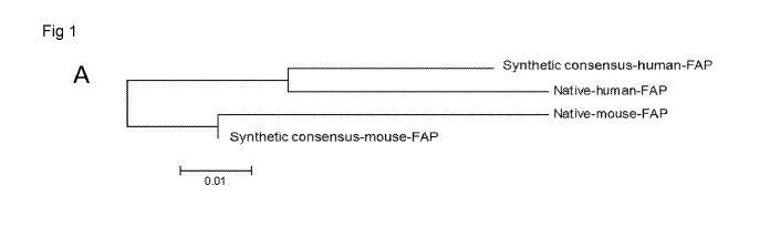
Figure 1, comprising Figure 1A through Figure 1D, depicts the design
of a FAP immunogenic composition using synthetic consensus technology for use
in
combination with tumor antigen-specific DNA based immunogenic composition
constructs. Figure 1A depicts a phylogenetic tree describing the genetic
relationships
between the optimized consensus sequences of the invention and the native
human
and mouse FAP sequences. Figure 1B depicts a schematic of the Mouse FAP
operably
linked to an IgE leader sequence (IgELS) and having a 5624A mutation to block
dipeptidyl peptidase and gelatinolytic activities. Figure 1C depicts a diagram
of the
mature murine FAP in natural homodimeric form, shown in cpk format. The
endogenous membrane tether in the fully wild-type FAP is not present in this
design.
[tCon changes relative to representative wild-type sequence are shown in red.
One of
two ablated active serine residues is visible in yellow located in a monomer
active site
6
CA 03037682 2019-03-20
WO 2018/057727
PCT/US2017/052701
pocket. Figure ID depicts an exemplary western blot showing expression of both
native mouse FAP and Con mouse FAP plasmids transfected into 293T cells. Non-
transfected cells, and cells transfected with a GFP-expressing plasmid were
used as
negative controls.
Figure 2, comprising Figure 2A through Figure 2E, depicts
experimental results demonstrating the immunogenicity of Con mouse FAP
vaccine
in C57B1/6 mice. Figure 2A depicts the experimental design. Mice were
immunized
three times at two-week intervals, and were sacrificed one week following
final
vaccination. Splenocytes were analyzed to examine T cell responses. Figure 2B
and
.. Figure 2C depict exemplary results demonstrating IFN-y ELISpot responses to
native
mouse FAP peptides (Figure 2B) or Con peptides matched to the vaccine
sequence
(Figure 2C). Figure 2D and Figure 2E depict exemplary results demonstrating
intracellular cytokine staining of CD8+ (Figure 2D) and CD4+ (Figure 2E) T
cells
following stimulation with native mouse FAP peptides for 5 hours. The 101.tg
dose of
FAP vaccine was used for this study. Significance was determined by a
student's t-
test for Figure 2D and Figure 2E. *p<0.05, **p<0.01, ***p<0.001. N=5 mice per
group, shown is a representative of two independent experiments.
Figure 3, comprising Figure 3A through Figure 3C, depicts
experimental results demonstrating a comparison of native and Con FAP
vaccines in
CD-I outbred mice. Figure 3A depicts exemplary results demonstrating the IFN-y
ELISpot responses to native mouse FAP peptides from individual CD-I outbred
mice
in naive control group (top), native mouse FAP vaccine group (middle) or Con
mouse FAP vaccine group (bottom). Immunized mice received 101.tg of DNA
plasmid. The immunization schedule for these mice was the same as in Figure 2.
Figure 3B depicts exemplary results demonstrating the total IFN-y ELISpot
responses
from the mice immunized in Figure 3A, not separated by pool. Figure 3C depicts
exemplary results demonstrating the endpoint binding titers from the mice in
Figure
3A against the native FAP protein (extracellular domain). Significance was
determined by two-way ANOVA followed by Tukey's HSD test for Figure 3B.
.. *p<0.05, **p<0.01, ***p<0.001. 10 mice were used in the naive group, and 15
mice
each were used in the Native FAP and Con FAP groups.
Figure 4, comprising Figure 4A through Figure 4E, depicts
experimental results demonstrating a comparison of native and Con FAP
vaccines in
C57B1/6 mice. Figure 4A depicts a diagram showing the experimental setup.
C57B1/6
7
CA 03037682 2019-03-20
WO 2018/057727
PCT/US2017/052701
mice were immunized three times at two-week intervals, and were sacrificed one
week following final vaccination. Splenocytes were analyzed to examine T cell
responses, and serum was collected to examine antibody responses. Figure 4B
depicts
exemplary results demonstrating IFN-y ELISpot responses to native mouse FAP
peptides. Figure 4C and Figure 4D depict exemplary results demonstrating
intracellular cytokine staining of CD8+ (Figure 4C) and CD4+ (Figure 4D) T
cells
following stimulation with native mouse FAP peptides for 5 hours. The 101.tg
dose of
native mouse FAP or Con mouse FAP vaccine was used for this study. Figure 4E
depicts exemplary results demonstrating endpoint binding titers from the mice
in
Figure 4A against the native FAP protein (extracellular domain). Significance
was
determined by a one-way ANOVA followed by Tukey's HSD test. *p<0.05,
"p<0.01, ***p<0.001. N=4-10 mice per group.
Figure 5, comprising Figure 5A through Figure 5E, depicts
experimental results demonstrating a comparison of native and Con FAP
vaccines in
Balb/c mice. Figure 5A depicts a diagram showing the experimental setup.
Balb/c
mice were immunized three times at two-week intervals, and were sacrificed one
week following final vaccination. Splenocytes were analyzed to examine T cell
responses, and serum was collected to examine antibody responses. Figure 5B
depicts
exemplary results demonstrating IFN-y ELISpot responses to native mouse FAP
peptides. Figure 5C and Figure 5D depict exemplary results demonstrating
intracellular cytokine staining of CD8+ (Figure 5C) and CD4+ (Figure 5D) T
cells
following stimulation with native mouse FAP peptides for 5 hours. The 101.tg
dose of
native mouse FAP or Con mouse FAP vaccine was used for this study. Figure 5E
depicts exemplary results demonstrating endpoint binding titers from the mice
in
Figure 5A against the native FAP protein (extracellular domain). Significance
was
determined by a one-way ANOVA followed by Tukey's HSD test. *p<0.05,
"p<0.01, ***p<0.001. N=4-10 mice per group.
Figure 6, comprising Figure 6A through Figure 6C, depicts
experimental results demonstrating the efficacy of FAP vaccine and combination
therapy in therapeutic lung tumor model. Figure 6A depicts a diagram showing
the
experimental setup. Mice were implanted with TC-1 cells on day 0, randomized
on
day 7 and immunized once weekly for a total of 4 immunizations. 101.tg of Con
FAP
DNA and 251.tg of Con mouse TERT DNA was used. Figure 6B depicts exemplary
results demonstrating tumor volume measurements over time for indicated
8
CA 03037682 2019-03-20
WO 2018/057727
PCT/US2017/052701
vaccination regimen for mice implanted with TC-1. Figure 6C depicts exemplary
results demonstrating mouse survival over time for indicated vaccination
regimen for
mice implanted with TC-1. Significance for tumor volume measurements was
determined by two-way ANOVA followed by Tukey's HSD test. Significance for
mouse survival was determined by Gehan-Breslow-Wilcoxon test. *p<0.05,
**p<0.01, ***p<0.001, ****p<0.0001. N=10 mice per group for TC-1 study. Shown
is a representative of two independent experiments.
Figure 7, comprising Figure 7A through Figure 7C, depicts exemplary
experimental results demonstrating the efficacy of FAP vaccine and combination
therapy in therapeutic prostate tumor model. Figure 7A depicts a diagram
showing the
experimental setup. Mice were implanted with TRAMP-C2 cells on day 0,
randomized on day 4 and immunized once weekly for a total of 4 immunizations.
10 g of Con FAP DNA and 20 g of Con PSMA was used. Figure 7B depicts
exemplary results demonstrating tumor volume measurements over time for
indicated
vaccination regimen for mice implanted with TRAMP-C2. Figure 7C depicts
exemplary results demonstrating mouse survival over time for indicated
vaccination
regimen for mice implanted with TRAMP-C2. Significance for tumor volume
measurements was determined by two-way ANOVA followed by Tukey's HSD test.
Significance for mouse survival was determined by Gehan-Breslow-Wilcoxon test.
*p<0.05, **p<0.01, ***p<0.001, ****p<0.0001. N=15 mice per group for TRAMP-
C2 study. Shown is a representative of two independent experiments for each
tumor
type.
Figure 8 depicts exemplary experimental results demonstrating the
expression of FAP in tumor cell lines. Western blot expression of mouse FAP in
the
mouse tumor cell lines TC-1 and TRAMP-C2. 293T cells transfected with native
mouse FAP plasmid were used as a positive control.
Figure 9, comprising Figure 9A through Figure 9D, depicts exemplary
experimental results demonstrating that the FAP vaccine induces FAP-specific
TILs.
Figure 9A depicts a diagram showing the experimental setup. Mice were
implanted
with TC-1 tumor cells on day 0, randomized on day 7 and immunized once weekly
for
a total of 2 immunizations. 10 g of Con FAP DNA was used. Mice were
sacrificed
on day 21, and splenocytes and TILs were harvested. Figure 9B depicts
exemplary
results demonstrating intracellular cytokine staining of CD8+ T cells in the
spleen
following stimulation with native mouse FAP peptides for 5 hours. Figure 9C
depicts
9
CA 03037682 2019-03-20
WO 2018/057727
PCT/US2017/052701
exemplary results demonstrating intracellular cytokine staining of tumor
infiltrating
lymphocytes (TILs) that were stimulated with native mouse FAP peptides for 5
hours.
Figure 9D depicts exemplary results demonstrating the frequency of CD8+ T
cells and
CD4+/CD25+/FoxP3+ Tregs in each tumor, as a percentage of CD45+/CD3+
lymphocytes, assessed by flow cytometry staining. Significance was determined
by a
student's t-test for panels B-D. *p<0.05, **p<0.01, ***p<0.001. N=9-10 mice
per
group, shown is a representative of two independent experiments.
Figure 10, comprising Figure 10A through Figure 10D, depicts
exemplary experimental results demonstrating the immune responses from mice
receiving combination mTERT + FAP vaccination. Mice were implanted with TC-1
tumor cells on day 0, randomized on day 7 and immunized once weekly for a
total of
2 immunizations. 10 g of Con FAP DNA or 25 g of mTERT DNA was used. Mice
were sacrificed on day 21, and splenocytes and TILs were harvested. Figure 10A
and
Figure 10B depict exemplary results demonstrating intracellular cytokine
staining of
CD8+ TILs following stimulation with native mouse FAP peptides (Figure 10A) or
native mouse TERT peptides (Figure 10B) for 5 hours. Figure 10C and Figure 10D
depict exemplary results demonstrating intracellular cytokine staining of CD8+
splenocytes following stimulation with native mouse FAP peptides (Figure 10C)
or
native mouse TERT peptides (Figure 10D) for 5 hours. Significance was
determined
using a one-way ANOVA followed by Tukey's HSD test. *p<0.05, **p<0.01,
***p<0.001. N=8-10 mice per group.
Figure 11, comprising Figure 11A through Figure 11H, depicts
exemplary experimental results demonstrating that the FAP vaccine alters the
tumor
microenvironment. Figure 11A depicts representative immunohistochemical
staining
of tissues from control mice or Con mouse FAP immunized mice for FAP
expression. Figure 11B depicts quantification of the percentage of area in the
tumor
covered by FAP-expressing cells. Figure 11C depicts representative
immunofluorescent images of tissues from control mice or Con mouse FAP
immunized mice for hyaluronan expression. Figure 11D depicts quantification of
the
percentage of area in the tumor covered by hyaluronan. Figure 11E depicts
representative immunofluorescent image of tissues from control mice or Con
mouse
FAP immunized mice for F4/80 and EpCAM expression. Figure 11F depicts
quantification of the percentage of area in the tumor covered by F4/80
expressing
cells. Figure 11G depicts representative immunofluorescent image of tissues
from
CA 03037682 2019-03-20
WO 2018/057727
PCT/US2017/052701
control mice or Con mouse FAP immunized mice for CD8a and EpCAM
expression. Figure 11H depicts quantification of the percentage of area in the
tumor
covered by CD8a expressing cells. N=6-8 mice per group. Image quantification
was
performed for at least 5 images per mouse. Significance was determined by a
student's t-test for Figure 11B through Figure 11D. *p<0.05, **p<0.01,
***p<0.001.
Scale bar= 100 m.
Figure 12, comprising Figure 12A through Figure 12D, depicts
exemplary experimental results demonstrating the impact of Con FAP vaccine on
immune cell subsets by flow cytometry. Mice were implanted with TC-1 tumor
cells
and immunized according to the schedule in Figure 9A. Tumors were harvested
for
surface staining of innate immune cell populations, according to the markers
indicated
in figure legend. Figure 12A through Figure 12D, depicts exemplary
experimental
results demonstrating the quantification of the total number of macrophages
(Figure
12A), B cells (Figure 12B), natural killer cells (Figure 12C) and dendritic
cells
(Figure 12D) per tumor. Significance was determined by a student's t-test. N=9-
10
mice per group, shown is a representative of two independent experiments.
Figure 13, comprising Figure 13A through Figure 13E, depicts
experimental results demonstrating the impact of Con FAP vaccine on
properties of
tumor infiltrating macrophages. Mice were implanted with TC-1 tumor cells and
immunized according to the schedule in Figure 9A. Tumors were harvested for
surface staining of innate immune cell populations, according to the markers
indicated
in figure legend. Figure 13A through Figure 13D, depicts exemplary
experimental
results demonstrating the fraction of Argl+ (Figure 13A), MHCII+ (Figure 13B),
CD68+ (Figure 13C), CD80+ (Figure 13D) and CD86+ (Figure 13E) macrophages
were quantified. Significance was determined by a student's t-test. N=9-10
mice per
group, shown is a representative of two independent experiments.
Figure 14, comprising Figure 14A through Figure 14C, depicts
characterization of the dominant epitopes for the optimized consensus mouse
FAP
vaccine. Figure 14A depicts a matrix map of 122 peptides in native mouse FAP
arranged in a matrix of 23 pools. Figure 14B depicts stimulation of C57B1/6
mice
with each pool of synthetic consensus FAP peptides. Figure 14C depicts
stimulation
of Balb/c mice with each pool of synthetic consensus FAP peptides.
Figure 15 depicts a list of the dominant immunogenic epitopes for the
native and synthetic consensus mouse FAP. The dominant immunogenic epitopes of
11
CA 03037682 2019-03-20
WO 2018/057727
PCT/US2017/052701
native FAP are provided as SEQ ID NO: 13 through SEQ ID NO: 21. The dominant
immunogenic epitopes of the optimized consensus FAP are provided as SEQ ID NO:
15 through SEQ ID NO: 18, and SEQ ID NO: 20 through SEQ ID NO: 24.
DETAILED DESCRIPTION
In one aspect, the present invention provides an immunogenic
composition targeting FAP. Further aspects of the present invention are
treatments
and/or preventions of cancer growth or metastasis using the disclosed
immunogenic
composition alone or in combination with additional cancer vaccines or
therapeutics.
The sequences encoding the antigens of the invention are genetically
diverged from the sequences of their native proteins, and thus, the optimized
consensus antigens of the invention are unique. The immunogenic composition of
the
present invention can be widely applicable to breaking tolerance to the tumor
microenvironment, and reducing or preventing tumor growth or metastasis
because of
the unique sequences of the encoded antigens. These unique sequences allow the
immunogenic composition to be universally protective against multiple types of
cancer.
The immunogenic composition can be used to protect against and treat
any number of cancers. The immunogenic composition can elicit both humoral and
cellular immune responses that target the tumor microenvironment antigen. The
immunogenic composition can elicit neutralizing antibodies and immunoglobulin
G
(IgG) antibodies that are reactive with the tumor microenvironment antigen.
The
immunogenic composition can also elicit a CD8+ T cell response that is
reactive to the
tumor microenvironment antigen and produce one or more of interferon-gamma
(IFN-
y) and tumor necrosis factor alpha (TNF-a). In one embodiment, the immunogenic
composition can also elicit a CD4+ T cell response that is reactive to the
tumor
microenvironment antigen and produce one or more of IFN-y and TNF-a.
Definitions
Unless otherwise defined, all technical and scientific terms used herein
have the same meaning as commonly understood by one of ordinary skill in the
art. In
case of conflict, the present document, including definitions, will control.
Preferred
methods and materials are described below, although methods and materials
similar or
equivalent to those described herein can be used in practice or testing of the
present
12
CA 03037682 2019-03-20
WO 2018/057727
PCT/US2017/052701
invention. All publications, patent applications, patents and other references
mentioned herein are incorporated by reference in their entirety. The
materials,
methods, and examples disclosed herein are illustrative only and not intended
to be
limiting.
The terms "comprise(s)," "include(s)," "having," "has," "can,"
"contain(s)," and variants thereof, as used herein, are intended to be open-
ended
transitional phrases, terms, or words that do not preclude the possibility of
additional
acts or structures. The singular forms "a," "and" and "the" include plural
references
unless the context clearly dictates otherwise. The present disclosure also
contemplates
.. other embodiments "comprising," "consisting of" and "consisting essentially
of," the
embodiments or elements presented herein, whether explicitly set forth or not.
"Adjuvant" as used herein means any molecule added to the
immunogenic composition described herein to enhance the immunogenicity of the
antigen.
"Antibody" as used herein means an antibody of classes IgG, IgM,
IgA, IgD or IgE, or fragments, fragments or derivatives thereof, including
Fab,
F(ab1)2, Fd, and single chain antibodies, diabodies, bispecific antibodies,
bifunctional
antibodies and derivatives thereof The antibody can be an antibody isolated
from the
serum sample of mammal, a polyclonal antibody, affinity purified antibody, or
mixtures thereof which exhibits sufficient binding specificity to a desired
epitope or a
sequence derived therefrom.
"Coding sequence" or "encoding nucleic acid" as used herein means
the nucleic acids (RNA or DNA molecule) that comprise a nucleotide sequence
which
encodes a protein. The coding sequence can further include initiation and
termination
signals operably linked to regulatory elements including a promoter and
polyadenylation signal capable of directing expression in the cells of an
individual or
mammal to which the nucleic acid is administered.
"Complement" or "complementary" as used herein means Watson-
Crick (e.g., A-T/U and C-G) or Hoogsteen base pairing between nucleotides or
nucleotide analogs of nucleic acid molecules.
"Consensus" or "Consensus Sequence" as used herein may mean a
synthetic nucleic acid sequence, or corresponding polypeptide sequence,
constructed
based on analysis of an alignment of multiple subtypes of a particular
antigen. The
sequence may be used to induce broad immunity against multiple subtypes,
serotypes,
13
CA 03037682 2019-03-20
WO 2018/057727
PCT/US2017/052701
or strains of a particular antigen. Synthetic antigens, such as fusion
proteins, may be
manipulated to generate consensus sequences (or consensus antigens).
"Electroporation," "electro-permeabilization," or "electro-kinetic
enhancement" ("EP") as used interchangeably herein means the use of a
transmembrane electric field pulse to induce microscopic pathways (pores) in a
bio-
membrane; their presence allows biomolecules such as plasmids,
oligonucleotides,
siRNA, drugs, ions, and water to pass from one side of the cellular membrane
to the
other.
As used herein, the term "expressible form" refers to gene constructs
that contain the necessary regulatory elements operably linked to a coding
sequence
that encodes a target protein or an immunomodulating protein, such that when
present
in the cell of the individual, the coding sequence will be expressed.
"Fragment" as used herein means a nucleotide sequence or a portion
thereof that encodes a polypeptide capable of eliciting an immune response in
a
mammal. The fragments can be DNA fragments selected from at least one of the
various nucleotide sequences that encode protein fragments set forth below.
"Fragment" or "immunogenic fragment" with respect to polypeptide
sequences means a polypeptide capable of eliciting an immune response in a
mammal
that cross reacts with a full length endogenous antigen. Fragments of
consensus
proteins can comprise at least 10%, at least 20%, at least 30%, at least 40%,
at least
50%, at least 60%, at least 70%, at least 80%, at least 90% or at least 95% of
a
consensus protein. In some embodiments, fragments of consensus proteins can
comprise at least 20 amino acids or more, at least 30 amino acids or more, at
least 40
amino acids or more, at least 50 amino acids or more, at least 60 amino acids
or more,
at least 70 amino acids or more, at least 80 amino acids or more, at least 90
amino
acids or more, at least 100 amino acids or more, at least 110 amino acids or
more, at
least 120 amino acids or more, at least 130 amino acids or more, at least 140
amino
acids or more, at least 150 amino acids or more, at least 160 amino acids or
more, at
least 170 amino acids or more, at least 180 amino acids or more, at least 190
amino
acids or more, at least 200 amino acids or more, at least 210 amino acids or
more, at
least 220 amino acids or more, at least 230 amino acids or more, or at least
240 amino
acids or more of a consensus protein.
As used herein, the term "genetic construct" refers to the DNA or RNA
molecules that comprise a nucleotide sequence which encodes a protein. The
coding
14
CA 03037682 2019-03-20
WO 2018/057727
PCT/US2017/052701
sequence includes initiation and termination signals operably linked to
regulatory
elements including a promoter and polyadenylation signal capable of directing
expression in the cells of the individual to whom the nucleic acid molecule is
administered. As used herein, the term "expressible form" refers to gene
constructs
that contain the necessary regulatory elements operable linked to a coding
sequence
that encodes a protein such that when present in the cell of the individual,
the coding
sequence will be expressed.
"Identical" or "identity" as used herein in the context of two or more
nucleic acids or polypeptide sequences, means that the sequences have a
specified
percentage of residues that are the same over a specified region. The
percentage can
be calculated by optimally aligning the two sequences, comparing the two
sequences
over the specified region, determining the number of positions at which the
identical
residue occurs in both sequences to yield the number of matched positions,
dividing
the number of matched positions by the total number of positions in the
specified
region, and multiplying the result by 100 to yield the percentage of sequence
identity.
In cases where the two sequences are of different lengths or the alignment
produces
one or more staggered ends and the specified region of comparison includes
only a
single sequence, the residues of single sequence are included in the
denominator but
not the numerator of the calculation. When comparing DNA and RNA, thymine (T)
and uracil (U) can be considered equivalent. Identity can be performed
manually or by
using a computer sequence algorithm such as BLAST or BLAST 2Ø
"Immune response" as used herein means the activation of a host's
immune system, e.g., that of a mammal, in response to the introduction of
antigen.
The immune response can be in the form of a cellular or humoral response, or
both.
"Nucleic acid" or "oligonucleotide" or "polynucleotide" as used herein
means at least two nucleotides covalently linked together. The depiction of a
single
strand also defines the sequence of the complementary strand. Thus, a nucleic
acid
also encompasses the complementary strand of a depicted single strand. Many
variants of a nucleic acid can be used for the same purpose as a given nucleic
acid.
Thus, a nucleic acid also encompasses substantially identical nucleic acids
and
complements thereof A single strand provides a probe that can hybridize to a
target
sequence under stringent hybridization conditions. Thus, a nucleic acid also
encompasses a probe that hybridizes under stringent hybridization conditions.
CA 03037682 2019-03-20
WO 2018/057727
PCT/US2017/052701
Nucleic acids can be single stranded or double stranded, or can contain
portions of both double stranded and single stranded sequence. The nucleic
acid can
be DNA, both genomic and cDNA, RNA, or a hybrid, where the nucleic acid can
contain combinations of deoxyribo- and ribo-nucleotides, and combinations of
bases
including uracil, adenine, thymine, cytosine, guanine, inosine, xanthine
hypoxanthine,
isocytosine and isoguanine. Nucleic acids can be obtained by chemical
synthesis
methods or by recombinant methods.
"Operably linked" as used herein means that expression of a gene is
under the control of a promoter with which it is spatially connected. A
promoter can
be positioned 5' (upstream) or 3' (downstream) of a gene under its control.
The
distance between the promoter and a gene can be approximately the same as the
distance between that promoter and the gene it controls in the gene from which
the
promoter is derived. As is known in the art, variation in this distance can be
accommodated without loss of promoter function.
A "peptide," "protein," or "polypeptide" as used herein can mean a
linked sequence of amino acids and can be natural, synthetic, or a
modification or
combination of natural and synthetic.
"Promoter" as used herein means a synthetic or naturally-derived
molecule which is capable of conferring, activating or enhancing expression of
a
nucleic acid in a cell. A promoter can comprise one or more specific
transcriptional
regulatory sequences to further enhance expression and/or to alter the spatial
expression and/or temporal expression of same. A promoter can also comprise
distal
enhancer or repressor elements, which can be located as much as several
thousand
base pairs from the start site of transcription. A promoter can be derived
from sources
including viral, bacterial, fungal, plants, insects, and animals. A promoter
can regulate
the expression of a gene component constitutively or differentially with
respect to
cell, the tissue or organ in which expression occurs or, with respect to the
developmental stage at which expression occurs, or in response to external
stimuli
such as physiological stresses, pathogens, metal ions, or inducing agents.
Representative examples of promoters include the bacteriophage T7 promoter,
bacteriophage T3 promoter, SP6 promoter, lac operator-promoter, tac promoter,
SV40
late promoter, SV40 early promoter, RSV-LTR promoter, CMV IE promoter, SV40
early promoter or SV40 late promoter and the CMV IE promoter.
16
CA 03037682 2019-03-20
WO 2018/057727
PCT/US2017/052701
"Signal peptide" and "leader sequence" are used interchangeably
herein and refer to an amino acid sequence that can be linked at the amino
terminus of
a tumor microenvironment protein set forth herein. Signal peptides/leader
sequences
typically direct localization of a protein. Signal peptides/leader sequences
used herein
preferably facilitate secretion of the protein from the cell in which it is
produced.
Signal peptides/leader sequences are often cleaved from the remainder of the
protein,
often referred to as the mature protein, upon secretion from the cell. Signal
peptides/leader sequences are linked at the N terminus of the protein.
"Subject" as used herein can mean a mammal that is capable of being
administered the immunogenic compositions described herein. The mammal can be,
for example, a human, chimpanzee, dog, cat, horse, cow, mouse, or rat.
"Substantially identical" as used herein can mean that a first and
second amino acid sequence are at least 60%, 65%, 70%, 75%, 80%, 81%, 82%,
83%,
84%, 85%, 86%, 87%, 88%, 89%, 90%, 91%, 92%, 93%, 94%, 95%, 96%, 97%,
98%,or 99% over a region of 1, 2, 3, 4, 5, 6, 7, 8, 9, 10, 11, 12, 13, 14, 15,
16, 17, 18,
19, 20, 21, 22, 23, 24, 25, 30, 35, 40, 45, 50, 55, 60, 65, 70, 75, 80, 85,
90, 95, 100,
200, 300, 400, 500, 600, 700, 800, 900, 1000, 1100 or more amino acids.
Substantially identical can also mean that a first nucleotide sequence and a
second
nucleotide sequence are at least 60%, 65%, 70%, 75%, 80%, 81%, 82%, 83%, 84%,
85%, 86%, 87%, 88%, 89%, 90%, 91%, 92%, 93%, 94%, 95%, 96%, 97%, 98%,or
99% over a region of 1, 2, 3, 4, 5, 6, 7, 8, 9, 10, 11, 12, 13, 14, 15, 16,
17, 18, 19, 20,
21, 22, 23, 24, 25, 30, 35, 40, 45, 50, 55, 60, 65, 70, 75, 80, 85, 90, 95,
100, 200, 300,
400, 500, 600, 700, 800, 900, 1000, 1100 or more nucleotides.
"Treatment" or "treating," as used herein can mean protecting of a
subject from a disease through means of preventing, suppressing, repressing,
or
completely eliminating the disease. In one embodiment, preventing the disease
involves administering an immunogenic composition of the present invention to
a
subject prior to onset of the disease. In one embodiment, preventing the
disease
involves administering an immunogenic composition of the present invention to
a
subject following a treatment so as to prevent reoccurrence or further
progression of
the disease. Suppressing the disease involves administering an immunogenic
composition of the present invention to a subject after induction of the
disease but
before its clinical appearance. Repressing the disease involves administering
an
17
CA 03037682 2019-03-20
WO 2018/057727
PCT/US2017/052701
immunogenic composition of the present invention to a subject after clinical
appearance of the disease.
"Variant" used herein with respect to a nucleic acid means (i) a
portion or fragment of a referenced nucleotide sequence; (ii) the complement
of a
referenced nucleotide sequence or portion thereof; (iii) a nucleic acid that
is
substantially identical to a referenced nucleic acid or the complement
thereof; or (iv) a
nucleic acid that hybridizes under stringent conditions to the referenced
nucleic acid,
complement thereof, or a sequences substantially identical thereto.
Variant can further be defined as a peptide or polypeptide that differs
in amino acid sequence by the insertion, deletion, or conservative
substitution of
amino acids, but retain at least one biological activity. Representative
examples of
"biological activity" include the ability to be bound by a specific antibody
or to
promote an immune response. Variant can also mean a protein with an amino acid
sequence that is substantially identical to a referenced protein with an amino
acid
sequence that retains at least one biological activity. A conservative
substitution of an
amino acid, i.e., replacing an amino acid with a different amino acid of
similar
properties (e.g., hydrophilicity, degree and distribution of charged regions)
is
recognized in the art as typically involving a minor change. These minor
changes can
be identified, in part, by considering the hydropathic index of amino acids,
as
understood in the art. Kyte et al., 1982, J. Mol. Biol. 157:105-132. The
hydropathic
index of an amino acid is based on a consideration of its hydrophobicity and
charge. It
is known in the art that amino acids of similar hydropathic indexes can be
substituted
and still retain protein function. In one aspect, amino acids having
hydropathic
indexes of 2 are substituted. The hydrophilicity of amino acids can also be
used to
reveal substitutions that would result in proteins retaining biological
function. A
consideration of the hydrophilicity of amino acids in the context of a peptide
permits
calculation of the greatest local average hydrophilicity of that peptide, a
useful
measure that has been reported to correlate well with antigenicity and
immunogenicity. Substitution of amino acids having similar hydrophilicity
values can
result in peptides retaining biological activity, for example immunogenicity,
as is
understood in the art. Substitutions can be performed with amino acids having
hydrophilicity values within 2 of each other. Both the hydrophobicity index
and the
hydrophilicity value of amino acids are influenced by the particular side
chain of that
amino acid. Consistent with that observation, amino acid substitutions that
are
18
CA 03037682 2019-03-20
WO 2018/057727
PCT/US2017/052701
compatible with biological function are understood to depend on the relative
similarity of the amino acids, and particularly the side chains of those amino
acids, as
revealed by the hydrophobicity, hydrophilicity, charge, size, and other
properties.
A variant may be a nucleotide sequence that is substantially identical
over the full length of the full gene sequence or a fragment thereof The
nucleotide
sequence may be 80%, 81%, 82%, 83%, 84%, 85%, 86%, 87%, 88%, 89%, 90%,
91%, 92%, 93%, 94%, 95%, 96%, 97%, 98%, 99%, or 100% identical over the full
length of the gene sequence or a fragment thereof A variant may be an amino
acid
sequence that is substantially identical over the full length of the amino
acid sequence
.. or fragment thereof The amino acid sequence may be 80%, 81%, 82%, 83%, 84%,
85%, 86%, 87%, 88%, 89%, 90%, 91%, 92%, 93%, 94%, 95%, 96%, 97%, 98%,
99%, or 100% identical over the full length of the amino acid sequence or a
fragment
thereof
"Vector" as used herein means a nucleic acid sequence containing an
origin of replication. A vector can be a viral vector, bacteriophage,
bacterial artificial
chromosome or yeast artificial chromosome. A vector can be a DNA or RNA
vector.
A vector can be a self-replicating extrachromosomal vector, and preferably, is
a DNA
plasmid.
For the recitation of numeric ranges herein, each intervening number
there between with the same degree of precision is explicitly contemplated.
For
example, for the range of 6-9, the numbers 7 and 8 are contemplated in
addition to 6
and 9, and for the range 6.0-7.0, the number 6.0, 6.1, 6.2, 6.3, 6.4, 6.5,
6.6, 6.7, 6.8,
6.9, and 7.0 are explicitly contemplated.
Description
The invention provides an optimized consensus sequence of a tumor
microenvironment antigen. In one embodiment, the antigen encoded by the
optimized
consensus sequence is capable of eliciting an immune response in a mammal. In
one
embodiment, the antigen encoded by the optimized consensus sequence can
comprise
an epitope(s) that makes it particularly effective as an immunogen against
which an
immune response can be induced.
The optimized consensus sequence can be a consensus sequence
derived from two or more native FAP proteins. The optimized consensus sequence
can comprise a consensus sequence and/or modification(s) for improved
expression.
19
CA 03037682 2019-03-20
WO 2018/057727
PCT/US2017/052701
Modification can include codon optimization, RNA optimization, addition of a
kozak
sequence for increased translation initiation, and/or the addition of an
immunoglobulin leader sequence to increase immunogenicity. The FAP antigen
encoded by the optimized consensus sequence can comprise a signal peptide such
as
an immunoglobulin signal peptide, for example, but not limited to, an
immunoglobulin E (IgE) or immunoglobulin (IgG) signal peptide. In some
embodiments, the antigen encoded by the optimized consensus sequence can
comprise a hemagglutinin (HA) tag. The FAP antigen encoded by the optimized
consensus sequence can be designed to elicit stronger cellular and/or humoral
immune
responses than a corresponding native antigen. The FAP antigen encoded by the
optimized consensus sequence can be designed to break tolerance and synergize
with
anti-cancer immune therapy.
In one embodiment, an optimized consensus FAP is designed to break
tolerance to native human FAP. In one embodiment, a human optimized consensus
.. FAP encoding sequence is as set forth in SEQ ID NO:1 or SEQ ID NO:3. In one
embodiment, a human optimized consensus FAP encoded antigen has an amino acid
sequence as set forth in SEQ ID NO:2 or SEQ ID NO:4.
In one embodiment, an optimized consensus FAP is designed to break
tolerance to native mouse FAP. In one embodiment, a mouse optimized consensus
FAP encoding sequence is as set forth in SEQ ID NO:5 or SEQ ID NO:7. In one
embodiment, a mouse optimized consensus FAP encoded antigen has an amino acid
sequence as set forth in SEQ ID NO:6 or SEQ ID NO:8.
In one embodiment, an optimized consensus encoded FAP antigen is
operably linked to one or more regulatory elements. In one embodiment, a
regulatory
element is a leader sequence. In one embodiment, the optimized consensus DNA
sequence operably linked to an IgE leader encoding sequence is set forth in
SEQ ID
NO:3 or SEQ ID NO:7. In one embodiment, the optimized consensus-encoded FAP
antigen operably linked to an IgE leader sequence is as set forth in SEQ ID
NO:4 or
SEQ ID NO:8.
In one embodiment, a regulatory element is a start codon. Therefore, in
one embodiment, the invention relates to a nucleic acid sequence as set forth
in SEQ
ID NO:1 or SEQ ID NO:5, or a fragment or homolog thereof, operably linked to a
nucleotide sequence comprising a start codon at the 5' terminus. In one
embodiment,
the invention relates to an amino acid sequence as set forth in SEQ ID NO:2 or
SEQ
CA 03037682 2019-03-20
WO 2018/057727
PCT/US2017/052701
ID NO:6, or a fragment or homolog thereof, operably linked to an amino acid
encoded
by a start codon (e.g., a Methionine) at the N-terminus.
In one embodiment, a regulatory element is at least one stop codon.
Therefore, in one embodiment, the invention relates to a nucleic acid sequence
as set
forth in SEQ ID NO:1, SEQ ID NO:3, SEQ ID NO:5 or SEQ ID NO:7 or a fragment
or homolog thereof, operably linked to a nucleotide sequence comprising at
least one
stop codon at the 3' terminus. In one embodiment, the nucleotide sequence is
operably linked to two stop codons to increase the efficiency of translational
termination.
In one embodiment, the optimized consensus sequence encoding a
FAP antigen can encode a peptide having the amino acid sequence set forth in
SEQ
ID NO:2, SEQ ID NO:4, SEQ ID NO:6 or SEQ ID NO:8. In one embodiment, the
optimized consensus sequence can have the nucleotide sequence set forth in SEQ
ID
NO:1, SEQ ID NO:3, SEQ ID NO:5 or SEQ ID NO:7. In some embodiments, the
sequence can be the nucleotide sequence having at least about 90%, 91%, 92%,
93%,
94%, 95%, 96%, 97%, 98%, 99% or 100% identity over an entire length of the
nucleotide sequence set forth in SEQ ID NO:1, SEQ ID NO:3, SEQ ID NO:5 or SEQ
ID NO:7. In other embodiments, sequence can be the nucleotide sequence that
encodes the amino acid sequence having at least about 90%, 91%, 92%, 93%, 94%,
95%, 96%, 97%, 98%, 99%, or 100% identity over an entire length of the amino
acid
sequence set forth in SEQ ID NO:2, SEQ ID NO:4, SEQ ID NO:6 or SEQ ID NO:8.
In some embodiments, the optimized consensus FAP antigen can be
encoded by an RNA that is a transcript from a DNA sequence having at least
about
96%, 97%, 98%, 99% or 100% identity over an entire length of the nucleic acid
sequence set forth in the SEQ ID NO:1, SEQ ID NO:3, SEQ ID NO:5 or SEQ ID
NO:7. In some embodiments, the optimized consensus FAP antigen can be encoded
by an RNA that encodes an amino acid sequence having at least about 96%, 97%,
98%, 99% or 100% identity over an entire length of the amino acid sequence set
forth
in SEQ ID NO:2, SEQ ID NO:4, SEQ ID NO:6 or SEQ ID NO:8.
The optimized consensus-encoded FAP antigen can be a peptide
having the amino acid sequence set forth in SEQ ID NO:2, SEQ ID NO:4, SEQ ID
NO:6 or SEQ ID NO:8. In some embodiments, the antigen can have an amino acid
sequence having at least about 90%, 91%, 92%, 93%, 94%, 95%, 96%, 97%, 98%,
21
CA 03037682 2019-03-20
WO 2018/057727
PCT/US2017/052701
99%, or 100% identity over an entire length of the amino acid sequence set
forth in
SEQ ID NO:2, SEQ ID NO:4, SEQ ID NO:6 or SEQ ID NO:8.
Immunogenic fragments of proteins with amino acid sequences
homologous to immunogenic fragments of SEQ ID NO:2, SEQ ID NO:4, SEQ ID
NO:6 or SEQ ID NO:8, can be provided. Such immunogenic fragments can comprise
at least 60%, at least 65%, at least 70%, at least 75%, at least 80%, at least
85%, at
least 90%, at least 95%, at least 96%, at least 97%, at least 98% or at least
99% of
proteins that are 95% homologous to SEQ ID NO:2, SEQ ID NO:4, SEQ ID NO:6 or
SEQ ID NO:8. Some embodiments relate to immunogenic fragments that have 96%
homology to the immunogenic fragments of consensus protein sequences herein.
Some embodiments relate to immunogenic fragments that have 97% homology to the
immunogenic fragments of consensus protein sequences herein. Some embodiments
relate to immunogenic fragments that have 98% homology to the immunogenic
fragments of consensus protein sequences herein. Some embodiments relate to
immunogenic fragments that have 99% homology to the immunogenic fragments of
consensus protein sequences herein. In some embodiments, immunogenic fragments
include a leader sequence, such as for example an immunoglobulin leader, such
as the
IgE leader. In some embodiments, immunogenic fragments are free of a leader
sequence.
In one embodiment, an immunogenic fragment of an optimized
consensus FAP antigen encodes at least one immunodominant or sub-
immunodominant epitope of a full length optimized consensus FAP antigen.
Exemplary immunodominant and sub-immunodominant epitopes of the full length
optimized consensus FAP antigen set forth in SEQ ID NO:6 include, but are not
limited to, peptides having an amino acid sequence as set forth in SEQ ID
NO:15,
SEQ ID NO:16, SEQ ID NO:17, SEQ ID NO:18, SEQ ID NO:20, SEQ ID NO:21,
SEQ ID NO:22, SEQ ID NO:23, and SEQ ID NO:24.
Some embodiments relate to immunogenic fragments of SEQ ID
NO:1, SEQ ID NO:3, SEQ ID NO:5 or SEQ ID NO:7 comprising at least 60%, at
least 65%, at least 70%, at least 75%, at least 80%, at least 85%, at least
90%, at least
95%, at least 96%, at least 97%, at least 98% or at least 99% of the full
length of SEQ
ID NO:1, SEQ ID NO:3, SEQ ID NO:5 or SEQ ID NO:7. Immunogenic fragments
can be at least 96%, at least 97% at least 98% or at least 99% homologous to
fragments of SEQ ID NO:1, SEQ ID NO:3, SEQ ID NO:5 or SEQ ID NO:7. In some
22
CA 03037682 2019-03-20
WO 2018/057727
PCT/US2017/052701
embodiments, immunogenic fragments include sequences that encode a leader
sequence, such as for example an immunoglobulin leader, such as the IgE
leader. In
some embodiments, fragments are free of coding sequences that encode a leader
sequence.
Immunogenic composition
Provided herein are immunogenic compositions, such as vaccines,
comprising an optimized consensus sequence, an optimized consensus-encoded
antigen, a fragment thereof, a variant thereof, or a combination thereof The
immunogenic composition can be used to reduce tumor growth or metastasis or
protect against tumor development, thereby treating, preventing, and/or
protecting
against cancer based pathologies. The immunogenic composition can
significantly
induce an immune response of a subject administered with the immunogenic
composition, thereby protecting against and treating cancer based pathologies
in the
subject.
The immunogenic composition can be a DNA vaccine, a peptide
vaccine, or a combination DNA and peptide vaccine. The DNA vaccine can include
an optimized consensus nucleotide sequence encoding an antigen. The nucleotide
sequence can be DNA, RNA, cDNA, a variant thereof, a fragment thereof, or a
combination thereof The nucleotide sequence can also include additional
sequences
that encode linker, leader, or tag sequences that are linked to the antigen by
a peptide
bond. The peptide vaccine can include an antigen, a variant thereof, a
fragment
thereof, or a combination thereof The combination DNA and peptide vaccine can
include the above described optimized consensus nucleotide sequence and the
encoded antigen.
In one embodiment, immunogenic composition of the invention can be
used to elicit protective anti-tumor immunity against, and prevent recurrence
of,
cancers characterized by tumor cells expressing FAP, e.g., cancer cells and
metastatic
tumor lesions.
In one embodiment, the compositions and methods described herein
are useful for treatment of cancer and tumor cells, i.e., both malignant and
benign
tumors, so long as the cells to be treated express FAP. Thus, in various
embodiments
of the methods and compositions described herein, the cancer can include,
without
limitation, prostate cancer, lung carcinomas, non-small cell lung carcinoma,
23
CA 03037682 2019-03-20
WO 2018/057727
PCT/US2017/052701
malignant sarcoma, breast cancer, pancreatic cancer, melanoma, blood cancers
(e.g.,
leukemia, lymphoma, myeloma), esophageal squamous cell carcinomas, bladder
cancer, colorectal cancer, esophagus, gastric cancer, hepatocarcinoma, head
and neck
cancer, brain cancer, anal cancer, synovial carcinoma, testicular cancer,
liver cancer,
cervical cancer, recurrent respiratory papillomatosis, skin cancer and stomach
cancer.
In one embodiment, an immunogenic composition of the invention
comprises a FAP antigen. In one embodiment, an immunogenic composition of the
invention comprises a FAP antigen and one or more additional cancer antigens.
In one embodiment, the immunogenic composition can be a vaccine.
The vaccine can be an attenuated live vaccine, a vaccine using recombinant
vectors to
deliver antigen, subunit vaccines, and glycoprotein vaccines, for example, but
not
limited, the vaccines described in U.S. Patent Nos.: 4,510,245; 4,797,368;
4,722,848;
4,790,987; 4,920,209; 5,017,487; 5,077,044; 5,110,587; 5,112,749; 5,174,993;
5,223,424; 5,225,336; 5,240,703; 5,242,829; 5,294,441; 5,294,548; 5,310,668;
5,387,744; 5,389,368; 5,424,065; 5,451,499; 5,453,3 64; 5,462,734; 5,470,734;
5,474,935; 5,482,713; 5,591,439; 5,643,579; 5,650,309; 5,698,202; 5,955,088;
6,034,298; 6,042,836; 6,156,319 and 6,589,529, which are each incorporated
herein
by reference.
The vaccine of the present invention can have features required of
effective vaccines such as being safe so that the vaccine itself does not
cause illness or
death; being protective against illness; inducing neutralizing antibody;
inducing
protective T cell responses; and providing ease of administration, few side
effects,
biological stability, and low cost per dose.
Combinational Immunogenic Compositions for Treating Particular
Cancers
The immunogenic composition can be in the form of various
combinations of the antigens as described above with one or more cancer
antigens to
treat particular cancers or tumors. Depending upon the combination of one or
more
cancer antigens, various cancers or other tumor types may be targeted with the
immunogenic composition. These cancers can include, but are not limited to
prostate
cancer, lung carcinomas, non-small cell lung carcinoma, malignant sarcoma,
breast
cancer, ovarian cancer, pancreatic cancer, melanoma, blood cancers (e.g.,
leukemia,
lymphoma, myeloma), esophageal squamous cell carcinomas, bladder cancer,
24
CA 03037682 2019-03-20
WO 2018/057727
PCT/US2017/052701
colorectal cancer, esophagus, gastric cancer, hepatocarcinoma, head and neck
cancer,
brain cancer, anal cancer, synovial carcinoma, testicular cancer, liver
cancer, cervical
cancer, recurrent respiratory papillomatosis, skin cancer and stomach cancer.
Figure 6
and Figure 7 provide examples of particular combinations of optimized
consensus
antigens and tumor antigens that may be used to treat particular cancers.
Cancer antigens
The immunogenic composition can comprise one or more cancer
antigens such as WT1, MUC1, LMP2, HPV E6 E7, EGFRvIII, HER-2/neu, Idiotype,
MAGE A3, p53 (non-mutant), NY-ESO-1, PSMA, GD2, CEA, MelanA/MART1,
Ras-mutant, gp100, p53 mutant, Proteinase 3 (PR1), Bcr-abl, Tyrosinase,
Survivin,
PSA, hTERT, EphA2, PAP, ML-IAP, AFP, EpCAM, ERG, NA17, PAX3, ALK,
Androgen Receptor, Cyclin Bl, Polysialic Acid, MYCN, TRP-2, RhoC, GD3,
Fucosyl GM1, Mesothelin, PSCA, MAGE Al, sLe(a), CYP1B1, PLAC1, GM3
ganglioside, BORIS, Tn, GloboH, ETV6-AML, NY-BR-1, RGS5, SART3, STn,
Carbonic anhydrase IX, PAX5, 0Y-TES1, Sperm Protein 17, LCK, HMWMAA,
Sperm fibrous sheath proteins, AKAP-4, 55X2, XAGE 1, B7H3, Legumain, Tie 2,
Page4, VEGFR2, MAD-CT-1 (protamine 2), MAD-CT-2, and FOS-related antigen 1
to treat or prevent a tumor associated pathology. The immunogenic composition
can
further combine one or more cancer antigens WT1, MUC1, LMP2, HPV E6 E7,
EGFRvIII, HER-2/neu, Idiotype, MAGE A3, p53 (non-mutant), NY-ES0-1, PSMA,
GD2, CEA, MelanA/MART1, Ras-mutant, gp100, p53 mutant, Proteinase 3 (PR1),
Bcr-abl, Tyrosinase, Survivin, PSA, hTERT, EphA2, PAP, ML-IAP, AFP, EpCAM,
ERG, NA17, PAX3, ALK, Androgen Receptor, Cyclin Bl, Polysialic Acid, MYCN,
TRP-2, RhoC, GD3, Fucosyl GM1, Mesothelin, PSCA, MAGE Al, sLe(a), CYP1B1,
PLAC1, GM3 ganglioside, BORIS, Tn, GloboH, ETV6-AML, NY-BR-1, RGS5,
SART3, STn, Carbonic anhydrase IX, PAX5, 0Y-TES1, Sperm Protein 17, LCK,
HMWMAA, Sperm fibrous sheath proteins, AKAP-4, 55X2, XAGE 1, B7H3,
Legumain, Tie 2, Page4, VEGFR2, MAD-CT-1 (protamine 2), MAD-CT-2, and FOS-
related antigen 1 with an optimized consensus FAP antigen for treating or
preventing
a tumor associated pathology. Other combinations of cancer antigens may also
be
applied for treating or preventing a tumor associated pathology.
Prostate Cancer Antigens
CA 03037682 2019-03-20
WO 2018/057727
PCT/US2017/052701
The immunogenic composition can comprise one or more cancer
antigens such as PSA, PSMA, or STEAP to treat or prevent prostate cancer (see
Figure 12). The immunogenic composition can further combine one or more cancer
antigens PSA, PSMA, or STEAP with a FAP antigen for treating or preventing
prostate cancer. Other combinations of cancer antigens may also be applied for
treating or preventing prostate cancer. Exemplary PSA, PSMA, and STEP
antigens, as
well as nucleic acid molecules encoding such antigens, are disclosed in PCT
application no. PCT/US11/60592 and corresponding US Patent No 8,927,692, which
are incorporated herein by reference.
Lung Cancer Antigens
The immunogenic composition can comprise one or more cancer
antigens such as TERT, CD22, MAGE-3 and NY-ESO-1 to treat or prevent lung
cancer (see Figure 13). The immunogenic composition can further combine one or
more cancer antigens TERT, CD22, MAGE-3 and NY-ESO-1 with a FAP antigen for
treating or preventing lung cancer. Other combinations of cancer antigens may
also be
applied for treating or preventing lung cancer.
Breast Cancer Antigens
The immunogenic composition can comprise one or more cancer
antigens such as HER2, MUC-1, CEA, MAGE-3 and NY-ESO-1 to treat or prevent
breast cancer. The immunogenic composition can further combine one or more
cancer
antigens HER2, MUC-1, CEA, MAGE-3 and NY-ESO-1 with a FAP antigen for
treating or preventing breast cancer. Other combinations of cancer antigens
may also
be applied for treating or preventing breast cancer.
Pancreatic Cancer Antigens
The immunogenic composition can comprise one or more cancer
antigens such as MUC-1, CEA, HER2, Mesothelin, Survivin, and VEGFR2 to treat
or
prevent pancreatic cancer. The immunogenic composition can further combine one
or
more cancer antigens MUC-1, CEA, HER2, Mesothelin, Survivin, and VEGFR2 with
a FAP antigen for treating or preventing pancreatic cancer. Other combinations
of
cancer antigens may also be applied for treating or preventing pancreatic
cancer.
26
CA 03037682 2019-03-20
WO 2018/057727
PCT/US2017/052701
Melanoma Antigens
The immunogenic composition can comprise one or more cancer
antigens such as tyrosinase, PRAME, or GP100-Trp2 to treat or prevent
melanoma.
The immunogenic composition can further combine one or more cancer antigen
tyrosinase, PRAME, or GP100-Trp2 with a FAP antigen for treating or preventing
melanoma. Other combinations of cancer antigens may also be applied for
treating or
preventing melanoma.
Liver Cancer Antigens
The immunogenic composition can comprise one or more cancer
antigens such as HBV core antigen, HBV surface antigen, HCVNS34A, HCVNS5A,
HCV NS5B, or HCVNS4B to treat or prevent liver cancer. The immunogenic
composition can further combine one or more cancer antigens HBV core antigen,
HBV surface antigen, HCVNS34A, HCVNS5A, HCV NS5B, or HCVNS4B with a
FAP antigen for treating or preventing liver cancer. Other combinations of
cancer
antigens may also be applied for treating or preventing liver cancer.
Glioblastoma Antigens
The immunogenic composition can comprise CMV to treat or prevent
glioblastoma. The immunogenic composition can further combine CMV with a FAP
antigen for treating or preventing glioblastoma. Other combinations of cancer
antigens may also be applied for treating or preventing glioblastoma.
Blood Cancer Antigens (e.g., leukemia, lymphoma, myeloma)
The immunogenic composition can comprise one or more cancer
antigens such as PRAME, WT-1, hTERT to treat or prevent blood cancers such as
leukemia, lymphoma and myeloma. The immunogenic composition can further
combine one or more cancer antigens PRAME, WT-1, hTERT with a FAP antigen
blood cancers such as leukemia, lymphoma and myeloma. Other combinations of
cancer antigens may also be applied for treating or preventing blood cancers
such as
leukemia, lymphoma and myeloma cancer.
Immune Response
27
CA 03037682 2019-03-20
WO 2018/057727
PCT/US2017/052701
The immunogenic composition can induce an immune response in the
subject administered the composition. The induced immune response can be
specific
for a native antigen. The induced immune response can be reactive with a
native
antigen related to the optimized consensus-encoded antigen. In various
embodiments,
related antigens include antigens having amino acid sequences having at least
90%, at
least 91%, at least 92%, at least 93%, at least 94%, at least 95%, at least
96%, at least
97%, at least 98%, at least 99%, or 100% homology to the amino acid sequence
of the
optimized consensus-encoded antigen. In various embodiments, related antigens
include antigens encoded by nucleotide sequences having at least 90%, at least
91%,
at least 92%, at least 93%, at least 94%, at least 95%, at least 96%, at least
97%, at
least 98%, at least 99%, or 100% homology to the optimized consensus
nucleotide
sequences disclosed herein.
The immunogenic composition can induce a humoral immune response
in the subject administered the immunogenic composition. The induced humoral
immune response can be specific for a native antigen. The induced humoral
immune
response can be reactive with the native antigen related to the optimized
consensus-
encoded antigen. The humoral immune response can be induced in the subject
administered the immunogenic composition by about 1.5-fold to about 16-fold,
about
2-fold to about 12-fold, or about 3-fold to about 10-fold. The humoral immune
response can be induced in the subject administered the immunogenic
composition by
at least about 1.5-fold, at least about 2.0-fold, at least about 2.5-fold, at
least about
3.0-fold, at least about 3.5-fold, at least about 4.0-fold, at least about 4.5-
fold, at least
about 5.0-fold, at least about 5.5-fold, at least about 6.0-fold, at least
about 6.5-fold, at
least about 7.0-fold, at least about 7.5-fold, at least about 8.0-fold, at
least about 8.5-
fold, at least about 9.0-fold, at least about 9.5-fold, at least about 10.0-
fold, at least
about 10.5-fold, at least about 11.0-fold, at least about 11.5-fold, at least
about 12.0-
fold, at least about 12.5-fold, at least about 13.0-fold, at least about 13.5-
fold, at least
about 14.0-fold, at least about 14.5-fold, at least about 15.0-fold, at least
about 15.5-
fold, or at least about 16.0- fold as compared to a subject not administered
the
immunogenic composition or a subject administered a non-optimized FAP antigen.
The humoral immune response induced by the immunogenic
composition can include an increased level of neutralizing antibodies
associated with
the subject administered the immunogenic composition as compared to a subject
not
administered the immunogenic composition. The neutralizing antibodies can be
28
CA 03037682 2019-03-20
WO 2018/057727
PCT/US2017/052701
specific for a native antigen related to the optimized consensus-encoded
antigen. The
neutralizing antibodies can be reactive with the native antigen genetically
related to
the optimized consensus antigen. The neutralizing antibodies can provide
protection
against and/or treatment of tumor growth, metastasis or tumor associated
pathologies
in the subject administered the immunogenic composition.
The humoral immune response induced by the immunogenic
composition can include an increased level of IgG antibodies associated with
the
subject administered the immunogenic composition as compared to a subject not
administered the immunogenic composition. These IgG antibodies can be specific
for
the native antigen genetically related to the optimized consensus antigen.
These IgG
antibodies can be reactive with the native antigen genetically related to the
optimized
consensus antigen. The level of IgG antibody associated with the subject
administered
the immunogenic composition can be increased by about 1.5-fold to about 16-
fold,
about 2-fold to about 12-fold, or about 3-fold to about 10-fold as compared to
the
subject not administered the immunogenic composition. The level of IgG
antibody
associated with the subject administered the immunogenic composition can be
increased by at least about 1.5-fold, at least about 2.0-fold, at least about
2.5-fold, at
least about 3.0-fold, at least about 3.5-fold, at least about 4.0-fold, at
least about 4.5-
fold, at least about 5.0-fold, at least about 5.5-fold, at least about 6.0-
fold, at least
about 6.5-fold, at least about 7.0-fold, at least about 7.5-fold, at least
about 8.0-fold, at
least about 8.5-fold, at least about 9.0-fold, at least about 9.5-fold, at
least about 10.0-
fold, at least about 10.5-fold, at least about 11.0-fold, at least about 11.5-
fold, at least
about 12.0-fold, at least about 12.5-fold, at least about 13.0-fold, at least
about 13.5-
fold, at least about 14.0-fold, at least about 14.5-fold, at least about 15.0-
fold, at least
about 15.5-fold, or at least about 16.0-fold as compared to a subject not
administered
the immunogenic composition or a subject administered a non-optimized FAP
antigen.
The immunogenic composition can induce a cellular immune response
in the subject administered the immunogenic composition. The induced cellular
immune response can be specific for a native antigen related to the optimized
consensus-encoded antigen. The induced cellular immune response can be
reactive to
the native antigen related to the optimized consensus-encoded antigen. The
induced
cellular immune response can include eliciting a CD8+ T cell response. The
elicited
CD8+ T cell response can be reactive with the native antigen genetically
related to the
29
CA 03037682 2019-03-20
WO 2018/057727
PCT/US2017/052701
optimized consensus antigen. The elicited CD8 + T cell response can be
polyfunctional. The induced cellular immune response can include eliciting a
CD8 + T
cell response, in which the CD8 + T cells produce interferon-gamma (IFN-y),
tumor
necrosis factor alpha (TNF-a), interleukin-2 (IL-2), or a combination of IFN-y
and
TNF-a.
The induced cellular immune response can include an increased CD8+
T cell response associated with the subject administered the immunogenic
composition as compared to the subject not administered the immunogenic
composition. The CD8 + T cell response associated with the subject
administered the
immunogenic composition can be increased by about 2-fold to about 30-fold,
about 3-
fold to about 25-fold, or about 4-fold to about 20-fold as compared to the
subject not
administered the immunogenic composition. The CD8 + T cell response associated
with the subject administered the immunogenic composition can be increased by
at
least about 1.5-fold, at least about 2.0-fold, at least about 3.0-fold, at
least about 4.0-
fold, at least about 5.0-fold, at least about 6.0-fold, at least about 6.5-
fold, at least
about 7.0-fold, at least about 7.5-fold, at least about 8.0-fold, at least
about 8.5-fold, at
least about 9.0-fold, at least about 9.5-fold, at least about 10.0-fold, at
least about
10.5-fold, at least about 11.0-fold, at least about 11.5-fold, at least about
12.0-fold, at
least about 12.5-fold, at least about 13.0-fold, at least about 13.5-fold, at
least about
14.0-fold, at least about 14.5-fold, at least about 15.0-fold, at least about
16.0-fold, at
least about 17.0-fold, at least about 18.0-fold, at least about 19.0-fold, at
least about
20.0-fold, at least about 21.0-fold, at least about 22.0-fold, at least about
23.0-fold, at
least about 24.0-fold, at least about 25.0-fold, at least about 26.0-fold, at
least about
27.0-fold, at least about 28.0-fold, at least about 29.0-fold, or at least
about 30.0-fold
as compared to a subject not administered the immunogenic composition or a
subject
administered a non-optimized FAP antigen.
The induced cellular immune response can include an increased
frequency of CD107a/IFNy/T-bet triple-positive CD8 T cells that are reactive
against
the native antigen. The frequency of CD107a/IFNy/T-bet triple-positive CD8 T
cells
associated with the subject administered the immunogenic composition can be
increased by at least about 2-fold, 3-fold, 4-fold, 5-fold, 6-fold, 7-fold, 8-
fold, 9-fold,
10-fold, 11-fold, 12-fold, 13-fold, 14-fold, 15-fold, 16-fold, 17-fold, 18-
fold, 19-fold,
or 20-fold as compared to a subject not administered the immunogenic
composition or
a subject administered a non-optimized FAP antigen.
CA 03037682 2019-03-20
WO 2018/057727
PCT/US2017/052701
The induced cellular immune response can include an increased
frequency of CD107a/IFNy double-positive CD8 T cells that are reactive against
the
native antigen. The frequency of CD107a/IFNy double-positive CD8 T cells
associated with the subject administered the immunogenic composition can be
increased by at least about 2-fold, 3-fold, 4-fold, 5-fold, 6-fold, 7-fold, 8-
fold, 9-fold,
10-fold, 11-fold, 12-fold, 13-fold, or 14-fold as compared to a subject not
administered the immunogenic composition or a subject administered a non-
optimized FAP antigen.
The cellular immune response induced by the immunogenic
composition can include eliciting a CD4+ T cell response. The elicited CD4+ T
cell
response can be reactive with the native antigen genetically related to the
optimized
consensus antigen. The elicited CD4+ T cell response can be polyfunctional.
The
induced cellular immune response can include eliciting a CD4+ T cell response,
in
which the CD4+ T cells produce IFN-y, TNF-a, IL-2, or a combination of IFN-y
and
TNF-a.
The induced cellular immune response can include an increased
frequency of CD4+ T cells that produce IFN-y. The frequency of CD4+IFN-y+ T
cells
associated with the subject administered the immunogenic composition can be
increased by at least about 2-fold, 3-fold, 4-fold, 5-fold, 6-fold, 7-fold, 8-
fold, 9-fold,
10-fold, 11-fold, 12-fold, 13-fold, 14-fold, 15-fold, 16-fold, 17-fold, 18-
fold, 19-fold,
or 20-fold as compared to a subject not administered the immunogenic
composition or
a subject administered a non-optimized FAP antigen.
The induced cellular immune response can include an increased
frequency of CD4+ T cells that produce TNF-a. The frequency of CD4+TNF-a+ T
cells associated with the subject administered the immunogenic composition can
be
increased by at least about 2-fold, 3-fold, 4-fold, 5-fold, 6-fold, 7-fold, 8-
fold, 9-fold,
10-fold, 11-fold, 12-fold, 13-fold, 14-fold, 15-fold, 16-fold, 17-fold, 18-
fold, 19-fold,
20-fold, 21-fold, or 22-fold as compared to a subject not administered the
immunogenic composition or a subject administered a non-optimized FAP antigen.
The induced cellular immune response can include an increased
frequency of CD4+ T cells that produce both IFN-y and TNF-a. The frequency of
CD4+IFN-y+TNF-a+ associated with the subject administered the immunogenic
composition can be increased by at least about 2-fold, 2.5-fold, 3.0-fold, 3.5-
fold, 4.0-
fold, 4.5-fold, 5.0-fold, 5.5-fold, 6.0-fold, 6.5-fold, 7.0-fold, 7.5-fold,
8.0-fold, 8.5-
31
CA 03037682 2019-03-20
WO 2018/057727
PCT/US2017/052701
fold, 9.0-fold, 9.5-fold, 10.0-fold, 10.5-fold, 11.0-fold, 11.5-fold, 12.0-
fold, 12.5-fold,
13.0-fold, 13.5-fold, 14.0-fold, 14.5-fold, 15.0-fold, 15.5-fold, 16.0-fold,
16.5-fold,
17.0-fold, 17.5-fold, 18.0-fold, 18.5-fold, 19.0-fold, 19.5-fold, 20.0-fold,
21-fold, 22-
fold, 23-fold 24-fold, 25-fold, 26-fold, 27-fold, 28-fold, 29-fold, 30-fold,
31-fold, 32-
fold, 33-fold, 34-fold, or 35-fold as compared to a subject not administered
the
immunogenic composition or a subject administered a non-optimized FAP antigen.
The immunogenic composition of the present invention can have
features required of effective vaccines such as being safe so the vaccine
itself does not
cause illness or death; is protective against illness resulting from exposure
to live
.. pathogens such as viruses or bacteria; induces neutralizing antibody to
prevent
invention of cells; induces protective T cells against intracellular
pathogens; and
provides ease of administration, few side effects, biological stability, and
low cost per
dose.
The immunogenic composition can further induce an immune response
when administered to different tissues such as the muscle or skin. The
immunogenic
composition can further induce an immune response when administered via
electroporation, or injection, or subcutaneously, or intramuscularly.
Fragments
In one embodiment, the immunogenic fragment is an immunogenic
fragment of a full length antigen of the invention. As used herein, an
immunogenic
fragment is a fragment of a full length nucleic acid or amino acid sequence
that can
induce an immune response significantly similar to that of the full length
sequence. In
one embodiment, an immunogenic fragment comprises an immunogenic epitope of a
.. full length sequence. In one embodiment, the immunogenic fragment induces
an
immune response at least about 0.7-fold, at least about 0.8-fold, at least
about 0.9-
fold, at least about 1.0-fold, at least about 1.1-fold, at least about 1.2-
fold, at least
about 1.3-fold, at least about 1.4-fold, at least about 1.5-fold, at least
about 2.0-fold or
greater than 2.0-fold as compared to the full length sequence.
The immunogenic fragment can induce a humoral immune response in
the subject administered the immunogenic fragment. The humoral immune response
can be induced in the subject administered the immunogenic fragment by about
1.5-
fold to about 16-fold, about 2-fold to about 12-fold, or about 3-fold to about
10-fold.
The humoral immune response can be induced in the subject administered the
32
CA 03037682 2019-03-20
WO 2018/057727
PCT/US2017/052701
immunogenic fragment by at least about 1.5-fold, at least about 2.0-fold, at
least about
2.5-fold, at least about 3.0-fold, at least about 3.5-fold, at least about 4.0-
fold, at least
about 4.5-fold, at least about 5.0-fold, at least about 5.5-fold, at least
about 6.0-fold, at
least about 6.5-fold, at least about 7.0-fold, at least about 7.5-fold, at
least about 8.0-
fold, at least about 8.5-fold, at least about 9.0-fold, at least about 9.5-
fold, at least
about 10.0-fold, at least about 10.5-fold, at least about 11.0-fold, at least
about 11.5-
fold, at least about 12.0-fold, at least about 12.5-fold, at least about 13.0-
fold, at least
about 13.5-fold, at least about 14.0-fold, at least about 14.5-fold, at least
about 15.0-
fold, at least about 15.5-fold, or at least about 16.0-fold as compared to a
subject not
administered immunogenic fragment.
The humoral immune response induced by the immunogenic fragment
can include an increased level of IgG antibodies associated with the subject
administered the immunogenic fragment as compared to a subject not
administered
the immunogenic fragment. The level of IgG antibody associated with the
subject
administered the immunogenic fragment can be increased by about 1.5-fold to
about
16-fold, about 2-fold to about 12-fold, or about 3-fold to about 10-fold as
compared to
the subject not administered the immunogenic fragment. The level of IgG
antibody
associated with the subject administered the immunogenic fragment can be
increased
by at least about 1.5-fold, at least about 2.0-fold, at least about 2.5-fold,
at least about
3.0-fold, at least about 3.5-fold, at least about 4.0-fold, at least about 4.5-
fold, at least
about 5.0-fold, at least about 5.5-fold, at least about 6.0-fold, at least
about 6.5-fold, at
least about 7.0-fold, at least about 7.5-fold, at least about 8.0-fold, at
least about 8.5-
fold, at least about 9.0-fold, at least about 9.5-fold, at least about 10.0-
fold, at least
about 10.5-fold, at least about 11.0-fold, at least about 11.5-fold, at least
about 12.0-
fold, at least about 12.5-fold, at least about 13.0-fold, at least about 13.5-
fold, at least
about 14.0-fold, at least about 14.5-fold, at least about 15.0-fold, at least
about 15.5-
fold, or at least about 16.0-fold as compared to a subject not administered
the
immunogenic fragment.
The immunogenic fragment can induce a cellular immune response in
the subject administered the immunogenic fragment. The induced cellular immune
response can be specific for a native antigen related to the optimized
consensus-
encoded antigen. The induced cellular immune response can be reactive to the
native
antigen related to the optimized consensus-encoded antigen. The induced
cellular
immune response can include eliciting a CD8+ T cell response. The elicited
CD8+ T
33
CA 03037682 2019-03-20
WO 2018/057727
PCT/US2017/052701
cell response can be reactive with the native antigen genetically related to
the
optimized consensus antigen. The elicited CD8 + T cell response can be
polyfunctional. The induced cellular immune response can include eliciting a
CD8 + T
cell response, in which the CD8 + T cells produce interferon-gamma (IFN-y),
tumor
necrosis factor alpha (TNF-a), interleukin-2 (IL-2), or a combination of IFN-y
and
TNF-a.
The induced cellular immune response can include an increased CD8+
T cell response associated with the subject administered the immunogenic
fragment as
compared to the subject not administered the immunogenic fragment. The CD8 + T
cell response associated with the subject administered the immunogenic
fragment can
be increased by about 2-fold to about 30-fold, about 3-fold to about 25-fold,
or about
4-fold to about 20-fold as compared to the subject not administered the
immunogenic
fragment. The CD8 + T cell response associated with the subject administered
the
immunogenic fragment can be increased by at least about 1.5-fold, at least
about 2.0-
fold, at least about 3.0-fold, at least about 4.0-fold, at least about 5.0-
fold, at least
about 6.0-fold, at least about 6.5-fold, at least about 7.0-fold, at least
about 7.5-fold, at
least about 8.0-fold, at least about 8.5-fold, at least about 9.0-fold, at
least about 9.5-
fold, at least about 10.0-fold, at least about 10.5-fold, at least about 11.0-
fold, at least
about 11.5-fold, at least about 12.0-fold, at least about 12.5-fold, at least
about 13.0-
fold, at least about 13.5-fold, at least about 14.0-fold, at least about 14.5-
fold, at least
about 15.0-fold, at least about 16.0-fold, at least about 17.0-fold, at least
about 18.0-
fold, at least about 19.0-fold, at least about 20.0-fold, at least about 21.0-
fold, at least
about 22.0-fold, at least about 23.0-fold, at least about 24.0-fold, at least
about 25.0-
fold, at least about 26.0-fold, at least about 27.0-fold, at least about 28.0-
fold, at least
about 29.0-fold, or at least about 30.0-fold as compared to a subject not
administered
the immunogenic fragment.
The induced cellular immune response can include an increased
frequency of CD107a/IFNy/T-bet triple-positive CD8 T cells that are reactive
against
the native antigen. The frequency of CD107a/IFNy/T-bet triple-positive CD8 T
cells
associated with the subject administered the immunogenic fragment can be
increased
by at least about 2-fold, 3-fold, 4-fold, 5-fold, 6-fold, 7-fold, 8-fold, 9-
fold, 10-fold,
11-fold, 12-fold, 13-fold, 14-fold, 15-fold, 16-fold, 17-fold, 18-fold, 19-
fold, or 20-
fold as compared to a subject not administered the immunogenic fragment.
34
CA 03037682 2019-03-20
WO 2018/057727
PCT/US2017/052701
The induced cellular immune response can include an increased
frequency of CD107a/IFNy double-positive CD8 T cells that are reactive against
the
native antigen. The frequency of CD107a/IFNy double-positive CD8 T cells
associated with the subject administered the immunogenic fragment can be
increased
by at least about 2-fold, 3-fold, 4-fold, 5-fold, 6-fold, 7-fold, 8-fold, 9-
fold, 10-fold,
11-fold, 12-fold, 13-fold, or 14-fold as compared to a subject not
administered the
immunogenic.
The cellular immune response induced by the immunogenic fragment
can include eliciting a CD4+ T cell response. The elicited CD4+ T cell
response can be
reactive with the native antigen genetically related to the optimized
consensus
antigen. The elicited CD4+ T cell response can be polyfunctional. The induced
cellular immune response can include eliciting a CD4+ T cell response, in
which the
CD4+ T cells produce IFN-y, TNF-a, IL-2, or a combination of IFN-y and TNF-a.
The induced cellular immune response can include an increased
frequency of CD4+ T cells that produce IFN-y. The frequency of CD4+IFN-y+ T
cells
associated with the subject administered the immunogenic fragment can be
increased
by at least about 2-fold, 3-fold, 4-fold, 5-fold, 6-fold, 7-fold, 8-fold, 9-
fold, 10-fold,
11-fold, 12-fold, 13-fold, 14-fold, 15-fold, 16-fold, 17-fold, 18-fold, 19-
fold, or 20-
fold as compared to a subject not administered the immunogenic fragment.
The induced cellular immune response can include an increased
frequency of CD4+ T cells that produce TNF-a. The frequency of CD4+TNF-a+ T
cells associated with the subject administered the immunogenic fragment can be
increased by at least about 2-fold, 3-fold, 4-fold, 5-fold, 6-fold, 7-fold, 8-
fold, 9-fold,
10-fold, 11-fold, 12-fold, 13-fold, 14-fold, 15-fold, 16-fold, 17-fold, 18-
fold, 19-fold,
20-fold, 21-fold, or 22-fold as compared to a subject not administered the
immunogenic fragment.
The induced cellular immune response can include an increased
frequency of CD4+ T cells that produce both IFN-y and TNF-a. The frequency of
CD4+IFN-y+TNF-a+ associated with the subject administered the immunogenic
fragment can be increased by at least about 2-fold, 2.5-fold, 3.0-fold, 3.5-
fold, 4.0-
fold, 4.5-fold, 5.0-fold, 5.5-fold, 6.0-fold, 6.5-fold, 7.0-fold, 7.5-fold,
8.0-fold, 8.5-
fold, 9.0-fold, 9.5-fold, 10.0-fold, 10.5-fold, 11.0-fold, 11.5-fold, 12.0-
fold, 12.5-fold,
13.0-fold, 13.5-fold, 14.0-fold, 14.5-fold, 15.0-fold, 15.5-fold, 16.0-fold,
16.5-fold,
17.0-fold, 17.5-fold, 18.0-fold, 18.5-fold, 19.0-fold, 19.5-fold, 20.0-fold,
21-fold, 22-
CA 03037682 2019-03-20
WO 2018/057727
PCT/US2017/052701
fold, 23-fold 24-fold, 25-fold, 26-fold, 27-fold, 28-fold, 29-fold, 30-fold,
31-fold, 32-
fold, 33-fold, 34-fold, or 35-fold as compared to a subject not administered
the
immunogenic fragment.
The immunogenic fragment of the present invention can have features
required of effective vaccines such as being safe so the vaccine itself does
not cause
illness or death; is protective against illness resulting from exposure to
live pathogens
such as viruses or bacteria; induces neutralizing antibody to prevent
invention of cells;
induces protective T cells against intracellular pathogens; and provides ease
of
administration, few side effects, biological stability, and low cost per dose.
The immunogenic fragment can further induce an immune response
when administered to different tissues such as the muscle or skin. The
immunogenic
fragment can further induce an immune response when administered via
electroporation, or injection, or subcutaneously, or intramuscularly.
Vector
The immunogenic composition can comprise one or more vectors that
include an optimized consensus nucleotide encoding the antigen. The one or
more
vectors can be capable of expressing the antigen. The vector can have a
nucleotide
sequence containing an origin of replication. The vector can be a plasmid,
bacteriophage, bacterial artificial chromosome or yeast artificial chromosome.
The
vector can be either a self-replicating extrachromosomal vector or a vector
which
integrates into a host genome.
The one or more vectors can be an expression construct, which is
generally a plasmid that is used to introduce a specific gene into a target
cell. Once
the expression vector is inside the cell, the protein that is encoded by the
gene is
produced by the cellular-transcription and translation machinery ribosomal
complexes. The plasmid is frequently engineered to contain regulatory
sequences that
act as enhancer and promoter regions and lead to efficient transcription of
the gene
carried on the expression vector. The vectors of the present invention express
large
amounts of stable messenger RNA, and therefore proteins.
The vectors may have expression signals such as a strong promoter, a
strong termination codon, adjustment of the distance between the promoter and
the
cloned gene, and the insertion of a transcription termination sequence and a
PTIS
(portable translation initiation sequence).
36
CA 03037682 2019-03-20
WO 2018/057727
PCT/US2017/052701
(1) Expression Vectors
The vector can be a circular plasmid or a linear nucleic acid. The
circular plasmid and linear nucleic acid are capable of directing expression
of a
particular nucleotide sequence in an appropriate subject cell. The vector can
have a
promoter operably linked to the antigen-encoding nucleotide sequence, which
may be
operably linked to termination signals. The vector can also contain sequences
required
for proper translation of the nucleotide sequence. The vector comprising the
nucleotide sequence of interest may be chimeric, meaning that at least one of
its
components is heterologous with respect to at least one of its other
components. The
expression of the nucleotide sequence in the expression cassette may be under
the
control of a constitutive promoter or of an inducible promoter, which
initiates
transcription only when the host cell is exposed to some particular external
stimulus.
In the case of a multicellular organism, the promoter can also be specific to
a
particular tissue or organ or stage of development.
(2) RNA Vectors
In one embodiment, the nucleic acid is an RNA molecule.
Accordingly, in one embodiment, the invention provides an RNA molecule
encoding
one or more MAYV antigens. The RNA may be plus-stranded. Accordingly, in some
embodiments, the RNA molecule can be translated by cells without needing any
intervening replication steps such as reverse transcription. A RNA molecule
useful
with the invention may have a 5' cap (e.g. a 7-methylguanosine). This cap can
enhance in vivo translation of the RNA. The 5' nucleotide of a RNA molecule
useful
with the invention may have a 5' triphosphate group. In a capped RNA this may
be
linked to a 7-methylguanosine via a 5'-to-5' bridge. A RNA molecule may have a
3'
poly-A tail. It may also include a poly-A polymerase recognition sequence
(e.g.
AAUAAA) near its 3' end. A RNA molecule useful with the invention may be
single-
stranded. In some embodiments, the RNA molecule is a naked RNA molecule. In
one
embodiment, the RNA molecule is comprised within a vector.
In one embodiment, the RNA has 5' and 3' UTRs. In one embodiment,
the 5' UTR is between zero and 3000 nucleotides in length. The length of 5'
and 3'
UTR sequences to be added to the coding region can be altered by different
methods,
including, but not limited to, designing primers for PCR that anneal to
different
37
CA 03037682 2019-03-20
WO 2018/057727
PCT/US2017/052701
regions of the UTRs. Using this approach, one of ordinary skill in the art can
modify
the 5' and 3' UTR lengths required to achieve optimal translation efficiency
following
transfection of the transcribed RNA.
The 5' and 3' UTRs can be the naturally occurring, endogenous 5' and
3' UTRs for the gene of interest. Alternatively, UTR sequences that are not
endogenous to the gene of interest can be added by incorporating the UTR
sequences
into the forward and reverse primers or by any other modifications of the
template.
The use of UTR sequences that are not endogenous to the gene of interest can
be
useful for modifying the stability and/or translation efficiency of the RNA.
For
example, it is known that AU-rich elements in 3' UTR sequences can decrease
the
stability of RNA. Therefore, 3' UTRs can be selected or designed to increase
the
stability of the transcribed RNA based on properties of UTRs that are well
known in
the art.
In one embodiment, the 5' UTR can contain the Kozak sequence of the
endogenous gene. Alternatively, when a 5' UTR that is not endogenous to the
gene of
interest is being added by PCR as described above, a consensus Kozak sequence
can
be redesigned by adding the 5' UTR sequence. Kozak sequences can increase the
efficiency of translation of some RNA transcripts, but does not appear to be
required
for all RNAs to enable efficient translation. The requirement for Kozak
sequences for
many RNAs is known in the art. In other embodiments, the 5' UTR can be derived
from an RNA virus whose RNA genome is stable in cells. In other embodiments,
various nucleotide analogues can be used in the 3' or 5' UTR to impede
exonuclease
degradation of the RNA.
In one embodiment, the RNA has both a cap on the 5' end and a 3'
poly(A) tail which determine ribosome binding, initiation of translation and
stability
of RNA in the cell.
In one embodiment, the RNA is a nucleoside-modified RNA.
Nucleoside-modified RNA have particular advantages over non-modified RNA,
including for example, increased stability, low or absent innate
immunogenicity, and
enhanced translation.
38
CA 03037682 2019-03-20
WO 2018/057727
PCT/US2017/052701
(3) Circular and Linear Vectors
The vector may be a circular plasmid, which may transform a target
cell by integration into the cellular genome or exist extrachromosomally
(e.g.,
autonomous replicating plasmid with an origin of replication).
The vector can be pVAX, pcDNA3.0, or provax, or any other
expression vector capable of expressing DNA encoding the antigen and enabling
a
cell to translate the sequence to an antigen that is recognized by the immune
system.
Also provided herein is a linear nucleic acid immunogenic
composition, or linear expression cassette ("LEC"), that is capable of being
efficiently
.. delivered to a subject via electroporation and expressing one or more
desired antigens.
The LEC may be any linear DNA devoid of any phosphate backbone. The DNA may
encode one or more antigens. The LEC may contain a promoter, an intron, a stop
codon, and/or a polyadenylation signal. The expression of the antigen may be
controlled by the promoter. The LEC may not contain any antibiotic resistance
genes
.. and/or a phosphate backbone. The LEC may not contain other nucleotide
sequences
unrelated to the desired antigen gene expression.
The LEC may be derived from any plasmid capable of being
linearized. The plasmid may be capable of expressing the antigen. The plasmid
can be
pNP (Puerto Rico/34) or pM2 (New Caledonia/99). The plasmid may be WLV009,
.. pVAX, pcDNA3.0, or provax, or any other expression vector capable of
expressing
DNA encoding the antigen and enabling a cell to translate the sequence to an
antigen
that is recognized by the immune system.
The LEC can be perM2. The LEC can be perNP. perNP and perMR
can be derived from pNP (Puerto Rico/34) and pM2 (New Caledonia/99),
respectively.
(4) Promoter, Intron, Stop Codon, and Polyadenylation Signal
The vector may have a promoter. A promoter may be any promoter
that is capable of driving gene expression and regulating expression of the
isolated
.. nucleic acid. Such a promoter is a cis-acting sequence element required for
transcription via a DNA dependent RNA polymerase, which transcribes the
antigen
sequence described herein. Selection of the promoter used to direct expression
of a
heterologous nucleic acid depends on the particular application. The promoter
may be
positioned about the same distance from the transcription start in the vector
as it is
39
CA 03037682 2019-03-20
WO 2018/057727
PCT/US2017/052701
from the transcription start site in its natural setting. However, variation
in this
distance may be accommodated without loss of promoter function.
The promoter may be operably linked to the nucleotide sequence
encoding the antigen and signals required for efficient polyadenylation of the
transcript, ribosome binding sites, and translation termination. The promoter
may be a
CMV promoter, SV40 early promoter, SV40 later promoter, metallothionein
promoter, murine mammary tumor virus promoter, Rous sarcoma virus promoter,
polyhedrin promoter, or another promoter shown effective for expression in
eukaryotic cells.
The vector may include an enhancer and an intron with functional
splice donor and acceptor sites. The vector may contain a transcription
termination
region downstream of the structural gene to provide for efficient termination.
The
termination region may be obtained from the same gene as the promoter sequence
or
may be obtained from different genes.
Multiple Vectors
The immunogenic composition may comprise a plurality of copies of a
single nucleic acid molecule such a single plasmid, or a plurality of copies
of two or
more different nucleic acid molecules such as two or more different plasmids.
For
example an immunogenic composition may comprise plurality of two, three, four,
five, six, seven, eight, nine or ten or more different nucleic acid molecules.
Such
compositions may comprise plurality of two, three, four, five, six, or more
different
plasmids.
Immunogenic compositions may comprise nucleic acid molecules,
such as plasmids, that collectively contain coding sequence for a single
antigen. In
one embodiment, the antigen is FAP. Immunogenic compositions may comprise
nucleic acid molecules, such as plasmids, that collectively contain coding
sequence
for multiple antigens. In one embodiment, the antigens are multiple antigens
selected
from FAP and an additional cancer antigen. In one exemplary embodiment, the
antigens are FAP and TERT. In another exemplary embodiment, the antigens are
FAP
and PSMA. Immunogenic compositions may comprise nucleic acid molecules, such
as plasmids, that collectively contain coding sequence for one or more antigen
and
one or more cancer antigen.
CA 03037682 2019-03-20
WO 2018/057727
PCT/US2017/052701
Excipients and other Components of the Immunogenic Composition
The immunogenic composition may further comprise a
pharmaceutically acceptable excipient. The pharmaceutically acceptable
excipient
may be functional molecules as vehicles, adjuvants, carriers, or diluents. The
pharmaceutically acceptable excipient may be a transfection facilitating
agent, which
may include surface active agents, such as immune-stimulating complexes
(ISCOMS), Freunds incomplete adjuvant, LPS analog including monophosphoryl
lipid A, muramyl peptides, quinone analogs, vesicles such as squalene and
squalene,
hyaluronic acid, lipids, liposomes, calcium ions, viral proteins, polyanions,
polycations, or nanoparticles, or other known transfection facilitating
agents.
The transfection facilitating agent is a polyanion, polycation, including
poly-L-glutamate (LGS), or lipid. The transfection facilitating agent is poly-
L-
glutamate, and more preferably, the poly-L-glutamate is present in the
immunogenic
composition at a concentration less than 6 mg/ml. The transfection
facilitating agent
may also include surface active agents such as immune-stimulating complexes
(ISCOMS), Freunds incomplete adjuvant, LPS analog including monophosphoryl
lipid A, muramyl peptides, quinone analogs and vesicles such as squalene and
squalene, and hyaluronic acid may also be used administered in conjunction
with the
genetic construct. In some embodiments, the DNA plasmid-based immunogenic
compositions may also include a transfection facilitating agent such as
lipids,
liposomes, including lecithin liposomes or other liposomes known in the art,
as a
DNA-liposome mixture (see for example W09324640), calcium ions, viral
proteins,
polyanions, polycations, or nanoparticles, or other known transfection
facilitating
agents. Preferably, the transfection facilitating agent is a polyanion,
polycation,
including poly-L-glutamate (LGS), or lipid. Concentration of the transfection
agent in
the immunogenic composition is less than 4 mg/ml, less than 2 mg/ml, less than
1
mg/ml, less than 0.750 mg/ml, less than 0.500 mg/ml, less than 0.250 mg/ml,
less than
0.100 mg/ml, less than 0.050 mg/ml, or less than 0.010 mg/ml.
The pharmaceutically acceptable excipient may be one or more
adjuvants. An adjuvant may be other genes that are expressed from the same or
from
an alternative plasmid or are delivered as proteins in combination with the
plasmid
above in the immunogenic composition. The one or more adjuvants may be
proteins
and/or nucleic acid molecules that encode proteins selected from the group
consisting
of: CCL20, a-interferon (IFN- a), 13-interferon (IFN-13), y-interferon,
platelet derived
41
CA 03037682 2019-03-20
WO 2018/057727
PCT/US2017/052701
growth factor (PDGF), TNFa, TNFO, GM-CSF, epidermal growth factor (EGF),
cutaneous T cell-attracting chemokine (CTACK), epithelial thymus-expressed
chemokine (TECK), mucosae-associated epithelial chemokine (MEC), IL-12, IL-15,
IL-18, IL-23, IL-28, MHC, CD80, CD86, IL-1, IL-2, IL-4, IL-5, IL-6, IL-10, IL-
18,
MCP-1, MIP-la, MIP-13, IL-8, L-selectin, P-selectin, E-selectin, CD34, GlyCAM-
1,
MadCAM-1, LFA-1, VLA-1, Mac-1, p150.95, PECAM, ICAM-1, ICAM-2, ICAM-3,
CD2, LFA-3, M-CSF, G-CSF, mutant forms of IL-18, CD40, CD4OL, vascular
growth factor, fibroblast growth factor, IL-7, nerve growth factor, vascular
endothelial
growth factor, Fas, TNF receptor, Flt, Apo-1, p55, WSL-1, DR3, TRAMP, Apo-3,
AIR, LARD, NGRF, DR4, DRS, KILLER, TRAIL-R2, TRICK2, DR6, Caspase ICE,
Fos, c-jun, Sp-1, Ap-1, Ap-2, p38, p65Rel, MyD88, IRAK, TRAF6, IkB, Inactive
NIK, SAP K, SAP-1, JNK, interferon response genes, NFkB, Bax, TRAIL,
TRAILrec, TRAILrecDRC5, TRAIL-R3, TRAIL-R4, RANK, RANK LIGAND,
0x40, 0x40 LIGAND, NKG2D, MICA, MICB, NKG2A, NKG2B, NKG2C,
NKG2E, NKG2F, TAP 1, TAP2 and functional fragments thereof, or a combination
thereof In some embodiments adjuvant may be one or more proteins and/or
nucleic
acid molecules that encode proteins selected from the group consisting of:
RANTES,
IL-12, IL-15, IL-23, IL-28, CTACK, TECK, MEC, 0X40 and DRS. Examples of IL-
12 constructs and sequences are disclosed in PCT application no.
PCT/US12/69017
and corresponding US Patent No: 9,272,024, which are incorporated herein by
reference. Examples of IL-15 constructs and sequences are disclosed in PCT
application no. PCT/U504/18962 and corresponding US Patent No. 8,173,786,
which
are each incorporated herein by reference. Examples of IL-23 constructs and
sequences are disclosed in PCT application no. PCT/US14/25348 and
corresponding
U.S. Application Serial No. 14/775,087, which are each incorporated herein by
reference. Examples of IL-28 constructs and sequences are disclosed in PCT
application no. PCT/U509/039648 and corresponding U.S. Application Serial No.
12/936,192, which are each incorporated herein by reference. Examples of IL-28
constructs and sequences are disclosed in PCT application no. PCT/U509/039648
and
corresponding U.S. Application Serial No. 12/936,192, which are each
incorporated
herein by reference. Examples of RANTES and other constructs and sequences are
disclosed in PCT application no. PCT/US1999/004332 and corresponding U.S.
Patent
No. 8,119,395, which are each incorporated herein by reference. Other examples
of
42
CA 03037682 2019-03-20
WO 2018/057727
PCT/US2017/052701
RANTES constructs and sequences are disclosed in PCT application no.
PCT/US11/024098 and corresponding U.S. Patent No. 9.034,313, which are
incorporated herein by reference. Examples of chemokines CTACK, TECK and MEC
constructs and sequences are disclosed in PCT application no.
PCT/U52005/042231
and corresponding U.S. Application Serial No. 11/719,646, which are each
incorporated herein by reference. Examples of 0X40 and other immunomodulators
are disclosed in U.S. Application Serial No. 10/560,653, which is incorporated
herein
by reference. Examples of DRS and other immunomodulators are disclosed in U.S.
Application Serial No. 09/622,452, which is incorporated herein by reference.
The immunogenic composition may comprise the consensus antigens
and plasmids at quantities of from about 1 nanogram to 100 milligrams; about 1
microgram to about 10 milligrams; or preferably about 0.1 microgram to about
10
milligrams; or more preferably about 1 milligram to about 2 milligram. In some
preferred embodiments, pharmaceutical compositions according to the present
invention comprise about 5 nanogram to about 1000 micrograms of DNA. In some
preferred embodiments, the pharmaceutical compositions contain about 10
nanograms
to about 800 micrograms of DNA. In some preferred embodiments, the
pharmaceutical compositions contain about 0.1 to about 500 micrograms of DNA.
In
some preferred embodiments, the pharmaceutical compositions contain about 1 to
about 350 micrograms of DNA. In some preferred embodiments, the pharmaceutical
compositions contain about 25 to about 250 micrograms, from about 100 to about
200
microgram, from about 1 nanogram to 100 milligrams; from about 1 microgram to
about 10 milligrams; from about 0.1 microgram to about 10 milligrams; from
about 1
milligram to about 2 milligram, from about 5 nanogram to about 1000
micrograms,
from about 10 nanograms to about 800 micrograms, from about 0.1 to about 500
micrograms, from about 1 to about 350 micrograms, from about 25 to about 250
micrograms, from about 100 to about 200 microgram of the consensus antigen or
plasmid thereof
In some embodiments, pharmaceutical compositions according to the
present invention comprise at least 10, 15, 20, 25, 30, 35, 40, 45, 50, 55,
60, 65, 70,
75, 80, 85, 90, 95 or 100 nanograms of nucleic acid of the vaccine. In some
embodiments, the pharmaceutical compositions can comprise at least 1, 5, 10,
15, 20,
25, 30, 35, 40, 45, 50, 55, 60, 65, 70, 75, 80, 85, 90, 95,100, 105, 110, 115,
120, 125,
130, 135, 140, 145, 150, 155, 160, 165, 170, 175, 180, 185, 190, 195, 200,
205, 210,
43
CA 03037682 2019-03-20
WO 2018/057727
PCT/US2017/052701
215, 220, 225, 230, 235, 240, 245, 250, 255, 260, 265, 270, 275, 280, 285,
290, 295,
300, 305, 310, 315, 320, 325, 330, 335, 340, 345, 350, 355, 360, 365, 370,
375, 380,
385, 390, 395, 400, 405, 410, 415, 420, 425, 430, 435, 440, 445, 450, 455,
460, 465,
470, 475, 480, 485, 490, 495, 500, 605, 610, 615, 620, 625, 630, 635, 640,
645, 650,
655, 660, 665, 670, 675, 680, 685, 690, 695, 700, 705, 710, 715, 720, 725,
730, 735,
740, 745, 750, 755, 760, 765, 770, 775, 780, 785, 790, 795, 800, 805, 810,
815, 820,
825, 830, 835, 840, 845, 850, 855, 860, 865, 870, 875, 880, 885, 890, 895.
900, 905,
910, 915, 920, 925, 930, 935, 940, 945, 950, 955, 960, 965, 970, 975, 980,
985, 990,
995 or 1000 micrograms of nucleic acid of the vaccine. In some embodiments,
the
pharmaceutical composition can comprise at least 1.5, 2, 2.5, 3, 3.5, 4, 4.5,
5, 5.5, 6,
6.5, 7, 7.5, 8, 8.5, 9, 9.5 or 10 mg or more of nucleic acid of the vaccine.
In other embodiments, the pharmaceutical composition can comprise
up to and including 15, 20, 25, 30, 35, 40, 45, 50, 55, 60, 65, 70, 75, 80,
85, 90, 95 or
100 nanograms of nucleic acid of the vaccine. In some embodiments, the
pharmaceutical composition can comprise up to and including 1, 5, 10, 15, 20,
25, 30,
35, 40, 45, 50, 55, 60, 65, 70, 75, 80, 85, 90, 95,100, 105, 110, 115, 120,
125, 130,
135, 140, 145, 150, 155, 160, 165, 170, 175, 180, 185, 190, 195, 200, 205,
210, 215,
220, 225, 230, 235, 240, 245, 250, 255, 260, 265, 270, 275, 280, 285, 290,
295, 300,
305, 310, 315, 320, 325, 330, 335, 340, 345, 350, 355, 360, 365, 370, 375,
380, 385,
.. 390, 395, 400, 405, 410, 415, 420, 425, 430, 435, 440, 445, 450, 455, 460,
465, 470,
475, 480, 485, 490, 495, 500, 605, 610, 615, 620, 625, 630, 635, 640, 645,
650, 655,
660, 665, 670, 675, 680, 685, 690, 695, 700, 705, 710, 715, 720, 725, 730,
735, 740,
745, 750, 755, 760, 765, 770, 775, 780, 785, 790, 795, 800, 805, 810, 815,
820, 825,
830, 835, 840, 845, 850, 855, 860, 865, 870, 875, 880, 885, 890, 895. 900,
905, 910,
915, 920, 925, 930, 935, 940, 945, 950, 955, 960, 965, 970, 975, 980, 985,
990, 995,
or 1000 micrograms of nucleic acid of the vaccine. In some embodiments, the
pharmaceutical composition can comprise up to and including 1.5, 2, 2.5, 3,
3.5, 4,
4.5, 5, 5.5, 6, 6.5, 7, 7.5, 8, 8.5, 9, 9.5 or 10 mg of nucleic acid of the
vaccine.
The immunogenic composition may be formulated according to the
mode of administration to be used. An injectable vaccine pharmaceutical
composition
may be sterile, pyrogen free and particulate free. An isotonic formulation or
solution
may be used. Additives for isotonicity may include sodium chloride, dextrose,
marmitol, sorbitol, and lactose. The vaccine may comprise a vasoconstriction
agent.
The isotonic solutions may include phosphate buffered saline. An immunogenic
44
CA 03037682 2019-03-20
WO 2018/057727
PCT/US2017/052701
composition may further comprise stabilizers including gelatin and albumin.
The
stabilizing may allow the formulation to be stable at room or ambient
temperature for
extended periods of time such as LGS or polycations or polyanions to the
vaccine
formulation.
The immunogenic composition may be stable for is stable at room
temperature (25 C) for more than 1 week, in some embodiments for more than 2
weeks, in some embodiments for more than 3 weeks, in some embodiments for more
than 4 weeks, in some embodiments for more than 5 weeks, and in some
embodiments for more than 6 weeks. In some embodiments, the vaccine is stable
for
more than one month, more than 2 months, more than 3 months, more than 4
months,
more than 5 months, more than 6 months, more than 7 months, more than 8
months,
more than 9 months, more than 10 months, more than 11 months, or more than 12
months. In some embodiments, the vaccine is stable for more than 1 year, more
than 2
years, more than years, or more than 5 years. In one embodiment, the
immunogenic
composition is stable under refrigeration (2-8 C). Accordingly, in one
embodiment,
the immunogenic composition does not require frozen cold-chain. An immunogenic
composition is stable if it retains its biological activity for a sufficient
period to allow
its intended use (e.g., to generate an immune response in a subject). For
example, for
immunogenic compositions that are to be stored, shipped, etc., it may be
desired that
the immunogenic compositions remain stable for months to years.
Method of Vaccination
Also provided herein is a method of treating, protecting against, and/or
preventing disease in a subject in need thereof by administering the
immunogenic
composition to the subject. Administration of the immunogenic composition to
the
subject can induce or elicit an immune response in the subject. The induced
immune
response can be used to treat, prevent, and/or protect against disease, for
example, one
or more tumor associated pathologies.
The induced immune response can include an induced humoral
immune response and/or an induced cellular immune response. The humoral immune
response can be induced by about 1.5-fold to about 16-fold, about 2-fold to
about 12-
fold, or about 3-fold to about 10-fold. The induced humoral immune response
can
include IgG antibodies and/or neutralizing antibodies that are reactive to the
antigen.
The induced cellular immune response can include a CD8+ T cell response, which
is
CA 03037682 2019-03-20
WO 2018/057727
PCT/US2017/052701
induced by about 2-fold to about 30-fold, about 3-fold to about25-fold, or
about 4-
fold to about 20-fold.
The immunogenic composition dose can be between 1 pg to 10 mg
active component/kg body weight/time, and can be 20 pg to 10 mg component/kg
body weight/time. The immunogenic composition can be administered every 1, 2,
3,
4, 5, 6, 7, 8, 9, 10, 11, 12, 13, 14, 15, 16, 17, 18, 19, 20, 21, 22, 23, 24,
25, 26, 27, 28,
29, 30, or 31 days. The number of immunogenic composition doses for effective
treatment can be 1, 2, 3, 4, 5, 6, 7, 8, 9, or 10.
The immunogenic composition can be formulated in accordance with
standard techniques well known to those skilled in the pharmaceutical art.
Such
compositions can be administered in dosages and by techniques well known to
those
skilled in the medical arts taking into consideration such factors as the age,
sex,
weight, and condition of the particular subject, and the route of
administration.
The immunogenic composition can be administered prophylactically or
therapeutically. In prophylactic administration, the immunogenic compositions
can be
administered in an amount sufficient to induce an immune response. In
therapeutic
applications, the immunogenic compositions are administered to a subject in
need
thereof in an amount sufficient to elicit a therapeutic effect. An amount
adequate to
accomplish this is defined as "therapeutically effective dose." Amounts
effective for
this use will depend on, e.g., the particular composition of the immunogenic
composition regimen administered, the manner of administration, the stage and
severity of the disease, the general state of health of the subject, and the
judgment of
the prescribing physician.
The immunogenic composition can be administered by methods well
known in the art as described in Donnelly et al. (1997, Ann. Rev. Immunol.
15:617-
648); Felgner et al. (U.S. Pat. No. 5,580,859, issued Dec. 3, 1996); Felgner
(U.S. Pat.
No. 5,703,055, issued Dec. 30, 1997); and Carson et al. (U.S. Pat. No.
5,679,647,
issued Oct. 21, 1997), the contents of all of which are incorporated herein by
reference in their entirety. The DNA of the immunogenic composition can be
complexed to particles or beads that can be administered to an individual, for
example, using a vaccine gun. One skilled in the art would know that the
choice of a
pharmaceutically acceptable carrier, including a physiologically acceptable
compound, depends, for example, on the route of administration of the
expression
vector.
46
CA 03037682 2019-03-20
WO 2018/057727
PCT/US2017/052701
The immunogenic composition can be delivered via a variety of routes.
Typical delivery routes include parenteral administration, e.g., intradermal,
intramuscular or subcutaneous delivery. Other routes include oral
administration,
intranasal, and intravaginal routes. For the DNA of the immunogenic
composition in
particular, the immunogenic composition can be delivered to the interstitial
spaces of
tissues of an individual (Feigner et al., U.S. Pat. Nos. 5,580,859 and
5,703,055, the
contents of all of which are incorporated herein by reference in their
entirety). The
immunogenic composition can also be administered to muscle, or can be
administered
via intradermal or subcutaneous injections, or transdermally, such as by
iontophoresis.
Epidermal administration of the immunogenic composition can also be employed.
Epidermal administration can involve mechanically or chemically irritating the
outermost layer of epidermis to stimulate an immune response to the irritant
(Carson
et al., U.S. Pat. No. 5,679,647, the contents of which are incorporated herein
by
reference in its entirety).
The immunogenic composition can also be formulated for
administration via the nasal passages. Formulations suitable for nasal
administration,
wherein the carrier is a solid, can include a coarse powder having a particle
size, for
example, in the range of about 10 to about 500 microns which is administered
in the
manner in which snuff is taken, i.e., by rapid inhalation through the nasal
passage
from a container of the powder held close up to the nose. The formulation can
be a
nasal spray, nasal drops, or by aerosol administration by nebulizer. The
formulation
can include aqueous or oily solutions of the immunogenic composition.
The immunogenic composition can be a liquid preparation such as a
suspension, syrup or elixir. The immunogenic composition can also be a
preparation
for parenteral, subcutaneous, intradermal, intramuscular or intravenous
administration
(e.g., injectable administration), such as a sterile suspension or emulsion.
The immunogenic composition can be incorporated into liposomes,
microspheres or other polymer matrices (Feigner et al., U.S. Pat. No.
5,703,055;
Gregoriadis, Liposome Technology, Vols. Ito III (2nd ed. 1993), the contents
of
which are incorporated herein by reference in their entirety). Liposomes can
consist of
phospholipids or other lipids, and can be nontoxic, physiologically acceptable
and
metabolizable carriers that are relatively simple to make and administer.
The immunogenic composition can be administered via
electroporation, such as by a method described in U.S. Patent No. 7,664,545,
the
47
CA 03037682 2019-03-20
WO 2018/057727
PCT/US2017/052701
contents of which are incorporated herein by reference. The electroporation
can be by
a method and/or apparatus described in U.S. Patent Nos. 6,302,874; 5,676,646;
6,241,701; 6,233,482; 6,216,034; 6,208,893; 6,192,270; 6,181,964; 6,150,148;
6,120,493; 6,096,020; 6,068,650; and 5,702,359, the contents of which are
incorporated herein by reference in their entirety. The electroporation may be
carried
out via a minimally invasive device.
The minimally invasive electroporation device ("MID") may be an
apparatus for injecting the immunogenic composition described above and
associated
fluid into body tissue. The device may comprise a hollow needle, DNA cassette,
and
fluid delivery means, wherein the device is adapted to actuate the fluid
delivery means
in use so as to concurrently (for example, automatically) inject DNA into body
tissue
during insertion of the needle into the said body tissue. This has the
advantage that the
ability to inject the DNA and associated fluid gradually while the needle is
being
inserted leads to a more even distribution of the fluid through the body
tissue. The
pain experienced during injection may be reduced due to the distribution of
the DNA
being injected over a larger area.
The MID may inject the immunogenic composition into tissue without
the use of a needle. The MID may inject the immunogenic composition as a small
stream or jet with such force that the immunogenic composition pierces the
surface of
the tissue and enters the underlying tissue and/or muscle. The force behind
the small
stream or jet may be provided by expansion of a compressed gas, such as carbon
dioxide through a micro-orifice within a fraction of a second. Examples of
minimally
invasive electroporation devices, and methods of using them, are described in
published U.S. Patent Application No. 20080234655; U.S. Patent No. 6,520,950;
U.S.
Patent No. 7,171,264; U.S. Patent No. 6,208,893; U.S. Patent NO. 6,009,347;
U.S.
Patent No. 6,120,493; U.S. Patent No. 7,245,963; U.S. Patent No. 7,328,064;
and U.S.
Patent No. 6,763,264, the contents of each of which are herein incorporated by
reference.
The MID may comprise an injector that creates a high-speed jet of
liquid that painlessly pierces the tissue. Such needle-free injectors are
commercially
available. Examples of needle-free injectors that can be utilized herein
include those
described in U.S. Patent Nos. 3,805,783; 4,447,223; 5,505,697; and 4,342,310,
the
contents of each of which are herein incorporated by reference.
48
CA 03037682 2019-03-20
WO 2018/057727
PCT/US2017/052701
A desired immunogenic composition in a form suitable for direct or
indirect electrotransport may be introduced (e.g., injected) using a needle-
free injector
into the tissue to be treated, usually by contacting the tissue surface with
the injector
so as to actuate delivery of a jet of the agent, with sufficient force to
cause penetration
of the immunogenic composition into the tissue. For example, if the tissue to
be
treated is mucosa, skin or muscle, the agent is projected towards the mucosal
or skin
surface with sufficient force to cause the agent to penetrate through the
stratum
comeum and into dermal layers, or into underlying tissue and muscle,
respectively.
Needle-free injectors are well suited to deliver immunogenic
compositions to all types of tissues, particularly to skin and mucosa. In some
embodiments, a needle-free injector may be used to propel a liquid that
contains the
immunogenic composition to the surface and into the subject's skin or mucosa.
Representative examples of the various types of tissues that can be treated
using the
invention methods include pancreas, larynx, nasopharynx, hypopharynx,
oropharynx,
lip, throat, lung, heart, kidney, muscle, breast, colon, prostate, thymus,
testis, skin,
mucosal tissue, ovary, blood vessels, or any combination thereof
The MID may have needle electrodes that electroporate the tissue. By
pulsing between multiple pairs of electrodes in a multiple electrode array,
for example
set up in rectangular or square patterns, provides improved results over that
of pulsing
between a pair of electrodes. Disclosed, for example, in U.S. Patent No.
5,702,359
entitled "Needle Electrodes for Mediated Delivery of Drugs and Genes" is an
array of
needles wherein a plurality of pairs of needles may be pulsed during the
therapeutic
treatment. In that application, which is incorporated herein by reference as
though
fully set forth, needles were disposed in a circular array, but have
connectors and
switching apparatus enabling a pulsing between opposing pairs of needle
electrodes.
A pair of needle electrodes for delivering recombinant expression vectors to
cells may
be used. Such a device and system is described in U.S. Patent No. 6,763,264,
the
contents of which are herein incorporated by reference. Alternatively, a
single needle
device may be used that allows injection of the DNA and electroporation with a
single
needle resembling a normal injection needle and applies pulses of lower
voltage than
those delivered by presently used devices, thus reducing the electrical
sensation
experienced by the patient.
The MID may comprise one or more electrode arrays. The arrays may
comprise two or more needles of the same diameter or different diameters. The
49
CA 03037682 2019-03-20
WO 2018/057727
PCT/US2017/052701
needles may be evenly or unevenly spaced apart. The needles may be between
0.005
inches and 0.03 inches, between 0.01 inches and 0.025 inches; or between 0.015
inches and 0.020 inches. The needle may be 0.0175 inches in diameter. The
needles
may be 0.5 mm, 1.0 mm, 1.5 mm, 2.0 mm, 2.5 mm, 3.0 mm, 3.5 mm, 4.0 mm, or
more spaced apart.
The MID may consist of a pulse generator and a two or more-needle
immunogenic composition injectors that deliver the immunogenic composition and
electroporation pulses in a single step. The pulse generator may allow for
flexible
programming of pulse and injection parameters via a flash card operated
personal
computer, as well as comprehensive recording and storage of electroporation
and
patient data. The pulse generator may deliver a variety of volt pulses during
short
periods of time. For example, the pulse generator may deliver three 15 volt
pulses of
100 ms in duration. An example of such a MID is the Elgen 1000 system by
Inovio
Biomedical Corporation, which is described in U.S. Patent No. 7,328,064, the
contents of which are herein incorporated by reference.
The MID may be a CELLECTRA (Inovio Pharmaceuticals, Blue Bell
PA) device and system, which is a modular electrode system, that facilitates
the
introduction of a macromolecule, such as a DNA, into cells of a selected
tissue in a
body or plant. The modular electrode system may comprise a plurality of needle
electrodes; a hypodermic needle; an electrical connector that provides a
conductive
link from a programmable constant-current pulse controller to the plurality of
needle
electrodes; and a power source. An operator can grasp the plurality of needle
electrodes that are mounted on a support structure and firmly insert them into
the
selected tissue in a body or plant. The macromolecules are then delivered via
the
hypodermic needle into the selected tissue. The programmable constant-current
pulse
controller is activated and constant-current electrical pulse is applied to
the plurality
of needle electrodes. The applied constant-current electrical pulse
facilitates the
introduction of the macromolecule into the cell between the plurality of
electrodes.
Cell death due to overheating of cells is minimized by limiting the power
dissipation
in the tissue by virtue of constant-current pulses. The Cellectra device and
system is
described in U.S. Patent No. 7,245,963, the contents of which are herein
incorporated
by reference.
The MID may be an Elgen 1000 system (Inovio Pharmaceuticals). The
Elgen 1000 system may comprise device that provides a hollow needle; and fluid
CA 03037682 2019-03-20
WO 2018/057727
PCT/US2017/052701
delivery means, wherein the apparatus is adapted to actuate the fluid delivery
means
in use so as to concurrently (for example automatically) inject fluid, the
described
immunogenic composition herein, into body tissue during insertion of the
needle into
the said body tissue. The advantage is the ability to inject the fluid
gradually while the
needle is being inserted leads to a more even distribution of the fluid
through the body
tissue. It is also believed that the pain experienced during injection is
reduced due to
the distribution of the volume of fluid being injected over a larger area.
In addition, the automatic injection of fluid facilitates automatic
monitoring and registration of an actual dose of fluid injected. This data can
be stored
by a control unit for documentation purposes if desired.
It will be appreciated that the rate of injection could be either linear or
non-linear and that the injection may be carried out after the needles have
been
inserted through the skin of the subject to be treated and while they are
inserted
further into the body tissue.
Suitable tissues into which fluid may be injected by the apparatus of
the present invention include tumor tissue, skin or liver tissue but may be
muscle
tissue.
The apparatus further comprises needle insertion means for guiding
insertion of the needle into the body tissue. The rate of fluid injection is
controlled by
.. the rate of needle insertion. This has the advantage that both the needle
insertion and
injection of fluid can be controlled such that the rate of insertion can be
matched to
the rate of injection as desired. It also makes the apparatus easier for a
user to operate.
If desired means for automatically inserting the needle into body tissue could
be
provided.
A user could choose when to commence injection of fluid. Ideally
however, injection is commenced when the tip of the needle has reached muscle
tissue
and the apparatus may include means for sensing when the needle has been
inserted to
a sufficient depth for injection of the fluid to commence. This means that
injection of
fluid can be prompted to commence automatically when the needle has reached a
.. desired depth (which will normally be the depth at which muscle tissue
begins). The
depth at which muscle tissue begins could for example be taken to be a preset
needle
insertion depth such as a value of 4 mm which would be deemed sufficient for
the
needle to get through the skin layer.
51
CA 03037682 2019-03-20
WO 2018/057727
PCT/US2017/052701
The sensing means may comprise an ultrasound probe. The sensing
means may comprise a means for sensing a change in impedance or resistance. In
this
case, the means may not as such record the depth of the needle in the body
tissue but
will rather be adapted to sense a change in impedance or resistance as the
needle
moves from a different type of body tissue into muscle. Either of these
alternatives
provides a relatively accurate and simple to operate means of sensing that
injection
may commence. The depth of insertion of the needle can further be recorded if
desired and could be used to control injection of fluid such that the volume
of fluid to
be injected is determined as the depth of needle insertion is being recorded.
The apparatus may further comprise: a base for supporting the needle;
and a housing for receiving the base therein, wherein the base is moveable
relative to
the housing such that the needle is retracted within the housing when the base
is in a
first rearward position relative to the housing and the needle extends out of
the
housing when the base is in a second forward position within the housing. This
is
advantageous for a user as the housing can be lined up on the skin of a
patient, and the
needles can then be inserted into the patient's skin by moving the housing
relative to
the base.
As stated above, it is desirable to achieve a controlled rate of fluid
injection such that the fluid is evenly distributed over the length of the
needle as it is
inserted into the skin. The fluid delivery means may comprise piston driving
means
adapted to inject fluid at a controlled rate. The piston driving means could
for
example be activated by a servo motor. However, the piston driving means may
be
actuated by the base being moved in the axial direction relative to the
housing. It will
be appreciated that alternative means for fluid delivery could be provided.
Thus, for
example, a closed container which can be squeezed for fluid delivery at a
controlled
or non-controlled rate could be provided in the place of a syringe and piston
system.
The apparatus described above could be used for any type of injection.
It is however envisaged to be particularly useful in the field of
electroporation and so
it may further comprises means for applying a voltage to the needle. This
allows the
needle to be used not only for injection but also as an electrode during,
electroporation. This is particularly advantageous as it means that the
electric field is
applied to the same area as the injected fluid. There has traditionally been a
problem
with electroporation in that it is very difficult to accurately align an
electrode with
previously injected fluid and so users have tended to inject a larger volume
of fluid
52
CA 03037682 2019-03-20
WO 2018/057727
PCT/US2017/052701
than is required over a larger area and to apply an electric field over a
higher area to
attempt to guarantee an overlap between the injected substance and the
electric field.
Using the present invention, both the volume of fluid injected and the size of
electric
field applied may be reduced while achieving a good fit between the electric
field and
the fluid.
Method of Preparing nucleic acid Plasmids
Provided herein is methods for preparing the nucleic acid plasmids that
comprise the nucleic acd based immunogenic compositions discussed herein. The
nucleic acid plasmids, after the final subcloning step into the mammalian
expression
plasmid, can be used to inoculate a cell culture in a large scale fermentation
tank,
using known methods in the art.
The nucleic acid plasmids for use with the EP devices of the present
invention can be formulated or manufactured using a combination of known
devices
and techniques. In some examples, the nucleic acid plasmids used in these
studies can
be formulated at concentrations greater than or equal to 10 mg/mL. The
manufacturing techniques also include or incorporate various devices and
protocols
that are commonly known to those of ordinary skill in the art, in addition to
those
described in U.S. Serial No. 60/939792, including those described in a
licensed
patent, US Patent No. 7,238,522, which issued on July 3, 2007. The above-
referenced
application and patent, US Serial No. 60/939,792 and US Patent No. 7,238,522,
respectively, are hereby incorporated in their entirety.
Method of Treatment
The immunogenic composition can be used to generate or elicit an
immune response in a mammal that is reactive or directed to FAP of a subject
in need
thereof In one embodiment the immunogenic composition can be used to prevent
or
treat a cancer in the subject. In one embodiment, the cancer expresses FAP.
Accordingly, the immunogenic composition can be used in a method that treats
and/or
prevents an FAP expressing cancer in the subject administered the immunogenic
composition. In one embodiment, the immunogenic composition can be used to
prevent a primary or initial occurrence of an FAP expressing cancer in a
subject. In
one embodiment, the immunogenic composition can be used to prevent recurrence
of
an FAP expressing cancer in a subject.
53
CA 03037682 2019-03-20
WO 2018/057727
PCT/US2017/052701
In some embodiments, the immune response can generate a humoral
immune response and/or an antigen-specific cytotoxic T lymphocyte (CTL)
response
that does not cause damage to or inflammation of various tissues or systems
(e.g.,
brain or neurological system, etc.) in the subject administered the
immunogenic
composition.
In some embodiments, the administered immunogenic composition can
increase survival of cancer, reduce tumor size, or a combination thereof in
the subject.
The administered immunogenic composition can increase survival of cancer by
5%,
6%, 7%, 8%, 9%, 10%, 11%, 12%, 13%, 14%, 15%, 16%, 17%, 18%, 19%, 20%,
21%, 22%, 23%, 24%, 25%, 26%, 27%, 28%, 29%, 30%, 31%, 32%, 33%, 34%,
35%, 36%, 37%, 38%, 39%, 40%, 41%, 42%, 43%, 44%, 45%, 46%, 47%, 48%,
49%, 50%, 51%, 52%, 53%, 54%, 55%, 56%, 57%, 58%, 59%, and 60% or more in
the subject. The administered immunogenic composition can reduce tumor size by
5%, 6%, 7%, 8%, 9%, 10%, 11%, 12%, 13%, 14%, 15%, 16%, 17%, 18%, 19%, 20%,
21%, 22%, 23%, 24%, 25%, 26%, 27%, 28%, 29%, 30%, 31%, 32%, 33%, 34%,
35%, 36%, 37%, 38%, 39%, 40%, 41%, 42%, 43%, 44%, 45%, 46%, 47%, 48%,
49%, 50%, 51%, 52%, 53%, 54%, 55%, 56%, 57%, 58%, 59%, 60%, 61%, 62%,
63%, 64%, 65%, 66%, 67%, 68%, 69%, and 70% or more in the subject after
immunization.
The administered immunogenic composition can increase a cellular
immune response in the subject by about 5-fold to about 6000-fold, about 50-
fold to
about 5500-fold, about 50-fold to about 5000-fold, about 50-fold to about 4500-
fold,
about 100-fold to about 6000-fold, about 150-fold to about 6000-fold, about
200-fold
to about 6000-fold, about 250-fold to about 6000-fold, or about 300-fold to
about
6000-fold. In some embodiments, the administered immunogenic composition can
increase the cellular immune response in the subject by about 5-fold, 10-fold,
20-fold,
30-fold, 40-fold, 50-fold, 100-fold, 150-fold, 200-fold, 250-fold, 300-fold,
350-fold,
400-fold, 450-fold, 500-fold, 550-fold, 600-fold, 650-fold, 700-fold, 750-
fold, 800-
fold, 850-fold, 900-fold, 950-fold, 1000-fold, 1100-fold, 1200-fold, 1300-
fold, 1400-
fold, 1500-fold, 1600-fold, 1700-fold, 1800-fold, 1900-fold, 2000-fold, 2100-
fold,
2200-fold, 2300-fold, 2400-fold, 2500-fold, 2600-fold, 2700-fold, 2800-fold,
2900-
fold, 3000-fold, 3100-fold, 3200-fold, 3300-fold, 3400-fold, 3500-fold, 3600-
fold,
3700-fold, 3800-fold, 3900-fold, 4000-fold, 4100-fold, 4200-fold, 4300-fold,
4400-
fold, 4500-fold, 4600-fold, 4700-fold, 4800-fold, 4900-fold, 5000-fold, 5100-
fold,
54
CA 03037682 2019-03-20
WO 2018/057727
PCT/US2017/052701
5200-fold, 5300-fold, 5400-fold, 5500-fold, 5600-fold, 5700-fold, 5800-fold,
5900-
fold, or 6000-fold.
The administered vaccine can increase interferon gamma (IFN-y)
levels in the subject by about 5-fold to about 6000-fold, about 50-fold to
about 5500-
fold, about 50-fold to about 5000-fold, about 50-fold to about 4500-fold,
about 100-
fold to about 6000-fold, about 150-fold to about 6000-fold, about 200-fold to
about
6000-fold, about 250-fold to about 6000-fold, or about 300-fold to about 6000-
fold. In
some embodiments, the administered vaccine can increase IFN-y levels in the
subject
by about 50-fold, 100-fold, 150-fold, 200-fold, 250-fold, 300-fold, 350-fold,
400-fold,
450-fold, 500-fold, 550-fold, 600-fold, 650-fold, 700-fold, 750-fold, 800-
fold, 850-
fold, 900-fold, 950-fold, 1000-fold, 1100-fold, 1200-fold, 1300-fold, 1400-
fold, 1500-
fold, 1600-fold, 1700-fold, 1800-fold, 1900-fold, 2000-fold, 2100-fold, 2200-
fold,
2300-fold, 2400-fold, 2500-fold, 2600-fold, 2700-fold, 2800-fold, 2900-fold,
3000-
fold, 3100-fold, 3200-fold, 3300-fold, 3400-fold, 3500-fold, 3600-fold, 3700-
fold,
3800-fold, 3900-fold, 4000-fold, 4100-fold, 4200-fold, 4300-fold, 4400-fold,
4500-
fold, 4600-fold, 4700-fold, 4800-fold, 4900-fold, 5000-fold, 5100-fold, 5200-
fold,
5300-fold, 5400-fold, 5500-fold, 5600-fold, 5700-fold, 5800-fold, 5900-fold,
or 6000-
fold.
The vaccine dose can be between 1 pg to 10 mg active component/kg
body weight/time and can be 20 pg to 10 mg component/kg body weight/time. The
vaccine can be administered every 1, 2, 3, 4, 5, 6, 7, 8, 9, 10, 11, 12, 13,
14, 15, 16,
17, 18, 19, 20, 21, 22, 23, 24, 25, 26, 27, 28, 29, 30, or 31 days. The number
of
vaccine doses for effective treatment can be 1, 2, 3, 4, 5, 6, 7, 8, 9, or 10.
Routes of Administration
The immunogenic or pharmaceutical composition can be administered
by different routes including orally, parenterally, sublingually,
transdermally, rectally,
transmucosally, topically, via inhalation, via buccal administration,
intrapleurally,
intravenous, intraarterial, intraperitoneal, subcutaneous, intramuscular,
intranasal,
intrathecal, and intraarticular or combinations thereof For veterinary use,
the
composition can be administered as a suitably acceptable formulation in
accordance
with normal veterinary practice. The veterinarian can readily determine the
dosing
regimen and route of administration that is most appropriate for a particular
animal.
CA 03037682 2019-03-20
WO 2018/057727
PCT/US2017/052701
The immunogenic composition can be administered by traditional syringes,
needleless
injection devices, "microprojectile bombardment gene guns", or other physical
methods such as electroporation ("EP"), "hydrodynamic method", or ultrasound.
The vector of the vaccine can be administering to the mammal by
several well-known technologies including DNA injection (also referred to as
DNA
vaccination) with and without in vivo electroporation, liposome mediated,
nanoparticle facilitated, recombinant vectors such as recombinant adenovirus,
recombinant adenovirus associated virus and recombinant vaccinia. The
optimized
consensus FAP antigen of the invention can be administered via DNA injection
along
with in vivo electroporation.
Kit
Provided herein is a kit, which can be used for treating a subject using
the method of vaccination described above. The kit can comprise the
immunogenic
composition.
The kit can also comprise instructions for carrying out the vaccination
method described above and/or how to use the kit. Instructions included in the
kit can
be affixed to packaging material or can be included as a package insert. While
instructions are typically written or printed materials, they are not limited
to such.
Any medium capable of storing instructions and communicating them to an end
user
is contemplated by this disclosure. Such media include, but are not limited
to,
electronic storage media (e.g., magnetic discs, tapes, cartridges), optical
media (e.g.,
CD ROM), and the like. As used herein, the term "instructions" can include the
address of an intern& site which provides instructions.
The present invention has multiple aspects, illustrated by the following
non-limiting examples.
EXPERIMENTAL EXAMPLES
The invention is further described in detail by reference to the
following experimental examples. These examples are provided for purposes of
illustration only, and are not intended to be limiting unless otherwise
specified. Thus,
the invention should in no way be construed as being limited to the following
examples, but rather, should be construed to encompass any and all variations
which
become evident as a result of the teaching provided herein.
56
CA 03037682 2019-03-20
WO 2018/057727
PCT/US2017/052701
Without further description, it is believed that one of ordinary skill in
the art can, using the preceding description and the following illustrative
examples,
make and utilize the present invention and practice the claimed methods. The
following working examples therefore, specifically point out the preferred
embodiments of the present invention, and are not to be construed as limiting
in any
way the remainder of the disclosure.
Example 1: Synthetic consensus FAP Immunogenic Composition
The FAP protein is a protease and gelatinase expressed on activated
fibroblasts. FAP is expressed in >90% of cancer associated fibroblasts in
human
carcinomas, including in prostate cancer and pancreatic cancer. FAP is also
expressed
in fibroblasts associated with wound healing and malignant cells of bone and
soft
tissue sarcoma. Antibodies directed at FAP (e.g. sibrotuzumab) and small
molecule
inhibitors of FAP (e.g. talabostat) are safe, but show minimal efficacy in
clinical
trials.
In the past decade there has been a surge in interest in developing
immune therapies targeting FAP-expressing cells (Fang et al., 2016, Mol Ther
Oncolytics, 3:16007; Gottschalk et al., 2013, PLoS One, 8:e82658; Zhang and
Ertl,
2016, Oncotarget, 7:23282-99; Xia et al., 2016, Cancer Immunol Immunother,
65:613-624; Wen et al., 2010, Cancer Sci, 101:2325-2332; Xia et al.,
2016,34:4526-
4535; Chen et al., 2015, Sci Rep, 5:14421; Loeffler et al., 2006, J Clin
Invest,
116:1955-1962). Here, a DNA vaccine targeting FAP has been developed that
incorporates novel improvements to the DNA vaccine design strategy. An
important
recent improvement that is incorporated is the use of synthetic micro-
consensus
(Kon) sequences to help break tolerance. It was previously demonstrated for a
different tumor associated antigen, Wilms tumor 1 (WT1), that a synthetic
consensus
vaccine sharing approximately 95% homology with native mouse WT1 was superior
at breaking tolerance and generating anti-tumor immunity in C57B1/6 mice
(Walters
et al., 2017, Mol Ther, 25:976-988). Here, this concept was extended using
genetically diverse outbred mice to demonstrate that this consensus vaccine
design for
FAP is superior to the native mouse FAP vaccine sequence. While individual
mice
immunized with the native FAP vaccine did show responses and were able to
break
57
CA 03037682 2019-03-20
WO 2018/057727
PCT/US2017/052701
tolerance, the responses were more broad and higher overall in the Con FAP
immunized mouse group.
Importantly, the Con FAP DNA vaccine that was developed
synergized with both TERT and PSMA tumor-targeting vaccines in generating more
robust anti-tumor immunity than each vaccine alone. The Con FAP vaccine may
alter the milieu of the tumor microenvironment to allow the PSMA vaccine to
have a
more robust anti-tumor effect (Yadav et al., 2014, Nature, 515:572-576).
This study demonstrates that FAP is a viable therapeutic vaccine target
for cancer immunotherapy, and shows particular efficacy when used in
combination
with tumor antigen vaccine therapy. Other gene therapy approaches for
targeting
FAP, such as chimeric antigen receptor therapy, have some toxicity concerns
(Wang
et al., 2014, Cancer Immunol Res, 2:154-166). Thus, DNA based vaccine
approaches
may be a safer and more readily available alternative.
The methods are now described.
DNA plasmids
The synthetic micro-consensus (iCon) FAP sequence was generated
by aligning over 20 FAP sequences from animals related to mouse and human,
such
as rat, macaque and hamster. These sequences were aligned using ClustalX2.
Only the
extracellular domain sequences (amino acids 26-761) were encoded in the
plasmid
and thus were used for the alignment (Figure 1A). An additional mutation,
S624A,
was introduced to block the dipeptidyl peptidase and gelatinolytic activities
of FAP.
All sequences were RNA and codon optimized with a Kozak sequence at the N
terminus, and an IgE leader sequence at the N terminus. All plasmids used were
cloned into the modified pVaxl vector (GenScript). The final Con mouse FAP
sequence shares 95.1% sequence identity with native mouse FAP, calculated
using
Mega6.
Cell Culture and Transfection
293T cells (ATCC) and TC-1 cells (gift from Dr. Yvonne Paterson)
were maintained in Dulbecco's Modified Eagle Medium (DMEM) supplemented with
10% fetal bovine serum (FBS). The TRAMP-C2 tumor cell line (ATCC) was
58
CA 03037682 2019-03-20
WO 2018/057727
PCT/US2017/052701
maintained in DMEM (with Glutamax + 4.5g/L D-Glucose) supplemented with 5%
FBS, 5% NuSerum IV, lOnM dehydroisoandrosterone, and 0.005mg/mL of bovine
insulin. All cell lines were routinely tested for Mycoplasma contamination,
and were
maintained at a low passage (<20 passages for cell culture, <5 passages for
implantation in mice). To confirm expression of FAP vaccine constructs, 293T
cells
were transfected with each plasmid using lipofectamine 3000 according to
manufacturer's guidelines. Cell lysate was collected 48 hours after
transfection. Cells
were lysed with RIPA lysis buffer (Cell Signaling Technology) supplemented
with
EDTA-free protease inhibitor (Roche).
Animal Immunization
C57B1/6, Balb/c and CD-1 outbred mice were purchased from Jackson
Laboratory. Mice were immunized by injecting 304 of DNA (fig quantities of DNA
are indicated in figure legends) into the tibialis interior (TA) muscle,
followed by
delivery of two 0.1 Amp electric constant current square-wave pulses using
electroporation (EP) using the CELLECTRAO-3P device (Inovio Pharmaceuticals).
The vaccine schedules used are indicated in each figure or figure legend.
Tumor challenge studies
For tumor challenge studies, 50,000 TC-1 cells or 1,000,000 TRAMP-
C2 cells were implanted subcutaneously into the right flanks of female C57B1/6
mice
or male C57B1/6 mice, respectively. One week (for TC-1 implantation) or four
days
(for TRAMP-C2 implantation) after implantation, mice were randomized into
treatment groups. Mice were then immunized once weekly for a total of four
.. immunizations. Tumors were monitored twice weekly, and measured using
electronic
calipers. Tumor volume was calculated using the formula:
Volume=(g/6)*(height)*(width2). Mice were euthanized when tumor diameters
exceeded 1.5cm.
Splenocyte and tumor infiltrating lymphocyte (TIL) isolation
Spleens from immunized mice were harvested in RPMI medium
supplemented with 10% FBS. Splenocytes were dissociated using a stomacher,
filtered and Red Blood Cells were lysed using ACK Lysis Buffer
(LifeTechnologies).
Cells were filtered through a 40um filter, and counted and plated for staining
or for
59
CA 03037682 2019-03-20
WO 2018/057727
PCT/US2017/052701
ELISpots. Tumors were mechanically dissociated using a scalpel, and then
incubated
in a mixture of Collagenase I, II and IV (170mg/L, ThermoFisher), DNAseI
(12.5mg/L, Roche), Elastase (25mg/L, Worthington) in a 50/50 mixture of
Hyclone
L-15 Leibowitz medium (ThermoFisher) and RPMI + 10% FBS + 1%
Penicillin/Streptomycin. Dissociated cells were then filtered twice through a
40[tm
filter, and plated for stimulation and staining.
ELISpot assay
ELISpot assays were performed using the MABTECH Mouse IFN-y
ELISpotPLUS plates. Briefly, 200,000 splenocytes were plated per well, and
stimulated for 24 hours in the presence of peptides (15-mer peptides
overlapping by 9
amino acids). Cells were stimulated with 51.1.g/mL of each peptide in RPMI +
10%
FBS media. Spots were developed and quantified according to the manufacturer's
instructions. Media alone and Concanavalin A stimulated cells were used as
negative
and positive controls, respectively. Spot forming units (SFU) per million
cells was
calculated by subtracting the media alone wells from the peptide stimulated
wells.
Spots were read using an ImmunoSpot CTL reader.
Intracellular Cytokine Staining and Flow cytometry
Splenocytes or TILs were stimulated with native mouse FAP peptides
for 5 hours with Protein Transport Inhibitor Cocktail (eBioscience). Cell
stimulation
cocktail (plus protein transport inhibitors) and complete media (R10) were
used as
positive and negative controls, respectively. During stimulation, cells were
incubated
with FITC a-mouse CD107a (clone 1D4B, Biolegend) to detect degranulation.
After
stimulation, cells were incubated with LIVE/DEAD violet to detect viability.
Cells
were then incubated with surface stain for 30 minutes at room temperature.
Cells were
then fixed and permeabilized using the FoxP3/transcription factor
fixation/permeabilization kit (eBioscience). Cells were then incubated in
intracellular
stain for 1 hour at 4 C. A list of antibodies used is included in the
Supplementary
Methods. All samples were run on a 14- or 18-color LSRII flow cytometer (BD
Bioscience), and analyzed using FlowJo software.
Flow cytometry staining antibodies
CA 03037682 2019-03-20
WO 2018/057727
PCT/US2017/052701
The following antibodies were used in this study: PECy5 aCD3 (clone
145-2C11, BD Pharmingen), BV510 aCD4+ (clone RM4-5, Biolegend), BV605
aTNFa (clone MP6-XT22), PE aT-bet (clone 4B10, Biolegend), APC aFoxP3 (clone
FJK-16s, eBioscience), APCCy7 aCD8+ (clone 53-6.7, Biolegend), AF700 aCD44
(clone IM7, Biolegend), APC aIFNy (clone XMG1.2, Biolegend), BV510 aCD11b
(M1/70, Biolegend), BV605 aCD11 c (N418, Biolegend), PE/Cy7 aCD68 (FA-11,
Biolegend), AF700 aCD86 (GL-1, Biolegend), PE aArgl (IC5868P, R&D), PE/Cy7
aCD86 (GL-1, Biolegend), PE/Cy7 aCD83 (Michel-19, Biolegend), BV650 aCD80
(16-10A1, Biolegend), APC aF4/80 (BM8, Biolegend), AF700 aF4/80 (BM8,
Biolegend), PE aB220 (RA3-6B2, Biolegend), FITC aCD45 (30-F11, Biolegend),
BV510 aNK1.1 (PK136, Biolegend), APC/Cy7 aMHCII (M5/114.15.2, Biolegend),
and PE/Cy7 aCD25 (PC61.5, eBioscience).
Western Blot
Cell lysate was run on a 4-12% Bis-Tris NuPAGE gel (ThermoFisher
Scientific) and subsequently transferred to a PVDF membrane (Millipore). The
membrane was blocked with Odyssey Blocking Buffer for 1 hour at room
temperature, and was incubated with primary antibody (Anti-FAP ABT11,
Millipore,
1:1000 or Anti-Actin AC-15, Sigma, 1:10,000) overnight at 4 C. The membrane
was
washed with 0.1% Tween-20 in PBS, and incubated with 1:10,000 dilution of
secondary antibodies IRDye 680RD goat anti-mouse and IRDye 800CW goat anti-
rabbit (LiCor). The membrane was developed and analyzed using the LiCor
Odyssey
CLx Imaging System.
ELISA
Serum was collected from mice prior to sacrifice to determine antibody
responses by enzyme-linked immunosorbent assay (ELISA). Maxisorp 96 well
plates
were coated with 11,1g/mL of mouse FAP protein (Separase recombinant protein,
MyBiosource, 26-761 amino acid fragment) in PBS overnight at 4 C. Plates were
blocked with 10% fetal calf serum (FCS) in PBS for 1 hour at room temperature.
All
washes were performed in 0.2% Tween-20 in PBS (PBST). For endpoint binding
titers, a 1:50 dilution of serum was used in 1% FCS in PBST, followed by 4-
four
dilutions for the dilution curve. Anti-mouse IgG HRP (1:5000) was used as a
secondary antibody for 1 hour at room temperature. Plates were developed for
10
61
CA 03037682 2019-03-20
WO 2018/057727
PCT/US2017/052701
minutes at room temperature using the Sigma Fast OPD tablets. The development
was
stopped using 1M H2504. Absorbance at 450nm was read using a microplate
reader..
Immunofluorescence/immunohistochemistry staining
For immunofluorescence or immunohistochemical staining, tissues
were embedded and frozen in OCT. (Tissue-Tek) on dry ice and stored at -80 C,
or
fixed in 10% neutral-buffered formalin and subsequently paraffin-embedded. For
hyaluronan staining, frozen tissues were sectioned onto PermaFrost slides, and
then
fixed in a mixture of 3.7% paraformaldehyde-PBS, 70% ethanol, 5% glacial
acetic
acid for 15 minutes at room temperature. Slides were then rinsed in PBS and
blocked
with 1% bovine serum albumin (BSA) in PBS for 30 minutes at room temperature.
Biotinylated Hyaluronan Binding Protein (HABP, Millipore) was added to the
blocker
at a dilution of 1:1000 at 4 C overnight. The slides were washed in PBS, and
then
Streptavidin AF488 conjugate was added in blocking solution at 1:500 for 1
hour in
the dark at room temperature. Slides were washed with PBS and mounted with
ProLong Gold Antifade with DAPI. For F4/80 and CD8+ staining, frozen sections
were fixed in 4% paraformaldehyde-PBS for 15 minutes at room temperature.
Slides
were rinsed in PBS and permeabilized with 0.5% Triton X-100 for 15 minutes at
room temperature. Slides were then blocked with 2.5% BSA and 5% goat serum in
PBS for 1 hour, and then with the Avidin/Biotin Blocking Kit (Vector Labs).
The
primary F4/80 (F4/80-biotin, BM8 1:2000) and CD8+a (CD8+a-biotin, 53-6.7,
1:2000) antibodies were incubated in blocking buffer overnight at 4 C. After
washing
with PBS, slides were incubated with TSA-Biotin (PerkinElmer) for 8 minutes at
room temperature for signal amplification, and then with Streptavidin AF488
(1:500)
for 30 minutes at room temperature. Slides were subsequently washed and
mounted
with ProLong Gold Antifade with DAPI. For FAP staining, paraffin embedded
tissues
were sectioned onto PermaFrost slides. Sections were deparaffinized,
rehydrated and
incubated with primary antibody (FAP-biotin, R&D BAF3715, 1:40) overnight at
4 C. Slides were then incubated with secondary antibody (Streptavidin-HRP,
1:1000)
for 1 hour at room temperature, and counterstained with hematoxylin. Staining
of
paraffin-embedded tissues was performed by the University of Pennsylvania
Cancer
Histology Core.
All slides were imaged using either a Zeiss LSM Confocal
microscope (immunofluorescence images, University of Pennsylvania Cell and
62
CA 03037682 2019-03-20
WO 2018/057727
PCT/US2017/052701
Developmental Biology Microscopy Core) or a Nikon 80i upright microscope for
bright field images. At least 5 images were taken per tumor sample for
quantification.
Image analysis was performed using the Fiji/ImageJ software.
Statistical Analysis
For animal experiments, error bars represent the mean standard error
of the mean (SEM). For experiments with more than two experimental groups,
statistical significance was determined by one- or two-way ANOVA, followed by
Tukey's post-hoc HSD test. For animal experiments with only two groups,
significance was determined using a two-tailed student's t-test. For mouse
tumor
survival studies, significance was determined by Gehan-Breslow-Wilcoxon test.
The results are now described
Design and in vitro expression of micro-consensus (1.1.Con) FAP DNA
vaccine
FAP is a membrane-bound enzyme with a large extracellular domain
and a small cytoplasmic tail and transmembrane domain. For the vaccine design,
a
plasmid was constructed that contains only the extracellular domain of FAP
(amino
acids 26-761) fused to an immunoglobulin E (IgE) leader sequence at the N
terminus
for efficiency of protein production and to facilitate secretion (Figure 1B).
To
facilitate breaking tolerance to the native mouse FAP (mFAP) sequence, a micro-
consensus ( Con) sequence was designed using sequence alignment from various
related species in the NCBI database, which provides some sequence diversity,
but
conserves structure. This [tCon sequence shares 95.1% homology to the native
protein
sequence (Figure 1A). Further, the optimized consensus FAP contains a 5624A
substitution in the catalytic domain to block to block both dipeptidyl
peptidase and
gelatinolytic activities of the soluble FAP enzyme (Figure 1B and Figure 1C).
Similarly a mFAP plasmid was generated for comparison purposes which shares
100% homology to the mFAP sequence, and otherwise contains the same sequence
optimizations, including addition of an IgE leader sequence and RNA/codon
optimization. Expression of both the native and [tCon FAP plasmids was
detected in
vitro in the lysates of transfected 293T cells (Figure 1D).
Immunogenicity of ttCon FAP DNA vaccine in C57B1/6 mice
63
CA 03037682 2019-03-20
WO 2018/057727
PCT/US2017/052701
In order to determine whether the Con FAP DNA vaccine was
immunogenic and capable of breaking tolerance in mice, C57B1/6 mice were
immunized with different doses of Con FAP DNA vaccine (5u,g, 10 g, 25u.g and
50u,g) by intramuscular injection with EP (Figure 2A). Mice were immunized
three
times at two week intervals, and splenocytes were harvested for analysis one
week
after final immunization (Figure 2A). Interferon y (IFN-y) ELISpots were
performed
using peptides exactly matched to the vaccine sequence ( Con peptides), or
peptides
matched to the mFAP sequence (native peptides). C57B1/6 mice generated robust
IFN-y ELISpots to both native FAP and Con FAP peptides, indicating that the
Con
FAP vaccine is capable of breaking tolerance in mice (Figure 2B and Figure
2C).
There was a dose-dependent effect of the vaccine against Con peptides;
however, the
dose-dependence for the native peptides reached maximum responses at the 10 g
dose (Figure 2B and Figure 2C). Therefore, a 10 g dose was used for the
remaining
experiments, and only responses to mFAP peptides (which match mouse FAP 100%)
are shown.
To further evaluate the CD8+ and CD4+ cytokine responses generated
against native FAP peptides, intracellular cytokine staining was performed on
stimulated splenocytes (Figure 2D and Figure 2E). A significant increase was
observed in IFN-y and TNF-a production in CD8+ T cells in Con FAP immunized
mice compared to naïve control mice (Figure 2D). Next the cytolytic potential
of the
CD8+ T cells generated by the Con FAP vaccine was evaluated using the
degranulation marker CD107a and the transcription factor T-bet, which is
expressed
in activated T cells (Figure 2D). A significant increase in CD8+ T cells that
were
simultaneously positive for IFN-y, CD107a and T-bet in Con FAP vaccinated
mice
was seen as compared to naïve control mice, indicating that this vaccine
induces
production of effector T cells with cytolytic killing potential (Figure 2D). A
significant increase in TNF-a production in CD4+ T cells was also observed in
Con
FAP immunized mice compared to naïve control mice (Figure 2E). There was a
trend
towards increased IFN-y production in CD4+ T cells as well; however, this
trend was
not statistically significant (Figure 2E).
Micro-consensus DNA vaccine design is superior to the native FAP
DNA in breaking tolerance and generating a CD8+ T cell response in a
genetically
diverse population of mice
64
CA 03037682 2019-03-20
WO 2018/057727
PCT/US2017/052701
The capacity of the Con FAP DNA vaccine to generate immune
responses in outbred mice (CD-1 ICR "Swiss" mice) was evaluated in comparison
to
a mFAP DNA vaccine (Figure 3). These genetically diverse mice were used as an
important indication of immune potency in a more relevant tolerance model for
extrapolation to outbred populations such as humans. Mice were immunized with
g of mFAP DNA vaccine or Con FAP DNA vaccine according to the schedule in
Figure 2A, and evaluated immune responses by IFN-y ELISpot (Figure 3A). While
variability was observed between the mice, due to the outbred nature of these
mice,
the overall immune response was higher for the Con FAP immunized group
10 compared to the native FAP immunized group (Figure 3A and Figure 3B).
Overall,
14/15 mice in the Con FAP group, compared to 9/15 mice in the native FAP
group,
generated an immune response above 100 SFU/million splenocytes (Figure 3A).
Responses observed in outbred mice (average of 407 SFU) were more diverse and
higher than those observed in C57B1/6 mice (average 195 SFU) (Figure 2B,
Figure
3B).
The antibody responses were also evaluated in these mice using mFAP
protein corresponding to the extracellular domain (amino acids 26-761) by
ELISA
(Figure 3C). Interestingly the majority of the mice immunized with either
native FAP
vaccine or Con FAP vaccine generated robust antibody responses (Figure 3C).
The
percentage of mice in the Con FAP group that generated antibody responses was
higher compared to the native FAP group (11/15 mice compared to 9/15 mice).
However, the difference was not statistically significant.
Micro-consensus FAP DNA vaccine comparison to native FAP DNA
vaccine in C57B1/6 and Balb/c mice
Next, the difference in immune responses generated from the Con
FAP vaccine was compared to the native FAP vaccine in the commonly used mouse
strains C57B1/6 and Balb/c mice. The same comparison in these mice, with the
same
immunization schedule and vaccine dose was performed (Figure 4A, 5A).
In the C57B1/6 strain, which tend to generate better Thl responses over
Th2 responses, it was found that the Con FAP vaccine generated a similar IFN-
y
ELISpot response compared to the native FAP vaccine (Figure 4B). These mice
generated similar IFN-y, TNF-a and IFN-y/T-bet/CD107a triple-positive CD8+ T
cell
CA 03037682 2019-03-20
WO 2018/057727
PCT/US2017/052701
responses to both native and Con FAP vaccines (Figure 4C). However, the
C57B1/6
mice generated improved IFN-y and TNF-a CD4+ T cell responses to the Con
vaccine compared to the native vaccine (Figure 4D). In C57B1/6 mice, the Con
FAP
vaccine did not improve antibody responses compared to native FAP vaccine
(Figure
4E). In fact, native FAP trended towards better antibody responses; however,
this
trend was not statistically significant.
In the Balb/c strain, which tend to generate better Th2 responses over
ml responses, the Con FAP vaccine generated superior IFN-y ELISpot responses
compared to the native FAP vaccine (Figure 5B). Balb/c mice generated better
IFN-y
.. and TNF-a responses in both CD8+ and CD4+ T cells (however, this was only
statistically significant for IFN-y production in CD8+ T cells) (Figure 5C and
Figure
5D). In addition, Balb/c mice generated more robust IFN-y/T-bet/CD107a triple-
positive CD8+ T cells upon immunization with Con FAP DNA vaccine compared to
native FAP DNA vaccine (Figure 5C). Strikingly, in Balb/c mice the native FAP
vaccine did not generate any detectable antibody titers, while the Con FAP
vaccine
generated robust antibody levels in 4/5 mice (Figure 5E).
These results indicate that the commonly used strains of mice may
skew the results of immune based studies, and that use of a genetically
diverse
population will be important for clinical application of an immune therapy.
Overall,
the Con vaccine showed improvements in some immune aspect of breaking
tolerance to native FAP antigen compared to the native FAP vaccine, both in
the more
immune tolerant Balb/c model and the immune responsive C57B1/6 model.
Micro-consensus FAP DNA vaccine synergizes with tumor antigen
.. DNA vaccines in multiple tumor models
After establishing that robust IFN-y and TNF-a immune responses are
generated with increased frequency against native antigen for the Con FAP DNA
vaccine, the therapeutic efficacy of Con FAP was evaluated in conjunction
with a
tumor associated antigen vaccine in a tumor challenge models. Combination
therapies
were tested with two vaccines that have been previously studied that target
the tumor
antigens PSMA or TERT (Figure 6, Figure 7) (Yan et al., 2013, Cancer Immunol
Res,
1:179-189; Ferraro et al., 2011, 7 Supp1:120-7). Female C57B1/6 mice were
66
CA 03037682 2019-03-20
WO 2018/057727
PCT/US2017/052701
implanted with the lung tumor cell line TC-1 (Figure 6A), and began
immunizations
on day 7 after tumor implantation. Mice were either immunized with Con FAP
DNA
vaccine alone, mTERT (mouse TERT) vaccine alone, or a combination of Con FAP
and mTERT tumor antigen vaccine, injected into the same leg. Mice were
immunized
once weekly for a total of four immunizations. For the TC-1 tumor model, the
combination of FAP and mTERT generated the most robust anti-tumor activity and
improvement in mouse survival compared to either vaccine alone (Figure 6B and
Figure 6C). To verify these results in a different tumor model, a similar
experiment
was performed using the Con FAP vaccine in combination with a PSMA vaccine in
.. the TRAMPC2 prostate tumor model (Figure 7). Male C57B1/6 mice were
implanted
with TRAMPC2 tumor cells, and began immunizations on day 4 after tumor
implantation (Figure 7A). For the TRAMPC2 tumor model, Con FAP DNA vaccine
alone had no impact on tumor growth, while the PSMA vaccine alone decreased
tumor volume (Figure 7B). However, the combination of PSMA and FAP decreased
.. tumor volume and improved tumor survival more than the PSMA vaccine alone,
indicating synergy between the two vaccines (Figure 7B Figure 7C).
It was confirmed that the Con FAP vaccine would only target cancer-
associated fibroblasts by probing for expression of FAP in both the TC-1 and
TRAMP-C2 cell lines (Figure 8), two cell lines that do not express FAP.
Micro-consensus FAP DNA vaccine induces FAP-specific tumor
infiltrating lymphocytes
Immune responses systemically and in the tumors of FAP-immunized
tumor bearing mice. Mice were implanted with TC-1 tumors, and began
immunizations 7 days after tumor implant (Figure 9A). The mice were immunized
twice at a one-week interval, and then sacrificed the mice one week after the
final
immunization (on day 21). The antigen-specific immune responses were evaluated
in
both splenocytes and in tumor-infiltrating lymphocytes from these mice.
Despite
giving the mice fewer immunizations over a shorter period of time, the mice
exhibited
superior CD8+ IFN-y and TNF-a production, as well as robust co-expression of
CD107a and IFN-y (Figure 9B). Furthermore, a robust FAP-specific T cell
response
was observed in tumor infiltrating lymphocytes as well, with a significant
increase in
IFN-y, TNF-a and IFN-y/CD107a co-production in CD8+ T cells within the tumor
67
CA 03037682 2019-03-20
WO 2018/057727
PCT/US2017/052701
(Figure 9C). Furthermore, when the relative proportion of CD8+ T cells and
regulatory T cells (CD3+/CD4+/CD25+/FoxP3+ cells) was examined an increase in
CD8+ T cells and a decrease in Tregs was observed upon FAP immunization
(Figure
9D).
Immune responses were compared in TC-1 tumor bearing mice
receiving treatment with FAP vaccine alone, mTERT vaccine alone, or the
combination therapy (Figure 10A through Figure 10D). As expected, mice
receiving
either FAP vaccine alone or mTERT vaccine alone induce robust CD8+ IFN-y and
TNF-a responses in both the spleen and tumor to FAP peptides or mTERT
peptides,
respectively (Figure 10A through Figure 10D). Interestingly, in mice receiving
combination therapy with mTERT and FAP simultaneously, the responses were
diminished compared to mice receiving each vaccine alone, suggesting antigen
interference (Figure 10A through Figure 10D). Despite this antigen
interference, there
was still improvement in anti-tumor responses in the combination therapy group
compared to each vaccine alone, suggesting that dual-targeting of fibroblasts
and
tumor cells is an important strategy for cancer immune therapy.
Synthetic consensus FAP DNA vaccine alters the immune
microenvironment of TC-1 tumors, increasing the proportion of CD8+ T cells and
reducing the proportion of macrophages in the tumor
Other studies have reported alterations in the immune
microenvironment upon immunization with vector-based, cell-based or DNA
vaccines
(Zhang and Ertl, 2016, Oncotarget, 7:23282-99; Xia et al., 2016, Cancer
Immunol
Immunother, 65:613-624; Chen et al., 2015, Sci Rep, 5:14421). Therefore, the
tumor
microenvironment of TC-1 tumors was evaluated upon immunization with Con FAP
using both immunohistochemical approaches as well as flow cytometry (Figure
11,
Figure 12). A decrease in the area of tumor sections covered by FAP-expressing
cells
and the amount of hyaluronan, an extracellular matrix glycosaminoglycan
secreted by
both fibroblasts and tumor cells, was observed upon vaccination with Con FAP
DNA (Figure 11A through Figure 11D). A decrease in F4/80+ macrophage
infiltration
and an increase in CD8+ T cell infiltration was also observed upon FAP
vaccination
(Figure 11E through Figure 11H). A decrease in the frequency of F4/80+/CD11b+
macrophages per tumor was also observed upon FAP vaccination by flow
cytometry,
68
CA 03037682 2019-03-20
WO 2018/057727
PCT/US2017/052701
but no change in the frequency of B cells, NK cells or dendritic cells was
observed
upon FAP vaccination (Figure 12A through Figure 12D). In order to distinguish
between the relative proportions of M1 polarized and M2 polarized macrophages
upon FAP vaccination, surface marker expression was examined on the tumor
infiltrating macrophages, including examination of expression of Argl, MHCII,
CD68, CD80 and CD86. No differences in marker expression was observed on these
infiltrating macrophages, suggesting that there was no skewing in macrophage
polarization upon vaccination with Con FAP vaccine (Figure 13A through Figure
13E).
Example 2: Immunogenic fragments of FAP
To characterize the responses that the mouse strains were having to
native FAP peptides and to determine the dominant epitopes, 122 peptides
representing different epitopes of FAP were generated for the optimized
consensus
FAP (Figure 14A). When cells were stimulated with each pool, there was a large
variety of responses (Figure 14B and Figure 14C), leading to the conclusion
that there
is not one dominant epitope, but rather multiple sub-dominant epitopes (Figure
15).
Several of the sub-dominant epitopes (e.g. SEQ ID NO: 22, SEQ ID NO: 23 and
SEQ
ID NO: 24) comprise mutations in the optimized consensus FAP with respect to
the
native FAP (Figure 15).
Example 3: Sequence Information
SEQ ID NO Sequence Type Description
1 nucleotide synthetic consensus human FAP
2 amino acid synthetic consensus human FAP
3 nucleotide synthetic consensus human FAP operably
linked to sequence encoding an IgE leader
4 amino acid synthetic consensus human FAP operably
linked to an IgE leader
5 nucleotide synthetic consensus mouse FAP
6 amino acid synthetic consensus mouse FAP
69
CA 03037682 2019-03-20
WO 2018/057727
PCT/US2017/052701
7 nucleotide synthetic consensus mouse FAP operably
linked to sequence encoding an IgE leader
8 amino acid synthetic consensus mouse FAP operably
linked to an IgE leader
9 nucleotide native mouse FAP
amino acid native mouse FAP
11 nucleotide native mouse FAP operably linked to an IgE
leader sequence
12 amino acid native mouse FAP operably linked to an IgE
leader sequence
13 amino acid immunodominant epitope of FAP
14 amino acid immunodominant epitope of FAP
amino acid immunodominant epitope of FAP
16 amino acid immunodominant epitope of FAP
17 amino acid immunodominant epitope of FAP
18 amino acid immunodominant epitope of FAP
19 amino acid immunodominant epitope of FAP
amino acid immunodominant epitope of FAP
21 amino acid immunodominant epitope of FAP
22 amino acid immunodominant epitope of FAP
23 amino acid immunodominant epitope of FAP
24 amino acid immunodominant epitope of FAP
It is understood that the foregoing detailed description and
accompanying examples are merely illustrative and are not to be taken as
limitations
upon the scope of the invention, which is defined solely by the appended
claims and
5 their equivalents.
Various changes and modifications to the disclosed embodiments will
be apparent to those skilled in the art. Such changes and modifications,
including
without limitation those relating to the chemical structures, substituents,
derivatives,
intermediates, syntheses, compositions, formulations, or methods of use of the
10 invention, may be made without departing from the spirit and scope
thereof