Note: Descriptions are shown in the official language in which they were submitted.
CA 03037715 2019-03-20
WO 2018/057637 PCT/US2017/052539
1
RAPID SKIN TREATMENT USING MICROCORING
Background
111 Many human health issues arise from the damage, deterioration, or
loss of tissue
due to disease, advanced age, and/or injury. These health issues can manifest
themselves in a
variety of alterations of tissue structure and/or function, including
scarring, sclerosis, tightness,
and laxity. In aesthetic medicine, elimination of excess tissue and/or skin
laxity is an important
concern that affects more than 25% of the U.S. population. In a recent survey
(September 2015)
of 1052 women in the US (ages 35-75), 78% of women surveyed felt that they had
sagging skin,
and 83% of these women were self-conscious about it. In addition, 86% of women
surveyed felt
that they had wrinkles.
[2] Conventional surgical therapies (e.g., a face lift, brow lift, or
breast lift) can be
effective in treating a wide variety of skin/tissue conditions, but are often
invasive, inconvenient,
and expensive. Invasive techniques carry elevated risks of side effects and
often require
prolonged healing times. These techniques often require trained physicians and
nurses, and need
to be carried out in a surgical environment. In addition, most conventional
therapies require
significant physician time, which is a main factor for the costs of these
treatments, as well as
significant preparation and/or healing times. These disadvantages of current
techniques are
reflected in the results of a recent survey (September 2015), wherein 74% of
women surveyed
would not consider having surgery to address their concerns of skin laxity.
Additionally,
scarring limits the applicability of surgery to certain treatment sites.
Although minimally
invasive methods are available, such methods are generally less effective than
surgical methods.
Surgical therapies remain the gold standard for lifting and/or tightening
skin, as compared to
energy-based techniques and injection-based techniques.
131 Injection-based techniques are available, but neurotoxins, such as
botulinum
toxin, have minimal or no direct effect on skin tightness or laxity, and
dermal fillers, such as
hyaluronic acid, do not directly tighten or reduce laxity of the skin.
CA 03037715 2019-03-20
WO 2018/057637 PCT/US2017/052539
2
[4] Methods using energy sources (e.g., laser, non-coherent light,
radiofrequency, or
ultrasound) can be effective at improving the architecture and the texture of
the skin, but are
much less effective at tightening the skin or reducing skin laxity. In
addition, energy-based
methods carry the risk of side effect (e.g., burns, skin bleaching, nerve
damage etc.), which
greatly reduces their applicability.
i5l Accordingly, there is a need for improved technologies, methods,
and/or devices
that combine the effectiveness of surgical interventions with convenience and
speed of
minimally-invasive techniques while significantly reducing side effects,
improving preparation,
aftercare, and healing time, as well as increasing the range of applicable
tissue types and patient
populations.
Summary
[6] Described herein are technologies, methods, and/or devices for
treating skin (e.g.,
eliminating tissue volume, tightening skin, lifting skin, and/or reducing skin
laxity) by
selectively excising a plurality of microcores without thermal energy being
imparted to
surrounding (e.g., non-excised) tissue. The technologies, methods, and/or
devices described
herein satisfy an unmet need for rapid and safe treatment of skin, including,
e.g., faster pre-
treatment preparation and post-treatment healing times compared to current
surgical and thermal
treatment methods.
171 In one aspect, the invention is directed to a method comprising
steps of excising a
plurality of microcores from a site on a surface of a human subject, wherein
each of the
microcores is characterized by a diameter of between 0.1 mm and 1.0 mm, and/or
a volume of
between 0.001 mm3 and 6.3 mm3, wherein the excising is completed within a time
period
between 1 minute and 2 hours.
[8] In one aspect, the invention is directed to a method comprising
steps of: excising
a plurality of microcores from a site on a surface of a human subject, wherein
each of the
microcores is characterized by a diameter of between 0.1 mm and 1.0 mm, and a
volume of
CA 03037715 2019-03-20
WO 2018/057637 PCT/US2017/052539
3
between 0.001 mm3 and 6.3 mm3, wherein the excising is performed at a rate of
between 100 to
30,000 cores / minute, or is performed in a single application of a needle
array.
191 In some embodiments, the microcores are sequestered.
[10] In some embodiments, the sequestered microcores are discarded and/or
used for
diagnostics.
[11] In some embodiments, wherein the plurality of microcores comprises at
least
1,500, at least 10,000, or at least 100,000 microcores.
[12] In some embodiments, wherein the site has dimensions of between 1 cm2
and 300
cm2 between 1.2 cm2 and 280 cm2, between 1.4 cm2 and 260 cm2, between 1.6 cm2
and 240 cm2,
between 1.8 cm2 and 220 cm2, between 2 cm2 and 200 cm2, between 2.2 cm2 and
180 cm2,
between 2.4 cm2 and 160 cm2, between 2.6 cm2 and 140 cm2, between 2.8 cm2 and
120 cm2,
between 3 cm2 and 100 cm2, between 3.2 cm2 and 80 cm2, between 3.4 cm2 and 60
cm2, between
3.6 cm2 and 40 cm2, between 3.8 cm2 and 20 cm2, between 4 cm2 and 10 cm2,
between 10 cm2
and 20 cm2, between 20 cm2 and 30 cm2, between 30 cm2 and 40 cm2, between 40
cm2 and 50
cm2, between 50 cm2 and 60 cm2, between 60 cm2 and 70 cm2, between 70 cm2 and
80 cm2,
between 80 cm2 and 100 cm2, between 100 cm2 and 120 cm2, between 120 cm2 and
140 cm2,
between 140 cm2 and 160 cm2, between 160 cm2 and 180 cm2, or between 180 cm2
and 200 cm2.
[13] In some embodiments, wherein the surface is selected from the group
consisting
of the face, eyelid, cheeks, chin, forehead, lips, or nose, neck, chest, arms,
hands, legs, abdomen,
buttock, and thigh.
[14] In some embodiments, the diameter is between about 0.14 mm and about
0.84
mm, 0.16 mm and about 0.82 mm, 0.18 mm and about 0.8 mm, 0.2 mm and about 0.78
mm, 0.22
mm and about 0.76 mm, 0.24 mm and about 0.74 mm, 0.26 mm and about 0.72 mm,
0.28 mm
and about 0.7 mm, 0.3 mm and about 0.68 mm, 0.32 mm and about 0.66 mm, 0.34 mm
and
about 0.64 mm, 0.36 mm and about 0.62 mm, 0.38 mm and about 0.6 mm, 0.4 mm and
about
0.58 mm, 0.42 mm and about 0.56 mm, 0.44 mm and about 0.54 mm, 0.46 mm and
about 0.52
mm, or 0.48 mm and about 0.5 mm.
CA 03037715 2019-03-20
WO 2018/057637 PCT/US2017/052539
4
[15] In some embodiments, the volume is between about 0.005 mm3 and 5.0
mm3,
0.01 mm3 and 4.0 mm3, 0.015 mm3 and 3.0 mm3, 0.02 mm3 and 2.0 mm3, 0.022 mm3
and 1.8
mm3, 0.024 mm3 and 1.6 mm3, 0.026 mm3 and 1.4 mm3, 0.028 mm3 and 1.2 mm3, 0.03
mm3 and
1.0 mm3, 0.032 mm3 and 0.8 mm3, 0.034 mm3 and 0.6 mm3, 0.036 mm3 and 0.4 mm3,
0.038 mm3
and 0.2 mm3, 0.04 mm3 and 0.1 mm3, or 0.06 mm3 and 0.08 mm3.
[16] In some embodiments, each of the microcores is characterized by a
length of
between 0.3 mm and 6.2 mm (e.g., between about 0.3 mm and 0.6 mm, 0.3 mm and
0.9 mm, 0.3
mm and 1.5 mm, 0.3 mm and 2.0 mm, 0.3 mm and 2.5 mm, 0.3 mm and 3.0 mm, 0.3 mm
and 3.5
mm, 0.3 mm and 4.0 mm, 0.3 mm and 4.5 mm, 0.3 mm and 5.0 mm, 0.3 mm and 5.5
mm, 0.3
mm and 6.0 mm, 0.3 mm and 6.2 mm, 0.6 mm and 0.9 mm, 0.6 mm and 1.5 mm, 0.6 mm
and 2.0
mm, 0.6 mm and 2.5 mm, 0.6 mm and 3.0 mm, 0.6 mm and 3.5 mm, 0.6 mm and 4.0
mm, 0.6
mm and 4.5 mm, 0.6 mm and 5.0 mm, 0.6 mm and 5.5 mm, 0.6 mm and 6.0 mm, 0.6 mm
and 6.2
mm, 0.9 mm and 1.5 mm, 0.9 mm and 2.0 mm, 0.9 mm and 2.5 mm, 0.9 mm and 3.0
mm, 0.9
mm and 3.5 mm, 0.9 mm and 4.0 mm, 0.9 mm and 4.5 mm, 0.9 mm and 5.0 mm, 0.9 mm
and 5.5
mm, 0.9 mm and 6.0 mm, 0.9 mm and 6.2 mm, 1.5 mm and 2.0 mm, 1.5 mm and 2.5,
mm, 1.5
mm and 3.0 mm, 1.5 mm and 3.5 mm, 1.5 mm and 4.0 mm, 1.5 mm and 4.5 mm, 1.5 mm
and 5.0
mm, 1.5 mm and 5.5 mm, 1.5 mm and 6.0 mm, 1.5 mm and 6.2 mm, 2.0 mm and 2.5
mm, 2.0
mm and 3.0 mm, 2.0 mm and 3.5 mm, 2.0 mm and 4.0 mm, 2.0 mm and 4.5 mm, 2.0 mm
and 5.0
mm, 2.0 mm and 5.5 mm, 2.0 and 6.0 mm, 2.0 mm and 6.2 mm, 2.5 mm and 3.0 mm,
2.5 mm
and 3.5 mm, 2.5 mm and 4.0 mm, 2.5 mm and 4.5 mm, 2.5 mm and 5.0 mm, 2.5 mm
and 5.5
mm, 2.5 mm and 6.0 mm, 2.5 mm and 6.2 mm, 3.0 mm and 3.5 mm, 3.0 mm and 4.0
mm, 3.0
mm and 4.5 mm, 3.0 mm and 5.0 mm, 3.0 mm and 5.5 mm, 3.0 and 6.0 mm, 3.0 mm
and 6.2
mm, 3.5 mm and 4.0 mm, 3.5 mm and 4.5 mm, 3.5 mm and 5.0 mm, 3.5 mm and 5.5
mm, 3.5
and 6.0 mm, 3.5 mm and 6.2 mm, 4.0 mm and 4.5 mm, 4.0 mm and 5.0 mm, 4.0 mm
and 5.5
mm, 4.0 and 6.0 mm, 4.0 mm and 6.2 5 mm, 4.5 mm and 5.0 mm, 4.5 mm and 5.5 mm,
4.5 and
6.0 mm, 4.5 mm and 6.2 mm, 5.0 mm and 5.5 mm, 5.0 mm and 6.0 mm, 5.0 mm and
6.2 mm,
5.5 mm and 6.0 mm, 5.5 mm and 6.2 mm, or 6.0 mm and 6.2 mm).
[17] In some embodiments, each of the microcores is characterized by a
length of
between 6.2 mm and 9.2 mm, 6.2 mm and 9 mm, 6.2 mm and 8.8 mm, 6.2 mm and 8.6
mm,
6.2 mm and 8.4 mm, 6.2 mm and 8.2 mm, 6.2 mm and 8 mm, 6.2 mm and 7.8 mm, 6.2
mm and
CA 03037715 2019-03-20
WO 2018/057637 PCT/US2017/052539
7.6 mm, 6.2 mm and 7.4 mm, 6.2 mm and 7.2 mm, 6.2 mm and 7 mm, 6.2 mm and 6.8
mm, 6.2
mm and 6.6 mm, or 6.2 mm and 6.4 mm.
[18] In some embodiments, a length of the micorcore is sufficient to obtain
a full
thickness core.
[19] In some embodiments, a length of the micorcore is sufficient to extend
into the
subcutaneous fat layer.
[20] In some embodiments, the time period is between 2 minutes and 1.5
hours,
between 3 minutes and 1.2 hours, between 4 minutes and 1 hour, between 5
minutes and 50
minutes, between 6 minutes and 45 minutes, between 7 minutes and 40 minutes,
between 8
minutes and 35 minutes, between 9 minutes and 30 minutes, or between 10
minutes and 25
minutes.
[21] In some embodiments, the rates are between about 120 and about 25,000
cores /
min, about 140 and about 20,000 cores / minute, about 160 and about 15,000
cores / minute,
about 180 and about 10,000 cores / minute, about 200 and about 5,000 cores /
minute, about 220
and about 4,000 cores / minute, about 220 and about 3,000 cores / minute
about, 240 and about
2,000 cores / minute, about 260 and about 1,000 cores / minute, about 280 and
about 900 cores /
minute, about 300 and about 800 cores / minute, about 320 and about 700 cores
/ minute, about
340 and about 600 cores / minute, about 360 and about 500 cores / minute, or
about 380 and
about 400 cores / minute.
[22] In some embodiments, the surface is the face and the time period is
between 15
minutes and 30 minutes.
[23] In some embodiments, the needle array comprises between 10 and 100,000
needles, between 20 and 50,000 needles, between 30 and 25,000 needles, between
40 and 15,000
needles, between 50 and 10,000 needles, between 60 and 8,000 needles, between
70 and 6,000
needles, between 80, and 4,000 needles, between 90 and 2,000 needles, between
100 and 1,000
needles, between 120 and 800 needles, between 140 and 600 needles, or between
160 and 400
needles.
CA 03037715 2019-03-20
WO 2018/057637 PCT/US2017/052539
6
[24] In some embodiments, the area or volumetric fraction of tissue excised
from the
site is between 0.1% and 65% of the area of the site.
[25] In some embodiments, the area or volumetric fraction of tissue excised
from the
site is 10% of the area of the site.
[26] In some embodiments, the microcores are excised without excising the
epidermal
layer.
[27] In some embodiments, the site is pre-treated prior to receiving
treatment using
microcoring, wherein the pre-treatment comprises elevating and/or stretching
the skin.
[28] In some embodiments, the method comprises determining the presence of
a nerve
beneath the surface of a site prior to removing/excising a microcore.
[29] In some embodiments, the presence of a nerve is determined via dynamic
sensing.
[30] In some embodiments, the presence of a nerve is determined via
detection using a
feedback sensor, wherein the sensor detects transition from one dermal layer
to another.
[31] In some embodiments, the presence of a nerve is determined via
detection using
nerve excitation.
[32] In some embodiments, the presence of a nerve is determined via
mapping.
[33] In some embodiments, the site is a heat-sensitive site or a light/UV-
sensitive site.
[34] In some embodiments, the site is a heat-sensitive site.
[35] In some embodiments, the site is a light/UV-sensitive site.
[36] In some embodiments, the site is located on the face.
[37] In some embodiments, the site is located on the neck.
[38] In some embodiments, the site is located on the face in close
proximity to an eye.
CA 03037715 2019-03-20
WO 2018/057637 PCT/US2017/052539
7
[39] In some embodiments, the site is located on the face in close
proximity to the
facial nerve or a facial nerve branch.
[40] In some embodiments, the facial nerve branch is the temporal branch,
the
zygomatic branch, the buccal branch, the marginal mandibular branch, or the
cervical branch.
[41] In some embodiments, the site is located over an area that comprises a
mechanical
implant, a dermal filler, or a breast implant.
[42] In some embodiments, the site is located over or near the thyroid
gland, thyroid
cartilage, trachea, a major blood vessel, or breast tissue.
[43] In some embodiments, the site is not located over an area that
comprises a
mechanical implant, a dermal filler, or a breast implant.
[44] In some embodiments, the site is not located over or near the thyroid
gland,
thyroid cartilage, trachea, a major blood vessel, or breast tissue.
[45] In some embodiments, the method comprises separating the dermal layer
from the
superficial muscular aponeurotic system (SMAS) layer.
[46] In some embodiments, the method does not comprise separating the
dermal layer
from the SMAS layer.
[47] In some embodiments, the method comprises removing the SMAS layer.
[48] In some embodiments, the subject has been treated with ultrasound
therapy, laser
therapy, radiofrequency, botox, dermafillers, or cosmetic surgery prior to
receiving treatment
using microcoring.
[49] In some embodiments, the subject has not been treated with ultrasound
therapy,
laser therapy, radiofrequency, botox, dermafillers, or cosmetic surgery prior
to receiving
treatment using microcoring.
[50] In some embodiments, the subject is between 40-70 years of age; has
Fitzpatrick
Skin Type 1, 2, or 3; has re-auricular wrinkle severity graded as >2 and/or
one or more of the
CA 03037715 2019-03-20
WO 2018/057637 PCT/US2017/052539
8
following: Nasolabial fold severity at rest >2 and <4; Marionette line
prominence at rest >2 and
<4; Oral commissure drooping at rest >2 and <4; or Jawline sagging at rest >2
and <4.
[51] In some embodiments, the subject does not have any of: lesions
suspicious for any
malignancy or the presence of actinic keratosis, melasma, vitiligo, cutaneous
papules/nodules or
active inflammatory lesions in the areas to be treated; history of keloid
formation or hypertrophic
scarring; history of trauma or surgery to the treatment areas; scar present in
the areas to be
treated; silicone or synthetic material injections in the areas to be treated;
injection of FDA-
approved dermal fillers in the past two years; injection of fat in the past
year; history of treatment
with dermabrasion, laser, or radiofrequency; history of treatment with
botulinum toxin injections
in the areas to be treated within the prior 6 months; active, chronic, or
recurrent infection; history
of compromised immune system or currently being treated with immunosuppressive
agents;
history of sensitivity to analgesic agents, Aquaphorg, topical or local
anesthetics (e.g., lidocaine,
benzocaine, procaine) or chlorhexidine, povidone-iodine or epinephrine;
excessive sun exposure
and use of tanning beds or tanning creams within 30 days prior to treatment;
treatment with
aspirin or other blood thinning agents within 14 days prior to treatment;
History or presence of
any clinically significant bleeding disorder; and history of drug and/or
alcohol abuse; and
wherein the subject is not an active smoker (0.5 pack/day) or has quit within
3 months prior to
treatment.
[52] In some embodiments, the subject has Fitzpatrick Skin Type 4, 5, or 6.
[53] In some embodiments, the subject, on Day 3 post treatment, experiences
ecchymosis, tenderness, pruritis, erythema/inflammation, crusting, hyper
pigmentation, hypo
pigmentation, swelling/fluid accumulation, and/or bleeding at an average
severity level of below
1.5 (on 0-4 severity scale), and wherein the subject exhibits no appearance of
scarring.
[54] In some embodiments, the subject, on Day 5 post treatment, experiences
ecchymosis, tenderness, pruritis, erythema/inflammation, crusting, hyper
pigmentation, hypo
pigmentation, swelling/fluid accumulation, and/or bleeding at an average
severity level of below
1.5 (on 0-4 severity scale), and wherein the subject exhibits no appearance of
scarring.
CA 03037715 2019-03-20
WO 2018/057637 PCT/US2017/052539
9
[55] In some embodiments, the subject, on Day 7 post treatment, experiences
a global
aesthetic improvement scale (GAIS) score of at least 3 (Improved).
[56] In some embodiments, the subject, on Day 7 post treatment, has re-
auricular
wrinkle severity improved by at least 1 level; Nasolabial fold severity at
rest improved by at least
1 level; Marionette line prominence at rest improved by at least 1 level; Oral
commissure
drooping at rest improved by at least 1 level; or Jawline sagging at rest
improved by at least 1
level.
[57] In some embodiments, the subject, 6 months post treatment, has re-
auricular
wrinkle severity improved by at least 1 level; Nasolabial fold severity at
rest improved by at least
1 level; Marionette line prominence at rest improved by at least 1 level; Oral
commissure
drooping at rest improved by at least 1 level; or Jawline sagging at rest
improved by at least 1
level.
[58] In some embodiments, a cosmetic effect is first detectable during
treatment, or
immediately after completion of treatment, or 1 min, 5 min, 10 min, 20 min, 30
min, 1 hour, 2
hours, 3 hours, 6 hours, 12 hours, 24 hours, 2 days, 3 days, 4 days, 5 days, 6
days, 7 days, 2
weeks, 3 weeks, 1 month, 2 months, 3 months, 4 months, 5 months, or 6 months
after completion
of treatment.
[59] In some embodiments, the cosmetic effect is cosmetic skin tightening.
[60] In some embodiments, the cosmetic effect is detectable across the full
site.
[61] In some embodiments, cosmetic skin tightening is detectable within a
time period
no longer than 7 days after completion of treatment.
[62] In some embodiments, the excising is carried out using an apparatus,
wherein the
apparatus comprises: at least one hollow needle comprising at least a first
prong provided at a
distal end of the hollow needle, wherein an angle between a lateral side of
the first prong and a
longitudinal axis of the hollow needle is at least about 20 degrees, and
wherein the hollow needle
is configured to remove a portion of the skin tissue when the hollow needle is
inserted into and
withdrawn from the skin tissue.
CA 03037715 2019-03-20
WO 2018/057637 PCT/US2017/052539
[63] In some embodiments, the excising is carried out using an apparatus,
wherein the
apparatus comprises: a needle assembly comprising a hollow needle, a z-
actuator, and a tissue
removal tool, wherein the hollow needle comprises at least a first prong
provided at a distal end
of the hollow needle and wherein an angle (a) between a lateral side of the
first prong and a
longitudinal axis of the hollow needle is at least about 20 degrees.
[64] In one aspect, the invention is directed to a method comprising steps
of: excising,
using a microcoring implement, a plurality of microcores from a site on a
surface of a human
subject, comprising microcoring a tissue only below an epidermis layer while
leaving the
epidermis, and/or other tissue layer above the layer to be excised, un-cored.
[65] In some embodiments, the microcoring implement has a first
configuration that
allows the microcoring implement to travel through a tissue layer without
microcoring said tissue
layer, and has a second configuration that allows for the formation of a
microcore.
[66] In some embodiments, the microcoring implement has a first
configuration
resembling a solid needle, and has a second configuration resembling a hollow
needle.
Brief Description of the Drawings
[67] FIG. 1 shows reference images for assessing pre-auricular wrinkle
severity using
the Lemperle Assessment Scale. Grading Scale: 0 = No wrinkles; 1 = Just
perceptible wrinkles;
2 = Shallow wrinkles; 3 = Moderately deep wrinkles; 4 = Deep wrinkles, well-
defined edges; 5 =
Very deep wrinkles, redundant fold.
[68] FIG. 2 shows the reference images for assessing nasolabial fold
severity at rest
using a scale of 1-5.
[69] FIG. 3 shows the reference images for assessing marionette line
prominence at
rest using a scale of 1-5.
[70] FIG. 4 shows the reference images for assessing oral commissure
drooping at rest
using a scale of 1-5.
CA 03037715 2019-03-20
WO 2018/057637 PCT/US2017/052539
11
[71] FIG. 5 shows the reference images for assessing jawline sagging at
rest using a
scale of 1-5.
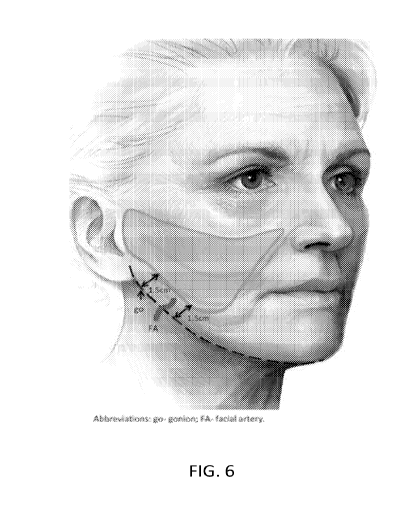
[72] FIG. 6 shows the treatment area for Part B of the Exemplary study.
Definitions
[73] "Animal:" As used herein refers to any member of the animal kingdom.
In some
embodiments, "animal" refers to humans, of either sex and at any stage of
development. In some
embodiments, "animal" refers to non-human animals, at any stage of
development. In certain
embodiments, the non-human animal is a mammal (e.g., a rodent, a mouse, a rat,
a rabbit, a
monkey, a dog, a cat, a sheep, cattle, a primate, and/or a pig). In some
embodiments, animals
include, but are not limited to, mammals, birds, reptiles, amphibians, fish,
insects, and/or worms.
In some embodiments, an animal may be a transgenic animal, genetically
engineered animal,
and/or a clone.
[74] "About:" As used herein, the term "about," as applied to one or more
values of
interest, refers to a value that is similar to a stated reference value. In
certain embodiments, the
term or "about" refers to a range of values that fall within 25%, 20%, 19%,
18%, 17%, 16%,
15%, 14%, 13%, 12%, 11%, 10%, 9%, 8%, 7%, 6%, 5%, 4%, 3%, 2%, 1%, or less in
either
direction (greater than or less than) of the stated reference value unless
otherwise stated or
otherwise evident from the context (except where such number would exceed 100%
of a possible
value).
[75] "Combination therapy:" As used herein, the term "combination therapy"
refers
to those situations in which a subject is simultaneously exposed to two or
more therapeutic
regimens (e.g., two or more therapeutic agents). In some embodiments, the two
or more
regimens may be administered simultaneously; in some embodiments, such
regimens may be
administered sequentially (e.g., all "doses" of a first regimen are
administered prior to
administration of any doses of a second regimen); in some embodiments, such
agents are
administered in overlapping dosing regimens. In some embodiments,
"administration" of
combination therapy may involve administration of one or more agents or
modalities to a subject
CA 03037715 2019-03-20
WO 2018/057637 PCT/US2017/052539
12
receiving the other agents or modalities in the combination. For clarity,
combination therapy
does not require that individual agents be administered together in a single
composition (or even
necessarily at the same time), although in some embodiments, two or more
agents, or active
moieties thereof, may be administered together in a combination composition,
or even in a
combination compound (e.g., as part of a single chemical complex or covalent
entity).
[76] "Comprising:" A composition or method described herein as "comprising"
one
or more named elements or steps is open-ended, meaning that the named elements
or steps are
essential, but other elements or steps may be added within the scope of the
composition or
method. To avoid prolixity, it is also understood that any composition or
method described as
"comprising" (or which "comprises") one or more named elements or steps also
describes the
corresponding, more limited composition or method "consisting essentially of'
(or which
"consists essentially of') the same named elements or steps, meaning that the
composition or
method includes the named essential elements or steps and may also include
additional elements
or steps that do not materially affect the basic and novel characteristic(s)
of the composition or
method. It is also understood that any composition or method described herein
as "comprising"
or "consisting essentially of' one or more named elements or steps also
describes the
corresponding, more limited, and closed-ended composition or method
"consisting of' (or
"consists of') the named elements or steps to the exclusion of any other
unnamed element or
step. In any composition or method disclosed herein, known or disclosed
equivalents of any
named essential element or step may be substituted for that element or step.
[77] "Cosmetic effect:" As used herein, "cosmetic effect" means a change in
a skin
appearance, e.g., elimination of tissue volume, tightening of skin, lifting of
skin, and/or reduction
skin laxity, that is visible, detectable, and/or quantifiable, e.g., a >1
point reduction of the
Lemperle Scale, e.g., for pre-auricular wrinkles (see FIG. 1), as judged by a
Live independent,
blinded reviewer; a >1 point reduction of the Lemperle Assessment Scale as
judged by a 3
member, blinded independent review committee comparing photographs before and
after
treatment; change in lower face scales as judged by independent, blinded
reviewer before and
after treatment; Evaluation of photographs of lower face scales by a 3 member
independent,
blinded review committee before and after treatment; or Evaluation, e.g., by
Live independent
review or by a 3 member committee, conducted before and after treatment using
the following
CA 03037715 2019-03-20
WO 2018/057637 PCT/US2017/052539
13
scales: >1 point reduction of the nasolabial fold scale score at rest as
described in FIG. 2; >1
point reduction of the marionette line scale score at rest as described in
FIG. 3; >1 point
reduction of the oral commissure scale score at rest as described in FIG. 4;
>1 point reduction of
the jawline scale score at rest described in FIG. 5.
[78] "Excising:" As used herein, "excising" means a tissue means forming a
tissue
portion (the "microcore"), e.g., by inserting a hollow needle into the site so
that the tissue portion
is formed inside the hollow needle and severed from surrounding tissue so that
a microcore that
is separate from other tissue is generated.
[79] "Full thickness core:" As used herein, "full thickness core" means a
microcore
whose depth extends through the entire dermal layer beyond the junction of the
dermal layer and
the subcutaneous fat layer, and into the subcutaneous fat layer.
[80] "Heat sensitive:" As used herein, "heat sensitive" or, e.g., a "heat-
sensitive site"
means, e.g., a site where exposure to radiation and/or elevated temperature is
associated with a
relatively high risk of unacceptable cosmetic and/or physiologic outcomes.
[81] "Improve," "increase" or "reduce:" As used herein or grammatical
equivalents
thereof, indicate values that are relative to a baseline measurement, such as
a measurement in the
same individual prior to initiation of a treatment described herein, or a
measurement in a control
individual (or multiple control individuals) in the absence of the treatment
described herein. In
some embodiments, a "control individual" is an individual afflicted with the
same form of
disease or injury as an individual being treated.
[82] "Microcoring:" As used herein, "microcoring" refers to technologies
that utilize
one or more (in some embodiments, a plurality, e.g., an array) hollow needles
or other non-
thermal implement of sufficiently small dimension to minimize the extent of
bleeding and/or
clotting within the holes or slits and/or to minimize scar formation to excise
and optionally
sequester tissue from a site.
[83] "Patient:" As used herein, the term "patient" refers to any organism
to which a
provided composition is or may be administered, e.g., for experimental,
diagnostic, prophylactic,
cosmetic, and/or therapeutic purposes. Typical patients include animals (e.g.,
mammals such as
CA 03037715 2019-03-20
WO 2018/057637 PCT/US2017/052539
14
mice, rats, rabbits, non-human primates, and/or humans). In some embodiments,
a patient is a
human. In some embodiments, a patient is suffering from or susceptible to one
or more disorders
or conditions. In some embodiments, a patient displays one or more symptoms of
a disorder or
condition. In some embodiments, a patient has been diagnosed with one or more
disorders or
conditions. In some embodiments, the disorder or condition is or includes
cancer, or presence of
one or more tumors. In some embodiments, the patient is receiving or has
received certain
therapy to diagnose and/or to treat a disease, disorder, or condition.
[84] "Prevent" or "prevention:" As used herein when used in connection with
the
occurrence of a disease, disorder, and/or condition, refers to reducing the
risk of developing the
disease, disorder and/or condition and/or to delaying onset of one or more
characteristics or
symptoms of the disease, disorder or condition. Prevention may be considered
complete when
onset of a disease, disorder or condition has been delayed for a predefined
period of time.
[85] "Response:" As used herein, a response to treatment may refer to any
beneficial
alteration in a subject's condition that occurs as a result of or correlates
with treatment. Such
alteration may include stabilization of the condition (e.g., prevention of
deterioration that would
have taken place in the absence of the treatment), amelioration of symptoms of
the condition,
and/or improvement in the prospects for cure of the condition, etc.
[86] "Risk:" As will be understood from context, "risk" of a disease,
disorder, and/or
condition comprises likelihood that a particular individual will develop a
disease, disorder,
and/or condition. In some embodiments, risk is expressed as a percentage. In
some
embodiments, risk is from 0, 1, 2, 3, 4, 5, 6, 7, 8, 9, 10, 20, 30, 40, 50,
60, 70, 80, 90 up to 100%.
In some embodiments risk is expressed as a risk relative to a risk associated
with a reference
sample or group of reference samples. In some embodiments, a reference sample
or group of
reference samples have a known risk of a disease, disorder, condition and/or
event. In some
embodiments a reference sample or group of reference samples are from
individuals comparable
to a particular individual. In some embodiments, relative risk is 0, 1, 2, 3,
4, 5, 6, 7, 8, 9, 10, or
more.
[87] "Sequestering:" As used herein, "sequestering" means, when used in
reference
to tissue, excising a microcore and then removing the excised microcore from
the excision site.
CA 03037715 2019-03-20
WO 2018/057637 PCT/US2017/052539
[88] "Subject:" As used herein, "subject" means an organism, typically a
mammal
(e.g., a human, in some embodiments including prenatal human forms). In some
embodiments, a
subject is suffering from a relevant disease, disorder or condition. In some
embodiments, a
subject is susceptible to a disease, disorder, or condition. In some
embodiments, a subject
displays one or more symptoms or characteristics of a disease, disorder or
condition. In some
embodiments, a subject does not display any symptom or characteristic of a
disease, disorder, or
condition. In some embodiments, a subject is someone with one or more features
characteristic
of susceptibility to or risk of a disease, disorder, or condition. In some
embodiments, a subject is
a patient. In some embodiments, a subject is an individual to whom diagnosis
and/or therapy is
and/or has been administered.
[89] "Substantially:" As used herein, the term "substantially" refers to
the qualitative
condition of exhibiting total or near-total extent or degree of a
characteristic or property of
interest. One of ordinary skill in the biological arts will understand that
biological and chemical
phenomena rarely, if ever, go to completion and/or proceed to completeness or
achieve or avoid
an absolute result. The term "substantially" is therefore used herein to
capture the potential lack
of completeness inherent in many biological and chemical phenomena.
[90] "Therapeutic agent:" As used herein, the phrase "therapeutic agent" in
general
refers to any agent that elicits a desired pharmacological effect when
administered to an
organism. In some embodiments, an agent is considered to be a therapeutic
agent if it
demonstrates a statistically significant effect across an appropriate
population. In some
embodiments, the appropriate population may be a population of model
organisms. In some
embodiments, an appropriate population may be defined by various criteria,
such as a certain age
group, gender, genetic background, preexisting clinical conditions, etc. In
some embodiments, a
therapeutic agent is a substance that can be used to alleviate, ameliorate,
relieve, inhibit, prevent,
delay onset of, reduce severity of, and/or reduce incidence of one or more
symptoms or features
of a disease, disorder, and/or condition. In some embodiments, a "therapeutic
agent" is an agent
that has been or is required to be approved by a government agency before it
can be marketed for
administration to humans. In some embodiments, a "therapeutic agent" is an
agent for which a
medical prescription is required for administration to humans.
CA 03037715 2019-03-20
WO 2018/057637 PCT/US2017/052539
16
[91] "Treatment:" As used herein, the term "treatment" (also "treat" or
"treating")
refers to any administration of a therapy that partially or completely
alleviates, ameliorates,
relives, inhibits, delays onset of, reduces severity of, and/or reduces
incidence of one or more
symptoms, features, and/or causes of a particular disease, disorder, and/or
condition. In some
embodiments, such treatment may be of a subject who does not exhibit signs of
the relevant
disease, disorder and/or condition and/or of a subject who exhibits only early
signs of the
disease, disorder, and/or condition. Alternatively or additionally, such
treatment may be of a
subject who exhibits one or more established signs of the relevant disease,
disorder and/or
condition. In some embodiments, treatment may be of a subject who has been
diagnosed as
suffering from the relevant disease, disorder, and/or condition. In some
embodiments, treatment
may be of a subject known to have one or more susceptibility factors that are
statistically
correlated with increased risk of development of the relevant disease,
disorder, and/or condition.
Detailed Description of Certain Embodiments
[92] Described herein are technologies, methods, and/or devices for
treating skin (e.g.,
eliminating tissue volume, tightening skin, lifting skin, and/or reducing skin
laxity) by
selectively excising a plurality of microcores without thermal energy being
imparted to the
surrounding (e.g., non-excised) tissue. In certain embodiments, the excising
is completed within
a certain time period, or is performed at a certain rate. In certain
embodiments, the treatment is
performed in specific areas not treatable with certain thermal methods, e.g.,
in the vicinity of
nerves and/or other heat sensitive areas. In certain embodiments, a cosmetic
effect is visible
during treatment, immediately after or within a very short time after
completion of treatment. In
certain embodiments, a cosmetic effect is visible 1 min, 5 min, 10 min, 20
min, 30 min, 1 hour, 2
hours, 3 hours, 6 hours, 12 hours, 24 hours, 2 days, 3 days, 4 days, 5 days, 6
days, 7 days, 2
weeks, 3 weeks, 1 month, 2 months, 3 months, 4 months, 5 months, or 6 months
after completion
of treatment.
CA 03037715 2019-03-20
WO 2018/057637 PCT/US2017/052539
17
Microcoring
[93] In general, the term "microcoring," as used herein, refers to
technologies that
utilize one or more (in some embodiments, a plurality, e.g., an array) hollow
needles or other
non-thermal implement of sufficiently small dimension to minimize the extent
of bleeding and/or
clotting within the holes or slits and/or to minimize scar formation to excise
and optionally
sequester tissue from a site. In some embodiments, excising a tissue means
forming a tissue
portion (the "microcore"), e.g., by inserting a hollow needle into the site so
that the tissue portion
is formed inside the hollow needle and severed from surrounding tissue so that
a microcore that
is separate from other tissue is generated.
[94] Moreover, microcoring technologies as described herein can include
sequestration
of the excised tissue. As used herein, the term "sequestering", when used in
reference to tissue,
means excising a microcore and then removing the excised microcore from the
excision site. In
certain embodiments, sequestered tissue is permanently disposed. In certain
embodiments,
sequestered tissue is used for diagnostic purpose, e.g., using biopsy and/or
histology techniques
known in the art. In many embodiments, technologies provided herein maximize
removal and
minimize risk of (partial or complete) re-insertion of extracted tissue.
[95] It should be understood that microcoring technologies, methods, and/or
devices
using hollow needles described herein serve for exemplary and/or illustrative
purposes, and that
other techniques and devices can be used to create microcores. Representative
such microcoring
techniques and devices are described, for example, in U.S. Patent Application
No. 14/910,767,
filed February 8, 2016, and/or Provisional Patent Application No. 62/314,748,
filed March 29,
2016, both of which are incorporated herein by reference in their entireties.
[96] Microcoring technologies described herein a number of advantageous
features.
For example, provided technologies may enable visualization of results in real
time during the
course of the treatment, e.g., through patient feedback and subsequent
treatment adjustment in
real time.
[97] Alternatively or additionally, apparatuses used for microcoring can
include micro-
sized features that can be beneficial for controlling extent of skin
treatment.
CA 03037715 2019-03-20
WO 2018/057637 PCT/US2017/052539
18
[98] Still further, in some embodiments, methods and/or devices described
herein may
require less skill than that of a surgeon. Thus, in certain embodiments,
patients may be treated in
an outpatient setting, rather than in an inpatient, surgical setting. In some
embodiments, subjects
may be treated at a spa, at a cosmetic salon, or at home. That is, the present
disclosure provides
technologies that are amenable to and/or permit consistent and/or reproducible
administration of
skin treatment services.
[99] In some embodiments, technologies, methods, and/or devices described
herein
have generally a lower risk profile and can provide more predictable results
and/or risk factors
than those for more invasive techniques (e.g., plastic surgery) or noninvasive
energy-based
techniques (e.g., laser, radiofrequency (RF), or ultrasound). In some
embodiments, non-thermal
fractional excision technologies, methods, and/or devices described herein
allow skin tightening,
skin lifting, and/or reduction of skin laxity without (or with significant
reduction of) one or more
common side effects of thermal ablation methods. Thermal ablation techniques
prevent and/or
inhibit skin tightening by allowing coagulation of tissue and formation of
rigid tissue cores that
cannot be compressed. Thermal ablation techniques create a three-dimensional
heat-affected
zone (HAZ) surrounding an immediate treatment site. While fractional ablative
lasers can be
used on or near heat-sensitive sites (e.g., eyes, nerves), i.e., when the
laser does not penetrate
more than 1 mm into the skin (resulting in a comparatively small HAZ), other
thermal ablation
techniques (e.g., ultrasound based techniques) cannot be used in the vicinity
of heat-sensitive
sites because the HAZ may extend to heat sensitive tissues potentially causing
(permanent)
damage. As will be appreciated by those skilled in the art reading the present
disclosure, a "heat-
sensitive site" is a site where exposure to radiation and/or elevated
temperature is associated with
a relatively high risk of unacceptable cosmetic and/or physiologic outcomes.
In any event,
technologies, methods, and/or devices described herein have generally a lower
risk profile at
least in part due to a zone of tissue injury that is smaller than the zone of
injury (e.g., the HAZ)
of thermal methods.
[100] In some embodiments, advantages of certain technologies, methods,
and/or
devices described herein include a lesser degree of erythema, faster
resolution of erythema, and
lower percent incidence, severity, term of skin discoloration
(hyperpigmentation or
CA 03037715 2019-03-20
WO 2018/057637 PCT/US2017/052539
19
hypopigmentation), and/or less swelling and/or inflammation, as compared, for
example, with
that observed with laser treatment and/or with ultrasound-based treatment.
[101] In some embodiments, certain technologies, methods, and/or devices
provided
herein can allow for rapid closing of holes or slits after excising tissue
(e.g., within a few seconds
after treating skin, such as within ten seconds), thereby minimizing extent of
bleeding and/or
clotting within holes or slits, and/or scar formation.
[102] In some embodiments, certain technologies, methods, and/or devices
provided
herein can be useful for maximizing treatment effect while minimizing
treatment time, e.g., by
using rapid-fire reciprocating needles or needle arrays, and/or by using large
needle arrays that
allow for simultaneous excision of tens, hundreds, or even thousands of
microcores.
[103] In some embodiments, technologies, methods, and/or devices described
herein
can be useful for maximizing tightening effect while minimizing healing time
and/or minimizing
the time in which a cosmetic effect occurs by optimizing tightening (e.g., by
controlling the
extent of skin pleating, such as by increasing the extent of skin pleating for
some applications or
skin regions and by decreasing the extent of skin pleating for other
applications or skin regions,
as described herein).
[104] In some embodiments, technologies, methods, and/or devices described
herein
can provide efficient clearance of sequestered or partially ablated tissue
and/or debris from
ablated tissue portions, thus reducing time for healing and improving the skin
tightening
treatment, e.g., relative to laser-based technologies.
[105] In some embodiments, technologies, methods, and/or devices described
herein
can allow for efficient and effective positioning of skin prior to, during,
and after excision and/or
tissue sequestration. Positioning the skin is critical to control skin-
tightening direction and
ensure ablation occurs in the desired location and desired dimensions (e.g.
thickness, width in a
preferred direction, e.g., along or orthogonal to Langer lines).
[106] Among other things, the present disclosure encompasses the insight
that
microcoring technologies can be developed (e.g., as described herein) that can
achieve desirable
CA 03037715 2019-03-20
WO 2018/057637 PCT/US2017/052539
procedure times and/or can significantly improve one or more aspects of
healing from a
procedure (e.g., a tissue removal procedure), compared to, e.g., thermal
methods.
Procedures
[107] In some embodiments, technologies, methods, and/or devices described
herein
may be used for cosmetic resurfacing of skin tissue by removing skin tissue
portions.
Technologies, methods, and/or devices described herein can be applied to treat
one or more skin
regions. In particular embodiments, these regions are treated with one or more
procedures to
improve skin appearance and/or to rejuvenate skin. In certain embodiments,
technologies,
methods, and/or devices described herein can be useful for skin tightening,
e.g., reducing skin
laxity (e.g., loose or sagging skin, or other skin irregularities).
[108] In certain embodiments, technologies, methods, and/or devices
described herein
can be useful for removal of, e.g., redundant or excess skin, pigment, hair
follicles, and/or
vessels in the skin, and/or for treating acne, allodynia, blemishes, ectopic
dermatitis,
hyperpigmentation, hyperplasia (e.g., lentigo or keratosis), loss of
translucency, loss of elasticity,
melasma (e.g., epidermal, dermal, or mixed subtypes), photodamage, rashes
(e.g., erythematous,
macular, papular, and/or bullous conditions), psoriasis, rhytides (or
wrinkles, e.g., lateral canthal
lines ("crow's feet"), age-related rhytides, sun-related rhytides, or heredity-
related rhytides),
sallow color, scar contracture (e.g., relaxation of scar tissue), scarring
(e.g., due to acne, surgery,
or other trauma), skin aging, skin contraction (e.g., excessive tension in the
skin), skin
irritation/sensitivity, striae (or stretch marks), tattoo removal, vascular
lesions (e.g., angioma,
erythema, hemangioma, papule, port wine stain, rosacea, reticular vein, or
telangiectasia), or any
other unwanted skin irregularities. The technologies, methods, and/or devices
described herein
may also be used to penetrate skin and trigger biological responses that may
contribute to new
skin tissue formation and tissue resurfacing and remodeling.
[109] In certain embodiments, technologies, methods, and/or devices
described herein
can be applied to a site located on any part or parts of the body, including
face (e.g., eyelid,
cheeks, chin, forehead, lips, or nose), neck, chest (e.g., as in a breast
lift), arms, hands, legs,
CA 03037715 2019-03-20
WO 2018/057637 PCT/US2017/052539
21
abdomen, buttock, and thigh. In certain specific embodiments, a treatment site
is located on the
face and/or neck. In certain embodiments, a site is located on a part of the
body that is heat
sensitive. Such a site is generally not amenable to treatment with deep-
penetrating thermal
methods. For example, lasers can only be used if laser energy is targeted to
the epidermis/dermis
only and thus creates only a comparatively small HAZ. In certain embodiments,
a site is located
on the face in close proximity to an eye. In certain embodiments, a site is
located on the face in
close proximity to the facial nerve or a facial nerve branch, e.g. the
temporal branch, the
zygomatic branch, the buccal branch, the marginal mandibular branch, or the
cervical branch.
[110] In certain embodiments, a site is located over or near the thyroid
gland, thyroid
cartilage, trachea, a major blood vessel, or breast tissue.
[111] In certain embodiments, a treatment site can have any size. In
certain
embodiments, a treatment site has an area of between 1 cm2 and 10,000 cm2,
between 10 cm2 and
5,000 cm2, between 100 cm2 and 2,500 cm2, or between 500 cm2 and 1,000 cm2. In
certain
embodiments, the treatment site has an area of between 1 cm2 and 300 cm2,
between 1.2 cm2 and
280 cm2, between 1.4 cm2 and 260 cm2, between 1.6 cm2 and 240 cm2, between 1.8
cm2 and 220
cm2, between 2 cm2 and 200 cm2, between 2.2 cm2 and 180 cm2, between 2.4 cm2
and 160 cm2,
between 2.6 cm2 and 140 cm2, between 2.8 cm2 and 120 cm2, between 3 cm2 and
100 cm2,
between 3.2 cm2 and 80 cm2, between 3.4 cm2 and 60 cm2, between 3.6 cm2 and 40
cm2, between
3.8 cm2 and 20 cm2, between 4 cm2 and 10 cm2, between 10 cm2 and 20 cm2,
between 20 cm2
and 30 cm2, between 30 cm2 and 40 cm2, between 40 cm2 and 50 cm2, between 50
cm2 and 60
cm2, between 60 cm2 and 70 cm2, between 70 cm2 and 80 cm2, between 80 cm2 and
100 cm2,
between 100 cm2 and 120 cm2, between 120 cm2 and 140 cm2, between 140 cm2 and
160 cm2,
between 160 cm2 and 180 cm2, or between 180 cm2 and 200 cm2.
[112] In certain embodiments, technologies, methods, and/or devices
described herein
may involve forming a plurality of holes in the skin, e.g., by contacting one
or more hollow
needles to the skin of a subject and excising or sequestering cored tissue
portions from the skin.
Penetration into the skin by, e.g., hollow needle(s), creates holes and so
effectively reduces tissue
volume and/or improves tissue quality upon healing. For example, forming a
series of cored
tissue portions (e.g., excising or sequestering about 20% of the total skin
area) and corresponding
CA 03037715 2019-03-20
WO 2018/057637 PCT/US2017/052539
22
holes in a high laxity skin region, and optionally subsequent compression of
the skin region to
close the holes may promote the growth of improved tissue (e.g., new skin). In
other
embodiments, treatment methods described herein can be used to reduce laxity
or the appearance
of laxity in the skin. In some embodiments, treatment methods described herein
can be used to
remove excess / redundant skin.
[113] In certain embodiments, technologies, methods, and/or devices
described herein
further comprise application of a dressing. Healing of tissue under a dressing
(e.g., a
compressive or occlusive dressing) allows for the existing tissue to span the
gap introduced by
the removal of cored tissue portions, thereby reducing skin volume and area
(e.g., by tightening
the skin). In some embodiments, application of a dressing (e.g., a compressive
or occlusive
dressing) may help to maintain moisture of a treated skin area and/or to
prevent delivered
therapeutic agents from leaking out of the skin. In some embodiments,
application of a dressing
may improve the healing profile of the treated region, e.g., by providing
hemostatic pressure to
cored regions.
[114] Microcoring can be performed by creating holes in the skin at various
hole
densities. In certain embodiments, tissue can be excised or sequestered from
the treatment
region with various hole densities (e.g., the number of holes per unit area)
corresponding to the
number and geometry of hollow needle(s) of the apparatus used and the number
of applications
of the hollow needle(s) to the treatment region. Different hole densities may
be desirable for
different regions of skin and for different conditions, and may be achieved
using different hollow
needle(s). For example, 15 holes corresponding to the size of a 19 gauge
needle and their
corresponding cored tissue portions may be created in a given treatment area
by actuation of a
single 19 gauge needle 15 times, or by actuating an array having five 19 gauge
needles three
times. Spacing the same number of holes further apart will result in a lower
hole density per unit
area. For example, 15 holes may be created within a 0.5 mm by 0.3 mm region or
within a 5 mm
by 3 mm region. In certain embodiments, technologies, methods, and/or devices
described
herein are configured to provide from about 10 to about 10000 cored tissue
portions per cm2 area
of a site (e.g., as described herein). An array of holes created by removal of
skin tissue portions
may be created in any beneficial pattern within a site. For example, a higher
density and/or
smaller spacing of tissue portions and corresponding holes can be excised or
sequestered in skin
CA 03037715 2019-03-20
WO 2018/057637 PCT/US2017/052539
23
in the center of a pattern or in thicker portions of skin. A pattern may be
semi-random or include
one or more of staggered rows and/or blocks, parallel rows and/or blocks, a
circular pattern, a
spiral pattern, a square or rectangular pattern, a triangular pattern, a
hexagonal pattern, a radial
distribution, or a combination of one or more such patterns. A pattern may
arise from the use of
one or more hollow needles (or other microcoring implements) with one or more
configurations
and numbers of hollow needles (or other microcoring implements) applied in any
ordered or
disordered manner. Modifications to the average length, diameter, shapes,
and/or other
characteristics of one or more hollow needles (or other microcoring
implements) used to treat a
skin region may also result in a specific pattern of holes in the skin. Such
patterns may be
optimized to promote unidirectional, non-directional, or multidirectional
contraction or
expansion of skin (e.g., in the x-direction, y-direction, x-direction, x-y
plane, y-z plane, x-z
plane, and/or xyz-plane), such as by modifying the average length, depth,
diameter, density,
orientation, and/or spacing between hollow needles. Additionally, the
orientation of a needle can
provide the basis for an oriented pattern. For example, proximal ends of
needles can be non-
uniform, and, e.g., may comprise one or more prongs. Insertion of needle tips
comprising prongs
into the skin can lead to cores that are not perfectly cylindrical throughout
the extent of the core.
To the extent that the cores are not cylindrical, patters may be produced in
treated skin, e.g., by
controlling the relative positions of the prongs during a coring process. For
example, prongs
may be held at a fixed angle relative to the x-y pattern of strikes, or the
prong positions may be
alternated to produce complex patterns.
[115] Micorcores can have any diameter. In certain embodiments, a
diameter largely
corresponds to the inner diameter of hollow needles described herein. In
certain embodiments,
the microcores may have a diameter of between about 0.1 mm and about 1.0 mm,
or between
0.14 mm and about 0.84 mm, 0.16 mm and about 0.82 mm, 0.18 mm and about 0.8
mm, 0.2 mm
and about 0.78 mm, 0.22 mm and about 0.76 mm, 0.24 mm and about 0.74 mm, 0.26
mm and
about 0.72 mm, 0.28 mm and about 0.7 mm, 0.3 mm and about 0.68 mm, 0.32 mm and
about
0.66 mm, 0.34 mm and about 0.64 mm, 0.36 mm and about 0.62 mm, 0.38 mm and
about 0.6
mm, 0.4 mm and about 0.58 mm, 0.42 mm and about 0.56 mm, 0.44 mm and about
0.54 mm,
0.46 mm and about 0.52 mm, or 0.48 mm and about 0.5 mm, (e.g., 0.14, 0.15,
0.16, 0.17, 0.18,
0.19, 0.2, 0.21, 0.22, 0.23, 0.24, 0.25, 0.26, 0.27, 0.28, 0.29, 0.3, 0.31,
0.32, 0.33, 0.34, 0.35,
CA 03037715 2019-03-20
WO 2018/057637 PCT/US2017/052539
24
0.36, 0.37, 0.38, 0.39, 0.4, 0.41, 0.42, 0.43, 0.44, 0.45, 0.46, 0.47, 0.48,
0.49, 0.5, 0.51, 0.52,
0.53, 0.54, 0.55, 0.56, 0.57, 0.58, 0.59, 0.6, 0.61, 0.62, 0.63, 0.64, 0.65,
0.66, 0.67, 0.68, 0.69,
0.7, 0.71, 0.72, 0.73, 5 0.74, 0.75, 0.76, 0.77, 0.78, 0.79, 0.8, 0.81, 0.82,
0.83, and 0.84 mm). In
certain embodiments, microcores may have a diameter of between about 0.1 mm
and about 1.0
mm, about 0.14 mm and about 0.84 mm, about 0.24 mm and about 0.40 mm (e.g.,
0.24, 0.25,
0.26, 0.27, 0.28, 0.29, 0.3, 0.31, 0.32, 0.33, 0.34, 0.35, 0.36, 0.37, 0.38,
0.39, and 0.4 mm). In a
certain embodiments, microcores may have a diameter of from about 0.25 mm to
about 0.6 mm.
In certain embodiments, depending on the microcoring technique used, the
diameter of each
microcore varies along the length of the microcore. The shape of a microcore
may be
cylindrical, conical, hemispherical, hyperboloid or any combination thereof,
or any other shape.
[116] Thickness of skin can vary significantly between different regions
of a body.
Skin in the periphery of an eye and an eyelid are very thin, while skin at or
near the sole of a foot
is very thick. In addition, scar tissue can be even thicker than normal skin.
Any portion of skin
can be excised using the technologies, methods, and/or devices described
herein. Tissue portions
created by microcoring may include epidermal tissue, dermal tissue,
subcutaneous fat, and/or
cells or tissue proximal to the dermal/fatty layer boundary (e.g., stem
cells). In certain
embodiments, a tissue portion may have a length that corresponds to depth of
penetration of the
skin layer with a microneedle. In certain embodiments, depth of penetration
may be (i) into the
dermal layer, (ii) through the entire dermal layer to the junction of a dermal
layer and the
subcutaneous fat layer, or (iii) into the subcutaneous fat layer. Total depth
of epidermal, dermal,
and subcutaneous fat layers may vary based on the region and age of the body
being treated. In
some instances, depth of the epidermal layer is between about 0.01 mm to 0.2
mm, and/or depth
of the dermal layer is between about 0.3 mm to 6.0 mm. In some embodiments,
total depth of
the epidermal and dermal layers may be between about 0.3 mm and 6.2 mm,
corresponding to a
possible tissue portion having a length of between about 0.3 mm and 6.2 mm
(e.g., between
about 0.3 mm and 0.6 mm, 0.3 mm and 0.9 mm, 0.3 mm and 1.5 mm, 0.3 mm and 2.0
mm, 0.3
mm and 2.5 mm, 0.3 mm and 3.0 mm, 0.3 mm and 3.5 mm, 0.3 mm and 4.0 mm, 0.3 mm
and 4.5
mm, 0.3 mm and 5.0 mm, 0.3 mm and 5.5 mm, 0.3 mm and 6.0 mm, 0.3 mm and 6.2
mm, 0.6
mm and 0.9 mm, 0.6 mm and 1.5 mm, 0.6 mm and 2.0 mm, 0.6 mm and 2.5 mm, 0.6 mm
and 3.0
mm, 0.6 mm and 3.5 mm, 0.6 mm and 4.0 mm, 0.6 mm and 4.5 mm, 0.6 mm and 5.0
mm, 0.6
CA 03037715 2019-03-20
WO 2018/057637 PCT/US2017/052539
mm and 5.5 mm, 0.6 mm and 6.0 mm, 0.6 mm and 6.2 mm, 0.9 mm and 1.5 mm, 0.9 mm
and 2.0
mm, 0.9 mm and 2.5 mm, 0.9 mm and 3.0 mm, 0.9 mm and 3.5 mm, 0.9 mm and 4.0
mm, 0.9
mm and 4.5 mm, 0.9 mm and 5.0 mm, 0.9 mm and 5.5 mm, 0.9 mm and 6.0 mm, 0.9 mm
and 6.2
mm, 1.5 mm and 2.0 mm, 1.5 mm and 2.5, mm, 1.5 mm and 3.0 mm, 1.5 mm and 3.5
mm, 1.5
mm and 4.0 mm, 1.5 mm and 4.5 mm, 1.5 mm and 5.0 mm, 1.5 mm and 5.5 mm, 1.5 mm
and 6.0
mm, 1.5 mm and 6.2 mm, 2.0 mm and 2.5 mm, 2.0 mm and 3.0 mm, 2.0 mm and 3.5
mm, 2.0
mm and 4.0 mm, 2.0 mm and 4.5 mm, 2.0 mm and 5.0 mm, 2.0 mm and 5.5 mm, 2.0
and 6.0
mm, 2.0 mm and 6.2 mm, 2.5 mm and 3.0 mm, 2.5 mm and 3.5 mm, 2.5 mm and 4.0
mm, 2.5
mm and 4.5 mm, 2.5 mm and 5.0 mm, 2.5 mm and 5.5 mm, 2.5 mm and 6.0 mm, 2.5 mm
and 6.2
mm, 3.0 mm and 3.5 mm, 3.0 mm and 4.0 mm, 3.0 mm and 4.5 mm, 3.0 mm and 5.0
mm, 3.0
mm and 5.5 mm, 3.0 and 6.0 mm, 3.0 mm and 6.2 mm, 3.5 mm and 4.0 mm, 3.5 mm
and 4.5
mm, 3.5 mm and 5.0 mm, 3.5 mm and 5.5 mm, 3.5 and 6.0 mm, 3.5 mm and 6.2 mm,
4.0 mm
and 4.5 mm, 4.0 mm and 5.0 mm, 4.0 mm and 5.5 mm, 4.0 and 6.0 mm, 4.0 mm and
6.2 5 mm,
4.5 mm and 5.0 mm, 4.5 mm and 5.5 mm, 4.5 and 6.0 mm, 4.5 mm and 6.2 mm, 5.0
mm and 5.5
mm, 5.0 mm and 6.0 mm, 5.0 mm and 6.2 mm, 5.5 mm and 6.0 mm, 5.5 mm and 6.2
mm, or 6.0
mm and 6.2 mm). In certain embodiments, the possible tissue portion has a
length between
about 0.3 and 2 mm, or between about 0.3 and 3 mm.
[117] In certain embodiments, excised tissue portions extend into the
subcutaneous fat
layer. In certain embodiments, a tissue portions created by microcoring can
extend from the
junction of the dermal layer and the subcutaneous fat layer into the
subcutaneous fat layer by
between about 0.1 mm and 3 mm, 0.2 mm and 2.8 mm, 0.3 mm and 2.6 mm, 0.4 mm
and 2.4
mm, 0.5 mm and 2.2 mm, 0.6 mm and 2 mm, 0.8 mm and 1.8 mm, or 1 mm and 1.6 mm.
Thus,
a possible tissue portion can have a length between about 6.2 mm and 9.2 mm,
6.2 mm and 9
mm, 6.2 mm and 8.8 mm, 6.2 mm and 8.6 mm, 6.2 mm and 8.4 mm, 6.2 mm and 8.2
mm, 6.2
mm and 8 mm, 6.2 mm and 7.8 mm, 6.2 mm and 7.6 mm, 6.2 mm and 7.4 mm, 6.2 mm
and 7.2
mm, 6.2 mm and 7 mm, 6.2 mm and 6.8 mm, 6.2 mm and 6.6 mm, or 6.2 mm and 6.4
mm.
[118] In certain embodiments, tissue portions are excised from scar tissue.
In certain
embodiments, an excised tissue portion can extend through the scar / skin
tissue layers into the
subcutaneous fat layer corresponding to a possible tissue portion having a
length between about
0.1 mm and 20 mm, 0.2 mm and 18 mm, 0.3 mm and 16 mm, 0.4 mm and 14 mm, 0.5 mm
and
CA 03037715 2019-03-20
WO 2018/057637 PCT/US2017/052539
26
12 mm, 0.6 mm and 10 mm, 0.8 mm and 8 mm, 1 mm and 6 mm, 1.2 mm and 4 mm, or
1.4 mm
and 2 mm.
[119] In certain embodiments, technologies, methods, and/or devices
described herein
further include technologies to ensure consistent depth of penetration and
orientation, e.g., depth
of penetration and orientation of hollow needles, into skin. In certain
embodiments,
technologies, methods, and/or devices described herein can be configured to
accommodate
different skin thicknesses. In certain embodiments, devices, e.g., devices
comprising
reciprocating needles, can include mechanical technologies (e.g., spacers) to
ensure constant
distance between a needle tip and skin when the needle is fully retracted, and
technologies for
adjustment of the distance the needle travels along its longitudinal axis on
each actuation.
[120] Micorcores can have any volume. In certain embodiments, volume
largely
corresponds to the cross-sectional area of a hollow needles described herein
multiplied by a
length of an excised tissue portion. In certain embodiments, microcores may
have a volume of
between about 0.001 mm3 and 6.3 mm3, 0.005 mm3 and 5.0 mm3, 0.01 mm3 and 4.0
mm3, 0.015
mm3 and 3.0 mm3, 0.02 mm3 and 2.0 mm3, 0.022 mm3 and 1.8 mm3, 0.024 mm3 and
1.6 mm3,
0.026 mm3 and 1.4 mm3, 0.028 mm3 and 1.2 mm3, 0.03 mm3 and 1.0 mm3, 0.032 mm3
and 0.8
mm3, 0.034 mm3 and 0.6 mm3, 0.036 mm3 and 0.4 mm3, 0.038 mm3 and 0.2 mm3, 0.04
mm3 and
0.1 mm3, or 0.06 mm3 and 0.08 mm3.
[121] In certain embodiments, technologies, methods, and/or devices
described herein
comprise microcoring a tissue below an epidermis layer only, leaving the
epidermis, or other
tissue layer above the layer to be excised, un-cored (i.e., no tissue is
excised from the epidermis
other tissue layer above the layer to be excised). In certain embodiments, a
microcoring
implement, e.g., a needle, has a first configuration that allows it to travel
through a tissue layer
with minimal damage to said tissue layer, e.g., by traveling through tissue
like a solid needle, and
has a second configuration that allows for the formation of a microcore, e.g.,
by traveling
through tissue like a hollow needle. In certain embodiments, a device
undergoes a change in
configuration from a first configuration to a second configuration as it
transitions longitudinally
from a first tissue layer into a second tissue layer. In certain embodiments,
the device undergoes
a change in configuration from a first configuration to a second configuration
as it transitions
CA 03037715 2019-03-20
WO 2018/057637 PCT/US2017/052539
27
longitudinally from an epidermis into a dermis. In certain embodiments, a
device undergoes a
change in configuration from a first configuration to a second configuration
as it transitions
longitudinally from the dermis into the subcutaneous fat layer. In certain
embodiments, a
process of microcoring a tissue below an epidermis layer only, e.g., leaving
the epidermis, or
other tissue layer above the layer to be excised, un-cored occurs
independently of treatment time
and/or treatment speed.
[122] Technologies, methods, and/or devices described herein can employ
microcoring
implements, e.g., hollow needles, that are arranged in any configuration. In
certain
embodiments, microcoring implements, e.g., hollow needles, are mounted on a
reciprocating
device configured to (i) translate the needles in a direction substantially
along the longitudinal
axis of the needles and/or (ii) translate the needles over the skin tissue in
one or two orthogonal
directions. In certain embodiments, a reciprocating device may have as few as
1 or as many as
hundreds of hollow needles. In certain embodiments, 1-100 hollow needles may
be present (e.g.,
1-10, 1-20, 1-30, 1-40, 1-50, 1-60, 1-70, 1-80, 1-90, 1-100, 3-10, 3-20, 3-30,
3-40, 3-50, 3-60, 3-
70, 3-80, 3-90, 3-100, 5-10, 5-20, 5-30, 5-40, 5-50, 5-60, 5-70, 5-80, 5-90, 5-
100, 10-20, 10-40,
10-60, 10-80, 10-100, 20-40, 20-60, 20-80, 20-100, 40-60, 40-80, 40-100, 60-
80, 60-100, or 80-
100 hollow needles). Use of an array of a plurality of hollow needles to
generate an array pattern
may facilitate skin treatment over larger areas and in less time.
[123] In certain embodiments, needles are mounted on a non-reciprocating
application
device (an "applique") in form of an array, e.g., and as described above. In
certain embodiments,
an applique, and thus each needle contained therein, is applied to the site
only once. In certain
embodiments, an applique, and thus each needle contained therein, is applied
to a site 2, 3, 4, or
more times. In certain embodiments, application of an applique is independent
of treatment time
and/or treatment speed, e.g., application of an applique can occur
instantaneously. In certain
embodiments, application of an applique occurs in less than about 1 second. In
certain
embodiments, an applique/array comprises at least 10 needles, at least 100
needles, at least 1000
needles, at least 10,000 needles, or at least 100,000 needles. In certain
embodiments, an array
comprises between 10 and 100,000 needles, between 20 and 50,000 needles,
between 30 and
25,000 needles, between 40 and 15,000 needles, between 50 and 10,000 needles,
between 60 and
8,000 needles, between 70 and 6,000 needles, between 80, and 4,000 needles,
between 90 and
CA 03037715 2019-03-20
WO 2018/057637 PCT/US2017/052539
28
2,000 needles, between 100 and 1,000 needles, between 120 and 800 needles,
between 140 and
600 needles, or between 160 and 400 needles.
[124] Technologies, methods, and/or devices described herein can comprise
one or
more microcoring implements, e.g., hollow needles, that may be configured to
provide from
about 10 to about 10000 cored tissue portions, or more, per cm2 area (e.g., 10
to 50, 10 to 100, 10
to 200, 10 to 300, 10 to 400, 10 to 500, 10 to 600, 10 to 700, 10 to 800, 10
to 900, 10 to 1000,10
to 2000, 10 to 4000, 10 to 6000, 10 to 8000, 10 to 10000, 50 to 100, 50 to
200, 50 to 300, 50 to
400, 50 to 500, 50 to 600, 50 to 700, 50 to 800, 50 to 900, 50 to 1000, 50 to
2000, 50 to 4000,
510 to 6000, 50 to 8000, 50 to 10000, 100 to 200, 100 to 300, 100 to 400, 100
to 500, 100 to 600,
100 to 700, 100 to 800, 100 to 900, 100 to 1000, 100 to 2000, 100 to 4000, 100
to 6000, 100 to
8000, 100 to 10000, 200 to 300, 200 to 400, 200 to 500, 200 to 600, 200 to
700, 200 to 800, 200
to 900, 200 to 1000, 200 to 2000, 200 to 4000, 200 to 6000, 200 to 8000, 200
to 10000, 300 to
400, 300 to 500, 300 to 600, 300 to 700, 300 to 800, 300 to 900, 300 to 1000,
300 to 2000, 300 to
4000, 300 to 6000, 300 to 8000, 300 to 10000, 400 to 500, 400 to 600, 400 to
700, 400 to 800,
400 to 900, 400 to 1000, 400 to 2000, 400 to 4000, 400 to 6000, 400 to 8000,
400 to 10000, 500
to 600, 500 to 700, 500 to 800, 500 to 900, 500 to 1000, 500 to 2000, 500 5 to
4000, 500 to 6000,
500 to 8000, 500 to 10000, 600 to 700, 600 to 800, 600 to 900, 600 to 1000,
600 to 2000, 600 to
4000, 600 to 6000, 600 to 8000, 600 to 10000, 700 to 800, 700 to 900, 700 to
1000, 700 to 2000,
700 to 4000, 700 to 6000, 700 to 8000, 700 to 10000, 800 to 900, 800 to 1000,
800 to 2000, 800
to 4000, 800 to 6000, 800 to 8000, 800 to 10000, 900 to 1000, 900 to 2000, 900
to 4000, 900 to
6000, 900 to 8000, 900 to 10000, 1000 to 2000, 1000 to 4000, 1000 to 6000,
1000 to 8000, 1000
to 10000, 2000 to 4000, 2000 to 6000, 2000 to 8000, 2000 to 10000, 4000 to
6000, 4000 to 8000,
4000 to 10000, 6000 to 8000, 6000 to 10000, and 8000 to 10000 tissue portions
per cm2 area) of
a skin region to which the apparatus is applied (e.g., a site or treatment
area).
[125] Any beneficial area or volumetric fraction of a skin region can be
removed. For
example, between about 1% to about 65% (e.g., an areal fraction between about
0.01 to about
0.65, such as 0.01 to 0.65, 0.01 to 0.6, 0.01 to 0.55, 0.01 to 0.5, 0.01 to
0.45, 0.01 to 0.4, 0.01 to
0.35, 0.01 to 0.3, 0.01 to 0.25, 0.01 to 0.2, 0.01 to 0.15, 0.01 to 0.1, 0.01
to 0.05, 0.03 to 0.65,
0.05 to 0.65, 0.07 to 0.65, 0.09 to 0.65, 0.1 to 0.65, 0.15 to 0.65, 0.2 to
0.65, 0.25 to 0.65, 0.3 to
0.65, 0.35 to 0.65, 0.4 to 0.65, 0.45 to 0.65, 0.5 to 0.65, 0.55 to 0.65, and
0.6 to 0.65) of tissue in
CA 03037715 2019-03-20
WO 2018/057637 PCT/US2017/052539
29
a treatment area or site may be removed. In certain embodiments, between about
1% to about
5% (e.g., an areal fraction between about 0.01 to about 0.05, such as 0.01 to
0.05, 0.01 to 0.045,
0.01 to 0.04, 0.01 to 0.035, 0.01 to 0.03, 0.01 to 0.025, 0.01 to 0.02, 0.01
to 0.015, 0.015 to 0.05,
0.02 to 0.05, 0.025 to 0.05, 0.03 to 0.05, 0.035 to 0.05, 0.04 to 0.05, and
0.045 to 0.05) of tissue
in a treatment area or site may be removed. In certain embodiments, between
about 2% to about
3% (e.g., an areal fraction between about 0.02 to about 0.03, such as 0.02 to
0.03, 0.02 to 0.028,
0.02 to 0.026, 0.02 to 0.024, 0.02 to 0.022, 0.022 to 0.03, 0.024 to 0.03,
0.026 to 0.03, 0.028 to
0.03; e.g., 0.025) of tissue in a treatment area or site may be removed.
[126] In some embodiments, skin may be elevated, compressed and/or
stretched
immediately prior to and/or during microcoring. In certain embodiments,
technologies, devices
and/or methods described herein comprise positioning skin using a compressive
and/or a
stretching force applied across the skin prior to or during microcoring. In
certain embodiments,
technologies, devices and/or methods comprise a positioning apparatus for
positioning skin, said
apparatus comprising, e.g., at least two sufficiently parallel tensioning rods
configured to elevate,
compress and/or stretch skin, or a plurality of microhooks or microbarbs
configured to elevate,
compress and/or stretch skin, or a vacuum source configured to elevate,
compress and/or stretch
skin.
[127] Technologies, methods, and/or devices described herein further
comprise methods
for procedure preparation, e.g., skin preparation and/or device preparation,
prior to initiation of a
microcoring procedure. Microcoring procedures take place under aseptic
conditions. Exemplary
preparation methods include cleaning a site with alcohol (e.g., ethanol) prior
to commencement
of treatment. In certain embodiments, the technologies, methods, and/or
devices further
comprise inducing local anesthesia / analgesia. In certain embodiments, a
local anesthetic (e.g.,
lidocaine, bupivacaine, or a sodium channel blocker) are injected into skin at
and/or near a site.
In certain specific embodiments, epinephrine is injected before, after, or
simultaneously with a
local anesthetic. Without wishing to be bound by theory, epinephrine acts as a
vasoconstrictor
and thus slows the absorption of a local anesthetic, thus prolonging the
action of the anesthetic.
In addition, epinephrine reduces bleeding during a microcoring procedure. In
certain
embodiments, a local anesthetic, e.g., lidocaine, and epinephrine are
administered together
diluted in a saline solution. In certain other embodiments, a local
anesthetic, e.g., lidocaine, is
CA 03037715 2019-03-20
WO 2018/057637
PCT/US2017/052539
used for nerve block and is injected in a small quantity near a nerve of
interest. In certain other
embodiments, a local anesthetic, e.g., lidocaine, is used in a tumescent
lidocaine/epinephrine
regimen. In certain other embodiments, a topical anesthetic, e.g., lidocaine,
can be applied prior
to commencement of a micorcoring procedure. In certain specific embodiments, a
topical
anesthetic is applied 30 minutes to 60 minutes prior to commencement of a
microcoring
procedure. In certain other embodiments, pain may be modulated by lowering the
temperature at
a skin surface, e.g., by contacting skin with a cold surface or by blowing
cold air over a skin
surface prior to, during, or after microcoring. In certain embodiments, total
preparation time to
commencement of the microcoring procedure comprises cleaning, sterilizing,
assembling,
maintaining, and/or testing a microcoring device. In certain embodiments,
total preparation time
prior to commencement of the microcoring procedure may be less than 2 hours,
less than 1 hour,
less than 45 minutes, less than 30 minutes, less than 25 minutes, less than 20
minutes, less than
15 minutes, less than 10 minutes, less than 5 minutes, or less than 1 minute.
[128] In
some embodiments, technologies, methods, and/or devices described herein
further comprise, utilize, or involve certain aspects of skin care after
completion of a microcoring
procedure. In certain embodiments, a sterile dressing, e.g., Vaseline is
applied to a site. In
certain embodiments, sterile Vaseline is applied immediately after completion
of a microcoring
procedure and/or for a period of one or more weeks thereafter. Without wishing
to be bound by
theory, a sterile dressing limits the risk of infection by creating a barrier
to infectious agents, and
maintains moisture of the skin. In certain embodiments, additional medication
may be
administered either locally or systemically. In certain other embodiments,
healing may be
accelerated by lowering the temperature at a skin surface, e.g., by contacting
skin with a cold
surface or by blowing cold air over a skin surface prior to, during, or after
microcoring. In
certain embodiments, total aftercare time immediately after completion of a
microcoring
procedure may be less than 2 hours, less than 1 hour, less than 45 minutes,
less than 30 minutes,
less than 25 minutes, less than 20 minutes, less than 15 minutes, less than 10
minutes, less than 5
minutes, or less than 1 minute. In certain embodiments, a patient having
undergone a
microcoring procedure may not require overnight stay at the treatment
facility. In certain
embodiments, a patient having undergone a microcoring procedure may be
discharged from the
treatment facility less than 2 hours, less than 1 hour, less than 45 minutes,
less than 30 minutes,
CA 03037715 2019-03-20
WO 2018/057637 PCT/US2017/052539
31
less than 25 minutes, less than 20 minutes, less than 15 minutes, less than 10
minutes, less than 5
minutes, or less than 1 minute after completion of treatment.
Exemplary Devices
[129] Representative microcoring techniques and devices are described, for
example, in
U.S. Patent Application No. 14/910,767, filed February 8, 2016, and/or
Provisional Patent
Application No. 62/314,748, filed March 29, 2016, both of which are
incorporated herein by
reference in their entireties.
[130] Technologies, methods, and/or devices described herein comprise,
utilize, or
involve hollow needles, needle assemblies, actuation units, apparatuses, kits,
and methods for
cosmetic resurfacing of skin tissue by removing portions of the skin tissue.
In some
embodiments, technologies, methods, and/or devices described herein comprise
an apparatus for
generating a cosmetic effect in the skin tissue that includes one or more
hollow needles each
having at least one prong. In some embodiments, an apparatus may also include
a mechanism
for removing tissue portion(s) from the hollow needle(s).
[131] In some embodiments, technologies, methods, and/or devices described
herein
comprise an apparatus for producing a cosmetic effect in a skin tissue that
includes at least one
hollow needle including at least a first prong provided at a distal end of the
hollow needle,
wherein an angle between a lateral side of the first prong and a longitudinal
axis of the hollow
needle is at least about 20 degrees, and wherein the hollow needle is
configured to remove a
portion of skin tissue when the hollow needle is inserted into and withdrawn
from skin tissue.
[132] In some embodiments, the angle between the lateral side of the first
prong and the
longitudinal axis of the hollow needle is between about 20 and about 40
degrees. In some
embodiments, the angle between the lateral side of the first prong and the
longitudinal axis of the
hollow needle is about 30 degrees.
[133] In some embodiments, a hollow needle further includes a second prong
at the
distal end of the hollow needle. In some embodiments, an angle between the
lateral side of the
CA 03037715 2019-03-20
WO 2018/057637 PCT/US2017/052539
32
second prong and the longitudinal axis of the hollow needle is at least about
20 degrees. In some
embodiments, the angle between the lateral side of the second prong and the
longitudinal axis of
the hollow needle is between about 20 and about 40 degrees. In some
embodiments, the lateral
side of the second prong and the longitudinal axis of the hollow needle is
about 30 degrees. In
some embodiments, the angle between a lateral side of the second prong and a
longitudinal axis
of the hollow needle is less than about 20 degrees. In some embodiments, the
angle between the
lateral side of the second prong and the longitudinal axis of the hollow
needle is between about 5
degrees and about 20 degrees.
[134] In some embodiments, the first prong includes an edge. In some
embodiments,
each of the first and second prongs includes an edge.
[135] In some embodiments, the first prong includes a flat tip. In some
embodiments,
each of the first and second prongs includes a flat tip. In some embodiments,
the flat tip has a
length and a width. In some embodiments, the length and/or the width is at an
angle relative to
the longitudinal axis of the hollow needle. In some embodiments, the length
and/or the width is
perpendicular to the longitudinal axis of the hollow needle.
[136] The technologies, methods, and/or devices described herein comprise,
utilize, or
involve a needle assembly including a hollow needle, a z-actuator, and a
tissue removal tool,
wherein the hollow needle includes at least a first prong provided at a distal
end of the hollow
needle and wherein an angle (a) between a lateral side of the first prong and
a longitudinal axis
of the hollow needle is at least about 20 degrees.
[137] In some embodiments, the hollow needle further includes a second
prong. In
some embodiments, an angle (a) between a lateral side of the second prong and
a longitudinal
axis of the hollow needle is at least about 20 degrees. In some embodiments,
an angle (a)
between a lateral side of the second prong and a longitudinal axis of the
hollow needle is less
than about 20 degrees.
[138] In some embodiments, the first prong includes an edge. In some
embodiments,
each of the first and second prongs includes an edge. In some embodiments, the
first prong
includes a flat tip. In some embodiments, each of the first and second prongs
includes a flat tip.
CA 03037715 2019-03-20
WO 2018/057637 PCT/US2017/052539
33
In some embodiments, the flat tip has a length and a width. In some
embodiments, the length
and/or the width is at an angle relative to the longitudinal axis of the
hollow needle. In some
embodiments, the length and/or the width is perpendicular to the longitudinal
axis of the hollow
needle.
[139] In some embodiments, a needle assembly further includes a support
base, a
scaffold, an aspiration tube, a trap, and/or a pressure generating source. In
some embodiments, a
needle assembly is configured to be detachably attached to an x- and/or y-
actuator.
Sites
[140] As discussed above, technologies, methods, and/or devices described
herein can
be applied to treat specific sites or skin regions. In certain embodiments,
technologies, methods,
and/or devices described herein can be useful for skin tightening, e.g.,
reducing skin laxity (e.g.,
loose or sagging skin or other skin irregularities) in said sites.
[141] In certain embodiments, technologies, methods, and/or devices
described herein
can be configured to treat any site. In certain embodiments, a site is
characterized by its size and
or location on a body as described above. In some embodiments, a site can be
located where
skin lacks a subcutaneous fat layer and/or is very thin, e.g., an eye lid, or
where the skin
comprises a subcutaneous fat layer, or where the skin comprises scar tissue,
or a combination
thereof.
[142] In some embodiments, a site can be located where skin lacks or
exhibits a low
density of hair follicles, or exhibits a low to moderate presence of hair
follicles, or exhibits a
moderate to high presence of hair follicles, or exhibits a high to very high
presence of hair
follicles.
[143] In certain embodiments, the site is characterized by a moderate to
high presence
of hair follicles.
CA 03037715 2019-03-20
WO 2018/057637 PCT/US2017/052539
34
[144] In certain embodiments, a site can be located where skin exhibits a
low perfusion
or blood supply, or exhibits a low to moderate perfusion or blood supply, or
exhibits a moderate
to high perfusion or blood supply, or exhibits a high to very high perfusion
or blood supply.
[145] Without wishing to be bound by theory, sites with higher presence of
hair follicles
and/or blood supply are likely to heal better than sites with a lower presence
of hair follicles
and/or blood supply. A site can be on or near a major blood vessel. Without
wishing to be
bound by theory, major blood vessels are generally located at a tissue depth
that is deeper than
the depth of penetration of the technologies, methods, and/or devices
described herein. In certain
embodiments, technologies, methods, and/or devices described can be applied in
a more
aggressive manner on sites that heal better (e.g., on the face) than other
sites. In certain
embodiments, technologies, methods, and/or devices described can be applied in
a more
aggressive manner on sites that contain more skin (e.g., the abdomen) than
other sites.
[146] In certain embodiments, technologies, methods, and/or devices
described herein
further comprise separating the dermal layer from the superficial muscular
aponeurotic system
(SMAS) layer. In certain embodiments, technologies, methods, and/or devices
described herein
do not comprise separating the dermal layer from the SMAS layer. In certain
embodiments,
technologies, methods, and/or devices described herein further comprise
removing the SMAS
layer or a fraction of the SMAS layer.
[147] In certain embodiments, a site is characterized by the
inapplicability of certain
thermal skin treatment methods. For example, certain thermal ablation
techniques cannot be
used in the vicinity of heat-sensitive sites (e.g., eyes, nerves) because
their HAZ may extend to
heat sensitive tissues potentially causing (permanent) damage. In certain
embodiments,
technologies, methods, and/or devices described herein can be used on or in
the vicinity of a
heat-sensitive site, e.g., an eye or a nerve.
[148] In certain embodiments, technologies, methods, and/or devices
described herein
can be used on or in the vicinity of a nerve. In certain embodiments,
technologies, methods,
and/or devices comprise determining the presence of a nerve beneath a surface
prior to
removing/excising a microcore. In certain embodiments, technologies, methods,
and/or devices
described herein comprise determining the presence of a nerve via dynamic
sensing. In certain
CA 03037715 2019-03-20
WO 2018/057637 PCT/US2017/052539
embodiments, a sensor is integrated into a microcoring device or is a stand-
alone unit. In certain
embodiments, technologies, methods, and/or devices described herein comprise
determining the
presence of a nerve via detection using a feedback sensor, wherein the sensor
detects transition
from one tissue (e.g., dermal) layer to another. Without wishing to be bound
by theory, in
certain embodiments, a transition can be detected via measurement of the
change of electric
properties of a tissue surrounding the tip of a microcoring needle. In certain
embodiments,
technologies, methods, and/or devices described herein comprise determining
presence of a
nerve, for example via detection using nerve excitation. Without wishing to be
bound by theory,
in certain embodiments, presence of a nerve may be detected by inducing a
small electric current
from the tip of a microcoring needle, which excites a nerve in the surrounding
tissue, resulting in
a noticeable twitch. In certain embodiments, technologies, methods, and/or
devices described
herein comprise treating a site that is located on the face in close proximity
to the facial nerve or
a facial nerve branch. In certain embodiments, the site is located on the face
in close proximity
to the temporal branch, the zygomatic branch, the buccal branch, the marginal
mandibular
branch, or the cervical branch.
[149] In certain embodiments, technologies, methods, and/or devices
described herein
comprise treating a site that is located over an area that comprises a
mechanical implant, a
dermal filler, or a breast implant. Mechanical implants containing metal may
absorb and conduct
heat such that the implant causes burns in the surrounding tissue. Certain
thermal methods may
reduce the effect of dermal fillers. Similarly, the materials in breast
implants, e.g., silicone, may
undergo undesired physical changes or impact the surrounding tissue when
subjected to the level
of heat generated by certain thermal skin treatment methods.
[150] For example, certain thermal skin treatment methods are based on the
application
of high energy ultrasound waves to a tissue. The thermal response varies from
tissue to tissue,
largely based on the water content of each tissue type. Thus, each tissue type
exhibits a different
and often unpredictable, response to high energy ultrasound, e.g., due to each
tissue type
reflecting, refracting, or absorbing ultrasound waves differently. Such
tissues include the thyroid
gland, thyroid cartilage, trachea, a major blood vessel, or breast tissue.
Thus, certain thermal
methods, such as ultrasound based methods, require the use of medical imaging
techniques
before or during each procedure to ensure only the desired tissue portion is
being treated where
CA 03037715 2019-03-20
WO 2018/057637 PCT/US2017/052539
36
the application of high energy ultrasound is considered or know to be safe. In
many tissue types
or sites, the response of the underlying tissue to high intensity ultrasound
has not been validated.
Without wishing to be bound by theory, technologies, methods, and/or devices
described herein
cause a more predictable tissue response, do not have the same restrictions as
thermal methods,
and may thus be applied to any part of the body. In certain embodiments,
technologies, methods,
and/or devices described herein comprise treating a site that is located over
or near the thyroid
gland, thyroid cartilage, trachea, a major blood vessel, or breast tissue.
[151] In certain embodiments, technologies, methods, and/or devices
described herein
comprise treating a site that is not located over an area that comprises a
mechanical implant, a
dermal filler, or a breast implant. In certain embodiments, technologies,
methods, and/or devices
described herein comprise treating a site that is not located over or near the
thyroid gland,
thyroid cartilage, trachea, a major blood vessel, or breast tissue.
Exemplary Conditions to be Treated
[152] Technologies, methods, and/or devices described herein can be used to
treat or
ameliorate a large number of conditions as described above.
[153] In certain embodiments, technologies, methods, and/or devices
described herein
can be used for cosmetic purposes, particularly skin rejuvenation. Generally,
skin rejuvenation
refers to the removal or reduction of blotches, scars, wrinkles, or lines in
the skin, particularly the
face. One method is the surgical removal of excess skin. This method carries
all the risks and
side effects of surgery, such as prolonged healing, risk of infection, and
scarring. An alternative
is thermal ultrasound therapy. The aim of thermal ultrasound is to bypass the
surface of the skin
to deliver an effective amount of ultrasound energy at certain target depths.
This thermal energy
aims to trigger a natural response under the skin, jumpstarting the
regenerative process that
produces collagen, thus rejuvenating the skin. Side effects of this therapy
may include redness,
swelling, discomfort, bruising, nerve damage, and scarring. Thermal ablation,
e.g., using
fractional CO2 lasers, is widely used for skin rejuvenation. Laser ablation
involves the layer-by-
layer removal of skin, with the aim that the skin cells formed during healing
give the skin a
CA 03037715 2019-03-20
WO 2018/057637 PCT/US2017/052539
37
tighter and younger appearance. It has been shown, however, that complications
with fractional
laser skin resurfacing, e.g., post-inflammatory hyperpigmentation prolonged
erythema, skin
swelling, and infection, are common and can cover the full spectrum of
severity and duration
(see Zhu et al., BioMed Research International, vol. 2016). In general, it has
been shown that a
greater likelihood of developing post-treatment complications can be observed
in sensitive
cutaneous areas and in patients with intrinsically darker skin phototypes or
predisposing medical
risk factors (see Metelitsa et al., Dermatol Surg. 2010 Mar;36(3):299-306).
Technologies,
methods, and/or devices described herein can be used to rejuvenate skin
avoiding the risks and
side effects associated with thermal methods by removing microcores from skin,
causing the skin
to tighten as the microscopic holes are closed.
[154] Technologies, methods, and/or devices described herein can be used
for treatment
of scars. Scars are characterized by fibroblast proliferation and
overexpression of collagen that
crosslinks and aligns in one specific direction. Scar tissue is often inferior
to healthy tissue, e.g.,
scars in the skin are less resistant to ultraviolet radiation, lack sweat
glands and hair follicles, and
are of inferior appearance. Scars can be caused by a variety of conditions,
e.g., trauma, abrasion,
acne etc., on a variety of tissues. Current treatment of scars include
chemical peels, filler
injections (e.g. collagen), dermabrasion, laser treatment, radiotherapy,
dressing, and steroids, all
of which can have significant limitations and/or side effects. Technologies,
methods, and/or
devices herein can be used to debulk scars by removing microcores from the
scar, thus breaking
up the hardened tissue and allowing healthy tissue to grow into the
microcavities. For example,
technologies, methods, and/or devices described herein can be applied to scars
on skin or other
tissue, such as muscles, e.g., scars on the heart muscle after myocardial
infarction.
[155] Technologies, methods, and/or devices described here in can be used
for removal
of tattoos. During tattooing, skin is penetrated by a needle carrying ink, and
ink particles are
inserted into the dermis. After the healing process, the ink pigment remains
trapped within
fibroblasts, ultimately concentrating in a layer just below the
dermis/epidermis boundary, where
it remains stable. Tattoo removal techniques involve using lasers to break up
the pigments, upon
which they are cleared by the body's immune system. A major obstacle is the
fact that lasers are
color sensitive. Thus, for multi-colored tattoos, repeated procedures are
usually necessary. The
procedures are often painful and carry risks of side effects, such as
scarring, keloid formation,
CA 03037715 2019-03-20
WO 2018/057637 PCT/US2017/052539
38
and hypopigmentation. Technologies, methods, and/or devices described herein
can be used to
remove tattoos by physically removing tissue containing pigment.
Subi ects
[156] Technologies, methods, and/or devices described herein can be used on
any
subject. In certain embodiments, a subject is an animal, wherein the animal
can be a mammal,
wherein the mammal can be a human.
[157] In some embodiments, subjects of any age can be treated with
technologies,
methods, and/or devices described herein, e.g., adult subjects underdoing
aesthetic wrinkle
treatment or children undergoing scar remodeling and treatment. In certain
embodiments, a
subject is under the age of 10 years. In certain embodiments, a subject is
over the age of 100
years. In certain embodiments, a subject is between 10 and 100 years, between
15 and 90 years,
between 20 and 85 years, between 25 and 80 years, between 30 and 75 years, or
between 40 and
70 years of age.
[158] In some embodiments, subjects of any skin type can be treated with
technologies,
methods, and/or devices described herein. In certain embodiments, a subject is
light-skinned. In
certain embodiments, a subject is dark-skinned. In certain embodiments, a
subject has
Fitzpatrick Skin Type 1, 2, or 3. In certain embodiments, a subject has
Fitzpatrick Skin Type 4,
5, or 6. Technologies, methods, and/or devices described herein are
particularly well suited for
patients with Fitzpatrick Skin Type 4, 5, or 6, as these subjects are prone to
experience
photobleaching when undergoing photodynamic or thermal therapy.
[159] In some embodiments, subjects with any condition of any facial skin
fold or
wrinkle category can be treated with technologies, methods, and/or devices
described herein. In
certain embodiments, a subject has re-auricular wrinkle severity graded as >1,
>2, >3, >4, or >5
(See FIG. 1). In certain embodiments, a subject has re-auricular wrinkle
severity graded as >2
and <4, or >1 and <5. In certain embodiments, a subject has nasolabial fold
severity at rest >1,
>2, >3, >4, or >5 (See FIG. 2). In certain embodiments, a subject has
nasolabial fold severity at
rest >2 and <4, or >1 and <5. In certain embodiments, a subject has marionette
line prominence
CA 03037715 2019-03-20
WO 2018/057637 PCT/US2017/052539
39
at rest 1, >2, >3, >4, or >5 (See FIG. 3). In certain embodiments, a subject
has marionette line
prominence at rest >2 and <4, or >1 and <5. In certain embodiments, a subject
has oral
commissure drooping at rest >1, >2, >3, >4, or >5 (See FIG. 4). In certain
embodiments, a
subject has oral commissure drooping at rest >2 and <4, or >1 and <5. In
certain embodiments, a
subject has jawline sagging at rest >1, >2, >3, >4, or >5 (See FIG. 5). In
certain embodiments, a
subject has jawline sagging at rest >2 and <4, or >1 and <5.
[160] In some embodiments, a subject to be treated with the technologies,
methods,
and/or devices described herein does not have any of: lesions suspicious for
any malignancy or
the presence of actinic keratosis, melasma, vitiligo, cutaneous
papules/nodules or active
inflammatory lesions in the areas to be treated; history of keloid formation
or hypertrophic
scarring.
[161] In some embodiments, a subject to be treated with technologies,
methods, and/or
devices described herein does not have any of: history of trauma or surgery to
the treatment
areas; scar present in the areas to be treated; silicone or synthetic material
injections in the areas
to be treated; injection of FDA-approved dermal fillers in the past two years;
injection of fat in
the past year; history of treatment with dermabrasion, laser, or
radiofrequency; history of
treatment with botulinum toxin injections in the areas to be treated within
the prior 6 months;
active, chronic, or recurrent infection; history of compromised immune system
or currently being
treated with immunosuppressive agents; history of sensitivity to analgesic
agents, Aquaphorg,
topical or local anesthetics (e.g., lidocaine, benzocaine, procaine) or
chlorhexidine, povidone-
iodine or epinephrine; excessive sun exposure and use of tanning beds or
tanning creams within
30 days prior to treatment; treatment with aspirin or other blood thinning
agents within 14 days
prior to treatment; history or presence of any clinically significant bleeding
disorder; and history
of drug and/or alcohol abuse; and is not an active smoker (0.5 pack/day) or
has quit within 3
months prior to treatment.
CA 03037715 2019-03-20
WO 2018/057637 PCT/US2017/052539
Timing
[162] Technologies, methods, and/or devices described herein allow for
rapid
microcoring of any site. Technologies, methods, and/or devices described
herein permit a
microcoring procedure to be completed within a certain time period that
commences at the
moment a first microneedle or microcoring device touches a tissue (e.g., the
skin) ("first
contact") and ends when a last microneedle or microcoring device is removed
from the tissue
("last contact"). In certain embodiments, more than one microneedle or
microcoring device
touches a tissue simultaneously during first contact. In certain embodiments,
more than one
microneedle or microcoring device is removed simultaneously from a tissue
during last contact.
In certain embodiments, a time period is less than about 30 minutes. In
certain embodiments, a
time period is less than about 1 minute. In certain embodiments, a time period
is between 1
second and 1 minute. In certain embodiments, a time period is between 1 minute
and 2 hours,
between 2 minutes and 1.5 hours, between 3 minutes and 1.2 hours, between 4
minutes and 1
hour, between 5 minutes and 50 minutes, between 6 minutes and 45 minutes,
between 7 minutes
and 40 minutes, between 8 minutes and 35 minutes, between 9 minutes and 30
minutes, or
between 10 minutes and 25 minutes.
[163] In certain embodiments, microcores are excised at a certain rate,
e.g., in
embodiments comprising a reciprocating microneedle or microcoring implement
arrangement
(e.g., microneedle arrangement). In certain embodiments, the excising of
tissue is performed at a
rate of between about 100 to 30,000 cores / minute, between about 120 and
about 25,000 cores /
minute, about 140 and about 20,000 cores / minute, about 160 and about 15,000
cores / minute,
about 180 and about 10,000 cores / minute, about 200 and about 5,000 cores /
minute, about 220
and about 4,000 cores / minute, about 220 and about 3,000 cores / minute
about, 240 and about
2,000 cores / minute, about 260 and about 1,000 cores / minute, about 280 and
about 900 cores /
minute, about 300 and about 800 cores / minute, about 320 and about 700 cores
/ minute, about
340 and about 600 cores / minute, about 360 and about 500 cores / minute, or
about 380 and
about 400 cores / minute.
CA 03037715 2019-03-20
WO 2018/057637 PCT/US2017/052539
41
Healing and Outcome
[164] In certain embodiments, technologies, methods, and/or devices
provided herein
offer particularly useful and/or effective microcoring strategies. In certain
embodiments,
technologies, methods, and/or devices provided herein are characterized by one
or more
desirable healing attributes. The microcoring technologies, methods, and/or
devices described
herein can have positive effects on healing, e.g., through selection of an
appropriate core depth,
size, and/or pattern. Healing and positive outcomes may be accelerated using
appropriate pre-
treatment and/or post-treatment technologies, methods, and/or devices, as
described above. In
certain embodiments, application of technologies, methods, and/or devices
provided herein can
comprise a subject experiencing improvements skin appearance and/or to
rejuvenation of skin
immediately after completion of treatment. In certain embodiments, application
of technologies,
methods, and/or devices provided herein can comprise a subject, on Day 3 post
treatment,
experiencing ecchymosis, tenderness, pruritis, erythema/inflammation,
crusting, hyper
pigmentation, hypo pigmentation, swelling/fluid accumulation, and/or bleeding
at an average
severity level of below 1.5 (on a 0-4 severity scale). In certain embodiments,
application of
technologies, methods, and/or devices provided herein can comprise a subject,
on Day 5 post
treatment, experiencing ecchymosis, tenderness, pruritis,
erythema/inflammation, crusting, hyper
pigmentation, hypo pigmentation, swelling/fluid accumulation, and/or bleeding,
at an average
severity level of below 1.5 (on 0-4 severity scale). Severity of a skin
condition can be
determined, e.g., via visual inspection and allocation of a severity score,
e.g., from 0 (no skin
changes) to 4 (severe skin change). In addition, an area fraction of a site
affected by a skin
condition can be determined by visual inspection and/or basic medical imaging
techniques. In
certain embodiments, application of technologies, methods, and/or devices
provided herein can
comprise a subject exhibiting no appearance of scarring 3 days, 4 days, 5
days, 6 days, 7 days, 2
weeks, 3 weeks, 4 weeks, 5 weeks, 6 weeks, 2 months, 3 months or 6 months
after treatment.
[165] In certain embodiments, application of technologies, methods, and/or
devices
provided herein can comprise a subject, on Day 7 post treatment, experiencing
a global aesthetic
improvement scale (GAIS) score of at least 3 (Improved). The GAIS score can be
determined by
observation and scoring, e.g., on a 5-1 scale wherein, 5 = worse; 4 = no
change; 3 = Improved; 2
CA 03037715 2019-03-20
WO 2018/057637 PCT/US2017/052539
42
= Much Improved; 1 = Exceptionally improved (see e.g., Han et al, Arch
Aesthetic Plast Surg
2014;20(3): 160-164).
[166] In certain embodiments, application of technologies, methods,
and/or devices
provided herein can comprise a subject, on Day 7 post treatment, experiencing
re-auricular
wrinkle severity improved by at least 1 level; Nasolabial fold severity at
rest improved by at least
1 level; Marionette line prominence at rest improved by at least 1 level; Oral
commissure
drooping at rest improved by at least 1 level; and/or Jawline sagging at rest
improved by at least
1 level. In certain embodiments, application of technologies, methods, and/or
devices provided
herein can comprise a subject, on Day 7 post treatment, experiencing re-
auricular wrinkle
severity improved by at least 2 levels; Nasolabial fold severity at rest
improved by at least 2
levels; Marionette line prominence at rest improved by at least 2 levels; Oral
commissure
drooping at rest improved by at least 2 levels; and/or Jawline sagging at rest
improved by at least
2 levels. In certain embodiments, application of technologies, methods, and/or
devices provided
herein can comprise a subject, on Day 7 post treatment, experiencing re-
auricular wrinkle
severity improved by at least 3 levels; Nasolabial fold severity at rest
improved by at least 3
levels; Marionette line prominence at rest improved by at least 3 levels; Oral
commissure
drooping at rest improved by at least 3 levels; and/or Jawline sagging at rest
improved by at least
3 levels. The determination of improvement levels is described above.
[167] In certain embodiments, application of technologies, methods,
and/or devices
provided herein can comprise a subject experiencing a significantly reduced
amount of swelling,
bruising, and/or pain compared with the amount of swelling, bruising, and/or
pain associated
with a standard surgical procedure (e.g., face lift) treating the same
condition. In certain
embodiments, application of technologies, methods, and/or devices provided
herein can
comprise a subject being able to return to work post-treatment within a
significantly shorter time
period compared with the time period associated with a standard surgical
procedure (e.g., face
lift) treating the same condition.
CA 03037715 2019-03-20
WO 2018/057637 PCT/US2017/052539
43
Drug Delivery
[168] In general, in some embodiments, one or more therapeutic agents may
be
delivered and/or administered as part of or in conjunction with one or more
technologies,
methods and/or devices as described herein. In general, a therapeutic agent
may be delivered by
any appropriate or feasible route of administration (e.g., topical, enteral,
parenteral, etc). In
some embodiments, a therapeutic agent may be administered before, during,
and/or after part or
all of a procedure as described herein.
[169] In some particular embodiments, technologies, methods, and/or devices
described
herein can be used to deliver one or more therapeutic agents to a treatment
site. Among other
things, the present disclosure encompasses the recognition that certain
technologies as described
herein have attributes that render them particularly advantageous for drug
delivery. For
example, the present disclosure encompasses the recognition that certain heat-
based strategies to
treating tissue can induce coagulation, cause scarring, and/or have other
effects that can inhibit or
interfere with drug delivery. By contrast, microcoring strategies as described
herein do not cause
such heat effects. Furthermore, provided microcoring strategies can provide
uniformity in
delivery setting (e.g., via substantial uniformity in dimensions ¨ e.g.,
diameter and/or depth ¨ of
core site). In addition, microcoring strategies as described can create a
channel through the
complete thickness of the dermis, whereas laser technologies typically only
ablate part of the
dermis.
[170] In some embodiments, hollow needles described herein may be
configured and/or
procedures may be performed so that one or more therapeutic agents is
delivered
[171] In some embodiments, hollow needles may be capable of creating direct
channels
or holes to the local blood supply and local perfusion by removing cored
tissue portions. Direct
channels or holes may be used to deliver useful therapeutic agents. Depending
on the size (e.g.,
diameter and/or active length) of hollow needles, holes having different
diameters and/or
penetration depths may be created. For example, hollow needles having a large
diameter (e.g.,
18 gauge) and/or a long active length may be used to create large and deep
holes that may be
used as delivery channels to deliver a large volume dose of one or more
therapeutic agents.
CA 03037715 2019-03-20
WO 2018/057637 PCT/US2017/052539
44
[172] In some embodiments, a therapeutic agent may be delivered by
injection through
a hollow needle. In some such embodiments, an injected composition comprising
a therapeutic
agent may be or comprise a liquid, a gel, a semi-solid, or a solid. In some
embodiments, an
injected composition may be or comprise an extended release formulation (e.g.,
a depot
formulation); in some embodiments, an injected composition may be or comprise
an immediate
release formulation. Alternatively or additionally, in some embodiments, an
injected
composition may be or comprise a delayed release composition.
[173] In some embodiments, a therapeutic agent may be delivered by needle
impregnation and/or coating, so that the agent is released from the needle or
a surface thereof.
[174] The present disclosure further encompasses the recognition that, in
some
embodiments, release or escape of a therapeutic agent administered via a
microhole as described
herein may be reduced, for example, by restricting one or more feature or
avenue of its escape or
release from the hole. In some embodiments, for example, holes may be plugged.
In some
embodiments, holes may be covered with a dressing (e.g., a compressive or
occlusive dressing)
and/or a closure (e.g., bandages, hemostats, sutures, or adhesives), for
example to prevent or
limit a delivered therapeutic agent from leaking out of the skin and/or to
maintain moisture of a
treated skin area.
[175] Delivery of a therapeutic agent through the holes created by hollow
needles may
provide precise control of dosing of therapeutic agents. In certain
embodiments, such dosing
may be more accurate than dosing using techniques employing solid microneedles
due to the
challenges in coating solid needles with a therapeutic agent and controllably
releasing such
agent. Without wishing to be bound by theory, because technologies, methods,
and/or devices
described herein do not cauterize the wounds created therewith, perfusion of
tissue is maintained
and a therapeutic agent or agents may enter the blood stream unimpeded. In
certain
embodiments, a therapeutic agent may be delivered in form of a liquid, a gel,
a matrix, a powder,
a microparticle, a nanoparticle, or an aerosol.
[176] Examples of useful therapeutic agents include one or more growth
factors (e.g.,
vascular endothelial growth factor (VEGF), platelet-derived growth factor
(PDGF), transforming
growth factor beta (TGF-f3), fibroblast growth factor (FGF), epidermal growth
factor (EGF), and
CA 03037715 2019-03-20
WO 2018/057637 PCT/US2017/052539
keratinocyte growth factor); one or more stem cells (e.g., adipose tissue-
derived stem cells and/or
bone marrow-derived mesenchymal stem cells); one or more skin whitening agents
(e.g.,
hydroquinone); one or more vitamin A derivatives (e.g., tretinoin), one or
more analgesics (e.g.,
paracetamol/acetaminophen, aspirin, a non steroidal antiinflammatory drug, as
described herein,
a cyclooxygenase-2-specific inhibitor, as described herein,
dextropropoxyphene, co-codamol, an
opioid (e.g., morphine, codeine, oxycodone, hydrocodone, dihydromorphine,
pethidine,
buprenorphine, tramadol, or methadone), fentanyl, procaine, lidocaine,
tetracaine, dibucaine,
benzocaine, p-butylaminobenzoic acid 2-(diethylamino) ethyl ester HC1,
mepivacaine,
piperocaine, dyclonine, or venlafaxine); one or more antibiotics (e.g.,
cephalosporin, bactitracin,
polymyxin B sulfate, neomycin, bismuth tribromophenate, or polysporin); one or
more
antifungals (e.g., nystatin); one or more antiinflammatory agents (e.g., a non-
steroidal anti-
inflammatory drug (NSAID, e.g., ibuprofen, ketoprofen, flurbiprofen,
piroxicam, indomethacin,
diclofenac, sulindac, naproxen, aspirin, ketorolac, or tacrolimus), a
cyclooxygenase-2-specific
inhibitor (COX-2 inhibitor, e.g., rofecoxib (Vioxx ), etoricoxib, and
celecoxib (Celebrex )), a
glucocorticoid agent, a specific cytokine 25 directed at T lymphocyte
function), a steroid (e.g., a
corticosteroid, such as a glucocorticoid (e.g., aldosterone, beclometasone,
betamethasone,
cortisone, deoxycorticosterone acetate, dexamethasone, fludrocortisone
acetate, hydrocortisone,
methylprednisolone, prednisone, prednisolone, or triamcinolone) or a
mineralocorticoid agent
(e.g., aldosterone, corticosterone, or deoxycorticosterone)), or an immune
selective
antiinflammatory derivative (e.g., phenylalanine-glutamine-glycine (FEG) and
its D-isomeric
form (feG))); one or more antimicrobials (e.g., chlorhexidine gluconate,
iodine (e.g., tincture of
iodine, povidone-iodine, or Lugol's iodine), or silver, such as silver nitrate
(e.g., as a 0.5%
solution), silver sulfadiazine (e.g., as a cream), or Ag+ in one or more
useful carriers (e.g., an
alginate, such as Acticoat including nanocrystalline silver coating in high
density polyethylene,
available from Smith & Nephew, London, U.K., or Silvercel including a mixture
of alginate,
carboxymethylcellulose, and silver coated nylon fibers, available from
Systagenix, Gatwick,
U.K.; a foam (e.g., Contreet Foam including a soft hydrophilic polyurethane
foam and silver,
available from Coloplast A/S, Humlebxk, Denmark); a hydrocolloid (e.g.,
Aquacel Ag
including ionic silver and a hydrocolloid, available from Conva Tec Inc.,
Skillman, NJ); or a
hydrogel (e.g., Silvasorb including ionic silver, available from Medline
Industries Inc.,
Mansfield, MA)); one or more antiseptics (e.g., an alcohol, such as ethanol
(e.g., 60-90%), 1-
CA 03037715 2019-03-20
WO 2018/057637 PCT/US2017/052539
46
propanol (e.g., 60-70%), as well as mixtures of 2-propanol/isopropanol; boric
acid; calcium
hypochlorite; hydrogen peroxide; manuka honey and/or methylglyoxal; a phenol
(carbolic acid)
compound, e.g., sodium 3,5-dibromo-4-hydroxybenzene sulfonate,
trichlorophenylmethyl
iodosalicyl, or triclosan; 5 a polyhexanide compound, e.g., polyhexamethylene
biguanide
(PHMB); a quaternary ammonium compound, such as benzalkonium chloride (BAC),
benzethonium chloride (BZT), cetyl trimethylammonium bromide (CTMB),
cetylpyridinium
chloride (CPC), chlorhexidine (e.g., chlorhexidine gluconate), or octenidine
(e.g., octenidine
dihydrochloride); sodium bicarbonate; sodium chloride; sodium hypochlorite
(e.g., optionally in
combination with boric acid in Dakin's solution); or a triarylmethane dye
(e.g., Brilliant Green));
one or more antiproliferative agents (e.g., sirolimus, tacrolimus,
zotarolimus, biolimus, or
paclitaxel); one or more emollients; one or more hemostatic agents (e.g.,
collagen, such as
microfibrillar collagen, chitosan, calcium-loaded zeolite, cellulose,
anhydrous aluminum sulfate,
silver nitrate, potassium alum, titanium oxide, fibrinogen, epinephrine,
calcium alginate, poly-N-
acetyl glucosamine, thrombin, coagulation factor(s) (e.g., II, V, VII, VIII,
IX, X, XI, XIII, or
Von Willebrand factor, as well as activated forms thereof), a procoagulant
(e.g., propyl gallate),
an anti-fibrinolytic agent (e.g., epsilon aminocaproic acid or tranexamic
acid), and the like); one
or more procoagulative agents (e.g., any hemostatic agent described herein,
desmopressin,
coagulation factor(s) (e.g., II, V, VII, VIII, IX, X, XI, XIII, or Von
Willebrand factor, as well as
activated forms thereof), procoagulants (e.g., propyl gallate),
antifibrinolytics (e.g., epsilon
aminocaproic acid), and the like); one or more anticoagulative agents (e.g.,
heparin or derivatives
thereof, such as low molecular weight heparin, fondaparinux, or idraparinux;
an anti-platelet
agent, such as aspirin, dipyridamole, ticlopidine, clopidogrel, or prasugrel;
a factor Xa inhibitor,
such as a direct factor Xa inhibitor, e.g., apixaban or rivaroxaban; a
thrombin inhibitor, such as a
direct thrombin inhibitor, e.g., argatroban, bivalirudin, dabigatran, hirudin,
lepirudin, or
ximelagatran; or a coumarin derivative or vitamin K antagonist, such as
warfarin (coumadin),
acenocoumarol, atromentin, phenindione, or phenprocoumon); one or more immune
modulators,
including corticosteroids and non-steroidal immune modulators (e.g., NSAIDS,
such as any
described herein); one or more proteins; and/or one or more vitamins (e.g.,
vitamin A, C, and/or
E). One or more of botulinum toxin, fat (e.g. autologous), hyaluronic acid, a
collagen-based
filler, or other filler may also be administered to the skin. Platelet rich
plasma may also be
administered to the skin. One or more therapeutic agents described herein may
be formulated as
CA 03037715 2019-03-20
WO 2018/057637 PCT/US2017/052539
47
a depot preparation. In general, depot preparations are typically longer
acting than non-depot
preparations. In some embodiments, depot preparations are prepared using
suitable polymeric or
hydrophobic materials (for example an emulsion in an acceptable oil) or ion
exchange resins, or
as sparingly soluble derivatives, for example, as a sparingly soluble salt.
[177] In certain embodiments, a therapeutic agent may include an
anticoagulative
and/or procoagulative agent. For instance, by controlling the extent of
bleeding and/or clotting
in treated skin regions, a skin tightening effect may be more effectively
controlled. Thus, in
certain embodiments, technologies, methods, and/or devices herein include or
can be used to
administer one or more anticoagulative agents, one or more procoagulative
agents, one or more
hemostatic agents, one or more fillers, or a combination thereof. In
particular embodiments, a
therapeutic agent controls the extent of bleeding, bruising, and/or clotting
in the treated skin
region, including the use one or more anticoagulative agents (e.g., to inhibit
clot formation prior
to skin healing or slit/hole closure) and/or one or more hemostatic or
procoagulative agents. In
certain embodiments, a therapeutic agent includes a hemostatic agent (e.g.,
epinephrine) to
control bleeding and/or bruising.
[178] In certain embodiments, a therapeutic agent may be delivered
transdermally,
intradermally, locally, subcutaneously, or in a combination thereof. In some
embodiments, a
therapeutic agent can be delivered in conjunction with a micocoring procedure
for scar
debulking. Without wishing to be bound by theory, this method would comprise
removing an
amount of scar tissue and the contemporaneous or subsequent administration of
drug inhibiting
or preventing new scar formation. Alternatively or additionally, another agent
such as botox,
antibiotics, anti-inflammatories, healing promoters (inhibit scar formation)
may be administered.
Alternatively or additionally, tissue bulking fat/plumping materials (e.g.,
particularly when scar
removal) may be administered. These steps may occur contemporaneously or in
any sequence.
Combination Therapies
In certain embodiments, technologies, methods, and/or devices provided herein
can be used in
combination with other technologies, methods and/or devices, e.g., non-thermal
or thermal skin
CA 03037715 2019-03-20
WO 2018/057637 PCT/US2017/052539
48
rejuvenation technologies. Without wishing to be bound by theory, application
of the
technologies, methods, and/or devices provided herein in combination with
other, potentially
more harmful and/or invasive technologies may greatly reduce the undesired
effects and/or
increase the cosmetic effect of such harmful and/or invasive technologies. In
certain
embodiments, technologies, methods, and/or devices provided herein can be used
in combination
with non-invasive fat removal technologies, e.g., Coolsculpting by Zeltiq or
Sculpsure by
Cynosure . In certain embodiments, technologies, methods, and/or devices
provided herein can
be used in combination with Invasive fat removal technologies, e.g., standard
liposuction or
energy-assisted liposuction techniques, e.g., Smartlipo by Cynosure (laser
assisted lipolysis), or
VASER by Solta Medical . In certain embodiments, technologies, methods,
and/or devices
provided herein can be used in combination with tightening technologies that
deposit energy
underneath the skin, e.g., Ultherapy by Ulthera (ultrasound energy),
Thermage by Solta
Medical (RF energy). In certain embodiments, technologies, methods, and/or
devices provided
herein can be used in combination with Invasive cellulite treatments, e.g.,
Cellfina by Merz , or
Cellulaze by Cynosure .
Exemplification
Example 1: Rapid Microcoring of Facial Sites
[179] The present Example demonstrates certain embodiments of rapid
microcoring in
accordance with the present disclosure. In particular, the present Example
describes certain
rapid microcoring technologies that can achieve scarless removal of excess
skin, e.g., from
certain facial sites.
[180] In this Example, provided technologies are applied to subjects with
pre-auricular
wrinkles meeting the Inclusion Criteria below (Part A) and/or mid- and lower-
face skin laxity
manifested by one or more of the following: deepening of the nasolabial folds
at rest;
prominence of marionette lines at rest; downturn of the oral commissures at
rest, sagging of the
skin at the jawline at rest meeting the Inclusion Criteria below (Part B).
CA 03037715 2019-03-20
WO 2018/057637 PCT/US2017/052539
49
[181] Part A: Subjects meeting the Inclusion Criteria for bilateral pre-
auricular
wrinkles on the Lemperle Assessment Scale (see FIG. 1) and mid- and lower-
face laxity (as
defined above) will be randomized to removal of pre-auricular wrinkles with
22G and 24G
micro-coring needles at densities (percent of skin removed per 1 cm2) of 5%,
7.5% or 10% on
the left and right pre-auricular areas.
[182] At the Day 30 visit, the pre-auricular treatment areas will be
assessed for
untoward healing outcomes (e.g., scarring, pigmentary changes) and local
adverse events. If, in
the Investigator's opinion, the subject's left and right treated areas are
healed without evidence
of untoward healing outcomes and no local adverse events have been noted,
which in the
Investigator's opinion would make further treatment inadvisable, the subject
will be eligible to
enter Part B of this study.
[183] Part B: Subjects will undergo bilateral micro-coring needle scarless
removal of
excess skin in an area outside of the pre-auricular areas treated in Part A as
described by
imaginary lines as follows:
= from the junction of the superior anterior helix of the ear and the pre-
auricular area extending
= medially to the superior border of the nasolabial fold at the junction of
the nasal ala and the
cheek, then
= Inferiorly along the nasolabial fold and slightly lateral to the oral
commissure to 1.5 cm
above the inferior border of the mandible and
= posteriorly to the junction of the ear lobule and the cheek (See FIG. 6).
[184] In both Part A and Part B there will be 3 subject cohorts based on
treatment
density (percent of tissue removed per 1 cm2): 5%, 7.5%, and 10%. In Part A
and Part B the
cohorts will be treated in escalating densities with subjects enrolled in the
next higher density
cohort following review of the safety and wound healing profile collected at
the Day 7 visit of
subjects in the completed cohort. If the safety and wound healing profiles of
the cohort are
deemed to be satisfactory by the Investigator, the next higher density cohort
will be enrolled and
treated.
CA 03037715 2019-03-20
WO 2018/057637 PCT/US2017/052539
[185] Each subject will have 2 treatment areas (left and right)
randomized to 1 needle
gauge and density. The same needle gauge and density will be used for the
subject in Part A and
Part B.
= 24G needle, 5% density (5 subjects)
= 22G needle, 5% density (5 subjects)
= 24G needle, 7.5% density (5 subjects)
= 22G needle, 7.5% density (5subjects)
= 24G needle, 10% density (5 subjects)
= 22G needle, 10% density (5 subjects)
[186] Parts A and B: Micro-core biopsies of treated pre-auricular and mid-
lower- face
areas and adjacent untreated control areas will be performed on Day 180 in
subjects that consent
to the procedure.
[187] Histology of treated and untreated control areas will be compared
using standard
and special histological techniques (see "Exploratory Endpoints" below).
[188] Study endpoints include certain safety endpoints, effectiveness
endpoints, and
exploratory endpoints.
[189] Safety Endpoints include the incidence and severity of systemic and
local adverse
events will be recorded at all visits.
[190] Effectiveness Endpoints include the overall aesthetic improvement.
These are
assessed via Subject reported Global Aesthetic Improvement Scale (Parts A and
B), Investigator
reported Global Aesthetic Improvement Scale (Parts A and B), and Subject
Satisfaction Scale
(Parts A and B).
[191] Effectiveness Endpoints include for Part A only: >1 point reduction
of the
Lemperle Scale as judged by a Live independent, blinded reviewer at each study
site at Baseline
CA 03037715 2019-03-20
WO 2018/057637 PCT/US2017/052539
51
and Day 30, 90 and 180 visits. Photographs will be taken with a Canfield Visia
to document the
appearance of treated areas at these visits; and >1 point reduction of the
Lemperle Assessment
Scale as judged by a 3 member, blinded independent review committee comparing
photographs
at Baseline to Day 30, 90, and 180 visits at study completion (all subjects,
last visit).
[192] Effectiveness Endpoints include for Part B: Live evaluation of
change in Lower
Face Scales by independent, blinded reviewer at Baseline, and Day 30, 90, 180
visits; Evaluation
of photographs of Lower Face Scales by 3 member independent, blinded review
committee at
Baseline and Day 30, 90 and 180 visits; and Evaluation of subjects, whether by
Live independent
review or by 3 member committee will be conducted at Baseline and Day 30, 90
and 180 visits
using the following scales:
= >1 point reduction of the nasolabial fold scale score at rest as
described in FIG. 2;
= >1 point reduction of the marionette line scale score at rest as
described in FIG. 3;
= >1 point reduction of the oral commissure scale score at rest as
described in FIG. 4;
= >1 point reduction of the jawline scale score at rest described in FIG.
5.
[193] Photographs will be taken with a Canfield Visia to document the
appearance of
treated areas at these visits.
[194] For the evaluation of exploratory endpoints histology of micro-core
skin biopsies
taken from the treated areas and adjacent untreated skin with a 21G needle
will be examined for
epidermal and dermal thickness, collagen types I and III, elastin,
myofibroblasts, vinculin, Perls'
Prussian Blue for iron, and Fontana Masson for melanin. For Part A only:
Measurement of
treatment area topography may be assessed using Canfield Primos Lite 45 X 30
performed on
selected subjects at selected study sites. For Part B only: Mid- and lower-
face skin areal
reduction calculated from Canfield Vectra H-1 measurements performed on
selected subjects at
selected study sites
[195] Inclusion criteria for this study are as follows:
= Males and females 40-70 years of age.
CA 03037715 2019-03-20
WO 2018/057637 PCT/US2017/052539
52
= Fitzpatrick Skin Type 1, 2, or 3 as judged by the Investigator.
= Pre-auricular wrinkle severity graded as >2 as judged by the Investigator
using the Lemperle
Assessment Scale (see FIG. 1) and one or more of the following:
o Nasolabial fold severity at rest >2 and <4 as assessed by the
Investigator using the
scale in FIG. 2;
o Marionette line prominence at rest >2 and <4 as assessed by the
Investigator using
the scale in FIG. 3;
o Oral commissure drooping at rest >2 and <4 as assessed by the
Investigator using
the scale represented in FIG. 4;
o Jawline sagging at rest >2 and <4 as assessed by the Investigator using
the scale
represented in FIG. 5.
= Able to provide written informed consent, understand and willing to
comply with all study
related procedures and follow-up visits
= Signed informed consent obtained before any study-specific procedure is
performed.
[196] Exclusion criteria for this study are as follows:
= Lesions suspicious for any malignancy or the presence of actinic
keratosis, melasma, vitiligo,
cutaneous papules/nodules or active inflammatory lesions in the areas to be
treated
= History of keloid formation or hypertrophic scarring
= History of trauma or surgery to the treatment areas
= Scar present in the areas to be treated
= Silicone or synthetic material injections in the areas to be treated
= Injection of FDA-approved dermal fillers in the past two years
CA 03037715 2019-03-20
WO 2018/057637 PCT/US2017/052539
53
= Injection of fat in the past year
= History of treatment with dermabrasion, laser, or radiofrequency
= History of treatment with botulinum toxin injections in the areas to be
treated within the prior
6 months
= Active smokers (0.5 pack/day) or having quit within 3 months prior to
treatment
= Active, chronic, or recurrent infection
= History of compromised immune system or currently being treated with
immunosuppressive
agents
= History of sensitivity to analgesic agents, Aquaphorg, topical or local
anesthetics (e.g.,
lidocaine, benzocaine, procaine) or chlorhexidine, povidone-iodine or
epinephrine
= Excessive sun exposure and use of tanning beds or tanning creams within
30 days prior to
treatment
= Treatment with aspirin or other blood thinning agents within 14 days
prior to treatment
= History or presence of any clinically significant bleeding disorder
= Co-morbid condition that in the Investigator's opinion could limit
ability to participate in the
study or to comply with follow-up requirements
= History of drug and/or alcohol abuse
= Any issue that, at the discretion of the Investigator, would interfere
with assessment of safety
or efficacy or compromise the subject's ability to undergo study procedures or
give informed
consent
= Treatment with an investigational device or agent within 30 days before
treatment. or during
the study period.
CA 03037715 2019-03-20
WO 2018/057637 PCT/US2017/052539
54
References
Alkilani et al., Transdermal Drug Delivery: Innovative Pharmaceutical
Developments
Based on Disruption of the Barrier Properties of the stratum corneum,
Pharmaceutics, Vol. 7,
No. 4, 2015.
Fabi, Noninvasive skin tightening: focus on new ultrasound techniques, Clin
Cosmet
Investig Dermatol. Vol 8, (2015).
Goldberg et al., Skin Rejuvenation with Non-Invasive Pulsed Electric Fields,
Sci Rep,
Vol. 5, (2015).
Han et al., Combined, Minimally Invasive, Thread-based Facelift, Arch
Aesthetic Plast
Surg, Vol. 20, No. 3, 2014
Lee et al., Combined Treatment with Botulinum Toxin and 595-nm Pulsed Dye
Laser for
raumatic Scarring, Ann Dermatol, Vol. 27, No. 6, 2015.
Paithankar et al., Acne Treatment Based on Selective Photothermolysis of
Sebaceous
Follicles with Topically Delivered Light-Absorbing Gold Microparticles.
Journal of Invest
Dermatol, Vol. 135 (2015).
Wong et al., Hypopigmentation Induced by Frequent Low-Fluence, Large-Spot-Size
QS
Nd:YAG Laser Treatments, Ann Dermatol,Vol. 27, No. 6, 2015.
Zhu et al., The Efficacy and Safety of Fractional CO2 Laser Combined with
Topical Type
A Botulinum Toxin for Facial Rejuvenation: A Randomized Controlled Split-Face
Study,
Hindawi BioMed Research International, Vol. 2016 (2016).
CA 03037715 2019-03-20
WO 2018/057637 PCT/US2017/052539
Equivalents
[197] Those skilled in the art will recognize, or be able to ascertain
using no more than
routine experimentation, many equivalents to the specific embodiments of the
invention
described herein. The scope of the present invention is not intended to be
limited to the above
Description, but rather is as set forth in the following claims: