Note: Descriptions are shown in the official language in which they were submitted.
- 1 -
SYSTEMS, APPARATUS, METHODS, AND PROCEDURES FOR THE
NON-INVASIVE TREATMENT OF TISSUE
USING MICROWAVE ENERGY
Field of the Invention
The present application relates to methods,
apparatuses, and systems for the non-invasive delivery of
energy, including microwave energy. In particular, the
present application relates to methods, apparatuses, and
systems for non-invasively delivering energy, such as,
e.g., microwave energy, to epidermal, dermal, and sub-
dermal tissue of an individual to achieve various
therapeutic and/or aesthetic results.
Background of the Invention
It is known that energy-based therapies can be
applied to tissue throughout the body to achieve numerous
therapeutic and/or aesthetic results. There remains a
continual need to improve on the effectiveness of these
energy-based therapies and provide enhanced therapeutic
results with minimal adverse side effects or discomfort.
Summary of the Invention
Systems and methods apply, in a non-invasive
manner, energy to a targeted tissue region employing a
controlled source of energy, an applicator, and an
applicator-tissue interface carried by the applicator.
The systems and methods can generate and apply energy in
a controlled fashion to form a predefined pattern of
lesions that provide therapeutic benefit, e.g., to
moderate or interrupt function of the sweat glands in the
underarm (axilla).
Brief Description of the Drawings
Fig. 1 is a perspective view of a system for
applying, in a non-invasive manner, forms of energy to
body tissue to achieve desired therapeutic and/or
Date Recue/Date Received 2020-09-25
- 2 -
aesthetic results comprising a console, an applicator,
and an applicator-tissue interface.
Figs. 2 to 4 are side and rear perspective views of
the console shown in Fig. 1.
Figs. 5 and 6 are perspective views of the
applicator and applicator-tissue interface shown in Fig.
1, with Fig. 5 showing the applicator-tissue interface
joined to the applicator for use and Fig. 6 showing the
applicator-tissue interface detached from the applicator
prior to or after use.
Fig. 7 is an exploded perspective view of the
applicator shown in Figs. 5 and 6.
Fig. 8 is an assembled interior view of the
applicator shown in Fig. 7.
Fig. 9 is an exploded perspective view of the
waveguide antenna array, waveguide cradle, and cooling
plate carried on-board the applicator shown in Fig. 7.
Fig. 10 is an assembled perspective view of the
waveguide antenna array, waveguide cradle, and cooling
plate shown in Fig. 9.
Fig. 11 is a bottom view, partially broken away, of
the waveguide antenna array, waveguide cradle, and
cooling plate shown in Fig. 10.
Fig. 12A is an exploded perspective view of the
applicator-tissue interface shown in Fig. 6.
Fig. 12B is an assembled side section perspective
view of the applicator-tissue interface shown in Fig.
12A.
Fig. 13 is an assembled, bottom perspective view of
the applicator-tissue interface attached to the waveguide
antenna array, waveguide cradle, and cooling plate of the
applicator for use.
Figs. 14A and 14B are top and bottom plane views of
a applicator-tissue interface with interior patterns
Date Recue/Date Received 2020-09-25
- 3 -
along its interior that may impress a "hickey pattern" on
the skin drawn into the chamber.
Fig. 15 is a schematic view of the system shown in
Fig. 1.
Figs. 16A and 16B are views of the custom designed
multi-function plug at one end of the special purpose
cable assembly that couples the applicator to the
console, as shown in Fig. 1.
Figs. 16C and 16D are views of the vacuum trap that
couples the applicator-tissue interface to the console,
as shown in Fig. 1.
Figs. 17 and 18 are schematic views of the
circuitry of the forward and reverse power detectors that
may be carried on-board the applicator shown in Fig. 1.
Fig. 19A is a perspective view of the LED Indicator
Board carried on-board the applicator and its
functionality.
Figs. 19B, C, D, and E are illustrative views of
LED displays that the LED Indicator Board shown in Fig.
19A can present to the caregiver holding the applicator.
Fig. 20 is a simplified anatomic side section view
of human skin.
Fig. 21A is a partially schematic side section view
of the applicator and applicator-tissue interface placed
into contact with human skin prior to application of
vacuum to the tissue acquisition chamber.
Fig. 21B is a partially schematic side section view
of the applicator and applicator-tissue interface placed
into contact with human skin after application of vacuum
to the tissue acquisition chamber to draw skin into the
chamber for treatment.
Fig. 22A is a partially schematic side section view
of the applicator and applicator-tissue interface placed
into contact with human skin after application of vacuum
Date Recue/Date Received 2020-09-25
- 4 -
to the tissue acquisition chamber to draw skin into the
chamber for treatment, and after the application of
energy through a single waveguide antenna.
Fig. 22B is a schematic view of a lesion formed
after the application of energy through a single
waveguide antenna as shown in Fig. 22A.
Fig. 23A is a partially schematic side section view
of the applicator and applicator-tissue interface placed
into contact with human skin after application of vacuum
to the tissue acquisition chamber to draw skin into the
chamber for treatment, and after the application of
energy through adjacent waveguide antennas in a phase
drive mode.
Fig. 23B is a schematic view of a lesion pattern
formed after the successive application of energy through
single and adjacent waveguide antennas as shown in Figs.
22A and 23A.
Figs. 24A and 24B are views of representative
treatment templates for use in methods and procedures
according to the present invention.
Fig. 25 are perspective views of packaging for the
applicator and applicator-tissue interface shown in Fig.
1 in kits together with instructions for use.
Fig. 26 is a perspective view of the display screen
shown in Fig. 1, showing a screen of a representative
graphical user interface.
Figs. 27 to 31 are schematic views of the logic and
control components of a representative graphical user
interface, which includes step-by-step instructions for
using the components of the system, with cross reference
to representative graphical screen shots.
Figs. 32 to 59 are screen shots of a representative
graphical user interface executed according to the logic
shown in Figs. 27 to 31.
Date Recue/Date Received 2020-09-25
- 5 -
Description of the Preferred Embodiments
This Specification discloses various systems and
methods for applying, in a non-invasive manner, forms of
energy to body tissue to achieve desired therapeutic
and/or aesthetic results. As described, the systems and
methods are particularly well suited for treating the
epidermal, dermal, and sub-dermal tissue of an individual
to treat, e.g., skin conditions, aesthetic conditions,
glandular structures, vascular structures, or hair
follicles. For this reason, the systems and methods will
be described in this context, and, in particular, in the
context of the application of electromagnetic microwave
energy to sweat glands to treat hyperhidrosis, or
excessive seating.
Still, it should be appreciated that the disclosed
systems and methods are applicable for use in applying,
in a non-invasive manner, microwave or other forms of
energy to treat other conditions elsewhere in the body.
Further, although the disclosure contained in this
Specification is detailed and exact to enable those
skilled in the art to practice the invention, the
physical embodiments disclosed are intended to exemplify
representative embodiments that highlight the technical
features of the invention. The technical features of the
invention may be embodied in other specific structures.
While the preferred embodiments have been described, the
details may be changed without departing from the
technical features of the invention as defined in the
claims.
I. System Overview
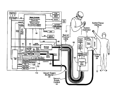
Fig. 1 shows a system 10 for applying, in a non-
invasive manner, energy to a targeted tissue region that
embodies the features of the invention. As shown in Fig.
Date Recue/Date Received 2020-09-25
-6-
1, the system 10 includes three main components. These
are a system console 12; a system applicator 14; and an
applicator-tissue interface 16 carried by the system
applicator 14.
In the Illustrative embodiment shown in Fig.1, and
as will be described in further detail later, the system
is particularly sized and configured to generate and
apply energy to the underarm (axilla) of an individual to
form a predefined pattern of lesions (see, e.g., Fig.
10 23B). The pattern of lesions serves, e.g., to moderate or
interrupt function of the sweat glands in the underarm.
In this illustrative arrangement, the system 10 and its
method of use can serve to treat, e.g., axillary
hyperhidrosis or underarm sweating/odor.
A. The System Console
In the Illustrative embodiment, the system console
12 may be a durable item capable of repeated re-use. As
Figs. 1 to 4 show, the system console 12 comprises a
cabinet or housing that is compact and capable of being
wheeled for transport and positioning alongside an
individual to be treated. Components housed within the
console support specified treatment functions. An AC
power cable 18 couples components within the system
console 12 to a standard AC power outlet (see Fig. 4). A
power supply within the system console 12 (see Fig. 15)
converts the power to 12V DC power for distribution to
the components housed within the system console 12.
In the illustrated embodiment, the specified system
functions include an energy generation function; a tissue
acquisition function; a lesion creation function; and a
lesion control function.
B. The System Applicator
The system applicator 14 also may be a durable item
capable of repeated re-use. The system applicator 14 may
Date Recue/Date Received 2020-09-25
- 7 -
be sized and configured to be, during use, conveniently
handled and manipulated in a hand of a caregiver (see
Fig. 1). As Figs. 2 and 3 show, the system applicator 14
may be conveniently rested in a holster 20 on the system
console 12.
As shown in Figs. 1 and 5 to 8, the system
applicator 14 comprises a pistol-grip housing made, e.g.,
of molded plastic material. Carried within the housing is
a waveguide antenna array 22 (see Figs. 7 to 11). In the
illustrated embodiment, the waveguide antenna array 22
comprises four waveguide antennas 24. It should be
appreciated that the number of antennas 24 can vary
according to the treatment objectives.
In use, the waveguide antenna array 22 radiates
energy provided by the energy generation function.
Components in the applicator 14 also act in concert
with components housed within the system console 12 to
carry out the lesion generation and lesion control
functions. More particularly, and will be described in
greater detail later, the lesion generation function
controlled within the console 12 operates a microwave
switch 26 in the applicator 14 (see Fig. 7) to
synchronize the radiation of energy by the antennas 24 in
the applicator 14 to form desired patterns of lesions in
the targeted tissue region (as Fig. 23B shows). Further,
and as will also be described in more detail later, the
lesion control function controlled within the console 12
provides a coolant that is circulated to a cooling plate
28 in the applicator 14 (see Fig. 7) that is in thermal
conductive contact with the targeted tissue region. The
temperature conditions of the cooling plate 28 control
expansion of the lesion in the targeted tissue region.
A "trigger" switch 30 on the system applicator 14
(see Fig. 7), which may, for example, be thumb actuated,
Date Recue/Date Received 2020-09-25
- 8 -
gives the caregiver direct control over initiation and
termination of treatment, subject to the overrides and
global control of the master controller of the system
console 12. Alternatively, or in combination, a foot
pedal control switch 32 can be provided for the same
purpose (see Fig. 1). A special purpose cable assembly
34 (see Figs. 1 and 16A/B) couples the components housed
in the system applicator 14 to the components housed
within the system console 12. The special purpose cable
assembly 34 includes a custom designed multi-function
plug 36 that couples to a dedicated connection 38 site on
the system console 12.
C. The Applicator-Tissue Interface
The applicator-tissue interface 16 may be a single
use, disposable item. More particularly, as shown in
Figs. 6; 12A/B; and 13, the interface 16 may be sized and
configured to be temporarily coupled to the system
applicator 14 during use (e.g., by a latching mechanism
40), and then detached after use for disposal, as shown
in Fig. 6. In this arrangement, the applicator-tissue
interface 16 can, after an incidence of use, be detached
from the system applicator 14, discarded, and replaced by
another unused applicator-tissue interface 16 prior to a
next incidence of use.
In use (e.g., see Figs. 21A and 21B), the
applicator-tissue interface 16 contacts the targeted
tissue region and passes the energy radiated by the
waveguide antenna array 22 to tissue. Components in the
applicator-tissue interface 16 also act in concert with
components housed within the system console 12 to carry
out the tissue acquisition function. For this purpose,
the applicator-tissue interface 16 includes a tissue
acquisition chamber 42, into which tissue is drawn to
elevate the dermis and hypodermis and localize and
Date Recue/Date Received 2020-09-25
- 9 -
stabilize the targeted tissue region in thermal
conductive contact with the cooling plate 28 as energy is
applied from the waveguide antenna array 22. In the
illustrated embodiment, the tissue acquisition function
includes the application of a vacuum to the tissue
acquisition chamber 42. For this
purpose, a vacuum
supply conduit 44 couples the components housed in the
applicator-tissue interface 16 to components housed
within the system console 12. The vacuum supply conduit
44 plugs into a dedicated connection site 48 on the
system console 12.
The application of the vacuum by the applicator-
tissue interface 16, as controlled by the tissue
acquisition function, provides uniformity and consistency
in acquiring tissue for treatment. It reduces
variability of treatment that may arise, e.g., due to
differences in manipulation of the applicator by a given
caregiver and/or difference among tissue topologies to be
treated.
The applicator-tissue interface 16 also includes a
multi-functional bio-barrier 50(see Fig. 12A). As will be
described in greater detail later, the multi-functional
bio-barrier 50 isolates the operational components in the
applicator 14 and the console 12 from contact with and
contamination by physiologic liquids (e.g., blood and
sweat) that may be present in the targeted tissue region.
The multi-functional bio-barrier 50 substantially
isolates the durable electrical and mechanical components
of the system 10 (e.g., the applicator 14 and console
12), from the physiologic conditions of the tissue
regions being treated, and vice versa.
II. The Functions of the System
As will be described, a master controller 58 housed
on-board the system console 12 (see Fig. 15) monitors,
Date Recue/Date Received 2020-09-25
- 10 -
controls, and coordinates the overall execution of the
specified energy generation function, the tissue
acquisition function, the lesion creation function, and
the lesion control function by the system 10. The on-
board master controller 58 serves to globally set and
control output power, as well as the sequence of the
application of the waveform to the system applicator and
waveguide antennas 24 within the applicator 14. The on-
board master controller 58 also monitors operational
conditions and initiates alarms when predetermined error
or out of bound conditions occur.
An applicator control board 60 housed within the
applicator 14 (see. e.g., Figs. 7 and 15) communicates
with the master controller 58 and is controlled by the
master controller 58 to support the energy generation
function, the lesion creation function, and the lesion
control function conducted by the applicator 14.
The master controller 58 may also implement a
graphical user interface 62 (see, e.g., Fig. 26). The
graphical user interface 62 may be generated on a display
screen 64 that articulates on the system console 12, as
Figs. 1 and 3 show). The graphical user interface 62
conveys status and operational information to the
caregiver and allows the caregiver to provide control
inputs. The graphical user interface 62 on the display
screen 64 communicates the control and alarm conditions
to the caregiver and allows for touch-screen interaction
and input from the caregiver. Further details of a
representative graphical user interface 62 will be
described later (and, in particular, are shown in Figs.
27 to 59).
The energy generation function; the tissue
acquisition function; the lesion creation function; and
the lesion control function, as well as the principal
Date Recue/Date Received 2020-09-25
- 11 -
cooperating components on the console 12, applicator 14,
and applicator-tissue interface 16 that execute these
functions will now be individually discussed in greater
detail.
A. The Energy Generation Function
Components carried on-board the system console 12
(see Fig. 15) generate an energy waveform selected to
achieve the desired therapeutic objective in the targeted
tissue region. These components include a microwave
generator 66 (Broadband Wireless, Model Number BW-5800-
125-HS). The master controller 58 on-board the system
console 12 includes preprogrammed rules or logic that set
and/or vary the output power of the microwave generator
66 according to the therapeutic objectives of a given
system.
Given the therapeutic objectives of treating
hyperhidrosis, the microwave generator 66, under the
control of the master controller 58, may generate at the
time of treatment a microwave signal that lays in the ISM
band of 5.775 to 5.825 GHz, with a frequency centered at
approximately 5.8 GHz. Of course, other waveforms or
variations in this waveform can be selected for
generation by the waveform generation function. A
microwave cable 68 in the special purpose cable assembly
34 couples the microwave signal to the system applicator
14.
The master controller 58 may set the power output
for the microwave signal at between approximately 40
Watts and approximately 100 Watts, where the power output
is measured into a 50 ohm load. As another example, the
master controller 58 may set a power output at
approximately 55 Watts measured into a 50 ohm load. The
power output may be matched to the impedance of the
system applicator 14, the special purpose cable assembly
Date Recue/Date Received 2020-09-25
- 12 -
34, and the applicator-tissue interface 16 to provide
appropriate power out of the system applicator 14 at the
frequency of interest.
The system applicator 14 carries the waveguide
antenna array 22 (see Figs. 9 and 10). The applicator
also carries a microwave switch 26 (see Figs. 7 and 8)
coupled to the microwave cable 68 of the special purpose
cable assembly 34. Feed connectors 70 from the switch 26
couple to the four waveguide antennas 24 of the waveguide
antenna array 22 (see Fig. 8).
The master controller 58 on-board the system
console 12 includes preprogrammed rules or logic to
distribute the microwave signal in a predetermined
pattern to the waveguide antennas 24. Preprogrammed rules
or logic on the applicator main board 60 convert the
control signal pattern to switching signals, which are
communicated to the microwave switch 26 in the applicator
14. In response, the antennas 24 radiate the microwave
signal through the applicator-tissue interface 16 in a
predetermined pattern (controlled by the lesion
generation function) to form prescribed lesion patterns
in the targeted tissue region (as shown, e.g., in Fig.
23B).
The assembly of the waveguide antenna array 22 can
vary. In the representative illustrated embodiment (see,
in particular, Figs. 9 and 10), the array of waveguide
antennas 24 is supported within the system applicator 14
by an antenna cradle 72 and waveguide assembly 74. A
cooling plate 28 supported on the antenna cradle 72 faces
the applicator-tissue interface 16. As will be described
in greater detail later, the cooling plate 28 is one
component of a cooling system controlled by the master
controller 58 that may serve to control lesion formation,
by, for example, preventing lesions from expanding toward
Date Recue/Date Received 2020-09-25
- 13 -
the surface of the skin as they are formed by the applied
microwave energy.
In the illustrated embodiment (see Fig. 9), the
waveguide assembly includes spacers 76 (which may be, for
example, a metal material such as copper or aluminum
shims) positioned between waveguide antennas 24. The
thickness of the spacers 76 is selected to manipulate the
shape of the power distribution pattern applied when, for
example, adjacent waveguide antennas 24 are commanded to
radiate power (which is called a phase drive mode, as
will be described in greater detail later). As shown in
Fig. 9, the heights of waveguide antennas 24 in the
waveguide antenna array 22 are staggered to facilitate
access to the feed connectors 70. Each waveguide antenna
24 may be manufactured by coating a dielectric center
region with a metal material, e.g., copper or nickel. The
thickness of the metal material may, at a minimum,
correspond to the skin depth of the applied microwave
energy at the frequency of interest, e.g., 5.8 GHz.
Typically, the thickness is significantly greater, e.g.,
.00025 inches or more.
In the illustrated embodiment (see Fig. 9), each
waveguide antenna 24 may include at least one scattering
element 78, which projects from its lower, tissue facing
surface, which can also be called the antenna aperture.
The scattering elements 78 are sized and configured to
optimize the size and shape the lesions. In the
illustrated embodiment, each scattering element 78
projects toward tissue generally from the center of the
respective waveguide antenna aperture. Still, in an
alternative embodiment, the scattering element 78 need
not be centered on the waveguide antenna aperture. The
scattering element 78 may project about 1 mm from the
aperture.
Date Recue/Date Received 2020-09-25
- 14 -
In the illustrated embodiment (see Fig. 9),
intermediate scattering elements 80 may be positioned
between the waveguide antennas 24. The intermediate
scattering elements 80 may be sized and configured to
optimize the size and shape of lesions developed in the
skin between waveguide antennas 24, for example, by
improving the Specific Absorption Rate (SAR) pattern in
tissue. By altering the material, size, and configuration
of the intermediate scattering elements 80, lesions
created in tissue by the waveguide antennas 24 can be
made larger and more spread out, or (conversely)
narrower, depending upon the therapeutic objectives. For
example, increasing the dielectric constant of an
intermediate scattering element 80 may reduce the size of
a lesion created in skin between waveguide antennas 24,
and vice versa.
The intermediate scattering elements 80 may be
manufactured from, for example, alumina or from a
material that is approximately 96% alumina.
Alternatively, the intermediate scattering elements 80
may be manufactured from, for example, silicone or
injected molded silicone. The intermediate scattering
elements 80 may be manufactured from a material having
approximately the same dielectric constant as the
scattering elements 78, e.g., a dielectric constant of
approximately 10, and more preferred, a dielectric
constant of approximately 3.
The intermediate scattering elements 80 may be
sized such that they have a width which is not more than
slightly wider than the separation distance between
apertures of the waveguide antennas 24, so that they do
not substantially interfere with the radiated energy. The
intermediate scattering elements 80 may be sized and
configured to modify and/or spread out the radiated
Date Recue/Date Received 2020-09-25
- 15 -
microwave field.
In a representative embodiment, the intermediate
scattering elements 80 may have an optimal length which
is shorter than the length of scattering elements 78,
e.g., approximately 7 mm in length, or more preferred 6.3
mm in length.
1. The Tissue Acquisition Function
Components carried on-board the system console 12
(see Fig. 15) generate negative pressure that is
communicated to the applicator-tissue interface 16 by the
vacuum supply conduit 44. As will be described in greater
detail later (and as generally illustrated in Fig. 12B),
the applicator-tissue interface 16 includes a formed
tissue acquisition chamber 42 with ports 82 through which
negative pressure is directed by the vacuum supply
conduit 44 to draw tissue into the acquisition chamber
42, as Fig, 21B shows). The negative pressure applied to
tissue in the acquisition chamber 42 localizes and
stabilizes the tissue while microwave energy is applied.
The tissue acquisition function can be accomplished
in concert with the tissue acquisition chamber 42 in
various ways. In the illustrated embodiment (see Fig.
15), the tissue acquisition function includes a motor-
driven vacuum pump 84 coupled via a one-way check valve
86 and accumulator (reservoir) 88 to a solenoid vacuum
valve 90. The check valve 86 between the vacuum pump 84
and the accumulator 88 allows the vacuum pump 84 to be
shut off when no additional vacuum is required. The
accumulator (reservoir) 88 may accommodate, e.g., at
least 30 cubic inches in volume to provide a large
capacity of vacuum.
The vacuum pump 84 may comprise, e.g., a scroll
vacuum pump with a brushless DC motor (Air Squared Model
No. V11H12N2.5). The solenoid vacuum valve 90 may
Date Recue/Date Received 2020-09-25
- 16 -
comprise. e.g., a solenoid valve, three way, normally
closed, exhaust to atmosphere (Model LW53KK8DGBG12/DC,
Peter Paul Electronics, Co.). The vacuum pump 84
maintains, e.g., a vacuum level of between minus 20
inches to minus 22 inches of Hg for proper tissue
acquisition.
As Fig. 15 shows, the motor-driven vacuum pump 84
may receive power through the power supply and power
printed circuit board (PCB) within the system console 12.
The solenoid vacuum valve 90 may also be coupled to and
controlled by the master controller 58 on-board the
system console 12, so that its operation can be
coordinated by the master controller 58 with the
generation and application of microwave energy, as well
as other functions of the system 10.
The motor-driven vacuum pump 84 creates negative
pressure. The solenoid vacuum valve 90 communicates with
the vacuum supply conduit 44. When opened by the master
controller 58, the solenoid vacuum valve 90 conveys
negative pressure generated by the vacuum pump 84 to the
tissue acquisition chamber 42 of the applicator-tissue
interface 16. Closing the solenoid vacuum valve 90
interrupts the supply of negative pressure to the
applicator-tissue interface 16.
Referring now to Figs. 12A and 12B, the applicator-
tissue interface 16 may comprise a body 92 formed from a
medical grade rigid or semi-rigid plastic material, e.g.,
polycarbonate. The body 92 may be formed, e.g., by
molding, into a bowl shape. Latching assembly 40 can be
integrally formed on the body 92 to couple to a mating
attachment member 94 on the system applicator 14 (see,
e.g., Fig. 5), to fasten the applicator-tissue interface
16 to the system applicator 14 at time of use and
disconnect the interface from the system applicator 14
Date Recue/Date Received 2020-09-25
- 17 -
after use (as Figs. 5 and 6 illustrate).
Within the bowl shaped body 92 (as best shown in
Fig. 12B), a waveguide holder gasket 96 is seated on
peripheral flange 98 formed in the bowl. The waveguide
holder gasket 96 is sized and configured, when the
interface body 92 is fastened to the system applicator 14
(see Fig. 13), to form a fluid-tight, pressure-tight seal
against the periphery of the cooling plate 28 on the
undersurface of the waveguide assembly.
Within the bowl shape body 92 (see Figs. 12A and
12B), spaced below and inward of the waveguide holder
gasket 96, is a tissue interface surface 100. In the
illustrated embodiment (as best shown in Fig. 12A), the
tissue interface surface 100 comprises a frame 102 with
upper and lower overlying adhesive panels 104. A first
bio-barrier component 52 is mounted on the upper adhesive
panel, and the lower adhesive panel adheres to an
interface surface support in the bowl, which the
waveguide holder gasket 96 peripherally surrounds.
In use, tissue being treated contacts the first
bio-barrier component 52 in thermal contact with at least
a portion of the cooling plate 28. The first bio-barrier
component 52 forms a part of the multi-functional bio-
barrier 50 of the applicator-tissue interface 16. The
first bio-barrier component 52 forms comprises the actual
tissue surface interface, which tissue acquired within
the tissue acquisition chamber 42 contacts as energy is
applied from the waveguide antenna array 22. The first
bio-barrier comprises 52 a material that is selected on
the basis of different, but overlapping physical
criteria.
One selection criteria for the first bio-barrier
component 52 is that the material is substantially
impermeable to both air and liquids, such as blood and/or
Date Recue/Date Received 2020-09-25
- 18 -
sweat, which may be present in the tissue acquisition
chamber 42. As the tissue acquisition function applies
vacuum to draw tissue within the tissue acquisition
chamber 42 into contact with the first bio-barrier
component 52, the first bio-barrier component 52 isolates
the components in the applicator 14 from contact with and
contamination by physiologic liquid in the targeted
tissue region.
An overlapping selection criteria for the first
bio-barrier component 52 is that the material, taking
into account its thickness, possesses requisite low
microwave conductivity, so that it efficiently passes the
microwave energy radiated by the waveguide antenna array
22 to the targeted tissue region acquired within the
tissue acquisition chamber 42, with minimal energy
absorption. This characteristic can be expressed as a
loss tangent tan 5 of 0.1 or less, and more desirably
approximately 0.0004.
The loss tangent tan 5 is similar to conductivity
u, but also takes into account the dielectric constant of
the material, as follows:
tan 5 = u/cos
where co is frequency, and
where s is permittivity
For example, at 5.8 Ghz, a range of conductivities
G suitable for use as the first bio-barrier component 52,
corresponding to a tan 5 equal to or less than 0.1, would
be o = 0.0 to 0.2 siemens/meter.
Another overlapping selection criteria for the
first bio-barrier component 52 is that the material,
taking into account its thickness, possesses requisite
high thermal conductivity, to efficiently allow thermal
conduction to occur between the targeted tissue region
acquiring within the tissue acquisition chamber 42 and
Date Recue/Date Received 2020-09-25
- 19 -
the cooling plate 28. For example, the material selected
should have a thermal conductivity of at least 0.1 watts
per meter-Kelvin (0.1 W/mK), and desirably 0.1 to 0.6
W/mK, and most desirably 0.25 to 0.45 W/mK.
Another overlapping selection criteria for the
first bio-barrier component 52 is that the material,
taking into account its thickness, possesses requisite
high heat transfer coefficient. The heat
transfer
coefficient can be expressed by the thermal conductivity
of the material divided by the thickness of the material.
For example, for a first bio-barrier component 52 with a
thermal conductivity of 0.1 and a thickness of .0005
inches, the heat transfer coefficient would be about 7874
W/m2K.
Other overlapping selection criteria for the first
bio-barrier component 52 is that the material is
sufficiently flexible to conform to the surface of the
cooling plate 28, while also being sufficiently strong to
resist tearing as a result of vacuum pressure or contact
with tissue.
In this respect, the first bio-barrier component 52
may be a nonporous membrane, e.g., polyethylene film,
nylon, or other suitable materials. The first bio-
barrier component 52 is desirably flexible and soft for
compliant contact with skin. The first bio-barrier
component 52 can comprise, e.g., polyethylene film
available from Fisher Scientific, or (alternatively)
Mylar film. The bio-barrier component 52 can be, e.g.,
about .0005 inch in thickness.
The applicator-tissue interface body 92 also
includes a skirt 106 (see Figs. 12A/B and 13) that
depends downwardly with an increasing diameter from the
body about the periphery of the applicator-tissue
interface surface. The downward depending skirt 106
Date Recue/Date Received 2020-09-25
- 20 -
defines a generally funnel-shaped open interior area or
chamber leading to the first bio-barrier component 52 of
the applicator-tissue interface 16 (see Fig. 13). This
chamber defines the tissue acquisition chamber 42
previously described. The skirt 106 may
comprise a
compliant medical grade plastic material (e.g., a thermal
plastic elastomer (TPE) such as urethane; or silicone; or
natural or synthetic rubber; or an elastomeric material)
and may be sized and configure to rest comfortably
against an external skin surface. When pressed with
sufficient pressure to compress against a tissue surface
(see Fig. 21A), the periphery of the skirt 106 forms a
generally fluid-tight, pressure-tight seal about the
tissue acquisition chamber 42.
The skirt 106 may include an alignment member 108
(see Fig. 5) positioned on each side of the skirt 106 to
provide a positioning point of reference to the caregiver
during manipulation of the interface, as will be
described in greater detail later. The funnel-shaped
contour of the skirt 106 may provide a skirt angle that
gives the caregiver a direct view of the alignment
members 108, while the caregiver manipulates the
applicator-tissue interface 16 attached to the system
applicator 14.
As Fig. 12A shows, the vacuum supply conduit 44
communicating with the tissue acquisition function of the
system console 12 (see also Fig. 15) is coupled to a port
formed on the body of the applicator-tissue interface 16.
The port communicates with a vacuum channel 110 formed in
the body (see Figs. 12A and 12B) that communicates with
the tissue acquisition chamber 42 adjacent the
applicator-tissue interface surface. The vacuum channel
110 may circumferentially encircle the tissue acquisition
channel at or near the applicator-tissue interface
Date Recue/Date Received 2020-09-25
- 21 -
surface. The vacuum channel 110 may include spaced-apart
apertures or ports 82 formed along the vacuum channel
(see Fig. 12B)(e.g. four ports, one adjacent each side
the applicator-tissue interface surface), to convey
negative pressure uniformly into the tissue acquisition
chamber 42 adjacent the applicator-tissue interface
surface. The ports 82 suction skin into the chamber and
position the skin against the first bio-barrier component
52 in thermal conductive contact with the cooling plate
28 (as Fig. 21B shows).
The frame 102 and panels 104 of the tissue
interface surface 100/52 may include formed apertures 112
(see Fig. 12A) that register when assembled to form a
vacuum balance path that communicates with the tissue
acquisition chamber 42. Negative pressure applied in the
chamber 42 is conveyed through the vacuum balance path
112 to the opposite side of the interface surface 100/52
to equalize pressure on both sides of the interface
surface 100/52.
A second bio-barrier component 54 of the multi-
functional bio-barrier 50 of the applicator-tissue
interface 16 desirably occupies the vacuum balance path
112. The second bio-barrier component 54 in the vacuum
balance path 112 (which can also be called the "vacuum
balance bio-barrier component") comprises a material that
is substantially impervious to liquid, but not to air.
The vacuum balance bio-barrier component 54 prevents
physiologic liquids such as blood and/or sweat that may
be present in the tissue acquisition chamber 42 from
being transported through the vacuum balance path 112
into the interior of the applicator 14. Candidate
materials for the second bio-barrier component 54 may
include pores sufficient to pass air (e.g., 0.45 pm) to
substantially equalize the vacuum pressure on the system
Date Recue/Date Received 2020-09-25
- 22 -
applicator side and the interface side of the surface,
without passing biological liquids from the acquisition
chamber 42 into the system applicator 14. The second
bio-barrier component 54 may comprise, e.g., a
hydrophobic membrane made from PTFE (Teflon) material.
The second bio-barrier component 54 can be, e.g., about
.005 inch in thickness.
The spaced-apart apertures or ports 82 formed along
the vacuum channel may include interior patterns 114
along its interior that can impress a "hickey pattern" on
the skin drawn into the chamber 42(see Figs. 14A and
14B). The existence
of "hickey patterns" with the
lesions can help guide the caregiver in successive
placements of the system applicator 14 to accurately
place a succession of lesions in the targeted tissue
region. Inconsistencies in the "hickey patterns" may also
alert the caregiver to gaps or inaccurately placed
lesions in the lesion pattern, to indicate a need to
return and fill in gaps and missed spots in the pattern.
The interior patterns 114 define notches that break
surface contact and may help to prevent skin from
blocking the vacuum apertures or ports.
In a representative embodiment, the tissue
acquisition chamber 42 is dimensioned approximately 1.54
inches by approximately 0.7 inches, having a depth
(without the skirt 106) of approximately 0.177 inch (4.5
mm). With the skirt 106, the depth of tissue acquisition
chamber 42 can be between approximately 6.5 mm to 11 mm,
depending upon the extent to which the compliant skirt
106 is compressed against the skin by the application of
vacuum. According to an embodiment of the invention the
four corners of the tissue acquisition chamber 42 may
have a radius of .1875 inches.
In this arrangement, the waveguide antenna array 22
Date Recue/Date Received 2020-09-25
- 23 -
on the opposite side of the tissue interface surface
100/52 include four antennas 24 and possesses dimensions
of approximately 1.34 inches by approximately 0.628
inches. The dimensions of the waveguide antenna array 22
and the tissue acquisition chamber 42 are desirably
optimized to minimize stray fields forming at the edges
of waveguide antenna array 22, as well as optimizing the
effective cooling area of the tissue interface surface.
The tissue acquisition chamber 42 is desirably optimized
to facilitate tissue acquisition without adversely
impacting cooling or energy transmission.
The vacuum supply conduit 44 may collect liquids
(e.g., sweat or blood) that escape during the treatment
process. For this reason, a third bio-barrier component
56 of the multi-functional bio-barrier 50 of the
applicator-tissue interface 16 is placed upstream of the
applicator-tissue interface 16 in-line in the vacuum
supply conduit 44 (see Fig. 15, as is also generally
shown in Fig. 1). The third bio-barrier component 56 is
selected to be substantially impervious to liquid, but
not to air. The third bio-barrier component 56 can
comprise, e.g., a hydrophobic filter (e.g., a Millex FH
filter made of 0.45 pm hydrophobic PTFE available from
Millipore) to keep liquids out of the system console 12.
The hydrophobic filter can be further characterized,
e.g., by accommodating an airflow of approximately 13.4
cubic feet per minute at approximately 10 pounds per
square inch.
The third bio-barrier component 56 can,
alternatively, comprise an in-line vacuum trap, as shown
in Figs. 16C and 16D. The vacuum trap may include a
formed housing 116 defining a vacuum inlet port 118 (with
which the vacuum supply conduit 44 communicates) and a
vacuum outlet port 120 (which plugs into the connection
Date Recue/Date Received 2020-09-25
- 24 -
site 48 on the console 12). The housing 116 defines an
interior chamber 122, which the vacuum flow between the
inlet and outlet ports 118 and 120 must traverse from the
applicator-tissue interface 16 to the system console 12.
A central ridge 124 on the exterior of the housing 116
may provide a gripping surface for the caregiver to hold
and manipulate the vacuum trap, e.g., while plugging the
vacuum supply connector 118 into and out of the mating
console vacuum supply receptacle 48.
The chamber 122 is compartmentalized by an interior
wall 126 into an inlet side 128, communicating with the
inlet port 118, and an outlet side 130 , communicating
with the outlet port 120. One or more apertures 132 in
the interior wall 130 define path(s) of flow
communication between the inlet and outlet sides 1w28 and
130 of the chamber 122.
Baffle plates 134 interfere with vacuum flow
through the aperture(s) 132 through the interior wall 16
between the inlet side 128 and outlet side 130 of the
chamber 122. The vacuum flow must veer around the baffle
plates 134 to transit through the chamber 122. An array
of annular baffles 136 is further circumferentially
placed around the inlet side 128 of the chamber 122. The
baffle plates 134 and annular baffles 136 form an array
of tortuous paths, through which vacuum flow transiting
the chamber must navigate. Air in the vacuum flow will
readily change direction to navigate the tortuous paths.
Physiologic liquid carried by the vacuum flow will not,
and will instead be captured by gravity in the nooks and
crannies of the tortuous paths through the chamber 122.
The vacuum trap thereby prevents physiologic liquid from
passing out of the outlet port 120 into the console 12.
2. The Lesion Creation Function
As will be described in greater detail later, the
Date Recue/Date Received 2020-09-25
- 25 -
microwave signal applied through the waveguide antennas
24 to tissue acquired within the tissue acquisition
chamber 42 creates lesions in the targeted tissue region,
as generally shown in Fig. 23B. In the treatment of
hyperhidrosis, the lesions may be formed in the lower
dermis and/or dermis/hypodermis of the skin. Components
carried on-board the system console 12 control the
microwave switch 26 carried on-board the applicator 14 to
sequence the application of the microwave signal by the
waveguide antennas 24 in prescribed manners to form the
lesions in selected patterns, as Fig. 23B illustrates.
The patterns are selected to be conducive to achieving
the therapeutic objectives of the system 10.
(iv) The Lesion Control function
Components carried on-board the system console 12
(see Fig. 15) generate and circulate cooling fluid
through coolant paths between the waveguide antenna array
22 and tissue cooling plate 28 carried in the system
applicator 14, as is generally shown in Figs. 10 and 11).
The tissue cooling plate 28 protects skin engaged with
the applicator-tissue interface 16 (see Fig. 21B) from
thermal damage by preventing lesions formed in the
dermis/hyperdermis from expanding toward the epidermis.
The cooling fluid may comprise, e.g., water, de-
ionized water, or other suitable fluid.
The lesion control function can be accomplished in
various ways. In the illustrated embodiment (see Fig.
15), the lesion control function includes a Peltier-
effect thermoelectric cooler (TEC) 138. As Fig. 15
shows, the TEC 138 receives power through the power
supply and power printed circuit board carried on-board
the system console 12, as do intake fans and chiller fans
140 that circulate air within the console 12 for
conveying from the console 12 heat generated by the
Date Recue/Date Received 2020-09-25
- 26 -
components, including the TEC 138. The TEC 138 chills
coolant in a reservoir 142 on-board the console 12. A
water pump 144 (also drawing power via the power printed
circuit) conveys chilled coolant from the reservoir 142.
The chilled coolant is circulated by a coolant supply
line 146 through coolant paths 148 (see Figs. 10 and 11)
in the waveguide antenna cradle 72 and between the
waveguide antenna array 22 and tissue cooling plate 28
carried in the system applicator 14. Coolant is returned
by a coolant return line 150 to the reservoir 142. The
coolant supply and return lines 146 and 150 extend
through the special purpose cable assembly 34 (see Figs.
16A and 16B) to the applicator 14. A cooling fluid
germicidal lamp 152 (e.g., 253.7 nm) may be provided to
prevent growth of microorganisms contaminants in the
coolant that could clog the fluid lines and reduce
coolant flow in the applicator 12. The cooling fluid
germicidal lamp 152 may be activated periodically, e.g.,
for a period of time (e.g., 10 minutes) following each
power-on cycle or each coolant refill.
As Fig. 15 shows, the TEC 138 may be coupled to and
controlled by the master controller 58, so that its
operation (like that of the components of the tissue
acquisition function) can be coordinated by the master
controller 58 with the generation and application of
microwave energy.
In the illustrated embodiment, the cooling plate 28
rests against the terminal surfaces of the scattering
elements 78 (see Figs. 10 and 11), but, in an alternate
embodiment, the terminal surfaces of the scattering
elements can be spaced out of contact with the cooling
plate 28. Cooling paths 148 for the waveguide antenna
array 22 are formed in the spaces between the cooling
plate 28 and each waveguide antenna 24, into which the
Date Recue/Date Received 2020-09-25
- 27 -
scattering elements 78 project. Coolant is circulated
through these paths 148, cooling each waveguide antenna
24 individually and the cooling plate 28 in general.
The flow rate of coolant through these paths 148
can be, e.g., approximately 425 milliliters per minute
+/- 45 milliliters per minute. Desirably, the paths 148
are sized and configured so that the flow rate of coolant
along each waveguide antenna 24 is substantially the
same. The temperature of the coolant can be, e.g.,
between approximately 8 degrees centigrade and
approximately 22 degrees centigrade, and preferably
approximately 15 degrees centigrade.
The scattering elements 78 may extend into at least
a portion of coolant paths 148. It is desirable that the
cooling paths 148 be smoothed or rounded or shaped in the
manner shown in Figs. 9, 10, and 11 to reduce the
generation and/or build up of air bubbles in the paths
148. The scattering elements 78, for example, may be
formed in the shape of ovals or racetracks or oblong
hexagons with "faceted" or tapering ends (see Figs. 9 to
11). Hydrophilic coatings may be used on some or all of
the cooling paths 148 to, e.g., reduce the formation of
bubbles.
The intermediate scattering elements 80 associated
with the waveguide antenna array 22 may also be located
in the coolant paths 148 between the antenna apertures.
In this arrangement, the intermediate scattering elements
80 may be positioned such that they facilitate equalized
cooling across the cooling plate 28, keeping in mind,
however, that their principal function is to influence
lesion size and shape in, for example, the phase drive
mode. The intermediate scattering elements 80 may be
sized such that they have a width which is not more than
slightly wider than the separation distance between
Date Recue/Date Received 2020-09-25
- 28 -
apertures of the waveguide antennas 24, so that they do
not substantially interfere with the radiated energy. The
intermediate scattering elements 80, which extend into
coolant paths 148, may likewise be smoothed or rounded or
shaped in the manner shown in Figs. 9, 10, and 11 to
prevent the generation and/or buildup of bubbles in the
paths. The
intermediate scattering elements 80, for
example, may be formed in the shape of ovals or
racetracks or oblong hexagons with "faceted" or tapering
ends (see Fig. 9 to 11), provided that they are sized and
configured to modify and/or spread out a microwave field
as it travels through the coolant path 148. In this
arrangement, the intermediate scattering elements 80 (and
the scattering elements 78) are desirably made of
materials which will not rust or degrade upon exposure to
the coolant.
Likewise, the cooling plate 28 may be laser cut
(with a thickness, e.g., of about .020 inch) with curved
corners.
Thermocouples 154 may be placed on the surface of
the cooling plate 28 opposite to the cooling path 148
(see Figs. 10 and 11), generally aligned with and between
the apertures of the waveguide antennas 24. The
thermocouples 154 can, if desired, be printed (sputtered)
on the cooling plate 28. The thermocouples 154 can, e.g.,
comprise a plurality of T-type thermocouples (e.g.,
seven) masked and sputtered with varying alloy
compositions of copper and nickel, e.g., constantan, such
as, e.g., 60% copper/40% nickel, and then re-masked and
sputtered with copper such that the copper and the
copper-nickel components form a junction that serves as a
thermocouple. One or more thermocouples 154 may also be
placed in the supply and return lines. The thermocouples
154 are coupled to the applicator main board 60, which
Date Recue/Date Received 2020-09-25
- 29 -
communicates sensed temperature conditions through the
special purpose cable assembly 34 to the master
controller 58 on board the system console 12. The sensed
temperature conditions are processed according to
preprogrammed logic residing in the master controller 58,
as part of the waveform generation function, and may be
used to adjust power supplied to the waveguide antennas
24 based upon temperature conditions sensed along the
cooling plate 28.
III. System Controllers
A. On Board the System Console
(The Master Controller)
The master controller 58 (see Fig. 15) resides on a
control printed circuit board in the system console 12.
The master controller 58 receives 12V power from the
power supply. The master controller 58 communicates with
components carried by the system console 12 as well as
components carried by the applicator.
The special purpose cable assembly 38 (see Fig. 1
and 16A/B) establishes a multi-purpose link between the
master controller 58 and the applicator 14. Extending
through the special purpose cable assembly (see Figs. 16A
and 16B) are the microwave energy cable 68 for conveying
the microwave signal from the generator controlled by the
master controller 58 to the microwave switch 26 on the
applicator 14; the coolant supply and return conduits 146
and 150 for conveying coolant to and from the coolant
reservoir 142 for circulation through the coolant paths
148 of the waveguide antenna array 22 and cooling plate
28 in the applicator; and a connector 156 establishing
communication links between the master controller 58 and
the applicator main control board 60 according to a
prescribed communication protocol (e.g., the CMX-RTX""
RTOS (embedded real-time operating system) Product Line,
Date Recue/Date Received 2020-09-25
- 30 -
available from CMX Systems, San Jose, California). The
bundling of multiple electrical and fluid conduits
through a single special purpose cable assembly 34 serves
to streamline the form and function of the system 10 and
simplify set up of the system 10, while also efficiently
supporting the diverse functions of the system 10 itself,
in terms of electrical waveform generation, coolant
circulation, and providing communication links. The
multi-special purpose cable assembly 34 facilitates
lengthening the cable (e.g., upwards to eight feet) for
better reach and ease of manipulation remote from the
system console 12.
As shown in Fig. 15, communications between the
master controller 58 and components residing in or on the
console 12 can include (i) the colored (e.g., blue) LED
on the console 12 that indicates when the generator is
applying microwave energy to the targeted tissue region;
(ii) the footswitch 32; (iii) the graphical user
interface screen; and (iv) a speaker to generate audible
alarms or status sounds. Also communicating with the
master controller 58 may be (v) a radio-frequency
identification (RFID) reader, which provokes signal
transmission from passive RFID tags 158 carried by the
applicator-tissue interface 16 or its packaging (see Fig.
25) to identify the applicator-tissue interface 12 prior
to use, as will be described later. The RFID reader can
also function, if desired, to erase information carried
by a RFID tag 158 after being read, e.g., to prevent
reuse of a given application-tissue interface 16
(alternatively, reading and identification functions
based upon bar-coded information may be used); and (vi) a
sensor 160 on the holster 20 that indicates when an
applicator resides on the holster 20.
Regarding the holster 20 (see Fig. 2), the holster
Date Recue/Date Received 2020-09-25
- 31 -
20 is operative for storing the applicator on the system
in two secure positions: 1) facing "in," when the
applicator-tissue interface 16 carried by the applicator
faces toward a power absorber 168 carried by the holster
20, in which the master controller 58 can execute a
"test" mode to verify the delivery of power, water temp
sensor response, antenna switching where the power is
safely contained by the power absorber 168, and 2) facing
"out" away from the power absorber 168 for easy access to
the attachment point for the applicator-tissue interface
16. When the applicator 14 is in the holster 20, the
"facing in" position information desirably verifies that
any power delivered during the "test mode" shall be
safely contained by the power absorber 168. The facing
"in" position is sensed by a magnetic sensor 160 on the
holster 20 and a magnet 162 carried by the applicator
that registers with the magnetic sensor 160 if and only
if the applicator 14 and applicator-tissue interface 16
is facing "in." The "facing out" position need not
necessarily be sensed.
Sensed operating conditions are also communicated
to the master controller 58. The sensed conditions may
include (i) sensed forward power signals detected by the
microwave generator 66; (ii) sensed reverse power
detected directly by the master controller 58; (iii)
sensed negative pressure levels in the vacuum supply line
44, which may be sensed both upstream and downstream of
the vacuum solenoid valve 90; (iv) sensed coolant flow in
the coolant supply line 146; (v) coolant level in the
coolant reservoir 142; and (vi) sensed temperature of the
thermoelectric cooler (TEC 138). Other sensed operating
conditions that also may be communicated to the master
controller 58 by the applicator main board 60, e.g., (i)
sensed forward and reverse power signals detected onboard
Date Recue/Date Received 2020-09-25
- 32 -
the applicator 14; (ii) microwave switch status
conditions; and (iii) sensed temperature conditions
processed by the main applicator board from signals
received by the thermocouples 154 residing on the cooling
plate 28 and coolant supply line 146 in the applicator.
The master controller 58 also communicates energy
generation signals to the microwave generator 66, and
operating signals to the solenoid vacuum valve 90.
Preprogrammed rules or logic on the master controller 58
process sensed information communicated to the master
controller 58 to generate command signals and alarms when
an out of bounds condition exists. Based upon the
processed information, the master controller 58 may,
e.g., increase or decrease fan speeds to maintain the TEC
138 at a desired temperature; increase or decrease
coolant flow; or alter power levels.
In a representative embodiment, the master
controller 58 desirably is capable of supporting
communication and control with the applicator 14. The
master controller 58 desirably receives from the
applicator information about the applicator temperatures,
antenna power and state of the "trigger" switch on the
applicator. The master controller 58 desirably sends to
the applicator 14 information describing which antennae
should be enabled. The master controller 58 desirably
sends to the applicator 14 commands to control the
applicator LED's, if any. The communication desirably
includes fault conditions detected in the applicator. The
master controller 58 is desirably capable of supporting
the following system states indicated on the applicator:
Ready, Treatment, Cooling and Fault.
The master controller 58 desirably serves to detect
failures or unexpected/out of tolerance behavior and
react to minimize risk of injury to the patient and, when
Date Recue/Date Received 2020-09-25
- 33 -
possible, damage to the device.
For example, a prescribed incremental loss of
vacuum (e.g., more than 5 inches of Hg vacuum) may
immediately pause the energy delivery cycle. If the
incremental loss persists for less than prescribed period
of time (e.g., less than 2 seconds), energy delivery may
resume when the vacuum level returns to the prescribed
level. If the incremental loss persists for longer than
the prescribed interval, the master controller 58 may
abort the therapy cycle and cause it to enter a post-cool
phase.
For example, loss of communications to the
applicator or the generator may cause the master
controller 58 to enter a safe state by aborting the
therapy cycle and causing it to enter the post-cool phase
by terminating energy delivery during loss of applicator
communications and disabling the amplifier (mute enabled)
during loss of amplifier communications.
For example, temperature monitoring at the
applicator cooling plate 28 may be used to detect
treatment conditions. For example, if the temperature
exceeds a predetermined amount (e.g., 40 C) within the
first 2 seconds of energy delivery, the master controller
58 may abort the therapy cycle and cause it to enter the
post-cool phase. If the temperature exceeds the
predetermined amount after the first 2 seconds of energy
delivery, the master controller 58 may immediately
terminate energy delivery to that antenna and initiate
energy delivery to the next antenna in the therapy
sequence.
For example, vacuum pump drive may be monitored and
compared to nominal drive levels with tolerance bounds to
make sure that excessive vacuum leaks that could occur
when the target tissue has not been properly acquired or
Date Recue/Date Received 2020-09-25
- 34 -
during loss of tissue acquisition are properly reported
to the caregiver through the user interface. If the
vacuum pump drive fluctuations are excessive during a
therapy cycle, the master controller 58 may abort the
therapy cycle and cause it to enter the post-cool phase.
For example, internal voltage monitoring of the
power supply voltage inside the generator and on the
master controller 58 may be used to determine if a fault
condition exists that may abort the therapy cycle and
cause the system to enter the post-cool phase.
For example, microwave power may be continuously
monitored in the console and at the applicator during
energy delivery and non energy-delivery phases of the
therapy cycle. During energy delivery, the microwave
power may be required to be within a specified range and
during the non energy-delivery phases of therapy it
should be less than the specified threshold. If a fault
condition exists, the master controller 58 may abort the
therapy cycle and cause the system to enter the post-cool
phase. If a fault condition exists during the non energy-
delivery phases of therapy, the generator may be disabled
(mute enabled).
For example, internal temperatures of critical
components may be monitored for detection of excessive
thermal conditions.
For example, if there are thermoelectric cooler
errors, temperature errors, flow rate errors, or water
level errors during a therapy cycle, the master
controller 58 may abort the therapy cycle and cause it to
enter the post-cool phase.
The master controller 58 may provide the capability
to store data associated with each treatment cycle that
the system performs. This data may be stored in memory
that is not erased when the power to the system is
Date Recue/Date Received 2020-09-25
- 35 -
removed or turned off. The information contained in the
data log may be made accessible through a service mode
screen on the graphical user interface 62. It may store
some or all of the following information for each
applicator placement in a treatment data log in a folder
in system memory: (i) Date and Time; (ii) Average forward
power; (iii) Maximum reverse power(from the master
controller 58)and/or from each applicator detector board,
as will be described below); (iv) Temperature rise
(delta) for each of the temperature sensors located on
the applicator cooling plate 28; (v) Maximum coolant
temperature; and/or (vii) Fault Events and associated
Error codes.
B. On Board the Applicator
As best shown in Figs. 7 and 8, a microwave switch
26 in the applicator has an input coupled to the
microwave energy supply lead. The microwave switch 26 can
comprise, e.g., a switch manufactured by Relcom
Technologies, Inc., Part No. PMT-SR019. Individual feed
connector cables 70 may lead from outputs of the switch
26 to individual detector boards 170, which include
directional couplers (see Fig. 17) to pass the microwave
signal to the waveguide antennas 24, as well as couple
the signal to forward and reverse power detection
circuitry.
The master controller 58 on-board the system
console 12 includes imbedded pre-programmed rules
establishing the desired switching patterns for applying
the microwave signal through the waveguide antennas 24.
The signals from the master controller 58 are converted
by the applicator main board into switching signals
which, in turn, control the operation of the switch to
execute these patterns.
1. Localized Forward and Reverse Power
Date Recue/Date Received 2020-09-25
- 36 -
Detection
As just stated, forward and reverse power signals
are detected by the master controller 58 as the microwave
signal is transmitted through the special purpose cable
assembly to the system applicator 14. These forward and
reverse power signals, local to the console 12, are
communicated to the master controller 58 for power
control purposes.
In addition, the system applicator 14 may carry
additional on-board a detector board circuit (see Figs.
17 and 18) for detecting forward and reverse power
locally on the applicator. These forward and reverse
power signals, local to the applicator 14, are
communicated by the applicator main board 60 to the
master controller 58 on-board the console 12 for
processing as further confirmation that power settings
are within prescribed bounds.
As shown in Fig. 7, the detector board circuit
comprises four detector boards 170, a given detector
being electrically coupled and dedicated to a single
waveguide antenna 24. The electrical configuration of
each detector board 170 is essentially the same, as will
now be described.
(a) Directional Coupler
Fig. 17 shows the circuit traced on each detector
board. The circuit includes directional coupler (also
shown in Fig. 18) having a thru line and a coupled line
(which have been generally described previously). The
thru line has an input that receives the microwave
signal, as switched to the individual waveguide antenna
24 by the system applicator main board 60, and an output
that conveys the received microwave signal to the
respective waveguide antenna 24. As best shown in Fig.
18, the coupled line runs in proximity to the thru line
Date Recue/Date Received 2020-09-25
- 37 -
for a fixed distance, to allow a sample of energy
travelling through the thru line to be transferred into
the coupled line. The coupled
line is directional,
meaning that it samples the forward power on one port of
the coupled line and the reflected power on the other
port. The length of the coupled section, the distance
between the thru and coupled lines, the optimization of
coaxial feeds to efficiently transfer power, the
mountings, and the way in which the ports of the coupler
are terminated determine the amount of power sampled in
the forward and reflected ports and the isolation between
the two ports.
In the illustrated embodiment (see Fig. 18), the
directional coupler circuit is implemented using
asymmetric stripline transmission lines (a stripline in
which the center conductor is not equidistant from the
upper and lower ground planes). Energy is fed into the
input port and out of the output port through the ground
plane which is closer to the center conductor of the
stripline. The forward and reverse sampling ports include
a transition from asymmetric stripline to microstrip
line. This allows for easy placement and assembly of the
discrete components of the detector circuit.
(b) Attenuator and DC Blocking Circuit
The forward and reverse sampled power ports feed
microwave energy into a microwave attenuator and DC
blocking circuit as shown in Fig. 17. The microwave
attenuator circuit serves to condition the amount of
microwave power to an optimal range of levels for the
power detector. The circuit also includes a DC block such
that no low-frequency signals can travel from the power
detector or signal conditioning circuitry down the high
frequency microwave path.
(c) Power Detector
Date Recue/Date Received 2020-09-25
- 38 -
The circuit (see Fig. 17) includes a reverse power
detector and a forward power detector. The power
detectors convert the high-frequency microwave signal to
a low-frequency AC or DC signal. The converted low-
frequency signal has a strength that is directly
proportional to or indicative of the strength of the
input high-frequency signal. The detectors can comprise,
e.g., an ADL-5510 Detector IC (available from Analog
Devices), or another active/passive detection device.
(d) Signal Conditioning Circuitry
The circuit (see Fig. 17) includes signal
conditioning circuitry for each forward and reverse power
detector. The signal conditioning circuitry performs
three main functions. It provides a clean (non-noisy)
supply voltage to the power detector and any other active
components. It filters out any unwanted internal or
external noise in the detector circuit. Finally, it
serves as a low-pass filter for the low-frequency output
of the power detector. This reduces any low-frequency AC
signal coming out of the power detector (i.e. as a result
of a pulse-width modulated microwave signals) to a DC
signal for subsequent digitization on the applicator main
board and transmission back to the master controller 58
onboard the console 12.
2. Temperature Sensing
The temperature condition measurements from the
thermocouples 154 along the cooling paths and at the
tissue cooling plate 28 are also communicated by the
system applicator main board to the master controller 58
of the system console 12 via the connection links 156
through the special purpose cable assembly 34. Based
upon this closed loop feedback, the logic residing in the
master controller 58 may control power as part of the
energy generation function.
Date Recue/Date Received 2020-09-25
- 39 -
3. The LED Indicator Board
In the illustrated embodiment (see Figs. 7 and
19A/B/C/D), the LED indicator board 172 includes an
appropriate number of LED's and/or lightpipes 174 that
are arranged on a display area visible to the caregiver
on the housing adjacent the switch 30.
In a representative embodiment, there are 15 LED's
and 7 lightpipes. The system applicator main board 60
communicates sensed status and operational information
from the master controller 58 to the LED indicator board
172. The LED indicator board 172, in turn, commands
operation the LED's and/or lightpipes 174 according to
pre-programmed rules in term of the display of colors
and/or light patterns and/or backlighting colors and
patterns, to visually communicate these status and
operational conditions to the caregiver.
The presence of desired operating conditions and
out of bounds conditions can be visually represented by
different backlighting colors, as can the status of a
treatment cycle. The nature and content of information
visually communicated by the LED indicator board can be
widely varied and tailored to the needs of the individual
system 10.
For example, LED's can be backlighted indicating
the status of operations (see Fig. 19B); for example, a
green backlight may indicate a system ready condition; a
blue backlight may indicate when energy is being applied
to tissue; and a red backlight may indicate a fault or
error condition. LED's may be sequentially illuminated
to indicate the status of treatment, e.g., a single LED
to indicate the initiation of treatment (see Fig. 19C),
all LED's illuminated indicating the end of treatment
(see Fig. 19E); and intermediate numbers of LED's being
illuminated as treatment proceeds (see Fig. 19D).
Date Recue/Date Received 2020-09-25
- 40 -
C. Conclusion
In the representative embodiments, decision making
function of the system applicator main board 60 may be
extensive or purposely limited. In the illustrated
embodiment, the pre-programmed rules residing on the
system applicator main board 60 may switch the microwave
switch 26 and LED's 174. The system applicator main board
60 may communicate to the master controller 58 any
detected error, the status of the power switch on the
system applicator 14, LED's and microwave switch 26
positions, sensed temperature conditions, and measured
forward and reverse power. The master controller 58 on-
board the system console 12 may make all other control
decisions based upon these data.
III. Use of the System
A. Anatomy of the Skin
Fig. 20 shows an idealized and simplified anatomic
schematic drawing of the human skin - the body's largest
organ -- and its structures. Fig. 20 shows in idealized
and simplified form the layered arrangement of the body's
covering and the hairs and glands embedded within the
skin and subcutaneous tissue.
The skin consists of the epidermis (a superficial
cellular layer) and the dermis (a deeper connective
tissue layer). The subcutaneous tissue below the dermis
(the hypodermis) is composed of loose, fatty connective
tissue. Located between the dermis and the underlying
deep fascia, the hypodermis contains hair follicles,
sweat glands, blood vessels, lymphatics, and cutaneous
nerves.
As shown in idealized and simplified form in Fig.
20, the interface between the dermis and the hypodermis
typically is non-linear and non continuous, comprising an
irregular interface, which may also include many tissue
Date Recue/Date Received 2020-09-25
- 41 -
structures or groups of tissue structures which cross and
interrupt the tissue interface.
The deep fascia is a dense, organized connective
tissue layer below the hypodermis that invests deep
structures such as the muscles.
The deep dermis and hypodermis may contain hair
follicles with their associated smooth arrector pili
muscles (which contact to cause "goose bumps") and
sebaceous glands (which, when compressed by contraction
of the arrector pill muscles, express their oily
secretion onto the skin surface).
The deep dermis and hypodermis may also contain a
larger number of sweat glands. Apocrine sweat glands
produce a complex secretion that may generate a strong
odor and are numerous in certain areas of the body, such
as under the arms and in the genital region. Eccrine
sweat glands are also generally distributed throughout
the entire hypodermis, and are numerous in the palms of
the hands, soles of the feet, and axilla.
The sweat glands produce perspiration in response
to stimuli, including emotional stimulation and to adjust
body temperature. Some people sweat more in warm
temperatures, when they exercise, or in response to
situations that make them nervous, angry, embarrassed, or
afraid.
B. Application of Microwave Energy to the
Skin Using the System
In use, the system 10 may apply microwave energy to
the skin, e.g., to treat hyperhidrosis. To set up for
use (as Fig. 1 generally shows), an unused applicator-
tissue interface 16 is joined to the system applicator
14. The vacuum supply conduit 44 can be carried in a
recess in the pistol grip of the system applicator 14, to
run along the special purpose cable assembly 34 for
Date Recue/Date Received 2020-09-25
- 42 -
coupling to a vacuum connection port 48 on the system
console 12. The special purpose cable assembly 34 is
likewise coupled to the special purpose connector 38 on
the system console 12, coupling the system applicator 14
to the system console 12. The main power switch of the
system console 12 is turned on, and the master controller
58 of the system console 12 executes a start up and
initialization routine. The caregiver sets the commanded
power and vacuum conditions. The master controller 58
initiates a constant circulation of coolant through the
system applicator 14.
After identifying the tissue region to be treated,
the caregiver places the compliant skirt 106 of the
interface body against skin in the targeted tissue region
(as shown in Fig. 21A). The edges of the compliant skirt
106 form a seal against the skin. The caregiver actuates
the external power switch on the housing. The system
console 12 supplies negative pressure through the vacuum
supply conduit 44 into the chamber 42. The negative
pressure in the chamber 42 serves to draw tissue into the
chamber 42 (i.e., elevate the tissue) into contact with
the applicator-tissue interface surface 100/52 in thermal
contact with a least a portion of the cooling plate 28
(as shown in Fig. 21B). The vacuum within the tissue
acquisition chamber 42 elevates the dermis and
hypodermis, separating dermis and hypodermis from muscle.
The vacuum within the tissue acquisition chamber 42
localizes and stabilizes the tissue region within the
chamber 42. By separating the dermis and hypodermis from
muscle, the vacuum within the tissue acquisition chamber
42 serves to protect muscle by limiting or eliminating
the electromagnetic energy that reaches muscle. The
vacuum is applied until a desired vacuum condition and/or
tissue temperature condition is achieved. Alternatively,
Date Recue/Date Received 2020-09-25
- 43 -
a delay period for a prescribed interval of time can
OCCUr.
Once the desired vacuum and/or temperature
condition is sensed (or after a prescribed delay, if
relied upon), the system console 12 supplies the
microwave signal to the system applicator 14. The
microwave signal generated by the system console 12 is
applied in a predetermined manner to the tissue region.
Its electromagnetic radiation may be radiated at a
frequency of, for example, between 5 and 6.5 GHz, or at a
frequency within that range of about 5.8 GHz, at a power
entering an individual antenna of between 20 to 60 W,
desirable between 25 and 45 W, more desirably between 32
to 38 W, and most desirably 35 W. It should be
appreciated that, if power is measured leaving the
generator, the power magnitudes expressed above will be
greater due to power loses through cables and other power
losses between the generator and the antenna. For a
relatively short cable, a power of 55W measured at the
generator will likely yield the desired power range at
the antenna. For longer cables, the power measured at
the generator must be increased (e.g., up to 65 W) to
achieve the desired range of power levels at the antenna.
The microwave power may be applied in succession to
individual antenna, e.g., in the progression antenna A,
then antenna B, then antenna C, and then antenna D, or
antenna A, then antenna C, then antenna B, and then
antenna D. The microwave may be applied in prescribed
time increments at each antenna, e.g., antenna A (3
seconds), antenna C (3 seconds), antenna B (3 seconds),
and antenna D (3 seconds), followed by a post-cooling
interval based upon time (e.g., 20 seconds), then
followed by a release of vacuum pressure.
The microwave power may, alternatively, be applied
Date Recue/Date Received 2020-09-25
- 44 -
to both individual antennas and split between pairs of
antennas in phase drive mode, as will be described in
greater detail later. The sequence can comprise, e.g.,
antenna A (2.5 seconds), then antennas A-B (2.5 seconds);
then antenna B (2.5 seconds), then antenna B-C (2.5
seconds), and so on, followed by a post-cooling interval
based upon time (e.g., 20 seconds), then followed by a
release of vacuum pressure.
C. Creation of Lesion Patterns
Components carried on-board the system console 12
(see Fig. 15) apply the microwave signal through the
waveguide antennas 24 to tissue acquired within the
tissue acquisition chamber 42 to carry out the lesion
creation function.
The microwave signal may be applied, for example,
in succession by each waveguide antenna. Alternatively,
the microwave signal may be applied in succession by a
single waveguide antenna (A), then concurrently by the
single waveguide antenna and the next adjacent waveguide
antenna (AB) with the microwave power applied to adjacent
antennas 24 in phase (i.e., such that the energy applied
results in constructive wave interference between the
radiated energy from each antenna at the targeted
region), and then by the next adjacent waveguide antenna
(B) alone, and then by the the waveguide antenna (B) and
its next adjacent antenna (C) (i.e., BC) with the
microwave power applied to adjacent antennas 24 in phase
(i.e., such that the energy applied results in
constructive wave interference between the radiated
energy from each antenna at the targeted region), and so
on in succession C-CD-D until all waveguide antennas 24
have been involved (which in shorthand is called a phase-
driven mode).
When pairs of antennas are switched to
Date Recue/Date Received 2020-09-25
- 45 -
simultaneously radiate energy, field interference
patterns are created due to two phenomena: (i) "standing
wave" interaction between forward travelling waves
propagating through the epidermis/dermis and reverse
travelling waves reflected off of the dermal/hypodermal
boundary, as well as (ii) "phase" interaction between the
signals radiated by each antenna.
First, interactions occur when energy radiated by
the two antennas propagates through the epidermis/dermis
and then reflects off of the dermal/hypodermal interface
back into the dermis. A standing wave pattern is created
where the forward and reflected signals generate an
interference pattern that varies primarily in the
direction perpendicular to the tissue planes (i.e. varies
with depth in tissue). The wavelength (and
correspondingly the frequency) of the radiated signal
determines the regions in which the standing wave pattern
is constructive and destructive. An "optimal standing
wave interference pattern" is created by choosing a
frequency (e.g. 5.8GHz) such that constructive
interference occurs in the deep dermal region and is
minimized in the shallower dermal and epidermal region.
Second, interactions occur when energy radiated by
each antenna interfere with each other. An antenna
interference pattern is created where the differences in
the phase of the radiated energy from each antenna
determine regions where the individually radiated signals
add constructively or destructively. The variation of the
interference pattern with phase occurs primarily in the
direction parallel to the tissue planes. An "optimal
antenna interference pattern" is created by choosing a
phase relationship between the two antennas such that
constructive interference occurs in the region between
the two antennas and destructive interference occurs in
Date Recue/Date Received 2020-09-25
- 46 -
the region underneath each individual antenna. A phase
difference of 0 degrees (i.e. "in-phase") between
radiated signals from the antennas is the optimal phase
relationship for achieving the "optimal antenna
interference pattern."
To achieve this phase relationship, phase-balanced
interconnecting cables can be utilized to connect
antennas with the same feed direction (e.g. antennas A
and B). Similarly, interconnecting cables with a 180
degree phase difference should be utilized to connect
antennas with opposite feed directions (e.g. antennas B
and C).
In phase-driven mode, when two antennas radiate
energy concurrently, the energy has the same frequency
and the antennas are driven in phase. The power provided
from the generator to the microwave switch is split
between the two antennas such that each radiates on-half
of the supplied power. The overall interference pattern
in tissue is optimal in terms of both the standing wave
interference pattern and the antenna interference
pattern. This occurs since the two interference phenomena
are largely independent, with the standing wave
interference occurring in the perpendicular direction and
the antenna interference occurring in the parallel
direction. As a result, an overall interference pattern
that is constructive in the deep dermal region between
the two antennas and is destructive in the shallow
dermal/epidermal region and in the region underneath
individual antennas is achieved.
If applied by an individual waveguide antenna at a
given frequency and power level (see Fig, 22A), a first
region of peak tissue effect initiates generally beneath
the scattering element 78 of the waveguide antenna and in
the deep dermis (above the interface between the dermis
Date Recue/Date Received 2020-09-25
- 47 -
and hypodermis). This is the result of the "optimal
standing wave pattern' creating a maximum power
absorption in the deep dermis in the region where
constructive interference occurs.
If applied concurrently through two adjacent
waveguide antennas 24 at the same frequency and in phase,
and at a total power from the generator that is split
equally between the antennas 24 (see Fig. 23A), a first
region of peak tissue effect initiates generally between
the scattering elements 78 of the waveguide antennas 24
above the interface between the dermis and hypodermis.
This constitutes the desired concurrent effect of both
standing wave interference pattern and antenna
interference pattern. The standing wave interference
pattern initiates in the region of peak tissue
temperature generally above the interface between the
dermis and hypodermis, just as when energy of the same
frequency is applied by an individual waveguide antenna.
The antenna interference pattern initiates in the region
of the first region of peak tissue effect generally
within the same tissue plane, but between the scattering
elements 78 of the waveguide antennas 24 and at generally
the same power level as when energy is applied by an
individual waveguide antenna (the power that has been
divided in half and fed into each antenna 24 is
recombined in tissue).
The peak tissue effect can be expressed, e.g., in
terms of peak Specific Absorption Rate (SAR), which is a
measure of the rate at which the energy is absorbed by
tissue (in terms of power absorbed per mass of tissue in
units of watts per kilogram). Alternatively, the peak
tissue effect can be expressed as, e.g., peak power loss
density, or a peak tissue temperature. (as Figs. 24A and
25A show), the first region of peak tissue effect
Date Recue/Date Received 2020-09-25
- 48 -
initiates in the dermis generally above the interface
between the dermis and hypodermis, due to a standing wave
effect the interface imposes upon the microwave signal in
the dermis. The interface reflects electromagnetic waves
radiated by the waveguide antenna to cause constructive
wave interference above the interface, initiating the
peak tissue effect.
As Figs. 22A and 23A show, successive second,
third, and fourth regions of tissue effects are observed
to spread from the first region due to
conductive/convective heating effects with reduced tissue
effect magnitudes at increasing radial distances from the
first region. These "ripple" regions of diminishing
tissue effects extend toward the epidermis and, in part,
can extend below the interface into the hypodermis, as
Figs. 22A and 23A show.
The tissue effects serve to create a localized
lesion in the first tissue region within the dermis (see
Figs. 22B and 23B). The localized heating effect in the
dermis can, by resulting conductive/convective heating
effects, damage or destroy structures in the dermis
and/or hypodermis, such as, for example, sweat glands in
the skin of an individual undergoing treatment.
The scattering element 78 and intermediate
scattering elements 80 may be used, for example, to
spread and flatten the first region of peak tissue effect
in terms of peak SAR, and/or peak power loss density,
and/or peak tissue temperature. The scattering element 78
and intermediate scattering elements 80 can thereby serve
to spread and flatten the lesion formed in first tissue
region to further control the localized effects. The
temperature conditions established by the cooling plate
28 keep the lesion from expanding toward the epidermis.
By programming the master controller 58 to switch
Date Recue/Date Received 2020-09-25
- 49 -
the waveguide antennas 24 in a predetermined pattern, the
microwave signal generated by the system console 12 can
be applied to the skin to form complex patterns of
lesions. For example, as shown in Fig. 23B, lesions may
be created in a predetermined order, such as, for example
A-B-C-D, where: A represents a lesion initiated directly
under waveguide antenna A; B represents a lesion
initiated directly under waveguide antenna B; C
represents a lesion initiated directly under waveguide
antenna C; and D represents a lesion initiated directly
under waveguide antenna D. Overlapping lesions can be
formed in the tissue intervals between lesions A-B-C-D by
creating lesions in a predetermined order, for example,
A-AB-B-BC-C-CD-D where: A represents a lesion initiated
directly under waveguide antenna A; AB represents a
lesion initiated under the intersection between waveguide
antenna A and waveguide antenna B; B represents a lesion
initiated directly under waveguide antenna B; BC
represents a lesion initiated under the intersection
between waveguide antenna B and waveguide antenna C; C
represents a lesion initiated directly under waveguide
antenna C; CD represents a lesion initiated under the
intersection between waveguide antenna C and waveguide
antenna D; and D represents a lesion initiated directly
25 under waveguide antenna D. A lesion AB may be created
between waveguide antenna A and waveguide antenna B, by
driving waveguide antenna A and waveguide antenna B
simultaneously in phase and with a balanced output from
each antenna. A lesion BC may be created between
waveguide antenna B and waveguide antenna C, by driving
waveguide antenna B and waveguide antenna C
simultaneously in phase and with a balanced output from
each waveguide antenna. A lesion CD may be created
between waveguide antenna C and waveguide antenna D, by
Date Recue/Date Received 2020-09-25
- 50 -
driving waveguide antenna C and waveguide antenna D
simultaneously in phase and with a balanced output from
each waveguide antenna.
It should be appreciated that power can be applied
homogenously, with the same power and time increments for
each antenna or each pair of antennas 24 (in phase drive
mode). Power can also
be applied differently among
different antennas 24 or pairs of antennas 24. Power can
be changed for different antennas 24 or pairs of antennas
24, and/or time can be varied for different antennas 24
or pairs of antennas 24. Thus, the energy delivered to a
given tissue region (energy being the product of power
and time) can be varied from tissue region to tissue
region being treated.
1. The Treatment Template
The system 10 may further include a treatment
template 176 (see Figs. 23A and 23B) to provide guidance
and placement information for system applicator 14 in a
matrix format. The treatment template 176 is sized and
configured to overlay an entire axilla (underarm) tissue
region targeted for treatment. The template 176 can
comprise a temporary tattoo applied to each underarm
(left side as shown in Fig. 24A and right side and shown
in Fig. 24B). Alternatively, the template 176 can
comprise a pattern applied by stamping on a tissue
region. The template 176 can comprise an overlay stencil
placed on the skin surface and applied by a marker pen
through the stencil. The template 176 can comprise an
overlay mesh sticker applied to the tissue region.
A family of templates 176 (see Fig. 25) can be
provided, with different sizes and arrays, to accommodate
the different anatomies of individuals.
The template 176 may include prescribed anesthesia
injection sites (small thru holes) to identify
Date Recue/Date Received 2020-09-25
- 51 -
appropriate points in the axilla for the injection of
anesthesia; and device alignment points in an x-y matrix
axis (1A to 10A and 1B to 10B, and more depending upon
the size of the axilla) to be used in conjunction with
alignment members 108 on the compliant skirt 106 to
provide a positioning point of reference to the caregiver
during use of the template 176.
IV. Instructions for Use
As Fig. 25 shows, the system applicator 14 and/or
applicator-tissue interface 16 of the system 10 can be
provided for use in sterile kits 180. In the illustrated
embodiment, each kit 180 includes an interior tray 182
made, e.g., from die cut cardboard, plastic sheet, or
thermo-formed plastic material. The system applicator 14
and applicator-tissue interface 16 is carried by a
respective tray 182. Either kit 180 can also include in
the tray or separately packaged a treatment template or
family of templates 176.
Each tray 182 may include a tear-away overwrap, to
peripherally seal the tray from contact with the outside
environment. Each kit 182 carrying the system applicator
14 and/or applicator-tissue interface 16 may be
sterilized by convention ethylene oxide (ETO)
sterilization techniques. In the illustrated embodiment,
the packaging for one or both the system applicator 14
and/or applicator-tissue interface 16 can carry passive
RFID tags 158 that interact with radio-frequency
identification (RFID) source on the console 12 (shown in
Fig. 15).
In the illustrated embodiment, one or both kits 180
also preferably include directions or instructions for
using 184 the system applicator 14 and applicator-tissue
interface 16 in conjunction with the system console 12 to
carry out a desired procedure. Exemplary directions will
Date Recue/Date Received 2020-09-25
- 52 -
be described later. The directions or instructions 184
can, of course vary, according to the particularities of
the desired procedure. Furthermore, the directions or
instructions 184 need not be physically present in the
kit. The directions or instructions 184 can be embodied
in separate instruction manuals, or in video or audio
tapes, or in electronic form. The
instructions or
directions can also be incorporated into a grapihical
user interface, as will be demonstrated later.
Representative instructions 184 direct use the
applicator-tissue interface 16 in concert with the system
applicator 14 and system console 12 to apply microwave
energy to the skin, e.g., to treat hyperhidrosis. These
instructions 184 can also be reflected on the graphical
user interface 62, as will now be described.
V. Graphical User Interface
The master controller 58 of the system console 12
can includes circuitry to implement a graphical user
interface 62 on the display screen 64, as generally shown
in Fig. 11. The graphical user interface 62 can provide
control and alarm conditions to the caregiver, and allow
for touch-screen interaction and input from the caregiver
to the master controller 58.
A representative screen for a graphical user
interface 62 is shown in Fig. 26. The logic and flow of
a representative graphical user interface 62 are shown
schematically in Figs. 37 to 31 with reference to
representative graphical screen prompts in Figs. 32 to
59.
As Fig. 27 shows, the logic and flow of the
graphical user interface 62 begins with a start-up
routine after power to console is turned on. The master
controller 58 determines whether the special purpose
cable is plugged in (see prompt in Fig. 32) and then
Date Recue/Date Received 2020-09-25
- 53 -
proceeds through a self-test routine (see Fig. 33). If
the applicator is not facing "in" on the holster 20, the
caregiver is instructed to position the applicator
correctly (see Fig. 34). A welcome screen confirms that
no errors are detected (see Fig. 35).
As Fig. 28 shows, the logic and flow of the
graphical user interface 62 next instructs placement of
the applicator-tissue interface on the applicator (see
Fig. 36). The caregiver is instructed to scan the RFID
tag 158 on the packaging (see Fig. 25). If the scan
confirms that the applicator-tissue interface 16 is
approved for use (see Fig. 37), the caregiver is
instructed to properly place the applicator 14 with the
applicator-tissue interface 16 attached in the facing
"in" position on the holster 20 (see Fig. 38) (with the
applicator-tissue interface facing the absorber on the
holster 20.
Regarding RFID communication, the master controller
58 desirably conditions the RFID reader to detect that an
appropriate applicator-tissue interface is being used
with the system 10 and to detect, e.g., reuse, if the
applicator-tissue interface 16 is intended to be a
disposable, single use component. The master controller
58 desirably includes the ability to read secure and
encrypted RFID tags 158 attached to the applicator-tissue
interface packaging (as Fig. 25 shows), which shall
include authorization for a single treatment session
(either full treatment or touch-up) and shall be marked
as "used" with the date and time of the start of the
authorized treatment session. The master controller 58
may retain enough information to restore a guided
treatment session to the last placement not fully
completed on interruption of the treatment session or
loss of power, or to restore the exposure count for a
Date Recue/Date Received 2020-09-25
- 54 -
"touch-up" treatment. If it has been longer than, e.g., a
4 hour expiration time for the applicator-tissue
interface that is intended to be disposable, resumption
of the treatment session is not allowed until an "un-
used" disposable RFID tag is read and marked as "used".
The master controller 58 may include a "Disposable
History" display, listing the date/time a disposable was
marked as "used", for the previous 200 disposables, as a
minimum. If a "used" disposable RFID tag is read, the
date and time the tag was marked as used will be
displayed.
The caregiver is then instructed to choose the mode
of treatment - regular or touch up (see Fig. 39).
Caregiver choices are communicated by touch screen inputs
marked by self-apparent, intuitive icons. If touch up
mode is selected before the regular mode (meaning that
there has been no prior treatment applied to the targeted
tissue region), the caregiver is instructed to proceed
with treatment in the touch up mode (see Fig. 58). The
touch up mode in this instance allows the caregiver to
directly control the selection of antennas 24 for
treatment.
If regular mode is selected (see Fig. 29), the
caregiver is guided through a treatment routine. In
preparation, the caregiver is instructed to enter the
height and weight of the individual to be treated (see
Fig. 40); to apply the template 176 (the transfer) (see
Fig. 41); to plan on applying anesthesia in a recommended
total amount and in recommended individual aliquots (see
Fig. 42); and to select to treat left armpit first and
the right armpit second, or vice versa (see Figs. 43 and
44). The caregiver is then instructed to apply the
anesthesia (guided by the template) to the first side
selected (in Fig. 45, it is the right side first), and
Date Recue/Date Received 2020-09-25
- 55 -
then apply anesthesia to the second side selected (in
Fig. 46, it is the left side). The caregiver is then
instructed to begin treatment on the selected right side
first (see Fig. 47).
As Fig. 30 shows, the caregiver is instructed
through the treatment routine, guided by the template
(see Figs. 48, 49, 50, 51, and 52). Guided by the
template, the caregiver systematically proceeds by
sections (1 to 12) and by regions (A and B) within each
section to activate the waveguide antenna array 22 under
the control of the master controller 58. The phase drive
sequence as described above is repeated at each region
for each section to lay down a pattern of lesions at each
region-section.
When the treatment routine is completed in one
side, the caregiver is asked whether it wants to proceed
to the next side, or touch up the same side. During
touch up, the caregiver can return to correct lesion
formation inconsistencies or gaps. Once touch up is
completed on that side (if selected), the caregiver is
prompted to switch to the next side (see Fig. 53, where
the left side is selected).
The caregiver is then instructed (see Fig. 53) to
choose the mode of treatment for the second selected side
- regular or touch up, as before described with respect
to the first selected side. If touch up mode is selected
before the regular mode (meaning that there has been no
prior treatment applied to the targeted tissue region on
that selected side), the caregiver is instructed to
proceed with treatment in the touch up mode. The touch
up mode in this instance allows the caregiver to directly
control the selection of antennas 24 for treatment on
that selected side.
If regular mode is selected for the second side (as
Date Recue/Date Received 2020-09-25
- 56 -
Fig. 53 shows), the caregiver the caregiver is instructed
through the treatment routine, guided by the template
(see Figs. 54 and 55). Guided by the template, the
caregiver systematically proceeds to treat the second
selected side by sections (1 to 12) and by regions (A and
B) within each section to activate the waveguide antenna
array 22 under the control of the master controller 58.
The phase drive sequence as described above is repeated
at each region for each section to lay down a pattern of
lesions at each region-section.
When the treatment routine is completed in the
second side, the caregiver is asked whether it wants to
end the session or touch up the just completed side (see
Fig. 56). During touch up (see Fig. 58), the caregiver
can return to correct lesion formation inconsistencies or
gaps. Once touch up is completed on that side (if
selected), or if the caregiver has selected to end the
session, the caregiver is prompted to remove the tissue-
applicator interface from the applicator (see Fig. 57)
and clean the applicator for its subsequent use. switch
to the next side (see Fig. 53, where the left side is
selected).
The graphical user interface 62 may also enable a
gear menu (see Fig. 31). The gear menu (shown in Fig.
59) permits the caregiver to select operating conditions
for the graphical user interface 62, such as, e.g.,
prompt volume; screen brightness and contrast, as well as
certain functional operations for the console and/or
applicator, such as coolant purge; power down;
cancellation of a procedure; or a change in power level.
The graphical user interface 62 may also enable the
graphical display of error conditions (see Fig. 31), such
as, e.g., equipment failure; premature termination; or
low coolant levels.
Date Recue/Date Received 2020-09-25
- 57 -
Further details of the form, fit, and function of a
representative graphical user interface 62 are shown in
Figs. 27 to 59.
According to an embodiment of the invention, a
system to apply energy to a targeted tissue region
includes an applicator and a tissue-applicator interface.
The applicator includes an applicator interior carrying
at least one energy emitter. According to an embodiment
of the invention, the tissue-applicator interface is
sized and configured to be attached to the applicator for
use in operative association with the energy emitter and
to be detached from the applicator after use. According
to an embodiment of the invention, the tissue-applicator
interface comprises a bio-barrier system that, when the
tissue-applicator interface is attached to the
applicator, isolates the applicator interior from contact
with physiologic liquids in the targeted tissue region.
According to an embodiment of the invention, the bio-
barrier system includes a first bio-barrier component
having a prescribed conductivity to pass energy from the
energy emitter to the targeted tissue region without
substantial interference and loss of power.
According to an embodiment of the invention, the
prescribed conductivity comprises a loss tangent tan 5 of
not greater than 0.1, where tan 5 = u/cos, where o is the
conductivity of the first bio-barrier component, co is the
frequency of the energy emitted by the energy emitter,
and F is the permittivity of the first bio-harrier
component.
According to an embodiment of the invention, the
tissue-applicator interface includes a tissue acquisition
chamber that acquires tissue in the targeted tissue
region for application of energy in response to negative
pressure generated by an external source and conveyed
Date Recue/Date Received 2020-09-25
- 58 -
into the tissue acquisition chamber.
According to an embodiment of the invention, the
bio-barrier system includes a second bio-barrier
component separate from the first bio-barrier component.
According to an embodiment of the invention, the second
bio-barrier is substantially permeable to air to balance
negative pressure between the tissue acquisition chamber
and the applicator interior when the tissue-applicator
interface is attached to the applicator. According to an
embodiment of the invention, the second bio-barrier
component is also substantially impermeable to liquids to
isolate the applicator interior from contact with
physiologic liquids in the targeted tissue region while
balancing the negative pressure.
According to an embodiment of the invention, the
first bio-barrier component is substantially impermeable
to air.
According to an embodiment of the invention, the
bio-barrier system includes a third bio-barrier component
separate from the first and second bio-barrier
components. According to an embodiment of the invention,
the third bio-barrier component is substantially
permeable to air to convey negative pressure from the
source into the tissue acquisition chamber. According to
an embodiment of the invention, the third bio-barrier
component is also substantially impermeable to liquids to
isolate the source from contact with physiologic liquids
in the targeted tissue region.
According to an embodiment of the invention, the
applicator includes a cooling plate, that, when the
tissue-applicator interface is attached to the
applicator, is sized and configured for thermal
conductive contact with the first bio-barrier component.
According to an embodiment of the invention, the first
Date Recue/Date Received 2020-09-25
- 59 -
bio-barrier component has a prescribed thermal
conductivity to allow thermal conduction to occur between
the cooling plate and the targeted tissue region without
substantial interference.
According to an embodiment of the invention, the
prescribed thermal conductivity of the first bio-barrier
component is at least 0.1 watts per meter-Kelvin (0.1
W/mK)
According to an embodiment of the invention, the
applicator is sized and configured for repeated use, and
the applicator is sized and configured for disposal after
a single use.
According to an embodiment of the invention, the
energy emitter is sized and configured to emit microwave
energy.
According to an embodiment of the invention,
instructions are included for using the system to treat
an axilla.
According to an embodiment of the invention, a
system to apply energy to a targeted tissue region
includes an applicator and a console. According to an
embodiment of the invention, the applicator carries at
least one energy emitter and a cooling plate. The
applicator includes an applicator controller
communicating with the energy emitter and a sensor
coupled to the cooling plate. According to an embodiment
of the invention, the console includes a generator to
generate a prescribed form of energy, and a cooler to
cool a coolant. According to an
embodiment of the
invention, the console includes a master controller
including an energy generation function coupled to the
generator to transmit energy to the energy emitter to
form lesions in the targeted tissue region and a lesion
control function coupled to the cooler to circulate
Date Recue/Date Received 2020-09-25
- 60 -
coolant to the coolant plate to control lesion formation,
According to an embodiment of the invention, a special
purpose cable system couples the applicator to the
console. According to an embodiment of the invention,
the special purpose cable system includes a cable to
convey energy from the generator to the energy emitter,
supply and return conduits separate from the cable to
circulate coolant to the cooling plate, and communication
channels separate from the cable and supply and return
conduits establishing a communication link between the
master controller and the applicator controller.
According to an embodiment of the invention, the
special purpose cable system includes a far end secured
to the applicator and a near end comprising a connector
sized and configured for releasable connection to a
mating special purpose connection site. According to an
embodiment of the invention, the mating special purpose
connection site is on the console.
According to an embodiment of the invention, the
prescribed form of energy comprises microwave energy.
According to an embodiment of the invention, the
prescribed form of energy comprises a microwave signal
that lays in the ISM band of 5.775 to 5.825 GHz, with a
frequency centered at approximately 5.8 GHz.
According to an embodiment of the invention, there
are included instructions for using the system to treat
an axilla.
According to an embodiment of the invention, a
method to apply energy to a targeted tissue region
provides a system, which is operated to form lesions in
the targeted tissue region.
According to an embodiment of the invention, the
method provides instructions for operating the system.
According to an embodiment of the invention, the
Date Recue/Date Received 2020-09-25
- 61 -
lesions are formed in an axilla.
According to an embodiment of an invention, the
lesions treat hyperhidrosis.
Various features of the invention are set forth in
the following claims.
Date Recue/Date Received 2020-09-25