Note: Descriptions are shown in the official language in which they were submitted.
CA 03039089 2019-04-01
WO 2018/067580
PCT/US2017/054956
METHODS TO IDENTIFY IMMUNOGENS BY TARGETING IMPROBABLE
MUTATIONS
[0001] This application claims the benefit of and priority to U.S. Application
Ser. No.
62/403,635 filed October 3, 2016, U.S. Application Ser. No. 62/476,985 filed
March 27, 2017,
U.S. Application Ser. No. 62/489,250 filed April 24, 2017, U.S. Application
Ser. No. 62/403,649
filed October 3, 2016, and International Application No. PCT/US17/20823 filed
March 3, 2017,
published as WO/2017/152146 on September 8, 2017, the entire content of each
application is
herein incorporated by reference.
GOVERNMENT SUPPORT
[0002] This invention was made with government support under Grant No. Al
100645 awarded
by the National Institutes of Health. The government has certain rights in the
invention.
TECHNICAL FIELD
[0003] The present invention relates, in general, to human immunodeficiency
virus (HIV), and,
in particular, to HIV-1 broadly neutralizing antibodies (bnAbs) and methods to
define the
probability of bnAb mutations and determine the functional significance of
improbable
mutations in bnAb development. The invention also relates to antibodies
comprising such
improbable mutation, antigens which bind to antibodies comprising such
improbable mutations,
and methods to identify such antigens. The invention also relates to
immunogenic compositions
comprising such antigens, and method for their use in vaccination regimens.
BACKGROUND
[0004] Development of an effective vaccine for prevention of HIV-1 infection
is a global
priority. To provide protection, an HIV-1 vaccine should induce broadly
neutralizing antibodies
(bnAbs). However, BnAbs have not been successfully induced by vaccine
constructs thus far.
SUMMARY OF THE INVENTION
[0005] HIV-1 broadly neutralizing antibodies (bnAbs) require high levels of
activation-induced
cytidine deaminase (AID) catalyzed somatic mutations for optimal
neutralization potency.
Probable mutations occur at sites of frequent AID activity, while improbable
mutations occur
where AID activity is infrequent. One bottleneck for induction of bnAbs is the
evolution of viral
envelopes (Envs) that can select bnAb B cell receptors (BCR) with improbable
mutations. The
invention provides methods to define the probability of bnAb mutations and
demonstrate the
functional significance of improbable mutations in heavy and/or light antibody
chains in bnAb
development. In some aspects the invention provides that bnAbs are enriched
for improbable
CA 03039089 2019-04-01
WO 2018/067580
PCT/US2017/054956
mutations, thus elicitation of at least some improbable mutations will be
critical for successful
vaccine induction of potent bnAb B cell lineages.
[0006] In some aspects the invention provides a mutation-guided vaccine
strategy for
identification of Envs that can select B cells with BCRs with key improbable
mutations required
for bnAb development. The analysis described herein suggests that through
generations of viral
escape, Env trimers evolved to hide in low probability regions of antibody
sequence space.
[0007] In some aspects the invention provides methods to determine the
probability of any
amino acid at any position at a given mutation frequency in heavy and light
antibody chains
during antibody maturation.
[0008] In one aspect the invention is directed to methods of identifying and
targeting improbable
mutations critical for BNAb development as a vaccine design strategy.
[0009] In certain aspects the invention is directed to methods to identify
functionally important
improbable mutations occurring during maturation of a broad neutralizing
antibody clone. The
invention is directed to methods to identify antigens which specifically or
preferentially bind
antibodies with these functionally important improbable mutation(s). Without
being bound by
theory, these improbable mutations are limiting steps in the maturation of
antibodies.
Identifying these functional mutations and antigens which bind to antibodies
comprising such
functional mutations is expected to provide a series of immunogens which start
a lineage by
targeting the B-cell receptor, and guide antibody maturation to desired
functional characteristics,
e.g. but not limited to antibody breadth, potency, etc.
[0010] The invention is directed to methods of identifying immunogens which
induce broad
neutralizing antibodies to a desired antigen, comprising: determining the
probability of any
amino acid at any position at a given mutation frequency in heavy and light
antibody chains;
identifying improbable mutations in a mature member of a broad neutralizing
antibody lineage;
making those antibody mutants; and functionally validating their importance by
testing for effect
in binding and neutralization breadth; identifying and selecting antigens,
e.g. but not limited to
HIV-1 envelopes, that preferentially bind those improbable and important
mutations, wherein
these selected antigens are used as immunogens, which are expected to direct
maturation of an
antibody clone for example but not limited to having broad neutralization
properties.
[0011] In some aspects the invention is directed to methods to identify
important mutations
which drive affinity maturation of a desired antibody. The methods of the
invention comprise:
a. Identifying/providing a first/mature antibody with desired properties, e.g.
but not
limited to an HIV-1 bnAb; providing includes without limitation providing the
amino acid and/or nucleic acid sequence of an antibody which has desired
functional characteristics;
2
CA 03039089 2019-04-01
WO 2018/067580
PCT/US2017/054956
b. Identifying or computationally deducing the unmutated common ancestor (UCA)
and/or intermediates; wherein in some embodiments the UCA is deduced based
on a single antibody with desired properties; where in some embodiments the
UCA is deduced based on information of multiple intermediate antibody
sequences, for example sequences organized in an antibody clonal tree;
c. Identifying and ranking mutations in the first/mature antibody compared to
the
UCA the intermediates and, for example but not limited to % mutation and # of
mutation in the mature antibody, and/or intermediates, in some embodiments
mutations are improbable if their probability is less than 2%; in some
embodiments these mutations are identified by a computations program called
ARMADILLO;
d. Determining which mutations are functionally important, e.g. for affinity
binding
and/or neutralization, or any other functional characteristics, by the
antibody or
intermediates against a panel of homologous and heterologous antigens (e.g.
HIV
envelopes and viruses);
e. Based on (d) identifying the one or more functional mutations which are
important for the affinity maturation and/or development of neutralization
breadth
of the desired antibody;
f. Recombinantly expressing a UCA antibody(ies) and one or more
antibody(ies)
comprising a functional mutation(s); and
g. Identifying antigens which bind differentially to the UCA antibody(ies) and
an
antibody(ies) comprising functional mutation(s).
[0012] In certain aspects the invention provides methods to identifying
antigens which bind
preferentially to important antibody mutations, thereby selecting these
important mutations and
driving the maturation of the antibody lineage.
[0013] In certain aspects, the invention provides methods to induce an immune
response
comprising administering immunogens identified by the methods of the
invention.
[0014] In certain aspects, the invention provides that improbable mutations to
critical amino
acids are potential bottlenecks in the development of breadth and/or potency
in BNAb lineages.
In certain aspects, the invention provides methods to identify these
improbable mutations by
simulating somatic hypermutation, and identifying functionally important
improbable mutations.
In certain aspects, the invention provides methods to select improbable
mutations by identifying
or designing immunogens that bind UCA or antibodies with these improbable
mutations,
wherein binding could be preferentially and/or with high specificity, affinity
or avidity.
3
CA 03039089 2019-04-01
WO 2018/067580
PCT/US2017/054956
[0015] In certain aspects, the invention is directed methods for identifying
improbable mutations
in the heavy or light chains of a mature, non-germline, non-UCA antibody,
wherein in some
embodiments the non-germline antibody is broadly neutralizing anti-HIV-1
antibody
comprising:
= (a) identifying at least one rare/improbable somatic mutation in the
heavy or light chain
variable domain of a broadly neutralizing anti-HIV-1 antibody, wherein
before/without/in
the absence of antigenic selection the rare/improbable somatic mutation occurs
at a
frequency of less than 2% in the sequence of an unmutated common ancestor
antibody of the
broadly neutralizing anti-HIV-1 antibody;
= (b) selecting the amino acid sequence of the broadly neutralizing anti-
HIV-1 antibody of
step (a) and reverting the at least one somatic mutation identified in step
(a) to its germline-
encoded amino acid(s) to thereby provide a reverted recombinant antibody;
= (c) expressing the reverted recombinant antibody of step (b) and testing
the reverted
expressed recombinant antibody for neutralizing activity against an HIV-1
virus or for
binding ability against the envelope of an HIV-1 virus, and
= (d) determining whether the rare/improbable somatic mutation identified
in step (a) is an
improbable functional mutation, wherein the somatic mutation identified in
step (a) is an
improbable functional mutation if the expressed reverted recombinant antibody
of step (c)
exhibits a reduction of neutralizing activity or reduction of envelope binding
as compared to
an antibody with the same amino acid sequence but for the reverted amino acid
sequence.
[0016] In certain aspects, the invention provides methods to identify HIV-
1 vaccine
antigens that specifically or preferentially bind an antibody with an
improbable functional
mutation comprising:
= (a) identifying at least one somatic mutation in the heavy or light chain
variable
domain of a mature, non-germline, non-UCA antibody, wherein in some
embodiments
the non-germline antibody is broadly neutralizing anti-HIV-1 antibody, wherein
before
antigenic selection the somatic mutation occurs at a frequency of less than 2%
in an
ancestor antibody of the broadly neutralizing anti-HIV-1 antibody;
= (b) selecting the amino acid sequence of the broadly neutralizing anti-
HIV-1
antibody of step (a) and reverting the at least one somatic mutation
identified in step (a)
to its germline-encoded amino acid(s) to thereby provide a recombinant
antibody;
= (c) expressing the recombinant antibody of step (b) and testing the
expressed
recombinant antibody for neutralizing activity against an HIV-1 virus or for
binding
ability against the envelope of an HIV-1 virus;
4
CA 03039089 2019-04-01
WO 2018/067580
PCT/US2017/054956
= (d) determining whether the somatic mutation identified in step (a)
functionally
significant by testing whether the expressed recombinant antibody of step (c)
exhibits a
reduction of neutralizing activity or reduction of envelope binding as
compared to an
antibody with the same amino acid sequence but for the reverted amino acid
sequence;
and
= (e) testing whether an anti-HIV-1 antibody with the improbable mutation
determined to be functionally significant in step (d) binds to an HIV-1
antigen with high
affinity, wherein if the anti-HIV-1 antibody binds with high affinity to the
HIV-1
antigen, then the antigen is identified as an HIV-1 vaccine antigen..
[0017] In certain embodiments of the methods, the HIV-1 vaccine antigen
identified in step (e)
is administered to a subject in an amount sufficient to induce the production
of broadly
neutralizing anti-HIV-1 antibodies in the subject. In certain aspects, the
invention provides
methods of inducing an immune response in a subject comprising administering
the antigen
identified in step (e) of the preceding claims, wherein the antigen is
administered in an amount
sufficient to effect such induction.
[0018] In certain embodiments of the methods, wherein before antigenic
selection the
improbable mutation occurs at a frequency of less than 1%, or 0.1% in an
ancestor antibody of
the broadly neutralizing anti-HIV-1 antibody lineage.
[0019] In certain embodiments of the methods, determining whether a mutation
is improbable
comprises antibody VH and/or VL sequence analysis with the ARMADiLLO program.
In
certain embodiments, the calculation of the frequency of the somatic mutation
occurring in the
ancestor antibody prior to antigenic selection is conducted with the ARMADiLLO
program.
[0020] In certain embodiments, an anti-HIV-1 antibody comprising an improbable
functional
mutation(s) binds with high affinity or has differential binding to an HIV-1
envelope antigen. In
certain embodiments, the antibody binds with a KD of least 10-8 or 10-9 to an
HIV-1 envelope
antigen.
[0021] In certain embodiments, testing the expressed recombinant antibody for
neutralizing
activity is conducted against a heterologous, difficult-to-neutralize HIV-1
virus. In certain
embodiments, the rare/improbable somatic mutation identified by the methods is
an improbable
functional mutation if the expressed recombinant antibody of step (c) exhibits
at least a 25%
reduction of neutralizing activity as compared to an antibody with the same
amino acid sequence
but for the reverted amino acid sequence. In certain embodiments, the
rare/improbable somatic
mutation identified in step (a) is an improbable functional mutation if the
expressed recombinant
antibody of step (c) exhibits substantially no neutralizing activity as
compared to an antibody
with the same amino acid sequence but for the reverted amino acid sequence. In
certain
CA 03039089 2019-04-01
WO 2018/067580
PCT/US2017/054956
embodiments, the rare/improbable somatic mutation identified in step (a) is an
improbable
functional mutation if the expressed recombinant antibody of step (c) exhibits
a reduction of
envelope binding of least one order of magnitude of KD as compared to an
antibody with the
same amino acid sequence but for the reverted amino acid sequence. In certain
embodiments,
high affinity is a KD of at least 10-8 or 10-9.
[0022] In certain embodiments, the methods comprise isolating the mature non-
germline
antibody and determining the amino acid and/or nucleic acid sequence of the
heavy or light
chain variable domain(s). In certain embodiments, the methods comprising
isolating and
determining the amino acid and/or nucleic acid sequence of the heavy or light
chain variable
domain of at least one additional antibody clonally related to the non-
germline antibody.
[0023] In certain embodiments, the methods comprise determining or inferring
the sequence of
the unmutated common ancestor antibody.
[0024] In certain embodiments, improbable somatic mutation is any one of the
mutations
described herein, including without limitations the improbable mutations in
Figure 36. In certain
embodiments, the broad neutralizing antibody is any one of the antibodies in
Figure 36.
[0025] In certain embodiments, two non-limiting examples of antigens
identified in step (e) are
listed in Figure 41.
[0026] In certain aspect the invention provides a recombinant heavy or light
chain variable
domain polypeptide of a mature antibody, which in some embodiment is a broadly
neutralizing
anti-HIV-1 antibody, wherein the sequence of at least the VH or the VL
polypeptide, or both
polypeptides, comprises at least one improbable mutation, and wherein the
sequence of each
polypeptide and the position of the improbable mutation are listed in Figure
36. An antibody or
a functional fragment thereof, wherein the antibody comprises a heavy and a
light chain variable
domain polypeptide of a broadly neutralizing anti-HIV-1 antibody, wherein the
sequence of at
least the VH or the VL polypeptide, or both polypeptides, comprises at least
one improbable
mutation, and wherein the sequence of each polypeptide of the broadly
neutralizing anti-HIV-1
antibody and the position of the improbable mutation are listed in Figure 36.
Figure 39A and
39B show the estimated number of improbable mutation count at a probability
cut off of less
than 2%, less than 1%, less 0.1% or less than 0.01%.
[0027] In certain embodiments, the invention provides methods to identify an
HIV-1 antigen
which binds to an anti-HIV-1 antibody comprising: testing whether a first anti-
HIV-1 antibody
with an improbable functional mutation binds to an HIV-1 antigen with high or
differential
affinity compared to a second antibody which has the same sequence but for the
improbable
mutation(s), wherein the first anti-HIV-1 antibody comprises a heavy or light
chain variable
domain polypeptide with at least one improbable mutation, and wherein the
sequence of each
6
CA 03039089 2019-04-01
WO 2018/067580
PCT/US2017/054956
polypeptide and the position of the improbable mutation is listed in Figure
36, and wherein if the
anti-HIV-1 antibody with an improbable mutation binds with high or
differential affinity to the
HIV-1 antigen, then the antigen is identified as an HIV-1 vaccine antigen. The
rare mutation
position in the second "comparator" antibody could be occupied by any suitable
amino acid. In
certain embodiments the first antibody does not comprise all improbable
mutations identified in
the mature antibodies listed in Figure 36. In certain embodiments, the first
antibody does not
comprise the combination(s) of improbable mutations present in intermediate
antibodies which
are members of known lineages of the broad neutralizing antibodies of Figure
36.
BRIEF DESCRIPTION OF THE DRAWINGS
[0028] The patent or application file contains at least one drawing executed
in color. To
conform to the requirements for PCT patent applications, many of the figures
presented herein
are black and white representations of images originally created in color.
[0029] Figures IA-B. DH270 lineage with time of appearance and neutralization
by selected
members. (A) Phylogenetic relationship of 6 mAbs and 93 NGS VHDJH sequence
reads in the
DH270 clonal lineage. External nodes (filled circles) represent VHDJH
nucleotide sequences of
either antibodies retrieved from cultured and sorted memory B cells (labeled)
or a curated
dataset of NGS VHDJH rearrangement reads (unlabeled). Coloring is by time of
isolation.
Samples from week 11, 19, 64, 111, 160, 186 and 240 were tested and time-
points from which
no NGS reads within the lineage were retrieved are reported in Figures 30A-C
of
WO/2017/152146. Internal nodes (open circles) represent inferred ancestral
intermediate
sequences. Units for branch-length estimates are nucleotide substitution per
site. (B)
Neutralization dendrograms display single mAb neutralization of a genetically
diverse panel of
207 HIV-1 isolates. Coloring is by IC50. See also Figure 33 of W0/2017/152146.
[0030] Figures 2A-D. Heterologous breadth in the DH270 lineage. (A)
Neutralizing activity of
DH270.1, DH270.5 and DH270.6 bnAbs (columns) for 207 tier 2 heterologous
viruses (rows).
Coloring is by neutralization IC50 ( g/m1). The first column displays presence
of a PNG site at
position 332 (blue), N334 (orange) or at neither one (black). The second
column indicates the
clade of each individual HIV-1 strain and is color coded as indicated: clade
A: green; clade B:
blue; clade C: yellow; clade D: purple; CRF01: pink; clade G: cyan; others:
gray. See also
Figure 33 of W0/2017/152146. (B). Heterologous neutralization of all DH270
lineage
antibodies for a 24-virus panel. Color coding for presence of PNG sites, clade
and IC50 is the
same of panel A. See also Figures 7A-D; Figures 34-35 of W0/2017/152146. (C)
Co-variation
between VII mutation frequencies (x-axis), neutralization breadth (y-axis, top
panels) and
potency (y-axis, bottom panels) of individual antibodies against viruses with
a PNG site at
7
CA 03039089 2019-04-01
WO 2018/067580
PCT/US2017/054956
position N332 from the larger (left) and smaller (right) pseudovirus panels.
(D) Correlation
between viral V1 loop length and DH270 lineage antibody neutralization. Top
panel:
neutralization of 17 viruses (with N332 and sensitive to at least one DH270
lineage antibody) by
selected DH270 lineage antibodies from UCA to mature bnAbs (x-axis). Viruses
are identified
by their respective V1 loop lengths (y-axis); for each virus, neutralization
sensitivity is indicated
by an open circle and resistance by a solid circle. Thep-value is a Wilcoxon
rank sum
comparison of V1 length distributions between sensitive and resistant viruses.
Bottom panel:
regression lines (ICso for neutralization vs. V1 loop length) for DH270.1 and
DH270.6, with a p-
value based on Kendall's tau.
[0031] Figures 3A-E. A single disfavored mutation early during DH270 clonal
development
conferred neutralizing activity to the V3 glycan bnAb DH270 precursor
antibodies. (A)
Nucleotide (nt) alignment of DH270.IA4 and DH272 to VH1-2*02 sequence at the
four VH
positions that mutated from DH270.UCA to DH270.IA4. The mutated codons are
highlighted in
yellow. AID hotspots are indicated by red lines (solid: canonical; dashed: non-
canonical); AID
cold spots by blue lines (solid: canonical; dashed: non-canonical) (20) . At
position 169,
DH270.IA4 retained positional conformity with DH272 but not identity
conformity (red boxes).
(B) Sequence logo plot of aa mutated from germline (top) in NGS reads of the
DH270 (middle)
and DH272 (bottom) lineages at weeks 186 and 111 post-transmission,
respectively. Red
asterisks indicate aa mutated in DH270.IA4. The black arrow indicates lack of
identity
conformity between the two lineages at aa position 57. (C) Sequence logo plot
of nucleotide
mutations (position 165-173) in the DH270 and DH272 lineages at weeks 186 and
111 post-
transmission, respectively. The arrow indicates position 169. (D) Effect of
reversion mutations
on DH270.IA4 neutralization. Coloring is by IC50. (E) Effect of G57R mutation
on DH270.UCA
autologous (top) and heterologous (bottom) neutralizing activity.
[0032] Figures 4A-C. Cooperation among DH270, DH272 and DH475 N332 dependent
V3
glycan nAb lineages. (A) Neutralizing activity of DH272, DH475 and DH270
lineage
antibodies (columns) against 90 autologous viruses isolated from CH848 over
time (rows).
Neutralization potency (IC50) is shown as indicated in the bar. For each
pseudovirus, presence of
an N332 PNG site and V1 loop length are indicated on the right. See also
Figures 34-35 of
W0/2017/152146. (B, C) Susceptibility to DH270.1 and to (B) DH475 or (C) DH272
of
autologous viruses bearing selected immunotype-specific mutations.
[0033] Figures 5A-H. Fab/scFv crystal structures and 3D-reconstruction of
DH270.1 bound
with the 92BR SOSIP.664 trimer. Superposition of backbone ribbon diagrams for
DH270
lineage members: UCA1 (gray), DH270.1 (green), and DH270.6 (blue) (A) alone,
(B) with the
DH272 cooperating antibody (red), (C) with PGT 128 (magenta), and (D) with
PGT124
8
CA 03039089 2019-04-01
WO 2018/067580
PCT/US2017/054956
(orange). Arrows indicate major differences in CDR regions. (E) Top and (F)
side views of a fit
of the DH270.1 Fab (green) and the BG505 SOSIP trimer (gray) into a map
obtained from
negative-stain EM. (G) Top and (H) side views of the BG505 trimer (PDB ID:
SACO) (28)
(gray, with V1/V2 and V3 loops highlighted in red and blue, respectively)
bound with PGT124
(PDB ID: 4R2G) (27) (orange), PGT128 (PDB ID: 3TYG) (17) (magenta), PGT135
(PDB ID:
4JM2) (22) (cyan) and DH270.1 (green), superposed. The arrows indicate the
direction of the
principal axis of each of the bnAb Fabs; the color of each arrow matches that
of the
corresponding bnAb. See also Figure 24.
[0034] Figures 6A-B. DH270 lineage antibody binding to autologous CH848 Env
components.
(A) Binding of DH270 lineage antibodies (column) to 120 CH848 autologous gp120
Env
glycoproteins (rows) grouped based on time of isolation (w: week; d: day;
black and white
blocks). The last three rows show the neutralization profile of the three
autologous viruses that
lost the PNG at position N332 (blue blocks). V1 aa length of each virus is
color-coded as
indicated. Antibody binding is measured in ELISA and expressed as log area
under the curve
(LogAUC) and color-coded based on the categories shown in the histogram. The
histogram
shows the distribution of the measured values in each category. The black
arrow indicated Env
10.17. Viruses isolated at and after week 186, which is the time of first
evidence of DH270
lineage presence, are highlighted in different colors according to week of
isolation. (B) Left:
Binding to CH848.TF mutants with disrupted N301 and/or N332 glycan sites.
Results are
expressed as LogAUC. VII mutation frequency is shown in parenthesis for each
antibody (see
also Figure 7A). Middle: Binding to CH848 Env trimer expressed on the cell
surface of CHO
cells. Results are expressed as maximum percentage of binding and are
representative of
duplicate experiments. DH270 antibodies are shown in red. Palivizumab is the
negative control
(gray area). The curves indicate binding to the surface antigen on a 0 to 100
scale (y-axis), the
highest peak between the test antibody and the negative control sets the value
of 100. Right:
Binding to free glycans measured on a microarray. Results are the average of
background-
subtracted triplicate measurements and are expressed in RU. Figures 2A-D.
[0035] Figures 7A-D. Characteristics of DH270 lineage monoclonal antibodies.
(A)
Immunogenetics of DH270 lineage monoclonal antibodies. (B) Phylogenetic
relationship of
VHDJH rearrangements of the unmutated common ancestor (DH270.UCA) and
maturation
intermediates DH270.IA1 through DH270.IA4 inferred from mature antibodies
DH270.1
through DH270.5. DH270.6 was not included and clusters close to DH270.4 and
DH270.5 as
shown in Figure 1. (C) Amino acid alignment of the VHDJH rearrangements of the
inferred
UCA and intermediate antibodies and DH270.1 through DH270.6 mature antibodies.
(D) Amino
acid alignment of VLJL rearrangements of the inferred UCA and intermediate
antibodies and
9
CA 03039089 2019-04-01
WO 2018/067580
PCT/US2017/054956
DH270.1 through DH270.6 mature antibodies. For DH270.6, all experimental data
presented in
this manuscript were obtained using the light chain sequence reported here.
The light chain
sequence of DH270.6 was subsequently revised to amino acids Q and A in
positions 1 and 3
(instead of T and L). This difference did not affect neutralization and
binding of DH270.6.
[0036] Figures 8A-C. DH270 lineage displays a N332-dependent V3 glycan bnAb
functional
profile. (A) DH270 antibody lineage neutralization of five HIV-1 pseudoviruses
and respective
N332A mutants. Data are expressed as IC50 [tg/ml. Positivity <10 [tg/m1 is
shown in bold. (B,
C) DH270.1 ability to compete gp120 Env binding of V3 glycan bnAbs PGT125 and
PGT128.
Inhibition by cold PGT125 or PGT128 (grey line) was used as control (see
Methods).
[0037] Figures 9A-D. DH475 and DH272 are strain-specific, N332-glycan
dependent
antibodies. (A) Phylogenetic trees of DH475 (top) and DH272 (bottom) clonal
lineages.
External nodes (filled circles) representing VHDJH observed sequences
retrieved from cultured
and sorted memory B cells (labeled) or NGS antibody sequences (unlabeled) are
colored
according to time point of isolation. Internal nodes (open circles) represent
inferred ancestral
intermediate sequences. Branch length estimates units are nucleotide
substitution per site. (B)
Immunogenetics of DH475 and DH272 monoclonal antibodies; (C) Binding of DH475
(top) and
DH272 (bottom) monoclonal antibodies to wild-type CH848TF gp120 Env (wild-type
(wt), on
the x-axis, and mutants with disrupted the 301 and/or 332 N-linked
glycosylation sites. Results
are expressed as LogAUC. (D) Heterologous neutralization profile of DH475 and
DH272
monoclonal antibodies expressed as IC50 pg/m1 on a multiclade panel of 24
viruses. White
square indicates IC50 > 50 [tg/ml, the highest antibody concentration tested.
Clades are reported
on the left and virus identifiers on the right. DH475 neutralized no
heterologous viruses and
DH272 neutralized one Tier 1 heterologous virus.
[0038] Figure 10. CH848 was infected by a single transmitted founder virus. 79
HIV-1 3' half
single genome sequences were generated from screening timepoint plasma.
Depicted is a
nucleotide Highlighter plot
(http://www.hiv.lanl.gov/content/sequence/HIGHLIGHT/
HIGHLIGHT XYPLOT/highlighter.html). Horizontal lines represent single genome
sequences
and tic marks denote nucleotide changes relative to the inferred TF sequence
(key at top,
nucleotide position relative to HXB2).
[0039] Figures 11A-B. CH848 was infected by a subtype C virus. (A) PhyML was
used to
construct a maximum likelihood phylogenetic tree comparing the CH848
transmitted founder
virus to representative sequences from subtypes Al, A2, B, C, D, Fl, F2, G, H,
and K
(substitution model: GTR+I+G, scale bar bottom right). The CH848 TF sequence
in the subtype
C virus cluster is shown in red. (B) Similarity to each subtype reference
sequence is plotted on
the y-axis and nucleotide position is plotted the x-axis (window size = 400
nt, significance
CA 03039089 2019-04-01
WO 2018/067580
PCT/US2017/054956
threshold = 0.95, key to right). The two bars below the x-axis indicate which
reference sequence
is most similar to the CH848 TF sequence ("Best Match") and whether this
similarity is
statistically significant relative to the second best match ("Significant").
[0040] Figure 12. Co-evolution of CH848 autologous virus and N332-dependent V3
glycan
antibody lineages DH272, DH475 and DH270. Mutations relative to the CH848 TF
virus in the
alignment of CH848 sequences with accompanying neutralization data
(Insertion/deletions =
black. Substitutions: red = negative charge; blue = positive charge; cyan =
PNG sites) (43). The
green line indicates the transition between DH272/DH475 sensitive and DH270
lineage sensitive
virus immunotypes at day 356 (week 51). Viruses isolated after week 186, time
of first evidence
of DH270 lineage presence, are highlighted in different colors according to
week of isolation.
[0041] Figures 13A-B. Mutations in CH848 Env over time. (A) Variable positions
that are close
to the PGT128 epitope in a trimer structure (PDB ID: 4TVP) (13) are
represented by spheres
color-coded by the time post-infection when they first mutate away from the
CH848 TF
sequence. The PGT128 antibody structure (PDB ID: 5C7K) (29) was used as a
surrogate for
DH270, as a high resolution structure is not yet available for DH270. Env
positions with either
main chain, side chain or glycans within 8.5A of any PGT128 heavy atom are
shown in yellow
surface and brown ribbon representations. Time of appearance of mutations are
color coded as
indicated. (B) Same as (A) for mutating Env sites that were autologous
antibody signatures of
antibody sensitivity and resistance.
[0042] Figure 14. Accumulation of amino acid mutations in CH848 virus
overtime. This figure
shows all of the readily aligned positions near the contact site of V3 glycan
antibodies in Figures
13A-B, (excluding amino acids that are embedded in the V1 hypervariable
regions). The
magenta 0 is a PNG site, whereas an N is an Asn that is not embedded in a
glycosylation site.
The logo plots represent the frequency of amino acids at each position, and
the TF amino acid is
left blank to highlight the differences overtime.
[0043] Figure 15. CH848 virus lineage maximum likelihood phylogenetic tree
rooted on the
transmitted founder sequence. The phylogenetic tree shows 1,223 Env protein
sequences
translated from single genome sequences. Sequences sampled prior to the
development of Tier 2
heterologous breadth (week 186) are shaded in grey and sequences from after
week 186 are
highlighted using the color scheme from Figure 12. Four viral clades with
distinct DH270
lineage phenotypes are indicated with a circle, triangle, cross and "X",
respectively.
[0044] Figures 16A-F. Inverse-correlation between the potency of V3 glycan
broadly
neutralizing antibodies and V1 length shown for the full panel of 207 viruses.
Correlation
between neutralization potency (y-axis) and V1 length of the respective
viruses (x-axis, n = 207)
of DH270 lineage bnAbs DH270.1 (A), DH270.5 (B), DH270.6 (C) and V3 glycan
bnAbs 10-
CA 03039089 2019-04-01
WO 2018/067580
PCT/US2017/054956
1074 (D), PGT121 (E) and PGT128 (F) isolated from other individuals.
Correlation p-values are
non-parametric two sided, Kendall's tau. Slopes show linear regression.
[0045] Figures 17A-B. Role of VH1-2*02 intrinsic mutability in determining
DH270 lineage
antibody somatic hypermutation. (A) The sequence logo plot shows the frequency
of VH1-2*02
amino acid (aa) mutations from germline at each position, calculated from an
alignment of
10,995 VH1-2*02 reads obtained from 8 HIV-1 negative individuals by NGS that
replicated
across two independent Illumina experiments (35). The logo plot shows the
frequency of
mutated aa at each position. The red line indicates the threshold of mutation
frequency (20%)
used to define frequently mutated aa. The VH aa sequences of DH270 lineage
antibodies,
DH272 and VRC01 are aligned on the top. The 12 red vertical stripes indicate
frequently
mutated aa that were also frequently mutated (>25% of the VH sequences of
isolated antibodies)
in the DH270 lineage. (B) VH aa encoded by VH1-2 sequences from genomic DNA
aligned to
DH270 lineage antibodies aa sequences (see "Sequencing of germline variable
region from
genomic DNA" in Methods).
[0046] Figures 18A-B. Effect of the G57R mutation on DH270.IA4 and DH270.UCA
binding
to Env 10.17 gp120. (A) Binding to Env 10.17 gp120 by wild-type DH270.IA4
(black) and
DH270.IA4 variants in which each mutated aa was reverted to germline (D31G,
blue; I34M,
orange; T555, green, R57G, red). Mean and standard deviation from duplicate
observations are
indicated for each datapoint and curve fitting (non-linear, 4-parameters) is
shown for each
dataset. Binding is quantified as background subtracted 0D450 values. (B)
Binding to Env 10.17
by wild-type DH270.UCA (black) and the DH270.UCA with the G57R mutation (red).
[0047] Figure 19. Virus signature analysis. Logo plots represent the frequency
of amino acids
mutations in CH848 virus quasispecies from transmitted founder at indicated
positions over
time. Red indicates a negatively charged amino acid, blue positive, black
neutral; the light blue
0 is a PNG site. The signatures outlined in detail in Figure 36 of
WO/2017/152146 are
summarized in the bottom right column where a red amino acid is associated
with resistance to
the antibody on the right, a blue amino acid is associated with sensitivity.
[0048] Figures 20A-F. Autologous Env V1 length associations with DH270 lineage
neutralization and gp120 binding. Eighty-two virus Envs - the subset from
Figures 34-35 of
WO/2017/152146 that were assayed for both neutralization (A-C) and binding (D-
F) to
DH270.1, DH270.4 and DH270.5 - were evaluated. The 3 Envs that had lost the
PNG site at
N332 were not included, as they were negative for all antibodies tested
independently of V1
length. Only points from positive results are plotted: IC50 <50 g/m1 for
neutralization in panels
A-C, and AUC >1 for binding in panels D-F. N is the number of positive sample.
12
CA 03039089 2019-04-01
WO 2018/067580
PCT/US2017/054956
[0049] Figures 21A-C. Sequence and structural comparison of DH270.UCA1 and
DH270.UCA3. Sequence alignments of UCA3 and UCAL (A) Heavy chains and (B)
light
chains, whose structures were obtained in this study, are aligned with UCA4,
the germline
antibody for the DH270 lineage (DH270.UCA). The UCA3 and UCA4 light chains are
identical. Asterisks indicate positions in which the amino acids are the same.
Colon ":", period
"." and blanks " " correspond to strictly conserved, conserved and major
differences,
respectively. (C) Superposition of UCA3 (cyan) and UCA1 (gray). Structural
differences in
CDR regions are indicated with an arrow.
[0050] Figure 22. Accumulation of mutation in DH270 lineage antibodies.
Mutations are
highlighted as spheres on the Fv region of each antibody, where the CDR
regions, labeled on the
backbone of the UCA, face outward. The G57R mutation is shown in red; the
other mutations
incurred between the UCA and IA4 are shown in orange. Mutations between
intermediates are
colored as follows: between IA2 and IA4, yellow; between IA1 and IA2, green;
between IA3
and IA4, magenta. Mutations between the late intermediates and DH270.1,
DH270.2, DH270.3,
DH270.4, and DH270.5 are in brown, light purple, dark purple, blue, and dark
blue, respectively.
[0051] Figures 23A-B. Negative stain EM of DH270 Fab in complex with the 92BR
SOSIP.664
trimer. (A) 2D class-averages of the complex. Fabs are indicated with a red
arrow. (B) Fourier
shell correlation curve for the complex along with the resolution determined
using FSC = 0.5.
[0052] Figure 24. DH270.1 and other N332 bnAbs bound to the 92BR SOSIP.664
trimer. Top
and side views of the BG505 trimer (PDB ID: SACO) (28) (gray, with V1/V2 and
V3 loops
highlighted in red and blue, respectively) bound with DH270.1 (green), PGT135
(PDB ID:
4JM2) (22) (cyan), PGT124 (PDB ID: 4R2G) (27) (orange) and PGT128 (PDB ID:
3TYG) (17)
(magenta) illustrate the different positions of the several Fabs on gp140. The
arrows indicate the
direction of the principal axis of each of the bnAb Fabs; the color of each
arrow matches that of
the corresponding bnAb.
[0053] Figures 25A-B. DH270.1 binding kinetics to 92BR SOSIP.664 trimers with
mutated
PNG sites. (A) Glycans forming a "funnel" are shown on the surface of the
trimer. V1-V2 and
V3 loops are colored red and blue, respectively. (B) Association and
dissociation curves, using
biolayer interferometry, against different 92BR SOSIP.664 glycan mutants.
[0054] Figures 26A-C. DH270.1 binding kinetics to 92BR SOSIP.664 trimer with
additional
mutations. (A) Sequence Logo of the V3 region of CH848 autologous viruses are
shown. (B)
Binding kinetics, using biolayer interferometry, against different 92BR
SOSIP.664 V3 loop
region mutants. (C) DH270.1 heavy chain mutants and 92BR SOSIP.664. Biolayer
interferometry association and dissociation curves for the indicated Fab
mutants for binding to
92BR SOSIP.664 (600nM curves are shown) Not shown are curves for DH270.1 heavy
chain
13
CA 03039089 2019-04-01
WO 2018/067580
PCT/US2017/054956
mutants K32A, R72A, D73A, S25D, S54D, S6OD and double mutant S75/77A for which
there
was little or no reduction in affinity.
[0055] Figures 27A-B. Man9-V3 glycopeptide binding of DH270 lineage
antibodies. DH270
lineage tree (A, top left) is shown with VH mutations of intermediates and
mature antibodies.
DH270.6 mAb, which clusters close to DH270.4 and DH270.5, is not shown in the
phylogenetic
tree. Binding of Man9-V3 glycopeptide and its aglycone form to DH270 lineage
antibodies was
measured by BLI assay using either biotinylated Man9-V3 (A) or biotinylated
aglycone V3 (B)
as described in Methods. DH270 lineage antibodies were each used at
concentrations of 5, 10,
25, 50, 100, 150 [tg/mL. Insets in (A) for UCA (150 [tg/mL), IA4 (100, 50, 25
[tg/mL), IA3 and
IA2 (100, 50, 25, 10 g/mL) show rescaled binding curves following subtraction
of non-specific
signal on a control antibody (Palivizumab). Rate (ka, kd) and dissociation
constants (Kd) were
measured for intermediate IA1 and mature mAbs with glycan-dependent binding to
Man9-V3.
Kinetics analyses were performed by global curve fitting using bivalent
avidity model and as
described in methods ("Affinity measurements" section). Inset in (B) show
overlay of binding of
each mAbs to Man9-V3 (blue) and aglycone V3 (red) at the highest concentration
used in each
of the dose titrations.
[0056] Figure 28. Example of an immunization regimen derived from studies of
virus-bnAb
coevolution in CH848. An immunization strategy composed of the following
steps: first, prime
with an immunogen that binds the UCA and the boost with immunogens with the
following
characteristics: i. engagement of DH270.IA4-like antibodies and selection for
the G57R
mutation; ii. Selection of antibodies that favor recognition of trimeric Env
and expand the
variation in the autologous signature residue to potentially expand
recognition of diversity in
population; iii. Exposing maturing antibodies to viruses with longer loops,
even though these
viruses are not bound or neutralized as well as viruses with shorter V1 loops,
as this is the main
constrain on antibody heterologous population neutralization breadth.
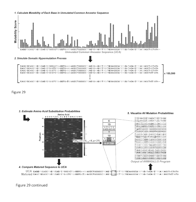
[0057] Figure 29. Computational method for estimating the probability of
antibody
mutations. The probability of an amino acid substitution during B cell
maturation in the
absence of selection is estimated by simulating the somatic hypermutation
process. 1) The
inferred unmutated common ancestor sequence (UCA) of the antibody of interest
is assigned
mutability scores according to a statistical model of AID targeting. 2) Bases
in the sequence are
then drawn randomly according to these scores and mutated according to a base
substitution
model (see Example 1). Rounds of single base mutation continue for the number
of mutations
observed in the antibody of interest with mutability scores updated as the
simulation proceeds.
The simulation is then repeated 100,000 times to generate a set of synthetic
matured sequences.
3) An amino acid positional frequency matrix is constructed from the simulated
sequences and
14
CA 03039089 2019-04-01
WO 2018/067580
PCT/US2017/054956
utilized to estimate the probability of amino acid substitutions. 4) The UCA
and matured
sequence are aligned and 5) the estimated probability of amino acid
substitutions identified in
the matured sequence are outputted.
[0058] Figures 30A-C. Improbable mutations confer heterologous neutralization
in bnAb
development. BnAbs A) CH235, B) VRCO1 and C) BF520.1 and their corresponding
mutants
with reverted improbable mutations were tested for neutralization against
heterologous viruses.
The reversion of improbable mutations in all three bnAbs diminished
neutralization potency.
[0059] Figures 31A-B. BnAbs are enriched for improbable antibody mutations.
(A) Table
of improbable mutations for a representative set of bnAbs (B) Histogram for
the distributions of
number of improbable mutations from antibody heavy chain sequences from three
groups:
"RV144-induced" antibodies were isolated from RV144 vaccinated subjects by
antigenically
sorting with RV144 immunogens (red shaded area); "Uninfected" antibodies
correspond to
duplicated NGS reads from IgG antibodies isolated from PBMC samples from 8 HIV-
uninfected
individuals (blue shaded area; see methods for details on sampling); a
representative set of
published bnAb antibody sequences are shown labeled above dotted lines that
correspond to
their number of improbable mutations (at the <2% level).
[0060] Figure 32. Mutation Guided Lineage Design Vaccine Strategy. Improbable
mutations
can act as important bottlenecks in the development of bnAbs and we propose
here a strategy to
specifically target those mutations for selection through vaccination. First,
for a specific bnAb
lineage, low probability mutations are identified computationally and
recombinant antibody
mutants corresponding to these mutations are produced (top panel). Binding and
neutralization
assays are performed to validate which of the improbable mutations are
functionally important
for lineage development (middle panel, left) and Envs are chosen that can
specifically bind the
corresponding antibody mutants (middle panel, right). These Envs are then used
in a sequential
immunization regimen to select the most difficult-to-induce, critical
mutations thus potentially
alleviating key bottlenecks in bnAb elicitation.
[0061] Figures 33A-B. ARMADiLLO output for DH270 heavy chain shows G57R
mutation
is improbable. (A) ARMADiLLO output for the DH270 heavy chain. The first three
rows of
each block corresponds to the DH270 UCA sequence and the following four rows
correspond to
the matured DH270 sequenced. The first row is the amino acid sequence for the
DH270 UCA.
The second row is the amino acid numbering (consecutively numbered starting at
1 for the first
residue) for the DH270 UCA. The third row is the nucleotide sequence with each
codon falling
under the amino acid designated in row 1. The mutability score calculated with
the S5F model is
shown below the base in each box in this row. Each box is highlighted at AID
hot spots (red;
mutability score>2) and cold spots (blue; mutability score <0.3). Row 8 is the
estimated
CA 03039089 2019-04-01
WO 2018/067580
PCT/US2017/054956
probability of the amino acid observed in the matured sequence (see methods
for how this is
calculated). The formatting pattern of rows 1-3 is repeated for the matured
DH270 in rows 4-7.
Amino acid substitutions are highlighted in yellow in row 4. Nucleotide
mutations are shown in
dark red text. Nucleotide mutations that are the result of mutations at AID
cold spots are shown
with an arrow below. (B) ARMADiLLO output for the VH chain of antibody CH235.
[0062] Figures 34A-C. Neutralization of improbable mutation reversion mutants
for
CH235, VRC01, and BF520.1. Curves of the percent neutralization of WT (red
line) A)
CH235 B) VRCO1 and C) BF520.1 and mutants containing reversions of identified
improbable
mutations against heterologous and autologous (CH505 T/F and 4501dG5 for CH235
and
VRC01, respectively) viruses. 50% neutralization is denoted by a dotted line.
[0063] Figures 35A-D. K19T mutation is conserved across all VH1-46 derived
bnAb
lineages and T19 position is proximal to N197 glycan site
[0064] A) Amino acid multiple sequence alignment of the heavy chains of the
three known
VH1-46 gene segment-derived CD4 binding site bnAbs: 8ANC131, 1B2530, and the
multiple
member CH235 lineage aligned to the CH235 UCA. The K19T mutation (red) is
observed in all
three lineages suggesting convergence of this mutation in three distinct
individuals. Dots denote
an amino acid match with the CH235 UCA in that position. B) The T19 position
(magenta) in
the CH235/gp120 complex structure (PDB: 5F9W) is outside of the CH235 (heavy
chain, blue;
light chain, gray) binding site. The complex structure was determined with
monomeric gp120
(green) and only minimal glycosylation (not shown) was resolved. C)
Superposition of the
CH235 complex onto a fully glycosylated SOSIP trimer (5FYL) revealed that T19
(magenta) is
in close proximity (7A) to the N197 glycan base (red) resolved in the trimer
structure (green). A
longer Lys residue in the 19th position may sterically clash with longer
glycans, providing a
structural rationale for the conservation of the Kl9T mutation in VH1-46
derived CD4 binding
site bnAbs. D) SPR sensorgrams for wildtype CH235 UCA and 5 UCA mutants
containing
improbable mutations show binding response to M5, a gp120 construct featuring
a single amino
acid mutation from the CH505 T/F that makes it more favorable for binding the
CH235.UCA.
[0065] Figures 36A-C. Representative bnAb sequences colored by mutation
probability.
Figure 36A shows Heavy chain sequences for a representative set of bnAbs are
highlighted by
their mutation probability as estimated by ARMADiLLO. UCA inference was
performed with
only the observed bnAb sequence as input and as such there may be substantial
uncertainty in
mutation calls within the CDR3s. Figure 36B shows Kappa chain sequences for a
representative set of bnAbs are highlighted by their mutation probability as
estimated by
ARMADiLLO. UCA inference was performed with only the observed bnAb sequence as
input
and as such there may be substantial uncertainty in mutation calls within the
CDR3s. Figure
16
CA 03039089 2019-04-01
WO 2018/067580
PCT/US2017/054956
36C shows Lambda chain sequences for a representative set of bnAbs are
highlighted by their
mutation probability as estimated by ARMADiLLO. UCA inference was performed
with only
the observed bnAb sequence as input and as such there may be substantial
uncertainty in
mutation calls within the CDR3s. Figures 36A, 36B and 36C use the following
legend:
Positions having black outline show mutations from the UCA sequences, and
among these are
mutations that are expected to occur frequently in the absence of selection
(high probability
mutations). Mutations that are expected to occur rarely in the absence of
selection (improbable
mutations) are colored in shades of gray: Black background, White Lettering:
<0.1%; Gray
background, White lettering: <1%; Gray background, Black lettering: <2%. Amino
acids
residing in CDRs are denoted with a line above them. The VH and VL sequences
in Figure 36
show a polypeptide sequence which comprises all improbable mutations with
probability of less
than 2%, less than 1%, or less than 0.1%. The invention contemplates
embodiments, wherein
the VH and VL polypeptide sequence(s) comprise any one of the improbable
mutations, or any
combination of the improbable mutations. In these embodiments wherein fewer
than all
improbable positions are changed to improbable mutation(s), any improbable
mutation position
could comprise an amino acid found in the UCA, or any other suitable amino
acid, for example
but not limited to an amino acid expected to occur frequently, or an amino
acid which is found at
the corresponding position of another lineage member.
[0066] Figures 37A-C. BnAbs have high mutation frequencies and mutation
frequency is
correlated with improbable mutations. A) Histograms for the distributions of
number of
improbable mutations (A) and mutation frequency (B) from antibody heavy chain
sequences
from three groups: "RV144-induced" antibodies were isolated from RV144
vaccinated subjects
by antigenically sorting with RV144 immunogens (red shaded area); "Uninfected"
antibodies
correspond to duplicated NGS reads from IgG antibodies isolated from PBMC
samples from 8
HIV-uninfected individuals (blue shaded area; see methods for details on
sampling); a
representative set of published bnAb antibody sequences are shown labeled
above dotted lines
that correspond to their mutation frequency (defined as total number of amino
acid mutations in
non-CDRH3 VDJ sequence divided by non-CDRH3 VDJ sequence length). Scatterplots
of B)
number of improbable mutations versus amino acid mutation frequency for 7588
NGS reads
from uninfected IgG antibodies from PBMC samples from 8 HIV-uninfected
individuals and C)
number of improbable mutations versus number of probable mutations (>2%).
Number of
improbable mutations was moderately correlated with number of probable
mutations (Pearson's
r=0.43). A stronger correlation was observed between improbable mutations and
mutation
frequency (Pearson's r=0.67) as expected because probable mutations are a
subset of the total
17
CA 03039089 2019-04-01
WO 2018/067580
PCT/US2017/054956
amino acid mutations used to calculate amino acid mutation frequency. Jitter
added in order to
alleviate over-plotting in panel C.
[0067] Figure 38 shows neutralization of bnAbs and mutants.
[0068] Figure 39A and 39B show the number of amino acid mutations and mutation
frequencies.
[0069] Figure 40 shows that hot spots are not uniformly distributed.
[0070] Figure 41 shows amino acid sequences of envelopes
CH848.3.D0949.10.17chim.6R.DS.SOSIP.664 and
CH848.3.D0949.10.17chim.6R.DS.SOSIP.664 N301A. The underlined sequence is the
signal
peptide in these envelopes. A skilled artisan can readily determine nucleic
acid sequences which
correspond to these amino acid sequences. These nucleic acid sequences could
be optimized for
expression is any suitable system.
[0071] Figure 42 shows Ramos B cells expressing broadly neutralizing antibody
UCA B cell
receptors.
DETAILED DESCRIPTION
[0072] During the development of bnAbs, B-cells undergo an evolutionary
process in order to
achieve high specificity recognition of antigen and this process is called
affinity maturation. As
with all evolutionary processes, there is diversification and selection. There
are two primary
diversification methods in that process. The first is the initial V(D)J
recombination event. This
defines the starting point for a clonal lineage. The second is somatic
hypermutation (SHM)
which is discussed in more detail. Somatic hypermutation is the process which
introduces
mutations within the antibody gene.
[0073] Selection of the survival of B cells that have undergone somatic
hypermutation is based
on affinity to antigen. This manifests as a competition with other B-cells in
the germinal center.
Somatic Hypermutation is mediated by Activation-Induced Cytidine Deaminase or
A.I.D.
[0074] Clonal lineages of antibodies trace the history of a clone as its
members acquire
mutations. Clonal lineages can be displayed as trees. Trees are rooted on the
initial VDJ
rearrangements and heavy and light chain pairing, which is referred as the
unmutated common
ancestor or UCA. A fundamental goal of HIV-1 vaccine development is to
recapitulate the
response infrequently observed in HIV-1 infection: that is the induction of
exquisitely potent,
broadly neutralizing antibodies.
[0075] To recapitulate the induction of a specific antibody lineage, at least
two essential
components are needed. First is to engage naive B cells with the germline-
encoded
characteristics important for neutralization of the lineage. In some
embodiments this is the same
18
CA 03039089 2019-04-01
WO 2018/067580
PCT/US2017/054956
heavy and light pairing. In other embodiments, this is the same signature
contact residues that
are encoded in a V gene segment. In other embodiments, this is a similar CDR
H3. In some
embodiments, this is any combination of those germline-encoded features. After
UCA is
engaged, it is long way to go to becoming a broad neutralizing antibody
(bnAb). In that process,
the UCA must now traverse the mutational space to acquire breadth and potency.
[0076] Second, after a lineage is initiated, it must accrue the specific,
critical somatic mutations
that are necessary for that lineage to acquire desired characteristics, e.g.
but not limited to
neutralization breadth. The mutational space could be visualized as a maze,
and the UCA and
subsequent intermediates must make the correct turns through the maze, by
making the right
mutations. Many of the paths will be off-target and lead to dark alleys and
dead ends. And
there will be forces that can steer the clone into these dark alleys such as
non-deletional modes
of immune tolerance referred to as "affinity reversion" or "antibody
redemption". Even when a
successful path is found, it may represent a subdominant part of the lineage.
[0077] A clonal lineage tree, when available, thus acts as a map, defining the
mutational
pathway that leads a UCA to mature to a BNAb. Such maps could be used to
recapitulate this
phenomenon in the vaccine setting. A key question in evaluating vaccine
induced lineages to
determine if lineages are on the right path to becoming a BNAb. Related to
that is to determine
if maturation is going off-target towards a dead-end.
[0078] Traditionally this is done by assessing whether the vaccinated lineages
share
commonalities with known BNAb lineages; whether they share heavy and light
chain gene
segment usage; whether they share mutations at the same positions; whether
these are positions
at contact sites in the complex; whether the lineages share mutations at the
same position, and
whether the change is to the same exact amino acid. However, evaluating shared
mutations does
not take into account an important factor¨namely that is the somatic
hypermutation process is
biased.
[0079] AID targeting is not uniformly random, it shows a preference towards
certain
microsequence motifs, called "hotspots", and away from other motifs called
"coldspots". Base
substitution is also dependent on the surrounding sequence. So this must be
accounted for when
comparing lineage members to BNAb sequences. Some mutations will occur in hot-
spots and
are more readily available prior to selection than mutations that occur in
cold-spots. This bias is
evident when the pattern of hot spots in V gene segments is analyzed. Figure
40 shows a plot
of mutability scores for VH1-2*02. This figure shows that the hot spots are
not uniformly
distributed. They occur in the CDR loop regions and mostly away from framework
regions as
expected. However, there are areas, especially in framework 3, that have more
hot spots than
one might expect. The result is that mutations tend to accrue where these hot
spots are enriched.
19
CA 03039089 2019-04-01
WO 2018/067580
PCT/US2017/054956
The figure shows the pattern plotted at the nucleotide level, but how that
manifests at the codon
level and how the hot spots may change as the antibody gene becomes more
mutated, will have
an effect on the pattern at the amino acid level as the clone matures.
[0080] For these analyses it would be useful to calculate the probability of
individual amino acid
mutations, not only for comparing lineages, but also for evaluating
bottlenecks in BNAb
developmental pathways. One such pathway is the one described in a lineage of
HIV-1 bnAb
referred to as DH270 lineage (Example 2).
[0081] To determine the probability of any amino acid at any position at a
given mutation
frequency three things are needed. We need the starting point, the UCA
sequence; and the
number of mutations in the observed mature sequence. This will define the
number of
opportunities the antibody has to get that specific mutation. Also needed is a
method for
simulating somatic hypermutation in the absence of selection. To do that
simulation and that
calculation, the invention provides a program called ARMADILLO, which stands
for Antigen
Receptor Mutation Analyzer for Detection of Low Likelihood Occurrences.
ARMADILLO
simulates the somatic hypermutation process using a statistical model of AID
targeting and
substitution, and estimates the probability of any observed amino acid
mutation in a matured
antibody sequence. It highlights those mutations that are improbable, prior to
selection. Both
heavy and light antibody chains could be analyzed by ARMADILLO. One
statistical model of
SHM is described by Yaari et al. in "Models of somatic hypermutation targeting
and substitution
based on synonymous mutations from high-throughput immunoglobulin sequencing
data." In
Front Immunol. 2013 Nov 15;4:358. doi: 10.3389/fimmu.2013.00358. eCollection
2013. The
model of Yaari et al. could be improved, and other models could also be used.
[0082] ARMADILLO can be used to retrospectively confirm an improbable, yet
critical
mutation. For a non-limiting embodiment see Example 2, and the output of the
program for the
V3 antibody DH270 (Figure 33). Zooming in on the G57R mutation in the
DH270.IA4
(Example 2), the top three rows show the UCA sequence. The program shows the
amino acid
Glycine (point) at position 57 (point) has the specific bases GGC in its codon
(point) and
highlights hotspots in red and cold spots in blue. The next three rows show
the mature DH270
sequence, highlighting in yellow that an amino acid substitution to Arginine
has occurred, and
that was the result of a mutation at a base that was in a cold spot. The
number in the last row,
here highlighted in magenta is the probability of this mutation occurring in
the absence of
selection, and this probability is 0.5%. And as Example 2 shows, this
improbable mutation was
critical to the acquisition of heterologous breadth and occurred early in the
DH270 lineage.
[0083] Having confirmed that ARMADILLO can be used retrospectively at the
DH270 lineage
and identify and quantify an improbable mutation important for the development
of that lineage,
CA 03039089 2019-04-01
WO 2018/067580
PCT/US2017/054956
the next step was to use it prospectively to predict important mutations based
on mutation
probabilities. For that we turned to the CH235 lineage that is a CD4 binding
site antibody
lineage, and the mature antibody CH235.12 in that lineage (lineage is from
patient CH505). See
Gao et al. Cell (2014) Volume 158, Issue 3,31 July 2014, Pages 481-491
Bonsignori et al. Cell
(2016) Volume 165, Issue 2, p449-463, 7 April 2016. Figure 36A shows the
ARMADILLO
output for the VH chain of antibody CH235.
[0084] Figure 35 shows the mapping of the contact sites from the crystal
structure of CH235.12
antibody. This figure shows that there was an improbable mutation that
occurred in Framework
1 that was not in a contact site. This mutation was Lysine to Threonine,i.e. K
to T. A sequence
alignment of the CH235 clone with two other VH1-46 derived BNAbs that are also
CD4 mimics,
8ANC131 and 1B2530, and showed, remarkably, that they both had the same exact,
improbable
mutation. And all but one member of the CH235 clone did as well. The CH235
structure
showed that this amino acid T19, was far from the antigen binding site in the
complex with
monomeric gp120 core. However, when we superposed the CH235 complex into a
recently
solved glycosylated trimer structure, it revealed a different story. The K19T
mutation position is
very close to the N197 glycan, a glycan that occurs in the V2 that is missing
in the gp120 core.
That led us to ask whether the role of this mutation is to accommodate the
N197 glycan. The
reversion mutation, Ti 9K, was made in CH235 and tested for neutralization.
While it had only a
marginal reduction in CH505 T/F neutralization, there was a loss of
neutralization of two tier
two viruses. So this single mutation reduced heterologous breadth. There was
no effect with
JRFL neutralization, likely because JFRL lacks the N197 glycan site. These
results demonstrate
that using the methods of the invention one can prospectively find
functionally relevant,
improbable mutations.
[0085] That we can estimate the probability of mutations along BNAb pathways,
and
successfully utilize that information to identify candidate mutations that are
critical to the
acquisition of breadth, leads us to propose the following immunization
strategy. (1) First,
identify the set of improbable mutations in the BNAb lineage that we are
trying to recapitulate.
(2) We then make those antibody mutants, and (3) functionally validate their
importance in the
lineage by testing for improvement in binding and neutralization breadth. (4)
Then, we choose
Envs that preferentially bind those improbable and important mutations. (5)
Finally we
immunize with those Envs in ascending order of the probability of mutations
for which we want
to select. These envelopes are expected to lead the clone to mature by
specifically selecting for
the hardest mutations to arise, while the clone makes the highly probable
mutations.
[0086] In some embodiments of the invention, each mutation has a probability
so ascending
order of that probability is a ranking. In some embodiments, the methods
identify the mutations
21
CA 03039089 2019-04-01
WO 2018/067580
PCT/US2017/054956
that have an effect on binding or neutralization. In some embodiments, the
methods first filter
mutations by probability, wherein to test functionally 10 mutants one selects
the ten lowest
probability mutants. Without bound by theory, not every tested mutation is
expected to have
functional effect on neutralization and/or binding. In some embodiments, the
mutations are
picked for analyses in ascending order of probability. In some embodiments, if
only few, e.g. 3,
could be tested for practical reasons, use the lowest 3 of the 5 in order. In
some embodiments
the methods also weigh the probability score by the frequency observed in the
clone if there are
multiple clonal members isolated. In non-limited embodiments, timing of
mutation
(earliness/lateness of mutation) occurrence within a clone is associated with
frequency/infrequency in the clone because of the way phylogenetic tree
inference is
constructed. In some embodiments the methods also weigh mutation occurrence in
the
phylogenetic tree.
[0087] In certain aspects, the invention provides methods of identifying and
selecting antigens,
e.g. but not limited to HIV-1 envelopes, that preferentially bind antibodies
with identified
improbable and important mutations, wherein these selected antigens are used
as immunogens.
which are expected to direct maturation of an antibody clone for example but
not limited to
having broad neutralization properties.
[0088] In certain embodiments an antibody or fragment thereof comprising
functional
mutation(s) binds specifically or preferentially to a particular target,
peptide, or polysaccharide
(such as an antigen present on the surface of a pathogen, for example gp120,
gp41), even where
the specific epitope may not be known, and do not bind in a significant amount
to other proteins
or polysaccharides present in the sample or subject. Specific binding between
and antibody and
an antigen can be determined by methods known in the art. Various binding and
screening
assays to isolate antigens which bind to an antibody with a functional
mutation(s), including
competitive binding assays, quantitative binding assays are known in the art.
Non-limiting
examples of such assays include phage display screening, ELISA, protein
arrays, etc. Antigens
can also be identified using phage display techniques. Such techniques can be
used to isolate an
initial antigen or to generate variants with altered specificity or avidity
characteristics. Various
techniques for making mutational, combinatorial libraries to generate diverse
antigens are known
in the art. Single chain Fv comprising the functional mutation(s) can also be
used as is
convenient. A skilled artisan appreciates that an antigen does not have to
bind exclusively to an
antibody with a specific functional mutation (e.g. X1), but that the antigen
could bind
preferentially or in some way detectably different to the antibody with
mutation X1 compared to
another antibody, for example to the UCA.
22
CA 03039089 2019-04-01
WO 2018/067580
PCT/US2017/054956
[0089] Antigens can be tested functionally for calcium flux, for example using
Ramos cell lines
expressing B cell receptors of desired specificity.
[0090] With reference to an antibody antigen complex, in certain embodiments
specific binding
of the antigen and antibody has a Kd of less than about 106 Molar, such as
less than about 106
Molar, 107 Molar, 108 Molar, 109, or even less than about 1010 Molar. With
reference to an
antibody antigen complex, in certain embodiments specific binding of the
antigen and antibody
has a detectably different Kd. Kd measurements of antibody binding to HIV-1
envelope, e.g.
gp41 or any other suitable peptide for the MPER antibodies, will be determined
by Surface
Plasmon Resonance measurements, for example using Biacore, or any other
suitable technology
which permits detection of interaction between two molecules in a quantitative
way.
[0091] The improbable mutation analysis is applicable to other antibodies
other than HIV-1
antibodies. For example, the analysis was conducted for a neutralizing flu
antibodies.
Improbable mutations were identified, and these are tested to determine their
effect on the
neutralization of the reverted antibody
[0092] A skilled artisan appreciates that the analysis identifying improbable
mutations is
applicable to other antibodies other than HIV-1 antibodies, for example but
not limited to flu
antibodies.
[0093] Antibody nomenclature and names: UCA4 = DH270.UCA; IA4 = DH270.IA4; IA3
=
DH270.IA3; IA2 = DH270.IA2; IA1 = DH270.IA1; DH270 = DH270.1; DH473 = DH270.2;
DH391 = DH270.3; DH429 = DH270.4; DH471 = DH270.5; DH542 = DH270.6; DH542-L4
(comprising VH from DH542 and VL from DH429), DH542_QSA.
EXAMPLES
[0094] The following specific examples are to be construed as merely
illustrative, and not
limitative of the remainder of the disclosure in any way whatsoever. Without
further elaboration,
it is believed that one skilled in the art can, based on the description
herein, utilize the present
invention to its fullest extent.
Example 1: Functional Improbable Antibody Mutations Critical for HIV Broadly
Neutralizing Antibody Development
[0095] HIV-1 broadly neutralizing antibodies (bnAbs) require high levels of
activation-induced
cytidine deaminase (AID) catalyzed somatic mutations for optimal
neutralization potency.
Probable mutations occur at sites of frequent AID activity, while improbable
mutations occur
where AID activity is infrequent. One bottleneck for induction of bnAbs is the
evolution of viral
envelopes (Envs) that can select bnAb B cell receptors (BCR) with improbable
mutations. Here
we define the probability of bnAb mutations and demonstrate the functional
significance of
23
CA 03039089 2019-04-01
WO 2018/067580
PCT/US2017/054956
improbable mutations in bnAb development. We show that bnAbs are enriched for
improbable
mutations, thus their elicitation will be critical for successful vaccine
induction of potent bnAb B
cell lineages. We outline a mutation-guided vaccine strategy for
identification of Envs that can
select B cells with BCRs with key improbable mutations required for bnAb
development. Our
analysis suggests that through generations of viral escape, Env trimers
evolved to hide in low
probability regions of antibody sequence space.
[0096] The goal of HIV-1 vaccine development is the reproducible elicitation
of potent, broadly
neutralizing antibodies (bnAbs) that can protect against infection of
transmitted/founder (TF)
viruses (Haynes and Burton, 2017). While ¨50% of HIV-infected individuals
generate bnAbs
(Hraber et al., 2014), bnAbs in this setting only arise after years of
infection (Bonsignori et al.,
2016; Doria-Rose et al., 2014; Liao et al., 2013b). BnAbs isolated from
infected individuals
have one or more unusual traits, including long third complementarity
determining regions
(CDR3s) (Yu and Guan, 2014), autoreactivity (Kelsoe and Haynes, 2017), large
insertions and
deletions (Kepler et al., 2014a), and high frequencies of somatic mutations
(Burton and
Hangartner, 2016). Somatic hypermutation of the B cell receptor (BCR) heavy
and light chain
genes is the primary diversification method during antibody affinity
maturation - the
evolutionary process that drives antibody development after initial BCR
rearrangement and leads
to high affinity antigen recognition (Teng and Papavasiliou, 2007). Not all
somatic mutations
acquired during antibody maturation are necessary for bnAb development; rather
high
mutational levels may reflect the length of time required to elicit bnAbs
(Georgiev et al., 2014;
Jardine et al., 2016). Therefore, shorter maturation pathways to
neutralization breadth involving
a critical subset of mutations is desirable, but vaccine design to achieve
this goal requires a
strategy to determine all key mutations (Haynes et al., 2012).
[0097] Mutation during antibody affinity maturation, like all evolutionary
processes, occurs
prior to selection and the principal mutational enzyme is activation-induced
cytidine deaminase
(AID) (Di Noia and Neuberger, 2007). AID preferentially targets nucleotide
sequence motifs
(referred to as "AID hot spots") or is shielded away from certain nucleotide
motifs (referred to
as "AID cold spots") (Betz et al., 1993; Yaari et al., 2013) and subsequent
repair of DNA lesions
results in a bias for which bases are substituted (Cowell and Kepler, 2000).
The result of this
non-uniformly random mutation process is that specific amino acid
substitutions occur with
varying frequencies prior to antigenic selection. Mutations at hot spots can
occur frequently in
the absence of antigen selection due to immune activation-associated AID
activity (Bonsignori et
al., 2016; Yeap et al., 2015). Improbable amino acid substitutions generally
require strong
antigenic selection to arise during maturation. Amino acid substitutions can
be improbable prior
24
CA 03039089 2019-04-01
WO 2018/067580
PCT/US2017/054956
to selection for two primary reasons: 1) base mutations must occur at AID cold
spots, or 2) due
to codon mapping, multiple base substitutions must occur for a specific amino
acid change to
take place. We have recently described a rare mutation in a bnAb unmutated
common ancestor
antibody (UCA) that only occurred when a virus bearing a distinct Env arose
three years after
HIV-1 infection (Bonsignori et al., 2017). Thus, the requirement for rare,
functional bnAb
mutations can be a key roadblock in HIV-1 bnAb development. Without being
bound by theory,
the invention provides that roadblocks are a general problem and thus a
frequent barrier in the
elicitation of bnAbs. Here we describe the identification of improbable
mutations in multiple
bnAb B cell lineages, determine the functional relevance of these mutations
for development of
bnAb potency, and outline a vaccine design strategy for choosing sequential
Envelopes capable
of selecting B cells with BCRs with improbable mutations.
[0098] Identification of Functional Improbable Antibody Mutations
[0099] To determine the role of rare mutational events in bnAb development, we
developed a
computational program to identify improbable antibody mutations. Our program,
Antigen
Receptor Mutation Analyzer for Detection of Low Likelihood Occurrences
(ARMADiLLO)
simulates the somatic hypermutation process using a statistical model of AID
targeting and base
substitution via DNA repair (Yaari et al., 2013) and estimates the probability
of any amino acid
substitution in an antibody based on the frequencies observed in the
computational simulation
(Figure 29).
[0100] First, we applied ARMADiLLO retrospectively to the analysis of a
mutation in a bnAb
lineage that occurred at an AID cold spot that we have previously shown was
functionally
important for neutralization (Bonsignori et al., 2017). The DH270 V3-glycan
bnAb lineage
developed a variable heavy chain (VII) complementary determining region 2 (CDR
H2) G57R
mutation that when analyzed with the ARMADiLLO program was predicted to occur
with <1%
frequency prior to selection (Figure 33). This mutation was functionally
critical because
reversion back to G57 in the DH270 bnAb lineage resulted in total loss of
neutralization potency
and breadth. See Example 2 and W02017/152146, the contents of which is hereby
incorporated
by reference in its entirety; see also (Bonsignori et al., 2017). Thus, the
ARMADiLLO program
can identify a known, key improbable mutation.
[0101] All BCR mutations arise during the stochastic process of somatic
hypermutation prior to
antigenic selection. In HIV-1 infection, antibody heterologous breadth is not
directly selected
for during bnAb development because BCRs only interact with autologous virus
Envs. Since
improbable bnAb mutations can confer heterologous breadth, they represent
critical events in
bnAb development, and make compelling targets for focusing selection with
immunogens. To
CA 03039089 2019-04-01
WO 2018/067580
PCT/US2017/054956
test this hypothesis, we analyzed three bnAb lineages with ARMADiLLO to
identify improbable
mutations (in one non-limiting embodiment defined as <2% estimated probability
of occurring
prior to selection) and then tested for the effect of these mutations on bnAb
neutralization during
bnAb B cell lineage development. Figure 39 shows the counts for different
probability cut off
values, e.g. a 1% probability cutoff and for 0.1%. If the cutoff is lowered,
the counts are
lowered. A skilled artisan appreciates that the cutoff doesn't change the
overall strategy, but
simply affects the number of mutations that would be considered functionally
important. The
goal is to select the mutations that are the most important (i.e. functional)
for heterologous
neutralization and least likely to occur. Without being bound by theory, the
2% cutoff was
chosen as it is expected to include mutations that are functionally important.
We chose three
lineages that allowed for study of different levels of maturation in bnAb
development: CH235,
mid-stage bnAb development (Bonsignori et al., 2017); VRC01, late stage bnAb
development
(Wu et al., 2015); and BF520.1, early stage bnAb development in an infant
(Simonich et al.,
2016).
[0102] Improbable Mutations Confer Heterologous Neutralization in CD4 Binding
Site
bnAb Lineages
[0103] CH235 is a CD4-binding site, CD4-mimicking (Gao et al., 2014) bnAb B
cell lineage
that evolved to 90% neutralization breadth and high potency over 5-6 years of
infection and
acquired 44 VII amino acid mutations (Bonsignori et al., 2016). We identified
improbable
mutations in the heavy chain of an early intermediate member of the lineage
(also termed
CH235), reverted each to their respective germline-encoded amino acid, and
then tested each
CH235 antibody mutant for neutralization against the heterologous, difficult-
to-neutralize (tier
2) (Seaman et al., 2010) TRO.11 virus (Figure 30A and Figure 34A). Single
amino acid
reversion mutations resulted in either a reduction or total loss of
heterologous HIV-1 TRO.11
neutralization for each of three improbable mutations, Kl9T, W47L and G5 SW
demonstrating
that improbable mutations in the CH235 lineage were indeed critical and could
confer
heterologous neutralization.
[0104] Identification of the Kl9T mutation was of particular interest because
the mutation was
observed in all but one member of the CH235 bnAb lineage and was also present
in two other
CD4-binding site bnAbs (Scheid et al., 2011) from different individuals that
shared the same VH
gene segment as CH235 (Figure 35A). Superposition of the CH235 complex into a
fully-
glycosylated trimer (Stewart-Jones et al., 2016) showed that the K19T mutation
position was in
close proximity to the N197 glycan site on the Env trimer (Figures 35B and
35C). The K19T
mutation shortened the amino acid at this position which could act to
accommodate larger glycan
26
CA 03039089 2019-04-01
WO 2018/067580
PCT/US2017/054956
forms at the heterogeneously glycosylated N197 position (Behrens et al., 2016)
providing a
structural rationale for the effect of this mutation on heterologous breadth.
Consistent with this
hypothesis, CH235 neutralization of JR-FL, a tier 2 heterologous virus lacking
the N197 glycan
site, was unaffected by the T19K reversion mutation (Figure 38 and Figure
34A). Moreover,
we introduced the K19T mutation into the CH235 UCA and observed improved
binding to an
early autologous Env suggesting that the improbable K19T mutation may have
been selected for
by an early variant of the autologous virus (Figure 35D).
[0105] We next asked what role improbable mutations played in the maturation
of a highly
broad and potent second CD4 binding site-targeting bnAb lineage, termed VRC01,
that acquired
43 amino
acid mutations (Zhou et al., 2010). We reverted improbable mutations in the
fully
matured VRC01 and tested for their effects on neutralization of the
heterologous tier 2 HIV-1
JR-FL (Figure 30B). Reversion of improbable mutations reduced potency of
heterologous
neutralization of HIV-1 JRFL demonstrating that in the VRCO1 CD4 binding site
B cell lineage,
single improbable amino acid substitutions can also have functional
consequences for
heterologous neutralization capacity. Improbable mutations identified by
ARMADiLLO in the
VRCO1 light chain showed an even larger effect on reducing neutralization than
heavy chain
mutations (Figure 38 and Figure 34B), further underscoring, along with an
atypically short
CDRL3 and a critical CDRL1 deletion (Zhou et al., 2013), the importance of
improbable events
in the maturation of the VRCO1 bnAb lineage.
[0106] An Improbable Mutation Associated with Accelerated BnAb Development
[0107] Babies are reported to develop bnAbs earlier after HIV-1 infection than
adults (Goo et
al., 2014; Muenchhoff et al., 2016). We analyzed the glycan-V3 epitope
targeting BF520.1
bnAb, isolated from an HIV-1 infected infant with many fewer mutations (12 VH
amino acid
mutations) compared to VRC01 and CH235 (Simonich et al., 2016). We identified
an
improbable mutation, N52A, located in the CDR H2 of BF520.1, reverted it to
germline, and
expressed the resultant antibody mutant (A52N). Heterologous neutralization of
the A52N
reversion mutant against tier 2 JR-FL virus was markedly reduced relative to
wildtype BF520.1
(Figure 30C). The A52N reversion mutation antibody reduced neutralization
potency for all tier
2 viruses that the BF520.1 bnAb could neutralize (Figure 38 and Figure 34C)
demonstrating
that the N52A mutation was critical to the neutralization potency of BF520.1
and suggested the
early acquisition of this improbable mutation may have played a role in the
relatively early
elicitation (<15 months) of a bnAb with limited mutation frequency. Thus, the
analysis of the
three bnAbs studied here demonstrated that the ARMADiLLO program prospectively
identified
improbable mutations in bnAbs spanning multiple epitope specificities at
distinct stages of bnAb
27
CA 03039089 2019-04-01
WO 2018/067580
PCT/US2017/054956
development and functional antibody analysis demonstrated improbable mutations
were critical
for bnAb development of neutralization breadth and potency.
[0108] Improbable Antibody Mutations Are Enriched in BnAbs
[0109] To provide a view of the scope of the problem for many bnAb B cell
lineages, we
estimated the number of improbable mutations for a representative set of known
bnAb lineages
spanning all known sites of vulnerability on the Env trimer (Figures 31A and
36, and Figure
39). Study of a representative sample of bnAb lineages is plausible because of
commonalities of
Env recognition by bnAb germline precursors (Andrabi et al., 2015; Bonsignori
et al., 2011;
Gorman et al., 2016; Zhou et al., 2013). Compared to Env-reactive antibodies
induced by an
HIV-1 vaccine candidate (the RV144 vaccine) (Rerks-Ngarm et al., 2009) or
antibodies isolated
from non-HIV-1 infected individuals (Williams et al., 2015), the broadest and
most potent HIV-
1 bnAbs had the highest numbers of improbable mutations (Figure 31B). This
result may follow
directly from the observations that bnAbs tend to be highly mutated (Figure
37A) (Burton and
Hangartner, 2016), and the number of improbable mutations an antibody
possesses is correlated
with its mutation frequency (Figure 37B) (Sheng et al., 2017). However, it is
not known why
most bnAbs are highly mutated. Recent work has shown that not all mutations in
bnAbs are
essential for neutralization activity (Jardine et al., 2016). One hypothesis
is that high mutation
frequency is due to the extended number of rounds of somatic hypermutation
required for a
lineage to acquire a specific subset of mutations (Klein et al., 2013). If
some of those specific
mutations are also improbable, it is very likely that more probable mutations
would be acquired
prior to attaining key improbable ones. We found that for many bnAbs the
number of
improbable mutations exceeded what would be expected given their high mutation
frequency
alone. This observation, along with our experimental observations
demonstrating that many
improbable mutations are important for neutralization capacity, is consistent
with the general
rule that improbable mutations act as key bottlenecks in the development of
bnAb neutralization
breadth. Thus, during chronic HIV-1 infection with persistent high viral loads
that are required
for bnAbs with improbable mutations to develop (Gray et al., 2011), excess
numbers of probable
mutations also accumulate. Probable mutations arise easily from the intrinsic
mutability of
antibody genes and unlike improbable mutations may not require Env selection
(Bonsignori et
al., 2016; Hwang et al., 2017; Neuberger et al., 1998). Thus, if the selection
of critical
improbable mutations can be targeted with Env immunogens, it should be
possible to accelerate
bnAb maturation and result in the induction of bnAb lineages with fewer
mutations than those
that occur in the setting of chronic HIV-1 infection.
[0110] Implications for Vaccine Design
28
CA 03039089 2019-04-01
WO 2018/067580
PCT/US2017/054956
[0111] The ability to identify functional improbable bnAb mutations using the
ARMADiLLO
program and antibody mutation functional studies informs a mutation-guided
vaccine design and
immunization strategy (Figure 32). The principal goal is to be able to choose
the correct
sequential Envs to precisely focus selection towards the most difficult to
induce mutations, while
allowing the easier, more probable mutations to occur due to antibody
intrinsic mutability from
immune activation-associated AID activity. In this strategy, improbable
mutations are identified
computationally using the ARMADiLLO program. Next, all improbable mutations
identified
are expressed as single amino acid substitution mutant antibodies and their
functional
importance validated by Env binding and neutralization assays. Envelopes that
bind with high
affinity are chosen as immunogens to select for these functional improbable
mutations. Last,
sequential immunization with the chosen immunogens are studied for
optimization of regimens
to select for B cells with BCRs with the required improbable mutations.
[0112] As expected, because improbable mutations arise as either neutral
mutations or by
selection by autologous virus, not all improbable mutations are required for
mediation of
heterologous neutralization (Figure 38). Similarly, it is important to note
that intrinsically
mutable positions (Neuberger et al., 1998) can also be capable of conferring
heterologous
breadth. In this regard we identified one such functionally important probable
intrinsic mutation
in the CH235 lineage, 557R (Figure 38). However, such highly probable
mutations, by
definition, should be easily inducible and are not likely to represent
barriers in bnAb
development.
[0113] Interestingly, bnAbs that demonstrated relatively low numbers of
improbable single
somatic mutations (Figure 31A) possessed other unusual antibody
characteristics that were due
to additional improbable events such as insertion/deletions (indels) or
extraordinary CDR H3
lengths. For example, the bnAbs with the two lowest number of improbable
mutations were
PGT128 and CAP256-VRC26.25. These bnAbs are notable for having the largest
indels
(PGT128; 11 aas) or the longest CDR H3 (CAP256-VRC26.25; 38 aas). In summary,
our data
presented here suggest Env trimers evolved to evade neutralizing B-cell
responses by hiding
within low probability regions of antibody sequence space. The ARMADiLLO
program and
mutation-guided vaccine design strategy presented here should be broadly
applicable for vaccine
design for other mutating pathogens.
[0114] Low probability mutation is the same as improbable or rare mutation.
Functional or
important mutations are improbable mutations which lead to loss of
neutralization breadth when
reverted back to a UCA amino acid.
[0115] Experimental Procedures
29
CA 03039089 2019-04-01
WO 2018/067580
PCT/US2017/054956
[0116] Analysis of the Probability of Antibody Mutations
[0117] The probability of an amino acid substitution at any given position in
the antibody
sequence of an antibody of interest was estimated using the ARMADiLLO program.
The
algorithm and the analysis performed using ARMADiLLO are described in
Supplemental
Experimental Procedures.
[0118] Antibody Site-directed Mutagenesis
[0119] BF520.1 mutant antibody genes were synthesized by Genscript and
recombinantly
produced. Mutations into antibody genes for CH235 and VRC01 mutants were
introduced using
the QuikChange II Lightning site-directed mutagenesis kit (Agilent
Technologies) following the
manufacturer's protocol. Single-colony sequencing was used to confirm the
sequences of the
mutant plasmid products. Primers used for introducing mutations are listed in
the Supplemental
Experimental Procedures.
[0120] Recombinant Antibody Production
[0121] Antibodies were recombinantly produced as previously described
(Saunders et al., 2017).
[0122] HIV-1 Neutralization
[0123] Antibody neutralization was measured in TZM-bl cell-based
neutralization assays as
previously described (Li et al., 2005; Sarzotti-Kelsoe et al., 2014). CH235
and BF520.1 and
selected mutants were assayed for neutralization using a global panel of 12
HIV-1 Env reference
strains (deCamp et al., 2014). Neutralization values are reported as
inhibitory concentrations of
antibody in which 50% of virus was neutralized (IC5o) with units in g/ml.
[0124] Antibody Binding Measurements
[0125] Binding of CH235.UCA and mutants to the monomeric CH505
transmitted/founder (T/F)
de1ta7 gp120 and monomeric CH505 M5 (early autologous virus variant) de1ta8
gp120
(Bonsignori et al., 2016; Gao et al., 2014) was measured by surface plasmon
resonance assays
(SPR) on a Biacore S200 instrument and data analysis was performed with the
S200
BIAevaluation software (Biacore/GE Healthcare) as previously described (Alam
et al., 2013;
Dennison et al., 2011).
[0126] Various other methods to determine and measure binding between an
antibody and an
antigen are known in the art and contemplated by the invention. Such methods
are used to
identify antigens which bind differentially to different antibodies such as a
UCA, and an
antibody variant having an improbable mutation(s).
[0127] REFERENCES FOR EXAMPLE 1
CA 03039089 2019-04-01
WO 2018/067580
PCT/US2017/054956
[0128] Alam, S.M., Liao, H.X., Tomaras, G.D., Bonsignori, M., Tsao, C.Y.,
Hwang, K.K.,
Chen, H., Lloyd, K.E., Bowman, C., Sutherland, L., etal. (2013). Antigenicity
and
immunogenicity of RV144 vaccine AIDSVAX clade E envelope immunogen is enhanced
by a
gp120 N-terminal deletion. J Virol 87, 1554-1568.
[0129] Andrabi, R., Voss, J.E., Liang, C.H., Briney, B., McCoy, L.E., Wu,
C.Y., Wong, C.H.,
Poignard, P., and Burton, D.R. (2015). Identification of Common Features in
Prototype Broadly
Neutralizing Antibodies to HIV Envelope V2 Apex to Facilitate Vaccine Design.
Immunity 43,
959-973.
[0130] Behrens, A.J., Vasiljevic, S., Pritchard, L.K., Harvey, D.J., Andev,
R.S., Krumm, S.A.,
Struwe, W.B., Cupo, A., Kumar, A., Zitzmann, N., etal. (2016). Composition and
Antigenic
Effects of Individual Glycan Sites of a Trimeric HIV-1 Envelope Glycoprotein.
Cell Rep 14,
2695-2706.
[0131] Betz, A.G., Rada, C., Pannell, R., Milstein, C., and Neuberger, M.S.
(1993). Passenger
transgenes reveal intrinsic specificity of the antibody hypermutation
mechanism: clustering,
polarity, and specific hot spots. Proc Natl Acad Sci U S A 90, 2385-2388.
[0132] Bonsignori, M., Hwang, K.K., Chen, X., Tsao, C.Y., Morris, L., Gray,
E., Marshall, D.J.,
Crump, J.A., Kapiga, S.H., Sam, N.E., etal. (2011). Analysis of a clonal
lineage of HIV-1
envelope V2/V3 conformational epitope-specific broadly neutralizing antibodies
and their
inferred unmutated common ancestors. J Virol 85, 9998-10009.
[0133] Bonsignori, M., Kreider, E.F., Fera, D., Meyerhoff, R.R., Bradley, T.,
Wiehe, K., Alam,
S.M., Aussedat, B., Walkowicz, WE., Hwang, K.K., etal. (2017). Staged
induction of HIV-1
glycan-dependent broadly neutralizing antibodies. Sci Transl Med 9.
[0134] Bonsignori, M., Zhou, T., Sheng, Z., Chen, L., Gao, F., Joyce, M.G.,
Ozorowski, G.,
Chuang, G.Y., Schramm, C.A., Wiehe, K., etal. (2016). Maturation Pathway from
Germline to
Broad HIV-1 Neutralizer of a CD4-Mimic Antibody. Cell 165, 449-463.
[0135] Burton, DR., and Hangartner, L. (2016). Broadly Neutralizing Antibodies
to HIV and
Their Role in Vaccine Design. Annu Rev Immunol 34, 635-659.
[0136] Cowell, L.G., and Kepler, T.B. (2000). The nucleotide-replacement
spectrum under
somatic hypermutation exhibits microsequence dependence that is strand-
symmetric and distinct
from that under germline mutation. J Immunol 164, 1971-1976.
[0137] deCamp, A., Hraber, P., Bailer, R.T., Seaman, M.S., Ochsenbauer, C.,
Kappes, J.,
Gottardo, R., Edlefsen, P., Self, S., Tang, H., etal. (2014). Global panel of
HIV-1 Env reference
strains for standardized assessments of vaccine-elicited neutralizing
antibodies. J Virol 88, 2489-
2507.
31
CA 03039089 2019-04-01
WO 2018/067580
PCT/US2017/054956
[0138] Dennison, S.M., Anasti, K., Scearce, R.M., Sutherland, L., Parks, R.,
Xia, S.M., Liao,
H.X., Gorny, M.K., Zolla-Pazner, S., Haynes, B.F., and Alam, S.M. (2011).
Nonneutralizing
HIV-1 gp41 envelope cluster II human monoclonal antibodies show polyre
activity for binding to
phospholipids and protein autoantigens. J Virol 85, 1340-1347.
[0139] Di Noia, J.M., and Neuberger, M.S. (2007). Molecular mechanisms of
antibody somatic
hypermutation. Annu Rev Biochem 76, 1-22.
[0140] Doria-Rose, N.A., Schramm, C.A., Gorman, J., Moore, P.L., Bhiman, J.N.,
DeKosky,
B.J., Ernandes, M.J., Georgiev, IS., Kim, H.J., Pancera, M., etal. (2014).
Developmental
pathway for potent V1V2-directed HIV-neutralizing antibodies. Nature 509, 55-
62.
[0141] Gao, F., Bonsignori, M., Liao, H.X., Kumar, A., Xia, S.M., Lu, X., Cai,
F., Hwang, K.K.,
Song, H., Zhou, T., etal. (2014). Cooperation of B cell lineages in induction
of HIV-1-broadly
neutralizing antibodies. Cell 158, 481-491.
[0142] Georgiev, IS., Rudicell, R.S., Saunders, K.O., Shi, W., Kirys, T.,
McKee, K., O'Dell, S.,
Chuang, G.Y., Yang, Z.Y., Ofek, G., etal. (2014). Antibodies VRC01 and 10E8
neutralize HIV-
1 with high breadth and potency even with Ig-framework regions substantially
reverted to
germline. J Immunol 192,1100-1106.
[0143] Goo, L., Chohan, V., Nduati, R., and Overbaugh, J. (2014). Early
development of
broadly neutralizing antibodies in HIV-1-infected infants. Nat Med 20, 655-
658.
[0144] Gorman, J., Soto, C., Yang, MM., Davenport, TM., Guttman, M., Bailer,
R.T.,
Chambers, M., Chuang, G.Y., DeKosky, B.J., Doria-Rose, N.A., etal. (2016).
Structures of
HIV-1 Env V1V2 with broadly neutralizing antibodies reveal commonalities that
enable vaccine
design. Nat Struct Mol Biol 23, 81-90.
[0145] Gray, ES., Madiga, MC., Hermanus, T., Moore, P.L., Wibmer, C.K., Tumba,
N.L.,
Werner, L., Mlisana, K., Sibeko, S., Williamson, C., etal. (2011). The
neutralization breadth of
HIV-1 develops incrementally over four years and is associated with CD4+ T
cell decline and
high viral load during acute infection. J Virol 85, 4828-4840.
[0146] Haynes, B.F., and Burton, D.R. (2017). Developing an HIV vaccine.
Science 355, 1129-
1130.
[0147] Haynes, B.F., Kelsoe, G., Harrison, S.C., and Kepler, T.B. (2012). B-
cell-lineage
immunogen design in vaccine development with HIV-1 as a case study. Nat
Biotechnol 30, 423-
433.
[0148] Hraber, P., Seaman, M.S., Bailer, R.T., Mascola, JR., Montefiori, D.C.,
and Korber, B.T.
(2014). Prevalence of broadly neutralizing antibody responses during chronic
HIV-1 infection.
AIDS 28, 163-169.
32
CA 03039089 2019-04-01
WO 2018/067580
PCT/US2017/054956
[0149] Hwang, J.K., Wang, C., Du, Z., Meyers, R.M., Kepler, T.B., Neuberg, D.,
Kwong, P.D.,
Mascola, J.R., Joyce, M.G., Bonsignori, M., etal. (2017). Sequence Intrinsic
Somatic Mutation
Mechanisms Contribute to Affinity Maturation of VRC01-class HIV-1 Broadly
Neutralizing
Antibodies. Proc Natl Acad Sci U S A In Press.
[0150] Jardine, J.G., Sok, D., Julien, J.P., Briney, B., Sarkar, A., Liang,
C.H., Scherer, E.A.,
Henry Dunand, C.J., Adachi, Y., Diwanji, D., etal. (2016). Minimally Mutated
HIV-1 Broadly
Neutralizing Antibodies to Guide Reductionist Vaccine Design. PLoS Pathog 12,
e1005815.
[0151] Kelsoe, G., and Haynes, B.F. (2017). Host controls of HIV broadly
neutralizing antibody
development. Immunol Rev 275, 79-88.
[0152] Kepler, TB., Liao, H.X., Alam, S.M., Bhaskarabhatla, R., Zhang, R.,
Yandava, C.,
Stewart, S., Anasti, K., Kelsoe, G., Parks, R., etal. (2014). Immunoglobulin
gene insertions and
deletions in the affinity maturation of HIV-1 broadly reactive neutralizing
antibodies. Cell Host
Microbe 16, 304-313.
[0153] Klein, F., Diskin, R., Scheid, J.F., Gaebler, C., Mouquet, H.,
Georgiev, IS., Pancera, M.,
Zhou, T., Incesu, R.B., Fu, B.Z., etal. (2013). Somatic mutations of the
immunoglobulin
framework are generally required for broad and potent HIV-1 neutralization.
Cell 153, 126-138.
[0154] Li, M., Gao, F., Mascola, JR., Stamatatos, L., Polonis, V.R.,
Koutsoukos, M., Voss, G.,
Goepfert, P., Gilbert, P., Greene, KM., et al. (2005). Human immunodeficiency
virus type 1 env
clones from acute and early subtype B infections for standardized assessments
of vaccine-
elicited neutralizing antibodies. J Virol 79,10108-10125.
[0155] Liao, H.X., Lynch, R., Zhou, T., Gao, F., Alam, S.M., Boyd, S.D., Fire,
AZ., Roskin,
KM., Schramm, C.A., Zhang, Z., etal. (2013). Co-evolution of a broadly
neutralizing HIV-1
antibody and founder virus. Nature 496, 469-476.
[0156] Muenchhoff, M., Adland, E., Karimanzira, 0., Crowther, C., Pace, M.,
Csala, A.,
Leitman, E., Moonsamy, A., McGregor, C., Hurst, J., etal. (2016).
Nonprogressing HIV-
infected children share fundamental immunological features of nonpathogenic
SIV infection. Sci
Transl Med 8, 358ra125.
[0157] Neuberger, M.S., Ehrenstein, MR., Klix, N., Jolly, C.J., Yelamos, J.,
Rada, C., and
Milstein, C. (1998). Monitoring and interpreting the intrinsic features of
somatic hypermutation.
Immunol Rev 162, 107-116.
[0158] Rerks-Ngarm, S., Pitisuttithum, P., Nitayaphan, S., Kaewkungwal, J.,
Chiu, J., Paris, R.,
Premsri, N., Namwat, C., de Souza, M., Adams, E., etal. (2009). Vaccination
with ALVAC and
AIDSVAX to prevent HIV-1 infection in Thailand. N Engl J Med 361, 2209-2220.
[0159] Sarzotti-Kelsoe, M., Bailer, R.T., Turk, E., Lin, CL., Bilska, M.,
Greene, KM., Gao, H.,
Todd, C.A., Ozaki, D.A., Seaman, M.S., etal. (2014). Optimization and
validation of the TZM-
33
CA 03039089 2019-04-01
WO 2018/067580
PCT/US2017/054956
bl assay for standardized assessments of neutralizing antibodies against HIV-
1. J Immunol
Methods 409, 131-146.
[0160] Saunders, K.O., Nicely, N.J., Wiehe, K., Bonsignori, M., Meyerhoff,
R.R., Parks, R.,
Walkowicz, WE., Aussedat, B., Wu, N.R., Cai, F., etal. (2017). Vaccine
Elicitation of High
Mannose-Dependent Neutralizing Antibodies against the V3-Glycan Broadly
Neutralizing
Epitope in Nonhuman Primates. Cell Rep 18, 2175-2188.
[0161] Scheid, J.F., Mouquet, H., Ueberheide, B., Diskin, R., Klein, F.,
Oliveira, T.Y., Pietzsch,
J., Fenyo, D., Abadir, A., Velinzon, K., etal. (2011). Sequence and structural
convergence of
broad and potent HIV antibodies that mimic CD4 binding. Science 333, 1633-
1637.
[0162] Seaman, M.S., Janes, H., Hawkins, N., Grandpre, L.E., Devoy, C., Giri,
A., Coffey, R.T.,
Harris, L., Wood, B., Daniels, M.G., etal. (2010). Tiered categorization of a
diverse panel of
HIV-1 Env pseudoviruses for assessment of neutralizing antibodies. J Virol 84,
1439-1452.
[0163] Sheng, Z., Schramm, C.A., Kong, R., Program, N.C.S., Mullikin, J.C.,
Mascola, JR.,
Kwong, P.D., and Shapiro, L. (2017). Gene-Specific Substitution Profiles
Describe the Types
and Frequencies of Amino Acid Changes during Antibody Somatic Hypermutation.
Front
Immunol 8, 537.
[0164] Simonich, C.A., Williams, K.L., Verkerke, H.P., Williams, J.A., Nduati,
R., Lee, K.K.,
and Overbaugh, J. (2016). HIV-1 Neutralizing Antibodies with Limited
Hypermutation from an
Infant. Cell 166, 77-87.
[0165] Stewart-Jones, GB., Soto, C., Lemmin, T., Chuang, G.Y., Druz, A., Kong,
R., Thomas,
P.V., Wagh, K., Zhou, T., Behrens, A.J., etal. (2016). Trimeric HIV-1-Env
Structures Define
Glycan Shields from Clades A, B, and G. Cell 165, 813-826.
[0166] Teng, G., and Papavasiliou, F.N. (2007). Immunoglobulin somatic
hypermutation. Annu
Rev Genet 41, 107-120.
[0167] Williams, W.B., Liao, H.X., Moody, M.A., Kepler, TB., Alam, S.M., Gao,
F., Wiehe,
K., Trama, A.M., Jones, K., Zhang, R., etal. (2015). HIV-1 VACCINES. Diversion
of HIV-1
vaccine-induced immunity by gp41-microbiota cross-reactive antibodies. Science
349, aab1253.
[0168] Wu, X., Zhang, Z., Schramm, C.A., Joyce, M.G., Kwon, Y.D., Zhou, T.,
Sheng, Z.,
Zhang, B., O'Dell, S., McKee, K., etal. (2015). Maturation and Diversity of
the VRC01-
Antibody Lineage over 15 Years of Chronic HIV-1 Infection. Cell 161, 470-485.
[0169] Yaari, G., Vander Heiden, J.A., Uduman, M., Gadala-Maria, D., Gupta,
N., Stern, J.N.,
O'Connor, K.C., Hafler, D.A., Laserson, U., Vigneault, F., and Kleinstein,
S.H. (2013). Models
of somatic hypermutation targeting and substitution based on synonymous
mutations from high-
throughput immunoglobulin sequencing data. Front Immunol 4, 358.
34
CA 03039089 2019-04-01
WO 2018/067580
PCT/US2017/054956
[0170] Yeap, L.S., Hwang, J.K., Du, Z., Meyers, R.M., Meng, F.L.,
Jakubauskaite, A., Liu, M.,
Mani, V., Neuberg, D., Kepler, T.B., etal. (2015). Sequence-Intrinsic
Mechanisms that Target
AID Mutational Outcomes on Antibody Genes. Cell 163, 1124-1137.
[0171] Yu, L., and Guan, Y. (2014). Immunologic Basis for Long HCDR3s in
Broadly
Neutralizing Antibodies Against HIV-1. Front Immunol 5, 250.
[0172] Zhou, T., Georgiev, I., Wu, X., Yang, Z.Y., Dai, K., Finzi, A., Kwon,
Y.D., Scheid, J.F.,
Shi, W., Xu, L., etal. (2010). Structural basis for broad and potent
neutralization of HIV-1 by
antibody VRC01. Science 329, 811-817.
[0173] Zhou, T., Zhu, J., Wu, X., Moquin, S., Zhang, B., Acharya, P.,
Georgiev, IS., Altae-
Tran, H.R., Chuang, G.Y., Joyce, M.G., etal. (2013). Multidonor analysis
reveals structural
elements, genetic determinants, and maturation pathway for HIV-1
neutralization by VRC01-
class antibodies. Immunity 39, 245-258.
[0174] Supplemental Experimental Procedures
[0175] Simulating the somatic hypermutation process
[0176] Because AID targets hot spots according to their underlying sequence
motifs, the
probability of mutations is sequence context dependent, making an analytical
computation of the
probability of a mutation in the absence of selection is all but intractable.
Instead, we take a
numerical approach via simulation. In this approach, we estimate the
probability of an amino
acid substitution by simulating the somatic hypermutation (SHM) process and
calculating the
observed frequency of that substitution in the simulated sequences.
[0177] The simulation proceeds as follows. Given a matured antibody nucleotide
sequence, we
first infer its unmutated common ancestor (UCA) sequence by a computational
tool called
Clonalyst (Kepler, 2013; Kepler et al., 2014). The UCA determines the initial
sequence and then
the differences from the UCA in the mature sequence define which positions are
mutations. In
addition, the UCA sequence is used to initially define the mutability score at
each nucleotide
position using the S5F model. The mutability score is turned into a
probability distribution that
we randomly sample from to select a nucleotide position to mutate. A
computational tool called
Cloanalyst is used to infer UCAs, so if there is one sequence one can infer
the UCA. If there are
multiple clonally related sequences, typically referred to as lineage, one can
infer a UCA using
Cloanalyst and multiple sequences may help add confidence in UCA positions
where there is
less confidence when just using one sequence, for example one sequence of a
bnAb.
[0178] In some embodiments, the availability of multiple clonally related
sequences might be
useful to inform the order of adding multiple functional mutations back to the
UCA sequence to
create intermediate antibodies used to identify antigens which would drive the
selection of a
CA 03039089 2019-04-01
WO 2018/067580
PCT/US2017/054956
functional mutation(s). Without the availability of clonally related
sequences, the order of
adding multiple functional mutations back to the UCA is determined
experimentally. With
reference to Figure 32 (Mutation Guided Lineage Design Vaccine Strategy), a
clonal lineage
would be informative to determine the order of mutation 1, mutation 2, etc.
[0179] For the analysis in this example, one mature sequence was inputted to
Cloanalyst to infer
the UCA, in order to put all BNAbs on the same playing field. A skilled
artisan appreciates that
an inferred UCA is likely not truly correct, unless it has been observed. In
all instances in this
example, the UCAs are inferred. So there is uncertainty in the inference. The
effect of the
uncertainty on ARMADiLLO is that if the wrong base is called in the UCA, it
would potentially
affect the mutability score which affects the random targeting of positions
for mutation
[0180] Next, the matured antibody nucleotide sequence is aligned to the UCA
nucleotide
sequence and the number of sites mutated, t, is computed. Starting with the
UCA sequence, first
(1) the mutability score of all consecutive sequence pentamers is computed
according to the 55F
mutability model (Yaari et al., 2013).
[0181] Second (2) The mutability scores for each base position in the sequence
are converted
into the probability distribution, Q, by:
ci
Qi [1]
L4=1 `q
[0182] where Ci is the mutability score at position i and L is the length of
the sequence. 3) A
base position, b, is drawn randomly according to Q. 4) The nucleotide n, at b,
is substituted
according to the 55F substitution model (Yaari et al., 2013), resulting in
sequence Si where j is
the number of mutations accrued during the simulation. The procedure then
iterates over steps
1-4 until j=t. This results in a simulated sequence, St, that has acquired the
same number of
nucleotide mutations as observed in the matured antibody sequence of interest.
If at any
iteration during the simulation a mutation results in a stop codon, that
sequence is discarded and
the process restarts from the UCA sequence. This simulation procedure is then
repeated to
generate 100,000 simulated matured sequences. These nucleotide sequences are
then translated
to amino acid sequences.
[0183] Estimating the probability of an amino acid substitution
[0184] The estimate of the probability of any amino acid substitution U ¨>Y at
site i given the
number of mutations t observed in the matured sequence of interest is then
calculated as the
amino acid frequency observed at site i in the simulated sequences according
to:
13 (Xi u,y1UCA, t) =¨N11(Xii =Y) [2]
i=o
36
CA 03039089 2019-04-01
WO 2018/067580
PCT/US2017/054956
[0185] where Xi is the amino acid at site i which has the amino acid U in the
UCA sequence
mutating to amino acid Y in the matured sequence of interest, UCA is the UCA
sequence, N is
the number of simulated sequences, 1 is an indicator function for observing
amino acid Y at site
i in the jth simulated sequence. This estimate is for an amino acid
substitution in the absence of
selection and we use this probability as a gauge of how likely it is that a B
cell would arise to
have this mutation prior to antigenic selection. Amino acid substitutions that
are the result of
mutations that occur in AID hot spots will have high probabilities, occur
frequently and a subset
of the reservoir of B cell clonal members would likely have these mutations
present prior to
antigenic selection. Amino substitutions that are the result of cold spot
mutations or require
multiple base substitutions will be much less frequent and could represent
significant hurdles to
lineage development and these substitutions may require strong antigenic
selection to be
acquired during B cell maturation.
[0186] Improbable mutations
[0187] The probability of a specific amino substitution at any given position
is the product of
two components. The first component is due to the bias of the AID enzyme in
targeting that
specific base position and the DNA repair mechanisms preference for
substituting to an
alternative base. Practically speaking, substitutions that require mutations
at AID cold spots
and/or result in disfavored base substitutions by DNA repair mechanisms are
infrequent and thus
improbable. The second component is the number and length of available paths
through codon
space to go from an amino acid encoded by the codon in the UCA to that of the
codon for the
substituted amino acid in the matured sequence. To illustrate this, we turn to
a practical example:
the TAT codon which encodes the amino acid, Tyr. From the TAT codon, 5 amino
acids are
achievable by a single nucleotide base substitution (C,D,F,H,N,S), 12 amino
acids by two base
substitutions (A,E,G,I,K,L,P,Q,R,T,V,W) and 1 amino acid (M) by three base
substitutions.
Without considering the bias of AID, the Y->M mutation starting from the TAT
codon is
inherently unlikely to occur because it requires three independent mutational
events to occur
within the same codon. By simulating the SHM process, ARMADiLLO captures the
interplay
of these two components and is able to estimate the probability of any amino
acid substitution
prior to selection by taking both components into account.
[0188] Without using ARMADiLLO one could use a reference set of NGS sequences
from
antibody repertoire sequencing and observe the frequency of an amino acid at a
given
position. So one could take 100 people, sequence their antibody repertoires,
then see how many
times in VH1-46 (CH235's V gene segment) does the K19 mutate to T. The
distinction here is
that frequency is after selection has occurred. Meaning there may be many
times in which K19
37
CA 03039089 2019-04-01
WO 2018/067580
PCT/US2017/054956
mutated to T, but it was not beneficial to the antibody's maturation, and so
would not be selected
and then ultimately observed in the NGS data. What ARMADILLO does is simulate
AID
targeting and substitution in order to estimate the probability of a mutation
BEFORE
selection. The interest here is in what happens prior to selection, because
the goal is designing
immunogens that act to do that selection.
[0189] Calculating the expected number of improbable mutations
[0190] The number of improbable amino acid mutations, M, in an antibody
sequence at a given
probability cutoff can be estimated by applying [2] and enumerating over the
entire amino acid
sequence. For example, CH235.12 is estimated to have M=16 improbable mutations
in its heavy
chain when improbable mutations are defined as amino acid substitutions with
<2% estimated
probability. We estimate the probability of getting M improbable mutations or
greater at a given
amino acid mutation frequency, u, from the empirical distribution of the
number of improbable
mutations observed in sequences simulated to acquire T amino acid mutations,
where T=u*L and
L is the length of the sequence. To calculate the empirical distribution of
improbable mutations
for each antibody sequence of interest, we first randomly draw 1000 sequences
from an antibody
sequence dataset generated from NGS sequencing of 8 HIV-1 negative individuals
and infer the
UCA of each sequence (REF). From these randomly sampled UCAs, we then simulate
the SHM
process using the same simulation procedure as detailed above and stop the
simulation when
each sequence acquires T amino acid mutations. This results in a set of 1000
simulated
sequences each with an amino acid mutation frequency of u. The probability of
observing M or
greater improbable mutations in the absence of selection is then:
1 P(X M) = ¨11(X, M) [31
N j_o
[0191] where N is the number of simulations (here N=1000), Xi is the number of
improbable
mutations in the jth simulated sequence (calculated from [2] over all amino
acid positions in the
sequence) and 1 is an indicator function. Here we exclude the CDR3 sequence
from our
calculations of both M and u as the inference of the UCA has widely varying
levels of
uncertainty in the CDR3 region depending on the input matured sequence.
[0192] Standard methods for determining selection at an amino acid site
typically rely on the
measure co which is the ratio of non-synonymous mutations to synonymous
mutations at that
position in a multiple sequence alignment of related gene sequences. Here, we
avoid this
measure of selection for two reasons. In many instances in this study we have
only two
sequences to compare, the UCA and the matured sequence. This does not provide
the number of
observations needed for co to reliably indicate selection. In some case, where
we do have
trmitiniP lnnl mernherc tn align, the number of mutational events at a site is
also not
38
CA 03039089 2019-04-01
WO 2018/067580
PCT/US2017/054956
sufficiently large enough for co to be reliable. Secondly, co is calculated
under the assumption
that non-synonymous mutations are of neutral fitness advantage. Clearly, due
to the sequence
dependence of AID targeting this assumption is violated in B cell evolution.
Instead, we employ
the heuristic that amino acid mutations that are estimated to be improbable
yet occur frequently
within a clone are likely to have been selected for. While indicative of
selection, this too can be
misleading if mutations occur early in a lineage, are neutral and generate a
cold spot or colder
spot, thus making it less likely for the position to mutate again. Thus, it is
apparent that much
work remains on developing rigorous methods for measuring selection in B cell
evolution. Our
approach here is to treat improbable amino acid mutations as candidates for
selection and to
ultimately confirm the fitness advantage conferred by such mutations through
experimentally
testing their effect on virus neutralization and antigen binding.
[0193] Antibody sequences from HIV-1 negative subjects
[0194] We utilized a previously described next generation sequencing dataset
generated from 8
HIV-1 negative individuals prior to vaccination (Williams et al., 2015).
Briefly, to mitigate error
introduced during the PCR amplification, we split the RNA sample into two
samples, A and B,
and performed PCR amplification on each, independently. Only VDJ sequences
that duplicated
identically in A and B were then retained. This approach allowed us to be
highly confident that
nucleotide variations from germline gene segments that occurred in the NGS
reads were
mutations and not error introduced during PCR. We refer to this dataset as
"uninfected".
[0195] Antibody sequences from RV144-vaccinated subjects
[0196] We utilized a previously described set of antibody sequences
(Easterhoff et al., 2017)
isolated from subjects enrolled in the RV144 HIV-1 vaccination trial (Rerks-
Ngarm et al., 2009).
Antibody sequences were isolated from peripheral blood mononuclear cells
(PBMC) from 7
RV144-vaccinated subjects that were antigen-specific single-cell sorted with
fluorophore-labeled
AE.A244 gp120 dll (Liao et al., 2013). We refer to this dataset as "RV144-
immunized".
[0197] Analysis of Improbable Mutations in BnAbs
[0198] Sequences of HIV-1 bnAbs were obtained either from NCBI GenBank or from
the
bNAber database(Eroshkin et al., 2014). For the comparison of improbable
mutations for the
representative set of bnAbs, improbable mutations were calculated using the
ARMADiLLO
program described above. UCAs were inferred using Cloanalyst (Kepler, 2013;
Kepler et al.,
2014). While many bnAbs had multiple clonal lineage member sequences
available, some
bnAbs had no other members isolated. Because of this, only the single sequence
of the matured
bnAb was used in the UCA inference in order to provide equal treatment of all
sequences.
Because uncertainty in the UCA inference is highest for the bases in the CDR3
region, precise
determination of some mutations in this region is not feasible and we
therefore ignored the
39
CA 03039089 2019-04-01
WO 2018/067580 PCT/US2017/054956
CDR3 region in our analysis of the representative set of bnAbs. In the
simulations, we
prohibited any mutations from occurring in the CDR3 region by setting the
probability of AID
targeting to 0 for each base in the CDR3. Neutralization data for the bnAbs
was obtained
through the CATNAP database(Yoon et al., 2015) and corresponds to
neutralization in the
global panel of 12 HIV-1 Env reference strains(deCamp et al., 2014). For the
calculation of
geometric mean neutralization, undetectable neutralization was set to 100
g/ml. Breadth was
reported for all viruses that were tested and for several bnAbs (8ANC131,
1B2530, N6, CH103,
BF520.1, PGT135, PGT145, VRC26.25, PGDM1400) neutralization data was not
available for
all 12 viruses in the global panel.
[0199] Table 1: Antibody Site-directed Mutagenesis Primers
Primer Chain Sequence
CH 235_1_47W Heavy gatccatcccatccattgaagcccctgtccag
CH235_W55G Heavy gtgcgacccccactagggtcgatccatccc
CH235_Q23K Heavy agtgacggtttcctgcaaggcatctggatacac
CH235_Q46E Heavy gatccatcccatcaactcaagcccctgtccaggg
CH235_R57S Heavy cgaccctagttggggtagcacaaactacgca
CH235_T19K Heavy gcctggggcctcagtgaaggtttcctgc
CH235_T19R Heavy caggaaaccctcactgaggccccagg
CH235_T19N Heavy tgcctggcaggaaacattcactgaggccccag
VRC01_E28T Heavy
gaatccaatttagcgtacaatcaataaacgtatatccagaagcccgacaagaaattctc
VRC01_P63K Heavy gccgtcaactacgcacgtaaacttcagggcagagt
VRC01_E16A Heavy caagaaattctcatcgacgcgccaggcttcttcatc
VRC01_Y28S Kappa accaggctaaggaaccactctgactggtccgacaag
VRC01_Y72F Kappa ctgatggtgagattgaagtctggcccccacc
VRC01_W68S Kappa tcagcggcagtcggtcggggccag
VRC01_N73T Kappa gtgggggccagactacactctcaccatcagc
VRC01_121L Kappa tggtccgacaagagaggatggctgtttcccc
[0200] EXAMPLE 1 SUPPLEMENTAL REFERENCES
[0201] deCamp, A., Hraber, P., Bailer, R.T., Seaman, M.S., Ochsenbauer, C.,
Kappes, J.,
Gottardo, R., Edlefsen, P., Self, S., Tang, H., etal. (2014). Global panel of
HIV-1 Env reference
strains for standardized assessments of vaccine-elicited neutralizing
antibodies. J Virol 88, 2489-
2507.
[0202] Easterhoff, D., Moody, M.A., Fera, D., Cheng, H., Ackerman, M., Wiehe,
K., Saunders,
K.O., Pollara, J., Vandergrift, N., Parks, R., etal. (2017). Boosting of HIV
envelope CD4
binding site antibodies with long variable heavy third complementarity
determining region in the
randomized double blind RV305 HIV-1 vaccine trial. PLoS Pathog 13, e1006182.
CA 03039089 2019-04-01
WO 2018/067580
PCT/US2017/054956
[0203] Eroshkin, A.M., LeBlanc, A., Weekes, D., Post, K., Li, Z., Rajput, A.,
Butera, S.T.,
Burton, D.R., and Godzik, A. (2014). bNAber: database of broadly neutralizing
HIV antibodies.
Nucleic Acids Res 42, D1133-1139.
[0204] Kepler, T.B. (2013). Reconstructing a B-cell clonal lineage. I.
Statistical inference of
unobserved ancestors. F1000Res 2, 103.
[0205] Kepler, TB., Munshaw, S., Wiehe, K., Zhang, R., Yu, J.S., Woods, C.W.,
Denny, TN.,
Tomaras, G.D., Alam, S.M., Moody, M.A., etal. (2014). Reconstructing a B-Cell
Clonal
Lineage. II. Mutation, Selection, and Affinity Maturation. Front Immunol 5,
170.
[0206] Liao, H.X., Bonsignori, M., Alam, S.M., McLellan, J.S., Tomaras, G.D.,
Moody, M.A.,
Kozink, D.M., Hwang, K.K., Chen, X., Tsao, C.Y., etal. (2013). Vaccine
induction of
antibodies against a structurally heterogeneous site of immune pressure within
HIV-1 envelope
protein variable regions 1 and 2. Immunity 38, 176-186.
[0207] Rerks-Ngarm, S., Pitisuttithum, P., Nitayaphan, S., Kaewkungwal, J.,
Chiu, J., Paris, R.,
Premsri, N., Namwat, C., de Souza, M., Adams, E., etal. (2009). Vaccination
with ALVAC and
AIDSVAX to prevent HIV-1 infection in Thailand. N Engl J Med 361, 2209-2220.
[0208] Williams, W.B., Liao, H.X., Moody, M.A., Kepler, TB., Alam, S.M., Gao,
F., Wiehe,
K., Trama, A.M., Jones, K., Zhang, R., etal. (2015). HIV-1 VACCINES. Diversion
of HIV-1
vaccine-induced immunity by gp41-microbiota cross-reactive antibodies. Science
349, aab1253.
[0209] Yaari, G., Vander Heiden, J.A., Uduman, M., Gadala-Maria, D., Gupta,
N., Stern, J.N.,
O'Connor, K.C., Hafler, D.A., Laserson, U., Vigneault, F., and Kleinstein,
S.H. (2013). Models
of somatic hypermutation targeting and substitution based on synonymous
mutations from high-
throughput immunoglobulin sequencing data. Front Immunol 4, 358.
[0210] Yoon, H., Macke, J., West, A.P., Jr., Foley, B., Bjorkman, P.J.,
Korber, B., and Yusim,
K. (2015). CATNAP: a tool to compile, analyze and tally neutralizing antibody
panels. Nucleic
Acids Res 43, W213-219.
Example 2: Staged induction of HIV-1 glycan-dependent broadly neutralizing
antibodies
[0211] Stages of V3-glycan neutralizing antibody maturation are identified
that explain the long
duration required for their development.
[0212] Abstract
[0213] A preventive HIV-1 vaccine should induce HIV-1 specific broadly
neutralizing
antibodies (bnAbs). However, bnAbs generally require high levels of somatic
hypermutation
(SHM) to acquire breadth and current vaccine strategies have not been
successful in inducing
bnAbs. Since bnAbs directed against a glycosylated site adjacent to the third
variable loop (V3)
of the HIV-1 envelope protein require limited SHM, the V3 glycan epitope is a
desirable vaccine
41
CA 03039089 2019-04-01
WO 2018/067580
PCT/US2017/054956
target. By studying the cooperation among multiple V3-glycan B-cell lineages
and their co-
evolution with autologous virus throughout 5 years of infection, we identify
here key events in
the ontogeny of a V3-glycan bnAb. Two autologous neutralizing antibody
lineages selected for
virus escape mutations and consequently allowed initiation and affinity
maturation of a V3-
glycan bnAb lineage. The nucleotide substitution required to initiate the bnAb
lineage occurred
at a low probability site for activation-induced cytidine deaminase activity.
Cooperation of B-
cell lineages and an improbable mutation critical for bnAb activity define the
necessary events
leading to V3-glycan bnAb development, explain why initiation of V3-glycan
bnAbs is rare, and
suggest an immunization strategy for inducing V3-glycan bnAbs.
[0214] Introduction
[0215] A vaccine to prevent HIV-1 infection should include immunogens that can
induce
broadly neutralizing antibodies (bnAbs) (1, 2). Of the five major targets for
bnAbs, the glycan-
rich apex of the HIV-1 envelope (Env) trimer and the base of the third
variable loop (V3) are
distinguished by the potency of antibodies directed against them (3-8).
Although these
antibodies have less breadth than those directed against the CD4 binding site
(CD4bs) or the
gp41 membrane-proximal region (MPER), one current goal of vaccine development
is to elicit
them in combination with other bnAb specificities to achieve broad coverage of
transmitted/founder (TF) viruses to prevent HIV-1 integration upon exposure
(1, 2).
[0216] Mapping the co-evolution of virus and antibody lineages over time
informs vaccine
design by defining the succession of HIV-1 Env variants that evolve in vivo
during the course of
bnAb development (9-11). Antibody lineages with overlapping specificities can
influence each
other's affinity maturation by selecting for synergistic or antagonistic
escape mutations: an
example of such "cooperating" lineages is provided by two CD4bs-directed bnAbs
that we
characterized previously (11, 12). Thus, cooperating antibody lineages and
their viral escape
mutants allow identification of the specific Envs, among the diverse
repertoire of mutated Envs
that develop within the autologous quasi-species in the infected individual,
that stimulate bnAb
development and that we wish to mimic in a vaccine.
[0217] Here we describe the co-evolution of an HIV-1 Env quasispecies and a
memory B-cell
lineage of gp120 V3-glycan directed bnAbs in an acutely infected individual
followed over time
as broadly neutralizing plasma activity developed. To follow virus evolution,
we sequenced
¨1,200 HIV-1 env genes sampled over a 5 year period; to follow the antibody
response, we
identified natural heavy- and light-chain pairs of six antibodies from a bnAb
lineage, designated
DH270, and augmented this lineage by next generation sequencing (NGS).
Structural studies
defined the position of the DH270 Fab on gp140 Env. We also found two B-cell
lineages
(DH272 and DH475) with neutralization patterns that likely selected for
observed viral escape
42
CA 03039089 2019-04-01
WO 2018/067580
PCT/US2017/054956
variants, which in turn stimulated the DH270 lineage to potent neutralization
breadth. We found
a mutation in the DH270 heavy chain that occurred early in affinity maturation
at a disfavored
activation-induced cytidine deaminase (AID) site and that was necessary for
bnAb lineage
initiation. This improbable mutation can explain the long period of antigenic
stimulation needed
for initial expansion of the bnAb B-cell lineage in this individual.
[0218] Results
[0219] Three N332 V3-glycan dependent antibody lineages
[0220] We studied an African male from Malawi (CH848) followed from the time
of infection
to 5 years post-transmission. He was infected with a clade C virus, developed
plasma
neutralization breadth 3.5 years post-transmission and did not receive
antiretroviral therapy
during this time as per country treatment guidelines. Reduced plasma
neutralization of N332A
Env-mutated HIV-1 pseudoviruses and plasma neutralization fingerprinting
demonstrated the
presence of N332-sensitive broadly neutralizing antibodies (bnAbs) (see Fig.
29 of
WO/2017/152146) (13). To identify these antibodies, we studied memory B cells
from weeks
205, 232, and 234 post-infection using memory B cell cultures (14) and antigen-
specific sorting
(15, 16) and found three N332-sensitive lineages, designated DH270, DH272 and
DH475. Their
genealogy was augmented by NGS of memory B-cell cDNA from seven time points
spanning
week 11 to week 240 post-transmission.
[0221] DH270 antibodies were recovered from memory B cells at all three
sampling times
(weeks 205, 232, and 234) and expansion of the clone did not occur until week
186 (Figure 1A;
see also Figures 30A-C of WO/2017/152146). Clonal expansion was concurrent
with
development of plasma neutralization breadth (see Figures 31 of
WO/2017/152146), and
members of the DH270 lineage also displayed neutralization breadth (Figure 1B;
see also
Figure 33 of WO/2017/152146). The most potent DH270 lineage bnAb (DH270.6) was
isolated
using a fluorophore-labeled Man9-V3 glycopeptide that is a mimic of the V3-
glycan bnAb
epitope (16) comprising a discontinuous 30 amino acid residue peptide segment
within gp120
V3 and representative of the PGT128-bound minimal epitope described by Pejchal
et al. (17).
The synthetic Man9-V3 glycopeptide includes high mannose glycan residues
(Man9) each at
N301 and N332 and was synthesized using a chemical process similar to that
described
previously (18, 19). V3 glycan bnAb PGT128 affinity for the Man9-V3
glycopeptide was similar
to that of PGT128 for the BG505 SOSIP trimer and Man9-V3 glycopeptide was
therefore an
effective affinity bait for isolating of V3 glycan bnAbs (16). The lineage
derived from a VH1-
2*02 rearrangement that produced a CDRH3 of 20 amino acid residues paired with
a light chain
encoded by V2,2-23 (Figures 7A-D). Neutralization assays and competition with
V3-glycan
bnAbs PGT125 and PGT128 confirmed lineage N332-dependence (Figures 8A-C).
43
CA 03039089 2019-04-01
WO 2018/067580
PCT/US2017/054956
[0222] The DH475 mAb was recovered from memory B cells at week 232 post-
transmission by
antigen-specific sorting using the fluorophore-labeled Man9-V3 glycopeptide (1
6) . The earliest
DH475 lineage VHDJH rearrangements were identified with NGS at week 64 post-
transmission
(Figure 9A; see also Figures 30A-C of WO/2017/152146). Its heavy chain came
from VH3-
23*01 (VH mutation frequency = 10.1%) paired with a V469* 02 light chain
(Figure 9B).
[0223] The DH272 mAb came from cultured memory B cells obtained at week 205
post-
transmission. DH272 lineage VHDJH rearrangements were detected as early as 19
weeks post-
transmission by NGS (Figure 9A; see also Figures 30A-C of WO/2017/152146). The
DH272
heavy chain used VH1-2*02, as did DH270, but it paired with a Vic 2-30 light
chain. Its CDRH3
was 17 amino acids long; VH mutation was 14.9%. DH272, an IgA isotype, had a 6-
nt deletion
in FRH3 (Figure 9B).
[0224] For both DH272 and DH475 lineages, binding to CH848 TF Env gp120
depended on the
N332 potential N-linked glycosylation (PNG) site (Figure 9C). DH272 binding
also depended
on the N301 PNG site (Figure 9C). Neither lineage had neutralization breadth
(Figure 9D).
[0225] Evolution of the CH848 virus quasispecies
[0226] We sequenced 1,223 HIV-1 3'-half single-genomes from virus in plasma
collected at 26
time points over 246 weeks. Analysis of sequences from the earliest plasma
sample indicated
that CH848 had been infected with a single, subtype clade C founder virus, ¨
17 (CI 14-19) days
prior to screening (Figures 10 and 11A-B). By week 51 post-infection, 91% of
the sequences
had acquired an identical, 10-residue deletion in variable loop 1, a region
that includes the
PGT128-proximal residues 133-135 and 141 (Figures 12 and 13A-B). Further
changes accrued
during the ensuing four years, including additional insertions and deletions
(indels) in V1,
mutations in the 324GDIR327 motif within the V3 loop, deletion or shifting of
N-linked
glycosylation sites at positions 301 and 322, and mutations at PGT128-proximal
positions in V1,
V3, and C4, but none of these escape variants went to fixation during 4.5
years of follow-up
(Figures 12-15).
[0227] Simultaneously with the first detection of DH270 lineage antibodies at
week 186, four
autologous virus clades emerged that defined distinct immunological resistance
profiles of the
CH848 autologous quasispecies (Figure 12). The first clade included viruses
that shifted the
potential N-glycosylation (PNG) site at N332 to 334 (Figure 12, open circles)
and despite this
mutation was associated with complete resistance to the DH270 lineage bnAbs,
this clade was
detected only transiently and at relatively low frequency (7-33% per sample),
suggesting a
balance where immune escape was countered by a cost in virological fitness.
Conversely, viruses
in the other three clades retained N332 and persisted throughout the 5 years
of sampling. Viruses
in the second clade resisted DH270 lineage neutralization and comprised gp120
Envs that were
44
CA 03039089 2019-04-01
WO 2018/067580
PCT/US2017/054956
not bound by the DH270 antibodies (Figure 12, triangles; see also Figures 34-
35 of
WO/2017/152146). The third and fourth clades defined autologous viruses whose
gp120 Env
was bound by DH270 lineage antibodies but that were either only weakly
neutralized by the
most mature members of the DH270 lineage (Figure 12, "X"; see also Figures 34-
35 of
WO/2017/152146) or were completely neutralization resistant (Figure 12, "+";
see also Figures
34-35 of WO/2017/152146), respectively. Persistence of four divergent clades
in the CH848
Env, each with distinctive immunological resistance phenotypes, suggests that
multiple
distinctive immune escape routes were explored and selected, allowing
continuing Env escape
mutations to accrue in distinct frameworks and exposing the antibody to Env
diversity that may
have been necessary to acquire neutralization breadth.
[0228] Ontogeny of DH270 lineage and acquisition of neutralization breadth
[0229] As with other V3-glycan bnAbs, viral neutralization clade specificity
and intra-clade
breadth of DH270 depended primarily on the frequency of the N332 glycosylation
site within the
relevant clade (Figure 2A). Only one of 62 pseudoviruses tested that lacked
the PNG site at
N332, the B clade virus 5768.04, was sensitive to DH270.5 and DH270.6 (see
Figure 33 of
WO/2017/152146). Across the full M group HIV-1 virus isolate panel used in
neutralization
assays, the loss of the PNG N332 sites accounted for 70% of the observed
neutralization
resistance. The circulating recombinant form CRF01 very rarely has this
glycosylation site (3%
of sequences in the Los Alamos database and 4% (1/23) in our test panel) and
DH270 lineage
antibodies did not neutralize CRF01 strains (Figure 2A). As a consequence of
the N332 PNG
site requirement of V3 glycan bnAbs to neutralize, in vitro estimation of
neutralizing breadth
was impacted simply by the fraction of CRFO1 viruses included in the panel.
Other V3-glycan
bnAbs (10-1074, PGT121 and PGT128) shared this N332 glycan dependency but
PGT121 and
PGT128 were not as restrictive (see Figure 33 of WO/2017/152146) (5, 6, 8).
Antibody 10-1074
was similar to DH270.6 in that it more strictly required the N332 PNG site,
and its neutralization
potency correlated with that of DH270.6 (Pearson's p =8.0e-13, r = 0.63) (8).
[0230] Heterologous breadth and potency of DH270 lineage antibodies increased
with
accumulation of VII mutations and although DH270.UCA did not neutralize
heterologous HIV-
1, five amino-acid substitutions in DH270.IA.4 (four in the heavy chain, one
in the light chain)
were sufficient to initiate the bnAb lineage and confer heterologous
neutralization (Figures 2B,
C; see also Figures 34-35 of WO/2017/152146).
[0231] The capacity of the early DH270 lineage members to neutralize
heterologous viruses
correlated with the presence of short V1 loops (Figure 2D). As the lineage
evolved, it gained
capacity to neutralize viruses with longer V1 loops, although with reduced
potency (Figure 2D
and Figures 16A-C). Neutralization of the same virus panel by V3 glycan bnAbs
10-1074,
CA 03039089 2019-04-01
WO 2018/067580
PCT/US2017/054956
PGT121 and PGT128 followed the same inverse correlation between potency and V1
length
(Figures 16D-F).
[0232] Mutations in the DH270 antibody lineage that initiated heterologous
neutralization
[0233] The likelihood of AID-generated somatic mutation in immunoglobulin
genes has strong
nucleotide-sequence dependence (20)(21). Moreover, we have recently shown for
CD4bs bnAbs
that VH sites of high intrinsic mutability indeed determine many sites of
somatic hypermutation
(11). Like the VRC01-class CD4bs bnAbs, both DH270 and DH272 used VH1-2*02
although
unlike the CD4bs bnAbs, V3 glycan bnAbs in general can use quite disparate VH
gene segments
(3, 17, 22-25), and antibodies in both lineages have mutations at the same
amino acid positions
that correspond to sites of intrinsic mutability that we identified in the VH1-
2*02 CD4bs bnAbs
(11) (Figure 17A). In HIV-1 negative individuals, we identified 20 aa that
frequently mutate
from the VH1-2*02 germline sequence (Figure 17A). Twelve of these 20 aa were
also frequently
mutated in DH270 lineage antibodies and 11 of these 12 aa mutated to one of
the two most
frequent aa mutated in non-HIV-1 VH1-2*02 sequences (identity conformity).
G57R was the
lone exception. DH272 mutated in 6 of these 12 positions and CD4bs bnAb VRC01
mutated in
11 out of 12 positions (Figure 17A).
[0234] Presence of the canonical VH1-2*02 allele in individual CH848 was
confirmed by
genomic DNA sequencing (Figure 17B). Four nucleotide changes in the DH270 UCA
conferred heterologous neutralization activity to the next intermediate
antibody (IA4). The
G92A and G102A nucleotide mutations in DH270.IA4 (and in DH272) occurred at
"canonical"
AID hotspots (DGYW) and encoded amino acid substitutions G31D and M34I,
respectively
(Figure 3A). G164C (G164A for DH272) was in a "non-canonical" AID hotspot with
a
comparable level of mutability (20) and encoded the S55T (N for DH272)
substitution (Figure
3A). In contrast, the G169C mutation in DH270.IA4, which encoded the G57R
amino acid
mutation, occurred at a site with a very low predicted level of mutability
(20), generated a
canonical cold spot (GTC) and disrupted the overlapping AID hotspot at G170
within the same
codon, which was instead used by DH272 and resulted in the G57V substitution
(Figure 3A).
Thus, while both the DH270 bnAb and DH272 autologous neutralizing lineages had
mutations at
Gly57, the substitution in the DH270 lineage (G57R) was an improbable event
whereas the
substitution (G57V) in the DH272 lineage was much more probable.
[0235] The G31D and M31I substitutions that occurred in AID hotspots became
fixed in both
lineages and S55T eventually became prevalent also in the DH272 lineage
(Figure 3B). By
week 111 post-transmission, all DH272 lineage VHDJH transcripts sequenced by
NGS harbored a
mutation in the Gly57 codon, which resulted in the predominance of an encoded
aspartic acid
(Figure 3B). In contrast, only 6/758 (0.8%) DH270 lineage transcripts isolated
186 weeks post-
46
CA 03039089 2019-04-01
WO 2018/067580
PCT/US2017/054956
transmission had Va157 or Asp57; 48/758 (6.3%) retained Gly57, while over two-
thirds, 514/758
(67.8%), had G57R (Figure 3B).
[0236] Since the rare G169C nucleotide mutation in DH270.IA4 introduced a cold
spot and
simultaneously disrupted the overlapping AID hotspot, it had a high
probability once it occurred
of being maintained, and indeed it was present in 523/758 (68%) DH270 lineage
VH sequences
identified with NGS at week 186 post-transmission (Figure 3C).
[0237] Reversion of Arg57 to Gly abrogated DH270.IA4 neutralization of
autologous and
heterologous HIV-1 isolates (Figure 3D). A DH270.IA4 R57V mutant, with the
base change
that would have occurred had the overlapping AID hotspot been used, also
greatly reduced
DH270.IA4 neutralization, confirming that Arg57, rather than the absence of
Gly57 was
responsible for the acquired neutralizing activity (Figure 3D). Finally, the
DH270.UCA G57R
mutant neutralized both autologous and heterologous viruses, confirming that
G57R alone could
confer neutralizing activity on the DH270 germline antibody (Figure 3E). Thus,
the improbable
G169C mutation conferred reactivity against autologous virus and initiated
acquisition of
heterologous neutralization breadth in the DH270 lineage.
[0238] A search for an Env that might select for the critical G57R mutation in
DH270 UCA or
IA4-like antibodies yielded Env 10.17 from week 135 of infection (Figures 18A,
B), which
derived from the only autologous virus Env that DH270.IA4 could bind.
DH270.IA4 binding to
Env 10.17 depended on presence of Arg57 and reversion of R57G was necessary
and sufficient
to abrogate binding (Figure 18A). Also, binding to Env 10.17 was acquired by
DH270.UCA
upon introduction of the G57R mutation (Figure 18B).
[0239] Autologous neutralizing antibody lineages that cooperated with DH270
[0240] Evidence for functional interaction among the three N332-dependent
lineages came from
the respective neutralization profiles against a panel of 90 autologous
viruses from
transmitted/founder to week 240 post-transmission (Figure 4A; see also Figures
34-35 of
WO/2017/152146). Both DH475 and DH272 neutralized autologous viruses isolated
during the
first year of infection that were resistant to most DH270 lineage antibodies
(only DH270.IA1
and DH270.4 neutralized weakly) (Figure 4A). DH475 neutralized viruses from
week 15
through week 39 and DH272 neutralized the CH848 transmitted/founder and all
viruses isolated
up to week 51, when viruses that resisted DH475 and DH272 became strongly
sensitive to the
more mature antibodies in the DH270 lineage (VII nt mutation frequency 5.6%)
(Figure 4A).
[0241] The identification of specific mutations implicated in the switch of
virus sensitivity was
complicated by the high levels of mutations accumulated by virus Env over time
(Figure 19; see
also Fig. 36 of WO/2017/152146). We identified virus signatures that defined
the DH270.1 and
DH272/DH475 immunotypes and introduced four of them, in various combinations,
into the
47
CA 03039089 2019-04-01
WO 2018/067580
PCT/US2017/054956
DH272/DH475-sensitive virus that was closest in sequence to the DH270.1-
sensitive
immunotype: a 10 amino-acid residue deletion in V1 (A134-143); a D185N
mutation in V2,
which introduced an N-linked glycosylation site; an N413Y mutation in V4,
which disrupted an
N-linked glycosylation site; and a 2 amino-acid residue deletion (M. 63-464)
in V5.
[0242] The large V1 deletion was critical for DH270.1 neutralization, with
smaller contributions
from the other changes; the V1 deletion increased virus resistance to DH475
(3.5-fold increase).
Vi -loop-mediated resistance to DH475 neutralization increased further when
combined with the
A463-464 V5 deletion (5-fold increase) (Figure 4B).
[0243] The V1 loop of the transmitted/founder virus (34 residues) was longer
than the average
V1 length of 28 residues (range 11 to 64) of HIV-1 Env sequences found in the
Los Alamos
Sequence Database (26). As we found for heterologous neutralization, DH270
lineage antibodies
acquired the ability to neutralize larger fractions of autologous viruses as
maturation progressed
by gaining activity for viruses with longer V1 loops, although at the expense
of lower potency
(Figures 20A-C). This correlation was less clear for gp120 binding (Figures
20D-F), however,
suggesting that the V1 loop-length dependency of V3 glycan bnAb neutralization
has a
conformational component. Thus, DH475 cooperated with the DH270 bnAb lineage
by selecting
viral escape mutants sensitive to bnAb lineage members.
[0244] For DH272, the viral variants that we made did not implicate a specific
cooperating
escape mutation. The A134-143 (V1 deletion) mutated virus remained sensitive
to DH272
neutralization; both combinations of the V1 deletion in our panel that were
resistant to DH272
and sensitive to DH270.1 included D185N, which on its own also caused DH272
resistance but
did not lead to DH270.1 sensitivity (Figure 4C). Thus, we have suggestive, but
not definitive,
evidence that DH272 also participated in selecting escape mutants for the
DH270 bnAb lineage.
[0245] Structure of DH270 lineage members
[0246] We determined crystal structures for the single-chain variable fragment
of DH270.1 and
the Fabs of DH270.UCA3, DH270.3, DH270.5 and DH270.6, as well as for DH272
(see Figure
32 of WO/2017/152146). Because of uncertainty in the inferred sequence of the
germline
precursor (Figures 21A, B), we also determined the structure of DH270.UCA1,
which has a
somewhat differently configured CDR H3 loop (Figure 21C); reconfiguration of
this loop
during early affinity maturation could account for the observed increase with
respect to the UCA
in heterologous neutralization by several intermediates. The variable domains
of the DH270
antibodies superposed well, indicating that affinity maturation modulated the
antibody-antigen
interface without substantially changing the antibody conformation (Figure
5A). Mutations
accumulated at different positions for DH270 lineage bnAbs in distinct
branches (Figure 22),
possibly accounting for their distinct neutralization properties. DH272 had a
CDRH3 configured
48
CA 03039089 2019-04-01
WO 2018/067580
PCT/US2017/054956
differently from that of DH270 lineage members and a significantly longer
CDRL1 (Figure 5B),
compatible with their distinct neutralization profiles.
[0247] We also compared the structures of DH270 lineage members with those of
other N332-
dependent bnAbs. All appear to have one long CDR loop that can extend through
the network of
glycans on the surface of the gp120 subunit and contact the "shielded" protein
surface. The
lateral surfaces of the Fab variable module can then interact with the
reconfigured or displaced
glycans to either side. PGT128 has a long CDRH2 (Figure 5C), in which a 6-
residue insertion
is critical for neutralization breadth and potency (5, 17) . PGT124 has a
shorter and differently
configured CDR H2 loop, but a long CDR H3 instead (Figure 5D) (27) .
[0248] Structure of the DH270 ¨ HIV Env Complex
[0249] We determined a three-dimensional (3D) image reconstruction, from
negative-stain
electron microscopy (EM), of the DH270.1 Fab bound with a gp140 trimer (92Br
SOSIP.664)
(Figures 5E, F and Figures 23A-B). The three DH270.1 Fabs project laterally,
with their axes
nearly normal to the threefold of gp140, in a distinctly more "horizontal"
orientation than seen
for PGT124, PGT135 and PGT128 (Figures 5G, H and Figure 24). This
orientational
difference is consistent with differences between DH270 and PGT124 or PGT128
in the lengths
and configurations of their CDR loops, which required an alternative DH270
bnAb position
when docked onto the surface of the Env trimer. We docked the BG505 SOSIP
coordinates (28)
and the Fab into the EM reconstruction, and further constrained the EM
reconstruction image by
the observed effects of BG505 SOSIP mutations in the gp140 surface image
(Figures 23A-B
and Figures 25A-B). Asp325 was essential for binding DH270.1since it is a
potential partner for
Arg57 on the Fab. Mutating Asp321 led to a modest loss in affinity; R327A had
no effect
(Figure 26A-C). These data further distinguish DH270 from PGT124 and PGT128.
Mutating
W101, Y105, D107, D115, Y116 or W117 in DH270.1 individually to alanine
substantially
reduced binding to the SOSIP trimer, as did pairwise mutation to alanines of
S106 and S109.
The effects of these mutations illustrate the critical role of the CDRH3 loop
in binding with
HIV-1 Env (Figures 26A-C).
[0250] DH270 UCA binding
[0251] The DH270 UCA did not bind to any of the 120 CH848 autologous gp120 Env
glycoproteins isolated from time of infection to 245 weeks post-infection,
including the TF Env
(Figure 6A). DH270 UCA, as well as maturation intermediate antibodies, also
did not recognize
free glycans or cell surface membrane expressed gp160 trimers (Figure 6B).
Conversely, the
DH270 UCA bound to the Man9-V3 synthetic glycopeptide mimic of the V3-glycan
bnAb gp120
epitope (Figure 27A) and also bound to the aglycone form of the same peptide
(Figure 27B).
Similarly, the early intermediate antibodies (IA4, IA3, IA2) each bound to
both the Man9-V3
49
CA 03039089 2019-04-01
WO 2018/067580
PCT/US2017/054956
glycopeptide and its aglycone form, and their binding was stronger to the
aglycone V3 peptide
than to the Man9-V3 glycopeptide (Figure 27B). Overall, DH270 UCA and early
intermediate
antibodies binding to the Man9-V3 glycopeptide was low (>1004) (Figure 27A).
DH270.1
nt mutation frequency: 5.6%) bound the glycopeptide with higher affinity than
did the aglycone
(KcLoycopeptide = 331nM) (Figures 27A, B) and, as mutations accumulated,
binding of the Man9-
V3 glycopeptide also increased, culminating in a Ka of 188 nM in the most
potent bnAb,
DH270.6, which did not bind to the aglycone-V3 peptide (Figures 27A, B). Thus,
both the
Man9-V3 glycopeptide and the aglycone-V3 peptide bound to the DH270 UCA, and
antibody
binding was independent of glycans until the DH270 lineage had acquired a
nucleotide mutation
frequency of ¨6%.
[0252] Discussion
[0253] We can reconstruct from the data presented here a plausible series of
events during the
development of a V3-glycan bnAb in a natural infection. The DH272 and DH475
lineages
neutralized the autologous TF and early viruses, and the resulting escape
viruses were
neutralized by the DH270 lineage. In particular, V1 deletions were necessary
for neutralization
of all but the most mature DH270 lineage antibodies. DH475 (and possibly
DH272) escape
variants stimulated DH270 affinity maturation, including both somatic
mutations at sites of
intrinsic mutability (//) and a crucial, improbable mutation at an AID
coldspot within CDRH2
(G57R). The G57R mutation initiated expansion of the DH270 bnAb lineage. The
low
probability of this heterologous neutralization-conferring mutation and the
complex lineage
interactions that occurred is one explanation for why it took 4.5 years for
the DH270 lineage to
expand.
[0254] The CH848 viral population underwent a transition from a long V1 loop
in the TF (34
residues) to short loops (16-17 residues) when escaping DH272/DH475 and
facilitating
expansion of DH270, to restoration of longer V1 loops later in infection as
resistance to DH270
intermediates developed. Later DH270 antibodies adapted to viruses with longer
V1 loops,
allowing recognition of a broader spectrum of Envs and enhancing breadth.
DH270.6 could
neutralize heterologous viruses regardless of V1 loop length, but viruses with
long loops tended
to be less sensitive to it. Association of long V1 loops with reduced
sensitivity was evident for
three other V3 glycan bnAbs isolated from other individuals and may be a
general feature of this
class.
[0255] The V1 loop deletions in CH848 autologous virus removed the PNG site at
position 137.
While the hypervariable nature of the V1 loop (which evolves by insertion and
deletion,
resulting in extreme length heterogeneity, as well as extreme variation in
number of PNG sites)
complicates the interpretation of direct comparisons among unrelated HIV-1
strains, it is worth
CA 03039089 2019-04-01
WO 2018/067580
PCT/US2017/054956
noting that a PNG in this region specified as N137 was shown to be important
for regulating
affinity maturation of the PGT121 V3 glycan bnAb family, with some members of
the lineage
evolving to bind (PGT121-123) and others (PGT124) to accommodate or avoid this
glycan (29).
[0256] Since we cannot foresee the susceptibility to a particular bnAb lineage
of each specific
potential transmitted/founder virus to which vaccine recipients will be
exposed, it will be
important for a vaccine to induce bnAbs against multiple epitopes on the HIV-1
Env to minimize
transmitted/founder virus escape (30, 31). In particular, induction of bnAb
specificities beyond
the HIV-1 V3 glycan epitope is critical for use in Asian populations where
CRFO1 strains, which
lack for the most part the N332 PNG required for efficient neutralization by
V3 glycan bnAbs, is
frequently observed.
[0257] Regarding what might have stimulated the UCA of the DH270 bnAb lineage,
the absence
of detectable binding to the CH848 TF Env raised at least two possibilities.
One is that the
lineage arose at the end of year 1, either from a primary response to viruses
present at that time
(e.g., with deletions in V1-V2) or from subversion of an antibody lineage
initially elicited by
some other antigen. The other is that some altered form of the CH848 TF
envelope protein (e.g.
shed gp120, or a fragment of it) exposed the V3 loop and the N301 and N332
glycans in a way
that bound and stimulated the germline BCR, even though the native CH848 TF
Env did not.
Our findings suggest that a denatured, fragmented or otherwise modified form
of Env may have
initiated the DH270 lineage. We cannot exclude that the DH270 UCA could not
bind to
autologous Env as an IgG but could potentially be triggered as an IgM B cell
receptor (BCR) on
a cell surface.
[0258] It will be important to define how often an improbable mutation such as
G57R
determines the time it takes for a bnAb lineage in an HIV-1 infected
individual to develop, and
how many of the accompanying mutations are necessary for potency or breadth
rather than being
non-essential mutations at AID mutational hotspots (11, 32). Mutations of the
latter type might
condition the outcome or modulate the impact of a key, improbable mutation,
without
contributing directly to affinity. Should the occurrence of an unlikely
mutation be rate-limiting
for breadth or potency in many other cases, a program of rational immunogen
design will need
to focus on modified envelopes most likely to select very strongly for
improbable yet critical
antibody nucleotide changes
[0259] The following proposal for a strategy to induce V3 glycan bnAbs
recreates the events
that led to bnAb induction in CH848: start by priming with a ligand that binds
the bnAb UCA,
such as the synthetic glycopeptide mimic of the V3-glycan bnAb gp120 epitope,
then boost with
an Env that can select G57R CDR H2 mutants, followed by Envs with progressive
V1 lengths
51
CA 03039089 2019-04-01
WO 2018/067580
PCT/US2017/054956
(Figure 28). We hypothesize that more direct targeting of V3-glycan UCAs and
intermediate
antibodies can accelerate the time of V3-glycan bnAb development in the
setting of vaccination.
[0260] A limitation of this approach is that the selection of immunogens was
based on the
analysis of a single lineage from a single individual and how frequently DH270-
like lineages are
present in the general population is unknown. Finally, our study describes a
general strategy for
the design of vaccine immunogens that can select specific antibody mutations
thereby directing
antibody lineage maturation pathways.
[0261] MATERIAL AND METHODS
[0262] Study Design. The CH848 donor, from which the DH270, DH272 and DH475
antibody
lineages were isolated, is an African male enrolled in the CHAVI001 acute HIV-
1 infection
cohort (33) and followed for 5 years, after which he started antiretroviral
therapy. During this
time viral load ranged from 8,927 to 442,749 copies/ml (median = 61,064
copies/10, and CD4
counts ranged from 288 to 624 cells/min' (median = 350 cells/ mm3). The time
of infection was
estimated by analyzing the sequence diversity in the first available sample
using the Poisson
Fitter tool as described in (10) . Results were consistent with a single
founder virus establishing
the infection (34) .
[0263] MAbs DH270.1 and DH270.3 were isolated from cultured memory B cells
isolated 205
weeks post-transmission (14) . DH270.6 and DH475 mAbs were isolated from Man9-
V3
glycopeptide-specific memory B cells collected 232 and 234 weeks post-
transmission,
respectively, using direct sorting. DH270.2, DH270.4 and DH270.5 mAbs were
isolated from
memory B cells collected 232 weeks post-transmission that bound to Consensus C
gp120 Env
but not to Consensus C N332A gp120 Env using direct sorting
[0264] Statistical Analyses. Statistical analysis was performed using R. The
specific tests used
to determine significance are reported for each instance in the text.
[0265] Flow Cytometry, Memory B Cell Cultures and mAb Isolation
[0266] A total of 30,700 memory B cells from individual CH848 were isolated
from PBMC
collected 205 weeks post-transmission using magnetic-activated cell sorting as
described in (14).
Memory B cells were cultured at limiting dilution at a calculated
concentration of 2 cells/well
for 2 weeks as described in (11) using irradiated CD4OL L cells (7,500 cGy) as
feeder cells at a
concentration of 5,000 cells/well; culture medium was refreshed 7 days after
plating. Cell culture
supernatants were screened for neutralization of autologous CH848.TF virus
using the tzm-bl
neutralization assay (14) and for binding to CH848.TF gp120 Env, CH848.TF
gp140 Env,
Consensus C gp120 Env and consensus C N332A gp120 Env. Concurrently, cells
from each
culture were transferred in RNAlater (Qiagen) and stored at -80 C until
functional assays were
completed.
52
CA 03039089 2019-04-01
WO 2018/067580
PCT/US2017/054956
102671 MAbs DH270.1 and DH270.3 were isolated from cultures that bound to
CH848.TF
gp120 Env and Consensus C gp120 but did not bind to C N332A gp120 Env. DH272
was
isolated from a culture that neutralized 99% CH848.TF virus infectivity. DH272
dependency to
N332-linked glycans was first detected on the transiently transfected
recombinant antibody
tested at higher concentration and confirmed in the purified recombinant
antibody. From the
stored RNAlater samples, mRNA of cells from these cultures was extracted and
retrotranscribed
as previously described (14).
[0268] DH270.6 and DH475 mAbs were isolated from Man9-V3 glycopeptide-specific
memory
B cells collected 232 and 234 weeks post-transmission, respectively, using
direct sorting (16).
Briefly, biotinylated Man9¨V3 peptides were tetramerized via streptavidin that
was conjugated
with either AlexaFluor 647 (AF647; ThermoScientific) or Brilliant Violet 421
(BV421)
(Biolegend) dyes. Peptide tetramer quality following conjugation was assessed
by flow
cytometry to a panel of well-characterized HIV-1 V3 glycan antibodies (PGT128,
and 2G12)
and linear V3 antibodies (F39F) attached to polymer beads. PBMCs from donor
CH848 were
stained with LIVE/DEAD Fixable Aqua Stain (ThermoScientific), anti-human IgM
(FITC), CD3
(PE-Cy5), CD235a (PE-Cy5), CD19 (APC-Cy7), and CD27 (PE-Cy7) (BD Biosciences);
anti-
human antibodies against IgD (PE); anti-human antibodies against CD10 (ECD),
CD38 (APC-
AF700), CD19 (APC-Cy7), CD16 (BV570), CD14 (BV605) (Biolegend); and
Man9G1cNac2 V3
tetramer in both AF647 and BV421. PBMCs that were Aqua Stain -, CD14-, CD16-,
CD3-,
CD235a-, positive for CD19+, and negative for surface IgD were defined as
memory B cells;
these cells were then gated for Man9¨V3+ positivity in both AF647 and BV421,
and were
single-cell sorted using a BD FACS Aria II into 96-well plates containing 20jd
of reverse
transcriptase buffer (RT).
[0269] DH270.2, DH270.4 and DH270.5 mAbs were isolated from memory B cells
collected
232 weeks post-transmission that bound to Consensus C gp120 Env but not to
Consensus C
N332A gp120 Env using direct sorting. Reagents were made using biotinylated
Consensus C
gp120 Env and Consensus C N332A gp120 Env by reaction with streptavidin that
was
conjugated with either AlexaFluor 647 (AF647; ThermoScientific) or Brilliant
Violet 421
(BV421) (Biolegend) dyes, respectively. Env tetramer quality following
conjugation was
assessed by flow cytometry to a panel of well-characterized HIV-1 V3 glycan
antibodies
(PGT128, and 2G12) and linear V3 antibodies (F39F) attached to polymer beads.
PBMCs were
stained as outlined for DH475 and DH270.6, however these cells were then gated
for Consensus
C gp120 positivity and Consensus C N332A gp120 negativity in AF647 and BV421,
respectively, and were single cell sorted and processed as outlined for DH475
and DH270.6.
53
CA 03039089 2019-04-01
WO 2018/067580
PCT/US2017/054956
[0270] For all antibodies, cDNA synthesis, PCR amplification, sequencing and
V(D)J
rearrangement analysis were conducted as previously described (11). Reported
mutation
frequency is calculated as frequency of nucleotide mutations in the V gene
region of antibody
sequence. CDRH3 lengths reported are defined as the number of residues after
the invariant Cys
in FR3 and before the invariant Trp in FR4.
[0271] Antibody production
[0272] Immunoglobulin genes of mAbs DH270.1 through DH270.6, DH272 and DH475
were
amplified from RNA from isolated cells, expression cassettes made, and mAbs
expressed as
described (12, 14). Inference of unmutated common ancestor (UCA) and
intermediate
antibodies DH270.IA1 through DH270.IA4 was conducted using methods previously
described
(36).
[0273] Heavy chain plasmids were co-transfected with appropriate light chain
plasmids at an
equal ratio in Expi 293 cells using ExpiFectamine 293 transfection reagents
(Thermo Fisher
Scientific) according to the manufacturer's protocols. We used the enhancer
provided with the
kit, transfected cultures were incubated at 37 C 8% CO2 for 2-6 days,
harvested, concentrated
and incubated overnight with Protein A beads at 4 C on a rotating shaker
before loading the
bead mixture in columns for purification; following PBS/NaCl wash, eluate was
neutralized with
trizma hydrochloride and antibody concentration was determined by Nanodrop.
Purified
antibodies were tested in SDS-Page Coomassie and western blots, and stored at
4 C.
[0274] Next-generation sequencing
[0275] PBMC-extracted RNA from weeks 11, 19, 64, 111, 160, 186, and 240 post-
infection
were used to generate cDNA amplicons for next-generation sequencing (Illumina
Miseq) as
described previously (35). Briefly, RNA isolated from PBMCs was separated into
two equal
aliquots before cDNA production; cDNA amplification and NGS were performed on
both
aliquots as independent samples (denoted A and B). Reverse transcription (RT)
was carried out
using human IgG, IgA, IgM, IgK and Ig), primers as previously described (12).
After cDNA
synthesis, IgG isotype IGHV1 and IGHV3 genes were amplified separately from
weeks 11, 19,
64, 111, 160, and 186. IGHV1-IGHV6 genes were amplified at week 240. A second
PCR step
was performed to add Nextera index sequencing adapters (Illumina) and
libraries were purified
by gel extraction (Qiagen) and quantified by quantitative PCR using the KAPA
SYBR FAST
qPCR kit (KAPA Biosystems). Each replicate library was sequencing using the
Illumina Miseq
V3 2x 300bp kit.
[0276] NGS reads were computationally processed and analyzed as previously
described (35).
Briefly, forward and reverse reads were merged with FLASH with average read
length and
fragment read length parameters set to 450 and 300, respectively. Reads were
quality filtered
54
CA 03039089 2019-04-01
WO 2018/067580
PCT/US2017/054956
using FASTX (http://hannonlab.cshl.edu/fastx_toolkit/) for sequences with a
minimum of 50
percent of bases with a Phred quality score of 20 or greater (corresponding to
99% base call
accuracy). Primer sequences were discarded and only unique nucleotide
sequences were
retained. To mitigate errors introduced during PCR amplification, reads
detected in sample A
and B with identical nucleotide VHDJH rearrangement sequences were delineated
as replicated
sequences. The total number of unique reads per sample and total number of
replicated
sequences ("Overlap") across samples for each time point is listed (see Figure
30 of
WO/2017/152146). We used replicated sequences to define presence of antibody
clonal lineages
at any time-point.
[0277] We identified clonally-related sequences to DH270, DH272 and DH475 from
the
longitudinal NGS datasets by the following procedure. First, the CDR H3 of the
probe-identified
clonal parent sequence was BLASTed (E-value cutoff =.01) against the pooled
sample A and B
sequence sets at each timepoint to get a candidate set of putative clonal
members ("candidate
set"). Next we identified replicated sequences across samples A and B in the
candidate set. We
then performed a clonal kinship test with the Cloanalyst software package
(http://www.bu.edukomputationalimmunology/research/software/) as previously
described (35)
on replicated sequences. Clonally-related sequences within Sample A and B
(including non-
replicated sequences) were identified by performing the same clonal kinship
test with Cloanalyst
on the candidate set prior to identifying replicated sequences.
[0278] Clonal lineage reconstruction was performed on the NGS replicated
sequences and
probe-identified sequences of each clone using the Cloanalyst software
package. A maximum of
100 sequences were used as input for inferring phylogenetic trees of clonal
lineages. Clonal
sequence sets were sub-sampled down to 100 sequences by collapsing to one
sequence within a
2 or 9 base pair difference radius for the DH272 and DH270 clones,
respectively.
[0279] The pre-vaccination NGS samples that were analyzed in Figure 17A were
obtained from
HIV-1 uninfected participants of the HVTN082 and HVTN204 trials as previously
described
(35).
[0280] Sequence Analysis of Antibody Clonal Lineages
[0281] Unmutated common ancestors (UCA) and ancestral intermediate sequences
were
computationally inferred with the Cloanalyst software package. Cloanalyst uses
Bayesian
inference methods to infer the full unmutated V(D)J rearrangement thereby
including a predicted
unmutated CDR3 sequence. For lineage reconstructions when only cultured or
sorted sequences
were used as input, the heavy and light chain pairing relationship was
retained during the
inference of ancestral sequences. UCA inferences were performed each time a
new member of
the DH270 clonal lineage was experimentally isolated and thus several versions
of the DH270
CA 03039089 2019-04-01
WO 2018/067580
PCT/US2017/054956
UCA were produced and tested. UCA1 and UCA3 were used for structural
determination.
UCA4 (referred to as DH270.UCA throughout the text), which was inferred using
the most
observed DH270 clonal members and had the lowest uncertainty of UCAs inferred
(as quantified
by the sum of the error probability over all base positions in the sequence),
was used for binding
and neutralization studies. Subsequently, the DH270 UCA was also re-inferred
when NGS data
became available. We applied a bootstrapping procedure to infer the UCA with
the NGS data
included, resampling clonal lineage trees 10 times with 100 input NGS
sequences each. The
UCA4 amino acid sequence was recapitulated by 7 out of 10 UCA inferences of
the resampled
NGS trees confirming support for UCA4.
[0282] Each inference of V(D)J calls is associated with a probability. The
probability of the
DH270 lineage to use the VH1-2 family gene was 99.99% and that of using allele
02 (VH1-
2*02) was 98.26%. Therefore, there was a 0.01% probability that the family was
incorrectly
identified and a 1.74% probability that the allele was incorrectly identified.
Therefore, we
sequenced genomic DNA of individual CH848. As previously reported, positional
conformity is
defined as sharing a mutation at the same position in the V gene segment and
identity conformity
as sharing the same amino acid substitution at the same position (11).
[0283] We refer to the widely established AID hot and cold spots (respectively
WRCY and SYC
and their reverse-complements) as "canonical" and to other hot and cold spots
defined by Yaari
et al. as "non-canonical" (20, 37-39).
[0284] Sequencing of germline variable region from genomic DNA
[0285] Genomic DNA was isolated from donor CH848 from PBMCs 3 weeks after
infection
(QIAmp DNA Blood mini kit; Qiagen). IGVH1-2 and IGVL2-23 sequences were
amplified
using 2 independent primer sets by PCR. To ensure amplification of non-
rearranged variable
sequences, both primer sets reverse primers aligned to sequences present in
the non-coding
genomic DNA downstream the V-recombination site. The forward primer for set 1
resided in the
IGVH1-2 and IGVL2-23 leader sequences and upstream of the leader in set 2. The
PCR
fragments were cloned into a pcDNA2.1 (TOPO-TA kit; Life technologies) and
transformed into
bacteria for sequencing of individual colonies. The following primers were
used: VH1-2_1_S:
tcctcttcttggtggcagcag; VH1-2_2_5: tacagatctgtcctgtgccct; VH1-2_1_tmAS:
ttctcagccccagcacagctg; VH1-2_2_TmAS: gggtggcagagtgagactctgtcaca; VL2-23_2_S:
agaggagcccaggatgctgat; VL2-23_1_5: actctcctcactcaggacaca; VL2-23_1_AS:
tctcaaggccgcgctgcagca; VL2-23_2_A5: agctgtccctgtcctggatgg.
[0286] We identified two variants of VH1-2*02: the canonical sequence and a
variant that
encoded a VH that differed by 9 amino acids. Of these 9 amino acids, only 1
was shared among
DH270 antibodies whereas 8 amino acids were not represented in DH270 lineage
antibodies
56
CA 03039089 2019-04-01
WO 2018/067580
PCT/US2017/054956
(Figure 17B). The VH1-2*02 variant isolated from genomic DNA did not encode an
arginine at
position 57. We conclude that between the two variants of VH1-2*02 identified
from genomic
DNA from this individual, the DH270 lineage is likely derived from the
canonical VH1-2*02
sequence.
[0287] Direct binding ELISA
[0288] Direct-binding ELISAs were performed as described (11). Briefly, 384-
well plates were
blocked for 1 h at room temperature (RT) or overnight at 4 C (both procedures
were previously
validated); primary purified antibodies were tested at a starting
concentrations of 100 g/ml,
serially three-fold diluted and incubated for 1 h at RT; HRP-conjugated human
IgG antibody
was added at optimized concentration of 1:30,000 in assay diluent for 1 hour
and developed
using TMB substrate; plates were read at 450 nm in a SpectraMax 384 PLUS
reader (Molecular
Devices, Sunnyvale, CA); results are reported as logarithm area under the
curve (LogAUC)
unless otherwise noted.
[0289] For biotinylated avi-tagged antigens, plates were coated with
streptavidin (2 g/m1);
blocked plates were stored at -20 C until used and biotinylated avi-tagged
antigens were added
at 2 ug/m1 for 30 minutes at RT.
[0290] Competition ELISAs were performed using 10 1 of primary purified
monoclonal
antibody, starting at 100 g/m1 and diluted in a two-fold concentration,
incubated for 1 h at RT.
Ten IA of biotinylated target Mab was added at the EC50 determined by a direct
binding of
biotinylated-Mab for one hour at RT. After background subtractions, percent
inhibition was
calculated as follows: 100-(test Ab triplicate mean/no inhibition control
mean)*100.
[0291] Assessment of virus neutralization
[0292] Antibody and plasma neutralization was measured in TZM-bl cell-based
assays.
Neutralization breadth of DH270.1, DH270.5 and DH270.6 was assessed using the
384-well
plate declination of the assay using an updated panel of 207 geographically
and genetically
diverse Env-pseudoviruses representing the major circulating genetic subtypes
and recombinant
forms as described (40). The data were calculated as a reduction in
luminescence units
compared with control wells, and reported as IC50 in ug/ml.
[0293] Single genome sequencing and pseudovirus production
[0294] 3' half genome single genome sequencing of HIV-1 from longitudinally
collected plasma
was performed as previously described (41, 42). Sequence alignment was
performed using
ClustalW (version 2.11) and was adjusted manually using Geneious 8 (version
8.1.6). Env amino
acid sequences were then aligned and evaluated for sites under selection using
code derived from
the Longitudinal Antigenic Sequences and Sites from Intra-host Evolution
(LASSIE) tool (43).
Using both LASSIE-based analysis and visual inspection, 100 representative env
genes were
57
CA 03039089 2019-04-01
WO 2018/067580
PCT/US2017/054956
selected for pseudovirus production. CMV promoter-ligated env genes were
prepared and used
to generate pseudotyped viruses as previously described (44).
[0295] Generation of cell surface-expressed CH848 Env trimer CHO cell line
[0296] The membrane-anchored CH848 TF Env trimer was expressed in CHO-S cells.
Briefly,
the CH848 env sequence was codon-optimized and cloned into an HIV-1-based
lentiviral vector.
A heterologous signal sequence from CD5 was inserted replacing that of the HIV-
1 Env. The
proteolytic cleavage site between gp120 and gp41 was altered, substituting
serine residues for
Arg508 and Arg511, the tyrosine at residues 712 was changed to alanine
(Y712A), and the
cytoplasmic tail was truncated by replacing the Lys808 codon with a sequence
encoding (Gly)3
(His)6 followed immediately by a TAA stop codon. This env-containing sequences
was inserted
into the vector immediately downstream of the tetracycline (tet)-responsive
element (TRE), and
upstream of an internal ribosome entry site (IRES) and a contiguous puromycin
(puro)-T2A-
EGFP open reading frame (generating K4831), as described previously for the
JRFL and CH505
Envs (45).
[0297] CHO-S cells (Invitrogen) modified to constitutively express the reverse
tet transactivator
(rtTA) were transduced with packaged vesicular stomatitis virus (VSV) G
glycoprotein-
pseudotyped CH848 Env expression vector. Transduced cells were incubated in
culture medium
containing 1jtg/m1 of doxycycline (dox) and selected for 7 days in medium
supplemented with
25 pg/m1 of puromycin, generating the Env expressor-population cell line
termed D831. From
D831, a stable, high-expressor clonal cell line was derived, termed D835. The
integrity of the
recombinant env sequence in the clonal cell lines was confirmed by direct
(without cloning)
sequence analysis of PCR amplicons.
[0298] Cell surface-expressed trimeric CH848 Env binding
[0299] D831 Selected TRE2.CH8481F-8.IRS6A Chinese Hamster Ovary Cells were
cultured in
DMEM/F-12 supplemented with HEPES and L-glutamine (Thermo Fischer,
Cat#11330057)
10% heat inactivated fetal bovine serum [FBS] (Thermo Fischer, Cat#10082147)
and 1%
Penicillin-Streptomycin (Thermo Fischer, Cat#15140163) and harvested when 70-
80% confluent
by trypsinization. A total 75,000 viable cells/well were transferred in 24-
well tissue culture
plates. After a 24-to-30-hour incubation at 37 C/5% CO2 in humidified
atmosphere, CH848
Envs expression was induced with 1[1g/mL doxycycline (Sigma-Aldrich,
Cat#D9891) treatment
for 16-20 hours. Cells were then washed in Stain buffer [PBS/2% FBS] and
incubated at 4 C for
30 minutes. Stain buffer was removed from cells and 0.2m1/well of DH270
lineage antibodies,
palivizumab (negative control) or PGT128 (positive control) were added at
optimal
concentration of 5[Ig/mL for 30 minutes at 4 C. After a 2X wash, cells were
stained with 40 ul
of APC-conjugated mouse anti-Human IgG (BD Pharmigen, Cat#562025) per well
(final volume
58
CA 03039089 2019-04-01
WO 2018/067580
PCT/US2017/054956
0.2 ml/well) for 30 minutes at 4 C. Unstained cells were used as further
negative control. Cells
were washed 3X and gently dissociated with 0.3m1/well PBS/5mM EDTA for 30
minutes at
4 C, transferred into 5mL Polystyrene Round-Bottom Tubes (Falcon, Cat#352054),
fixed with
0.1mL of BD Cytofix/Cytoperm Fixation solution (BD Biosciences, Cat#554722)
and kept on
ice until analyzed using a BD LSRFortessa Cell Analyzer. Live cells were gated
through
Forward/Side Scatter exclusion, and then gated upon GFP+ and APC.
[0300] Oligomannose Arrays
[0301] Oligomannose arrays were printed with glycans at 100, 33, and 10 uM (Z
Biotech).
Arrays were blocked for lh in Hydrazide glycan blocking buffer. Monoclonal
antibodies were
diluted to 50 ug/mL in Hydrazide Glycan Assay Buffer, incubated on an
individual subarray for
1 h, and then washed 5 times with PBS supplemented with 0.05% tween-20 (PBS-
T). Subarrays
that received biotinylated Concanavalin A were incubated with streptavidin-Cy3
(Sigma),
whereas all other wells were incubated with anti-IgG-Cy3 (Sigma) for lh while
rotating at 40
rpm covered from light. The arrays were washed 5 times with 70 uL of PBS-T and
then washed
once with 0.01X PBS. The washed arrays were spun dry and scanned with a
GenePix 4000B
(Molecular Devices) scanner at wavelength 532 nm using GenePix Pro7 software.
The
fluorescence within each feature was background subtracted using the local
method in GenePix
Pro7 software (Molecular Devices). To determine glycan specific binding, the
local background
corrected fluorescence of the print buffer alone was subtracted from each
feature containing a
glycan.
[0302] Synthesis of Man9-V3 glycopeptide
[0303] A 30-amino acid V3 glycopeptide with oligomannose glycans (Man9-V3),
based on the
clade B JRFL mini-V3 construct (16), was chemically synthesized as described
earlier (18).
Briefly, after the synthesis of the oligomannose glycans in solution phase
(18), two partially
protected peptide fragments were obtained by Fmoc-based solid phase peptide
synthesis, each
featuring a single unprotected aspartate residue. The Man9G1cNAc2 anomeric
amine was
conjugated to each fragment (D301 or D332) using our one-flask
aspartylation/deprotection
protocol yielding the desired N-linked glycopeptide. These two peptide
fragments were then
joined by native chemical ligation immediately followed by cyclization via
disulfide formation
to afford Man9-V3¨biotin. The control peptide, aglycone V3-biotin, had
identical amino acid
sequence as its glycosylated counterpart.
[0304] Affinity measurements
[0305] Antibody binding kinetic rate constants (ka, kd) of the Man9-V3
glycopeptide and its
aglycone form (16) were measured by Bio-layer Interferometry (BLI, ForteBio
Octet Red96)
measurements. The BLI assay was performed using streptavidin coated sensors
(ForteBio) to
59
CA 03039089 2019-04-01
WO 2018/067580
PCT/US2017/054956
capture either biotin-tagged Man9-V3 glycopeptide or Aglycone-V3 peptide. The
V3 peptide
immobilized sensors were dipped into varying concentrations of antibodies
following blocking
of sensors in BSA (0.1%). Antibody concentrations ranged from 0.5 to 150 ug/mL
and non-
specific binding interactions were subtracted using the control anti-RSV
Palivizumab (Synagis)
mAb. Rate constants were calculated by global curve fitting analyses to the
Bivalent Avidity
model of binding responses with a 10 min association and 15 min dissociation
interaction time.
The dissociation constant (Kd) values without avidity contribution were
derived using the initial
components of the association and dissociation rates (kal and kdl)
respectively. Steady-state
binding Kd values for binding to Man9-V3 glycopeptide with avidity
contribution were derived
using near steady-state binding responses at varying antibody concentrations
(0.5-80 g/mL) and
using a non-linear 4-paramater curve fitting analysis.
[0306] HIV-1 Env site-directed mutagenesis
[0307] Deletion Mutant of CH0848.d0274.30.07 env gene was constructed using In
Fusion HD
EcoDry Cloning kit (Clontech) as per manufacturer instructions. Quick Change
II Site-Directed
Mutagenesis kit (Agilent Technologies) was used to introduce point mutations.
All final env
mutants were confirmed by sequencing.
[0308] Antibody site-directed mutagenesis
[0309] Site-directed mutagenesis of antibody genes was performed using the
Quikchange II
lightening multi-site-directed mutagenesis kit following manufacturer's
protocol (Agilent).
Mutant plasmid products were confirmed by single-colony sequencing. Primers
used for
introducing mutations were: DH270 JA4 J331G:
cccagtgtatatagtagccggtgaaggtgtatcca;
DH270.IA4 I34M: tcgcacccagtgcatatagtagtcggtgaaggtgt; DH270.IA4 T5 5S:
gatggatcaaccctaactctggtcgcacaaactat; DH270.IA4 R57G:
tgtgcatagtttgtgccaccagtgttagggttgat;
DH270.IA4 R5 7V: cttctgtgcatagtttgtgacaccagtgttagggttgatc; DH270.UCA G5 7R:
atcaaccctaacagtggtcgcacaaactatgcaca.
[0310] Env glycoprotein expression
[0311] The codon-optimized CH848-derived env genes were generated by de novo
synthesis
(GeneScript, Piscataway, NJ) or site-directed mutagenesis in mammalian
expression plasmid
pcDNA3.1/hygromycin (Invitrogen) as described (10), and stored at ¨80 C until
use.
[0312] Expression and purification of DH270 lineage members for
crystallization studies
[0313] The heavy- and light-chain variable and constant domains of the DH270
lineage Fabs
were cloned into the pVRC-8400 expression vector using Notl and Nhel
restriction sites and the
tissue plasminogen activator signal sequence. The DH270.1 single chain
variable fragment
(scFv) was cloned into the same expression vector. The C terminus of the heavy-
chain constructs
and scFv contained a noncleavable 6x histidine tag. Site-directed mutagenesis
was carried out,
CA 03039089 2019-04-01
WO 2018/067580
PCT/US2017/054956
using manufacturer's protocols (Stratagene), to introduce mutations into the
CDR regions of
DH270.1. Fabs were expressed and purified as described previously (46). The
DH270.1 scFv
was purified the same way as the Fabs.
[0314] Crystallization, structure determination, and refinement
[0315] All His-tagged Fabs and scFv were crystallized at 20-25mg/mL. Crystals
were grown in
96-well format using hanging drop vapor diffusion and appeared after 24-48 h
at 20 C. Crystals
were obtained in the following conditions: 2.5M ammonium sulfate and 100mM
sodium acetate,
pH 5.0 for DH272; 1.5M ammonium sulfate and 100mM sodium acetate pH 4.0 for
UCAl; 20%
PEG 4K, 100mM sodium acetate, pH 5 and 100mM magnesium sulfate for UCA3; 100mM
sodium acetate, pH 4.5, 200mM lithium sulfate, and 2.5M NaCl for DH270.1; 1.4M
lithium
sulfate and 100mM sodium acetate, pH 4.5 for DH270.3; 40% PEG 400 and 100mM
sodium
citrate, pH 4.0 for DH270.5; and 30% PEG 4K, 100mM PIPES pH 6, 1M NaCl for
DH270.6. All
crystals were harvested and cryoprotected by the addition of 20-25% glycerol
to the reservoir
solution and then flash-cooled in liquid nitrogen.
[0316] Diffraction data were obtained at 100 K from beam lines 24-ID-C and 24-
ID-E at the
Advanced Photon Source using a single wavelength. Datasets from individual
crystals (multiple
crystals for UCA1, DH270.1 and DH270.5) were processed with HKL2000. Molecular
replacement calculations for the free Fabs were carried out with PHASER, using
13.2 from the
CH103 lineage [Protein Data Bank (PDB) ID 4QHL] (46) or VRC01 from the
VRC01/gp120
complex [Protein Data Bank (PDB) ID 4LST] (47) as the starting models.
Subsequent structure
determinations were performed using DH270 lineage members as search models.
The Fab
models were separated into their variable and constant domains for molecular
replacement.
[0317] Refinement was carried out with PHENIX, and all model modifications
were carried out
with Coot. During refinement, maps were generated from combinations of
positional, group B-
factor, and TLS (translation/libration/screw) refinement algorithms. Secondary-
structure
restraints were included at all stages for all Fabs; noncrystallographic
symmetry restraints were
applied to the DH270.1 scFv and UCA3 Fab throughout refinement. The resulting
electron
density map for DH270.1 was further improved by solvent flattening, histogram
matching, and
non-crystallographic symmetry averaging using the program PARROT. Phase
combination was
disabled in these calculations. After density modification, restrained
refinement was performed
using Refmac in Coot. Structure validations were performed periodically during
refinement
using the MolProbity server. For the final refinement statistics see Figure 32
of
WO/2017/152146.
[0318] Design of the 92BR SOSIP.664 construct
61
CA 03039089 2019-04-01
WO 2018/067580
PCT/US2017/054956
103191 To generate the clade B HIV-1 92BR SOSIP.664 expression construct we
followed
established SOSIP design parameters (48). Briefly, the 92BR SOSIP.664 trimer
was engineered
with a disulfide linkage between gp120 and gp41 by introducing A501C and T605C
mutations
(HxB2 numbering system) to covalently link the two subunits of the heterodimer
(48). The
I559P mutation was included in the heptad repeat region 1 (HR1) of gp41 for
trimer
stabilization, and part of the hydrophobic membrane proximal external region
(MPER), in this
case residues 664-681 of the Env ectodomain, was deleted (48). The furin
cleavage site between
gp120 and gp41 (508REKR511) was altered to 506RRRRRR511 to enhance cleavage
(48). The
resulting, codon-optimized 92BR SOSIP.664 env gene was obtained from GenScript
(Piscataway, NJ) and cloned into pVRC-8400 as described above for Fabs using
Nhel and NotI.
[0320] Purification of Envs for analysis by biolayer interferometry and
negative stain EM
[0321] SOSIP.664 constructs were transfected along with a plasmid encoding the
cellular
protease furin at a 4:1 Env:furin ratio in HEK 293F cells. Site-directed
mutagenesis was
performed using manufacturer's protocols (Stratagene) for mutations in the V3
region and
glycosylation sites. The cells were allowed to express soluble SOSIP.664
trimers for 5-7 days.
Culture supernatants were collected and cells were removed by centrifugation
at 3,800 x g for 20
min, and filtered with a 0.2[tm pore size filter. SOSIP.664 proteins were
purified by flowing the
supernatant over a lectin (Galanthus nivalis) affinity chromatography column
overnight at 4 C.
The lectin column was washed with lx PBS and proteins were eluted with 0.5M
methyl-a-D-
mannopyranoside and 0.5M NaCl. The eluate was concentrated and loaded onto a
Superdex 200
10/300 GL column (GE Life Sciences) prequilibrated in a buffer of 10mM Hepes,
pH 8.0,
150mM NaCl and 0.02% sodium azide for EM, or in 2.5mM Tris, pH 7.5, 350mM
NaCl, 0.02%
sodium azide for binding analysis, to separate the trimer-size oligomers from
aggregates and
gp140 monomers.
[0322] Electron Microscopy
[0323] Purified 92BR SOSIP.664 trimer was incubated with a five molar excess
of DH270.1
Fab at 4 C for 1 hour. A 34 aliquot containing ¨0.01 mg/ml of the Fab - 92BR
SOSIP.664
complex was applied for 15 s onto a carbon coated 400 Cu mesh grid that had
been glow
discharged at 20 mA for 30s, followed by negative staining with 2% uranyl
formate for 30 s.
Samples were imaged using a FEI Tecnai T12 microscope operating at 120kV, at a
magnification of 52,000x that resulted in a pixel size of 2.13 A at the
specimen plane. Images
were acquired with a Gatan 2K CCD camera using a nominal defocus of 1,500 nm
at 10 tilt
increments, up to 50 . The tilts provided additional particle orientations to
improve the image
reconstructions.
[0324] Negative Stain Image Processing and 3D Reconstruction
62
CA 03039089 2019-04-01
WO 2018/067580
PCT/US2017/054956
[0325] Particles were picked semi-automatically using EMAN2 and put into a
particle stack.
Initial, reference-free, two-dimensional (2D) class averages were calculated
and particles
corresponding to complexes (with three Fabs bound) were selected into a
substack for
determination of an initial model. The initial model was calculated in EMAN2
using 3-fold
symmetry and EMAN2 was used for subsequent refinement using 3-fold symmetry.
In total,
5,419 particles were included in the final reconstruction for the 3D average
of 92BR SOSIP.664
trimer complex with DH270.1. The resolution of the final model was determined
using a
Fourier Shell Correlation (FSC) cut-off of 0.5.
[0326] Model fitting into the EM reconstructions
[0327] The cryo-EM structure of PGT128-liganded BG505 SOSIP.664 (PDB ID: SACO)
(28)
and crystal structure of DH270.1 were manually fitted into the EM density and
refined by using
the UCSF Chimera 'Fit in map' function.
[0328] Biolayer Interferometry
[0329] Kinetic measurements of Fab binding to Envs were carried out using the
Octet QKe
system (ForteBio); 0.2mg/mL of each His-tagged Fab was immobilized onto an
anti-Human
Fab-CH1 biosensor until it reached saturation. The SOSIP.664 trimers were
tested at
concentrations of 200nM and 600nM in duplicate. A reference sample of buffer
alone was used
to account for any signal drift that was observed during the experiment.
Association and
dissociation were each monitored for 5 min. All experiments were conducted in
the Octet
instrument at 30 C in a buffer of 2.5mM Tris, pH 7.5, 350mM NaCl and 0.02%
sodium azide
with agitation at 1,000 rpm. Analyses were performed using nonlinear
regression curve fitting
using the Graphpad Prism software, version 6.
[0330] Protein Structure Analysis and Graphical Representations
[0331] The Fabs and their complexes analyzed in this study were superposed by
least squares
fitting in Coot. All graphical representations with protein crystal structures
were made using
PyMol.
[0332] Definition of immunological virus phenotypes and virus signature
analysis
[0333] The maximum likelihood trees depicting the heterologous virus panel and
the full set of
Env sequences for the subject CH848 were created using the Los Alamos HIV
database PhyML
interface. HIV substitution models (49) were used and the proportion of
invariable sites and the
gamma parameters were estimated from the data. Illustrations were made using
the Rainbow
Tree interface that utilizes Ape. The analysis that coupled neutralization
data with the within-
subject phylogeny based on Envs that were evaluated for neutralization
sensitivity was
performed using LASSIE (43). Signature analysis was performed using the
methods fully
described in (50, 51).
63
CA 03039089 2019-04-01
WO 2018/067580
PCT/US2017/054956
[0334] Heat maps and logo plots
[0335] Heat maps and logo plots were generated using the Los Alamos HIV
database web
interfaces (www.hivlanl.gov, version Dec. 2015, HEATMAP and Analyze Align).
[0336] Selection of CH848 Env signatures for antibody lineage cooperation
studies.
[0337] We previously studied cooperation between lineages that occurred soon
after infection, at
a time when diversity in the autologous quasispecies was limited (12). In
contrast, in CH848 the
earliest autologous quasispecies transition in sensitivity to DH272/DH475
neutralization to
DH270 lineage members occurred between week 39 and week 51, when multiple
virus variants
were circulating. Viral diversity made it impractical to test all the possible
permutations or
mutations from the transmitted founder virus. To select a smaller pool of
candidate mutations,
we sought the two most similar CH848 Env sequences at the amino acid level
with opposite
sensitivity to DH272/DH475 and DH270.1 neutralization around week 51 and
identified clones
CH0848.3.d0274.30.07 and CH0848.3.d0358.80.06 being the most similar (sim:
0.98713).
Among the differences in amino acid sequences between these two clones, the
four that we
selected (4134-143 in V1); D185N in V2; N413Y in V4; 4463-464 in V5) were the
only ones
consistently different among all clones with differential sensitivity to DH272
and DH270.1. We
elected to use DH270.1 for these cooperating studies as the least mutated
representative of
DH270 antibodies that gained autologous neutralization at week 51. The Dl 85N
and N413Y
mutations were also identified by the signature analysis shown in Figure 19
(see also Figure
36).
[0338] Example 2 References and Notes
1. D. R. Burton, J. R. Mascola, Antibody responses to envelope
glycoproteins in HIV-1
infection. Nature immunology 16, 571-576 (2015).
2. J. R. Mascola, B. F. Haynes, HIV-1 neutralizing antibodies:
understanding nature's
pathways. Immunological Reviews 254, 225-244 (2013).
3. L. M. Walker, M. Huber, K. J. Doores, E. Falkowska, R. Pejchal, J. P.
Julien, S. K.
Wang, A. Ramos, P. Y. Chan-Hui, M. Moyle, J. L. Mitcham, P. W. Hammond, 0. A.
Olsen, P.
Phung, S. Fling, C. H. Wong, S. Phogat, T. Wrin, M. D. Simek, W. C. Koff, I.
A. Wilson, D. R.
Burton, P. Poignard, Broad neutralization coverage of HIV by multiple highly
potent antibodies.
Nature 477, 466-470 (2011).
4. L. M. Walker, S. K. Phogat, P. Y. Chan-Hui, D. Wagner, P. Phung, J. L.
Goss, T. Wrin,
M. D. Simek, S. Fling, J. L. Mitcham, J. K. Lehrman, F. H. Priddy, 0. A.
Olsen, S. M. Frey, P.
W. Hammond, S. Kaminsky, T. Zamb, M. Moyle, W. C. Koff, P. Poignard, D. R.
Burton, Broad
and potent neutralizing antibodies from an African donor reveal a new HIV-1
vaccine target.
Science 326, 285-289 (2009).
64
CA 03039089 2019-04-01
WO 2018/067580
PCT/US2017/054956
5. K. J. Doores, L. Kong, S. A. Krumm, K. M. Le, D. Sok, U. Laserson, F.
Garces, P.
Poignard, I. A. Wilson, D. R. Burton, Two classes of broadly neutralizing
antibodies within a
single lineage directed to the high-mannose patch of HIV envelope. Journal of
virology 89,
1105-1118 (2015).
6. D. Sok, K. J. Doores, B. Briney, K. M. Le, K. L. Saye-Francisco, A.
Ramos, D. W. Kulp,
J. P. Julien, S. Menis, L. Wickramasinghe, M. S. Seaman, W. R. Schief, I. A.
Wilson, P.
Poignard, D. R. Burton, Promiscuous glycan site recognition by antibodies to
the high-mannose
patch of gp120 broadens neutralization of HIV. Science translational medicine
6, 236ra263
(2014).
7. D. Sok, U. Laserson, J. Laserson, Y. Liu, F. Vigneault, J. P. Julien, B.
Briney, A. Ramos,
K. F. Saye, K. Le, A. Mahan, S. Wang, M. Kardar, G. Yaari, L. M. Walker, B. B.
Simen, E. P. St
John, P. Y. Chan-Hui, K. Swiderek, S. H. Kleinstein, G. Alter, M. S. Seaman,
A. K.
Chakraborty, D. Koller, I. A. Wilson, G. M. Church, D. R. Burton, P. Poignard,
The effects of
somatic hypermutation on neutralization and binding in the PGT121 family of
broadly
neutralizing HIV antibodies. PLoS pathogens 9, e1003754 (2013).
8. H. Mouquet, L. Scharf, Z. Euler, Y. Liu, C. Eden, J. F. Scheid, A.
Halper-Stromberg, P.
N. Gnanapragasam, D. I. Spencer, M. S. Seaman, H. Schuitemaker, T. Feizi, M.
C.
Nussenzweig, P. J. Bjorkman, Complex-type N-glycan recognition by potent
broadly
neutralizing HIV antibodies. Proceedings of the National Academy of Sciences
of the United
States of America 109, E3268-3277 (2012).
9. B. F. Haynes, G. Kelsoe, S. C. Harrison, T. B. Kepler, B-cell-lineage
immunogen design
in vaccine development with HIV-1 as a case study. Nature Biotechnology 30,
423-433 (2012).
10. H. X. Liao, R. Lynch, T. Zhou, F. Gao, S. M. Alam, S. D. Boyd, A. Z.
Fire, K. M.
Roskin, C. A. Schramm, Z. Zhang, J. Zhu, L. Shapiro, J. C. Mullikin, S.
Gnanakaran, P. Hraber,
K. Wiehe, G. Kelsoe, G. Yang, S. M. Xia, D. C. Montefiori, R. Parks, K. E.
Lloyd, R. M.
Scearce, K. A. Soderberg, M. Cohen, G. Kamanga, M. K. Louder, L. M. Tran, Y.
Chen, F. Cai,
S. Chen, S. Moquin, X. Du, M. G. Joyce, S. Srivatsan, B. Zhang, A. Zheng, G.
M. Shaw, B. H.
Hahn, T. B. Kepler, B. T. Korber, P. D. Kwong, J. R. Mascola, B. F. Haynes, Co-
evolution of a
broadly neutralizing HIV-1 antibody and founder virus. Nature 496, 469-476
(2013).
11. M. Bonsignori, T. Zhou, Z. Sheng, L. Chen, F. Gao, M. G. Joyce, G.
Ozorowski, G. Y.
Chuang, C. A. Schramm, K. Wiehe, S. M. Alam, T. Bradley, M. A. Gladden, K. K.
Hwang, S.
Iyengar, A. Kumar, X. Lu, K. Luo, M. C. Mangiapani, R. J. Parks, H. Song, P.
Acharya, R. T.
Bailer, A. Cao, A. Druz, I. S. Georgiev, Y. D. Kwon, M. K. Louder, B. Zhang,
A. Zheng, B. J.
Hill, R. Kong, C. Soto, J. C. Mullikin, D. C. Douek, D. C. Montefiori, M. A.
Moody, G. M.
Shaw, B. H. Hahn, G. Kelsoe, P. T. Hraber, B. T. Korber, S. D. Boyd, A. Z.
Fire, T. B. Kepler,
CA 03039089 2019-04-01
WO 2018/067580
PCT/US2017/054956
L. Shapiro, A. B. Ward, J. R. Mascola, H. X. Liao, P. D. Kwong, B. F. Haynes,
Maturation
Pathway from Germline to Broad HIV-1 Neutralizer of a CD4-Mimic Antibody. Cell
165, 449-
463 (2016).
12. F. Gao, M. Bonsignori, H. X. Liao, A. Kumar, S. M. Xia, X. Lu, F. Cai,
K. K. Hwang, H.
Song, T. Zhou, R. M. Lynch, S. M. Alam, M. A. Moody, G. Ferrari, M. Berrong,
G. Kelsoe, G.
M. Shaw, B. H. Hahn, D. C. Montefiori, G. Kamanga, M. S. Cohen, P. Hraber, P.
D. Kwong, B.
T. Korber, J. R. Mascola, T. B. Kepler, B. F. Haynes, Cooperation of B cell
lineages in induction
of HIV-1-broadly neutralizing antibodies. Cell 158, 481-491 (2014).
13. M. Pancera, T. Zhou, A. Druz, I. S. Georgiev, C. Soto, J. Gorman, J.
Huang, P. Acharya,
G. Y. Chuang, G. Ofek, G. B. Stewart-Jones, J. Stuckey, R. T. Bailer, M. G.
Joyce, M. K.
Louder, N. Tumba, Y. Yang, B. Zhang, M. S. Cohen, B. F. Haynes, J. R. Mascola,
L. Morris, J.
B. Munro, S. C. Blanchard, W. Mothes, M. Connors, P. D. Kwong, Structure and
immune
recognition of trimeric pre-fusion HIV-1 Env. Nature 514, 455-461 (2014).
14. M. Bonsignori, K. K. Hwang, X. Chen, C. Y. Tsao, L. Morris, E. Gray, D.
J. Marshall, J.
A. Crump, S. H. Kapiga, N. E. Sam, F. Sinangil, M. Pancera, Y. Yongping, B.
Zhang, J. Zhu, P.
D. Kwong, S. O'Dell, J. R. Mascola, L. Wu, G. J. Nabel, S. Phogat, M. S.
Seaman, J. F.
Whitesides, M. A. Moody, G. Kelsoe, X. Yang, J. Sodroski, G. M. Shaw, D. C.
Montefiori, T. B.
Kepler, G. D. Tomaras, S. M. Alam, H. X. Liao, B. F. Haynes, Analysis of a
clonal lineage of
HIV-1 envelope V2/V3 conformational epitope-specific broadly neutralizing
antibodies and
their inferred unmutated common ancestors. Journal of virology 85, 9998-10009
(2011).
15. E. S. Gray, M. A. Moody, C. K. Wibmer, X. Chen, D. Marshall, J. Amos,
P. L. Moore,
A. Foulger, J. S. Yu, B. Lambson, S. Abdool Karim, J. Whitesides, G. D.
Tomaras, B. F.
Haynes, L. Morris, H. X. Liao, Isolation of a monoclonal antibody that targets
the alpha-2 helix
of gp120 and represents the initial autologous neutralizing-antibody response
in an HIV-1
subtype C-infected individual. Journal of virology 85, 7719-7729 (2011).
16. S. M. Alam, B. Aussedat, Y. Vohra, R. R. Meyerhoff, E. M. Cale, W. E.
Walkowicz, N.
A. Radakovich, L. Armand, R. Parks, L. Sutherland, R. Scearce, M. G. Joyce, M.
Pancera, A.
Druz, I. Georgiev, T. Von Holle, A. Eaton, C. Fox, S. G. Reed, M. K. Louder,
R. T. Bailer, L.
Morris, S. Abdool Karim, M. Cohen, H. X. Liao, D. Montefiori, P. K. Park, A.
Fernandez-
Tejada, K. Wiehe, S. Santra, T. B. Kepler, K. 0. Saunders, J. Sodroski, P. D.
Kwong, J. R.
Mascola, M. Bonsignori, M. A. Moody, S. J. Danishefsky, B. F. Haynes, Mimicry
of an HIV
broadly neutralizing antibody epitope with a synthetic glycopeptide. under
review.
17. R. Pejchal, K. J. Doores, L. M. Walker, R. Khayat, P. S. Huang, S. K.
Wang, R. L.
Stanfield, J. P. Julien, A. Ramos, M. Crispin, R. Depetris, U. Katpally, A.
Marozsan, A. Cupo, S.
Maloveste, Y. Liu, R. McBride, Y. Ito, R. W. Sanders, C. Ogohara, J. C.
Paulson, T. Feizi, C. N.
66
CA 03039089 2019-04-01
WO 2018/067580
PCT/US2017/054956
Scanlan, C. H. Wong, J. P. Moore, W. C. Olson, A. B. Ward, P. Poignard, W. R.
Schief, D. R.
Burton, I. A. Wilson, A potent and broad neutralizing antibody recognizes and
penetrates the
HIV glycan shield. Science 334, 1097-1103 (2011).
18. B. Aussedat, Y. Vohra, P. K. Park, A. Fernandez-Tejada, S. M. Alam, S.
M. Dennison, F.
H. Jaeger, K. Anasti, S. Stewart, J. H. Blinn, H. X. Liao, J. G. Sodroski, B.
F. Haynes, S. J.
Danishefsky, Chemical synthesis of highly congested gp120 V1V2 N-glycopeptide
antigens for
potential HIV-1-directed vaccines. Journal of the American Chemical Society
135, 13113-13120
(2013).
19. S. M. Alam, S. M. Dennison, B. Aussedat, Y. Vohra, P. K. Park, A.
Fernandez-Tejada, S.
Stewart, F. H. Jaeger, K. Anasti, J. H. Blinn, T. B. Kepler, M. Bonsignori, H.
X. Liao, J. G.
Sodroski, S. J. Danishefsky, B. F. Haynes, Recognition of synthetic
glycopeptides by HIV-1
broadly neutralizing antibodies and their unmutated ancestors. Proc Natl Acad
Sci USA 110,
18214-18219 (2013).
20. G. Yaari, J. A. Vander Heiden, M. Uduman, D. Gadala-Maria, N. Gupta, J.
N. Stern, K.
C. O'Connor, D. A. Hafler, U. Laserson, F. Vigneault, S. H. Kleinstein, Models
of somatic
hypermutation targeting and substitution based on synonymous mutations from
high-throughput
immunoglobulin sequencing data. Frontiers in immunology 4, 358 (2013).
21. We accessed the SF5 mutability model dataset at
http://clip.med.yale.eduishmidownload.php.
22. L. Kong, J. H. Lee, K. J. Doores, C. D. Murin, J. P. Julien, R.
McBride, Y. Liu, A.
Marozsan, A. Cupo, P. J. Klasse, S. Hoffenberg, M. Caulfield, C. R. King, Y.
Hua, K. M. Le, R.
Khayat, M. C. Deller, T. Clayton, H. Tien, T. Feizi, R. W. Sanders, J. C.
Paulson, J. P. Moore,
R. L. Stanfield, D. R. Burton, A. B. Ward, I. A. Wilson, Supersite of immune
vulnerability on
the glycosylated face of HIV-1 envelope glycoprotein gp120. Nature structural
& molecular
biology 20, 796-803 (2013).
23. J. P. Julien, D. Sok, R. Khayat, J. H. Lee, K. J. Doores, L. M. Walker,
A. Ramos, D. C.
Diwanji, R. Pejchal, A. Cupo, U. Katpally, R. S. Depetris, R. L. Stanfield, R.
McBride, A. J.
Marozsan, J. C. Paulson, R. W. Sanders, J. P. Moore, D. R. Burton, P.
Poignard, A. B. Ward, I.
A. Wilson, Broadly neutralizing antibody PGT121 allosterically modulates CD4
binding via
recognition of the HIV-1 gp120 V3 base and multiple surrounding glycans. PLoS
pathogens 9,
e1003342 (2013).
24. M. Pancera, Y. Yang, M. K. Louder, J. Gorman, G. Lu, J. S. McLellan, J.
Stuckey, J.
Zhu, D. R. Burton, W. C. Koff, J. R. Mascola, P. D. Kwong, N332-Directed
broadly neutralizing
antibodies use diverse modes of HIV-1 recognition: inferences from heavy-light
chain
complementation of function. PloS one 8, e55701 (2013).
67
CA 03039089 2019-04-01
WO 2018/067580
PCT/US2017/054956
25. P. L. Moore, E. S. Gray, C. K. Wibmer, J. N. Bhiman, M. Nonyane, D. J.
Sheward, T.
Hermanus, S. Bajimaya, N. L. Tumba, M. R. Abrahams, B. E. Lambson, N.
Ranchobe, L. Ping,
N. Ngandu, Q. Abdool Karim, S. S. Abdool Karim, R. I. Swanstrom, M. S. Seaman,
C.
Williamson, L. Morris, Evolution of an HIV glycan-dependent broadly
neutralizing antibody
epitope through immune escape. Nature medicine 18, 1688-1692 (2012).
26. LANL HIV Sequence Database
(h0p://www.hiv.lanl.govicontentisequence/HIV/mainpage.html)
27. F. Garces, D. Sok, L. Kong, R. McBride, H. J. Kim, K. F. Saye-
Francisco, J. P. Julien, Y.
Hua, A. Cupo, J. P. Moore, J. C. Paulson, A. B. Ward, D. R. Burton, I. A.
Wilson, Structural
evolution of glycan recognition by a family of potent HIV antibodies. Cell
159, 69-79 (2014).
28. J. H. Lee, N. de Val, D. Lyumkis, A. B. Ward, Model Building and
Refinement of a
Natively Glycosylated HIV-1 Env Protein by High-Resolution Cryoelectron
Microscopy.
Structure 23, 1943-1951 (2015).
29. F. Garces, J. H. Lee, N. de Val, A. T. de la Pena, L. Kong, C.
Puchades, Y. Hua, R. L.
Stanfield, D. R. Burton, J. P. Moore, R. W. Sanders, A. B. Ward, I. A. Wilson,
Affinity
Maturation of a Potent Family of HIV Antibodies Is Primarily Focused on
Accommodating or
Avoiding Glycans. Immunity 43, 1053-1063 (2015).
30. M. Bonsignori, D. C. Montefiori, X. Wu, X. Chen, K. K. Hwang, C. Y.
Tsao, D. M.
Kozink, R. J. Parks, G. D. Tomaras, J. A. Crump, S. H. Kapiga, N. E. Sam, P.
D. Kwong, T. B.
Kepler, H. X. Liao, J. R. Mascola, B. F. Haynes, Two distinct broadly
neutralizing antibody
specificities of different clonal lineages in a single HIV-1-infected donor:
implications for
vaccine design. Journal of virology 86, 4688-4692 (2012).
31. K. Wagh, T. Bhattacharya, C. Williamson, A. Robles, M. Bayne, J.
Garrity, M. Rist, C.
Rademeyer, H. Yoon, A. Lapedes, H. Gao, K. Greene, M. K. Louder, R. Kong, S.
A. Karim, D.
R. Burton, D. H. Barouch, M. C. Nussenzweig, J. R. Mascola, L. Morris, D. C.
Montefiori, B.
Korber, M. S. Seaman, Optimal Combinations of Broadly Neutralizing Antibodies
for
Prevention and Treatment of HIV-1 Clade C Infection. PLoS pathogens 12,
e1005520 (2016).
32. L. S. Yeap, J. K. Hwang, Z. Du, R. M. Meyers, F. L. Meng, A.
Jakubauskaite, M. Liu, V.
Mani, D. Neuberg, T. B. Kepler, J. H. Wang, F. W. Alt, Sequence-Intrinsic
Mechanisms that
Target AID Mutational Outcomes on Antibody Genes. Cell 163, 1124-1137 (2015).
33. G. D. Tomaras, N. L. Yates, P. Liu, L. Qin, G. G. Fouda, L. L. Chavez,
A. C. Decamp,
R. J. Parks, V. C. Ashley, J. T. Lucas, M. Cohen, J. Eron, C. B. Hicks, H. X.
Liao, S. G. Self, G.
Landucci, D. N. Forthal, K. J. Weinhold, B. F. Keele, B. H. Hahn, M. L.
Greenberg, L. Morris,
S. S. Karim, W. A. Blattner, D. C. Montefiori, G. M. Shaw, A. S. Perelson, B.
F. Haynes, Initial
B-cell responses to transmitted human immunodeficiency virus type 1: virion-
binding
68
CA 03039089 2019-04-01
WO 2018/067580
PCT/US2017/054956
immunoglobulin M (IgM) and IgG antibodies followed by plasma anti-gp41
antibodies with
ineffective control of initial viremia. Journal of virology 82, 12449-12463
(2008).
34. G. M. Shaw, E. Hunter, HIV transmission. Cold Spring Harbor
perspectives in medicine
2, (2012).
35. W. B. Williams, H. X. Liao, M. A. Moody, T. B. Kepler, S. M. Alam, F.
Gao, K. Wiehe,
A. M. Trama, K. Jones, R. Zhang, H. Song, D. J. Marshall, J. F. Whitesides, K.
Sawatzki, A.
Hua, P. Liu, M. Z. Tay, K. E. Seaton, X. Shen, A. Foulger, K. E. Lloyd, R.
Parks, J. Pollara, G.
Ferrari, J. S. Yu, N. Vandergrift, D. C. Montefiori, M. E. Sobieszczyk, S.
Hammer, S. Karuna, P.
Gilbert, D. Grove, N. Grunenberg, M. J. McElrath, J. R. Mascola, R. A. Koup,
L. Corey, G. J.
Nabel, C. Morgan, G. Churchyard, J. Maenza, M. Keefer, B. S. Graham, L. R.
Baden, G. D.
Tomaras, B. F. Haynes, HIV-1 VACCINES. Diversion of HIV-1 vaccine-induced
immunity by
gp41-microbiota cross-reactive antibodies. Science 349, aab1253 (2015).
36. T. B. Kepler, Reconstructing a B-cell clonal lineage. I. Statistical
inference of
unobserved ancestors. F1000Res 2, 103 (2013).
37. L. G. Cowell, T. B. Kepler, The nucleotide-replacement spectrum under
somatic
hypermutation exhibits microsequence dependence that is strand-symmetric and
distinct from
that under germline mutation. Journal of Immunology 164, 1971-1976 (2000).
38. A. G. Betz, C. Rada, R. Pannell, C. Milstein, M. S. Neuberger,
Passenger transgenes
reveal intrinsic specificity of the antibody hypermutation mechanism:
clustering, polarity, and
specific hot spots. Proceedings of the National Academy of Sciences of the
United States of
America 90, 2385-2388 (1993).
39. R. Bransteitter, P. Pham, P. Calabrese, M. F. Goodman, Biochemical
analysis of
hypermutational targeting by wild type and mutant activation-induced cytidine
deaminase. The
Journal of biological chemistry 279, 51612-51621 (2004).
40. M. S. Seaman, H. Janes, N. Hawkins, L. E. Grandpre, C. Devoy, A. Gin,
R. T. Coffey, L.
Harris, B. Wood, M. G. Daniels, T. Bhattacharya, A. Lapedes, V. R. Polonis, F.
E. McCutchan,
P. B. Gilbert, S. G. Self, B. T. Korber, D. C. Montefiori, J. R. Mascola,
Tiered categorization of
a diverse panel of HIV-1 Env pseudoviruses for assessment of neutralizing
antibodies. Journal
of virology 84, 1439-1452 (2010).
41. J. F. Salazar-Gonzalez, M. G. Salazar, B. F. Keele, G. H. Learn, E. E.
Giorgi, H. Li, J. M.
Decker, S. Wang, J. Baalwa, M. H. Kraus, N. F. Parrish, K. S. Shaw, M. B.
Guffey, K. J. Bar, K.
L. Davis, C. Ochsenbauer-Jambor, J. C. Kappes, M. S. Saag, M. S. Cohen, J.
Mulenga, C. A.
Derdeyn, S. Allen, E. Hunter, M. Markowitz, P. Hraber, A. S. Perelson, T.
Bhattacharya, B. F.
Haynes, B. T. Korber, B. H. Hahn, G. M. Shaw, Genetic identity, biological
phenotype, and
69
CA 03039089 2019-04-01
WO 2018/067580
PCT/US2017/054956
evolutionary pathways of transmitted/founder viruses in acute and early HIV-1
infection. The
Journal of experimental medicine 206, 1273-1289 (2009).
42. B. F. Keele, E. E. Giorgi, J. F. Salazar-Gonzalez, J. M. Decker, K. T.
Pham, M. G.
Salazar, C. Sun, T. Grayson, S. Wang, H. Li, X. Wei, C. Jiang, J. L.
Kirchherr, F. Gao, J. A.
Anderson, L. H. Ping, R. Swanstrom, G. D. Tomaras, W. A. Blattner, P. A.
Goepfert, J. M.
Kilby, M. S. Saag, E. L. Delwart, M. P. Busch, M. S. Cohen, D. C. Montefiori,
B. F. Haynes, B.
Gaschen, G. S. Athreya, H. Y. Lee, N. Wood, C. Seoighe, A. S. Perelson, T.
Bhattacharya, B. T.
Korber, B. H. Hahn, G. M. Shaw, Identification and characterization of
transmitted and early
founder virus envelopes in primary HIV-1 infection. Proceedings of the
National Academy of
Sciences of the United States of America 105, 7552-7557 (2008).
43. P. Hraber, B. Korber, K. Wagh, E. E. Giorgi, T. Bhattacharya, S.
Gnanakaran, A. S.
Lapedes, G. H. Learn, E. F. Kreider, Y. Li, G. M. Shaw, B. H. Hahn, D. C.
Montefiori, S. M.
Alam, M. Bonsignori, M. A. Moody, H. X. Liao, F. Gao, B. F. Haynes,
Longitudinal Antigenic
Sequences and Sites from Intra-Host Evolution (LASSIE) Identifies Immune-
Selected HIV
Variants. Viruses 7, 5443-5475 (2015).
44. J. L. Kirchherr, X. Lu, W. Kasongo, V. Chalwe, L. Mwananyanda, R. M.
Musonda, S.
M. Xia, R. M. Scearce, H. X. Liao, D. C. Montefiori, B. F. Haynes, F. Gao,
High throughput
functional analysis of HIV-1 env genes without cloning. Journal of virological
methods 143,
104-111 (2007).
45. E. P. Go, A. Herschhorn, C. Gu, L. Castillo-Menendez, S. Zhang, Y. Mao,
H. Chen, H.
Ding, J. K. Wakefield, D. Hua, H. X. Liao, J. C. Kappes, J. Sodroski, H.
Desaire, Comparative
Analysis of the Glycosylation Profiles of Membrane-Anchored HIV-1 Envelope
Glycoprotein
Trimers and Soluble gp140. Journal of virology 89, 8245-8257 (2015).
46. D. Fera, A. G. Schmidt, B. F. Haynes, F. Gao, H. X. Liao, T. B. Kepler,
S. C. Harrison,
Affinity maturation in an HIV broadly neutralizing B-cell lineage through
reorientation of
variable domains. Proceedings of the National Academy of Sciences of the
United States of
America 111, 10275-10280 (2014).
47. T. Zhou, J. Zhu, X. Wu, S. Moquin, B. Zhang, P. Acharya, I. S.
Georgiev, H. R. Altae-
Tran, G. Y. Chuang, M. G. Joyce, Y. D. Kwon, N. S. Longo, M. K. Louder, T.
Luongo, K.
McKee, C. A. Schramm, J. Skinner, Y. Yang, Z. Yang, Z. Zhang, A. Zheng, M.
Bonsignori, B.
F. Haynes, J. F. Scheid, M. C. Nussenzweig, M. Simek, D. R. Burton, W. C.
Koff, J. C.
Mullikin, M. Connors, L. Shapiro, G. J. Nabel, J. R. Mascola, P. D. Kwong,
Multidonor analysis
reveals structural elements, genetic determinants, and maturation pathway for
HIV-1
neutralization by VRC01-class antibodies. Immunity 39, 245-258 (2013).
CA 03039089 2019-04-01
WO 2018/067580
PCT/US2017/054956
48. R. W. Sanders, R. Derking, A. Cupo, J. P. Julien, A. Yasmeen, N. de
Val, H. J. Kim, C.
Blattner, A. T. de la Pena, J. Korzun, M. Golabek, K. de Los Reyes, T. J.
Ketas, M. J. van Gils,
C. R. King, I. A. Wilson, A. B. Ward, P. J. Klasse, J. P. Moore, A next-
generation cleaved,
soluble HIV-1 Env trimer, BG505 SOSIP.664 gp140, expresses multiple epitopes
for broadly
neutralizing but not non-neutralizing antibodies. PLoS pathogens 9, e1003618
(2013).
49. D. C. Nickle, L. Heath, M. A. Jensen, P. B. Gilbert, J. I. Mullins, S.
L. Kosakovsky
Pond, HIV-specific probabilistic models of protein evolution. PloS one 2, e503
(2007).
50. S. Gnanakaran, M. G. Daniels, T. Bhattacharya, A. S. Lapedes, A. Sethi,
M. Li, H. Tang,
K. Greene, H. Gao, B. F. Haynes, M. S. Cohen, G. M. Shaw, M. S. Seaman, A.
Kumar, F. Gao,
D. C. Montefiori, B. Korber, Genetic signatures in the envelope glycoproteins
of HIV-1 that
associate with broadly neutralizing antibodies. PLoS computational biology 6,
el000955 (2010).
51. .. T. Bhattacharya, M. Daniels, D. Heckerman, B. Foley, N. Frahm, C.
Kadie, J. Carlson,
K. Yusim, B. McMahon, B. Gaschen, S. Mallal, J. I. Mullins, D. C. Nickle, J.
Herbeck, C.
Rousseau, G. H. Learn, T. Miura, C. Brander, B. Walker, B. Korber, Founder
effects in the
assessment of HIV polymorphisms and HLA allele associations. Science 315, 1583-
1586 (2007).
52. L. Kong, A. Torrents de la Pena, M. C. Deller, F. Garces, K. Sliepen,
Y. Hua, R. L.
Stanfield, R. W. Sanders, I. A. Wilson, Complete epitopes for vaccine design
derived from a
crystal structure of the broadly neutralizing antibodies PGT128 and 8ANC195 in
complex with
an HIV-1 Env trimer. Acta crystallographica. Section D, Biological
crystallography 71, 2099-
2108 (2015).
[0339] Data and materials availability.
[0340] The V(D)J rearrangement sequences of DH272, DH475 and the DH270 lineage
antibodies (DH270.UCA, DH270.IA1 through IA4, and DH270.1 through 6) have been
deposited in GenBank with accession numbers KY354938 through KY354963. NGS
sequence
data for clones DH270, DH272 and DH475 have been deposited in GenBank with
accession
numbers KY347498 through KY347701. Coordinates and structure factors for UCA1,
UCA3,
DH270.1, DH270.3, DH270.5, DH270.6, and DH272 have been deposited in the
Protein Data
Bank with accession code 5UOR, 5U15, 5UOU, 5TPL, 5TPP, 5TQA, and 5TRP,
respectively.
The EM map of the 92BR SOSIP.664 trimer in complex with DH270.1 has been
deposited in the
EM Data Bank with accession code EMD-8507.
Example 3: Mouse models
[0341] Once a functional mutation is identified, various antigens are tested
for their ability to
bind differentially to an antibody comprising this functional mutation
compared to a UCA
71
CA 03039089 2019-04-01
WO 2018/067580
PCT/US2017/054956
antibody. In Example 1, one such mutation was identified-_G57R. An HIV-1
envelope antigen
SOSIP CH84810.17 N301A was found to bind best to the UCA antibody DH270.UCA4.
An
intermediate antibody DH270 .14 carrying this mutation was found to bind to an
HIV-1 envelope
antigen SOSIP CH848 10.17.
[0342] MU378 is a DH270.UCA4 knock-in mouse study. This is a mouse model with
the VH
and VL chain of DH270UCA.4, so the mouse can make endogenous mouse antibodies
as well as
DH270.UCA4. It is primed with 10.17 SOSIP that has an N301A mutation that
bound to the
DH270.UCA4 antibody best. After two immunizations of that prime, the mouse is
boosted with
10.17 SOSIP without the N301A (adding the glycan back). The immunogens are
delivered in
with a suitable adjuvant, e.g. but not limited to GLA-SE, polyIC. The control
group gets
adjuvant only. In MU378 the mice are so-called constitutive heavy and light
chain mice. In this
model, the DH270.UCA4 is sensitive to tolerance mechanisms and only a small %
of the UCA4
gets out to the periphery in these mice because of problems with the UCA4
light chain.
[0343] MU379 is another mouse study. For MU379, the mice are constitutive
HC/conditional
LC. This is a mouse system, where the UCA uses one light chain to start, gets
past the deletional
checkpoints and then switches to the bonafide UCA4 light chain. The result is
that much more
UCA4 effectively gets to the periphery. The immunization regimen is the same
in MU378 and
MU379, so the only variable changed is the constitutive to conditional UCA4
light chain. The
hope is that the 10.17 N301A binds well to the UCA4 activating that lineage.
Then the boost
with 10.17 preferentially binds intermediates with G57R and does not bind as
well to the
UCA4. So the expectation is that there will be selection for UCA4+G57R with
this
regimen. The readout will be a comparison of the frequency of sequences with
G57R in the
treatment group vs. the control (adjuvant only) group. If there is a
significant difference in
G57R frequency, it suggests the immunogen is selecting for G57R and would
demonstrate that
an antigen could be used to select an antibody with a single amino acid
substitution.
Example 4: Calcium flux with Ramos cells.
[0344] We have developed BNAb UCA Ramos cells, including cell lines for CH103
antibodies,
DH270, CH235, DH511 UCAs and a control, CH65. Additional cell lines will be
made for
CH01 and VRC01 UCAs, and the DH270 intermediate, IA4. These cell lines, and
others,
comprising without limitation any desired improbable mutation and/or
improbable functional
mutation, will be used for testing calcium flux to test and select immunogens
with the mutation
guided design strategy.
72