Note: Descriptions are shown in the official language in which they were submitted.
CA 03039500 2019-04-04
COMPOSITION FOR THE PREVENTION OR THE TREATMENT OF VISUAL IMPAIRMENTS
COMPRISING URSODEOXYCHOLIC ACID
Field
This disclosure relates to a composition for the prevention or the treatment
of visual
impairments comprising ursodeoxycholic acid (UDCA). More particularly, this
disclosure relates
to a pharmaceutical composition being capable for an oral administration, an
intravitreal
injection, intravenous injection, or an eye drop administration by aqueous
solubilized
ursodeoxycholic acid, and excellent for the prevention or the treatment of
visual impairments
such as macular degeneration, glaucoma, and diabetic retinopathy.
Description of Related Art
Macular degeneration, glaucoma and diabetic retinopathy are the three most
common
eye diseases that are the leading causes of blindness. The major causes of
blindness are age-
related eye diseases associated with an aging society. According to a study
conducted in the
United Kingdom in 2006, these three most common blindness diseases account for
about 74%
of all blindness diseases.
About 2% of adults age 40 and older have glaucoma. Glaucoma is a group of eye
diseases that damages the optic nerve of the eye and finally causes vision
loss by an abnormal
increase in internal eye pressure. Glaucoma can be divided into 'primary
glaucoma', which is
related to cardiovascular diseases or diabetes, and 'secondary glaucoma',
which is caused by
cataract, uveitis, or eye surgery complications. There are no early warning
signs or symptoms,
but the vision of people with glaucoma can only be narrower and darker than
that of the normal
one. Glaucoma is also easily left untreated because there are no symptoms at
the beginning,
and if the optic nerve is damaged, there is no way to restore it with
medication or surgery.
Prevention and early detection are the solutions to slow the progression of
the disease.
Diabetic retinopathy is a complication that occurs in circulatory disturbances
of the
peripheral blood vessels of the retina. Diabetic retinopathy affects most of
those who have
diabetes for 15 years or more. Diabetic retinopathy is classified as non-
proliferative diabetic
retinopathy and proliferative diabetic retinopathy. The non-proliferative
diabetic retinopathy,
which accounts for approximately 80% of diabetic retinopathy, is a disease in
which breakdown
of retinal capillaries result in fluid leaking into the center of the retina
and causing difficulties with
color discrimination and difficulties with night vision. Proliferative
diabetic retinopathy is caused
1
CA 03039500 2019-04-04
by the growth of abnormal new blood vessels due to lack of oxygen in the blood
vessels, which
easily burst due to the effect of diabetes and can cause large hemorrhages in
the eye (vitreous
hemorrhage), leading to retinal detachment and eventually serious visual
impairment. In
addition, fibrous tissues next to the new blood vessels proliferate and later
contract to cause the
retina supposed to be flattened to wrinkle and re-bleed, which further leads
to complete loss of
vision. Even though laser treatment (retinal photocoagulation) and surgical
treatment
(vitrectomy) are currently available, the successful recovery of visual acuity
is often
unsatisfactory since diabetic retinopathy causes overall damage to the retina.
Macular degeneration is the most common cause (57.2%) of blindness and visual
impairment among the three most common eye diseases (British Journal of
Ophthalmology,
2006), and is a dangerous disease that can cause vision loss due to the
degenerative changes
of the macula at the central portion of the retina with aging. Macular
degeneration typically
occurs in elderly people, wherein the risk of getting macular degeneration
increases for those
over the age of 65 and the prevalence is nearly 30% for those over the age of
75. In Korea, the
number of patients with macular degeneration is increased to 140,000 in 2013
(40% increase
from 2009 to 2013, published by National Health Insurance Review & Assessment
Service) and
the prevalence has been rapidly increased to 6.4% over 40 years old and 16.5%
over 65 years
old (2008 to 2012, published by Korea National Health and Nutrition
Examination Survey in
2012) due to changes in the environment such as aging and using the computer.
Macular degeneration or age-related macular degeneration is clinically divided
into two
types: "dry (atrophic and non-exudative)" and "wet (neovascular and
exudative)". Of these,
severe loss of (eye) vision is usually seen in wet macular degeneration, but
20% of blindness
due to this disease is also caused by dry macular degeneration. Dry or non-
neovascular
macular degeneration is a condition in which lesions such as atrophy of the
retinal pigment
epithelium or drusen are formed in the retina. The photoreceptor cells of the
macula slowly
break down and the visual acuity gradually decreases over time, and it is
likely to progress
toward wet macular degeneration. Wet or neovascular macular degeneration may
cause
choroidal neovascularization under the retina, resulting in severe vision
distortion due to
hemorrhage and exudation from the new blood vessels. It may cause blindness in
a certain
period of time later after onset.
Factors that may increase these macular degenerative diseases include smoking,
hypertension, cholesterol, obesity, atherosclerosis, family history, etc. in
addition to aging. They
may also be caused by the side effects of the treatments for other diseases
including malaria
2
CA 03039500 2019-04-04
treatment such as chloroquine. However, the precise cause is unknown yet, and
apoptosis of
the cells related to the macula is considered to be the main cause.
In the case of dry macular degeneration, there is no fundamental treatment.
Therefore,
it is only satisfied with symptomatic treatment such as taking an antioxidant,
but cannot prevent
the progression of dry macular degeneration or treat the disease. In addition,
there is no cure
method for inhibiting apoptosis of macular-related cells even in the case of
wet macular
degeneration. The only methods to prevent and treat visual impairment are
intravitreal injection
of an antibody therapeutic agent for inhibiting the activity of vascular
endothelial growth factor
(VEGF), focal laser treatment or photodynamic therapy depending on the type
and the position
of neovascularization alone or a combination therapy with an intravitreal
injection of an antibody
therapeutic agent.
Treatment methods of macular degeneration approved by the FDA include Visudyne
drug treatment, intravitreal injection with anti-vascular endothelial growth
factor antagonist such
as Lucentise or Eylea , Macugen , implantable telescope and the like.
Visudyne is the very first drug therapy for the treatment of wet macular
degeneration.
However, according to the clinical trial results, the group treated with
Visudyne has statistically
significant visual acuity compared to those who had placebo treatment at the
end of the one-
year treatment period (Visudyne 86%, placebo 67%), but no significant
difference at the end of
the two-year treatment period (Visudyne 79%, placebo 72%).
Macugene is a substance that inhibits the activity of vascular endothelial
growth factors
and is administered once every six weeks by intravitreal injection into the
eyeballs. In clinical
trials, 33% of the group treated with Macugen has showed sustained or improved
visual acuity
compared to 22% of the control group. Macugene actually lowers the rate of
visual impairment
in patients with macular degeneration caused by aging. However, Macugen has
lower rate of
preventing progression to vision loss and of improving vision than Lucentis
or Eylea . In
addition, less than 1% of the Macugen-treated patients have serious side
effects such as
endophthalmitis or retinal detachment, and 40% of the patients complain of
side effects such as
vitreous floaters or discomfort in the eyes.
The antibody drug of Lucentis has been approved by the FDA in June, 2006 and
is
more effective than Visudyne or Macugen in treating wet (age-related)
macular degeneration.
Lucentis has the ability to inhibit the activity of vascular endothelial
growth factor (VEGF) and is
administered once every four weeks by intravitreal injection into the eye.
Lucentis exhibits 40%
vision improvement rate and 90% vision maintenance effect. However,
intravitreal injection
generally gives patients a sense of fear and discomfort.
3
CA 03039500 2019-04-04
Another antibody drug of Eylee has been approved by the FDA in November, 2011
and
is also a therapeutic agent with the ability to inhibit the activity of
vascular endothelial growth
factor in wet (age-related) macular degeneration like Lucentise. Eylea is
administered once
every 8 weeks after 3 initial monthly doses by intravitreal injection into the
eyeballs. Eylea has
the same therapeutic effect as Lucentise, but with fewer intravitreal
injections. Eylee is also
administered by intravitreal injection, which causes fear and discomfort to
patients during
injection like Lucentise.
Implantable telescope has been approved by the FDA in July, 2010 and is
assistive
device that expands the image on the retina to improve the damaged central
vision in
patients with severe macular degeneration. However, it does not prevent the
progression of
macular degeneration or improve macular degeneration.
The macular degeneration treatment methods together with their respective
disadvantages and side effects described above result in considerable
treatment costs for the
patient and the corresponding country. The Ministry of Health and Welfare of
Korea has revised
r Regulations on the standards and methods for medical care expenses] to
expand the
application of national health insurance benefits including increase of the
number of times of
use of macular degeneration agents and replacement treatment. Insurance
coverage of macular
degeneration agents (Lucentise, Eylea ) has increased from 10 times to 14
times since
November 1, 2014. When the macular degeneration agent is provided 14 times to
each of
140,000 patients, the health insurance subsidies cost about KRW 14 million per
patient and
about KRW 2 trillion for the total number of patients.
In order to overcome the disadvantages of the conventional anti-vascular
endothelial
growth factor antagonists which are expensive and provide fear and discomfort
of intravitreal
injection, there have been many efforts for the development of low-molecular
synthetic
compounds including bile acids such as ursodeoxycholic acid (UDCA) or
tauroursodeoxycholic
acid (tUDCA), which can be metabolized into UDCA in vivo, for the treatment of
wet (exudative
.. or age-related) macular degeneration.
Some scientists around the world have demonstrated that UDCA or tUDCA can
protect
and treat retinal cells in retinal degenerative animal models. However, the
key is formulation and
delivery system for delivering a drug into the body, which is whether it has
industrial availability.
Ultimately, an important factor is how to easily deliver a therapeutically
active amount into the
4
CA 03039500 2019-04-04
eyeballs of the human body without side effects while increasing patient's
convenience.
Intravitreal injection method can be invented by dissolving UDCA/tUDCA in
water.
However, there is no successful example of intravitreal injection of UDCA due
to its practically
insoluble chemical property in water up to date. The crystalline type of
ursodeoxycholic acid has
the acid dissociation constant, pKa, of 5.1, has an acicular structure, and is
practically insoluble
in water. Thus, it does not dissolve uniformly in water at a desired
concentration. If UDCA is not
aqueous solubilized uniformly in water, it cannot be absorbed well when it is
administered to the
inside of eyeball, cannot be washed off in tears, and can cause irritation due
to residual
crystalline forms of UDCA. Therefore, there has been no UDCA preparation for
intravitreal
injection, which is safe in the eye and does not cause inflammation and
abnormal reaction of
the retina after intravitreal injection.
Subcutaneous injection method of bile acid has also been tried. Dr. Boatright
of Emory
University, USA, conducted an experiment to test whether treatment of mice
undergoing retinal
degeneration could be inhibited with tauroursodeoxycholic acid (tUDCA) and
found that tUDCA
greatly slowed retinal degeneration in mice (Non-patent document 1). However,
the drug
delivery method was to dissolve tUDCA in a sodium carbonate buffer and
subcutaneously inject
it at the nape of the neck near the eyeball. The reason for using tUDCA, which
is a water
soluble metabolite of UDCA, instead of UDCA, is that its solubility in water
is slightly higher than
that of UDCA (solubility of UDCA; 20mg/f; solubility of tUDCA; 200mg/f).
However, the
crystalline form of tUDCA is practically insoluble in water. It is thus first
dissolved in DMSO
solution and then mixed with a PBS (pH 7.2) solution in a ratio of 1:4 to have
solubility up to
about 200 mg/f. Despite this, the stability of the solution can be maintained
for only about 1 day.
In addition, since the crystalline form of tUDCA is a strong hydrophilic
taurine metabolite of
UDCA, it is difficult to penetrate across the cell membranes of the human body
and also difficult
to make any preparation form due to its strong acidity (pH: 1 or less). That
is, even when tUDCA
is solubilized in a sodium carbonate buffer, tUDCA precipitates out after a
certain period of time,
so that it is difficult to maintain the stability of the drug formulation for
more than one day. Thus,
it is not suitable for the use as a drug due to poor stability. Although it is
dissolved in water at a
low concentration, there is no safety data on the absorption, distribution,
metabolism and
excretion, which shows that it is not precipitated out in the human body and
thus safe after
subcutaneous injection. It is also dangerous to provide subcutaneous injection
at the nape of
the neck as done in mice since there is no known subcutaneous injection at the
nape of the
neck for eye or intraocular treatment in the human body. Furthermore, tUDCA is
more expensive
than UDCA due to its high manufacturing cost and its toxicity, side effects,
and/or mechanism of
5
CA 03039500 2019-04-04
action are still unknown. Thus, there are many problems in commercializing
tUDCA to be used
for intravitreal injection.
Intraperitoneal injection of bile acid has also been tried. Dr. Sejoon Woo of
the Seoul
National University College of Medicine conducted an experiment to test
whether UDCA and
tUDCA could inhibit the development of choroidal neovascularization when it
was dissolved in a
sodium carbonate buffer and intraperitoneally injected to mice (Non-patent
document 2).
However, intraperitoneal injection cannot be applied to the human body since
intraperitoneal
injection is rarely used in the human body as a drug delivery system.
Alternatively, oral administration of the crystalline form of UDCA (tablets,
capsules)
could be tried to deliver it to the blood vessel and the eyeballs. However, up
to now, there has
been no example or attempt reporting oral administration of the crystalline
form of UDCA to
deliver a therapeutically active amount of UDCA to the inside of eyeballs
through blood-retinal
barriers (BRB) for the treatment of macular degeneration.
The reason is that, first of all, ursodeoxycholic acid has planar amphipathic
molecule
characteristics having both a hydrophobic surface without any substituents and
a hydrophilic
surface with hydroxyl groups and it exists in protonated form like other
dihydroxy-bile acids.
Thus, it is substantially insoluble in water (solubility: 53pM). UDCA is also
classified as an
entero-hepatic circulation material. When taken orally, it is slightly soluble
in the vicinity of the
duodenum, absorbed by 95% or more to the liver through the small intestine,
and again reaches
the small intestine. Because of the high hepatic first-pass clearance, all the
amount of the
absorbed drug, taken through oral administration, is removed by perfusion
through the portal
vein, so that the amount entering the systemic circulation through the blood
is very small. In
other words, there is little chance of mass transfer to blood. Therefore, it
is necessary to devise
specific formulations or delivery systems in order to deliver a large amount
of crystalline form of
UDCA to the blood by oral administration. In addition, the retinal blood
vessel has a blood-
retinal barrier (BRB) structure having selective permeability to protect
retinal nerve cells from
external substances. In other words, the BRB structure is an optional barrier
to prevent toxic
substances flowing along with the blood from entering the retina and
destroying the nerve
tissues and to protect the nerve cells. The BRB structure is composed of both
an inner (inner
BRB) and an outer barrier (outer BRB). The inner BRB is formed of tight
junctions with retinal
capillary endothelial cells to protect the retinal nerve from external toxic
substances, and retinal
pigment epithelial cell of outer BRB functions to selectively block the
movement of substances
from the leaky choroid to the sub-retinal space to protect the retina. Any
preparation for oral
administration of UDCA, which can easily deliver the bile acid into the
eyeballs across the
blood-retinal barrier as well as within the blood, has not yet been developed.
6
CA 03039500 2019-04-04
Furthermore, an eye drop preparation can be invented. However, the crystalline
form of
UDCA is classified as a skin irritant and has the acid dissociation constant,
pKa, of about 5.0,
which is acidic in water. Thus, there is a serious disadvantage in development
as an eye drop
preparation because it can cause skin irritation when it is in contact with
the eye or around the
.. eye. That is, since this crystalline form of UDCA has an acicular structure
with a very sharp
structure, when it comes into contact with the eye, it may enter into the
cornea, surrounding
tissues, pores and wounds. Since the pH of the eye is 7.4, the crystalline
form of UDCA does
not dissolve in this condition and does not wash down, but keeps staying on
the spot while
constantly irritating the contact area to cause inflammation. Therefore,
direct use as an eye drop
is not appropriate unless there is any specific formulation or delivery system
for the eye drop.
Furthermore, intravenous injection can be considered. However, as described
for the
eye drop, because UDCA is practically insoluble in water and precipitates out
at a pH of around
7.0 and thus cannot be solubilized uniformly in water in the form of a single
molecule, it may
block blood vessels or inflammation when administered in the blood vessels so
that direct use
as intravenous injection is not appropriate unless there is any specific
formulation or delivery
system for the intravenous injection.
There have been no commercially available preparations for delivering the bile
acid
which can easily deliver a therapeutically active amount of the crystalline
form of UDCA into the
eyeballs without side effects, be applicable to the human body, provide
comfort to the patient
and increase therapeutic effects. Thus, any preparation for delivering the
bile acid, which can be
just orally administered to deliver a therapeutically active amount of
UDCA/tUDCA across the
blood-retinal barrier to the eyeballs without adverse effects and provides
equal therapeutic
efficacy which the existing antibody drug, Lucentis or Eylea , does, can be
the best formulation
in terms of improving patient convenience and improving visual acuity by
eliminating various
.. disadvantages associated with the intravitreal injection.
In light of the foregoing research results, it is necessary to develop
appropriate
preparations to eliminate irritation caused by chemical properties of the bile
acid having a strong
acidity and an acicular structure and be well soluble in water in a high
concentration without
causing precipitation in order to efficiently deliver UDCA or tUDCA, which is
effective in
preventing the retina degradation, to the retina, and appropriate delivery
systems to the eyeballs
such as an oral administration, an intravitreal injection, or an eye drop
administration. Among
these, the most preferred pharmaceutical preparation is an oral administration
which can be
used non-invasively so as not to cause fear and pain to the patient. That is,
the oral
administration alone allows the therapeutically active amount of UDCA/tUDCA to
reach the
7
CA 03039500 2019-04-04
blood and further to the retina within the eyeballs across the blood-retina
barrier to protect the
retina. Until now, there has been no attempt to deliver UDCA/tUDCA to the
inside of the
eyeballs across the blood-retinal barrier by oral administration or
intravitreal injection in order to
protect the retina and to prevent or treat visual impairments.
SUMMARY
An object of this disclosure is to resolve problems associated with the
crystalline form of
UDCA which is classified as a skin irritant, is strongly acidic, has acicular
crystal structure,
causes irritation when directly contacted with the eye due to insolubility in
water, causes
adverse reactions to the retina upon intravitreal injection, and is harmful to
human body. That is,
an object of this disclosure is to provide a composition for the prevention or
the treatment of
visual impairments such as macular degeneration, glaucoma and diabetic
retinopathy, wherein
ursodeoxycholic acid is aqueous solubilized in water to be a clear aqueous
solution form to be
used as intravitreal injection or eye drop.
Another object of this disclosure is to resolve problems associated with the
crystalline
form of UDCA which is self-associated in the form of micelles even though it
is hardly aqueous
solubilized in water due to its unique chemical properties of UDCA having both
hydrophilic and
hydrophobic properties at the same time. That is, another object of this
disclosure is to provide a
composition for the prevention or the treatment of visual impairments such as
macular
degeneration, glaucoma and diabetic retinopathy, wherein UDCA is aqueous
solubilized in water
to be a clear aqueous solution form not to form precipitates and cause self-
association at a pH
of about 7.4, which is the tear pH, or in any pH in the human body without
adverse reactions to
the retina.
Still another object of this disclosure is to resolve problems associated with
the
crystalline form of UDCA which is classified as an enterohepatic circulating
material due to its
chemical properties when taken orally and cannot be delivered to the blood and
further to the
eyeballs at a high concentration due to the high hepatic first-pass clearance.
That is, another
object of this disclosure is to provide a composition for the prevention or
the treatment of visual
impairments such as macular degeneration, glaucoma and diabetic retinopathy,
wherein UDCA
is aqueous solubilized in water to be a clear aqueous solution form to deliver
a therapeutically
active amount into the eyeballs across the blood plasma and blood-retinal
barrier by an oral
administration alone.
8
CA 03039500 2019-04-04
It is therefore an object of this disclosure to provide a composition for the
prevention or
the treatment of visual impairments such as macular degeneration, glaucoma and
diabetic
retinopathy comprising aqueous solubilized ursodeoxycholic acid, which
increases the
intraocular absorption with a therapeutically active amount by oral
administration alone in order
to eliminate pain and fear of intravitreal injection and to increase patient's
convenience.
Still another object of this disclosure is to provide a composition for the
prevention or
the treatment of visual impairments, wherein UDCA is aqueous solubilized in
water to be a clear
aqueous solution form to effectively deliver UDCA/tUDCA to the eyeballs
without causing
inflammation or side effects by intravitreal injection or oral administration
alone not only to inhibit
the development of choroidal neovascularization but also to promote recovery
of retinal function
and prevent the expression of vascular endothelial growth factor(VEGF).
Still another object of this disclosure is to provide a composition for the
prevention or
the treatment of visual impairments having excellent drug stability, which can
prevent self-
association of the crystalline form of ursodeoxycholic acid molecules caused
by unique
chemical properties of the ursodeoxycholic acid having both hydrophilic and
hydrophobic
properties at the same time, be mixed well with hydrophilic and hydrophobic
materials when eye
drop and intravitreal injection are prepared, and does not form precipitates
even after a long
period of time.
Still another object of this disclosure is to provide a composition for the
prevention or
the treatment of macular degeneration diseases, which can provide a
synergistic effect to
prevent or treat macular degeneration diseases by co-administration with a
conventional
therapeutic agent for the age-related macular degeneration disease such as a
protein
antagonist including Lucentis and Eyleae by intravitreal injection or co-
administration with
Visudyne.
According to one aspect of this disclosure, there is provided a composition
for the
prevention or the treatment of visual impairments comprising active
ingredients of: (a)
ursodeoxycholic acid (UDCA); (b) an aqueous soluble starch conversion product;
and (c) water,
wherein the composition comprises a clear aqueous solution of an aqueous
solubilized
ursodeoxycholic acid for all pH values.
According to one embodiment of this disclosure, the composition may be
prepared for
an intravitreal injection, delivering the UDCA to the retina without causing
skin irritation or
9
CA 03039500 2019-04-04
inflammation in the eye after intravitreal injection.
According to one embodiment of this disclosure, a single dose of the UDCA of
the
composition for the intravitreal injection may be 50-1000 at a concentration
of 0.1-1.5 mg/ml.
According to one embodiment of this disclosure, the composition may be
prepared for
.. an oral administration, delivering the UDCA to the blood and then further
delivering a
therapeutically active amount thereof to the eyeball across the blood-retinal
barrier.
According to one embodiment of this disclosure, a daily dose of the UDCA of
the
composition for the oral administration may be 5-30 mg/kg.
According to one embodiment of this disclosure, the composition may be orally
administered at least once a day for 20 days or more.
According to one embodiment of this disclosure, the UDCA of the composition
may start
to be distributed in the eye within 5-10 minutes after oral administration and
stay for a certain
time, about 1 hour, and then wash-out.
According to one embodiment of this disclosure, the composition may be
prepared for
an intravenous injection to be administered directly to the blood without
blocking blood vessels
and causing skin irritation.
According to one embodiment of this disclosure, the composition may be
administered
as an eye drop.
According to one embodiment of this disclosure, the UDCA of composition
administered
as an eye drop may be carried from outside the eye to inside without causing
skin irritation or
adverse reactions around the eye or the eyeball.
According to one embodiment of this disclosure, a single dose of the UDCA of
the
composition for the eye drop may be 30 - 50 il at a concentration of 0.1 - 2.0
mg/ml.
According to one embodiment of this disclosure, the number of eye drops of the
UDCA
of the composition may be 1-10 times a day, but it is not limited thereto.
According to one embodiment of this disclosure, wherein the visual impairment
may be
selected from macular degeneration, glaucoma, and diabetic retinopathy.
According to one embodiment of this disclosure, the visual impairment may be
macular
degeneration.
According to one embodiment of this disclosure, the visual impairment may be
wet age-
related macular degeneration.
According to one embodiment of this disclosure, the composition may have at
least one
of functions of inhibiting the development of choroidal neovascularization,
promoting the
recovery of retinal function, and regulating the expression level of vascular
endothelial growth
factor (VEGF).
CA 03039500 2019-04-04
According to one embodiment of this disclosure, the UDCA may be an aqueous
solubilized ursodeoxycholic acid selected from an aqueous soluble
ursodeoxycholic acid, an
aqueous soluble ursodeoxycholic acid derivative, an ursodeoxycholic acid salt,
and a
ursodeoxycholic acid conjugated with an amine.
According to one embodiment of this disclosure, the UDCA may be at least one
aqueous solubilized ursodeoxycholic acid selected from an ursodeoxycholic acid
(UDCA), a
tauroursodeoxycholic acid (tUDCA) and a glycoursodeoxycholic acid (gUDCA).
According to one embodiment of this disclosure, the UDCA may be present in a
therapeutically active amount.
According to one embodiment of this disclosure, the UDCA may be used in an
amount
of 0.01 - 5 parts by weight based on the total weight of the composition.
According to one embodiment of this disclosure, the UDCA may be used in an
amount
of 0.04- 0.16 parts by weight based on the total weight of the composition.
According to one embodiment of this disclosure, the aqueous soluble starch
conversion
product may be maltodextrin and the maltodextrin may be used in an amount of 1
- 70 parts by
weight based on the total weight of the composition.
According to one embodiment of this disclosure, the pH value of the
composition may
be 3 - 9, and the aqueous soluble starch conversion product may be
maltodextrin, and the
minimum weight ratio of the ursodeoxycholic acid to the maltodextrin may be
1:16 - 1:30.
According to one embodiment of this disclosure, the pH value of the
composition may
be 6.5 - 8, and the aqueous soluble starch conversion product may be
maltodextrin, and the
minimum weight ratio of the ursodeoxycholic acid to the maltodextrin may be
1:13 - 1:30.
According to one embodiment of this disclosure, the aqueous soluble starch
conversion
product may be at least one selected from maltodextrin, dextrin, liquid
glucose, corn syrup solid,
soluble starch, dextran, guar gum, pectin and soluble non-starch
polysaccharide.
According to one embodiment of this disclosure, the composition may be
formulated
into a syrup form, a cream form, a paste form or a dried form.
According to one embodiment of this disclosure, the composition may be
administered
together with a therapeutic agent for macular degeneration.
According to one embodiment of this disclosure, the therapeutic agent for
macular
degeneration may be an anti-vascular endothelial growth factor antagonist.
According to one embodiment of this disclosure, the therapeutic agent for
macular
degeneration may be prepared for an intravitreal injection.
The composition for the prevention or the treatment of visual impairments
according to
11
CA 03039500 2019-04-04
an embodiment of this disclosure is a form of ursodeoxycholic acid preparation
in which
ursodeoxycholic acid is aqueous solubilized in water as a clear aqueous
solution so as to
resolve fundamental problems associated with conventional crystalline form of
UDCA causing
skin irritation when directly contacted with the eye.
In the composition for the prevention or the treatment of visual impairments
according
to an embodiment of this disclosure, the UDCA is aqueous solubilized in water
at a high
concentration in a single molecule form and stays stable for a certain period
of time. Thus, it can
be formulated for intravitreal injection which was impossible with the
conventional crystalline
form of UDCA due to self-association thereof.
The composition for the prevention or the treatment of visual impairments
according to
an embodiment of this disclosure is a form of ursodeoxycholic acid preparation
in which
ursodeoxycholic acid is aqueous solubilized in water as a clear aqueous
solution at a high
concentration so as to resolve problems associated with conventional
crystalline form of UDCA
preparation (tablet, capsule), which cannot carry UDCA at a high concentration
in the blood as
being classified as an entero-hepatic circulation material. Thus, it can
deliver a therapeutically
active amount of UDCA to the eyeballs through the blood vessel and across the
blood-retinal
barrier when administered orally. Therefore, a sufficient amount of UDCA
effective for the
treatment can be delivered to the eyeballs by oral administration alone,
thereby eliminating the
pain and fear of intravitreal injection to patients and improving patient's
convenience.
The composition for the prevention or the treatment of visual impairments
according to
an embodiment of this disclosure is well absorbed without causing any abnormal
reaction to the
retina during intravitreal injection not only to inhibit the development of
choroidal
neovascularization but also to promote recovery of retinal function and
regulated the expression
level of vascular endothelial growth factor (VEGF).
The composition for the prevention or the treatment of visual impairments
according to
an embodiment of this disclosure can be used as an eye drop by solving
irritation of the eye,
which is the disadvantage of conventional crystalline form of UDCA. Therefore,
it is possible to
eliminate pain and fear of intravitreal injection to patients and to increase
patient's convenience.
The composition for the prevention or the treatment of visual impairments
according to
an embodiment of this disclosure allows providing eye drop and intravitreal
injection
preparations for the prevention or the treatment of visual impairments with
high stability which
does not cause discomfort such as harmful foreign materials to and irritation
in the eye and
does not precipitate with pH changes.
According to one embodiment of this disclosure, it is possible to provide a
composition
for the prevention or the treatment of macular degeneration diseases that can
efficiently transfer
12
tUDCA or gUDCA, which is an in vivo metabolite of UDCA, or UDCA, into the
eyeballs without
any abnormal reaction of the retina.
According to one embodiment of this disclosure, the composition for the
prevention or
the treatment of macular degeneration diseases can achieve equal or better
preventive or
therapeutic effects of macular degeneration diseases to or than Lucent's or
Eylea , which is
an intravitreal injectional protein antagonist.
According to another embodiment of this disclosure, the composition for the
prevention
or the treatment of macular degeneration diseases can be synergistically
effective for treating
macular degeneration diseases by administering it together with a known
macular degeneration
therapeutic agent.
According to one embodiment of this disclosure, the composition for the
prevention or
the treatment of macular degeneration diseases can achieve an effect of
preventing or treating
macular degeneration diseases by oral administration or eye drops in addition
to intravitreal
injection. This method of oral administration or eye drops has the advantage
of maximizing the
convenience to patients for the treatment of macular degeneration.
According to one embodiment of this disclosure, the composition for the
prevention or
the treatment of macular degeneration diseases is a low-molecular-weight
chemical compound,
so that it can be manufactured with low cost compared to high cost antibody
drugs.
Accordingly, this disclosure can effectively prevent or treat visual
impairment diseases
such as macular degeneration, glaucoma, and diabetic retinopathy at a lower
cost than an
antibody drug such as Lucentis or Eylea .
BRIEF DESCRIPTION OF THE DRAWINGS
FIG. 1 illustrates whether a clear aqueous solution is formed or not based on
pH value
of the ursodeoxycholic acid solution prepared in Example 3 of this disclosure.
FIG. 2 illustrates whether a clear aqueous solution is formed or not based on
pH value
of the ursodeoxycholic acid solution prepared in Example 4 of this disclosure.
FIG. 3 illustrates whether a clear aqueous solution is formed or not based on
pH value
of the ursodeoxycholic acid solution prepared in Example 5 of this disclosure.
FIG. 4 illustrates whether a clear aqueous solution is formed or not based on
pH value
of the ursodeoxycholic acid solution prepared in Example 6 of this disclosure.
FIG. 5 illustrates whether a clear aqueous solution is formed or not based on
pH value
13
Date Recue/Date Received 2020-08-31
CA 03039500 2019-04-04
of the ursodeoxycholic acid solution prepared in Example 7 of this disclosure.
FIG. 6 is a mimetic diagram illustrating an overall test method of evaluating
the efficacy
in a laser-induced choroidal neovascularization (CNV) animal model in order to
determine an
anti-angiogenic activity by the intravitreal injection of the composition of
this disclosure, wherein
the mouse is treated with laser and then injected with 2 pL of PBS (phosphate
buffered saline)
or Eylea (10 mg/mL) as control, or 2 pL of aqueous solubilized
ursodeoxycholic acid of
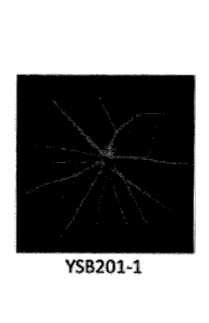
YSB201-1 (0.39 mg/mL, Example 8), YSB201-2 (0.78 mg/mL, Example 9) or YSB201-3
(1.56
mg/mL, Example 10) to each of two eyeballs three times at 2-day intervals.
FIG. 7A - FIG. 7F are fluorescence images and a graph of quantified
fluorescence
illustrating comparative reduction in laser-induced choroidal damage
(neovascularization) after
intravitreal injection using YSB201-1 (Example 8), YSB201-2 (Example 9), or
YSB201-3
(Example 10) according to the embodiments of this disclosure or a conventional
VEGF antibody
drug, Eylea (control group).
FIG. 8A - FIG. 8F are optical coherence tomography images of retina and a
graph of
.. quantified CNV lesions illustrating comparative reduction in laser-induced
choroidal damage
(neovascularization) after intravitreal injection using YSB201-1 (Example 8),
YSB201-2
(Example 9), or YSB201-3 (Example 10) according to the embodiments of this
disclosure or a
conventional VEGF antibody drug, Eylea (control group).
FIG. 9A¨ FIG. 9G are graphs illustrating electroretinogram (ERG) measured at
day 15
using a single white light under the dark-adapted state after the mouse with
laser-damaged
retina was treated three times with YSB201-1 (Example 8), YSB201-2 (Example
9), or YSB201-
3 (Example 10) according to the embodiments of this disclosure or once with
Eylea (control
group) through intravitreal injection.
FIG. 10 is a diagram illustrating the effect on the expression level of
vascular
endothelial growth factor (VEGF) in the choroid and the retina damaged by
laser after the
mouse with laser-damaged retina was treated with YSB201-1 (Example 8), YSB201-
2 (Example
9), or YSB201-3 (Example 10) according to the embodiments of this disclosure
or Eylea
(control group) through intravitreal injection.
FIG. 11 is a mimetic diagram illustrating an overall test method of evaluating
the efficacy
in a laser-induced choroidal neovascularization (CNV) animal model in order to
determine an
anti-angiogenic activity by the oral administration of the composition of this
disclosure, wherein
the mouse was orally administered with olive oil (control group) or an aqueous
solubilized
ursodeoxycholic acid of YSB201-4 (125 mg/kg/day, Example 11) or YS13201-5 (250
mg/kg/day,
Example 12), once a day from 10 days before laser injury to the retina of the
mouse to 10 days
after laser injury.
14
CA 03039500 2019-04-04
FIG. 12A ¨ FIG. 120 are fluorescence images and a graph of quantified
fluorescence
illustrating comparative reduction in laser-induced choroidal damage
(neovascularization) after
oral administration of YSB201-4 (Example 11) or YSB201-5 (Example 12)
according to
embodiments of this disclosure.
FIG. 13A ¨ 13D are optical coherence tomography images of retina and a graph
of
quantified CNV lesions illustrating comparative reduction in laser-induced
choroidal damage
(neovascularization) after oral administration of YSB201-4 (Example 11) or
YSB201-5 (Example
12) according to embodiments of this disclosure.
FIG. 14A - FIG. 14E are graphs illustrating electroretinogram (ERG) measured
at day
15 using a single white light under the dark-adapted state after the mouse was
orally
administered with olive oil (control group) or an aqueous solubilized
ursodeoxycholic acid,
YSB201-4 (Example 11) or YSB201-5 (Example 12) once a day from 10 days before
laser injury
to the retina of the mouse to 10 days after laser injury.
FIG. 15 is an image measuring the effect on the expression level of vascular
endothelial
growth factor (VEGF) in the choroid and the retina damaged by laser after the
mouse with laser-
damaged retina was orally administered with YSB201-4 (Example 11) or YSB201-5
(Example
12) according to the embodiments of this disclosure.
FIG. 16 and FIG. 17 are pharmacokinetic (PK) data illustrating the changes in
the
concentration of bile acids delivered into plasma over time after oral
administration of YSB201.
FIG. 18A, FIG. 18B, and FIG. 19 are pharmacokinetic (PK) data illustrating the
changes
in the concentration of bile acids delivered into the eyeballs over time after
oral administration of
YSB201. In FIG. 18B, UDCA type bile acids indicate the sum of concentrations
of UDCA and
tUDCA and gUDCA, which are in vivo metabolites thereof, having cytoprotective
effect, and
other bile acids that function as a surfactant indicate the sum of
concentrations of other bile
acids other than those three kinds of bile acids.
FIG. 20 and FIG. 21 are pharmacokinetic (PK) data illustrating the changes in
the
concentration of bile acids delivered into stomach over time after oral
administration of YSB201.
FIG. 22 is pharmacokinetic (PK) data illustrating the changes in the sum of
the
concentrations of UDCA and tUDCA and gUDCA, which are in vivo metabolites
thereof, and the
sum of concentrations of other bile acids delivered to plasma over time after
oral administration
of YSB201.
FIG. 23 is pharmacokinetic (PK) data illustrating the changes in the sum of
the
concentrations of UDCA and tUDCA and gUDCA, which are in vivo metabolites
thereof, and the
sum of concentrations of other bile acids delivered to the eyeballs over time
after oral
administration of YSB201.
CA 03039500 2019-04-04
FIG. 24 is pharmacokinetic (PK) data illustrating the changes in the sum of
the
concentrations of UDCA and tUDCA and gUDCA, which are in vivo metabolites
thereof, and the
sum of concentrations of other bile acids delivered to stomach over time after
oral
administration of YSB201.
DETAILED DESCRIPTION
In order that the invention may be more readily understood, certain terms are
first
defined herein for convenience. Unless otherwise defined herein, the
scientific and technical
terms used in this disclosure shall have the meaning generally understood by
those who are
skilled in the art. Unless clearly used otherwise, expressions in the singular
number include a
plural meaning, and those in the plural number include a singular meaning.
As used herein, the term "treating" or "treatment" encompasses preventing,
ameliorating, mitigating and/or managing visual impairments and/or conditions
by the
administration of a composition of this disclosure.
As used herein, the term "comprising as an active ingredient" or "comprising a
therapeutically active amount" is meant to include a certain amount of an
active ingredient,
enough to provide the effects for the prevention and the treatment of visual
impairments as a
composition, a composition for intravitreal injection, a composition for oral
administration, a
composition for eye drop, and a composition for intravenous injection.
As used herein, the term "prevention" means all actions that at least reduce
parameters,
for example the degree of symptoms, associated with the conditions to be
treated after a drug is
orally administered from before a visual impairment is occurred to after.
The terms "clear aqueous solution" or "clear aqueous solution" used in this
disclosure
mean a transparent aqueous solution in a solution state in which there are
substantially no
visually observed precipitates in naked eye.
The term "visual impairment" used in this disclosure includes macular
degeneration,
glaucoma and diabetic retinopathy.
According to one aspect of this disclosure, there is provided a composition
for the
prevention or the treatment of visual impairments including: (a)
ursodeoxycholic acid(UDCA); (b)
an aqueous soluble starch conversion product; and (c) water, wherein the
composition
comprises a clear aqueous solution of an aqueous solubilized ursodeoxycholic
acid for all pH
values.
The ursodeoxycholic acid is hydrophilic bile acid, which can be administered
orally. The
ursodeoxycholic acid in the human body is as low as about 3% of total bile
content, but it is also
16
CA 03039500 2019-04-04
present in the bile of the human body. UDCA is the only US FDA-approved drug
as a
therapeutic agent for primary biliary cirrhosis.
The UDCA has pharmacological actions of antioxidant, anti-inflammatory, and
anti-
apoptosis. These actions are very important mechanisms in the treatment of
visual impairments
and are more pronounced when UDCA acts as a single molecule. Therefore, the
key factor is to
make the substance with such effects be absorbed and be delivered to the
inside of the human
eyeballs. The crystalline form of UDCA is classified as an irritant and is an
amphipathic
molecule that has both hydrophilic and lipophilic properties, so it is almost
insoluble in water.
Even when aqueous solubilized in a small amount, it is in the form of dimers,
tetramers, or
micelles. It is thus difficult to function as a UDCA single molecule. However,
the composition of
this disclosure enables the crystalline form of UDCA to be solubilized at a
high concentration in
water to function as a single molecule.
The aqueous solubilized ursodeoxycholic acid is stabilized with maltodextrin
in water
and as a result, the solubility of pure ursodeoxycholic acid molecules in
water can be increased
by 3,000 times or more. The aqueous solubilized ursodeoxycholic acid, which is
dissolved in
water by the above method, exist as a single molecule form and nonionic
molecular state
having amphipathic properties due to its molecular nature, so that an
absorption rate of the
ursodeoxycholic acid can be drastically increased because it is absorbed in
vivo by passive
mechanism in addition to high intercellular and intracellular diffusion
through fast dispersion by
the concentration difference. All the take together, the aqueous solubilized
ursodeoxycholic acid,
in which an active ingredient of ursodeoxycholic acid is dissolved in water at
a high
concentration up to 60 g/L, is the most ideal multi-functional drug that can
prevent, alleviate or
treat visual impairments such as macular degeneration when it is applied
through intravitreal
injection, oral administration, intravenous injection or eye drop.
The composition of this disclosure may include, but is not limited to, the
solubility of
UDCA in the composition can be about 3,000 times higher than the conventional
commercialized UDCA preparation (0.15 M vs. 0.05 mM) and can be about 300
times or much
higher compared to the taurine-conjugated form of ursodeoxycholic acid (TUDCA)
(0.15 M vs.
0.45 mM). However, it is not limited thereto. Accordingly, the applicant has
completed this
disclosure by using aqueous solubilized UDCA.
According to one embodiment of this disclosure, the composition may be
prepared for
intravitreal injection to deliver UDCA of the composition to the retina, and
may not cause skin
17
CA 03039500 2019-04-04
irritation or inflammation in the eye after injection.
According to one embodiment of this disclosure, the composition for the
intravitreal
injection of this disclosure significantly inhibits the choroidal
neovascularization of the retina,
promotes regeneration of retinal cells, and down-regulates the expression
level of vascular
endothelial growth factor. According to experimental results of Examples
related to the
intravitreal injections of this disclosure and FIG. 7A - FIG. 10, it is
clearly confirmed that when
the composition is injected into the eyeballs of mice, the effect is equal to
or better than that with
the VEGF antibody injection agent, Eylee, which is currently used for the
treatment for macular
degeneration.
According to one embodiment of this disclosure, a single dose of the UDCA of
the
composition for the intravitreal injection may be 50 - 100 I at a
concentration of 0.1-1.5 mg/ml,
including but not limited to at a concentration of 0.1 - 3.0 mg/ml, preferably
at a concentration of
0.1 -1.5 mg/ml. When the UDCA concentration of the single injection is 0.1 or
more, the effect is
more obvious. When the UDCA concentration is more than 1.5 mg/ml, the effect
is substantially
equal to the effect shown with 1.5 mg/ml, so that an economically efficient
amount can be
provided. In the case of the eyeballs of the human body, the amount to be
injected once a day
may be 50- 100 pl. However, it is not limited thereto.
According to one embodiment of this disclosure, the composition may be
prepared for
oral administration to deliver the UDCA to the blood and further deliver a
therapeutically active
amount into the eyeballs across the blood-retinal barrier.
When UDCA is administered orally in a tablet or capsule form which is the
conventional
formulation of the crystalline form of UDCA, about 30 - 60% of it is absorbed
along the jejunum
and ileum by nonionic passive diffusion and the crystalline structure of UDCA
(crystalline form
of UDCA), due to insolubility, is absorbed only by a small amount (up to 20%
of intakes) at the
ileum of the colon by the active transport mechanism. When UDCA is absorbed by
hepatocytes,
it can be conjugated to tUDCA and gUDCA which are excreted by hepatic first-
pass clearance
as bile acids secreted from the human body. Therefore, the concentration of
UDCA in the blood
after oral administration is very low. Accordingly, there has been no example
provided with
composition for the prevention or the treatment of visual impairments such as
macular
degeneration which delivers UDCA to the eyeballs.
Alternatively, according to one embodiment of this disclosure, the
pharmacokinetic
study results of the composition upon oral administration (125 mg/Kg, Example
11) to mice
show that, unlike conventional crystalline form of UDCA formulations (tablets,
capsules), the
18
CA 03039500 2019-04-04
highest UDCA concentration in the blood is 36.56 3.30 iLig/mL, which is 12.7-
fold higher, and
Tmax is 5 minutes which is 48 times faster. Therefore, the high UDCA
concentration in the blood
can be achieved even with the same oral dose compared to the existing
formulation and the
preventive or therapeutic effect of visual impairments such as macular
degeneration can be
achieved.
According to one embodiment of this disclosure, the composition for oral
administration
of this disclosure can remarkably inhibit the choroidal neovascularization of
the retina, promote
the regeneration of retinal cells, and down-regulate the expression level of
vascular endothelial
growth factor. According to experimental results of Examples related to the
oral administration
and FIG. 12A - FIG. 15, it is clearly confirmed that the composition is
remarkably more effective
when administered orally, compared with the control group.
According to one embodiment of this disclosure, a daily oral dose of UDCA of
the
composition may be 5 - 30 mg/kg. The effect is more obvious when the daily
oral dose of UDCA
is 5 mg/kg or more. The effect is substantially equivalent to that shown with
30 mg/kg when it
exceeds 30 mg/kg, so that an economically efficient amount can be provided.
The daily dose of
UDCA of the composition may be 10 - 30 mg/kg, 15 - 30 mg/kg, 20 - 30 mg/kg, 25
- 30 mg/kg, 5
- 25 mg/kg, 10 - 25 mg/kg, 15 - 25 mg/kg, 20 - 25 mg/kg, 5 - 20 mg/kg, 10 - 20
mg/kg, 15 - 20
mg/kg, 5 - 15 mg/kg, and 10 - 15 mg/kg. However, it is not limited thereto. An
interval of oral
administration of UDCA may be 1 day, 2 days, 3 days, 4 days, 5 days, 6 days,
and 7 days,
depending on the symptoms. However, it is not limited thereto. In addition,
the dose is suitable
for the human body, but is not limited thereto.
According to one embodiment of this disclosure, the composition may be orally
administered at least once a day for 20 days or more. However, it is not
limited thereto.
According to one embodiment of this disclosure, the UDCA of the composition
starts to
be distributed in the eyeballs within 5 - 10 minutes after oral
administration, and may stay in the
eyeballs for a predetermined time, about 1 hour, and then wash out.
According to one embodiment of this disclosure, the composition may be
administered
directly to the blood through intravenous injection without blocking the blood
vessel and causing
skin irritation.
According to one embodiment of this disclosure, the composition may be
administered
as eye drop.
According to one embodiment of this disclosure, the UDCA of the composition
administered as the eye drop can be delivered from the outside of the eye to
the inside of the
19
CA 03039500 2019-04-04
eye without causing skin irritation and adverse reactions around the eyes or
the eye.
According to one embodiment of this disclosure, a single eye drop dose of UDCA
of the
composition may be 30 - 50 pl at a concentration of 0.1 mg/ml - 2.0 mg/ml,
preferably a
concentration of 0.1 mg/ml - 1.5 mg/ml. However, it is not limited thereto.
When the single eye
drop concentration of UDCA of the composition is more than 0.1 mg/ml, the
effect is more
obvious. When it is more than 2.0 mg/ml, the effect is substantially
equivalent to that shown with
2.0 mg/ml, so that an economically efficient amount can be provided.
According to an embodiment of this disclosure, the appropriate number of drops
per
day of the UDCA of the composition may be one to ten times a day. However, it
is not limited
thereto.
The eye drop according to an embodiment of this disclosure may include a
chelating
agent. The chelating agent is not particularly limited as long as it is a
compound chelating metal
ions. Example of the chelating agent may include edetic acid (ethylenediamine
tetra acetic acid)
such as monosodium edetate, disodium edetate, trisodium edetate, tetrasodium
edetate,
dipotassium edetate, tripotassium edetate, tetrapotassium edetate and the
like, or salts thereof;
citric acid such as monosodium citrate, disodium citrate, trisodium citrate,
monopotassium
citrate, dipotassium citrate, tripotassium citrate and the like, or salts
thereof; metaphosphoric
acid such as sodium metaphosphate, potassium metaphosphate and the like, or
salts thereof;
pyrophosphoric acid such as sodium pyrophosphate, potassium pyrophosphate and
the like, or
salts thereof; polyphosphoric acid such as sodium polyphosphate, and potassium
polyphosphate and the like, or salts thereof; malic acid such as monosodium
malate, disodium
malate, monopotassium malate, dipotassium malate and the like, or salts
thereof; tartaric acid
such as sodium tartrate, potassium tartrate and potassium sodium tartrate and
the like, or salts
thereof; phytic acid such as sodium phytate, potassium phytate and the like,
or salts thereof.
However, it is not limited thereto.
In addition, edetic acid, citric acid, metaphosphoric acid, pyrophosphoric
acid,
polyphosphoric acid, malic acid, tartaric acid, phytic acid, and salts thereof
may include
hydrates and organic solvates thereof.
In this disclosure, preferred examples of the chelating agent include edetic
acid, a salt
of edetic acid (edetate), citric acid, a salt of citric acid (citrate),
metaphosphoric acid, a salt of
metaphosphoric acid (metaphosphate), pyrophosphoric acid, a salt of
(polyphosphate), and
more preferred examples include sodium edetate (including a hydrate thereof
such as disodium
edetate hydrate and the like), citric acid (including a hydrate thereof such
as citrate
monohydrate and the like), sodium metaphosphate, and sodium polyphosphate.
CA 03039500 2019-04-04
The eye drop according to an embodiment of this disclosure may further include
a
preservative. Examples of the preservative may include benzalkonium chloride,
benzethonium
chloride, chlorhexidine gluconate, paraben, sorbic acid, chlorobutanol, boric
acid, chlorite and
the like, preferably benzalkonium chloride. However, it is not limited
thereto.
The eye drop may further include a pharmaceutically acceptable additive, if
necessary.
Examples including a buffering agent such as sodium phosphate, sodium
hydrogenphosphate,
sodium dihydrogenphosphate, sodium acetate, epsilon-aminocaproic acid; an
isotonizing agent
such as sodium chloride, potassium chloride and concentrated glycerin; and a
surfactant such
as polyoxyethylene sorbitan monolete, polyoxyl 40 stearate and polyoxyethylene
hardened
castor oil may be selected and added as needed.
According to one embodiment of this disclosure, examples of the visual
impairment may
include macular degeneration, glaucoma, and diabetic retinopathy.
According to one embodiment of this disclosure, the visual impairment may be
macular
degeneration.
According to one embodiment of this disclosure, the visual impairment may be
wet age-
related macular degeneration.
According to one embodiment of this disclosure, the composition may function
as one
or more of the functions of inhibiting the development of choroidal
neovascularization,
promoting the recovery of retinal function, and regulating the expression
level of vascular
endothelial growth factor (VEGF).
According to one embodiment of this disclosure, the UDCA which is selected
from an
aqueous soluble ursodeoxycholic acid, an aqueous soluble ursodeoxycholic acid
derivative, an
ursodeoxycholic acid salt, and an ursodeoxycholic acid conjugated with an
amine can be
aqueous solubilized ursodeoxycholic acid. An aqueous soluble metal salt of
ursodeoxycholic
acid and an aqueous soluble 0-sulfonated bile acid are also included as an
aqueous soluble
ursodeoxycholic acid salt. However, it is not limited thereto.
According to one embodiment of this disclosure, the UDCA may be at least one
aqueous solubilized UDCA selected from a ursodeoxycholic acid (UDCA), a
tauroursodeoxycholic acid (tUDCA) and a glycoursodeoxycholic acid (gUDCA). The
tauroursodeoxycholic acid (tUDCA) and glycoursodeoxycholic acid (gUDCA) are in
vivo
metabolites or derivatives of UDCA. The tUDCA is a UDCA derivative which is
the taurine
conjugate form of UDCA and the gUDCA is a UDCA derivative which is the glycine
conjugate
21
CA 03039500 2019-04-04
form of UDCA.
According to one embodiment of this disclosure, the UDCA may be present in a
therapeutically active amount. The therapeutically active amount means an
amount enough to
provide the effects for the prevention and the treatment of visual impairments
as a composition,
a composition for intravitreal injection, a composition for oral
administration, a composition for
eye drop, and a composition for intravenous injection such as an amount
capable of preventing
or treating a visual impairment.
According to one embodiment of the disclosure, the ursodeoxycholic acid is
included in
an amount of 0.01 - 5 parts by weight based on the total weight of the
composition. If the
amount of ursodeoxycholic acid is less than 0.01 part by weight based on the
total weight of the
composition, the effects for the prevention or the treatment of visual
impairments may be
insignificant. On the other hand, if the amount of ursodeoxycholic acid is
more than 5 parts by
weight, a clear aqueous solution may not be formed. However, it is not limited
thereto. When
cloudy precipitates are formed instead of a clear aqueous solution, it may be
difficult to use it as
an intravitreal injection, an oral administration agent, and an eye drop.
When precipitates are formed, ursodeoxycholic acid may not be dissolved in
water and
thus exist in a crystalline form of UDCA. When this is used for preparing eye
drop or intravitreal
injection, it may cause skin irritation due to the crystalline form of UDCA.
Thus, the preparation
of a clear aqueous solution is required to remove all of the crystalline form
of UDCA that can
cause skin irritation.
UDCA may be included in an amount of 0.01 - 5 parts by weight, including but
not
limited to 0.1 - 5 parts by weight, 1 - 5 parts by weight, 2 - 5 parts by
weight, 3 - 5 parts by
weight, 4 - 5 parts by weight, 0.01 - 3 parts by weight, 0.1 - 3 parts by
weight, 1 - 3 parts by
weight, 2 - 3 parts by weight, 0.01 - 2.5 parts by weight, 0.1 - 2.5 parts by
weight, 1 - 2.5 parts
by weight based on the total weight of the composition. For the intravitreal
injection, UDCA may
be included in an amount of 0.05 - 0.2 parts by weight, more preferably 0.04 -
0.16 parts by
weight, and even more preferably 0.04 - 0.07 parts by weight based on the
total weight of the
composition. For the oral administration, UDCA may be included in an amount of
0.1 - 2.5 parts
by weight, preferably 1 - 2.5 parts by weight based on the total weight of the
composition.
According to one embodiment of this disclosure, the aqueous soluble starch
conversion
product is maltodextrin, and the maltodextrin is used in an amount of 1.0 - 70
parts by weight
based on the total weight of the composition. However, it is not limited
thereto. When the
22
CA 03039500 2019-04-04
amount of maltodextrin is less than 1.0 part by weight, an effective amount of
UDCA cannot be
dissolved in water, resulting in poor effects for the prevention or the
treatment of visual
impairments. On the other hand, when the amount of maltodextrin is more than
70 parts by
weight, precipitates are formed, resulting in skin irritation in the eye since
UDCA or maltodextrin
precipitates out of the aqueous solution.
The maltodextrin may be included in an amount of 1 - 60 parts by weight,
including but
not limited to 5 - 60 parts by weight, 10 - 60 parts by weight, 20 - 60 parts
by weight, 30 - 60
parts by weight, 40 - 60 parts by weight, 50 - 60 parts by weight, 1 - 50
parts by weight, 5 - 50
parts by weight, 10 - 50 parts by weight, 20 - 50 parts by weight, 30 - 50
parts by weight, 40 - 50
parts by weight, 1 - 40 parts by weight, 5 - 40 parts by weight, 10 - 40 parts
by weight, 20 - 40
parts by weight, 30 - 40 parts by weight, 1 - 30 parts by weight, 5 - 40 parts
by weight, 10 - 30
parts by weight, 20 - 30 parts by weight, 1 - 20 parts by weight, 5 - 20 parts
by weight, 10 - 20
parts by weight, 1 - 10 parts by weight, 5 - 10 parts by weight based on the
total weight of the
composition.
According to one embodiment of this disclosure, the aqueous soluble starch
conversion
product is maltodextrin, and the minimum weight ratio of maltodextrin to the
ursodeoxycholic
acid may be 1:30, including but not limited to 1:25, 1:20, 1:15, 1:12, or 1:6.
An amount of the
aqueous soluble starch conversion product with high molecular weight used in
the composition
can be defined as an aqueous solubilized amount of the selected
ursodeoxycholic acid at a
desired concentration and the pH range described herein. The minimum amount of
maltodextrin
may be equally applied to the case of tauroursodeoxycholic acid and
glycoursodeoxycholic acid.
According to one embodiment of this disclosure, there is provided a
composition for the
prevention or the treatment of visual impairments, wherein the pH value is 3 -
9 and the
aqueous soluble starch conversion product is maltodextrin, wherein minimum
weight ratio of the
UDCA to maltodextrin is 1:16 - 1:30. Minimum weight ratio of the UDCA to
maltodextrin may be
1:16 - 1:20, 1:16 - 1:25, 1:16 - 1:30, 1:20 - 1:25, 1:20 - 1:30, or 1:25 -
1:30. However, it is not
limited thereto.
When the pH value is from 3 or higher to less than 6 and the minimum weight
ratio of
UDCA to maltodextrin is 1:1 - 1:15, precipitates may be formed, resulting in
no clear aqueous
solution. However, it is not limited thereto.
According to one embodiment of this disclosure, there is provided a
composition for the
prevention or the treatment of visual impairments, wherein the pH value is 6 -
9, the aqueous
soluble starch conversion product is maltodextrin, and the minimum weight
ratio of UDCA to
maltodextrin is 1:13 - 1:30. However, it is not limited thereto.
The aqueous soluble starch conversion product of this disclosure includes a
23
CA 03039500 2019-04-04
carbohydrate obtained directly from partial or incomplete hydrolysis of starch
under various pH
conditions. Non-limiting examples of the aqueous soluble starch conversion
product may
include maltodextrin, dextrin, liquid glucose, corn syrup solid (dried powder
of liquid glucose).
The corn syrup solid may be Maltrin M200 and the maltodextrin may be Maltrin
M700, both of
which are manufactured by GFCTM (Grain Processing Corporation), located in
Muscatin, Iowa,
USA. However, it is not limited thereto.
If the starch conversion product consists of a polymer, the polymer may
include at least
one reducing end and at least one non-reducing end. The polymer may be linear
or branched.
The molecular weight may be about 100 mass units or more, or 106 mass units or
more. The
high molecular weight aqueous soluble starch conversion product, though not
limited thereto,
may have a molecular weight of 105 mass units or more.
According to one embodiment of this disclosure, the composition may further
include a
soluble non-starch polysaccharide. The soluble non-starch polysaccharide may
be obtained
under various pH conditions by various hydrolysis or synthesis mechanisms. Non-
limiting
examples of the soluble non-starch polysaccharide may include dextran, guar
gum, pectin,
indigestible soluble fibers, and the like. If the soluble non-starch
polysaccharide consists of a
polymer, the polymer may have at least one reducing end and at least one non-
reducing end.
The polymer may be a linear or branched polymer. The molecular weight of the
polysaccharide
of this disclosure may be at least about 100 mass units, or at least about 106
mass units,
.. preferably at least 105 mass units. However, it is not limited thereto. The
composition may be
provided as a composition which is an aqueous solution comprising a
combination of the
aqueous soluble starch conversion product and/or the aqueous soluble non-
starch
polysaccharide.
According to one embodiment of this disclosure, the minimum weight ratio of
ursodeoxycholic acid to liquid glucose (e.g., corn syrup) needed to prevent
precipitation of the
composition is about 1:25 (i.e., about 12.5 g per 500 mg of ursodeoxycholic
acid in 100 ml of
water or about 25 g per 1 g of ursodeoxycholic acid in 200 ml of water).
However, it is not
limited thereto.
In addition, the minimum amount of dried powder of liquid glucose (corn syrup
solids,
.. e.g., Maltrin M200) needed to prevent precipitation of the composition from
the dosage form of
this disclosure is about 30 g per 1 g of ursodeoxycholic acid in 100 ml of
water, or about 60 g
per 2 g of ursodeoxycholic acid in 200 ml of water. However, it is not limited
thereto.
The minimum amount of the soluble non-starch polysaccharide required to
prevent
precipitation of the composition from the dosage form of this disclosure is
about 50 g of guar
.. gum per 500 mg of ursodeoxycholic acid in 100 ml of water, or 80g of pectin
per 500 mg of
24
CA 03039500 2019-04-04
ursodeoxycholic acid in 100 ml of water. However, the minimum amount of the
soluble non-
starch polysaccharide or aqueous soluble starch conversion product with high
molecular weight
may be determined mainly by the absolute amount of ursodeoxycholic acid in the
solution
preparation rather than the concentration.
The composition of this disclosure may further include dietary fiber when
formulated for
oral administration. Non-limiting examples of the dietary fiber include guar
gum, pectin, psyllium,
oat rubber, soybean fiber, oat bran, corn hull, cellulose, and wheat bran.
The composition of this disclosure may further include an emulsifying agent
and a
suspending agent. Non-limiting examples of the emulsifying agent may include
guar gum, pectin,
acacia, carrageenan, sodium
carboxymethylcellulose, hydroxymethylcellu lose,
hydroxypropylcellulose, methylcellulose, polyvinylalcohol, povidone,
tragacanth gum, xanthan
gum and sorbitan ester.
The composition of this disclosure may further include a pharmaceutically
acceptable
additive. Non-limiting examples of the pharmaceutically acceptable additive
may include starch,
gelatinized starch, microcrystalline cellulose, lactose, povidone, colloidal
silicon dioxide, calcium
hydrogenphosphate, lactose, mannitol, glutinous, arabic gum, pregelatinized
starch, corn starch,
powder cellulose, hydroxypropylcellulose, opadry, sodium starch glycolate,
carnauba wax,
synthetic aluminum silicate, stearic acid, magnesium stearate, aluminum
stearate, calcium
stearate, white sugar, dextrose, sorbitol and talc. The pharmaceutically
acceptable additive
according to this disclosure is preferably included in the composition in an
amount of 0.1 - 90
parts by weight. However, it is not limited thereto. In addition, the
composition of this disclosure
may be administered in various forms of parenteral administration at the time
of actual clinical
administration. In the case of formulation, a diluent or excipient such as a
filler, an extender, a
binder, a wetting agent, a disintegrating agent, and a surfactant may be
added. Examples of
parenteral administrations may include sterilized aqueous solutions, non-
aqueous solutions,
suspensions, emulsions, freeze-dried preparations, suppositories, and
injections.
According to one embodiment of this disclosure, there is provided a
composition for the
prevention or the treatment of visual impairments, wherein the pH of the
composition is in the
range of 1 - 10, and wherein the composition is in a clear aqueous solution
state at the pH value.
The composition may be solubilized in water and may be in the form of a clear
aqueous solution
without precipitation at the pH described above. That is, the composition may
be in the form of a
clear aqueous solution without precipitation of UDCA even after several months
(1,2, 3, 4, 5, 6,
7, 8, 9, 10, 11, or 12 months). A selected pH range that does not precipitate
the ursodeoxycholic
acid and the aqueous soluble starch conversion product in the composition may
be about pH 1
CA 03039500 2019-04-04
¨ about pH 10, preferably about pH 3 - about pH 9, more preferably pH 6 - pH
8, and most
preferably pH 6.5 - pH 8. However, it is not limited thereto. The composition
may further include
an acid, a base and a buffering agent if it is necessary to maintain the pH
described above. The
pH adjusting material may be, but is not limited to, HCI, H3PO4, H2SO4, HNO3,
CH3COOH, citric
acid, malic acid, tartaric acid, lactic acid, phosphate, eidetic acid, and
alkali. The properties and
methods for using such pH adjusting materials are well known in the art. The
pH range is the pH
level of any subset that can be obtained in an aqueous system sufficient to
allow various
formulations to remain in solution from the preparation and to be injected to
the eyeballs or be
administered orally to the blood, depending on the method of administration.
Thus, the
composition may be used as a formulation in solution, without the composition
according to this
disclosure being precipitated at the pH level of the mouth, stomach, and
intestines.
According to some embodiments of this disclosure, ursodeoxycholic acid remains
dissolved under acidic conditions as a free ursodeoxycholic acid even though
it is generally
insoluble under acidic conditions. The composition may further include another
composition in
which the composition remains soluble when added to any formulation. In
addition, the
composition may provide a clear and stable solution for providing the
composition for the
prevention or the treatment of visual impairments such as macular degeneration
diseases in the
form of intravitreal injection, oral administration, eye drop, or intravenous
injection.
According to one embodiment of this disclosure, the composition may be
formulated
into a syrup form, a cream form, a paste form, or a dried form. The syrup may
be, but not limited
to, normal syrup or thick syrup.
According to one embodiment of this disclosure, the composition may be dried
and
formulated in powder form. The powder-type composition can be stored and
handled easily and
can be easily formulated into a desired form.
According to one embodiment of this disclosure, the composition may be
administered
in combination with another therapeutic agent for macular degeneration
diseases.
According to one embodiment of this disclosure, another therapeutic agent for
macular
degeneration diseases may be an anti-vascular endothelial growth factor
antibody. Examples
thereof may include Eylea , Avastin , Macugen , Lucentis or a combination
thereof. However,
it is not limited thereto. In addition, the composition may be administered in
combination with
Visudyne injection. However, it is not limited thereto.
According to one embodiment of this disclosure, the therapeutic agent for the
macular
26
CA 03039500 2019-04-04
degeneration disease may be for intravitreal injection.
This disclosure contains extensive information on the current awareness of the
genetics,
biochemistry, and cell biology of macular degenerative diseases, but future
research may reveal
that aspects of these perceptions are either inaccurate or incomplete. Thus,
those skilled in the
art will understand that this disclosure is not limited to a particular model
or mechanism of action
whether part of this disclosure is taken or not.
In addition, those skilled in the art will recognize that other equivalent or
alternative
compositions and methods may be utilized. For example, although it has been
described that a
plurality of compounds can be administered together with aqueous solubilized
ursodeoxycholic
acid, it is understood that other compounds may also be included.
Also, the application of another drug may be performed at the same time as the
administration of the aqueous solubilized ursodeoxycholic acid composition of
this disclosure, or
they may be administered separately in the same or overlapped time period (for
example, at the
same time, the same date, or the same week).
Hereinafter, this disclosure will be described in more detail with reference
to Examples.
Examples
Materials and Methods
<Contract Research Organization>
1) Preparation for animal models of macular degeneration and animal test of
pharmacokinetics - 126 Infrastructure Center for Ocular Disease of Inje
University Busan Paik
Hospital
2) Pharmacokinetic study - College of Pharmacy, Inje University
< Preparation for animal models of macular degeneration >
1) All animals were acclimatized for approximately 7 days to be adapted to the
laboratory environment.
2) The animals that did not show any clinical sign of diseases or wounds and
showed
appropriate weight were used in the study. A control group and a treatment
group of the animals
were randomly assigned on the basis of the most recently measured body weight.
3) The mice were housed in the group of three in a mice cage during the entire
test
period.
4) The test environment was constantly controlled to have 19 - 20 C of
temperature, 40
27
CA 03039500 2019-04-04
- 60% of humidity, and 150 - 300 Lux of room light.
5) After 057 BL/6 mice (Orient Bio, Korea) were acclimatized to be adapted to
the
environment for 1 week, 7-week old mice were treated with the image-guided
laser (Phoenix,
USA) especially optic nerve area at 3, 6, 9, and 12 o'clock positions. The
laser was irradiated at
532 nm, 100 ms/70 ms, 200 mW, and a spot size was 50 pm.
< Analysis of Macular Degeneration Therapeutic Effects >
The following tests were performed to determine the effects of samples on
inhibition of
the development of choroidal neovascularization and recovery of retinal cell
functions using the
mouse model of laser-induced choroidal neovascularization (CNV).
1) Fundus Fluorescein Angiography (FFA)
Both eyes of CNV mice were dilated and intraperitoneally injected with 1%
fluorescein
(Sigma, USA) to dye the blood vessels. The mice were anesthetized and
fluorescent fundus
images of the laser-induced neovascularization were taken with the retinal
imaging microscope
(Micron IV image) at 5 minutes after fluorescein injection. The CNV lesions
were represented by
the corrected total fluorescence (CTF) calibrated by using Image J program.
Here, CTF can be
calculated by the following equation.
*CTF= (Integrated Density)-[(Area of selected lesion) x (Mean fluorescence of
background readings)]
2) Optical Coherence Tomography (OCT)
After both eyes of CNV mice were dilated and anesthetized, tomography of the
laser-
induced neovascularization was taken using the image-guided OCT system by
positioning the
pupils dilated mice in front of the OCT system and the guide line in the
center of the CNV with
guidance of bright-field live fundus image to scan each formed choroidal
neovascularization.
3) Electroretinography (ERG)
The mice were dark adapted for 24 hours before test and all tests were
performed in a
darkroom. The eyes of mice were dilated and general anesthetized by
intraperitoneal injection
with a mixture of ketamine (30 mg/kg) and xylazine hydrochloride (2.5 mg/kg)
in order to detect
the function of the retina. Electrodes were placed in skin, tail and cornea to
run
electroretinography. A single white light was used to stimulate the retina to
obtain electrical
activity of the retina. The amplitude from the trough of A-wave to the peak of
B-wave was
.. measured and evaluated as an index of the function of the retina.
28
CA 03039500 2019-04-04
4) Western blot analysis
After 15 days from laser injury, the mice were euthanized and the choroid and
retinal
layers were separated by removing the sclera, cornea and lens from the
eyeballs after
extracting the eyeballs. The choroidal and retinal tissues were washed twice
with PBS, and
proteins were extracted by homogenizing the tissue with Pro-PREP (iNtRON,
Korea). The
extracted proteins were quantified using the BCA protein assay kit (Thermo
scientific, USA) and
20 [ig of the proteins were used for Western blotting. After blocking the
membrane with 5%
skimmed milk for 1 hour, the primary antibody was diluted to 1:1000 in TBS-T
and the
membrane was incubated in the primary antibody solution overnight at 4 C.
After washing the
membrane with TBS-T, the secondary antibody was diluted to 1: 5000 in 3%
skimmed milk, the
membrane was incubated in the secondary antibody solution at room temperature
for 1 hour,
and then target proteins were detected using a Chemi image system.
< Analysis of pharmacokinetics of aqueous solubilized UDCA >
For the pharmacokinetic study, a test sample YSB201-4 was orally administered
to
C57BL/6 mouse model, and the pharmacokinetic trends of the test sample in the
plasma and
the living organs were analyzed over time.
1) Methods
(1) The test sample was orally administered to mice, and blood and various
tissue
samples were collected at each hour. An organic solvent was used to extract
the drug
components in the tissue samples, and the concentration thereof was
quantitatively analyzed by
HPLC fluorescence detector. The tissue samples were taken at 0, 5, 10, 30
minutes and 1, 2, 4,
.. 10, 24, 48, and 72 hours after oral administration. Blood and tissue
samples were collected from
4 mice at each hour. Blood and tissue samples from the control group having
the fasting and
dietary treatments as in the test group were collected at 0, 4, 10, 24, 48,
and 72 hours.
(2) A suitable extraction method was established by extracting the test sample
components in the tissue sample using an organic solvent and by calculating
its recovery (%).
The high-performance liquid chromatography (HPLC) was used in order to
quantitatively
analyze the bile acid components in various biological tissues, using enzyme
reaction and
fluorescence detection.
(3) Pharmacokinetic parameters after oral administration were calculated by
applying
the results of concentration analysis of drug components in blood and tissues
to WinNonLin,
29
CA 03039500 2019-04-04
which is the pharmacokinetic analysis program. The PK profile of the YSB201-4
preparation
was confirmed.
(4) The animal laboratory of lndang Biomedical Research Center of lnje
University
Pusan Paik Hospital received a certificate from the Korea Ministry of Food and
Drug Safety in
2014, and the test protocol was approved by the IACUC (Institutional Animal
Care and Use
Committee, IACUC No. IJUBPH_2016-001-02) of College of Medicine, Inje
University.
2) Test samples and equipment
(1) YSB201-4 was used as a test sample.
(2) The various bile acids used as standard substances were gUDCA, tUDCA,
UDCA,
GCA (glycocholic acid hydrate), TCA (taurocholic acid sodium salt hydrate), CA
(cholic acid),
GCDCA (glycochenodeoxycholic acid), TCDCA (taurochenodeoxycholic acid), GDCA
(glycodeoxycholic acid), TDCA (taurodeoxycholic acid), CDCA (chenodeoxycholic
acid), DCA
(deoxycholic acid), GLCA (glycolithocholic acid sodium salt), TLCA
(taurolithocholic acid), and
LCA (lithocholic acid). They were purchased from Sigma-Aldrich.
(3) The analytical equipment was the 2695 Alliance high performance liquid
chromatography (HPLC) instrument from the US Water Company. BilePak 11 column
(4.6125
mm, JASCO, Japan) and EnzymePak 3a-HSD column (4.635 mm, JASCO, Japan) were
also
used.
3) Drug administration method - oral administration
The animals were fasted for 12 hours immediately prior to oral administration,
and then
the dose of each individual was calculated based on the body weight measured
after fasting for
12 hours prior to oral administration and orally administered using a
disposable syringe
equipped with sonde. Solid diet was re-fed after 4 hours from the oral
administration.
4) Test animals
36 C57BL/6 mice (female) weighing 16 - 18 g were obtained from CoreTech Co.,
Ltd.
(Pyongtaek, Gyeonggi-do, Korea) and acclimatized for 7 days and then used for
the test. At the
first administration, the mice with about 20% of the total average weight
were used. The mice
were housed at temperature of 19 - 25 C, humidity of 40 - 60%, and room light
of 150 - 300Lux.
Cleaning of the test room and the cage was performed according to the standard
operation
procedure of lndang Biomedical Research Center, lnje University Busan Paik
hospital.
CA 03039500 2019-04-04
5) Experimental design for grouping and dosing
Experimental design for grouping and dosing for the control and test groups
are shown
in Table 1.
Table 1
Oral dose Time after No of mice
No Group Treatment
mg/kg administration Female
1 4h 3
2 10h 3
3 Control None None 24h 3
4 48h 3
72h 3
Omin 3
6
5min 4
7 10min 4
8 YSB201 Oral 30min 4
9 Test administration 1h 4
(12.5mg/m1) 125mg/kg
2h 4
11 4h 4
12 10h 4
13 24h 4
14 48h 4
72h 4
6) Sample analysis
An analysis system of bile acids of JASCO, Japan was used to analyze blood and
10 biological samples of the mice. Concentrations of bile acids were
quantitatively analyzed using
a fluorescence detector (excitation: 345 nm, emission: 470 nm) of Waters
Alliance 2695 HPLC
system. HPLC conditions are summarized in Table 2.
Table 2
31
CA 03039500 2019-04-04
< HPLC Conditions >
Items Conditions
Separation Column Guard column/BilePak 11 column (4.6 x 125 nm)
Enzyme Column EnzymePak 3a-HSD column (4.6x35 mm)
Column Temp. 25 t (Column oven)
Sample Temp. 10 t (Auto Sampler)
A: Acetonitrile/Methano1/30 mM ammonium acetate (30/30/40)
Eluent
B: Acetonitrile/Methano1/30 mM ammonium acetate (20/20/60)
Flow Rate 1 mL/min
0.3 mM NAD, 10 mM KH2PO4, 1 mM EDTA-2Na,
Reagent Solution
0.05% 2-mercaptoethanol, pH 7.8 (adjusting with KOH)
Reagent Solution Flow
1 mL/min
Rate
Fluorescence
Excitation: 345 nm, Emission: 470 nm
Wavelength
Time (min) Eluent A (%) Eluent B (%)
0 0 100
Eluent Gradient
32 100 0
Condition
60 100 0
65 0 100
Analysis Time 65 min
Injection Volume 10 pL
7) Data and statistical analysis
Test results were summarized using Microsoft Excel 2010 and additional
pharmacokinetic analyses were performed on the results of blood concentration
analysis using
the Pharsight WinNonlin 7.0 program (Certara, USA). The pharmacodynamic
profile was
evaluated using GraphPad Prism 5.0, wherein a P-value of 0.05 or less was
considered
32
CA 03039500 2019-04-04
significant.
1. Preparation of aqueous solubilized ursodeoxycholic acid in a clear aqueous
solution
(Example 1) A clear aqueous solution with 1:6 of weight ratio of UDCA to
maltodextrin
A clear aqueous stock solution of aqueous solubilized UDCA containing natural
UDCA
and aqueous soluble starch having low dextrose equivalent was prepared.
Particularly, 6.7 g of sodium hydroxide pellets were aqueous solubilized in
400 ml of
purified water. 60 g of UDCA was dissolved in the sodium hydroxide solution
while stirring at
room temperature. 360 g of maltodextrin was added slowly to the clear solution
while stirring. A
preservative was then added in an amount appropriate for the pharmaceutical
formulation to the
clear solution obtained by performing ultrasonication (750W, 20kHz) at high
throughput and the
pH was adjusted by the dropwise addition of HCI. Purified water was added and
adjusted to be
a total of 1,000 ml. If necessary, the clear solution was filtered through a
suitable filtration
apparatus. This filtration is important for removing impurities from the raw
material or
sterilization, but it is not intended to remove the granular material because
the solution is
already clear. As shown in Table 3, the prepared ursodeoxycholic acid solution
formed a clear
aqueous solution at pH 10.3, 9.2, and 6.7 without visual precipitation, but
formed precipitates at
pH 5.4.
(Example 2) A clear aqueous solution with 1:12 of weight ratio of UDCA to
maltodextrin
A clear aqueous stock solution of aqueous solubilized UDCA containing natural
UDCA
and aqueous soluble starch having low dextrose equivalent was prepared.
Particularly, it was prepared in accordance with the same procedure as in
Example 1,
except that 720 g of maltodextrin as one high molecular weight aqueous soluble
starch
conversion product per 60 g of ursodeoxycholic acid was used. As shown in
Table 3, the
prepared ursodeoxycholic acid solution formed a clear aqueous solution at pH
9.6, 7.3, 6.5, and
6.0 without any visible precipitation, but formed precipitates at pH 5.5.
(Example 3) A clear aqueous solution with 1:15 of weight ratio of UDCA to
maltodextrin
A clear aqueous stock solution of aqueous solubilized UDCA containing natural
UDCA
33
CA 03039500 2019-04-04
and aqueous soluble starch having low dextrose equivalent was prepared.
Particularly, it was prepared in accordance with the same procedure as in
Example 1,
except that 750 g of maltodextrin as one high molecular weight aqueous soluble
starch
conversion product per 50 g of ursodeoxycholic acid was used. 5.7 g of sodium
hydroxide
pellets were dissolved in 400 ml of purified water and then used. As shown in
Table 3, the
prepared ursodeoxycholic acid solution formed a clear aqueous solution at pH
9.5, 8.9, 7.9, 7.1,
and 6.0 without visual precipitation, but formed precipitates at pH 5.5. FIG.
1 is images
illustrating whether a clear aqueous solution of the ursodeoxycholic acid
solution is formed or
not at each pH value.
(Example 4) A clear aqueous solution with 1:20 of weight ratio of UDCA to
maltodextrin
A clear aqueous stock solution of aqueous solubilized UDCA containing natural
UDCA
and aqueous soluble starch having low dextrose equivalent was prepared.
Particularly, it was prepared in accordance with the same procedure as in
Example 1,
except that 350 g of maltodextrin as one high molecular weight aqueous soluble
starch
conversion product per 17.5 g of ursodeoxycholic acid was used. 2.0 g of
sodium hydroxide
pellets were dissolved in 400 ml of purified water and then used. As shown in
Table 3, the
prepared ursodeoxycholic acid solution formed a clear aqueous solution at pH
9.4, 7.1, 6.1, and
5.5 without visual precipitation, but formed precipitates at pH 5.1. FIG. 2 is
images illustrating
whether a clear aqueous solution of the ursodeoxycholic acid solution is
formed or not at each
pH value.
(Example 5) A clear aqueous solution with 1:25 of weight ratio of UDCA to
maltodextrin
A clear aqueous stock solution of aqueous solubilized UDCA containing natural
UDCA
and aqueous soluble starch having low dextrose equivalent was prepared.
Particularly, it was prepared in accordance with the same procedure as in
Example 1,
except that 350 g of maltodextrin as one high molecular weight aqueous soluble
starch
conversion product per 14 g of ursodeoxycholic acid was used. 1.7 g of sodium
hydroxide
pellets were dissolved in 400 ml of purified water and then used. As shown in
Table 3, the
prepared ursodeoxycholic acid solution formed a clear aqueous solution at pH
9.6, 6.1, and 5.1
without visual precipitation, but formed precipitates at pH 4Ø FIG. 3 is
images illustrating
whether a clear aqueous solution of the ursodeoxycholic acid solution is
formed or not at each
34
CA 03039500 2019-04-04
pH value.
(Example 6) A clear aqueous solution with 1:30 of weight ratio of UDCA to
ma ltodextri n
A clear aqueous stock solution of aqueous solubilized UDCA containing natural
UDCA
and aqueous soluble starch having low dextrose equivalent was prepared.
Particularly, it was prepared in accordance with the same procedure as in
Example 1,
except that 750 g of maltodextrin as one high molecular weight aqueous soluble
starch
conversion product per 25 g of ursodeoxycholic acid was used. 2.8 g of sodium
hydroxide
pellets were dissolved in 400 ml of purified water and then used. As shown in
Table 3, the
prepared ursodeoxycholic acid solution formed a clear aqueous solution at pH
9.0, 8.0, 7.0, 6.0,
5.1, 4.1, and 2.9 without visual precipitation. FIG. 4 is images illustrating
whether a clear
aqueous solution of the ursodeoxycholic acid solution is formed or not at each
pH value.
(Example 7) A clear aqueous solution containing UDCA/tUDCA/gUDCA and with
1:30 of weight ratio of UDCA/tUDCA/gUDCA to maltodextrin
A clear aqueous stock solution of aqueous solubilized UDCA containing UDCA and
UDCA derivatives and aqueous soluble starch having low dextrose equivalent was
prepared.
Particularly, 0.3 g of sodium hydroxide pellet was dissolved in 500 ml of
purified water.
Then, 1.0 g of ursodeoxycholic acid, 0.5 g of tauroursodeoxycholic acid, and
0.5 g of
glycoursodeoxycholic acid were dissolved in the sodium hydroxide solution
while stirring at
room temperature. 60 g of maltodextrin was added slowly to the clear solution
while stirring. A
preservative was then added in an amount appropriate for the pharmaceutical
formulation to the
clear solution obtained by performing ultrasonication (750W, 20kHz) at high
throughput and the
pH was adjusted by the dropwise addition of HCI. Purified water was added and
adjusted to be
a total of 1,000 ml. As shown in Table 3, the prepared ursodeoxycholic acid
solution formed a
clear aqueous solution at pH 10.2, 9.0, 8.1, 7.1, 6.1, 5.1, 4.1, and 2.9
without visual precipitation.
FIG. 5 is images illustrating whether a clear aqueous solution of the
ursodeoxycholic acid
solution is formed or not at each pH value.
Table 3
CA 03039500 2019-04-04
Whether a clear aqueous solution was formed depending on the pH value of
prepared
aqueous solubilized UDCA according to each Example
Weight ratio of
Amount of Amount of
Example UDCA to maltodextrin pH Value Clarity
Remarks
/L)
maltodextrin UDCA (g (g/L)
10.3 Clear
9.2 Clear
1 1:6 60 360
6.7 Clear
5.4 Precipitates
9.6 Clear
7.3 Clear
2 1:12 60 720 6.5 Clear
6.1 Clear
5.5 Precipitates
9.5 Clear
8.9 Clear
7.9 Clear
3 1:15 50 750 FIG. 1
7.1 Clear
6.0 Clear
5.5 Precipitates
9.4 Clear
7.1 Clear
4 1:20 17.5 350 6.1 Clear FIG. 2
5.5 Clear
5.1 Precipitates
9.6 Clear
6.1 Clear
1:25 14 350 FIG. 3
5.1 Clear
4.0 Precipitates
9.0 Clear
8.0 Clear
7.0 Clear
1:30
6 25 750 6.0 Clear FIG. 4
5.1 Clear
4.1 Clear
2.9 Clear
10.2 Clear
9.0 Clear
8.1 Clear
UDCA: 1.0g 7.1 Clear
7 1:30 tUDCA: 0.5g 60 FIG. 5
6.1 Clear
g UDCA:0.5g
5.1 Clear
4.1 Clear
2.9 Clear
(Example 8-12) Clear aqueous solutions of aqueous solubilized UDCA, YSB201-1,
36
CA 03039500 2019-04-04
YSB201-2, YSB201-3, YSB201-4, and YSB201-5
A stock solution of YSB201 was first prepared by dissolving UDCA (25 g) in 400
ml
NaOH (2.7 g) solution. 745 g of maltodextrin was added slowly to the obtained
clear solution
while vigorous stirring. The pH was then adjusted to 6.8 by the addition of
HCI while performing
.. ultrasonication (750W, 20kHz) at high throughput. Pharmaceutical grade
water was added to
the obtained clear solution to be a total of 1,000 ml. The YSB201 stock
solution was diluted with
pharmaceutical grade water to have a desired UDCA concentration and sterilized
by a 0.2 jinn
sterilizing filtration apparatus to provide YSB201-1 (Example 8), YSB201-2
(Example 9),
YSB201-3 (Example 10), YSB201-4 (Example 11), and YSB201-5 (Example 12) as
test
.. samples. This filtration is important for removing impurities from the raw
material or sterilization,
but it is not intended to remove the granular material because the solution is
already clear.
Table 4
Samples and UDCA Concentrations
UDCA Concentration
Samples Remarks
(mg/ml)
YSB201-1(Ex 8) 0.39 For intravitreal injection
YSB201-2(Ex 9) 0.78 For intravitreal injection
YSB201-3(Ex 10) 1.56 For intravitreal injection
YSB201-4(Ex 11) 12.5 For oral administration
YSB201-5(Ex 12) 25.0 For oral administration
Comparative Example. Preparation of a positive control group of Eylea
A 10 mg/ml positive control group, Eyle0, was prepared using PBS for
intravitreal
administration.
37
CA 03039500 2019-04-04
Hereinafter, all test samples were stored at 4 C.
2. Evaluation of the effectiveness in inhibiting choroidal neovascularization
of
test samples YSB201 (Example 8 - Example 10) by intravitreal injection
Object:
This study was conducted to investigate the efficacy of an anti-angiogenic
activity of
test samples YSB201 through the intravitreal injection in a laser-induced
choroidal
neovascularization (CNV) mouse model which the characteristic of age-related
macular
degeneration, choroidal neovascularization, is induced (see FIG. 6).
2-1. Intravitreal injection of the test sample
Type, intravitreal dose and concentration of test samples administered are
summarized
in Table 5. The mice used in this study were 36 C57BL/6 female mice weighing
16 - 18 g/mouse.
The mice were acclimatized for 6 days and then used for the test. At the first
administration, the
mice with about 20% of the total average weight were used.
The mice were pupils dilated by dropping a dilating agent, Tropherine eye drop
(Hanmi
Pharm. Co. Ltd.), for 10 minutes and anesthetized by intraperitoneal injection
with ketamine (30
mg/kg) and xylazine hydrochloride (2.5 mg/kg). The test sample was injected
three times, each
with 2 pl every two days, to both eyeballs of the mice, using an Ultra-Micro
Pump (syringe with
35 gauge filled with the test sample in 100 pl glass syringe).
Table 5
38
CA 03039500 2019-04-04
Type, Intravitreal dose and Concentration
Dose Concentration No.of mice
No Group Treatment
(1.11) (mg/m I) Female
1 Control 6
2 Negative Vehicle (1X PBS) 2 1X PBS 6
3 YSB201-1 2 0.39 6
Laser-
4 YSB201-2 2 0.78 6
induced
CNV YSB201-3 2 1.56 6
6 Positive Vehicle
2 10 6
(Eylea )
2-1-1. Fundus Fluorescein Angiography (FFA)
FIG. 7A - FIG. 7E are fluorescence images illustrating choroidal
neovascularization
5 generated with injection of fluorescein after 14 days of laser injury and
anti-angiogenic activity
with administration of test samples. FIG. 7F is a graph illustrating
quantitative values thereof
obtained by eliminating values exceeding 2x10⁶ of the CTF calculated to
correct the
background. 2 pl of the positive control group, Eylea was injected once into
each eyeball. 2 pl
of the test sample, YSB201 was injected into each eyeball at day 1, day 3, and
day 6 after laser
injury.
As a result of the test, the reduction effect in choroidal neovascularization
was observed
in the group administered with YSB201 and Eylea. Particularly, the test
samples of YSB201-1
(Example 8, FIG. 70) and YSB201-2 (Example 9, FIG. 7D) and Eylea (Comparative
Example,
FIG. 7B) statistically significantly inhibited the choroidal
neovascularization (p<0.01).
2-1-2. Optical Coherence Tomography (OCT)
FIG. 8A - FIG. 8E are retina tomography images to observe the choroidal
neovascularization and FIG. 8F is a graph that quantifies the size of CNV
lesions.
As a result, choroidal neovascularization was observed in the retinas of all
mice at day
14 after laser injury. CNV lesions were observed to be smaller in the group
administered with
YSB201 (Example 8 - Example 10) directly to the eyeballs compared with the
control group
administered with PBS. Particularly, the test samples YSB201-1 (Example 8,
FIG. 8C) and
YSB201-2 (Example 9, FIG. 8D) and Eylea (FIG. 8B) statistically significantly
inhibited
choroidal neovascularization (p<0.001).
39
CA 03039500 2019-04-04
2-1-3. Electroretinography (ERG)
FIG. 9A - FIG. 9F are the test results that measured the response degree of
retina to
the white light after dark adaptation of group with intravitreal injection at
day 15 after laser injury.
Experimental results showed that 15 days later after laser injury, the
response degree
to the white light was reduced because the retinal function was deteriorated
due to the CNV.
However, ERG response was increased with the group administered with YSB201
(Example 8 -
Example 10, FIG. 9D - FIG. 9F) and Eylea (FIG. 9C) by intravitreal injection
due to recovery of
the retinal function. ERG response was not statistically significant in the
group administered with
YSB201-3 even though it was higher compared with the group administered with
PBS.
2-1-4. Western blot analysis
FIG. 10 illustrates the result of Western blot analysis that analyzes the
expression of
vascular endothelial growth factor (VEGF) in the choroid and the retina of the
group with
intravitreal administration at day 15 later after laser injury.
The expression of VEGF was significantly increased in the group administered
with
PBS in the eyeballs compared with the normal group, while the expression of
VEGF was
decreased in the group administered with YSB201 (Example 8 - Example 10) and
Eylea in the
eyeballs. In detail, the expression of VEGF was decreased in the group
administered with
Eylea compared with that in the group administered with PBS, but not
significantly decreased
compared with that in the group administered with YSB201 (Example 8 - Example
10). This
suggests that Eylea inhibits the activity of VEGF secreted from choroidal and
retinal cells due
to the nature of protein antibody, but it does not prevent from increasing the
expression of VEGF
in choroidal and retinal cells. In other words, Eylea has a disadvantage in
that it cannot
securely and continuously inhibit the intracellular VEGF expression at the
gene level. On the
other hand, in the case of YSB201-treated group (Example 8 - Example 10), the
expression
level of VEGF in the choroid and retinal cells was much lower than that of
Eylea. This is
because YSB201 (Example 8 - Example 10) down-regulated the expression of VEGF
in
choroidal and retinal cells at the gene level, and securely and continuously
inhibited the
neovascularization unlike Eylea.
3. Evaluation of the effectiveness in inhibiting choroidal neovascularization
of
YSB201 (Example 11 and Example 12) by oral administration
Object: This study was conducted to investigate the efficacy of an anti-
angiogenic
activity of test samples YSB201 (Example 11 and Example 12) through oral
administration
which delivers UDCA to the eyeballs across the blood-retinal barrier in a
laser-induced choroidal
CA 03039500 2019-04-04
neovascularization (CNV) mouse model which the characteristic of age-related
macular
degeneration, choroidal neovascularization, is induced (see FIG. 11).
3-1. Oral administration of the test sample
Type, dose and concentration of test samples administered are summarized in
Table 6.
The mice used in this study were 24 C57BL/6 female mice weighing 16 - 18
g/mouse. The mice
were acclimatized for 6 days and then used for the test. At the first
administration, the mice with
about 20% of the total average weight were used.
The dose of each individual was calculated based on the body weight measured
immediately prior to administration and then orally administered using a
disposable syringe
equipped with sonde. Oral administration was provided once a day between 11:00
am and 2:00
pm from 10 days before laser injury. After laser injury, the mice were also
orally administered
once a day for 10 days between 11:00 am and 2:00 pm, and the mice were
euthanized on day
to prepare measurement samples.
Table 6
Type, Oral dose and Concentration
Dose Concentration No.of mice
No Group Treatment
mg/kg mg/kg/day (m9/m)) Female
1 Control 6
Vehicle
2 6
Laser- (oleve oil)
induced
3 YSB201-4 125 125 12.5 6
CNV
4 YSB201-5 250 250 25 6
3-1-1. Fundus Fluorescein Angiography (FFA)
FIG. 12A - FIG. 120 are fluorescence images illustrating choroidal
neovascularization
generated with injection of fluorescein after 13 days of laser injury and anti-
angiogenic activity
with administration of test samples. FIG. 12D is a graph illustrating
quantitative values thereof
obtained by eliminating values exceeding 2x10⁶ of the CTF calculated to
correct the
background. The results showed that the group orally administered with YSB201
(Example 10
and Example 11) showed a high reduction pattern in CNV lesions compared with
the control
group (vehicle), and YSB201-4 (125 mg/kg/day, Example 11) showed the best
efficacy (p
<0.001). YSB201-5 (250 mg/kg) showed a tendency to decrease CNV lesions
compared with
41
CA 03039500 2019-04-04
the control group, but it was not statistically significant.
3-1-2. Optical Coherence Tomography (OCT)
FIG. 13A - FIG. 13C are retina tomography images to observe the choroidal
neovascularization and FIG. 13D is a graph that quantifies the size of the CNV
lesion.
As a result, choroidal neovascularization was observed in the retinas of all
mice at day
13 after laser injury. CNV lesions were observed to be much smaller in the
group orally
administered with YSB201-4 (Example 11) compared with the control group
administered with
olive oil.
3-1-3. Electroretinography (ERG)
FIG. 14A - FIG. 14E are the test results that measured the response degree of
retina to
the white light after dark adaptation of group with oral administration at day
14 after laser injury.
Experimental results showed that at day 14 later after laser injury, the
response degree
to the white light was reduced because the retinal function was deteriorated
due to the CNV.
However, ERG response was increased in the group orally administered with
YSB201-4
(Example 11, FIG. 14C) so that the retinal function was recovered up to 73%
compared with the
normal group, which was statistically significant (p<0.05). On the other hand,
ERG response
was not statistically significant in the group administered with YSB201-5
(Example 12, FIG. 14D)
even though it was increased B-wave value compared with the control group.
3-1-4. Western blot analysis
FIG. 15 illustrates the result of Western blot analysis that measured the
expression
level of vascular endothelial growth factor (VEGF) in the choroid and the
retina of the group with
oral administration at day 14 after laser injury.
The expression of VEGF was increased in the group without oral administration
and the
group administered with olive oil, while the expression of VEGF was
significantly decreased in
the group orally administered with YSB201-4 (Example 11).
Conclusions on the Inhibitory Effect of Choroidal Neovascularization
This test was conducted to investigate the possibility of the aqueous
solubilized UDCA
(YSB201) in clear aqueous solution as a therapeutic agent for wet macular
degeneration. It was
confirmed, using the CNV mouse model that induces choroidal neovascularization
associated
with the wet macular degeneration, that YSB201 had the anti-angiogenic
activity.
The choroidal neovascularization was induced by irradiating laser to the
Bruch's
42
CA 03039500 2019-04-04
membrane of a mouse to partially destroy it. Two tests were conducted to
investigate preventive
and therapeutic efficacies of test samples in macular degeneration, of which
one test was
conducted by orally administering 125 mg/Kg of YSB201-4 (Example 11) or 250
mg/Kg of
YSB201-5 (Example 12) daily from 9 days before laser irradiation and the other
was conducted
by intravitreally injecting YSB201 directly to the eyeballs.
As a result of the intravitreal injection test, YSB201-1 (Example 8) and
YSB201-2
(Example 9) were able to inhibit the choroidal neovascularization to a similar
degree as the
positive control group, Eylea . No adverse reactions to the retina due to
intravitreal injection
were observed. When the retinal tomography was taken to determine the
formation of CNV
lesions, the group injected with only PBS showed the formation of CNV lesions
to the extent of
retinal edema and also showed partial retinal degradation. However, the CNV
lesion was
reduced in the group administered with YSB201 and retinal degradation was
inhibited in the
group administered with YSB201-1 (Example 8) or YSB201-2 (Example 9), which
was
confirmed in the ERG test. In the western blot study using proteins extracted
from the retina, it
was observed that inhibition of the expression of VEGF protein of the positive
control group of
Eylea was less compared to that of YSB201.
In the oral administration test, YSB201-4 (125 mg/ml, Example 11) was found to
inhibit
choroidal neovascularization and further to be more potent in inhibition of
the formation of CNV
lesions even though the concentration was half of YSB201-5 (Example 12). In
the
electroretinography (ERG) to investigate the functions of the retina, a
decrease in amplitude, a
typical characteristic of retinal disease in CNV mice, was observed. The EGR
response is
separated into two components of a-wave and b-wave in which the a-wave
(receptor potential)
is a negative wave derived from the photoreceptor by photostimulation and
reflects the function
of the photoreceptors and the b-wave (Muller cell potential) is derived from
Muller cells during
the transmission process of the photoreceptors, resulting in a sudden positive-
potential wave. In
the normal retina, a-wave is negative and b-wave is positive, so the potential
difference between
these two waves can be used to determine the function of the retina.
It is known that a-wave and b-wave are not lost, but amplitudes thereof are
decreased
in the clinical macular degeneration. YSB201-4 (Example 11) shows the best
efficacy and also
effective reduction in the expression of VEGF. When fluorescein is
intraperitoneally injected on
day 13 after laser injury, it is confirmed with fundus fluorescein angiography
that the CNV
lesions are significantly reduced in the group administered with YSB201.
In conclusion, YSB201 is effective in reducing the CNV lesions by effectively
inhibiting
43
CA 03039500 2019-04-04
the expression of VEGF when administered orally or directly to the retina.
4. Pharmacokinetic analysis of aqueous solubilized UDCA in a clear aqueous
solution in plasma and eyeballs
Object: This study was conducted to investigate whether a drug ingredient
could be
delivered to the plasma and sequentially into the eyeball across the blood-
retinal barrier in a
therapeutically active amount and to investigate pharmacokinetics in other
tissues when the test
sample VSB201-4 (125 mg/kg, Example 11) was orally administered to C57BL/6
mice.
4-1. Pharmacokinetic analysis
4-1-1. Plasma sample analysis
Plasma samples were analyzed after oral administration of YSB201-4 (125 mg/kg,
Example 11). The concentration of UDCA in the blood of 1, 2, 3 and 4 test
groups reached the
maximum plasma concentration of 36.53 3.32 (standard error value) jig/mL
between 5 and 10
minutes immediately after oral administration. The concentration of UDCA in
the plasma was
decreased after 4 hours (FIG. 16 and FIG. 17).
4-1-2. Pharmacokinetic data analysis
Pharmacokinetics were evaluated by oral administration of YSB201-4 (125 mg/kg,
Example 11) to measure pharmacokinetic parameters (Table 7). The test results
showed that
the time to reach the maximum blood plasma concentration was between 5 and 10
minutes and
the half-life was estimated to be about 1.5- 2 hours.
Table 7
44
CA 03039500 2019-04-04
Pharmacokinetic analysis with the plasma concentration of UDCA in the test
mice
(1, 2, 4 groups) after oral administration of YSB201-4 (125 mg/kg, Example 11)
. ,751,441
g4g.thl?.F.isiÃ44.4agfj
49,:1';;;I;Citig
. .= . :
==== = ======,'..4k7fil!gift,M; "
Cm ax 36.53
31.72 45.78 31.72 36.90 36.11 18.16 27.39 47.59
(pg/mL) ( 3.32)
0.083
Tmax (hr) 0.083 0.083 - 0.083 0.083 0 0.08 0.08
( 0)
AUC0_48 6.65
7.42 5.65 8.10 5.42 6.55 19.76 4.78 8.98
(hr=pg/mL) ( 0.657)
AUC 6.65
(hr-pg/mL) 7.42 5.65 8.10 5.42
( 0.657) 6.55 19.76 4.78
8.98
k (hr1) 0.58 0.57 0.56 0.73 0.384 0.381 15.13 0.27
0.55
(I 0.034)
T112 (hr) 1.20 1.21 1.25 0.96 1.15 1.15 11.59
0.94 1.40
( 0.067)
0.418
CL/F (L/hr) 0.36 0.48 0.33 0.50 0.412 19.51 0.30
0.56
( 0.041)
0.688
V/F (L) 0.63 0.83 0.60 0.69 ( 00:2) 0.682 15.15 0.54
0.86
1.21
MRT(hr) 1.43 1.35 1.01 1.05 ( 0.107) 1.20 17.63
0.90 1.59
*The mean value is expressed as mean standard error
4-2-1. Sample analysis in the eyeball tissue
After oral administration of YSB201-4 (125 mg/kg, Example 11), the sample in
the
eyeball tissues of the test mice (1, 2, 3, and 4 groups) (n=4) was analyzed.
The maximum
concentration of UDCA in the eyeball was 8.05 3.66 pg/g tissue with Tma. of
0.1 hour.
tUDCA, which is an in vivo metabolite of UDCA and known for impact on cell
protection,
was also delivered into the eyeball over time, showing the maximum
concentration of 6.51
2.47 pg/g tissue at TMaX of 1.38 hours (Table 8, FIG. 18A, FIG. 18B, and FIG.
19).
Table 8
CA 03039500 2019-04-04
Pharmacokinetic analysis with the concentration of bile acids in the eyes of
the test mice
(1, 2, 3, 4 groups) (n=4) after oral administration of YSB201-4 (125 mg/kg,
Example 11)
<The standard error of the mean value is indicated in parentheses>
Parameter (Units) TU DCA UDCA TCA CA
6.51 8.05 4.21 0,86
emax (09/9 tissue).
(2.47) (3.66) (0.83) (0.51)
1.38 0.10 0.67 0.04
Tmax (hr)
(0,38) (0.02) (0.20) (0.02)
10,41 1.64 2.53 0,14
AUC (h r .14 / g tissue) (0.60) (0.75) (0.02)
According to FIG. 18A, FIG. 18B, and FIG. 19, if YSB201-4 (Example 11) is
orally
administrated, the aqueous solubilized UDCA was quickly absorbed into the
blood and further
delivered to the eyeballs across the blood-retinal barrier. This means that it
can function
effectively while staying in the eyeballs for over 2 hours. The concentration
of UDCA gradually
decreased after Tmax, but its in vivo metabolite, tUDCA, was delivered and
stayed for 4 hours to
provide cytoprotection. And after that, the concentration of tUDCA also
gradually decreased.
Thus, while the total bile acids stayed in the eyeballs for 4 hours, the sum
of the
concentrations of UDCA-based bile acids (UDCA, TUDCA, and GUDCA) that provide
cytoprotection were always higher than the sum of other bile acids(e.g., TCA,
CA) that have
surfactant function. This means that the cytoprotective function can inhibit
the surfactant
function to protect retinal cells.
Therefore, the oral administration of YSB201-4 (Example 11) allows delivering
UDCA
and UDCA-based bile acids to the eyeballs at high concentrations to inhibit
choroidal
neovascularization and simultaneously recover the damaged retinal cells
effectively.
4-3-1. Analysis of samples in stomach tissues
The stomachs of the test mice were analyzed after homogenously fragmented and
extracted under the same analytical conditions as in 4-1-1. The
concentration of UDCA in the
stomach of the test mice of 1, 2, 3, and 4 groups was increased rapidly after
5 minutes and
disappeared after 4 hours after oral administration of the test sample (FIG.
20 and FIG. 21).
4-4-1. Pharmacokinetic data analysis of each tissue sample
46
CA 03039500 2019-04-04
Pharmacokinetic analysis of the concentration of UDCA in plasma, eyeballs and
gastrointestinal tissues (liver, stomach, small intestine, large intestine) of
the test mice of 1, 2, 3,
and 4 groups was performed after oral administration of YSB201-4 (125 mg/kg,
Example 11)
(Table 9). The maximum concentration (Cmõ) of UDCA in eyeballs was about 0.2
times of UDCA
Cmõ in the plasma, and the Cmax in liver and stomach was about 1.6 - 28 times
of UDCA Cmax in
the plasma. The reason why Tmõ in plasma is as fast as 0.083 hour is because
Tmõ in liver and
stomach is fast (0.1 h, 0.71 h) (Table 9)
Table 9
Pharmacokinetic analysis with the concentration of UDCA in the plasma and the
tissues
of the test mice (1, 2, 3, 4 groups) (n=4) after oral administration of YSB201-
4 (125 mg/kg,
Example 11)
Para¨
small large
meter plasma eyes liver stomach intestine intestine
rtit s)
Cl-cla 36.56 S..05 56.03( 965 357.3 22.16
tiuolg 1µ.1- ' 661
(2.;:,0) (222_6) (111A) 005)
tissue) 1.1/4'
Tmex 0.092 0,10 DAC. ci Tit 1
(0.0) (0.u) (0.44.0 (0.77)
(0.G3)
AU C
og 6.67 1.64 1.41.1 2112 6165 212.2
(0-65) El'CI) (33 (62.4.1) t.2037) t72.2)
tissue)
AU 2.M3 4.5; 14..40 2.44 15.13 1.26
refo
The standard error of the mean value is indicated in parentheses.
Units of AUC and the concentration of UDCA in plasma are hr,ug/mL and ggImL,
respectively.
4-5-1. Changes in UDCA-based bile acids and other bile acids
The total concentration of UDCA and UDCA-based bile acids, which are tUDCA and
gUDCA produced by in vivo metabolism after oral administration of YSB201, was
sharply
increased in plasma, eyeballs and stomach tissues. The concentration of these
UDCA and
47
CA 03039500 2019-04-04
UDCA-based bile acids was significantly higher than that of other bile acids
(FIG. 22 - FIG. 24).
Conclusion of pharmacokinetic analysis
Oral administration of the aqueous solubilized UDCA in a clear solution
(YSB201)
allows delivering UDCA to plasma at high concentration and sequentially
delivering therapeutic
active amount of UDCA to the eyeballs across the blood-retinal barrier. Since
UDCA in the
eyeballs does not disappear immediately but stays for about 2 hours to provide
its effects and
sequentially tUDCA, which is a metabolite of UDCA, is delivered in the
eyeballs and stays for
about 4 hours to continuously provide its effects, oral administration of
YSB201 can effectively
be used for the prevention and treatment of macular degeneration.
The spirit of the present disclosure has been described by way of example
hereinabove,
and the present disclosure may be variously modified, altered, and substituted
by those skilled
in the art to which the present disclosure pertains without departing from
essential features of
the present disclosure. Accordingly, the exemplary embodiments disclosed in
the present
disclosure and the accompanying drawings do not limit but describe the spirit
of the present
disclosure, and the scope of the present disclosure is not limited by the
exemplary
embodiments and accompanying drawings. The scope of the present disclosure
should be
interpreted by the following claims and it should be interpreted that all
spirits equivalent to the
following claims fall within the scope of the present disclosure.
48