Note: Descriptions are shown in the official language in which they were submitted.
CA 03039553 2019-04-05
WO 2018/064778
PCT/CA2017/051204
TISSUE PRINTER
FIELD
The present disclosure relates to a printer device for conformally printing
layers of biopolymers or engineered tissues onto surfaces for in vitro and/or
in
vivo applications or wounded areas.
BACKGROUND
Skin is the largest organ of the body and possesses a unique layered
organization of cells and extracellular matrix components. This spatial
composition
is in part responsible for organ function. Patients who suffer from skin
injuries
such as patients with acute complex wounds and severe burns often lose large
skin regions, rendering them vulnerable to opportunistic infections and
dehydration. In regard to full thickness wounds where the dermis, epidermis,
and
.. hypodermis are destroyed, current treatment options include covering the
wound
site to provide a temporary barrier against bacterial and water loss, then
isolating
skin from healthy regions of the body to redistribute across the wounded
region as
a meshed graft, referred to as autografting. Since their introduction by Earl
C.
Padgett in 1937, dermatomes have been used to surgically harvest skin from
donor sites for autografting, but they create another wound. Meshing allows
the
coverable wound area to exceed the size of the harvest site. Less frequently
practiced micrografting allows covering an area up to hundred-fold greater
than
the harvest sitel. While autografting is the gold standard in current clinical
practice, in patients with large wounds, complex wounds or large burns there
is
not enough donor skin available for autografting, leaving a large area
ungrafted or
uncovered which is associated with poor outcomes. Skin substitutes or even
cell
therapies have been introduced to overcome this limitation.
A large number of skin substitutes have been developed based on both
natural and synthetic polymers. While tissue-engineered skin substitutes are
commercially available, the long required times for cell expansion/growth and
their
high cost prevent their broad clinical adaptation. One of the current gold
standards
is a collagen-based wound dressing that was developed more than 35 years ago2,
allows residual healthy cells to migrate through the provided pores, and
requires
1
CA 03039553 2019-04-05
WO 2018/064778
PCT/CA2017/051204
2-3 weeks for a new dermal layer to be reconstituted. Direct deposition of
cells
onto the wound area have been proposed as a faster and more effective
treatment, but the lack of extracellular matrix components results in a lack
of
structural integrity and skin tissue architecture. Cell spray technologies
have been
clinically applied to homogeneously deposit autologous cells at low
concentrations, without expansion onto partial thickness wounds and have
demonstrated improved outcomes3.
Increasingly, additive manufacturing approaches have been employed to
create cell-laden, architected biopolymeric constructs that recapitulate
aspects of
the structure of intact human ti55ue54-6. Demonstrated 3D bioprinting
approaches
include filament and microdrop extrusion7,8, stereolithography9,10, inkjet-
printing11,12, laser-assisted printing13'14 and replica molding15. They are
primarily
used for in vitro studies with different cell types and biopolymers. The
latter are
sometimes referred to as "bioinks" and include both natural (e.g., alginate,
collagen, fibrin, gelatin, agarose, dextran) and synthetic (e.g., poly
ethylene glycol
and polycaprolactone) biopolymers. Protein-based materials of choice include
collagen, the most abundant protein in mammalian tissues, and fibrin, a
protein
involved in various steps of wound healing. Cells favor these soft gels with
high
porosity and water content16,17. However, any manipulation of centimeter-sized
bioprinted sheets made from such mechanically weak gels is a challenging task
without substrate support. One strategy to overcome this limitation is
utilizing a
multimaterial approach that involves printing a support structure from a
synthetic
polymer7. Another challenge is the long gelation times on the order of several
minutes typically associated with protein-based biopolymers.
Strategies to mitigate this limitation have been the addition of a more
rapidly gelling bi0p01ymer18, and to print and subsequently remove a
sacrificial
material. While current 3D bioprinting approaches have been successful in
defining engineered constructs with tissue-relevant architecture in vitro,
translation
to in vivo is a multi-step, demanding process that relies on the use of
synthetic
supporting materials as scaffolds exceeding the mechanical properties of
natural
tissues. On the other hand, current approaches for the in-situ formation of
tissues
demonstrated via using injectable hydr0ge1519, self-assembly of microscale
building blocks at the wound bed20, spraying of rapidly crosslinking cell-
containing
hydrogel precursors21,22, and various photo cross-linkable cartilage fillers
and
2
CA 03039553 2019-04-05
WO 2018/064778
PCT/CA2017/051204
adhesives23-25 lack deterministic control over the spatial organization of
cells and
biopolymers.
SUMMARY
Disclosed herein is an instrument that enables the in situ formation of
architected planar biomaterials and tissues by translating a printer head
along a
deposition surface. In handheld embodiments of the instrument, cell-laden
biopolymer solutions are perfused through a moving microfabricated printer
head
and deposited onto a stationary planar surface or a wound. The printer head
may
be translated via a drive mechanism. Different embodiments of the instrument
are
disclosed for in vivo application in small animals, as well as for large
animal and
clinical application. A stationary embodiment of the instrument is well suited
for
the continuous formation and roll-to-roll processing of planar biomaterials
and
tissues.
The present disclosure provides bioprinter for controlled in-situ formation
and deposition of biopolymeric sheets and planar tissues on surfaces,
comprising:
a) support frame and a printhead attached to the support frame, the
printhead including a first array of extrusion channels and at least a second
array
of extrusion channels located with respect to the first array such that in
operation
the first array is proximally adjacent to the surface, an end section of the
printhead
having a width W such that the first and second arrays span the width IN;
b) a first reservoir attached to the frame, the first array of extrusion
channels being in flow communication with the first reservoir of biopolymer to
be
extruded onto the surface, a second reservoir of liquid attached to the frame,
the
second array being in flow communication with the second reservoir of liquid
to be
extruded along with the extruded biopolymer, and including a first dispensing
mechanism associated with the first reservoir being configured to dispense
biopolymer at a flow rate of QM, and a second dispensing mechanism associated
with the second reservoir being configured to dispense the liquid at a flow
rate of
QC;
c) a drive mechanism attached to the frame such that when activated by
the operator, the printhead is driven along the surface located a vertical
height H
above the surface at a preselected velocity V;
d) a controller connected to the drive mechanism and the first dispensing
mechanism and programmed such upon activating the drive mechanism, the first
3
CA 03039553 2019-04-05
WO 2018/064778
PCT/CA2017/051204
dispensing mechanism dispenses biopolymer at the flow rate QM a layer of
thickness t, which satisfies the condition QM = IN.V.H(6(t/H) ¨
6(t/H)2+3(t/H)2 GLOM) / (6(t/H) GLOM¨ 6(t/H) +6).
The drive mechanism may be configured to provide variable velocities V,
and wherein the controller is programmed with instructions to control the
first
dispensing mechanism to responsively adjust the flow rate QM such that for a
given velocity V the flow rate conditions are maintained.
The second dispensing mechanism may be operably coupled with the
controller and is configured to dispense the liquid at the flow rate QC which
satisfies the condition
QC = 0.5 W.V. (H ¨ t)
The drive mechanism may be configured to provide variable velocities V,
and wherein the controller is programmed with instructions to control the
first and
second dispensing mechanisms to responsively adjust the flow rates QM and QC
such that for a given velocity V the conditions are maintained.
The exit section of the printhead may include an overhanging section
extending outwardly from a top surface of the second array, the overhanging
protruding section extending outwardly from the exit section by a length L.
The length L may be equal to or greater than the value of H.
The first array of extrusion channels may be in flow communication with the
first reservoir via a bifurcating channel network comprised of a first channel
connected to the first reservoir which bifurcates into two channels which
further
bifurcates until a final number of channels equals a number of extrusion
channels
in the first array, and an end of each channel is adjacent an end of a
corresponding extrusion channel in the first array, and wherein the second
array
of extrusion channels are in flow communication with the second reservoir via
a
bifurcating channel network comprised of a first channel connected to the
second
reservoir which bifurcates into two channels which further bifurcates until a
final
number of channels equals a number of extrusion channels in the second array,
and an end of each channel is adjacent an end of a corresponding extrusion
channel in the second array.
4
CA 03039553 2019-04-05
WO 2018/064778
PCT/CA2017/051204
Hydraulic diameters of the channels in the bifurcating channel networks
decrease from each inlet to each exit going from the reservoir to the printer
head
in accordance with Murray's law.
The bioprinter may further comprise a handle for allowing a user to grasp
and use the bioprinter during dispensing operations so that the bioprinter is
a
handheld bioprinter.
The drive mechanism may comprise a pair of axel mounted rollers
connected to the drive mechanism, and wherein the printer head is positioned
between the rollers, and wherein the end section includes a circular guidance
feature maintains a consistent gap height between the channel device exit and
deposition surface regardless of changing the deposition angle, and wherein
during operation upon activation of the drive mechanism, the pair of axel
mounted
rollers are rotationally driven such that the handheld bioprinter moves along
the
surface at the velocity V.
The drive mechanism may comprise a roller connected to the drive
mechanism, and wherein the roller is positioned behind the printhead, and
wherein end section contains a circular guidance feature to maintain a
consistent
gap height between the channel device exit and deposition surface regardless
of
changing the deposition angle, and wherein during operation upon activation of
the drive mechanism, the roller is rotationally driven such that the handheld
bioprinter moves along the surface at the velocity V.
The drive mechanism may comprise a translation mechanism attached to
the frame, the printhead being mounted on the translation mechanism, the
translation mechanism being configured to move the printer head at the
velocity V
with respect to the surface.
BRIEF DESCRIPTION OF DRAWINGS
Embodiments will now be described, by way of example only, with
reference to the drawings, in which:
Figure 1. Schematic illustration of handheld bioprinter in embodiment with
side-mounted wheels.
Figure 2. Schematic illustration of handheld bioprinter in embodiment with
drive mechanism behind printer head.
5
CA 03039553 2019-04-05
WO 2018/064778
PCT/CA2017/051204
Figure 3. Schematic of handheld bioprinter in embodiment with scanning
printer head.
Figure 4. Schematic of handheld bioprinter in embodiment with spatially
fixed printer head depositing onto conveyor belt.
Figure 5. Schematic illustration for stripe patterned sheet formation.
Figure 6. Schematic illustration for deposition of parallel fibers and
undulated sheets.
Figure 7. Schematic illustration of spot patterned sheet formation.
Figure 8. Frontal view of microchannel network in printer head design in 3D
printed embodiment.
Figure 9. Side view of printer head design.
Figure 10. Printer heads manufactured in thermoplastic substrates using
thermal embossing or micro-injection molding.
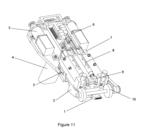
Figure 11. Perspective view of handheld bioprinter in embodiment with
rollers mounted on both sides of printer head.
Figure 12. Side and frontal views of handheld bioprinter in embodiment with
rollers mounted on both sides of printer head.
Figure 13. Exploded view of handheld bioprinter in embodiment with rollers
mounted on both sides of printer head.
Figure 14. Exploded view of handheld bioprinter in embodiment with rollers
mounted behind printer head.
Figure 15. Perspective view of handheld bioprinter in embodiment with
rollers mounted behind printer head.
Figure 16. Side and frontal views of handheld bioprinter in embodiment with
rollers mounted behind printer head.
Figure 17. Schematic illustration of continuous sheet formation in
embodiment where printer head is mounted in spatially fixed position above
moving conveyor surface. Biopolymer sheet produced from one or multiple
biopolymer precursor solutions that may contain colloidal payloads. Cross-
linker
solution (top) and biopolymer solution (bottom) form stratified flow at
printer head
exit, initiating gelation at interface.
Figure 18. Schematic illustration of experimental configuration for sheet
formation in configuration with conveyor belt. A two-layered print head is
supplied
with polymer and cross-linker solutions. Solutions are supplied by reservoirs
either
6
CA 03039553 2019-04-05
WO 2018/064778
PCT/CA2017/051204
via pressure-controlled delivery or via flow rate controlled delivery from
external
syringe pumps. A stationary printer head with protruding section (top) and a
moving conveyor belt (bottom) establish hydrodynamic boundary conditions for
sheet formation and gelation. A stepper motor translates belt at velocity V.
Figure 19. Photograph of 3D printed microfabricated printer head fabricated
for bioprinter embodiment 1. Scale bar 10 mm.
Figure 20. Comparison of side view images after manual deposition of 100p1
droplet of fibrin/HA biopolymer solution (left) and sheet deposited using
handheld
bioprinter embodiment 1 (right). Agarose substrates hydrated with cross-linker
solution were used in both cases. Images were acquired at 4-degree-angle.
Figure 21. Representative optical profilometry image and cross-sectional
view of t=0.3mm sheet with obtained with handheld bioprinter embodiment 1, W=
14mm.
Figure 22. Analytical model prediction (solid lines) indicating QC/(/V.V.H)
and QM/(/V.V.H) conditions required for viscosity ratio pC/pM=0.01 to fulfill
zero
pressure gradient condition. Dashed line corresponds to QC/(/V.V.H) = t/H.
Gelation neglected in model.
Figure 23. Measurement and model predictions for dimensionless sheet
thickness, t*=t/H, as function of dimensionless biopolymer flow rate,
QM*=QM/(VVVH). Measurements obtained for handheld bioprinter embodiment 1.
Figure 24. Characterization of gelation kinetics based on measurement of
time-dependent changes in normalized turbidity of fibrin-based sheets with
different thicknesses. Measurements obtained for handheld bioprinter
embodiment 1.
Figure 25. Microstructure characterization of printed sheets with different
biopolymer compositions using scanning electron microscopy. Measurements
performed for sheets prepared with handheld bioprinter embodiment 1.
Figure 26. Measured Young's moduli (left) and elongations at break (right)
for sheets consisting of fibrin-HA/Col, fibrin-HA, collagen-alginate and
alginate.
Measurements performed for sheets prepared with handheld bioprinter
embodiment 1.
Figure 27. Left: Confocal image of bi-layer sheet prepared by subsequent
deposition of a 0.2mm thickness alginate sheet with payload of green
fluorescent
microparticles (bottom layer), and a 0.1mm thickness alginate sheet with red
7
CA 03039553 2019-04-05
WO 2018/064778
PCT/CA2017/051204
fluorescent microparticles (top layer). Right: Confocal image of three-layer
sheet
prepared by subsequent deposition of a 0.5mm (bottom layer) fibrin-HA sheet
with
blue microparticles, a 0.2mm (middle layer) alginate-collagen sheet with FITC-
conjugated collagen and a 0.15mm (top layer) alginate sheet with red
microparticles. Scale bars 0.1mm. Data obtained for sheets prepared with
handheld bioprinter embodiment 1.
Figure 28. Homogenous printed sheet contains human dermal fibroblasts
(FBs). Live cells indicated by calcein stain, and dead cells indicated by
fluorescent
ethidium homodimer-1. (b) Quantitative assessment of FB viability in printed
fibrin/HA/collagen-I bioink with >90% cell viability during 10-day culture.
(c)
Various concentrations of cells were printed in bioink and quantified using
Hoechst nuclear staining, showing no loss in total cell number due to
printing. (d)
Bilayer construct printed in stepwise fashion. Keratinocytes (k14 & phalloidin
co-
stain) printed on top of FBs (phalloidin) resembling bi-layered structure of
skin.
Data obtained for sheets prepared with handheld bioprinter embodiment 1.
Figure 29. (a) FBs printed within 1.25% fibrin/0.25%collagen/0.25% HA
extracellular matrix material and stained with Hoechst and phalloidin shows
attachment and elongation of cells during 12 hr. (b) Comparison between day 0
and day 3 of human keratinocytes (KCs) printed in fibrin gel using
immunofluorescent staining for cell nucleus, actin, and keratin-14 indicating
cell
grouping and clustering by day 3. (c) Quantitative assessment of FB and KC
cell
numbers of over 3 days of culture. Scale bars: 0.1mm (a, b). Data obtained for
sheets prepared with handheld bioprinter embodiment 1.
Figure 30. Top: Representative photograph showing in situ deposition of
0.25mm thick fibrin-HA/collagen sheet on top of a full thickness excisional
porcine
wound using handheld Skin Printer and close-up view of deposition within wound
area with microfluidic cartridge. Bottom: Day 0 control area and wound 5 min
after
deposition of a layer of biomaterial. Scale bars are lOmm. Data obtained for
sheets prepared with handheld bioprinter embodiment 1.
Figure 31. (a) Trichrome staining indicates extent of granulation tissue
formation and reepithelialization. Arrows in (a) indicate border between newly
formed granulation tissue and intact skin. Arrowheads mark epithelialized
area.
Arrowhead at the center of treated wound shows complete re-epithelializion,
while
central arrowhead in control wound shows non re-epithelialized zone at wound
8
CA 03039553 2019-04-05
WO 2018/064778 PCT/CA2017/051204
center. (b) Keratin 10 staining is showing comparable extent of differentiated
keratinocytes. At the corner of the Printed wounds more keratin posive cells
were
observed, supporting the notion that printed material fibrin-HA sheets likely
enhance wound healing. (c) a-SMA staining reveals comparable number of
.. positive cells in both printed and control wounds. Scale bars: 1mm(a right,
b left),
0.1mm (b right), and 0.05mm (c). Data obtained for sheets prepared with
handheld bioprinter embodiment 1.
Figure 32. Schematic of biomaterials and cells organized into stripe patterns
using microfabricated printer head. (b) Representative confocal image of
striped
monolayer. (c) Relative stripe width Wstripe/WO as function of flow rate
ratio. (d)
Representative images for pressure-controlled spotting. (e) Spot volume as
function of reservoir head pressure for 200m5 actuation time. Scale bars 2mm
(b),
6mm (d). Data obtained for sheets prepared with handheld bioprinter embodiment
1.
Figure 33. (a) Schematic of biomaterials and cells organized into undulated
sheets or parallel fibers using microfluidic cartridge. (b) Representative
bright field
image of an adulated sheet with 8 peaks. The image is capture at 4 degrees.
The
insert shows a zoomed in image of two neighboring peaks at 2 degrees. (c)
Representative reconstructed confocal image of cross-section of a sheet with
four
peeks. (d) Representative reconstructed confocal image of cross-section of a
bi-
layered sheet. The first layer (green) is homogenous. The top layer is made of
four parallel stripes. (e) Mesh pattern printed by printing eight parallel
stripes
perpendicular to one another. (f) Representative multi-material organization
of
Fibrin-HA stripes within alginate sheet. Scale bars 5mm (b), 0.2mm (c, d), 4
mm
(e), 0.5mm (f). Data obtained for sheets prepared with handheld bioprinter
embodiment 1.
Figure 34. (a) Stipe-patterned fibrin-HA sheet deposited on murine wound.
(b). Fluorescent image of stripe-pattern deposition directly on murine
excisional
wound model. (c) Representative image of 4 stripes printed on 8mm wound
.. model. The dashed circle shows the wound edge. The arrow shows the
initiation
phase of the print until it reaches steady-state deposition. 1pm green
fluorescent
microparticles used as label. (d) Normalized fluorescence intensity across in
situ
printed striped alginate sheet (solid line) and fibrin sheet (dashed line) (e)
Estimate of nominal in-plane resolution for bioprinted fibrin-HA sheet
deposited on
9
RECTIFIED SHEET (RULE 91.1)
CA 03039553 2019-04-05
WO 2018/064778
PCT/CA2017/051204
flat surface with inclination angle. (*) Indicates width of channel delivering
bioink
stripes on microfluidic cartridge without flow focusing feature. (**)
Indicates
improved resolution achieved by 3D printed microfluidic cartridge with
internal
flow-focusing features. Scale bars 2mm. Data obtained for sheets prepared with
handheld bioprinter embodiment 1.
DETAILED DESCRIPTION
Various embodiments and aspects of the disclosure will be described with
reference to details discussed below. The following description and drawings
are
illustrative of the disclosure and are not to be construed as limiting the
disclosure.
The figures are not to scale. Numerous specific details are described to
provide a
thorough understanding of various embodiments of the present disclosure.
However, in certain instances, well-known or conventional details are not
described in order to provide a concise discussion of embodiments of the
present
disclosure.
As used herein, the terms, "comprises" and "comprising" are to be
construed as being inclusive and open ended, and not exclusive. Specifically,
when used in the specification and claims, the terms "comprises" and
"comprising"
.. and variations thereof mean the specified features, steps or components are
included. These terms are not to be interpreted to exclude the presence of
other
features, steps or components.
As used herein, the term "exemplary", "illustrative" and "for example" mean
"serving as an example, instance, or illustration," and should not be
construed as
preferred or advantageous over other configurations disclosed herein.
As used herein, the terms "about" and "approximately" are meant to cover
variations that may exist in the upper and lower limits of the ranges of
values,
such as variations in properties, parameters, and dimensions. In one non-
limiting
example, the terms "about" and "approximately" mean plus or minus 10 percent
or
less.
Unless defined otherwise, all technical and scientific terms used herein are
intended to have the same meaning as commonly understood to one of ordinary
skill in the art.
3D bioprinting strategies aim at reconstituting structural elements of native
.. tissues by controlling the position of different cell types and
extracellular matrix
CA 03039553 2019-04-05
WO 2018/064778
PCT/CA2017/051204
components. The provided microenvironment and spatial organization influence
cell migration, elongation, clustering, proliferation, differentiation, and
function.
Current bioprinting platforms offer promising in-vitro results but are not yet
compatible with clinically relevant settings. Higher print rates, reduced
preparation
and wait times, and compact solutions for on-site deposition or transfer of
organ-
scale printed tissues are required to ultimately treat patients with acute and
complex wounds that are amongst the most impacfful clinical and economical
challenges. Disclosed herein is a handheld skin printer that overcomes these
limitations by in-situ formation of wound-adhesive skin substitutes.
The present disclosure provides bioprinter for controlled in-situ formation
and deposition of biopolymeric sheets and planar tissues on surfaces,
comprising:
a) support frame and a printhead attached to the support frame, the
printhead including a first array of extrusion channels and at least a second
array
of extrusion channels located with respect to the first array such that in
operation
the first array is proximally adjacent to the surface, an end section of the
printhead
having a width W such that the first and second arrays span the width W;
b) a first reservoir attached to the frame, the first array of extrusion
channels being in flow communication with the first reservoir of biopolymer to
be
extruded onto the surface, a second reservoir of liquid attached to the frame,
the
second array being in flow communication with the second reservoir of liquid
to be
extruded along with the extruded biopolymer, and including a first dispensing
mechanism associated with the first reservoir being configured to dispense
biopolymer at a flow rate of QM, and a second dispensing mechanism associated
with the second reservoir being configured to dispense the liquid at a flow
rate of
QC;
c) a drive mechanism attached to the frame such that when activated by
the operator, the printhead is driven along the surface located a vertical
height H
above the surface at a preselected velocity V;
d) a controller connected to the drive mechanism and the first dispensing
mechanism and programmed such upon activating the drive mechanism, the first
dispensing mechanism dispenses biopolymer at the flow rate QM a layer of
thickness t, which satisfies the condition QM = W.V.H(6(t/H) ¨
6(t/H)2+3(t/H)2 Gic/[10) / (6(t/H) Gic/[10¨ 6(t/H) +6).
11
CA 03039553 2019-04-05
WO 2018/064778
PCT/CA2017/051204
In an embodiment the drive mechanism is configured to provide variable
velocities V, and wherein the controller is programmed with instructions to
control
the first dispensing mechanism to responsively adjust the flow rate QM such
that
for a given velocity V the flow rate conditions are maintained.
In an embodiment the second dispensing mechanism may be operably
coupled with the controller and is configured to dispense the liquid at the
flow rate
QC which satisfies the condition
QC = 0.5 W.V. (H ¨ t)
In an embodiment the drive mechanism is configured to provide variable
velocities V, and wherein the controller is programmed with instructions to
control
the first and second dispensing mechanisms to responsively adjust the flow
rates
QM and QC such that for a given velocity V the flow rate conditions are
maintained.
In an embodiment the exit section of the printhead includes an overhanging
section extending outwardly from a top surface of the second array, the
overhanging protruding section extending outwardly from the exit section by a
length L.
In an embodiment the length L is equal to or greater than the value of H.
In an embodiment the first array of extrusion channels are in flow
communication with the first reservoir via a bifurcating channel network
comprised
of a first channel connected to the first reservoir which bifurcates into two
channels which further bifurcates until a final number of channels equals a
number of extrusion channels in the first array, and an end of each channel is
adjacent an end of a corresponding extrusion channel in the first array, and
wherein the second array of extrusion channels are in flow communication with
the second reservoir via a bifurcating channel network comprised of a first
channel
connected to the second reservoir which bifurcates into two channels which
further bifurcates until a final number of channels equals a number of
extrusion
channels in the second array, and an end of each channel is adjacent an end of
a
corresponding extrusion channel in the second array.
12
CA 03039553 2019-04-05
WO 2018/064778
PCT/CA2017/051204
In an embodiment the hydraulic diameters of the channels in the bifurcating
channel networks decrease from each inlet to each exit going from the
reservoir to
the printer head in accordance with Murray's law.
In an embodiment the bioprinter further comprises a handle for allowing a
user to grasp and use the bioprinter during dispensing operations so that the
bioprinter is a handheld bioprinter.
In an embodiment the drive mechanism comprises a pair of axel mounted
rollers connected to the drive mechanism, and wherein the printer head is
positioned between the rollers, and wherein the end section includes a
circular
guidance feature maintains a consistent gap height between the channel device
exit and deposition surface regardless of changing the deposition angle, and
wherein during operation upon activation of the drive mechanism, the pair of
axel
mounted rollers are rotationally driven such that the handheld bioprinter
moves
along the surface at the velocity V.
In an embodiment the drive mechanism comprises a roller connected to the
drive mechanism, and wherein the roller is positioned behind the printhead,
and
wherein end section contains a circular guidance feature to maintain a
consistent
gap height between the channel device exit and deposition surface regardless
of
changing the deposition angle, and wherein during operation upon activation of
the drive mechanism, the roller is rotationally driven such that the handheld
bioprinter moves along the surface at the velocity V.
In an embodiment the drive mechanism comprises a translation
mechanism attached to the frame, the printhead being mounted on the
translation
mechanism, the translation mechanism being configured to move the printer head
at the velocity V with respect to the surface.
The tissue printing device will now be described with reference to the
Figures and the following parts list.
13
CA 03039553 2019-04-05
WO 2018/064778
PCT/CA2017/051204
Parts List
Label Part Description
1 Printer head Printhead manufactured using one of the
manufacturing processes: 3D-printing, thermal
embossing, or micro-injection molding. Device bottom
translates proximal to deposition surface. Device exit
of width W, optional protruding section of length L,
positioned at height H above deposition surface.
Biomaterial or tissue sheet of thickness t produced.
2 Front block Serves to mount drive mechanism to base plate.
3 Temperature Controls temperature of solutions within reservoirs
control and printer head, prior to extrusion. Thermoelectric
element, cooler, aluminum syringe jacket, two wells
for pressure-controlled delivery of secondary
biopolymer solution.
4 Handle Used for holding of instrument by human operator.
Enables positioning of handheld bioprinter above
target surface or wound. May include switch to initiate
controlled sheet deposition.
Dispensing Modular dispensing system controlling flow rates QM
system and QC for solutions supplied from separate
reservoirs. Dispensing systems individually consist of
a stepper motor, a belt-drive with pulleys, a screw-
based linear translation mechanism, a push pin and a
push button.
6 Reservoir
scaffold
7 Base plate Plate for mounting of reservoirs, dispensing system,
handle, and printer head holder
8 Reservoirs Biopolymer solution supplied from corresponding
reservoir at flow rate QM, and flow confining solution
at flow rate QC. Reservoirs may include standard BD
syringes with sizes (1cc, 3cc, 5cc, 10cc, 20cc).
9 Tubing Delivers solutions from reservoirs to printer head.
Roller driving Defines deposition speed V along surface. Consists
system of one or two rollers, a belt drive with two pulleys,
one
shaft, and one stepper motor.
11 Printer head Holding mechanism for accommodating printer head
holder and adapter. Spring-loaded to gently push printer
head with consistent force against deposition surface.
12 Printed Homogenous or heterogeneous biopolymer or tissue
biomaterial or sheet
tissue sheet
13 Surface or Deposition or wound surface.
Wound
14 Switch Start/stop switch to drive motor activity.
Conveyor belt Conveyor belt moving at velocity V.
14
CA 03039553 2019-04-05
WO 2018/064778
PCT/CA2017/051204
Disclosed herein is a bioprinter for controlled deposition of biopolymeric
sheets onto surfaces which includes a support frame having a printer head
attached thereto.
The precursor solution that constitutes the printed sheet is a mixture of
natural or
synthetic biopolymer solution with cells and/or growth factors, but is not
limited to
extracellular matrix materials or any structural analogs. Synthetic polymers
approved for clinical use and shown to be effective may be a potential
application
due to its advantage of large-scale synthesis without batch-to-batch
variation.
The biopolymer is loaded onto one of the handheld printers (Figure 1, 11, 12,
13)
reservoir including but not limited to standard BD syringes ranging from 1-
20cc or
3D printed features, and maintained at a desired storage temperature. As the
biopolymer solution is perfused through the printer head and deposited on site
of
the injury, it is polymerized and thus solidifies. The solidification can be
induced
via different mechanisms that include ionically induced, pH induced, and
temperature induced gelation, as well as enzymatic reactions and
polymerization
induced by ultraviolet light and combinations thereof. For natural biopolymers
like
fibrinogen, the crosslinking can be initiated from the plasma in the wound
bed.
The printer head includes a first array of extrusion channels and a second
array of
extrusion channels located with respect to the first array such that in
operation, as
the printer is dispensing or extruding one or more layers, the first array is
proximally adjacent to the surface on which the layer(s) is being deposited. A
biopolymer that may contain a cross-linker for premixing is generally perfused
through the first array of channels so that a uniform coating is applied to
the
wound, or another surface depending on how the bioprinter is configured,
discussed hereinafter. A confining or secondary fluid that may contain a cross-
linker is perfused through the second array of channels and delivered to the
biopolymer coating on the side adjacent to the wound or deposition surface. If
a
cross-linker was added to the confining fluid, the cross-linker is diffusively
transported into the biopolymer layer.
The end section of the printer head (see Figure 8, 9, 10, 19) from which
the layer is being dispensed has a width W such that the first and second
arrays
span this width W. A first reservoir is attached to the base plate and is in
upstream
flow communication with the first array of extrusion channels which when in
operation will contain the biopolymer of viscosity [inn to be extruded
directly onto
CA 03039553 2019-04-05
WO 2018/064778
PCT/CA2017/051204
the surface. A second reservoir of a confining or secondary liquid is attached
to
the frame which is in upstream flow communication with the second array
through
which the secondary liquid of viscosity [Lc is co-extruded on top of the first
layer. A
dispensing mechanism associated with the first reservoir is configured to
supply
the biopolymer at a volumetric flow rate of QM, and another dispensing
mechanism associated with the second reservoir is configured to supply the
secondary or confining liquid at a volumetric flow rate of QC.
The bioprinter includes a drive mechanism attached to the frame such that
when activated, the print head is driven along the surface located a vertical
height
H above the surface at a preselected velocity V. A controller is connected to
the
drive mechanism and the first dispensing mechanism and the controller is
programmed such upon activating the drive mechanism, the first dispensing
mechanism dispenses biopolymer of thickness t at the flow rate QM which
satisfies the condition:
QM = W.V.H(6(t/H) ¨ 6(t/H)2+3(t/H)2 ([1c/m)) / (6(t/H) (K41.0¨ 6(t/H) +6)
see Figure 22.
In most cases the confining solution is of much lower viscosity, [Lc,
compared with the viscosity of the biopolymer solution, [IM, and the
relationship
simplifies to QM = W=V=t. W is a design feature of the print head and thus is
fixed,
and is usually selected to be in a range from about 5 mm to about 30 mm. As
discussed later, the forces across the print head are preferably even to
ensure
uniform dispensing laterally across the width of the first array of extrusion
channels in the print head. The thickness t of the dispensed biopolymer is
typically in the range in range from about 0.01 mm to about 1 mm, see Figures
21
and 23.
The thickness t should be less than approximately 1 mm in order to allow
nutrient supply for cells without vascularization. The thickness of the
biopolymer
sheet may be selected according to the target tissue thickness in healthy
skin, and
the severity of the wound. If the skin injury is only partial thickness and
the dermal
layer remains intact, the sheet thickness that is to be printed is about 0.3
mm. For
basal lamina, only 0.01 mm sheet of an engineered composition is necessary. If
16
CA 03039553 2019-04-05
WO 2018/064778
PCT/CA2017/051204
the dermal layer is also damaged, (full thickness injuries) thicker sheets are
necessary. Dermal and epidermal layer in the skin have different thicknesses
on
the body based on the injury. The layers are printed sequentially or co-
extruded
simultaneously. Each layer can have different composition and cell type load
(see
Figures 25, 26, 28 and 29).
V is typically in the range from about 1 mm/sec to about 20 mm/sec,
velocities in the range of 1 to 8mm/s may be preferred. For the case of slow
gelation (e.g., thick sheet, no premixing) a lower velocity may be preferred
(see
Figures 14, 16, 24) where the measurement and model predictions for sheet
thickness are provided, modifying the velocity V corresponds to the change in
sheet material thickness t. The product of print head width W and velocity V
determines the coverable area per time. For large skin injuries (40% burn in
an
average size male translates to approximately one square meter) there is an
interest in covering wounds rapidly. With a W = 20mm printhead the printer can
cover one square meter in less than half an hour. H should be at least twice
the
target sheet thickness, t. H may vary between 0.15mm and 2mm. t may vary
between 0.01mm and 1mm.
As noted above, the dispensing mechanism and drive mechanism are
configured to give a biopolymer flow rate QM which satisfies the flow
condition
QM = W.V. t. The operator decides the target thickness. Velocity V is selected
depending on the gelation kinetics. Flow rate QM (and QC) is for a given print
head design (L, H, and W) calculated using the above relationship and selected
by the operator on a computer through a user interface. In an embodiment this
will be done via a single switch on the handle (see Figure 2, and Part 4 in
Figure
15).
The bioprinter may be configured to provide variable velocities V, and the
controller is programmed with instructions to control the first dispensing
mechanism to responsively adjust the flow rate QM such that for a given
velocity
V the flow rate conditions are maintained.
In an embodiment the flow rate of the liquid, QC, may also be controlled to
satisfy certain conditions. In this case the second dispensing mechanism is
also
connected to the controller and is configured to dispense the liquid at the
flow rate
QC which satisfies the condition
QC = 0.5 W.V.(H ¨ t).
17
CA 03039553 2019-04-05
WO 2018/064778
PCT/CA2017/051204
When the flow rates QM and QC are both selected to satisfy the above
conditions, the drive mechanism is configured to provide variable velocities
V, and
the controller is programmed with instructions to control the first and second
dispensing mechanisms to responsively adjust the flow rates QM and QC such
that for a given velocity V the above-noted flow rates are maintained.
The controller may be a computer microprocessor with a visual display
indicating the flow rates of QM and QC, and the velocity of bioprinter motion
in the
lateral direction V. Values for QM, QC, and V can be input through the
computer
and the corresponding motor speeds will be updated in real time. An on/off
switch
located on the handle will start or stop the extrusion and/or lateral motion
of the
handheld bioprinter.
The bioprinter may be configured such that the exit section of the printer
head includes an overhanging section extending outwardly from a top surface of
the second array. This overhanging protruding section extends outwardly from
the
exit section by a length L which is equal to or greater than the value of H as
shown in Figure 17.
The first array of extrusion channels are in flow communication with the first
reservoir via a bifurcating channel network comprised of a first channel
connected
to the first reservoir which bifurcates into two channels which further
bifurcates
until a final number of channels equals a number of extrusion channels in the
first
array wherein the downstream end of each channel is adjacent an end of a
corresponding extrusion channel in the first array. The second array of
extrusion
channels are in flow communication with the second reservoir in the same way
as
the first array described above.
Alternatively, the biopolymer and the cross linker can be pre-mixed within
the printer head. In this case, the fluids that are being delivered from the
reservoirs pass through a microfabricated mixer prior to the extrusion
channels. In
this configuration, the flow confining solution may consist of a buffer
solution
without promoting gelation. The sheet thickness in this case is
t=((QM+QC,PREMIX)/VV*V), where QC,PREMIX is the volumetric flow rate of the
crosslinker added to the biopolymer solution for pre-mixing.
Alternatively, an ultraviolet light source may be positioned directly on top
of
the cartridge exit. The solidification may be initiated by free radical
polymerization
of the biopolymer. The biopolymer is mixed with a photo initiator before being
18
CA 03039553 2019-04-05
WO 2018/064778
PCT/CA2017/051204
loaded on to the handheld bioprinter, or the photo initiator can be mixed
inline on
the print head. As the biopolymer is being extruded on site of the injury, a
sheet of
ultraviolent light generated by a light emitting or laser diode may serve to
polymerize the biopolymeric sheet. The sheet thickness in this case is t =
(QM/WV).
The solidification mechanism can also be applied by thermal gelation. In
this embodiment, the biopolymer solution is either kept in either a heated or
cooled condition within the reservoir and print head. As the biopolymer exits
the
cartridge and comes in contact with the wounded area, it gels and solidifies.
The
sheet thickness is t = (QMANN).
The hydraulic diameters of the channels in the bifurcating channel networks
decrease from each inlet to each exit going from the reservoir to the printer
head
to increase the flow resistance at the distribution channels and ensure the
uniformity of the deposited sheet One way of increasing the flow resistance in
each step of bifurcation is decreasing the hydraulic diameter of the daughter
branches in accordance with Murray's law. Murray's law predicts the dimensions
of branches in a transport network to minimize the work attributed to the
transport
and maintenance of the medium. For n daughter branches splitting from a
common mother branch, Murray's law states that r3 = ri3 + r23 + r33 + + r3"
where r is the radius of the parent branch and are the
radii of the daughter
branches (Sherman, TF, J Gen Physiol, 1981). Due to the increase in channel
width while keeping the channel depth constant for subsequent branching
architecture, there is a decrease in resistance to reduce the pressure
subsequently the chance of biomaterial clogging (see Figure 8).
A non-limiting example of the reservoir and dispensing mechanism
illustrated in the Figures described hereinafter is a syringe with a plunger
with the
plunger connected to a motor, such as a stepper motor which drives, via a
toothed
motor shaft, a toothed gear belt which is engaged with a toothed gear on the
plunger so that the controller acts to control the rotation of the stepper
motor shaft.
It will be appreciated that this embodiment is not limited to stepper motors,
servomotors, DC motors, pneumatic drivers, or other types of linear drives.
In addition, other types of dispensing mechanisms may be used other than
the above described motor, gear and toothed gear belt. For example, a square
wave pressure signal can be applied to the air-filled reservoir headspace. A
19
CA 03039553 2019-04-05
WO 2018/064778
PCT/CA2017/051204
solenoid valve controlled with an Arduino Mega microcontroller to change the
frequency and duty cycle of the pressure signal can be used (see Figure 7).
The
spot size and volume can be controlled by adjusting the upper pressure level
and
the valve open times. Target volumetric flow rates QM and QC may also be
indirectly selected by individually controlling the head pressure of
reservoirs using
a pressure regulator. The relationship between the applied inlet pressure and
the
obtained flow rate can be obtained for the different fluids from calibration
measurements.
The microfluidic printhead can allow the organization of the biomaterial in
the planar direction. Multiple reservoirs can be attached to the distributing
channel
within a layer and deposited in a stripe configuration. The geometry and the
widths of the deposited stripes can be controlled by tuning the relative flow
rates
of the biopolymer solutions coming from each reservoir (see Figures 5, 6, 32).
The crosslinker and biopolymer can be coextruded in a planar geometry to
achieve undulating sheets or parallel fibers (see Figure 6, 33).
The tissue bioprinter may be configured to be mounted independent of an
human operator, for example in cases where it is desired to produce a coating
on
a moving conveyor (see Figures 4, 18), for instance to enable continuous roll-
to-
roll processing of biomaterial and tissue sheets.
In another embodiment, the bioprinter may be configured to be handheld by
a clinician running the device over the surface with one hand. In this
situation the
bioprinter is configured to have a handle to be gripped by the clinician. The
handle
may be ergonomically designed with a velocity control switch or button for the
clinician to engage as the printer is moved over the wound area of the
patient. In
.. one embodiment the velocity control switch is configured to give one set
velocity
when the switch is engaged, in another embodiment the velocity control switch
may be configured to give a variable velocity depending on how far the
clinician
depresses the switch.
For the handheld bioprinter the drive mechanism may comprise a pair of
axel mounted rollers connected to the drive mechanism, and wherein the printer
head is positioned between the rollers, and the angle between the end of the
printer head and the surface is maintained by the human operator. The exit
section of the print head is located one drive wheel radius below the axis of
rotation and the direction of extrusion is tangential to the drive wheel. This
CA 03039553 2019-04-05
WO 2018/064778
PCT/CA2017/051204
configuration allows for consistent sheet deposition even during small changes
of
deposition angle. The bottom side of the print head is positioned in close
proximity
of the deposition surface and H is maintained by design. In the case where the
printer head is positioned above a conveyor at a fixed angle the height H can
be
selected independently and maintained over the course of the deposition.
During
operation upon activation of the drive mechanism, the pair of axel mounted
rollers
are rotationally driven by a motor, such as, but not limited to, a stepper
motor,
toothed gear and gear belt forming part of the drive mechanism, such that the
handheld bioprinter moves along the surface at the selected velocity V.
Alternatively, the drive mechanism comprises a roller connected to the
drive mechanism, and the roller is positioned behind the print head (Figure
2). In
this case, the print head is mounted in such a fashion that it may rotate
around the
rotational axis of the roller. The end section of print head out of which the
biopolymer is extruded is brought into contact with the deposition surface by
a
.. spring mechanism. During operation upon activation of the drive mechanism,
the
roller is rotationally driven by a motor such as, but not limited, to a
stepper motor,
toothed gear and gear belt forming part of the drive mechanism such that the
handheld bioprinter moves along the surface at the selected velocity V (Figure
14,
15). The drive mechanism is not restricted to stepper motors, toothed gears
and
toothed gear belts. Other types of drive mechanisms may be comprised of servo
motors, and pneumatic drives.
Alternatively, the movement can be achieved by the operator actively
moving the printer. In this embodiment, the movement is measured with an idler
wheel, or a contact free motion detection method, like any motion sensor, an
accelerometer, or laser light. In this case, the movement is registered and
calculated by the computer and the syringe pumps or air pressure governing the
flow rate of the biopolymer and crosslinker is controlled and adjusted in a
closed
loop.
Alternatively, the handheld bioprinter may be configured to remain
.. stationary during deposition and only the printer head is moved via a
translation
mechanism forming part of the drive mechanism with the printer head being
mounted on the translation mechanism (Figure 3). The translation mechanism is
connected to the controller which is programed to instruct the translation
21
CA 03039553 2019-04-05
WO 2018/064778
PCT/CA2017/051204
mechanism to move the printer head at the selected velocity V with respect to
both the surface and the rest of the handheld bioprinter.
The bioprinter device enables the controlled deposition of biopolymeric
sheets onto a substantially flat or curved surfaces. The materials are
deposited
onto flat or curved surfaces, or directly onto wound areas. Sheets may have a
homogeneous or heterotypic composition (Figure 20, 27, 32, 33, 34). Aspect
ratios (width to height, w/t) are between 10 and 3,000.
The bioprinter device enables the controlled deposition of biopolymeric
sheets onto a substantially flat or curved surfaces (Figure 30, 31, 34). The
materials are deposited onto flat or curved surfaces, or directly onto wound
areas.
Sheets may have a homogeneous or heterotypic composition. Aspect ratios (width
to height, w/t) may be in a range between 10 and 3,000.
The print mechanism is not limited to producing homogenous layered
sheets. Using the printer disclosed herein, more complex tissues can be
fabricated in-situ from a bottom-up approach. Each layer may be tuned to have
the desired geometry and composition. Any additional layer may be deposited on
top of the mentioned layered.
The application of in-situ bioprinting using the present device and strategy
is not
limited to topical and skin surgeries. Any tissue adhesive, or more complex
geometries can be applied and implemented on internal organs as well in a
surgery.
The application of the present printer is not restricted to the mentioned cell
types. Other cell types like IFS derived cells, and other micro-organisms like
bacteria, and fungi can be printed in-situ and organized within hydrogel
sheets
using this method. The load can have emulsions of microparticles, gold and
silver
nanoparticles, microbubbles, graphite, conductive inks, and any other mixture
and
suspension of the mentioned materials can also be printed using this method.
The
application of this method is not limited to biomaterials. Beauty supplies,
tattoos,
creams, topical coverings, motion and flex sensors, conductive inks, among
others, can be patterned and printed in-situ.
The use of the present tissue printer in both in-vitro and in-vivo studies
will
now be described with the following non-limiting examples
22
CA 03039553 2019-04-05
WO 2018/064778
PCT/CA2017/051204
IN-VITRO AND IN-VIVO STUDIES
Particularly, both in-vitro and in-vivo studies were performed using the
handheld embodiment of the tissue printer. For the former, the inventors
coated
the bottom surface of a dish or multi-well plate with a hydrogel layer (e.g.,
agarose
or gelatin) and hydrated it with the cross-linker solution. The hydrophilic
and
biologically inert surface ensures printing consistency and provides the
bioprinted
skin tissues with mechanical support during culture. After the handheld tissue
printer deposited the bioink layer, gelation was induced by diffusive release
of
cross-linker from below as well as the cross-linker layer co-extruded at the
top.
.. Depending on the application, the bioprinted skin substitutes may be
cultured in
the same dish, or cut and transferred after 2-10 min (depending on the sheet
thickness) to another dish, multi-well plate, transwell insert, or to a wound
site. As
a case study that serves to illustrate the compatibility of the approach with
direct
deposition in-vivo, we deposited the bio-ink layer directly onto a wound bed.
Methods
Preparation of Agarose Substrate
A solution of 2% agarose (UltraPure Agarose, 16500100, lnvitrogen) in de-
ionized (DI) water was prepared by microwave heating. The solution was allowed
to cool to 60 C prior to being poured into sterile square petri dishes (model
Z692344, Sigma Aldrich) and resulted in a 3 mm thick gel. The gel solidified
at
room temperature for 30 min prior to use. For preparation of sodium alginate-
based sheets, 50 mM calcium chloride (CCL302, BioShop) was added to the
solution prior to microwave treatment. For printing of fibrin-based sheets, 2
ml of
50 IU thrombin (T4648, Sigma Aldrich) in PBS (10010023, Gibco) was pipetted to
hydrate the agarose substrate before extrusion.
Bioink Preparation
Bioinks with three different compositions were prepared. For alginate-
collagen sheets, sodium alginate (Pronva UPLVG, Novamatrix) was dissolved in
DMEM (11965-084, Gibco) and 20 mM HEPES (15630080, Gibco) and filtered
using 0.1[1m syringe microfilter (Millipore). Collagen type 1 (rat tail,
354249,
Corning) was balanced to a pH of 7 using 1 M NaOH in PBS. The two stock
solutions were mixed to obtain a final concentration of 5 mg/ml collagen and 2
%
alginate. The solution was kept on ice prior to use. To prepare the bioink for
the
dermal layer 5% fibrinogen (F8630, Sigma) was dissolved at 37 C in PBS with
23
CA 03039553 2019-04-05
WO 2018/064778
PCT/CA2017/051204
mild agitation for 2 h. 1% HA (sodium hyaluronate Pharma Grade 80, Novamatrix)
was dissolved in PBS. The solutions were mixed at a ratio of 1:1 and then
filtered.
Collagen type 1 solution was balanced with NaOH to a pH of 7 and mixed with
the
filtered Fibrin/HA solution to obtain a final concentration of 1.25%
Fibrinogen,
0.25% HA and 0.25% Collagen. The solution was kept on ice prior to use. The
bioink for the epidermal layer was prepared with a final concentration of 2.5%
fibrinogen and 0.25% HA.
For printing the fibrin based sheets, a layer of 50 IU thrombin was co-
extrusion above the fibrinogen based dermal and epidermal bioinks. The rapid
enzymatic reaction between fibrinogen and thrombin is mass transfer limited in
the
considered case. The selected approach allowed the formation of sheets on the
site of the deposition which solidified at time scales between tens of seconds
and
several minutes, depending on the thrombin concentration and sheet thickness,
t.
The gelation time is directly dependent on the sheet thickness. For the dermal
bioink consisting of a mixture of collagen and fibrinogen, the gelation of
fibrinogen
occurs first and is induced by the diffusion of thrombin. As a result the
sheet
thickness and composition are maintained while the slower thermally induced
gelation of neutral pH collagen progresses.
The alginate-based sheets were prepared by co-extrusion of 10 mM calcium
chloride above the biopolymer layer. Similarly, rapid ionic cross-linking of
alginate
preceded the slower thermal gelation of neutral pH collagen. After the
gelation of
the sheet was completed, alginate was removed by incubating the sheet in
1mg/m1 alginate lyase (A1603, Sigma) for 30 min.
Physical Characterization of Deposited Skin Substitutes
Physical characterization of the deposited sheets included sheet thickness
and contact angle measurements, the measurement of spot and stripe sizes,
tensile strength. The microstructure was characterized by scanning electron
microscopy (SEM) for samples.
Sheet Thickness
The precursor solutions were mixed with 5% 0.2 pm diameter fluorescent
microparticles (FP-0245-2 or FP-0256-2, Spherotech). The sheets were
transferred onto microscope cover slides and imaged using a confocal
microscope
(model Al, Nikon) using a digital camera (model Retiga 2000R Fast 1394, Q
Imaging). The images were analyzed using the ImageJ software. Thickness of 5
24
CA 03039553 2019-04-05
WO 2018/064778
PCT/CA2017/051204
random points on each sample were averaged and a total of 5 random points
were selected for each experimental condition. The sheet thickness was also
determined using an optical profilometer (model Contour GT-K, Bruker). The
sheets were sectioned while attached to the agarose substrate and transferred
to
the profilometer stage. The Vision64 software program was used to analyze and
export the sheet thickness data. 3D profile data were then imported into
MatLab.
Reported thickness data correspond to local averages over a 0.5x0.5 mm2 region
of interest for each of the 5 randomly selected points on a sample. For each
experimental condition, n=3 sheets were measured. Figure 21 shows the
measurement data. The flow rates that are used in this research for obtaining
various thicknesses are derived from the model shown in Figure 22. Figure 23
shows measurement data for sheets prepared in different biopolymers compared
with model predictions.
Contact Angle
We deposited sheets of alginate-collagen, fibrin-collagen and fibrin on
agarose that were lOmm long, 14mm wide and 250pm thick. In a parallel test, a
comparable volume of the fibrin-HA bioink, 35 pL, was pipetted onto an agarose
substrate and allowed to gel under saturated atmosphere (humidity 100%) within
an incubator. The shapes of the deposited droplet and the sheet obtained using
the handheld Skin Printer were photographed with a Drop Shape Analyzer
(DSA30, KRUSS) at 2 inclination angle with respect to the substrate plane.
Turbidity Measurements
In-situ turbidity measurements (1ST) were conducted as follows. The beam
of a continuous wave argon-ion laser (A= 488nm, 200mW, Spectra-Physics) was
expanded ten times, guided with a mirror to vertically penetrate an optically
clear
measurement section and collected by an amplified photodetector (Thorlabs).
Sheet deposition experiments were conducted on top of the measurement
section, within an agarose coated petri dish. The absorbance of the petri dish
and
the agarose was found to be negligible. The handheld tissue printer was used
to
deposit sheets of fibrin-HA bioink with different thicknesses (100, 200, 400,
and
600 pm) on top of the agarose layer at a deposition speed of V= 4mm/s. The
voltage signal (U) generated from the transmitted laser light was acquired
using
an oscilloscope (Tektronix). To quantify 1ST, the recorded voltage on the
oscilloscope was converted to absorbance by A = ¨log(U /U0) = al where U0 is
CA 03039553 2019-04-05
WO 2018/064778
PCT/CA2017/051204
the voltage read by the oscilloscope in the absence of a sheet, a is the
absorption
coefficient (cm-1), / is the length of the light path equivalent to the sheet
thickness,
t. The turbidity, r = (1) /n10, was plotted over time as shown in Figure 24.
Turbidity measurements were recorded approximately 3s after the cartridge of
the
handheld tissue printer translated out of the light path.
Stripe and Spot Sizes
Two syringes of bioink were prepared and loaded onto the handheld tissue
printer. A secondary biopolymer solution contained fluorescent Nile red
microparticles (FCM-1056-2, Spherotech) and a primary biopolymer solution
without fluorescent particles. The cross-linker solution was supplied with an
external syringe pump (PHD 2000, Harvard Apparatus). Stripe patterned sheets
were deposited using a dedicated microfluidic cartridge design. Its
microchannel
configuration allowed the formation of biopolymeric sheets where stripes or
spots
of the secondary biopolymer were periodically incorporated within the primary
biopolymer solution. Varying the flow rate ratio of the secondary and primary
solutions allowed the relative stripe widths to be controllably varied. Upon
gelation, stripe-patterned sheets were transferred onto cover slides for
confocal
microscopy. The reported stripe width represents an ensemble average over the
individual stripe widths measured at 3 points (top, middle, bottom), over all
stripes.
ImageJ was used for image analysis.
In order to obtain spotted patterns, another dedicated microfluidic cartridge
design was prepared that supplied the secondary biopolymer solution from an on-
chip reservoir (i.e., instead of a syringe pump). After priming the reservoir
with the
fluorescently labelled secondary biopolymer solution, a square-wave pressure
signal was applied to the air-filled reservoir headspace. A solenoid valve
(LHL
series, Lee Company) controlled with an Arduino Mega microcontroller to change
the frequency and duty cycle of the pressure signal. Spot size and volume were
controlled by adjusting the upper pressure level, and the valve open times.
Tensile Properties
Uniaxial tensile measurements of the collagen and agarose sheets were
measured using a custom tensile tester based on the design described by
Tremblay et a!32. The hydrated sample sheets of approximately 10 mm length
26
CA 03039553 2019-04-05
WO 2018/064778
PCT/CA2017/051204
were on two opposing sides held by custom C-shaped clamps with sandpaper
attached to clamp surfaces. Clamps were positioned in the vertical direction,
z,
using manual translation stages (MT113, Thorlabs). Motion along the direction
of
pulling, x, was controlled by a linear voice coil motor (LVCM-051-051-01,
MotiCont) on a ball bearing slide (37-360, Edmund Optics). A motion controller
(DMC-4143, Galil) was addressed using a custom LabVIEW software program
and controlled the displacement of the voice coil motors in feedback mode with
an
optical encoder (MII1610S-40, Celera Motion) signal. Samples were pulled at a
speed of 0.01 mm/s and displaced until fracture. A Load cell (Model 31 Low,
.. Honeywell) measured the force at a given displacement. A DAQ Card (USB-
1208L5, Measurement Computing) and amplifier (Model UV-10, Honeywell) were
used to transfer the signal from the load cell to the motion controller.
Sample
length and width were evaluated using Zeiss and Nikon Ti inverted microscopes,
to calculate the cross-sectional area. The latter in combination with the
motor
position and the load-cell corrected force resulted in a stress-strain curve.
A linear
regression was fitted to the elastic region to calculate the Young's modulus
of
each sample. Figure 26 shows results from tensile measurements.
Microstructure
Alginate, collagen, and fibrin based biomaterials were fixated for one hour
with Karnovsky's style fixative in Sorensen's buffer at room temperature, then
dehydrated in serial ethanol washes with solutions containing between 30% and
100% ethanol. Samples were then dried using a critical point dryer. Gold was
sputtered, prior to imaging on a scanning electron microscope (S-3400N,
Hitachi)
using an accelerating voltage of 30 kV. Figure 25 shows electron micrographs
of
printed biomaterial sheets with different compositions.
In-vitro Characterization
Cell Sources:
HDFa were obtained from healthy human normal skin after surgery. Cells
were cultured in growth medium (DMEM, 10% fetal bovine serum (FBS) and
1% antibiotic/antimycotic (Ab/Am) until near confluency and split into further
passages by treatment with 0.05% trypsin-EDTA treatment. Human umbilical
epidermal keratinocytes (C-011-5C, Gibco lnvitrogen) were cultured according
to
27
CA 03039553 2019-04-05
WO 2018/064778
PCT/CA2017/051204
company instructions in EpiLife Medium with 1% HKGS and 1% Ab/Am and
trypsinized using the same trypsin-EDTA solution as for fibroblasts. Cell
passages
3-5 were used throughout this study.
Deposition of Cell-Populated Skin Substitutes
For in vitro preparation of skin substitutes, the dermal bioink contained
0.5.106 human primary fibroblasts and then deposited as a 8p=500 pm thickness
sheet on agarose. The sheet was incubated at 37 C for 20 min to allow for
complete thermal gelation of collagen. The epidermal bioink contained 1.5.106
human primary keratinocytes. The epidermal layer was printed sequentially on
top
of the dermal layer with a thickness tE=200 pm. Depending on the study,
different
patterns of epidermal layer (homogeneous or striped) were deposited on top of
the dermal layer. The sheets were then immersed in culture media (EpiLife
Medium, with 60 pM calcium, GibcoTm). The sheets were the detached from the
agarose substrate, sectioned to desired sizes and cultured in multiwell
plates.
Cell Viability
Human dermal fibroblasts and keratinocytes were cultured in collagen/fibrin
and fibrin gels, respectively, for three days in EpiLife media with HKGS
growth
supplement and 1% penicillin-streptomycin. Cells were then stained using
calcein
and ethidium homodimer for analysis of live versus dead cells, in addition to
Hoechst as a cell nucleus stain. Confocal images of cells in biomaterials in
individual 96-well plates were taken at 4x and 10x magnification with three
biological replicates. Percentage viability was calculated by using the ImageJ
software to count the number of cells stained positive for either calcein or
ethidium
homodimer and comparing them with the total number of cells as indicated using
Hoechst nuclear stain as shown in Figure 28a,b.
Cell Density
Human dermal fibroblasts and keratinocytes were cultured for three days
using the previously described method. Cell density was observed by staining
the
cells with Hoechst, imaging with confocal microscopy, and automatically
calculating the total number of cells using ImageJ software.
Immunohistochemistry
Cells in sheets were fixed with 4% paraformaldehyde in HBSS for 1 h at
room temperature then washed with HBSS. They were permeabilized with 0.5%
Triton X-100 in HBSS for 30 min at room temperature and then washed with
28
CA 03039553 2019-04-05
WO 2018/064778
PCT/CA2017/051204
HBSS. Cells were blocked with block buffer (1% BSA in 0.25% Triton X-100 in
HBSS) for 1 hour. Antibodies were diluted in block buffer and incubated
overnight
at 4 C. Primary antibodies included fluorescein phalloidin (Life Technologies)
and
cytokeratin 14 (Santa Cruz Biotechnology). In cases where only phalloidin
staining
was performed, the mounting step was performed next. With keratinocytes,
samples were washed with HBSS then incubated with secondary Alexa Fluor
antibodies (Life Technologies). After 3 washes, slides were mounted with
Vectashield mounting medium with DAPI (Vector Laboratories). Images were
taken on Apotome Axiovert whole field fluorescence or Observer Z1 spinning
disk
confocal microscope (both Zeiss).
Histology
Tissue specimens were fixed in 10% buffered formalin overnight at 4 C,
stored in 70% ethanol and embedded in paraffin. Specimens were cut into 5 pm
sections in the centre of the wound. Trichrome reagents were obtained from EMS
(Hatfield) unless otherwise stated. Briefly, paraffin embedded slides were
deparaffinized with citrosol, followed by rehyd ration through grades of
ethanol to
water. Slides were placed in Bouin's solution for 1 h at 60 C and washed in
water.
Hematoxylin (Sigma) and Biebrich scarlet-acid fuchsin solution were stained
for
10 min each, respectively with washes in between. Slides were differentiated
in
phosphomolybdic-tungstic acid for 15 min, and transferred to aniline blue for
5 min. Slides were rinsed and differentiated in 1% acetic acid for 2 min.
Slides
were dehydrated through 95% ethanol and absolute ethanol followed by clearing
in citrosol. Slides were mounted with SHUR/Mount xylene-based liquid mounting
medium (Triangle Biomedical Sciences). Images were acquired using a light
microscope (Leica DM 2000LED).
For immunohistochemistry staining, paraffin embedded skin tissue slides
were deparaffinized with citrosol followed by rehydration. Antigen decloaker
(1X,
Biocare) was added to the slides in a preheated decloaking chamber for 4
minutes
at 110 C. Samples were blocked with 3% H202 for 10 min, then washed with
washing buffer (0.05 M Tris-HCI, 0.15 M NaCI, 0.05% Tween 20 in DI water).
Primary antibody was diluted in PBS and incubated at room temperature for 1 h.
Primary antibody used was cytokeratin 14 (Santa Cruz Biotechnology). Next,
slides were incubated for 15 min first with goat-on-rodent probe (Biocare
Medical),
and secondly with goat-on-rodent HRP-polymer. The betazoid DAB chromogen kit
29
CA 03039553 2019-04-05
WO 2018/064778
PCT/CA2017/051204
(Biocare Medical) was added for 5-10 min and the reaction was terminated with
running water. Nuclear staining was done with hematoxylin for 30 s, followed
by
differentiation with 3 dips in 1.5% acid alcohol and bluing in 0.1% sodium
bicarbonate for 10 s. Sections were dehydrated through 95% and absolute
ethanol to citrosol and mounted with SHUR/Mount as previously described.
Images were acquired using LeicaDM 2000LED light microscope.
Sheet homogeneity and uniformity.
Figure 20 shows a bright field image of a t=300pm sheet produced with the
Skin Printer (right) in comparison with manually pipetted hydrogel precursor
(left).
Both images were taken at a 4 angle against the flat surface that was coated
with
a hydrated agarose layer. The pipetted hydrogel forms a dome-shaped, curved
structure with a non-uniform thickness. Despite the small contact angle
uniform
spreading of the hydrogel is prevented by gelation of the hydrogel progressing
from the perimeter. The sheets printed with the handheld Skin Printer,
however,
exhibit a consistent thickness t since the hydrogel precursor solution is
uniformly
distributed along the lateral direction, y, using parallel channels on the
microfluidic
cartridge and rapid gelation occurs uniformly in the sheet-normal direction.
As a
result, deposited t=300pm thick and wo=14mm wide sheets have uniform
thicknesses and sharp edges as shown in an optical profilometer scan (Fig.
20b).
The nonuniformity which arises from the contact lines of the sheet to the
agarose
layer span only for less than 5% of the sheet width from each side.
We next discuss the thickness t as a function of the printing parameters. We
consider the laminar flow of a layered fluid between the two surfaces and
apply
lubrication theory28. Since wo/H>10 we approximate the hydraulic diameter as
2H.
We neglect the pressure gradient in z-direction and inertia forces. The
continuity
equation and the simplified momentum balance result in a single elliptic
differential
equation that describes the pressure gradient along the film. An analytical
model
is has been derived. The model considers the viscosities (jum, pc) and flow
rates of
the biopolymer and the cross-linker solutions (QC, QM) and predicts the
biopolymer sheet thickness t (Fig. 22). The analytically predicted sheet
thickness
is in excellent agreement with values measured for bioprinted fibrin-based and
alginate-based sheets (Fig. 23). Sheet thicknesses between t=100pm and 600pm
can be reliably obtained using the considered microfluidic cartridge (H= 1
mm).
Printing thicker sheets would require a modified cartridge design (H> 1 mm) .
In
CA 03039553 2019-04-05
WO 2018/064778 PCT/CA2017/051204
addition, larger sheet thicknesses increase diffusion length, -t/2, diffusion
time,
-t214, and gelation time and decrease thickness uniformity. Consistent sheet
deposition is achieved for the zero-pressure gradient case. For given
viscosities
Pm and pc, the flow rates QM and QC are selected such that the pressure is
invariant along the direction of deposition gd = 0), and backflow or overflow
are
avoided.
The handheld tissue printer disclosed herein is compatible with different
biopolymers. Here we show the compatibility with a polysaccharide-based
biopolymer, (alginate) which uses ionic crosslinking, and a protein based
biopolymer, (fibrin) with enzymatic reaction. In the alginate only and
alginate-
collagen cases, gelation is induced by ionic cross-linking with calcium
chloride.
Fibrin and fibrin-collagen sheets are prepared by an enzymatic reaction
between
fibrinogen precursors and thrombin. The compositions of the epidermal and
dermal layer bioinks and cross-linker solutions are summarized in Table 1.
Both
bioink choices are highly biocompatible, biodegradable and do not require
secondary washing steps prior to in-vitro culture or direct in-vivo
implementation.
Viscosity
Main Secondary Crosslin
Gelation
Bioink pH Media
Biopolymer Biopolymer (at 1 s-1 ker rate
shear rate)
DMEM
50 mM
Calcium 200
Alginate 2% (w/v) 7 20mM 80 mPas
Chloride
pm/min
HEPE
in PBS
Polysacchari
de based DMEM _____________________ 200
7 50 mM
pm/min, 1(
Collagen type (modified Calcium
Alginate 2% (w/v) 20mM 108 mPas
-30min
1 (2.5 mg/ml) with Chloride
HEPE
thermal
NaOH) in PBS
gelation
Hyaluronic 50 IU
Fibrinogen (20 PBS or
acid 0.5% 7 1.18 Pas
Thrombin Fig. 2e
mg/ml) DMEM
(w/v) in PBS
Protein
based Hyaluronic 7
50 IU Fig. 2e, 10
Fibrinogen (20 acid 0.5%, (modified PBS or
30 min
0.95 Pas Thrombin
mg/ml) collagen type with DMEM
in PBS thermal
1 (2.5 mg/ml) NaOH)
gelation
Table 1 formulation of the different biopolymer and crosslinker combinations
used
in this research.
31
CA 03039553 2019-04-05
WO 2018/064778
PCT/CA2017/051204
Understanding the progress of sheet gelation is a crucial aspect of the
application of handheld tissue printer. Gelation is initiated at the interface
between
the biopolymer and cross-linker layers and propagates throughout the thickness
of
the biopolymer to result in complete gelation of the sheet. For alginate-based
sheets, ionic gelation is induced rapidly upon contact with calcium chloride
co-
delivered from above. The gelation of fibrin is a slower process. In order to
retain
the deposited sheet architecture, e.g., multi-layered or stripe-patterned
sheets,
while gelation occurs, we increased the viscosity of fibrin-based bioink by
adding
hyaluronic acid. We performed systematic turbidity measurements to assess the
kinetics of gelation. Figure 24 shows the turbidity measurement of the fibrin-
HA
sheets at different thicknesses that were varied between t=100pm and 600pm.
Gelation was induced by inter-diffusion of thrombin from above (co-extruded)
and
below (diffusively released).
The ability to controllably deposit both polysaccharide-based (e.g., alginate)
and protein-based materials (e.g., fibrin-collagen) and mixtures thereof allow
us to
select the biomaterial composition in light of favorable printing behavior, as
well as
cell attachment and function. Fig. 25 shows representative scanning electron
microscopy (SEM) images of the microstructure of the four bioink compositions
considered in this paper. Fig. 26 shows the Young's modulus and elongation at
break of the deposited sheets. Alginate-based composites show higher Young's
modulus, compared with fibrin-based sheets. The latter exhibit higher
elasticity
and 2.6 times higher elongation at break (constant strain).
To obtain multilayered sheets with controllable thicknesses, sheets can be
consecutively deposited using the handheld Skin Printer. The stepwise approach
enables the deposition of multilayered sheets with the matrix or the cellular
composition varying in the vertical (z) direction. Fig. 27 shows a confocal
micrograph of a sheet that was formed in three consecutive steps: First, a
500pm
layer of fibrin with a payload of 0.1pm blue colored polystyrene particles
were
deposited. After 5 min, a mixture of alginate and FITC conjugated collagen
type 1
was deposited on top of the first layer. After 30min, the final 150pm thick
layer of
alginate containing 0.2pm Nile red particles was deposited.
32
CA 03039553 2019-04-05
WO 2018/064778
PCT/CA2017/051204
Stripe and spot patterned sheets
The ability to control the sheet composition in both the printing direction
(x)
and the lateral direction (y) allows architected sheets to be produced without
the
need for moving parts. Respective biopolymer flow rates may be controlled
using
on board syringe pumps (Fig. 5 or Fig. 32a) to provide striped patterns or
pressure control to provide spotted patterns (Fig. 7). Stripes narrower than
the
smallest feature size of the microfabricated cartridge were realized by
utilizing
hydrodynamic focusing29. Here, we demonstrate the incorporation of four
stripes
of a secondary biopolymer using an approach developed previously 30. The
relative stripe width, wstr,pe/wo, was altered by changing the flow rate ratio
Qm/Qc
as shown in Fig. 32b,c. Spots were patterned instead of stripes when the
secondary biopolymer solution was delivered from a pressurized well and a time-
dependent head pressure was applied. Manipulating the frequency and the duty
cycle of the square wave pressure signal can result in different spot sizes.
The
distance between subsequent spots is governed by valve off time as well as the
flow rate of the primary biopolymer solution (Fig. 32d,e).
Handheld Tissue Printer In Vitro Studies
The handheld tissue printer is capable of depositing mammalian cells
without impacting cell viability due to material choice and printing
parameters. For
in vitro experiments we selected a bioink composition including hyaluronic
acid,
fibrinogen, and type-I collagen. Gelation of the fibrinogen component is
induced
by the enzymatic activity of thrombin at neutral pH and room temperature. The
addition of hyaluronic acid improved the viscosity and printability of the
bioink
without adversely impacting cell viability. Additionally, the printing
approach is
characterized by low shear rates (in the order of 1/s) that do not damage the
cells.
Figures 28a and 28b show that human dermal fibroblasts (HDFa) embedded in
the fibrin-based biomaterial exhibited more than 90% viability based on a
live/dead assay performed after 10 days in culture. We found, for five
different
concentrations of cells ranging from 0.1 to 10 million/ml, the original cell
seeding
density to be consistent with the cell density obtained at time zero after
performing
a Hoechst nuclear stain and confocal microscopy on printed sheets. No cell or
biomaterials clumps or aggregates were observed immediately following
printing,
indicating that the delivered cells remain uniformly dispersed within the
biomaterial.
33
CA 03039553 2019-04-05
WO 2018/064778
PCT/CA2017/051204
FBs were mixed with fibrin-collagen-HA hydrogel precursor and printed
using the handheld tissue printer. Bioprinted sheets were fixed and stained
for
nuclei and cytoskeleton at different time points post printing (0, 3, 6 and 12
hrs).
The results suggest that the cells adapt to the 3D scaffold without impacting
morphology, as they exhibit elongation and attachment within the first few
hours of
printing (Fig. 29a) comparable to standard cell culture conditions. Compared
to
acellular scaffolds such as Integra TM which relies on neighboring cells from
the
host to migrate and populate the delivered material, our approach utilizes a
hydrogel pre-mixed with cells prior to deposition to facilitate cell survival
and
.. interaction with the surrounding microenvironment31. This can promote a
faster
dermal layer reconstruction in vivo compared to acellular matrices.
Keratinocytes (HEKa) are the essential cell component of the skin
epidermal layer. Human KCs were mixed at a concentration of 1.25 million/ml
and
printed in a 8=200pm layer using our Fibrin/HA bioink. Collagen-I materials
were
omitted from this composition to mimic the epidermal layer undergoing wound
repair. By culturing the KC sheets over a three-day period, we demonstrated
that
printed keratinocytes exhibit normal morphology in this 3D matrix (Fig. 29c).
On
day 0, the cells are dispersed individually and homogenously distributed
within the
sheet. Within three days of 3D culture, the printed keratinocytes had a
clustered
morphology as visualized using z-stack confocal microscopy, suggesting normal
epithelial activity. Additionally, we demonstrate that keratinocytes can also
be
organized in distinct patterns including stripes. The width and distance of
the
stripes can be governed by tuning the volumetric flow rate of the fluids and
the
design of the microfluidic printer head as illustrated in Fig. 34a. Here, four
keratinocyte-containing stripes with wstr,pe=500pm were patterned in an 8mm
wide
sheet and visualized using phalloidin staining.
To mimic the layered architecture of skin, a bi-layered sheet containing
both keratinocytes and dermal fibroblasts was deposited (Fig. 28d). First, a
500pm thick layer of human dermal fibroblasts at a concentration of 4x10⁵
cells/ml
.. in a collagen/fibrin matrix was deposited. Second, a 100pm layer of
keratinocytes
embedded within a fibrin/HA matrix was deposited on top of the dermal layer.
lmmunostaining of the bi-layered construct revealed cell compartmentalization
in a
distinctive layered structure as illustrated with two distinct, yet connected,
layers of
cells. Cell numbers were quantified using a Hoechst nuclear stain and confocal
34
CA 03039553 2019-04-05
WO 2018/064778
PCT/CA2017/051204
microscopy to show that the total cell numbers increased over a three-day
culture
period, suggesting continued cell growth and proliferation (Fig. 29b).
Case Study for In Vivo Deposition Using Porcine Model
A male Yorkshire pig with a weight of 25 kg was exposed to excisional skin
biopsy following the reviewed protocol by Sunnybrook animal care committee
(AUP #: 16-600). The pig was housed in individual pens at room temperature and
at a 12hr light-dark cycle with food and water ad libitum at Sunnybrook
Research
Institute. Feeding and daily care was performed by the in-house animal staff
and
overseen by an assigned veterinarian. Standard diet and animal care standard
operation procedures were obeyed. All animals were fasted for at least 6 hours
before surgery and assessed daily using a standardized protocol elaborated
together with the veterinarian. Pain medication was adjusted accordingly.
After
induction of anesthesia, hair was removed via electrical shaving followed by
chemical depilation. The operation area was disinfected with povidone Iodine,
skin
excision in the previously marked areas was performed with a scalpel, the rest
of
the operation with a monopolar diathermy knife that was also used for
hemostasis.
Porcine skin shares several characteristics of human skin, which makes it
an ideal model for evaluating the feasibility of utilizing tissue printer in a
clinically
relevant setting. We deposited onto 20mmx40mm full-thickness excisional skin
wounds and compared with the contralateral wound which did not receive any
material (control, n=4). Wounds were marked on the back of the animal before
the
operation and full-thickness excisional wounds were inflicted. After the
excision,
the wounds were covered by direct deposition of a 300pm fibrin-HA sheet
(Figure
30a-d), or nothing as a control. Bleeding stopped after approximately 5min in
the
wounds covered by the wound-adhesive sheets while control wounds achieved
hem ostasis after tens of minutes.
Microscopic analysis of the harvested healed wounds (sacrificed on day
20) revealed that both treated and control wounds formed a complete
granulation
tissue (Fig. 31) and the level of collagen deposition and cellularity of both
treated
and control wounds are comparable. Out of 4 control wounds, only 1 of them
showed a complete re-epithelialization while 3 out of 4 treated wounds had a
complete re-epithelialization (non-significant parametric test). To further
evaluate
and compare the wound healing characteristics, the healed wounds were stained
CA 03039553 2019-04-05
WO 2018/064778
PCT/CA2017/051204
with alpha-smooth muscle actin (a-SMA) and Keratin 10 (K10) antibodies. a-SMA
is transiently expressed by myofibroblasts during skin healing and serves as a
marker to evaluate the progression of skin healing.
The present data do not show a significant difference between treated
wounds compared with the control wounds. Stains for K10, a marker of
differentiation of cells in the epidermal layer, did not suggest a significant
difference between the extents of differentiated cells in treated wounds in
comparison with the control. The in-vivo results illustrate that the in-situ
deposition
of skin substitutes provides a non-detrimental hemostatic barrier immediately
after
application, and does not impede normal re-epithelization and wound
contraction.
As noted above, at the end of the experiment after 20 days, the swine was
euthanized and the wounds/scars were excised, fixed in formaldehyde and send
for histological preparation. All tissue for staining was embedded in
paraffin, cut
into 5pm thick slices and placed on standard glass slides for trichrome
staining
and immunohistochemical staining. For staining, after deparaffinization with
citrosol (CitriSolv HybridTM, Decon Labs), the tissue containing slides were
incubated in Bouin's solution (Bouin's Fixative, Electron Microscopy Sciences)
for
24 h at room temperature. Staining was done as follows: hematoxylin (Harris
Hematoxylin Solution, Sigma-Aldrich), Bibrich Scarlet Acid Fuchsin (Electron
Microscopy Sciences), followed by aniline blue (Electron Microscopy Sciences).
Summary
Disclosed herein is a bioprinter as a technology platform that enables the
controllable in-situ deposition of architected, layered biomaterials and
layered
tissues onto target surfaces. Available biopolymer deposition rates are 0.3-
1.6
cm2/s depending on the cartridge used and printer forward speed, which exceed
the ones of most extrusion-based bioprinters by at least one order of
magnitude. While demonstrated for fibroblasts and keratinocytes in the skin
context, the present inventors contemplate this approach is compatible with a
wide range of cell types. Beyond the employed biomaterials, the inventors
contemplate a wide range of natural and synthetic biopolymer solutions to be
compatible with the presently disclosure.
An engineered skin substitute (ESS) is readily deposited onto either flat
surfaces (in-vitro) or wound beds (in-vivo). The approach side steps the
otherwise
36
CA 03039553 2019-04-05
WO 2018/064778
PCT/CA2017/051204
required additional washing and incubation steps as well as intricate
manipulation
of the weak and large-aspect ratio constructs. The ESS is prepared within
minutes
at clinically relevant rates. The reverse dermatome is straightforward to use
and
does not require complex in-situ scanning and multi-axis printhead
translation. We
anticipate the required level of operator training to be comparable with the
one
required for a regular dermatome.
The handheld tissue printer disclosed herein is a compact instrument
(weight less than 0.8kg) designed for routine use in operating rooms. The in-
vivo
results obtained for the selected case study of biopolymer deposition onto a
porcine wound model suggest the method to be conducive to the delivery of
cells,
growth factors, and extracellular matric materials (ECMs) for wound healing
studies in a clinical setting.
This description is exemplary and should not be interpreted as limiting the
invention or its applications. Specific parts or part numbers mentioned in the
description may be substituted by functional equivalents.
The specific embodiments described above have been shown by way of
example, and it should be understood that these embodiments may be
susceptible to various modifications and alternative forms. It should be
further
understood that the claims are not intended to be limited to the particular
forms
disclosed, but rather to cover all modifications, equivalents, and
alternatives falling
within the spirit and scope of this disclosure.
37
CA 03039553 2019-04-05
WO 2018/064778
PCT/CA2017/051204
References
1 Svensjd, T. et al. Autologous skin transplantation: comparison of
minced
skin to other techniques. Journal of Surgical Research 103, 19-29
(2002).
2 Yannas, I., Burke, J., Orgill, D. & Skrabut, E. Wound tissue can utilize
a
polymeric template to synthesize a functional extension of skin. Science
215, 174-176 (1982).
3 Gerlach, J. C. et al. Method for autologous single skin cell
isolation for
regenerative cell spray transplantation with non-cultured cells. The
International Journal of Artificial Organs 34, 271-279 (2011).
4 Murphy, S. V. & Atala, A. 3D bioprinting of tissues and organs.
Nature
Biotechnology 32, 773-785 (2014).
5 Tasoglu, S. & Demirci, U. Bioprinting for stem cell research. Trends
in
Biotechnology 31, 10-19 (2013).
6 Pati, F., Gantelius, J. & Svahn, H. A. 3D Bioprinting of Tissue/Organ
Models. Angewandte Chemie-International Edition 55, 4650-4665,
doi:10.1002/anie.201505062 (2016).
7 Kang, H. W. etal. A 3D bioprinting system to produce human-
scale tissue constructs with structural integrity. Nature
Biotechnology 34, 312-+, doi:10.1038/nbt.3413 (2016).
8 Pataky, K. etal. Microdrop Printing of Hydrogel Bioinks into 3D
Tissue
Like Geometries. Advanced Materials 24, 391-396 (2012).
9 Dhariwala, B., Hunt, E. & Boland, T. Rapid prototyping of tissue-
engineering constructs, using photopolymerizable hydrogels and
stereolithography. Tissue Engineering 10, 1316-1322 (2004).
10 Dendukuri, D., Pregibon, D. C., Collins, J., Hatton, T. A. & Doyle,
P. S.
Continuous-flow lithography for high-throughput microparticle synthesis.
Nature Materials 5, 365-369 (2006).
11 Xu, T., Jin, J., Gregory, C., Hickman, J. J. & Boland, T. Inkjet
printing of
viable mammalian cells. Biomaterials 26, 93-99 (2005).
12 Boland, T., Xu, T., Damon, B. & Cui, X. Application of inkjet
printing to
tissue engineering. Biotechnology Journal 1, 910-917 (2006).
13 Odde, D. J. & Renn, M. J. Laser-guided direct writing for
applications in biotechnology. Trends in Biotechnology 17, 385-
389 (1999).
14 Barron, J. A., Ringeisen, B. R., Kim, H., Spargo, B. J. & Chrisey,
D. B.
Application of laser printing to mammalian cells. Thin Solid Films 453,
383-387 (2004).
15 Norotte, C., Marga, F. S., Niklason, L. E. & Forgacs, G. Scaffold-
free
vascular tissue engineering using bioprinting. Biomaterials 30, 5910-5917
(2009).
16 Melchels, F. P., Dhert, W. J., Hutmacher, D. W. & Malda, J.
Development and characterisation of a new bioink for additive tissue
manufacturing. Journal of Materials Chemistry B 2, 2282-2289 (2014).
17 Ferris, C. J., Gilmore, K. J., Beirne, S., McCallum, D. & Wallace, G. G.
Bio-ink for on-demand printing of living cells. Biomaterials Science 1, 224-
230 (2013).
18 Onoe, H. et al. Metre-long cell-laden microfibres exhibit tissue
morphologies and functions. Nature Materials 12, 584-590 (2013).
38
CA 03039553 2019-04-05
WO 2018/064778
PCT/CA2017/051204
19 Yu, L. & Ding, J. Injectable hydrogels as unique biomedical
materials.
Chemical Society Reviews 37, 1473-1481(2008).
20 Griffin, D. R., Weaver, W. M., Scumpia, P. 0., Di Carlo, D. &
Segura, T.
Accelerated wound healing by injectable microporous gel scaffolds
assembled from annealed building blocks. Nature Materials 14, 737-744
(2015).
21 Navarro, F. et al. Sprayed keratinocyte suspensions accelerate
epidermal
coverage in a porcine microwound model. Journal of Burn Care & Research
21, 513 (2000).
22 Falanga, V. et al. Autologous bone marrow¨derived cultured mesenchymal
stem cells delivered in a fibrin spray accelerate healing in murine and
human cutaneous wounds. Tissue Engineering 13, 1299-1312 (2007).
23 Lee, S.-H. & Shin, H. Matrices and scaffolds for delivery of
bioactive
molecules in bone and cartilage tissue engineering. Advanced Drug Delivery
Reviews 59, 339-359 (2007).
24 de las Heras Alarcon, C., Farhan, T., Osborne, V. L., Huck, W. T. S.
& Alexander, C. Bioadhesion at micro-patterned stimuli-responsive
polymer brushes. Journal of Materials Chemistry 15, 2089-2094
(2005).
25 Foster, K. et al. Efficacy and safety of a fibrin sealant for adherence
of
autologous skin grafts to burn wounds: results of a phase 3 clinical study.
Journal of Burn Care & Research 29, 293-303 (2008).
26 Wijnen, B., Hunt, E. J., Anzalone, G. C. & Pearce, J. M. Open-source
syringe pump library. PloS One 9, e107216 (2014).
27 Au, A. K., Huynh, W., Horowitz, L. F. & Folch, A. 3D-printed
microfluidics.
Angewandte Chemie International Edition (2016).
28 Batchelor, G. K. An introduction to fluid dynamics. (Cambridge
University
Press, 2000).
29 Knight, J. B., Vishwanath, A., Brody, J. P. & Austin, R. H.
Hydrodynamic
focusing on a silicon chip: mixing nanoliters in microseconds. Physical
Review Letters 80, 3863 (1998).
30 Leng, L., McAllister, A., Zhang, B., Radisic, M. & Gunther, A.
Mosaic
hydrogels: one-step formation of multiscale soft materials. Advanced
Materials 24, 3650-3658 (2012).
31 Slaughter, B. V., Khurshid, S. S., Fisher, 0. Z., Khademhosseini, A. &
Peppas, N A. Hydrogels in regenerative medicine. Advanced Materials 21,
3307-3329 (2009).
32 Tremblay, D., Cuerrier, C. M., Andrzejewski, L., O'Brien, E. R. &
Pelling, A. E.
A novel stretching platform for applications in cell and tissue
mechanobiology. Journal of Visualized Experiments, e51454-e51454 (2014).
33 Emerson, D. R., CieSlicki, K., Gu, X. & Barber, R. W. Biomimetic
design
of microfluidic manifolds based on a generalised Murray's law. Lab on a
Chip 6, 447-454 (2006).
34 Pinkus, 0. The Reynolds Centennial - a Brief-History of the Theory
of
Hydrodynamic Lubrication. J Tribol-T ASME 109, 2-20 (1987).
39