Note: Descriptions are shown in the official language in which they were submitted.
CA 03039637 2019-04-05
WO 2018/065824 PCT/IB2017/001376
DEVICES FOR USE IN INTERVENTIONAL AND SURGICAL PROCEDURES
AND METHODS OF USE THEREOF
CROSS-REFERENCE TO RELATED APPLICATIONS
[0001] This application is an international (PCT) application relating to
and claiming the
benefit of commonly-owned, copending U.S. Provisional Patent Application No.
62/405,673,
entitled "DEVICE FOR USE IN SURGICAL PROCEDURES AND METHODS OF USE
THEREOF," filed October 7, 2016, the contents of which are incorporated by
reference herein in
their entirety.
FIELD
[0002] The present invention relates to medical imaging. More
particularly, the present
invention relates to a device that is configured to attach to a distal end of
a bronchoscope, to
enable navigation of the device when the device is positioned within a
patient's body, and to
enable determination of the depth of the device based on a two-dimensional
medical image
showing the device positioned within the patient's body. The present invention
also relates to a
method for using such a device.
BACKGROUND
[0003] Bronchoscopes are medical devices that are used to obtain images
of body
cavities within the body of a patient (e.g., within a patient's lung). To
properly evaluate the
images obtained using a bronchoscope, the position of the bronchoscope in
three dimensions
(i.e., including the depth of the bronchoscope within the body) must be known.
1
CA 03039637 2019-04-05
WO 2018/065824 PCT/IB2017/001376
SUMMARY
[0004] In an embodiment, a device configured to be attached to a
bronchoscope includes
an applicator, a shaft, a catheter, a guide wire, a connector, a handle, and a
radio opaque material,
the applicator having a proximal end, a distal end, and an internal channel
extending from the
proximal end to the distal end, the shaft having a proximal end, a distal end,
and an internal
channel extending from the proximal end to the distal end, the shaft being
configured to be
slidably received within the internal channel of the applicator, the catheter
configured to be
positioned within the internal channel of the shaft, the guide wire positioned
within the catheter,
the connector configured to be attached to the distal end of the applicator,
configured to engage a
bronchoscope, and configured so as to be rotatable with respect to the shaft,
the handle attached
to the proximal end of the applicator, the handle comprising a trigger
operable to selectively lock
or unlock sliding motion of the shaft with respect to the applicator, the
radio opaque material
attached to an outer portion of the device, the radio opaque material being
positioned in a
predetermined pattern.
[0005] In an embodiment, the pattern is non-uniform. In an embodiment,
the pattern
includes the radio opaque material having a first density at a first location
and a second density at
a second location, the first and second densities being different from one
another. In an
embodiment, the radio opaque material is positioned (a) on the catheter, (b)
on the guide wire, or
(c) on both the catheter and the guide wire.
[0006] In an embodiment, the proximal end of the applicator includes a
luer lock
entrance. In an embodiment, the connector includes a luer lock plug that is
connected to the luer
lock entrance of the proximal end of the applicator.
2
CA 03039637 2019-04-05
WO 2018/065824 PCT/IB2017/001376
[0007]
In an embodiment, the guide wire is either flexible, rigid, pre-curved, and or
configured to be curved. In an embodiment, the catheter includes a pull wire
that is configured
to control a curvature of the guide wire. In an embodiment, the grip handle is
configured to
rotate with respect to the shaft.
In an embodiment, the device also includes a
polytetrafluoroethylene tube positioned within the shaft and configured to
guide movement of
the catheter.
[0008]
In an embodiment, a method for medical imaging includes providing a
bronchoscope; the method also including providing a device configured to be
attached to the
bronchoscope, the device including an applicator, a shaft, a catheter, a guide
wire, a connector, a
handle, and a radio opaque material, the applicator having a proximal end, a
distal end, and an
internal channel extending from the proximal end to the distal end, the shaft
having a proximal
end, a distal end, and an internal channel extending from the proximal end to
the distal end, the
shaft being configured to be slidably received within the internal channel of
the applicator, the
catheter configured to be positioned within the internal channel of the shaft,
the guide wire
positioned within the catheter, the connector configured to be attached to the
distal end of the
applicator, configured to engage a bronchoscope, and configured so as to be
rotatable with
respect to the shaft, the handle attached to the proximal end of the
applicator, the handle
comprising a trigger operable to selectively lock or unlock sliding motion of
the shaft with
respect to the applicator, the radio opaque material attached to an outer
portion of the device, the
radio opaque material being positioned in a predetermined pattern; the method
also including
attaching the device to the bronchoscope; the method also including placing
the bronchoscope
within a body cavity of a body of a patient; the method also including
obtaining at least one
medical image of at least a portion of the body of the patient, the at least a
portion including the
3
CA 03039637 2019-04-05
WO 2018/065824 PCT/IB2017/001376
body cavity; and the method also including determining a depth of the device
within the body
based on at least the predetermined pattern and the at least one medical
image.
[0009] In an embodiment, the medical image is an X-ray.
[0010]
BRIEF DESCRIPTION OF THE DRAWINGS
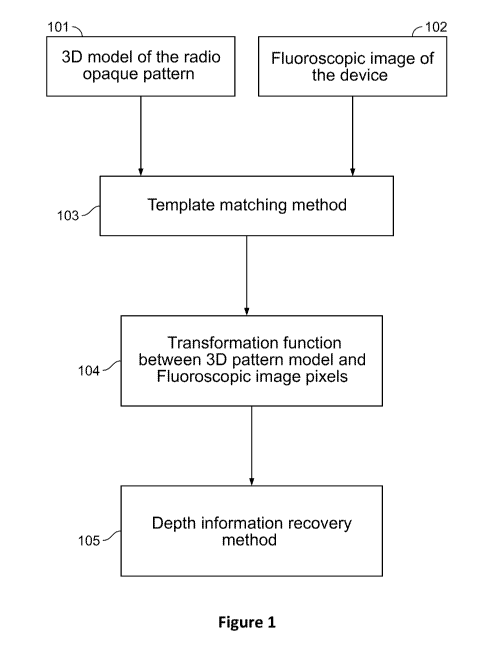
[0011] Figure 1 shows a flowchart of an exemplary method.
[0012] Figure 2A shows a plot of density of radio opaque material along
the length of an
exemplary device.
[0013] Figure 2B shows a plot of grayscale intensity in a fluoroscopic
image of the
device of Figure 2A.
[0014] Figure 2C shows a plot of grayscale intensity in a fluoroscopic
image of the
device of Figure 2A with the device partially occluded.
[0015] Figure 2D shows the correlation between the grayscale intensity of
the imaged
device and the density of radio opaque material.
[0016] Figure 2E shows a rendering of an exemplary device including a
pattern of radio
opaque material as positioned in a patient's lung and partially occluded.
[0017] Figure 2F shows a chart of a first exemplary pattern of radio
opaque material on
an exemplary device.
[0018] Figure 2G shows a rendering of an exemplary device including a
pattern of radio
opaque material as positioned in a patient's lung and partially occluded, the
device having radio
opaque material of a density as shown in Figure 2A.
4
CA 03039637 2019-04-05
WO 2018/065824 PCT/IB2017/001376
[0019] Figure 211 shows exemplary rings of radio opaque material of
varying size and
varying spacing along the length of an exemplary device.
[0020] Figure 21 shows a chart of a second exemplary pattern of radio
opaque material
on an exemplary device.
[0021] Figure 3A shows an exemplary device including an applicator, a
catheter, and a
guide wire, the device being shown disassembled.
[0022] Figure 3B shows the applicator of Figure 3A in an extended
position.
[0023] Figure 3C shows the applicator of Figure 3A in a retracted
position.
[0024] Figure 4A shows the device of Figure 3A, the device being shown
assembled.
[0025] Figure 4B shows the device of Figure 4A, the device being shown
with a guide
wire extended.
[0026] Figure 5 shows an exploded view of the applicator shown in Figure
3A.
[0027] Figure 6A shows the applicator of Figure 3A, a trigger of the
applicator being
shown in an unlocked position.
[0028] Figure 6B shows the applicator of Figure 3A, a trigger of the
applicator being
shown in a locked position.
[0029] Figure 7A shows a partial sectional view of the applicator shown
in Figure 6A.
[0030] Figure 7B shows a partial sectional view of the applicator shown
in Figure 6B.
[0031] Figure 8A shows a portion of the applicator of Figure 3A, the
applicator being
viewed from the opposite direction from that shown in Figure 3A.
[0032] Figure 8B shows a partial sectional view of the applicator of
Figure 3A.
CA 03039637 2019-04-05
WO 2018/065824 PCT/IB2017/001376
[0033] Figure 9A shows the exemplary assembled device of Figure 4A, the
applicator of
the device being shown in an extended position and in proximity to disengaged
connector
portions.
[0034] Figure 9B shows the exemplary assembled device of Figure 4A, the
distal
portion of the shaft being shown in proximity to a removable connector
portion.
[0035] Figure 10 shows an exploded view of an exemplary shaft of the
exemplary
applicator of Figure 3A.
[0036] Figure 11A shows a sectional view of an exemplary wire extraction
button of the
exemplary applicator of Figure 3A.
[0037] Figure 11B shows an exploded view of the exemplary wire extraction
button of
Figure 11A.
[0038] Figure 12 shows a sheath luer lock entrance of the exemplary
applicator of
Figure 3A.
[0039] Figure 13A shows an exemplary luer lock plug that is configured to
engage an
exemplary connector of the applicator of Figure 3A.
[0040] Figure 13B shows the exemplary luer lock plug of Figure 13A
engaging the
exemplary connector of the applicator of Figure 3A.
DETAILED DESCRIPTION
[0041] The present invention will be further explained with reference to
the attached
drawings, wherein like structures are referred to by like numerals throughout
the several views.
The drawings shown are not necessarily to scale, with emphasis instead
generally being placed
6
CA 03039637 2019-04-05
WO 2018/065824 PCT/IB2017/001376
upon illustrating the principles of the present invention. Further, some
features may be
exaggerated to show details of particular components.
[0042] The figures constitute a part of this specification and include
illustrative
embodiments of the present invention and illustrate various objects and
features thereof. Further,
the figures are not necessarily to scale, some features may be exaggerated to
show details of
particular components. In addition, any measurements, specifications and the
like shown in the
figures are intended to be illustrative, and not restrictive. Therefore,
specific structural and
functional details disclosed herein are not to be interpreted as limiting, but
merely as a
representative basis for teaching one skilled in the art to variously employ
the present invention.
[0043] Among those benefits and improvements that have been disclosed,
other objects
and advantages of this invention will become apparent from the following
description taken in
conjunction with the accompanying figures. Detailed embodiments of the present
invention are
disclosed herein; however, it is to be understood that the disclosed
embodiments are merely
illustrative of the invention that may be embodied in various forms. In
addition, each of the
examples given in connection with the various embodiments of the invention
which are intended
to be illustrative, and not restrictive.
[0044] Throughout the specification and claims, the following terms take
the meanings
explicitly associated herein, unless the context clearly dictates otherwise.
The phrases "in one
embodiment" and "in some embodiments" as used herein do not necessarily refer
to the same
embodiment(s), though it may. Furthermore, the phrases "in another embodiment"
and "in some
other embodiments" as used herein do not necessarily refer to a different
embodiment, although
it may. Thus, as described below, various embodiments of the invention may be
readily
combined, without departing from the scope or spirit of the invention.
7
CA 03039637 2019-04-05
WO 2018/065824 PCT/IB2017/001376
[0045] The term "based on" is not exclusive and allows for being based on
additional
factors not described, unless the context clearly dictates otherwise. In
addition, throughout the
specification, the meaning of "a," "an," and "the" include plural references.
The meaning of "in"
includes "in" and "on."
[0046] As used herein, the term "radio opaque" refers to a material that
is characterized
in that electromagnetic radiation (including, but not limited, to X-rays) is
unable to pass through
such a material.
[0047] In some embodiments, the present invention is a device,
comprising:
an applicator;
a shaft;
a catheter;
a guide wire;
a connector;
a handle;
a trigger;
a luer lock plug; and
a radio opaque material;
wherein the applicator has an inner open channel from a proximal end to a
distal
end of the applicator,
wherein the inner open channel of the applicator is of a sufficient size to
house the shaft;
wherein the shaft is of a sufficient size to house the catheter and the guide
wire,
8
CA 03039637 2019-04-05
WO 2018/065824 PCT/IB2017/001376
wherein the catheter and the guide wire are configured to have an
extraction button which allow the guide wire to protrude out the
catheter,
wherein the catheter and the guide wire are configured to have pre-
curved distal tip
wherein the catheter proximal end is configured to have a luer lock
entrance
wherein the guide wire is configured to be connected or detached
from the catheter,
wherein the shaft is configured allow displacement inside and outside the
applicator,
wherein the shaft distal end is configured to allow the shaft to rotate,
wherein the shaft distal end is configured to be connected or detached
from the connector,
wherein the distal end of the applicator is attached to the connector,
wherein the connector is configured to attach to a bronchoscope,
wherein the connector is configured to be connected or detached
from the bronchoscope,
wherein the connector is configured to include a luer lock plug,
wherein the proximal end of the applicator is attached to the handle,
wherein the handle comprises a switch configured to lock and
unlock the handle,
9
CA 03039637 2019-04-05
WO 2018/065824 PCT/IB2017/001376
wherein the handle is configured to rotate from an open position to
a closed position,
wherein the shaft is configured to rotate with the handle, and
wherein the radio opaque material is attached to an outer portion of the
device.
[0048] In some embodiments, the radio opaque material is dispersed in a
pattern.
[0049] In some embodiments, the pattern is not uniform.
[0050] In some embodiments, the dispersed pattern comprises a plurality
of deposited
densities of the radio opaque material on the outer portion of the device.
[0051] In some embodiments, a first deposited density of the deposited
densities is not
identical to a second deposited density of the deposited densities.
[0052] In some embodiments, the pattern comprises at least one shape.
[0053] In some embodiments, the at least one shape can be a ring.
[0054] In some embodiments, the ring can be an unbroken ring.
[0055] In some embodiments, the ring can be a broken ring.
[0056] In some embodiments, the pattern is in a longitudinal conformation
in reference to
the applicator.
[0057] In some embodiments, the grip handle is free to rotate with
respect to the shaft. In
some embodiments, the grip handle is constrained from rotation with respect to
the shaft. In
some embodiments, the grip handle is selectively either free to rotate with
respect to the shaft or
constrained from rotation with respect to the shaft. In some embodiments, the
selective freedom
or restriction of rotation of the grip handle with respect to the shaft is
independent from
restriction of longitudinal motion of the shaft.
[0058] In some embodiments, the guide wire is curved.
CA 03039637 2019-04-05
WO 2018/065824 PCT/IB2017/001376
[0059] In some embodiments, the catheter is curved.
[0060] In some embodiments, the catheter has a pull wire allowing the
curvature of the
distal end of the catheter to be manipulated.
[0061] In some embodiments, the shaft includes a mechanism allowing
rotation of the
handle to be controlled.
[0062] In some embodiments, the device includes a locking mechanism
configured to
selectively lock or unlock movement of the catheter along a longitudinal axis
of the device, while
allowing the catheter to rotate about the longitudinal axis.
[0063] In some embodiments, the shaft includes a groove that allows the
catheter to be
inserted along the side of the shaft.
[0064] In some embodiments, the device includes a polytetrafluoroethylene
tube located
inside the shaft so as to hold the catheter and guide the catheter outside the
shaft.
[0065] In some embodiments, the guide wire can be extracted from the
catheter by
demand in order to control the effective curvature of the distal tip of the
device. In some
embodiments, the device includes a manipulator that is configured to control
the motion of the
guide wire.
[0066] In some embodiments, the guide wire can be detached from the
catheter.
[0067] In some embodiments, the catheter can be detached from the handle.
[0068] In some embodiments, the handle is configured to be detached from
the connector
without firs extracting the catheter and/or the guide wire from the device.
[0069] In some embodiments, the connector is configured to allow the
device to be
detached from the bronchoscope without first extracting the catheter and/or
the guide wire from
the device
11
CA 03039637 2019-04-05
WO 2018/065824 PCT/IB2017/001376
[0070] In some embodiments, the connector includes a luer lock plug
configured to be
positioned therein so as to allow for connection of a slip tip or a luer lock
syringe.
[0071] In some embodiments, the catheter includes a luer lock entrance
configured to be
positioned therein so as to allow for connection of a slip tip or a luer lock
syringe.
[0072] In some embodiments, the catheter can be used without the guide
wire.
[0073] In some embodiments, the handle has a component configured to
provide for data
storage and for contactless communication. In some embodiments, the device
stores a unique
identifier that can be read in a contactless manner (e.g., through radio-
frequency identification or
near-field communication technology). In some embodiments, the handle includes
an electronic
device having general computing, data storage and wireless communication
abilities. In some
embodiments, a unique identifier is stored in the handle. In some embodiments,
the handle
unique identifier includes unique barcode that can be read by a barcode
reader. In some
embodiments, the barcode is stamped on the handle. In some embodiments, the
barcode is
stamped on the handle package. In some embodiments, the barcode is included in
a product
label.
[0074] In some embodiments, a radio opaque material includes, but is not
limited to,
materials including barium, iodine, or any combination thereof In some
embodiments, two or
more radio opaque materials are used in conjunction with one another.
[0075] Figure 3A shows the elements of an exemplary device 1. In some
embodiments,
the device 1 includes an applicator 10, a catheter 11 and a guide wire 12. In
some embodiments,
the applicator 10 includes a grip handle 13 that allows the user to pull,
push, or rotate the grip
handle 13 from a closed (retracted) position to an open (extended) position.
In some
embodiments, the applicator 10 includes an applicator shaft 16 that allows the
grip handle 13 to
12
CA 03039637 2019-04-05
WO 2018/065824 PCT/IB2017/001376
slide along the applicator shaft 16 (i.e., along a longitudinal axis) while
avoiding relative rotation
between the applicator shaft 16 and the grip handle 13. In some embodiments,
the applicator
shaft 16 includes an internal passage that is configured to receive the
catheter 11. Consequently,
in some embodiments, rotation of grip the handle 13 causes the applicator
shaft 16 to rotate
therewith. In some embodiments, rotation of the grip handle 13 with respect to
the shaft 16 can
be selectively locked or unlocked, such that, when unlocked, the grip handle
13 is free to rotate
with respect to the shaft 16. In some embodiments, the applicator 10 includes
a connector
element 15 that enables connection of the applicator 10 to any commercially
used bronchoscope.
In some embodiments, the connector element 15 includes a connector portion 40
that is
permanently connected to the shaft 16. In some embodiments, the connector
portion 40 is
configured to connect the device 1 to a commercially used bronchoscope. In
some embodiments,
the connector portion 40 is connected to a bronchoscope by manually rotating
swivel ring 43 in
one direction, so as to move the swivel ring 43 toward and press a connector
coupling 44 against
the bronchoscope. In some embodiments, to detach the device 1 from the
bronchoscope, the
swivel ring 43 is manually rotated in the other direction, thereby moving the
swivel ring 43 away
from the connector coupling 44 and releasing pressure by the connector
coupling 44 on the
bronchoscope.
[0076] In some embodiments, the connector element 15 includes a connector
portion 41
that can be detached from the shaft 16, and a connector portion 42 that can be
detached from the
shaft 16. In some embodiments, the connector portion 41 and the connector
portion 42 may be
connected to the shaft 16 by a snap 45 that is located at the distal end 32 of
the shaft 16. In some
embodiments, the connector portion 41 can be connected to a commercially
available
bronchoscope by sliding the connector portion 41 over an entrance port of the
bronchoscope. In
13
CA 03039637 2019-04-05
WO 2018/065824 PCT/IB2017/001376
some embodiments, the connector portion 41 includes a connector slider 47 that
is configured to
slide over the entrance port of the bronchoscope and thereby lock the
connector portion 41 to the
bronchoscope. In some embodiments, the connector portion 41 includes a release
button 48 that
is operable to release the connector portion 41 from the bronchoscope. In some
embodiments,
the connector portion 42 includes a connector clasp 46. In some embodiments,
the connector
portion 42 can be connected to a commercially available bronchoscope by
closing the connector
clasp 46 against an entrance port of the bronchoscope. In some embodiments,
the connector
portion 42 can be removed from a commercially available bronchoscope by
opening the
connector clasp 46. In some embodiments, the connector portion 41 and the
connector portion
42 can be connected to a bronchoscope in the absence of the applicator 10.
[0077] In some embodiments, the grip handle 13 includes a trigger 14 that
is configured
to lock the grip handle 13 at any position along its travel between its open
and closed positions
(e.g., along the applicator shaft 16). In some embodiments, the distal end of
the shaft 16 is
configured to act as a swivel, allowing the shaft 16 and the grip handle 13 to
rotate with respect
to the connector element 15 along the longitudinal axis to any desired angle.
[0078] Figure 3B shows the device 1 of Figure 3A in its open (extended)
position. The
connector element 15 is extended distally from the grip handle 13. Figure 3C
shows the device
1 from Figure 3B in its closed (retracted) position. The connector element 15
is in its closest
proximity to the grip handle 13. Figure 4A shows the device 1 of Figure 3A, as
configured with
both the catheter 11 and the guide wire 12 connected to grip handle 13. Figure
4B shows the
device 1 of Figure 4A, but with the guide wire 12 extended. In some
embodiments, the device 1
includes a wire extraction button 33, which is configured to allow the guide
wire 12 to be
14
CA 03039637 2019-04-05
WO 2018/065824 PCT/IB2017/001376
extended. In some embodiments, as shown in Figure 4B, the guide wire 12 is
flexible and can
be positioned as needed.
[0079] Figure 5 shows an exploded view of the applicator 10. The grip
handle 13 is
divided into two side portions 13A and 13B. Screws 28 are configured to
connect the two side
portions 13A and 13B to one another. The applicator 10 includes a trigger 14,
a lever 17, a hinge
19, and a spring 27, which will be described in detail with reference to
Figures 6A and 6B
below. The applicator 10 also includes an inlet tube 21 that is configured to
receive the catheter
11.
[0080] Figure 6A shows the device 1 with the trigger 14 in its unlocked
position, in
which the shaft 16 is allowed to move with respect to the grip handle 13.
Figure 6B shows the
device 10 with the trigger 14 in its locked position, in which the shaft 16 is
allowed to move with
respect to the grip handle 10. Figure 7A shows a sectional view of the device
1 with the trigger
14 in its unlocked position. Figure 7B shows a sectional view of the device 10
with the trigger
14 in its locked position. The device 1 includes a lock lever 17 that is
pivotably engaged with a
hinge 19. The shaft 16 has a grooved portion 20. The trigger 14 has an angled
surface 18 that is
configured to engage the lock lever 17 when the trigger 14 is in its locked
position, and to
disengage the lock lever 17 when the trigger 14 is in its unlocked position.
When the angled
surface 18 of the trigger 14 engages the lock lever 17 (e.g., as shown in
Figure 7B), the lock
lever 17 pivots about the hinge 19 to a position such that the lock lever 17
engages the grooved
portion 20 of the shaft 16, thereby preventing the shaft 16 from axial motion
with respect to the
grip handle 13. Conversely, when the angled surface 18 of the trigger 14
disengages the lock
lever 17 (e.g., as shown in Figure 7A), the lock lever pivots about the hinge
19 to a position such
CA 03039637 2019-04-05
WO 2018/065824 PCT/IB2017/001376
that the lock lever 17 does not engage the grooved portion 20 of the shaft 16,
thereby allowing
the shaft 16 to move axially with respect to the grip handle 13.
[0081] Figure 8A shows a perspective view of the grip handle 13 in a
direction facing
toward the distal end of the grip handle 13. The grip handle 13 includes an
inlet port 22 that
allows insertion of the catheter 11 into the applicator 10. Figure 8B shows a
sectional view of a
portion of the grip handle 13. The grip handle 13 includes an inlet tube 21
extending from inlet
port 22 to the internal passage of the shaft 16, and configured to allow
passage of the catheter 11.
[0082] Figure 9A and Figure 9B show an opening 24 along the shaft 16 that
allows the
inlet tube 21 to slide from its extended position (i.e., as shown in Figure
3B) to its closed
position (i.e., as shown in Figure 3C). In some embodiments, in order to
prevent the catheter 11
and the guide wire 12 from buckling and protruding from the shaft 16 due to
friction in a
bronchoscope that is connected to the device 1, a polytetrafluoroethylene
("PTFE", such as the
material sold under the trade name TEFLON by DuPont) tube 23 is positioned
inside the shaft 16
to act as a flexible barrier. In some embodiments, the PTFE tube 23 is
positioned around the
shaft 16 rather than inside the shaft 16. In some embodiments, rather than a
PTFE tube 23, a
spring, telescoping material or other flexible material that can withstand the
buckling force is
used. Figure 9A shows the PTFE tube 23 in an extended position. Figure 9B
shows the PTFE
tube 23 in a compressed position. In some embodiments, the PTFE tube 23 is
connected to the
connector element 15 at the distal end of the PTFE tube 23 and to the inlet
tube 21 at the
proximal end of the PTFE tube 23. As shown in Figure 9B, when the connector
element 15 is
positioned proximate to the grip handle 13, the PTFE tube 23 is compressed.
[0083] Figure 10 shows an exploded view of the shaft 16. In some
embodiments, the
shaft 16 includes a swivel mechanism. In some embodiments, a PTFE tube 23 is
positioned
16
CA 03039637 2019-04-05
WO 2018/065824 PCT/IB2017/001376
within the shaft 16 to act as a flexible barrier. In some embodiments, a shaft
distal end 32 is free
to rotate with respect to the shaft 16. In some embodiments, the swivel
mechanism also includes
two washers 29 and 30 and two o-rings 31 that provide control to the rotation.
In some
embodiments, the shaft distal end 32 is configured to be attached to the
connector 15.
[0084]
Figure 11A and Figure 11B show a sectional view and an exploded view,
respectively, of a wire extraction button 33. In some embodiments, the wire
extraction button 33
presses against a spring 35, which biases the wire extraction button 33 to a
position in which the
wire extraction button 33 restrains movement of the guide wire 12. In some
embodiments, the
wire extraction button 33 is removably coupled to a sheath luer lock entrance
34, which is
configured to allow connection to a syringe. In some embodiments, the wire
extraction button
33 can be removed to expose the sheath luer lock entrance 34. Figure 12 shows
the proximal
portion of the applicator 10 with the sheath luer lock entrance 34 exposed.
[0085]
Figure 13A shows a luer lock plug 36, which can be connected to the connector
portion 41 or the connector portion 42 to allow a syringe connection to the
connector 15. Figure
13B shows the luer lock plug 36 as connected to the connector 15.
[0086]
In some embodiments, the present invention relates to a radio opaque pattern
on a
device, where the radio opaque pattern can be visualized by a user (e.g., a
doctor, etc.) and used
to identify the specific portion of the device visible on the x-ray image,
e.g., by correlating
portions of the device with the observed density of the radio opaque material.
In some
embodiments, the radio opaque material is positioned on the catheter 11 of the
device 1. In some
embodiments, the radio opaque material is positioned on the guide wire 12 of
the device 1. In
some embodiments, the radio opaque material is positioned on both the catheter
11 and the guide
17
CA 03039637 2019-04-05
WO 2018/065824 PCT/IB2017/001376
wire 12 of the device 1, which cooperate to produce a combined "effective"
pattern of radio
opaque material on the device 1.
[0087] In some embodiments, the device 1 of the current invention has a
radio opaque
material positioned in a pattern which can be observed (e.g., but not limited
to, using X-ray
images of the device), where the pattern has been manufactured by applying
variable amount(s)
of radio opaque material along the device. In some embodiments, the
correlation between the
function of radio opaque material density along the device and the function of
grayscale intensity
in the x-ray image allows the detection of a specific portion of the device on
the fluoroscopic
image in spite of partial occlusion by other radio opaque objects on the
image. In some
embodiments, the higher density of radio opaque material in the device results
in lower gray-
scale intensities visualized by the X-ray image and vice versa. Figure 2A
shows a plot of radio
opaque material density along the length of an embodiment of device (Y axis),
as plotted against
the length of the device (X axis). Figure 2B shows one-dimensional gray scale
levels (Y axis) of
a device with material density as shown in Figure 2A, as imaged by a
fluoroscope along the
length of the device (X axis). Taken together, Figures 2A and 2B show that the
density of the
radio-opaque material is correlated with gray-scale image function.
[0088] Figure 2C shows one-dimensional gray scale levels (Y axis) of a
partial device
protruding from a bronchoscope (as compared to Figure 2B, which illustrates
the full image of
the device), imaged by a fluoroscope along to the length of the device (X
axis). The zero value
between positions x2 and x3 along the X axis illustrates an occlusion that
blocks the X-ray
radiation in this interval. Figure 2D shows the absolute value of the
correlation function
between the partially imaged device (i.e., as shown in Figure 2C) and the
density of the radio
opaque material (i.e., as shown in Figure 2A). The position of the peak in
Figure 2D can be
18
CA 03039637 2019-04-05
WO 2018/065824 PCT/IB2017/001376
utilized to calculate the translation between pixels in Figure 2C and 3
dimensional model
coordinates in Figure 2A. Figure 2E shows a representation of an X-Ray image
showing a
bronchoscope 241 and device 242 (e.g., the device 1) with radio opaque
material, as positioned
within the chest of a patient. At position 243, the device 242 is occluded by
an ECG patch.
[0089] In some embodiments, the radio opaque material is arranged along
the device 1 in
a pattern. In some embodiments, the pattern includes differently sized rings
extending around
the device. In some embodiments, the pattern includes rings irregularly spaced
along the device.
Figure 2F shows a table showing a first pattern comprised of rings of radio
opaque material
located at different spacing from one another and having different lengths.
Figure 21 shows a
table showing a second pattern comprised of rings of radio opaque material
located at different
spacing from one another and having different lengths. It will be apparent to
those of skill in the
art that the specific patterns represented by Figure 2F and Figure 21 are only
exemplary and that
other patterns are possible.
[0090] Figure 2G shows a representation of an X-Ray image showing a
bronchoscope
261 and device 262 (e.g., the device 1) having radio opaque material that is
patterned as shown
in Figure 2A. Figure 211 shows an illustration of a pattern of radio opaque
material containing
rings of variable size, placed in positions at varying intervals along the
outer portion of a device
(e.g., the device 1).
[0091] In a non-limiting example, when a portion of a pattern of radio
opaque material is
visible, a user can calculate the one-dimensional translation (e.g.,
correlation) between the
imaged pattern and the density function. The relation between the radio
opacity of the device
and the gray-scale levels can be used for this purpose. In another non-
limiting example, a user
can use a template matching method that searches for the highest correlation
between the gray-
19
CA 03039637 2019-04-05
WO 2018/065824 PCT/IB2017/001376
scale levels of the visible segment of the device in the image and the radio
opaque density profile
of the device. Such a method is robust to occlusion and noise caused by
objects that are behind
or above the device with respect to the projection direction from an X-ray
tube to an image
intensifier. In some embodiments, Figure 2D shows an exemplary correlation
function between
the device's partial image as shown in Figure 2C and the device's pattern of
radio opaque
material density as shown in Figure 2A. For instance, the translation between
the density
function at point x0 in Figure 2A to the pixel gray-scale level at point xl on
Figure 2C
corresponds to the peak position at the point x4 in the correlation function
shown in Figure 2D.
As a result, although the device as represented by Figure 2C is partially
visible and partially
occluded in the area between points x2 and x3, it is possible to perform
device localization on the
image and correlate each pixel of the visible device, as represented by Figure
2D to the known
model for the device, as represented by Figure 2A.
[0092] In some embodiments, a unique radio opaque pattern is manufactured
through
attaching radio opaque rings of variable size to the device at specific
positions along the device's
longitude direction axis, as illustrated by Figure 211. The unique radio
opaque pattern assists a
user in estimating the transformation function between the imaged device's
pixels and
predesigned device model for manufacturing. This transformation function can
be estimated by
finding a function that satisfies the constraints imposed by the different
marker sizes and
locations on the device. A non-limiting example for such design, which is
robust to occlusion of
several markers on x-ray image, is provided in Figure 2F.
[0093] In some embodiments, a medical image (e.g., an X-ray image) of at
least a portion
of a body of patient with the device 1 (i.e., which includes the radio opaque
material) positioned
within the body of the patient can be analyzed to determine the depth of the
device 1 within the
CA 03039637 2019-04-05
WO 2018/065824 PCT/IB2017/001376
body based on knowledge of the positioning of the radio opaque material.
In some
embodiments, the current invention relates to a method to recover 3-
dimensional depth
information in such cases, where due to occlusions and noise of the 2-
dimensional image as an
input, such as X-ray image or video image sequence, some markers may not be
detected, by
means of unique pattern on the device as shown, for example, in Figure 2A. The
occlusion and
noise of the input image or video image sequence may be caused by occlusion of
medical
devices, high density tissue such as ribs, patient pace makers, ECG cables,
etc. as illustrated by
Figure 2E.
[0094]
Figure 1 shows a flowchart of a process for determining the depth of an
exemplary device (e.g., the device 1 of Figure 3A). The process receives, as
inputs, a density
model (101) of the radio opaque material along the device (e.g., the
information shown in Figure
2A) and fluoroscopic image data (102) showing the device positioned within the
patient's body.
A transformation function (104) between the model and the image pixels is
calculated using a
template matching method (103). In some embodiments, the template matching
method is
performed as described above with reference to Figures 2A-2D. The
transformation function is
used for depth information recovery (105).
[0095]
In some embodiments, the depth of the device can be calculated from a single
image based on prior knowledge the physical dimensions of the specific radio
opaque pattern.
For instance, given the known physical distance between two points that are
identified and
located in the intra operative image, one can determine the relative depth
between these two
points. In some embodiments, such a technique for determining relative depth
is carried out as
described in International Patent Application Publication No. WO/2015/101948,
the contents of
which are incorporated herein by reference in their entirety. More
particularly, in some
21
CA 03039637 2019-04-05
WO 2018/065824 PCT/IB2017/001376
embodiments, a device (e.g., the device 1) or a portion thereof (e.g., the
portion between two of
the stripes shown in Figure 211) having a known length "L3" and located in
three-dimensional
space within a patient's body is projected into an imaging plane to create a
projection image
including such a device. The observed (i.e., projected) length of the same
device (or device
portion) in the two-dimensional imaging plane is "L2". As shown in Figure 12
of International
Patent Application Publication No. WO/2015/101948, an angle a of the device
(or device
portion) in space can be determined by solving the equation L2 = L3 cos a. The
relative depth D
between the two ends can then be determined by calculating D = L3 sin a.
[0096] In some embodiments, the depth of the device can be calculated
using the
methods described in International Patent Application Publication No.
WO/2017/153839, the
contents of which are incorporated herein by reference in their entirety. In
some embodiments,
such determination is performed according to the following process. In some
embodiments, the
device is imaged by an intraoperative device and projected to an imaging
plane. In some
embodiments, a predefined distance "m" between two radiopaque regions "F" and
"G" on the
device (e.g., two of the stripes shown in Figure 2H) is considered as an
input. In some
embodiments, point "F" results from a projection of two possible 3D locations
A and B, having
different depth from one another. In some embodiments, point "G" results from
a projection of
two possible depth locations C and D, having different depth from one another,
and where C
corresponds to A and D corresponds to B. In some embodiments, 3D distances
between the
back-projected location pairs AC and BD are measured. In some embodiments, the
3D distances
AC and BD are compared to the distance "m", and either points A and C or
points B and D are
selected based on the best fit. In some embodiments, the depth is that
corresponding to the
selected pair of locations.
22
CA 03039637 2019-04-05
WO 2018/065824 PCT/IB2017/001376
[0097] In some embodiments, the depth recovery can be performed using a
combination
of a known patient anatomy and pose estimation approach. In some embodiments,
the
knowledge of the unique radio opaque pattern can be combined with the
knowledge of the
patient's anatomical bronchial tree (e.g., as extracted from the pre-operative
image) and the
knowledge of the current pose of the imaging device relative to the patient
(e.g., a point of view
that allows projecting 3D information from a pre-operative image to the
current image acquired
from the imaging device). Since an instrument is located inside a discrete
anatomical space, the
current pose estimation information can be used to limit the possible
solutions. Furthermore, the
matching between the instrument location and possible anatomical location on
the bronchial tree
can be recovered by solving an optimization problem with respect to the
following parameters:
an assumption of the anatomical location of the tool, a pose estimation, and
potential 3d anatomy
changes. In some embodiments, such an approach is described in greater detail
in International
Patent Application Publication No. W02015/101948.
[0098] In some embodiments, the depth estimation can be performed from a
sequence of
two or more images by (a) finding corresponding points between views, for
example, by tracking
or matching by visual similarity; (b) finding pose relative differences using,
for example, a jig,
human anatomy, or any other pose estimation algorithm (e.g., those described
in International
Patent Application Publication No. WO/2017/153839); and (c) reconstructing
three-dimensional
information of the matching points from multiple images with known poses using
methods that
are known in the art (e.g., triangulation, a stereo corresponding point based
technique, a non-
stereo corresponding contour method, a surface rendering technique, etc.).
[0099] In some embodiments, the device provides increased maneuverability
inside a
body cavity, e.g., but not limited to, bronchial airways, compared to typical
methods. In some
23
CA 03039637 2019-04-05
WO 2018/065824 PCT/IB2017/001376
embodiments, the device is as seen in the non-limiting example shown in
Figures 3A-13B. In
some embodiments, the exemplary device allows increased accuracy while
navigating with one
hand and supports the standard diagnostic and therapeutic device's entrance
from the other. In
some embodiments, the guide wire is pre-curved. In some embodiments, the
catheter is pre-
curved. In some embodiments, both the guide wire and the catheter are pre-
curved. In some
embodiments, the guide wire is straight. In some embodiments, the catheter is
straight. In some
embodiments, both the guide wire and the catheter are straight. In some
embodiments, the guide
wire is configured to be bent as needed. In some embodiments, the catheter is
configured to be
bent as needed. In some embodiments, both the guide wire and the catheter are
configured to be
bent as needed. In some embodiments, the guide wire is configured to protrude
past the tip of
the catheter, while adding extra bending to the device. This feature allows
for increased
maneuverability of the device during the navigation inside the lung.
[0100] In some embodiments, the device including the radio opaque
material includes an
endoscope, an endo-bronchial tool, and/or a robotic arm.
[0101] While a number of embodiments of the present invention have been
described, it
is understood that these embodiments are illustrative only, and not
restrictive, and that many
modifications may become apparent to those of ordinary skill in the art.
Further still, the various
steps may be carried out in any desired order (and any desired steps may be
added and/or any
desired steps may be eliminated).
24