Note: Descriptions are shown in the official language in which they were submitted.
CA 03039667 2019-04-05
WO 2018/081329 PCT/US2017/058386
PURIFICATION PROCESS FOR REMOVAL OF TYROSINE
SULFATION ANTIBODY VARIANTS; PURIFIED COMPOSITIONS
Field of the Invention
The present invention relate to compositions comprising antibodies and antigen-
binding fragments thereof that lack tyrosine sulfation as well as methods of
purification for
preparing compositions.
Background of the Invention
Tyrosine sulfation is a post-translational modification (PTM) where a sulfate
trioxide
(SO3) group is covalently bound to the hydroxyl group on the side chain of the
amino acid
tyrosine group. This PTM occurs in the trans-Golgi network and is catalyzed by
two
enzymes, tyrosylprotein sulfotransferases (TPSTs). The molecular mechanism
involves the
transfer of an activated sulfate from 3'-phosphoadenosine-5-phosphosulfate to
tyrosine,
and has been found on a variety of proteins and peptides. Recent findings
indicate that
tyrosylprotein sulfotransferase 2 recognizes tyrosines flanked by acid
residues for sulfation.
This PTM is responsible for strengthening interactions between proteins and
occurs on
secreted and trans-membrane spanning proteins. Some chemokine receptors have
been
shown to be tyrosine sulfated such as at the N-terminal extracellular domain
of CCR5, the
principle HIV-1 and several glycoprotein hormone receptors. For example, the
native form
of the leech-derived thrombin inhibiting peptide hirudin, is tyrosine
sulfated. Interestingly,
the two recombinant forms of hirudin (Revasc and Refludan) used for treating
various blood
clotting disorders are not sulfated. Sulfation increases the mass of a
biomolecule by 80 Da,
which is the same mass difference as a phosphate moiety (P03). Unlike P03,
which forms
a fairly stable P-0 bond, the SO3 is very labile and readily decomposes under
high
temperature and low pH conditions.
The presence of different PTM variants in a therapeutic antibody preparation
leads
to heterogeneity which, depending on the location of the modification, can
lead to variations
in antibody potency, bioavailability or immunogenicity. Such issues also
create issues
before regulatory agencies. Though tyrosine sulfation has been described in
chemokine
receptors and other proteins, there is a need to identify if such
modifications occur in
antibody preparations and, if identified, to remove them.
1
CA 03039667 2019-04-05
WO 2018/081329 PCT/US2017/058386
Summary of the Invention
The present invention provides a composition comprising an anti-LAG3 antibody
or
antigen-binding fragment thereof (e.g., Ab1, Ab2, Ab3, Ab4, Ab5, Ab6, Ab7, Ab8
or Ab9)
that, for example, comprises:
a light chain variable domain comprising:
CDR-L1 that comprises the amino acid sequence: KASQSLDYEGDSDMN (SEQ ID NO:
38);
CDR-L2 that comprises the amino acid sequence: GASNLES (SEQ ID NO: 39); and
CDR-L3 that comprises the amino acid sequence: QQSTEDPRT (SEQ ID NO: 40);
and/or
a heavy chain variable domain comprising:
CDR-H1 that comprises the amino acid sequence: DYNVD (SEQ ID NO: 33);
CDR-H2 that comprises the amino acid sequence:
DINPNNGGTIYAQKFQE (SEQ ID NO: 59);
DINPNSGGTIYAQKFQE (SEQ ID NO: 60);
DINPNDGGTIYAQKFQE (SEQ ID NO: 61);
DINPNQGGTIYAQKFQE (SEQ ID NO: 62);
DINPNGGGTIYAQKFQE (SEQ ID NO: 63); or
DINPNX1GGTIYX2QKFX3X4 (SEQ ID NO: 64) wherein, X1= D,N, S or Q, X2= A or s,
X3=
or K, and X4= E or G; and CDR-H3: NYRWFGAMDH (SEQ ID NO: 35); which lacks
detectable levels of sulfated tyrosine on CDR-L1. For example, in an
embodiment of the
invention, the antibodies or fragments in the composition further lack
detectable levels of
sulfated tyrosine in one or more members selected from the group consisting of
FR-L1, FR-
L2, CDR-L2, FR-L3, CDR-L3, FR-L4, FR-H1, CDR-H1, FR-H2,CDR-H2, FR-H3,CDR-H3,
FR-H4 and a constant domain. In an embodiment of the invention, the antibody
or fragment
comprises engineered yeast or CHO N-linked glycans. In an embodiment of the
invention,
an anti-LAG3 antibody (e.g., Ab1, Ab2, Ab3, Ab4, Ab5, Ab6, Ab7, Ab8 or Ab9)
containing
composition comprises one or more species of the antibody lacking tyrosine
sulfation and
having molecular weights of about 148590 Da, 148752 Da and/or 148914 Da (e.g.,
having
GOF and/or G1F glycan species, e.g., as set forth in Table 1, N-terminal heavy
chain
glutamine converted to pyroglutamate and/or C-terminal heavy chain lysine
removed).
The present invention also provides a method for removing tyrosine sulfated
antibodies or antigen-binding fragments thereof (e.g., Ab1, Ab2, Ab3, Ab4,
Ab5, Ab6, Ab7,
Ab8 or Ab9) from an aqueous mixture comprising antibodies or antigen-binding
fragments
that comprise one or more sulfated tyrosines (e.g., on CDR-L1) and antibodies
or antigen-
binding fragments lacking sulfated tyrosine comprising adjusting the pH of the
mixture to
about 6.5 to about 7.0 or about 6.5 to about 7.5, contacting the mixture with
an anion
exchange resin, and removing and retaining a non-resin bound aqueous fraction
of the
2
CA 03039667 2019-04-05
WO 2018/081329 PCT/US2017/058386
mixture from the resin. In an embodiment of the invention, the method
comprises washing
the column with an aqueous composition, e.g., under isocratic conditions, and
removing
and retaining the wash composition from the resin. In an embodiment of the
invention, the
resin is in a column and the method comprises adding said mixture to the
column and
.. collecting the flow-through fraction from the column. In an embodiment of
the invention, the
method comprises equilibrating a chromatography resin, comprising a
dimethylaminopropyl
anion exchange ligand, in a chromatography column with 25 mM sodium phosphate
pH 6.5,
adjusting the pH of the mixture to about 6.5, applying the mixture to the
column, collecting
flow-through fraction form the column, washing the resin in the column with 25
mM sodium
phosphate pH 6.5 and collecting the flow-through fraction from the wash. In an
embodiment
of the invention, the method comprises equilibrating a chromatography resin,
comprising a
quarternized polyethyleneimine anion exchange ligand, in a chromatography
column with
25 mM sodium phosphate pH 7.0; optionally, 5 mM NaCI, adjusting the pH of the
mixture to
about 7.0, applying the mixture to the column, collecting flow-through
fraction form the
column, washing the resin in the column with 25 mM sodium phosphate pH 7.0;
optionally,
5 mM NaCI and collecting the flow-through fraction from the wash. In an
embodiment of the
invention, the A280 absorbance of the anion exchange chromatography flow-
through is
monitored and collected and retained when the A280 first reaches at least
about 2.5
absorbance units/cm; and not collected or retained when the A280 falls below
about 1.0
absorbance units/cm. In an embodiment of the invention, the methods of the
present
invention further comprise purifying the antibody or antigen-binding fragment
by cation
exchange chromatography, further anion exchange chromatography in bind-elute
mode,
hydrophobic interaction chromatography, protein-A chromatography, protein-L
chromatography, protein-G chromatography, hydroxyapatite chromatography, size
exclusion chromatography, fractional precipitation, filtration, centrifugation
or viral
inactivation. In an embodiment of the invention, the immunoglobulin light
chains and/or
heavy chains of the antibody or antigen-binding fragment are expressed in a
Chinese
hamster ovary cell. In an embodiment of the invention, the antibody or antigen-
binding
fragment comprises:
a light chain variable domain comprising:
CDR-L1 that comprises the amino acid sequence: KASQSLDYEGDSDMN (SEQ ID NO:
38);
CDR-L2 that comprises the amino acid sequence: GASNLES (SEQ ID NO: 39); and
CDR-L3 that comprises the amino acid sequence: QQSTEDPRT (SEQ ID NO: 40);
and/or
a heavy chain variable domain comprising:
CDR-H1 that comprises the amino acid sequence: DYNVD (SEQ ID NO: 33);
CDR-H2 that comprises the amino acid sequence:
3
CA 03039667 2019-04-05
WO 2018/081329 PCT/US2017/058386
DINPNNGGTIYAQKFQE (SEQ ID NO: 59);
DINPNSGGTIYAQKFQE (SEQ ID NO: 60);
DINPNDGGTIYAQKFQE (SEQ ID NO: 61);
DINPNQGGTIYAQKFQE (SEQ ID NO: 62);
DINPNGGGTIYAQKFQE (SEQ ID NO: 63); or
DINPNX1GGTIYX2QKFX3X4 (SEQ ID NO: 64) wherein, X1= D,N,S or Q, X2= A or S, X3=
Q
or K, and X4= E or G; and CDR-H3: NYRWFGAMDH (SEQ ID NO: 35). Compositions
that are
the product of such a method are also part of the present invention. In an
embodiment of
the invention, an anti-LAG3 antibody (e.g., Ab1, Ab2, Ab3, Ab4, Ab5, Ab6, Ab7,
Ab8 or Ab9)
is purified by AEX chromatography wherein the antibodies lacking sulfated
tyrosine, having
molecular weights of about 148590 Da, 148752 Da and/or 148914 Da (e.g., having
GOF
and/or G1F glycan species, e.g., as set forth in Table 1, N-terminal heavy
chain glutamine
converted to pyroglutamate and/or C-terminal heavy chain lysine removed).
Brief Description of the Drawings
Figure 1. Overlay of IEX-HPLC UV profile of AEX feed (bold trace), strip
(light trace)
and pool fraction (dashed trace).
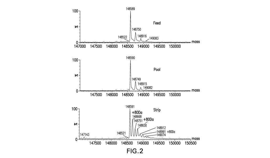
Figure 2. Intact mass spectrum of AEX feed, pool and strip samples.
Figure 3. Reduced light chain mass spectrum of AEX pool and strip samples.
Figure 4A-B. UV trace of reduced LysC peptide mapping of AEX pool and strip
fractions.
Figure 5A-C. (A)CID fragmentation spectrum of light chain AA25-43+80 Da in 400-
1800 m/z (B) Zoomed in m/z 300-1100 (C) Zoomed in m/z 1200-2000.
Figure 6. ETD fragmentation of light chain peptide AA25-43+80 Da. The 80Da
.. attached fragment ions were labeled.
Figure 7A-B. (A) Deconvoluted intact mass spectra of AEX strip fraction with
and
without alkaline phosphatase treatment. (B) Deconvoluted intact mass spectra
of chicken
ovalbumin with and without alkaline phosphatase treatment.
Figure 8A-B. (A) Normalized concentrations of mAb AEX pool and strip were
subjected to reduced SDS-PAGE, probed for the human heavy (HC) and light
chains (LC)
by western hybridization (upper panel), then stripped and re-probed for
antisulfotyrosine
(lower panel). See the indications for HC and LC at the far right. (B)
Normalized
concentrations of different CHO-derived mAbs in addition to AEX strip and pool
are
subjected to reduced SDS PAGE, probed for the human HC and LC by western
hybridization, then stripped and re-probed for anti sulfotyrosine. For both
(A) and ( B)
4
CA 03039667 2019-04-05
WO 2018/081329 PCT/US2017/058386
MagicMark XP was used as a protein molecular weight standard, and equal
amounts of
HEK293 and EGF-treated A431 cell extracts are analyzed as controls.
Figure 9A-C. SIC of (A) L025-43+80 Da from AEX Strip Fraction, (B) Synthetic
Peptide XSXSXDYEGDSDXXXXXXX (SEQ ID NO: 65)+Phosphorylation and (C) Synthetic
.. Peptide XSXSXDYEGDSDXXXXXXX (SEQ ID NO: 65)+Sulfation.
Figure 10. mAb tyrosine (Y31) site showing the CDR loops in ribbon diagram for
both the heavy and light chain.
Figure 11. Predominant N-linked glycans for monoclonal antibodies produced in
Chinese hamster ovary cells (CHO N-linked glycans) and in engineered yeast
cells
(engineered yeast N-linked glycans): squares: N-acetylglucosamine (GIcNac);
circles:
mannose (Man); diamonds: galactose (Gal); triangles: fucose (Fuc).
Detailed Description of the Invention
Certain antibodies and other proteins expressed in Chinese hamster ovary (CHO)
cells are contaminated with a sulfated tyrosine variants. Mass spectrographic
analysis of
such variants is characterized by an adduct of about +80 Da which corresponds
to the mass
of an added sulfate group. Such adducts are also alkaline phosphatase
resistant and
reactive with anti-sulfated tyrosine antibodies. The present invention
provides a method for
purifying a composition including such contaminant tyrosine sulfated variants
as well as
.. antibody compositions essentially free of the variants.
In accordance with the present invention there may be employed conventional
molecular biology, microbiology, and recombinant DNA techniques within the
skill of the art.
Unless otherwise defined herein, scientific and technical terms used in
connection with the
present invention shall have the meanings that are commonly understood by
those of
ordinary skill in the art. Further, unless otherwise required by context,
singular terms shall
include the plural and plural terms shall include the singular. Generally,
nomenclatures used
in connection with, and techniques of biochemistry, enzymology, molecular and
cellular
biology, microbiology, genetics and protein and nucleic acid chemistry and
hybridization
described herein are those well known and commonly used in the art. The
methods and
techniques of the present invention are generally performed according to
conventional
methods well known in the art and as described in various general and more
specific
references that are cited and discussed throughout the present specification
unless
otherwise indicated. See, e.g., James M. Cregg (Editor), Pichia Protocols
(Methods in
Molecular Biology), Humana Press (2010), Sambrook etal. Molecular Cloning: A
.. Laboratory Manual, 2d ed., Cold Spring Harbor Laboratory Press, Cold Spring
Harbor, N.Y.
(1989); Ausubel etal., Current Protocols in Molecular Biology, Greene
Publishing
Associates (1992, and Supplements to 2002); Harlow and Lane, Antibodies: A
Laboratory
5
CA 03039667 2019-04-05
WO 2018/081329 PCT/US2017/058386
Manual, Cold Spring Harbor Laboratory Press, Cold Spring Harbor, N.Y. (1990);
Taylor and
Drickamer, Introduction to Glycobiology, Oxford Univ. Press (2003);
Worthington Enzyme
Manual, Worthington Biochemical Corp., Freehold, N.J.; Handbook of
Biochemistry: Section
A Proteins, Vol I, CRC Press (1976); Handbook of Biochemistry: Section A
Proteins, Vol II,
CRC Press (1976); Essentials of Glycobiology, Cold Spring Harbor Laboratory
Press
(1999), Animal Cell Culture (R.I. Freshney, ed. (1986)); Immobilized Cells And
Enzymes
(IRL Press, (1986)); B. Perbal, A Practical Guide To Molecular Cloning (1984).
A sulfated tyrosine includes a tyrosine having an added sulfate group, e.g.,
having
the structure:
0
HO
401
C-0_1
Chromatography
The present invention provides a method for removing contaminant variant
antibodies or antigen-binding fragments (e.g., Ab1-Ab9) thereof that comprise
sulfated
tyrosine from a composition, e.g., a composition that comprises a mixture of
antibodies or
fragments, some of which having sulfated tyrosine and some of which lacking
the sulfated
tyrosine to generate a composition comprising undetectable levels of tyrosine
sulfated
variants (e.g., tyrosine sulfated CDR-L1, e.g., of Ab1 or Ab6). In an
embodiment of the
invention, the composition is treated by anion exchange (AEX) chromatography
in flow-
through mode to remove tyrosine sulfated variants. In an embodiment of the
invention, the
AEX resin has a dimethylaminopropyl ligand (i.e., a ligand that includes a
dimethylaminopropyl moiety). For example, in an embodiment of the invention,
the
composition that is subjected to the AEX chromatography is the product of
prior protein-A
chromatographic purification. In an embodiment of the invention, the
composition is pH
adjusted to a pH of about 6.5, e.g., with Tris (e.g., 0.5M, 0.725M or 1M)
prior to AEX
treatment (e.g., having a dimethylaminopropyl ligand). In an embodiment of the
invention,
6
CA 03039667 2019-04-05
WO 2018/081329
PCT/US2017/058386
the AEX column (e.g., having a dimethylaminopropyl ligand) is equilibrated,
e.g., with
sodium phosphate, e.g., 25 mM, e.g., sodium phosphate pH 5, 6.2 or 6.5. The
column
(e.g., having a dimethylaminopropyl ligand) can, in an embodiment of the
invention, be
washed with buffer (e.g., with sodium phosphate, e.g., 25 mM, e.g., sodium
phosphate pH
6.5) to recover antibody or fragment within the column, but not tightly bound
to the AEX
resin. Flow-through, not tightly bound to the AEX resin, is collected (e.g.,
in fractions) and,
for example, pooled. In an embodiment of the invention, after use, the column
is stripped,
e.g., with 1M NaCI.
Mass spectrometric analysis of the AEX flow-through material revealed several
glycosylated species of Ab6 lacking tyrosine sulfation on CDR-L1. These
species are
summarized below in Table 1. These theoretical masses refer to the calculated
mass of the
Ab6 molecule with an N-terminal glutamine on the heavy chain converted to N-
terminal
pyroglutamic acid (pE1) and a C-terminal lysine on the heavy chain removed (-
K).
Table 1 Intact Mass Summary
a
/GOF, pE1, /GDF. pE1,
/G1F, pE1, -K
"
Theoretical Mass (Da) 148590 148752
148914
Observed Mass in Pool Fraction (Da) 148590 148749
148915
* Refer to figure 11 for the identity of the glycan species GOF and G1F
The present invention includes a composition comprising anti-LAG3 antibodies
(e.g.,
Ab1, Ab2, Ab3, Ab4, Ab5, Ab6, Ab7, Ab8 or Ab9; preferably Ab6) lacking
detectable levels
of tyrosine sulfation, e.g., on CDR-L1, comprising species having one or more
molecular
weights of about 148590, 148749, and/or 148915; and/or comprising the glycan
species
GOF and/or Cl F.
Flow-through mode refers to purification of a polypeptide, using a
chromatography
resin, by a method that does not include an elution step for the recovery of
the polypeptide.
In such a method, the polypeptide of interest does not bind tightly to the
resin, but
contaminant substances to be removed from the polypeptides of interest do bind
tightly to
the resin. For example, an AEX resin is used in flow-through mode in a method
comprising
loading a composition that comprises contaminant variant antibodies having
tyrosine
sulfation and antibodies lacking tyrosine sulfation onto a column containing
the AEX resin
and collecting and retaining the antibody or fragment in the flow-through of
the column.
Unbound antibody lacking sulfation can be washed out of the column (and
retained) under
conditions that do not lead to elution, e.g., isocratic conditions. In such a
method, the
7
CA 03039667 2019-04-05
WO 2018/081329 PCT/US2017/058386
contaminant remains bound to the column and the antibody lacking the tyrosine
sulfation
would remain in the flow-through.
Bind/elute mode refers to purification of a polypeptide using a chromatography
resin
by a method that includes an elution step. In such a method, the polypeptide
of interest
binds tightly to the resin, but contaminant substances to be removed from the
polypeptides
of interest do bind tightly to the resin. With a chromatography column, the
contaminants
flow through the column and remain largely unbound to the resin. Bound
antibodies,
following an optional wash, are unbound and collected and retained when
exposed to an
elution buffer that causes unbinding from the resin.
A chromatography resin ligand is a substance that is fixed to a stationary
phase
particle (e.g., a sepharose particle), which reversibly binds a desired
molecule (e.g.,
antibody or contaminant) present in the multi-component mobile phase.
In an embodiment of the invention, the AEX resin has the ligand quarternized
polyethyleneimine (i.e., a ligand that includes a quarternized
polyethyleneimine moiety). In
an embodiment of the invention, the resin (e.g., having a quarternized
polyethyleneimine
ligand) is pre-equilibrated with 1M NaCI. In an embodiment of the invention,
the resin (e.g.,
having a quarternized polyethyleneimine ligand) is equilibrated with sodium
phosphate, e.g.,
mM and NaCI, e.g., 5 mM, pH about 7Ø In an embodiment of the invention, the
column
(e.g., having a quarternized polyethyleneimine ligand) is loaded with the feed
and washed
20 with sodium phosphate, e.g., 25 mM and NaCI, e.g., 5 mM; pH about 7.0;
and the flow-
through is collected, e.g., in fractions, e.g., and pooled. In another
embodiment of the
invention, the method of the invention comprises equilibrating a
chromatography resin,
comprising an anion exchange ligand, in a chromatography column with about 10-
50 mM
sodium phosphate; pH about 6.5 to 7.5, adjusting the pH of the mixture to
about 6.5 to 7.5,
25 applying the mixture to the column, collecting flow-through fraction
from the column,
washing the resin in the column with about 10-50 mM sodium phosphate; pH about
6.5 to
7.5 and collecting flow-through fraction from the wash. In a further
embodiment of the
invention, the method of the invention comprises equilibrating a
chromatography resin,
comprising an anion exchange ligand, in a chromatography column with about 10-
50 mM
sodium phosphate; pH about 6.5 to 7.0, adjusting the pH of the mixture to
about 6.5 to 7.0,
applying the mixture to the column, collecting flow-through fraction from the
column,
washing the resin in the column with about 10-50 mM sodium phosphate; pH about
6.5 to
7.0 and collecting flow-through fraction from the wash.
Any suitable quantity of antibody or antigen-binding fragment can be loaded
onto a
chromatography resin, e.g., a chromatography column (e.g., AEX having a
quarternized
polyethyleneimine ligand or dimethylaminopropyl ligand). For example, in an
embodiment
of the invention, about 100, 110, 120, 130, 140, 150, 100-150, 160, 170, 180,
190, 200,
8
CA 03039667 2019-04-05
WO 2018/081329 PCT/US2017/058386
300, 150-200, 100-200, 250-350, or 280-320 grams of material, e.g., antibody
or fragment,
is loaded per liter of resin (e.g., AEX having a quarternized
polyethyleneimine ligand or
dimethylaminopropyl ligand).
If a chromatography column is used (e.g., containing an AEX resin having a
quarternized polyethyleneimine ligand or dimethylaminopropyl ligand), any
acceptable
dimension can be used. For example, in an embodiment of the invention, the
column
diameter or height is about 7, 8, 9, 10, 11, 12, 13, 14, 15, 16, 17, 18, 19,
20, 21, 22, 23, 24,
25, 26, 27, 28, 29 or 30 cm.
Flow rate refers to the volume of mobile phase passing through the column
(e.g.,
containing an AEX resin having a quarternized polyethyleneimine ligand or
dimethylaminopropyl ligand) over a period of time. In an embodiment of the
invention, the
flow rate is about 5, 10, 15, 20, 25, 30, 35, 40, 45, 50, 55, 60, 65, 70, 75,
80, 85, 90, 95,
100, 105, 110, 115, 120, 125, 130, 135, 140, 145, 150, 155, 160, 165, 170,
175, 180, 185,
190, 195, 200, 205, 210, 215 liters per hour.
In an embodiment of the invention, the absorbance at 280 nm (A280) of the flow-
through of the column (e.g., containing an AEX resin having a quarternized
polyethyleneimine ligand or dimethylaminopropyl ligand) is monitored. In an
embodiment of
the invention, the antibody or fragment product in the major A280 peak of the
flow-through is
collected and retained. In an embodiment of the invention, flow-through is
collected when
the A280 reaches about 1.0, 1.5, 2.0, 2.5 or 3.0 A280 absorbance units per cm
(path length)
and collection ceases when the A280 drops below about 1.0, 1.5, 2.0, 2.5 or
3.0 A280
absorbance units per cm (path length).
In order to protect chromatography columns (e.g., containing an AEX resin
having a
quarternized polyethyleneimine ligand or dimethylaminopropyl ligand) from
clogging due to
particulate matter in the mobile phase, a pre-column filter can be used. In an
embodiment
of the invention, the filter is a polyethersulfone membrane. Also, a post-
column filter can be
used to filter out any particulates from the flow-through. In an embodiment of
the invention,
the filter has a 0.2 or 0.5 p.m pore size.
The presence of the variant having sulfated tyrosine can be confirmed, e.g.,
by mass
spectrographic analysis of flow-through fractions. Sulfated variants will have
a higher mass
than non-sulfated variants. For example, in an embodiment of the invention,
the sulfated
variant is about 80 Da heavier than variants lacking sulfation. In an
embodiment of the
invention, the sulfation is resistant to digestion by phosphatase and the
sulfated peptide has
different fragmentation pattern by electron transfer dissociation (ETD)
compared to
phosphorylated peptides.
9
CA 03039667 2019-04-05
WO 2018/081329 PCT/US2017/058386
In an embodiment of the invention, a composition comprising antibodies (e.g.,
Ab1,
Ab2, Ab3, Ab4, Ab5, Ab6, Ab7, Ab8 or Ab9; preferably Ab6) lacking tyrosine
sulfation refers
to a composition lacking detectable tyrosine sulfation (e.g., at CDR-L1). A
composition
comprising undetectable levels of tyrosine sulfation (e.g., at CDR-L1)
comprises a level that
cannot be observed by mass spectrometric analysis of the composition. For
example, in an
embodiment of the invention, mass spectrometric analysis of the composition is
performed
by intact and reduced mass measurement and reduced peptide mapping of the
immunoglobulin peptides of the composition. In an embodiment of the invention,
the
reduced peptide mapping includes denaturation and reduction of the antibody
immunoglobulin disulfide bonds and alkylation of the free cysteines, followed
by enzymatic
digestion (e.g., using LysC, Trypsin or GluC). The enzymatic digested peptides
were
analyzed by mass spectrometry. In an embodiment of the invention, an
"undetectable" level
refers to less than about 0.5% (less than about 0.4, 0.3, 0.2, 0.1%) tyrosine
sulfated species
(e.g., on CDR-L1) compared to unmodified species in the composition.
Molecular weight of a polypeptide can be calculated, e.g., based on the known
weights of the amino acids (modified or unmodified/sulfated or unsulfated) and
known
modifications (e.g. oxidation, deamidation, glycosylation, C and N terminal
modification).
Molecular weight can be measured by mass spectrometric analysis, e.g., when
coupled with
liquid chromatography. In an embodiment of the invention, the mass
spectrometry is
quadrupole time-of-flight (Q-TOF) mass spectrometry or Orbitrap mass
spectrometry.
The term "chromatography" refers to the process by which a solute of interest,
e.g., a
substance in a composition is separated from other substances in the
composition by
contacting the substances to a resin which acts as an adsorbent. The adsorbent
which
adsorbs or retains a substance more or less strongly due, e.g., to properties
of the solute,
such as pl, hydrophobicity, size and structure, under particular buffering
conditions of the
process. Chromatography can be performed by traditional methods of percolation
of a
composition through a bed of chormatography resin, e.g., through a column
containing the
resin. Batch chromatography purification includes preparing a slurry of the
resin and
contacting the antibody or fragment containing composition with the slurry to
adsorb the
substance to be separated to the resin. The solution comprising the substance
not bound to
the resin is separated from the slurry, e.g., by allowing the slurry to settle
and removing the
supernatant and the non-bound substance can be retained or discarded. The
slurry is
optionally subjected to one or more wash steps. If desired, the slurry can be
contacted with
an appropriate elution buffer to desorb resin-bound substances from the resin.
The
desorbed substance can be retained or discarded. In an embodiment of the
invention,
sulfated tyrosine variants of an antibody in a composition are bound to an
anion exchange
resin while non-sulfated tyrosine variants do not bind significantly to the
resin.
CA 03039667 2019-04-05
WO 2018/081329 PCT/US2017/058386
In an embodiment of the invention, an antibody or antigen-binding fragment
thereof
is purified by protein-A or protein-G chromatography. Protein-G and protein-A
are bacterial
proteins from Group G Streptococci and Staphylococcus aureus, respectively.
The affinity
of protein-G and protein-A for the Fc region of IgG-type antibodies forms the
basis for
purification of IgG, IgG fragments containing the Fc region, and IgG
subclasses. Protein-A
or protein-G can be coupled to solid phase such as sepharose, which can be
used for
protein-A or protein-G chromatography. The present invention includes methods
for making
a composition comprising an antibody or antigen-binding fragment thereof
lacking
detectable levels of sulfated tyrosine variant or for purifying an antibody or
antigen-binding
fragment thereof to remove the sulfated tyrosine variants by a method
including AEX
chromatography in flow-through mode and protein-A and/or protein-G.
In an embodiment of the invention, an antibody or antigen-binding fragment
thereof
is purified by multimodal chromatography (mixed-mode). Multimodal or mixed-
mode protein
chromatography is based on resins that have been functionalized with ligands
capable of
multiple modes of interaction, e.g., ion exchange, hydroxyapatite, affinity,
size exclusion,
and/or hydrophobic interactions. The present invention includes methods for
making a
composition comprising an antibody or antigen-binding fragment thereof lacking
detectable
levels of sulfated tyrosine variant or for purifying an antibody or antigen-
binding fragment
thereof to remove the sulfated tyrosine variants by a method including AEX
chromatography
in flow-through mode and mixed mode chromatography.
In an embodiment of the invention, an antibody or antigen-binding fragment
thereof
is purified by protein-L chromatography. Protein L is a Peptostreptococcus ma
gnus protein
that binds immunoglobulins through the immunoglobulin light chain. Protein L
binds to
representatives of all antibody classes, including IgG, IgM, IgA, IgE, and
IgD. Recombinant
protein L binds to the variable region of the kappa light chain of
immunoglobulins and
immunoglobulin fragments. Protein L binds to three of four kappa light chain
subtypes in
humans (1, 3, and 4) and kappa 1 in mice. The present invention includes
methods for
making a composition comprising an antibody or antigen-binding fragment
thereof lacking
detectable levels of sulfated tyrosine variant or for purifying an antibody or
antigen-binding
fragment thereof to remove the sulfated tyrosine variants by a method
including AEX
chromatography in flow-through mode and protein-L chromatography.
In an embodiment of the invention, an antibody or antigen-binding fragment
thereof
is purified by hydrophobic interaction chromatography (H IC). HIC separates
proteins with
differences in hydrophobicity. Separation is based on the reversible
interaction between a
protein and the hydrophobic surface of a chromatography medium. The present
invention
includes methods for making a composition comprising an antibody or antigen-
binding
fragment thereof lacking detectable levels of sulfated tyrosine variant or for
purifying an
11
CA 03039667 2019-04-05
WO 2018/081329 PCT/US2017/058386
antibody or antigen-binding fragment thereof to remove the sulfated tyrosine
variants by a
method including AEX chromatography in flow-through mode and HIC.
In an embodiment of the invention, an antibody or antigen-binding fragment
thereof
is purified by size exclusion chromatography (SEC). SEC separates proteins
with
differences in molecular size. The present invention includes methods for a
composition
comprising making an antibody or antigen-binding fragment thereof lacking
detectable
levels of sulfated tyrosine variant or for purifying an antibody or antigen-
binding fragment
thereof to remove the sulfated tyrosine variants by a method including AEX
chromatography
in flow-through mode and SEC chromatography.
In an embodiment of the invention, the antibody or antigen-binding fragment is
subjected to viral inactivation. For example, in an embodiment of the
invention, viral
inactivation is done by pH treatment of compositions including an antibody or
antigen-
binding fragment thereof. Specifically, direct exposure of a composition to pH
extremes can
be used for viral clearance. For example, pH treatment is, in an embodiment of
the
invention, low pH treatment (e.g., pH 3.0-3.6). In an embodiment of the
invention, the
antibodies or antigen-binding fragments are subject to high pH treatment. In
an
embodiment of the invention, viral inactivation is performed with solvent or
detergent of
compositions including an antibody or antigen-binding fragment thereof. The
present
invention includes methods for making a composition comprising an antibody or
antigen-
binding fragment thereof lacking detectable levels of sulfated tyrosine
variant or for purifying
an antibody or antigen-binding fragment thereof to remove the sulfated
tyrosine variants by
a method including AEX chromatography in flow-through mode and viral
inactivation.
"Ion exchange" separates molecules on the basis of differences in their net
surface
charge. Molecules vary considerably in their charge properties and will
exhibit different
degrees of interaction with charged chromatography resins according to
differences in their
overall charge, charge density, and surface charge distribution. In an
embodiment of the
invention, an antibody or antigen-binding fragment thereof is purified by ion
exchange
chromatography. "Ion-exchange chromatography" includes cation exchange, anion
exchange, and mixed mode chromatographies.
The phrase "ion exchange" resin refers to a solid phase that is negatively
charged
(i.e., a cation exchange) or positively charged (i.e., an anion exchange).
In an embodiment of the invention, an antibody or antigen-binding fragment
thereof
is purified by cation exchange chromatography. A "cation exchange" resin
refers to a solid
phase which is negatively charged, and which has free cations for exchange
with cations in
an aqueous solution passed over or through the solid phase. Any negatively
charged ligand
attached to the solid phase suitable to form the cation exchange resin can be
used. Cation
exchange materials include, but are not limited to those having the ligand:
sulfopropyl (SP) -
12
CA 03039667 2019-04-05
WO 2018/081329 PCT/US2017/058386
CH2-0H2-CH2-S03- ; methyl sulfonate (S) -CH2-S03-; or carboxymethyl (CM) -CH2-
000-.
The present invention includes methods for making a composition comprising
antibody or
antigen-binding fragment thereof lacking detectable levels of sulfated
tyrosine variant or for
purifying an antibody or antigen-binding fragment thereof to remove the
sulfated tyrosine
variants by a method including AEX chromatography in flow-through mode and
cation
exchange chromatography.
In an embodiment of the invention, an antibody or antigen-binding fragment
thereof
is purified by anion exchange chromatography. An "anion exchange" resin refers
to a solid
phase which is positively charged, thus having one or more positively charged
ligands
attached thereto. Any positively charged ligand attached to the solid phase
suitable to form
the anionic exchange resin can be used. Anion exchange materials include, but
are not
limited to those having the ligand: quaternary ammonium (Q) -CH2-N-F-(CH3)3;
diethylaminoethyl (DEAE) -CH2-CH2-N+-(CH2-CH3)2; or diethylaminopropyl (ANX) -
CH2-
CHOH-CH2-N+-(CH2-CH3)2. The GoPure D 50 pm column has a dimethylaminopropyl
functional group. The present invention includes methods for making a
composition
comprising an antibody or antigen-binding fragment thereof lacking detectable
levels of
sulfated tyrosine variant or for purifying an antibody or antigen-binding
fragment thereof to
remove the sulfated tyrosine variants by a method including AEX chromatography
in flow-
through mode and AEX chromatography (in bind/elute mode) chromatography.
The term "solid phase" or "stationary phase" is used to mean any non-aqueous
matrix to which one or more ligands (e.g., anion exchange ligands or cation
exchange
ligands) can adhere or alternatively, in the case of size exclusion
chromatography, it can
refer to the gel structure of a resin. The mobile phase is the liquid, e.g.,
aqueous substance
that carries the antibody or antigen-binding fragment over the solid phase is
a
chromatographic purification. The mobile phase may include the loading buffer
that is
applied to the column. Examples of materials that can be used to form the
solid phase
include polysaccharides (such as agarose and cellulose) and other mechanically
stable
matrices such as silica (e.g., controlled pore glass),
poly(styrenedivinyl)benzene,
polyacrylamide, ceramic particles and derivatives of any of these.
An "equilibration" buffer or solution is used to adjust the pH and
conductivity of the
chromatography resin prior to loading with the mixture containing the antibody
or antigen-
binding fragment for purification. Suitable buffers or solutions that can be
used for this
purpose are well known in the art, e.g., such as buffers described above, and
include any
buffer at pH that is compatible with the selected resin used in the
chromatography step for
purifying the protein of interest.
A "loading" buffer or solution is used to load the mixture containing the
antibody or
antigen-binding fragment onto a purification resin (e.g., anion exchange resin
or cation
13
CA 03039667 2019-04-05
WO 2018/081329 PCT/US2017/058386
exchange resin). Any appropriate solution can be used as the loading buffer.
In an
embodiment of the invention, the loading buffer is prepared from a buffered
mixture derived
from a previous purification step such as the elution buffer.
The terms "wash" buffer or solution is a composition used to elute one or more
.. impurities from the purification resin (e.g., anion exchange resin or
cation exchange resin)
prior to eluting the antibody or antigen-binding fragment. The term "washing"
describes the
passing of an appropriate composition through or over the chromatography
resin. In an
embodiment of the invention, the wash is isocratic. Under isocratic wash
conditions, the
mobile phase of the chromatography remains essentially the same.
Though tyrosine sulfated variant antibodies and antigen-binding fragments are
contaminants, the present invention includes compositions comprising such
variants e.g.,
bound to an AEX chromatography resin or unbound in the absence of un-tyrosine
sulfated
variants. The unbound variants can be obtained by eluting from the AEX column
following
removal from the un-tyrosine sulfated antibodies and fragments.
An "elution" buffer dissociates a molecule (e.g., an antibody or antigen-
binding
fragment thereof) bound to a chromatography resin.
Upstream Processing
Antibodies and antigen-binding fragments which are to be purified of
contaminant
tyrosine sulfated variants can be generated by host cell expression. For
example, a method
of the present invention includes, in an embodiment, prior to removal of the
variants, the
expression of the heavy and/or light immunoglobulin chains in a host cell in a
culture
medium under conditions favorable to such expression and isolation of the
antibodies or
antigen-binding fragments from the host cell and/or culture medium. The
present invention
.. includes methods for making a composition comprising an antibody or antigen-
binding
fragment thereof lacking detectable levels of sulfated tyrosine variants or
for purifying an
antibody or antigen-binding fragment thereof to remove the sulfated tyrosine
variants by a
method including host cell expression and AEX chromatography in flow-through
mode.
The scope of the present invention includes methods for producing a
composition
comprising antibodies or antigen-binding fragments which are free of tyrosine
sulfation (e.g.,
on CDR-L1 thereof) comprising (i) introducing a polynucleotide encoding
immunoglobulin
light and/or heavy chains of said antibodies or fragments into a host cell
(e.g., a CHO cell)
and (ii) culturing the host cell under conditions favorable to expression of
the
immunoglobulin chains in the cell, e.g., wherein the antibody or antigen-
binding fragment
.. having the immunoglobulin chain(s) is secreted from the host cell into the
culture medium,
and (iii) isolating the immunoglobulin chain polypeptide(s) from the host cell
and/or culture
14
CA 03039667 2019-04-05
WO 2018/081329 PCT/US2017/058386
medium by a method that includes anion exchange chromatography in flow-through
mode
as is discussed herein.
For example, the antibodies or fragments can be released from a host cell by
lysis,
e.g., methods such as grinding/abrasion (e.g., with glass beads), French press
cell lysis,
enzymatic digestion or sonication. Lysed cells, including the soluble and
insoluble materials
therefrom, form a cell lysate. The present invention includes methods for
making an
antibody or antigen-binding fragment thereof lacking sulfated tyrosine variant
or for purifying
an antibody or antigen-binding fragment thereof to remove the sulfated
tyrosine variants by
a method including cell lysis and AEX chromatography in flow-through mode.
In an embodiment of the invention, antibodies or antigen-binding fragments are
purified by methods including centrifugation. Centrifugation of a cell lysate
or other
suspension removes most particulate matter, such as cell debris, from the
aqueous fraction
containing the antibody or fragment. For example, in an embodiment of the
invention,
centrifugation is performed (e.g., on a cell lysate including discarding the
lysate solid
fraction of the lysate) at about 40,000 to 50,000 X g for 15-30 minutes. In an
embodiment
of the invention, cells are removed from a liquid cell culture medium by
centrifugation. For
example, centrifugation using a gravitational force within a range of about
8,000 X g to
about 15,000 X g (e.g., about 8000, 9000, 10000, 11000, 12000, 13000, 14000 or
15000),
e.g., characterized by a 0/SIGMA ratio ranging between about 0.9 X 10⁻⁹ and
2.8x 10⁹. In
an embodiment of the invention, the liquid centrate is depth filtered (e.g.,
with a pore size of
0.1 to about 0.2 !Am). The present invention includes methods for making an
antibody or
antigen-binding fragment thereof lacking sulfated tyrosine variant or for
purifying an
antibody or antigen-binding fragment thereof to remove the sulfated tyrosine
variants by a
method including centrifugation and AEX chromatography in flow-through mode.
In an embodiment of the invention, immunoglobulin heavy and light chains are
expressed in the host cell fused to a secretion signal sequence and secreted
from the host
cells into the culture medium of the host cells.
In an embodiment of the invention, antibodies or antigen-binding fragments are
purified by filtration (e.g., before or after AEX chromatographic
purification). For example, in
an embodiment of the invention, an aqueous composition comprising the antibody
or
antigen-binding fragment is filtered to remove solid particulate material,
e.g., through a filter
having a pore size of about 1 lam, 0.45 lam or 0.22 .M. In an embodiment of
the invention,
the filter is made of cellulose acetate or polyvinylidene fluoride (PVDF). The
present
invention includes methods for making an antibody or antigen-binding fragment
thereof
lacking sulfated tyrosine variant or for purifying an antibody or antigen-
binding fragment
CA 03039667 2019-04-05
WO 2018/081329 PCT/US2017/058386
thereof to remove the sulfated tyrosine variants by a method including AEX
chromatography
in flow-through mode and filtration.
In an embodiment of the invention, antibodies or antigen-binding fragments are
purified by fractional precipitation. Increased salt concentration can enhance
hydrophobic
.. interaction between proteins and result in a selective precipitation. In an
embodiment of the
invention, an aqueous composition comprising the antibody or fragment is
precipitated in
the presence of ammonium sulfate, dextran sulfate, polyvinylpyrrolidine,
polyethylene glycol
(PEG; e.g., PEG4000), acetone, polyethyleneimine, protamine sulfate,
streptomycin sulfate,
or caprylic acid. The present invention includes methods for making an
antibody or antigen-
binding fragment thereof lacking sulfated tyrosine variant or for purifying an
antibody or
antigen-binding fragment thereof to remove the sulfated tyrosine variants by a
method
including AEX chromatography in flow-through mode and fractional
precipitation.
In an embodiment of the invention, a host cell, in which an immunoglobulin
chain is
expressed, is a mammalian cell, such as a Chinese hamster ovary (CHO) cell, a
mouse
myeloma cell, a PER cell, a hybridoma cell or a fungal or yeast cell, e.g.,
Pichia such as
Pichia pastoris or Saccharomyces cerevisiae. In an embodiment of the
invention, the host
cell, e.g., CHO cell, lacks glutamine synthase.
In an embodiment of the invention, the polynucleotide(s) encoding the
immunoglobulin heavy and/or light chain is/are operably linked to one or more
expression
control sequences such as a promoter. For example, the immunoglobulin is in an
expression vector. To achieve high levels of antibody or antigen-binding
fragment
expression, a strong promoter/enhancer such as the cytomegalovirus (CMV)
promoter
and/or elongation factor alpha (EF1a) promoter can be used to drive
immunoglobulin heavy
chain and/or light chain expression.
In an embodiment of the invention, an intron sequence in the 5 untranslated
region
is included after the promoter/enhancer to increase export of transcribed mRNA
to the
cytoplasm from the nucleus, and one or more 3' polyadenylation signal
sequences are
included to maximize mRNA levels. In an embodiment of the invention, a
polyadenylation
signal sequence is the SV40 late or early polyadenylation signal sequence or
the bovine
.. growth hormone polyadenylation sequence. In an embodiment of the invention,
a
consensus Kozak sequence is created by placing GCC GCC(A/G)CC (SEQ ID NO: 69)
immediately in front of the first translation initiation codon to enhance
translation initiation.
In an embodiment of the invention, a signal peptide sequence is placed
immediately in front
of an immunoglobulin chain to direct antibody or fragment secretion.
The conditions of cell culture can be monitored and adjusted as needed. For
example, conditions such as pH, cell count, cell viability and temperature can
be monitored
and adjusted. In an embodiment of the invention, the temperature of a cell
culture is
16
CA 03039667 2019-04-05
WO 2018/081329 PCT/US2017/058386
adjusted, e.g., from 37 C to 30-35 C at 48 hours post-inoculation. Dissolved
oxygen is, in
an embodiment of the invention, monitored and/or adjusted to a set point such
as 20-50%.
In an embodiment of the invention, dissolved CO2 is monitored and/or adjusted,
e.g., to no
greater than about 120-150 mm Hg. In an embodiment of the invention,
osmolality is
monitored and/or adjusted, e.g., to about 270-330 mOsm/kg.
Antibodies
The present invention provides compositions comprising antibodies and antigen-
binding fragments thereof that lack detectable levels of sulfated tyrosine as
well as methods
for isolating compositions comprising such antibodies and fragments. For
example, in an
embodiment of the invention, the antibody or fragment comprises a sulfated
tyrosine and
binds to an antigen selected from: PD1, 0D27, LAG3, CTLA4, BTLA, TIM3, ICOS,
B7-H3,
B7-H4, 00137, GITR, PD-L1, PD-L2, ILT1, ILT2 CEACAM1, CEACAM5, TIM3, TIGIT,
VISTA, ILT3, ILT4, ILT5, ILT6, ILT7, ILT8, CD40, 0X40, CD137, KIR2DL1,
KIR2DL2,
KIR2DL3, KIR2DL4, KIR2DL5A, KIR2DL5B, KIR30L1, KIR3DL2, KIR3DL3, NKG2A,
NKG2C, NKG2E, IL-10, IL-17 or TSLP.
The term "LAG3", with respect to the polypeptide to which antibodies and
antigen-
binding fragments of the present invention bind, refers to human and
cynomolgous monkey,
e.g., Macaca fascicularis or Macaca mulatta LAG3 as well as fragments thereof
such as
the mature fragment thereof lacking the signal peptide.
Examples of the immunoglobulin chains of anti-LAG3 antibodies (e.g., Ab1, Ab2,
Ab3, Ab4, Ab5, Ab6, Ab7, Ab8 or Ab9 disclosed in W02016028672) lacking
tyrosine
sulfation include those summarized below. For example, wherein the antibody or
fragment
comprises one or more of the CDRs and/or immunoglobulin chains set forth
below. In an
embodiment of the invention, the contaminant antibody or antigen-binding
fragment
comprises a CDR-L1having the amino acid sequence KASQSLDYEGDSDMN (SEQ ID NO:
38)
wherein the Y (bold and underscored) is sulfated.
In an embodiment of the invention, the anti-LAG3 antibody or antigen-binding
fragment comprises the 4A10 heavy chain immunoglobulins and/or light chain
immunoglobulins; VH and/or VL chains or the light chain CDRs and/or heavy
chain CDRs
(e.g., 4A10 CDR-L1, CDR-L2, CDR-L3, CDR-H1, CDR-H2 and CDR-H3).
In an embodiment of the invention, for any of Ab1, Ab2, Ab3, Ab4, Ab5, Ab6,
Ab7,.
Ab8 or Ab9, any N-terminal heavy chain glutamine is converted to pyroglutamate
and/or any
C-terminal heavy chain lysine is removed.
4A10- VH sequence
17
CA 03039667 2019-04-05
WO 2018/081329 PCT/US2017/058386
ATGAAATGCAGC TGGGTCATCTTC TTC C TGATGGCAGTGGTTATAGGAATCAATTCAGAG GT T CAGC T
GC T C CAGT C
TGGGGCAGAACTTGTGAGGTCAGGGGCCTCAGTCAAGTTGTCCTGCACAGCCTCTGGCTTCAACATTGAAGACTACT
ATAT GCACTGGAT GAAACAGAGGCCT GAACAGGGCCTGGAGT GGAT T GGAT GGAT T GAT CCT GT
GAAT GGT GATAC T
GAATAT GCCCCGAAGT TCCAGGGCAAGGCCAC TAT GACT GCAGACACATCCT CCAACACAGCCTACCTACAC
CT CAA
CAGCCT GACAT CTGAGGACACT GCCGT CTAT TACT GTAAT TT CTAT GATGGT TACCTCT T TGCT
TT CT GGGGCCAAG
GGACCCT GGT CACT GT CT CT GCA
(SEQ ID NO: 1; wherein the CDRs are underscored and wherein the signal
sequence is in
bold font)
MKCSWVIFFLMAVVI GINS EVQLLQSGAELVRSGASVKLS CTAS GFN I EDYYMHWMKQRPEQGLEWI
GWIDPVNGDT
EYAPKFQGKATMTADT S SNTAYLHLNS LT S EDTAVYYCNFYDGYLFAFWGQGTLVTVSA
(SEQ ID NO: 2; wherein the CDRs are underscored and wherein the signal
sequence is in
bold font)
CDR-H1: GFNIEDYYMH (SEQ ID NO: 3)
CDR-H2: WIDPVNGDTEYAPKFQG (SEQ ID NO: 4)
CDR-H3: YDGYLFAF (SEQ ID NO: 5)
4A10¨ VI sequence
ATGAGGTGCCTAGCTGAGTTCCTGGGGCTGCTTGTGCTCTGGATCCCTGGAGCCATTGGGGATATT GT GC T GAC
T CA
GGCTGCACCCTCTGTACCTGTCACTCCTGGAGAGTCAGTGTCCATCTCCTGCAGGTCTAGTAAGAGTCTCCTGCATA
GT GATGGCAACACT TATCT GTAT T GGCT CCT GCAGAGGCCAGGCCAGT CT CCTCAGCT
CCTGATATAT CGGATGT CC
AACCTTGCCTCAGGGGTCCCAGACAGGTTCAGCGGCAGTGGGTCAGGAACTGTTTTCACACTGAGAATCAGCAGACT
GGAGGCT GAGGATGT GGGTATT TATTACTGTATGCAACAT CTAGAATATCCT TT CACGT T
TGGAGGGGGGACCAAGC
T GGAAATAAAA
(SEQ ID NO: 6; wherein the CDRs are underscored and wherein the signal
sequence is in
bold font)
MRCLAEFLGLLVLWIPGAI GDIVLTQAAP SVPVT PGESVS I SCRS SKS LLHS DGNTYLYWLLQRPGQS
PQL L I YRMS
NLAS GVPDRFS GS GS GTVFTLRI SRLEAEDVGIYYCMQHLEYP FT FGGGT KL EI K
(SEQ ID NO: 7; wherein the CDRs are underscored and wherein the signal
sequence is in
bold font)
CDR-L1: RSSKSLLHSDGNTYLY (SEQ ID NO: 8)
CDR-L2: YRMSNLAS (SEQ ID NO: 9)
CDR-L3: MQHLEYP FT (SEQ ID NO: 10)
In an embodiment of the invention, the anti-LAG3 antibody or antigen-binding
fragment comprises the 19E8 heavy chain immunoglobulins and/or light chain
18
CA 03039667 2019-04-05
WO 2018/081329 PCT/US2017/058386
immunoglobulins; VH and/or VL chains or the light chain CDRs and/or heavy
chain CDRs
(e.g., 19E8 CDR-L1, CDR-L2, CDR-L3, CDR-H1, CDR-H2 and CDR-H3):
19E8¨ VH sequence
ATGGGATGGAGCTGGATCTTTCTTTTCCTCCTGTCAGGAACTGCAGGTGTCCGTTGCCAGAT CCGACTGCAGCAGTC
TGGACCTGAGCTGGTGAAGCCTGGGGCTTCAGTGAAGATATCCTGCAAGGCTTCTGGGTCCTCCTTCACTGACTACT
ATATAAACT G G GT GAAGCAGAAG C CT GGACAGGGACTT GAGT G GAT T G GAT G GAT T TAT C
CT GGAAGCGGTAAT T CT
AT CTACAAT GAGAACT TCAAGGCCAAGGCCACAT T GACT GTAGACACATCCT CCAGCACAGCCTACAT
GCAT CT CAG
CAGCCT GACAT CT GAGGACACT GCT GT CTAT T TCT GT GCAAGAGAGGCT GAT TACGACGAT GCT
TT GGACTACT GGG
GT CAAGGAACCT CGGT CACCGT CT CCT CA
(SEQ ID NO: 11; wherein the CDRs are underscored and wherein the signal
sequence is in
bold font)
MGWSWIFLFLLSGTAGVRCQ I RLQQS GP ELVKP GASVKI S CKAS GS S FTDYYINWVKQKPGQGLEWI
GWI YP GS GNS
I YNENFKAKAT LTVDT S S STAYMHLS S LT S EDTAVYFCAREADYDDALDYWGQGT SVTVS S
(SEQ ID NO: 12; wherein the CDRs are underscored and wherein the signal
sequence is in
bold font)
CDR-H1: GS SFTDYYIN (SEQ ID NO: 13)
CDR-H2: WI YPGS GNS I YNENFKA (SEQ ID NO: 14)
CDR-H3: EADYDDALDY (SEQ ID NO: 15)
19E8¨ VL sequence
ATGGTATCCACACCTCAGTTCCTTGTATTTTTGCTTTTCTGGATTCCAGCCTCCAGAGGTCACAT CT T GCT
GACT CA
GT CT CCAGCCAT TCT GTCT GT GAGTCCAGGAGAAAGAGT CAGT T TCT CCT
GCAGGGCCAGTCAGAGCATT GGCACAA
GCATACACT GGTAT CAGCAAAGAACAAAT GGT TCT CCAAGGCT T CT CATAAAGTAT GCT T CT GAGT
CTAT CT CT GGG
AT C C CT T C CAGGT T TAGT GGCAGT GGAT CAGGGACAGAT T T TAC T C T TAGCAT
CAACAGT GT GGAGT CAGAAGATAT
T GCAGAT TAT TACT GT CAACAAAGTAATAGC T GGC CAAC GTACAC GT T C GGAGGGGGGAC
CAAGCT GGAAATAAAA
(SEQ ID NO: 16; wherein the CDRs are underscored and wherein the signal
sequence is in
bold font)
MVS TPQFLVFLLFWI PASRGH I LLTQS PAILSVS PGERVS FS CRASQ S I GT S I HWYQQRTNG
SPRLLIKYASES I SGI PSRFSGSGSGTDFTLS INSVESEDIADYYCQQSNS WPTYTFGGGTKLEIK
(SEQ ID NO: 17; wherein the CDRs are underscored and wherein the signal
sequence is in
bold font)
CDR-L1: RASQSIGTSIH (SEQ ID NO: 18)
CDR-L2: YASESIS (SEQ ID NO: 19)
CDR-L3: QQSNSWPTYT (SEQ ID NO: 20)
19
CA 03039667 2019-04-05
WO 2018/081329 PCT/US2017/058386
In an embodiment of the invention, the anti-LAG3 antibody or antigen-binding
fragment comprises the 1109 heavy chain immunoglobulins and/or light chain
immunoglobulins, VH and/or VL chains or the light chain CDRs and/or heavy
chain CDRs
(e.g., 1109 CDR-L1, CDR-L2, CDR-L3, CDR-H1, CDR-H2 and CDR-H3):
1109¨ VLI sequence
ATGAGATGGAGCTGTATCATCCTCTTCTTGGTAGCAACAGCTACAGGTGTCAACTCCCAGGTCCAACT GCAGCAGCC
TGGGGCT GAGCT T GT GAT GC CT GGGGCTTCAGCGAAGAT GT C CT GCAAGGCT T CT
GGCTACACACTCACT GACTACT
GGAT GCACTGGGTGAAGCAGAGGCCTGGACAAGGCCTT GAGT GGATCGGAGCGATT GATATT T CT GATAGT
TAT T CT
AGCTACAATCAAAAGTTCAAGGGCAAGGCCACATT GACT GTAGACGAATCCTCCAGCACAGCCTACAT GCAGCT
CAC
CAGC CT GACAT CT GAGGACT CT GC GGT CTAT TACT GT GCAAGAT CCC CTT T
CTACAATAGTAGAGGGGGGAACTACT
TT GACTACT GGGGC CAAGGCAC CACT CT CACAGT CT CCT CA
(SEQ ID NO: 21; wherein the CDRs are underscored and wherein the signal
sequence is in
bold font)
WS CI I LFLVATATGVNSQVQLQQPGAELVMPGASAKMS CKAS GYT LT DYW
MHWVKQRP GQ GL EW I GAI DI SDS YS SYNQKFKGKATLTVDES S STAYMQLT S LT S
EDSAVYYCARS P FYN S RGGNYF
DYWGQGTTLTVS S
(SEQ ID NO: 22; wherein the CDRs are underscored and wherein the signal
sequence is in
bold font)
CDR-H1: GYT LT DYWMH (SEQ ID NO: 23)
CDR-H2: AIDISDSYSSYNQKFKG (SEQ ID NO: 24)
CDR-H3: SP FYNSRGGNYFDY (SEQ ID NO: 25)
1109¨ VL sequence
ATGATGTCCTCTGCTCAGTTCCTTGGTCTCCTGTTGCTCTGTTTTCAAGGTACCAGATGTGATATCCAGAT GACACA
GACTACATCCTCCCT GT CT GCCT CT CT GGGAGACAGAGT CAC CAT CAGTT
GCAGGGCAAGTCAGGACATTAGCAATT
AT T TAAAC T GGTAT CAGCAGAAAC CAGAT GGAAC T GT TAAAC T C CT GAT C TACTACACAT
CAAGAT TACAC T CAGGA
GT CCCAT CAAGGTT CAGT GGCAGT GGGT CT GGAACAGAT TAT T CT CT CAC CAT TAGCAAC CT
GGAGCAAGAAGATAT
T GCCACT TACT T TT GCCAACAGGGTGATACGCTTCCTCCGTGGACGTTCGGT GGAGGCACCAAGCT
GGAAATCAAA
(SEQ ID NO: 26; wherein the CDRs are underscored and wherein the signal
sequence is in
bold font)
mmS SAQFLGLLLLCFQGTRC D I QMTQTTSSL SAS L GDRVT I S CRASQD I
SNYLNWYQQKPDGTVKLL I YYT S RLHS G
VP SRFSGSGSGTDYSLTI SNLEQEDIATYFCQQGDTLP PWTFGGGTKLEIK
(SEQ ID NO: 27; wherein the CDRs are underscored and wherein the signal
sequence is in
bold font)
CA 03039667 2019-04-05
WO 2018/081329 PCT/US2017/058386
CDR-L1: RASQDISNYLN (SEQ ID NO: 28)
CDR-L2: YT S RLHS (SEQ ID NO: 29)
CDR-L3: QQGDTLP PWT (SEQ ID NO: 30)
In an embodiment of the invention, the anti-LAG3 antibody or antigen-binding
fragment comprises the 22D2 heavy chain immunoglobulins and/or light chain
immunoglobulins; VH and/or VL chains or the light chain CDRs and/or heavy
chain CDRs
(e.g., 2202 CDR-L1, CDR-L2, CDR-L3, CDR-H1, CDR-H2 and CDR-H3):
2202- \ft sequence
ATGGGATGGACCTGGATCTTTCTCTTCTTCCTGTCAGGAACTGCAGGTGTCCTCTCTGAGGT C CT GCT
GCTACAGT C
T GGACCT GAACT GGT GAAGCCT GGGACT T CAGT GAAAAT CCCCT GCAAGGCT T CT
GGATACACATT CACT GACTACA
AC GT GGACT GGGT GAAGCAGCGCCAT GGAAAGGGCCTT GAGT GGAT T GGAGATAT TAAT
CCAAACAAT GGT GGTAC T
AT CTACAGT CAGAAAT T CAAGGGCAAGGCCACAT T GACT GTT GACAAGT CCT CCAGCACAGCCT T
CAT GGAGCT CCG
CAGCCT GACAT CT GAGGACACT GCAGT CTAT T T CT GT GCAAGGAACTATAGGT GGT TT GGT
GCTAT GGACCACT GGG
GT CAAGGAACCT CAGT CACCGT CT CCT CAGCCAAAACAACAGCCCCAT CGGT CTAT CCACT G
(SEQ ID NO: 31; wherein the CDRs are underscored and wherein the signal
sequence is in
bold font)
MGWTWIFLFFLSGTAGVLSEVLLLQS GP ELVK P GT SVK I PCKAS GYT FT DYNVDWVKQRHGKGL EW
I GDIN PN
NGGT YS QKFKGKAT LTVDK S S STAFMELRS LT S EDTAVYFCARNYRW FGAMDHWGQGT SVTVS S
(SEQ ID NO: 32; wherein the CDRs are underscored and wherein the signal
sequence is in
bold font)
CDR-H1: DYNVD (SEQ ID NO: 33)
CDR-H2: DINPNNGGTIYSQKFKG (SEQ ID NO: 34)
CDR-H3: NYRWFGAMDH (SEQ ID NO: 35)
2202- VL sequence
ATGGAGACAGACACAATC C TGC TATGGGTGC TGC TGCTC TGGGTTC CAGGTTCCAC TGGTGACATTGT
GT T GAC C CA
AT CT CCAGCT T CTT T GGCT GT GT CT CCAGGGCAGAGGGCCACCATT T CCT GCAAGGCCAGT
CAAAGT CTT GATTAT G
AAGGT GATAGT GATAT GAAT T GGTACCAACAGAAAC CAGGACAGCCACCCAGACT CCT CAT CT CT
GGT GCAT CCAAT
CTAGAGT CT GGGAT CCCAGCCAGGTT CAGT GGCAGT GGGT CT GGGACAGACT T CACT GT TAACAT
CCAT CCT GT GGA
GGAGGAGGAT GCT GCAACCTAT TACT GT CAGCAAAGTACT GAGGAT CCT CGGACGT T CGGT
GGAGGCACCAAGCT GG
AAAT CAAACGGGCT GAT GCT GCACCAACT GTAT CCAT CT T CCCACCAT CCAGT GAGCAGT TAACAT
CT GGAGGT GCC
T CAGT CGT GT GCTT CT T GAACAACTT CTACCCCAAAGACAT CAAT GT CAAGT GGAAGAT T GAT
GGCAGT GAAC GACA
AAATGGCG
(SEQ ID NO: 36; wherein the CDRs are underscored and wherein the signal
sequence is in
bold font)
21
CA 03039667 2019-04-05
WO 2018/081329 PCT/US2017/058386
ME TDTI LLWVLLLWVPGS TGDIVLTQS PAS LAVS P GQRAT I S CKASQ S LDYEGD S DMNWYQQKP
GQ P P RLL I SGASN
LES GI PARFS GS GS GT DFTVNIHPVEEEDAATYYCQQST EDP RT FGGGTKLEI K
(SEQ ID NO: 37; wherein the CDRs are underscored and wherein the signal
sequence is in
bold font)
CDR-L1: KASQSLDYEGDS DMN (SEQ ID NO: 38)
CDR-L2: GASNLES (SEQ ID NO: 39)
CDR-L3: QQ S T EDP RT (SEQ ID NO: 40).
In an embodiment of the invention, the anti-LAG3 antibody or antigen-binding
fragment
comprises the Ab1, Ab2, Ab3, Ab4, Ab5, Ab6, Ab7, Ab8 or Ab9 heavy chain
immunoglobulins and/or light chain immunoglobulins; VH and/or VL chains or the
light chain
CDRs and/or heavy chain CDRs (e.g., Ab1, Ab2, Ab3, Ab4, Ab5, Ab6, Ab7, Ab8 or
Ab9
CDR-L1, CDR-L2, CDR-L3, CDR-H1, CDR-H2 and CDR-H3):
= Abl: humanized light chain 45AGX Humanized x [LAG3_H] mAb
(LB145.22D2.E1.D1 (VL3) ) Kappa (PX) (or the variable domain thereof) and
humanized heavy chain 53AHH Humanized x [LAG3_1-I] mAb (LB145.22D2.E1 .D1
VH6) IgG1 / Kappa (PX) (or the variable domain thereof); for example
comprising:
a light chain immunoqlobulin comprising the amino acid sequence:
DIVMTQT P LS L SVT P GQPAS I S CKASQ S LDYEGDS DMNWYLQKP GQP PQLL I YGASNLES
GVP DRFS GSGS GTDFT L
KI S RVEAEDVGVYYCQQS T EDP RT FGGGTKVEI KRTVAAP SVFI FP P S
DEQLKSGTASVVCLLNNFYP REAKVQWKV
DNALQS GNSQESVT EQDS KDSTYS LS S T LT L S KADYEKHKVYACEVTHQGL S SPVTKSFNRGEC
(SEQ ID NO: 41); and
a heavy chain immunoqlobulin comprising the amino acid sequence:
QMQLVQ S GPEVKKP GT SVKVSCKASGYT FT DYNVDWVRQARGQRLEWI GDINPNNGGT I YAQKFQERVT
ITVDKS T S
TAYMELS S LRS EDTAVYYCARNYRWFGAMDHWGQGTTVTVS SAS TKGP SVFP LAP S SKS T
SGGTAALGCLVKDYFP E
PVTVSWNS GALT SGVHT FPAVLQ S SGLYSLS SVVTVPSS SLGTQTYI
CNVNHKPSNTKVDKKVEPKSCDKTHTCPPC
PAP ELLGGP SVFLFP P KP KDTLMI
SRTPEVTCVVVDVSHEDPEVKFNWYVDGVEVHNAKTKPREEQYNSTYRVVSVL
TVLHQDWLNGKEYKCKVSNKAL PAP IEKT I S KAKGQPREPQVYT LP P S RDELTKNQVSLT CLVKGFYP
SDIAVEWES
NGQPENNYKTTPPVLDSDGS FFLYSKLTVDKSRWQQGNVFSCSVMHEALHNHYTQKSLSLSPGK
(SEQ ID NO: 42); or
a light chain immunoglobulin variable domain comprising the amino acid
sequence:
DIVMTQT P LS L SVT P GQPAS I S CKASQ S LDYEGDS DMNWYLQKP GQP PQLL I YGASNLES
GVP DRFS GSGS GTDFT L
KI S RVEAEDVGVYYCQQS T EDP RT FGGGTKVEI K
(amino acids 1-111 of SEQ ID NO: 41 (CDRs underscored)); and
a heavy chain immunoqlobulin variable domain comprisinq the amino acid
sequence:
QMQLVQ S GPEVKKP GT SVKVS CKAS GYT FT DYNVDWVRQARGQRLEWI GD I N PNNGGT I
YAQKFQERVT I TVDKS T S
TAYMELS SLRSEDTAVYYCARNYRWFGAMDHWGQGTTVTVSS
(amino acids 1-119 of SEQ ID NO: 42 (CDRs underscored))
22
CA 03039667 2019-04-05
WO 2018/081329 PCT/US2017/058386
; or comprising the CDRs:
CDR-L1: KASQSLDYEGDS DMN (SEQ ID NO: 38);
CDR-L2: GASNLES (SEQ ID NO: 39);
CDR-L3: QQ S T EDP RT (SEQ ID NO: 40);
CDR-H1: DYNVD (SEQ ID NO: 33);
CDR-H2: DINPNNGGT I YAQKFQE (SEQ ID NO: 59); and
CDR-H3: NYRWFGAMDH (SEQ ID NO: 35)
= Ab2: humanized light chain 45AGX Humanized x [LAG3_1-I] mAb
(LB145.22D2.E1.D1 (VL3) ) Kappa (PX) (or the variable domain thereof) and
humanized heavy chain 56AHH Humanized x [LAG3_1-I] mAb (LB145.22D2.E1.D1
VH6 N555) IgG1 / Kappa (PX) (or the variable domain thereof); for example:
cornprising:
a light chain immunoglobulin comprising the amino acid sequence:
DIVMTQT P LS L SVT P GQPAS I S CKASQ S LDYEGDS DMNWYLQKP GQP PQLL I YGASNLES
GVP DRFS GSGS GTDFT L
KI S RVEAEDVGVYYCQQS T EDP RT FGGGTKVEI KRTVAAP SVFI FP P S
DEQLKSGTASVVCLLNNFYP REAKVQWKV
DNALQS GNSQESVT EQDS KDSTYS LS S T LT L S KADYEKHKVYACEVTHQGL S SPVTKSFNRGEC
(SEQ ID NO: 43); and
a heavy chain immunoglobulin comprising the amino acid sequence:
QMQLVQ S GPEVKKP GT SVKVSCKASGYT FT DYNVDWVRQARGQRLEWI GDINPNS GGT I
YAQKFQERVT ITVDKS T S
TAYMELS S LRS EDTAVYYCARNYRWFGAMDHWGQGTTVTVS SAS TKGP SVFP LAP S SKS T
SGGTAALGCLVKDYFP E
PVTVSWNS GALT SGVHT FPAVLQ S SGLYSLS SVVTVPSS SLGTQTYI
CNVNHKPSNTKVDKKVEPKSCDKTHTCPPC
PAP ELLGGP SVFLFP PKPKDTLMI
SRTPEVTCVVVDVSHEDPEVKFNWYVDGVEVHNAKTKPREEQYNSTYRVVSVL
TVLHQDWLNGKEYKCKVSNKAL PAP IEKT I S KAKGQPREPQVYT LP P S RDELTKNQVSLT CLVKGFYP
SDIAVEWES
NGQPENNYKTTPPVLDSDGS FFLYSKLTVDKSRWQQGNVFSCSVMHEALHNHYTQKSLSLSPGK
(SEQ ID NO: 44); or
a light chain immunoglobulin variable domain comprising the amino acid
sequence:
DIVMTQT P LS L SVT P GQPAS I S CKASQ S LDYEGDS DMNWYLQKP GQP PQLL I YGASNLES
GVP DRFS GSGS GTDFT L
KI S RVEAEDVGVYYCQQS T EDP RT FGGGTKVEI K
(amino acids 1-111 of SEQ ID NO: 43 (CDRs underscored)); and
a heavy chain immunoglobulin variable domain comprising the amino acid
sequence:
QMQLVQ S GPEVKKP GT SVKVSCKASGYT FT DYNVDWVRQARGQRLEWI GDINPNS GGT I
YAQKFQERVT ITVDKS T S
TAYMELS SLRSEDTAVYYCARNYRWFGAMDHWGQGTTVTVSS
(amino acids 1-119 of SEQ ID NO: 44 (CDRs underscored))
; or comprising the CDRs:
CDR-L1: KASQSLDYEGDS DMN (SEQ ID NO: 38);
CDR-L2: GASNLES (SEQ ID NO: 39);
CDR-L3: QQ S T EDP RT (SEQ ID NO: 40);
23
CA 03039667 2019-04-05
WO 2018/081329 PCT/US2017/058386
CDR-H1: DYNVD (SEQ ID NO: 33);
CDR-H2: DINPNS GGT I YAQKFQE (SEQ ID NO: 60); and
CDR-H3: NYRWFGAMDH (SEQ ID NO: 35)
= Ab3: humanized light chain 45AGX Humanized x [LAG3_1-I] mAb
(LB145.22D2.E1.D1 (VL3) ) Kappa (PX) (or the variable domain thereof) and
humanized heavy chain 54AHH Humanized x [LAG3_1-I] mAb (LB145.22D2.E1.D1
VH6 N55D) IgG1 / Kappa (PX) (or the variable domain thereof); ; for example
cornprising:
a light chain immunoglobulin comprising the amino acid sequence:
DIVMTQT P LS L SVT P GQPAS I S CKASQ S LDYEGDS DMNWYLQKP GQP PQLL I YGASNLES
GVP DRFS GSGS GTDFT L
KI S RVEAEDVGVYYCQQS T EDP RT FGGGTKVEI KRTVAAP SVFI FP P S
DEQLKSGTASVVCLLNNFYP REAKVQWKV
DNALQS GNSQESVT EQDS KDSTYS LS S T LT L S KADYEKHKVYACEVTHQGL S SPVTKSFNRGEC
(SEQ ID NO: 45)
a heavy chain immunoglobulin comprising the amino acid sequence:
QMQLVQ S GPEVKKP GT SVKVSCKASGYT FT DYNVDWVRQARGQRLEWI GDINPNDGGT I YAQKFQERVT
ITVDKS T S
TAYMELS S LRS EDTAVYYCARNYRWFGAMDHWGQGTTVTVS SAS TKGP SVFP LAP S SKS T
SGGTAALGCLVKDYFP E
PVTVSWNS GALT SGVHT FPAVLQ S SGLYSLS SVVTVPSS SLGTQTYI
CNVNHKPSNTKVDKKVEPKSCDKTHTCPPC
PAP ELLGGP SVFLFP PKPKDTLMI
SRTPEVTCVVVDVSHEDPEVKFNWYVDGVEVHNAKTKPREEQYNSTYRVVSVL
TVLHQDWLNGKEYKCKVSNKAL PAP IEKT I S KAKGQPREPQVYT LP P S RDELTKNQVSLT CLVKGFYP
SDIAVEWES
NGQPENNYKTTPPVLDSDGS FFLYSKLTVDKSRWQQGNVFSCSVMHEALHNHYTQKSLSLSPGK
(SEQ ID NO: 46); or
a light chain immunoglobulin variable domain comprising the amino acid
sequence:
DIVMTQT P LS L SVT P GQPAS I S CKASQ S LDYEGDS DMNWYLQKP GQP PQLL I YGASNLES
GVP DRFS GSGS GTDFT L
KI S RVEAEDVGVYYCQQS T EDP RT FGGGTKVEI K
(amino acids 1-111 of SEQ ID NO: 45 (CDRs underscored)); and
a heavy chain immunoglobulin variable domain comprising the amino acid
sequence:
QMQLVQ S GPEVKKP GT SVKVSCKASGYT FT DYNVDWVRQARGQRLEWI GDINPNDGGT I YAQKFQERVT
ITVDKS T S
TAYMELS SLRSEDTAVYYCARNYRWFGAMDHWGQGTTVTVSS
(amino acids 1-119 of SEQ ID NO: 46 (CDRs underscored))
; or comprising the CDRs:
CDR-L1: KASQSLDYEGDS DMN (SEQ ID NO: 38);
CDR-L2: GASNLES (SEQ ID NO: 39);
CDR-L3: QQ S T EDP RT (SEQ ID NO: 40);
CDR-H1: DYNVD (SEQ ID NO: 33);
CDR-H2: DINPNDGGT I YAQKFQE (SEQ ID NO: 61); and
CDR-H3: NYRWFGAMDH (SEQ ID NO: 35)
24
CA 03039667 2019-04-05
WO 2018/081329 PCT/US2017/058386
= Ab4: humanized light chain 45AGX Humanized x [LAG3_1-I] mAb
(LB145.22D2.E1.D1 (VL3) ) Kappa (PX) (or the variable domain thereof) and
humanized heavy chain 52AHH Humanized x [LAG3_1-I] mAb (LB145.2202.E1.D1
VH6 N55Q) IgG1 / Kappa (PX) (or the variable domain thereof); ; for example
comprising:
a light chain immunoglobulin comprising the amino acid sequence:
DIVMTQT P LS L SVT P GQPAS I S CKASQ S LDYEGDS DMNWYLQKP GQP PQLL I YGASNLES
GVP DRFS GSGS GTDFT L
KI S RVEAEDVGVYYCQQS T EDP RT FGGGTKVEI KRTVAAP SVFI FP P S
DEQLKSGTASVVCLLNNFYP REAKVQWKV
DNALQS GNSQESVT EQDS KDSTYS LS S T LT L S KADYEKHKVYACEVTHQGL S SPVTKSFNRGEC
(SEQ ID NO: 47); and
a heavy chain immunoglobulin comprising the amino acid sequence:
QMQLVQ S GPEVKKP GT SVKVSCKASGYT FT DYNVDWVRQARGQRLEWI GDINPNQGGT I YAQKFQERVT
ITVDKS T S
TAYMELS S LRS EDTAVYYCARNYRWFGAMDHWGQGTTVTVS SAS TKGP SVFP LAP S SKS T
SGGTAALGCLVKDYFP E
PVTVSWNS GALT SGVHT FPAVLQ S SGLYSLS SVVTVPSS SLGTQTYI
CNVNHKPSNTKVDKKVEPKSCDKTHTCPPC
PAP ELLGGP SVFLFP PKPKDTLMI
SRTPEVTCVVVDVSHEDPEVKFNWYVDGVEVHNAKTKPREEQYNSTYRVVSVL
TVLHQDWLNGKEYKCKVSNKAL PAP IEKT I S KAKGQPREPQVYT LP P S RDELTKNQVSLT CLVKGFYP
SDIAVEWES
NGQPENNYKTTPPVLDSDGS FFLYSKLTVDKSRWQQGNVFSCSVMHEALHNHYTQKSLSLSPGK
(SEQ ID NO: 48); or
a light chain immunoglobulin variable domain comprising the amino acid
sequence:
DIVMTQT P LS L SVT P GQPAS I S CKASQ S LDYEGDS DMNWYLQKP GQP PQLL I YGASNLES
GVP DRFS GSGS GTDFT L
KI S RVEAEDVGVYYCQQS T EDP RT FGGGTKVEI K
(amino acids 1-111 of SEQ ID NO: 47 (CDRs underscored)); and
a heavy chain immunoglobulin variable domain comprising the amino acid
sequence:
QMQLVQ S GPEVKKP GT SVKVSCKASGYT FT DYNVDWVRQARGQRLEWI GDINPNQGGT I YAQKFQERVT
ITVDKS T S
.. TAYMELS SLRSEDTAVYYCARNYRWFGAMDHWGQGTTVTVSS
(amino acids 1-119 of SEQ ID NO: 48 (CDRs underscored))
; or comprising the CDRs:
CDR-L1: KASQSLDYEGDS DMN (SEQ ID NO: 38);
CDR-L2: GASNLES (SEQ ID NO: 39);
CDR-L3: QQ S T EDP RT (SEQ ID NO: 40);
CDR-H1: DYNVD (SEQ ID NO: 33);
CDR-H2: DINPNQGGT I YAQKFQE (SEQ ID NO: 62); and
CDR-H3: NYRWFGAMDH (SEQ ID NO: 35)
= Ab5: humanized light chain 45AGX Humanized x [LAG3_1-I] mAb
(LB145.22D2.E1.D1 (VL3) ) Kappa (PX) (or the variable domain thereof) and
humanized heavy chain 57AHH Humanized x [LAG3_H] mAb (LB145.2202.E1.01
VH6) IgG4 5228P (PX) (or the variable domain thereof); ; for example
comprising:
CA 03039667 2019-04-05
WO 2018/081329 PCT/US2017/058386
a light chain immunoglobulin comprising the amino acid sequence:
DIVMTQT P LS L SVT P GQPAS I S CKASQ S LDYEGDS DMNWYLQKP GQP PQLL I YGASNLES
GVP DRFS GSGS GTDFT L
KI S RVEAEDVGVYYCQQS T EDP RT FGGGTKVEI KRTVAAP SVFI FP P S
DEQLKSGTASVVCLLNNFYP REAKVQWKV
DNALQS GNSQESVT EQDS KDSTYS LS S T LT L S KADYEKHKVYACEVTHQGL S SPVTKSFNRGEC
(SEQ ID NO: 49); and
QMQLVQ S GPEVKKP GT SVKVS CKAS GYT FT DYNVDWVRQARGQRLEWI GD I N PNNGGT I
YAQKFQERVT I TVDKS T S
TAYMELS S LRS EDTAVYYCARNYRWFGAMDHWGQGTTVTVS SAS TKGP SVFP LAP CSRS T SES
TAALGCLVKDYFP E
PVTVSWNS GALT SGVHT FPAVLQ S SGLYSLS SVVTVPSS S LGTKTYT CNVDHKP
SNTKVDKRVESKYGP P CP PCPAP
EFLGGP SVFL FP PKPKDT LMI S RT PEVT CVVVDVSQEDP EVQFNWYVDGVEVHNAKTKP
REEQFNSTYRVVSVLTVL
HQDWLNGKEYKCKVSNKGL P SS I EKT I
SKAKGQPREPQVYTLPPSQEEMTKNQVSLTCLVKGFYPSDIAVEWESNGQ
PENNYKTTPPVLDSDGSFFLYSRLTVDKSRWQEGNVFSCSVMHEALHNHYTQKSLSLSLGK
(SEQ ID NO: 50); or
a light chain immunoglobulin variable domain comprising the amino acid
sequence:
DIVMTQT P LS L SVT P GQPAS I S CKASQ S LDYEGDS DMNWYLQKP GQP PQLL I YGASNLES
GVP DRFS GSGS GTDFT L
KI S RVEAEDVGVYYCQQS T EDP RT FGGGTKVEI K
(amino acids 1-111 of SEQ ID NO: 49 (CDRs underscored)); and
a heavy chain immunoglobulin variable domain comprising the amino acid
sequence:
QMQLVQ S GPEVKKP GT SVKVS CKAS GYT FT DYNVDWVRQARGQRLEWI GD I N PNNGGT I
YAQKFQERVT I TVDKS T S
TAYMELS SLRSEDTAVYYCARNYRWFGAMDHWGQGTTVTVSS
(amino acids 1-119 of SEQ ID NO: 50 (CDRs underscored))
; or comprising the CDRs:
CDR-L1: KASQSLDYEGDS DMN (SEQ ID NO: 38);
CDR-L2: GASNLES (SEQ ID NO: 39);
CDR-L3: QQ S T EDP RT (SEQ ID NO: 40);
CDR-H1: DYNVD (SEQ ID NO: 33);
CDR-H2: DINPNNGGT I YAQKFQE (SEQ ID NO: 59); and
CDR-H3: NYRWFGAMDH (SEQ ID NO: 35)
= Ab6: humanized light chain 45AGX Humanized x [LAG3_1-I] mAb
(LB145.22D2.E1.D1 (VL3) ) Kappa (PX) (or the variable domain thereof) and
humanized heavy chain 73AHD Humanized x [LAG3_H] mAb (LB145.22D2.E1.D1
VH6 N55D / VL3) IgG4 S228P / Kappa (PX) (or the variable domain thereof); for
example comprising:
a light chain immunoglobulin comprising the amino acid sequence:
DIVMTQT P LS L SVT P GQPAS I S CKASQ S LDYEGDS DMNWYLQKP GQP PQLL I YGASNLES
GVP DRFS GSGS GTDFT L
KI S RVEAEDVGVYYCQQS T EDP RT FGGGTKVEI KRTVAAP SVFI FP P S
DEQLKSGTASWCLLNNFYP REAKVQWKV
DNALQS GNSQESVT EQDS KDSTYS LS S T LT L S KADYEKHKVYACEVTHQGL S SPVTKSFNRGEC
(SEQ ID NO: 51); and
a heavy chain immunoglobulin comprising the amino acid sequence:
26
CA 03039667 2019-04-05
WO 2018/081329 PCT/US2017/058386
QMQLVQ S GPEVKKP GT SVKVSCKASGYT FT DYNVDWVRQARGQRLEWI GDINPNDGGT I YAQKFQERVT
ITVDKS T S
TAYMELS S LRS EDTAVYYCARNYRWFGAMDHWGQGTTVTVS SAS TKGP SVFP LAP CSRS T SES
TAALGCLVKDYFP E
PVTVSWNS GALT SGVHT FPAVLQ S SGLYSLS SVVTVPSS S LGT KTYT CNVDHKP
SNTKVDKRVESKYGP P CP PCPAP
EFLGGP SVFL FP PKP KDT LMI
SRTPEVTCVVVDVSQEDPEVQFNWYVDGVEVHNAKTKPREEQFNSTYRVVSVLTVL
HQDWLNGKEYKCKVSNKGL PSS I EKT I
SKAKGQPREPQVYTLPPSQEEMTKNQVSLTCLVKGFYPSDIAVEWESNGQ
PENNYKTTPPVLDSDGSFFLYSRLTVDKSRWQEGNVFSCSVMHEALHNHYTQKSLSLSLGK
(SEQ ID NO: 52); or
a light chain immunoglobulin variable domain comprising the amino acid
sequence:
DIVMTQT P LS L SVT P GQPAS I S CKASQ S LDYEGDS DMNWYLQKP GQP PQLL I YGASNLES
GVP DRFS GSGS GTDFT L
KI S RVEAEDVGVYYCQQS T EDP RT FGGGTKVEI K
(amino acids 1-111 of SEQ ID NO: 51 (CDRs underscored)); and
a heavy chain immunoglobulin variable domain comprising the amino acid
sequence:
QMQLVQ S GPEVKKP GT SVKVSCKASGYT FT DYNVDWVRQARGQRLEWI GDINPNDGGT I YAQKFQERVT
ITVDKS T S
TAYMELS SLRSEDTAVYYCARNYRWFGAMDHWGQGTTVTVSS
(amino acids 1-119 of SEQ ID NO: 52 (CDRs underscored))
; or comprising the CDRs:
CDR-L1: KASQSLDYEGDS DMN (SEQ ID NO: 38);
CDR-L2: GASNLES (SEQ ID NO: 39);
CDR-L3: QQ S T EDP RT (SEQ ID NO: 40);
CDR-H1: DYNVD (SEQ ID NO: 33);
CDR-H2: DINPNDGGT I YAQKFQE (SEQ ID NO: 61); and
CDR-H3: NYRWFGAMDH (SEQ ID NO: 35)
= Ab7: humanized light chain 45AGX Humanized x [LAG3_1-I] mAb
(LB145.22D2.E1.D1 (VL3) ) Kappa (PX) (or the variable domain thereof) and
humanized heavy chain 21AHG Humanized x [LAG3_H] mAb (LB145.22D2.E1.D1
VH6 N555 / VL3) IgG4 5228P / Kappa (PX) (or the variable domain thereof); for
example comprising:
a light chain immunoglobulin comprising the amino acid sequence:
DIVMTQT P LS L SVT P GQPAS I S CKASQ S LDYEGDS DMNWYLQKP GQP PQLL I YGASNLES
GVP DRFS GSGS GTDFT L
KI S RVEAEDVGVYYCQQS T EDP RT FGGGTKVEI KRTVAAP SVFI FP P S
DEQLKSGTASWCLLNNFYP REAKVQWKV
DNALQS GNSQESVT EQDS KDSTYS LS S T LT L S KADYEKHKVYACEVTHQGL S S PVT KS
FNRGEC
(SEQ ID NO: 53); and
a heavy chain immunoglobulin comprising the amino acid sequence:
QMQLVQ S GPEVKKP GT SVKVSCKASGYT FT DYNVDWVRQARGQRLEWI GDINPNS GGT I
YAQKFQERVT ITVDKS T S
TAYMELS S LRS EDTAVYYCARNYRWFGAMDHWGQGTTVTVS SAS TKGP SVFP LAP CSRS T SES
TAALGCLVKDYFP E
PVTVSWNS GALT SGVHT FPAVLQ S SGLYSLS SVVTVPSS S LGT KTYT CNVDHKP
SNTKVDKRVESKYGP P CP PCPAP
EFLGGP SVFL FP PKP KDT LMI
SRTPEVTCVVVDVSQEDPEVQFNWYVDGVEVHNAKTKPREEQFNSTYRVVSVLTVL
27
CA 03039667 2019-04-05
WO 2018/081329 PCT/US2017/058386
HQDWLNGKEYKCKVSNKGL P SS I EKT I S KAKGQP REPQVYTL P P SQEEMTKNQVS LTCLVKGFYP
SDIAVEWESNGQ
PENNYKTTPPVLDSDGSFFLYSRLTVDKSRWQEGNVFSCSVMHEALHNHYTQKSLSLSLGK
(SEQ ID NO: 54); or
a light chain immunoglobulin variable domain comprising the amino acid
sequence:
DIVMTQT P LS L SVT P GQPAS I S CKASQ S LDYEGDS DMNWYLQKP GQP PQLL I YGASNLES
GVP DRFS GSGS GTDFT L
KI S RVEAEDVGVYYCQQS T EDP RT FGGGTKVEI K
(amino acids 1-111 of SEQ ID NO: 53 (CDRs underscored)); and
a heavy chain immunodobulin variable domain comprising the amino acid
sequence:
QMQLVQ S GPEVKKP GT SVKVSCKASGYT FT DYNVDWVRQARGQRLEWI GDINPNS GGT I
YAQKFQERVT ITVDKS T S
TAYMELS SLRSEDTAVYYCARNYRWFGAMDHWGQGTTVTVSS
(amino acids 1-119 of SEQ ID NO: 54 (CDRs underscored))
; or comprising the CDRs:
CDR-L1: KASQSLDYEGDS DMN (SEQ ID NO: 38);
CDR-L2: GASNLES (SEQ ID NO: 39);
CDR-L3: QQ S T EDP RT (SEQ ID NO: 40);
CDR-H1: DYNVD (SEQ ID NO: 33);
CDR-H2: DINPNS GGT I YAQKFQE (SEQ ID NO: 60); and
CDR-H3: NYRWFGAMDH (SEQ ID NO: 35)
= Ab8: humanized light chain 45AGX Humanized x [LAG3_1-I] mAb
(LB145.22D2.E1.D1 (VL3) ) Kappa (PX) (or the variable domain thereof) and
humanized heavy chain 80AHG Humanized x [LAG3_1-I] mAb (LB145.22D2.E1.D1
VH6 N55Q / VL3) IgG4 5228P / Kappa (PX) (or the variable domain thereof); for
example comprising:
a light chain immunoqlobulin comprising the amino acid sequence:
DIVMTQT P LS L SVT P GQPAS I S CKASQ S LDYEGDS DMNWYLQKP GQP PQLL I YGASNLES
GVP DRFS GSGS GTDFT L
KI S RVEAEDVGVYYCQQS T EDP RT FGGGTKVEI KRTVAAP SVFI FP P S
DEQLKSGTASVVCLLNNFYP REAKVQWKV
DNALQS GNSQESVT EQDS KDSTYS LS S T LT L S KADYEKHKVYACEVTHQGL S SPVTKSFNRGEC
(SEQ ID NO: 55); and
a heavy chain immunodobulin comprising the amino acid sequence:
QMQLVQ S GPEVKKP GT SVKVSCKASGYT FT DYNVDWVRQARGQRLEWI GDINPNQGGT I YAQKFQERVT
ITVDKS T S
TAYMELS S LRS EDTAVYYCARNYRWFGAMDHWGQGTTVTVS SAS TKGP SVFP LAP CSRS T SES
TAALGCLVKDYFP E
PVTVSWNS GALT SGVHT FPAVLQ S SGLYSLS SVVTVPSS S LGTKTYT CNVDHKP
SNTKVDKRVESKYGP P CP PCPAP
EFLGGP SVFL FP PKPKDT LMI S RT PEVT CVVVDVSQEDP EVQFNWYVDGVEVHNAKTKP
REEQFNSTYRVVSVLTVL
HQDWLNGKEYKCKVSNKGL P SS I EKT I
SKAKGQPREPQVYTLPPSQEEMTKNQVSLTCLVKGFYPSDIAVEWESNGQ
PENNYKTTPPVLDSDGSFFLYSRLTVDKSRWQEGNVFSCSVMHEALHNHYTQKSLSLSLGK
(SEQ ID NO: 56); or
28
CA 03039667 2019-04-05
WO 2018/081329
PCT/US2017/058386
a light chain immunoglobulin variable domain comprising the amino acid
sequence:
DIVMTQT P LS L SVT P GQPAS I S CKASQ S LDYEGDS DMNWYLQKP GQP PQLL I YGASNLES
GVP DRFS GSGS GTDFT L
KI S RVEAEDVGVYYCQQS T EDP RT FGGGTKVEI K
(amino acids 1-111 of SEQ ID NO: 55 (CDRs underscored)); and
a heavy chain immunoglobulin variable domain comprising the amino acid
sequence:
QMQLVQ S GPEVKKP GT SVKVSCKASGYT FT DYNVDWVRQARGQRLEWI GDINPNQGGT I YAQKFQERVT
ITVDKS T S
TAYMELS SLRSEDTAVYYCARNYRWFGAMDHWGQGTTVTVSS
(amino acids 1-119 of SEQ ID NO: 56 (CDRs underscored))
; or comprising the CDRs:
CDR-L1: KASQSLDYEGDS DMN (SEQ ID NO: 38);
CDR-L2: GASNLES (SEQ ID NO: 39);
CDR-L3: QQ S T EDP RT (SEQ ID NO: 40);
CDR-H1: DYNVD (SEQ ID NO: 33);
CDR-H2: DINPNQGGT I YAQKFQE (SEQ ID NO: 62); and
CDR-H3: NYRWFGAMDH (SEQ ID NO: 35)
or
= Ab9: humanized light chain 45AGX Humanized x [LAG3_1-I] mAb
(LB145.22D2.E1.D1 (VL3) ) Kappa (PX) (or the variable domain thereof) and
humanized heavy chain 72AHD Humanized x [LAG3_1-I] mAb (LB145.22D2.E1.D1
VH6 N55G / VL3) IgG4 S228P / Kappa (PX)) (or the variable domain thereof); for
example comprising:
a light chain immunoglobulin comprising the amino acid sequence:
DIVMTQT P LS L SVT P GQPAS I S CKASQ S LDYEGDS DMNWYLQKP GQP PQLL I YGASNLES
GVP DRFS GSGS GTDFT L
KI S RVEAEDVGVYYCQQS T EDP RT FGGGTKVEI KRTVAAP SVFI FP P S
DEQLKSGTASVVCLLNNFYP REAKVQWKV
DNALQS GNSQESVT EQDS KDSTYS LS S T LT L S KADYEKHKVYACEVTHQGL S SPVTKSFNRGEC
(SEQ ID NO: 57); and
a heavy chain immunoglobulin comprising the amino acid sequence:
QMQLVQ S GPEVKKP GT SVKVSCKASGYT FT DYNVDWVRQARGQRLEWI GDINPNGGGT I YAQKFQERVT
ITVDKS T S
TAYMELS S LRS EDTAVYYCARNYRWFGAMDHWGQGTTVTVS SAS TKGP SVFP LAP CSRS T SES
TAALGCLVKDYFP E
PVTVSWNS GALT SGVHT FPAVLQ S SGLYSLS SVVTVPSS S LGTKTYT CNVDHKP
SNTKVDKRVESKYGP P CP PCPAP
EFLGGP SVFL FP PKPKDT LMI S RT PEVT CVVVDVSQEDP EVQFNWYVDGVEVHNAKTKP
REEQFNSTYRVVSVLTVL
HQDWLNGKEYKCKVSNKGL P SS I EKT I
SKAKGQPREPQVYTLPPSQEEMTKNQVSLTCLVKGFYPSDIAVEWESNGQ
PENNYKTTPPVLDSDGSFFLYSRLTVDKSRWQEGNVFSCSVMHEALHNHYTQKSLSLSLGK
.. (SEQ ID NO: 58); or
a light chain immunoglobulin variable domain comprising the amino acid
sequence:
DIVMTQT P LS L SVT P GQPAS I S CKASQ S LDYEGDS DMNWYLQKP GQP PQLL I YGASNLES
GVP DRFS GSGS GTDFT L
KI S RVEAEDVGVYYCQQS T EDP RT FGGGTKVEI K
29
CA 03039667 2019-04-05
WO 2018/081329 PCT/US2017/058386
(amino acids 1-111 of SEQ ID NO: 57 (CDRs underscored)); and
a heavy chain immunoqlobulin variable domain comprising the amino acid
sequence:
QMQLVQS GPEVKKP GT SVKVSCKASGYT FT DYNVDWVRQARGQRLEWI GD IN PNGGGT I
YAQKFQERVT I TVDKS T S
TAYMELS SLRSEDTAVYYCARNYRWFGAMDHWGQGTTVTVSS
(amino acids 1-119 of SEQ ID NO: 58 (CDRs underscored))
; or comprising the CDRs:
CDR-L1: KASQSLDYEGDS DMN (SEQ ID NO: 38);
CDR-L2: GASNLES (SEQ ID NO: 39);
CDR-L3: QQ S T ED P RT (SEQ ID NO: 40);
CDR-H1: DYNVD (SEQ ID NO: 33);
CDR-H2: DINPNGGGT YAQKFQE (SEQ ID NO: 63); and
CDR-H3: NYRWFGAMDH (SEQ ID NO: 35)
In an embodiment of the invention, the CDR-H2 of any anti-LAG3 antibody or
antigen-
binding fragment thereof of the present invention comprises the amino acid
sequence:
DIN PNX1GGT I YX2QKFX3X4 (SEQ ID NO: 64)
wherein,
X1= D,N,S or Q
X2= A or S
X3= Q Or K
X4= E or G
The present invention includes antibodies and antigen-binding fragments
thereof
(e.g., 4A10, 19E8, 11C9 and/or 22D2; e.g., Ab1, Ab2, Ab3, Ab4, Ab5, Ab6, Ab7,
Ab8 and/or
Ab9) comprising N-linked glycans that are typically added to immunoglobulins
produced in
Chinese hamster ovary cells (CHO N-linked glycans) or to engineered yeast
cells
(engineered yeast N-linked glycans), such as, for example, Pichia pastor/s.
For example, in
an embodiment of the invention, the antibody or antigen-binding fragment
comprises one or
more of the "engineered yeast N-linked glycans" or "CHO N-linked glycans" that
are set
forth in Figure 11 (e.g., GO and/or GO-F and/or G1 and/or G1-F and/or G2-F
and/or Man5).
In an embodiment of the invention, the antibody or antigen-binding fragment
comprises the
engineered yeast N-linked glycans, i.e., GO and/or G1 and/or G2, optionally,
further
including Man5. In an embodiment of the invention, the antibody or antigen-
binding
fragment comprise the CHO N-linked glycans,
GO-F, G1-F and G2-F, optionally, further
including GO and/or G1 and/or G2 and/or Man5. In an embodiment of the
invention, about
80% to about 95% (e.g., about 80-90%, about 85%, about 90% or about 95%) of
all N-
linked glycans on the antibody or antigen-binding fragment immunoglobulin
chains are
CA 03039667 2019-04-05
WO 2018/081329
PCT/US2017/058386
engineered yeast N-linked glycans or CHO N-linked glycans. See Nett et al.
Yeast. 28(3):
237-252 (2011); Hamilton etal. Science. 313(5792): 1441-1443 (2006); Hamilton
etal. Curr
Opin Biotechnol. 18(5): 387-392 (2007). For example, in an embodiment of the
invention,
an engineered yeast cell is GFI5.0 or YGLY8316 or strains set forth in U.S.
Patent No.
7,795,002 or Zha etal. Methods Mol Biol. 988:31-43 (2013). See also
international patent
application publication no. W02013/066765.
Tyrosine sulfation variants of anti-LAG3 antibodies (e.g., Ab1, Ab2, Ab3, Ab4,
Ab5,
Ab6, Ab7, Ab8 and/or Ab9) comprise molecular weights of about 148670 Da,
148832 Da
and/or 148994 Da. Variants lacking the tyrosine sulfation comprise molecular
weights of
about 148590 Da, 148752 Da and/or 148914 Da.
"Isolated" antibodies or antigen-binding fragments thereof are at least
partially free of
other biological molecules from the cells or cell culture from which they are
produced. Such
biological molecules include nucleic acids, proteins, lipids, carbohydrates,
or other material
such as cellular debris and growth medium. An isolated antibody or antigen-
binding
fragment may further be at least partially free of expression system
components such as
biological molecules from a host cell or of the growth medium thereof.
Generally, the term
"isolated" is not intended to refer to a complete absence of such biological
molecules or to
an absence of water, buffers, or salts or to components of a pharmaceutical
formulation that
includes the antibodies or fragments.
An antigen-binding fragment of an antibody is a portion of an antibody that
retains
the ability to bind specifically to the antigen bound by the full-length
antibody. Examples of
antigen-binding fragments include, but are not limited to, Fab, Fab', F(ab')2,
and Fv
fragments; diabodies; single-chain antibody molecules, e.g., sc-Fv; nanobodies
and
multispecific antibodies formed from antibody fragments.
In general, the basic antibody structural unit comprises a tetramer. Each
tetramer
includes two identical pairs of polypeptide chains, each pair having one
"light" and one
"heavy" chain. See generally, Fundamental Immunology Ch. 7 (Paul, W., ed., 2nd
ed.
Raven Press, N.Y. (1989).
Monoclonal antibodies are substantially homogeneous antibodies, i.e., the
antibody
molecules comprising the population are identical in amino acid sequence
except for
possible naturally occurring mutations that may be present in minor amounts.
See Kohler et
al. (1975) Nature 256: 495; U.S. Pat. No. 4,816,567; Clackson et al. (1991)
Nature 352:
624-628; Marks etal. (1991) J. Mol. Biol. 222: 581-597; and Presta (2005) J.
Allergy Olin.
lmmunol. 116:731.
A chimeric antibody is an antibody having the variable domain from a first
antibody
and the constant domain from a second antibody, where the first and second
antibodies are
from different species. (U.S. Pat. No. 4,816,567; and Morrison etal., (1984)
Proc. Natl.
31
CA 03039667 2019-04-05
WO 2018/081329
PCT/US2017/058386
Acad. Sci. USA 81: 6851-6855). Typically, the variable domains are obtained
from an
antibody from an experimental animal (the "parental antibody"), such as a
rodent, and the
constant domain sequences are obtained from human antibodies, so that the
resulting
chimeric antibody will be less likely to elicit an adverse immune response in
a human
subject than the parental (e.g., mouse) antibody.
A humanized antibody contains sequences from both human and non-human (e.g.,
mouse or rat) antibodies. In general, the humanized antibody will comprise
substantially all
of at least one, and typically two, variable domains, in which all or
substantially all of the
hypervariable loops correspond to those of a non-human immunoglobulin, and all
or
substantially all of the framework (FR) regions are those of a human
immunoglobulin
sequence. The humanized antibody may optionally comprise at least a portion of
a human
immunoglobulin constant region (Fc).
lmmunoglobulins may be assigned to different classes depending on the amino
acid
sequences of the constant domain of their heavy chains. There are at least
five major
classes of immunoglobulins: IgA, IgD, IgE, IgG and IgM, and several of these
may be
further divided into subclasses (isotypes), e.g. IgG-1, IgG-2, IgG-3 and IgG-
4, IgA-1 and
IgA-2. The invention comprises antibodies and antigen-binding fragments (e.g.,
anti-LAG3)
of any of these classes or subclasses of antibodies.
In one embodiment, the antibody or antigen-binding fragment (e.g., anti-LAG3)
comprises a heavy chain constant region, e.g. a human constant region, such as
yl , y2, y3,
or y4 human heavy chain constant region or a variant thereof. In another
embodiment, the
antibody or antigen-binding fragment (e.g., anti-LAG3) comprises a light chain
constant
region, e.g. a human light chain constant region, such as lambda or kappa
human light
chain region or variant thereof. By way of example, and not limitation, the
human heavy
chain constant region can be y4 and the human light chain constant region can
be kappa.
In an alternative embodiment, the Fc region of the antibody is y4 with a
Ser228Pro mutation
(Schuurman, J et. al., Mol. lmmunol. 38: 1-8, 2001).
Examples
These examples illustrate the present invention and are not intended to be
limiting
thereto.
Example 1: Identification Of Antibody Tyrosine Sulfation Variants And
Purification Methods. In this example, the presence of tyrosine sulfation
antibody variants
of Ab6 were identified and a purification method for removing the variants was
developed.
32
CA 03039667 2019-04-05
WO 2018/081329 PCT/US2017/058386
Materials and Methods
Alkaline phosphatase was available from New England Biolabs (Ipswich, MA).
Anti-
tyrosine sulfation antibody was available from Millipore (Billerica, MA).
Synthetic peptide
was purchased from AnaSpec (Fremont, CA).
Anion exchange (AEX) chromatography
AEX chromatography was performed using POROS GoPure D Pre-packed Column,
0.5 x 5 cm, 1 mL in a flow through mode by using a GE Akta Avant system. The
protein-A
chromatography purified mAb was pH adjusted to pH 6.5 with 1M Tris and was
loaded on
the column. Prior to protein loading, the column was equilibrated with 25 mM
sodium
phosphate pH 6.5, post loading the column was washed with 25 mM sodium
phosphate pH
6.5 and striped with 1M NaCI. The absorbance at 280 nm was monitored for the
duration of
the run. Fractions, pool and strip, and AEX load were collected and analyzed.
Ion exchange HPLC
Ion exchange HPLC was performed on a MabPac SCX-10 column (4 x 250 mm,
3.14 ml) at ambient temperature by using an Agilent 1600 series system. Mobile
phase B
was 30 mM sodium phosphate pH 8.0 and mobile A was 25 mM MES, pH 5.8. The
column
was first equilibrated at 14% mobile phase B at a flow rate of 1.0 mL/min for
10 min. The
mAb protein was then eluted from the column using a gradient of mobile phase B
(14% to
80% in 18 min). The column was then cleaned with 100% mobile B for 3 min and
re-
equilibrated at 14% mobile phase B for the next sample analysis. The
absorbance at 280
nm of the eluate was monitored throughout the LC run.
Intact and reduced LC/MS
20 pg of sample was diluted to 0.5 mg/mL with 50 mM Tris buffer pH 8Ø The RP-
HPLC separation was performed using Waters Acquity UPLC H-Class. The column
used
was Acquity UPLC BEH300 C4, 1.7 pm, 1.0 x 100 mm (Waters, Milford, MA; -0-
(Si)(CH3)2-
04H9 ligand). Mobile phases were 0.1% formic acid (FA) in water as mobile A
and 0.1% FA
in acetonitrile (ACN) as mobile B. The LC flow rate was 0.08 mL/min and the
column
temperature was maintained at 80 C. The antibody was eluted using a gradient
of 4 ¨ 15
min of 30% ¨ 90% B. MS spectra were acquired on a Waters Xevo G2 Q-TOF system
which was scanned in a range of m/z 800 ¨ 4000.
20 pg of sample was diluted by a reducing buffer (50 mM Tris pH 8.0,
containing 6 M
Guanidine HCI) to a final volume of 100 pL. Two microliters of 1M
dithiothreitol (DTT)
(Sigma-Aldrich, St. Louis, MO) solution was added to each of the samples
followed by
incubation at 56 C for 20 minutes. The RP-UPLC separation was performed on a
Waters
33
CA 03039667 2019-04-05
WO 2018/081329
PCT/US2017/058386
Acquity UPLC H-Class. The column used was Acquity UPLC, BEH300 04, 2.1 x
100mm,
1.7um (Waters). MS spectra were acquired on a Waters Xevo G2 Q-TOF system
which
was scanned in a range of m/z 600 ¨ 3000. MS data was analyzed by MaxEnt1 of
MassLynx 4.1.
Peptide mapping LC/MS
100 pg of a sample was buffer exchanged to 100 uL denaturing buffer containing
50
mM Tris pH 8.0, 6 M Guanidine HCI and 5 mM EDTA. The reducing reactions were
conducted at 56 C for 30 minutes with 20 mM DTT in the solution. The samples
were
alkylated with 50 mM iodoacetamide at room temperature for 30 minutes in dark.
The
alkylation reaction was terminated by adding 1pL of a 500 mM DTT solution. The
reduced
and alkylated samples were diluted with a digestion buffer (50 mM Tris pH 8.0)
to a final
volume of 300 pL, before adding Lys-C enzyme (Wako, Richmond, VA) with an
enzyme:substrate ratio of 1:20 (w:w). The solution was incubated at 37 C for 4
hour. The
peptides were separated by RP-HPLC on a Waters Acquity UPLC H-Class using a
HALO
Peptide ES-C18, 2.1 x 150 nm, 2.7 pm column (MAC-MOD Analytical, Inc., Chadds
Ford,
PA). MS spectra were acquired on a Waters Xevo G2 Q-TOF system scanned in a
range of
m/z 100 ¨ 2000. MS data was analyzed by BiopharmaLynx 1.3 (Waters).
Target MS/MS
LC/MS/MS of target peptide was conducted on a LTQ-Orbitrap MS system (Thermo
Fisher, Waltham, MA). Resolution of 17500 in FT mode was applied for MS/MS
acquisition.
The peptides were separated by Waters Acquity UPLC H-Class using a HALO
Peptide ES-
018 column, 2.1X150 mm, 2.7 pm. MS/MS was scanned in m/z ranges depending on
the
m/z values of the precursor ions. Normalized fragmentation energy was set at
35% for CID
fragmentation and 35% for ETD fragmentation. MS2 data was manually
interpreted.
Alkaline phosphatase treatment
10 ug of mAb protein in AEX strip fraction were diluted in 50uL phosphatase
reaction
buffer. 1uL (10 unit) alkaline phosphatase from calf intestinal (New England
Biolabs,
1pswish, MA) was added for incubation at 37 C for 1 hour. Chicken ovalbumin
(Sigma) was
treated side by side as a positive control. 10uL solution was injected to
LC/MS for mass
analysis.
Western Blotting
Magic Mark XP Western Standard (I nvitrogen) and specific concentrations of
both
monoclonal antibodies (mAb) and control cell extracts (HEK293 whole cell
extract and EGF
34
CA 03039667 2019-04-05
WO 2018/081329 PCT/US2017/058386
stimulated A431 Cell lysate (Millilpore)) were reduced with 8-Mercaptoethanol
plus heating
at 95 C then resolved by Tris-glycine based SDS PAGE using a 4-20% gradient
gel
(Novex). Resolved proteins were subsequently electro-transferred onto
nitrocellulose
membrane and washed overnight in Tris-buffered saline plus .05% Tween20 (TBST)
(Sigma) with rocking at 4 C. Membranes were then blocked for 1 hour in Tris-
buffered
saline plus 1% BSA (TBS-BSA) (Sigma) at room temperature with continuous
rocking.
Primary antibodies (anti-sulfotyrosine/anti-tyrosine sulfation (Millipore) or
anti-human IgG
(H+L) (Jackson ImmunoResearch Labratories Inc.)) were diluted into TBS-BSA and
incubated with the membrane for 2 h at room temperature. After washing with
TBST, HRP-
conjugated secondary antibodies (goat-anti-mouse or goat-anti-rabbit (Thermo
Scientific))
were diluted into 5% Non-fat milk protein plus .05% Tween20-phosphobuffered
saline
(lnvitrogen) and incubated at room temperature for -I hour. After a final
washing with TBST,
chemilluminesence substrates (Thermo Scientific) were used for development;
signals were
recovered by exposure to photographic film (GE Healthcare Life Sciences) and
subsequent
processing. Nitrocellulose membrane stripping in between primary antibodies
was done as
indicated previously (Kaufmann SH, E.C., Shaper JH., The erasable Western
blot. Anal
Biochem., 1987. 161(1): p. 89-95).
Results and Discussion
Separation of mAb molecule
Anion exchange chromatography (AEX) is typically utilized as a polishing step
during
monoclonal antibody purification. This step typically is operated in a flow-
through mode,
where the mAb flows through the column and in-process impurities (HCP, DNA)
bind to the
column. During the AEX development, it was noted that there was a fraction of
the mAb
loaded on the column bound to the resin, which affects protein recovery. The
bound
fraction of the protein eluted in the strip fraction of the AEX
chromatography. To
characterize the mAb bound to AEX column, fractions of the AEX chromatography
were
analyzed: "load" refer to the sample before AEX purification; "pool" refers to
flow-through
portion of the sample and "strip" refers to the bound fraction of the sample.
Load, pool and
strip fraction from AEX chromatography were initially analyzed by IEX-HPLC
chromatography. Figure 1 shows the I EX-HPLC profile of mAb in AEX load, pool
and strip
fraction. As shown in Figure 1, the strip fraction had a significantly high
amount of acidic
variants as compared to the load and pool: -65% acid variants in strip
fraction (light trace)
compared to 23% in pool fraction (dashed trace) and 33% in feed fraction (bold
trace).
Additional difference was noted in the acidic pre-main peak. This peak was
present in the
AEX feed at higher levels, where in the AEX pool, this peak was minimal. The
strip fraction
CA 03039667 2019-04-05
WO 2018/081329 PCT/US2017/058386
was enriched with the acidic pre-main peak. AEX chromatography was also
performed at
pH 7.0 or 7.5 with the same buffer and salt conditions as described above. At
pH 7.0,
similar reduction of the acidic pre-main peak was also observed in the AEX
pool.
Analysis of intact and reduced protein by mass spectrometry
To characterize the impurities, all three fractions (AEX load, pool and strip)
were
analyzed by intact and reduced LC/MS using Q-TOF MS. Figure 2 shows the
deconvoluted
mass spectra of the intact molecule. Three main glycoforms were observed in
all three
fractions: GOF/GOF, GOF/G1F and G1F/G1F with mass of 148591 Da, 148751 Da and
148912 Da, respectively. The calculated intact mass of this molecule with
GOF/GOF is
148590 Da. The mass errors for intact mass measurement are all within 25 ppm.
Additional
species were only detected in AEX strip fraction. These species correspond to
80 Da mass
increase (148668, 148830, 148991 Da) of the three major glycoforms (GOF/GOF,
G1F/G0F,
G1F/G1F). To locate the modification, the light chain and heavy chain mass
were measured
after the disulfide bonds cleavage by reducing agent DTT. No difference was
detected on
heavy chain mass of strip fraction and pool fraction, suggesting the
modification is not
located on heavy chain (data not shown). As shown in Figure 3, light chain apo
form mass
(23674 Da) and glycated light chain mass (23836 Da) were detected in both
fractions (strip
and pool fraction). A peak with 80 Da increase of light chain was only
observed at 23754 Da
in the AEX strip fraction. The mass error of reduced mass measurement is
within 20 ppm.
These data suggest that the 80 Da modifications are located on light chain of
Ab6.
Analysis of mAb antibody by peptide mapping
To further locate the modification site, AEX strip and pool fractions were
reduced,
alkylated and then digested by LysC enzyme. The peptide mixtures were mass
mapped by
Q-TOF MS. When comparing the UV trace of these two fractions, two differences
were
noticed. As shown in Figure 4 (a) and (b), two new peaks were detected at
retention time
37.6 min and 65.2 min in AEX strip fraction. The observed m/z in the new peaks
are
1165.4796 (2+) at 37.6min and 1476.7372 (4+) Da at 65.2 min. The observed
masses
correspond to light chain peptide AA25-43+80Da and AA25-78 +80 Da with mass
error of
6.4 ppm and 9.8 ppm, respectively. Light chain peptide AA25-78 contains one
mis-cleavage
site. The modified and unmodified form of light chain peptide AA25-43 and AA25-
78 were
labeled in Figure 4. The level of this modified peptide was estimated to be
20.9% and
21.6% for AA25-43 and AA25-78 compared to their apo forms based on the peak
area in
extracted ion chromatogram (SIC).
MS/MS fragmentation of modified peptide
36
CA 03039667 2019-04-05
WO 2018/081329 PCT/US2017/058386
There are two possibilities of modification with 80 Da increase in mass:
phosphorylation (+79.9663 Da) and sulfation (+79.9568 Da). The theoretical
mass
difference of these two modifications is only 0.0095 Da, which makes it
difficult to be
differentiated by mass only. Initially, the target peptide AA25-78 was
fragmented by
collision induced dissociation (CID) and the produced fragments were analyzed
by LTQ-
Orbitrap MS. As shown in Figure 5, complete loss of modification group (80Da)
from
precursor ion was observed. Only fragments from peptide backbone were
detected, which
confirms the peptide sequence of L025-43. While no site specific information
was obtained
from CID fragmentation. It has been reported that sulfated tyrosine (sY) is
very labile and
could be easily lost under standard CID conditions (Nemeth-Cawley JF1, K.S.,
Rouse JO.,
Analysis of sulfated peptides using positive electrospray ionization tandem
mass
spectrometry. J Mass Spectrom., 2001. 36(12): p. 1301-11). It was not possible
to obtain
site-specific information on the location of the sulfate moieties using the
positive ion CID
MS/MS as none of the original precursor ions were present at the time of
peptide backbone
fragmentation. In contrast, phosphorylated peptides tend to persist under CID
and peptide
backbone fragmentation allows for the site-specific identification of the
modification
(Nemeth-Cawley JF1, K.S., Rouse JO., Analysis of sulfated peptides using
positive
electrospray ionization tandem mass spectrometry. J Mass Spectrom., 2001.
36(12): p.
1301-11). In Figure 5, a neutral loss of 80 Da from precursor ion was
observed. It's known
that the characteristic neutral loss ion for phosphorylation is -H3PO4 (-98
Da) and
characteristic fragment ion is P03- (-79 Da). While for sulfation, the
characteristic neutral
loss ions and fragment ions are both -SO3 ion with 80 Da (Monigatti F, H.B.,
Steen H.,
Protein sulfation analysis--A primer. Biochim Biophys Acta., 2006. 1764(12):
p. 1904-13).
The CID MS2 data suggests the 80 Da modification is sulfation.
Another widely used fragmentation mechanism is electron transfer dissociation
(ETD). It transfers electron to a multiply protonated peptide/protein, which
could lead to the
cleavage of the N-Ca backbone bonds and generate c- and z-type fragment ions
without
loss of the information of the PTM localization (Mikesh LM, U.B., Chi A, Coon
JJ, Syka JE,
Shabanowitz J, Hunt OF., The utility of ETD mass spectrometry in proteomic
analysis.
Biochim Biophys Acta., 2006. 1764(12): p. 1811-22). ETD can provide
complementary
information with CID: ETD process allows retaining the SO3 group and thus the
amino acid
localization, while CID preferably fragments labile modifications. In our
case, the target
peptide was analyzed by LTQ-Orbitrap with ETD fragmentation and high
resolution mass
detection. As shown in Figure 6, partial loss of 80 Da modifications was
observed on
precursor ion. Fragment ions with SO3 group (80 Da) attached are labeled.
Based on the
detection of SO3 attached fragment ions (c9, c11, c12-16), the modification
site is identified
to be tyrosine 31 on light chain, which is in the CDR1 region of the mAb
molecule.
37
CA 03039667 2019-04-05
WO 2018/081329 PCT/US2017/058386
Alkaline phosphatase treatment
Since phosphorylation and sulfation of tyrosine are isobaric, alkaline
phosphatase
was used here to distinguish these two modifications (Yu Y, H.A., Moore KL,
Leary JA.,
Determination of the sites of tyrosine 0-sulfation in peptides and proteins.
Nat Methods,
2007. 4(7): p. 583-8). Alkaline phosphatase has been widely used for removing
phosphorylation group from proteins. Chicken albumin was used as a positive
control as
this protein has been widely known for its phosphorylation and glycosylation
form. Chicken
albumin and mAb in AEX strip fraction were treated with phosphatase and
incubate at 37 C
side by side. Figure 7 shows the measured intact mass of mAb and chicken
ovalbumin
before and after phosphatase treatment. As shown in Figure 7(a), no mass
change was
observed for mAb. While for chicken albumin (Figure 7 (b)), an obvious mass
shift of 160
Da was observed for all the major glycoforms. Since chicken albumin contains
two
phosphorylation sites, the loss of 160 Da confirms the activity of alkaline
phosphotase. As
no mass change was detected before and after phosphatase treatment, it
suggests that this
mAb in AEX strip is not phosphorylated.
Western blot
Thus far, LC/MS analysis has been used to investigate the nature 80 Da adduct
to
tyrosine 31 on the light chain CDR. M52 analysis and mass analysis of the mAb
AEX strip
fraction after phosphatase-treatment have suggested that the 80 Da adduct is
sulfation on
tyrosine 31. However, the ability of these mass analysis-based techniques to
directly
distinguish between tyrosine-sulfation and phosphorylation is problematic due
to the similar
molecular mass of these two groups. To begin addressing this problem, Western
blotting
with an anti-sulfotyrosine-specific monoclonal antibody was applied to confirm
the presence
of tyrosine sulfation in the mAb AEX strip fraction (Xu J, D.X., Tang M, Li L,
Xiao L, Yang L,
Zhong J, Bode AM, Dong Z, Tao Y, Cao Y., Tyrosylprotein sulfotransferase-1 and
tyrosine
sulfation of chemokine receptor 4 are induced by Epstein-Barr virus encoded
latent
membrane protein 1 and associated with the metastatic potential of human
nasopharyngeal
carcinoma. PLoS One., 2013. 8(3): p. e56114). In Figure 8a (upper panel), the
normalized
concentrations of mAb AEX pool and strip fractions were subjected to reduced
SDS PAGE,
probed for the human heavy and light chains by western hybridization. The
increased
concentrations of heavy- and light chains from pool and strip were then
"stripped" of the first
detecting antibodies and re-probed with an anti-sulfotyrosine-specific
monoclonal antibody
(lower panel). As shown in Figure 8a (lower panel), positive signals were only
detected on
light chain of strip fractions, suggesting that it contains tyrosine
sulfation. As a control for
38
CA 03039667 2019-04-05
WO 2018/081329 PCT/US2017/058386
cross reactivity with phosphorylation, lane one was loaded with a commercial
source of
EGF-treated A431 cell extract that is enriched with phosphorylated proteins.
The lack of
positive signal in lane one of the lower panel shows the anti-sulfotyrosine
monoclonal
antibody does not have strong cross reactivity with phosphorylation (Figure
8a, lower
panel). Further, HEK 293 extract, which was suggested by the manufacturer as a
positive
control for the anti-sulfotyrosine monoclonal antibody, was loaded in lane 2.
The positive
signal below the bottom-20 KDa is consistent with manufacturer's analysis
(Figure 8a, lower
panel). In Figure 8b, normalized concentrations of different CHO-derived mAbs
(mAb1, 2
and 3) in addition to AEX strip and pool are subjected to reduced SDS PAGE,
probed for
the human heavy and light chains by western hybridization (upper panel), then
stripped and
re-probed for antisulfotyrosine (lower panel). In agreement with Figure 8a,
only the AEX
mAb strip shows a positive signal at the light chain position when probed with
the anti-
sulfotyrosin antibody. No positive signal was observed on tyrosine sulfation
for the other
three CHO-derived Merck mAbs when similar amounts of protein were analyzed.
This is
consistent with our observation that no increased level of AEX acidic peak or
tyrosine
sulfation hotspot was detected on these three mAbs (data not shown).
Comparison of retention time with synthetic peptide with sulfation or
phospohorylation
To further distinguish phosphorylation and sulfation, synthetic peptide with
identical
sequence of LC AA25-43 (XSXSXDYEGDSDX)0(XXXX) (SEQ ID NO: 65) modified with
either phosphorylation or sulfation on the Y31 were analyzed by LC/MS. Figure
9 shows
the SIC of synthetic peptide XSXSXDYEGDSDXXXXXXX (SEQ ID NO:
65)+ phosphorylation, XSXSXDYEGDSDXXXXXXX (SEQ ID NO: 65)+sulfation and AA25-
43+80Da in AEX strip. Synthetic peptide with sulfation elutes at the same
retention time
with AEX strip, while the synthetic peptide with phosphorylation elutes
earlier than AEX
strip. This further confirms our observation that Y31 on light chain is
sulfated.
Structure of tyrosine sulfation site
The protein tyrosine sulfation reaction is catalyzed by the Golgi enzyme
called the
tyrosylprotein sulfotransferase. Previous studies indicated that TPSTs
recognize accessible
tyrosine residues that are usually surrounded by several acidic residues
within -5 to +5
positions (Hortin G, F.R., Gordon JI, Strauss AW., Characterization of sites
of tyrosine
sulfation in proteins and criteria for predicting their occurrence. Biochem
Biophys Res
Commun., 1986. 141(1): p. 326-33; Rosenquist GL, N.H.J., Analysis of sequence
requirements for protein tyrosine sulfation. Protein Sci., 1993. 2(2): p. 215-
22; Teramoto
Ti, F.Y., Kawaguchi Y, Kurogi K, Soejima M, Adachi R, Nakanishi Y, Mishiro-
Sato E, Liu
MC, Sakakibara Y, Suiko M, Kimura M, Kakuta Y, Crystal structure of human
tyrosylprotein
39
CA 03039667 2019-04-05
WO 2018/081329
PCT/US2017/058386
sulfotransferase-2 reveals the mechanism of protein tyrosine sulfation
reaction. Nat
Commun., 2013.4: p. 1572). The acceptor tyrosine needs to have acidic residues
nearby
to enable the recognition of positively charged residues in TPST2 substrate
binding site.
The acceptor tyrosine also needs to be in an intrinsically flexible region to
fit into the deep
cleft of TPST2. However, no general consensus sequence for tyrosine sulfation
sites has
been defined. The most common features describing the sequence surroundings of
sulfated tyrosine includes presence of one acidic amino acid within two
residues of the
tyrosine; presence of at least three acidic amino acid within 5 residues and
presence of
turn-inducing amino acids nearby, etc (Monigatti F, H.B., Steen H., Protein
sulfation
analysis--A primer. Biochim Biophys Acta., 2006. 1764(12): p. 1904-13). Figure
10 shows
the structure of the mAb tyrosine site in the context of CDR loops in ribbon
diagram, which
was generated by MOE software (Chemical Computing Group, Montreal, Canada).
The
sequence near to light chain Y31 on this mAb is: XSXSXDYEGDSDXXXXXXX (SEQ ID
NO:
65). In this sequence, the adjacent residues of Y31 are both acidic: Aspartic
acid (D) and
Glutamic Acid (E). A total of four acidic residues are within five residues of
Y31: three D
and one E. Four turn inducing residues are close to Y31: three serine(S) and
one
glycine(G). The unique structure of Y31 with neighboring acidic amino acids
and elements
of local secondary structure play an essential role to make this modification
happen.
We describe here the evidence that points to the presence of an unexpected 0-
linked tyrosine sulfation in a CHO produced antibody. The location of this
labile
modification was found in CDR1 region of light chain, as identified by mass
spectrometry
with ETD fragmentation. This tyrosine sulfation was further confirmed by
phosphatase
treatment, Western blot experiment using anti-tyrosine sulfation antibody and
retention time
correlation with synthetic sulfated peptide. Structural analysis of CDR
tyrosine confirms the
impact of acidic residues on sulfation. The neighboring acidic amino acid
residues and
elements of local secondary structure might play an essential role to make Y31
a hotspot for
sulfation.
The present invention is not to be limited in scope by the specific
embodiments
described herein. Indeed, the scope of the present invention includes
embodiments
specifically set forth herein and other embodiments not specifically set forth
herein; the
embodiments specifically set forth herein are not necessarily intended to be
exhaustive.
Various modifications of the invention in addition to those described herein
will become
apparent to those skilled in the art from the foregoing description. Such
modifications are
intended to fall within the scope of the claims.
Patents, patent applications, publications, product descriptions, and
protocols are
cited throughout this application, the disclosures of which are incorporated
herein by
reference in their entireties for all purposes. This application claims
priority to U.S.
CA 03039667 2019-04-05
WO 2018/081329 PCT/US2017/058386
provisional application No. 62/414,209 incorporated herein by reference in its
entirety. All
references cited herein are incorporated by reference to the same extent as if
each
individual publication, database entry (e.g. Genbank sequences or Genel D
entries), patent
application, or patent, was specifically and individually indicated to be
incorporated by
reference. This statement of incorporation by reference is intended by
Applicants, pursuant
to 37 C.F.R. 1.57(b)(1), to relate to each and every individual publication,
database entry
(e.g. Genbank sequences or GenelD entries), patent application, or patent,
each of which is
clearly identified in compliance with 37 C.F.R. 1.57(b)(2), even if such
citation is not
immediately adjacent to a dedicated statement of incorporation by reference.
The inclusion
of dedicated statements of incorporation by reference, if any, within the
specification does
not in any way weaken this general statement of incorporation by reference.
Citation of the
references herein is not intended as an admission that the reference is
pertinent prior art,
nor does it constitute any admission as to the contents or date of these
publications or
documents. To the extent that the references provide a definition for a
claimed term that
conflicts with the definitions provided in the instant specification, the
definitions provided in
the instant specification shall be used to interpret the claimed invention.
41