Note: Descriptions are shown in the official language in which they were submitted.
CA 03040703 2019-04-15
WO 2018/075521
PCT/US2017/056984
SYSTEMS AND METHODS FOR MEDICAL DIAGNOSIS AND BIOMARKER
IDENTIFICATION USING PHYSIOLOGICAL SENSORS AND MACHINE
LEARNING
CROSS-REFERENCE TO RELATED APPLICATION
[0001] This application claims priority to and the benefit of, and
incorporates herein by
reference in their entireties, U.S. Provisional Patent Application Nos.
62/409,042 and
62/429,906, which were filed on October 17 and December 5, 2016, respectively.
FIELD OF THE INVENTION
[0002] In various embodiments, the present invention relates generally to
monitoring of
biological processes, and in particular to computationally inferring
physiological conditions
and their change over time from analysis of physiological and other data.
BACKGROUND
A. Acoustic Biosensing (Auscultation)
[0003] Stethoscopes are widely used by health professionals to aid in the
detection of
body sounds. The procedures for listening to and analyzing body sounds, called
auscultation,
are often difficult to learn due to the typically low sound volume produced by
an acoustic
stethoscope. Electronic stethoscopes have been developed to amplify the faint
sounds from
the body. However, such devices may suffer from distortion and ambient noise
pickup. The
distortion and noise are largely due to the performance of the acoustic-to-
electrical
transducers, which differ in operation from the mechanical diaphragms used in
acoustic
stethoscopes.
[0004] Traditional acoustic stethoscopes convert the movement of the
stethoscope
diaphragm into air pressure, which is directly transferred via tubing to the
listener's ears. The
listener therefore hears the direct vibration of the diaphragm via air tubes.
Unfortunately,
inefficient acoustic energy transfer via the air tubes causes diminished
volume and sound
clarity. Existing electrical stethoscope transducers are typically one of two
types: (1)
microphones mounted behind the stethoscope diaphragm, or (2) piezo-electric
sensors
mounted on, or physically connected to, the diaphragm.
1
CA 03040703 2019-04-15
WO 2018/075521
PCT/US2017/056984
[0005] Microphones mounted behind the stethoscope diaphragm pick up the
sound
pressure created by the stethoscope diaphragm, and convert it to electrical
signals. The
microphone itself has a diaphragm, and thus the acoustic transmission path
comprises or
consists of a stethoscope diaphragm, the air inside the stethoscope housing,
and finally the
microphone's diaphragm. The existence of two diaphragms, and the intervening
air path, can
result in excess ambient noise pickup by the microphone, as well as
inefficient acoustic
energy transfer. This inefficient acoustic energy transfer is a prevalent
problem in the below-
described electrical stethoscopes. Existing electronic stethoscopes use
additional technologies
to counteract this fundamentally inferior sensing technique, such as adaptive
noise canceling
and various mechanical isolation mountings for the microphone. However, these
merely
compensate for the inherent inadequacies of the acoustic-to-electrical
transducers.
[0006] Piezo-electric sensors operate on a somewhat different principle
than merely
sensing diaphragm sound pressure. Piezo-electric sensors produce electrical
energy by
deformation of a crystal substance. In one case, the diaphragm motion deforms
a
piezoelectric sensor crystal mechanically coupled to the stethoscope
diaphragm, resulting in
an electrical signal. The problem with this sensor is that the conversion
mechanism can
produce signal distortion compared with sensing the pure motion of the
diaphragm. The
resulting sound is thus somewhat different in tone, and distorted compared
with an acoustic
stethoscope.
[0007] Capacitive acoustic sensors are in common use in high-performance
microphones
and hydrophones. A capacitive microphone utilizes the variable capacitance
produced by a
vibrating capacitive plate to perform acoustic-to-electrical conversion. A
capacitive
microphone placed behind a stethoscope diaphragm would suffer from the same
ambient
noise and energy transfer problems that occur with any other microphone
mounted behind a
stethoscope diaphragm.
[0008] Acoustic-to-electrical transducers operate on a capacitance-to-
electrical
conversion principle detecting diaphragm movement directly, converting the
diaphragm
movement to an electrical signal which is a measure of the diaphragm motion.
Further
amplification or processing of the electrical signal facilitates the
production of an amplified
sound with characteristics very closely resembling the acoustic stethoscope
sound, but with
increased amplification, while maintaining low distortion.
2
CA 03040703 2019-04-15
WO 2018/075521
PCT/US2017/056984
[0009] This is a significant improvement over the more indirect diaphragm
sound sensing
produced by the microphonic or piezoelectric approaches described above. Since
the
diaphragm motion is sensed directly, the sensor is less sensitive to outside
noise, and the
signal is a more accurate measure of the diaphragm movement. With an acoustic
stethoscope,
diaphragm movement produces the acoustic pressure waves sensed by the
listener's ears.
With an acoustic-to-electrical sensor, that same diaphragm movement produces
the electrical
signal in a direct manner. The signal is used to drive an acoustic output
transducer such as
earphones or headphones, to set up the same acoustic pressure waves impinging
on the
listener's ears.
[0010] While acoustic-to-electrical transducers overcome many of the
inherent problems
faced by earlier stethoscope designs, it adds considerable white noise to the
signal. White
noise is a sound that contains every frequency within the range of human
hearing (generally
from 20 hertz to 20 kHz) in equal amounts. Most people perceive this sound as
having more
high-frequency content than low, but this is not the case. This perception
occurs because each
successive octave has twice as many frequencies as the one preceding it. For
example, from
100 Hz to 200 Hz, there are one hundred discrete frequencies. In the next
octave (from 200
Hz to 400 Hz), there are two hundred frequencies.
[0011] As a result, the listener has difficulty discerning the human body
sound from the
white noise. For sounds of the body with higher intensities (i.e., louder
sounds) the listener
can hear the body sounds well, but lower-intensity sounds disappear into the
background
white noise.
[0012] FIG. 1 shows the frequency bands associated with various bodily
sounds of
clinical interest. The figure reveals that most of the significant cardiac,
respiratory, digestive,
and movement-related sound information occurs in frequencies below those
associated with
speech, and in fact most information lies below the threshold of human
audibility (since this
increases sharply as frequency falls below about 500 Hz. Noises caused by
movements of
muscles, tendons, ligaments, adjacent organs in the chest cavity, etc. are
rarely detected and
analyzed today due to their low frequency band and the limits of conventional
detection
approaches. Hence, improved detection techniques would facilitate acquisition
of acoustic
signals that, alone or in combination with other biologically relevant signals
and information,
could be used to monitor physiological conditions and diagnose disease.
3
CA 03040703 2019-04-15
WO 2018/075521
PCT/US2017/056984
B. Electrical Biosensing
[0013] The dipole is the elemental unit of cardiac activity. Each dipole
consists of a
positive (+) and negative (-) charge generated by the action of ion channels.
As activation
spreads, the sources sum together and act as a continuous layer of sources.
Stated simply, an
electric dipole consists of two particles with charges equal in magnitude and
opposite in sign
separated by a short distance. In the heart, the charged particles are ions
such as sodium
(Na), potassium (K+), calcium (Ca2+), phosphates (P043"), and proteins. The
separation is the
distance across the cardiac cell membrane. Because they are too large to pass
through the
small cell membrane channels, the negatively charged particles remain in the
cell, whereas
the positive ions move back and forth through specific channels and "ion
pumps" to create
polarization and depolarization across the membrane.
[0014] If enough dipoles are present together, they create a measurable
voltage. Resting
cardiac cells within the heart are normally at ¨70mV. This means that at rest,
there is
naturally a charge imbalance present in the heart. This imbalance, called
polarization of the
cell, attracts positive ions toward the interior of the cell. When a cardiac
cell is activated by
an outside stimulus, channels in the cell membrane activate, and the excess
positive ions
outside of the cell rush into the cell. This process, called depolarization,
makes the cell less
negatively charged and is associated with "activation" of the cardiac cell.
When millions of
these cells activate together, the heart contracts and pumps blood to the rest
of the body. The
combined activation of these cells generates enough voltage to be measured on
the surface of
the skin by an electrocardiogram (ECG). The resulting intracardiac electrogram
(EGM)
extends beyond the area of the dipole signal by a factor of five, reducing
resolution and
acuity.
[0015] For over 100 years, voltage has been the major electrical
measurement in cardiac
medicine. Voltage readings, however, include both the localized charge (dipole
density) as
well as the sum of the surrounding sources, providing a broad, blended view of
cardiac
activity that limits diagnostic resolution.
4
CA 03040703 2019-04-15
WO 2018/075521
PCT/US2017/056984
SUMMARY
[0016] Embodiments of the present invention utilize the signal produced by
physiological
and, in some embodiments, environmental sensors to infer, computationally, a
physiological
parameter of the patient. The physiological sensors, mostly passive sensors in
all
embodiments, may include a vibro-acoustic sensor in contact with a patient
over at least the
frequency band 0.001 Hz to 40 kHz and a bio-electric sensor to measure
electrical fields and
electrical impulses, and various other sensors described in detail herein. The
physiological
parameter may be the magnitude or existence of an internal process, such as
blood flow; the
presence of a biomarker; or the existence or likelihood of a disease. In some
embodiments,
the computational inference is based on additional data such as the patient's
position,
orientation, environmental, and/or historical health information of the
patient. Biosensors in
accordance herewith separate dipole density from voltage to increase
diagnostic specificity
and capability.
[0017] Accordingly, in one aspect, the invention pertains to a system for
receiving and
transducing biological events into electrical signals and diagnosing a medical
condition based
thereon. In various embodiments, the system comprises a sensor array
comprising a vibro-
acoustic sensor for measuring body sounds of a patient and a bio-electric
sensor for
measuring a bio-electric signal of the patient; a processor; and a machine
learning module,
executable by the processor and trained on signals characteristic of the
sensor array, the
machine learning module receiving signals from the sensors and, based on the
training,
outputting a probability indicative of a physiological condition. For example,
the
physiological condition may be a biomarker as defined below.
[0018] In some embodiments, the sensor array further comprises one or more
sensors for
measuring at least one environmental stimulus or condition. For example, the
environmental
stimulus or condition may be at least one of skin temperature, ambient
temperature,
barometric pressure, 9-axis motion, geolocation, location-dependent real-time
weather
conditions, galvanic skin response, or pollution.
[0019] Alternatively or in addition, the sensor array comprises at least
one sensor for
measuring at least one of wavelength transmittance/absorbance, oxygen
saturation, ambient
temperature, skin temperature, body core temperature, ACG, BCG, ECG, EMG, EOG,
EEG,
CA 03040703 2019-04-15
WO 2018/075521
PCT/US2017/056984
UWB, VOC excretion or vocal tonal inflection. The system may also include one
or more
optical sensors.
[0020] In various embodiments, the system further comprises a database of
longitudinal
health records, the health record of a patient being monitored by the sensors
providing an
input to the machine learning module. The machine learning module may be
one or
more neural networks, e.g., a recurrent neural network, a feedforward neural
network, or an
ensemble of neural networks. The machine learning module may be local or
remote from the
sensors and in communication therewith via a network.
[0021] The vibro-acoustic sensor and the bio-electric sensor may each
produce time-
varying signals in a time-synchronized fashion. For example, the signals may
be received by
the machine learning module as catenated raw amplitude sequences or as
combined short-
time Fourier transform spectra.
[0022] The sensor array may be connected by wires or may communicate
wirelessly, e.g.,
for purposes of telemetry, control, and/or power transference. The system may
also include at
least one acoustic stimulus generator.
[0023] As used herein, the terms "approximately," "roughly," and
"substantially" mean
10%, and in some embodiments, 5%. Reference throughout this specification to
"one
example," "an example," "one embodiment," or "an embodiment" means that a
particular
feature, structure, or characteristic described in connection with the example
is included in at
least one example of the present technology. Thus, the occurrences of the
phrases "in one
example," "in an example," "one embodiment," or "an embodiment" in various
places
throughout this specification are not necessarily all referring to the same
example.
Furthermore, the particular features, structures, routines, steps, or
characteristics may be
combined in any suitable manner in one or more examples of the technology. The
headings
provided herein are for convenience only and are not intended to limit or
interpret the scope
or meaning of the claimed technology.
6
CA 03040703 2019-04-15
WO 2018/075521
PCT/US2017/056984
BRIEF DESCRIPTION OF THE DRAWINGS
[0024] In the drawings, like reference characters generally refer to the
same parts
throughout the different views. Also, the drawings are not necessarily to
scale, with an
emphasis instead generally being placed upon illustrating the principles of
the invention. In
the following description, various embodiments of the present invention are
described with
reference to the following drawings, in which:
[0025] FIG. 1 shows the frequency bands associated with various bodily
sounds of
clinical interest.
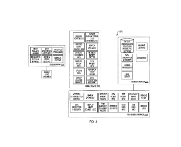
[0026] FIG. 2 schematically illustrates a representative architecture
implementing the
functionality of the present invention.
DETAILED DESCRIPTION
A. Core Architecture
[0027] Embodiments of the present invention pertain to wearable sensor
arrays that can
identify and monitor diagnostic digital biomarkers (as defined below). Various
embodiments
feature advantageous improvements to sensor sensitivity, specifically vibro-
acoustic and bio-
electric sensors that can monitor sounds, vibrations and electrical fields and
impulses of the
target living organism (e.g., a human patient), then apply techniques of
machine learning to
monitor and diagnose healthy vs. disease states. Machine learning may be
either supervised
learning (parametric/non-parametric algorithms, support vector machines,
kernels, neural
networks), unsupervised learning (clustering, dimensionality reduction,
recommender
systems, deep learning), or combinations thereof. Embodiments of the invention
may utilize
one or more physiological sensors as well as environmental sensors and other
sources of
health-related information.
[0028] Refer first to FIG. 2, which illustrates a system-level view of a
representative
topology 200 implementing an embodiment of the present invention, which
includes various
optional components. The system 200 includes one or more sensors 210; an
optional mobile
device 220 that receives and controls sensor signals and relays them to a back-
end server 230,
and also receives processed data from server 230 for display to the user; and
a web-based
interface 240, which may exist separately from or serve as an alternative to
mobile device
220 with additional capabilities including access to relevant patient
information. Typically,
7
CA 03040703 2019-04-15
WO 2018/075521
PCT/US2017/056984
the interface 240 is implemented on a general-purpose computer or workstation,
while the
mobile device may be a "smart" phone or tablet running an on-board application
("app"). In
general operation, one or more sensors 210 detect one or more patient
conditions and output
electrical (analog and/or digital) signals indicative of the sensed condition.
Sensors may be
located individually, in redundant clusters (e.g. one cluster containing one
vibro-acoustic
sensor and two non-contact bio-electric field/impulse sensors). Sensors may
also be located
internal to the patch 210a or external to the patch 210b (interfacing the body
or environment).
Suppose, for example, that the sensors monitor cardiac parameters and that the
system is
configured to predict whether the patient will go into cardiac arrest during a
medical
procedure. In this implementation, the sensor array 210 may include a vibro-
acoustic sensor,
a plurality of bio-electric sensors for an ECG unit, and a MEMS (or other)
sensor to detect
the patient's position and/or orientation; the outputs of all of these sensors
are relevant to the
likelihood of cardiac arrest, and are provided to a machine learning module in
the server 230.
As described in greater detail below, the machine learning module predicts the
likelihood of
cardiac arrest given the incoming signals from the sensors 210. For example,
the signals may
be repeatedly sampled over a time window and the synchronized raw signal
amplitude
patterns from each sensor catenated into a single feature vector that is used
to query the
machine learning module, which has previously been trained on similar feature
vectors. The
raw data may be stored in a time-indexed log in a memory to facilitate
synchronization, and
may also be stored in a database to facilitate selective retrieval by the
mobile device 220 or
interface 240. For example, successive one-second windows of data may be
provided to the
machine learning module, which each time returns a likelihood of cardiac
arrest. More
generally, the database may be used to store biomarkers based on data obtained
across
multiple patients. For example, the data gathered from patients' chests prior
to and during
heart attacks can be used to create novel digital biomarkers for diagnosing
and predicting
cardiac arrest. Specific subset data of the digital biomarker may further be
used for predicting
future tangential or causative diseases.
[0029] The processing rate of the machine learning module limits the rate
at which the
one-second data windows can be ingested and processed ¨ e.g., if the machine
learning
module needs three seconds to process data and return a result, the throughput
rate is 1/3 sec-1,
and analysis findings are displayed on the device 220 ("Display Analysis
Findings") and
updated every three seconds. The manner in which these findings are displayed
depends on
8
CA 03040703 2019-04-15
WO 2018/075521
PCT/US2017/056984
design preferences; a raw likelihood may be displayed in percentage terms, or
a color code
(e.g., red, yellow and green graphics) indicative of the current risk level
may be displayed
instead or in addition to the percentage. Device 220 may receive raw
probability data from
server 230 and format a display using on-board software, or may receive a
displayable image
in markup format from a conventional web server module in server 230; the
received image
is displayed by device 220 in a browser app.
[0030] In addition, the clinician may wish to view the sensor data
directly. To support
this, the device 220 may include mass storage for caching a time window of
sensor data
("Record Heart & ECG") and displaying the data in a useful format. As used
herein, the term
"display" is not limited to a visual rendering on a screen but also includes
aural reproduction,
e.g., of a sensed heartbeat, or tactile reproduction as discussed, for
example, in U.S. Serial No.
15/471,815, filed on March 28, 2017 and entitled "Haptic Feedback And
Interface Systems,"
the entire disclosure of which is hereby incorporated by reference. Using
mobile device 220,
the user may query the server database for earlier records (e.g., ECG traces)
for comparative
purposes, and may request patient records. The queries of sensor raw data and
the physician's
understanding and interpretation of such data may also serve as input to the
machine learning
module. To support privacy and security requirements, the devices 220, 240 may
include data
encryption and authentication software that serves as a front end to an
electronic medical
records (EMR) facility.
[0031] The sensor array 210, server 230, and mobile device 220 and/or
interface device
240 may communicate via one or more networks. The term "network" is herein
used broadly
to connote wired or wireless networks of computers or telecommunications
devices (such as
wired or wireless telephones, tablets, etc.). For example, a computer network
may be a
personal area network (PAN), a local area network (LAN) or a wide area network
(WAN).
When used in a PAN networking environment, computers and sensor arrays may be
connected to the PAN through radios such as Bluetooth. When used in a LAN
networking
environment, computers may be connected to the LAN through a modem, network
interface
or adapter. When used in a WAN networking environment, computers typically
include a
modem or other communication mechanism. Modems may be internal or external.
Networked computers may be connected over the Internet or any other system
that provides
communications. Some suitable communications protocols include TCP/IP, UDP,
and
Bluetooth. For wireless communications, protocols may include IEEE 802.11x
("Wi-Fi"),
Bluetooth, ZigBee, IrDa, near-field communication (NFC), or other suitable
protocol.
9
CA 03040703 2019-04-15
WO 2018/075521
PCT/US2017/056984
Furthermore, components of the system may communicate through a combination of
wired or
wireless paths, and communication may involve both computer and
telecommunications
networks.
[0032] It should also be stressed that the distribution of functionality
illustrated in FIG. 2
is representative only. The functionality may be spread arbitrarily over
multiple
intercommunicating devices, or may be centralized in a single device, e.g., a
laptop or even a
tablet with sufficient processing capacity. To support privacy and security
requirements, the
functionality may also be spread over multiple devices by taking into
consideration data
encryption and authentication requirements of EMIR. Additionally,
functionality may be
spread over multiple devices based on disposability and reusability (e.g.,
sensor arrays may
be disposable, whereas the processing, data storage and communication modules
may be
reusable).
[0033] The system 200 (or server 230) may be or include a general-purpose
computing
device in the form of a computer including a processing unit, a system memory,
and a system
bus that couples various system components including the system memory to the
processing
unit. Computers typically include a variety of computer-readable media that
can form part of
the system memory and be read by the processing unit. By way of example, and
not
limitation, computer-readable media may comprise computer storage media and
communication media. The system memory may include computer storage media in
the form
of volatile and/or nonvolatile memory such as read only memory (ROM) and
random access
memory (RAM). A basic input/output system (BIOS), containing the basic
routines that help
to transfer information between elements, such as during start-up, is
typically stored in ROM.
RAM typically contains data and/or program modules that are immediately
accessible to
and/or presently being operated on by processing unit. The data or program
modules may
include an operating system, application programs, other program modules, and
program data.
The operating system may be or include a variety of operating systems such as
Microsoft
WINDOWS operating system, the Unix operating system, the Linux operating
system, Apple
OS X, or another operating system or platform.
[0034] The computing environment may also include other removable/non-
removable,
volatile/nonvolatile computer storage media. For example, a hard disk drive
may read from
or write to non-removable, nonvolatile magnetic disks. A magnetic disk drive
may read from
or writes to a removable, nonvolatile magnetic disk, and an optical disk drive
may read from
or write to a removable, nonvolatile optical disk such as a CD-ROM, DVD-ROM,
Blu-ray, or
CA 03040703 2019-04-15
WO 2018/075521
PCT/US2017/056984
other optical media. Other removable/non-removable, volatile/nonvolatile
computer storage
media that can be used in the exemplary operating environment include, but are
not limited to,
magnetic tape cassettes, flash memory cards, digital versatile disks, digital
video tape, solid
state RAM, solid state ROM, and the like. The storage media are typically
connected to the
system bus through a removable or non-removable I/0 interface.
[0035] The processing unit that executes commands and instructions may be a
general
purpose computer, but may utilize any of a wide variety of other technologies
including a
special-purpose computer, a microcomputer, mini-computer, mainframe computer,
programmed microprocessor, microcontroller, peripheral integrated circuit
element, a CSIC
(customer-specific integrated circuit), ASIC (application-specific integrated
circuit), a logic
circuit, a digital signal processor, a programmable logic device such as an
FPGA (field-
programmable gate array), PLD (programmable logic device), PLA (programmable
logic
array), precise timing protocol component (PTP) providing a system with a
notion of global
time on a network, RFlD processor, smart chip, or any other device or
arrangement of
devices that is capable of implementing the steps of the processes of the
invention.
[0036] The various modules shown in FIG. 2, including the machine learning
module,
may be implemented by computer-executable instructions, such as program
modules, and
executed by a computer. Generally, program modules include routines, programs,
objects,
components, data structures, etc. that performs particular tasks or implement
particular
abstract data types. Any suitable programming language may be used in
accordance with the
various embodiments of the invention. Illustratively, the programming language
used may
include assembly language, Accord, Apache Mahout, Basic, C, C++, C*, Caffe,
Clojure,
Cloudera Oryx, COBOL, ConvNetJS, Cuda, PyTorch, Theano and TensorFlow, dBase,
DeepLearn.js, Forth, FORTRAN, GoLearn, Haskell, H20, Java, Mathematica,
MATLAB,
Modula-2, Pascal, Prolog, Python, R, REXX, Scala, and/or JavaScript, Scikit-
learn, Shogun,
Spark MLlib, Weka for example. Further, it is not necessary that a single type
of instruction
or programming language be utilized in conjunction with the operation of the
system and
method of the invention. Rather, any number of different programming languages
may be
utilized as is necessary or desirable.
[0037] While computer system 200 is described herein with reference to
particular blocks,
it is to be understood that the blocks are defined for convenience of
description and are not
intended to imply a particular physical arrangement of component parts.
Further, the blocks
need not correspond to physically distinct components. To the extent that
physically distinct
11
CA 03040703 2019-04-15
WO 2018/075521
PCT/US2017/056984
components are used, connections between components (e.g., for data
communication) can be
wired and/or wireless as desired.
[0038] Having described the general features of the system 200, the sensor
array and
machine learning module will now be described in greater detail.
B. Sensors
B.1 Vibro-Acoustic Sensors
[0039] The sensor array 210 desirably includes a vibro-acoustic transducer
arrangement
optimized for sensing and transducing acoustic phenomena occurring within a
target living
organism's or patient's body, and manifesting themselves at the skin surface
with frequencies
ranging from 0.001 Hz to 40 kHz. Strategies for effectively coupling to the
skin include
judicious mismatching of mechanical impedance, the use of impedance-matching
gels or
liquids, a shaped (e.g., domed) pickup, material selection, and/or a
peripheral leaf-spring
arrangement permitting relative movement between inner and peripheral
diaphragm portions
as described, for example, in U.S. Serial No. 15/471,812, filed on March 28,
2017 and
entitled "Vibro-Acoustic Transducer," the entire disclosure of which is hereby
incorporated
by reference.
[0040] In various embodiments described in the '812 application, the sensor
device
comprises a diaphragm having an outer peripheral portion and an inner portion.
The inner
movable portion is attached to the outer portion by a plurality of leaf
springs constraining
relative movement between the inner portion and the peripheral portion. The
sensor device
also includes a coil disposed over at least one side of the diaphragm, and at
least one magnet
operatively disposed with respect to the coil to cause current to flow through
the coil upon
relative movement between the movable portion and the peripheral portion. The
spring
stiffness or spring compliance of the leaf springs may be selectively chosen
to optimize the
frequency response of the sensor.
[0041] In some embodiments described in the '812 application, the inner
portion is fixed
and the outer peripheral portion is movable with respect thereto; in other
embodiments, the
outer portion is fixed and the inner peripheral portion is movable with
respect thereto. For
example, in a particular embodiment, the outer fixed portion of the diaphragm
has a shape
and the inner movable portion is defined within a plurality of slots through
the diaphragm and
arranged in a series. The series defines a closed sequence concentric with and
having the
shape of the outer fixed portion, and each pair of slots is parallel and has
an overlap portion
12
CA 03040703 2019-04-15
WO 2018/075521
PCT/US2017/056984
and a non-overlap portion, the overlap portion defining an intervening strip
corresponding to
one of the leaf springs. In some cases, the slots are filled with a
thixotropic material. In
some embodiments, the coil and the at least one magnet are circular, while in
other
embodiments, one or both have a different shape.
[0042] More generally, the vibro-acoustic sensor used herein may be
optimized to the
viscoelastic properties of target tissues in order to maximize the quality of
data gathered.
Optimization factors include but are not limited to the viscoelastic parameter
range of the
target tissue, target living organism specific or patient specific variations
in tissue
composition, and sensor-attachment interface material Target tissue
viscoelastic parameters
can be characterized broadly (e.g., the whole chest cavity) or restricted to
localized areas or
target tissue response (e.g. cardiac functionality, factoring out pulmonary
input factors). For
example, it is known that individual target tissues have specific viscoelastic
factors that
contribute to the desired target vibro-acoustic information to be detected ¨
e.g., for a cardiac
target, the major factors are the muscular contractions and blood flow.
Furthermore,
measuring a pregnant woman's abdomen creates additional challenges for the
measurement
or propagation of soundwaves, vibrations, light or electromagnetic waves due
to the complex
interface of new tissue and water layers caused by the presence of the
amniotic cavity, uterine
wall and other collagenic and tissue interfaces not normally found in adults.
These tissue
interfaces, which grow and move with fetal maturation and movement, can change
the
propagation of sound, vibrations and light, making it more difficult to record
inputs or image
inside the body.
[0043] Additionally, the correlations between the mechanical properties and
material
properties of certain muscular tissues may be monitored in real time to
characterize their
viscoelastic properties. Such information is used to generate stress and
strain models,
characterize the creep and strain-rate sensitivity of biological tissues
(e.g., skeletal
musculature atrophy and bone porosity), and monitor environmental and disease
effects on
tissue over periods of time (e.g., changes in bone viscoelasticity over time
in microgravity
and zero-gravity conditions).
[0044] Sensor attachment interface material may additionally affect the
quality of the
obtained vibro-acoustic signals. The fabric, gel patch, adhesive, or other
interface is selected
for optimal vibro-acoustic damping. Furthermore, the center and peripheral
edges of the
sensor may comprise or consist of differing material or differing amounts of
material to
further control viscoelastic damping.
13
CA 03040703 2019-04-15
WO 2018/075521
PCT/US2017/056984
B.2 Non-Contact Bio-electric Sensors
[0045] The sensor array 210 may include sensors for one or more bio-
electric time-
varying signals, i.e., the change in electric current produced by electrical
potential differences
across a specialized tissue, organ or cell system like the nervous system.
[0046] A bio-electric sensor may be capacitive so it does not rely on ohmic
contact to the
body for measuring bio-electrical signals (see, e.g., U.S. Patent Nos.
3,882,846 and 3,500,823,
the contents of which are incorporated herein by reference). This facilitates
data collection
across the target living organism (e.g. human body), and confers the ability
to measure
electrocardiography and other electrical fields and impulses without direct
skin contact.
Measurements such as ECG depend on being able to extract the small
electrophysiological
signals from the much larger noise signals. Unlike the silver/silver chloride
(Ag/AgC1)
electrodes used in clinical settings, bio-electric sensors in accordance
herewith may make a
high-impedance contact to the skin. This allows accurate and convenient
measurement of the
ECG. For such sensors, no gel, paste or other preparation is required at the
sensor-skin
interface. The connection is not affected by changes in skin impedance brought
on by
perspiration.
[0047] Data from sensor arrays as described herein may include near real-
time,
ambulatory electrocardigraphy (ECG), vectorcardiography (VCG),
ballistocardiography
(BCG), phonocardiography (PCG), and acoustic cardiography (ACG). ACG
synchronizes
cardiac sounds with the bio-electric sensor's electrocardiogram information
and provides a
comprehensive assessment of both mechanical and electrical functions of the
heart. ACG is
applied to heart failure diagnosis and ischemic heart disease detection, as
well as other
diseases including LV hypertrophy, pericarditis, sleep apnea and ventricular
fibrillation.
BCG measures cardiac ballistic forces with ultra-high resolution, enabling
blood pressure to
be measured "beat-to-beat" non-invasively with a wearable sensor. Vector
cardiography, the
electrical depolarization of the human heart, can be estimated and, if
desired, visualized using
vibro-acoustic data generated using sensors described herein.
[0048] The sensor data may be used to extrapolate various models and used
to diagnose
many heart diseases in just a few beats. Furthermore, sensor arrays in
accordance herewith
may contain memory and processing in a lightweight package and can easily
transmit data
wirelessly or via a wired connection. Various embodiments may additionally be
indicated for
14
CA 03040703 2019-04-15
WO 2018/075521
PCT/US2017/056984
heart failure follow-up in homes, clinics and hospitals as well as in the
microgravity or zero-
gravity of space.
[0049] Additionally or alternatively, in certain embodiments, specific
target tissues may
be locally stimulated to produce a response to be recorded by the sensor array
(e.g., acoustic
signals introduced into the body from a speaker can actively change the data
captured by the
above mentioned vibro-acoustic sensor) or a response from the target living
organism (e.g.
fetus). The stimulation mechanisms may be sound applied to the skin,
vibrations, ultrasound,
photonic, laser, a set period of motion (wave), or other bands within the
electromagnetic
spectrum, etc. This functionally can be used to stimulate certain conditions
such as stress,
functional movement, and various other activities.
B.3 3D and 4D Imaging
[0050] Certain embodiments of the system have advantageous qualities for
imaging by
monitoring the vibro-acoustic and bio-electric signals coming from certain
tissues. While
conventional imaging systems may operate by inducing a sound and then
interpreting the
reflection, embodiments of the present invention performs the inverse whereby
the signal
source is coming towards the sensors without the need for a reflection. For
example, sonar,
radar and ultrasound transmit an electromagnetic signal and then interpret the
reflection off
the object (e.g., different tissues, amnion, organs, abscess, other localized
infections, etc.)
being studied. Sensors in accordance herewith, when placed in multiple
locations on the body,
may directly record the vibro-acoustic and bio-electric signals to create a 3D
map of the
signals and construct an image utilizing the collected signals. The system
works in a fashion
similar to the hammerhead shark, which utilizes bio-electric sensors to
visualize the location
and approximate size of prey buried under the sand before attacking. In much
the same way,
embodiments of the present invention measure the amplitude and voltage
potential directly
across the contours of the patient's body as well as sounds, vibrations and
pressure waves
through a networked array of bio-electric, vibro-acoustic sensors and
optionally including
other sensors mentioned herein (e.g., position, temperature, UWB, etc.) while
incorporating
environment sensors as well). In some embodiments of the invention, a finite-
element model
mesh is used to approximate the cardiac geometry from 1) time-gated, reality-
based structural
information, 2) continuous target tissue pressure, and/or 3) tissue elastance
determined from
CA 03040703 2019-04-15
WO 2018/075521
PCT/US2017/056984
bio-electric and vibro-acoustic data. Rendered tissue or fetal volumes may be
shown in 3D as
well as displayed in time-resolved 4D animations.
[0051] This imaging approach can be used to image the fetal womb. The
networked bio-
electric and vibro-acoustic and other sensors (such as for position, to
observe changes in fetal
structures and tissues when the mother is supine or prone, for example)
measure bio-electric
signals and sounds, vibrations and pressure waves coming from the fetal heart,
circulation
and other functional areas of the fetal and maternal body to turn in these
signals into images
and data for machine learning. Furthermore, interference of the signals from
the fetus will be
disrupted by tissues external to the fetus (such as the amniotic cavity,
amniotic fluid volume,
compliance of the uterine wall, or blood flow exchange across the placenta,
for example)
which can inform on dimensions, compliance, stiffness (such as a digital
palpation) using the
sensors surrounding the womb. These measurements and imaging can either be
recognized
instantly through pattern recognition using machine learning or in some cases
the pattern can
change over time to better observe and identify diseased or "healthy" states,
providing
reassurance (so no action or intervention needs to occur in an otherwise
confusing situation
possibly requiring premature cesarean section or other potentially dangerous
intervention) or
indicating the need for clinicians to escalate treatment and/or intervene.
[0052] In one embodiment, the vibro-acoustic sensor, bio-electric sensors
and other
sensors mentioned herein are woven into a flexible garment placed around the
entire womb of
the expectant mother. The system then records vibro-acoustic signals from the
moving fetus's
heart, blood flow turbulence, motion, and other biological sounds.
Furthermore, the
"signature" of the fetus's bio-electric signals may reveal variations in mass,
position, and
state of the fetus and overall heath or disease. By measuring both vibro-
acoustic and bio-
electric fields either instantaneously or over time, embodiments of the
invention may search
for patterns of healthy vs. disease states, which may be correlated with
environmental
information (growth chart from medical record, weight of mother, etc.) and
physiology scores
(i.e., heart rate variability, fetal kicks per unit time, etc.) in order to
study thousands of babies
and their different biomarkers (e.g., Gestational Diabetes Mellitus,
preeclampsia, early
delivery, cesarean birth, having a big baby which can complicate delivery,
infection, etc. or
predicting a baby born with having low blood sugar, breathing problems,
jaundice, cord
strangulation, hypoxia, etc.). In another embodiment, ultrasound or UWB waves
can be used
as an adjunct to the passive system above in order to potentially improve the
resolution of
features, compliance of tissues, or more accurate changes.
16
CA 03040703 2019-04-15
WO 2018/075521
PCT/US2017/056984
[0053] This approach can also be used on sound waves emanating from inside
the body to
assess the potential riskiness of atherosclerotic plaques, compliance of
arteries and arterioles
along the heart or elsewhere, cardiac output, cardiac enlargement, carotid
intimal medial
thickness, to screening for chronic liver or kidney disease (an acoustic
palpation is able to
determine the stiffness or compliance of the liver or kidneys), or to improve
drug delivery by
localizing the effects: bio-electric signatures change based on metabolic
activity and
increased or decreased emittance of electric impulses, and so can reveal the
effects or effects
of pharmaceutical products over time, and therefore combine with other
lifestyle and health
information collected from the sensors associated with a particular patient.
This virtual
palpation technique images tissue stiffness differences associated with
different pathologies.
Systems in accordance herewith can be used as an adjunct to conventional
ultrasound for
clinicians, since images acquired using the vibro-acoustic sensor in the range
of 10kHz to
40kHz can be compared to conventional ultrasound images to provide additional
information
and, often, improved contrast.
[0054] One specific response outcome obtained by applying stimuli is
acoustic- and bio-
electric-based 3D imaging of various tissues throughout the body. As mentioned
above, the
vibro-acoustic sensor data and bio-electric sensor data can be obtained and
display three
dimensional images of the internal structure of the target living organism.
Compared to
conventional imaging methods through which data is obtained using high-powered
energy
sources (e.g., X-ray, ultrasound, gamma rays, etc.), this low-powered
alternative can be
realized as a wearable to generate real-time and time-lapsed 3D imaging in a
manner that is
completely passive, low cost and safe (even ultrasound imaging can cause
cavitation of
tissues, which may not be safe when applied to fetuses or across sensitive
areas of the body).
[0055] In various embodiments, other physiological sensors including, but
not limited, to
a pulse oximeter for wavelength transmittance/absorbance and oxygen
saturation, an ambient
skin/core temperature thermometer, optical sensors, camera systems, photonic
sensors,
infrared sensors, near- and far-infrared sensors, and a UV sensors for overall
physical
assessment, ultrasound for internal organ scan, electromyography (EMG) for
mechanical
properties of muscles at rest and in contraction, electroencephalogram (EEG)
for electrical
activity for functional status of the brain, electrooculography (EOG) for
changes in
resting/active electric potentials of the eye retina function, and/or a
volatile organic
compound (VOC) detector for organic compounds in excretions (e.g.,
perspiration and breath)
may be employed. Such sensors may be placed in separate, non-physically
tethered arrays
17
CA 03040703 2019-04-15
WO 2018/075521
PCT/US2017/056984
(e.g., one array for an EEG may be in the form of a cap, one array for a VOC
may be in the
form of a patch so that perspiration from a target region can be tested). Some
or all of these
other physiological sensor outputs may be relevant for evaluation of the
cardiopulmonary
state in this example. In certain embodiments, additional physical sensors are
incorporated
into the sensor array. Another exemplary physiological and imaging sensor is
the ultra-
wideband (UWB) sensor which is a low power, non-ionizing electromagnetic wave,
high-
penetration alternative to other imaging methods (MRI, X-ray), making it
suitable for a
wearable or implantable application.
[0056] In one embodiment, the optical sensor is a pulse plethysmograph
(PPG) used to
measure one or more of various conditions including heart rate, blood oxygen
saturation,
body hydration, severity of venous reflex disease, venous function, and cold
sensitivity.
[0057] In various other embodiments, other biosensors can be used to obtain
data through
specific biorecognition of various elements (e.g., enzymes, antibodies,
protein, nucleic acid,
ion receptors, cell types) in samples obtained from the target living
organism. Specific
biosensors include but are not limited to surface plasmon resonance (SPR)
biosensors for
detecting proteins and toxins, evanescent wave fluorescence biosensors for
detecting
biodefence and toxins, bioluminescent optical fiber biosensors for detecting
genotoxins,
waveguide interferometric biosensors for detecting cellular response and
viruses,
ellipsometric biosensors for detecting viral receptors, reflectometric
interference
spectroscopy biosensors for detecting xenobiotics and tumor cells, and surface-
enhanced
Raman scattering biosensors for detecting cancer proteins.
B.3 Environmental and Other Sensors
[0058] Sensor array 210 may include one or more microphones. For example,
tonal
inflection changes can reveal mood changes or emotional response, which may
then be
correlated to the simultaneously measured physiological response. Tonal
response may
further show a change in psychological disposition.
[0059] In certain embodiments, one or more environmental sensors are
incorporated into
the sensor array. Environmental sensors can measure skin temperature, ambient
temperature,
barometric pressure, 9-axis motion detection (3-axis magnetometer, 3-axis
accelerometer, 3-
axis gyroscope), which may be realized in MEMS form), geolocation, location-
dependent
real-time weather conditions (wind, humidity, rain, specific storm conditions,
UV index),
galvanic skin response, and pollution (air, light, noise, water, soil,
proximal radioactivity,
18
CA 03040703 2019-04-15
WO 2018/075521
PCT/US2017/056984
visual and other ambient conditions and contaminants). A sensor (or sensor
system) may be
used to track the patient's position and/or orientation, since these may be
relevant to a
biomarker. The patient's location can be improved by Wi-Fi, Bluetooth, and
integration of
various wireless communication protocols for more accurate location
determination. Within
a "smart home" (with connected devices as described above), systems in
accordance herewith
may be connected to "Internet of things" devices whose states can inform on
the health status
of a patient and whose operation may enhance patient convenience. Home
sensors, for
example, can include access to medicine containers (smart containers that show
when
medicine was administered, such as when bottle was opened and closed) and
smart toilets
(reading urine, fecal, or other metabolite analysis). Clothing cameras may be
used to
determine what the patient is wearing; overtime, the patient's clothing habits
can inform on
the overall change in a patient's mental state (such as a depressive, euphoric
or stressed
emotional state). Smart scales will inform on weight which can give insight
into a patient's
hydration status, and when combined with other sensor readings may provide
data on the
daily routine and habits that may correlate to specific outcomes.
B.4 Biomarker Identification and Use
[0060] As used
herein, the term "biomarker" refers to an association between one or more
measurable signals and one or more physiological or disease states. These
signals are
measured using the sensors 210, and analysis thereof using machine learning
techniques, as
described below, can be used to detect the presence and state of a biomarker
in a patient. For
example, a biomarker may be expressed in terms of a probability estimated
using linear
regression or a neural network applied to input signals from one or more
sensors.
[0061] For
example, with enough population data from one or more (and desirably many)
demographics, a normal standard of individuals who have not manifested
precursor
symptoms or symptoms of known disease states may be created and specific
deviations
therefrom can be assigned as separately diagnosable disease states (e.g., type
of disease,
precursor event identification, progression status, treatment options and
recommendations,
etc.). While no individual is "healthy" s/he may be at a baseline current
state where certain
disease states are either undetectable, misdiagnosed, or have yet to manifest
currently
detectable symptoms. With accumulation of population data, a better
understanding of
"health" can be contextualized and monitored on a spectrum of higher
precision. As a result,
any disease state and/or associated precursors can be monitored for
progression and
19
CA 03040703 2019-04-15
WO 2018/075521
PCT/US2017/056984
regression including first and second derivatives to obtain safety and
efficacy data of
treatments (e.g. pharmaceutical therapy, physical therapy, cognitive therapy,
spiritual therapy,
etc.) The result is a database of "virtual patients" for evaluation of new
interventions by
"phenotypes," enabling eventual customization of treatment by patient
characteristics. In
addition to direct/absolute measures, derived measures including heart rate
variability, FFT,
pulse transit time, harmonic expansion/compression, spectrograph
amplitude/frequency
envelope, etc. may be better predictors of specific biomarkers. For each
different population
(e.g., a population in microgravity, zero gravity, altered
acceleration/simulated gravity), the
standard digital biomarker may be adaptively calibrated as certain disease
states may have
different contributing factors, attributes, progression rates, and treatments.
For example, in
microgravity environments, the heart does not work as hard due to the lowered
resistance of
gravity, thereby causing the heart to become approximately 10% more spherical
in the micro-
gravity of low Earth orbit and zero gravity of outer space. Changes in
relevant digital
biomarkers of astronauts from normal gravity to microgravity to zero gravity
environments
may be observed using the techniques and systems described herein.
[0062] Diagnostic digital biomarkers may be tailored for each individual
patient by
including as input a patient's longitudinal health records as well as recorded
or self-reported
family history. Once the individual's personal information is integrated, a
personalized digital
biomarker or phenotypic fingerprint is generated, thereby allowing for the
possibility of
customized healthcare. Such information may be further strengthened by
correlations found
in genotypic similarities through DNA banks. Additional sources of data and
types of
information of interest include but is not limited to: (a) disease data of the
more than 30,000
diseases currently known in medical fields (e.g. cardiovascular, nervous
system,
inflammation, immune, metabolic, infectious disease, etc. and various
combinations thereof)
and/ or (b) microbiome, transcriptome, proteome, metabolome, etc. to further
understand
gene expression. The above information is currently and will further be
accumulated in
databases. It is well known that certain genetic subsets of the population
suffer from
increased hypertension and increase response to sodium, and with this type of
geographic
DNA data for example, we can better influence the system to accurately predict
or
recommend tests or exams to doctors.
CA 03040703 2019-04-15
WO 2018/075521
PCT/US2017/056984
C. Machine Learning Module
[0063] As noted above, the machine learning module is typically realized in
software, i.e.,
executable instructions stored in the memory of server 230 and executed by the
processor.
The topology shown in FIG. 2 is illustrative only; the machine learning module
may, for
example, be implemented in a cloud configuration and deployed on a remote
server,
receiving input (e.g., feature vectors) from sensor array 210, mobile device
220, server 230
and/or interface device 240.
[0064] The machine learning module may implement supervised learning
(parametric/non-parametric algorithms, support vector machines, kernels,
neural networks),
unsupervised learning (clustering, dimensionality reduction, recommender
systems, deep
learning), or combinations thereof depending on the signals analyzed and the
nature of the
biomarker. Multiple time-varying signals are well-suited to analysis and
classification by a
neural network.
[0065] Conventional computer programs use an algorithmic approach to
problem-solving,
i.e., the computer follows a set of instructions in order to solve the
problem. Unless the
specific steps that the computer needs to follow are known, the computer
cannot solve the
problem. That restricts the problem-solving capability of conventional
computers to
problems that we already understand and know how to solve. Biomarkers,
however, may not
be amenable to algorithmic processing, i.e., the relationship between a time-
varying signal
and a physiological condition may be complex and unpredictable.
[0066] Neural networks process information in a manner similar to the human
brain. The
network is composed of a large number of highly interconnected processing
elements
(neurons) working in parallel to solve a specific problem. Neural networks
learn by example;
they cannot be programmed to perform a specific task. The examples must be
selected
carefully, otherwise useful time is wasted or, worse, the network might
function incorrectly.
[0067] Neural networks can recognize diseases using sensor data since there
is no need to
provide a specific algorithm to identify the disease. Neural networks learn by
example, so
the details of how to recognize the disease are not needed. What is needed,
instead, is a set of
examples that are representatives of all the variations of the disease. The
examples need to
be selected very carefully if the system is to perform reliably and
efficiently. Neural
networks are particularly well-suited to providing sensor fusion (i.e.,
combining signal values
from several different sensors). Sensor fusion enables a neural network to
learn complex
relationships among the individual sensor values, which would otherwise be
lost if the values
21
CA 03040703 2019-04-15
WO 2018/075521
PCT/US2017/056984
were individually analyzed. In medical modeling and diagnosis, this implies
that even
though each sensor in a set may be sensitive only to a specific physiological
variable, a neural
network is capable of detecting complex medical conditions by fusing the data
from the
individual sensors.
[0068] Caffe, CUDA, PyTorch, Theano and TensorFlow are suitable neural
network
platforms (and may be cloud-based or local to an implemented system in
accordance with
design preferences). The key in realizing the benefits of the invention is to
finely tune the
neural network to vibro-acoustic and bio-electric signals. In some
embodiments, input data
includes not only sensor data but portions of the patient's longitudinal
health record, which
has significant information about the patient's current disease states,
medications and medical
history.
[0069] The input to a neural network may be a vector of input values (or
"feature" vector).
At least the vibro-acoustic and bio-electric sensors will typically provide
output in the form
of a time-varying signal, digitized as a sequence of amplitude values. Hence,
the neural
network (or other machine-learning construct) used herein should be configured
to process a
plurality of signals, some of which are time-varying signals, as input. This
can be
accomplished in various ways. One approach to processing time-varying signals
is to use a
recurrent neural network, in which connections between processing elements
form a directed
cycle and exhibit dynamic temporal behavior. This facilitates direct analysis
of time-varying
signals. Another approach, as noted above and which can be implemented on a
conventional
feedforward (e.g., convolutional or recursive) neural network, repeatedly
sample the sensors'
outputs over a synchronized time window. The synchronized raw signal amplitude
patterns
from each sensor may be combined (e.g., by simple concatenation) into a single
feature
vector that is used to query the machine learning module, which has previously
been trained
on similar feature vectors. The time-varying sensor signals may also be
processed rather than
used in raw form. For example, the short-time Fourier transform may be used to
determine
the sinusoidal frequency and phase content of discrete portions of a time-
varying signal
within a time window. In some circumstances, the frequency distribution may
provide a
more robust feature vector than the amplitude sequence. The frequency
distributions of the
different signals may be catenated or added together, e.g., with different
weights assigned to
spectra corresponding to the different signals in order to optimize
performance of the neural
network.
22
CA 03040703 2019-04-15
WO 2018/075521
PCT/US2017/056984
[0070] Processing multiple input parameters ¨ e.g., in addition to the time-
varying
sensor signals, the input vector may include diverse information such as
elements of the
patient's health records, the patient's current position and orientation, etc.
¨ can also be
accomplished in various ways. As explained above, these different forms of
data can be
concatenated into a large feature vector, added (e.g., in a weighted fashion),
or simply
provided as separate inputs to a neural network configured for input fusion.
[0071] It should also be noted that neural networks tend to perform better
at classification
tasks than regression tasks. Hence, if the desired output is a probability
(e.g., of the presence
of a disease condition), a probability range of 0 to 99 can be divided into
sub-ranges (e.g.,
class probabilities representing each of 10 separate sub-ranges (classes) 0-9,
10-19, 20-29,
etc.). If the various input data elements are correlated, an ensemble learning
approach can be
used. See, e.g., Guo et al., "Input Partitioning Based on Correlation for
Neural Network
Learning, I Clean Energy Tech. 1(4):335-38 (2013).
[0072] Therefore the neural network will further benefit from various
implementations of
optimization methods and filters including but not limited to low-pass (LP)
filters, high-pass
(HP) filters, bandpass (BP) filters, bandstop (BS) filters, infinite-impulse
response (IIR)
filters and various binary successive approximation (BSA), frequency-response-
masking
(FRM)-based linear-phase finite-impulse response (FIR) digital filters, and
combinations
thereof to identify and remove non-physiological signals captured by vibro-
acoustic sensors
as background "ambient noise" and enhance low threshold sounds.
D. Applications
[0073] As noted, the present invention may be deployed across diverse
applications in
medicine. Below, we focus on several representative applications.
D.1 Cardiopulmonary Applications
[0074] In certain embodiments, the vibro-acoustic, bio-electric, and any
number of
additional sensors are placed in an array encompassing (or wrapping around)
the torso to
allow for simultaneous auscultation. Cardiac auscultation can then be
simultaneously
completed at all four major sites: mitral area (at the apex beat, as the left
ventricle is closest
to the thoracic cage) , tricuspid area (inferior right sternal margin at the
point closest to the
valve in which auscultation is possible), the pulmonary area (left second
intercostal space
close to the sternum where the infundibulum is closest to the thoracic cage),
and aortic area
23
CA 03040703 2019-04-15
WO 2018/075521
PCT/US2017/056984
(right second intercostal space close to the sternum where the ascending aorta
is nearest the
thoracic cage). Certain sounds such as the aortic and pulmonic sounds are
detected best
during the S2 heart sound produced by the closing of the semilunar valves of
the heart
compared to during the Si heart sound produced by the closing of the
atrioventricular valves.
Furthermore, according to the disease state and physiological variation from
patient to patient,
the sounds may be more prominent in certain positions (e.g. sitting up or
leaning forward at
45 elicits changes in the amplitude and frequency of mitral valve murmurs as
the patient
leans forward to move the beating heart wall closer to the chest wall).
Similarly, pulmonary
auscultation is commonly completed over each of the five lobes of the lungs
from both the
anterior and posterior sides. With a wrap-around array configuration, more
than two, or all
cardiac, pulmonary and any additional auscultation sites may be monitored
simultaneously,
thereby mitigating variations in a patient's breath, position, and condition
as can be the case
during a traditional auscultation exam. The vibro-acoustic sensors may detect
different states
of disease in the lungs such as wheezing from asthma, fluid collecting in the
base of the lungs
that sounds like crackling as the alveolar sacks expand, or pulmonary
infections such as
pneumonia.
[0075] Various wrap-around array configurations may be selected for
individual patient
variation (e.g. size, body style, gender), duration of use and/or placement,
or may be
universally adaptable with built-in adjustability for improved data
acquisition quality and to
be more cost-effective. For example, the use may dictate the type of adhesive
option selected
for the sensor array from: 1) no adhesive for use in garments, 2) adhesive for
sensitive skin
that can be removed and re-applied multiple times, 3) sports-grade adhesive
that will last, e.g.,
15 days, and 4) veterinary-grade adhesive for livestock. As a cost-savings
example, certain
components such as the wireless electronics module may be reusable whereas the
sensor
array and adhesives may be disposable. In the flexible sensory array
embodiments, the
flexible portions may further include strain gauges (e.g., MEMS-based) to
additionally record
stretching and movement of the localized skin under the patch.
[0076] For example, the sensor array may have a substantially straight-line
configuration
with flexible curvature to align with the contour of various portions of the
body, or may have
a curved (e.g., U or C shape) configuration enabling one or more sensors to be
conveniently
positioned over each of the patient's auscultation points. Alternatively, the
sensor array may
take the form of a wearable vest with an array of connected sensors arranged
to monitor torso
organs and detect adventitious breath sounds, which are abnormal sounds that
are heard over
24
CA 03040703 2019-04-15
WO 2018/075521
PCT/US2017/056984
a patient's lungs and airways. These sounds include abnormal sounds such as
fine and coarse
crackles (sometimes called rales), wheezes (sometimes called rhonchi), pleural
rubs and
stridor. Adventitious breath sounds are important signs used for diagnosing
numerous
cardiac and pulmonary conditions. The sensor array signals may thereby be
translated into
respiration rate, breathing pattern, and posture data.
[0077] In another embodiment, using bio-electric sensors and vibro-acoustic
sensors
combined with machine learning, in addition to the system recording and
identifying the P
wave, QRS complex, T waves, and U waves, systems in accordance herewith may
identify
and track digital biomarkers based on H-wave peaks corresponding to the timing
of the His
bundle depolarization, a feature not normally observed in conventional surface
ECGs (0.5-30
Hz bandwidth). When combined with vibro-acoustic sensors, the effect of this H
wave peak
on the cardiac output, valve murmurs and carotid artery flow, when time-
synchronized, can
determine diseased properties of the heart's biology such as the sources of
arrhythmias, the
effect of the arrhythmia on the heart, locations of myocardial infarction or
worsening of a
clinically significant valve murmurs. The time relation between the H peak and
the atrial and
ventricular depolarizations in the heart is a useful diagnostic signature that
conventionally can
only be monitored using invasive intracardiac techniques where the sensor is
inserted into an
artery via a cardiac catheter.
[0078] A complex input system of digital biomarkers may also be combined
with
environmental inputs to monitor patients with heart failure. After an initial
physical
evaluation by the physician or clinician, sensors may be placed on the
patient's torso (e.g.,
integrated within a turtleneck garment, shirt, vest or jacket) to maximize the
signal recording
and establish a baseline for the patient's auscultation sounds, heart rate,
bowel sounds, and/or
electrical activity during physical maneuvers (tracked by the position sensor)
and other non-
invasive monitoring inputs. A patient diagnosed with heart failure may be
fitted with a sensor
array (e.g., in the form of a horseshoe) to monitor cardiopulmonary signals
during the
subsequent 30 days. During this follow-up time period, sensed environmental
conditions (e.g.,
from a smart scale (providing weight loss/gain and impedance (fat gain / loss)
data), a smart
toilet's notice of urine color change (indicating hydration status), and/or a
smart car (showing
decrease in reaction time indicating mental and/or physiological status)), may
be combined
with other system sensors measuring, for example, increased fluid in the lungs
(crackling at
the base of the lungs indicates fluid buildup as picked up by the vibro-
acoustic sensor),
dyspnea (shortness of breath after climbing up stairs as measured by detection
of labored
CA 03040703 2019-04-15
WO 2018/075521
PCT/US2017/056984
breathing and the sensed position of the patient on those stairs) and
distension in the carotid
artery as picked up by the vibro-acoustic sensor and position sensors over the
base of the
neck. It should be noted that the bio-electric sensors may not detect any
pathology or
changes in the ECG, but may nonetheless serve as a reference correlating the
opening and
closing of each valve in relation to sensed fluid flows. In this example, no
single sensor can
diagnose heart failure, but a collection of evaluated signals may provide a
high degree of
statistical confidence that a particular patient has early or late stage heart
failure. Furthermore,
patterns of the onset of this activity across populations and wide
demographics of patients,
when correlated with their DNA for personalized medicine, can enable
prediction of the onset
of disease, and give the care team the option to adjust medications or
escalate care. After
enough training, systems in accordance herewith may be capable of intervening
autonomously or at least suggesting changes to the patient's medication and
treatment
regimen.
[0079] Clinicians and nurses delivering babies can be an overwhelming
experience for
the clinician and the mother, so having a hands-free system whereby the above
embodiments
and combinations thereof can further be incorporated into an automated voice-
command
feedback system allows clinician to obtain and record data quickly, thereby
reducing
procedure time dramatically and allowing the clinician to focus. In addition,
when clinicians
by using the same equipment and process, variability among clinicians'
assessments and
diagnoses can be reduced or at least correlated as well as creating multiple
reproducible data
points per individual patient so that a baseline normal state can be created
and any disease
progression (either recovering or worsening of condition) can be tracked.
[0080] Additionally, all of the above applications benefit from further
physiological
response data obtained from the optional sensors described or from a database
of recorded
environmental data at the relevant location.
D.2 Vascular Surgery Application
[0081] When the wall of a blood vessel weakens, a balloon-like dilation
called an
aneurysm sometimes develops. This happens most often in the abdominal aorta,
an essential
blood vessel that supplies blood to the legs. Every year, 200,000 people in
the U.S. are
diagnosed with an abdominal aortic aneurysm (AAA). The most common treatment
is the
placement of an aortic abdominal graft through endovascular surgery in which a
synthetic
graft is inserted through the femoral artery and threaded up to the aorta with
a catheter. The
26
CA 03040703 2019-04-15
WO 2018/075521
PCT/US2017/056984
graft is placed at the site of the aneurysm and reinforces the weakened
section of the aorta to
prevent rupture.
[0082] If an aortic abdominal graft ruptures, the patient will quickly lose
so much blood
s/he may die. There is currently no commonly accepted way to tell if the graft
is failing. A
wearable or implantable vibro-acoustic sensor may be used ¨ e.g., in
conjunction with a
CPU and a neuromorphic processor along with memory and communications
capability ¨ to
detect graft failure. The sensor array may be worn externally or implanted
next to a just-
completed aortic abdominal graft. An embedded neuromorphic processor is
trained on the
sounds of blood flowing past the just-introduced graft. This training phase
occurs over a
relatively short period of time, e.g., a few days, following which the
neuromorphic processor
is switched into diagnostic mode. The wearable or implant then communicates
(e.g.,
wirelessly) with an external store-and-forward device that relays information
to a call center
and/or prescribing clinician, or stores data for proximate or remote
retrieval. More generally,
various embodiments of the sensor array described herein may be implantable
and placed
near a surgical site to monitor recovery and detect the need for follow-up
treatment.
[0083] As noted, the present invention may be deployed across diverse
applications in
medicine. Other medical applications include but are not limited to:
anesthesiology,
dermatology, endocrinology, gastroentology, hematology, ophthalmology,
pathology,
radiology, urology, professional sports medicine, physical therapy, etc. The
sensor
applications may uncover previously unknown correlations among the various
fields. The
sensor array may alternatively be used for personal health and fitness.
27
CA 03040703 2019-04-15
WO 2018/075521
PCT/US2017/056984
D.3 Non-Human Applications
[0084] Bovine respiratory disease ("BRD") is the most common disease
affecting cattle
in North America. BRD affects the respiratory tracts and can often be fatal,
causing billions
of dollars in economic losses for ranchers, dairymen and feed lot operators.
Just as in
humans, digital biomarkers of BRD may be created. Using sensor arrays as
described herein,
producers (e.g. ranchers, dairymen, feed lots and veterinarians) can detect
BRD early,
determine the severity of the disease and select an appropriate treatment
regimen, which may
help them improve cardiopulmonary-health related outcomes.
[0085] The terms and expressions employed herein are used as terms and
expressions of
description and not of limitation, and there is no intention, in the use of
such terms and
expressions, of excluding any equivalents of the features shown and described
or portions
thereof. In addition, having described certain embodiments of the invention,
it will be
apparent to those of ordinary skill in the art that other embodiments
incorporating the
concepts disclosed herein may be used without departing from the spirit and
scope of the
invention. Accordingly, the described embodiments are to be considered in all
respects as
only illustrative and not restrictive.
[0086] What is claimed is:
28