Note: Descriptions are shown in the official language in which they were submitted.
EITLE OF INVENTION
TRANSSEPTAL INSERTION DEVICE
[00011
FIELD OF THE INVENTION
[0002] The present invention relates generally to cardiac catheters, and more
particularly,
to a transseptal insertion device which is suitable for facilitating quick and
safe transseptal
puncture and insertion of a needle or catheter through a cardiac septum to
provide access to the
left atrium in implementation of a left atrial intervention.
BACKGROUND OF THE INVENTION
(0003] Cardiac catheterization is a medical procedure in which a long thin
tube or
catheter is inserted through an artery or vein into specific areas of the
heart for diagnostic or
therapeutic purposes. More specifically, cardiac chambers, vessels and valves
may be
catheterized.
[0004] Cardiac catheterization may be used in procedures such as coronary
angiography
and left ventricular angiography. Coronary angiography facilitates
visualization of the coronary
vessels and finding of potential blockages by taking X-ray images of a patient
who has received
1
Date Recue/Date Received 2023-05-11
CA 03041032 2019-04-17
WO 2018/075426 PCT/US2017/056843
a dye (contrast material) injection into a catheter previously injected in an
artery. Left ventricular
angiography enables examination of the left-sided heart chambers and the
function of the left-
sided valves of the heart, and may be combined with coronary angiography.
Cardiac
catheterization can also be used to measure pressures throughout the four
chambers of the heart
and evaluate pressure differences across the major heart valves. In further
applications, cardiac
catheterization can be used to estimate the cardiac output, or volume of blood
pumped by the
heart per minute.
[0005] Some medical procedures may require catheterization into the left
atrium of the
heart. For this purpose, in order to avoid having to place a catheter in the
aorta, access to the left
atrium is generally achieved by accessing the right atrium, puncturing the
interatrial septum
between the left and right atria of the heart, and threading the catheter
through the septum and
into the left atrium. Transseptal puncture must be carried out with extreme
precision, as
accidental puncturing of surrounding tissue may cause very serious damage to
the heart. In
addition, transseptal puncture may require complicated instruments which are
not helpful in
guaranteeing the precision of the puncture.
[0006] Accordingly, there is an established need for a device that is suitable
for
facilitating quick and safe transseptal puncturing to provide access to the
left atrium in
implementation of a left atrial intervention.
SUMMARY OF THE INVENTION
[0007] It is an object of the present invention to provide a device that is
suitable for
facilitating quick and safe transseptal puncturing to provide access to the
left atrium in
implementation of a left atrial intervention.
[0008] The present invention is directed to a transseptal insertion device
which is suitable
for facilitating quick and safe transseptal insertion of a needle or catheter
through an interatrial
cardiac septum to provide access to the left atrium in implementation of a
left atrial intervention.
The transseptal insertion device is elongated yet has a relatively reduced
length, and can be
2
CA 03041032 2019-04-17
WO 2018/075426 PCT/US2017/056843
easily and safely turned within an atrium of the heart to achieve a correct
orientation towards the
cardiac septum.
[0009] Introducing a first implementation of the invention, the present
invention includes
a transseptal insertion device which is suitable for facilitating a precise
and safe transseptal
insertion of a needle or catheter through a cardiac septum, comprising a
device housing and a
slidable body slidably disposed in the device housing. The slidable body
includes a pusher and a
guide element extending from the pusher. The guide element us extendable and
retractable from
a distal end of the device housing.
[0010] In a second aspect, the guide element may be formed as a web.
[0011] In another aspect, the device housing may include a housing interior
and an
annular housing gap surrounding the housing interior, and the guide element
may be slidably
disposed within the housing gap.
[0012] In another aspect, the device housing may include an outer housing
wall, an inner
housing wall, a housing interior formed by the inner housing wall and an
annular housing gap
surrounding the housing interior.
[0013] In still another aspect, the pusher may include a front pusher ring, a
rear pusher
ring spaced-apart from the front pusher ring and at least one pusher rod
extending between the
front pusher ring and the rear pusher ring.
[0014] In yet another aspect, the one or more pusher rods may extend between
the front
pusher ring and the rear pusherring.
[0015] In another aspect, the guide element may extend from the front pusher
ring of the
pusher.
[0016] In another aspect, the guide element may include multiple, parallel,
spaced-apart
longitudinal webbing elements and multiple, annular transverse webbing
elements provided at
spaced-apart intervals with respect to each other along the longitudinal
webbing elements.
3
CA 03041032 2019-04-17
WO 2018/075426 PCT/US2017/056843
[0017] In another aspect, multiple anchors may terminate the respective
longitudinal
webbing elements of the guide element for impingement against the cardiac
septum in insertion
of a needle or catheter through an orifice in the septum.
[0018] In another aspect, at least one of the pusher and the guide element can
be
inflatable.
[0019] In another aspect, the device housing can include an outer housing wall
defining a
housing interior and a pusher channel extending through the outer housing wall
generally parallel
and adjacent to the housing interior. The guide element can further include a
pusher having an
inflatable pusher rod slidably disposed in the pusher channel and an
inflatable pusher ring
terminating and disposed in fluid communication with the pusher rod.
[0020] These and other objects, features, and advantages of the present
invention will
become more readily apparent from the attached drawings and the detailed
description of the
preferred embodiments, which follow
[0021] Objects of the invention and its particular features and advantages
will become
more apparent from consideration of the following drawings and accompanying
detailed
description. It should be understood that the detailed description and
specific examples, while
indicating the preferred embodiment of the invention, are intended for
purposes of illustration
only and are not intended to limit the scope of the invention.
BRIEF DESCRIPTION OF THE DRAWINGS
[0022] The preferred embodiments of the invention will hereinafter be
described in
conjunction with the appended drawings provided to illustrate and not to limit
the invention,
where like designations denote like elements, and in which:
[0023] FIG. 1 presents a front perspective view of a transseptal insertion
device in
accordance with a first embodiment of the present invention, the device shown
exploded and
accompanied by a catheter;
4
CA 03041032 2019-04-17
WO 2018/075426 PCT/US2017/056843
[0024] FIG. 2 presents a front perspective view of the transseptal insertion
device of FIG.
1 in a first, retracted position, with the catheter extending partially
through the device;
[0025] FIG. 3 presents a rear perspective view of the transseptal insertion
device of FIG.
1 in a second, advanced position, with the catheter extending through the
device and protruding
distally from the device;
[0026] FIG. 4 presents a cross-sectional view of the transseptal insertion
device and
catheter of FIG. I prior to puncturing an interatrial cardiac septum, the
transseptal insertion
device and catheter shown in the first, retracted position of FIG. 2, the
cross section taken along
section plane 4-4 indicated in FIG. 2;
[0027] FIG. 5 presents a similar cross-sectional view of the transseptal
insertion device
and catheter of FIG. 1, the interatrial cardiac septum shown punctured, the
transseptal insertion
device shown in the second, advanced position of FIG. 3, and the catheter
shown extending
through the cardiac septum;
[0028] FIG. 6 is a cross-sectional view, taken along section plane 6-6
indicated in FIG. 4;
[0029] FIG. 7 presents a front perspective view of a transseptal insertion
device in
accordance with a second embodiment of the present invention, the device shown
exploded and
accompanied by a catheter
[0030] FIG. 8 presents a front perspective view of the transseptal insertion
device of FIG.
7 in a first, retracted position, with the catheter extending partially
through the device;
[0031] FIG. 9 presents a rear perspective view of the transseptal insertion
device of FIG
7 in a second, advanced position, with the catheter extending through the
device and protruding
distally from the device;
[0032] FIG. 10 presents a cross-sectional view of the transseptal insertion
device and
catheter of FIG. 7 prior to puncturing an interatrial cardiac septum, the
transseptal insertion
device and catheter shown in the first, retracted position of FIG. 8, the
cross section taken along
section plane 10-10 indicated in FIG. 8;
CA 03041032 2019-04-17
WO 2018/075426 PCT/US2017/056843
[0033] FIG. 11 presents a similar cross-sectional view of the transseptal
insertion device
and catheter of FIG. 7, the interatriaI cardiac septum shown punctured, the
transseptal insertion
device shown in the second, advanced position of FIG. 9, and the catheter
shown extending
through the cardiac septum;
[0034] FIG. 12 is a cross-sectional view, taken along section plane 6-6
indicated in FIG.
10;
[0035] FIG, 13 presents a front perspective view of a transseptal insertion
device in
accordance with a third embodiment of the present invention, the device shown
exploded and
accompanied by a catheter;
[0036] FIG, 14 presents a cross-sectional view of the transseptal insertion
device and
catheter of FIG. 13 prior to puncturing an interatrial cardiac septum, the
transseptal insertion
device and catheter shown in a retracted position;
[0037] FIG, 15 presents an enlarged view of the distal end of the intermediate
catheter of
FIG. 14; and
[0038] FIG. 16 presents a front perspective view of a transseptal insertion
device in
accordance with a fourth embodiment of the present invention, the device shown
exploded and
accompanied by a catheter
[0039] Like reference numerals refer to like parts throughout the several
views of the
drawings.
DETAILED DESCRIPTION OF THE, INVENTION
[0040] In the following description, numerous details are set forth for the
purpose of
example and explanation; however, one of ordinary skill in the art will
realize that the invention
may be practiced without the use of these specific details.
6
CA 03041032 2019-04-17
WO 2018/075426 PCT/US2017/056843
[0041] The following detailed description is merely exemplary in nature and is
not
intended to limit the described embodiments or the application and uses of the
described
embodiments. As used herein, the word "exemplary" or "illustrative" means
"serving as an
example, instance, or illustration." Any implementation described herein as
"exemplary" or
"illustrative" is not necessarily to be construed as preferred or advantageous
over other
implementations. All of the implementations described below are exemplary
implementations
provided to enable persons skilled in the art to make or use the embodiments
of the disclosure
and are not intended to limit the scope of the disclosure, which is defined by
the claims. For
purposes of description herein, the terms "upper", "lower", "left", "rear",
"right", "front",
"vertical", "horizontal", and derivatives thereof shall relate to the
invention as oriented in FIG. 1.
Furthermore, there is no intention to be bound by any expressed or implied
theory presented in
the preceding technical field, background, brief summary or the following
detailed description. It
is also to be understood that the specific devices and processes illustrated
in the attached
drawings, and described in the following specification, are simply exemplary
embodiments of
the inventive concepts defined in the appended claims. Hence, specific
dimensions and other
physical characteristics relating to the embodiments disclosed herein are not
to be considered as
limiting, unless the claims expressly stateotherwise.
[0042] Shown throughout the figures, the present invention is directed toward
a
transseptal insertion device which is suitable for facilitating quick and safe
transseptal puncturing
of an interatrial septum and insertion of a catheter therethrough to provide
access to the left
atrium in implementation of a left atrial intervention.
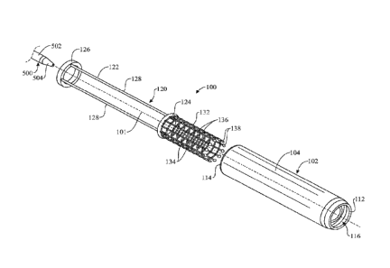
[0043] Referring initially to FIGS. 1-6, a transseptal insertion device 100 is
illustrated in
accordance with an exemplary embodiment of the present invention. As shown,
the transseptal
insertion device 100 is generally elongated and arranged along a longitudinal
axis 101. The
transseptal insertion device 100 may include a device housing 102. The device
housing 102 may
be generally elongated and cylindrical in shape arranged about the
longitudinal axis 101, with an
outer housing wall 104 and an inner housing wall 106 (FIGS. 4-6). The inner
housing wall 106
may be generally parallel to and concentric with the outer housing wall 104
and about the
7
CA 03041032 2019-04-17
WO 2018/075426 PCT/US2017/056843
longitudinal axis 101. An annular housing gap 108 may be formed by and between
the outer
housing wall 104 and the inner housing wall 106. A housing interior 110 may be
formed by and
within the inner housing wall 106. The device housing 102 may have a distal
end 112 and a
proximal end 114. In some embodiments, the distal end 112 of the device
housing 102 may be
tapered in longitudinal cross-section, as best shown in FIGS. 4 and 5. A front
housing opening
116 may be disposed in communication with the housing gap 108 and the housing
interior 110 at
the distal end 112 of the device housing 102. A rear housing opening 118 may
be disposed in
communication with the housing interior 110 at the proximal end 114 of the
device housing 102.
[0044] Further, the transseptal insertion device 100 includes a slidable body
120 which is
arranged inside the device housing 102 and slidably or longitudinally
translatable relative to the
device housing 102. The slidable body 120 of the present embodiment is
composed of a pusher
122 and a webbed guide element 132. In some embodiments, the slidable body
120, such as the
pusher 122 and webbed guide element 132, can be formed into a single-piece
unit such as by
injection molding, welding or the like.
[0045] In certain embodiments, the slidable body 120 is covered with fabric
such as
PTFE/Dacron which makes it non-porous.
[0046] As best shown in FIG. 1, the pusher 122 is slidably disposed within the
housing
gap 108 between the outer housing wall 104 and the inner housing wall 106 of
the device
housing 102. In some embodiments, the pusher 122 may include a front pusher
ring 124 and a
rear pusher ring 126 which is spaced-apart from the front pusher ring 124. At
least one elongated
pusher rod 128 may extend between the front pusher ring 124 and the rear
pusher ring 126. In
some embodiments, multiple pusher rods 128 may extend between the front pusher
ring 124 and
the rear pusher ring 126 in generally parallel relationship to each other
around the circumference
of the front pusher ring 124 and the rear pusher ring 126.
[0047] With continued reference to FIG. 1, the webbed guide element 132
extends
forwardly from the pusher 122, such as from the front pusher ring 124 of the
pusher 122. The
webbed guide element 132 delimits an internal space 133 transversely, in order
to provide a
8
CA 03041032 2019-04-17
WO 2018/075426 PCT/US2017/056843
guiding effect of a needle or catheter traveling longitudinally trough the
internal space 133, as
will be explained in greater detail hereinafter. The webbed guide element 132
may be generally
elongated and cylindrical in shape, as shown. In some embodiments, the webbed
guide element
132 may be transversely expandable and/or retractable, i.e. allow for a
variation of its diameter;
for instance, the webbed guide element 132 bay be expandable to a conical
shape according to
which a distal end of the webbed guide element 132 would have a larger
diameter than a proximal
end of the webbed guide element 132. Should the webbed guide element 132 be
expandable,
expansion is limited to a certain extent in order for the webbed guide element
132 to still provide
the aforementioned guiding effect. In some embodiments, as shown in the
present drawing, the
webbed guide element 132 can include multiple, parallel longitudinal webbing
elements 134
which are disposed in spaced-apart relationship to each other around the
circumference of the
webbed guide element 132; in turn, multiple, parallel, spaced-apart transverse
webbing elements
136 may connect the longitudinal webbing elements 134 to each other in the
webbed guide
element 132. Alternative embodiments are contemplated, however, in which the
construction of
the webbed guide element 132 may vary; for instance, and without limitation,
the webbed guide
element can be made of oblique messing elements forming a net. The webbed
guide element 132
can be made of nitinol, for instance and without limitation. In some
embodiments, as shown in
the present illustrations, widened sections or anchors 138 may terminate the
distal ends of the
respective longitudinal webbing elements 134 of the webbed guide element 132.
The anchors 138
can be arranged around a perimeter of the webbed guide element 132 and
substantially coplanar
to one another on a plane that is transverse to the longitudinal axis 101 of
the transseptal insertion
device 100. The anchors 138 can be made of tantalum, for instance and without
limitation.
[0048] As shown in FIGS. 2 and 3, the slidable body 120 can slidably adopt
different
longitudinal positions within the device housing 102. In a first or retracted
position, shown in
FIG. 2, the slidable body 120 is retracted relative to the device housing 102
so that the webbed
guide element 132 is located generally inside the device housing 102 and the
pusher 122 is
protruding rearwardly from the proximal end 114 of the device housing 102. In
a second or
advanced position, the slidable body 120 is moved forward relative to the
device housing 102 so
that the slidable body 120 advances through the front housing opening 116 and
protrudes
9
CA 03041032 2019-04-17
WO 2018/075426 PCT/US2017/056843
outwardly from the distal end 112 of the device housing 102, the webbed guide
element 132
extends outwardly and distally from the device housing 102 and the pusher 122
is generally
received within the device housing 102. More specifically, in the second,
advanced position, the
pusher rods 128 can be received within the device housing 102, as shown, while
the rear pusher
ring 126 of the pusher 122 remains outside the device housing 102 and rests on
the proximal end
114 of the device housing 102 to block the slidable body 120 from further
advancing forward
through the device housing 102.
[0049] For purposes that will be described hereinafter, the slidable body is
used to anchor
into the left atrial appendage in the eventuality of a perforation during a
left atrial appendage
procedure. Since it is non-porous, it will act as an occlusion balloon and
prevent further
extravasation of blood in the pericardial sac till a more definitive procedure
may be performed or
the bleeding stops
[0050] For purposes that will be described hereinafter, a catheter 500
carrying a spear or
needle can be inserted through the transseptal insertion device 100 and,
guided by the slidable
body 120, protrude outwardly from the distal end 112 of the device housing 102
as shown in FIG.
3.
[0051] In certain embodiments, the slidable body is used to remove an
implanted mitral
regulation (Mitraclipe) device. The slidable body is used to anchor onto the
anterior and posterior
leaflets of the mitral valve. Once anchored, there is either a mechanical,
magnetic or
electromagnetic lever that attaches to the mitral regulation device and
stabilizes it. Energy is then
delivered to the mitral valve to via the slidable body 120 to ablate the
anterior and posterior
leaflets. The mitral regulation device is thereby released and removed from
the body. The slidable
body 120 may also be used in the absence of a mitral regulation device on the
mitral valve and
may be used to ablate the anterior mitral leaflet prior to mitral valve
implantation to prevent left
ventricular outflow tract obstruction. In this instance, the anterior mitral
leaflet would be
stabilized with a set of stabilizers which would be housed within the slidable
body 120. The
stabilizers would be used to stabilize the anterior mitral leaflet first and
then the slidable body
CA 03041032 2019-04-17
WO 2018/075426 PCT/US2017/056843
would be used to deliver energy to ablate the anterior mitral leaflet. The
ablated tissue would then
be removed from the body using the stabilizer.
[0052] In certain embodiments, the slidable body is used to anchor into the
pulmonary
veins. Radiofrequency energy or other forms of energy may be delivered via the
cable and the
slidable body to the pulmonary veins to result in electrical ablation
[0053] A typical application of the transseptal insertion device 100 to
puncture the
interatrial cardiac septum 520 is now described with reference to FIGS. 4 and
5.
[0054] Initially, the transseptal insertion device 100 is arranged in the
retracted or first
position (described heretofore with reference to FIG. 2) in which the slidable
body 120 is retracted
relative to the device housing 102 and the distal end 112 of the device
housing 102 provides a
tapered, front or distal end of the transseptal insertion device 100. The
transseptal insertion device
100 is then inserted into the right atrium 510 of the heart through a catheter
(hereinafter be
referred to as "external catheter" for clarity purposes) extending through a
vein; the external
catheter and the vein are not shown in the drawings so as not to obscure the
invention.
[0055] Once the transseptal insertion device 100 reaches the right atrium 510,
a second,
separate catheter 500 carrying a spear or needle (not shown) therewithin is
extended through the
slidable body 120 and the housing interior 110 of the device housing 102. The
catheter 500 may
have a conventional design with an elongated, typically flexible catheter body
502 and a tapered
catheter tip 504 which terminates the catheter body 502. Before or after
inserting the second,
separate catheter 500 into the transseptal insertion device 100, the surgeon
slowly moves the
transseptal insertion device 100 to place it near, and facing, a target point
522 or area of the
cardiac septum 520 to be punctured, as shown in FIG. 4.
[0056] Once the transseptal insertion device 100 is arranged facing the target
point 522
of the cardiac septum 520, the transseptal insertion device 100 is operated to
switch from the
retracted position of FIG. 2 to the advanced position of FIG. 3; in other
words, the slidable body
120 is pushed forward relative to the device housing 102 so that the webbed
guide element 132
protrudes distally from the distal end 112 of the device housing 102. The
transseptal insertion
11
CA 03041032 2019-04-17
WO 2018/075426 PCT/US2017/056843
device 100 is arranged sufficiently close to the cardiac septum 520; thus, by
pushing the slidable
body 120 forward, the webbed guide element 132 eventually touches and rests on
the cardiac
septum 520. If present, the anchors 138 can engage the cardiac septum 520 to
contribute to
stabilize the webbed guide element 132 onto the cardiac septum 520 so that the
webbed guide
element 132 remains around the target point 522.
[0057] Once the webbed guide element 132 rests on the cardiac septum 520, the
webbed
guide element 132 and cardiac septum 520 enclose the internal space 133 of the
webbed guide
element 132 and the target point 522 of the cardiac septum 520. The spear or
needle may then be
advanced through the catheter 500 and towards the cardiac septum 520,
puncturing the cardiac
septum 520 and forming an orifice 530 in the cardiac septum 520. The slidable
body 120 being
arranged in the housing gap 108 between the outer housing wall 104 and the
inner housing wall
106 of the device housing 102 contributes to stabilize the slidable body 120,
and thus to maintain
the webbed guide element 132 in a same position, providing a safe and precise
aim when
puncturing the cardiac septum 520.
[0058] Having created an orifice 530 in the cardiac septum 520, the catheter
500 may
then be inserted through the orifice 530 and into the left atrium 512 of the
patient's heart in order
to proceed with the left atrium intervention as known in the art. The
transseptal insertion device
100 may be maintained in the position of FIG. 5 to stabilize the catheter 500
and maintain its
correct orientation relative to the cardiac septum 520.
[0059] After the cardiac catheterization procedures are completed, the
catheter 500 may
be withdrawn from the left atrium 512 through the orifice 530 and retracted
back into the webbed
guide element 132. Next, the webbed guide element 132 may be withdrawn from
engagement
with the cardiac septum 520 and into the housing gap 108, as illustrated in
FIG. 4. Finally, the
transseptal insertion device 100 may be removed from the right atrium 510
through the external
catheter.
[0080] It will be appreciated by those skilled in the art that the transseptal
insertion device
100 facilitates safer and quicker insertion of the spear or needle and the
catheter 500 through the
12
CA 03041032 2019-04-17
WO 2018/075426 PCT/US2017/056843
cardiac septum 520, and thus, quicker and safer access to the left atrium 512,
minimizing the risk
of damaging surrounding tissue during insertion of the needle or catheter 500.
[0061] Referring next to FIGS. 7-12, a second illustrative embodiment of the
transseptal
insertion device is generally indicated by reference numeral 200. In the
transseptal insertion
device 200, elements which are analogous to the respective elements of the
device 100 that was
heretofore described with respect to FIGS. 1-6 are designated by the same
respective numerals in
the 200-299 series in FIGS. 7-12. Unlike the previous device housing 102, the
device housing
202 of the transseptal insertion device 200 of the present embodiment has a
single housing wall
204. As illustrated in FIGS. 10 and 11, a pusher channel 262 extends through
and along the
housing wall 204 from the proximal end 214 to the distal end 212 of the device
housing 202. As
illustrated in FIG. 11, the distal end 212 of the device housing 202 may have
a concave seating
area 260 which encircles the front housing opening 216.
[0062] Similarly to the previous embodiment, as illustrated in FIGS. 7-9, the
slidable
body 220 of the present embodiment includes a pusher 222 and a guide element
232 extending
from the pusher 222. The pusher 222 depicted herein consists of a single
pusher rod 228. In turn,
the guide element 232 is formed as a ring extending transversely from the
pusher 222, the ring
being shaped and sized to be received within the concave seating area 260 of
the device housing
202. The slidable body 220 can be inflatable; for instance, and without
limitation, the pusher rod
228 and the guide element 232 can be hollow, flexible and in fluid
communication with one
another. The pusher rod 228 is disposed for slidable displacement in the
pusher channel 262 of
the device housing 202. The pusher rod 228 may be disposed in fluid
communication with an
inflating fluid source (not illustrated) which introduces a supply of
pressurized inflatable fluid
(not illustrated) through the pusher rod 228 into the guide element 232 to
inflate the pusher rod
228 and the guide element 232, for purposes which will be hereinafter
described.
[0063] Similarly to the previous embodiment, as shown in FIGS. 8 and 9, the
slidable
body 220 of the present embodiment can slidably adopt different longitudinal
positions within
the device housing 202. In a first or retracted position, shown in FIG. 2, the
slidable body 220 is
retracted relative to the device housing 202 so that the annular guide element
232 is resting against
13
CA 03041032 2019-04-17
WO 2018/075426 PCT/US2017/056843
the concave seating area 260 of the device housing 202. In a second or
advanced position, the
slidable body 220 is moved forward relative to the device housing 202 so that
the pusher rod 228
advances through the pusher channel 262 of the device housing 202 and the
slidable body 220
protrudes outwardly from the distal end 212 of the device housing 202, so that
the guide element
232 is spaced apart from the distal end 212 of the device housing 212. A
catheter 500 carrying a
spear or needle can be inserted through the transseptal insertion device 200
and, guided by the
annular guide element 232 of the slidable body 220, protrude outwardly from
the distal end 212
of the device housing 202 as shown in FIG. 9.
[0064] A typical application of the transseptal insertion device 200 to
puncture the
interatrial cardiac septum 520 is now described with reference to FIGS. 10 and
11.
[0065] Initially, the transseptal insertion device 200 is arranged in the
retracted or first
position (described heretofore with reference to FIG. 8) in which the slidable
body 220 is retracted
relative to the device housing 202 and the distal end 212 of the device
housing 202 together with
the rounded annular guide element 232 provide a rounded, front or distal end
of the transseptal
insertion device 200. The transseptal insertion device 200 is then inserted
into the right atrium
510 of the heart through a catheter (hereinafter be referred to as "external
catheter" for clarity
purposes) extending through a vein; the external catheter and the vein are not
shown in the
drawings so as not to obscure the invention.
[0066] Once the transseptal insertion device 200 reaches the right atrium 510,
a second,
separate catheter 500 carrying a spear or needle (not shown) therevvithin is
extended through the
slidable body 220 and the housing interior 210 of the device housing 202. The
catheter 500 may
have a conventional design with an elongated, typically flexible catheter body
502 and a tapered
catheter tip 504 which terminates the catheter body 502. Before or after
inserting the second,
separate catheter 500 into the transseptal insertion device 200, the surgeon
slowly moves the
transseptal insertion device 200 to place it near, and facing, a target point
522 or area of the
cardiac septum 520 to be punctured, as shown in FIG. 10.
14
CA 03041032 2019-04-17
WO 2018/075426 PCT/US2017/056843
[0067] Once the transseptal insertion device 200 is arranged facing the target
point 522
of the cardiac septum 520, the transseptal insertion device 200 is operated to
switch from the
retracted position of FIG. 8 to the advanced position of FIG. 9; in other
words, the slidable body
220 is pushed forward relative to the device housing 202 so that the annular
guide element 232
separates distally from the distal end 212 of the device housing 202. The
transseptal insertion
device 200 is arranged sufficiently close to the cardiac septum 520; thus, by
pushing the slidable
body 220 forward, the annular guide element 232 eventually touches and rests
on the cardiac
septum 520.
[0068] Once the annular guide element 232 rests on the cardiac septum 520, the
annular
guide element 232 and cardiac septum 520 enclose the internal space 233 of the
webbed guide
element 232 and the target point 522 of the cardiac septum 520. The spear or
needle may then be
advanced through the catheter 500 and towards the cardiac septum 520,
puncturing the cardiac
septum 520 and forming an orifice 530 in the cardiac septum 520.
[0069] Having created an orifice 530 in the cardiac septum 520, the catheter
500 may
then be inserted through the orifice 530 and into the left atrium 512 of the
patient's heart in order
to proceed with the left atrium intervention as known in the art. The
transseptal insertion device
200 may be maintained in the position of FIG. 11 to stabilize the catheter 500
and maintain its
correct orientation relative to the cardiac septum 520.
[0070] After the cardiac catheterization procedures are completed, the
catheter 500 may
be withdrawn from the left atrium 512 through the orifice 530 and retracted
back into the webbed
guide element 232. Next, the annular guide element 232 may be withdrawn from
engagement
with the cardiac septum 520 and into the housing gap 208, as illustrated in
FIG. 4. Finally, the
transseptal insertion device 200 may be removed from the right atrium 510
through the external
catheter.
[0071] Referring next to FIGS. 13-15, a third illustrative embodiment of the
transseptal
insertion device is generally indicated by reference numeral 300. In the
transseptal insertion
device 300, elements which are analogous to the respective elements of the
device 100 that was
CA 03041032 2019-04-17
WO 2018/075426 PCT/US2017/056843
heretofore described with respect to FIGS. 1-6 are designated by the same
respective numerals in
the 300-399 series in FIGS. 13-15. The transseptal insertion device 300 of the
present
embodiment further includes an intermediate catheter 370 or thin tube
comprising an internal
space 372. As best shown in FIG. 14, the intermediate catheter 370 is arranged
within the housing
interior 310 of the device housing 302, between the inner housing wall 306 and
the catheter 500.
In other words, the intermediate catheter 370 is housed within the device
housing 302 and in turn
receives the catheter 500 intended to puncture and/or pass through the cardiac
septum 520. As
best shown in FIGS. 13 and 15, an outer, optionally annular ultrasound
transducer 374 is carried
by the intermediate catheter 370 at or near the distal end thereof The
intermediate catheter 370
can be extended outwardly and distally from the device housing 302 allowing
for the ultrasound
transducer 374 to capture ultrasound images of the surroundings of the
transseptal insertion
device 300 and facilitate a precise execution of the transseptal puncturing
procedure.
[0072] In certain embodiments, the device includes a front-facing ultrasound
transducer
and/or a side-facing ultrasound transducer. In certain embodiments, the front-
facing ultrasound
transducer and/or a side-facing ultrasound transducer include a chip or
ultrasound chip designed
to convey and store electronic signals from the ultrasound transducer.
[0073] Referring next to FIG. 16, a fourth illustrative embodiment of the
transseptal
insertion device is generally indicated by reference numeral 400. In the
transseptal insertion
device 400, elements which are analogous to the respective elements of the
device 100 that was
heretofore described with respect to FIGS. 1-6 are designated by the same
respective numerals in
the 400- 499 series in FIG. 16. The transseptal insertion device 400 of the
present embodiment
further includes an optionally annular ultrasound transducer 474 carried by
the slidable body 420,
for instance by the front pusher ring 424 of the pusher 422. When the
transseptal insertion device
400 is arranged in the advanced position, i.e. the slidable body 420 extends
distally from the
device housing 402, the ultrasound transducer 480 can capture ultrasound
images of the
surroundings of the transseptal insertion device 400 to facilitate a precise
execution of the
transseptal puncturing procedure.
16
CA 03041032 2019-04-17
WO 2018/075426 PCT/US2017/056843
[0074] The transseptal insertion device of the present invention can
successfully assist
the surgeon in carrying out at least one of the following techniques:
visualization and stabilization
of the intra atrial septum; visualization and stabilization of the fossa
ovalis; guidance for
transseptal puncture and across septum into safe zone of left atrium (away
from structures such
as aorta); guidance into the left atrium (for isolation of pulmonary veins for
AFib ablation);
visualization of the left atrium; guidance into the pulmonary veins;
visualization and stabilization
of the pulmonary veins, and more specifically of the ostium of the pulmonary
veins; visualization
and stabilization of the left atrial appendage; guidance into the left atrial
appendage; visualization
and stabilization of the mitral valve; and guidance into the mitral valve and
left ventricle.
[0075] Since many modifications, variations, and changes in detail can be made
to the
described preferred embodiments of the invention, it is intended that all
matters in the foregoing
description and shown in the accompanying drawings be interpreted as
illustrative and not in a
limiting sense. Thus, the scope of the invention should be determined by the
appended claims
and their legal equivalents.
17